quelques chiffres sommaireperso.numericable.fr/frederic.marion-poll/agroparistech/... ·...
TRANSCRIPT

1/30/2013
1
Experimental approaches
Marion-Poll Frédéric
AgroParisTechDépartement Sciences de la Vie et SantéCNRS LEGS Gif-sur-Yvette
The brain…
Phrénology FJ Gall (1757-1828) Electroencephalography (EEG)
Quelques chiffres
Brain (weight g) Brain
Adult human 1300-1400
Newborn 350-400
Elephantt 6000
Girafe 680
Chimpanzee 420
Dog 72
Cat 30
Rabbit 10-13
Rat (400 g) 2
Human 100 billions
Octopus 300 millionsAplysia 20 milles
Surface cerebralcortex
2200-2400 cm2
N neurones cortex 10 billions
N synapses cortex 60 1012
N fibres nerf optique 1.2 millions
Encore…
Sommaire
Anatomy◦ Nerve centrers, sensory organs, effectors◦ Cellules, synapses, ontogenesis
Electrophysiology, Pharmacology◦ Membranes, neuromediators, electrophysiology,
functional marking, ..◦ Voltage-sensitive dyes
Examples
1/30/2013
2
Introduction Objectives: understand how a nervous
system works Experimental approaches:◦ Anatomy◦ Electrophysiology◦ Behavior◦ Pathology◦ Genetics
Main techniques used
Tools
Stereomicroscopy Photonic microscopy Scanning electron microscopy Transmission electron microscopy Confocal laser microscopy 2-photons microscopy …
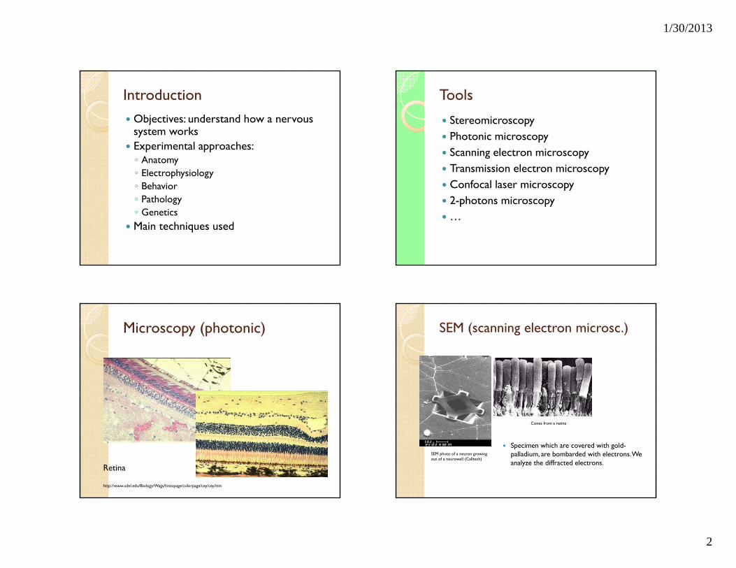
Microscopy (photonic)
Retina
http://www.udel.edu/Biology/Wags/histopage/colorpage/cey/cey.htm
SEM (scanning electron microsc.)
Specimen which are covered with gold-palladium, are bombarded with electrons. Weanalyze the diffracted electrons.
SEM photo of a neuron growing out of a neurowell (Calltech)
Cones from a retina
1/30/2013
3
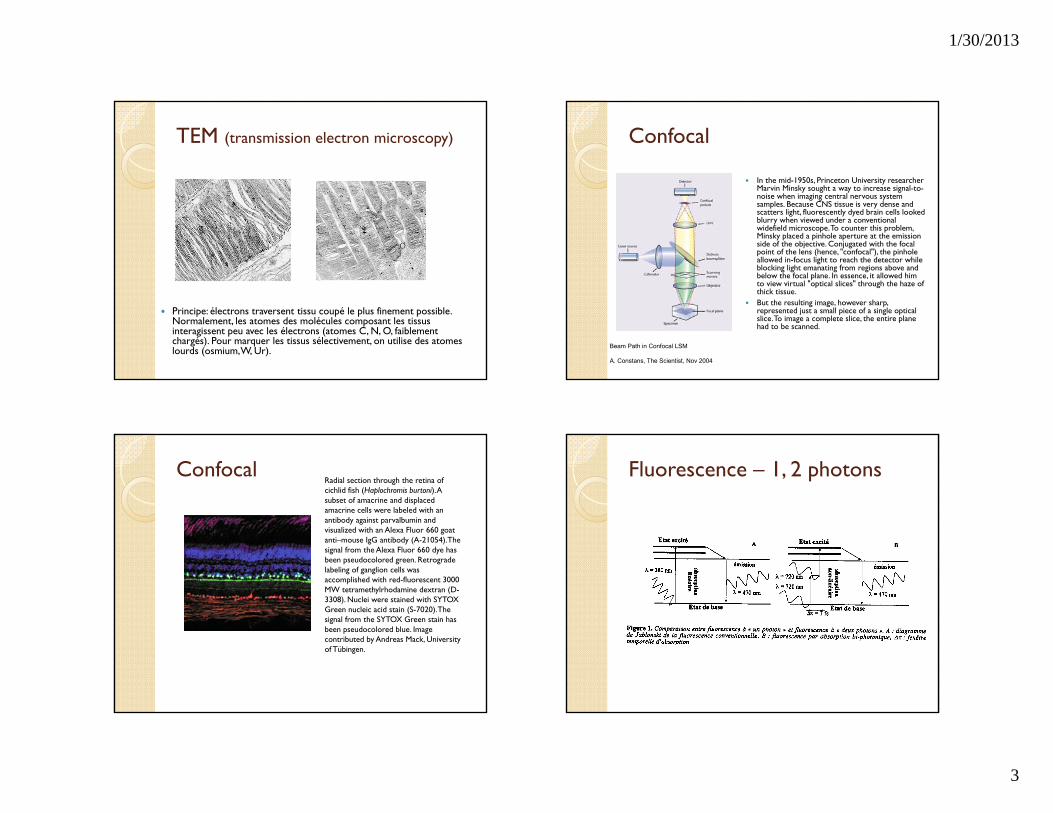
TEM (transmission electron microscopy)
Principe: électrons traversent tissu coupé le plus finement possible. Normalement, les atomes des molécules composant les tissus interagissent peu avec les électrons (atomes C, N, O, faiblement chargés). Pour marquer les tissus sélectivement, on utilise des atomes lourds (osmium, W, Ur).
Confocal
In the mid-1950s, Princeton University researcher Marvin Minsky sought a way to increase signal-to-noise when imaging central nervous system samples. Because CNS tissue is very dense and scatters light, fluorescently dyed brain cells looked blurry when viewed under a conventional widefield microscope. To counter this problem, Minsky placed a pinhole aperture at the emission side of the objective. Conjugated with the focal point of the lens (hence, "confocal"), the pinhole allowed in-focus light to reach the detector while blocking light emanating from regions above and below the focal plane. In essence, it allowed him to view virtual "optical slices" through the haze of thick tissue.
But the resulting image, however sharp, represented just a small piece of a single optical slice. To image a complete slice, the entire plane had to be scanned.
Beam Path in Confocal LSM
A. Constans, The Scientist, Nov 2004
ConfocalRadial section through the retina of cichlid fish (Haplochromis burtoni). A subset of amacrine and displaced amacrine cells were labeled with an antibody against parvalbumin and visualized with an Alexa Fluor 660 goat anti–mouse IgG antibody (A-21054). The signal from the Alexa Fluor 660 dye has been pseudocolored green. Retrograde labeling of ganglion cells was accomplished with red-fluorescent 3000 MW tetramethylrhodamine dextran (D-3308). Nuclei were stained with SYTOX Green nucleic acid stain (S-7020). The signal from the SYTOX Green stain has been pseudocolored blue. Image contributed by Andreas Mack, University of Tübingen.
Fluorescence – 1, 2 photons
1/30/2013
4
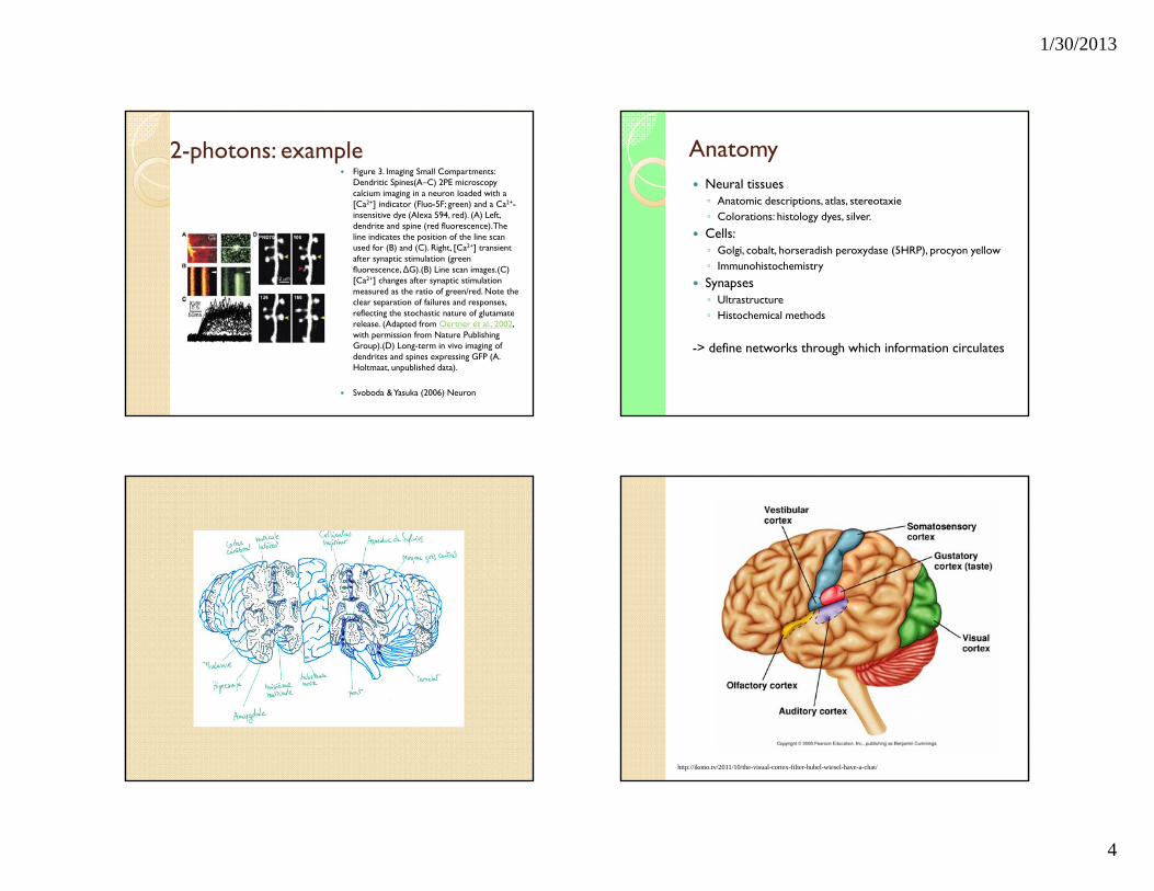
2-photons: example Figure 3. Imaging Small Compartments:
Dendritic Spines(A–C) 2PE microscopy calcium imaging in a neuron loaded with a [Ca2+] indicator (Fluo-5F; green) and a Ca2+-insensitive dye (Alexa 594, red). (A) Left, dendrite and spine (red fluorescence). The line indicates the position of the line scan used for (B) and (C). Right, [Ca2+] transient after synaptic stimulation (green fluorescence, ∆G).(B) Line scan images.(C) [Ca2+] changes after synaptic stimulation measured as the ratio of green/red. Note the clear separation of failures and responses, reflecting the stochastic nature of glutamate release. (Adapted from Oertner et al., 2002, with permission from Nature Publishing Group).(D) Long-term in vivo imaging of dendrites and spines expressing GFP (A. Holtmaat, unpublished data).
Svoboda & Yasuka (2006) Neuron
Anatomy Neural tissues◦ Anatomic descriptions, atlas, stereotaxie◦ Colorations: histology dyes, silver.
Cells:◦ Golgi, cobalt, horseradish peroxydase (5HRP), procyon yellow◦ Immunohistochemistry
Synapses◦ Ultrastructure◦ Histochemical methods
-> define networks through which information circulates
http://ikono.tv/2011/10/the-visual-cortex-filter-hubel-wiesel-have-a-chat/
1/30/2013
5
http://medocs.ucdavis.edu/chahph/403/syllabus/vesicle.htm http://www.cajal.csic.es/valverde/spines.htm
Des souris élevées dans l’obscurité ont moins d’épines dendritiques dans les cellules pyramidales du cortex visuel de souris. Gauche: souris normale (48 j) – Aire IV du cortex visuel. Droite: souris maintenue à l’obscurité. Coloration de Golgi (dessins à la chambre claire).
Coloration de Golgi
http://www.cajal.csic.es/valverde/spines.htm
Some basic principles of input-output operation of the cerebral cortex appear reflected in this drawing. Specific afferent fibers (aff) from the thalamus (thalamo-cortical fibers) enter the cortex from the white matter on a slanting course. As they ascend, they ramify profusely in layers IV and III contacting one intrinsic cell (B) located in layer III. The axon of this cell (drawn in red) develops into numerous branches (local axonal field) some of which will connect with the apical dendrite of the pyramidal cell (A) which in turn, sends the axon (ax) outside the cerebral cortex running down in the white matter.
Camera lucida drawing from a Golgi preparation of the visual cortex of a mouse 10-days-old.
Chandelier cells represented a novel type of neuron in the mammalian cerebral cortex. At first, it was thought to be specific for certain subjects, but it was soon demonstrated to be present in the cerebral cortex of every mammalian species from insectivores to man. The example shown above is a camera lucida drawing of one chandelier cell in the visual cortex corresponding to a kitten 1 month old. The axon of this cell (ax) develops into numerous collaterals ending in the form of vertically oriented, long bouton aggregates or "cartridges" (e.g. ct) formed of small axonal dilatations with varying degrees of complexity, from a single row of axonal dilatations to extremely complicated hollow cylinder-shaped formations.
The Golgi picture of this cell type is most suggestive of target specificity and, so, it was soon demonstrated, with aid of the electron microscope, that the "cartridges" make specific (inhibitory) synaptic contacts exclusively with the initial portions of axons of pyramidal cells, therefore representing a most powerful inhibitory mechanism to control the output of pyramidal cells.
http://www.cajal.csic.es/valverde/spines.htm
1/30/2013
6
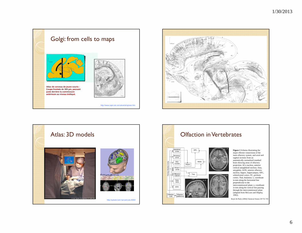
Golgi: from cells to maps
http://www.cajal.csic.es/valverde/spines.htm
Atlas de cerveau de jeune souris : Coupe frontale de 300 µm, passant juste derrière la commissure antérieure au niveau inidiqué.
http://splweb.bwh.harvard.edu:8000/
Atlas: 3D models Olfaction in Vertebrates
Figure 1 Schema illustrating the major efferent connections of the main olfactory system, and axial and sagittal sections from an anatomically normalized standard brain showing areas of olfactory projection. ACo nucleus, anterior cortical amygdaloid nucleus; Amy, amygdala; AON, anterior olfactory nucleus; hippoc, hippocampus, OFC, orbitofrontal cortex; PC, piriform cortex; Thal, thalamus; x, coordinate in mm along the horizontal line perpendicular to the intercommissural plane; z, coordinate in mm along the vertical line passing through the intercommissural plane (adapted from McLean and Shipley, 1992).
Royet & Plaily (2004) Chemical Senses 29:731-745
1/30/2013
7
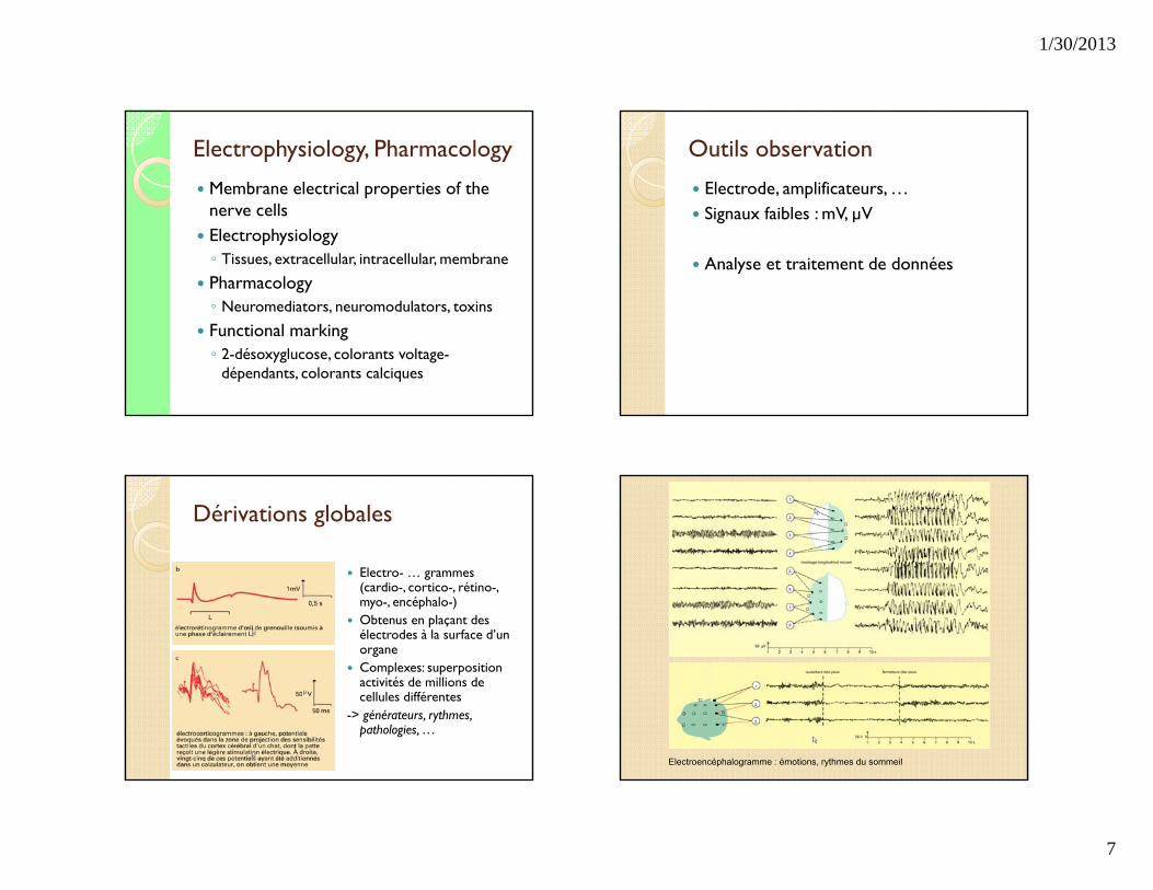
Electrophysiology, Pharmacology
Membrane electrical properties of the nerve cells
Electrophysiology◦ Tissues, extracellular, intracellular, membrane
Pharmacology◦ Neuromediators, neuromodulators, toxins
Functional marking◦ 2-désoxyglucose, colorants voltage-
dépendants, colorants calciques
Outils observation
Electrode, amplificateurs, … Signaux faibles : mV, µV
Analyse et traitement de données
Dérivations globales
Electro- … grammes(cardio-, cortico-, rétino-, myo-, encéphalo-)
Obtenus en plaçant des électrodes à la surface d’un organe
Complexes: superposition activités de millions de cellules différentes
-> générateurs, rythmes, pathologies, …
Electroencéphalogramme : émotions, rythmes du sommeil
1/30/2013
8
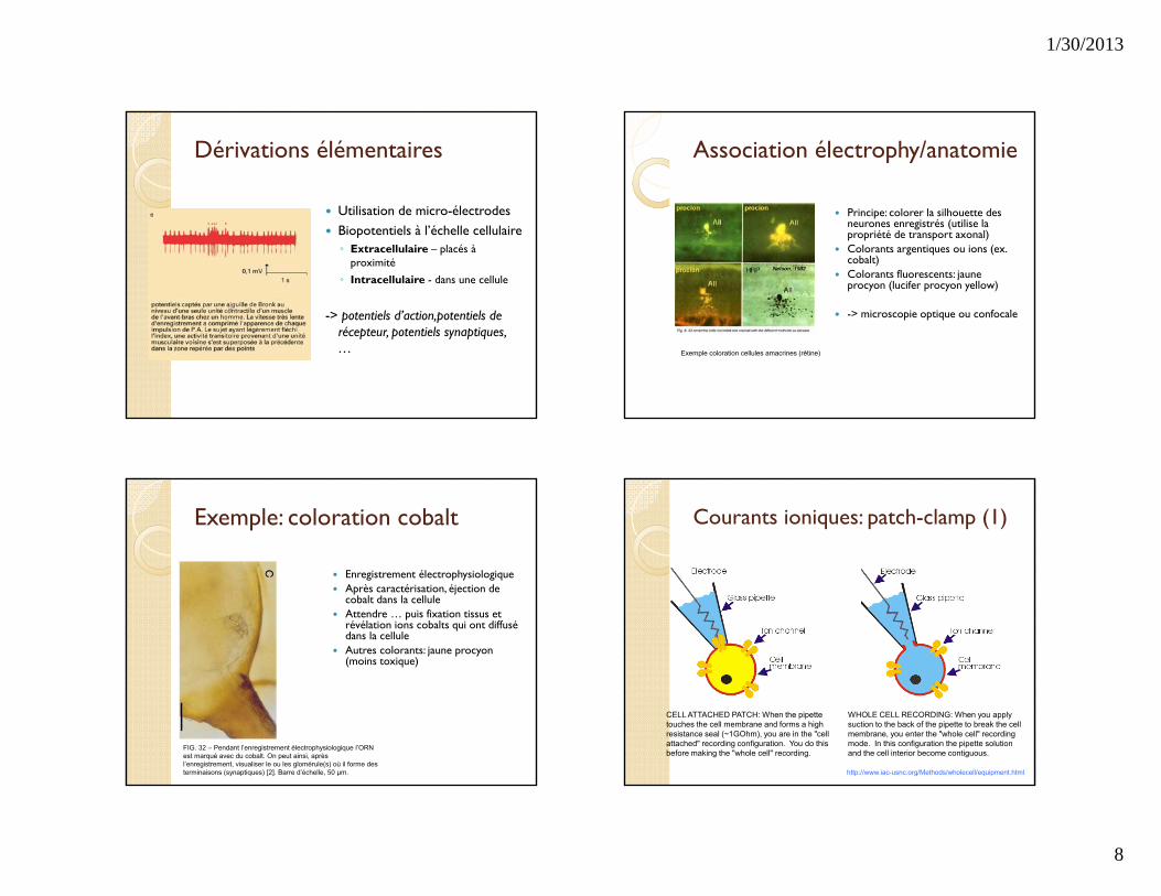
Dérivations élémentaires
Utilisation de micro-électrodes Biopotentiels à l’échelle cellulaire ◦ Extracellulaire – placés à
proximité ◦ Intracellulaire - dans une cellule
-> potentiels d’action,potentiels de récepteur, potentiels synaptiques, …
Association électrophy/anatomie
Principe: colorer la silhouette des neurones enregistrés (utilise la propriété de transport axonal)
Colorants argentiques ou ions (ex. cobalt)
Colorants fluorescents: jaune procyon (lucifer procyon yellow)
-> microscopie optique ou confocale
Exemple coloration cellules amacrines (rétine)
Exemple: coloration cobalt
Enregistrement électrophysiologique Après caractérisation, éjection de
cobalt dans la cellule Attendre … puis fixation tissus et
révélation ions cobalts qui ont diffusé dans la cellule
Autres colorants: jaune procyon (moins toxique)
FIG. 32 – Pendant l’enregistrement électrophysiologique l’ORN est marqué avec du cobalt. On peut ainsi, après l’enregistrement, visualiser le ou les glomérule(s) où il forme des terminaisons (synaptiques) [2]. Barre d’échelle, 50 µm.
Courants ioniques: patch-clamp (1)
CELL ATTACHED PATCH: When the pipette touches the cell membrane and forms a high resistance seal (~1GOhm), you are in the "cell attached" recording configuration. You do this before making the "whole cell" recording.
WHOLE CELL RECORDING: When you apply suction to the back of the pipette to break the cell membrane, you enter the "whole cell" recording mode. In this configuration the pipette solution and the cell interior become contiguous.
http://www.iac-usnc.org/Methods/wholecell/equipment.html
1/30/2013
9
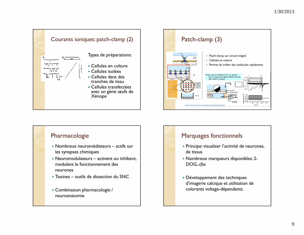
Courants ioniques: patch-clamp (2)
Types de préparations:
Cellules en culture Cellules isolées Cellules dans des
tranches de tissu Cellules transfectées
avec un gène: œufs de Xénope
Patch-clamp (3)
Patch-clamp sur circuit intégré Cellules en culture
Permet de cribler des molécules rapidement
http://www.eng.yale.edu/posters150/pdf/reed2.pdf
Pharmacologie
Nombreux neuromédiateurs – actifs sur les synapses chimiques
Neuromodulateurs – activent ou inhibent, modulent le fonctionnement des neurones
Toxines – outils de dissection du SNC
Combinaison pharmacologie / neuroanatomie
Marquages fonctionnels
Principe: visualiser l’activité de neurones, de tissus
Nombreux marqueurs disponibles: 2-DOG, cfos
Développement des techniques d’imagerie calcique et utilisation de colorants voltage-dépendants.
1/30/2013
10
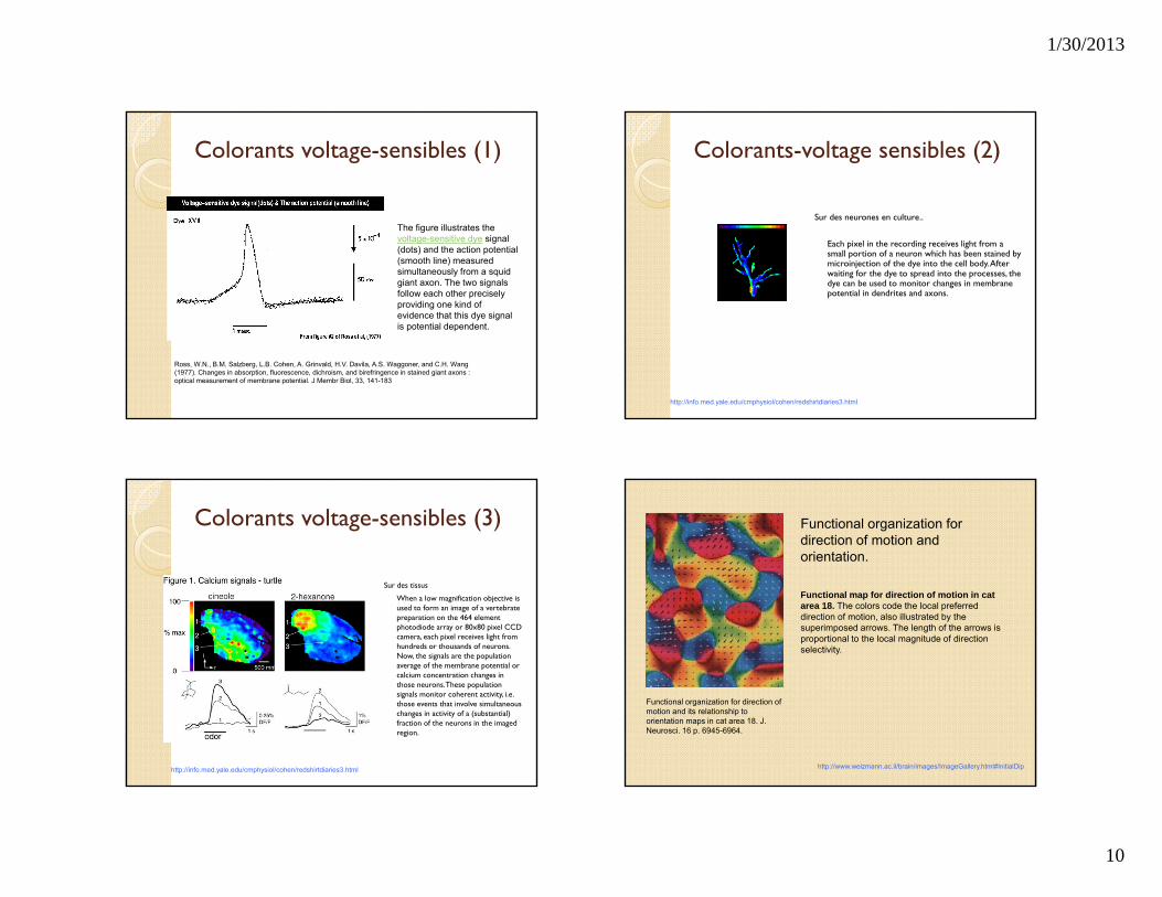
Colorants voltage-sensibles (1)
The figure illustrates the voltage-sensitive dye signal (dots) and the action potential (smooth line) measured simultaneously from a squid giant axon. The two signals follow each other precisely providing one kind of evidence that this dye signal is potential dependent.
Ross, W.N., B.M. Salzberg, L.B. Cohen, A. Grinvald, H.V. Davila, A.S. Waggoner, and C.H. Wang (1977). Changes in absorption, fluorescence, dichroism, and birefringence in stained giant axons : optical measurement of membrane potential. J Membr Biol, 33, 141-183
Colorants-voltage sensibles (2)
Sur des neurones en culture..
Each pixel in the recording receives light from a small portion of a neuron which has been stained by microinjection of the dye into the cell body. After waiting for the dye to spread into the processes, the dye can be used to monitor changes in membrane potential in dendrites and axons.
http://info.med.yale.edu/cmphysiol/cohen/redshirtdiaries3.html
Colorants voltage-sensibles (3)
Sur des tissus
When a low magnification objective is used to form an image of a vertebrate preparation on the 464 element photodiode array or 80x80 pixel CCD camera, each pixel receives light from hundreds or thousands of neurons. Now, the signals are the population average of the membrane potential or calcium concentration changes in those neurons. These population signals monitor coherent activity, i.e. those events that involve simultaneous changes in activity of a (substantial) fraction of the neurons in the imaged region.
http://info.med.yale.edu/cmphysiol/cohen/redshirtdiaries3.html
Functional organization for direction of motion and orientation.
Functional map for direction of motion in cat area 18. The colors code the local preferred direction of motion, also illustrated by the superimposed arrows. The length of the arrows is proportional to the local magnitude of direction selectivity.
Functional organization for direction of motion and its relationship to orientation maps in cat area 18. J. Neurosci. 16 p. 6945-6964.
http://www.weizmann.ac.il/brain/images/ImageGallery.html#InitialDip
1/30/2013
11
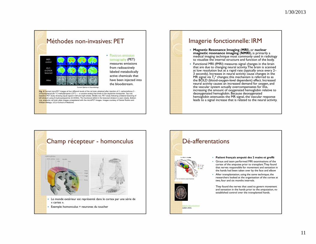
Méthodes non-invasives: PET
Positron emission tomography (PET) measures emissions from radioactively labeled metabolically active chemicals that have been injected into the bloodstream.
Fig. 4 Coronal microPET images at four different levels of the rat brain obtained after injection of 2 -carbomethoxy-3 -(4-fluorophenyl)-[N-11C-methyl]tropane (CFT) — a cocaine analog that binds to the dopamine transporter. Top row, baseline PET study showing equal signal in left and right striata. Middle row, PET study following unilateral lesioning of left striatum using 6-hydroxydopamine (6-OHDA). The functional deficit in the lesioned striatum is seen clearly. Bottom row, anatomic rat brain atlas images coregistered with the microPET images. Images courtesy of Daniel Rubins and William Melega, UCLA School of Medicine.
Imagerie fonctionnelle: IRM Magnetic Resonance Imaging (MRI), or nuclear
magnetic resonance imaging (NMRI), is primarily a medical imaging technique most commonly used in radiology to visualize the internal structure and function of the body.
Functional MRI (fMRI) measures signal changes in the brain that are due to changing neural activity. The brain is scanned at low resolution but at a rapid rate (typically once every 2–3 seconds). Increases in neural activity cause changes in the MR signal via T2* changes; this mechanism is referred to as the BOLD (blood-oxygen-level dependent) effect. Increased neural activity causes an increased demand for oxygen, and the vascular system actually overcompensates for this, increasing the amount of oxygenated hemoglobin relative to deoxygenated hemoglobin. Because deoxygenated hemoglobin attenuates the MR signal, the vascular response leads to a signal increase that is related to the neural activity.
Champ récepteur - homonculus
Le monde extérieur est représenté dans le cortex par une série de « cartes ».
Exemple: homonculus = neurones du toucher
Dé-afferentations
Patient français amputé des 2 mains et greffé Giraux and team performed MRI examinations of the
cortex of the amputee prior to transplant. They found that nerves responsible for movement and sensation in the hands had been taken over by the face and elbow
After transplantation, using the same technique, the researchers looked at the organisation of the cortex at two, four and six months intervals.
They found the nerves that used to govern movement and sensation in the hands prior to the amputation, re-established control over the transplanted hands.
Nature NeuroscienceJuillet 2001.
1/30/2013
12
Et la génétique?
Développements très rapides des méthodes génétiques: souris, Drosophile, Caenorhabditis (nématode)
Expression ectopique de gènes Inhibition de l’expression de gènes (souris
knockout) Utilisation de gènes rapporteurs (ex.: GFP)
Marquage: fluorophores et anticorps
Fruit fly 3rd instar wing imaginal disk labeled for three genes involved with patterning the wing.
The three genes imaged and their respective fluorochrome labels are◦ (a) vestigial (fluorescein - 496 nanometers); ◦ (b) apterous (lissamine rhodamine - 572
nanometers); and ◦ (c) CiD (cyanine 5 - 649 nanometers).
The merged composite of the three spatial expression domains of the wing patterning genes is shown in the lower right (image (d)).
Confocal microscopy: Optical sections collected simultaneously at three different excitation wavelengths (488, 568, and 647 nanometers) using a single krypton/argon laser.
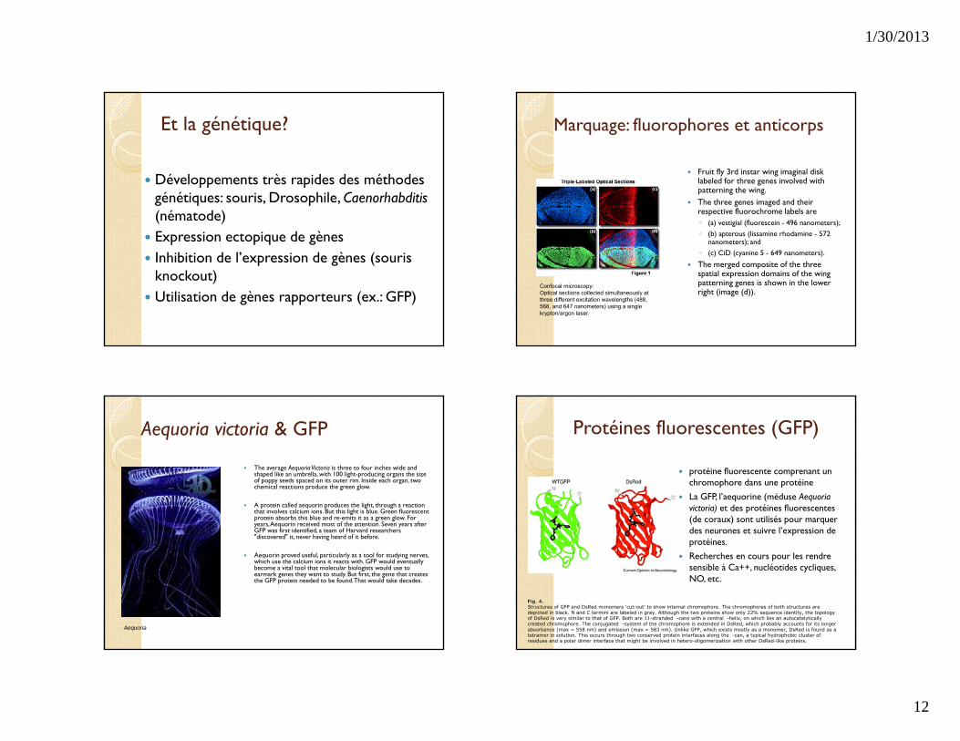
Aequoria victoria & GFP
The average Aequoria Victoria is three to four inches wide and shaped like an umbrella, with 100 light-producing organs the size of poppy seeds spaced on its outer rim. Inside each organ, two chemical reactions produce the green glow.
A protein called aequorin produces the light, through a reaction that involves calcium ions. But this light is blue. Green fluorescent protein absorbs this blue and re-emits it as a green glow. For years, Aequorin received most of the attention. Seven years after GFP was first identified, a team of Harvard researchers "discovered" it, never having heard of it before.
Aequorin proved useful, particularly as a tool for studying nerves, which use the calcium ions it reacts with. GFP would eventually become a vital tool that molecular biologists would use to earmark genes they want to study. But first, the gene that creates the GFP protein needed to be found. That would take decades.
Aequoria
Protéines fluorescentes (GFP)
protéine fluorescente comprenant un chromophore dans une protéine
La GFP, l’aequorine (méduse Aequoria victoria) et des protéines fluorescentes (de coraux) sont utilisés pour marquer des neurones et suivre l’expression de protéines.
Recherches en cours pour les rendre sensible à Ca++, nucléotides cycliques, NO, etc.
Fig. 4.Structures of GFP and DsRed monomers 'cut-out' to show internal chromophore. The chromophores of both structures are depicted in black. N and C termini are labeled in gray. Although the two proteins show only 22% sequence identity, the topology of DsRed is very similar to that of GFP. Both are 11-stranded -cans with a central -helix, on which lies an autocatalytically created chromophore. The conjugated -system of the chromophore is extended in DsRed, which probably accounts for its longer absorbance (max = 558 nm) and emission (max = 583 nm). Unlike GFP, which exists mostly as a monomer, DsRed is found as a tetramer in solution. This occurs through two conserved protein interfaces along the -can, a typical hydrophobic cluster of residues and a polar dimer interface that might be involved in hetero-oligomerization with other DsRed-like proteins.
1/30/2013
13
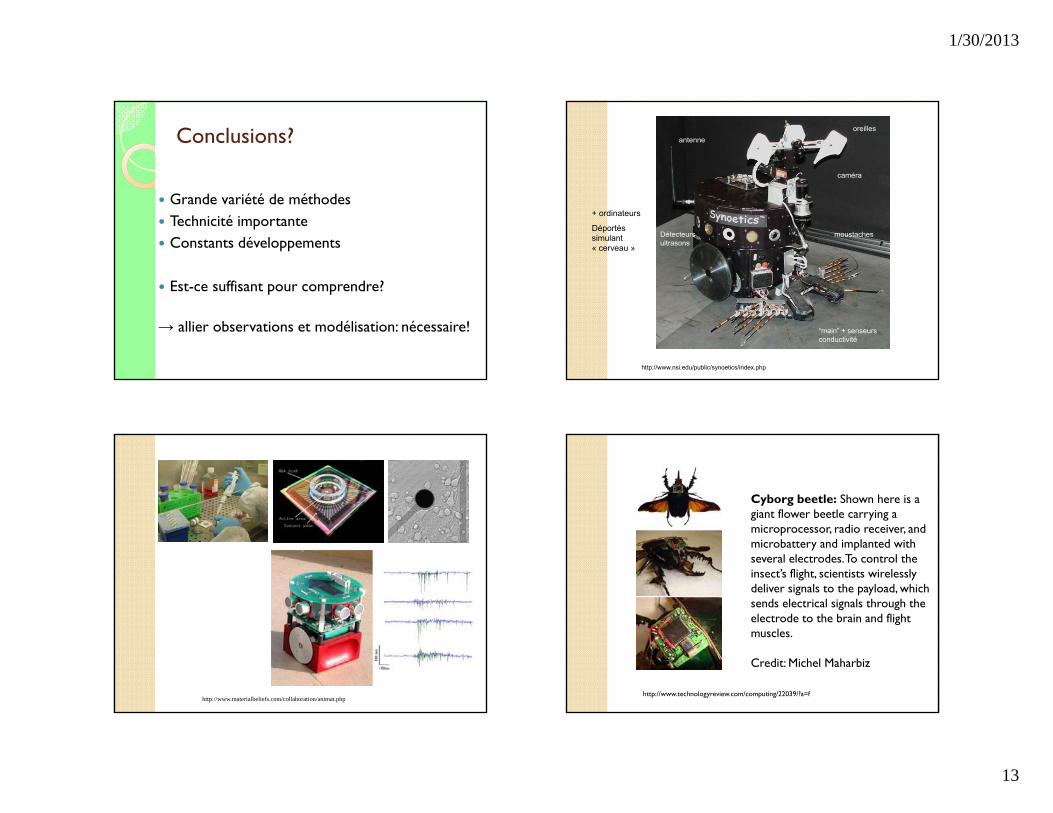
Conclusions?
Grande variété de méthodes Technicité importante Constants développements
Est-ce suffisant pour comprendre?
→ allier observations et modélisation: nécessaire!
http://www.nsi.edu/public/synoetics/index.php
antenneoreilles
Détecteursultrasons
moustaches
“main” + senseurs conductivité
caméra
+ ordinateurs
Déportés simulant « cerveau »
http://www.materialbeliefs.com/collaboration/animat.php
Cyborg beetle: Shown here is a giant flower beetle carrying a microprocessor, radio receiver, and microbattery and implanted with several electrodes. To control the insect’s flight, scientists wirelessly deliver signals to the payload, which sends electrical signals through the electrode to the brain and flight muscles.
Credit: Michel Maharbiz
http://www.technologyreview.com/computing/22039/?a=f
1/30/2013
14
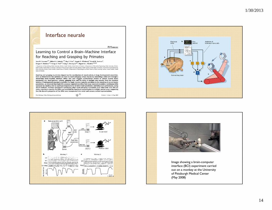
Interface neurale
Image showing a brain-computer interface (BCI) experiment carried out on a monkey at the University of Pittsburgh Medical Center (May 2008)
1/30/2013
15
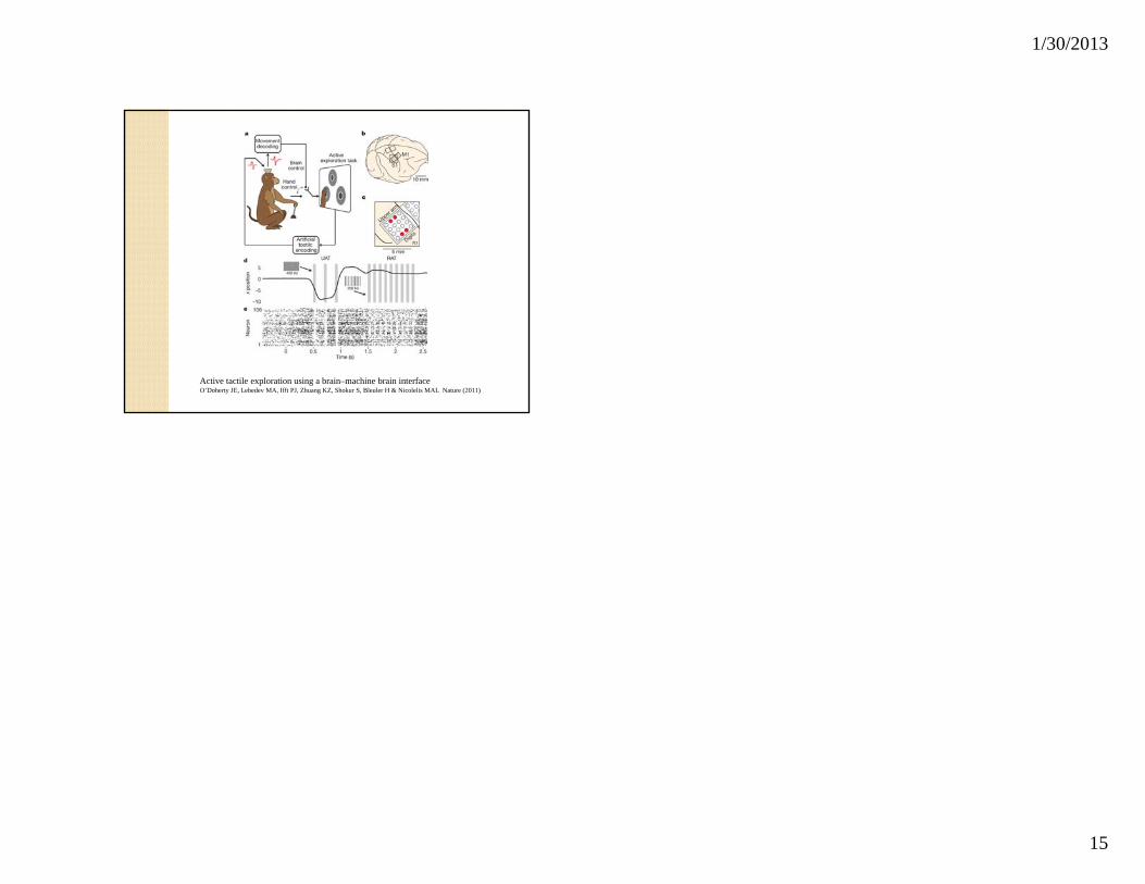
Active tactile exploration using a brain–machine brain interfaceO’Doherty JE, Lebedev MA, Ifft PJ, Zhuang KZ, Shokur S, Bleuler H & Nicolelis MAL Nature (2011)