ferritin in - pnas · cording to the chou-fasman and garnier-arguthorp- ... ferritin (4), actually...
TRANSCRIPT

Proc. Natl. Acad. Sci. USAVol. 88, pp. 8222-8226, September 1991Biochemistry
Ferritin gene transcription is regulated by iron in soybeancell culturesANNE-MARIE LESCURE*, DOMINIQUE PROUDHON*, HtLtNE PESEY*, MARIA RAGLANDt,ELIZABETH C. THEILtf, AND JEAN-FRANSOIS BRIAT*t*Laboratoire de Biologie Mol6culaire V6g6tale, Centre National de la Recherche Scientifique (Unite de Recherche Associ6e 1178) and Universitd JosephFourier, B.P. 53X, F-38041 Grenoble Cedex, France; and tDepartment of Biochemistry, North Carolina State University, Raleigh, NC 27695-7622
Communicated by Charles S. Levings III, May 31, 1991
ABSTRACT Iron-regulated ferritin synthesis in animals isdominated by translational control of stored mRNA; iron-induced transcription of ferritin genes, when it occurs, changesthe subunit composition of ferritin mRNA and protein and iscoupled to translational control. Ferritins in plants and animalshave evolved from a common progenitor, based on the simi-larity of protein sequence; however, sequence divergence oc-curs in the C termini; structure prediction sgg that plantferriti has the E-helix, which, in horse ferritin, forms a largechannel at the tetrameric interface. In contemporary plants, atransit peptide is encoded by ferritin mRNA to target theprotein to plastids. Iron-regulated synthesis offerritin in plantsand animals appears to be very different since the 50- to 60-foldincreases of ferritin protein, previously observed to be inducedby iron in cultured soybean cells, is accompanied by anequivalent accumulation of hybridizable ferritin mRNA and byincreased transcription of ferritin genes. Ferritin mRNA fromiron-induced cells and the constitutive ferritin mRNA fromsoybean hypocotyls are identical. The iron-induced protein istranslocated normally to plastids. Differences in animal ferritinstructure coincide with the various iron storage functions(reserve for iron proteins and detoxification). In contrast, theconstancy of structure of soybean ferritin, iron-induced andconstitutive, coupled with the potential for vacuolar storage ofexcess iron in plants suggest that rapid synthesis of ferritinfrom a stored ferritin mRNA may not be needed in plants fordetoxification of iron.
Ferritin synthesis and accumulation is regulated by iron, themetal stored inside the protein coat as hydrated ferric oxide(1-3). Ferritin is widely distributed among eukaryotes andprokaryotes; the similar structure of ferritins in plants andanimals suggests a common evolutionary origin. In contem-porary plant ferritins, a transit peptide targets the protein toplastids (4). Ferritin stores iron for protein synthesis and fordetoxification of iron excess; vacuoles may also detoxify ironin plants (5, 6).
Regulation of ferritin gene expression by iron was firstobserved decades ago in animals (7, 8) and plants (9); themechanism in animals is mainly posttranscriptional, involv-ing ferritin mRNA storage and translational competition(10-12). Detection of ferritin mRNA-specific trans-actingfactors (13), isolation and cloning of regulator protein(s)(14-16), and identification of a conserved regulatory se-quence (17-21) with a distinctive structure (22) have madeferritin mRNA a model for translational control (23, 24).Iron-induced transcription and/or accumulation of ferritinmRNA also occur in animals. The effects are subunit-specificand are coupled to increases in translation (25-28) and,apparently, to detoxification of the excess iron.
In plants, regulation of the ferritin gene by iron has notbeen studied as thoroughly. Earlier measurements of trans-latable ferritin mRNAs suggested that in contrast to animals,iron regulation may affect mRNA transcription rather thantranslation in bean leaves (29) and cultured soybean cells(30). Measurement of ferritin mRNA by translation in cell-free extracts could have been complicated by ferritin-mRNA-specific translational effects (13). To determine if the regu-lation of ferritin expression by iron is different in plants andanimals, we have investigated the synthesis and sequence offerritin mRNA§ in iron-induced cultured soybean cells andthe fate of the ferritin produced. The results show that ironinduced a 45-fold increase in the transcription rate ofsoybeanferritin mRNA and a corresponding increase in the accumu-lation of ferritin protein. The sequence of induced ferritinmRNA was identical to its hypocotyl counterpart and thetransport of the newly synthesized ferritin to plastids ap-peared to be normal. Thus the domination of translationalevents in iron-regulated ferritin gene expression (24) appearsto be specific to animals. In plants, by contrast, iron has adramatic effect directly on transcription of ferritin mRNA.
MATERIALS AND METHODSMaterials. Isolation ofa partial ferritin cDNA (SoF35) from
a soybean hypocotyl cDNA library has been describedelsewhere (4). The glyceraldehyde-3-phosphate dehydroge-nase C probe (GapC) from pea (31) was a gift from R. Cerff(Technical University, Braunschweig, F.R.G.). Soybean cellcultures (Glycine max cv. Mandarin line Sbe4) were grown asdescribed (32). Ferritin synthesis was induced by 500 IAMiron(III) citrate as described (30).cDNA Cloning, Sequencing, and Preiction of Protein Sec-
ondary Structure. A cDNA library, constructed usingpoly(A)+ RNA from iron-induced soybean cell suspensions(30), EcoRI-Not I adaptors (Pharmacia), and EcoRI arms ofANM1149 (33, 34), and the cDNA library from hypocotyls (4)were screened with 32P-labeled SoF35, a fragment of hypo-cotyl ferritin cDNA (4). After phage purification (35) andsubcloning into pEMBL18 or pUC18, the complete sequencewas obtained from both strands by the dideoxynucleotidemethod (36). Prediction of protein secondary structure (ac-cording to the Chou-Fasman and Garnier-Arguthorp-Robson methods) used the programs of the University ofWisconsin Genetics Computer Group.RNA Isolation and Characterization. Total RNAs were
extracted from frozen cells by using guanidine hydrochloride(37), analyzed on Northern blots with the cDNA clone SoF35as a probe (4) or on dot blots on nitrocellulose [hybridized for48 h at 420C in 50o (vol/vol) formamide/5 x SSPE (1x SSPE
Abbreviation: IRE, iron regulatory element.tTo whom reprint requests should be addressed.§The sequences reported in this paper have been deposited in theGenBank data base (accession nos. M64337 from cultured soybeancells and M58336 from soybean hypocotyl).
8222
The publication costs of this article were defrayed in part by page chargepayment. This article must therefore be hereby marked "advertisement"in accordance with 18 U.S.C. §1734 solely to indicate this fact.
Biochemistry: Lescure et al.
= 0.18 M NaCi/10 mM sodium phosphate, pH 7.4/1 mMEDTA) and washed with 50o formamide/5x SSPE at 420C],and quantitated using a Beckman scintillation counter;amounts of RNA were 10 and 5 Ag, respectively.In Vitro Transcription in Isolated Nuclei. Crude nuclei (10 g
of cells) were prepared as described (38) and could be storedat -70'C for 2 months in a Triton-free buffer containing 30o(vol/vol) glycerol (39) at 4 mg of proteins per ml. Protein wasmeasured according to Bradford (40). Transcription reactions(250 ,g of protein) were incubated 10 min at 300C, andpurified transcripts (41) were hybridized (1 x 10⁷ cpm/ml) todenatured plasmids (5 ,ug) on nitrocellulose. Quantitation ofautoradiograms used a Uniscan II spectrophotometer at 595nm (Labsystems, Les Ullis, France).
Immunolocalization of Ferritin by Electron Microscopy.Preparation of soybean cell samples for electron microscopyusing 200-mesh nickel grids (42) (Touzard et Matignon, Paris)and protein A-gold to localize ferritin has been described (43).Grids were analyzed at a magnification of x20,000.
RESULTS
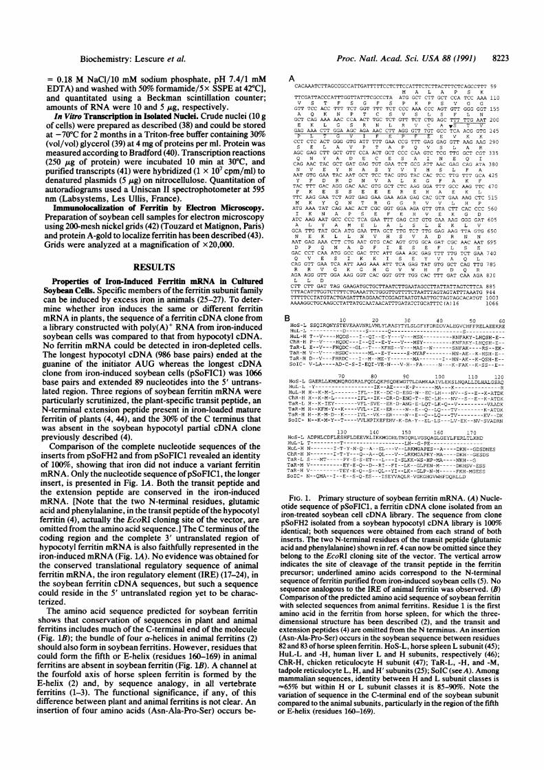
Properties of Iron-Induced Ferritin mRNA in CulturedSoybean Cells. Specific members of the ferritin subunit familycan be induced by excess iron in animals (25-27). To deter-mine whether iron induces the same or different ferritinmRNA in plants, the sequence of a ferritin cDNA clone froma library constructed with poly(A)+ RNA from iron-inducedsoybean cells was compared to that from hypocotyl cDNA.No ferritin mRNA could be detected in iron-depleted cells.The longest hypocotyl cDNA (986 base pairs) ended at theguanine of the initiator AUG whereas the longest cDNAclone from iron-induced soybean cells (pSoFIC1) was 1066base pairs and extended 89 nucleotides into the 5' untrans-lated region. Three regions of soybean ferritin mRNA wereparticularly scrutinized, the plant-specific transit peptide, anN-terminal extension peptide present in iron-loaded matureferritin of plants (4, 44), and the 30% of the C terminus thatwas absent in the soybean hypocotyl partial cDNA clonepreviously described (4).Comparison of the complete nucleotide sequences of the
inserts from pSoFH2 and from pSoFIC1 revealed an identityof 100%, showing that iron did not induce a variant ferritinmRNA. Only the nucleotide sequence ofpSoFIC1, the longerinsert, is presented in Fig. lA. Both the transit peptide andthe extension peptide are conserved in the iron-inducedmRNA. [Note that the two N-terminal residues, glutamicacid and phenylalanine, in the transit peptide ofthe hypocotylferritin (4), actually the EcoRI cloning site of the vector, areomitted from the amino acid sequence.] The C terminus ofthecoding region and the complete 3' untranslated region ofhypocotyl ferritin mRNA is also faithfully represented in theiron-induced mRNA (Fig. LA). No evidence was obtained forthe conserved translational regulatory sequence of animalferritin mRNA, the iron regulatory element (IRE) (17-24), inthe soybean ferritin cDNA sequences, but such a sequencecould reside in the 5' untranslated region yet to be charac-terized.The amino acid sequence predicted for soybean ferritin
shows that conservation of sequences in plant and animalferritins includes much of the C-terminal end of the molecule(Fig. 1B); the bundle of four a-helices in animal ferritins (2)should also form in soybean ferritins. However, residues thatcould form the fifth or E-helix (residues 160-169) in animalferritins are absent in soybean ferritin (Fig. 1B). A channel atthe fourfold axis of horse spleen ferritin is formed by theE-helix (2) and, by sequence analogy, in all vertebrateferritins (1-3). The functional significance, if any, of thisdifference between plant and animal ferritins is not clear. Aninsertion of four amino acids (Asn-Ala-Pro-Ser) occurs be-
Proc. Natl. Acad. Sci. USA 88 (1991) 8223
ACACAAATCTTAGCCGCCATTGATTTTTCCTCTTCCATTTCTCTTACTTTCTCAGCCTTT 59
M A L A P S KTTCGATTACCCATTTGGTTATTTCGCCCTA ATG GCT CTT GCT CCA TCC AAA 110V S T F S G F S P K P S V G G
GTT TCC ACC TTT TCT GGT TTT TCT CCC AAA CCC AGT GTT GGG GGT 155A Q K N P T C S V S L S F L N
GCT CAG AAA AAC CCA ACT TGC TCT GTT TCT CTG AGC TTT TTG AAT 200E K L G S R N L RK V C A vS T V
GAG AAA CTT GGA AGC AGA AAC CTT AGG GTT TGT GCC TCA ACG GTG 245P L T G V I F E P F E E V K K
CCT CTC ACT GGG GTG ATT TTT GAA CCG TTT GAG GAG GTT AAG AAG 290S E L A V P T A P Q V S L A R
AGC GAG CTT GCT GTT CCA ACT GCT CCC CAA GTC TCG TTG GCT CGT 335Q N Y A D E C E S A I N E Q I
CAG AAC TAC GCT GAT GAG TGT GAA TCT GCG ATT AAC GAG CAG ATA 380N V E Y N A S Y V Y H S L F A
AAT GTG GAA TAC AAT GCT TCC TAC GTG TAC CAC TCC TTG TTT GCA 425Y F D R D N V A L K G F A K F
TAC TTT GAC AGG GAC AAC GTG GCT CTC AAG GGA TTT GCC AAG TTC 470F K E S S E E E R ES A E K L
TTC AAG GAA TCT AGT GAG GAA GAA AGA GAG CAC GCT GAA AAG CTC 515M K Y Q N T R G G R V V L H P
ATG AAA TAT CAG AAC ACT CGC GGT GGA AGA GTT GTA CTT CAC CCC 560I K N A P S E F E H V E K G D
ATC AAG AAT GCC CCC TCA GAA TTT GAG CAT GTG GAA AAG GGG GAT 605A L Y A M E L A L S L E K L V
GCA TTG TAT GCA ATG GAA TTA GCT TTG TCT TTG GAG AAG TTA GTG 650N E K L L N V H S V A D R N N
AAT GAG AAA CTT CTG AAT GTG CAC AGT GTG GCA GAT CGC AAC AAT 695D P Q M A D F I E S E F L S E
GAC CCT CAA ATG GCC GAC TTC ATT GAA AGC GAG TTT TTG TCT GAA 740Q V E S I K K I S E Y V A Q L
CAG GTT GAA TCA ATT AAG AAA ATT TCA GAG TAT GTG GCT CAG TTG 785R R V G K G H G V W H F D Q R
AGA AGG GTT GGA AAG GGT CAC GGT GTT TGG CAC TTT GAT CAA AGA 830L L D
CTT CTT GAT TAG GAAGATGCTGCTTAATCTTGAATAGCCTTATTATTAGTCTTCA 885TTTACATTTGGTCTTTTCTGAAATTCTGGGTTGTTTTCTAATTTAGTAGTATTTAAATG 944TTTTTCCTATGTACTGAGATTTAGGAACTCGGAGTAATGTAATTGCTAGTAGCACATGT 1003AAAAGGCTGCAAGCCTATTATGCAA.TAACATTTGATATCTGCATTTC( A) 16 1066
B 10 20 30 40 50 60HOS-L SSQIRQNYSTEVEAAVNRLVNLYLRASYTYLSLGFYFDRDDVALEGVCHFFRELAEEKREHuL-L ----------D------S------ Q---------------------- SHuL-H T--V----HQDS---I--QI--E-Y-X--V---MSX-------KNFAKY-LHQSH-E--ChR-H P--V----HQDC---I--QI--E-Y---V---MSY---------KNFAVY-LHQSH-E--TaR-L E--V---FHQDC--GL--T---KFHS--V---MAS--N------SNFAK----RS--EK-TaR-M V--V----HSDC------ML--E-Y-----S-MYAF--------HN-AE--K-HSH-E--TaR-H D--V---FHRDC --- I--M--ME-Y------ MA------- I--HN-AK--K-QSH-E--SoIC- V-LA----AD-C-S-I-EQI-VE-N---V-H--FA --- -N---K-FAK--K-SS--E--
70 80 90 100 110 120HoS-L GAERLLKMQNQRGGRALFQDLQKPSQDEWGTTLDAMKAAIVLEKSLNQALLDLHALGSAQHuL-L -Y----------------- IK--AE----K-P -----MA---K--------------RHuL-H H--K-M-L -------IFL--IK--DC-D-ESG-N--EC-LH---NV--S--E--K-ATDKChR-H H--K-M-L-------IFL--IK--DR-D-ENG-T--EC-LH---NV--S--E--K-ATDKTaR-L H--K-IEY-------VFL-SVE--ER-D-ANG-E-LQT-LK-Q--V---------VAADKTaR-M H--KFM-Y--K----VVL--IK--ER----N--E--Q--LQ---TV--------K-ATDKTaR-H H--K-M-D--K----IVL--VK--ER----N--E--Q--LQ---TV--------KV--DKSoIC- H--K-M-Y--T ----VVLHPIKEFEHV-K-DA-Y--EL-LS---LV-EK--NV-SVADRN
130 140 150 160 170HoS-L ADPHLCDFLESHFLDEEVJKLIKKMGDHLTNIQRLVGSQAGLGEYLFERLTLKHDHuL-L T---------T-------------------LH--G-PE----------------HuL-H N-------I-T-Y-N-Q--A--EL ---V--LRKMGAPES--A----DKH--GDSDNESChR-H N-------I-T-Y---Q--A--QL---V--LRKMGAPKY-MA----DKH--GESDSTaR-L S--- MT----- PY-S-S-ET-- -L-I--SLKK-WS-HP-MA----NKH--GTaR-M V----------EY-E-Q--D--RI--FI--LK--GLPEN-M-----DKHSV-ESSTaR-H V---------TEY-E-Q--S--QL--YI--LK--GLP-N-M-----FKH-MGESSSoIC- N--QMA--I--E--S-Q-ES--- ISEYVAQLR-VGKGHGVWHFDQRLLD
FIG. 1. Primary structure of soybean ferritin mRNA. (A) Nucle-otide sequence of pSoFIC1, a ferritin cDNA clone isolated from aniron-treated soybean cell cDNA library. The sequence from clonepSoFH2 isolated from a soybean hypocotyl cDNA library is 100%oidentical; both sequences were obtained from each strand of bothinserts. The two N-terminal residues of the transit peptide (glutamicacid and phenylalanine) shown in ref. 4 can now be omitted since theybelong to the EcoRI cloning site of the vector. The vertical arrowindicates the site of cleavage of the transit peptide in the ferritinprecursor; underlined amino acids correspond to the N-terminalsequence of ferritin purified from iron-induced soybean cells (5). Nosequence analogous to the IRE of animal ferritin was observed. (B)Comparison of the predicted amino acid sequence of soybean ferritinwith selected sequences from animal ferritins. Residue 1 is the firstamino acid in the ferritin from horse spleen, for which the three-dimensional structure has been described (2), and the transit andextension peptides (4) are omitted from the N terminus. An insertion(Asn-Ala-Pro-Ser) occurs in the soybean sequence between residues82 and 83 ofhorse spleen ferritin. HoS-L, horse spleen L subunit (45);HuL-L and -H, human liver L and H subunits, respectively (46);ChR-H, chicken reticulocyte H subunit (47); TaR-L, -H, and -M,tadpole reticulocyte L, H, and H' subunits (25); SoIC (see A). Amongmammalian sequences, identity between H and L subunit classes is-65% but within H or L subunit classes it is 85-90%o. Note thevariation of sequence in the C-terminal end of the soybean subunitcompared to the animal subunits, particularly in the region ofthe fifthor E-helix (residues 160-169).

8224 Biochemistry: Lescure et al.
tween residues 82 and 83 (Fig. 1) at a splicejunction in animalferritin genes (2).
Effect of Iron on Ferritin mRNA Accumulation in CulturedSoybean Cells. Iron increased the amount of translatableferritin mRNA in bean leaves and in soybean cell cultures by=40-fold (29, 30). To quantify the effect of iron on inductionof ferritin mRNA and protein, total cellular RNAs of iron-treated soybean cells were analyzed by electrophoresis andby hybridization. Only one size class of ferritin mRNA wasobserved, corresponding to 1400 nucleotides (Fig. 2A), aswas observed in normal soybean hypocotyl (4). The absenceof detectable ferritin mRNA in iron-depleted soybean cellssuggests that the mRNA concentration was lower than inmature leaves where an equivalent amount of RNA gave areadily detectable ferritin mRNA signal (4).
Quantitation of the iron-induced increase in hybridizableferritin mRNA was obtained by dot-blot analysis of totalRNA, using glyceraldehyde-3-phosphate dehydrogenase asan internal -control. The results show that ferritin mRNAincreased as much as 52-fold during incubation of the soy-bean cells with iron (Fig. 2 B and C), which corresponds wellto the increase in the amount offerritin protein induced in thecultured cells (Table 1). Iron increased ferritin mRNA onlytransiently (maximum after 15 h) in analogy to animal cellssuch as cultured HeLa cells (27) and adult liver (26).
Effect of Iron on Ferritin mRNA Synthesis in CulturedSoybean Cells. Iron-induced accumulation of ferritin mRNA(Fig. 2) could result from either a change in ferritin mRNAstability or synthesis. To determine if iron stimulation oftranscription accounted for the 52-fold increase in accumu-lation offerritin mRNA observed (Fig. 2), synthesis offerritinmRNA was measured in nuclei isolated from the iron-treatedsoybean cells, compared to nuclei from iron-deprived cells.Iron had no effect on glyceraldehyde-3-phosphate dehydro-genase gene transcription (Fig. 3). In contrast, ferritin genetranscripts could only be detected in the iron-treated cells.Based on the lowest concentration of ferritin mRNA thatcould have been detected, the synthesis of ferritin mRNAwas induced up to 45-fold by iron, accounting for most, if notall, of the iron-induced accumulation of ferritin mRNA ob-served (Fig. 2) and the increase in ferritin protein (Table 1).
Intracellular Transport ofIron-Induced Ferritin. Normally,ferritin in plants is found in plastids (51) after transport andprocessing of a precursor (29, 30, 52). To determine if theresponse of soybean cells to excess iron corresponds to thenormal intracellular physiology of ferritin, the fate of iron-
Aorl-
1 2
Table 1. Effect of iron on ferritin mRNA and protein in culturedsoybean cells
Value Relative
Ferritin mRNA, protein, or iron 0 AuM 500 1AM increasemRNA synthesis,* OD595 0.007 0.315 45mRNA (12-15 h)t
Hybridization, cpm x 10-3 0.12 6.4 52Translation, OD595 <0.02 0.089 >40
Protein (72 h),* pmol/mg ofcells <0.005 0.30 >60
Iron (72 h),tnmol/mg of cells ND 0.54
Intracellular iron (72 h)t,nmol/mg of cells 0.4 10.5 26
Soybean cells were cultured in the presence of 500 ,uM iron citrateor its absence. Results are the average of two or three experiments.The time selected for the data on mRNA and protein illustrates themaximum effect of iron. Iron-induced transcription and accumula-tion of translatable ferritin mRNA preceded accumulation of ferritinprotein (ref. 30 and this work). ND, not done.*Quantitation was achieved by scanning autoradiograms of hybridsobtained between nuclear run-on transcripts and various DNAprobes immobilized on nitrocellulose filters (Fig. 3). Absorbance ofthe pUC negative control (OD595 = 0.02) was deduced from theassays.tData were obtained by scanning (at 595 nm) the autoradiograms ofthe in vitro translation experiments reported in ref. 30. Hybridiza-tion data were from Fig. 2C.tData were taken from ref. 5. Note that, in contrast to animals (1-3),the iron content of iron-induced ferritin in cultured soybean cellswas comparable to that in constitutive conditions [e.g., in pea (48,49), lentil (48), or soybean (50)].
induced ferritin was monitored by immunoelectron micros-copy. No ferritin was detectable in iron-deficient cells (Fig.4A). However, after 24 h of culture with iron citrate, gold-decorated ferritin could be seen in amyloplastids (Fig. 4B).The amount of ferritin in plastids was even higher after 48 h,even though ferritin mRNA had declined to 28% of themaximum (Fig. 2C). The presence of ferritin outside theplastids after 48 h (Fig. 4C) indicates either ferritin that is yetto be incorporated into plastids or leakage from the amylo-plasts. In contrast to the iron-rich ferritin observed earlier byelectron microscopy studies (9, 51), the use of an antibody(Fig. 4) permits the detection of iron-rich and iron-poorferritins and ferritin precursors.
C
0
x
25 S- E0.c,
18 S- B 0 15 24 48 hAs Gap **9
Fer 0 Time after iron addition, h
FIG. 2. Effect of iron on ferritin mRNA concentration in soybean cultured cells. (A) Iron stimulates ferritin mRNA accumulation. Total RNAwas isolated from cells grown with or without 500 ,uM iron citrate for 15 h. RNAs (10 Ag) were analyzed on a Northern blot using the 32P-labeledferritin cDNA insert of pSoF35. Lanes: 1, RNA from iron-deprived cells; 2, RNA from iron-induced cells. Positions of the 25S and 18S rRNAsare indicated. Two independent experiments have been performed giving the same result. Note the absence of detectable ferritin mRNA beforetreatment with iron. (B) Kinetics of ferritin mRNA accumulation after iron addition. Total cellular RNA was isolated from cells 0, 15, 24 and48 h after addition of 500 AM iron citrate. RNA (5 or 10 ,ug) were analyzed using labeled GapC (Gap) and ferritin (Fer) cDNAs as probes. Theseresults represent two experiments with two RNA concentrations each. (C) Quantification of ferritin mRNA accumulation. Radioactivity wasquantitated by excising the RNA and measuring radioactivity in a Beckman scintillation counter. Experimental variation is between 5 and 10%.
Proc. Nad. Acad. Sci. USA 88 (1991)

Proc. Natl. Acad. Sci. USA 88 (1991) 8225
1 2
Gap
Fe r
Puc
FIG. 3. Iron stimulates the rate of ferritin gene transcription. Invitro nuclear transcription run-on assays with nuclei purified fromcells grown without (lane 1) or with (lane 2) 500 A.M iron citrate for15 h. pSoF35 (Fer; 5 Ag; linearized with Pst I), pPS46A3 (glyceral-dehyde-3-phosphate dehydrogenase, Gap; 5 ,ug), and pUC18 (Puc; 5
,ug; linearized with EcoRI) were immobilized on nitrocellulose filters.Radioactive run-on nuclear transcripts (2 x 10⁷cpm) were hybridizedto the various probes. Hybrids were quantitated by scanning theautoradiograms at 595 nm with an Uniscan II spectrophotometer.The results are representative of two experiments.
DISCUSSIONFerritin from plants and animals share similar morphologiesand primary sequence suggesting a common evolutionaryprecursor (4). Nevertheless, the apparent absence of theC-terminal E-helix in soybean ferritin (Fig. 1B) does notaffect iron core formation. Ferritin gene regulation also hascommon features in plants and animals, since excess ironleads to the accumulation of ferritin protein to store iron.
In animals, the ferritin multigene family is expressed cellspecifically with ferritin mRNA varying in amounts andcomposition. Translational control offerritin mRNA leads tochanges in ferritin synthesis that is dependent on intracellulariron concentration. Iron uptake in animal cells is regulatedcoordinately with iron storage, through a common regulatorprotein (53) and structural motif (the IRE) in ferritin andtransferrin receptor mRNAs (23, 24). In certain cell types,changes in ferritin gene transcription accompany changes intranslation (25-27), but whether the IRE sequence in theDNA is involved is not known. Ferritin genes are alsoregulated transcriptionally during development and bymonokines, growth factors, and hormones (1, 3, 28).
If the common evolutionary features of ferritin genes inplant and animal cells extended from protein structure togenetic regulation by iron, the amount of ferritin mRNA insoybean cells would not be significantly changed by iron; largeamounts of ferritin mRNA would be found stored in iron-depleted plant cells. Such is not the case: no ferritin mRNAwas detected by hybridization (Fig. 2) or translation (30) iniron-depleted cultured soybean cells; similar results wereobtained (29) when translatable ferritin mRNA in leaves fromiron-deficient and iron-loaded bean plants was measured.
In plants, 50- to 60-fold increases in ferritin protein inducedby iron were accompanied by equivalent increases in thetranscription and accumulation of hybridizable ferritinmRNA (Table 1). By contrast, 40- to 50-fold increases inferritin protein synthesis induced by iron in animals could beachieved with no change in the amount or type oftranslatableor hybridizable ferritin mRNA (11, 25, 26).
Induction of transcription of ferritin mRNA by iron inanimal cells is accompanied by changes in the protein struc-ture (25-29) and, often, in the iron content (25-27). Incontrast, in the plant cell both the ferritin mRNA and proteininduced in iron-treated soybean cells appear to be unchangedfrom the normal or constitutive type (Fig. L4 and refs. 5 and30). Moreover, the physiology of the iron-induced ferritin isunchanged, based on iron content, subunit size (5, 30), andintracellular transport (Fig. 4). Iron simply induces the plant
C
I
FIG. 4. Intracellular distribution of iron-induced ferritin in cul-tured soybean cells. Soybean cells untreated (A) or treated with 500AM iron citrate for 24 h (B) and 48 h (C) were used for immuno-electron microscopy with rabbit polyclonal antibodies to pea seedferritin (49) and goat anti-rabbit IgG coupled with 15-nm goldparticles. Gold decoration offerritin is observed only after cells havebeen treated with iron for 24 or 48 h and is mainly found inamyloplastids. IG, gold label. (x 16,000.)
cells to synthesize more constitutive ferritin mRNA andprotein. At the present time, evidence for the absence oftranslational regulation of ferritin in plants includes transla-tion rates related to concentrations of hybridizable animalferritin mRNA in wheat germ extract (13) and absence ofIRErecognition proteins in plant cells (54). However, the heter-ogeneity of ferritin genomic sequences in pea, soybean, andmaize (M.R., Y. Kimata, and E.C.T., unpublished data)emphasizes that understanding of ferritin gene regulation inplants may not yet be complete.An explanation of the difference in iron regulation of
ferritin synthesis in plants and animals may relate to differ-ences in ferritin function. In animals, ferritin is mainly acytoplasmic protein although incorporation into lysosomesappears to occur by autophagy. Iron stored in ferritin inanimal cells is used for at least three purposes, (i) a reservoirfor iron proteins within the cell, (ih) an iron reserve for othercell types, and (iii) a detoxification site for toxic concentra-tions of iron that coincide with variations in ferritin expres-sion and protein structure (25, 26). Although the function ofiron stored in ferritin in plants has not been fully explored, the
A
B
Biochemistry: Lescure et al.

8226 Biochemistry: Lescure et al.
most likely function is a reservoir for iron proteins. Forexample, ferritin mRNA and chloroplast iron accumulateduring leaf maturation (4, 55) and ferritin stored in cotyledonsand embryos is degraded during the first days after germi-nation (48-50, 56, 57). Plant cells may not need to synthesizealternate ferritins for detoxification of iron because, in con-trast to animals, plant vacuoles can be used. In fact, vacuolesare very large in the iron-treated soybean cells (5). Thepercentage of the excess intracellular iron stored in plantsappears to be inversely related to the low plastid number (5).In addition, in yeast excess iron is concentrated in vacuoles(6). The use of vacuoles to safely store potentially toxicexcess iron would eliminate the necessity of rapidly synthe-sizing ferritin and storing ferritin mRNA required in animals.Whether any plant cells depend on iron-dependent transla-tional control of ferritin synthesis remains to be determined.
Note Added in Proof. An E-helix can be predicted at the C terminusof the soybean hypocotyl/cultured cell ferritin by using the combi-nation program of Eliopoulos et al. (58) but not by using defaultparameters of the Chou-Fasman or Garnier-Arguthorp-Robsonprograms.
We gratefully acknowledge Prof. R. Cerff for the gift of pea GapCcDNA clone, Dr. D. Georges and Mrs. A. M. Boutineau for theirhelp with electron microscopy, and Prof. P. M. Harrison and Mr. S.Labreaux for the protein structure predictions using the method ofEliopoulos et al. (58). This work was supported in part by NationalInstitutes of Health Grant DK 20251 and by the North CarolinaAgriculture Research Service (to M.R. and E.C.T.) and in part by theCentre National de la Recherche Scientifique and by Ministere de laRecherche et de la Technologie Grant 89 C 0869 (to J.F.B.).
1. Theil, E. C. (1987) Annu. Rev. Biochem. 56, 289-315.2. Harrison, P. M., Artymiuk, P. J., Ford, G. C., Lawson,
D. M., Smith, J. M. A., Treffry, A. & White, J. L. (1989) inBiomineralization: Chemical and Biomedical Perspectives,eds. Mann, S., Webb, J. & Williams, R. J. P. (VCH, Weinheim,F.R.G.), pp. 257-294.
3. Crichton, R. R. (1990) Adv. Protein Chem. 40, 281-353.4. Ragland, M., Briat, J. F., Gagnon, J., Laulhere, J. P., Mas-
senet, 0. & Theil, E. C. (1990) J. Biol. Chem. 265, 18339-18344.
5. Lescure, A. M., Massenet, 0. & Briat, J. F. (1990) Biochem.J. 272, 147-150.
6. Ragguzi, F., Lesuisse, E. & Crichton, R. R. (1988) FEBS Lett.231, 253-258.
7. Granick, S. (1946) J. Biol. Chem. 164, 737-746.8. Drysdale, J. W. & Munro, H. N. (1966) J. Biol. Chem. 241,
3630-3637.9. Seckbach, J. J. (1968) J. Ultrastruct. Res. 22, 313-423.
10. Zahringer, J., Baliga, B. S. & Munro, H. N. (1976) Proc. Natl.Acad. Sci. USA 73, 857-861.
11. Shull, G. E. & Theil, E. C. (1982) J. Biol. Chem. 257, 14187-14191.
12. Walden, W. E. & Thach, R. E. (1986) Biochemistry 25, 2033-2041.
13. Dickey, L. F., Wang, Y. H., Shull, G. E., Wortman, I. A. &Theil, E. C. (1988) J. Biol. Chem. 263, 3071-3074.
14. Walden, W. E., Patino, M. M. & Gaffield, L. (1989) J. Biol.Chem. 264, 13765-13769.
15. Rouault, T. A., Hentze, M. W., Haile, D. J., Harford, J. B. &Klausner, R. D. (1989) Proc. Natl. Acad. Sci. USA 86, 5768-5772.
16. Rouault, T. A., Teng, C. K., Kaptain, S., Burgess, W. H.,Haile, D. J., Samaniego, F., McBride, 0. W., Harford, J. B. &Klausner, R. D. (1990) Proc. Natl. Acad. Sci. USA 87, 7958-7962.
17. Leibold, E. A. & Munro, H. N. (1988) Proc. Natl. Acad. Sci.USA 85, 2171-2175.
18. Aziz, N. & Munro, H. N. (1987) Proc. Natl. Acad. Sci. USA84, 8478-8482.
19. Hentze, M. W., Rouault, T. A., Caughman, S. W., Dancis, A.,
Harford, J. B. & Klausner, R. D. (1987) Proc. Natl. Acad. Sci.USA 84, 6730-6734.
20. Koeller, D. M., Casey, J. L., Hentze, M. W., Gerhardt,E. M., Chan, L. N., Klausner, R. D. & Harford, J. B. (1989)Proc. Natl. Acad. Sci. USA 86, 3574-3578.
21. Mullner, E. W. & Kuhn, L. (1988) Cell 53, 815-825.22. Wang, Y. H., Sczekan, S. R. & Theil, E. C. (1990) Nucleic
Acids Res. 18, 4463-4468.23. Klausner, R. D. & Harford, J. B. (1989) Science 246, 870-872.24. Theil, E. C. (1990) J. Biol. Chem. 265, 4771-4774.25. Dickey, L. F., Sreedharan, S., Theil, E. C., Didsbury, J. R.,
Wang, Y. H. & Kaufman, R. E. (1987) J. Biol. Chem. 262,7901-7907.
26. White, K. & Munro, H. N. (1988) J. Biol. Chem. 263, 8938-8942.
27. Cairo, G., Bardella, L., Schiaffonati, L., Arosio, P., Levi, S. &Bernelli-Zazzera, A. (1985) Biochem. Biophys. Res. Commun.133, 314-321.
28. Theil, E. C. (1990) Adv. Enzymol. 63, 421-449.29. Van der Mark, F., Bienfait, H. F. & Van der Ende, H. (1983)
Biochem. Biophys. Res. Commun. 115, 463-469.30. Proudhon, D., Briat, J. F. & Lescure, A. M. (1989) Plant
Physiol. 90, 586-590.31. Brinkmann, H. (1989) Ph.D. thesis (Universitt Joseph Fourier,
Grenoble, France), pp. 54-56.32. Leguay, J. J. & Jouanneau, J. P. (1987) Plant Cell Rep. 6,
225-238.33. Murray, N. E. (1983) in Lambda II, eds. Hendrix, R. W.,
Roberts, J. W., Stahl, F. W. & Weisberg, R. A. (Cold SpringHarbor Lab., Cold Spring Harbor, NY), pp. 395-432.
34. Martin, W., Lagrange, T., Li, Y. F., Bizance-Seyer, C. &Mache, R. (1991) Curr. Genet. 18, 553-556.
35. Sambrook, J., Maniatis, T. & Fritsch, E. F. (1989) MolecularCloning:A Laboratory Manual (Cold Spring Harbor Lab., ColdSpring Harbor, NY).
36. Sanger, F., Nicklen, S. & Coulson, A. R. (1977) Proc. Nat!.Acad. Sci. USA 74, 5463-5467.
37. Logeman, J., Shell, J. & Willmitzer, L. (1987) Anal. Biochem.163, 16-20.
38. Shih, M. C. & Goodman, H. M. (1988) EMBO J. 7, 873-898.39. Maurel, C., Leguay, J. J. & Jouanneau, J. P. (1989) Plant
Physiol. Biochem. 27, 67-74.40. Bradford, M. M. (1976) Anal. Biochem. 72, 248-254.41. Hagen, G. & Guilfoyle, T. J. (1985) Mol. Cell. Biol. 5, 1197-
1203.42. Aguettaz, P., Seyer, P., Pesey, H. & Lescure, A. M. (1987)
Plant Mol. Biol. 8, 169-177.43. Bendayan, M. & Zollinger, M. (1983) J. Histochem. Cytochem.
31, 101-109.44. Laulhere, J. P., Laboure, A. M. & Briat, J. F. (1989) J. Biol.
Chem. 264, 3629-3635.45. Heusterspreute, M. & Crichton, R. R. (1981) FEBS Lett. 129,
322-327.46. Boyd, D., Vecoli, C., Belcher, D. M., Jain, S. K. & Drysdale,
J. W. (1985) J. Biol. Chem. 260, 11755-11761.47. Stevens, P. W., Dodgson, J. B. & Engel, J. D. (1987) Mol. Cell.
Biol. 7, 1751-1758.48. Crichton, R. R., Ponce-Ortiz, Y., Koch, M. H. J., Parfait, R.
& Stuhrmann, H. B. (1978) Biochem. J. 171, 349-356.49. Laulhere, J. P., Lescure, A. M. & Briat, J. F. (1988) J. Biol.
Chem. 263, 10289-10294.50. Sczekan, S. R. & Joshi, J. G. (1987) J. Biol. Chem. 262,
13780-13788.51. Seckbach, S. (1982) J. Plant Nutr. 5, 369-394.52. Van der Mark, F., van der Briel, W. & Huisman, H. G. (1983)
Biochem. J. 214, 943-950.53. Brown, P. H., Daniels-McQueen, S., Walden, W. E., Patino,
M. M., Gaffield, L., Bielser, D. & Thach, R. E. (1989) J. Biol.Chem. 264, 13383-13386.
54. Rothenberger, S., Mfullner, E. W. & Kfhn, L. C. (1990) Nu-cleic Acids Res. 18, 1175-1179.
55. Seckbach, J. (1972) Planta Med. 21, 267-272.56. Lobreaux, S. & Briat, J. F. (1991) Biochem. J. 274, 601-606.57. Tiffin, L. 0. & Channey, R. L. (1973) Plant Physiol. 52,
586-593.58. Eliopoulos, E. E., Geddes, A. J., Brett, M., Pappin, D. J. C. &
Findlay, J. B. C. (1982) Int. J. Biol. Macromol. 4, 263-268.
Proc. Nad. Acad. Sci. USA 88 (1991)