caractérisation moléculaire de la modulation spatio
TRANSCRIPT
Université de Montréal
Caractérisation moléculaire de la modulation spatio-
temporelle des fonctions du phagosome
Par
Guillaume Goyette
Département de pathologie et biologie cellulaire
Faculté de médecine
Thèse présentée à la faculté des études supérieures
En vue de l’obtention du grade Philosophiae Doctor (Ph.D.)
en pathologie et biologie cellulaire
24 avril 2009
© Guillaume Goyette 2009
ii
Université de Montréal
Faculté des études supérieures
Cette thèse intitulée :
Caractérisation moléculaire de la modulation spatio-temporelle des fonctions du
phagosome
présentée par :
Guillaume Goyette
a été évaluée par un jury composé des personnes suivantes :
Jacques Paiement,
président-rapporteur
Michel Desjardins, directeur de recherche
Dorin-Lucian Ghitescu, membre du jury
John J.M. Bergeron, examinateur externe
Nicole Leclerc, représentante du doyen de la FES
iii
Résumé
La phagocytose est un processus par lequel des cellules spécialisées du système
immunitaire comme les macrophages ingèrent des microorganismes envahisseurs afin
de les détruire. Les microbes phagocytés se retrouvent dans un compartiment
intracellulaire nommé le phagosome, qui acquiert graduellement de nombreuses
molécules lui permettant de se transformer en phagolysosome possédant la capacité de
tuer et dégrader son contenu. L’utilisation de la protéomique a permis de mettre en
évidence la présence de microdomaines (aussi nommés radeaux lipidiques ou radeaux
membranaires) sur les phagosomes des macrophages. Notre équipe a démontré que ces
radeaux exercent des fonctions cruciales au niveau de la membrane du phagosome.
D’abord nous avons observé que la survie du parasite intracellulaire L. donovani est
possible dans un phagosome dépourvu de radeaux lipidiques. Parallèlement nous
avons constaté qu’un mutant de L. donovani n’exprimant pas de LPG à sa surface
(LPG-) est rapidement tué dans un phagosome arborant des radeaux membranaires.
Pour comprendre le mécanisme de perturbation des microdomaines du phagosome par
la molécule LPG, nous avons provoqué la phagocytose de mutants LPG- du parasite et
comparé par microscopie les différences avec le parasite de type sauvage. Nous avons
ainsi démontré que le LPG de L. donovani est nécessaire et suffisant au parasite pour
empêcher la maturation normale du phagosome. Nous avons également découvert que
la molécule LPG permet d’empêcher la formation des radeaux lipidiques sur le
phagosome et peut aussi désorganiser les radeaux lipidiques préexistants. Enfin, nous
avons montré que l’action de LPG est proportionnelle au nombre d’unités répétitives
de sucres (Gal(�1,4)-Man�1-PO4) qui composent cette molécule. Nos travaux ont
démontré pour la première fois le rôle important de ces sous-domaines membranaires
dans la maturation du phagosome. De plus, nos conclusions seront des pistes à suivre
au cours des études cliniques ayant pour but d’enrayer la leishmaniose.
Le second objectif de ce travail consistait à effectuer la caractérisation des radeaux
lipidiques par une analyse protéomique et lipidomique à l’aide de la spectrométrie de
masse. Nous avons ainsi entrepris l’identification systématique des protéines présentes
dans les radeaux membranaires des phagosomes et ce, à trois moments clés de leur
iv
maturation. Le traitement des phagosomes purifiés avec un détergent nous a permis
d’isoler les «Detergent Resistent Membranes» (DRMs) des phagosomes, qui sont
l’équivalent biochimique des radeaux membranaires. Nous avons ainsi établi une liste
de 921 protéines associées au phagosome, dont 352 sont présentes dans les DRMs. Les
protéines du phagosome sont partagées presque également entre trois tendances
cinétiques (augmentation, diminution et présence transitoire). Cependant, une analyse
plus spécifique des protéines des DRMs démontre qu’une majorité d’entre elles
augmentent en fonction de la maturation. Cette observation ainsi que certains de nos
résultats montrent que les radeaux lipidiques des phagosomes précoces sont soit très
peu nombreux, soit pauvres en protéines, et qu’ils sont recrutés au cours de la
maturation du phagosome. Nous avons aussi analysé les phospholipides du phagosome
et constaté que la proportion entre chaque classe varie lors de la maturation. De plus,
en regardant spécifiquement les différentes espèces de phospholipides nous avons
constaté que ce ne sont pas uniquement les espèces majoritaires de la cellule qui
dominent la composition de la membrane du phagosome.
L’ensemble de nos résultats a permis de mettre en évidence plusieurs fonctions
potentielles des radeaux lipidiques, lesquelles sont essentielles à la biogenèse des
phagolysosomes (signalisation, fusion membranaire, action microbicide, transport
transmembranaire, remodelage de l’actine). De plus, la cinétique d’acquisition des
protéines de radeaux lipidiques indique que ceux-ci exerceraient leurs fonctions
principalement au niveau des phagosomes ayant atteint un certain niveau de
maturation. L’augmentation du nombre de protéines des radeaux membranaires qui
s’effectue durant la maturation du phagosome s’accompagne d’une modulation des
phospholipides, ce qui laisse penser que les radeaux membranaires se forment
graduellement sur le phagosome et que ce ne sont pas seulement les protéines qui sont
importées.
Mots-clés : phagosome, radeaux lipidiques, radeaux membranaires, protéomique,
Leishmania, immunité
v
Abstract
Macrophages are specialized cells of the immune system which mediate destruction
and killing of invading micro-organisms. They do so by engulfing them by a process
called phagocytosis. Microbes are then captured in an intracellular compartment, the
phagosome, which gradually acquire molecules able to attack and degrade its cargo.
Use of proteomics let us demonstrate the presence of flotillin-1 enriched
microdomains (also called lipid rafts or membrane rafts) on the phagosomes. Our team
demonstrated the crucial importance of these rafts in the phagocytosis process. Indeed,
survival of L. donovani correlates with its presence in a ‘raftless’ phagosome while a
mutated L. donovani without LPG is rapidly killed in a phagosome containing lipid
rafts.
To understand the membrane raft destabilisation mechanism mediated the LPG
molecule, we induced phagocytosis of parasites devoid of LPG (LPG-) and compared
it to the wild type parasite by microscopy. We first demonstrated that LPG alone is
necessary to prevent normal maturation of the phagosome. Additionally, we
discovered that the LPG molecule not only inhibits lipid rafts formation on the
phagosome but also disorganise pre-existing lipid rafts. This effect of LPG is
proportional to the number of repetitive sugar units (Gal(�1,4)-Man�1-PO4) which
compose this molecule. Our work demonstrated for the first time an important role of
the membrane rafts in the phagosome maturation. Moreover, our conclusions will give
new interesting leads for clinical studies on leishmaniosis.
The second goal of this work was to characterise them with proteomics and lipidomics
tools. To do this, we undertook the systematic identification of proteins present on
both subdomains of the phagosome (lipid rafts versus the rest of the phagosomal
membrane). To achieve this, we purified phagosomes, from which we isolated lipid
rafts by floating Triton X-100 insoluble membranes (DRMs for Detergent Insoluble
Membranes). After that, we identified proteins by mass spectrometry.
vi
Because phagosome is not a static organelle and its protein composition is constantly
changing, we did our analysis on three representative maturation time-points (early
phagosomes, intermediate phagosomes and phagolysosomes). Thereby, we established
a list of 921 phagosome-associated proteins including 352 associated to DRMs. These
proteins have three different behaviours during phagosome maturation; some of them
decrease in quantity, others increase, while the rest are transiently present. Each of
these kinetic sub-groups covers about a third of the phagosome proteins, while the
group where proteins are increasing during maturation is particularly abundant in
DRMs. This shows that early phagosomes contain either very few lipid rafts or rafts
with low amounts of proteins, and these rafts are recruited during phagosome
maturation. We also analysed phospholipids of the phagosome membrane and found
that the proportions of the classes changes with time. Moreover, by looking
specifically to different species of phospholipids, we noticed that the main species of
the cell are not necessary the most important in the phagosome membrane.
Together, these results let us discover several potential functions of the phagosome
membrane rafts (signalling, membrane fusion, microbicidal processes, transmembrane
transport and actin dynamics). Furthermore, kinetics of the proteins acquisition on the
membrane rafts reveals that these are playing their roles mainly on matures
phagosomes. This increasing number of proteins in microdomains is accompanied by a
modulation of the phospholipids, which let us think that membrane rafts assemble
themselves gradually on the phagosome and this is not only proteins that are imported
into them.
Keywords : phagosome, lipid rafts, membrane rafts, proteomics, Leishmania,
immunity
vii
Remerciements
Je remercie mon directeur de thèse, Docteur Michel Desjardins, qui a su voir mon
potentiel et m’a offert de découvrir la biologie cellulaire lors d’un stage d’été, qui à été
suivi d’une maîtrise et d’un long doctorat. J’ai énormément appris, que ce soit au
niveau du travail de laboratoire, du travail d’équipe, de la rédaction de textes
scientifiques et de l’autonomie. Un très grand merci à Jean-François Dermine qui m’a
initié aux deux projets à la base de mon travail de thèse et avec qui j’ai appris tout en
m’amusant. Merci aussi à Isabelle Jutras qui m’a beaucoup aidé dans l’analyse des
résultats et dans la rédaction de mon deuxième article. Merci à Jonathan Boulais, le
biologiste devenu bioinformaticien pour les besoins de son projet et qui a su partager
son temps et ses connaissances de programmation avec moi. Je remercie aussi Sophie
Duclos pour ses conseils tout au long de mes années de doctorat et particulièrement
pour la révision scientifique de cette thèse. Merci beaucoup à ma correctrice Lorraine
Goyette qui a donné beaucoup de temps pour que mon texte scientifique respecte les
nombreuses règles du français. Sans leur aide je n’aurais pas pu faire un travail aussi
complet. Merci aussi à tous mes collègues passés : Mathieu Houde, Sylvain Brunet,
Pascale Gueirard, Isabelle Morow, Étienne Gagnon, François St-Louis et mes
collègues présents : Luc English, François-Xavier Campbell Valois, Magali Chemali,
Matthias Trost, Peter Kong, Shayan Sadeghi, qui ont tous contribué à ce travail d’une
façon ou d’une autre. Merci tout particulièrement aux deux meilleures techniciennes
du monde Cristianne Rondeau et Annie Laplante sans qui le laboratoire serait
beaucoup moins productif et agréable.
Un merci énorme à Marie-Eve Fortin pour sa patience, ses encouragements et son aide
dans la logistique. Merci Papa et Maman pour tout votre support depuis les 29
dernières années. Un merci spécial à Denise et François Fortin pour leur aide
appréciable en fin de rédaction. Merci finalement à toute ma famille et mes amis qui,

viii
malgré toutes ces années sans savoir exactement ce que je faisais au laboratoire, m’ont
encouragé sans relâche.
ix
Cette thèse est dédiée à Marie-Eve,
Nicolas et Éloïse, vous étiez ma
motivation pour accomplir ce travail.

x
Table des matières
������ ������������������������������������������������������������������������������������������������������������������������������������������������� ���
���� � ������������������������������������������������������������������������������������������������������������������������������������������������ �
����� ������� ������������������������������������������������������������������������������������������������������������������������������������� ���
�������������� ���������������������������������������������������������������������������������������������������������������������������������� �
�������������� ������������������������������������������������������������������������������������������������������������������������������������
��������������������������������������������������������������������������������������������������������������������������������������������������� ��
������������������ ������������������������������������������������������������������������������������������������������������������������������
������������� ����� ������������������ ���������������������������������������������������������������� ��
������� ���� �����������������������������������������������������������������������������������������������������������������������������������������
����������� � �����������������������������������������������������������������������������������������������������������������������������
���������� !�"#$%#�&'()& ������������������������������������������������������������������������������������������������������������������� ��
����*'#�� +�!�"#$%#�&'()& � ������������������������������������������������������������������������������������������������������������ �,
�������)���#"� #)�&� ����������������������������������������������������������������������������������������������������������������������������� �,
��������*'�$)�-� !�"#$%#�&'()& � �������������������������������������������������������������������������������������������� �,
����������*'�$)�-� �' ������������������������������������������������������������������������������������������������������������� �,
����������*'�$)�-� !-'&+$"*+��) ��������������������������������������������������������������������������������������� �.
����������*'�$)�-� */&-�-� 0�� 1 ����������������������������������������������������������������������������������������� �2
��������3��')��� ����������������������������������������������������������������������������������������������������������������������� �2
��������*&��#�� #)�&�!�"4#')��� �������������������������������������������������������������������������������������������������� �5
�������������#"� #)�&���#6#"!� �'�� ����������������������������������������������������������������������������������� �5
�������������#"� #)�&���#6#"!- �� ��������������������������������������������������������������������������������������� �7
�������������#"� #)�&�!*'"��'%*�$#�"� #���) $#)%&�8�� ������������������������������������������������� �7
��������3�* �#-!4#')�������������������������������������������������������������������������������������������������������������� �,
��������&-�'� !�+�+/�#�� ������������������������������������������������������������������������������������������������������� �.
������#)-�#)�&�!-$%#�& &+� ��������������������������������������������������������������������������������������������������������� 35
�������'�!�9�'#)�&�!-$%#�& &+������������������������������������������������������������������������������������������������� 35
���������:# � �#/������������������������������������������������������������������������������������������������������������������������ 35
�����$�&)*&+�!-$%#�& &+� ��������������������������������������������������������������������������������������������������������������� 3�
������#!�#-;"�$�!�<-� !-$%#�& &+����������������������������������������������������������������������������������������������� 37
������+$"�'#)�&�!-�*)�'-"-+��!&$"# +�<-� ����������������������������������������������������������������������������������� 37
�����:�* ��)#)�&�'�&� *�!#� "�$%#�& &+������������������������������������������������������������������������������������� 37
����3�#++#= *'�*)# � ����������������������������������������������������������������������������������������������������������������������� 3.
����7�+$"�'#)�&�!�"4�;&'( )�!#� "#$%#�&'()& � ��������������������������������������������������������������������������� 3.
xi
����,�&!-"#)�&�!-$�&)*&+�$#�"4��)��9*�&�=�#++# ��������������������������������������������������������������������� 32
����� ������ ������������������������������������������������������������������������������������������������������������������������������������
����� ���'&�)�;)�!���� ������ ��������������������������������������������������������������������������������������������������������� 7�
��� ('"�!�6��!���� ������ ������������������������������������������������������������������������������������������������������������������ 7�
����� %�>"�+#++�98�� ��������������������������������������������������������������������������������������������������������������������� 7�
����� %�>"�+&- )�<-������������������������������������������������������������������������������������������������������������������������ 7.
����� -")-��!� ��� %+#��#��"#/&�#)&��� ��������������������������������������������������������������������������������������� 7.
�����"�$&$%& $%&�"('#�0�:�1����������������������������������������������������������������������������������������������������������������� 72
������)�-')-��!-�:� ������������������������������������������������������������������������������������������������������������������������� 72
������-)#���8 �!-�:� �������������������������������������������������������������������������������������������������������������������� ,�
������&�')�&� !-�:�������������������������������������������������������������������������������������������������������������������������� ,�
���� �������������������� �����������������������������������������������������������������������������������������������������������
����*'&-6��)�!� �#!�#-;+�+/�#�#��� ��������������������������������������������������������������������������������������������� ,3
���?*)*�&�*�*�)*!� "�$�!� @"# &-�'�!� �#!�#-;+�+/�#�#��� ����������������������������������������������������� A5
������� #'�!� ��# ����������������������������������������������������������������������������������������������������������������������������� A�
�������'%&"� )*�&" ������������������������������������������������������������������������������������������������������������������������������ A�
������� �#��"�& �!� �������������������������������������������������������������������������������������������������������������������������� A�
����)�-')-��!� �#!�#-;+�+/�#�#��� ������������������������������������������������������������������������������������������������� A�
����� &+$& �)�&�"�$�!�<-�!� �#!�#-;+�+/�#�#��� ��������������������������������������������������������������������� A�
����� &+$& �)�&�!�99*���)�!� !�-;9�-�""�) !�"#+�+/�#��������������������������������������������������������� A7
�����-)�� '#�#')*�� )�<-� !� �#!�#-;+�+/�#�#��� ������������������������������������������������������������������� A,
��3:#�)�)�&���+��) *"�')�9!� $�&)*��� !#� "� �#!�#-;+�+/�#�#��� ������������������������������������������� AA
��3���'�� "�$�!�<-� ������������������������������������������������������������������������������������������������������������������������� AA
��3��:�&)*��� )�#� +�+/�#�#��� !� �#!�#-; �������������������������������������������������������������������������������� .5
��3��:�&)*��� )�-')-�#"� !� �#!�#-;+�+/�#�#��� �������������������������������������������������������������������� .�
��3������ '#6*&"��� ��������������������������������������������������������������������������������������������������������������������� .�
��3������ 9"&)�""��� ����������������������������������������������������������������������������������������������������������������������� .3
��3�3�*)%&!� !4#�#"( �!� $�&)*��� !� �#!�#-;+�+/�#�#��� ������������������������������������������������� .7
��3�3��� &"#)�&�!� ��� ������������������������������������������������������������������������������������������������������������� .7
��3�3�����;)�#')�&�!�$�&)*��� $#�!� !*)�����) ��������������������������������������������������������������� .,
��3�3�����++-�&/-6#�!#�� ����������������������������������������������������������������������������������������������������� .2
��3�3�����$�')�&+*)���!�+# � ������������������������������������������������������������������������������������������������������� .2
��3�3��� &"#)�&� #� !*)�����)������������������������������������������������������������������������������������������������������ 2�
��3�3����'�& '&$�$%&)&��<-������������������������������������������������������������������������������������������������������� 2�
��3�3������#� 9��)!B*��������)��+&"*'-"� 9"-&�� '��)� ���������������������������������������������������� 2�
��3�3�����-�6�!�$#�)�'-"�-��<-� �������������������������������������������������������������������������������������������� 2�
��3�3����-)�� +*)%&!� !�+�'�& '&$��$%&)&��<-� ����������������������������������������������������������� 2�
��3�3�3��'�& '&$�*"�')�&��<-� ���������������������������������������������������������������������������������������������������� 2�
��3�3�7 ()&+*)�����9"-; �������������������������������������������������������������������������������������������������������������� 23
xii
��3�3�,���������������������������������������������������������������������������������������������������������������������������������������� 2,
��7�&�')�&� !� �#!�#-;+�+/�#�#��� ������������������������������������������������������������������������������������������������ 2A
��7��:"#)�9&�+�!� ���#"� #)�&���������������������������������������������������������������������������������������������������������� 2A
��7���(�#$ ��++-�&"&��<-� ������������������������������������������������������������������������������������������������������������� 2A
��7���- �&�+�+/�#�#��� ������������������������������������������������������������������������������������������������������������������� 2.
��7�3��!&'()& � ���������������������������������������������������������������������������������������������������������������������������������� 22
��7�,��)��#')�&� #6�'"� #���) $#)%&�8�� ������������������������������������������������������������������������������������ 22
�������������� ����� ����������� �� ��� ��������������������������������������������������������������������
����C�������:�������������?D:��?���������������������������������������������������������������������������������������53
��������������������������� ��������������������������������������������������������������������������������������������������
�������������������������� ��������������������������������������������������������������������������������������������������
����������������� ���������� ������ ������������������������������������������������������������������������
��� ������������� ����������������������������������������������������������������������������������������������������������������������������A�
��������������� �������������������� �������� ��������������������������������������������������������������
���� !����"���#��������������� �������������������� � ���#������ ������������������������������
���� !��� ����� ��#� ���������#�����$�� ������� ���� �����������������������������������������������������������������
%����&��������� !� ���#� �������������������� �������������������������������������������������������������������
����������������� ���'������#��(�����$��������$�&���$��#��������������������������������������������������
�� �������� �������� ��������� �������������������� ������$�� �������������������������������
�� �)���� �������� ��� �����'�#�� � � ������'�#����#� ��������������������������������������������������
�� �������� �� ��� �����*+% � ����� ����� ������ ����� �������� ��� ����� �� ���������,
���������������� �������� ���� �#� �*+% �� ������ ����������������#���������������
���� �� �����������������������������������������������������������������������������������������������������������������������������������-.
/ ��� �#��� ����������������������0�1���#������ ���'�����#�������������������������������������������������-�
�&�')�&� +�'�&/�'�!� ���������������������������������������������������������������������������������������������������������������������������� �.3
�- �&�+�+/�#�#��� ������������������������������������������������������������������������������������������������������������������������������� �.7
��&���8 �!� '&�$ +-")�6* �'-"#��� ���������������������������������������������������������������������������������������������������� �.,
�*�-"#)�&�!�"4#$&$)& ��)!�"4#-)&$%#��� ������������������������������������������������������������������������������������������ �.,
:�* ��)#)�&� -�"� �?'"# �������������������������������������������������������������������������������������������������������������� �..
:�* ��)#)�&�'�&� *� -�"� �?'"# �� ����������������������������������������������������������������������������������������������� �..
�* �'-"� ��'&-6��)� !�'"#)%����09&�+#)�&��)/&-���&���+��)1 ����������������������������������������������������� �.2
��#� $&�))�#� +�+/�#�#��� ������������������������������������������������������������������������������������������������������������������ �25
&+$"�;�!�"4�;&'( )� ��������������������������������������������������������������������������������������������������������������������������� �2�
��+&!�"#��!�"4#')��� ��������������������������������������������������������������������������������������������������������������������������� �23
�� �)���� ��##�#���� ��#����� �����#��� !� ������������� ��� �������������������� �������,�
xiii
����� ���������� ��#����� ��� ����� �� �� ���� ����������##�����#���##�#�����������������,�
�� ������ �����������������������������������������������������������������������������������������������������������������������������������������22
��������������� ������������������������������������������������������������������������������������������������������������������
���������������� ��������������������������������������������������������������������������������������������������������������������� �
�����������������::���������� ������������������������������������������������������������������������������������������������������� ��
������������������::������������� ������������������������������������������������������������������������������������������ ��
������������ �������������������������������������������������������������������������������������������������������������������������������� ����
����������� ��3 ��������������������������������������������������������������������������������������������������������������������������� �����
xiv
Liste des tableaux
������������� ����� ������������������ ���������������������������������������������������������������� ��
������E:��������������?��������������������������������������������������� ���������7A
�������������� ����� ����������� �� ��� ��������������������������������������������������������������������
��������������������������� ��������������������������������������������������������������������������������������������������
�������������������������� ��������������������������������������������������������������������������������������������������
����������������� ���������� ������ ������������������������������������������������������������������������
��������������� ������������������������������������������������������������������������������������������������������������������
���������������� ��������������������������������������������������������������������������������������������������������������������� �
��::�������D�������������?�2��:?������:������� ���������������������������������������������������������������� ��
��::�������D�������������?��75:�������:��������:?���������� �����������������������������������������
��::�������D�����:������=:����������� ���������: ���������������������������������������������������������������
��::�������D����3��:�����������������������?�:?������ ����������������������������������������������� �����
��::�������D����7�������?�:?��:?���:������?�:?�������������������?������������F����
�����������������������������������������������������������������������������������������������������������������������������������������������������������
��::�������D����,�������?�:?��:?���:����������������?������������������?��������
������������:� �������� ����������������������������������������������������������������������������������������������������������������
��::�������D����A�������?�:?��:?���:����������������?�:�����������������?��������
������������:� �������� ����������������������������������������������������������������������������������������������������������������
xv
Liste des figures
������������� ����� ������������������ ���������������������������������������������������������������� ��
�������E������������� ��������:?�� D���� ����������������������������������������������������������������������������7
�������E���� ������������D:������ �:������ ������������������������������������������������������������������������������A
�������E���������������� ���������������:��G��������������������������������D:����
:?������� �������������������������������������������������������������������������������������������������������������������������������������������
������3E��������������������������� �:������ �����������������������������������������������������������������������
������7E�������������������������:��� �:���� �:�����:����:?�� D���������H�
���������������4 �������������������������������������������������������������������������������������������������������������������������3
������,E������������:�����:���������������:��� ����:�������:���������������������������������A
������AE��������������� ���������������� ��:������4::�������������:����G�� ��������2
������.E��:������:?�������������:��������55��::������35:����������� �����:?������
������������������������������������������������������������������������������������������������������������������������������������������������������������33
������2E���������:���������� ���������� ����:���:?����������������������������������������������������3A
�������5E��:?������������������� �������������:��4����������=������������������������������������������75
��������E D �������������?�������������������������������������������������������������������������������������������������������7�
��������E���� �������:� ���������������������������������������������������������������������������������������������������������������,5
��������E����������������:���G���:��:���:����������I�������22A����������������������������������,,
�������3E������������������:���G������G�4���������������� �:���J����4?��� ����������������,.
�������7E ���������:������:�����������������������������������������������������������������������������������������������������A�
�������,E����� �����4���� ���:��������������� ������������������������������������������������������������������A2
�������AE������������������:���G������:����:���� �������������������������������������������������������������.�
�������.E:��� �:������������������:�������������� ������������������������������������������������������������������..
�������2E:��� �:��� ��:�������4���D��������:� ��������������� ����������������������������������������25
�������5EG���G������?�����4��D������������:���G��� �����������������������������������������������������������25
��������E������������� ������������������:��:������������������������:������� ������
������������������ �����������������������������������������������������������������������������������������������������������������������5�
�������������� ����� ����������� �� ��� ��������������������������������������������������������������������
��������������������������� ��������������������������������������������������������������������������������������������������
�����������:�����������?�:?�������������� �������������������������������������������������������������������������
�������:�:��������?��� ��������K�������������=���� ?���� ��������� �������������������������������7
�������:�:��������?��� ���������������:?��������D���� � �������?�����������������2
xvi
����3��:������:��:����������:?�������� ���������D:���������?������������D��?���?��D
��������?���:������ ��:�������� �������������������������������������������������������������������������������������������������������
�������������������������� ��������������������������������������������������������������������������������������������������
���������?�:?������:�������������������DL��?�����������������L������������
���������������� ������������������������������������������������������������������������������������������������������������������������7�
���������������������?�:�:���� �������::�� ? ����������������������������������������������������������������������73
���������:���=���:���:����������:?������:������������������������������������������������������������������������7,
������3��D��� ����?���:��=�������� ����������:?��������������� ��������������������������������7.
������7�������:?������� ��������������������������������������������������������������������������������������������������������������,5
������,���:����� ���D������?����� �����:���0� �1���������������������������������������������������������������������,�
������A���:����� ���D������?�:?��������������� ����������������������������������������������������������������,3
����������������� ���������� ������ ������������������������������������������������������������������������
��������������� ������������������������������������������������������������������������������������������������������������������
���������������� ��������������������������������������������������������������������������������������������������������������������� �
��::�������D���������:���=���:���:����������:?��������� ���������:���:�������� �������� ��
��::�������D��������L������=��������������:���������������������:������� ���������������������������� �
��::�������D����������� ����� �������F�������?�:?�������������� �������������������������������������
��::�������D������3�����������:?��������� ������D����?������������������������������������������������
xvii
Liste des abréviations
ABC ATP Binding Casette
ABC ATP-binding cassette transporter
Acsl4 Acyl-CoA synthetase 4
ADP Adénosine diphosphate
Ankfy 1 Ankyrin repeat and FYVE domain-containing protein 1
ANT2 Adenine nucleotide translocator 2
APH-1 Anterior Pharynx-Defective 1
APP Amyloid Precursor Protein
ARP2/3 Actin-related protein 2/3
Asah1 Acid ceramidase
ASCT2 ASC-like Na(+)-dependent neutral amino acid transporter
ATP Adénosine triphosphate
B2m Beta-2 microglobulin
CbG Cyclic b-1-2-glucan
CHMP4B Charged multivesicular body protein 4b
CHTX Cholera Toxin B subunit
Ckap5 Cytoskeleton-associated protein 5
CMC Concentration micellaire critique
CMH Complexe majeur d'histocompatibilité
CR Complement Receptor
CRP C-reactive protein
DRMs Detergent Resistant Membranes
DSMs Detergent soluble membranes
EEA1 Early Endosome Antigen 1
ELISA Enzyme-Linked ImmunoSorbent Assay
ESCRT Endosomal sorting complex required for transport
ESCRT Endosomal Sorting Complex Required for Transport)
xviii
FcgR Récepteur Fcg
FRAP Fluorescence Recovery After Photobleaching
FRET Fluorescence Resonance Energy Transfer
GAPDH Glyceraldehyde-3-phosphate dehydrogenase
GAPDH Plyceraldehyde 3-phosphate dehydrogenase
Gba Glucosylceramidase precursor
GDP Guanosine diphosphate
GEF Guanine nucleotide Exchange Factor
GFP Green Fluorescent Protein
GILT Lysosomal thiol reductase
GLUT1 Glucose transporter type 1
GM1 Monosialotetrahexosylganglioside
GPI Glycosylphosphatidylinositol
GPI-APs GPI anchored protein
GTP Guanosine triphosphate
HA Hémagglutinine d’influenza
HRP HorseRadish Peroxidase
ICAT Isotope-coded affinity tag
ICAT Isotope-coded affinity tags
IgG Immunoglobulines de type G
IL-1 lnterleukine 1
IL-2R Récepteur de l’interleukine 2
INF-g Interféron-gamma
Ipa Invasion plasmid antigens
ITAM Immunoreceptor Tyrosine-based Activation Motif
ITIM Immunoreceptor Tyrosine-based Inhibition Motif
LAMP1 Lysosomal-Associated Membrane Protein 1
LAMP2 Lysosomal-Associated Membrane Protein 2
LC3 MAP1A/1B light chain 3 B
xix
LFA1 Leukocyte Function Associated antigen 1
LIMP-2 Lysosome membrane protein 2
Lipa Lysosomal acid lipase 1
LPG lipophosphoglycan
LPS Lipopolysaccharide
LR8 Tolerance-related and induced transcript protein
LRG-47 Interferon inducible protein 1
LRP1 Low-density lipoprotein receptor-related protein 1
MARCO Macrophage Receptor with COllagenous structure
MbCD Méthyl-b-cyclodextrine
MBL Mannan-binding lectin
ME Microscope électronique
MHC Major histocompatibility complex
MHC II li Major Histocompatibility Class II-associated invariant chain
mLPG LPG métacyclique
MPT Mitochondrial permeability transition
MS Mass Spectrometer
MVBs MultiVesicular Bodies
Myosin hc Myosin heavy chain
Myosin lc Myosin light chain
NCTN Nicastrin
NPC2 Niemann Pick type C2
Otase Leucyl-cystinyl aminopeptidase
PA Acide phosphatidique
PC Phosphatidyl choline
PCA Protein-fragment complementation assay
PE Phosphatidyl ethanolamine
PEN-2 Presenilin ENhancer 2
PG Phosphatidyl glycerol
xx
PHB Domaine d’homologie à la prohibitine
PI Phosphatidyl inositol
PI3K Phosphatidylinositol 3-kinase
PKC Protéine kinase C
PrP Prion Protein
PrPsc Prion Protein scrapie-isoform
Prx-4 Peroxiredoxin 4
PS Phosphatidyl serine
PtdIns(3,4)p2 Phosphatidylinositol-3,4-biphosphate
PtdIns(3,4,5)P3 Phosphatidylinositol-3,4,5-triphosphate
PtdIns(4,5)P2 Phosphatidylinositol-4,5-biphosphate
PTdIns4P Phosphatidylinositol-4-phosphate
PTP Permeability transition pore
PTP Phosphotyrosine phosphatase
RabGDI Rab GDP-Dissociation Inhibitor
RE Réticulum endoplasmique
RELISA Raft ELISA
RPC Redundant peptide count
SbV Antimoine pentavalent
SDS-PAGE Sodium Dodecyl Sulfate PolyAcrylamide Gel Electrophoresis
SERCA2 Sarcoplasmic/Endoplasmic Reticulum calcium ATPase 2
SH2 Src Homology 2
SHP-1 Src Homology domain Protein 1
SILAC Stable isotope labelling with amino acids in cell culture
SM Sphigomyeline
SMACs Supra-Molecular Activation Complexes
SNAREs Soluble N-ethylmaleimide sensitive Attachment protein REceptors)
SNAT Sodium-coupled Neutral Amino acid Transporter 1
SNR Signal Noise Ratio
xxi
SPT Single Particle Tracking
SR Scavenger Receptor
STRING Search Tool for the Retrieval of Interacting Genes/Proteins
T3SS Type 3 Secretion System
TCL Total cell lysate
TCR T-Cell Receptor
Tf-R Récepteur de la transferrine
TLR2 Toll-like receptor 2
TLR7 Toll-like receptor 7
TM Total membranes
Tmp 14 Tripartite motif protein 14
TMP21-l 21 kDa transmembrane-trafficking protein
TNFa Tumor Necrosis Factor alpha
V-ATPase Pompe adénosine triphosphatase vacuolaire
VDAC Voltage-Dependent Anion Channel
VIH Virus de l’immunodéficience humaine
CHAPITRE I. INTRODUCTION. REVUE DE LA
LITTÉRATURE
23
Introduction
1 La phagocytose
1.1 Origines de la phagocytose
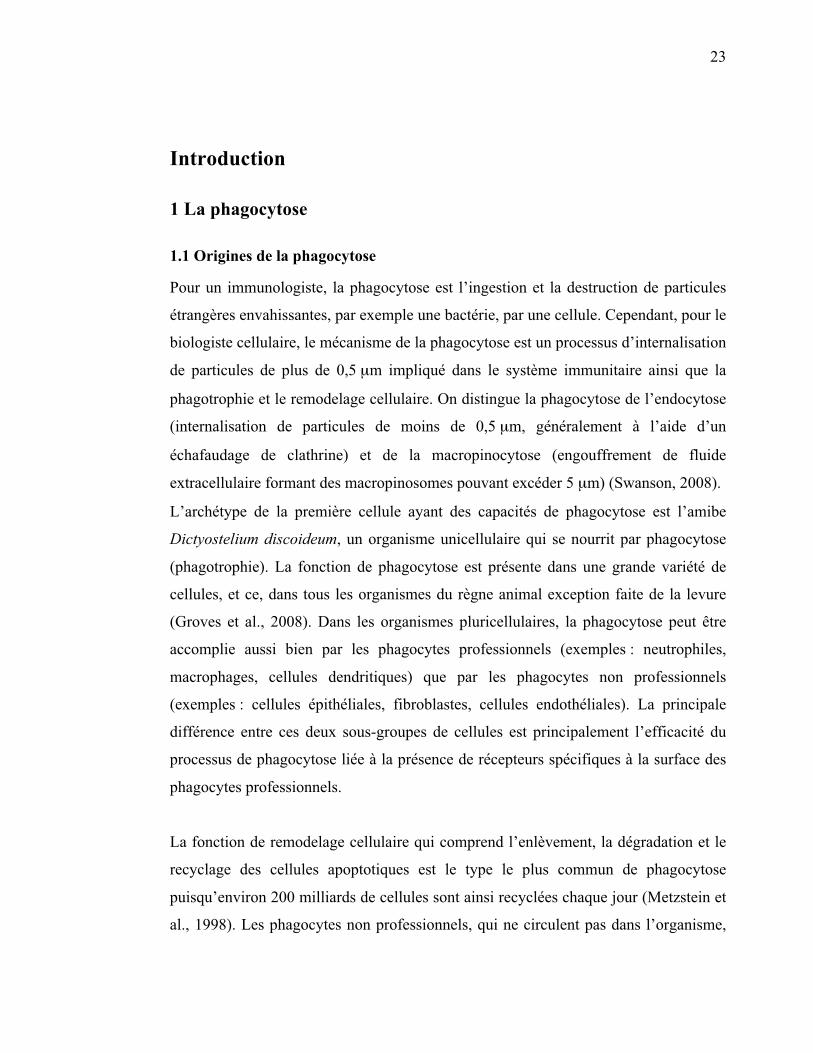
Pour un immunologiste, la phagocytose est l’ingestion et la destruction de particules
étrangères envahissantes, par exemple une bactérie, par une cellule. Cependant, pour le
biologiste cellulaire, le mécanisme de la phagocytose est un processus d’internalisation
de particules de plus de 0,5 μm impliqué dans le système immunitaire ainsi que la
phagotrophie et le remodelage cellulaire. On distingue la phagocytose de l’endocytose
(internalisation de particules de moins de 0,5 μm, généralement à l’aide d’un
échafaudage de clathrine) et de la macropinocytose (engouffrement de fluide
extracellulaire formant des macropinosomes pouvant excéder 5 μm) (Swanson, 2008).
L’archétype de la première cellule ayant des capacités de phagocytose est l’amibe
Dictyostelium discoideum, un organisme unicellulaire qui se nourrit par phagocytose
(phagotrophie). La fonction de phagocytose est présente dans une grande variété de
cellules, et ce, dans tous les organismes du règne animal exception faite de la levure
(Groves et al., 2008). Dans les organismes pluricellulaires, la phagocytose peut être
accomplie aussi bien par les phagocytes professionnels (exemples : neutrophiles,
macrophages, cellules dendritiques) que par les phagocytes non professionnels
(exemples : cellules épithéliales, fibroblastes, cellules endothéliales). La principale
différence entre ces deux sous-groupes de cellules est principalement l’efficacité du
processus de phagocytose liée à la présence de récepteurs spécifiques à la surface des
phagocytes professionnels.
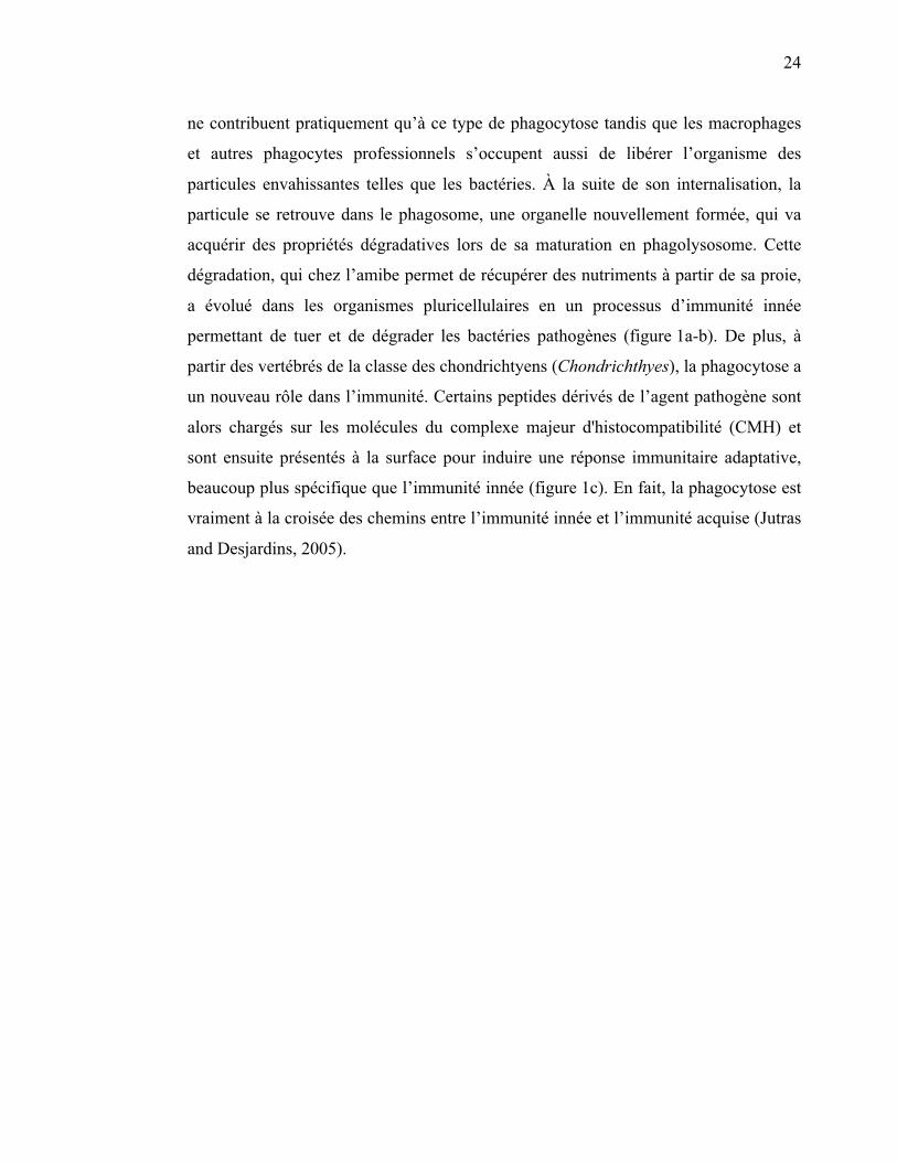
La fonction de remodelage cellulaire qui comprend l’enlèvement, la dégradation et le
recyclage des cellules apoptotiques est le type le plus commun de phagocytose
puisqu’environ 200 milliards de cellules sont ainsi recyclées chaque jour (Metzstein et
al., 1998). Les phagocytes non professionnels, qui ne circulent pas dans l’organisme,
24
ne contribuent pratiquement qu’à ce type de phagocytose tandis que les macrophages
et autres phagocytes professionnels s’occupent aussi de libérer l’organisme des
particules envahissantes telles que les bactéries. À la suite de son internalisation, la
particule se retrouve dans le phagosome, une organelle nouvellement formée, qui va
acquérir des propriétés dégradatives lors de sa maturation en phagolysosome. Cette
dégradation, qui chez l’amibe permet de récupérer des nutriments à partir de sa proie,
a évolué dans les organismes pluricellulaires en un processus d’immunité innée
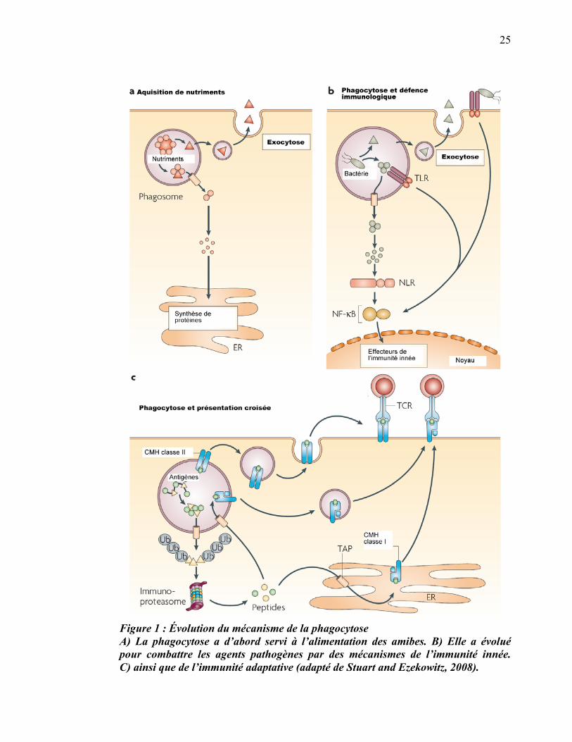
permettant de tuer et de dégrader les bactéries pathogènes (figure 1a-b). De plus, à
partir des vertébrés de la classe des chondrichtyens (Chondrichthyes), la phagocytose a
un nouveau rôle dans l’immunité. Certains peptides dérivés de l’agent pathogène sont
alors chargés sur les molécules du complexe majeur d'histocompatibilité (CMH) et
sont ensuite présentés à la surface pour induire une réponse immunitaire adaptative,
beaucoup plus spécifique que l’immunité innée (figure 1c). En fait, la phagocytose est
vraiment à la croisée des chemins entre l’immunité innée et l’immunité acquise (Jutras
and Desjardins, 2005).
25
Figure 1 : Évolution du mécanisme de la phagocytose A) La phagocytose a d’abord servi à l’alimentation des amibes. B) Elle a évolué pour combattre les agents pathogènes par des mécanismes de l’immunité innée. C) ainsi que de l’immunité adaptative (adapté de Stuart and Ezekowitz, 2008).
26
1.2 Mécanisme de la phagocytose
1.2.1 Internalisation
1.2.1.1 Récepteurs de la phagocytose
La reconnaissance de la particule à phagocyter est la première étape de la phagocytose.
Celle-ci est médiée par une variété de récepteurs à la surface du phagocyte, lesquels
lient la particule directement ou indirectement (par l’intermédiaire des opsonines).
Quelques-uns de ces récepteurs sont aptes à déclencher la phagocytose tandis que
d’autres semblent seulement lier la particule à phagocyter pour augmenter l’efficacité
de l’internalisation. Il y a quatre principales classes de récepteurs de la phagocytose : i)
les récepteurs Fc; ii) les récepteurs du complément; iii) les récepteurs « éboueurs »
(SR pour Scavenger Receptor); et iv) les lectines (Underhill and Ozinsky, 2002).
1.2.1.1.1 Récepteurs Fc
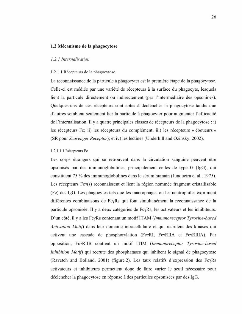
Les corps étrangers qui se retrouvent dans la circulation sanguine peuvent être
opsonisés par des immunoglobulines, principalement celles de type G (IgG), qui
constituent 75 % des immunoglobulines dans le sérum humain (Junqueira et al., 1975).
Les récepteurs Fc�(s) reconnaissent et lient la région nommée fragment cristallisable
(Fc) des IgG. Les phagocytes tels que les macrophages ou les neutrophiles expriment
différentes combinaisons de Fc�Rs qui font simultanément la reconnaissance de la
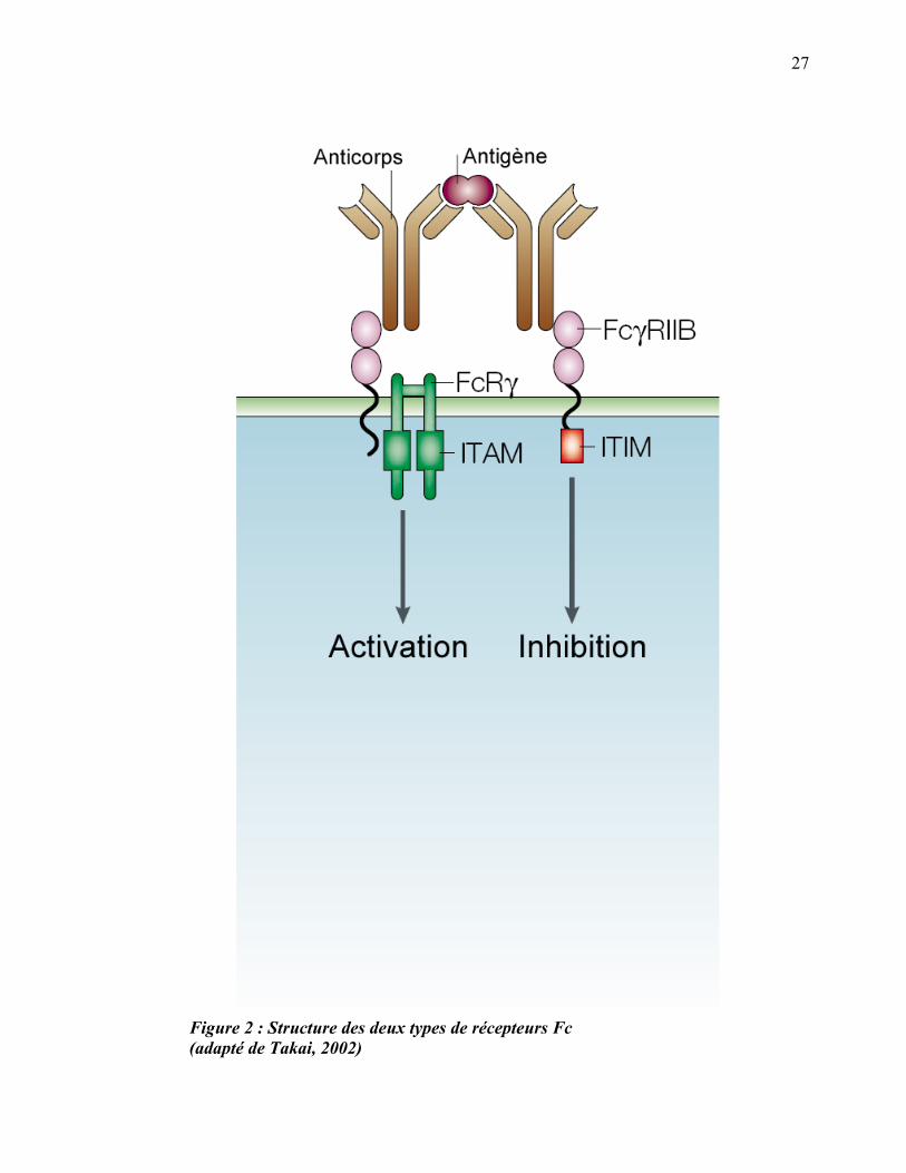
particule opsonisée. Il y a deux catégories de Fc�Rs, les activateurs et les inhibiteurs.
D’un côté, il y a les Fc�Rs contenant un motif ITAM (Immunoreceptor Tyrosine-based
Activation Motif) dans leur domaine intracellulaire et qui recrutent des kinases qui
activent une cascade de phosphorylation (Fc�RI, Fc�RIIA et Fc�RIIIA). Par
opposition, Fc�RIIB contient un motif ITIM (Immunoreceptor Tyrosine-based
Inhibition Motif) qui recrute des phosphatases qui inhibent le signal de phagocytose
(Ravetch and Bolland, 2001) (figure 2). Les taux relatifs d’expression des Fc�Rs
activateurs et inhibiteurs permettent donc de faire varier le seuil nécessaire pour
déclencher la phagocytose en réponse à des particules opsonisées par des IgG.
27
Figure 2 : Structure des deux types de récepteurs Fc (adapté de Takai, 2002)
28
1.2.1.1.2 Récepteurs du complément
Le système du complément est un ensemble de protéines circulantes ou membranaires
du sang, principalement sécrétées par le foie. Initialement, le rôle qu’on leur
reconnaissait était de complémenter l'action des immunoglobulines sériques, d'où leur
nom. Les particules étrangères peuvent être opsonisées par les protéines du
complément et ainsi être reconnues par des récepteurs spécifiques au complément (CR
pour Complement Receptor). Les CRs impliqués dans la phagocytose incluent CR1,
CR3 (ou intégrine �M�2 ou CD11b/CD18 ou Mac1) et CR4 (intégrine �X�2 ou
CD11c/CD18 ou gp150/95).
CR1 est une molécule constituée d’un domaine transmembranaire, d’un large domaine
extracellulaire de reconnaissance du ligand et d’une petite queue intracellulaire. Ce
récepteur reconnaît les protéines C1q, C4b et C3b du complément ainsi que d’autres
opsonines comme la mannan-binding lectin (MBL). CR1 ne peut pas, à lui seul,
déclencher l’internalisation d’une particule sans autres signaux, mais l’activation de
CR1 agit de façon synergique avec la signalisation provenant des Fc�Rs (Ghiran et al.,
2000). Les CR3 et CR4 sont des hétérodimères constitués d’une chaîne bêta commune
(CD18) couplée à une chaîne alpha spécifique, CD11b et CD11c respectivement. Tous
deux reconnaissent l’opsonine iC3b. Ils ont aussi besoin d’autres signaux pour induire
la phagocytose de particules opsonisées. L’étape d’activation nécessaire pour
déclencher la phagocytose par les CRs peut être induite par des cytokines
inflammatoires (Tumor Necrosis Factor alpha, TNF�), par des molécules
microbiennes (lipopolysaccharide, LPS) ou par l’adhésion avec un autre récepteur
(fibronectine). Cette préactivation augmente le nombre de CR3 à la surface ainsi que
leur affinité (Jones et al., 1998; Sengelov et al., 1993). De plus, la co-ligation des
récepteurs du complément et des récepteurs Fc est bien connue pour produire des
effets coopératifs dans le cadre de la phagocytose (Underhill and Ozinsky, 2002).
29
1.2.1.1.3 Récepteurs éboueurs (SRs)
Deux membres de la famille des SRs ont été impliqués dans la liaison et
l’internalisation des microbes, SR-A et MARCO (Macrophage Receptor with
COllagenous structure). SR-A est un homotrimère transmembranaire qui lie
directement les composantes bactériennes telles que la paroi cellulaire, l’acide
teichoïque et le LPS. MARCO lie une variété de particules incluant les bactéries
Gram-positives, les bactéries Gram-négatives ainsi que des particules artificielles
comme le latex (Palecanda et al., 1999; van der Laan et al., 1999). Contrairement au
FR�R ou au CR3 qui permettent de reconstituer la phagocytose lorsqu’on les transfecte
dans une cellule non-phagocytique, SR-A et MARCO ne confèrent qu’une capacité de
liaison aux bactéries, sans permettre une internalisation significative. Pour que la
phagocytose ait lieu par l’intermédiaire de ces récepteurs, il faut sans doute la
participation de co-récepteurs (Underhill and Ozinsky, 2002).
1.2.1.1.4 Lectines
Les phagocytes des mammifères expriment une grande variété de lectines de surface
qui permettent la reconnaissance des sucres du soi ou du non-soi. Le récepteur du
mannose (qui lie le �-mannane) et dectine-1 (qui lie le �-glucane) peuvent médier la
phagocytose de la levure ou du zymosan (paroi cellulaire de la levure, composée
principalement de mannoprotéines/�-mannanes et de �-glucanes) (Brown and Gordon,
2001; Ezekowitz et al., 1990). De nombreuses observations ont permis de déterminer
que le récepteur du mannose est suffisant pour lier et induire la phagocytose d’une
particule (Underhill and Ozinsky, 2002). Dectine-1 a aussi été reconnue comme
suffisante pour déclencher la phagocytose du zymosan. Par contre, dectine-1 n’est pas
le seul récepteur des �-glucanes pour la phagocytose, car CR3 a aussi une haute
affinité pour ce sucre (Ross, 2000).
30
1.2.1.2 Réorganisation de l’actine
1.2.1.2.1 Signalisation en aval des Fc�Rs
Lors de la phagocytose médiée par les Fc�Rs, le phagosome est formé par l’avancée de
pseudopodes guidés par les récepteurs, à la manière d’une fermeture éclair, d’où le
nom dans la littérature du zipper model (figure 3). Dans ce type de phagocytose, la
membrane est apposée intimement à la surface de la particule et c’est cette dernière
qui détermine la forme du phagosome (Swanson, 2008) (figure 6a). Une fois que la
particule est liée aux récepteurs de surface du phagocyte, une polymérisation localisée
et transitoire de l’actine est nécessaire pour accomplir l’internalisation. La
réorganisation de l’actine est régulée par des voies de signalisation activées en aval des
récepteurs liés.
31
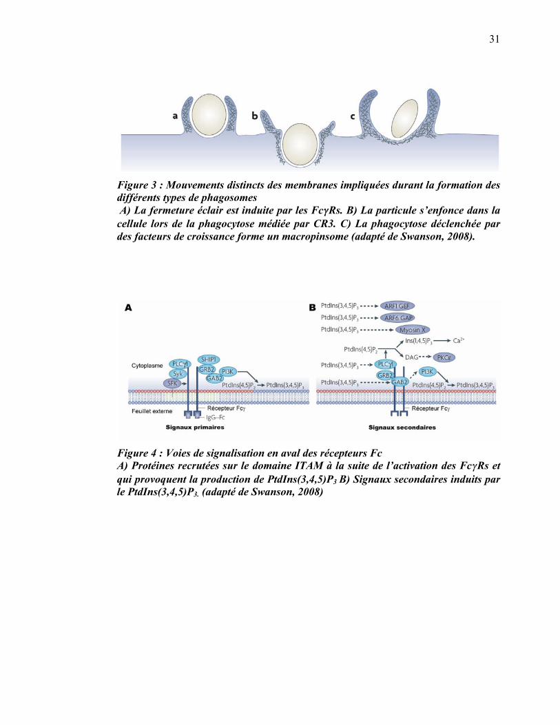
Figure 3 : Mouvements distincts des membranes impliquées durant la formation des différents types de phagosomes A) La fermeture éclair est induite par les Fc��Rs. B) La particule s’enfonce dans la cellule lors de la phagocytose médiée par CR3. C) La phagocytose déclenchée par des facteurs de croissance forme un macropinsome (adapté de Swanson, 2008).
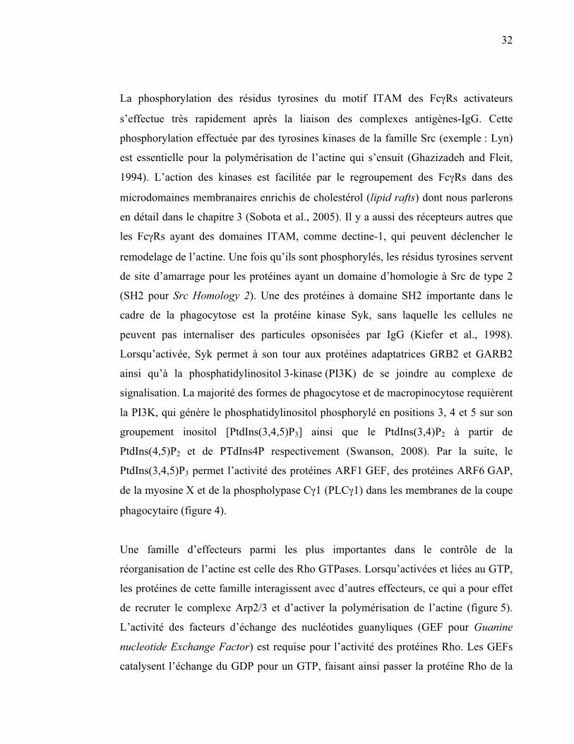
Figure 4 : Voies de signalisation en aval des récepteurs Fc A) Protéines recrutées sur le domaine ITAM à la suite de l’activation des Fc�Rs et qui provoquent la production de PtdIns(3,4,5)P3 B) Signaux secondaires induits par le PtdIns(3,4,5)P3. (adapté de Swanson, 2008)
32
La phosphorylation des résidus tyrosines du motif ITAM des Fc�Rs activateurs
s’effectue très rapidement après la liaison des complexes antigènes-IgG. Cette
phosphorylation effectuée par des tyrosines kinases de la famille Src (exemple : Lyn)
est essentielle pour la polymérisation de l’actine qui s’ensuit (Ghazizadeh and Fleit,
1994). L’action des kinases est facilitée par le regroupement des Fc�Rs dans des
microdomaines membranaires enrichis de cholestérol (lipid rafts) dont nous parlerons
en détail dans le chapitre 3 (Sobota et al., 2005). Il y a aussi des récepteurs autres que
les Fc�Rs ayant des domaines ITAM, comme dectine-1, qui peuvent déclencher le
remodelage de l’actine. Une fois qu’ils sont phosphorylés, les résidus tyrosines servent
de site d’amarrage pour les protéines ayant un domaine d’homologie à Src de type 2
(SH2 pour Src Homology 2). Une des protéines à domaine SH2 importante dans le
cadre de la phagocytose est la protéine kinase Syk, sans laquelle les cellules ne
peuvent pas internaliser des particules opsonisées par IgG (Kiefer et al., 1998).
Lorsqu’activée, Syk permet à son tour aux protéines adaptatrices GRB2 et GARB2
ainsi qu’à la phosphatidylinositol 3-kinase (PI3K) de se joindre au complexe de
signalisation. La majorité des formes de phagocytose et de macropinocytose requièrent
la PI3K, qui génère le phosphatidylinositol phosphorylé en positions 3, 4 et 5 sur son
groupement inositol [PtdIns(3,4,5)P3] ainsi que le PtdIns(3,4)P2 à partir de
PtdIns(4,5)P2 et de PTdIns4P respectivement (Swanson, 2008). Par la suite, le
PtdIns(3,4,5)P3 permet l’activité des protéines ARF1 GEF, des protéines ARF6 GAP,
de la myosine X et de la phospholypase C�1 (PLC�1) dans les membranes de la coupe
phagocytaire (figure 4).
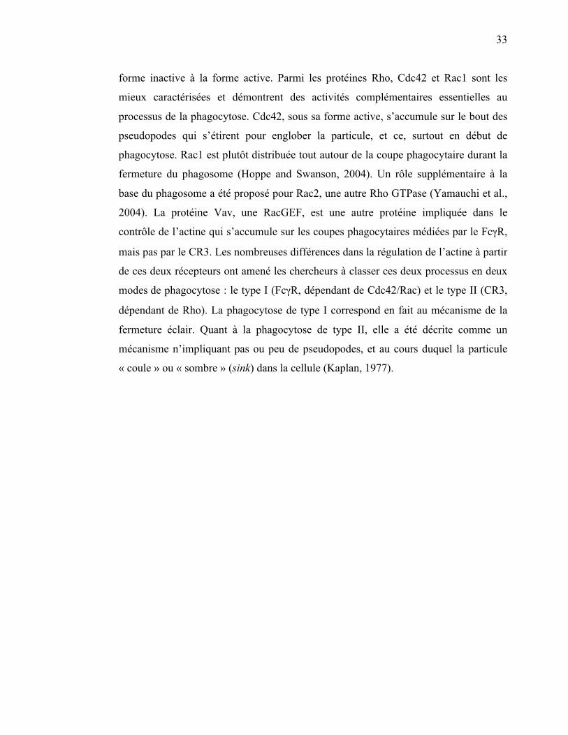
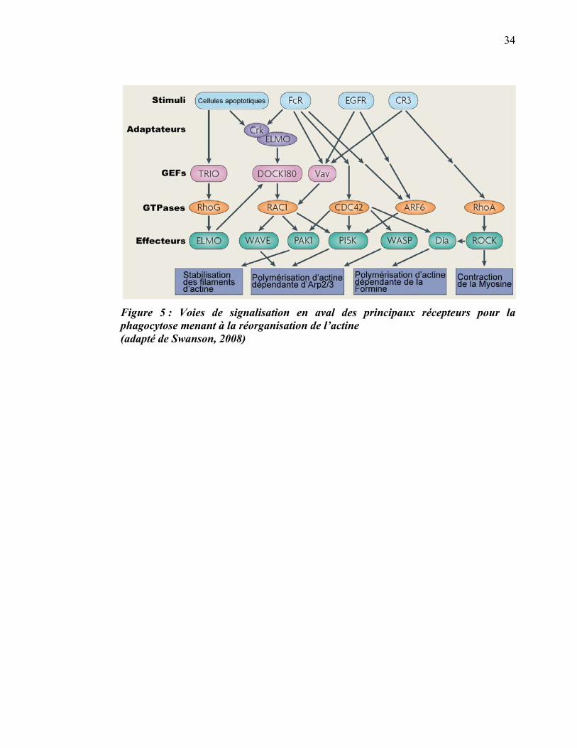
Une famille d’effecteurs parmi les plus importantes dans le contrôle de la
réorganisation de l’actine est celle des Rho GTPases. Lorsqu’activées et liées au GTP,
les protéines de cette famille interagissent avec d’autres effecteurs, ce qui a pour effet
de recruter le complexe Arp2/3 et d’activer la polymérisation de l’actine (figure 5).
L’activité des facteurs d’échange des nucléotides guanyliques (GEF pour Guanine
nucleotide Exchange Factor) est requise pour l’activité des protéines Rho. Les GEFs
catalysent l’échange du GDP pour un GTP, faisant ainsi passer la protéine Rho de la
33
forme inactive à la forme active. Parmi les protéines Rho, Cdc42 et Rac1 sont les
mieux caractérisées et démontrent des activités complémentaires essentielles au
processus de la phagocytose. Cdc42, sous sa forme active, s’accumule sur le bout des
pseudopodes qui s’étirent pour englober la particule, et ce, surtout en début de
phagocytose. Rac1 est plutôt distribuée tout autour de la coupe phagocytaire durant la
fermeture du phagosome (Hoppe and Swanson, 2004). Un rôle supplémentaire à la
base du phagosome a été proposé pour Rac2, une autre Rho GTPase (Yamauchi et al.,
2004). La protéine Vav, une RacGEF, est une autre protéine impliquée dans le
contrôle de l’actine qui s’accumule sur les coupes phagocytaires médiées par le Fc�R,
mais pas par le CR3. Les nombreuses différences dans la régulation de l’actine à partir
de ces deux récepteurs ont amené les chercheurs à classer ces deux processus en deux
modes de phagocytose : le type I (Fc�R, dépendant de Cdc42/Rac) et le type II (CR3,
dépendant de Rho). La phagocytose de type I correspond en fait au mécanisme de la
fermeture éclair. Quant à la phagocytose de type II, elle a été décrite comme un
mécanisme n’impliquant pas ou peu de pseudopodes, et au cours duquel la particule
« coule » ou « sombre » (sink) dans la cellule (Kaplan, 1977).
34
Figure 5 : Voies de signalisation en aval des principaux récepteurs pour la phagocytose menant à la réorganisation de l’actine (adapté de Swanson, 2008)
35
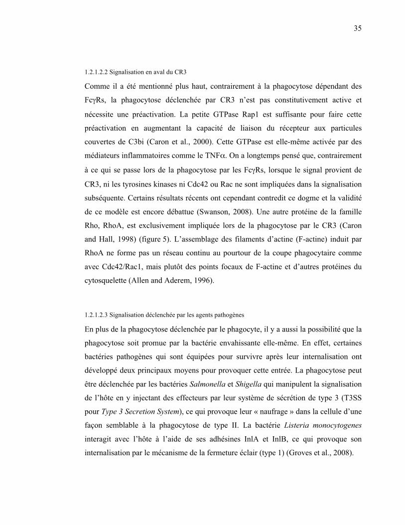
1.2.1.2.2 Signalisation en aval du CR3
Comme il a été mentionné plus haut, contrairement à la phagocytose dépendant des
Fc�Rs, la phagocytose déclenchée par CR3 n’est pas constitutivement active et
nécessite une préactivation. La petite GTPase Rap1 est suffisante pour faire cette
préactivation en augmentant la capacité de liaison du récepteur aux particules
couvertes de C3bi (Caron et al., 2000). Cette GTPase est elle-même activée par des
médiateurs inflammatoires comme le TNF�. On a longtemps pensé que, contrairement
à ce qui se passe lors de la phagocytose par les Fc�Rs, lorsque le signal provient de
CR3, ni les tyrosines kinases ni Cdc42 ou Rac ne sont impliquées dans la signalisation
subséquente. Certains résultats récents ont cependant contredit ce dogme et la validité
de ce modèle est encore débattue (Swanson, 2008). Une autre protéine de la famille
Rho, RhoA, est exclusivement impliquée lors de la phagocytose par le CR3 (Caron
and Hall, 1998) (figure 5). L’assemblage des filaments d’actine (F-actine) induit par
RhoA ne forme pas un réseau continu au pourtour de la coupe phagocytaire comme
avec Cdc42/Rac1, mais plutôt des points focaux de F-actine et d’autres protéines du
cytosquelette (Allen and Aderem, 1996).
1.2.1.2.3 Signalisation déclenchée par les agents pathogènes
En plus de la phagocytose déclenchée par le phagocyte, il y a aussi la possibilité que la
phagocytose soit promue par la bactérie envahissante elle-même. En effet, certaines
bactéries pathogènes qui sont équipées pour survivre après leur internalisation ont
développé deux principaux moyens pour provoquer cette entrée. La phagocytose peut
être déclenchée par les bactéries Salmonella et Shigella qui manipulent la signalisation
de l’hôte en y injectant des effecteurs par leur système de sécrétion de type 3 (T3SS
pour Type 3 Secretion System), ce qui provoque leur « naufrage » dans la cellule d’une
façon semblable à la phagocytose de type II. La bactérie Listeria monocytogenes
interagit avec l’hôte à l’aide de ses adhésines InlA et InlB, ce qui provoque son
internalisation par le mécanisme de la fermeture éclair (type 1) (Groves et al., 2008).
36
1.2.1.2.4 Réseau d’actine
L’actine se concentre principalement dans les pseudopodes qui s’allongent et elle
persiste jusqu’à la fermeture du phagosome (figure 6b). Le réseau de F-actine et des
protéines contractiles associées créent une contraction qui permet l’entrée active de la
particule (Swanson et al., 1999). Plusieurs classes de myosines sont impliquées dans la
phagocytose médiée par Fc�R, incluant les myosines 1C, II, IX et X (Swanson, 2008)
(figure 6c). Le mécanisme précis de la fermeture de la coupe phagocytaire n’est pas
connu, mais nécessite probablement la constriction de ses extrémités pour ne plus
former qu’une petite ouverture, suivie d’une scission qui sépare le phagosome de la
membrane plasmique.
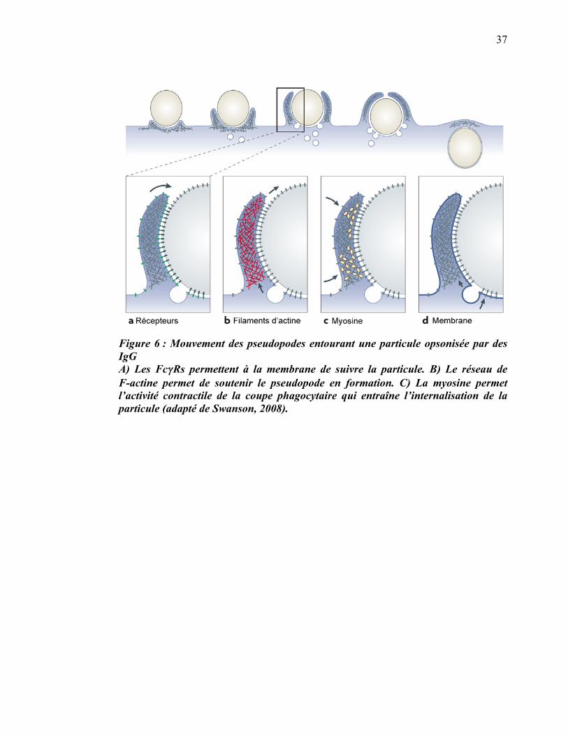
37
Figure 6 : Mouvement des pseudopodes entourant une particule opsonisée par des IgG A) Les Fc��Rs permettent à la membrane de suivre la particule. B) Le réseau de F-actine permet de soutenir le pseudopode en formation. C) La myosine permet l’activité contractile de la coupe phagocytaire qui entraîne l’internalisation de la particule (adapté de Swanson, 2008).
38
1.2.1.3 Sources de membranes
La phagocytose de grosses particules nécessite, en plus de la contribution de la
membrane plasmique, la contribution des membranes des organelles intracellulaires.
La membrane du phagosome est donc fournie à des degrés variables à partir des
endosomes de recyclage (Niedergang et al., 2003), des endosomes tardifs (Huynh et
al., 2007b), des lysosomes (Czibener et al., 2006; Tardieux et al., 1992), des granules
azurophiles (Suzaki et al., 1997) ainsi que du réticulum endoplasmique (Gagnon et al.,
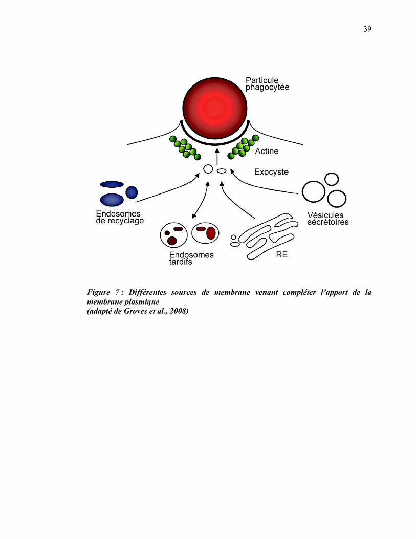
2002) (figure 7).
39
Figure 7 : Différentes sources de membrane venant compléter l’apport de la membrane plasmique (adapté de Groves et al., 2008)
40
1.2.2 Maturation du phagosome
1.2.2.1 Acidification du phagosome
La maturation du phagosome peut être considérée comme la dernière étape du
processus de la phagocytose, et c’est à cette étape que la bactérie ou la cellule
apoptotique ingérée est dégradée. Les analyses du phagosome à différents moments
ont révélé que la vacuole phagocytaire évolue dans la cellule en passant par une série
de stades menant à son acidification progressive et éventuellement à sa fusion avec les
lysosomes pour former le phagolysosome. Durant ces étapes d’acidification, le cargo
se dissocie des récepteurs, qui peuvent être recyclés, et la membrane phagosomiale
acquiert successivement les petites GTPases rab5 et rab7 (Rink et al., 2005). Cette
acidification est essentielle à la maturation, car c’est seulement lorsque le pH dans la
lumière du phagosome est assez bas que les protéases acidiques de la famille des
cathepsines deviennent actives et jouent leur rôle majeur dans la dégradation de la
particule (Lennon-Dumenil et al., 2002). L’acidification du phagosome survient en
deux étapes. D'abord, une acidification précoce diminue légèrement le pH (Hackam et
al., 1997) puis la pompe adénosine triphosphatase vacuolaire (V-ATPase) est recrutée
au phagosome et accélère le processus (Beyenbach and Wieczorek, 2006). La
V-ATPase est un complexe de 12 à 14 sous-unités réparties en deux structures : le
complexe V1 (sous-unités A à H) qui accomplit l’hydrolyse de l’adénosine
triphosphate (ATP), et le complexe V0 (sous-unités a, c, c', c'' et d) qui transporte des
protons vers l’intérieur du phagosome. L’énergie tirée de l’hydrolyse de l’ATP permet
à la V-ATPase de transporter les ions H+ contre leur gradient de concentration.
1.2.2.2 GTPases Rab
La famille des protéines Rab regroupe des protéines qui lient soit la guanosine
triphosphate (GTP) ou la guanosine diphosphate (GDP); elles se retrouvent, de ce fait,
dans le vaste groupe des GTPases, tout comme les Rho. Elles sont très importantes
dans le transport vésiculaire, particulièrement dans l’endocytose et la phagocytose.
41
Ces protéines ont un rôle d’interrupteur moléculaire. Dans leur conformation inactive,
les protéines Rab sont liées à la GDP et restent dans le cytoplasme en complexe avec
un inhibiteur de dissociation de la GDP (RabGDI pour Rab GDP-Dissociation
Inhibitor) (Ullrich et al., 1994). Un événement de signalisation provoque la relâche de
la protéine Rab par la RabGDI, ce qui expose la queue prénylée de la Rab, lui
permettant alors de se lier à la membrane (du phagosome, pour les Rabs qui nous
intéressent). Les GTPases Rab peuvent ensuite interagir avec un GEF, ce qui va
provoquer l’échange du GDP pour du GTP. La protéine rab maintenant active peut
alors lier des protéines effectrices (Grosshans et al., 2006) qui vont permettre
l’exécution de la maturation du phagosome. Les principales Rabs impliquées dans la
maturation du phagosome sont rab5 et rab7 (Desjardins et al., 1994b).
Rab5 est surtout impliquée dans la fusion homotypique des endosomes, mais on la
retrouve transitoirement sur les phagosomes précoces. Son recrutement est fait par la
protéine EEA1 (Early Endosome Antigen 1) qui elle-même se lie aux endosomes et
aux phagosomes précoces grâce à son domaine d’interaction au PtdIns(3,4,5)P3 (Lawe
et al., 2002). La présence de rab5 et de protéines de la famille des SNAREs (Soluble
N-ethylmaleimide sensitive Attachment protein REceptors) sur le phagosome permet
d’accomplir la fusion entre les phagosomes nouvellement refermés et les endosomes
précoces. La fusion entre le phagosome et l’endosome n’est pas complète, ce serait
plutôt un mécanisme transitoire de fusion/fission permettant l’échange sélectif de
molécules entre la lumière des deux vésicules (Desjardins, 1995; Desjardins et al.,
1997; Duclos and Desjardins, 2000). Ce mécanisme nommé le ‘kiss and run’ n’est pas
propre aux phagosomes, mais se retrouve aussi dans la biogenèse des lysosomes
(Duclos et al., 2003). On sait que l’alternance entre les formes GDP et GTP de rab5
permet de contrôler cette fusion transitoire puisque les macrophages exprimant de
façon stable un mutant de Rab5 défectueux dans son activité GTPasique forment des
phagosomes géants résultant de la fusion complète avec les endosomes (Duclos and
Desjardins, 2000).
42
La mobilisation des effecteurs de rab5, dont rabenosyn-5 (Nielsen et al., 2000)
contribue également au recrutement de rab7. Le mécanisme précis de la mobilisation
de rab7 sur le phagosome n’est pas connu, mais un modèle propose que rab5 soit
échangée par rab7 lors d’un processus appelé la conversion des rabs (Rink et al.,
2005). Dès qu’il est positif pour rab7, le phagosome devient apte à passer aux étapes
suivantes de sa maturation incluant la fusion avec les endosomes tardifs et les
lysosomes (Roberts et al., 2006; Vieira et al., 2003).
La fusion efficace du phagosome avec les vésicules endocytaires ainsi qu’une
acidification appropriée ont des implications capitales pour la fonction la plus évoluée
des phagosomes : la présentation antigénique. Une autre petite GTPase, rab27a, a aussi
un rôle dans la maturation du phagosome, rôle qui a été récemment démontré dans ce
contexte (Jancic et al., 2007). L’arrivée de NOX2, une NADPH oxidase qui génère des
dérivés réactifs de l’oxygène et qui dépend de rab27a, permet de réduire l’acidification
du phagosome. La perte de rab27a mène à une suracidification du phagosome, ce qui
réduit grandement l’efficacité de la présentation antigénique.
LAMP1 (Lysosomal-Associated Membrane Protein 1) et LAMP2 sont des
glycoprotéines spécifiquement localisées sur les lysosomes (Eskelinen et al., 2003) et
sont recrutées sur le phagosome lors de sa maturation. Leur fonction précise n’est pas
encore élucidée, mais cette fonction semble importante, du moins chez le nématode
Caenorhabditis elegans dans les dernières étapes de la phagocytose. En leur absence,
la maturation du phagosome est arrêtée avant d’acquérir Rab7 (Huynh et al., 2007a).
Comme certains agents pathogènes arrivent à utiliser la machinerie phagocytique pour
entrer, survivre ou se répliquer à l’intérieur de l’hôte, comprendre les différentes
étapes de la maturation du phagosome pourrait être la clé pour l’élaboration de
thérapies.
43
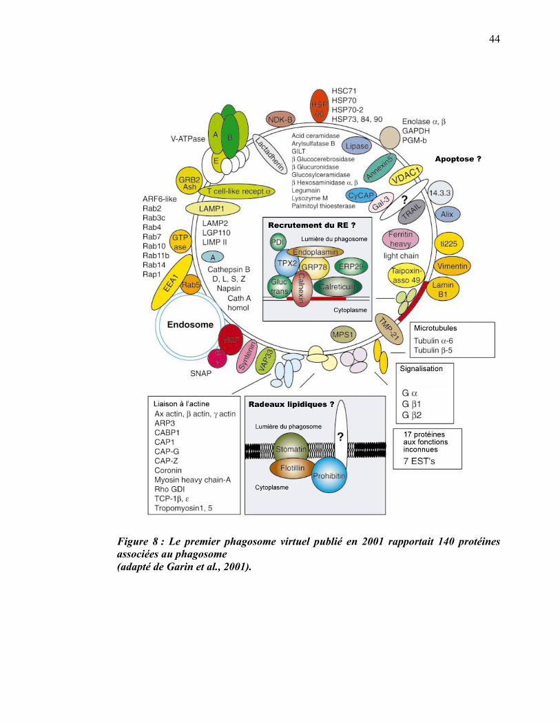
1.3 Le protéome du phagosome
L’utilisation de microbilles de polystyrène (souvent appelées billes de latex) a permis
d’établir un protocole afin d’effectuer l’isolement des phagosomes de macrophages
(Desjardins 1994a). Une approche protéomique haut débit a ensuite été utilisée pour
obtenir une liste de plus de 140 protéines associées à cette organelle (Garin et al.,
2001). Cette première étude à grande échelle du protéome du phagosome n’a pas
abordé les fonctions ou la régulation de ces protéines dans le cadre de la maturation,
mais a permis le développement d’un modèle descriptif des joueurs impliqués
(figure 8).
44
Figure 8 : Le premier phagosome virtuel publié en 2001 rapportait 140 protéines associées au phagosome (adapté de Garin et al., 2001).
45
1.3.1 Radeaux lipidiques du phagosome
La liste des protéines, générée par spectrométrie de masse, a permis d’établir des
hypothèses sur les fonctions du phagosome dont certaines étaient jusque-là
insoupçonnées. D’abord, la présence de flotilline-1, de stomatine et de prohibitine a
permis à notre équipe de déterminer que la membrane des phagosomes comprend des
microdomaines membranaires ayant les propriétés caractéristiques des radeaux
lipidiques décrits sur la membrane plasmique (Dermine et al., 2001; Simons and
Ikonen, 1997). L’isolement biochimique de ces microdomaines a permis de déterminer
qu’ils ségréguaient un sous-ensemble de protéines dont la V-ATPase, l’actine et les
sous-unités �, �1 et �2 des protéines G hétérotrimériques (Dermine et al., 2001).
1.3.2 Implication du réticulum endoplasmique
Par suite de la découverte de protéines typiquement associées au réticulum
endoplasmique (RE) dans l’étude protéomique (exemple : calnexine, calréticuline),
notre laboratoire a émis l’hypothèse d’une implication de cette organelle dans la
phagocytose (Garin et al., 2001). Une étude plus fonctionnelle impliquant beaucoup de
microscopie a permis de formuler un modèle de phagocytose où la membrane du RE
fusionne avec la membrane plasmique au moment de la formation du phagosome
(Gagnon et al., 2002). Cette source de membrane serait utilisée, selon ce modèle,
lorsque les particules internalisées sont trop grandes pour que la membrane plasmique
soit suffisante à elle seule.
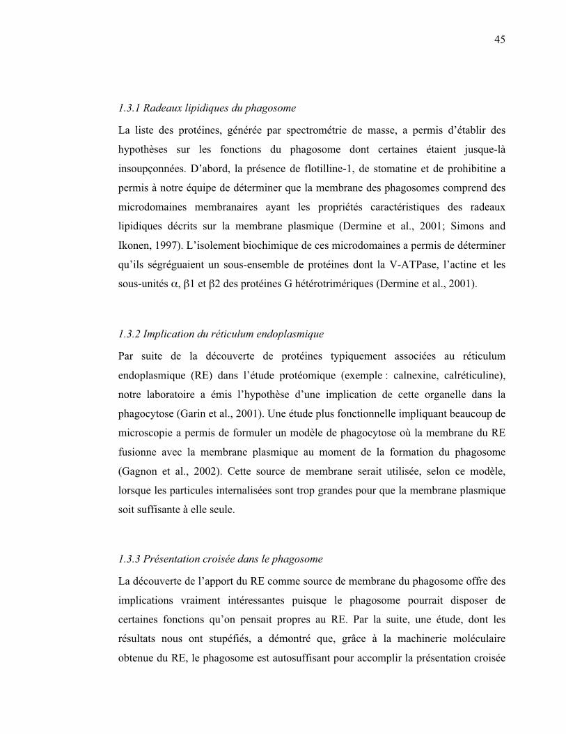
1.3.3 Présentation croisée dans le phagosome
La découverte de l’apport du RE comme source de membrane du phagosome offre des
implications vraiment intéressantes puisque le phagosome pourrait disposer de
certaines fonctions qu’on pensait propres au RE. Par la suite, une étude, dont les
résultats nous ont stupéfiés, a démontré que, grâce à la machinerie moléculaire
obtenue du RE, le phagosome est autosuffisant pour accomplir la présentation croisée

46
d’antigènes exogènes sur les CMH de classe 1 (article III en annexe, Houde et al.,
2003) (figure 9).
47
Figure 9 : Modèle de la présentation croisée effectuée par le phagosome (adapté de Houde et al., 2003)
48
1.3.4 Gamma-sécrétase
La gamma-sécrétase (�-sécrétase) est un complexe de plusieurs sous-unités ayant des
activités spécifiques de protéolyse. Lui-même intégral à la membrane, ce complexe
clive certaines protéines transmembranaires à l’intérieur même du domaine
transmembranaire. Le substrat le plus connu de la �-sécrétase est la protéine précurseur
de l’amyloïde (APP pour Amyloid Precursor Protein) qui, lorsque clivée, produit un
court peptide, le bêta-amyloïde, dont la forme anormalement repliée est impliquée
dans la maladie d’Alzheimer (Selkoe and Kopan, 2003). Deux des substrats de la �-
sécrétase avaient déjà été décrits comme étant associés à la membrane du phagosome
(Leemans et al., 2003; Ogden et al., 2001). Les analyses protéomiques chez la souris et
la drosophile ont permis de démontrer la présence de quatre composants de la �-
sécrétase [préséniline, nicastrine, APH-1 (Anterior Pharynx-Defective 1), et PEN-2
(Presenilin ENhancer 2)] qui y sont fonctionnels (Jutras et al., 2005). D’une manière
intéressante, ces protéines restent abondantes sur le phagosome tout au long de sa
maturation, contrairement aux marqueurs précoces qui décroissent (exemple : Rab5) et
aux marqueurs lysosomiaux qui croissent (exemple : LAMP1). Une observation
encore plus captivante a été que les sous-unités de la �-sécrétase se retrouvent avec
flotilline-1 dans les radeaux lipidiques des phagosomes (Jutras et al., 2005).
1.3.5 Implication de l’exocyste dans la phagocytose
Un développement récent a émergé de l’étude protéomique des phagosomes des
cellules phagocytaires S2 de la mouche Drosophila melanogaster (Stuart et al., 2007).
Plusieurs sous-unités du complexe de l’exocyste, une machinerie multi-moléculaire
qui dirige des vésicules sécrétoires vers la membrane plasmique, ont été identifiées sur
le phagosome. Les membranes des vésicules d’exocytose semblent donc également
impliquées dans la formation du phagosome. D’ailleurs, les sous-unités de l’exocyste
s’avèrent importantes dans le processus de la phagocytose, car lorsqu’on diminue leur
49
expression par ARN interférent, la phagocytose des bactéries diminue de façon
significative (article IV en annexe, Stuart et al., 2007).
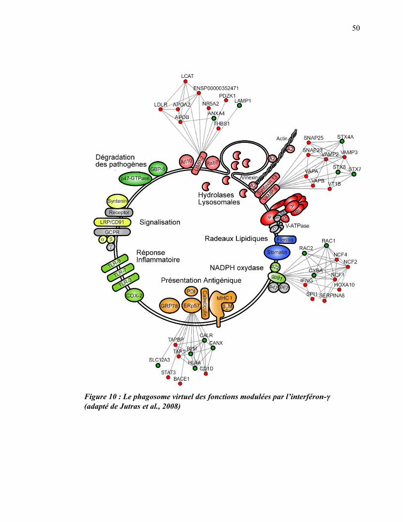
1.3.6 Modulation du protéome par l’interféron-gamma
Au cours d’une autre étude protéomique, nous sommes allés plus loin et nous nous
sommes penchés sur la modulation des fonctions du phagosome par la cytokine
inflammatoire interféron-gamma (INF-�). Ayant isolé des phagosomes de
macrophages traités ou non à l’INF-�, notre équipe a quantifié la différence
d’expression des protéines entre ces deux conditions. Nous avons ainsi observé que
167 protéines phagosomiales sont régulées en présence de l’INF-�, dont plus de 90 %
qui sont surexprimées (Jutras et al., 2008). Les protéines du phagosome dont
l’expression est la plus augmentée par cette cytokine font partie des groupes impliqués
dans les fonctions microbicides (exemples : NADPH oxydase, V-ATPase), la fusion
membranaire (exemples : syntaxine-7, syntaxine-13, rab7), la signalisation (exemple :
GTPases) et la présentation antigénique (exemples : protéasome, CMH-I)(Jutras et al.,
2008) (figure 10).
50
Figure 10 : Le phagosome virtuel des fonctions modulées par l’interféron-�� (adapté de Jutras et al., 2008)
51
2 Leishmania
2.1 Mise en contexte de Leishmania
Leishmania est un parasite protozoaire de l’ordre des kinétoplastidés, qui comprend
entre autres Trypanosoma brucei, agent de la maladie du sommeil, et Trypanosoma
cruzi, agent de la maladie de Chagas. Les différentes espèces de Leishmania infectent
plus de 15 millions de personnes, et on dénombre deux millions de nouveaux cas
chaque année (Herwaldt, 1999). Elles sont responsables de maladies dans un large
spectre de sévérité, allant de l’affection cutanée relativement contenue à la
manifestation viscérale progressive qui peut être fatale. Dans ce dernier cas,
L. donovani, l’agent de la leishmaniose viscérale (Kala-azar), se dissémine et infecte
les macrophages du foie, de la rate et de la moelle épinière (Lodge and Descoteaux,
2008).
Il n’existe pas encore de vaccins efficaces pour prévenir la maladie, et les tentatives de
contrôle du vecteur sont, pour l’instant, restées sans succès. Le traitement des
infections dues à ce parasite repose donc essentiellement sur la chimiothérapie. La
pharmacopée disponible contre Leishmania est limitée et le traitement prescrit en
première intention est un composé à base d’antimoine pentavalent (SbV), une
molécule utilisée depuis plus de 50 ans (Ouellette et al., 2003).
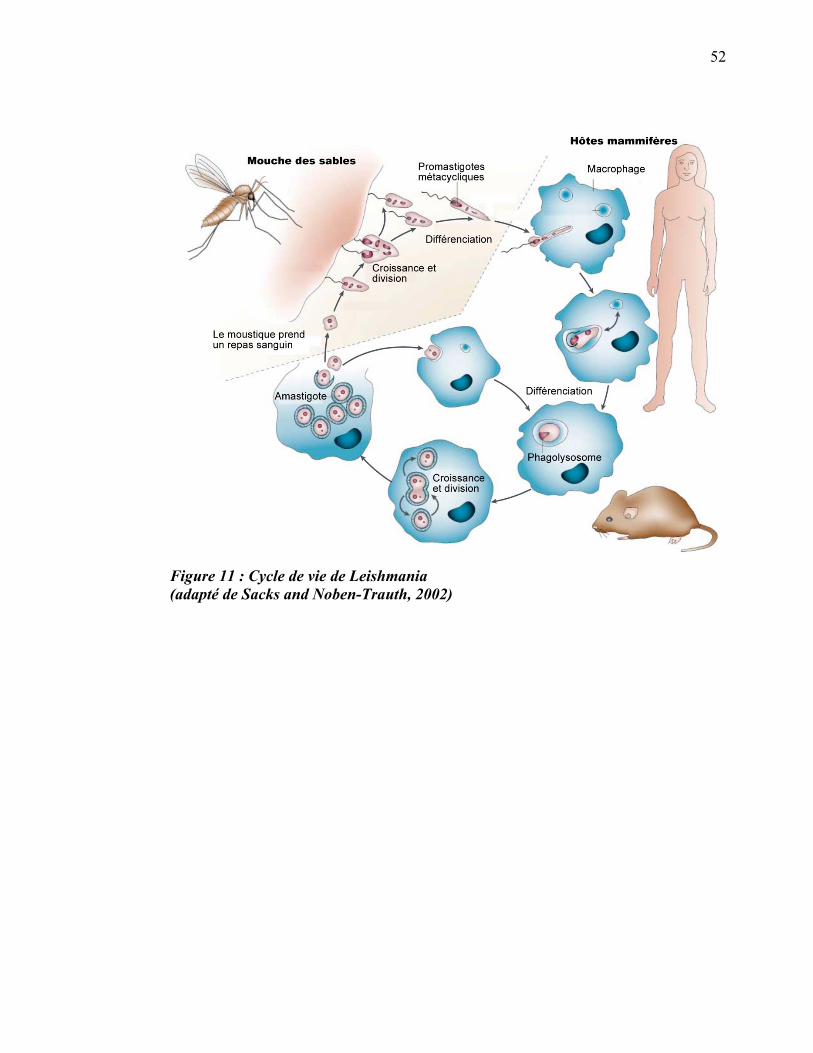
2.2 Cycle de vie de Leishmania
Le parasite intracellulaire du genre Leishmania est un protiste ayant un cycle de vie en
deux stades puisqu’il subit de nombreux changements en passant d’un de ses hôtes
obligatoires à l’autre. La forme flagellée (promastigote, allongée et mobile) de
Leishmania se retrouve chez les moustiques de la sous-famille des Phlebotominae
tandis que la forme aflagellée (amastigote, arrondie et non mobile) se retrouve chez les
mammifères (rongeurs, canidés, humains) (Alexander and Russell, 1992) (figure 11).
52
Figure 11 : Cycle de vie de Leishmania (adapté de Sacks and Noben-Trauth, 2002)

53
2.2.1 Chez le mammifère
Lors de son repas sanguin, le moustique infecté relâche des Leishmania promastigotes
sous leur forme virulente (promastigotes métacycliques) dans le derme du mammifère.
Les parasites peuvent alors se retrouver dans la circulation sanguine de leur nouvel
hôte où ils subissent l’assaut du système du complément. La molécule de
lipophosphoglycan (LPG) de Leishmania lui permet de résister aux mécanismes
lytiques du complément en empêchant l’insertion du complexe C5b-9 (MAC) dans sa
membrane (Puentes et al., 1990). De plus, chez L. donovani, la protéase de surface
gp63 coupe le C3b en un fragment inactif, l’iC3b, qui recouvre alors le parasite et
facilite sa capture par la voie de CR3. Ce mode de capture est avantageux pour le
promastigote puisqu’il n’active pas la cascade lytique du complément (Brittingham et
al., 1995; Mosser and Brittingham, 1997) ni la flambée oxydative (Wright and
Silverstein, 1983). L’internalisation de Leishmania peut aussi se faire via d’autres
récepteurs, dont les récepteurs à mannose-fucose (Wilson and Pearson, 1986) et les
récepteurs de la protéine C-réactive (CRP pour C-reactive protein) (Culley et al.,
1996) qui ne déclenchent pas l’activation des macrophages et, par conséquent,
favorisent la survie du parasite (Astarie-Dequeker et al., 1999; Bodman-Smith et al.,
2002; Wilson and Pearson, 1986). Le LPG est impliqué dans la reconnaissance du
parasite par les récepteurs du macrophage, mais n’est pas essentiel à la liaison puisque
les parasites mutants, qui sont déficients pour cette molécule, sont phagocytés aussi
efficacement, si non plus, que le type sauvage (McNeely and Turco, 1990).
Une fois dans le phagosome, les promastigotes utilisent des mécanismes pour en
perturber la maturation. Une étude de Michel Desjardins et d’Albert Descoteaux a
montré que l’inhibition de la fusion phagosome-endosome par Leishmania contribue à
sa survie (Desjardins and Descoteaux, 1997). Contrairement aux endosomes précoces,
les interactions des phagosomes contenant L. major ou L. donovani avec les
endosomes tardifs sont inhibées (Dermine et al., 2000; Desjardins and Descoteaux,
1997), et le recrutement des protéines tardives du phagosome telles que Rab7 et
LAMP1 est différé (Holm et al., 2001; Scianimanico et al., 1999). Cette inhibition
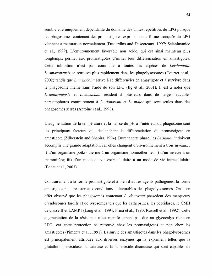
54
semble être uniquement dépendante du domaine des unités répétitives du LPG puisque
les phagosomes contenant des promastigotes exprimant une forme tronquée du LPG
viennent à maturation normalement (Desjardins and Descoteaux, 1997; Scianimanico
et al., 1999). L’environnement favorable non acide, qui est ainsi maintenu plus
longtemps, permet aux promastigotes d’initier leur différenciation en amastigotes.
Cette inhibition n’est pas commune à toutes les espèces de Leishmania.
L. amazonensis se retrouve plus rapidement dans les phagolysosomes (Courret et al.,
2002) tandis que L. mexicana arrive à se différencier en amastigote et à survivre dans
le phagosome même sans l’aide de son LPG (Ilg et al., 2001). Il est à noter que
L. amazonensis et L. mexicana résident à plusieurs dans de larges vacuoles
parasitophores contrairement à L. donovani et L. major qui sont seules dans des
phagosomes serrés (Antoine et al., 1998).
L’augmentation de la température et la baisse du pH à l’intérieur du phagosome sont
les principaux facteurs qui déclenchent la différenciation du promastigote en
amastigote (Zilberstein and Shapira, 1994). Durant cette phase, les Leishmania doivent
accomplir une grande adaptation, car elles changent d’environnement à trois niveaux :
i) d’un organisme poïkilotherme à un organisme homéotherme; ii) d’un insecte à un
mammifère; iii) d’un mode de vie extracellulaire à un mode de vie intracellulaire
(Bente et al., 2003).
Contrairement à la forme promastigote et à bien d’autres agents pathogènes, la forme
amastigote peut résister aux conditions défavorables des phagolysosomes. On a en
effet observé que les phagosomes contenant L. donovani possèdent des marqueurs
d’endosomes tardifs et de lysosomes tels que les cathepsines, les peptidases, le CMH
de classe II et LAMP1 (Lang et al., 1994; Prina et al., 1990; Russell et al., 1992). Cette
augmentation de la résistance n’est manifestement pas due au glycocalyx riche en
LPG, car cette protection se retrouve chez les promastigotes et non chez les
amastigotes (Pimenta et al., 1991). La survie des amastigotes dans les phagolysosomes
est principalement attribuée aux diverses enzymes qu’ils expriment telles que la
glutathion peroxidase, la catalase et la superoxide dismutase qui sont capables de

55
neutraliser les composés oxydatifs toxiques produits dans les phagolysosomes des
macrophages lors de la flambée oxydative (Channon and Blackwell, 1985). Une autre
façon, pour les amastigotes, de survivre à l’intérieur des macrophages consiste à
inhiber les voies de signalisation de la cellule hôte en vue de perturber certaines
fonctions cellulaires. Chez les cellules infectées par Leishmania, on a observé une
diminution de la réponse de la cellule au LPS, à l’IFN-� et à certains activateurs de la
protéine kinase C (PKC) (Descoteaux et al., 1991; Ray et al., 2000).
Les autres transformations associées à la différenciation des Leishmania ont été
étudiées par des analyses génomiques. Les résultats combinés de nombreuses études
montrent qu’il y a un très petit nombre de gènes différentiellement exprimés (0,2 % à
5 %) entre la forme promastigote et la forme amastigote (Cohen-Freue et al., 2007).
Ceci s’explique par la particularité du génome de Leishmania d’être pratiquement
exprimé de façon constitutive. En effet, il ne semble pas exister de régulation au
niveau de l’initiation de la transcription, mais essentiellement une régulation post-
transcriptionelle (Ouellette et al., 2003). Par la suite, les équipes cherchant à
caractériser les différences entre les deux stades de Leishmania se sont tournées vers la
protéomique. Les premières études protéomiques utilisant la méthode du gel 2D ont
permis de faire une première caractérisation spécifique au stade pour les espèces
L. infantum (Acestor et al., 2002; El Fakhry et al., 2002), L. donovani (Bente et al.,
2003), L. mexicana (Nugent et al., 2004) et L. panamensis (Walker et al., 2006). En
moyenne, 200 taches ont été analysées sur les gels 2D pour chacune de ces espèces, et
5 à 12 % de ces taches montraient une expression différentielle selon le stade (Cohen-
Freue et al., 2007).
Plus particulièrement pour L. donovani, 100 protéines sur les 2000 du génome ont été
identifiées comme étant différentiellement exprimées entre les deux formes du parasite
(Bente et al., 2003). Ces protéines peuvent être classées en cinq groupes selon la
cinétique d’expression à différents stades (promastigote au jour 0, amastigote au jour
1, amastigote au jour 2 à 5). Elles se classent également en six catégories
fonctionnelles : i) réponse au stress; ii) assemblage du cytosquelette; iii) métabolisme

56
de l’énergie et phosphorylation; iv) cycle cellulaire et prolifération; v) métabolisme
des acides aminés; vi) enzymes protéolytiques (Bente et al., 2003) (tableau 1).
Quelques espèces ont, par la suite, été analysées par une approche quantitative plus
précise comme la technologie isotope-coded affinity tag (ICAT) (Cohen-Freue et al.,
2007).
57
Tableau 1 : Protéines de Leishmania donovani différentiellement régulées lors de sa différenciation. – sous exprimée + sur exprimée (adapté de Bente et al., 2003)
58
2.2.2 Chez le moustique
Lorsque le moustique prend un repas sanguin chez un mammifère infecté, il ingère des
macrophages parasités. Les amastigotes contenus dans les macrophages sont, par la
suite, libérés dans le système digestif de la mouche, où ils se différencient en
promastigotes réplicatifs, appelés procycliques, capables de s’attacher à l’épithélium
intestinal (Sacks et al., 1995) (figure 11). À ce stade, les promastigotes ne sont pas
infectieux. Ces derniers subissent ensuite un procédé de maturation, appelé
métacyclogenèse, au cours duquel ils se différencient en une forme métacyclique non
réplicative, mais très virulente (Sacks, 1989). La forme métacyclique infectieuse migre
ultimement dans la trompe (proboscide) de l’insecte et sera ainsi transmise à l’hôte
lors du prochain repas sanguin (Sacks and Kamhawi, 2001).
2.2.3 Culture des Leishmania en laboratoire
Pour étudier les Leishmania et pouvoir faire des expériences d’infection dans des
lignées cellulaires de macrophages, des techniques ont été développées pour la culture
de Leishmania en laboratoire. Dans différents milieux de culture relativement
complexes, pendant la phase de croissance logarithmique à 25°C, le parasite est sous la
forme promastigote. Quand la forme promastigote atteint la phase stationnaire, il y a
transformation en forme métacyclique suivie de l’expression de marqueurs spécifiques
(McKean et al., 2001). La forme amastigote des parasites était originalement recueillie
à partir de culture de macrophages ou d’animaux infectés. Depuis, des conditions de
culture en milieu stérile (milieu axénique) ont été développées et permettent la
multiplication de la forme amastigote de certaines espèces de Leishmania caractérisées
par l’expression de marqueurs amastigotes spécifiques. Les stimuli qui favorisent la
transformation in vitro de promastigotes en amastigotes sont le pH acide (5,5 à 4,8) et
la température (37oC) (Bente et al., 2003; Sereno and Lemesre, 1997).
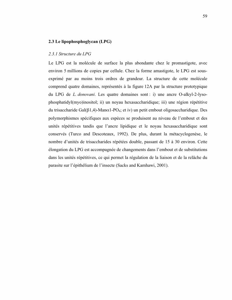
59
2.3 Le lipophosphoglycan (LPG)
2.3.1 Structure du LPG
Le LPG est la molécule de surface la plus abondante chez le promastigote, avec
environ 5 millions de copies par cellule. Chez la forme amastigote, le LPG est sous-
exprimé par au moins trois ordres de grandeur. La structure de cette molécule
comprend quatre domaines, représentés à la figure 12A par la structure prototypique
du LPG de L. donovani. Les quatre domaines sont : i) une ancre O-alkyl-2-lyso-
phosphatidyl(myo)inositol; ii) un noyau hexasaccharidique; iii) une région répétitive
du trisaccharide Gal(�1,4)-Man�1-PO4; et iv) un petit embout oligosaccharidique. Des
polymorphismes spécifiques aux espèces se produisent au niveau de l’embout et des
unités répétitives tandis que l’ancre lipidique et le noyau hexasaccharidique sont
conservés (Turco and Descoteaux, 1992). De plus, durant la métacyclogenèse, le
nombre d’unités de trisaccharides répétées double, passant de 15 à 30 environ. Cette
élongation du LPG est accompagnée de changements dans l’embout et de substitutions
dans les unités répétitives, ce qui permet la régulation de la liaison et de la relâche du
parasite sur l’épithélium de l’insecte (Sacks and Kamhawi, 2001).
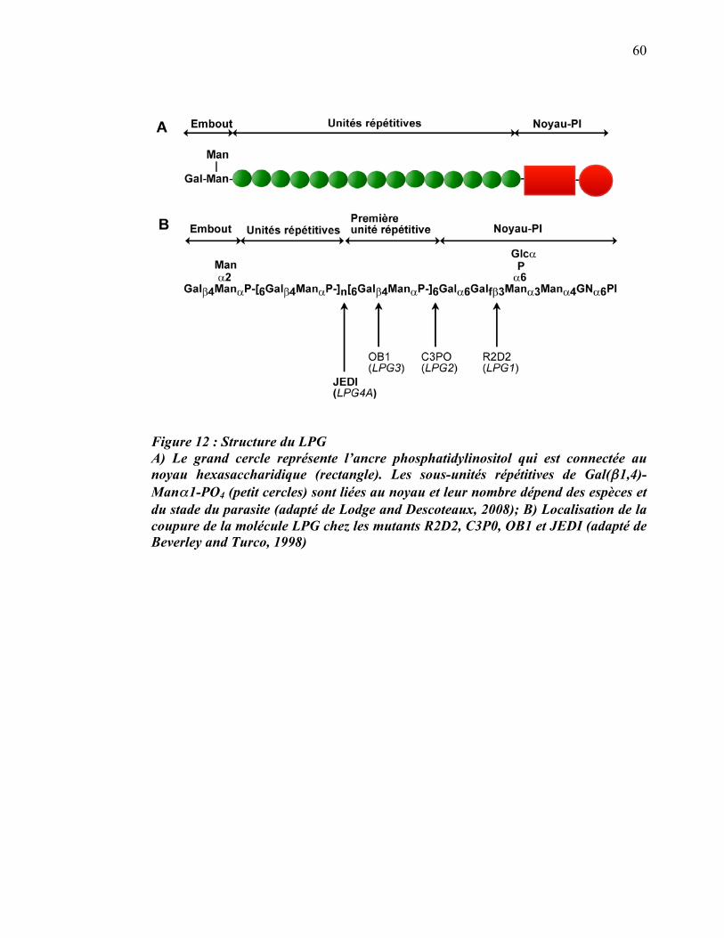
60
Figure 12 : Structure du LPG A) Le grand cercle représente l’ancre phosphatidylinositol qui est connectée au noyau hexasaccharidique (rectangle). Les sous-unités répétitives de Gal(��1,4)-Man�1-PO4 (petit cercles) sont liées au noyau et leur nombre dépend des espèces et du stade du parasite (adapté de Lodge and Descoteaux, 2008); B) Localisation de la coupure de la molécule LPG chez les mutants R2D2, C3P0, OB1 et JEDI (adapté de Beverley and Turco, 1998)
61
2.3.2 Mutagenèse du LPG
Une combinaison de mutants pour la synthèse des unités de la molécule
Gal(�1,4)-Man�1-PO4 ou pour la molécule LPG elle-même a été utilisée pour
enquêter sur les rôles du LPG dans le cycle de vie de Leishmania. Un des sucres de
l’embout du LPG inclut un groupement galactosyl qui peut lier les lectines telles que
l’agglutinine de ricin. En soumettant des Leishmania donovani hautement
mutagénisées à de multiples cycles d’agglutination avec l’agglutinine du ricin et en les
enrichissant pour les cellules non-agglutinées, il devient possible d’obtenir des
parasites dépourvus de LPG détectable (King and Turco, 1988). Ces mutants
provenant de la perte d’une des protéines essentielles à la fabrication ou à
l’assemblage des molécules LPG ont été appelés « mutants lpg- ». Les quatre premiers
mutants lpg- caractérisés ont été nommés en l’honneur des films de Star Wars (R2D2
ou lpg1-/-, C3PO ou lpg2-/-, OB1ou lpg3-/- et JEDI ou lpg4-/-) (figure 12B) (Beverley
and Turco, 1998).
2.3.3 Fonctions du LPG
Plusieurs rôles du LPG ont été proposés pour la survie du parasite. Chez l’insecte, le
LPG forme un glycocalyx dense apte à protéger les promastigotes contre la
dégradation par les enzymes digestives (Schlein et al., 1990). Le polymorphisme
structural du LPG permet aux espèces de Leishmania d’utiliser des vecteurs différents,
ce qui favorise leur dissémination sans que les parasites compétitionnent entre eux
(Pimenta et al., 1994b).
Chez le mammifère, le LPG accomplit plusieurs fonctions avant l’internalisation des
parasites dans les macrophages. Comme il a été mentionné précédemment, c’est le
LPG qui protège le parasite contre les attaques du complément. D’ailleurs, la longueur
doublée du LPG des promastigotes métacycliques joue un grand rôle dans cette
fonction puisque cette forme de Leishmania résiste beaucoup mieux à la lyse que les
procycliques (Franke et al., 1985; Puentes et al., 1990). Le LPG a aussi un rôle dans la
62
fixation de Leishmania aux récepteurs CR3 du macrophage, ce rôle a été
évoqué précédemment.
Le LPG aurait aussi plusieurs rôles dans le phagosome, dont l’inactivation des
enzymes dégradatives contenues dans les lysosomes (El-On et al., 1980). Grâce à ses
charges négatives, le LPG servirait aussi de chélateur de calcium et d’autres cations
divalents, ce qui préviendrait la production de radicaux hydroxyles toxiques lors de
l’activation des macrophages (Eilam et al., 1985; Homans et al., 1992). Le LPG a aussi
des effets reconnus sur la signalisation impliquée dans plusieurs fonctions cellulaires
(Houslay, 1991) comme la production d’IL-1 (Olivier et al., 2005) et d’IL-12
(Sutterwala et al., 1997). Le LPG affecte également des fonctions propres au
phagosome, incluant la génération de radicaux libres de l’oxygène et le remodelage de
l’actine entourant le phagosome (Lodge and Descoteaux, 2008). De plus, le LPG serait
impliqué dans l’inhibition des voies de signalisation de PKC (Holm et al., 2001) et de
la voie JAK2/STAT1 dépendante de SHP-1(Blanchette et al., 1999).
Finalement, les données du groupe de Desjardins ont démontré que la molécule LPG
est responsable de l’inhibition de la fusion phagosome-endosome (Desjardins and
Descoteaux, 1997). En effet, les promastigotes mutants de L. donovani lpg2-/- ,qui
expriment un LPG tronqué sans unité disaccharidique répétitive (Descoteaux et al.,
1995), permettent la fusion et la maturation des phagosomes, contrairement aux
promastigotes de type sauvage. Cette même étude a démontré que la molécule LPG, à
elle seule, était responsable de cette inhibition en montrant que l’opsonisation des
mutants lpg2-/- avec du LPG purifié rétablissait le blocage de la fusion (Desjardins
and Descoteaux, 1997). Le nombre d’unités disacchariques répétitives du LPG serait la
clé d’une inhibition efficace de la fusion puisque le mutant RT5 de L. donovani (qui
exprime seulement de 3 à 5 répétitions au lieu de 15) se retrouve dans des phagosomes
dont le taux de fusion est intermédiaire entre celui des phagosomes contenant le type
sauvage et celui des phagosomes contenant le type mutant lpg2-/- (McNeely and
Turco, 1990).
63
Le gène lpg2 code pour la manosyl transférase, un transporteur intracellulaire qui
permet la translocation de résidus mannose du cytoplasme vers l’appareil de Golgi
(Ma et al., 1997). L’absence de ce transporteur empêche la fabrication des unités de
sucre répétitives et l’ajout de ces molécules n’est plus possible sur le LPG, ni sur les
phosphoglycans sécrétés, ni sur les protéines comme la phosphatase alcaline (Ilg et al.,
1994). Un autre gène contribuant à la biosynthèse du LPG est le lpg1, codant pour une
galactofuranosyl transférase (Huang and Turco, 1993). Cette enzyme joue son rôle
dans la synthèse du noyau hexasaccharidique, qui n’est présent que dans la molécule
LPG. Chez le parasite lpg1-/-, on a donc une molécule LPG tronquée et aucune autre
molécule n’est touchée. Par conséquent, le mutant lpg1-/- permet de distinguer l’effet
de LPG de celui des autres molécules possédant des unités répétitives.
L’équipe de Desjardins a, par la suite, montré que les mutants lpg1-/- et lpg2-/- avaient
le même taux d’inhibition de la fusion, ce qui renforce l’idée que c’est vraiment le
LPG qui en est le responsable (Dermine et al., 2000). Le LPG de L. major inhibe
encore plus la fusion que celui de L. donovani, et on a d’abord pensé que cet effet était
dû aux chaînes latérales de sucre, présentes sur le LPG de L. major, mais pas sur celui
de L. donovani. Cependant, ce n’est pas le cas puisque le mutant Spock de L. major,
qui est dépourvu de ces extensions de sucre, inhibe autant la fusion que le type
sauvage. Ce serait plutôt la longueur de la molécule qui en serait responsable (Butcher
et al., 1996). Finalement, il a aussi été démontré que l’inhibition de la fusion des
phagosomes par LPG est un processus actif nécessitant que Leishmania soit vivante
(Dermine et al., 2000).
64
3 Les radeaux membranaires
3.1 Découverte des radeaux membranaires
La première description de sous-domaines membranaires a été faite par Simons et van
Meer, qui étudiaient les différences lipidiques et protéiques entre la région apicale et
basolatérale des cellules polarisées (Simons and van Meer, 1988). Ils ont proposé en
1988 que cette asymétrie est possible grâce à des domaines membranaires du trans
Golgi, dont le feuillet luminal est enrichi en sphingolipides et contient les protéines
destinées à la zone apicale de la cellule. Ces lipides et protéines seraient acheminés
aux membranes apicales sous forme de vésicules. Brown et Rose ont été les premiers à
faire l’isolation de sous-domaines enrichis en glycolipides, aussi nommés membranes
résistantes au détergent (DRMs pour Detergent Resistant Membranes), par extraction
au Triton X-100 (TX-100) puis flottaison par centrifugation dans un gradient de
sucrose. Ils ont montré que l’insolubilité au TX-100 des protéines à ancre
glycosylphosphatidylinositol (GPI) n’est pas causée par une association au
cytosquelette ni même par interaction avec d’autres protéines. Ce sont en effet les
propriétés de leur composition lipidique particulière qui rendent ces domaines plus
résistants à la solubilisation.
Le terme « raft » a été utilisé pour la première fois par Brown en 1994 (Brown, 1994),
mais ce n’est que trois ans plus tard que Simons et Ikonen ont décrit le modèle des
radeaux lipidiques (lipids rafts) tel qu’il est encore généralement accepté aujourd’hui
(Simons and Ikonen, 1997). Selon ce modèle nommé l’hypothèse des radeaux (the raft
hypothesis), les radeaux lipidiques sont de petites régions de la membrane plasmique
formées de façon transitoire par l’enrichissement de certains lipides (sphingolipides,
cholestérol) et de certaines protéines, et qui jouent un rôle particulier par rapport au
reste de la membrane (figure 13). Il a rapidement été proposé que le concept des
DRMs et celui des radeaux lipidiques puissent être physiquement et conceptuellement
liés (Arni et al., 1998). Durant les 10 dernières années, les termes DRMs et radeaux
lipidiques ont souvent été utilisés à tort comme synonymes. On sait maintenant que le
65
contenu lipidique des DRMs ne se retrouve pas tel quel dans les microdomaines
lipidiques. Il faut donc faire bien attention et distinguer les radeaux lipidiques
(structures in vivo enrichies de sphingolipides et de cholestérol) des DRMs, qui sont le
résultat d’une solubilisation partielle des membranes et qui n’existent pas sans
détergent (Lichtenberg et al., 2005).
Maintenant que cette distinction est faite, il est reconnu que l’isolation des DRMs
suivie de leur analyse est une bonne façon d’étudier le partitionnement de certains
groupes de protéines selon leurs affinités biochimiques. Une fois qu’il est établi par
d’autres techniques (comme la microscopie), qu’une membrane comporte des radeaux
lipidiques, l’analyse comparative des DRMs et du reste de la membrane, soit les
domaines membranaires solubles aux détergents (DSMs pour Detergent Soluble
Membranes), nous permet de distinguer deux sous-groupes distincts de protéines qui
ont toutes les chances de représenter les protéines de radeaux lipidiques versus les
protéines de non-radeaux (Hanzal-Bayer and Hancock, 2007). Une série de
précautions peuvent être utilisées pour renforcer l’analyse des DRMs servant à étudier
les radeaux lipidiques : marqueurs internes, analyse des lipides, déprivation du
cholestérol, utilisation de différents détergents (Babiychuk and Draeger, 2006). Du
point de vue de l’équipe pionnière dans l’étude des radeaux lipidiques, la méthode par
isolation des DRMs est adéquate pour évaluer l’association et la dissociation de
protéines dans les radeaux lipidiques lors de changements biochimiques et/ou
physiologiques (Lingwood and Simons, 2007).
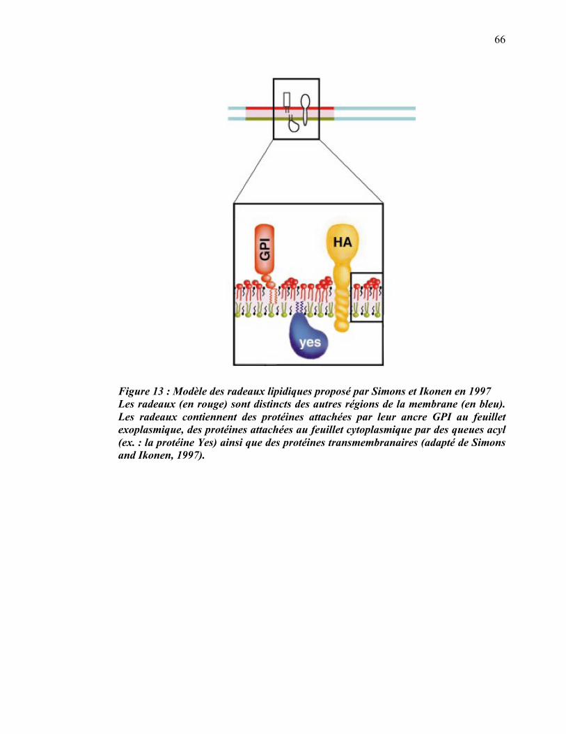
66
Figure 13 : Modèle des radeaux lipidiques proposé par Simons et Ikonen en 1997 Les radeaux (en rouge) sont distincts des autres régions de la membrane (en bleu). Les radeaux contiennent des protéines attachées par leur ancre GPI au feuillet exoplasmique, des protéines attachées au feuillet cytoplasmique par des queues acyl (ex. : la protéine Yes) ainsi que des protéines transmembranaires (adapté de Simons and Ikonen, 1997).
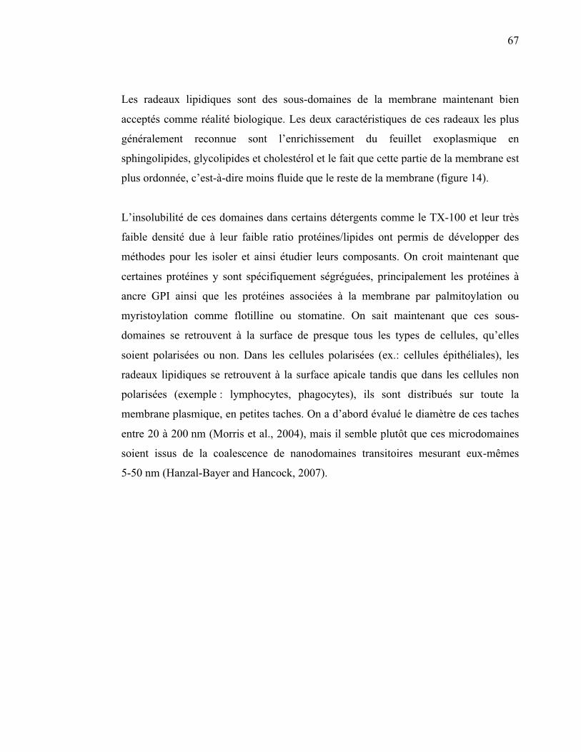
67
Les radeaux lipidiques sont des sous-domaines de la membrane maintenant bien
acceptés comme réalité biologique. Les deux caractéristiques de ces radeaux les plus
généralement reconnue sont l’enrichissement du feuillet exoplasmique en
sphingolipides, glycolipides et cholestérol et le fait que cette partie de la membrane est
plus ordonnée, c’est-à-dire moins fluide que le reste de la membrane (figure 14).
L’insolubilité de ces domaines dans certains détergents comme le TX-100 et leur très
faible densité due à leur faible ratio protéines/lipides ont permis de développer des
méthodes pour les isoler et ainsi étudier leurs composants. On croit maintenant que
certaines protéines y sont spécifiquement ségréguées, principalement les protéines à
ancre GPI ainsi que les protéines associées à la membrane par palmitoylation ou
myristoylation comme flotilline ou stomatine. On sait maintenant que ces sous-
domaines se retrouvent à la surface de presque tous les types de cellules, qu’elles
soient polarisées ou non. Dans les cellules polarisées (ex.: cellules épithéliales), les
radeaux lipidiques se retrouvent à la surface apicale tandis que dans les cellules non
polarisées (exemple : lymphocytes, phagocytes), ils sont distribués sur toute la
membrane plasmique, en petites taches. On a d’abord évalué le diamètre de ces taches
entre 20 à 200 nm (Morris et al., 2004), mais il semble plutôt que ces microdomaines
soient issus de la coalescence de nanodomaines transitoires mesurant eux-mêmes
5-50 nm (Hanzal-Bayer and Hancock, 2007).
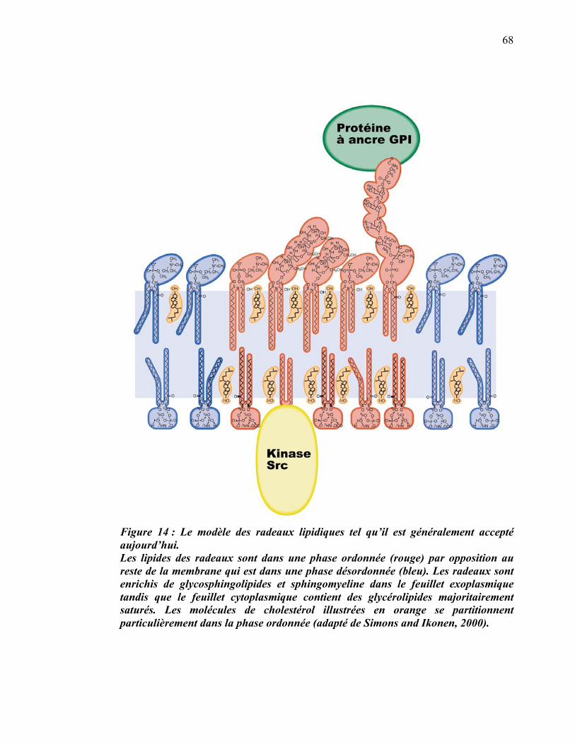
68
Figure 14 : Le modèle des radeaux lipidiques tel qu’il est généralement accepté aujourd’hui. Les lipides des radeaux sont dans une phase ordonnée (rouge) par opposition au reste de la membrane qui est dans une phase désordonnée (bleu). Les radeaux sont enrichis de glycosphingolipides et sphingomyeline dans le feuillet exoplasmique tandis que le feuillet cytoplasmique contient des glycérolipides majoritairement saturés. Les molécules de cholestérol illustrées en orange se partitionnent particulièrement dans la phase ordonnée (adapté de Simons and Ikonen, 2000).
69
Une nouvelle terminologie a été adoptée par les spécialistes de l’étude des radeaux
lipidiques lors du congrès international Keystone symposium on lipid rafts and cell
function, qui a eu lieu en mars 2006. À partir de ce moment, le terme de plus en plus
répandu et accepté est « radeau membranaire » (membrane rafts) et sa définition est :
Les radeaux membranaires sont de petits domaines (10-200 nm) hétérogènes,
hautement dynamiques et enrichis en stérol et sphingolipides qui compartimentalisent
des processus cellulaires. De petits radeaux peuvent parfois être stabilisés pour
former de plus grandes plateformes via des interactions protéine-protéine et protéine-
lipide.
Le rapport découlant de cette conférence mentionne que le terme lipid rafts a été
changé pour tenir compte des nouvelles observations selon lesquelles non seulement
les lipides, mais aussi les protéines sont responsables de la formation et du maintien de
ces microdomaines (Pike, 2006). La taille des radeaux membranaires a été ajustée à
10-200 nm pour tenir compte de la majorité des observations rapportées étant donné
que les sous-domaines de moins de 10 nm sont trop transitoires et s’apparentent trop
aux domaines formés par les fluctuations thermodynamiques des lipides (Veatch and
Keller, 2005). Les structures plus grandes telles que les synapses immunologiques sont
reconnues comme étant issues de la coalescence de plusieurs radeaux membranaires
(Huppa and Davis, 2003).
Les termes radeaux membranaires et microdomaines membranaires seront donc
utilisés pour le reste de cette thèse pour décrire la structure biologique qui forme un
sous-domaine de la membrane plasmique ou de la membrane d’une organelle comme
le phagosome. Le terme DRM sera utilisé pour mentionner le mélange de protéines et
de lipides qui flotte à l’interface supérieure d’un gradient de densité (sucrose ou autre)
suite à la solubilisation partielle de cellules ou d’organelles dans du TX-100 1% à 4°C.
Le reste de la membrane qui a été soumis au détergent, mais qui n’a pas flotté dans le
gradient, sera identifié DSM.
70
3.2 Hétérogénéité des lipides à la source des radeaux membranaires
Les membranes cellulaires sont composées d'une bicouche de lipides, ce sont des
lipides amphipathiques (des phospholipides dans la plupart des cas), chaque lipide
ayant sa tête polaire hydrophile (chargés négativement) orientée vers l’extérieur de la
membrane et sa queue hydrophobe orientée vers l'intérieur. Les phospholipides que
l’on retrouve le plus dans les membranes font partie du sous-groupe des
phosphoacylglycérol (ou glycérophospholipides). Ils se composent d'une molécule de
glycérol estérifiée à deux acides gras et un phosphate. Les deux acides gras de
longueur et de saturation variables sont nommés R-1 et R-2 (Figure 15A). Les
différents groupes polaires associés au phosphate définissent les classes de
phospholipides dont les pricipales sont les phosphatidyl choline (PC), les phosphatidyl
serine (PS), les phosphatidyl inositol (PI), les phosphatidyl ethanolamine (PE), et les
phosphatidyl glycerol (PG) .Un autre sous-groupe de lipides, particulièrement
important dans l’étude des radeaux membranaires est celui des sphingolipides. Chez
les sphingolipides, le porteur de la fonction alcool, comme le glycérol dans les
glycérophospholipides, est la sphingosine. Le représentant le plus abondant de ce
groupe est la sphyngomyéline (SM). Contrairement au modèle de la membrane
plasmique de Singer et Nicholson (Singer and Nicolson, 1972), l’hypothèse des
radeaux prévoit que la membrane n’est pas complètement fluide et ce, à cause de la
propension de certains lipides à se rassembler en microdomaines.
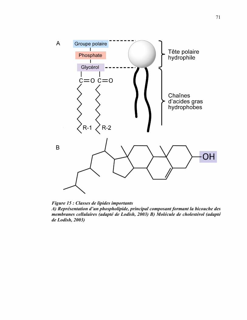
71
Figure 15 : Classes de lipides importants A) Représentation d’un phospholipide, principal composant formant la bicouche des membranes cellulaires (adapté de Lodish, 2003) B) Molécule de cholestérol (adapté de Lodish, 2003)
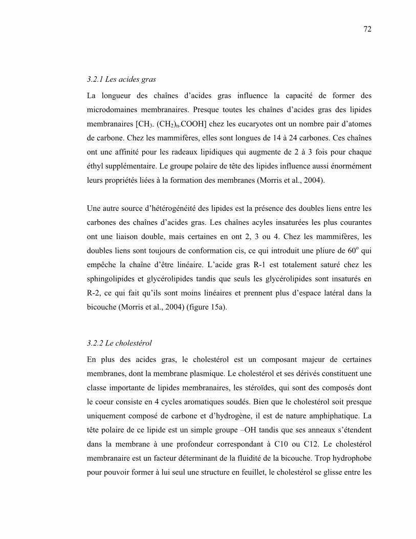
72
3.2.1 Les acides gras
La longueur des chaînes d’acides gras influence la capacité de former des
microdomaines membranaires. Presque toutes les chaînes d’acides gras des lipides
membranaires [CH3- (CH2)n-COOH] chez les eucaryotes ont un nombre pair d’atomes
de carbone. Chez les mammifères, elles sont longues de 14 à 24 carbones. Ces chaînes
ont une affinité pour les radeaux lipidiques qui augmente de 2 à 3 fois pour chaque
éthyl supplémentaire. Le groupe polaire de tête des lipides influence aussi énormément
leurs propriétés liées à la formation des membranes (Morris et al., 2004).
Une autre source d’hétérogénéité des lipides est la présence des doubles liens entre les
carbones des chaînes d’acides gras. Les chaînes acyles insaturées les plus courantes
ont une liaison double, mais certaines en ont 2, 3 ou 4. Chez les mammifères, les
doubles liens sont toujours de conformation cis, ce qui introduit une pliure de 60o qui
empêche la chaîne d’être linéaire. L’acide gras R-1 est totalement saturé chez les
sphingolipides et glycérolipides tandis que seuls les glycérolipides sont insaturés en
R-2, ce qui fait qu’ils sont moins linéaires et prennent plus d’espace latéral dans la
bicouche (Morris et al., 2004) (figure 15a).
3.2.2 Le cholestérol
En plus des acides gras, le cholestérol est un composant majeur de certaines
membranes, dont la membrane plasmique. Le cholestérol et ses dérivés constituent une
classe importante de lipides membranaires, les stéroïdes, qui sont des composés dont
le coeur consiste en 4 cycles aromatiques soudés. Bien que le cholestérol soit presque
uniquement composé de carbone et d’hydrogène, il est de nature amphiphatique. La
tête polaire de ce lipide est un simple groupe –OH tandis que ses anneaux s’étendent
dans la membrane à une profondeur correspondant à C10 ou C12. Le cholestérol
membranaire est un facteur déterminant de la fluidité de la bicouche. Trop hydrophobe
pour pouvoir former à lui seul une structure en feuillet, le cholestérol se glisse entre les
73
phospholipides : son groupe hydroxyle prend contact avec la phase aqueuse, près des
têtes polaires des phospholipides, alors que son groupe polycyclique stéroïde interfère
avec leurs chaînes acyles et tend à les immobiliser (figure 15B).
3.2.3 Les Gangliosides
Ce groupe comprend des molécules composées d’un céramide lié par un lien glycoside
à une chaîne d’oligosaccharides et d’acides N-acetylneuraminique (NANA, un sucre
acidique aussi appelé acide sialique) (Leray, 2009). Au niveau de la membrane
plasmique, les gangliosides sont ségrégués avec 1es autres sphingolipides et le
cholestérol dans les radeaux membranaires. Dans ces microdomaines, la grande
surface occupée par les chaînes d’oligosaccharide procure une courbature à la
membrane. Les fonctions biologiques des gangliosides sont principalement accomplies
grâce à leur localisation dans les radeaux membranaires ou dans les cavéoles. Le GM1
(monosialotetrahexosylganglioside), qui est le ganglioside prototypique, est un
membre du sous-groupe des gangliosides qui contiennent un seul résidu d’acide
sialique (Christie, 2008). Le GM1 a d’importantes propriétés physiologiques qui
influencent la plasticité et les mécanismes de réparation des neurones. En plus de ses
fonctions dans la physiologie du cerveau, le GM1 peut être lié par la toxine du choléra
(Mocchetti, 2005). Cette dernière caractéristique a permis de développer des
molécules permettant de détecter le GM1 à partir de la sous-unité B de la toxine du
cholera liée à un fluorophore ou une enzyme de détection.
3.3 Structure des radeaux membranaires
3.3.1 Composition lipidique des radeaux membranaires
On ne peut pas affirmer que la composition des DRMs est exactement celle des
radeaux membranaires, car le processus d’extraction lui-même modifie cette
composition (Lichtenberg et al., 2005). On a souvent mentionné le fait que la fraction
insoluble à 4°C ne correspond pas nécessairement à une structure dans la membrane in
vivo à 37°C. D’autres critiques indiquent que l’observation des radeaux par
74
microscopie n’est possible que par la coalescence artificielle de certains composants
de la membrane par les anticorps utilisés, mais la faisabilité biologique de cette
coalescence n’a été démontrée clairement que pour la synapse immunologique (Harder
and Engelhardt, 2004).
Quelques études récentes ont tenté, avec un certain succès, de répondre aux détracteurs
de l’hypothèse des radeaux et de l’isolation des DRMs. Chen et collaborateurs ont
montré qu’en utilisant les cations Mg2+ et K+ dans le tampon de solubilisation pour
imiter l’environnement intracellulaire, il est possible d’isoler des DRMs qui sont alors
stables même à 37°C (Chen et al., 2009). Les DRMs ainsi isolées avec la même
concentration de détergent contiennent les mêmes protéines et lipides que les DRMs
isolées à 4°C. De leur côté, Lingwood et collaborateurs ont montré que la membrane
est apte à faire la coalescence de ses nanodomaines en radeaux membranaires à 37°C
(Lingwood et al., 2008). Malgré ces différences et faute de caractérisation précise des
radeaux membranaires in vivo, la composition des DRMs est utilisée pour tenter
d’élucider la structure des radeaux membranaires.
Les DRMs sont enrichies en cholestérol par rapport aux membranes totales de la
cellule (32 mol% contre 12 mol%). On y trouve aussi un accroissement en
sphingomyéline (14 mol% contre 1 mol%) (Brown, 1994). On rapporte finalement un
accroissement moyen des glycolipides de 5 fois dans les microdomaines par rapport à
la cellule totale.
Le cholestérol interagit surtout avec les lipides à longue chaîne saturée ( � C14 ), cette
liaison reposant sur les interactions de van der Waals. Les liaisons doubles ne sont
tolérées qu’après les 12 premiers carbones. Le cholestérol agit comme espaceur
hydrophobe qui permet aux lipides saturés de s’empaqueter étroitement et donc
d’interagir davantage entre eux sur toute leur longueur. Le cholestérol se retrouve dans
les deux feuillets et le ratio cholestérol/lipide est en moyenne 30 mol% dans les
DRMs.
75
3.3.2 Composition différente des deux feuillets de la membrane
Puisque les sphingolipides sont situés seulement dans le feuillet externe de la bicouche
lipidique, le feuillet interne est a priori plus susceptible d’être solubilisé par les
détergents. Cependant les évidences expérimentales indiquent qu’il n’en est rien. De
ce point de vue, les détergents se révèlent donc très efficaces pour isoler les radeaux
membranaires. En bref, la composition générale des feuillets externes est caractérisée
par un enrichissement en sphingolipides, glycérophospholipides et cholestérol, et les
principales protéines à y prendre place sont les protéines à ancre GPI. Du côté interne
ou cytosolique, les lipides sont principalement des phospholipides saturés et du
cholestérol auxquels sont ancrées des protéines comme flotilline, stomatine, cavéoline
et les protéines kinases non-récepteurs (figure 14).
On sait que les microdomaines membranaires existent autant sur les feuillets internes
qu’externes, et plusieurs évidences nous permettent de croire qu’ils sont couplés même
si cette question a été longtemps débattue. Premièrement, il est possible de co-
immunopurifier les parties externes et internes des microdomaines (Morris et al.,
2004). Ensuite, des analyses statistiques, après observation au microscope électronique
(ME), ont montré que les deux couches sont associées (Prior et al., 2003). Une
indication fonctionnelle du couplage des couches externe et interne des radeaux
provient de l’étude d’un important groupe de virus incluant HIV-1. Lors du
bourgeonnement, le ciblage du feuillet interne des radeaux par le virus (via la protéine
Gag chez HIV-1) résulte en l’inclusion de composants du feuillet externe dans leur
capside (sphingolipides, protéines à ancre GPI) (Morris et al., 2004). La façon la plus
simple d’expliquer ce phénomène est que les deux feuillets sont couplés au niveau des
radeaux. Il est cependant clair que la partie non-radeaux de la membrane, aussi appelée
phase désordonnée, ne permet pas le couplage entre les deux feuillets à cause de la très
grande mobilité des lipides et de l’espace entre chaque molécule qui est à la fois grand
et variable (65-75 Å) (Scheiffele et al., 1999). Dans les microdomaines membranaires
enrichis en lipides ordonnés et pauvres en protéines, il semble plus probable que le
couplage dépend plus des lipides que des protéines. Le modeste groupe hydrophile du
76
cholestérol (-OH) n’est pas aussi fortement ancré à l’interface aqueuse que celui plus
gros des acides gras. Il en résulte que la molécule de cholestérol vibre constamment
dans l’axe des z (perpendiculaire à la surface de la membrane), pénétrant alors jusqu’à
30% dans le feuillet opposé (Endress et al., 2002). On a donc pensé que c’est un
phénomène d’« interdigitation » des chaînes de phospholipides (Simons and Toomre,
2000) et du cholestérol qui serait la force majeure du couplage.
Des données plus récentes nous apprennent que l’interdigitation des chaînes des
phospholipides n’est pas une force assez grande pour lier les deux feuillets et que la
présence du cholestérol l’abolit (McIntosh et al., 1992; Schram and Thompson, 1995).
Le cholestérol pourrait cependant être en grande partie responsable de ce couplage par
le phénomène de la translocation interfeuillets. Ce modèle est basé sur le fait que le
cholestérol peut être échangé librement entre les deux feuillets pour atteindre
l’équilibre de son potentiel chimique (μc) contrairement aux phospholipides dont les
échanges interfeuillets sont régulés par des enzymes (Collins, 2008).
Les propriétés des microdomaines (peu élastiques et peu d’espace entre les lipides)
nous permettent de penser que ces domaines sont naturellement très planaires.
Cependant les radeaux membranaires existent dans différentes membranes courbées
[endosome (Kirkham and Parton, 2005; Nada et al., 2009), phagosome (Dermine et al.,
2001; Dermine et al., 2005) péroxisome (Boukh-Viner et al., 2005)]. Dans tous ces
cas, une ou plusieurs protéines insérées en abondance semblent être ce qui permet aux
deux feuillets de rester couplés malgré la courbure. Parmi les candidates pour jouer ce
rôle, on retrouve la cavéoline pour les courbures concaves et la prominine pour les
courbures convexes.
3.3.3 Autres caractéristiques des radeaux membranaires
Trop petits pour la résolution de la microscopie à fluorescence, mais suffisamment
grands pour celle du microscope électronique, il se peut que les radeaux membranaires
aient été observés par ME durant de nombreuses années sans qu’on le sache. Ces
77
domaines sont relativement pauvres en protéines, notamment en protéines
transmembranaires : cette caractéristique nous permet de les reconnaître comme étant
une zone de plus faible densité protéique. Individuellement petits, mais pouvant être
très nombreux, ils couvrent de 20 % (cellules musculaires) à 30-40 % (lumière du
Golgi) de la surface de la membrane. Ils couvrent vraisemblablement plus de surface
relative du côté apical des cellules épithéliales (Morris et al., 2004).
3.4 Partitionnement sélectif des protéines dans les radeaux membranaires
3.4.1 Ancres lipidiques
Un des déterminants importants de la présence d’une protéine dans les radeaux
membranaires est la présence d’une ancre glycosylphosphatidylinositol (GPI) sur cette
protéine (Chatterjee and Mayor, 2001). La majorité des protéines à ancre GPI connues
se retrouvent dans les DRMs (Rietveld et al., 1999). Des protéines ayant différentes
fonctions telles que la signalisation, l’immunité, la pathologie du prion ou du
trypanosome, se retrouvent liées aux DRMs grâce à leur ancre GPI. Une ancre GPI est
une structure glycolipidique ajoutée de façon post-traductionelle en C-terminal de
nombreuses protéines eucaryotes. Cette modification permet à la protéine de s’attacher
au feuillet externe de la membrane plasmique (Tiede et al., 1999). L’ancre GPI est
constituée de trois domaines: i) un lien phosphoéthanolamine ; ii) un cœur de glucides
très conservé sur lequel sont ajoutées différentes chaînes latérales ; iii) une queue de
phospholipides variant de 14 à 28 carbones pouvant être saturée ou insaturée (Paulick
and Bertozzi, 2008) (figure 16). Une des principales conditions connues pour le
partitionnement des ancres GPI dans les DRMs est la longueur de la chaîne
d’hydrocarbones (�C16). Les protéines à ancre GPI (GPI-APs pour GPI anchored
proteins) ont généralement deux chaînes d’acides gras saturés dans leur partie
phosphatidylinositol (PI) à la différence des autres molécules de PI de la cellule qui
ont une insaturation en sn2. Cette saturation générée par les protéines PGAP2 et
PGAP3 semble importante pour la localisation dans les DRMs puisque, dans les
cellules mutantes pour ces deux enzymes, les GPI-APs ont une insaturation et ne se
retrouvent pas dans les DRMs (Maeda et al., 2007). Il est possible que ces chaînes

78
insaturées (donc droites) s’associent avec deux chaînes insaturées tout aussi droites de
la partie céramide des sphingolipides et/ou avec la chaîne hydrocarbonée du
cholestérol (Kinoshita et al., 2008). Ce lien GPI-APs avec les principaux constituants
des radeaux membranaires pourrait expliquer le grand enrichissement des GPI-APs
dans les DRMs.
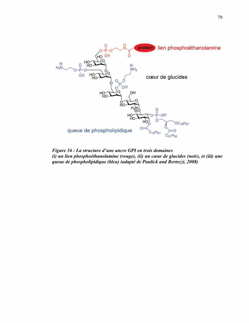
79
Figure 16 : La structure d’une ancre GPI en trois domaines (i) un lien phosphoéthanolamine (rouge), (ii) un cœur de glucides (noir), et (iii) une queue de phospholipidique (bleu) (adapté de Paulick and Bertozzi, 2008)
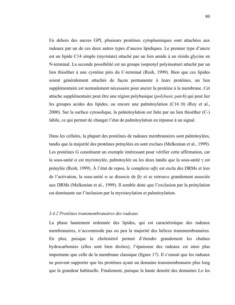
80
En dehors des ancres GPI, plusieurs protéines cytoplasmiques sont attachées aux
radeaux par un de ces deux autres types d’ancres lipidiques. Le premier type d’ancre
est un lipide C14 simple (myristate) attaché par un lien amide à un résidu glycine en
N-terminal. La seconde possibilité est un groupe isoprenyl polyinsaturé attaché par un
lien thioéther à une cystéine près du C-terminal (Resh, 1999). Bien que ces lipides
soient généralement attachés de façon permanente à leurs protéines, un lien
supplémentaire est normalement nécessaire pour ancrer la protéine à la membrane. Cet
attache supplémentaire peut être une région polybasique (polybasic patch) qui peut lier
les groupes acides des lipides, ou encore une palmitoylation (C16 :0) (Roy et al.,
2000). Sur la surface cytosolique, la palmitoylation est faite par un lien thioéther (C-)
labile, ce qui permet de changer l’état de palmitoylation en réponse à un signal.
Dans les cellules, la plupart des protéines de radeaux membranaires sont palmitoylées,
tandis que la majorité des protéines prénylées en sont exclues (Melkonian et al., 1999).
Les protéines G constituent un exemple intéressant pour vérifier cette affirmation, car
la sous-unité � est myristoylée, palmitoylée ou les deux tandis que la sous-unité � est
prénylée (Resh, 1999). À l’état de repos, le complexe ��� est exclu des DRMs et lors
de l’activation, la sous-unité � se dissocie de �� et se retrouve grandement associée
aux DRMs (Melkonian et al., 1999). Il semble donc que l’exclusion par la prénylation
est dominante sur l’inclusion par la myristoylation et palmitoylation.
3.4.2 Protéines transmembranaires des radeaux
La phase hautement ordonnée des lipides, qui est caractéristique des radeaux
membranaires, n’accommode pas ou peu la majorité des hélices transmembranaires.
En plus, puisque le cholestérol permet d’étendre grandement les chaînes
hydrocarbonées (elles sont bien droites), l’épaisseur des radeaux est ainsi plus
importante que celle de la membrane classique (figure 17). Il s’ensuit que les radeaux
ne peuvent supporter que les protéines ayant un domaine transmembranaire plus long
que la grandeur habituelle. Finalement, puisque la haute densité des domaines Lo les

81
rend moins perméables aux ions, leur potentiel membranaire est plus grand et ceci
altère la conformation des domaines transmembranaires.
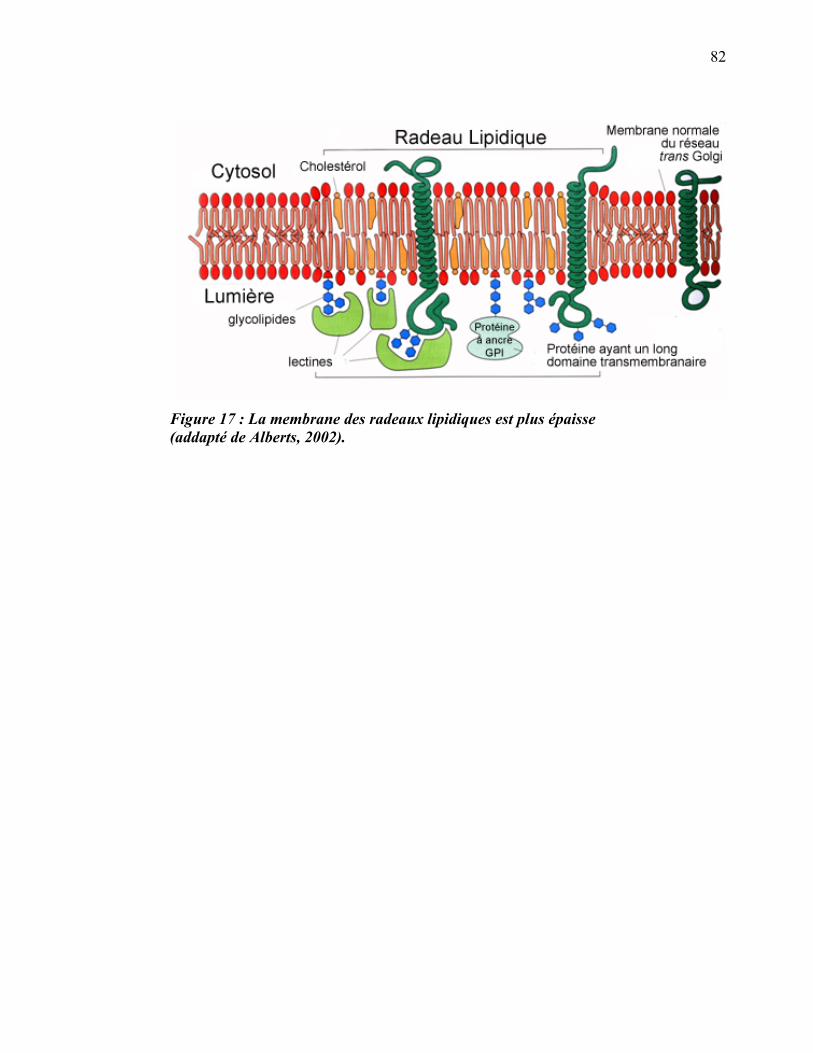
82
Figure 17 : La membrane des radeaux lipidiques est plus épaisse (addapté de Alberts, 2002).
83
Qu’en est-il alors de ces protéines transmembranaires qui occupent, malgré tout, les
radeaux lipidiques de façon constitutive? La majorité de celles-ci surmontent les
problèmes mentionnés précédemment en ayant plusieurs palmitates attachés de façon
covalente à la région juxtamembranaire de leur région cytoplasmique. Ces palmitates
sembleraient agir comme un bouclier, protégeant le domaine transmembranaire pour
lui permettre de demeurer dans la phase Lo. Certaines protéines semblent avoir un
domaine transmembranaire adapté aux radeaux. Par exemple, la protéine
hémagglutinine d’influenza (HA), qui est dirigée vers les microdomaines et s’y insère,
possède un domaine transmembranaire composé de 27 acides aminés hydrophobes et
non de 25 comme c’est généralement le cas (Morris et al., 2004).
3.4.3 Protéines structurales des radeaux membranaires
3.4.3.1 Les cavéolines
Une des protéines les plus typiques des radeaux membranaire est la cavéoline. En fait,
l’insertion de la cavéoline dans la membrane plasmique forme des invaginations de
50-80 nm nommées cavéoles (Ikonen et al., 2004). Les cavéoles sont enrichies de
sphingolipides et de cholestérol, se retrouvent dans la fraction DRMs à la suite d’un
traitement au Triton X-100 et sont considérées comme un sous-groupe de radeaux
membranaires présent dans les cellules exprimant la cavéoline (Simons and Ikonen,
2000). Il y a trois membres dans la famille des cavéolines. Cavéoline-1 (anciennement
cavéoline) est co-exprimée avec cavéoline-2 dans les cellules comportant de
nombreuses cavéoles telles que les cellules endothéliales, les adipocytes et les
fibroblastes tandis que cavéoline-3 est spécifique aux cellules musculaires (Ikonen et
al., 2004). Pour ce qui est des macrophages de souris, des résultats contradictoires ont
été publiés, mais il semble finalement que cavéoline-1 et 2 soient exprimées dans les
macrophages primaires tandis que seule cavéoline-2 est exprimée chez les
macrophages J774 (Gargalovic and Dory, 2001). Dans le cas des J774, il n’est pas
étonnant de retrouver principalement cavéoline-2 dans l’appareil de Golgi puisqu’il a
été démontré que le transport de cavéoline-2 à la membrane plasmique est dépendant
84
de cavéoline-1 (Parolini et al., 1999). Les cavéolines sont impliquées principalement
dans la régulation du transport des lipides tels que le cholestérol et la modulation de la
signalisation cellulaire (Gargalovic and Dory, 2001).
3.4.3.2 Les flotillines
Reggie-1 et 2 ont originalement été identifiées dans les neurones du nerf optique en
régénération des poissons rouges (Carassius auratus) (Schulte et al., 1997). Ces
protéines ont subséquemment été identifiées après la flottaison des complexes
insolubles (DRMs) de cellules endothéliales, ce qui leur a valu le nouveau nom de
flotillines (flotilline-1 étant reggie-2 et vice-versa) (Bickel et al., 1997). Dans les
cellules n’exprimant pas de cavéoline (ex. : érythocytes, plaquettes, lymphocytes), les
protéines majeures des radeaux lipidiques font partie d’une large famille de protéines
dont les flotillines et les stomatines définissent les deux types principaux. Les
flotillines et stomatines se lient à la surface cytoplasmique des radeaux, probablement
par palmitoylation. Les stomatines diffèrent des flotillines par 25 acides aminés
supplémentaires en N-terminal.
Les flotillines ont deux domaines hydrophobes, et il a été suggéré qu’ils sont des
domaines transmembranaires (Bickel, 2002). Le second de ces domaines n’est que de
18 acides aminés, ce qui est la bonne longueur pour un domaine TM dans la
membrane de l’appareil de Golgi. Puisqu’un fragment de 14 kDa en N-terminal est
protégé de la protéolyse du côté cytoplasmique du Golgi, il est probable que le
domaine de 18 acides aminés traverse effectivement la membrane du Golgi (qui est
fortement enrichie en radeaux lipidiques) (Gkantiragas et al., 2001). Cependant,
flotilline-1 exprimée à la membrane plasmique n’est pas protégée contre la protéolyse
cytoplasmique et son lien à la membrane dépend principalement de la palmitoylation.
Cette flotilline de la membrane plasmique ne passe pas par le Golgi durant sa
biosynthèse, ce qui suggère qu’il y a au moins deux routes biosynthétiques qui
diffèrent selon la destination de cette protéine (Morrow et al., 2002). Les flotillines
contiennent de plus un domaine peu caractérisé appelé le domaine d’homologie à la
85
prohibitine (PHB). Ce domaine est conservé évolutivement et est partagé par plusieurs
protéines membranaires eucaryotes et procaryotes. Bien que les protéines ayant un
domaine PHB montrent des différences pour leur localisation intracellulaire, de
nombreux membres de cette famille sont décrits comme étant associés aux DRMs
(Morrow et al., 2002). Ceci laisse sous-entendre que le domaine PHB a un rôle à jouer
dans l’association avec les radeaux membranaires. L’équipe de Parton a d’ailleurs
démontré que l’association aux membranes ainsi que le ciblage vers les
microdomaines sont en grande partie dépendants d’une palmitoylation sur la cystéine
34 située dans le domaine PHB (Morrow et al., 2002).
Il y a plusieurs fonctions potentielles des cavéolines, des stomatines et des flotillines
qui ont été avancées : régulation du contenu en lipides et en protéines de leur domaine,
ciblage moléculaire, guidage de leur sous-domaine vers un compartiment cellulaire
particulier, liaison directe avec certaines protéines en les restreignant ainsi aux
radeaux. Dans leur revue, Morris et collaborateurs notent que la flotilline est souvent
exprimée au niveau des membranes concaves [phagosomes (Dermine et al., 2001),
endosomes tardifs (Gagescu et al., 2000), vésicules d’exportation
biosynthétiques (Marchand et al., 2002)], mais pas dans les membranes convexes
[vésicules secrétées des érythrocytes (Salzer et al., 2002) ou les granules alpha des
plaquettes (Mairhofer et al., 2002)].En plus de remplir des fonctions potentielles dans
la structure des radeaux membranaires, la flotilline est impliquée dans la signalisation
de l’insuline pour engendrer la redistribution du transporteur de glucose dans les
adipocytes (Baumann et al., 2000).
3.4.4 Méthodes d’analyse des protéines des radeaux membranaires
3.4.4.1 Isolation des DRMs
L’isolation des DRMs a été la méthode la plus utilisée pour étudier les protéines des
radeaux membranaires. Cette méthode a l’avantage d’être utilisable sur de grandes
quantités de cellules dans des études de cartographie protéomique. C’est de plus une
méthode parfaite pour faire une approche systématique lorsqu’on veut étudier les
protéines des radeaux lipidiques d’une cellule ou d’une organelle pour la première
86
fois, car en plus d’être relativement simple à mettre au point et à pratiquer, elle est
rapide et nécessite seulement une ultracentrifugeuse comme équipement spécialisé. Le
coût raisonnable des réactifs pour cette technique la rend disponible pour tous les
laboratoires de recherche, contrairement à certaines autres techniques. Cependant,
comme il a été mentionné précédemment, cette méthode a quelques désavantages et
c’est pourquoi différentes solutions de remplacement ont été développées. Brièvement,
le principal désavantage de cette technique est qu’il a été démontré que la composition
lipidique des DRMs n’est pas identique à celle des radeaux membranaires
(Lichtenberg et al., 2005). La solubilisation des membranes pour former des micelles
ne dépend pas seulement de la composition en lipides de cette membrane, mais aussi
du détergent utilisé et des conditions expérimentales telles que la composition de la
phase aqueuse et la température. De plus, la prémisse qui sous-tend que la grande
concentration en cholestérol et en sphingomyéline est nécessairement la cause de
l’insolubilité des radeaux membranaires s’est avérée fausse après des essais avec des
membranes artificielles (Lichtenberg et al., 2005). Finalement, il reste possible que les
protéines extraites par le détergent soient sélectionnées selon leurs propriétés
physiques (hydrophobicité, longueur du domaine transmembranaire) plutôt que selon
leur distribution dans les microdomaines (Munro, 2003). Malgré ces quelques défauts,
cette méthode est de loin celle qui a permis le plus grand nombre de découvertes dans
le domaine des radeaux membranaires. Classiquement, cette méthode consiste en
l’extraction des protéines avec des détergents suivie de l’analyse des protéines
effectuée par immunobuvardage, spectrométrie de masse ou par une combinaison de
ces deux techniques.
3.4.4.1.1 Extraction de protéines par des détergents
Les détergents sont des molécules amphipathiques qui désorganisent les membranes
en s’intercalant dans les bicouches phospholipidiques et en solubilisant les lipides et
les protéines. Certains détergents sont des produits naturels (exemple : désoxycholate
de sodium), mais la plupart sont des molécules de synthèse élaborées pour le nettoyage
ou la dispersion des mélanges d’huile et d’eau. Les détergents ioniques, comme le
87
sodium dodécylsulfate (SDS), contiennent un groupement chargé (figure 18A). Les
détergents non ioniques tels que le TX-100 et l’octyglucoside sont dépourvus de
groupement chargé. À des concentrations très faibles, les détergents se solubilisent
dans l’eau et prennent la forme de molécules isolées. Lorsque la concentration
augmente, les molécules commencent à former des micelles, de petits agrégats
sphériques dans lesquels les parties hydrophiles des molécules sont dirigées vers
l’extérieur et les parties hydrophobes regroupées au centre (figure 18B). La
concentration micellaire critique (CMC) à laquelle les micelles se forment est
caractéristique de chaque détergent et dépend des structures de ses parties
hydrophobes et hydrophiles. Les détergents ioniques se fixent aux régions
hydrophobes exposées des protéines membranaires ainsi qu’aux parties centrales
hydrophobes des protéines hydrosolubles, ce qui les dénature. Les détergents non
ioniques ne dénaturent pas les protéines et sont donc utiles pour les extraire des
membranes avant de les purifier. Ces détergents agissent de manière différente selon
leur concentration. À des concentrations supérieures à la CMC, ils solubilisent les
membranes biologiques en formant des micelles mixtes de détergent, de
phospholipides et de protéines membranaires intrinsèques (figure 18C).
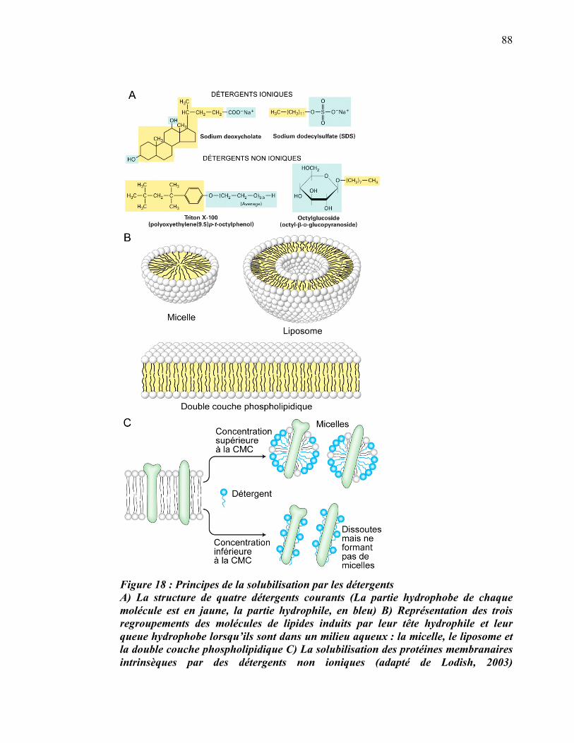
88
Figure 18 : Principes de la solubilisation par les détergents A) La structure de quatre détergents courants (La partie hydrophobe de chaque molécule est en jaune, la partie hydrophile, en bleu) B) Représentation des trois regroupements des molécules de lipides induits par leur tête hydrophile et leur queue hydrophobe lorsqu’ils sont dans un milieu aqueux : la micelle, le liposome et la double couche phospholipidique C) La solubilisation des protéines membranaires intrinsèques par des détergents non ioniques (adapté de Lodish, 2003)
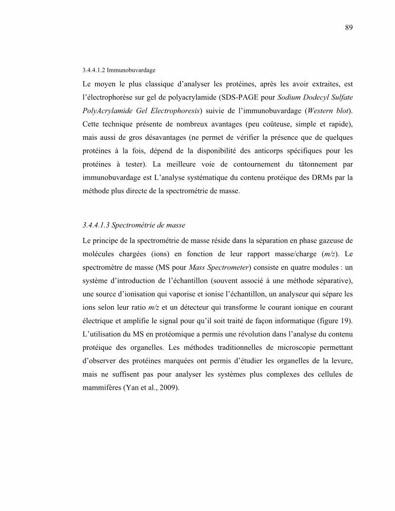
89
3.4.4.1.2 Immunobuvardage
Le moyen le plus classique d’analyser les protéines, après les avoir extraites, est
l’électrophorèse sur gel de polyacrylamide (SDS-PAGE pour Sodium Dodecyl Sulfate
PolyAcrylamide Gel Electrophoresis) suivie de l’immunobuvardage (Western blot).
Cette technique présente de nombreux avantages (peu coûteuse, simple et rapide),
mais aussi de gros désavantages (ne permet de vérifier la présence que de quelques
protéines à la fois, dépend de la disponibilité des anticorps spécifiques pour les
protéines à tester). La meilleure voie de contournement du tâtonnement par
immunobuvardage est L’analyse systématique du contenu protéique des DRMs par la
méthode plus directe de la spectrométrie de masse.
3.4.4.1.3 Spectrométrie de masse
Le principe de la spectrométrie de masse réside dans la séparation en phase gazeuse de
molécules chargées (ions) en fonction de leur rapport masse/charge (m/z). Le
spectromètre de masse (MS pour Mass Spectrometer) consiste en quatre modules : un
système d’introduction de l’échantillon (souvent associé à une méthode séparative),
une source d’ionisation qui vaporise et ionise l’échantillon, un analyseur qui sépare les
ions selon leur ratio m/z et un détecteur qui transforme le courant ionique en courant
électrique et amplifie le signal pour qu’il soit traité de façon informatique (figure 19).
L’utilisation du MS en protéomique a permis une révolution dans l’analyse du contenu
protéique des organelles. Les méthodes traditionnelles de microscopie permettant
d’observer des protéines marquées ont permis d’étudier les organelles de la levure,
mais ne suffisent pas pour analyser les systèmes plus complexes des cellules de
mammifères (Yan et al., 2009).
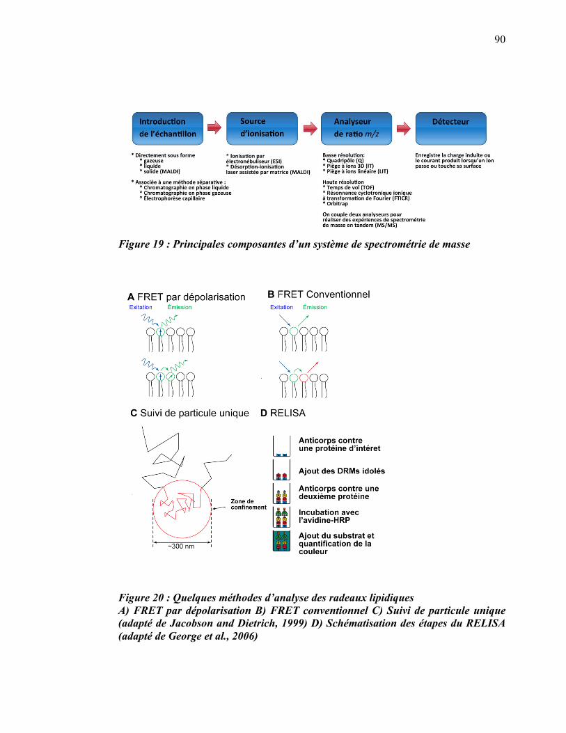
90
Figure 19 : Principales composantes d’un système de spectrométrie de masse
Figure 20 : Quelques méthodes d’analyse des radeaux lipidiques A) FRET par dépolarisation B) FRET conventionnel C) Suivi de particule unique (adapté de Jacobson and Dietrich, 1999) D) Schématisation des étapes du RELISA (adapté de George et al., 2006)
91
3.4.4.2 Isolation sans détergent
Une méthode alternative basée sur la sonication des cellules a été développée pour
isoler les radeaux membranaires sans les biais de la méthode avec le détergent (Smart
et al., 1995; Song et al., 1996). Un protocole plus rapide et d’une plus grande
reproductibilité a été développé en remplaçant la sonication par le passage dans une
seringue (Macdonald and Pike, 2005). La pureté de l’isolation a aussi été améliorée
grâce au remplacement de l’EDTA par un tampon contenant du calcium et du
magnésium. Cependant, ce protocole n’a jamais obtenu la popularité de la méthode
originale avec le TX-100.
3.4.4.3 Microscope photonique
L’observation directe des radeaux membranaires n’est pas possible par microscopie
photonique, car sa résolution (> 100 nm) est trop petite. Cependant, des techniques
spécialisées ont permis d’évaluer le rayon des radeaux à 26 ± 13 nm (Pralle et al.,
2000). Cette pratique de la microscopie étant la plus accessible, on l’a souvent utilisée
pour confirmer la présence de certaines protéines dans les radeaux membranaires.
Malgré la résolution insuffisante, le microscope confocal utilisé en conjonction avec la
microscopie à fluorescence a permis de démontrer la localisation plus ségréguée de
certaines protéines par rapport à d’autres. Cette méthode a souvent été utilisée pour
confirmer la localisation de deux protéines dans des sous-domaines distincts de la
membrane (Dermine et al., 2005; Malinska et al., 2003). Cette approche est aussi un
moyen permettant de démontrer qu’un certain stimulus provoque la translocation
d’une protéine vers les microdomaines (Janes et al., 1999; Stauffer and Meyer, 1997).
3.4.4.3.1 Transfert d'énergie entre molécules fluorescentes
Une méthode alternative développée plus récemment est devenue très populaire pour
caractériser les radeaux membranaires : le transfert d'énergie entre molécules
fluorescentes (FRET pour Fluorescence Resonance Energy Transfer). Le FRET
92
permet d’observer la distribution de molécules à une échelle inférieure à celle de la
microscopie confocale (< 300 nm). Cette méthode est basée sur le transfert d’énergie
entre un fluorophore donneur à l’état excité vers un accepteur à l’état basal
(figure 20B). Ce transfert ne nécessite pas de contact direct, mais l’efficacité est
relative à la proximité entre le donneur et l’accepteur (1-10 nm). La technique du
FRET permet donc de détecter si la distribution entre deux molécules est
complètement aléatoire ou si elle ne l’est pas (ce qui arrive lorsque ces molécules sont
ségréguées dans des microdomaines). Il y a deux principales façons d’utiliser ce
transfert d’énergie dans l’étude des microdomaines : i) mesurer la diminution de
fluorescence par désactivation (quenching) du donneur ou ii) mesurer l’augmentation
de fluorescence de l’accepteur. Les conclusions obtenues par ces méthodes peuvent
être validées en photoblanchissant (photobleaching) l’accepteur, ce qui va changer son
spectre d’absorption et libérer le donneur de sa désactivation.
Une des premières démonstrations de la présence de microdomaines dépendant du
cholestérol ayant été faite par FRET est celle de Nichols en 2003 (Nichols, 2003). Il a
démontré qu’un regroupement se produit entre les molécules de GM1 étiquetées avec
la sous-unité B de la toxine de choléra (GM1-CTB) et la protéine fluorescente verte
(GFP pour Green Fluorescent Protein) couplée à une ancre GPI (GPI-GFP) tandis que
le récepteur de la transferrine (Tf-R) (un marqueur classique des non-radeaux) ne se
regroupe pas en microdomaines.
Le FRET a aussi été utilisé pour examiner la taille des microdomaines et a permis
d’évaluer que chacun contient aussi peu que 3-4 protéines à ancre GPI (Sharma et al.,
2004). Un problème dans l’analyse spatio-temporelle d’une molécule cible par FRET
est que la seconde molécule utilisée pour le transfert d’énergie (le biosenseur) est elle-
même soumise à une diffusion dans la membrane. Très récemment et en utilisant une
méthode de FRET permettant d’éliminer l’effet de la diffusion du biosenseur, une
équipe a démontré qu’après l’activation par EGF, les protéines Src de la membrane
sont relocalisées et se regroupent dans les radeaux membranaires (Lu et al., 2008).
93
3.4.4.3.2 Suivi de particule unique
Le suivi de particule unique (SPT pour Single Particle Tracking) est une autre
méthode de microscopie photonique permettant de franchir la limite de la résolution
optique restreinte par la diffraction. Des informations sur l’hétérogénéité des
membranes cellulaires peuvent être obtenues par cette technique et ses variantes en
caractérisant le mouvement d’une molécule par rapport au mouvement causé par le
mouvement Brownien (Saxton and Jacobson, 1997). Les études de SPT détectent ce
qu’on appelle des zones transitoires de confinement (figure 20C) qui ont été évaluées à
200-300 nm. Cette méthode a permis de supporter l’hypothèse des radeaux en
montrant que 35-37% de la protéine à ancre GPI Thy-1 et du lipide GM1 sont ainsi
confinés (Sheets et al., 1997).
3.4.4.3.3 Autres méthodes de microscopie photonique
D’autres méthodes ont été utilisées durant les dix dernières années, mais avec moins
de succès que celles présentées plus haut. Le recouvrement de fluorescence après
photoblanchiment (FRAP pour Fluorescence Recovery After Photobleaching) est une
méthode établie depuis longtemps pour examiner la mobilité translationelle des
molécules fluorescentes dans les membranes cellulaires. Une des études les plus
exhaustives des radeaux membranaires par cette approche démontre que le FRAP n’est
pas adéquat pour fournir une conclusion définitive sur l’existence et la nature des
microdomaines membranaires (Kenworthy et al., 2004).
3.4.4.4 Microscope électronique
Une étude sur la taille des radeaux membranaires par microscopie électronique a
évalué à 20-22 nm le rayon des zones contenant des billes d’or rattachées au marqueur
de radeaux GFP-th (Prior et al., 2003), ce qui situerait le diamètre des radeaux
membranaires entre 15 et 44 nm en tenant compte de la longueur des anticorps et du
rayon des particules d’or (Parton and Hancock, 2004). D’autres études de localisation
de deux marqueurs de radeaux (Thy-1 + LAT, Fc�RI ou la protéine du prion) montrent
la présence de domaines élémentaires d’environ 50 nm pouvant fusionner pour former

94
des super-rafts excédant parfois 500 nm (Madore et al., 1999; Wilson et al., 2004). Fait
intéressant : plusieurs de ces études démontrent que le lipide GM1 se retrouve dans
des microdomaines différents des marqueurs protéiques, ce qui suggère la présence de
différents types de microdomaines dans une même membrane (Lagerholm et al.,
2005).
3.4.4.5 Cytométrie en flux
Matkó et collaborateurs ont développé une technique basée sur la cytométrie en flux
pour identifier des protéines associées aux radeaux membranaires. En utilisant des
anticorps fluorescents contre les protéines à tester, ils vérifient la variation de
fluorescence après un traitement au détergent (Gombos et al., 2004). Les protéines
ayant la moins grande sensibilité au traitement sont considérées comme les protéines
de DRMs. Un contrôle fait avec la méthyl-�-cyclodextrine (M�CD) permet de vérifier
que cette insolubilité est bel et bien induite par l’association à un microdomaine riche
en cholestérol et non pas à un ancrage au cytosquelette. Cette méthode a été utilisée
par Wolf et collaborateurs pour comparer l’association aux radeaux des principaux
antigènes de la membrane plasmique des monocytes au repos par rapport aux
monocytes activés. Ils ont pu ainsi confirmer les résultats qu’une autre équipe avait
obtenus par la méthode du FRET, ces résultats étant que CD14, CD32 et CD55 sont
constitutivement associés aux DRMs, CD81 l’est suite à l’activation au LPS tandis que
CD71 est soluble (Pfeiffer et al., 2001).
Dernièrement, Morales-García et collaborateurs ont utilisé le cytomètre en flux pour
analyser l’hétérogénéité des radeaux membranaires (Morales-Garcia et al., 2008). Leur
but était de développer une méthode pour identifier deux molécules ou plus dans un
radeau en particulier afin de comparer l’ensemble de ces sous-domaines et de
confirmer que différents radeaux ont une composition différente comme l’ont suggéré
plusieurs équipes (Pike, 2004). Cette méthode consiste premièrement à fractionner les
membranes des cellules par un procédé d’extraction des radeaux sans détergent
(Macdonald and Pike, 2005), et deuxièmement à utiliser des anticorps fluorescents

95
contre les molécules ciblées pour analyser le tout dans le cytomètre. Le GM1 étant
utilisé comme marqueur des radeaux, cette méthode a permis à Morales-Garcia et
collaborateurs de distinguer, par exemple, des populations de radeaux GM1+/CD14+
et d’autres GM1+/CD14-.
96
3.4.4.6 ELISA
Une autre approche permettant d’observer l’hétérogénéité des radeaux membranaires
est basée sur la technique de dosage immunoenzymatique sur support solide (ELISA
pour Enzyme-Linked ImmunoSorbent Assay) (George et al., 2006). Il est de plus en
plus clair que l’activation des lymphocytes T provoque une coalescence de radeaux
membranaires comprenant les récepteurs des cellules T (TCR pour T-Cell Receptor) et
les protéines adaptatrices nécessaires à la transduction du signal. Cependant, les
méthodes d’isolation des DRMs et d’analyse par SDS-PAGE (ou même par MS) ne
permettent pas de distinguer si les protéines sont à l’intérieur d’un même radeau
membranaire ou dans deux sous-domaines semblables mais distincts. Le RELISA
(pour Raft ELISA) a donc été développé pour remédier à cette lacune (figure 20D).
D’abord, un anticorps contre une protéine spécifique des radeaux membranaires est
fixé au fond d’une plaque de microtitration. Puis, des DRMs sont ajoutées et, après
lavage des membranes non liées, sont incubées avec un anticorps biotinylé détectant
une seconde protéine des radeaux membranaires (ou une protéine qui s’y trouve
potentiellement). L’étape suivante est l’ajout de l’avidine-HRP qui lie spécifiquement
la biotine de l’anticorps et permet un dosage grâce à l’activité enzymatique de la partie
HRP (HorseRadish Peroxidase). Il suffit ensuite d’ajouter un substrat chromogénique
qui change de couleur sous l’action du HRP en présence de H2O2. Ce changement de
couleur est finalement dosé dans un spectrophotomètre. L’étude réalisée en utilisant
cette méthode a démontré que, même si la majorité des DRMs des cellules T
comprennent la protéine à ancre GPI Thy-1, elles diffèrent pour ce qui est des
protéines de signalisation telles que CD3-� (George et al., 2006).
97
3.5 Fonctions des radeaux membranaires
3.5.1 Plateforme de signalisation
Une protéine qui se partitionne dans les radeaux membranaires est, par le fait même,
généralement concentrée. En effet, certaines kinases sont présentes à 20-80% dans ces
microdomaines qui couvrent une surface d’environ 10-20% de la membrane totale. Le
fait d’avoir en si grande proportion une protéine dans des domaines aussi restreints
explique qu’elles y sont concentrées. Ceci peut s’avérer un phénomène important pour
une protéine qui doit s’associer pour induire une cascade de signalisation, par
exemple. Un grand nombre de récepteurs sont connus pour être dirigés vers les
microdomaines à un moment ou un autre de leur cycle (exemple : certains récepteurs à
7 domaines trans-membranaires couplés aux protéines G). Ces récepteurs ainsi que
leurs effecteurs sont majoritairement palmitoylés à différents degrés. On a aussi
démontré que ces microdomaines contiennent plusieurs protéines acylées comme les
kinases de la famille Src (ex : Fyn, Lck) (Simons and Ikonen, 1997). Cette
compartimentalisation de la membrane permet aussi d’autres mécanismes cellulaires
importants dont un des mieux décrits est la synapse immunologique (Miyoshi et al.,
1999).
3.5.2 Synapse immunologique
La synapse immunologique est définie comme étant l’interface stable et aplatie formée
entre un lymphocyte activé et la cellule qu’il est en train de reconnaître (Huppa and
Davis, 2003). Il a été démontré que les molécules clés de la signalisation des
lymphocytes T, le TCR et l’intégrine LFA1 (abréviation de Leukocyte Function
Associated antigen 1), ne sont pas seulement présentes à l’interface de la synapse
immunologique, mais qu’elles sont groupées en zones grandement organisées. Ces
zones sont appelées complexes d’activation supra-moléculaires (SMACs pour Supra-
Molecular Activation Complexes). Le centre des SMACs est enrichi du TCR et d’un
de ses effecteurs, la protéine kinase � (PKC-�). Les protéines impliquées dans
98
l’adhésion cellulaire (LFA1, taline) sont, quant à elles, situées dans la région
périphérique des SMACs tandis que les plus grosses molécules impliquées dans la
synapse (CD43, CD45) sont encore plus distales (Huppa and Davis, 2003). La
localisation spatio-temporelle de ces protéines est le facteur clé permettant les activités
de signalisation entre les cellules participant à la synapse immunologique. L’hypothèse
selon laquelle les radeaux membranaires sont impliqués dans cette organisation a été
corroborée quand il a été découvert que l’acylation de Lck et la palmitoylation de LAT
sont nécessaires à la transmission du signal émanant du TCR. En effet, ces deux
modifications déterminent fréquemment la localisation des protéines dans les
domaines enrichis de cholestérol. Par la suite, cette idée a été à nouveau renforcée par
des expériences montrant que la déprivation de cholestérol membranaire, qui
déstabilise les radeaux membranaires, permet de moduler la signalisation du TCR
(Kabouridis and Jury, 2008).
3.5.3 Fusion membranaire
L’eau est exclue des microdomaines ordonnés, car il y a peu d’accepteurs ou de
donneurs de ponts hydrogène disponibles pour les molécules d’eau, puisqu’un fort
réseau de liaisons hydrogène existe entre les différents lipides. Ceci permet à deux
membranes de se rapprocher plus que la normale et favorise leur fusion (Tatulian and
Tamm, 2000). Cette fonction potentielle des radeaux lipidiques est révélée par l’étude
protéomique des DRMs. Par exemple, dans les DRMs des vésicules du transporteur de
glucose (Glut4) et de la membrane plasmique des adipocytes qui fusionnent ensemble,
on retrouve des v et des t-SNAREs, qui sont des protéines bien connues pour leur
implication dans la fusion membranaire (Chamberlain and Gould, 2002; Hanzal-Bayer
and Hancock, 2007). Lors de l’infection des cellules par un grand nombre de virus, la
fusion avec la membrane plasmique est dépendante des sphingolipides et du
cholestérol (Brugger et al., 2006; Nguyen and Hildreth, 2000).
99
3.5.4 Endocytose
Il y a quatre types distincts d’endocytoses qui ont été décrits : les vésicules
enveloppées de clathrine, indépendantes des radeaux, ainsi que trois autres processus
qui eux impliquent les radeaux lipidiques (Hanzal-Bayer and Hancock, 2007). Le
premier de ceux-ci, l’endocytose médiée par les cavéoles, est hautement régulé,
nécessite la phosphorylation d’au moins une protéine (potentiellement la cavéoline) et
forme une organelle au pH neutre nommée cavéosome (Kirkham and Parton, 2005;
Parton et al., 2006). Le deuxième de ces processus, l’internalisation des récepteurs de
l’interleukine 2 (IL-2R), est le résultat d’un type d’endocytose qui dépend de la
dynamine et qui implique probablement les radeaux membranaires puisqu’elle
coïncide avec le recrutement d’IL-2R dans les DRMs (Lamaze et al., 2001). Et
finalement le troisième, l’endocytose impliquée dans le recyclage des protéines à ancre
GPI des DRMs de la membrane plasmique qui achemine les GPI-APs dans les
endosomes de recyclage sans passer par les endosomes précoces (Sabharanjak et al.,
2002).
3.5.6 Interactions avec les agents pathogènes
Dans les dernières années, de nombreuses études sur les agents pathogènes (bactéries,
virus, parasites et prions) ont montré que ceux-ci ont développé des moyens d’infecter
les mammifères en se servant des propriétés des radeaux membranaires (figure 21). La
bactérie Neisseriae gonorrhoeae utilise les radeaux membranaires formés à la surface
des macrophages activés pour les infecter (Hauck et al., 2000). Les études montrent
aussi que les radeaux membranaires sont importants pour l’internalisation de la
bactérie Escherichia coli, dont l’adhésine FimH lie CD48 des cellules épithéliales, une
GPI-AP (Shin et al., 2000). Les E. coli ainsi internalisées peuvent survivre et se
répliquer en inhibant la fusion de leur vésicule avec les lysosomes. Un prétraitement
des cellules avec des drogues réduisant le taux de cholestérol membranaire diminue
significativement l’internalisation de E coli, ce qui indique le rôle important des
radeaux membranaires dans cette infection. Pseudomonas aeruginosa est un autre
100
exemple de bactérie qui utilise les radeaux membranaires comme porte d’entrée dans
les cellules des mammifères. À l’instar de N. gonorrhoeae, P. aeruginosa stimule
l’activité de la sphyngomyélinase acide, ce qui dérégule l’apoptose et permet au
parasite de survivre plus longtemps dans la cellule (Manes et al., 2003). Shigella
flexneri provoque son entrée par macropinocytose dans la cellule en secrétant des
antigènes du plasmide d’évasion (Ipa pour Invasion plasmid antigens). L’interaction
entre IpaB et le récepteur CD44 de la cellule dépend du partitionnement de CD44 dans
les radeaux pour activer le remodelage de l’actine nécessaire aux étapes subséquentes
de l’engouffrement de la bactérie (Lafont et al., 2002). Les mycobactéries sont
phagocytées par les macrophages et peuvent aussi survivre en empêchant la fusion des
phagosomes qui les contiennent avec des lysosomes. Cette inhibition est possible
grâce au recrutement à la membrane du phagosome de la protéine associée aux
radeaux TACO/Coronine-1 (Gatfield and Pieters, 2000).
Les prions sont une classe de protéines qui causent des maladies dégénératives des
neurones telles que la maladie de Creutzfeldt–Jakob, l’encéphalite spongiforme
bovine, la tremblante du mouton et le kuru. Les prions (PrP pour Prion Protein) sont
des composants normaux des cellules de mammifères, mais un changement de
conformation vers la forme PrPsc (Prion Protein scrapie-isoform) provoque la
maladie. Les radeaux membranaires sont nécessaires pour la conversion des PrP en
PrPsc et la diminution de sphingolipides ou de cholestérol dans les membranes inhibe
cette conversion (Baron et al., 2002).
L’entrée dans la cellule du virus de l’immunodéficience humaine (VIH) est aussi
inhibée lorsqu’on détruit les radeaux membranaires. La protéine gp 120 de VIH se lie
à CD4 qui est constitutivement localisé dans les DRMs de la cellule. Cette liaison
semble provoquer une coalescence des radeaux membranaires qui permet au virus
d’interagir en plus avec ses co-récepteurs CCR5 et CXCR4 (Jolly and Sattentau, 2005;
Popik et al., 2002). La composition de l’enveloppe du virus a, de plus, mis en évidence
que les radeaux membranaires sont aussi impliqués dans le bourgeonnement du virus
(Brugger et al., 2006).
101
Certains parasites intracellulaires utilisent aussi les radeaux membranaires au cours de
leur pathogénicité, notamment Plasmodium falciparum, qui envahit les érythrocytes
causant la malaria. L’extraction du cholestérol des érythrocytes empêche le parasite de
causer son infection. Le rôle des radeaux membranaires dans l’infection de
P. falciparum est aussi confirmé par le fait que les interactions entre le pathogène et
les érythrocytes produisent la dissociation de flotilline-1 des DRMs (Nagao et al.,
2002). Finalement, sans qu’un lien n’ait été établi entre l’internalisation du parasite
Leishmania donovani et les radeaux membranaires, il a été démontré que la molécule
de surface LPG (lipophosphoglycan) de ce pathogène lui permet de désorganiser les
radeaux membranaires du phagosome qui le contient. Cette désorganisation des DRMs
du phagosome permet au parasite de survivre et de se répliquer dans une vésicule qui
ne peut plus fusionner avec les endosomes et lysosomes (Dermine et al., 2005).
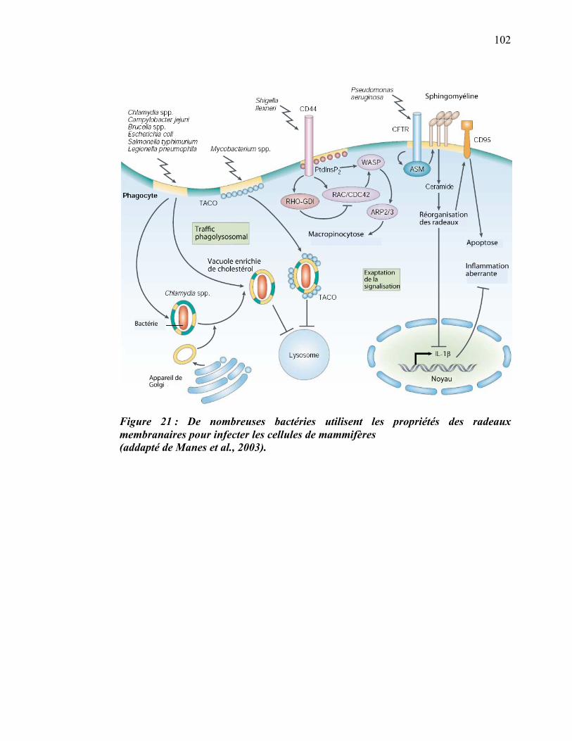
102
Figure 21 : De nombreuses bactéries utilisent les propriétés des radeaux membranaires pour infecter les cellules de mammifères (addapté de Manes et al., 2003).

CHAPITRE II. INTRODUCTION. TRAVAUX DE
DOCTORAT
104
Buts, données préliminaires et hypothèses de travail
Le premier but de ce travail de doctorat était de décrire l’implication de la molécule
LPG de L. donovani dans l’inhibition de certaines fonctions du phagosome. Au
moment d’élaborer mon projet, la capacité de L. donovani à bloquer la maturation du
phagosome en inhibant la fusion avec les endosomes était déjà connue (Desjardins and
Descoteaux, 1997). Plus particulièrement, il avait été démontré que cette inhibition
était un processus actif médié par la molécule LPG du parasite (Dermine et al., 2000).
Par la suite, une étude protéomique a révélé que flotilline-1 fait partie des protéines du
phagosome, indiquant la présence possible de radeaux lipidiques sur cette organelle
(Garin et al., 2001). En démontrant que les radeaux lipidiques enrichis de flotilline-1 et
du lipide GM1 sont bel et bien présents sur les phagosomes, mes prédécesseurs du
laboratoire du Dr Desjardins ont observé que L. donovani empêche l’arrivée de
flotilline-1 sur la majorité des phagosomes, et que cette inhibition est causée, elle
aussi, par le LPG. En effet, un mutant déficient pour le LPG (lpg2-) affecte moins le
recrutement de flotilline-1 sur le phagosome (Dermine et al., 2001). L’hypothèse
formulée pour cette première partie du projet de doctorat est donc :
La molécule lipophosphoglycan (LPG) de Leishmania donovani est directement
responsable des effets inhibiteurs du parasite sur certaines des fonctions cruciales des
phagosomes (fusion, acidification) qu’elle cause en ciblant les radeaux membranaires,
ce qui permet au parasite de survivre à la phagocytose.
Pour tester cette hypothèse, nous avons provoqué la phagocytose de Leishmania de
type sauvage ou lpg2- puis nous avons observé le comportement de marqueurs
spécifiques pour les radeaux lipidiques par microscopie confocale, microscopie
électronique et immunobuvardage sur les phagosomes de Leishmania.
Le second but de ce travail était d’effectuer une caractérisation approfondie des
radeaux lipidiques du phagosome au niveau protéomique et lipidomique, mais plus
105
particulièrement au niveau protéomique. L’identification des protéines composant ces
sous-domaines constituait la première étape logique qui a permis par la suite de mieux
comprendre et d’analyser les fonctions spécifiques des radeaux lipidiques sur le
phagosome.
Les radeaux membranaires ont été décrits comme des sous-domaines de la membrane
enrichis de certaines protéines dont le rapprochement leur permet d’exercer leurs
fonctions. Notre groupe a été le premier à démontrer la présence de radeaux lipidiques
sur le phagosome et très peu de choses sont actuellement connues sur la composition
protéique et la dynamique d’acquisition de ces sous-domaines au cours de la biogenèse
des phagosomes. Nous avons émis l’hypothèse suivante :
Les radeaux membranaires du phagosome et leur équivalent biochimique (DRMs)
contiennent un sous-ensemble spécifique de protéines, tout comme les radeaux de la
membrane plasmique décrits par d’autres équipes. Nous allons plus loin en suggérant
que la composition protéique des radeaux membranaires varie au cours de la
maturation du phagosome.
Pour tester cette hypothèse, nous avons fractionné de façon biochimique les
microdomaines (DRMs et DSMs) de phagosomes purifiés à trois moments clés de leur
maturation (0, 30 et 240 minutes de chasse). Nous avons séparé les protéines sur un
gel de polyacrylamide avant de soumettre les protéines ainsi récoltées au spectromètre
de masse. Une analyse bio-informatique approfondie des spectres de masse a ensuite
été effectuée afin d’identifier et de comparer les protéines présentes dans les DRMs
et/ou les DSMs en fonction de la maturation des phagosomes. Pour affiner notre
analyse du protéome de cette organelle, nous avons aussi procédé à une étude des
phospholipides du phagosome pour tenter de déterminer si la modulation des protéines
des microdomaines coïncide avec un changement au niveau des phospholipides.
Les chapitres suivants (III et IV) présentent les études réalisées dans le but de vérifier
ces hypothèses.
CHAPITRE III. RÉSULTATS ARTICLE 1
Goyette, G., Dermine, J-F., Houde, M., Turco, S.J., Desjardins, M. (2005)
Leishmania donovani lipophosphoglycan disrupts phagosome microdomains in J774
macrophages. Cell Microbiol 7(9), 1263-1270
107
Leishmania donovani lipophosphoglycan disrupts phagosome microdomains in
J774 macrophages
Guillaume Goyette 1†, Jean-François Dermine 1† , Mathieu Houde 1 , Salvatore J.
Turco 2 and Michel Desjardins 1,3 *
† These authors contributed equally.
1 Département de Pathologie et Biologie Cellulaire, Université de Montréal, C. P.
6128, Succ. Centre ville, Montreal, H3C 3J7, Canada.
2 Department of Biochemistry, University of Kentucky, Medical Center, Lexington,
KY 40536, USA.
3 Caprion Pharmaceuticals Inc., Montreal, Canada
.
108
Summary
Clearance of pathogens by phagocytosis and their killing in phagolysosomes is a key
aspect of our innate ability to fight infectious agents. Leishmania parasites have
evolved ways to survive and replicate in macrophages by inhibiting phagosome
maturation and avoiding the harsh environment of phagolysosomes. We describe here
that during this process Leishmania donovani uses a novel strategy involving its
surface lipophosphoglycan (LPG), a virulence factor impeding many host functions, to
prevent the formation or disrupt lipid microdomains on the phagosome membrane.
LPG acts locally on the membrane and requires its repetitive carbohydrate moieties to
alter the organization of microdomains. Targeting and disruption of functional foci,
where proteins involved in key aspects of phagolysosome biogenesis assemble, is
likely to confer a survival advantage to the parasite.
Introduction
Phagocytosis plays a key role in our innate ability to restrict the spread of infectious
diseases. Microorganisms internalized by phagocytosis are sequestered in
phagosomes, which are initially inapt at killing and degrading pathogens. They acquire
their microbicidal properties through a complex maturation process, involving
sequential fusion with endocytic organelles, leading to the formation of
phagolysosomes (Desjardins, 2003). The complexity of this process is only beginning
to be deciphered, as it encompasses the coordinated activity of hundreds of proteins
(Garin et al., 2001). Through evolution, pathogens have developed strategies to adapt
and survive in their hosts by avoiding the harsh environment of phagolysosomes. Sets
of evidence indicate that the strategy used by the intracellular pathogen Leishmania to
survive in mammalian macrophages relies on the use of its cell surface
lypophosphoglycan (LPG) to alter some of the functional properties of the host
phagosomes where they reside. Indeed, LPG has been shown to inhibit the ability of
phagosomes to fuse with late endocytic organelles and lysosomes (Desjardins and
Descoteaux, 1997; Dermine et al., 2000; Spath et al., 2003). In addition to its effect on
109
membrane fusion, LPG is known to have pleiotropic deleterious effects on host cells,
altering functions as diverse as signal transduction, the oxydative burst and actin
organization (Descoteaux and Turco, 2002). The mechanism by which a single
molecule like LPG can interfere with a variety of key host functions is puzzling. We
showed recently that unlike LPG-/- mutants, wild-type Leishmania donovani reside
within phagosomes not displaying flotillin-1-enriched microdomains, suggesting that
this parasite might target these structures for its survival in macrophages (Dermine
et al., 2001). As microdomains have been shown to play key roles in various cellular
functions, including signal transduction (Simons and Vaz, 2004), targeting of these
structures in phagosomes by pathogens might confer a survival advantage. We show
here that Leishmania promastigotes use LPG to directly alter the formation of
phagosome lipid microdomains and/or disorganize these structures after phagocytosis.
This process is likely to explain the multiple effects of LPG on macrophage functions,
and to be beneficial to the parasite because phagosome microdomains contain proteins
involved in important aspects of phagolysosome biogenesis.
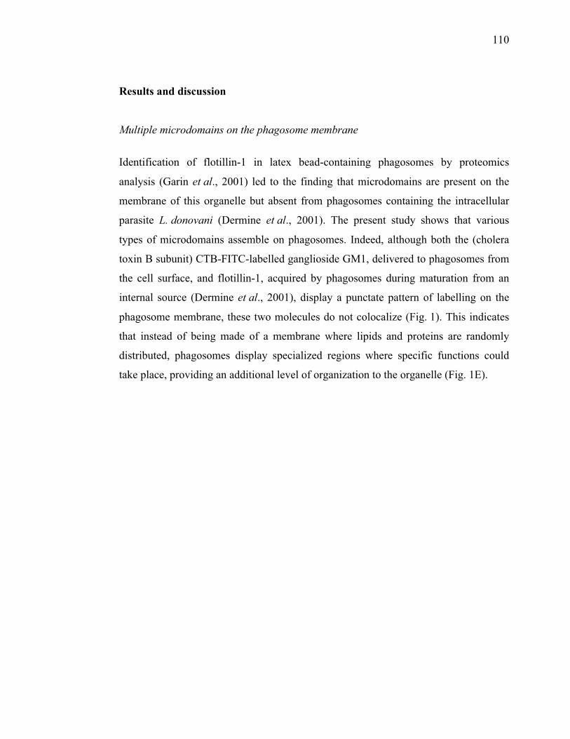
110
Results and discussion
Multiple microdomains on the phagosome membrane
Identification of flotillin-1 in latex bead-containing phagosomes by proteomics
analysis (Garin et al., 2001) led to the finding that microdomains are present on the
membrane of this organelle but absent from phagosomes containing the intracellular
parasite L. donovani (Dermine et al., 2001). The present study shows that various
types of microdomains assemble on phagosomes. Indeed, although both the (cholera
toxin B subunit) CTB-FITC-labelled ganglioside GM1, delivered to phagosomes from
the cell surface, and flotillin-1, acquired by phagosomes during maturation from an
internal source (Dermine et al., 2001), display a punctate pattern of labelling on the
phagosome membrane, these two molecules do not colocalize (Fig. 1). This indicates
that instead of being made of a membrane where lipids and proteins are randomly
distributed, phagosomes display specialized regions where specific functions could
take place, providing an additional level of organization to the organelle (Fig. 1E).
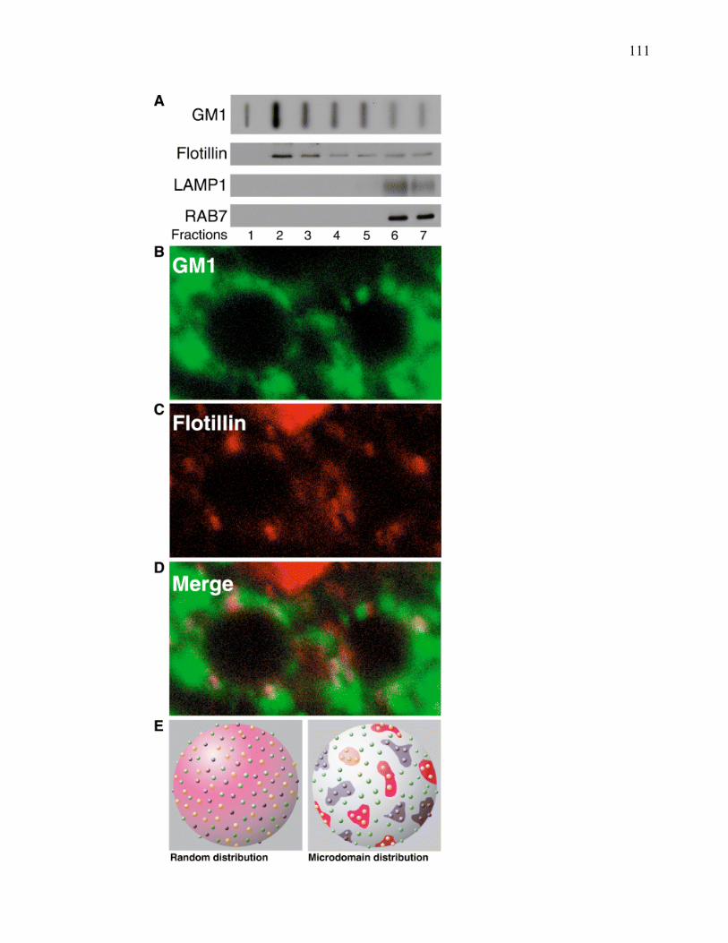
111
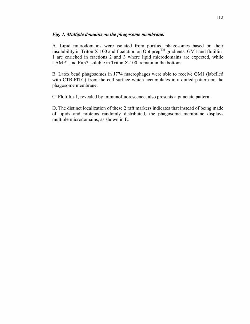
112
Fig. 1. Multiple domains on the phagosome membrane.
A. Lipid microdomains were isolated from purified phagosomes based on their insolubility in Triton X-100 and floatation on OptiprepTM gradients. GM1 and flotillin-1 are enriched in fractions 2 and 3 where lipid microdomains are expected, while LAMP1 and Rab7, soluble in Triton X-100, remain in the bottom.
B. Latex bead phagosomes in J774 macrophages were able to receive GM1 (labelled with CTB-FITC) from the cell surface which accumulates in a dotted pattern on the phagosome membrane.
C. Flotillin-1, revealed by immunofluorescence, also presents a punctate pattern.
D. The distinct localization of these 2 raft markers indicates that instead of being made of lipids and proteins randomly distributed, the phagosome membrane displays multiple microdomains, as shown in E.
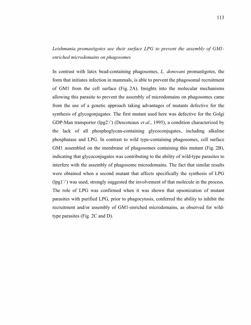
113
Leishmania promastigotes use their surface LPG to prevent the assembly of GM1-
enriched microdomains on phagosomes
In contrast with latex bead-containing phagosomes, L. donovani promastigotes, the
form that initiates infection in mammals, is able to prevent the phagosomal recruitment
of GM1 from the cell surface (Fig. 2A). Insights into the molecular mechanisms
allowing this parasite to prevent the assembly of microdomains on phagosomes came
from the use of a genetic approach taking advantages of mutants defective for the
synthesis of glycogonjugates. The first mutant used here was defective for the Golgi
GDP-Man transporter (lpg2-/-) (Descoteaux et al., 1995), a condition characterized by
the lack of all phosphoglycan-containing glycoconjugates, including alkaline
phosphatase and LPG. In contrast to wild type-containing phagosomes, cell surface
GM1 assembled on the membrane of phagosomes containing this mutant (Fig. 2B),
indicating that glycoconjugates was contributing to the ability of wild-type parasites to
interfere with the assembly of phagosome microdomains. The fact that similar results
were obtained when a second mutant that affects specifically the synthesis of LPG
(lpg1-/-) was used, strongly suggested the involvement of that molecule in the process.
The role of LPG was confirmed when it was shown that opsonization of mutant
parasites with purified LPG, prior to phagocytosis, conferred the ability to inhibit the
recruitment and/or assembly of GM1-enriched microdomains, as observed for wild-
type parasites (Fig. 2C and D).
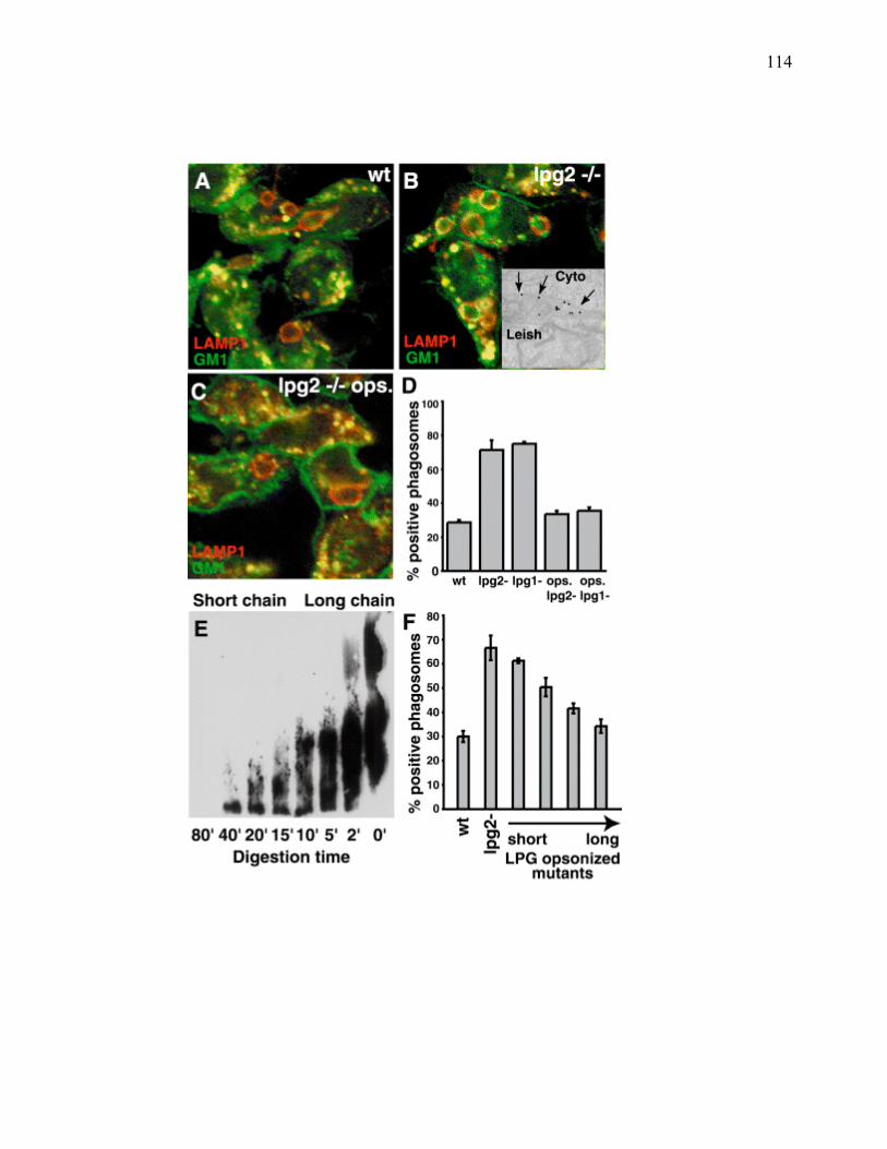
114
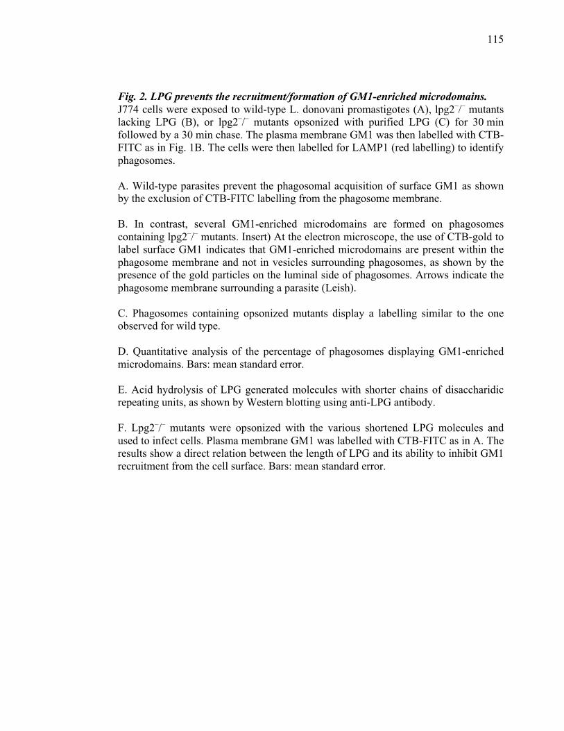
115
Fig. 2. LPG prevents the recruitment/formation of GM1-enriched microdomains. J774 cells were exposed to wild-type L. donovani promastigotes (A), lpg2–/– mutants lacking LPG (B), or lpg2–/– mutants opsonized with purified LPG (C) for 30 min followed by a 30 min chase. The plasma membrane GM1 was then labelled with CTB-FITC as in Fig. 1B. The cells were then labelled for LAMP1 (red labelling) to identify phagosomes. A. Wild-type parasites prevent the phagosomal acquisition of surface GM1 as shown by the exclusion of CTB-FITC labelling from the phagosome membrane. B. In contrast, several GM1-enriched microdomains are formed on phagosomes containing lpg2–/– mutants. Insert) At the electron microscope, the use of CTB-gold to label surface GM1 indicates that GM1-enriched microdomains are present within the phagosome membrane and not in vesicles surrounding phagosomes, as shown by the presence of the gold particles on the luminal side of phagosomes. Arrows indicate the phagosome membrane surrounding a parasite (Leish). C. Phagosomes containing opsonized mutants display a labelling similar to the one observed for wild type. D. Quantitative analysis of the percentage of phagosomes displaying GM1-enriched microdomains. Bars: mean standard error. E. Acid hydrolysis of LPG generated molecules with shorter chains of disaccharidic repeating units, as shown by Western blotting using anti-LPG antibody. F. Lpg2–/– mutants were opsonized with the various shortened LPG molecules and used to infect cells. Plasma membrane GM1 was labelled with CTB-FITC as in A. The results show a direct relation between the length of LPG and its ability to inhibit GM1 recruitment from the cell surface. Bars: mean standard error.
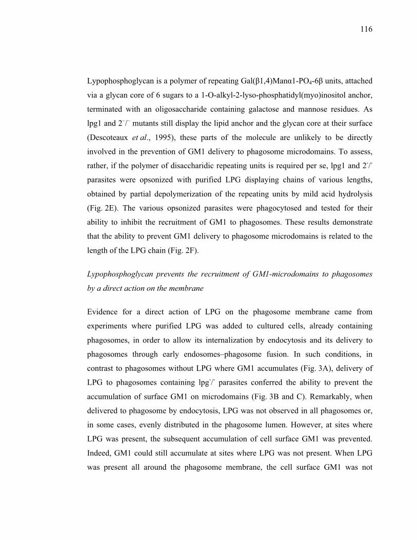
116
Lypophosphoglycan is a polymer of repeating Gal(�1,4)Man�1-PO4-6� units, attached
via a glycan core of 6 sugars to a 1-O-alkyl-2-lyso-phosphatidyl(myo)inositol anchor,
terminated with an oligosaccharide containing galactose and mannose residues. As
lpg1 and 2–/– mutants still display the lipid anchor and the glycan core at their surface
(Descoteaux et al., 1995), these parts of the molecule are unlikely to be directly
involved in the prevention of GM1 delivery to phagosome microdomains. To assess,
rather, if the polymer of disaccharidic repeating units is required per se, lpg1 and 2-/-
parasites were opsonized with purified LPG displaying chains of various lengths,
obtained by partial depolymerization of the repeating units by mild acid hydrolysis
(Fig. 2E). The various opsonized parasites were phagocytosed and tested for their
ability to inhibit the recruitment of GM1 to phagosomes. These results demonstrate
that the ability to prevent GM1 delivery to phagosome microdomains is related to the
length of the LPG chain (Fig. 2F).
Lypophosphoglycan prevents the recruitment of GM1-microdomains to phagosomes
by a direct action on the membrane
Evidence for a direct action of LPG on the phagosome membrane came from
experiments where purified LPG was added to cultured cells, already containing
phagosomes, in order to allow its internalization by endocytosis and its delivery to
phagosomes through early endosomes–phagosome fusion. In such conditions, in
contrast to phagosomes without LPG where GM1 accumulates (Fig. 3A), delivery of
LPG to phagosomes containing lpg-/- parasites conferred the ability to prevent the
accumulation of surface GM1 on microdomains (Fig. 3B and C). Remarkably, when
delivered to phagosome by endocytosis, LPG was not observed in all phagosomes or,
in some cases, evenly distributed in the phagosome lumen. However, at sites where
LPG was present, the subsequent accumulation of cell surface GM1 was prevented.
Indeed, GM1 could still accumulate at sites where LPG was not present. When LPG
was present all around the phagosome membrane, the cell surface GM1 was not
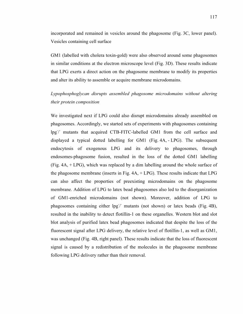
117
incorporated and remained in vesicles around the phagosome (Fig. 3C, lower panel).
Vesicles containing cell surface
GM1 (labelled with cholera toxin-gold) were also observed around some phagosomes
in similar conditions at the electron microscope level (Fig. 3D). These results indicate
that LPG exerts a direct action on the phagosome membrane to modify its properties
and alter its ability to assemble or acquire membrane microdomains.
Lypophosphoglycan disrupts assembled phagosome microdomains without altering
their protein composition
We investigated next if LPG could also disrupt microdomains already assembled on
phagosomes. Accordingly, we started sets of experiments with phagosomes containing
lpg-/- mutants that acquired CTB-FITC-labelled GM1 from the cell surface and
displayed a typical dotted labelling for GM1 (Fig. 4A, - LPG). The subsequent
endocytosis of exogenous LPG and its delivery to phagosomes, through
endosomes-phagosome fusion, resulted in the loss of the dotted GM1 labelling
(Fig. 4A, + LPG), which was replaced by a dim labelling around the whole surface of
the phagosome membrane (inserts in Fig. 4A, + LPG). These results indicate that LPG
can also affect the properties of preexisting microdomains on the phagosome
membrane. Addition of LPG to latex bead phagosomes also led to the disorganization
of GM1-enriched microdomains (not shown). Moreover, addition of LPG to
phagosomes containing either lpg-/- mutants (not shown) or latex beads (Fig. 4B),
resulted in the inability to detect flotillin-1 on these organelles. Western blot and slot
blot analysis of purified latex bead phagosomes indicated that despite the loss of the
fluorescent signal after LPG delivery, the relative level of flotillin-1, as well as GM1,
was unchanged (Fig. 4B, right panel). These results indicate that the loss of fluorescent
signal is caused by a redistribution of the molecules in the phagosome membrane
following LPG delivery rather than their removal.
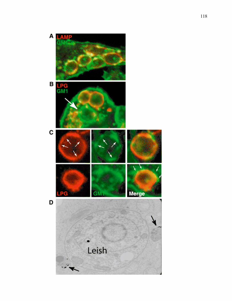
118
119
Fig. 3. LPG prevents the recruitment of GM1 to phagosomes by a direct action on the membrane.
A. Cells infected with lpg2–/– mutants and labelled for GM1 display phagosomes with several GM1-enriched microdomains, as shown in Fig. 2B.
B. In contrast, when purified LPG was added to lpg2–/– phagosomes by endocytosis (endosome–phagosome fusion), the recruitment of surface GM1 was inhibited specifically at sites where LPG was present (no yellow overlay). In the cases where LPG did not reach phagosomes (large arrow), the recruitment of GM1 was not prevented.
C. Two examples taken from experiments like in B are shown. In the upper panels, GM1 is observed only at sites where LPG is absent (small arrows). When LPG is present all around the phagosome membrane, GM1 is not incorporated and remains in vesicles around phagosomes (arrows in lower panel).
D. An example of GM1 (CTB-gold) in vesicles (arrows) around a phagosome is shown.
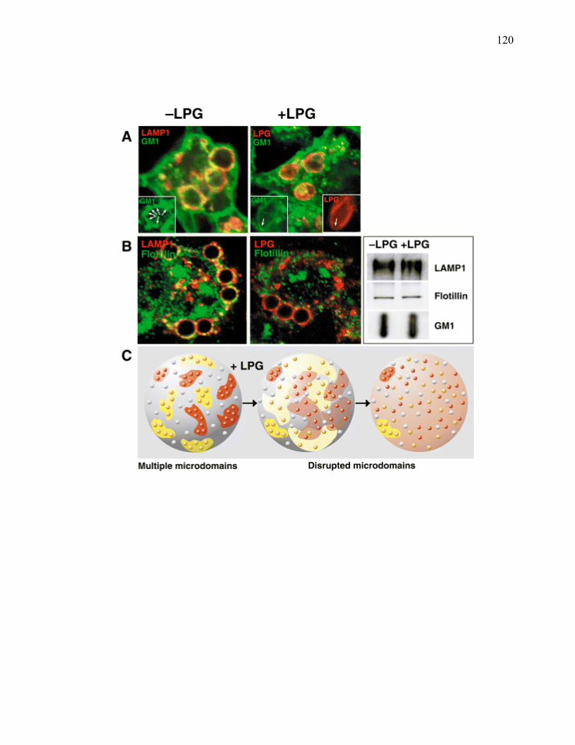
120
121
Fig. 4. LPG disrupts preexisting phagosome microdomains by promoting their disassembly rather than by altering their protein composition.
A. Phagosomes formed by the internalization of lpg2–/– mutants were allowed to receive surface GM1 (see 2B). Purified LPG was then added in the culture medium (+lPG) or not (–lPG), a process that allows the transfer of LPG to phagosomes (see 3B). Phagosomes were then identified using antibodies against LPG or LAMP1. Before the addition of LPG, phagosomes displayed several GM1-enriched microdomains (white arrows in insets), the subsequent delivery of LPG to phagosomes resulted in the loss of the dotted signal replaced by a faint CTB-FITC signal all around the phagosome membrane (the left inset in the +lPG panel), except in one region where LPG is not detected (arrow).
B. While late phagosomes containing latex beads normally display a punctate pattern for flotillin-1 (–lPG), the delivery of LPG to this compartment also disrupts flotillin-1-enriched microdomains (loss of yellow overlay). Interestingly, Western and dot blot analysis for LAMP1, flotillin-1, and GM1 indicates that the delivery of LPG has no effect on the relative amounts of these molecules on phagosomes.
C. When LPG is added to phagosomes and, in the context of the infection when it is released by promastigote parasites after their internalization by macrophages, microdomains dissolve and their components either lipids or proteins are redistributed to the whole surface of the organelle, leading to potential functional losses.
122
Although the precise functions of phagosome membrane microdomains are poorly
understood, proteomics analyses indicate that they might be involved in signal
transduction, membrane fusion, actin organization, and the oxidative burst (Dermine
et al., 2001; G. Goyette and M. Desjardins, unpubl.). As these processes play key roles
in phagolysosome biogenesis, alteration of phagosome microdomains could thus
represent a relevant strategy for the establishment of a replicative niche in host cells.
Interestingly, the first reports of a deleterious effect of LPG on host cells described an
attenuation of the oxidative burst and an inhibition of signalling through protein kinase
C (PKC) activity (McNeely and Turco, 1990; Descoteaux et al., 1992). LPG was also
shown to inhibit phagosome–lysosome fusion, a key process for the killing and
degradation of Leishmania (Desjardins and Descoteaux, 1997; Dermine et al., 2000).
The NADPH oxidase, involved in the oxidative burst, various PKC isoenzymes, and
several proteins involved in membrane fusion were recently shown to be present in
lipid microdomains (Chamberlain et al., 2001; Lang, 2002; Becart et al., 2003; Shao
et al., 2003; Vilhardt and Van Deurs, 2004).
Further links exist between LPG and the disruption of cellular functions associated to
membrane microdomains. Infection of CD4+ T cells by HIV, a process involving
membrane fusion and lipid microdomains (Manes et al., 2000; Liao et al., 2001), is
inhibited by LPG (Easterbrook et al., 1995). When introduced in erythrocyte ghost
membranes, LPG blocks binding, as well as fusion, of Sendai and influenza viruses
(Miao et al., 1995; Takeda et al., 2003), a process occurring in lipid microdomains
(Liao et al., 2001).
Insights into the molecular mechanisms by which LPG could alter phagosome
membrane microdomains at the onset of infection come from the life cycle of the
parasite and the physicochemical properties of this glycoconjugate. LPG is a
glycosylphosphatidylinositol (GPI)-anchored glycolipid present within lipid
microdomains at the surface of the promastigote form of the parasite (Denny et al.,
2001), the flagellated developmental stage transmitted by sandflies to the mammalian
123
host. After internalization in macrophages by phagocytosis, LPG is shed from the
parasite surface (Kaneshiro et al., 1982; Denny et al., 2001) and inserted in host cell
membranes (Tolson et al., 1990), modifying their physical organization (Miao et al.,
1995). This points to a possible mechanism by which the negatively charged sugar
chain of LPG directly interferes with the clusterization of molecules into
microdomains. In this regard, it has recently been proposed that a phase transition of
membrane components from a liquid to a solid crystalline phase could be central to the
functional role of membrane microdomains (Joly, 2004). According to this concept,
the disrupting action of LPG could be resulting from the capacity of the heavily
charged head of LPG to interfere with the assembly of such structures, or even to
cause their disassembly, as observed for the GM1 containing structures.
Experimental procedures
Cell culture
The J774 murine macrophage cell line and the L. donovani wild types and mutant
lpg2-/- and lpg1-/- strains (kind gift from A. Descoteaux) were cultured as described
previously (Duclos et al., 2000). Briefly, L. donovani parasites (strain 1S) were grown
at 26°C in modified M199 medium, pH 7.3 and used in stationary phase as described
previously (Desjardins and Descoteaux, 1997). The choice of this type of parasite was
motivated in order to reproduce the conditions in which LPG was originally shown to
inhibit phagosome membrane fusion (Desjardins and Descoteaux, 1997).
GM1-rafts labelling and immunofluorescence
J774 cells grown on coverslips were infected with L. donovani strain 1S, with LPG-
mutants (lpg2-/- and lpg1-/-) lacking the surface glycolipid lipophosphoglycan (LPG),
or fed 3 �m latex beads (Polyscience) for 30 min followed by a 30 min chase without
bead or parasite. The cell surface GM1 was then labelled by incubating cells with
5 �g ml�1 of CTB-FITC (Sigma) in culture medium at 4°C to prevent internalization of
the toxin. After washing unbound CTB, cells were further incubated for 2 h in normal
medium, a time period sufficient to allow the delivery of cell surface GM1 to
124
phagosomes. Cells were then fixed and processed as described (Dermine et al., 2001).
To reveal phagosomes, cells were incubated with the rat anti-LAMP1 1D4B for 1 h. In
some experiments, phagosomes were also doubly labelled for flotillin-1 and either
LAMP1 or cholera toxin as described previously (Dermine et al., 2001).
Effect of purified LPG on phagosome lipid microdomains
The effect of purified LPG on phagosome microdomains was tested in two distinct
conditions, to (i) see if LPG can inhibit the phagosomal acquisition of GM1 from the
cell surface (as well as flotilin-1 from an internal source) and (ii) see if LPG can alter
the organization of microdomains already present on phagosomes. In the first case,
J774 cells displaying phagosomes containing either LPG-deficient Leishmania or latex
beads were allowed to internalize purified LPG (1 mg ml�1 in PBS) by endocytosis for
30 min, followed by a further incubation of 30 min without LPG, allowing the delivery
of LPG to phagosomes through endosomes-phagosome fusion. The cell surface GM1
was then labelled as described in the previous section, and cells observed at the
confocal microscope (Leica Microsystems model DM IRBE) to determine if GM1-
enriched microdomains were present on phagosomes. In the second case, phagosomes
were formed by the internalization of LPG-deficient Leishmania as above. The cell
surface GM1 was then labelled with CTB-FITC and cells incubated to allow the
formation of CTB-FITC-labelled GM1-enriched microdomains on phagosomes.
Purified LPG was then internalized as above.
Western and slot blotting
Western blotting for flotillin-1, LAMP-1 and Rab7 was performed using the 1D4B rat
monoclonal antibody (Developmental Studies Hybrodoma Bank, University of Iowa)
directed against LAMP1, a polyclonal antibody specific for flotillin-1 (kind gift from
G. van der Goot) and a polyclonal antibody against Rab7 (Santa Cruz). To detect the
lipid ganglioside GM1, slot blotting with CHTX-HRP was used.
125
Lypophosphoglycan purification and digestion, and opsonization of the L. donovani
mutants
Lypophosphoglycan from promastigote cultures was extracted in solvent E as
previously described (Orlandi and Turco, 1987). To shorten the length of LPG, 15 �g
of purified LPG from L. donovani was cleaved at acid-labile phosphodiester bridges
by incubation in 0.01 N HCl for the indicared time points at 60°C. The samples were
then treated with 0.01 N NaOH to neutralize acidity and put on ice. To verify the
proper digestion of LPG, SDS-PAGE separation of the fragments on 12% gels was
performed prior to Western blotting with the CA7AE antibody, which recognizes the
repeating units of LPG (Tolson et al., 1989).
To opsonize LPG-deficient parasites, 1.0 � 106lpg2–/– and lpg1-/- L. donovani were
incubated for 30 min at room temperature in 15 �l of 1 mg ml�1 of full length or
digested LPG in PBS.
Isolation of phagosome microdomains
Lipid microdomains were isolated from phagosomes made as described previously
(Desjardins et al., 1994). Briefly, the phagosome pellet was resuspended in 0.3 ml of
TNE-Triton buffer (25 mM Tris, 150 mM NaCl, 5 mM EDTA, complete protease
inhibitor (Boehringer), pH 7.4 and 1% Triton X-100), transferred to an Eppendorf tube
and shaken gently 30 min at 4°C to solubilize phagosomal membranes. Latex beads
were then pelleted twice by centrifugation and the final supernatant containing the
solubilized and insoluble phagosome components was added to 0.6 ml 60%
OptiprepTM to obtain a final concentration of 40% which was then poured at the
bottom of an Ultraclear centrifuge tube (Beckman). Finally, 2.4 ml of 30% OptiprepTM
and 0.9 ml of TNE buffer (with protease inhibitors) were layered on top. After a 4 h
centrifugation at 40 000 r.p.m. (SW60 rotor) to float the insoluble rafts, 7 fractions of
0.6 ml were collected from the top. Proteins were then precipitated with
methanol/chloroform and resuspended in Laemmli buffer for Western blotting. Ten
126
microlitres of each fraction were collected prior to precipitation for GM1 slot-blot
analysis.
127
Acknowledgements
We thank E. Joly, I. Jutras and A. Descoteaux for their comments. S.J.T. is supported
by NIH Grant AI20941. This work was supported by grants from the CIHR and
Genome Québec/Canada.
128
References
Becart, S., Setterblad, N., Ostrand-Rosenberg, S., Ono, S.J., Charron, D., and
Mooney, N. (2003) Intracytoplasmic domains of MHC class II molecules are essential
for lipid-raft-dependent signaling. J Cell Sci 116: 2565–2575.
Chamberlain, L.H., Burgoyne, R.D., and Gould, G.W. (2001) SNARE proteins are
highly enriched in lipid rafts in PC12 cells: implications for the spatial control of
exocytosis. Proc Natl Acad Sci USA 98: 5619–5624.
Denny, P.W., Field, M.C., and Smith, D.F. (2001) GPI-anchored proteins and
glycoconjugates segregate into lipid rafts in Kinetoplastida. FEBS Lett 491: 148–153.
Dermine, J.-F., Scianimanico, S., Prive, C., Descoteaux, A., and Desjardins, M.
(2000) Leishmania promastigotes require lipophosphoglycan to actively modulate the
fusion properties of phagosomes at an early step of phagocytosis. Cell Microbiol 1:
19–32.
Dermine, J.-F., Duclos, S., Garin, J., St-Louis, F., Rea, S., Parton, R.G., and
Desjardins. M. (2001) Flotillin-1-enriched lipid raft domains accumulate on maturing
phagosomes. J Biol Chem 276: 18507–18512.
Descoteaux, A., and Turco. S.J. (2002) Functional aspects of the Leishmania
donovani lipophosphoglycan during macrophage infection. Microbes Infect 4: 975–
981.
Descoteaux, A., Matlashewski, G., and Turco, S.J. (1992) Inhibition of macrophage
protein kinase C-mediated protein phosphorylation by Leishmania donovani
lipophosphoglycan. J Immunol 149: 3008–3015.
Descoteaux, A., Luo, Y., Turco, S.J., and Beverley, S.M. (1995) Specialized
pathway affecting virulence glycoconjugates of Leishmania. Science 269: 1869–1872.
129
Desjardins, M. (2003) ER-mediated phagocytosis: a new membrane for new
functions. Nat Rev Immunol 3: 280–291.
Desjardins, M., and Descoteaux, A. (1997) Inhibition of phagolysosomal biogenesis
by the Leishmania lipophosphoglycan. J Exp Med 185: 2061–2068.
Desjardins, M., Hubert, L.A., Parton, R.G., and Griffiths, G. (1994) Biogenesis of
phagolysosomes proceeds through a sequential series of interactions with the
endocytic apparatus. J Cell Biol 124: 677–688.
Duclos, S., Diez, R., Garin, J., Papadopoulou, B., Descoteaux, A., Stenmark, H.,
and and. Desjardins, M. (2000) Rab5 regulates the kiss and run fusion between
phagosomes and endosomes and the acquisition of phagosome leishmanicidal
properties in RAW 264.7 macrophages. J Cell Sci 113: 3531–3541.
Easterbrook, M.D., Levy, M.H., Gomez, A.M., Turco, S.J., Epand, R.M., and
Rosenthal, K.L. (1995) Inhibition of HIV-1-induced syncytia formation and
infectivity by lipophosphoglycan from Leishmania. J Acquir Immune Defic Syndr 10:
496–505.
Garin, J., Diez, R., Kieffer, S., Dermine, J.F., Duclos, S., Gagnon, E., et al. (2001)
The phagosome proteome: insight into phagosome functions. J Cell Biol 152: 165–
180.
Joly, E. (2004) Hypothesis: could the signalling function of membrane microdomains
involve a localized transition of lipids from liquid to solid state? BMC Cell Biol 5: 3.
Kaneshiro, E.S., Gottlieb, M., and Dwyer, D.M. (1982) Cell surface origin of
antigens shed by Leishmania donovani during growth in axenic culture. Infect Immun
37: 558–567.
130
Lang, M.L. (2002) IgA Fc receptor (FcalphaR) cross-linking recruits tyrosine kinases,
phosphoinositide kinases and serine/threonine kinases to glycolipid rafts. Biochem J
364: 517–525.
Liao, Z., Cimakasky, L.M., Hampton, R., Nguyen, D.H., and Hildreth, J.E. (2001)
Lipid rafts and HIV pathogenesis: host membrane cholesterol is required for infection
by HIV type 1. AIDS Res Hum Retroviruses 17: 1009–1019.
McNeely, T.B., and Turco, S.J. (1990) Requirement of lipophosphoglycan for
intracellular survival of Leishmania donovani within human monocytes. J Immunol
144: 2745–2750.
Manes, S., del Real, G., Lacalle, R.A., Lucas, P., Gomez-Mouton, C., Sanchez-
Palomino, S., et al. (2000) Membrane raft microdomains mediate lateral assemblies
required for HIV-1 infection. EMBO Rep 1: 190–196.
Miao, L., Stafford, A., Nir, S., Turco, S.J., Flanagan, T.D., and Epand, R.M.
(1995) Potent inhibition of viral fusion by the lipophosphoglycan of Leishmania
donovani. Biochemistry 34: 4676–4683.
Orlandi, P.A.Turco, S.J. (1987) Structure of the lipid moiety of the Leishmania
donovani lipophosphoglycan. J Biol Chem 262: 10384–10391.
Shao, D., Segal, A.W., and Dekker, L.V. (2003) Lipid rafts determine efficiency of
NADPH oxidase activation in neutrophils. FEBS Lett 550: 101–106.
Simons, K., and Vaz, W.L. (2004) Model systems, lipid rafts, and cell membranes.
Annu Rev Biophys Biomol Struct 33: 269–295.
Spath, G.F., Garraway, L.A., Turco, S.J., and Beverley, S.M. (2003) The role (s) of
lipophosphoglycan (LPG) in the establishment of Leishmania major infections in
mammalian hosts. Proc Natl Acad Sci USA 100: 9536–9541.
131
Takeda, M., Leser, G.P., Russell, C.J., and Lamb, R.A. (2003) Influenza virus
hemagglutinin concentrates in lipid raft microdomains for efficient viral fusion. Proc
Natl Acad Sci USA 100: 14610–14617.
Tolson, D.L., Turco, S.J., Beecroft, R.P., and Pearson, T.W. (1989) The
immunochemical structure and surface arrangement of Leishmania donovani
lipophosphoglycan determined using monoclonal antibodies. Mol Biochem Parasitol
35: 109–118.
Tolson, D.L., Turco, S.J., and Pearson, T.W. (1990) Expression of a repeating
phosphorylated disaccharide lipophosphoglycan epitope on the surface of
macrophages infected with Leishmania donovani. Infect Immun 58: 3500–3507.
Vilhardt, F., and Van Deurs, B. (2004) The phagocyte NADPH oxidase depends on
cholesterol-enriched membrane microdomains for assembly. EMBO J 23: 739–748.
CHAPITRE IV. RÉSULTATS ARTICLE 2
Goyette, G., Carruthers, N., Jutras, I., Boulais, J., Duclos, S., Laplante, A.,
Landry C.R., Thibault, P., Michnick, S.W., Lajoie, G., Desjardins, M. (2009)
Molecular characterization of the spatio-temporal modulation of phagosome functions
during phagolysosome biogenesis (en preparation)
133
Molecular characterization of the spatiotemporal modulation of phagosome
functions during phagolysosome biogenesis
Guillaume Goyette1, Nicholas J Carruthers2, Isabelle Jutras1, Jonathan Boulais1,
Sophie Duclos1, Annie Laplante1, Christian R Landry3,4, Stephen W Michnick3, Pierre
Thibault5, Gilles Lajoie4, and Michel Desjardins1**
1Département de pathologie et biologie cellulaire, Université de Montréal, C.P. 6128,
Succ centre ville, Montréal, Québec, H3C 3J7, Canada
2Department of Biochemistry, University of Western Ontario, Siebens-Drake Research
Institute, London, ON, N6A 5C1, Canada
3Département de biochimie, Université de Montréal, C.P. 6128, Succ centre ville,
Montréal, Québec, H3C 3J7, Canada
4Département de Biologie et Institut de Biologie Intégrative et des Systèmes (IBIS),
Université Laval, Québec, G1V 0A6, Canada
5Institute for Research in Immunology and Cancer, C.P. 6128, Succ centre ville,
Montréal, Québec, H3C 3J7, Canada
**Address correspondence to: Michel Desjardins, Département de pathologie et
biologie cellulaire, Université de Montréal, C.P. 6128, Succ centre ville, Montréal,
Québec, H3C 3J7, Canada
134
Abstract
The killing of microorganisms and the processing of peptides for presentation at the
cell surface require specific conditions established in phagosomes during
phagolysosome biogenesis. However, nascent phagosomes formed at the cell surface
are unable to perform most of their functions related to innate and adaptive immunity.
These are acquired through phagosome maturation, a process involving interaction
with series of organelles, modulating the fine composition of phagosomes. In the
present study, we use proteomics and lipidomics approaches, as well as a protein-
fragment complementation assay in yeasts, to define novel protein-protein interactions
and characterize the nature of the changes occurring at phagosomes as they mature
into fully functional organelles. Our data indicate that the membrane of phagosomes is
modified in complex ways by the acquisition and loss of sets of proteins and lipids,
enabling the sequential establishment of specific functional properties. We were able
to identify 124 different species of lipids and characterize their modulation on
maturing phagosomes. Furthermore, we showed that several groups of proteins shuffle
from and into membrane microdomains, supporting the concept that modulation of the
functional properties of phagosomes during phagolysosome biogenesis occurs by a
reorganization of the organelle through time and space.
135
Introduction
Phagocytosis, the mechanism by which large particles are internalized, leads to the
formation of phagosomes, a specialized organelle in which the engulfed material is
degraded1, 2. In mammals, various cells including macrophages, neutrophils and
dendritic cells display remarkable phagocytic activities, rapidly eliminating
microorganisms, foreign inert particles, and apoptotic cells. The killing of
microorganisms by professional phagocytes precludes the emergence of infectious
diseases. This innate immune process is followed by the degradation of microbes in a
highly concentrated mixture of hydrolases, activated by the acidic pH generated in the
phagosome lumen, generating antigenic peptides displayed at the cell surface, hence
enabling their recognition by T lymphocytes3. The peptides not loaded on MHC
molecules are further degraded in phagolysosomes, the end products likely recycled
from phagosomes by transporters whose identity is largely unknow 1. The
establishment of these functional properties involves a complex reorganization of
phagosomes through time, referred to as phagolysosome biogenesis4, 5. This highly
regulated process involves the fusion of nascent phagosomes with trans Golgi-derived
vesicles, early endosomes, late endosomes and ultimately lysosomes1, 2. These fusion
events profoundly alter the proteome of phagosomes during phagolysosome biogenesis 6 and hence regulate the functional properties of phagosomes.
The capacity to kill and degrade microbes is only one of the many functions that
phagosomes acquire during phagolysosome biogenesis. In a previous study, we
identified more than 140 proteins associated with phagosomes7, leading to the
description of novel phagosomal functions such as antigen cross-presentation8. This
proteomics study also highlighted the presence on phagosomes of proteins known to
segregate into lipid rafts at the cell surface, such as flotillin-1 and prohibitin, leading to
the proposal that membrane microdomains might also assemble on phagosomes.
Membrane microdomains are enriched in cholesterol and sphingolipids and are
biochemically defined by their insolubility in non-ionic detergents. At the plasma
membrane, these structures may constitute foci of specialized functions, notably for
136
signal transduction. Further biochemical and morphological analyses confirmed the
presence of membrane microdomains on phagosomes9. Recent data indicated that two
phagosomal protein complexes, V-ATPase and NADPH oxidase, may use membrane
microdomains as assembly platforms10. The significance of membrane microdomains
in phagosome functions was highlighted by the finding that at least two unrelated
pathogens, the gram negative bacteria Brucella and the intracellular parasite
Leishmania donovani, target lipid rafts as a strategy to evade the defence mechanisms
of their hosts11, 12. Altogether, these data indicate that the functional properties of the
phagosome are regulated by a complex remodelling of its proteome both through time,
during phagolysosome biogenesis, and space, through the spatial segregation of
proteins into membrane microdomains. The molecular characterization of the
spatiotemporal remodeling of phagosomes should provide unique insights into the
mechanisms used by pathogens to alter the functional properties of this organelle.
In the present study we used quantitative proteomics and lipidomics approaches to
characterize for the first time the spatiotemporal modifications of both proteins and
lipids occurring during the biogenesis of an intracellular organelle. Our data indicate
that segregation of sets of proteins in sub-regions of the phagosome membrane is an
important aspect of phagolysosome biogenesis.
137
Materials and Methods
Cell Culture and Phagosome Isolation
The murine macrophage-like cell line J774 was cultured in Dulbecco’s modified
Eagle’s medium high glucose (Sigma) supplemented with 10% heat-inactivated foetal
bovine serum, 1% glutamine, 100 units/ml penicillin, and 100 mg/ml streptomycin at
37 °C in a 5% CO2 atmosphere. Cells were grown to 80% confluence in Petri dishes
prior to each experiment. To form phagosomes, J774 macrophages were fed with 0.8
mm blue dyed latex beads (Estapor® Microsphères) diluted 1:50 in culture medium
without serum. Cells were allowed to internalize beads for 15 or 30 min at 37 °C. Cells
were then washed two times for 5 min at room temperature with PBS to remove non-
internalized beads, and were further incubated for increasing periods of time (0 min,
30 min, 240 min) to obtain early and late phagosomes. Phagosomes were then isolated
on sucrose step gradients as described previously 9. Purified phagosomes were
resuspended in TNE buffer (25 mM Tris, 150 mM NaCl, 5 mM EDTA), transferred to
an Eppendorf tube and frozen at -20oC. Samples were identified as 30/0, 30/30 or
30/240 phagosomes, referring to the 3 time-points (pulse/chase periods) analyzed.
Phagosomes were also isolated at 15/0 for lipidomics analysis.
Isolation of phagosome microdomains
Isolated phagosomes (0.15 ml suspension of phagosomes purified from 1.4 x 10⁸ cells)
were equilibrated to 1% Triton X-100 on ice by adding an identical volume of TNE
buffer containing 2% Triton X-100, and shaken gently 30 min at 4oC to solubilize
phagosomal membranes. Latex beads were then pelleted twice by microcentrifugation
(5 min at 15 000 rpm), and the final supernatant containing both the solubilized and
the detergent-resistant phagosome components brought to a final concentration of 40%
OptiprepTM, by adding 0.6 ml of 60% OptiprepTM stock in TNE buffer. This was
poured at the bottom of an Ultraclear centrifuge tube (Beckman). Finally, 3.0 ml of
30% OptiprepTM and 0.6 ml of TNE buffer were layered on top. After 4 h of
centrifugation at 40 000 rpm (SW60 rotor), to float the insoluble membranes, 7
fractions of 0.6 ml were collected from the top. Proteins were then precipitated with
138
methanol/chloroform according to established protocols13 and resuspended in Laemmli
buffer for Western blotting. To measure the lipid distribution in the raft gradient, 10ml
of each fraction was collected prior to precipitation for slot-blotting.
Western and slot blotting
For Western blot analysis, an identical amount of phagosomes from each sample was
used, based on the latex-bead concentration. 10 μL of the purified phagosome sample
resuspended in TNE buffer was diluted in 2 ml of distilled water, and the absorbance
at 750 nm was measured in an Ultrospec™ 2100 pro spectrophotometer (GE
Healthcare). The phagosome samples were adjusted to have the same absorbance by
volume and then pelleted by microcentrifugation (5 min at 15 000 rpm). The protein
concentration in the total cell lysate (TCL) and total membranes (TM) was evaluated
using the EZQ assay (Molecular Probes) and identical amounts of protein were loaded
for Western blot. The mouse anti-Flotillin-1 mAb and the mouse anti-Nicastrin IgG2a
were from BD Bioscience, the rabbit anti-Rab5a polyclonal antibody was from Santa
Cruz. The 1D4B rat anti-LAMP1 monoclonal antibody was from the Developmental
Studies Hybrodoma Bank University of Iowa. NaK ATPase � mouse monoclonal
IgG1 was from ABR Affinity BioReagents. RGS19 chicken polyclonal antibody and
the Gp91phox was from ABCAM. Monoclonal antibody directed against the N-
terminal peptide of human stomatin was a kind gift from Dr. R. Prohaska14. To detect
the lipid ganglioside GM1, we used slot blotting with HRP-conjugated cholera toxin
subunit B (CHTX-HRP) from Sigma.
Sample preparation for Protein Mass Spectrometry and Data Analysis
Two fractions of the OptiprepTM gradient (Fig.1B) were processed for protein
identification by mass spectrometry. The proteins in fraction 1, which contained
detergent resistant membranes (DRMs), and the proteins in fraction 7, which contained
the bulk of solubilized phagosome membranes (or detergent soluble membranes;
DSMs), were precipitated with methanol/chloroform, resuspended in Laemmli buffer
and loaded on a NuPage 12% Bis-Tris pre-cast gel (Invitrogen) for a 3 cm migration.
After a silver nitrate or a Coomassie blue staining, the resulting 3 cm gel was cut in
139
1mm bands irrespective of band positioning. Gel bands were subjected to reduction,
alkylation and in-gel tryptic digestion by automation in a MassPrep Workstation
(Micromass, Manchester, UK) as previously described15. The mass spectrometer was a
QTOF Micro from Waters Micromass equipped with a Nanosource modified with a
nanospray adapter (New Objective, Woburn, MA) to hold the PicoFrit column tip near
the sampling cone. Mass spectrometric data were acquired by employing the Data
Directed Analysis feature available on MassLynx (Micromass) operating software
with a 1, 1, 4 duty cycle (1 second in MS mode 1 peptide selected for fragmentation,
maximum of 4 seconds in MS/MS acquisition mode). A total of 6 experimental
conditions were analysed, namely fraction 1 (DRM) and fraction 7 (DSM) of
phagosomes isolated at 3 different time-points (30/0, 30/30 and 30/240). For each
condition, samples generated from 3 independent experiments were analyzed in the
mass spec (n=3).
MS/MS raw data were transferred from the QTOF Micro computer to a 50 terabyte
server and automatically manipulated for generation of peak lists by employing
Distiller version 1.1 (http://www.matrixscience.com/distiller.html) software with peak
picking parameters set at 20 as for Signal Noise Ration (SNR) and at 0.7 for
Correlation Threshold (CT). This reduced noise and produced a list of distinct peptide
peaks in which all members of the isotopic clusters were collapsed into an equivalent
monoisotopic peak. The peak listed data was then searched against a copy of the
Universal Protein Resource (UniProt) (http://www.pir.uniprot.org/) data base by
employing Mascot (http://www.matrixscience.com) version 1.9.03, and restricting the
search to a maximum of 1 missed (trypsin) cleavage, fixed carbamidomethyl
alkylation of cysteines, variable oxidation of methionine, ±0.5 mass unit tolerance on
parent and fragment ions. The search was limited to the Mus musculus taxonomy
(51635 sequences; 21798332 residues). Mascot results for each sample (based on
spectra assigned to tryptic peptide sequences at the 95% confidence level) generated
peptide identifications were then linked to the proteins and sorted by protein to
produce an initial list of protein identifications. However, this list was quite redundant
since about 5% of the spectra match more than one peptide and 40% of the peptides
140
identified occur in more than one protein. Consequently, the protein lists were
processed by a grouping algorithm 16 to generate a list of proteins defined by distinct
sets of proteins i.e. the minimum number of protein sequences needed to explain the
peptides observed. The 1365 proteins of this preliminary list were then annotated
manually using public databases to assign them the most accepted names (consensus);
Uniprot (http://www.uniprot.org/uniprot/), Ensembl (http://www.ensembl.org),
GeneCard (http://www.genecards.org/). By keeping only the proteins identified at least
in 2 of the 3 repeat samples for a given condition, we came to a total of 921 proteins.
These were further annotated with tools that predict transmembrane domains
(TMHMM (http://www.cbs.dtu.dk/services/TMHMM-2.0/), protein function (Gene
Ontology (http://www.geneontology.org/), and Expasy (http://us.expasy.org) and
literature search. Data mining of the final list was conducted in a MySQL database
using Navicat 8 (http://navicat.com/) as graphical user interface.
Protein relative abundance was determined using a redundant peptide counting
approach17, 18. The average value of the redundant peptide counts (RPC) from the three
identification cycles was determined at each time-point in fractions 1 and 7. The
average RPC values were used to generate heat maps representing the abundance of
the identified proteins at the different time-points, either in the total phagosome
(average of fraction 1 and fraction 7) or specifically in DRMs (average in fraction 1).
Heat maps were generated with MatLab (Mathworks). Proteins were grouped into
three temporal clusters according to the following parameters: proteins with average
RPC values that decreased with time (decreased cluster), proteins with average RPC
values that increased with time (increasing cluster), proteins with an average RPC
value at the 30/30 time-point corresponding to more than 66% of the values at the 30/0
and the 30/240 time-points (transient cluster). The relative enrichment of proteins in
DRMs was estimated as the ratio of the average RPC value in fraction 1 to the sum of
the average RPC values in fractions 1 and 7. In order to test whether the peptide count
profiles were reproducible within time-points and protein fractions (DSMs, DRMs),
while being distinct among fractions, we examined the patterns of correlation of the
peptide profiles (RPC across all proteins). We correlated the profiles of peptide counts
141
of all of the 18 different protein samples with each other (non-parametric Spearman
correlation coefficient, �). The correlation coefficients (varying from 0 to 1) are
represented as a heatmap (Figure 2B). This matrix was then clustered by the complete
aggregation method for hierarchical clustering using distances derived from the
correlation coefficient (1- �). All these analyses were performed in R19.
Protein-protein interaction networks
To highlight potential protein-protein interactions, the 921 phagosomal proteins were
submitted to STRING (Search Tool for the Retrieval of Interacting Genes/Proteins
http://string.embl.de) version 8, using the mouse database (Jensen et al. 2009). The
STRING analysis generated a network of 726 proteins involved in 3293 interactions
with a minimal confidence score of 0.400. We also considered protein-protein
interactions data from a recent experiment on the budding yeast Saccharomcyes
cerevisiae. We previously established a genome-wide map of the yeast protein
interaction using a Protein-fragment complementation assay (PCA), with proteins
endogenously tagged20. We therefore examined whether the mouse proteins identified
as being associated with the phagosome in this study had yeast orthologs that have
been shown to interact by PCA. Using yeast-to-mouse orthologies obtained from
Ensemble (http://www.ensembl.org/), we found interactions among yeast proteins
orthologous to the set of phagosome proteins identified by MS. These data were used
to generate a protein-protein interaction network map with Cytoscape 2.6.021.
Lipid extraction
Total lipids were extracted from purified phagosomes or post nuclear supernatant
(PNS). PNS were extracted after scraping 5 Petri dishs of J774 macrophages grown to
80% confluence, followed by 3 washes in 4oC PBS and one wash in 1 mL HB (8.55%
W/W sucrose, 1% imidazole, pH 7.4) and finally resuspended in 1 mL HB. Cells
where then disrupted in a Potter-Elvehjem homogenizer and centrifuged 5 min @ 2000
rpm. The 1 mL upper phase, which constitutes the PNS was finally collected, washed
once with PBS and subjected to chloroform/methanol extraction as described below.
142
The phagosome samples were adjusted to have the same absorbance by volume as
explained above and then pelleted by microcentrifugation (5 min at 15 000 rpm).
Phagosomes were resuspended in 50 ul PBS and internal standards were added in
proportion to the phagosome absorbance. Specifically, 1 nmol of phosphatidyl
glycerol (14:0/14:0), 10 nmol of phosphatidyl choline (14:0/14:0), 5 nmol of
phosphatidyl ethanolamine (17:0/17:0) and 10 nmol of reserpine were added per 200
absorbance units. Lipids were extracted using a variant of the Bligh and Dyer
method22. 300 μl of chloroform/methanol (1:2, vol/vol) was added to the resuspended
phagosomes along with 30 μl of water for 380 μl of chloroform/methanol/aqueous at a
ratio 1:2:0.8. The phagosomes were sonicated on ice for 30 minutes with occasional
vortexing. The latex beads were removed by centrifugation at 5000 RCF for 5 min,
and the supernatant was taken for analysis. 125 μl of chloroform followed by 125 μl of
water were added, followed by vortexing and centrifugation at 5000 RCF for 5 min to
induce bilayer formation. The lower fraction was taken, dried by speed-vac and
resuspended in 100 μl of chloroform/methanol/water 7:2:1 (vol/vol), and stored at -
80°C.
FTMS analysis of Lipids
Phagosome lipid extracts were analyzed by direct infusion nano-electrospray
ionization into a QFT 12T Fourier Transform mass spectrometer (FTMS; Varian Inc,
Palo Alto, CA). Lipid extracts were diluted 1:10 in chloroform/methanol/water (7:2:1)
containing 0.1% formic acid for analysis of cationic phospholipids and sphingomyelin
or 0.1% ammonium hydroxide for anionic phospholipids. Each sample was sprayed
into the mass spectrometer for 5 minutes at a flow rate of 0.5 ul/min. The triple
quadrapole was set to RF only mode to pass all ions into the trapping hexapole, which
accumulated ions for 1-2 seconds before passing them into the FTMS cell. The
instrument parameters were set so that the resolution of all ions of interest exceeded
300K. At this resolution, isotope peaks do not interfere with neighboring lipid species.
Using Omega FTdoc software (Varian), each transient was converted to a mass
spectrum using a Blackman window and one zero fill, and then calibrated internally on
reserpine and phosphatidyl inositol (38:4) in negative ion mode and on phosphatidyl

143
choline (30:3) and phosphatidyl choline (36:4) in positive ion mode. Spectra where
then summed for each sample using a m/z tolerance of 5 ppm. Peak detection was
done automatically in the FTdoc software, and a list of masses and intensities was
exported to a text file for analysis.
Lipid Quantification
Mass/intensity tables were imported into R19 where they were compared against a lipid
library with a tolerance of 0.5 ppm. The lipid library was generated by searching the
lipid MAPS consortium's online databases (http://www.lipidmaps.org ) against mass
spectra from representative phagosome samples. These were converted to a table
containing the elemental composition, m/z value and lipid class for each lipid species
identified. The intensities for each lipid species were normalized to the total ion
intensity for their sample with the internal standards removed. There was no
significant change in the ratio of overall signal intensity to the intensity of internal
standards across different phagosome maturities. The median coefficient of variation
for the intensity of lipid species that were found in all samples was 15%.
144
Results
Proteomics and lipidomics approaches were used to carry out a large-scale
characterization of the spatio-temporal modifications occurring to phagosome proteins
and lipids during phagolysosome biogenesis. We analyzed phagosomes at three
different steps of maturation including early phagosomes (30 min of latex bead
internalization), maturing phagosomes (30 min pulse of bead and 30 min chase), and
phagolysosomes (30 min pulse and 240 min of chase). Western blot analyses were
performed to assess the maturation of phagosomes within the three time points chosen
for our study (Fig. 1A). As expected, the late endosomal/lysosomal markers LAMP1
and Flotillin-1 both increased during phagosome maturation9, while the cell surface
marker Na,K-ATPase � subunit and the NADPH oxidase gp91phox subunit both
decreased with time23. The proteins present in distinct phagosomal membrane
microdomains were separated into a Triton X-100-soluble fraction and a Triton X-100-
resistant membrane fraction for each time point. This procedure led to the flotation of
detergent resistant membranes (DRMs) in fractions 1 and 2 (see Fig. 1B), as assessed
by the enrichment of Flotillin-1 and the ganglioside GM1 (Fig.1C). In contrast, Rab5a
and Lamp1, two abundant proteins of phagosomes, were not enriched in these
fractions and remained with the bulk of detergent soluble membranes (DSMs) at the
bottom of the gradient in fraction 6 and 7 (Fig.1C).
To identify proteins associated with DRMs, the first fraction of the OptiprepTM
gradient was analyzed by mass spectrometry, while fraction 7 was used to identify
proteins associated with DSMs. To be included in the list, a protein had to be
identified in at least 2 of the 3 repeat samples for a given condition (for example 2 out
of 3 in the early phagosome DRMs). This approach ensured the selection of highly
representative proteins for each sample. Thus, a total of 921 proteins were identified
by LC-MS/MS in a Q-TOF (Suppl. Table 1). Of these, 352 proteins were found in
DRMs at some point during phagosome maturation (Suppl. Table 2). Grouping of
proteins based on their identification in the different phagosome preparations indicated
that a majority of proteins are present in all time points studied, limiting the possibility
145
to highlight changes occurring during phagosome maturation (Fig. 2A). However,
measurement of the relative abundance of each phagosomal protein allowed us to
determine at which time point and in what part of the membrane any protein reached
its maximum abundance level during phagolysosome biogenesis. To measure the
relative abundance of proteins, we favored a redundant peptide counting approach17
used previously to assess maturation events along the biosynthetic pathway18. This
approach is well suited to compare a high number of conditions and/or samples in a
high dynamic range24. Redundant peptide counting compares the number of tryptic
peptides detected in the mass spectrometer for any given protein in all of the samples.
Thus, the presence of a protein in a given sample can be coupled with a value of its
relative abundance and compared to the other samples. A correlation matrix (Fig. 2B)
and a hierarchical clustering analysis of the 18 samples (Fig 2C) were used to assess
the efficiency of the redundant peptide counting approach. The latter analysis, which
groups samples according to their level of similarity, led to the efficient clustering of
the phagosome preparations according to their time point and their position in the
membrane (DRMs vs DSMs), highlighting the reproducibility and specificity of each
group of samples. Furthermore, the validity of the peptide counting approach was
highlighted by the fact that several known early and late markers of phagosome
maturation were more abundant in the early or late phagosome time point respectively.
For example, we observed increasing levels of lysosomal markers, such as LAMP1,
LAMP2, LIMP2, Niemann Pick type C2, prosaposin, and Rab7, over time (Suppl.
Table I). These markers are indeed expected to be recruited to phagosomes during
phagolysosome biogenesis25. Several established lipid rafts protein markers, such as
Flotillin-1, Stomatin, the Src-family kinase Lyn, CD44, and heterotrimeric G proteins 26 were identified in fraction 1. Moreover Flotillin-1, Flotillin-2, and Stomatin were
predominantly identified at the 30/240 time-point, in agreement with previous results
showing that the recruitment of Flotillin-1 to phagosomes occurs late during the
maturation process, when measured by both Western blotting and
immunofluorescence microscopy9.
146
The relative abundance values obtained with the spectral count analyses allowed us to
generate "heat maps" highlighting three distinct temporal behaviours (Fig. 3). Proteins
showing their peak of abundance at the 30/0, the 30/30, or the 30/240 time-point were
respectively grouped in the decreasing, transient, or increasing clusters. When
considering the entire phagosome proteome, proteins appeared to be distributed almost
equally between the decreasing (33%), transient (38%), and increasing clusters (27%)
(Fig. 3A). In contrast, the phagosomal proteins associated with DRMs displayed
important variations between the different temporal clusters, as only 5% of the
proteins present in DRMs were maximal at the 30/0 time-point, while 32% were
maximal at the 30/30 time-point, and 61% were maximal at the 30/240 time-point
(Fig. 3B). These data provide the first indication that DRMs are either recruited and/or
formed on the phagosome membrane during maturation, pointing out that membrane
microdomains may specifically regulate the functional properties acquired during
phagosome maturation (Suppl. Fig. 1). Western-blot analysis of a representative set of
proteins from different temporal and spatial clusters validated the results obtained by
the proteomics analyses (Suppl. Fig. 2).
Our study provides the first large scale analysis of the dynamic modulation of an
organelle proteome during its biogenesis through time and space. Fig. 4 shows the
complex potential interaction map (STRING database) occurring between proteins
grouped according to their peak of abundance during phagolysosome biogenesis and
their distribution in specific microdomains of the phagosome membrane. This
representation indicates that very few of the proteins present on early phagosomes are
sequestered in DRMs (Fig. 4A). In contrast, a large number of proteins are present in
DRMs on phagosomes isolated at later stages of maturation, clearly showing that the
assembly of proteins in membrane microdomains is a key feature of the maturation
process. We also considered protein-protein interactions data from a recent experiment
on the budding yeast Saccharomcyes cerevisiae. We established a genome-wide map
of the yeast protein interaction using protein-fragment complementation assay (PCA),
with proteins endogenously tagged20. This study uncovered many interactions among
proteins involved in autophagy and among vacuolar proteins. We therefore examined
147
whether the mouse proteins identified as being associated with phagosome had yeast
orthologs that were part of the published interactome. Using mouse-to-yeast orthology
obtained from Ensembl (23 November 2007), we looked for yeast orthologues of our
921 representative phagosomal proteins. Of the 483 yeast orthologs, 87 formed a
network of 118 interactions according to the PCA database. Converting this network
back to mouse proteins gave us 94 proteins involved in 160 hypothetical protein-
proteins interactions that we added on top of the STRING network using Cytoscape.
These include, for example, interaction between the yeast vacuolar Ca2+ ATPase
PMC1 and the vacuolar ATPase VMA13, which have as mouse orthologs ATP2B1
and ATP6V1H. Another example is the interaction between ARC40, a subunit of the
ARP2/3 complex, and the � subunit of the capping protein (CP) CAP2, which are
orthologous to the ARPC1B and CAPZB that are part of the ARP2/3 and Capping
complex respectively. These interactions inferred by orthology with yeast proteins are
represented with red edges in Fig. 4B. Annotation of the proteins present on DRMs,
based on literature searches and data available in UniProt, provided valuable insights
regarding the modulation of phagosome functional properties during phagolysosome
biogenesis, and the significance of spatial modulation in this process. Key functional
properties modulated by the assembly of proteins in membrane microdomains,
including signal transduction, the movement of molecules through various
transporters, and the sequestration of molecules involved in membrane fusion, are
highlighted in Figure 4C. Altogether, these data allowed us to draw the first molecular
model of phagosome maturation that considers the modulation of the phagosome
proteome during phagolysosome biogenesis, and the dynamic spatial reorganization of
the proteome through the grouping of selected proteins in organized membrane
microdomains (Fig. 5 and Suppl. Fig 3). A possible link between the proteins present
in DRMs and the alteration of phagosome functions by the intracellular parasite
Leishmania is also presented in Supp. Fig. 4.
We extended the present study to lipidomics analyses in order to generate the first
characterization of lipid changes occurring during the biogenesis of an organelle. In
addition to the three time points analysed by proteomics, we added an earlier time
148
point to determine as closely as possible the original lipid composition of nascent
phagosomes (15 min of latex bead internalization). After the extraction of lipids from
purified phagosomes, we performed analyses in a Fourier Transform mass
spectrometer in both positive and negative ion mode (Fig 6A). In negative mode, we
were able to detect and quantify 4 different classes of phospholipids, namely
phosphatidyl glycerol (PG), phosphatidyl ethanolamine (PE), phosphatidyl inositol
(PI) and phosphatidyl serine (PS). In the positive mode, we measured phosphatidyl
choline (PC) and sphigomyelin (SM) content. Altogether, our study revealed 6 classes
of phospholipids, each of them including a wide range of molecules with different
fatty acid tail length and saturation levels, for a total of 124 phospholipid species. For
comparison, we included analyses of whole macrophage extracts to measure the total
lipid content of the cell. Overall, when classes of lipids were considered, minor
differences were observed between the total lipids of macrophages and those present
in the phagosome membrane (Fig. 6B). However, striking differences were observed
when specific forms of lipids were compared (Fig. 6C). Indeed, the main species
identified on phagosomes were not necessarily the most abundant in the cell. Notably,
the most abundant PC species found on phagosomes (30:3) is the twelfth in
importance in the macrophage extract. Some variations were also observed for PE.
Our results indicate that the lipid composition of phagosomes vary with time, most
notably for PS, going from 25% of the total ion intensity on early phagosomes to 19%
on mature phagolysosomes, PE, decreasing from 12% to 7%, and PG, that increased
from 4% to 15% (Fig. 7A). Important variations were also observed when specific
species of a given lipid were compared. For all species of PI and PS, a slight decrease
was observed during maturation, with the exception of PI(34:1) and PS(35:0) that
increased.
149
Discussion
The study of the biogenesis of membrane organelles is hampered by the difficulty to
isolate these cellular structures at various stages of their formation. Phagosome are
particularly well-suited for this type of study because these organelles are formed de
novo after the internalization of large particulate materials at the cell surface. A
method of choice has been to use latex particles to form phagosomes in a variety of
cells, and take advantage of the low buoyant density of this material to isolate highly
enriched preparations of phagosomes on sucrose gradients27. Considering the key roles
played by phagosomes in both innate and adaptive immunity28, their thorough
characterization is part of a strategy to gain unique insights into the molecular
mechanisms regulating immune functions. The functional properties of phagosomes
are acquired through a complex maturation process by which nascent phagosomes,
formed at the cell surface, are transformed into phagolysosomes. Phagolysosome
biogenesis has initially been described as a process occurring through time.
Accordingly, different sets of proteins have been assigned to early or late
phagosomes4, 5, 29. More detailed analyses using proteomics led to the identification of
a large group of proteins on phagosomes from different species and model organisms6,
7, 23, 30-34, providing unique insights into the potential functions of this organelle. One
of the early novel concepts derived from these studies was the fact that phagosomes
display membrane microdomains where specific proteins assemble7, 9, 12. This finding
indicated that rather than being an organelle where lipids and proteins are randomly
distributed, the phagosome membrane displays microdomains where specialized
functions occur. Hence the regulation of phagosome functions during phagolysosome
biogenesis occurs not only on a temporal basis, but also at a spatial level. The data
presented here characterize, for the first time, the complex changes and reorganization
of both phagosome proteins and lipids occurring through time and space during
phagolysosome biogenesis.
Our data indicate that the phagosome proteome is extensively modified through time.
Specific modifications of the phagosome composition during phagolysosome
150
biogenesis have been described in previous studies, highlighting, for example, the
acquisition of proteins linked to the ability of phagosomes to kill and degrade
microorganisms for antigen presentation25, 34, 35. As expected, phagolysosome
biogenesis is accompanied by the acquisition of various hydrolases. Our data indicate
that cathepsin B, D, L and Z increased throughout phagolysosome maturation, while
cathepsin C and S reached their maximal level in intermediate phagosomes, suggesting
that these enzymes are transferred to phagosomes from different sources. The proton
pump V-ATPase involved in the acidification of the phagosome lumen and the
activation of hydrolases is also accumulating on maturing phagosomes. The eight
subunits of the V1, as well as three of the four subunits of the V0, were shown to
increase with time. V-ATPases may have an additional function during phagosome
maturation. It has been proposed that the V0 domain plays a direct role in membrane
fusion36. The identification of trans-complexes between V0 domains in adjacent
membranes showing the same dependence on inhibitors and activators as the fusion
process itself led to the proposition that, after SNARE mediated docking, these
transcomplexes promote the mixing of the lipid bilayers by virtue of the highly
hydrophobic proteolipid subunits36. Furthermore, the role of the lipid environment as
regulator of V-ATPase activity was addressed by Lafoucade and colleagues who
concluded that the DRMs association increased the pump activity by promoting V1-
V0 coupling37. Their results also indicated that the V1/V0 ratio increased during
endosome maturation. Our data indicate that the V1/V0 ratio also increases during
phagosome maturation, as well as the enrichment of both complexes in lipid
microdomains, suggesting that the pumping activity of the V-ATPase preferentially
takes place in DRMs (Suppl. Table 4).
Contrary to our data showing that similar amounts of proteins reach their maximum
level at the three time points of phagosome maturation that we studied, very few
proteins are present in DRMs in early phagosomes. Instead, the relative amount of
most of the proteins present in these structures either increase and then decrease, or
continually increase during maturation. It is still poorly understood whether DRMs
form de novo on phagosomes or are acquired already assembled through fusion events
151
with other organelles like endosomes and lysosomes. Our results showing that sets of
proteins present at high levels in DRMs of phagolysosomes are initially observed in
soluble parts of the membrane suggest that functional microdomains do assemble from
within the membrane of phagosomes. This is the case for a group of SNARE proteins
composed of Syntaxin 8, vti1b, vti1a, VAMP7 and VAMP4, and known to interact
during membrane fusion38-40. All of these proteins reach a maximal level at 30/30 in
DSMs but peak in DRMs at 30/240 (Suppl. Table 5). In some cases, interacting
proteins present in DRMs (e.g. MCT1, Basigin (CD147), Sialophorin (CD43), Integrin
�-2 (CD18), Integrin �-5 (CD49e), CD5, and CD941-44) were subsequently detected in
soluble parts of the phagosome membrane, suggesting that modulation and/or
termination of protein activity might also occur through spatial reorganization. Our
study reveal that a large proportion of the phagosome proteins will shuffle between
DSMs and DRMs at some point during phagolysosome biogenesis, suggesting that
complex modulation of the phagosome functions occurs through spatial organization
of its membrane components. This trend is observed for proteins involved in signalling
such as the heterotrimeric G� subunit, 14-3-3 zeta, Lyn, and Unc-93B. Unc-93B,
which interact with TLR-3, 7, and 9, is needed for TLRs signalling45. These proteins
were shown to first interact in the ER before being transferred to endocytic organelles
where they play their nucleotide sensing functions46. Our data indicate that TLR-7 and
Unc-93B are recruited simultaneously to DRMs from DSMs during phagosome
maturation, suggesting that their transfer from the ER occurs in a soluble region of the
membrane.
In the case of VAMP7, this protein has been shown to regulate the fusion between late
endosomes and lysosomes47. Since phagosomes initially fuse with early endosomes,
and then with late endosomes and lysosomes48, VAMP7 is likely to be functional at a
late point during phagosome maturation, when it is present within DRMs. Fusion of
phagosomes with late endocytic organelles is inhibited by the intracellular parasite
Leishmania donovani49. Inhibition of this process, which requires the parasite surface
molecule lipophosphoglycan (LPG), is one of the multiple deleterious effects of
Leishmania on phagosome functions. LPG is also known to alter the generation of
152
oxygen superoxides, and the integrity of the actin network surrounding phagosomes50.
Interestingly, our data indicate that proteins involved in all of these functions,
including the NADPH oxidase subunits p22phox and gp91phox, as well as protein
involved in actin remodelling (e.g. actin, cofilin 1, profiling 1, formin-like 1, CapZ �-
1, actinin-� 4) are present on DRMs at some points during phagosome maturation (see
Fig. Suppl. 4). Hence, it is conceivable that Leishmania disrupts multiple functions at
once by altering the integrity of membrane microdomains.
The nature of the phospholipid composition of phagosomes, as well as the changes
occurring to lipids during phagolysosome biogenesis are poorly characterized29. Our
data indicate that the phagosome membrane displays a complex set of lipids, with
more than 120 species identified. Two interesting observations can be highlighted
from our results. First, the most abundant form of a given lipid in the cell is not
necessarily the predominant species present on the phagosome membrane. For
example, the major form of PC in the phagosome membrane (PC 30:3) is only the 12th
most abundant in the cell. Considering that the plasma membrane (PM) and the
endoplsmic reticulum (ER) constitute the large majority of the membrane present in
eukaryotic cells, these data suggest that phagosomes are unlikely to be formed from a
major contribution of a single organelle. Rather, multiple organelles including the PM,
a variety of endocytic organelles, as well as the ER are likely to contribute membrane
to some extents28. Second, the ratio of the various lipids present on phagosomes vary
considerably during phagolysosome biogenesis, indicating that the functional
properties associated with this process are likely to be modulated by lipid molecules.
The influence of the lipid environment on the molecular mechanisms regulating
phagosome functions is a wide pan of the biology of this organelle that needs to be
addressed in the future.

153
Figures
Figure 1. The phagosome proteome differ not only with maturation but also between different membrane subdomains
(A) J774 macrophages were fed latex beads for 30 min and chased for the indicated
periods prior to phagosome isolation. Equivalent amounts of phagosomes from each
time point were separated by SDS-PAGE. Western blotting was performed for the
indicated proteins. LAMP1 and Flotillin-1 are protein markers that increase during
phagolysosome biogenesis, while the a subunit of Na,K-ATPase and gp91phox are
decreasing. (B) DRMs were extracted from purified phagosomes with 1% Triton X-
100 and isolated on an OptiprepTM-step gradient. Seven fractions were collected from
the gradient and the total proteins in each fraction were separated by SDS-PAGE, prior
to Western blotting for the indicated proteins. (C) A similar amount of proteins from
isolated total membranes (TM) was also loaded. An identical volume from each
fraction was used for the slot-blotting of ganglioside GM1. Proteins associated with
DRMs are enriched in fractions 1 and 2, as demonstrated by the enrichment in this
fraction of Flotillin-1 and GM1, two established markers of DRMs. LAMP1 and
Rab5a are not enriched in fraction 1. Representative blots are shown using DRMs
from 30/240 phagosomes. Similar results were obtained with phagosomes from 30/0
and 30/30 time-points.
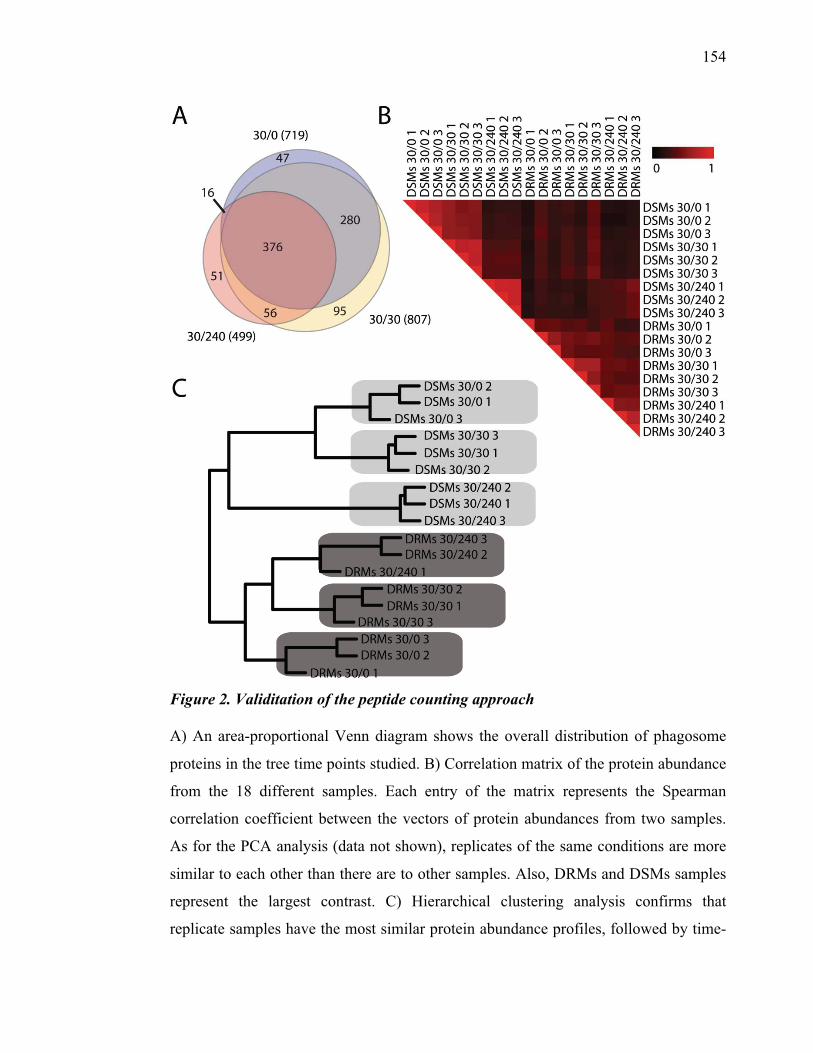
154
Figure 2. Validitation of the peptide counting approach A) An area-proportional Venn diagram shows the overall distribution of phagosome
proteins in the tree time points studied. B) Correlation matrix of the protein abundance
from the 18 different samples. Each entry of the matrix represents the Spearman
correlation coefficient between the vectors of protein abundances from two samples.
As for the PCA analysis (data not shown), replicates of the same conditions are more
similar to each other than there are to other samples. Also, DRMs and DSMs samples
represent the largest contrast. C) Hierarchical clustering analysis confirms that
replicate samples have the most similar protein abundance profiles, followed by time-
155
points and DRMs and DSMs samples. The proteomic profiles of the raft and non-raft
samples at different time points are distinct and reproducible. In both B and C, protein
abundance was estimated from peptide counts from the MS/MS analyses.
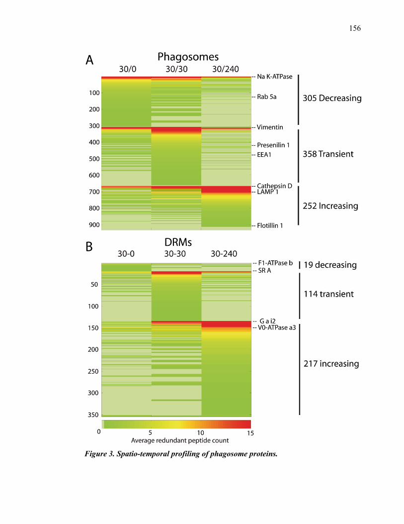
156
Figure 3. Spatio-temporal profiling of phagosome proteins.
157
(A) Heat map representing the abundance of the 931 proteins identified in phagosomes
(y-axis) at the 3 time-points analyzed (30/0, 30/30, 30/240). Protein abundances
correspond to the average RPC values in fractions 1 and 7, of the 3 separate
identification cycles performed per time-point . Increasing average RPC values are
represented in a gradual color scale from green (low protein abundance) to yellow to
red (high protein abundance). The identified proteins were grouped in 3 temporal
clusters: 305 proteins displayed decreasing average RPC values (decreased cluster),
358 proteins displayed a transient increase of their average RPC value at the 30/30
time-point (up-and-down cluster), and 252 proteins displayed increasing average RPC
values (increased cluster). The localization of 6 maturation markers is shown (Rab5a is
decreasing, Presenilin and EEA1 are transiently increasing, Cathepsin D, LAMP 1 and
Flotillin-1 are increasing). (B) Heat map representing the abundance of the 354
proteins identified in DRMs at the 3 time-points analyzed (30/0, 30/30, 30/240) as in
(A) Protein abundances correspond to the average RPC values in fraction 1, of the 3
separate identification cycles performed per time-point. 19 proteins were grouped in
the decreased cluster, 114 were grouped in the up-and-down cluster, 217 proteins were
grouped in the increased cluster.
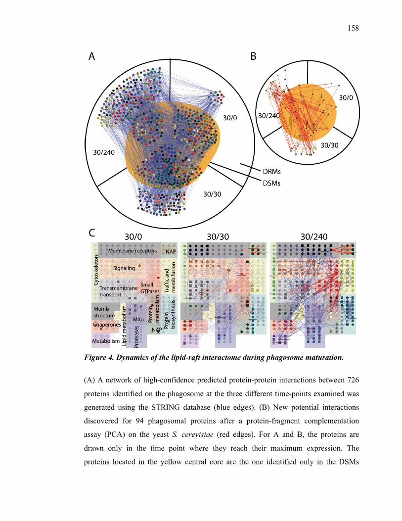
158
Figure 4. Dynamics of the lipid-raft interactome during phagosome maturation.
(A) A network of high-confidence predicted protein-protein interactions between 726
proteins identified on the phagosome at the three different time-points examined was
generated using the STRING database (blue edges). (B) New potential interactions
discovered for 94 phagosomal proteins after a protein-fragment complementation
assay (PCA) on the yeast S. cerevisiae (red edges). For A and B, the proteins are
drawn only in the time point where they reach their maximum expression. The
proteins located in the yellow central core are the one identified only in the DSMs
159
fraction of the phagosome. The proteins were clustered in functional sub-groups,
represented by dots of different colors (light green; cytoskeleton, black; membrane
receptors, green; nucleus-associated proteins [NAP], orange; signaling, red; small
GTPases, yellow-green; trafficking and membrane fusion, grey; transmembrane
transport, brown; protein metabolism, turquoise; protein biosynthesis, dark green;
membrane structure, yellow; lipid metabolism, blue; proteases, purple; metabolism,
white; unknown.
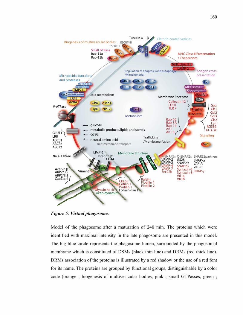
160
Figure 5. Virtual phagosome.
Model of the phagosome after a maturation of 240 min. The proteins which were
identified with maximal intensity in the late phagosome are presented in this model.
The big blue circle represents the phagosome lumen, surrounded by the phagosomal
membrane which is constituted of DSMs (black thin line) and DRMs (red thick line).
DRMs association of the proteins is illustrated by a red shadow or the use of a red font
for its name. The proteins are grouped by functional groups, distinguishable by a color
code (orange ; biogenesis of multivesicular bodies, pink ; small GTPasses, green ;
161
membrane structure, dark blue ; clathrin-coated vesicles, blue ; regulation of apoptosis,
autophagy and mitochondrial, light blue ; microbicidal functions and proteases, red
;MHC Class II Presentation and chaperones, purple ; antigen cross presentation, black
; membrane receptors, orange-red ; signaling, yellow ; trafficking and membrane
fusion, light green ; actin dynamics, gray ; transmembrane transport) ABC transporter:
ATP-binding cassette transporter; GLUT1: Glucose transporter type 1 ;LRP1:
Lipoprotein receptor-related protein; TLR7: Toll-like receptor 7.
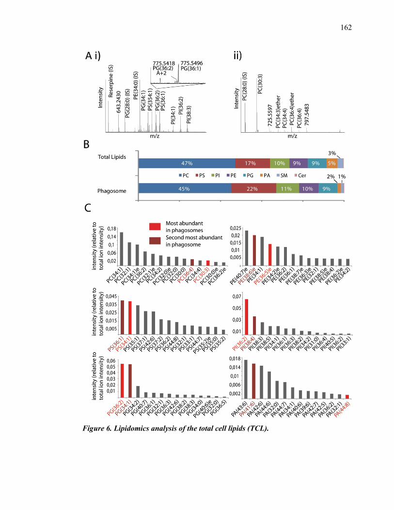
162
Figure 6. Lipidomics analysis of the total cell lipids (TCL).
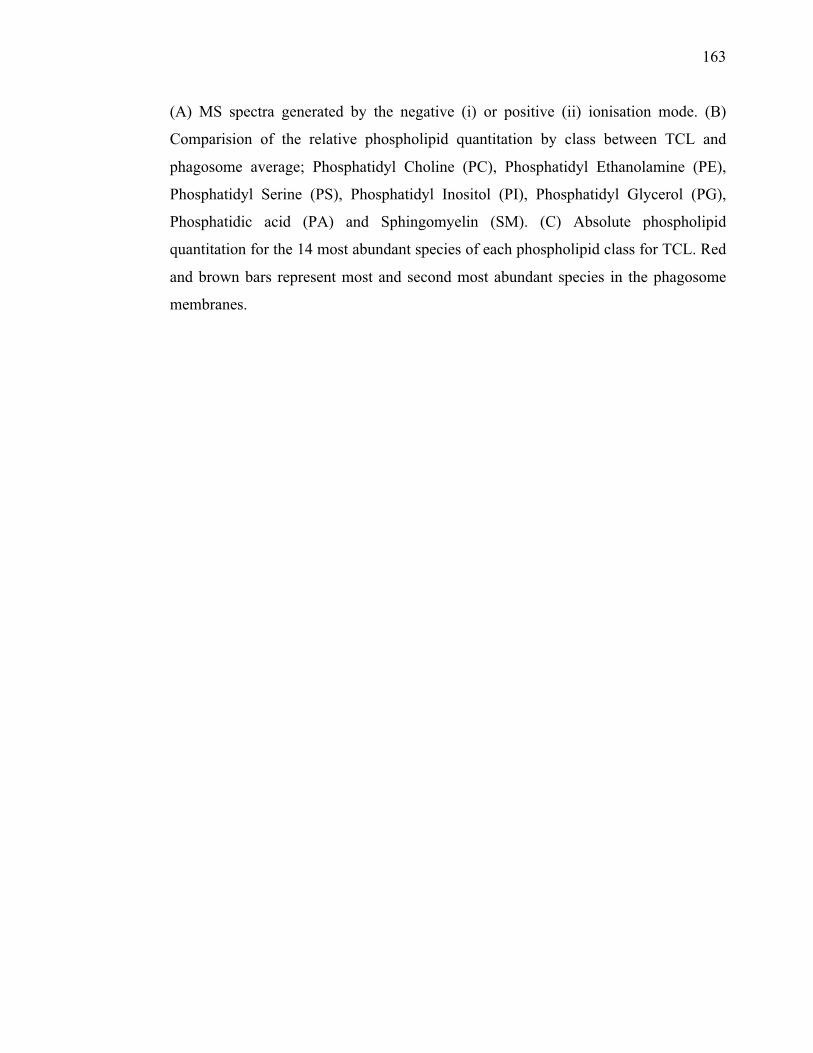
163
(A) MS spectra generated by the negative (i) or positive (ii) ionisation mode. (B)
Comparision of the relative phospholipid quantitation by class between TCL and
phagosome average; Phosphatidyl Choline (PC), Phosphatidyl Ethanolamine (PE),
Phosphatidyl Serine (PS), Phosphatidyl Inositol (PI), Phosphatidyl Glycerol (PG),
Phosphatidic acid (PA) and Sphingomyelin (SM). (C) Absolute phospholipid
quantitation for the 14 most abundant species of each phospholipid class for TCL. Red
and brown bars represent most and second most abundant species in the phagosome
membranes.
164
Figure 7. Lipidomics analysis of the phagosome maturation.
(A) Relative phospholipid quantitation by classes for 15/0, 30/0, 30/30 and 30/240
phagosomes (B) Intensities for the 5 most abundant species of Phosphatidyl Choline
(PC), Phosphatidyl Ethanolamine (PE), Phosphatidyl Serine (PS), Phosphatidyl
Inositol (PI) and Phosphatidyl Glycerol (PG) during phagosome maturation.
165
References
1. Haas, A. The phagosome: compartment with a license to kill. Traffic 8, 311-
330 (2007).
2. Jutras, I. et al. Gamma-secretase is a functional component of phagosomes. J
Biol Chem 280, 36310-36317 (2005).
3. Vyas, J.M., Van der Veen, A.G. & Ploegh, H.L. The known unknowns of
antigen processing and presentation. Nat Rev Immunol 8, 607-618 (2008).
4. Desjardins, M., Huber, L.A., Parton, R.G. & Griffiths, G. Biogenesis of
phagolysosomes proceeds through a sequential series of interactions with the
endocytic apparatus. J Cell Biol 124, 677-688 (1994).
5. Pitt, A., Mayorga, L.S., Stahl, P.D. & Schwartz, A.L. Alterations in the protein
composition of maturing phagosomes. J Clin Invest 90, 1978-1983 (1992).
6. Gotthardt, D. et al. Proteomics fingerprinting of phagosome maturation and
evidence for the role of a Galpha during uptake. Mol Cell Proteomics 5, 2228-
2243 (2006).
7. Garin, J. et al. The phagosome proteome: insight into phagosome functions. J
Cell Biol 152, 165-180 (2001).
8. Houde, M. et al. Phagosomes are competent organelles for antigen cross-
presentation. Nature 425, 402-406 (2003).
9. Dermine, J.F. et al. Flotillin-1-enriched lipid raft domains accumulate on
maturing phagosomes. J Biol Chem 276, 18507-18512 (2001).
10. Vilhardt, F. & van Deurs, B. The phagocyte NADPH oxidase depends on
cholesterol-enriched membrane microdomains for assembly. EMBO J 23, 739-
748 (2004).
11. Arellano-Reynoso, B. et al. Cyclic beta-1,2-glucan is a Brucella virulence
factor required for intracellular survival. Nat Immunol 6, 618-625 (2005).
12. Dermine, J.F., Goyette, G., Houde, M., Turco, S.J. & Desjardins, M.
Leishmania donovani lipophosphoglycan disrupts phagosome microdomains in
J774 macrophages. Cell Microbiol 7, 1263-1270 (2005).
166
13. Wessel, D. & Flugge, U.I. A method for the quantitative recovery of protein in
dilute solution in the presence of detergents and lipids. Analytical biochemistry
138, 141-143 (1984).
14. Hiebl-Dirschmied, C.M., Adolf, G.R. & Prohaska, R. Isolation and partial
characterization of the human erythrocyte band 7 integral membrane protein.
Biochim Biophys Acta 1065, 195-202 (1991).
15. Wasiak, S. et al. Enthoprotin: a novel clathrin-associated protein identified
through subcellular proteomics. J Cell Biol 158, 855-862 (2002).
16. Kearney, R. et al. Elimination of redundant protein identifications in high
throughput proteomics. Conf Proc IEEE Eng Med Biol Soc 5, 4803-4806
(2005).
17. Blondeau, F. et al. Tandem MS analysis of brain clathrin-coated vesicles
reveals their critical involvement in synaptic vesicle recycling. Proc Natl Acad
Sci U S A 101, 3833-3838 (2004).
18. Gilchrist, A. et al. Quantitative proteomics analysis of the secretory pathway.
Cell 127, 1265-1281 (2006).
19. R-Team, R.D.C. (ed. R.F.f.S. Computing)Vienna, Austria; 2009).
20. Tarassov, K. et al. An in vivo map of the yeast protein interactome. Science
320, 1465-1470 (2008).
21. Cline, M.S. et al. Integration of biological networks and gene expression data
using Cytoscape. Nat Protoc 2, 2366-2382 (2007).
22. Bligh, E.G. & Dyer, W.J. A rapid method of total lipid extraction and
purification. Can J Biochem Physiol 37, 911-917 (1959).
23. Jutras, I. et al. Modulation of the phagosome proteome by interferon-gamma.
Mol Cell Proteomics 7, 697-715 (2008).
24. Bantscheff, M., Schirle, M., Sweetman, G., Rick, J. & Kuster, B. Quantitative
mass spectrometry in proteomics: a critical review. Anal Bioanal Chem 389,
1017-1031 (2007).
25. Kinchen, J.M. & Ravichandran, K.S. Phagosome maturation: going through the
acid test. Nat Rev Mol Cell Biol 9, 781-795 (2008).
26. Pike, L.J. The challenge of lipid rafts. J Lipid Res (2008).
167
27. Desjardins, M. & Griffiths, G. Phagocytosis: latex leads the way. Curr Opin
Cell Biol 15, 498-503 (2003).
28. Jutras, I. & Desjardins, M. Phagocytosis: at the crossroads of innate and
adaptive immunity. Annu Rev Cell Dev Biol 21, 511-527 (2005).
29. Desjardins, M. et al. Molecular characterization of phagosomes. J Biol Chem
269, 32194-32200 (1994).
30. Burlak, C., Whitney, A.R., Mead, D.J., Hackstadt, T. & Deleo, F.R. Maturation
of human neutrophil phagosomes includes incorporation of molecular
chaperones and endoplasmic reticulum quality control machinery. Mol Cell
Proteomics 5, 620-634 (2006).
31. Okada, M. et al. Proteomic analysis of phagocytosis in the enteric protozoan
parasite Entamoeba histolytica. Eukaryot Cell 4, 827-831 (2005).
32. Rogers, L.D. & Foster, L.J. The dynamic phagosomal proteome and the
contribution of the endoplasmic reticulum. Proc Natl Acad Sci U S A 104,
18520-18525 (2007).
33. Stuart, L.M. et al. A systems biology analysis of the Drosophila phagosome.
Nature 445, 95-101 (2007).
34. Trost, M. et al. The phagosomal proteome in interferon-gamma-activated
macrophages. Immunity 30, 143-154 (2009).
35. Ramachandra, L., Simmons, D. & Harding, C.V. MHC molecules and
microbial antigen processing in phagosomes. Curr Opin Immunol (2009).
36. Peters, C. et al. Trans-complex formation by proteolipid channels in the
terminal phase of membrane fusion. Nature 409, 581-588 (2001).
37. Lafourcade, C., Sobo, K., Kieffer-Jaquinod, S., Garin, J. & van der Goot, F.G.
Regulation of the V-ATPase along the endocytic pathway occurs through
reversible subunit association and membrane localization. PLoS ONE 3, e2758
(2008).
38. Antonin, W., Fasshauer, D., Becker, S., Jahn, R. & Schneider, T.R. Crystal
structure of the endosomal SNARE complex reveals common structural
principles of all SNAREs. Nat Struct Biol 9, 107-111 (2002).
168
39. Flowerdew, S.E. & Burgoyne, R.D. A VAMP7/Vti1a SNARE complex
distinguishes a non-conventional traffic route to the cell surface used by
KChIP1 and Kv4 potassium channels. Biochem J 418, 529-540 (2009).
40. Mallard, F. et al. Early/recycling endosomes-to-TGN transport involves two
SNARE complexes and a Rab6 isoform. J Cell Biol 156, 653-664 (2002).
41. Kleene, R. et al. Prion protein regulates glutamate-dependent lactate transport
of astrocytes. J Neurosci 27, 12331-12340 (2007).
42. Khunkaewla, P. et al. LFA-1-mediated leukocyte adhesion regulated by
interaction of CD43 with LFA-1 and CD147. Mol Immunol 45, 1703-1711
(2008).
43. Kishimoto, H. & Sprent, J. Several different cell surface molecules control
negative selection of medullary thymocytes. J Exp Med 190, 65-73 (1999).
44. Toyo-oka, K. et al. Association of a tetraspanin CD9 with CD5 on the T cell
surface: role of particular transmembrane domains in the association. Int
Immunol 11, 2043-2052 (1999).
45. Brinkmann, M.M. et al. The interaction between the ER membrane protein
UNC93B and TLR3, 7, and 9 is crucial for TLR signaling. J Cell Biol 177,
265-275 (2007).
46. Nishiya, T. & DeFranco, A.L. Ligand-regulated chimeric receptor approach
reveals distinctive subcellular localization and signaling properties of the Toll-
like receptors. J Biol Chem 279, 19008-19017 (2004).
47. Pryor, P.R. et al. Combinatorial SNARE complexes with VAMP7 or VAMP8
define different late endocytic fusion events. EMBO Rep 5, 590-595 (2004).
48. Desjardins, M., Nzala, N.N., Corsini, R. & Rondeau, C. Maturation of
phagosomes is accompanied by changes in their fusion properties and size-
selective acquisition of solute materials from endosomes. J Cell Sci 110 ( Pt
18), 2303-2314 (1997).
49. Desjardins, M. & Descoteaux, A. Inhibition of phagolysosomal biogenesis by
the Leishmania lipophosphoglycan. J Exp Med 185, 2061-2068 (1997).
50. Lodge, R. & Descoteaux, A. Leishmania invasion and phagosome biogenesis.
Subcell Biochem 47, 174-181 (2008).
169
51. Stow, J.L., Manderson, A.P. & Murray, R.Z. SNAREing immunity: the role of
SNAREs in the immune system. Nat Rev Immunol 6, 919-929 (2006).
52. Kanehisa, L. in The KEGG PATHWAY database2009).
53. Hay, J.C., Chao, D.S., Kuo, C.S. & Scheller, R.H. Protein interactions
regulating vesicle transport between the endoplasmic reticulum and Golgi
apparatus in mammalian cells. Cell 89, 149-158 (1997).
54. Bao, Y., Lopez, J.A., James, D.E. & Hunziker, W. Snapin interacts with the
Exo70 subunit of the exocyst and modulates GLUT4 trafficking. J Biol Chem
283, 324-331 (2008).
55. Steegmaier, M. et al. Three novel proteins of the syntaxin/SNAP-25 family. J
Biol Chem 273, 34171-34179 (1998).
56. Su, Q., Mochida, S., Tian, J.H., Mehta, R. & Sheng, Z.H. SNAP-29: a general
SNARE protein that inhibits SNARE disassembly and is implicated in synaptic
transmission. Proc Natl Acad Sci U S A 98, 14038-14043 (2001).
57. Tsui, M.M. & Banfield, D.K. Yeast Golgi SNARE interactions are
promiscuous. J Cell Sci 113 ( Pt 1), 145-152 (2000).
58. Dilcher, M., Kohler, B. & von Mollard, G.F. Genetic interactions with the
yeast Q-SNARE VTI1 reveal novel functions for the R-SNARE YKT6. J Biol
Chem 276, 34537-34544 (2001).
59. Wickner, W. & Schekman, R. Membrane fusion. Nat Struct Mol Biol 15, 658-
664 (2008).
60. Tani, K. et al. Mapping of functional domains of gamma-SNAP. J Biol Chem
278, 13531-13538 (2003).

CHAPITRE V. DISCUSSION GÉNÉRALE ET
CONCLUSION
171
Discussion générale
Hétérogénéité des radeaux membranaires du phagosome
Notre équipe avait déjà démontré la présence de microdomaines enrichis sur les
phagosomes (Dermine et al., 2001). Ces microdomaines se distinguent du reste de la
membrane du phagosome, car ils contiennent seulement certaines protéines
spécifiques comme la flotilline-1, à l’instar des radeaux membranaires décrits par
Simons et Ikonen (Simons and Ikonen, 1997). Étant donné que ces microdomaines
présentent les mêmes caractéristiques que les radeaux lipidiques, c’est-à-dire : i) ils
sont distribués en motifs ponctués sur la membrane lorsqu’observés en microscopie
confocale; ii) ils peuvent être purifiés biochimiquement grâce à leur insolubilité au
TX-100 à 4oC; et iii) ils contiennent une des protéines les plus caractéristiques des
radeaux membranaires (flotilline-1), nous avons vérifié qu’ils étaient aussi enrichis en
GM1. Le marquage par la sous-unité B de la toxine du choléra liée à un fluorophore
(CTB-FITC) nous a permis de constater que le GM1 s’accumule lui aussi en motifs
ponctués sur le phagosome. Provenant de la membrane plasmique, le GM1 est livré tôt
au phagosome par les endosomes précoces, contrairement à flotilline-1 qui arrive plus
tard et d’une source externe (Dermine et al., 2001). Une autre disparité entre ces deux
marqueurs de radeaux membranaires est qu’ils ne colocalisent pas sur le phagosome,
ce qui laisse supposer la présence de plusieurs types de radeaux lipidiques sur le
phagosome (chapitre III, figure 1D-E). Ce n’est pas la première mention d’une telle
hétérogénéité des radeaux membranaires (Pike, 2004).
Le LPG empêche le recrutement des radeaux membranaires sur le
phagosome
Lorsque L. donovani est phagocytée, le phagosome qui la contient est incapable
d’acquérir le GM1, tout comme il a déjà été démontré pour flotilline-1 (Dermine et al.,
2001). Le mutant lpg2-/-, ayant un défaut dans l’assemblage des chaînes contenant du
172
mannose, ne peut pas accomplir cet effet. Celui-ci est donc attribuable aux molécules
de polysaccharides de surface du parasite. Puisque l’arrivée des marqueurs de radeaux
est inhibée à un niveau très similaire chez le mutant lpg1-/-, où seule la molécule LPG
est manquante, nous déduisons que LPG est essentiel à cette action de L. donovani.
Nous avons renforcé cette conclusion en démontrant que le LPG opsonisé sur le
parasite mutant (lpg1- ou lpg2-) permet de récupérer l’inhibition du recrutement des
radeaux sur le phagosome. Fait intéressant, cette approche avec le parasite mutant
opsonisé nous a permis de vérifier l’hypothèse voulant que la longueur de la molécule
LPG soit proportionnelle à son effet sur le phagosome. Cette action de LPG sur les
membranes du phagosome est très localisée puisque dans la situation où cette
molécule est absente sur une partie du phagosome, le GM1 peut y être recruté.
Le LPG désorganise les radeaux lipidiques préexistants
Nous avons aussi montré que la livraison de LPG dans des phagosomes déjà formés et
marqués par GM1 permet de déstabiliser les radeaux membranaires de ces
phagosomes. En effet, suite à l’arrivée par endocytose de LPG, la distribution de GM1
passe du motif ponctué classique à une répartition uniforme de faible intensité. Une
vérification par immunobuvardage montre que les constituants des radeaux lipidiques
ne sont pas enlevés des phagosomes, mais seulement répartis sur toute la surface. Ceci
nous a emmenés à proposer un modèle dans lequel les microdomaines des phagosomes
sont dissous par l’action du LPG (chapitre III, figure 4C).
Une étude similaire à la nôtre a montré que la bactérie Brucella abortus était aussi
dotée d’une molécule de surface capable de déstabiliser les radeaux membranaires du
phagosome pour empêcher la fusion phagosome-lysosome (Arellano-Reynoso et al.,
2005). Dans le cas de B. abortus, la molécule virulente est le cyclic �-1-2-glucan
(C�G) de l’enveloppe de la bactérie. Le C�G n’a vraisemblablement pas le même
mode d’action sur les radeaux que le LPG puisque sa structure s’apparente plutôt à
celle des cyclodextrines utilisées pour extraire le cholestérol membranaire. Cependant,
l’existence de ces deux cas, où un parasite vise les radeaux membranaires pour
173
survivre, amène à constater que ces microdomaines ont une importance capitale au bon
fonctionnement du phagosome.
Mode d’action du LPG sur les radeaux membranaires
Nous savons que la membrane de Leishmania contient elle-même des radeaux
membranaires où se partitionnent spécialement les molécules à ancre GPI dont fait
partie le LPG et d’autres facteurs de virulence comme le GP63 (Denny et al., 2001).
Plus précisément, chez la Leishmania promastigote procyclique, le GP63 est associé
aux DRMs tandis que le LPG se trouve dans la partie non radeau ou DSMs. Cette
différence pourrait s’expliquer par le fait que l’ancre GPI du LPG comprend une
liaison de type lyso-alkyl plutôt qu’une liaison acyl/alkyl de GP63, et que le type, la
longueur et le nombre des ancres GPI font partie des critères sensibles pour
l’association aux DRMs (Benting et al., 1999). Nous avons mentionné dans la section
2 du chapitre I que la métacyclogenèse de Leishmania donovani est accompagnée de
changements dans la molécule LPG. Il se trouve que ces changements permettent le
recrutement du LPG métacyclique (mLPG) dans les DRMs du parasite (Denny et al.,
2004). Étrangement, ceci ne s’explique pas par une modification de l’ancre GPI, qui
est identique pour les deux formes du parasite (McIntosh et al., 1992). Il se peut que ce
soit le nombre d’ancres GPI par molécule qui augmente, ou bien que les LPG
s’oligomérisent pour s’associer aux DRMs comme on l’a observé chez certaines GPI-
APs (Cunningham et al., 2003), ou encore qu’ils s’associent à d’autres molécules des
DRMs comme la protéine B (Denny et al., 2001; Pimenta et al., 1994a).
De plus, la capacité du LPG à s’insérer dans les membranes du macrophage a été
démontrée il y a longtemps (Tolson et al., 1990). Il a été suggéré que Leishmania
pouvait se dépouiller activement de ses molécules LPG (Kaneshiro et al., 1982;
Pimenta et al., 1994a) comme c’est le cas pour d’autres molécules à ancre GPI
(Thomas et al., 1990) au cours du processus d’infection. Ceci nous amène à proposer
que lorsque la Leishmania promastigote se retrouve dans le phagosome, elle sécrète
des LPG qui vont s’insérer dans les radeaux membranaires du phagosome grâce à leurs
ancres GPI. Selon toute vraisemblance, ce n’est pas simplement cette insertion qui
174
cause les effets délétères du LPG puisque les mutants lpg1- et lpg2- possèdent toujours
cette ancre GPI et n’ont pas d’effets sur les radeaux. Les effets de LPG seraient donc
attribuables aux unités répétitives de Gal(�1,4)-Man�1-PO4, ce qui concorde avec une
portée de moins en moins marquée lorsqu’on raccourcit le LPG par une hydrolyse
acide (chapitre III figure 2E-F).
D’autres études sur les effets de LPG dans différents contextes pointent aussi vers la
partie phosphoglycane (PG) du LPG. Par exemple, le PG, et non pas l’ancre GPI,
serait responsable de l’inhibition de la fusion virale avec les membranes d’érythrocytes
(Miao, 1995), de l’inhibition de la production d’IL-12 par les macrophages (Piedrafita
et al., 1999) et de l’inhibition de l’activation des monocytes humains (Giorgione et al.,
1996). À l’inverse, d’autres effets du LPG sur les cellules hôtes seraient imputables à
la partie GPI, comme l’inhibition de la PKC� (McNeely et al., 1989) tandis que
l’inhibition de la migration transendothéliale des monocytes semble nécessiter les
deux parties (Lo et al., 1998). Dans le cas de l’interaction de LPG sur les phagosomes,
il se peut très bien que l’ancre GPI ait un rôle à jouer, mais tout ce que nous savons
pour l’instant c’est que la déstabilisation et l’inhibition du recrutement des radeaux
lipidiques nécessitent obligatoirement la partie PG du LPG. Les effets du LPG que
nous avons décrits pourraient être causés par les charges négatives qui sont distribuées
sur la longue chaîne de polysaccharide du LPG. Cette chaîne encombrante et chargée a
vraisemblablement des particularités lui permettant d’interférer avec l’assemblage des
molécules en microdomaines.
Le mécanisme proposé du LPG concorde avec l’« hypothèse du champagne »
proposée par Étienne Joly. À l’instar du champagne, qui a besoin de sites de
nucléation pour libérer ses bulles, les membranes pourraient être maintenues dans la
phase liquide désordonnée et avoir besoin d’un stimulus qui permette la formation des
radeaux en phase cristalline solide (Joly, 2004). Selon cette théorie, l’encombrement
stérique des molécules LPG empêcherait l’assemblage des molécules sous la forme de
microdomaines ou causerait leur déstabilisation.
175
Le protéome du phagosome varie plutôt en quantité qu’en qualité
Les études précédentes sur la maturation du phagosome n’ont cherché à caractériser
que quelques-uns des principaux marqueurs des phagosomes précoces et tardifs
(Desjardins et al., 1994a; Desjardins et al., 1994b; Pitt et al., 1992). D’un autre côté,
les études du phagosome utilisant la protéomique à grande échelle se sont limitées à
décrire l’organelle d’un point de vue statique sans tenir compte de son évolution dans
le temps (Burlak et al., 2006; Garin et al., 2001; Gotthardt et al., 2006; Jutras et al.,
2008; Okada et al., 2005; Rogers and Foster, 2007; Stuart et al., 2007; Trost et al.,
2009). Bien des questions à propos des mécanismes précis ayant lieu au cours des
différentes étapes de la phagocytose sont donc restées en suspens. Notre approche,
consistant à isoler des phagosomes après trois différentes périodes de maturation et à
les analyser en parallèle par spectrométrie de masse, a été développée dans le but
d’apporter les premières réponses à cette problématique fort complexe. Les
phagosomes analysés dans notre étude (chapitre IV) sont tous issus d’une phagocytose
de microbilles de polystyrène de 30 minutes, suivie d’un lavage et d’une chasse de 0,
30 ou 240 minutes, ce qui permet respectivement la formation de phagosomes
précoces, de phagosomes intermédiaires et de phagolysosomes.
Nous ne pouvions pas simplement comparer les trois types de phagosomes en
déterminant les protéines présentes ou absentes puisque moins du tiers des protéines
sont uniques à un stade donné (figure 2A, chapitre IV). Nous avons donc choisi une
méthode pour quantifier l’abondance relative des protéines pour chacune de nos
conditions expérimentales. En plus des trois différentes phases, notre étude a
caractérisé les protéines selon qu’elles sont associées ou non dans les microdomaines
du phagosome, ce qui nous faisait un total de six conditions expérimentales à analyser.
Les méthodes de protéomique quantitatives classiques comme le marquage
métabolique (exemple : le SILAC pour Stable isotope labelling with amino acids in
cell culture) ou le marquage des protéines/peptides (exemple : le ICAT pour Isotope-
coded affinity tags) sont très précises, mais très complexes ou impossibles à utiliser
176
pour comparer directement plus de deux échantillons (Bantscheff et al., 2007). La
méthode que nous avons utilisée, le compte de spectres (spectral count), aussi appelé
compte redondant de peptides, est une des techniques dites « sans marquage »
récemment développée (Liu et al., 2004) et qui a fait ses preuves en permettant la
réalisation d’une étude complexe sur les composantes de la voie de sécrétion des
protéines (Gilchrist et al., 2006). Cette technique tire avantage du fait que le nombre
de peptides redondants du spectromètre de masse pour une protéine est proportionnel à
la concentration de cette protéine. De plus, les méthodes de quantification sans
marquage méritent d’être considérées pour plusieurs autres raisons. Au niveau
technique, elles permettent d’éviter les étapes de marquage qui sont longues et
coûteuses. Au niveau de la stratégie d’analyse, ces techniques n’augmentent pas la
complexité des spectres de masse à analyser comme le font les techniques utilisant des
isotopes, ce qui permet à l’appareil une meilleure profondeur d’analyse puisqu’on lui
évite d’avoir à fragmenter chacune des formes du peptide marqué (Bantscheff et al.,
2007). Il a aussi été démontré que les méthodes sans marquage offrent une plus grande
portée dynamique, ce qui est un avantage certain lors de l’analyse de grands ensembles
de protéines comme dans notre cas.
Les protéines du phagosome ont trois principaux comportements
cinétiques
Puisque notre méthode de quantification permet d’assigner les quantités relatives des
protéines pour chacune des phases de la phagocytose, nous avons généré une figure de
type « carte thermique » (heat map) pour représenter les différents comportements
cinétiques des 921 protéines (chapitre IV, figure 3A). Les protéines y sont d’abord
classées selon leur maximum d’intensité, ce qui les classe en trois catégories : i)
protéines dont la quantité relative augmente; ii) protéines dont le maximum est au
temps intermédiaire (ou transitoires sur le phagosome); et iii) protéines qui diminuent.
Comme on peut le voir par les protéines identifiées à la figure 3A, nos résultats
confirment parfaitement les connaissances antérieures et regroupent les marqueurs
précoces dans le premier groupe (Na-K ATPase, Rab5a), les marqueurs tardifs dans le

177
troisième groupe (Cathepsine D, LAMP1, Flotillin-1) tandis que les protéines
transitoires suivent une cinétique déjà établie (Dermine et al., 2001; Desjardins et al.,
1994a; Desjardins et al., 1994b; Jutras et al., 2008). Fait à noter, chacun de ces groupes
cinétiques représente environ un tiers des protéines du phagosome, témoignant ainsi
d’un certain équilibre dans le nombre de protéines différentes, et ce, malgré le grand
dynamisme du phagosome. La distribution dans les trois groupes n’est pas répartie de
la même façon lorsqu’on regarde seulement les protéines des DRMs (chapitre IV,
figure 3B). Nous y reviendrons dans une prochaine section de ce chapitre.
Les fonctions du phagosome sont révélées sous un nouvel angle
La génération de cartes thermiques représentant les principales catégories
fonctionnelles de notre liste de protéines permet de voir dans quel ordre ces protéines
sont recrutées sur le phagosome. On apprend ainsi que les SNARES impliquées dans
la fusion avec le RE (VAMP-3, Sec22b, Syntaxin 5) arrivent relativement tôt tandis
que celles reconnues dans la fusion avec les endosomes tardifs (Vti1b, Syntaxin-8)
arrivent plus tard (voir le chapitre VI, tableau supplémentaire 5). Ces résultats sont
toutefois à prendre avec réserve puisque la majorité de ces SNAREs sont présentes à
tous les stades de maturation et nécessitent un ou des partenaires ainsi que des
protéines accessoires pour jouer leur rôle. Une analyse plus détaillée et fonctionnelle
de ces protéines sur le phagosome serait une des suites intéressantes de mon projet.
Les petites GTPases les mieux connues sur le phagosome sont Rab5 et Rab7.
Conformément à leur implication dans les mécanismes précoces et tardifs de la
phagocytose, ces protéines se retrouvent respectivement au premier rang des GTPases
qui diminuent et de celles qui augmentent. Spécifions que les protéines de chaque
sous-groupe sont classées selon le nombre de peptides redondants, du plus grand au
plus petit. On remarque aussi que Rab27a, impliquée dans la régulation de
l’acidification afin de permettre la présentation d’antigènes (Jancic et al., 2007), fait
partie des GTPases qui sont présentes de façon transitoire sur le phagosome. Rab21 et
Rab22, impliquées dans la régulation du trafic membranaire des endosomes, sont aussi
178
à leur maximum à 30/30. Notre analyse a de plus permis d’identifier Rab14, pour
laquelle on a établi un rôle de stimulation de la fusion entre les phagosomes et les
endosomes précoces, mais pas avec les endosomes tardifs (Kyei et al., 2006). Ceci
concorde avec sa décroissance dans le phagosome entre 30/30 et 30/240.
La catégorie fonctionnelle nommée « métabolisme » regroupe une grande variété
d’enzymes dont celles impliquées dans la biosynthèse des protéines, dans la
modification post-traductionnelle ou dans la dégradation. Le premier constat est que,
contrairement à ce à quoi on pourrait s’attendre chez une organelle dont la fonction
première est de dégrader différentes particules, les protéases ne sont pas les protéines
les plus nombreuses dans cette catégorie. Une grande variété d’enzymes métaboliques
est présente à chaque étape de la phagocytose. Cependant, la zone rouge regroupant
entre autres Cathepsin B, D, Z en plus de Nicastrine indique que ce sont ces protéases
qui sont les protéines du métabolisme qui augmentent le plus dans le temps sur le
phagosome, ce qui n’est pas du tout étonnant.
Pour ce qui est des récepteurs sur le phagosome, on pourrait s’attendre à ce qu’ils
fassent partie des principaux types de récepteurs de phagocytose et qu’ils soient
présents principalement sur les phagosomes précoces puisqu’ils diminuent dans le
temps suite à leur recyclage vers la membrane plasmique. On retrouve en effet les
récepteurs de type éboueurs (scavengers receptors) ainsi que le récepteur du
complément CR5 parmi les récepteurs qui sont surtout présents sur les jeunes
phagosomes. Cependant, les récepteurs Fc� arrivent étonnamment plus tardivement sur
le phagosome, même que Fc�RII est maximal au temps le plus tardif. La présence de
Toll-like receptor 7 (TLR7) dans les phagosomes plus tardifs n’est toutefois pas
inattendue puisque ce récepteur est principalement exprimé de façon intracellulaire et
qu’on lui attribue un rôle dans la reconnaissance d’agents pathogènes à l’intérieur du
phagolysosome (Nishiya and DeFranco, 2004). On ne s’étonnera pas non plus de
retrouver les molécules du complexe majeur d’histocompatibilité (CMH I H-2D, Beta-
microglobuline, CMH I H-2K, CMH II li) plus tardivement associées au phagosome
puisqu’il n’y a pas de peptides prêts à être chargés en début de phagocytose.
179
Il est fort intéressant de voir que les transporteurs transmembranaires sont présents en
si grand nombre (63) sur le phagosome puisque les études protéomiques précédentes
n’avaient pas ou peu rapporté de protéines dans cette catégorie importante. En effet,
bien que peu décrits, les mécanismes de recyclage des éléments issus de la dégradation
des particules sont importants pour le bon fonctionnement du phagosome. La tendance
la plus notable est la prépondérance des sous-unités de la V-ATPase dans les
transporteurs qui sont au maximum dans le phagolysosome. Ceci concorde avec la
deuxième vague d’acidification du phagosome dont il était question dans
l’introduction (Beyenbach and Wieczorek, 2006). Nos autres données indiquent que le
transport des acides aminés (par SNAT2, ASC2, CD98) a principalement lieu chez les
phagosomes intermédiaires tandis que les transporteurs de polypeptides (Sec61) et de
protéines (SCAMP 3, Importin beta 1) sont présents sur les phagosomes précoces et
tendent à diminuer ensuite. Cette cinétique d’expression des transporteurs
transmembranaires sur les phagosomes concorde avec la disponibilité de peptides issus
de la particule présente dans les jeunes phagosomes qui sont dégradés en acides
aminés par la suite.
Les protéines associées aux DRMs sont distinctes du reste des
protéines des phagosomes
Comme on peut le constater en comparant les figures 3A et 3B du chapitre IV, les
protéines associées aux DRMs suivent les trois mêmes tendances cinétiques que celles
du phagosome, mais pas du tout dans les mêmes proportions, comme le démontre la
figure supplémentaire 1 en annexe. Chez les protéines des DRMs, la tendance
principale est à l’augmentation, avec 61 % des protéines qui sont en nombre maximal
au temps le plus tardif. Ceci montre que la composition en protéines de ces sous-
domaines se forme graduellement au cours de la phagocytose. Il est peu probable que
les radeaux membranaires soient présents tout au long de la maturation du phagosome
et que les protéines qui y sont associées augmentent autant. Les radeaux membranaires
sont vraisemblablement absents ou peu présents sur les phagosomes précoces, ce qui
180
nous laisse croire qu’ils ne proviennent pas de la membrane plasmique. Qu’ils soient
formés directement sur le phagosome ou qu’ils soient recrutés lors des fusions avec les
endosomes et lysosomes, leur fonction ne peut s’exercer que lorsque les machineries
protéiques jouant ces rôles y sont réunies, soit après une maturation du phagosome.
Les fonctions spécifiques aux radeaux membranaires des phagosomes peuvent être
déduites de nos données d’enrichissement des protéines concernant chacune des
fonctions dans les DRMs (figure supplémentaire 2 en annexe). On voit alors que 8 des
19 fonctions répertoriées sur le phagosome sont plus représentées dans les DRMs.
Parmi celles-ci, on retrouve les fonctions déjà envisagées pour les radeaux
membranaires des phagosomes (signalisation, petites GTPases, cytosquelette)
(Dermine et al., 2001). Les autres fonctions qui semblent majoritairement exercées par
les radeaux sont assez surprenantes (Métabolisme, Transport transmembranaire). Il
serait intéressant de refaire cette analyse avec le compte de peptides pour chaque
groupe fonctionnel au lieu du nombre de protéines.
Le recrutement des protéines dans les DRMs est un aspect important
de la maturation du phagosome
La figure 6A du chapitre IV donne un aperçu visuel des proportions des protéines pour
lesquelles on a détecté ou non des peptides dans les DRMs. Il ne s’y trouve que les
708 protéines de notre liste qui étaient répertoriées dans la base de données
d’interactions STRING. Ces protéines forment un réseau potentiel comprenant 3133
interactions protéine-protéine. En partant de midi et dans le sens horaire, chaque tiers
de cette sphère représente les protéines dont le nombre maximum est respectivement à
0, 30 et 240 minutes de maturation du phagosome. La première tendance que l’on peut
voir est que les protéines passent d’une minorité dans les DRMs initiaux à une
majorité dans les DRMs tardifs (figure supplémentaire 3 en annexe). Il est intéressant
de constater que les interactions des protéines des DRMs précoces et intermédiaires se
font majoritairement avec des protéines situées hors des DRMs (barres bleues, rouges
et vertes sont plus petites que les mauves, cyan et orange) tandis que ces interactions
181
sont équilibrées entre les DRMs et DSMs dans le phagolysosome. Autrement dit, pour
les phagosomes précoces et intermédiaires (30/0 et 30/30), les protéines qui pourraient
interagir ensemble sont partagées entre DRMs et DSMs et ne peuvent pas jouer leur
rôle alors que dans les DRMs tardifs (30/240), un plus grand nombre d’interactions
peuvent avoir lieu donc plus de machineries moléculaires sont actives.
Notre étude rapporte aussi de nombreuses interactions potentielles qui n’avaient pas
encore été décrites (chapitre IV, figure 6B). La base de données d’interactions obtenue
par nos collaborateurs grâce à un essai sur la levure nous donne de nombreuses pistes
pour enquêter sur les mécanismes permettant aux protéines d’accomplir leurs
fonctions sur le phagosome. Par exemple, l’interaction entre l’ATPase PMC1
l’ATPase VMA13 chez la levure aurait comme équivalent, chez la souris, une
interaction entre la P-type ATPase et la sous-unité H de la V-ATPase.
En regardant plus spécifiquement les protéines associées aux DRMs, on constate à
nouveau que leur nombre augmente beaucoup durant la formation du phagolysosome
(chapitre IV, figure 6C). Les trois panneaux de cette figure comportent en arrière-plan
toutes les protéines associées au DRMs répertoriées, par nos collaborateurs, dans la
base de données STRING ou dans la banque d’informations d’interactions chez la
levure. Les protéines affichées en plus foncé sont celles dont la quantité maximale est
à cette étape de la maturation. Parmi ces protéines, seulement trois sont exprimées de
façon constante (TRAP-delta, V-ATPase E, Malate déshydrogénase 2) et sont, par
conséquent, présentes sur les trois panneaux. Parmi les autres, on retrouve 17 protéines
précoces, 91 qui sont transitoires et 168 dont l’expression est maximale au dernier
temps de maturation étudié. Fait intéressant, parmi les 17 protéines des DRMs initiaux,
nous retrouvons 7 marqueurs habituellement attribués aux mitochondries (Cytochrome
b5 A, Cytochrome b5 reductase, Malate dehydrogénase, NADH déshydrogenase 1
beta 4, F-ATPase beta et les transporteurs VDAC1 et 2). Ces résultats vont à
l’encontre des conclusions récentes de Foster et collaborateurs qui déclarent que : i) les
mitochondries ne contiennent pas de radeaux membranaires; et que ii) les protéines
des mitochondries ne se retrouvent pas non plus dans des radeaux ailleurs dans la
182
cellule (Zheng et al., 2009). Le concept établissant que les mitochondries soient
dépourvues de radeaux membranaires est généralement reconnu; cependant, plusieurs
études protéomiques ont rapporté la présence des VDACs et de la F-ATPase dans les
DRMs de différents types cellulaires (Bae et al., 2004; Man et al., 2005; McMahon et
al., 2006). Les aspects négligés par l’équipe de Foster sont que les radeaux
membranaires ne se retrouvent pas uniquement sur la membrane plasmique et que
l’isolation d’organelles précédant la séparation des DRMs permet de détecter des
protéines que la faible concentration relative ne permet pas de détecter dans une
préparation de membranes cellulaires totales. Une des forces de notre approche est que
nous effectuons l’isolation des phagosomes avec le même protocole pour les
phagosomes précoces, intermédiaires ou tardifs. Les contaminants protéiques issus de
l’imperfection de la méthode de purification des phagosomes (exemple : histones) ou
causés par les étapes expérimentales (exemple : trypsine) sont généralement détectés
en faibles quantités et avec un compte de peptides semblable pour les trois temps
mesurés. Les protéines présentes en plus grande quantité dans les DRMs précoces
représentent d’ailleurs un comportement qui est contre la tendance générale, ce qui est
une autre indication que ce ne sont pas des contaminants. D’ailleurs, la sélection
rigoureuse des protéines que nous avons effectuée pour obtenir notre liste finale de
921 protéines a éliminé 444 protéines, soit près du tiers des protéines initialement
identifiées. Ces protéines éliminées comprennent la grande majorité des faux positifs,
qu’ils soient induits par contamination ou par une identification erronée du spectre de
masse.
La figure 6C du chapitre IV présente les protéines associées aux DRMs en tenant
compte de leur mode de recrutement. En analysant les données de spectrométrie de
masse, nous avons remarqué que les protéines qu’on retrouve dans les DRMs y sont
recrutées soit après leur passage dans la partie DSM des phagosomes (carrés), soit
directement d’une autre source (cercles). On voit donc, par cette figure, qu’il semble y
avoir deux modes de recrutement des protéines dans les radeaux membranaires des
phagosomes : i) un stimulus qui permet le passage d’une protéine de la membrane
normale vers les microdomaines; ii) l’arrivée sur le phagosome de nouveaux radeaux
183
membranaires contenant des protéines qui s’ajoutent aux DRMs des phagosomes. La
distinction entre ces deux modes de régulation est très importante puisqu’elle implique
que ce n’est pas parce qu’une protéine est présente sur le phagosome à un moment
précis qu’elle y joue son rôle à ce même moment. Ces résultats nous laissent
effectivement croire que certaines protéines présentes au début de la phagocytose n’y
jouent pas leur rôle avant d’être recrutées aux radeaux membranaires du phagosome.
D’autres possibilités sont aussi envisageables à propos de ce passage des membranes
solubles aux membranes insolubles. Il se peut aussi qu’une protéine y soit inhibée ou
que son activité y soit modifiée. Un cas intéressant est la protéine RGS19 dont la
quantité augmente dans les DRMs, mais qui reste stable dans les DSMs (chapitre IV,
figure 5). Fait étonnant, la quantité totale de cette protéine associée aux phagosomes
diminue dans le temps (figure supplémentaire 4 en annexe). Une étude cinétique de
RGS19 sur le phagosome omettant la dimension spatiale (microdomaines) aurait pu
avoir comme conclusion que cette protéine joue principalement son rôle au début de la
maturation. Notre approche spatio-temporelle nous a permis de constater que la
diminution globale de RGS19 est accompagnée d’un enrichissement dans les DRMs.
Cette protéine qui est impliquée dans l’inhibition de certaines voies de signalisation
augmente l’activité GTPase des sous-unités alpha des protéines G hétérotrimériques,
ce qui les amène à leur état inactif lié au GDP. Le transfert de RGS19 de la partie
soluble vers la partie insoluble du phagosome s’accompagne justement d’un transfert
dans le même sens de quatre protéines G alpha. En effet, les protéines G alpha,
G alpha 13, G alpha i2 ainsi que G alpha i3 augmentent dans la partie soluble pendant
les 30 premières minutes de maturation puis diminuent ensuite, tandis qu’elles sont à
leur expression maximale dans la partie insoluble à 240 minutes (table 1, en annexe).
La G alpha i2 semble particulièrement abondante dans les radeaux membranaires du
phagosome avec une moyenne de 85,2 spectres générés pour les trois réplicas de
l’expérience. Les G beta 1 et 2 suivent aussi cette cinétique tandis que les G gamma 2,
5 et 10 ont exclusivement été détectés dans la partie non-radeau des phagosomes.
Une autre population de protéines que nous avons observée lors de notre analyse est
celle qui est recrutée dans les radeaux membranaires de façon transitoire. Sur les 114
184
protéines dont le compte de peptides dans les DRMs est à son maximum à 30 minutes
de maturation, près de la moitié (54) sont aussi exprimées majoritairement dans la
partie soluble (DSMs) des phagosomes. Ce sous-ensemble de protéines semble donc
être en équilibre entre les deux différents sous-domaines du phagosome. Une quantité
équivalente (49) des protéines transitoires des DRMs est en décroissance dans les
DSMs. Ce sont donc des protéines qui sont majoritairement insolubles sur les
phagosomes précoces, qui sont recrutées dans les radeaux des phagosomes
intermédiaires et qui disparaissent ensuite du phagosome.
Nos résultats permettent de mettre à jour le phagosome virtuel
La grande quantité de nouvelles données générées lors de cette série d’analyses nous a
permis de construire un nouveau modèle du protéome du phagosome. Nous avons
donc fait une mise à jour du phagosome virtuel (chapitre IV, figure 7). Parmi les
protéines que nous avons identifiées, nous avons pu en regrouper de nombreuses selon
leurs rôles potentiels sur le phagosome.
Fonctions microbicides
La phagocytose des microbes conduit à leur mort rapide et le complexe de la NADPH
oxydase y joue un rôle important. Les deux sous-unités membranaires de ce complexe
(p22phox et gp91phox) ainsi que Rac1 et Rac2, qui sont des régulateurs connus de
l’activité de la NADPH oxydase (Bokoch, 2005), ont été identifiés sur les phagosomes
précoces, ayant une abondance maximale à 30/0 ou 30/30. La protéine
kinase C (PKC), un régulateur important de l’activité de la NADPH oxydase, a
aussi été identifiée sur les phagosomes précoces. PKC est en effet requis pour la
phosphorylation de la sous-unité p47phox, ce qui provoque un accroissement de
l’activité du complexe enzymatique (Bey, J. Immunol, 2004). La PKC a aussi une
fonction dans le confinement de Listeria monocytogenesis dans le phagosome
(Schwegmann et al., 2007). Les composants p22phox et gp91phox ont tous deux été
identifiés dans les DRMs, conformément à la proposition affirmant que les radeaux

185
membranaires jouent le rôle de plateforme d’assemblage du complexe de la NADPH
oxydase. L’enrichissement de ces sous-unités dans les DRMs est maximal au temps
30/240, ce qui suggère que, malgré une diminution de leur quantité globale sur le
phagosome, l’activité de la NADPH oxydase persiste sur les phagolysosomes. Les
propriétés oxydatives du phagosome peuvent aussi être modulées par la protéine heme
oxygenase-1 (HO-1), qui a été identifiée sur les phagosomes précoces. HO-1 possède
des propriétés anti-oxidantes (Taille et al., 2004) et peut augmenter la phagocytose lors
d’un choc septique microbien, et ce, en augmentant la production de monoxyde de
carbone (Chung et al., 2008).
Fusion membranaire
La plus importante famille de protéines impliquées dans la fusion membranaire est
probablement les SNAREs. Quelques SNAREs on déjà été décrites comme étant
associées aux DRMs (Lang, 2007). L’utilisation de différentes conditions
expérimentales sur différents types cellulaires rend difficile l’élaboration de la liste des
SNAREs qui sont associées ou non aux DRMs. On peut en avoir un bon aperçu en
choisissant les SNAREs qui sont associées à plus de 50 % aux DRMs dans au moins
une sorte de cellules (Syntaxin 3, Syntaxin 4, SNAP-23 et VAMP2) (Lang, 2007). Nos
résultats montrent, pour la première fois, que 11 SNAREs sont associées aux DRMs
des phagosomes, dont 10 ayant un ratio d’enrichissement dans les DRMs qui s’accroît
durant la maturation (table supplémentaire 5, en annexe). Nous avons également
identifié 4 protéines accessoires des SNAREs qui partagent le même comportement.
Les 11 autres SNAREs et protéines partenaires identifiées sur le phagosome n’étaient
présentes que dans la partie DSM du phagosome, ce qui suggère que différents sous-
groupes de ces protéines de fusion sont distribués indépendamment sur le phagosome.
Tel que Lang l’a proposé dans le cas de l’exocytose, cette ségrégation des SNAREs du
phagosome sert peut-être à séparer spatialement les différents types d’évènements de
fusion (Lang, 2007).
186
Biogenèse des corps multivésiculaires
Notre analyse protéomique a permis de relever la présence de différents composants
du complexe de triage endosomal requis pour les machineries de transport (ESCRT
pour Endosomal Sorting Complex Required for Transport). Ce complexe est
nécessaire pour la formation des corps multivessiculaires (MVBs pour MultiVesicular
Bodies) qui est accomplie par l’invagination des membranes endosomales (Williams
and Urbe, 2007), ce qui suggère que ce processus peut aussi avoir lieu sur le
phagosome. D’une façon qui concorde avec la dynamique connue d’assemblage du
complexe ESCRT, les trois composants du complexe ESCRT-I (TSG101, VPS28 et
VPS37) ont été identifiés tôt sur les phagosomes, avec une abondance maximale après
30 minutes de maturation. Un seul composant d’ESCRT-II a été identifié dans notre
analyse (VPS25) et seulement dans les phagosomes 30/30, suggérant que le complexe
ESCRT-II au complet peut être recruté dans un stade de maturation entre 30/30 et
30/240 minutes. D’un autre coté, la formation des MVBs pourrait peut-être avoir lieu
indépendamment du complexe ESCRT-II puisqu’il a été suggéré qu’ESCRT-I recrute
directement ESCRT-III grâce à ALIX (Odorizzi, 2006). Conformément à cette
hypothèse, ALIX a été identifiée sur les phagosomes de tous les stades étudiés, avec
un maximum à 30/30. Les composants du complexe ESCRT-III formant un lacis sur
les membranes (CHMP4, CHMP5) on été identifiés ainsi que BROX, une protéine
dont l’interaction avec CHMP4 a récemment été découverte (Ichioka et al., 2008).
Nous avons de plus identifié VPS4 qui est l’ATPase responsable du désassemblage
ATP-dépendant du lacis d’ESCRT-III. Il est à noter que CHMP4, CHMP5 et ALIX
sont associées aux DRMs avec une abondance maximale au temps 30/240.
Régulation de l’apoptose et de l’autophagie
Un stress cellulaire peut déclencher soit la voie apoptotique, soit la voie autophagique,
selon la nature et l’intensité du stress impliqué (Maiuri et al., 2007b). Contrairement à
l’apoptose, l’autophagie permet aux cellules de s’adapter au stress bien qu’une
autophagie massive puisse aussi mener à la mort cellulaire. Différentes protéines
impliquées dans la régulation de l’apoptose ont été identifiées sur le phagosome,
incluant Aven, BCL-XL, BAD, BAX et BAK. Aven, un inhibiteur de l’apoptose qui
187
interfère avec l’activation des caspases (Chau et al., 2000), est présent sur les
phagosomes précoces avec BCL-XL, qui lui, restreint l’activité des protéines pro-
apoptotiques de la famille BCL-2 (Youle, 2007). D’un autre coté, les protéines pro-
apoptotiques BAD, BAK et BAX affichent une abondance maximale au temps 30/0,
30/30 et 30/240 respectivement. BAD agit comme un senseur du stress cellulaire et
amorce l’oligomérisation de BAX et/ou BAK dans la membrane externe des
mitochondries, où ils forment des canaux par lesquels les protéines mitochondriales
traversent vers le cytoplasme. Cette étape cruciale est souvent décrite comme le point
de l’engagement dans la voie de l’apoptose (Maiuri et al., 2007b).
En plus de leur rôle dans le déclenchement de l’apoptose, différents facteurs
apoptotiques bien caractérisés tels que BAX/BAK, BAD et BCL-XL peuvent aussi
réguler l’autophagie (Maiuri et al., 2007b). Il a récemment été démontré que BAD est
nécessaire pour l’autophagie induite par stress nutritif, probablement en empêchant
l’interaction entre BCL-XL et Becline-1, une protéine essentielle pour la formation des
autophagosomes (Maiuri et al., 2007a). Becline-1 à été identifiée sur les phagosomes
intermédiaires tandis que LC3, un marqueur des autophagosomes, est recruté à cette
étape, mais montre un maximum d’abondance sur les phagolysosomes. Il a été
rapporté que Becline-1 et LC3 s’associent à des phagosomes contenant des levures
mortes ayant des cinétiques similaires et il a été suggéré qu’ils aient un rôle dans la
maturation du phagosome (Sanjuan et al., 2007).
Cette dernière étude rapporte cependant que les phagosomes contenant des billes de
latex sont dépourvus des marqueurs des autophagosomes. Nos observations de ces
marqueurs sur nos phagosomes peuvent résulter du traitement que nous avons fait et
qui est susceptible de déclencher l’autophagie. En effet, pour réduire la contamination
de nos échantillons par les protéines du sérum, qui par leur abondance pourraient
masquer l’identification par spectrométrie de masse des protéines de plus faible
concentration, nous avons lavé les cellules dans du PBS avant leur incubation dans un
milieu de culture sans sérum pour les étapes d’internalisation et de maturation. Le
stress cellulaire résultant de cette déprivation de nutriments chez des cellules en
188
confluence peut avoir induit l’autophagie, ou plus spécifiquement la mitophagie, soit
la dégradation sélective des mitochondries (Maiuri et al., 2007b). L’identification de
protéines mitochondriales dans nos préparations de phagosomes peut donc résulter de
la fusion entre les autophagosomes et les phagosomes précoces.
Présentation sur les CMH classe II
Notre analyse à permis d’identifier Cystatine C sur le phagosome, celle-ci ayant
comme fonction d’inhiber les cathepsines et, par le fait même, de réguler la
dégradation de la chaîne Ii du CMH classe II (aussi sur le phagosome) qui est
dépendante de la cathepsine S (Kitamura et al., 2005). La cathepsine S est également
présente sur le phagosome, avec un maximum dans les phagosomes intermédiaires,
tout comme GILT (ou Lysosomal thiol reductase) qui est aussi impliqué dans la
présentation d’antigènes exogènes sur le CMH classe II.
Présentation croisée sur les CMH classe I
De plus en plus d’indices suggèrent que les phagosomes pourraient être des organelles
autosuffisantes pour accomplir la présentation de peptides exogènes sur les molécules
du CMH de classe I, ce processus étant appelé la présentation croisée (Guermonprez et
al., 2003; Houde et al., 2003). Le translocon Sec61 et l’ATPase p97, qui sont des
candidats pour jouer le rôle de la rétrotranslocation de protéines à partir de la lumière
du phagosome vers le cytosol (Ackerman et al., 2006), ainsi que des sous-unités du
protéasome, dont le rôle serait de dégrader les protéines rétrotransloquées, ont été
identifiés sur les phagosomes précoces. Des composants du complexe de chargement
des peptides sur le CMH-I tels que calréticuline, tapasine et la chaîne alpha du CMH-I
sont également détectés en abondance sur les jeunes phagosomes puis leur quantité
diminue durant la maturation. De plus, des enzymes impliquées dans la N-
glycosylation, incluant les oligosaccharyltransférases ribophorine 1 et 2, ou impliquées
dans le traitement des groupements N-glycan, comme la glucosidase 1, ont aussi été
trouvées sur les phagosomes précoces. Ceci concorde avec la découverte récente que
la machinerie de glycosylation du RE est recrutée au phagosome et que la machinerie
189
de dégradation qui y est associée participe à la présentation croisée (Ackerman et al.,
2006).
Dans le cadre des travaux de Mathieu Houde sur la présentation croisée par les
phagosomes, nous avons constaté, dans une expérience d’immunofluorescence, que le
marquage des phagosomes pour les sous-unités du protéasome est discontinu (article
III, figure 2C, annexe), ce qui ressemble à la distribution des protéines des radeaux
membranaires comme flotilline-1 (chapitre III figure 1C). Nous avons donc vérifié la
présence des sous-unités du protéasome dans les DRMs des phagosomes et constaté
qu’ils n’y sont pas (annexe III article 3, figure 2D). Toutes les sept sous-unités du
protéasome que mon analyse à révélé ont été détectées dans la partie soluble (DSMs)
du phagosome, avec un maximum d’intensité à 0 minutes (26S non-ATPase sous-unité
régulatrice 7, composant C9), 30 minutes (alpha 2, beta 3, beta 4) ou 240 minutes de
maturation (26S non-ATPase sous-unité régulatrice 13, beta 2).
Vésicules recouvertes de clathrine (formation et bourgeonnement)
Nous avons trouvé plusieurs sous-unités de l’adaptor protein complex 1 et 2 (AP-1 et
AP-2) ainsi que la dynamine sur les phagosomes. La phosphatidylinositol-binding
clathrin assembly protein effectue le recrutement et l’assemblage de la clathrine et de
l’AP-2 à la membrane. Nous avons, bien entendu, retrouvé la chaîne lourde de la
clathrine, dont la quantité augmente avec la maturation du phagosome. Nos analyses
ont aussi permis de découvrir l’association de CALM au phagosome, CALM étant une
protéine d’assemblage de la structure de clathrine qui ressemble à AP180. Comme
AP180, CALM a : i) un domaine ANTH (pour AP180 N-terminal homology) qui lui
permet de lier les lipides de la membrane; et ii) des motifs spécifiques liant la clathrine
et le complexe AP-2, qui sont les principaux composants des vésicules des clathrines
(Harel et al., 2008).
190
Transport transmembranaire
Les protéines VDAC (Voltage-Dependent Anion Channel) sont bien connues pour leur
localisation dans la membrane externe des mitochondries, où elles jouent leur rôle de
canal transmembranaire sélectif pour certains ions et réagissent au potentiel
membranaire. Dans la conformation ouverte (lorsque le potentiel membranaire est
bas), les VDACs transportent des métabolites essentiels, notamment l’ATP, l’ADP, le
phosphate inorganique et le cholestérol (Goncalves et al., 2008; Papadopoulos et al.,
1997). Les petits ions tels que le K+, le Na+ et le Ca+ peuvent être échangés par ces
canaux même dans leur conformation fermée (Lemasters and Holmuhamedov, 2006;
Reymann et al., 1998). Notre analyse révèle la présence de VDAC-1 et VDAC-2 sur
les phagosomes, principalement les phagosomes précoces avec un ratio important dans
les DRMs.
Les VDACs ont aussi été trouvées chez les endosomes et on leur a attribué un rôle
dans l’acidification puisqu’elles font le transport des ions Cl-, ce qui permet
d’équilibrer les charges positives des ions H+ (Reymann et al., 1998). On a aussi
démontré leur présence dans les cavéoles (Bàthori, 1999) et dans les radeaux
lipidiques (Bahamonde et al., 2003) de la membrane plasmique où ils joueraient un
rôle précédemment attribué aux canaux Maxi Cl-. Le relâchement de molécules dont le
cytochrome c par les canaux VDACs est déclenché par la protéine pro-apoptotique
BAX qui, comme nous l’avons mentionné précédemment, a aussi été trouvée sur le
phagosome précoce. On retrouve un autre composant du pore de transition de la
perméabilité (PTP pour Permeability Transition Pore) sur le phagosome, ANT2
(adenine nucleotide translocator 2) (Tsujimoto and Shimizu, 2007), qui est dans la
membrane soluble des phagosomes précoces et passe aux DRMs durant la maturation.
Le diamètre du pore des VDACs est de 2.5 à 3.0 nm, ce qui est suffisant pour laisser
passer des nucléotides et de petites molécules, mais insuffisant pour laisser passer une
protéine repliée comme le cytochrome c. La découverte de la capacité des VDACs à
s’oligomériser a conduit Shoshan-Barmatz et confrères à proposer que ce sont les
oligomères de VDACs qui permettent la relâche du cytochrome c (Shoshan-Barmatz et
al., 2008). Constatant l’enrichissement de ces canaux dans les DRMs, il est tentant
191
d’aller plus loin et de proposer que cette oligomérisation des VDACs est régulée par
leur recrutement dans les radeaux membranaires. Finalement, une autre protéine
interagissant avec VCAC a été identifiée dans les phagosomes, soit la glycéraldéhyde
3-phosphate déshydrogénase (GAPDH). GAPDH déclenche l’ouverture du PTP par
son interaction avec VDAC, ce qui lui confère un rôle possible dans l’oligomérisation
du transporteur. GAPDH est présente sur les phagosomes des trois temps de
maturation et sa quantité va en croissant dans les DSMs aussi bien que dans les DRMs.
La Na+,K+-ATPase est une protéine intégrale trouvée dans les cellules de tous les
eucaryotes supérieurs et qui est responsable de la translocation des ions Na+ et K+ à
travers la membrane en utilisant l’ATP comme source d’énergie. Pour trois ions Na+
éjectés de la cellule, 2 K+ sont pompés à l’intérieur (Lingrel and Kuntzweiler, 1994).
Puisque nous avons trouvé les sous-unités alpha-1, alpha-3 et beta-3 principalement
sur les phagosomes 30/0 et 30/30, nous pouvons supposer que La Na+,K+-ATPase est
active et qu’elle transfèrent 3 Na+ vers le phagosome pour 2 K+ vers le cytoplasme et
ce, dans les premières étapes de la maturation du phagosome. Leur rôle pourrait être
l’établissement d’un potentiel membranaire au niveau du phagosome, potentiel
semblable à celui de la membrane plasmique. À première vue, cet échange d’ions
semble favoriser la mise en place d’un potentiel membranaire positif pour le
phagosome, mais en regardant plus en détail le protéome du phagosome on peut
déduire que les ions Na+ sont retournés au cytosol par SNAT-1 et 2 (Sodium-coupled
Neutral Amino acid Transporter) qui font le transport couplé de Na+ et d’acides
aminés (Burkhalter et al., 2007). Les deux SNATs sont à leur maximum à 30/0, mais
seul SNAT-2 a été détectée dans les DRMs, principalement à 30/30. Une autre
protéine de transport couplée au Na+ à été trouvée sur la phagosome, le Na+-
dependent ascorbic acid transporter (Slc23a2).
Parmi les transporteurs identifiés principalement sur les phagosomes intermédiaires, il
y en a trois de la famille des transporteurs ABC (ATP-binding cassette transporters).
ABCB1 et ABCB6 sont redirigés vers les radeaux des phagolysosomes tandis
qu’ABCC1 est principalement associé aux membranes solubles du phagosome. Les
192
transporteurs ABC déplacent surtout des molécules vers l’extérieur de la membrane
plasmique ou vers l’intérieur d’organelles comme le phagosome. ABCA1 a d’ailleurs
déjà été observé dans les radeaux lipidiques des macrophages (Bared et al., 2004).
La sélectivité des transporteurs ABC est souvent assez large. ABCB1 transporte des
produits métaboliques, des lipides et des stérols, en plus de drogues et d’autres
xénobiotiques. ABCC1 est un transporteur important d’une variété de molécules
organiques conjuguées, tels que des conjugués au glutathion, en plus d’être un facteur
de résistance pour de nombreuses drogues anti-cancérigènes (Choudhuri and Klaassen,
2006). Certains de ces conjugués au glutathion sont impliqués dans la détoxification et
la protection contre les stress oxydatifs (Leite et al., 2007a). ABCC1 serait aussi
impliqué dans la relâche de médiateurs de l’inflammation (Leite et al., 2007a; Leite et
al., 2007b).
Les principales protéines impliquées dans le transport transmembranaire qui sont
enrichies sur les phagosomes tardifs sont les différentes sous-unités du complexe de la
V-ATPase (chapitre IV, figure 4B). La V-ATPase est une enzyme hétéromultimérique
composée d’un complexe catalytique V1 qui est périphérique (cytoplasmique) et
attaché au complexe V0, un pore à protons transmembranaire. Les sous-unités A à H
composent le complexe V1 tandis que les composants a, c, c’, c’’ et d forment le
complexe V0. Notre analyse par spectrométrie de masse a pu identifier des isoformes
de chacune des 8 composantes de la partie V1 ainsi que les sous-unités a et d de V0
(chapitre IV, table1). La grande hydrophobicité des sous-unités c, qui sont intégrales à
la membrane peut expliquer pourquoi ils ne sont pas parmi les protéines détectées. On
peut toutefois assumer qu’elles sont présentes sur nos phagosomes puisque ces sous-
unités sont essentielles aux fonctions principales des V-ATPases et aussi parce que ces
sous-unités ont été identifiées au niveau des phagosomes des cellules S2 (Stuart et al.,
2007) et des endosomes de cellules BHK (Baby Hamster Kidney cells) (Lafourcade et
al., 2008).
193
Le complexe V1 catalyse l’hydrolyse de l’ATP et l’énergie relâchée par cette réaction
est utilisée pour permettre la rotation de la structure en anneau du complexe V0, ce qui
produit un mouvement transmembranaire de protons (Steinberg et al., 2007). Les
V-ATPases sont les grandes responsables de l’acidification du phagosome puisqu’elles
peuvent pomper les protons contre leur gradient de concentration grâce à l’énergie
tirée de l’ATP. Des études chez le poisson-zèbre ont révélé que la V-ATPase pourrait
avoir un rôle dans la maturation en plus de celui qu’elle a dans l’acidification. La sous-
unité a1 du complexe V0 est requise pour la fusion du phagosome avec les lysosomes
(Peri and Nusslein-Volhard, 2008). Notons que l’Aldolase, qui est aussi présente sur
les phagosomes, aurait un rôle de régulation de la V-ATPase, ce rôle étant la
dissociation réversible des complexes V1 et V0 (Jefferies et al., 2008).
Il est intéressant de constater que plusieurs sous-unités de la V-ATPase, plus
particulièrement celles du complexe V0, sont associées aux DRMs. Le rôle de
l’environnement lipidique sur la régulation de la V-ATPase a déjà reçu l’attention de
l’équipe de Gisou van der Goot, qui a conclu que l’association de ce complexe aux
DRMs augmente l’activité de pompage en promouvant le couplage V1-V0
(Lafourcade et al., 2008). Leurs résultats indiquent aussi que le ratio V1/V0 augmente
durant la maturation des endosomes. Sur le phagosome, nous observons aussi un
accroissement global dans le ratio V1/V0, mais plus particulièrement, nous voyons un
enrichissement en sous-unités des deux complexes dans les DRMs. Toutes les sous-
unités de cette pompe augmentent sur le phagosome durant la maturation. Les sous-
unités D à H augmentent et demeurent associées à la partie non-radeau tandis que les
sous-unités a1, a3, et d de V0 ainsi que la sous-unité accessoire S1 sont hautement
enrichies dans les DRMs, surtout à 30/240 (chapitre IV, table1).
Complexe de l’exocyste
Le complexe de l’exocyste est composé de 8 protéines (Sec3, Sec5, Sec6, Sec8, Sec10,
Sec15, Exo70, et Exo84) et joue un rôle dans le trafic vésiculaire, principalement dans
la liaison à des vésicules et dans leur ciblage vers la membrane plasmique où ces
194
vésicules fusionnent. Malgré qu’aucune de ces protéines n’ait été identifiée sur le
phagosome de la souris, une autre étude protéomique de notre laboratoire a montré que
ce complexe est présent sur les phagosomes de drosophiles (Stuart et al., 2007). Pour
renforcer les résultats de l’étude effectuée par nos collègues, nous avons vérifié la
présence des protéines Sec6 et Sec8 par immunobuvardage sur nos préparations de
phagosomes de souris. Il s’est finalement avéré que ces deux protéines aussi étaient
présentes sur les phagosomes murins avec des cinétiques semblables à celles observées
chez la mouche (article IV, en annexe, figure 4B).
Remodelage de l’actine
Notre liste protéomique révèle 30 protéines impliquées dans le cytosquelette d’actine,
comprenant les sous-unités alpha, bêta et gamma de l’actine ainsi que des protéines
accessoires. La majorité de ces protéines ne semblent pas du tout associées aux
radeaux membranaires puisqu’aucun de leurs peptides n’a été détecté dans les DRMs
précoces. En ajoutant les autres protéines du cytosquelette (tubuline, myosine, etc.)
que nous avons trouvées sur le phagosome, nous avons une liste de 47 composants
dont 16 sont associés aux DRMs. Sur les phagosomes intermédiaires, il y a un grand
regroupement de protéines comme filamine et coronine, permettant la liaison des
filaments d’actine à la membrane du phagosome. Notons aussi la présence de
l’Intégrine b1, aussi à son maximum à 30/30 et qui est reconnue comme un récepteur
membranaire liant la filamine (Legate and Fassler, 2009). La moésine, aussi impliquée
dans la liaison de la F-actine à la membrane est, quant à elle, associée aux DRMs.
Toujours sur le phagosome intermédiaire, nombre de protéines de liaison à l’actine qui
permettent la formation de réseaux (MACF1, Actinine-�, Arp 2/3-4, IQGAP-1,
Taline-1) ou qui régulent la polymérisation (Cap 1, Cap Z �) sont à leur maximum. On
retrouve aussi des protéines complémentaires dans les phagosomes intermédiaires,
mais celles-ci ont leur maximum d’abondance plus tard (Actinine-�, Arp2/3-5, Cap Z
�).
195
En regardant les changements qui s’opèrent sur le phagosome plus tardif à propos des
protéines du cytosquelette d’actine, il semble que l’ancrage des filaments d’actine
provienne des radeaux lipidiques. En effet, pour le temps 30/240, les protéines faisant
le pont entre la membrane et la F-actine sont majoritairement dans les DRMs. Le
récepteur CD44 qui est enrichi dans les DRMs tardifs est connu pour lier l’actine par
l’intermédiaire d’ezrine que l’on a aussi identifiée sur le phagosome tardif. Cofiline-1,
Profiline-1 et Formine-like 1 ont toutes été impliquées dans la liaison de l’actine et
nous les avons retrouvées dans les DRMs (Michelot et al., 2007). Même les chaînes
lourdes et légères de la myosine ont été identifiées dans les DRMs des
phagolysosomes avec des ratios d’enrichissement importants.
Les fonctions cellulaires altérées par le LPG sont dépendantes des
radeaux membranaires
Comme nous l’avons mentionné précédemment, le parasite Leishmania possède des
moyens pour inhiber plusieurs aspects de la phagocytose. Un des premiers effets de
Leishmania sur le phagosome ayant été décrit est l’inhibition du remodelage de
l’actine, qui permet de désassembler le manteau d’actine recouvrant le phagosome qui
vient de se former. La présence de Leishmania promastigote provoque en fait une
accumulation anormale de F-actine sur le phagosome, ce qui empêche ce dernier de
poursuivre sa maturation (Lodge and Descoteaux, 2008). La protéine Cdc42 reste plus
longtemps associée au phagosome en présence de la molécule LPG de Leishmania. De
plus Arp2/3, WASP, �-actinin, Myosin II et Nck sont aussi retenues sur les
phagosomes lorsque du LPG est présent dans le phagosome. Cdc42 joue un rôle
central dans l’accumulation de la F-actine puisque l’expression d’un mutant dominant-
négatif de Cdc42 empêche l’accumulation de la F-actine ainsi que la rétention de
WASP et de la myosine (Lodge and Descoteaux, 2005). Puisque plusieurs de ces
protéines se trouvent dans les DRMs, il se peut que leur dérégulation soit causée par la
déstabilisation des radeaux membranaires que nous avons démontrée au chapitre III.
Un mécanisme pouvant expliquer le maintien de l’activité de Cdc42 sur le phagosome
196
serait que le LPG active une RhoGEF spécifique à Cdc42 ou inhibe une de ses
RhoGAP. Il se trouve aussi que RhoGEF 2 est associée aux DRMs précoces.
Normalement le lipide PI(4,5)P2, qui est présent sur le phagosome lors de sa
formation, s’en détache peu après l’internalisation. Ce retrait de PI(4,5)P2 (par
hydrolyse) déclenche le désassemblage de la F-actine autour du phagosome (Scott et
al., 2005). Lorsqu’il y a du LPG dans le phagosome, les radeaux membranaires sont
dispersés, et il se peut que cela empêche l’hydrolyse du PI(4,5)P2; et que, par
conséquent, cela empêche le désassemblage du manteau d’actine. Conformément à cet
énoncé, il a été démontré que certaines protéines possédant un ancrage acyle pouvaient
recruter le PIP2 aux microdomaines lipidiques (Tong et al., 2008).
L’assemblage de la NADPH oxydase fait aussi partie des mécanismes inhibés par le
LPG de Leishmania (Lodge et al., 2006). Nous avons déjà mentionné que notre
analyse protéomique a permis d’identifier deux sous-unités (p22phox et gp91phox) et
plusieurs régulateurs de ce complexe qui sont présents dans les DRMs des
phagosomes. La désorganisation des radeaux lipidiques par le LPG serait l’explication
la plus simple pour expliquer cet effet.
L’induction MyD88 dépendante de l’interleukine 1 (IL-1) est inhibée par le LPG dans
un processus qui implique vraisemblablement les Toll-like receptors (TLRs) (Olivier
et al., 2005). Le LPG a aussi été impliqué dans l’inhibition de la production d’IL-12,
cette fois par une interaction avec le récepteur Fc� dont l’effet de répression sur
l’IL-12 a été démontré (Sutterwala et al., 1997). Le LPG aurait aussi un effet sur
l’IL-10, et ce serait aussi suite à une interaction avec le Fc�. Notre hypothèse du
chapitre III, voulant que l’effet pléiotropique de LPG soit expliqué par la
désorganisation des radeaux membranaires qui inhibe la fonction des protéines qui s’y
trouvent, est validée de nouveau par la présence de TLR7 et Fc� dans les DRMs du
phagosome.
197
Le LPG est aussi impliqué dans une rapide élévation de Ca2+ intracellulaire. Se
pourrait-il que ce soit causé par les trois transporteurs de Ca2+ retrouvés dans les
DRMs du phagosome (PMCA1, SERCA2, P-type Atpase)? Leishmania peut aussi
inhiber la voie de signalisation JAK2/STAT1 et ce processus d’inactivation serait
fonction de l’activation de phosphotyrosine phosphatases (PTP), particulièrement
SHP-1 (Blanchette, 1999), qui fait aussi partie de la liste des protéines insolubles du
phagosome. De plus, l’induction de SHP-1 par Leishmania serait essentielle pour
l’inhibition de la génération d’oxyde nitrique (Forget et al., 2006).
Il a finalement été démontré que des antigènes solubles de Leishmania permettent au
parasite d’augmenter l’expression de P-gp (ABCB1) et de MRP1 (ABCC1), ce qui
rendrait le parasite capable de résister à l’antimoine destiné à soigner la leishmaniose
viscérale (Mookerjee Basu et al., 2008). Cette modulation de ces deux transporteurs
ABC pourrait aussi être expliquée par leur présence dans les DRMs.
La composition en phospholipides des phagosomes est distincte de
celle de la cellule
Dans l’article du chapitre III, nous avons choisi d’étoffer notre cartographie
protéomique du phagosome en ajoutant une analyse lipidomique. Nous sommes la
première équipe à avoir entrepris de faire un atlas complet des phospholipides du
phagosome. La spectrométrie de masse a été utilisée pour identifier 7 différentes
classes de phospholipides : phosphatidylcholine (PC), phosphatidylsérine (PS),
phosphatidylinositol (PI), phosphatidyléthanolamine (PE), phosphatidylglycérol (PG),
acide phosphatidique (PA) et sphingomyéline (SM). Ces classes de phospholipides,
identifiables par leur groupement polaire distinctif, se sous-divisent en de nombreuses
espèces ayant des chaînes d’acides gras de différentes longueurs et comportant plus ou
moins d’insaturations. Notre étude a pu déterminer la présence de 124 espèces de
phosopholipides dans les phagosomes. De cette étude préliminaire sur la composition
des lipides de la membrane phagosomiale, nous pouvons d’abord retenir que la
composition lipidique des phagosomes ressemble grandement à celle de la cellule
198
entière en ce qui concerne le ratio des classes de phospholipides. Cependant, en
regardant les espèces dominantes pour chaque classe, il apparaît qu’elles diffèrent
parfois entre la cellule et les phagosomes. Par exemple, les deux espèces majoritaires
de la phosphatidylcholine dans le phagosome, PC (30 :3) et PC (36 :4), sont les 12e et
10e en importance parmi les phospholipides totaux de la cellule. Les
phosphatidyléthanolamine aisi que les acide phosphatidiques sont aussi des cas où les
espèces ne sont pas proportionnelles entre les phagosomes et l’ensemble des
membranes de la cellule (voir Figure 6 de l’article II).
Grâce à nos données sur la composition en phospholipides tout au long de la
maturation des phagosomes, nous avons constaté que la composition en
phospholipides du phagosome n’est pas statique. Les changements les plus importants
sont une diminution en PS et en PE ainsi qu’une augmentation en PG (voir Figure 7 de
l’article II).
Pour cette étude préliminaire des lipides de la membrane, nous nous sommes
concentrés sur les phospholipides facilement ionisables et qui sont détectés par le
spectromètre de masse sans prétraitements, soit les PC, PS, PI, PE, PG et PA. Nous
avons très peut détecté la sphingomyéline et nous n’en avons donc pas parlé dans notre
discussion. Les PA étaient détectables dans le mode negatif d’ionisation mais les
écart-types très grands nous interdisent d’analyser leur variation dans le temps sur les
phagosmes. Globalement nos résultats montre que la quantitée totale de
phospholipides par phagosomes reste stable (Fig. 7 article II). Il est intéressant de
constater que, entre les phagosomes précoces (15/0) et tardifs (30/240), la diminution
de 6% des PS et la diminution de 5% des PE sont compensées par l’augmentation de
11% des PG.
Malheureusement, nous n’avons pas réussis à analyser les phospholipides des DRMs
purifiés, nous avons donc analysé le phagosome entier. La raison de cette difficulté à
analyser les DRMs semble être contamination inévitable par le Triton X-100. En effet,
ce détergent utilisé pour séparer les DRMs du reste de la membrane phagosomiale a
199
une structure amphipatique très semblable aux phospholipides, et ceci a pour effet de
masquer le signal des vrais phospholipides.
Outre le cholestérol et la sphingomyéline, la littérature sur les radeaux membranaires
ne décrit pas la teneur en phospholipides de ces microdomaines. Il est documenté que
les radeaux sont enrichis de phospholipides saturés (Fridriksson et al., 1999; Simons
and Toomre, 2000) mais il n’y a pas d’études rapportant une plus grande concentration
d’une ou de plusieurs des principales classes de phospholipides. Nous ne pouvons pas
conclure que les changements de phospholipides mis en évidence au cours de la
maturation du phagosome sont directement attribuables à l’acroissement des radeaux
membranaires sur les phagolysosomes. Cependant, cette possibilité sera três
intéressante à vérifier dans le futur. Il faudra passer par dessus les obstacles que nous
avons rencontrés pour analyser les phospholipides des DRMs en parallèle à l’analyse
des phagosomes entiers. De plus, il serait opportun d’ajouter l’analyse du cholestérol
aux futures études lipidomiques, malgré la plus grande difficulté technique que pose ce
lipide. Une alternative à la spectrométrie de masse pour quantifier le cholestérol serait
les ensembles de dosage enzymatiques qui sont disponibles depuis quelques temps sur
le marché.
Conclusion
Les travaux du chapitre III (article I) de cette thèse ont permis de démontrer que la
molécule LPG du parasite Leishmania donovani est le facteur de virulence nécessaire
et suffisant à sa survie dans le phagosome. Nos résultats démontrent que c’est en
désorganisant les radeaux membranaires déjà formés ou en inhibant le recrutement de
nouveaux radeaux sur le phagosome que le LPG joue son rôle. L’étude du chapitre IV
(article II) est la première analyse protéomique et lipidomique spatio-temporelle du
phagosome, c’est-à-dire qu’elle tient compte à la fois de la maturation et des
sous-domaines du phagosome. Nous avons constaté qu’une fraction importante des
protéines du phagosome sont graduellement recrutées dans les DRMs et qu’elles
proviennent de deux sources (DSMs du phagosome ou autres vésicules endocytaires).
200
Nos résultats nous permettent de confirmer de nombreuses fonctions connues du
phagosome (fusion membranaire, signalisation, métabolisme, action microbicide,
présentation sur les CMH I et II) et de prévoir de nouvelles fonctions pour cette
surprenante organelle (transport transmembranaire, biogenèse de corps
multivésiculaires, régulation de l’apoptose/autophagie, formation de vésicules de
clathrine et remodelage de l’actine). Parmi ces fonctions, plusieurs ont été décrites
comme étant inhibées ou modulées par le LPG de L. donovani. Nous proposons donc
que les nombreux effets de cette molécule de virulence sur les fonctions du phagosome
découlent de la déstabilisation des radeaux membranaires des phagosomes, laquelle
empêche les protéines de se rassembler correctement et de jouer leurs rôles. L’analyse
des phospholipides que nous avons fait en parrallèle n’est que le début d’une étude
plus poussée mas nous révèle déjà que la membrane du phagosome est distincte du
reste des membranes de la cellule et que les lipides qui la compose varient durant la
maturation du phagosome.
CHAPITRE VI. BIBLIOGRAPHIE
202
Acestor, N., S. Masina, J. Walker, N.G. Saravia, N. Fasel, and M. Quadroni. 2002.
Establishing two-dimensional gels for the analysis of Leishmania proteomes. Proteomics. 2:877-9.
Ackerman, A.L., A. Giodini, and P. Cresswell. 2006. A role for the endoplasmic reticulum protein retrotranslocation machinery during crosspresentation by dendritic cells. Immunity. 25:607-17.
Alberts, B. 2002. Molecular biology of the cell. Garland Science, New York. xxxiv, [1548] p. pp.
Alexander, J., and D.G. Russell. 1992. The interaction of Leishmania species with macrophages. Adv Parasitol. 31:175-254.
Allen, L.A., and A. Aderem. 1996. Molecular definition of distinct cytoskeletal structures involved in complement- and Fc receptor-mediated phagocytosis in macrophages. J Exp Med. 184:627-37.
Antoine, J.C., E. Prina, T. Lang, and N. Courret. 1998. The biogenesis and properties of the parasitophorous vacuoles that harbour Leishmania in murine macrophages. Trends Microbiol. 6:392-401.
Arellano-Reynoso, B., N. Lapaque, S. Salcedo, G. Briones, A.E. Ciocchini, R. Ugalde, E. Moreno, I. Moriyon, and J.P. Gorvel. 2005. Cyclic beta-1,2-glucan is a Brucella virulence factor required for intracellular survival. Nat Immunol. 6:618-25.
Arni, S., S.A. Keilbaugh, A.G. Ostermeyer, and D.A. Brown. 1998. Association of GAP-43 with detergent-resistant membranes requires two palmitoylated cysteine residues. J Biol Chem. 273:28478-85.
Astarie-Dequeker, C., E.N. N'Diaye, V. Le Cabec, M.G. Rittig, J. Prandi, and I. Maridonneau-Parini. 1999. The mannose receptor mediates uptake of pathogenic and nonpathogenic mycobacteria and bypasses bactericidal responses in human macrophages. Infect Immun. 67:469-77.
Babiychuk, E.B., and A. Draeger. 2006. Biochemical characterization of detergent-resistant membranes: a systematic approach. Biochem J. 397:407-16.
Bae, T.J., M.S. Kim, J.W. Kim, B.W. Kim, H.J. Choo, J.W. Lee, K.B. Kim, C.S. Lee, J.H. Kim, S.Y. Chang, C.Y. Kang, S.W. Lee, and Y.G. Ko. 2004. Lipid raft proteome reveals ATP synthase complex in the cell surface. Proteomics. 4:3536-48.
Bahamonde, M.I., J.M. Fernandez-Fernandez, F.X. Guix, E. Vazquez, and M.A. Valverde. 2003. Plasma membrane voltage-dependent anion channel mediates antiestrogen-activated maxi Cl- currents in C1300 neuroblastoma cells. J Biol Chem. 278:33284-9.
Bantscheff, M., M. Schirle, G. Sweetman, J. Rick, and B. Kuster. 2007. Quantitative mass spectrometry in proteomics: a critical review. Anal Bioanal Chem. 389:1017-31.
Bared, S.M., C. Buechler, A. Boettcher, R. Dayoub, A. Sigruener, M. Grandl, C. Rudolph, A. Dada, and G. Schmitz. 2004. Association of ABCA1 with syntaxin 13 and flotillin-1 and enhanced phagocytosis in tangier cells. Mol Biol Cell. 15:5399-407.
203
Baron, G.S., K. Wehrly, D.W. Dorward, B. Chesebro, and B. Caughey. 2002. Conversion of raft associated prion protein to the protease-resistant state requires insertion of PrP-res (PrP(Sc)) into contiguous membranes. EMBO J. 21:1031-40.
Baumann, C.A., V. Ribon, M. Kanzaki, D.C. Thurmond, S. Mora, S. Shigematsu, P.E. Bickel, J.E. Pessin, and A.R. Saltiel. 2000. CAP defines a second signalling pathway required for insulin-stimulated glucose transport. Nature. 407:202-7.
Bente, M., S. Harder, M. Wiesgigl, J. Heukeshoven, C. Gelhaus, E. Krause, J. Clos, and I. Bruchhaus. 2003. Developmentally induced changes of the proteome in the protozoan parasite Leishmania donovani. Proteomics. 3:1811-29.
Benting, J., A. Rietveld, I. Ansorge, and K. Simons. 1999. Acyl and alkyl chain length of GPI-anchors is critical for raft association in vitro. FEBS Lett. 462:47-50.
Beverley, S.M., and S.J. Turco. 1998. Lipophosphoglycan (LPG) and the identification of virulence genes in the protozoan parasite Leishmania. Trends Microbiol. 6:35-40.
Beyenbach, K.W., and H. Wieczorek. 2006. The V-type H+ ATPase: molecular structure and function, physiological roles and regulation. J Exp Biol. 209:577-89.
Bickel, P.E. 2002. Lipid rafts and insulin signaling. Am J Physiol Endocrinol Metab. 282:E1-E10.
Bickel, P.E., P.E. Scherer, J.E. Schnitzer, P. Oh, M.P. Lisanti, and H.F. Lodish. 1997. Flotillin and epidermal surface antigen define a new family of caveolae-associated integral membrane proteins. J Biol Chem. 272:13793-802.
Blanchette, J., N. Racette, R. Faure, K.A. Siminovitch, and M. Olivier. 1999. Leishmania-induced increases in activation of macrophage SHP-1 tyrosine phosphatase are associated with impaired IFN-gamma-triggered JAK2 activation. Eur J Immunol. 29:3737-44.
Bodman-Smith, K.B., M. Mbuchi, F.J. Culley, P.A. Bates, and J.G. Raynes. 2002. C-reactive protein-mediated phagocytosis of Leishmania donovani promastigotes does not alter parasite survival or macrophage responses. Parasite Immunol. 24:447-54.
Bokoch, G.M. 2005. Regulation of innate immunity by Rho GTPases. Trends Cell Biol. 15:163-71.
Boukh-Viner, T., T. Guo, A. Alexandrian, A. Cerracchio, C. Gregg, S. Haile, R. Kyskan, S. Milijevic, D. Oren, J. Solomon, V. Wong, J.M. Nicaud, R.A. Rachubinski, A.M. English, and V.I. Titorenko. 2005. Dynamic ergosterol- and ceramide-rich domains in the peroxisomal membrane serve as an organizing platform for peroxisome fusion. J Cell Biol. 168:761-73.
Brittingham, A., C.J. Morrison, W.R. McMaster, B.S. McGwire, K.P. Chang, and D.M. Mosser. 1995. Role of the Leishmania surface protease gp63 in complement fixation, cell adhesion, and resistance to complement-mediated lysis. J Immunol. 155:3102-11.
Brown, D. 1994. GPI-anchored proteins and detergent-resistant membrane domains. Braz J Med Biol Res. 27:309-15.
Brown, G.D., and S. Gordon. 2001. Immune recognition. A new receptor for beta-glucans. Nature. 413:36-7.
204
Brugger, B., B. Glass, P. Haberkant, I. Leibrecht, F.T. Wieland, and H.G. Krausslich. 2006. The HIV lipidome: a raft with an unusual composition. Proc Natl Acad Sci U S A. 103:2641-6.
Burkhalter, J., H. Fiumelli, J.D. Erickson, and J.L. Martin. 2007. A critical role for system A amino acid transport in the regulation of dendritic development by brain-derived neurotrophic factor (BDNF). J Biol Chem. 282:5152-9.
Burlak, C., A.R. Whitney, D.J. Mead, T. Hackstadt, and F.R. Deleo. 2006. Maturation of human neutrophil phagosomes includes incorporation of molecular chaperones and endoplasmic reticulum quality control machinery. Mol Cell Proteomics. 5:620-34.
Butcher, B.A., S.J. Turco, B.A. Hilty, P.F. Pimenta, M. Panunzio, and D.L. Sacks. 1996. Deficiency in beta1,3-galactosyltransferase of a Leishmania major lipophosphoglycan mutant adversely influences the Leishmania-sand fly interaction. J Biol Chem. 271:20573-9.
Caron, E., and A. Hall. 1998. Identification of two distinct mechanisms of phagocytosis controlled by different Rho GTPases. Science. 282:1717-21.
Caron, E., A.J. Self, and A. Hall. 2000. The GTPase Rap1 controls functional activation of macrophage integrin alphaMbeta2 by LPS and other inflammatory mediators. Curr Biol. 10:974-8.
Chamberlain, L.H., and G.W. Gould. 2002. The vesicle- and target-SNARE proteins that mediate Glut4 vesicle fusion are localized in detergent-insoluble lipid rafts present on distinct intracellular membranes. J Biol Chem. 277:49750-4.
Channon, J.Y., and J.M. Blackwell. 1985. A study of the sensitivity of Leishmania donovani promastigotes and amastigotes to hydrogen peroxide. II. Possible mechanisms involved in protective H2O2 scavenging. Parasitology. 91 ( Pt 2):207-17.
Chatterjee, S., and S. Mayor. 2001. The GPI-anchor and protein sorting. Cell Mol Life Sci. 58:1969-87.
Chau, B.N., E.H. Cheng, D.A. Kerr, and J.M. Hardwick. 2000. Aven, a novel inhibitor of caspase activation, binds Bcl-xL and Apaf-1. Mol Cell. 6:31-40.
Chen, X., A. Jen, A. Warley, M.J. Lawrence, P.J. Quinn, and R.J. Morris. 2009. Isolation at physiological temperature of detergent-resistant membranes with properties expected of lipid rafts: the influence of buffer composition. Biochem J. 417:525-33.
Choudhuri, S., and C.D. Klaassen. 2006. Structure, function, expression, genomic organization, and single nucleotide polymorphisms of human ABCB1 (MDR1), ABCC (MRP), and ABCG2 (BCRP) efflux transporters. Int J Toxicol. 25:231-59.
Christie, W.W. 2008. The Lipid Library. Chung, S.W., X. Liu, A.A. Macias, R.M. Baron, and M.A. Perrella. 2008. Heme
oxygenase-1-derived carbon monoxide enhances the host defense response to microbial sepsis in mice. J Clin Invest. 118:239-47.
Cohen-Freue, G., T.R. Holzer, J.D. Forney, and W.R. McMaster. 2007. Global gene expression in Leishmania. Int J Parasitol. 37:1077-86.
Collins, M.D. 2008. Interleaflet coupling mechanisms in bilayers of lipids and cholesterol. Biophys J. 94:L32-4.
205
Courret, N., C. Frehel, N. Gouhier, M. Pouchelet, E. Prina, P. Roux, and J.C. Antoine. 2002. Biogenesis of Leishmania-harbouring parasitophorous vacuoles following phagocytosis of the metacyclic promastigote or amastigote stages of the parasites. J Cell Sci. 115:2303-16.
Culley, F.J., R.A. Harris, P.M. Kaye, K.P. McAdam, and J.G. Raynes. 1996. C-reactive protein binds to a novel ligand on Leishmania donovani and increases uptake into human macrophages. J Immunol. 156:4691-6.
Cunningham, O., A. Andolfo, M.L. Santovito, L. Iuzzolino, F. Blasi, and N. Sidenius. 2003. Dimerization controls the lipid raft partitioning of uPAR/CD87 and regulates its biological functions. EMBO J. 22:5994-6003.
Czibener, C., N.M. Sherer, S.M. Becker, M. Pypaert, E. Hui, E.R. Chapman, W. Mothes, and N.W. Andrews. 2006. Ca2+ and synaptotagmin VII-dependent delivery of lysosomal membrane to nascent phagosomes. J Cell Biol. 174:997-1007.
Denny, P.W., M.C. Field, and D.F. Smith. 2001. GPI-anchored proteins and glycoconjugates segregate into lipid rafts in Kinetoplastida. FEBS Lett. 491:148-53.
Denny, P.W., D. Goulding, M.A. Ferguson, and D.F. Smith. 2004. Sphingolipid-free Leishmania are defective in membrane trafficking, differentiation and infectivity. Mol Microbiol. 52:313-27.
Dermine, J.F., S. Duclos, J. Garin, F. St-Louis, S. Rea, R.G. Parton, and M. Desjardins. 2001. Flotillin-1-enriched lipid raft domains accumulate on maturing phagosomes. J Biol Chem. 276:18507-12.
Dermine, J.F., G. Goyette, M. Houde, S.J. Turco, and M. Desjardins. 2005. Leishmania donovani lipophosphoglycan disrupts phagosome microdomains in J774 macrophages. Cell Microbiol. 7:1263-70.
Dermine, J.F., S. Scianimanico, C. Prive, A. Descoteaux, and M. Desjardins. 2000. Leishmania promastigotes require lipophosphoglycan to actively modulate the fusion properties of phagosomes at an early step of phagocytosis. Cell Microbiol. 2:115-26.
Descoteaux, A., Y. Luo, S.J. Turco, and S.M. Beverley. 1995. A specialized pathway affecting virulence glycoconjugates of Leishmania. Science. 269:1869-72.
Descoteaux, A., S.J. Turco, D.L. Sacks, and G. Matlashewski. 1991. Leishmania donovani lipophosphoglycan selectively inhibits signal transduction in macrophages. J Immunol. 146:2747-53.
Desjardins, M. 1995. Biogenesis of phagolysosomes: the 'kiss and run' hypothesis. Trends Cell Biol. 5:183-6.
Desjardins, M., J.E. Celis, G. van Meer, H. Dieplinger, A. Jahraus, G. Griffiths, and L.A. Huber. 1994a. Molecular characterization of phagosomes. J Biol Chem. 269:32194-200.
Desjardins, M., and A. Descoteaux. 1997. Inhibition of phagolysosomal biogenesis by the Leishmania lipophosphoglycan. J Exp Med. 185:2061-8.
Desjardins, M., L.A. Huber, R.G. Parton, and G. Griffiths. 1994b. Biogenesis of phagolysosomes proceeds through a sequential series of interactions with the endocytic apparatus. J Cell Biol. 124:677-88.
206
Desjardins, M., N.N. Nzala, R. Corsini, and C. Rondeau. 1997. Maturation of phagosomes is accompanied by changes in their fusion properties and size-selective acquisition of solute materials from endosomes. J Cell Sci. 110 ( Pt 18):2303-14.
Duclos, S., R. Corsini, and M. Desjardins. 2003. Remodeling of endosomes during lysosome biogenesis involves 'kiss and run' fusion events regulated by rab5. J Cell Sci. 116:907-18.
Duclos, S., and M. Desjardins. 2000. Subversion of a young phagosome: the survival strategies of intracellular pathogens. Cell Microbiol. 2:365-77.
Eilam, Y., J. El-On, and D.T. Spira. 1985. Leishmania major: excreted factor, calcium ions, and the survival of amastigotes. Exp Parasitol. 59:161-8.
El-On, J., D.J. Bradley, and J.C. Freeman. 1980. Leishmania donovani: action of excreted factor on hydrolytic enzyme activity of macrophages from mice with genetically different resistance to infection. Exp Parasitol. 49:167-74.
El Fakhry, Y., M. Ouellette, and B. Papadopoulou. 2002. A proteomic approach to identify developmentally regulated proteins in Leishmania infantum. Proteomics. 2:1007-17.
Endress, E., H. Heller, H. Casalta, M.F. Brown, and T.M. Bayerl. 2002. Anisotropic motion and molecular dynamics of cholesterol, lanosterol, and ergosterol in lecithin bilayers studied by quasi-elastic neutron scattering. Biochemistry. 41:13078-86.
Eskelinen, E.L., Y. Tanaka, and P. Saftig. 2003. At the acidic edge: emerging functions for lysosomal membrane proteins. Trends Cell Biol. 13:137-45.
Ezekowitz, R.A., K. Sastry, P. Bailly, and A. Warner. 1990. Molecular characterization of the human macrophage mannose receptor: demonstration of multiple carbohydrate recognition-like domains and phagocytosis of yeasts in Cos-1 cells. J Exp Med. 172:1785-94.
Forget, G., D.J. Gregory, L.A. Whitcombe, and M. Olivier. 2006. Role of host protein tyrosine phosphatase SHP-1 in Leishmania donovani-induced inhibition of nitric oxide production. Infect Immun. 74:6272-9.
Franke, E.D., P.B. McGreevy, S.P. Katz, and D.L. Sacks. 1985. Growth cycle-dependent generation of complement-resistant Leishmania promastigotes. J Immunol. 134:2713-8.
Fridriksson, E.K., P.A. Shipkova, E.D. Sheets, D. Holowka, B. Baird, and F.W. McLafferty. 1999. Quantitative analysis of phospholipids in functionally important membrane domains from RBL-2H3 mast cells using tandem high-resolution mass spectrometry. Biochemistry. 38:8056-63.
Gagescu, R., N. Demaurex, R.G. Parton, W. Hunziker, L.A. Huber, and J. Gruenberg. 2000. The recycling endosome of Madin-Darby canine kidney cells is a mildly acidic compartment rich in raft components. Mol Biol Cell. 11:2775-91.
Gagnon, E., S. Duclos, C. Rondeau, E. Chevet, P.H. Cameron, O. Steele-Mortimer, J. Paiement, J.J. Bergeron, and M. Desjardins. 2002. Endoplasmic reticulum-mediated phagocytosis is a mechanism of entry into macrophages. Cell. 110:119-31.
207
Gargalovic, P., and L. Dory. 2001. Caveolin-1 and caveolin-2 expression in mouse macrophages. High density lipoprotein 3-stimulated secretion and a lack of significant subcellular co-localization. J Biol Chem. 276:26164-70.
Garin, J., R. Diez, S. Kieffer, J.F. Dermine, S. Duclos, E. Gagnon, R. Sadoul, C. Rondeau, and M. Desjardins. 2001. The phagosome proteome: insight into phagosome functions. J Cell Biol. 152:165-80.
Gatfield, J., and J. Pieters. 2000. Essential role for cholesterol in entry of mycobacteria into macrophages. Science. 288:1647-50.
George, S., M.D. Nelson, N. Dollahon, and A. Bamezai. 2006. A novel approach to examining compositional heterogeneity of detergent-resistant lipid rafts. Immunol Cell Biol. 84:192-202.
Ghazizadeh, S., and H.B. Fleit. 1994. Tyrosine phosphorylation provides an obligatory early signal for Fc gamma RII-mediated endocytosis in the monocytic cell line THP-1. J Immunol. 152:30-41.
Ghiran, I., S.F. Barbashov, L.B. Klickstein, S.W. Tas, J.C. Jensenius, and A. Nicholson-Weller. 2000. Complement receptor 1/CD35 is a receptor for mannan-binding lectin. J Exp Med. 192:1797-808.
Gilchrist, A., C.E. Au, J. Hiding, A.W. Bell, J. Fernandez-Rodriguez, S. Lesimple, H. Nagaya, L. Roy, S.J. Gosline, M. Hallett, J. Paiement, R.E. Kearney, T. Nilsson, and J.J. Bergeron. 2006. Quantitative proteomics analysis of the secretory pathway. Cell. 127:1265-81.
Giorgione, J.R., S.J. Turco, and R.M. Epand. 1996. Transbilayer inhibition of protein kinase C by the lipophosphoglycan from Leishmania donovani. Proc Natl Acad Sci U S A. 93:11634-9.
Gkantiragas, I., B. Brugger, E. Stuven, D. Kaloyanova, X.Y. Li, K. Lohr, F. Lottspeich, F.T. Wieland, and J.B. Helms. 2001. Sphingomyelin-enriched microdomains at the Golgi complex. Mol Biol Cell. 12:1819-33.
Gombos, I., Z. Bacso, C. Detre, H. Nagy, K. Goda, M. Andrasfalvy, G. Szabo, and J. Matko. 2004. Cholesterol sensitivity of detergent resistance: a rapid flow cytometric test for detecting constitutive or induced raft association of membrane proteins. Cytometry A. 61:117-26.
Goncalves, R.P., N. Buzhysnskyy, and S. Scheuring. 2008. Mini review on the structure and supramolecular assembly of VDAC. J Bioenerg Biomembr. 40:133-8.
Gotthardt, D., V. Blancheteau, A. Bosserhoff, T. Ruppert, M. Delorenzi, and T. Soldati. 2006. Proteomics fingerprinting of phagosome maturation and evidence for the role of a Galpha during uptake. Mol Cell Proteomics. 5:2228-43.
Grosshans, B.L., D. Ortiz, and P. Novick. 2006. Rabs and their effectors: achieving specificity in membrane traffic. Proc Natl Acad Sci U S A. 103:11821-7.
Groves, E., A.E. Dart, V. Covarelli, and E. Caron. 2008. Molecular mechanisms of phagocytic uptake in mammalian cells. Cell Mol Life Sci. 65:1957-76.
Guermonprez, P., L. Saveanu, M. Kleijmeer, J. Davoust, P. Van Endert, and S. Amigorena. 2003. ER-phagosome fusion defines an MHC class I cross-presentation compartment in dendritic cells. Nature. 425:397-402.
208
Hackam, D.J., O.D. Rotstein, W.J. Zhang, N. Demaurex, M. Woodside, O. Tsai, and S. Grinstein. 1997. Regulation of phagosomal acidification. Differential targeting of Na+/H+ exchangers, Na+/K+-ATPases, and vacuolar-type H+-atpases. J Biol Chem. 272:29810-20.
Hanzal-Bayer, M.F., and J.F. Hancock. 2007. Lipid rafts and membrane traffic. FEBS Lett. 581:2098-104.
Harder, T., and K.R. Engelhardt. 2004. Membrane domains in lymphocytes - from lipid rafts to protein scaffolds. Traffic. 5:265-75.
Harel, A., F. Wu, M.P. Mattson, C.M. Morris, and P.J. Yao. 2008. Evidence for CALM in directing VAMP2 trafficking. Traffic. 9:417-29.
Hauck, C.R., H. Grassme, J. Bock, V. Jendrossek, K. Ferlinz, T.F. Meyer, and E. Gulbins. 2000. Acid sphingomyelinase is involved in CEACAM receptor-mediated phagocytosis of Neisseria gonorrhoeae. FEBS Lett. 478:260-6.
Herwaldt, B.L. 1999. Leishmaniasis. Lancet. 354:1191-9. Holm, A., K. Tejle, K.E. Magnusson, A. Descoteaux, and B. Rasmusson. 2001.
Leishmania donovani lipophosphoglycan causes periphagosomal actin accumulation: correlation with impaired translocation of PKCalpha and defective phagosome maturation. Cell Microbiol. 3:439-47.
Homans, S.W., A. Mehlert, and S.J. Turco. 1992. Solution structure of the lipophosphoglycan of Leishmania donovani. Biochemistry. 31:654-61.
Hoppe, A.D., and J.A. Swanson. 2004. Cdc42, Rac1, and Rac2 display distinct patterns of activation during phagocytosis. Mol Biol Cell. 15:3509-19.
Houde, M., S. Bertholet, E. Gagnon, S. Brunet, G. Goyette, A. Laplante, M.F. Princiotta, P. Thibault, D. Sacks, and M. Desjardins. 2003. Phagosomes are competent organelles for antigen cross-presentation. Nature. 425:402-6.
Houslay, M.D. 1991. 'Crosstalk': a pivotal role for protein kinase C in modulating relationships between signal transduction pathways. Eur J Biochem. 195:9-27.
Huang, C., and S.J. Turco. 1993. Defective galactofuranose addition in lipophosphoglycan biosynthesis in a mutant of Leishmania donovani. J Biol Chem. 268:24060-6.
Huppa, J.B., and M.M. Davis. 2003. T-cell-antigen recognition and the immunological synapse. Nat Rev Immunol. 3:973-83.
Huynh, K.K., E.L. Eskelinen, C.C. Scott, A. Malevanets, P. Saftig, and S. Grinstein. 2007a. LAMP proteins are required for fusion of lysosomes with phagosomes. EMBO J. 26:313-24.
Huynh, K.K., J.G. Kay, J.L. Stow, and S. Grinstein. 2007b. Fusion, fission, and secretion during phagocytosis. Physiology (Bethesda). 22:366-72.
Ichioka, F., R. Kobayashi, K. Katoh, H. Shibata, and M. Maki. 2008. Brox, a novel farnesylated Bro1 domain-containing protein that associates with charged multivesicular body protein 4 (CHMP4). FEBS J. 275:682-92.
Ikonen, E., S. Heino, and S. Lusa. 2004. Caveolins and membrane cholesterol. Biochem Soc Trans. 32:121-3.
Ilg, T., M. Demar, and D. Harbecke. 2001. Phosphoglycan repeat-deficient Leishmania mexicana parasites remain infectious to macrophages and mice. J Biol Chem. 276:4988-97.
209
Ilg, T., Y.D. Stierhof, M. Wiese, M.J. McConville, and P. Overath. 1994. Characterization of phosphoglycan-containing secretory products of Leishmania. Parasitology. 108 Suppl:S63-71.
Jacobson, K., and C. Dietrich. 1999. Looking at lipid rafts? Trends Cell Biol. 9:87-91. Jancic, C., A. Savina, C. Wasmeier, T. Tolmachova, J. El-Benna, P.M. Dang, S.
Pascolo, M.A. Gougerot-Pocidalo, G. Raposo, M.C. Seabra, and S. Amigorena. 2007. Rab27a regulates phagosomal pH and NADPH oxidase recruitment to dendritic cell phagosomes. Nat Cell Biol. 9:367-78.
Janes, P.W., S.C. Ley, and A.I. Magee. 1999. Aggregation of lipid rafts accompanies signaling via the T cell antigen receptor. J Cell Biol. 147:447-61.
Jefferies, K.C., D.J. Cipriano, and M. Forgac. 2008. Function, structure and regulation of the vacuolar (H+)-ATPases. Arch Biochem Biophys. 476:33-42.
Jolly, C., and Q.J. Sattentau. 2005. Human immunodeficiency virus type 1 virological synapse formation in T cells requires lipid raft integrity. J Virol. 79:12088-94.
Joly, E. 2004. Hypothesis: could the signalling function of membrane microdomains involve a localized transition of lipids from liquid to solid state? BMC Cell Biol. 5:3.
Jones, S.L., U.G. Knaus, G.M. Bokoch, and E.J. Brown. 1998. Two signaling mechanisms for activation of alphaM beta2 avidity in polymorphonuclear neutrophils. J Biol Chem. 273:10556-66.
Junqueira, L.C.U., J. Carneiro, and A.N. Contopoulos. 1975. Basic histology. In A Concise medical library for practitioner and student. Lange Medical Publications, Los Altos, Calif. v.
Jutras, I., and M. Desjardins. 2005. Phagocytosis: at the crossroads of innate and adaptive immunity. Annu Rev Cell Dev Biol. 21:511-27.
Jutras, I., M. Houde, N. Currier, J. Boulais, S. Duclos, S. LaBoissiere, E. Bonneil, P. Kearney, P. Thibault, E. Paramithiotis, P. Hugo, and M. Desjardins. 2008. Modulation of the phagosome proteome by interferon-gamma. Mol Cell Proteomics. 7:697-715.
Jutras, I., A. Laplante, J. Boulais, S. Brunet, G. Thinakaran, and M. Desjardins. 2005. Gamma-secretase is a functional component of phagosomes. J Biol Chem. 280:36310-7.
Kabouridis, P.S., and E.C. Jury. 2008. Lipid rafts and T-lymphocyte function: implications for autoimmunity. FEBS Lett. 582:3711-8.
Kaneshiro, E.S., M. Gottlieb, and D.M. Dwyer. 1982. Cell surface origin of antigens shed by Leishmania donovani during growth in axenic culture. Infect Immun. 37:558-67.
Kaplan, G. 1977. Differences in the mode of phagocytosis with Fc and C3 receptors in macrophages. Scand J Immunol. 6:797-807.
Kenworthy, A.K., B.J. Nichols, C.L. Remmert, G.M. Hendrix, M. Kumar, J. Zimmerberg, and J. Lippincott-Schwartz. 2004. Dynamics of putative raft-associated proteins at the cell surface. J Cell Biol. 165:735-46.
Kiefer, F., J. Brumell, N. Al-Alawi, S. Latour, A. Cheng, A. Veillette, S. Grinstein, and T. Pawson. 1998. The Syk protein tyrosine kinase is essential for Fcgamma receptor signaling in macrophages and neutrophils. Mol Cell Biol. 18:4209-20.
210
King, D.L., and S.J. Turco. 1988. A ricin agglutinin-resistant clone of Leishmania donovani deficient in lipophosphoglycan. Mol Biochem Parasitol. 28:285-93.
Kinoshita, T., M. Fujita, and Y. Maeda. 2008. Biosynthesis, remodelling and functions of mammalian GPI-anchored proteins: recent progress. J Biochem. 144:287-94.
Kirkham, M., and R.G. Parton. 2005. Clathrin-independent endocytosis: new insights into caveolae and non-caveolar lipid raft carriers. Biochim Biophys Acta. 1745:273-86.
Kitamura, H., H. Kamon, S. Sawa, S.J. Park, N. Katunuma, K. Ishihara, M. Murakami, and T. Hirano. 2005. IL-6-STAT3 controls intracellular MHC class II alphabeta dimer level through cathepsin S activity in dendritic cells. Immunity. 23:491-502.
Kyei, G.B., I. Vergne, J. Chua, E. Roberts, J. Harris, J.R. Junutula, and V. Deretic. 2006. Rab14 is critical for maintenance of Mycobacterium tuberculosis phagosome maturation arrest. EMBO J. 25:5250-9.
Lafont, F., G. Tran Van Nhieu, K. Hanada, P. Sansonetti, and F.G. van der Goot. 2002. Initial steps of Shigella infection depend on the cholesterol/sphingolipid raft-mediated CD44-IpaB interaction. EMBO J. 21:4449-57.
Lafourcade, C., K. Sobo, S. Kieffer-Jaquinod, J. Garin, and F.G. van der Goot. 2008. Regulation of the V-ATPase along the endocytic pathway occurs through reversible subunit association and membrane localization. PLoS ONE. 3:e2758.
Lagerholm, B.C., G.E. Weinreb, K. Jacobson, and N.L. Thompson. 2005. Detecting microdomains in intact cell membranes. Annu Rev Phys Chem. 56:309-36.
Lamaze, C., A. Dujeancourt, T. Baba, C.G. Lo, A. Benmerah, and A. Dautry-Varsat. 2001. Interleukin 2 receptors and detergent-resistant membrane domains define a clathrin-independent endocytic pathway. Mol Cell. 7:661-71.
Lang, T. 2007. SNARE proteins and 'membrane rafts'. J Physiol. 585:693-8. Lang, T., R. Hellio, P.M. Kaye, and J.C. Antoine. 1994. Leishmania donovani-infected
macrophages: characterization of the parasitophorous vacuole and potential role of this organelle in antigen presentation. J Cell Sci. 107 ( Pt 8):2137-50.
Lawe, D.C., A. Chawla, E. Merithew, J. Dumas, W. Carrington, K. Fogarty, L. Lifshitz, R. Tuft, D. Lambright, and S. Corvera. 2002. Sequential roles for phosphatidylinositol 3-phosphate and Rab5 in tethering and fusion of early endosomes via their interaction with EEA1. J Biol Chem. 277:8611-7.
Leemans, J.C., S. Florquin, M. Heikens, S.T. Pals, R. van der Neut, and T. Van Der Poll. 2003. CD44 is a macrophage binding site for Mycobacterium tuberculosis that mediates macrophage recruitment and protective immunity against tuberculosis. J Clin Invest. 111:681-9.
Legate, K.R., and R. Fassler. 2009. Mechanisms that regulate adaptor binding to beta-integrin cytoplasmic tails. J Cell Sci. 122:187-98.
Leite, D.F., J. Echevarria-Lima, J.B. Calixto, and V.M. Rumjanek. 2007a. Multidrug resistance related protein (ABCC1) and its role on nitrite production by the murine macrophage cell line RAW 264.7. Biochem Pharmacol. 73:665-74.
Leite, D.F., J. Echevarria-Lima, S.C. Ferreira, J.B. Calixto, and V.M. Rumjanek. 2007b. ABCC transporter inhibition reduces zymosan-induced peritonitis. J Leukoc Biol. 82:630-7.
211
Lemasters, J.J., and E. Holmuhamedov. 2006. Voltage-dependent anion channel (VDAC) as mitochondrial governator--thinking outside the box. Biochim Biophys Acta. 1762:181-90.
Lennon-Dumenil, A.M., A.H. Bakker, R. Maehr, E. Fiebiger, H.S. Overkleeft, M. Rosemblatt, H.L. Ploegh, and C. Lagaudriere-Gesbert. 2002. Analysis of protease activity in live antigen-presenting cells shows regulation of the phagosomal proteolytic contents during dendritic cell activation. J Exp Med. 196:529-40.
Leray, C. 2009. CYBERLIPID CENTER. Lichtenberg, D., F.M. Goni, and H. Heerklotz. 2005. Detergent-resistant membranes
should not be identified with membrane rafts. Trends Biochem Sci. 30:430-6. Lingrel, J.B., and T. Kuntzweiler. 1994. Na+,K(+)-ATPase. J Biol Chem. 269:19659-
62. Lingwood, D., J. Ries, P. Schwille, and K. Simons. 2008. Plasma membranes are
poised for activation of raft phase coalescence at physiological temperature. Proc Natl Acad Sci U S A. 105:10005-10.
Lingwood, D., and K. Simons. 2007. Detergent resistance as a tool in membrane research. Nat Protoc. 2:2159-65.
Liu, H., R.G. Sadygov, and J.R. Yates, 3rd. 2004. A model for random sampling and estimation of relative protein abundance in shotgun proteomics. Anal Chem. 76:4193-201.
Lo, S.K., L. Bovis, R. Matura, B. Zhu, S. He, H. Lum, S.J. Turco, and J.L. Ho. 1998. Leishmania lipophosphoglycan reduces monocyte transendothelial migration: modulation of cell adhesion molecules, intercellular junctional proteins, and chemoattractants. J Immunol. 160:1857-65.
Lodge, R., and A. Descoteaux. 2005. Modulation of phagolysosome biogenesis by the lipophosphoglycan of Leishmania. Clin Immunol. 114:256-65.
Lodge, R., and A. Descoteaux. 2008. Leishmania invasion and phagosome biogenesis. Subcell Biochem. 47:174-81.
Lodge, R., T.O. Diallo, and A. Descoteaux. 2006. Leishmania donovani lipophosphoglycan blocks NADPH oxidase assembly at the phagosome membrane. Cell Microbiol. 8:1922-31.
Lodish, H.F. 2003. Molecular cell biology. W.H. Freeman and Company, New York. xxxiii, 973 , [79] p. pp.
Lu, S., M. Ouyang, J. Seong, J. Zhang, S. Chien, and Y. Wang. 2008. The spatiotemporal pattern of Src activation at lipid rafts revealed by diffusion-corrected FRET imaging. PLoS Comput Biol. 4:e1000127.
Ma, D., D.G. Russell, S.M. Beverley, and S.J. Turco. 1997. Golgi GDP-mannose uptake requires Leishmania LPG2. A member of a eukaryotic family of putative nucleotide-sugar transporters. J Biol Chem. 272:3799-805.
Macdonald, J.L., and L.J. Pike. 2005. A simplified method for the preparation of detergent-free lipid rafts. J Lipid Res. 46:1061-7.
Madore, N., K.L. Smith, C.H. Graham, A. Jen, K. Brady, S. Hall, and R. Morris. 1999. Functionally different GPI proteins are organized in different domains on the neuronal surface. EMBO J. 18:6917-26.
212
Maeda, Y., Y. Tashima, T. Houjou, M. Fujita, T. Yoko-o, Y. Jigami, R. Taguchi, and T. Kinoshita. 2007. Fatty acid remodeling of GPI-anchored proteins is required for their raft association. Mol Biol Cell. 18:1497-506.
Mairhofer, M., M. Steiner, W. Mosgoeller, R. Prohaska, and U. Salzer. 2002. Stomatin is a major lipid-raft component of platelet alpha granules. Blood. 100:897-904.
Maiuri, M.C., G. Le Toumelin, A. Criollo, J.C. Rain, F. Gautier, P. Juin, E. Tasdemir, G. Pierron, K. Troulinaki, N. Tavernarakis, J.A. Hickman, O. Geneste, and G. Kroemer. 2007a. Functional and physical interaction between Bcl-X(L) and a BH3-like domain in Beclin-1. EMBO J. 26:2527-39.
Maiuri, M.C., E. Zalckvar, A. Kimchi, and G. Kroemer. 2007b. Self-eating and self-killing: crosstalk between autophagy and apoptosis. Nat Rev Mol Cell Biol. 8:741-52.
Malinska, K., J. Malinsky, M. Opekarova, and W. Tanner. 2003. Visualization of protein compartmentation within the plasma membrane of living yeast cells. Mol Biol Cell. 14:4427-36.
Man, P., P. Novak, M. Cebecauer, O. Horvath, A. Fiserova, V. Havlicek, and K. Bezouska. 2005. Mass spectrometric analysis of the glycosphingolipid-enriched microdomains of rat natural killer cells. Proteomics. 5:113-22.
Manes, S., G. del Real, and A.C. Martinez. 2003. Pathogens: raft hijackers. Nat Rev Immunol. 3:557-68.
Marchand, S., A. Devillers-Thiery, S. Pons, J.P. Changeux, and J. Cartaud. 2002. Rapsyn escorts the nicotinic acetylcholine receptor along the exocytic pathway via association with lipid rafts. J Neurosci. 22:8891-901.
McIntosh, T.J., S.A. Simon, D. Needham, and C.H. Huang. 1992. Interbilayer interactions between sphingomyelin and sphingomyelin/cholesterol bilayers. Biochemistry. 31:2020-4.
McKean, P.G., P.W. Denny, E. Knuepfer, J.K. Keen, and D.F. Smith. 2001. Phenotypic changes associated with deletion and overexpression of a stage-regulated gene family in Leishmania. Cell Microbiol. 3:511-23.
McMahon, K.A., M. Zhu, S.W. Kwon, P. Liu, Y. Zhao, and R.G. Anderson. 2006. Detergent-free caveolae proteome suggests an interaction with ER and mitochondria. Proteomics. 6:143-52.
McNeely, T.B., G. Rosen, M.V. Londner, and S.J. Turco. 1989. Inhibitory effects on protein kinase C activity by lipophosphoglycan fragments and glycosylphosphatidylinositol antigens of the protozoan parasite Leishmania. Biochem J. 259:601-4.
McNeely, T.B., and S.J. Turco. 1990. Requirement of lipophosphoglycan for intracellular survival of Leishmania donovani within human monocytes. J Immunol. 144:2745-50.
Melkonian, K.A., A.G. Ostermeyer, J.Z. Chen, M.G. Roth, and D.A. Brown. 1999. Role of lipid modifications in targeting proteins to detergent-resistant membrane rafts. Many raft proteins are acylated, while few are prenylated. J Biol Chem. 274:3910-7.
Metzstein, M.M., G.M. Stanfield, and H.R. Horvitz. 1998. Genetics of programmed cell death in C. elegans: past, present and future. Trends Genet. 14:410-6.
213
Michelot, A., J. Berro, C. Guerin, R. Boujemaa-Paterski, C.J. Staiger, J.L. Martiel, and L. Blanchoin. 2007. Actin-filament stochastic dynamics mediated by ADF/cofilin. Curr Biol. 17:825-33.
Miyoshi, H., K.A. Smith, D.E. Mosier, I.M. Verma, and B.E. Torbett. 1999. Transduction of human CD34+ cells that mediate long-term engraftment of NOD/SCID mice by HIV vectors. Science. 283:682-6.
Mocchetti, I. 2005. Exogenous gangliosides, neuronal plasticity and repair, and the neurotrophins. Cell Mol Life Sci. 62:2283-94.
Mookerjee Basu, J., A. Mookerjee, R. Banerjee, M. Saha, S. Singh, K. Naskar, G. Tripathy, P.K. Sinha, K. Pandey, S. Sundar, S. Bimal, P.K. Das, S.K. Choudhuri, and S. Roy. 2008. Inhibition of ABC transporters abolishes antimony resistance in Leishmania Infection. Antimicrob Agents Chemother. 52:1080-93.
Morales-Garcia, M.G., J.J. Fournie, M.M. Moreno-Altamirano, G. Rodriguez-Luna, R.M. Flores, and F.J. Sanchez-Garcia. 2008. A flow-cytometry method for analyzing the composition of membrane rafts. Cytometry A. 73:918-25.
Morris, R., H. Cox, E. Mombelli, and P.J. Quinn. 2004. Rafts, little caves and large potholes: how lipid structure interacts with membrane proteins to create functionally diverse membrane environments. Subcell Biochem. 37:35-118.
Morrow, I.C., S. Rea, S. Martin, I.A. Prior, R. Prohaska, J.F. Hancock, D.E. James, and R.G. Parton. 2002. Flotillin-1/reggie-2 traffics to surface raft domains via a novel golgi-independent pathway. Identification of a novel membrane targeting domain and a role for palmitoylation. J Biol Chem. 277:48834-41.
Mosser, D.M., and A. Brittingham. 1997. Leishmania, macrophages and complement: a tale of subversion and exploitation. Parasitology. 115 Suppl:S9-23.
Munro, S. 2003. Lipid rafts: elusive or illusive? Cell. 115:377-88. Nada, S., A. Hondo, A. Kasai, M. Koike, K. Saito, Y. Uchiyama, and M. Okada. 2009.
The novel lipid raft adaptor p18 controls endosome dynamics by anchoring the MEK-ERK pathway to late endosomes. EMBO J.
Nagao, E., K.B. Seydel, and J.A. Dvorak. 2002. Detergent-resistant erythrocyte membrane rafts are modified by a Plasmodium falciparum infection. Exp Parasitol. 102:57-9.
Nguyen, D.H., and J.E. Hildreth. 2000. Evidence for budding of human immunodeficiency virus type 1 selectively from glycolipid-enriched membrane lipid rafts. J Virol. 74:3264-72.
Nichols, B.J. 2003. GM1-containing lipid rafts are depleted within clathrin-coated pits. Curr Biol. 13:686-90.
Niedergang, F., E. Colucci-Guyon, T. Dubois, G. Raposo, and P. Chavrier. 2003. ADP ribosylation factor 6 is activated and controls membrane delivery during phagocytosis in macrophages. J Cell Biol. 161:1143-50.
Nielsen, E., S. Christoforidis, S. Uttenweiler-Joseph, M. Miaczynska, F. Dewitte, M. Wilm, B. Hoflack, and M. Zerial. 2000. Rabenosyn-5, a novel Rab5 effector, is complexed with hVPS45 and recruited to endosomes through a FYVE finger domain. J Cell Biol. 151:601-12.
214
Nishiya, T., and A.L. DeFranco. 2004. Ligand-regulated chimeric receptor approach reveals distinctive subcellular localization and signaling properties of the Toll-like receptors. J Biol Chem. 279:19008-17.
Nugent, P.G., S.A. Karsani, R. Wait, J. Tempero, and D.F. Smith. 2004. Proteomic analysis of Leishmania mexicana differentiation. Mol Biochem Parasitol. 136:51-62.
Odorizzi, G. 2006. The multiple personalities of Alix. J Cell Sci. 119:3025-32. Ogden, C.A., A. deCathelineau, P.R. Hoffmann, D. Bratton, B. Ghebrehiwet, V.A.
Fadok, and P.M. Henson. 2001. C1q and mannose binding lectin engagement of cell surface calreticulin and CD91 initiates macropinocytosis and uptake of apoptotic cells. J Exp Med. 194:781-95.
Okada, M., C.D. Huston, B.J. Mann, W.A. Petri, Jr., K. Kita, and T. Nozaki. 2005. Proteomic analysis of phagocytosis in the enteric protozoan parasite Entamoeba histolytica. Eukaryot Cell. 4:827-31.
Olivier, M., D.J. Gregory, and G. Forget. 2005. Subversion mechanisms by which Leishmania parasites can escape the host immune response: a signaling point of view. Clin Microbiol Rev. 18:293-305.
Ouellette, M., M. Olivier, S. Sato, and B. Papadopoulou. 2003. [Studies on the parasite Leishmania in the post-genomic era]. Med Sci (Paris). 19:900-9.
Palecanda, A., J. Paulauskis, E. Al-Mutairi, A. Imrich, G. Qin, H. Suzuki, T. Kodama, K. Tryggvason, H. Koziel, and L. Kobzik. 1999. Role of the scavenger receptor MARCO in alveolar macrophage binding of unopsonized environmental particles. J Exp Med. 189:1497-506.
Papadopoulos, V., H. Amri, N. Boujrad, C. Cascio, M. Culty, M. Garnier, M. Hardwick, H. Li, B. Vidic, A.S. Brown, J.L. Reversa, J.M. Bernassau, and K. Drieu. 1997. Peripheral benzodiazepine receptor in cholesterol transport and steroidogenesis. Steroids. 62:21-8.
Parolini, I., M. Sargiacomo, F. Galbiati, G. Rizzo, F. Grignani, J.A. Engelman, T. Okamoto, T. Ikezu, P.E. Scherer, R. Mora, E. Rodriguez-Boulan, C. Peschle, and M.P. Lisanti. 1999. Expression of caveolin-1 is required for the transport of caveolin-2 to the plasma membrane. Retention of caveolin-2 at the level of the golgi complex. J Biol Chem. 274:25718-25.
Parton, R.G., and J.F. Hancock. 2004. Lipid rafts and plasma membrane microorganization: insights from Ras. Trends Cell Biol. 14:141-7.
Parton, R.G., M. Hanzal-Bayer, and J.F. Hancock. 2006. Biogenesis of caveolae: a structural model for caveolin-induced domain formation. J Cell Sci. 119:787-96.
Paulick, M.G., and C.R. Bertozzi. 2008. The glycosylphosphatidylinositol anchor: a complex membrane-anchoring structure for proteins. Biochemistry. 47:6991-7000.
Peri, F., and C. Nusslein-Volhard. 2008. Live imaging of neuronal degradation by microglia reveals a role for v0-ATPase a1 in phagosomal fusion in vivo. Cell. 133:916-27.
Pfeiffer, A., A. Bottcher, E. Orso, M. Kapinsky, P. Nagy, A. Bodnar, I. Spreitzer, G. Liebisch, W. Drobnik, K. Gempel, M. Horn, S. Holmer, T. Hartung, G. Multhoff, G. Schutz, H. Schindler, A.J. Ulmer, H. Heine, F. Stelter, C. Schutt,
215
G. Rothe, J. Szollosi, S. Damjanovich, and G. Schmitz. 2001. Lipopolysaccharide and ceramide docking to CD14 provokes ligand-specific receptor clustering in rafts. Eur J Immunol. 31:3153-64.
Piedrafita, D., L. Proudfoot, A.V. Nikolaev, D. Xu, W. Sands, G.J. Feng, E. Thomas, J. Brewer, M.A. Ferguson, J. Alexander, and F.Y. Liew. 1999. Regulation of macrophage IL-12 synthesis by Leishmania phosphoglycans. Eur J Immunol. 29:235-44.
Pike, L.J. 2004. Lipid rafts: heterogeneity on the high seas. Biochem J. 378:281-92. Pike, L.J. 2006. Rafts defined: a report on the Keystone Symposium on Lipid Rafts
and Cell Function. J Lipid Res. 47:1597-8. Pimenta, P.F., P. Pinto da Silva, D. Rangarajan, D.F. Smith, and D.L. Sacks. 1994a.
Leishmania major: association of the differentially expressed gene B protein and the surface lipophosphoglycan as revealed by membrane capping. Exp Parasitol. 79:468-79.
Pimenta, P.F., E.M. Saraiva, E. Rowton, G.B. Modi, L.A. Garraway, S.M. Beverley, S.J. Turco, and D.L. Sacks. 1994b. Evidence that the vectorial competence of phlebotomine sand flies for different species of Leishmania is controlled by structural polymorphisms in the surface lipophosphoglycan. Proc Natl Acad Sci U S A. 91:9155-9.
Pimenta, P.F., E.M. Saraiva, and D.L. Sacks. 1991. The comparative fine structure and surface glycoconjugate expression of three life stages of Leishmania major. Exp Parasitol. 72:191-204.
Pitt, A., L.S. Mayorga, P.D. Stahl, and A.L. Schwartz. 1992. Alterations in the protein composition of maturing phagosomes. J Clin Invest. 90:1978-83.
Popik, W., T.M. Alce, and W.C. Au. 2002. Human immunodeficiency virus type 1 uses lipid raft-colocalized CD4 and chemokine receptors for productive entry into CD4(+) T cells. J Virol. 76:4709-22.
Pralle, A., P. Keller, E.L. Florin, K. Simons, and J.K. Horber. 2000. Sphingolipid-cholesterol rafts diffuse as small entities in the plasma membrane of mammalian cells. J Cell Biol. 148:997-1008.
Prina, E., J.C. Antoine, B. Wiederanders, and H. Kirschke. 1990. Localization and activity of various lysosomal proteases in Leishmania amazonensis-infected macrophages. Infect Immun. 58:1730-7.
Prior, I.A., C. Muncke, R.G. Parton, and J.F. Hancock. 2003. Direct visualization of Ras proteins in spatially distinct cell surface microdomains. J Cell Biol. 160:165-70.
Puentes, S.M., R.P. Da Silva, D.L. Sacks, C.H. Hammer, and K.A. Joiner. 1990. Serum resistance of metacyclic stage Leishmania major promastigotes is due to release of C5b-9. J Immunol. 145:4311-6.
Ravetch, J.V., and S. Bolland. 2001. IgG Fc receptors. Annu Rev Immunol. 19:275-90. Ray, M., A.A. Gam, R.A. Boykins, and R.T. Kenney. 2000. Inhibition of interferon-
gamma signaling by Leishmania donovani. J Infect Dis. 181:1121-8. Resh, M.D. 1999. Fatty acylation of proteins: new insights into membrane targeting of
myristoylated and palmitoylated proteins. Biochim Biophys Acta. 1451:1-16.
216
Reymann, S., W. Haase, W. Krick, G. Burckhardt, and F.P. Thinnes. 1998. Endosomes: another extra-mitochondrial location of type-1 porin/voltage-dependent anion-selective channels. Pflugers Arch. 436:478-80.
Rietveld, A., S. Neutz, K. Simons, and S. Eaton. 1999. Association of sterol- and glycosylphosphatidylinositol-linked proteins with Drosophila raft lipid microdomains. J Biol Chem. 274:12049-54.
Rink, J., E. Ghigo, Y. Kalaidzidis, and M. Zerial. 2005. Rab conversion as a mechanism of progression from early to late endosomes. Cell. 122:735-49.
Roberts, E.A., J. Chua, G.B. Kyei, and V. Deretic. 2006. Higher order Rab programming in phagolysosome biogenesis. J Cell Biol. 174:923-9.
Rogers, L.D., and L.J. Foster. 2007. The dynamic phagosomal proteome and the contribution of the endoplasmic reticulum. Proc Natl Acad Sci U S A. 104:18520-5.
Ross, G.D. 2000. Regulation of the adhesion versus cytotoxic functions of the Mac-1/CR3/alphaMbeta2-integrin glycoprotein. Crit Rev Immunol. 20:197-222.
Roy, M.O., R. Leventis, and J.R. Silvius. 2000. Mutational and biochemical analysis of plasma membrane targeting mediated by the farnesylated, polybasic carboxy terminus of K-ras4B. Biochemistry. 39:8298-307.
Russell, D.G., S. Xu, and P. Chakraborty. 1992. Intracellular trafficking and the parasitophorous vacuole of Leishmania mexicana-infected macrophages. J Cell Sci. 103 ( Pt 4):1193-210.
Sabharanjak, S., P. Sharma, R.G. Parton, and S. Mayor. 2002. GPI-anchored proteins are delivered to recycling endosomes via a distinct cdc42-regulated, clathrin-independent pinocytic pathway. Dev Cell. 2:411-23.
Sacks, D., and S. Kamhawi. 2001. Molecular aspects of parasite-vector and vector-host interactions in leishmaniasis. Annu Rev Microbiol. 55:453-83.
Sacks, D., and N. Noben-Trauth. 2002. The immunology of susceptibility and resistance to Leishmania major in mice. Nat Rev Immunol. 2:845-58.
Sacks, D.L. 1989. Metacyclogenesis in Leishmania promastigotes. Exp Parasitol. 69:100-3.
Sacks, D.L., P.F. Pimenta, M.J. McConville, P. Schneider, and S.J. Turco. 1995. Stage-specific binding of Leishmania donovani to the sand fly vector midgut is regulated by conformational changes in the abundant surface lipophosphoglycan. J Exp Med. 181:685-97.
Salzer, U., P. Hinterdorfer, U. Hunger, C. Borken, and R. Prohaska. 2002. Ca(++)-dependent vesicle release from erythrocytes involves stomatin-specific lipid rafts, synexin (annexin VII), and sorcin. Blood. 99:2569-77.
Sanjuan, M.A., C.P. Dillon, S.W. Tait, S. Moshiach, F. Dorsey, S. Connell, M. Komatsu, K. Tanaka, J.L. Cleveland, S. Withoff, and D.R. Green. 2007. Toll-like receptor signalling in macrophages links the autophagy pathway to phagocytosis. Nature. 450:1253-7.
Saxton, M.J., and K. Jacobson. 1997. Single-particle tracking: applications to membrane dynamics. Annu Rev Biophys Biomol Struct. 26:373-99.
Scheiffele, P., A. Rietveld, T. Wilk, and K. Simons. 1999. Influenza viruses select ordered lipid domains during budding from the plasma membrane. J Biol Chem. 274:2038-44.
217
Schlein, Y., L.F. Schnur, and R.L. Jacobson. 1990. Released glycoconjugate of indigenous Leishmania major enhances survival of a foreign L. major in Phlebotomus papatasi. Trans R Soc Trop Med Hyg. 84:353-5.
Schram, V., and T.E. Thompson. 1995. Interdigitation does not affect translational diffusion of lipids in liquid crystalline bilayers. Biophys J. 69:2517-20.
Schulte, T., K.A. Paschke, U. Laessing, F. Lottspeich, and C.A. Stuermer. 1997. Reggie-1 and reggie-2, two cell surface proteins expressed by retinal ganglion cells during axon regeneration. Development. 124:577-87.
Schwegmann, A., R. Guler, A.J. Cutler, B. Arendse, W.G. Horsnell, A. Flemming, A.H. Kottmann, G. Ryan, W. Hide, M. Leitges, C. Seoighe, and F. Brombacher. 2007. Protein kinase C delta is essential for optimal macrophage-mediated phagosomal containment of Listeria monocytogenes. Proc Natl Acad Sci U S A. 104:16251-6.
Scianimanico, S., M. Desrosiers, J.F. Dermine, S. Meresse, A. Descoteaux, and M. Desjardins. 1999. Impaired recruitment of the small GTPase rab7 correlates with the inhibition of phagosome maturation by Leishmania donovani promastigotes. Cell Microbiol. 1:19-32.
Scott, C.C., W. Dobson, R.J. Botelho, N. Coady-Osberg, P. Chavrier, D.A. Knecht, C. Heath, P. Stahl, and S. Grinstein. 2005. Phosphatidylinositol-4,5-bisphosphate hydrolysis directs actin remodeling during phagocytosis. J Cell Biol. 169:139-49.
Selkoe, D., and R. Kopan. 2003. Notch and Presenilin: regulated intramembrane proteolysis links development and degeneration. Annu Rev Neurosci. 26:565-97.
Sengelov, H., L. Kjeldsen, M.S. Diamond, T.A. Springer, and N. Borregaard. 1993. Subcellular localization and dynamics of Mac-1 (alpha m beta 2) in human neutrophils. J Clin Invest. 92:1467-76.
Sereno, D., and J.L. Lemesre. 1997. Axenically cultured amastigote forms as an in vitro model for investigation of antileishmanial agents. Antimicrob Agents Chemother. 41:972-6.
Sharma, P., R. Varma, R.C. Sarasij, Ira, K. Gousset, G. Krishnamoorthy, M. Rao, and S. Mayor. 2004. Nanoscale organization of multiple GPI-anchored proteins in living cell membranes. Cell. 116:577-89.
Sheets, E.D., G.M. Lee, R. Simson, and K. Jacobson. 1997. Transient confinement of a glycosylphosphatidylinositol-anchored protein in the plasma membrane. Biochemistry. 36:12449-58.
Shin, J.S., Z. Gao, and S.N. Abraham. 2000. Involvement of cellular caveolae in bacterial entry into mast cells. Science. 289:785-8.
Shoshan-Barmatz, V., N. Keinan, and H. Zaid. 2008. Uncovering the role of VDAC in the regulation of cell life and death. J Bioenerg Biomembr. 40:183-91.
Simons, K., and E. Ikonen. 1997. Functional rafts in cell membranes. Nature. 387:569-72.
Simons, K., and E. Ikonen. 2000. How cells handle cholesterol. Science. 290:1721-6. Simons, K., and D. Toomre. 2000. Lipid rafts and signal transduction. Nat Rev Mol
Cell Biol. 1:31-9.
218
Simons, K., and G. van Meer. 1988. Lipid sorting in epithelial cells. Biochemistry. 27:6197-202.
Singer, S.J., and G.L. Nicolson. 1972. The fluid mosaic model of the structure of cell membranes. Science. 175:720-31.
Smart, E.J., Y.S. Ying, C. Mineo, and R.G. Anderson. 1995. A detergent-free method for purifying caveolae membrane from tissue culture cells. Proc Natl Acad Sci U S A. 92:10104-8.
Sobota, A., A. Strzelecka-Kiliszek, E. Gladkowska, K. Yoshida, K. Mrozinska, and K. Kwiatkowska. 2005. Binding of IgG-opsonized particles to Fc gamma R is an active stage of phagocytosis that involves receptor clustering and phosphorylation. J Immunol. 175:4450-7.
Song, K.S., S. Li, T. Okamoto, L.A. Quilliam, M. Sargiacomo, and M.P. Lisanti. 1996. Co-purification and direct interaction of Ras with caveolin, an integral membrane protein of caveolae microdomains. Detergent-free purification of caveolae microdomains. J Biol Chem. 271:9690-7.
Stauffer, T.P., and T. Meyer. 1997. Compartmentalized IgE receptor-mediated signal transduction in living cells. J Cell Biol. 139:1447-54.
Steinberg, B.E., K.K. Huynh, and S. Grinstein. 2007. Phagosomal acidification: measurement, manipulation and functional consequences. Biochem Soc Trans. 35:1083-7.
Stuart, L.M., J. Boulais, G.M. Charriere, E.J. Hennessy, S. Brunet, I. Jutras, G. Goyette, C. Rondeau, S. Letarte, H. Huang, P. Ye, F. Morales, C. Kocks, J.S. Bader, M. Desjardins, and R.A. Ezekowitz. 2007. A systems biology analysis of the Drosophila phagosome. Nature. 445:95-101.
Stuart, L.M., and R.A. Ezekowitz. 2008. Phagocytosis and comparative innate immunity: learning on the fly. Nat Rev Immunol. 8:131-41.
Sutterwala, F.S., G.J. Noel, R. Clynes, and D.M. Mosser. 1997. Selective suppression of interleukin-12 induction after macrophage receptor ligation. J Exp Med. 185:1977-85.
Suzaki, E., H. Kobayashi, Y. Kodama, T. Masujima, and S. Terakawa. 1997. Video-rate dynamics of exocytotic events associated with phagocytosis in neutrophils. Cell Motil Cytoskeleton. 38:215-28.
Swanson, J.A. 2008. Shaping cups into phagosomes and macropinosomes. Nat Rev Mol Cell Biol. 9:639-49.
Swanson, J.A., M.T. Johnson, K. Beningo, P. Post, M. Mooseker, and N. Araki. 1999. A contractile activity that closes phagosomes in macrophages. J Cell Sci. 112 ( Pt 3):307-16.
Taille, C., J. El-Benna, S. Lanone, M.C. Dang, E. Ogier-Denis, M. Aubier, and J. Boczkowski. 2004. Induction of heme oxygenase-1 inhibits NAD(P)H oxidase activity by down-regulating cytochrome b558 expression via the reduction of heme availability. J Biol Chem. 279:28681-8.
Takai, T. 2002. Roles of Fc receptors in autoimmunity. Nat Rev Immunol. 2:580-92. Tardieux, I., P. Webster, J. Ravesloot, W. Boron, J.A. Lunn, J.E. Heuser, and N.W.
Andrews. 1992. Lysosome recruitment and fusion are early events required for trypanosome invasion of mammalian cells. Cell. 71:1117-30.
219
Tatulian, S.A., and L.K. Tamm. 2000. Secondary structure, orientation, oligomerization, and lipid interactions of the transmembrane domain of influenza hemagglutinin. Biochemistry. 39:496-507.
Thomas, J.R., R.A. Dwek, and T.W. Rademacher. 1990. Structure, biosynthesis, and function of glycosylphosphatidylinositols. Biochemistry. 29:5413-22.
Tiede, A., I. Bastisch, J. Schubert, P. Orlean, and R.E. Schmidt. 1999. Biosynthesis of glycosylphosphatidylinositols in mammals and unicellular microbes. Biol Chem. 380:503-23.
Tolson, D.L., S.J. Turco, and T.W. Pearson. 1990. Expression of a repeating phosphorylated disaccharide lipophosphoglycan epitope on the surface of macrophages infected with Leishmania donovani. Infect Immun. 58:3500-7.
Tong, J., L. Nguyen, A. Vidal, S.A. Simon, J.H. Skene, and T.J. McIntosh. 2008. Role of GAP-43 in sequestering phosphatidylinositol 4,5-bisphosphate to Raft bilayers. Biophys J. 94:125-33.
Trost, M., L. English, S. Lemieux, M. Courcelles, M. Desjardins, and P. Thibault. 2009. The phagosomal proteome in interferon-gamma-activated macrophages. Immunity. 30:143-54.
Tsujimoto, Y., and S. Shimizu. 2007. Role of the mitochondrial membrane permeability transition in cell death. Apoptosis. 12:835-40.
Turco, S.J., and A. Descoteaux. 1992. The lipophosphoglycan of Leishmania parasites. Annu Rev Microbiol. 46:65-94.
Ullrich, O., H. Horiuchi, C. Bucci, and M. Zerial. 1994. Membrane association of Rab5 mediated by GDP-dissociation inhibitor and accompanied by GDP/GTP exchange. Nature. 368:157-60.
Underhill, D.M., and A. Ozinsky. 2002. Phagocytosis of microbes: complexity in action. Annu Rev Immunol. 20:825-52.
van der Laan, L.J., E.A. Dopp, R. Haworth, T. Pikkarainen, M. Kangas, O. Elomaa, C.D. Dijkstra, S. Gordon, K. Tryggvason, and G. Kraal. 1999. Regulation and functional involvement of macrophage scavenger receptor MARCO in clearance of bacteria in vivo. J Immunol. 162:939-47.
Veatch, S.L., and S.L. Keller. 2005. Seeing spots: complex phase behavior in simple membranes. Biochim Biophys Acta. 1746:172-85.
Vieira, O.V., C. Bucci, R.E. Harrison, W.S. Trimble, L. Lanzetti, J. Gruenberg, A.D. Schreiber, P.D. Stahl, and S. Grinstein. 2003. Modulation of Rab5 and Rab7 recruitment to phagosomes by phosphatidylinositol 3-kinase. Mol Cell Biol. 23:2501-14.
Walker, J., J.J. Vasquez, M.A. Gomez, J. Drummelsmith, R. Burchmore, I. Girard, and M. Ouellette. 2006. Identification of developmentally-regulated proteins in Leishmania panamensis by proteome profiling of promastigotes and axenic amastigotes. Mol Biochem Parasitol. 147:64-73.
Williams, R.L., and S. Urbe. 2007. The emerging shape of the ESCRT machinery. Nat Rev Mol Cell Biol. 8:355-68.
Wilson, B.S., S.L. Steinberg, K. Liederman, J.R. Pfeiffer, Z. Surviladze, J. Zhang, L.E. Samelson, L.H. Yang, P.G. Kotula, and J.M. Oliver. 2004. Markers for detergent-resistant lipid rafts occupy distinct and dynamic domains in native membranes. Mol Biol Cell. 15:2580-92.
220
Wilson, M.E., and R.D. Pearson. 1986. Evidence that Leishmania donovani utilizes a mannose receptor on human mononuclear phagocytes to establish intracellular parasitism. J Immunol. 136:4681-8.
Wright, S.D., and S.C. Silverstein. 1983. Receptors for C3b and C3bi promote phagocytosis but not the release of toxic oxygen from human phagocytes. J Exp Med. 158:2016-23.
Yamauchi, A., C. Kim, S. Li, C.C. Marchal, J. Towe, S.J. Atkinson, and M.C. Dinauer. 2004. Rac2-deficient murine macrophages have selective defects in superoxide production and phagocytosis of opsonized particles. J Immunol. 173:5971-9.
Yan, W., R. Aebersold, and E.W. Raines. 2009. Evolution of organelle-associated protein profiling. J Proteomics. 72:4-11.
Youle, R.J. 2007. Cell biology. Cellular demolition and the rules of engagement. Science. 315:776-7.
Zheng, Y.Z., K.B. Berg, and L.J. Foster. 2009. Mitochondria do not contain lipid rafts, and lipid rafts do not contain mitochondrial proteins. J Lipid Res. 50:988-98.
Zilberstein, D., and M. Shapira. 1994. The role of pH and temperature in the development of Leishmania parasites. Annu Rev Microbiol. 48:449-70.
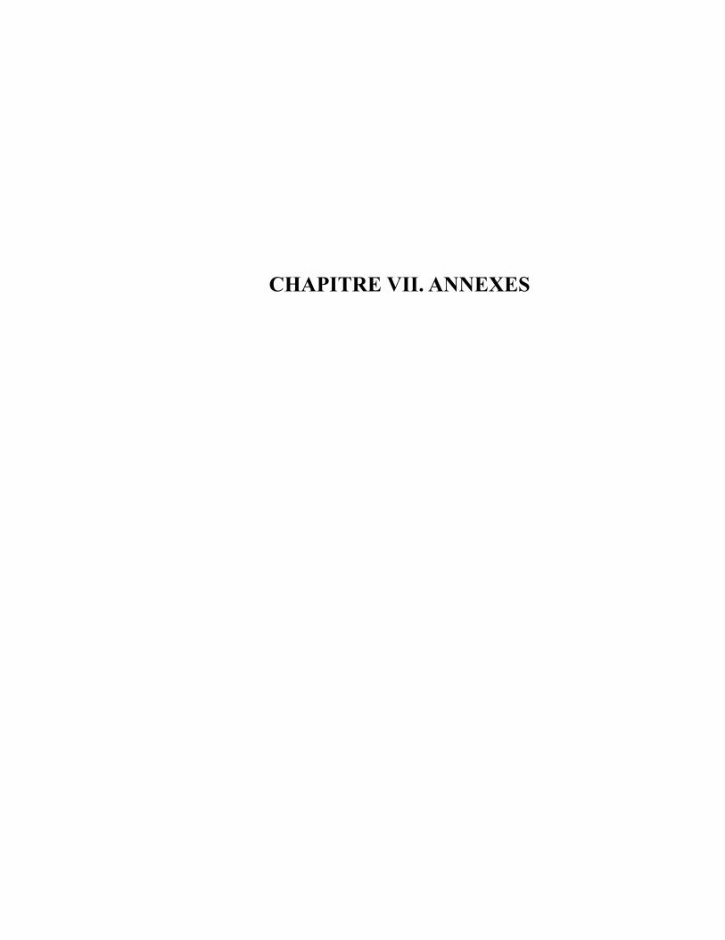
CHAPITRE VII. ANNEXES
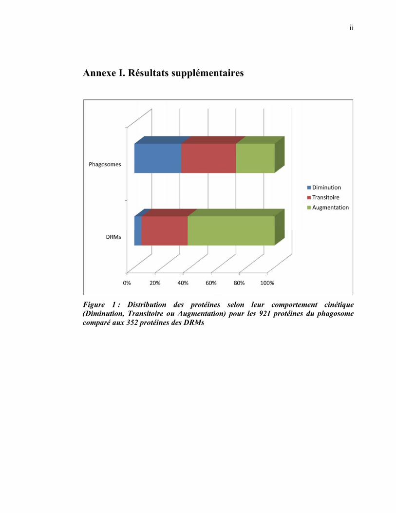
ii
Annexe I. Résultats supplémentaires
Figure 1 : Distribution des protéines selon leur comportement cinétique (Diminution, Transitoire ou Augmentation) pour les 921 protéines du phagosome comparé aux 352 protéines des DRMs
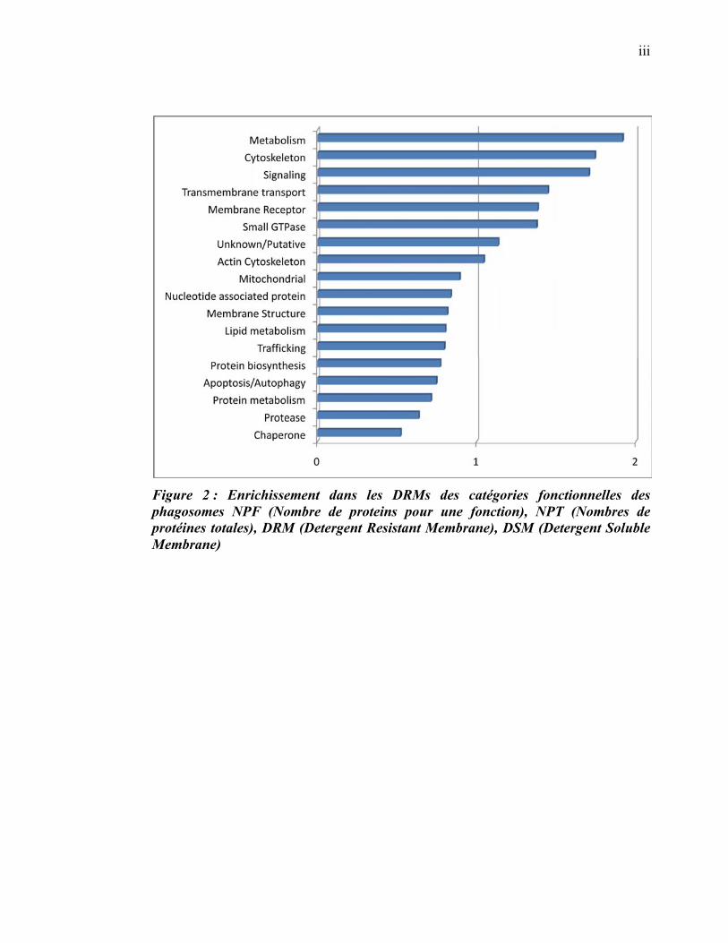
iii
Figure 2 : Enrichissement dans les DRMs des catégories fonctionnelles des phagosomes NPF (Nombre de proteins pour une fonction), NPT (Nombres de protéines totales), DRM (Detergent Resistant Membrane), DSM (Detergent Soluble Membrane)
iv
Figure 3 Interactions des protéines des différents groupes spatio-temporels des phagosomes
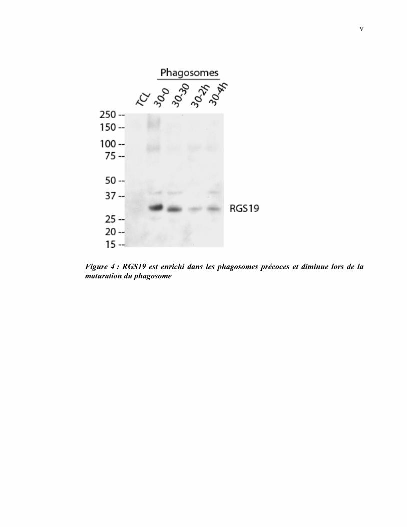
v
Figure 4 : RGS19 est enrichi dans les phagosomes précoces et diminue lors de la maturation du phagosome

vi
Annexe II. Résultats supplémentaires Article 2
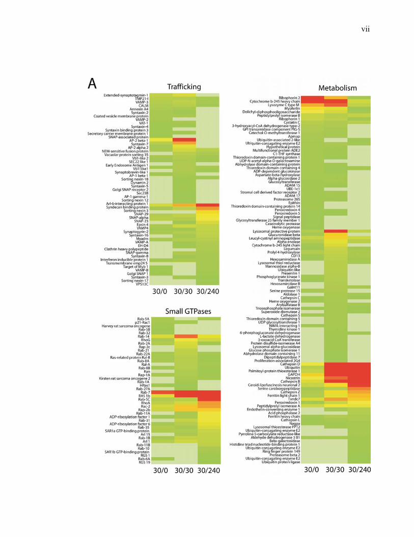
vii
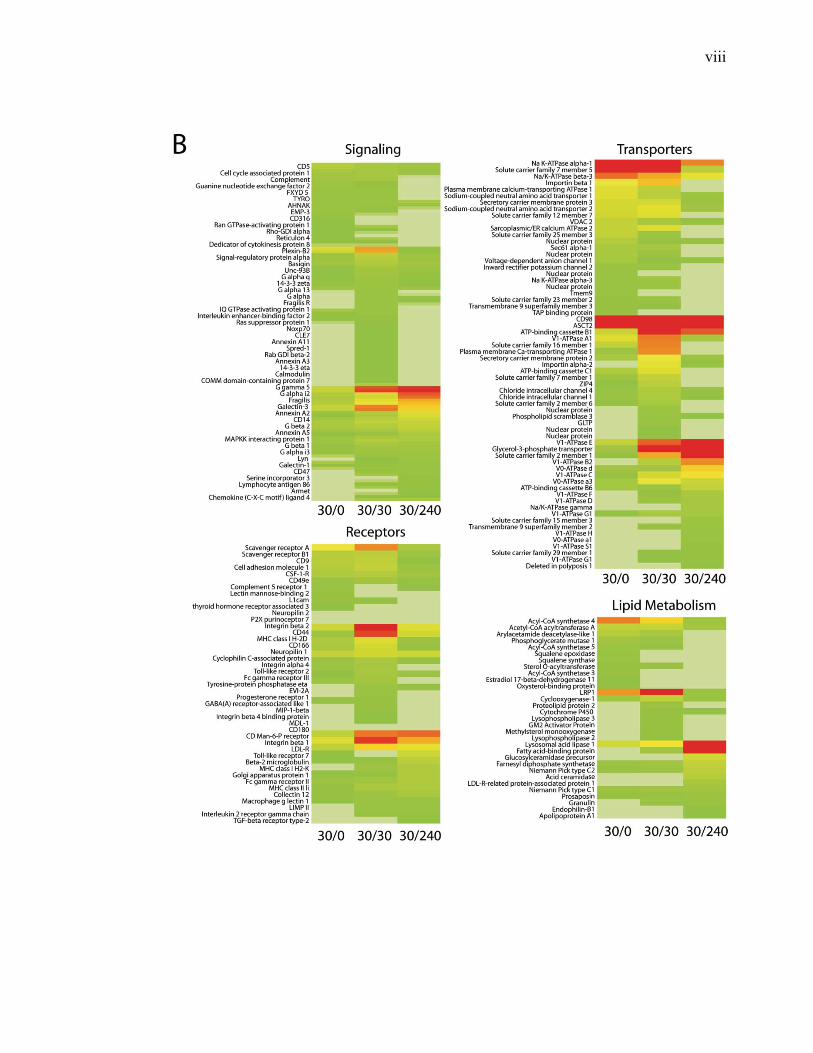
viii
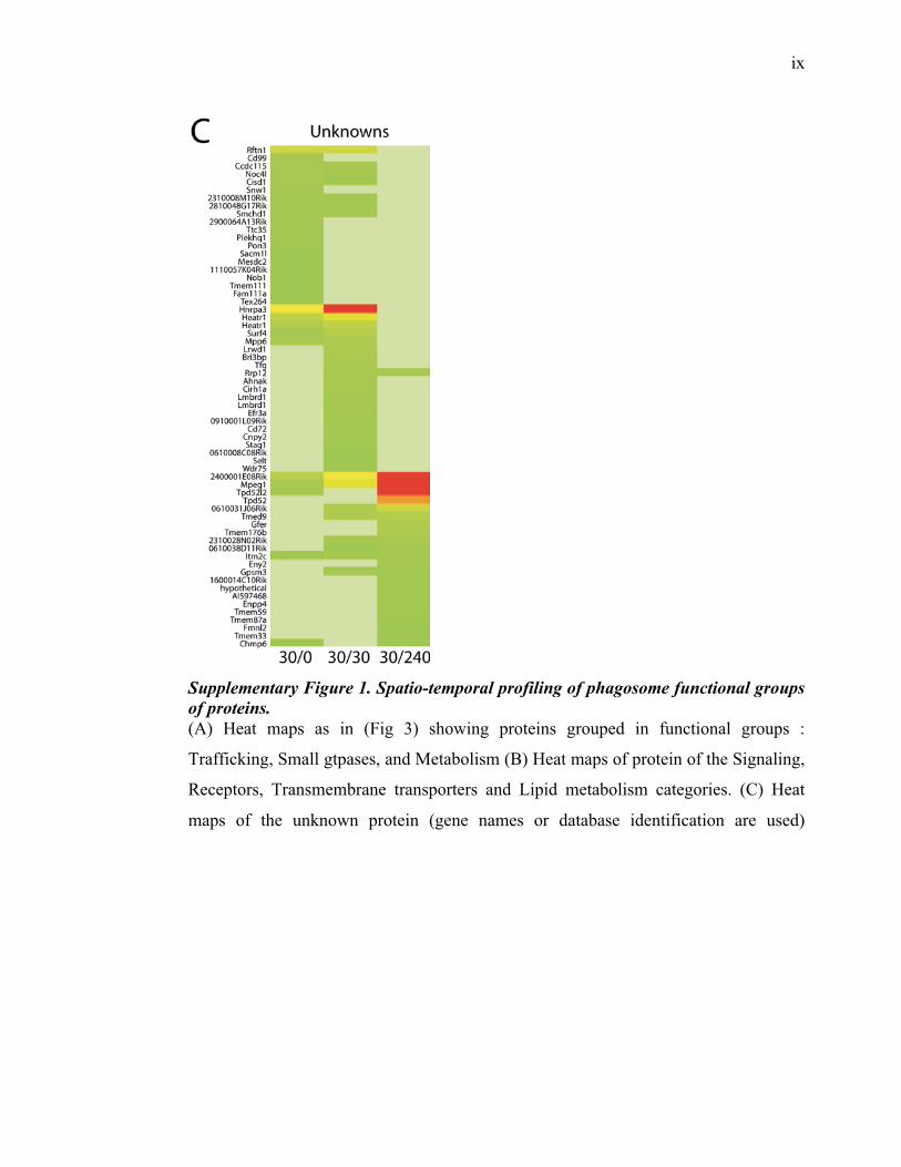
ix
Supplementary Figure 1. Spatio-temporal profiling of phagosome functional groups of proteins. (A) Heat maps as in (Fig 3) showing proteins grouped in functional groups :
Trafficking, Small gtpases, and Metabolism (B) Heat maps of protein of the Signaling,
Receptors, Transmembrane transporters and Lipid metabolism categories. (C) Heat
maps of the unknown protein (gene names or database identification are used)
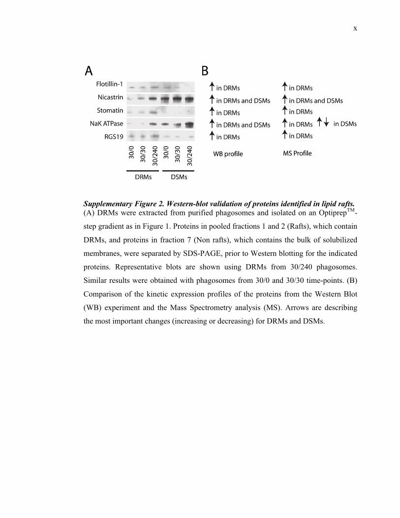
x
Supplementary Figure 2. Western-blot validation of proteins identified in lipid rafts. (A) DRMs were extracted from purified phagosomes and isolated on an OptiprepTM-
step gradient as in Figure 1. Proteins in pooled fractions 1 and 2 (Rafts), which contain
DRMs, and proteins in fraction 7 (Non rafts), which contains the bulk of solubilized
membranes, were separated by SDS-PAGE, prior to Western blotting for the indicated
proteins. Representative blots are shown using DRMs from 30/240 phagosomes.
Similar results were obtained with phagosomes from 30/0 and 30/30 time-points. (B)
Comparison of the kinetic expression profiles of the proteins from the Western Blot
(WB) experiment and the Mass Spectrometry analysis (MS). Arrows are describing
the most important changes (increasing or decreasing) for DRMs and DSMs.
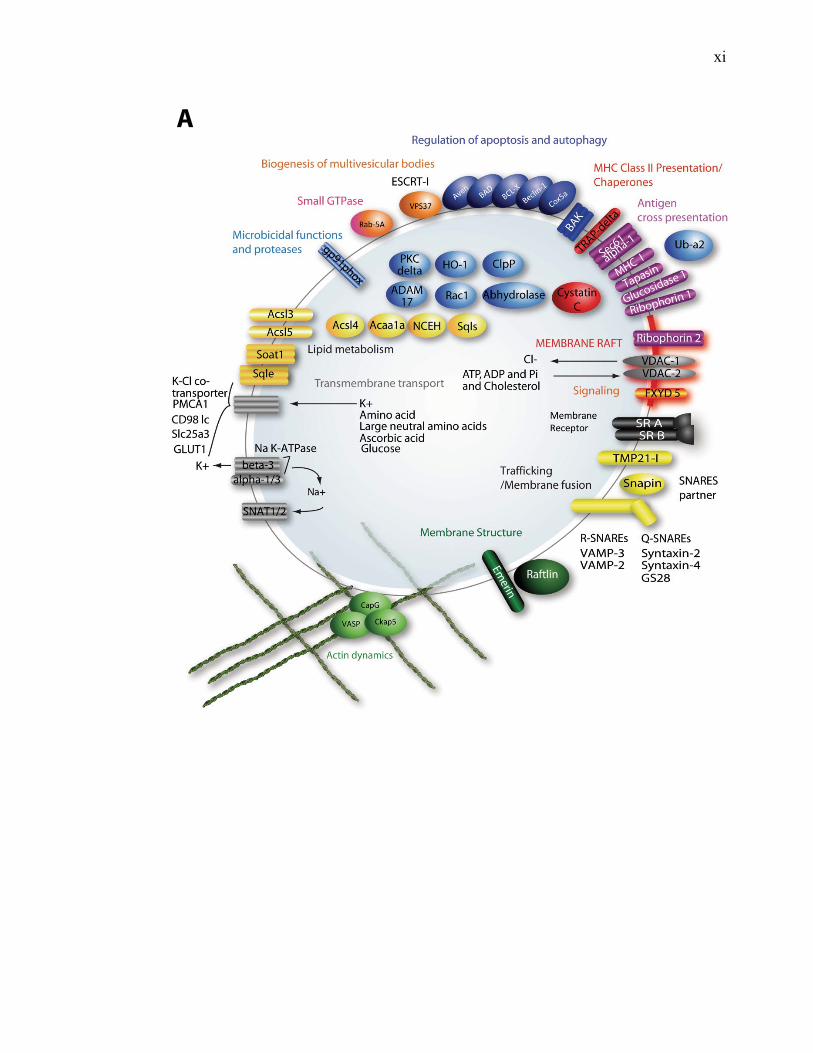
xi
A
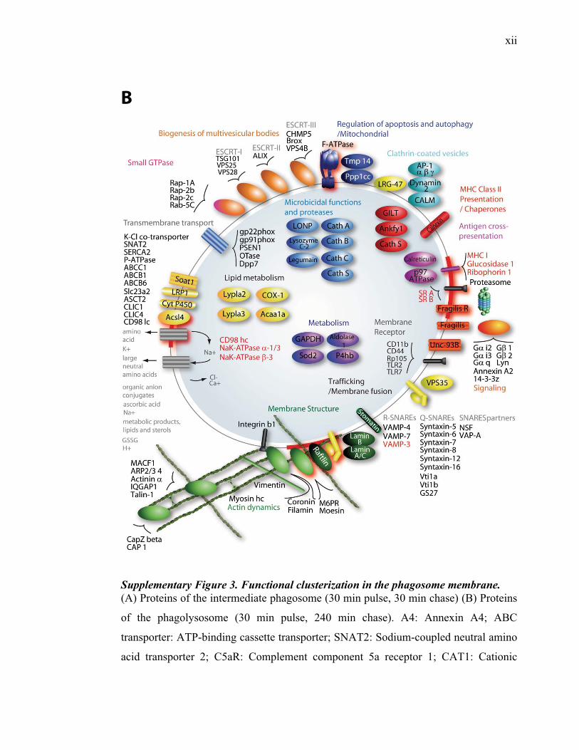
xii
B
Supplementary Figure 3. Functional clusterization in the phagosome membrane. (A) Proteins of the intermediate phagosome (30 min pulse, 30 min chase) (B) Proteins
of the phagolysosome (30 min pulse, 240 min chase). A4: Annexin A4; ABC
transporter: ATP-binding cassette transporter; SNAT2: Sodium-coupled neutral amino
acid transporter 2; C5aR: Complement component 5a receptor 1; CAT1: Cationic
xiii
amino acid transporter 1; CIC: Chloride intracellular channels 1 and 4; ENT1:
Equilibrative nucleoside transporter 1; Gals: Galectins 1, 3, and 9; GLUT1: Glucose
transporter type 1 and 6; GPCR: G-protein coupled receptor; FcgR: IgG Fc receptor II
and III; IL2Rg: Interleukin-2 Receptor g chain; Kir2.1: Inward rectifier potassium
channel 2; LC3: Microtubule-associated protein 1 light chain 3; LDLR: Low-density
lipoprotein receptor; LRP1: Lipoprotein receptor-related protein; M6PR: Mannose-6-
phosphate receptor; MP1: Mitogen-activated protein kinase kinase 1-interacting
protein 1; PHT2: Peptide/histidine transporter 2; P2X7: P2X purinergic receptor 7;
RIP: Receptor-interacting serine/threonine-protein kinase; SIRP: Signal-regulatory
protein; SPX2: Sugar phosphate exchanger 2; TLR: Toll-like receptor 2 and 7; ZIP4:
Zinc transporter 4
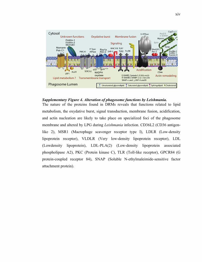
xiv
Supplementary Figure 4. Alteration of phagosome functions by Leishmania. The nature of the proteins found in DRMs reveals that functions related to lipid
metabolism, the oxydative burst, signal transduction, membrane fusion, acidification,
and actin nucleation are likely to take place on specialized foci of the phagosome
membrane and altered by LPG during Leishmania infection. CD36L2 (CD36 antigen-
like 2), MSR1 (Macrophage scavenger receptor type I), LDLR (Low-density
lipoprotein receptor), VLDLR (Very low-density lipoprotein receptor), LDL
(Lowdensity lipoprotein), LDL-PLA(2) (Low-density lipoprotein associated
phospholipase A2), PKC (Protein kinase C), TLR (Toll-like receptor), GPCR84 (G
protein-coupled receptor 84), SNAP (Soluble N-ethylmaleimide-sensitive factor
attachment protein).
xv
Supplementary Table 1. List of the 921 phagosomal proteins
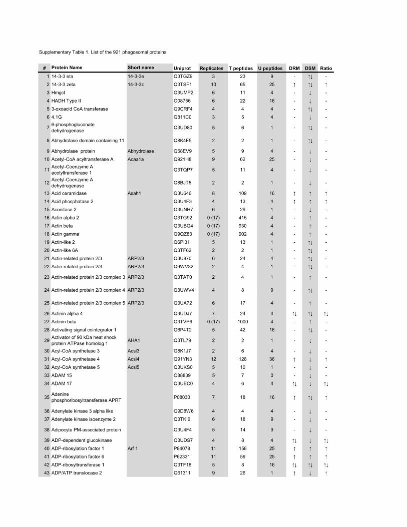
Supplementary Table 1. List of the 921 phagosomal proteins
# Protein Name Short name Uniprot Replicates T peptides U peptides DRM DSM Ratio1 14-3-3 eta 14-3-3e Q3TGZ9 3 23 9 - �� -
2 14-3-3 zeta 14-3-3z Q3TSF1 10 65 25 � �� �
3 Hmgcl Q3UMP2 6 11 4 - � -
4 HADH Type II O08756 6 22 16 - � -
5 3-oxoacid CoA transferase Q9CRF4 4 4 4 - �� -
6 4.1G Q811C0 3 5 4 - � -
7 6-phosphogluconate dehydrogenase Q3UD80 5 6 1 - �� -
8 Abhydrolase domain containing 11 Q8K4F5 2 2 1 - �� -
9 Abhydrolase protein Abhydrolase Q58EV9 5 9 4 - � -
10 Acetyl-CoA acyltransferase A Acaa1a Q921H8 9 62 25 - � -
11 Acetyl-Coenzyme A acetyltransferase 1 Q3TQP7 5 11 4 - � -
12 Acetyl-Coenzyme A dehydrogenase Q8BJT5 2 2 1 - � -
13 Acid ceramidase Asah1 Q3U646 8 109 16 � � �
14 Acid phosphatase 2 Q3U4F3 4 13 4 � � �
15 Aconitase 2 Q3UNH7 6 29 1 - � -
16 Actin alpha 2 Q3TG92 0 (17) 415 4 - � -
17 Actin beta Q3UBQ4 0 (17) 930 4 - � -
18 Actin gamma Q9QZ83 0 (17) 902 4 - � -
19 Actin-like 2 Q6PI31 5 13 1 - �� -
20 Actin-like 6A Q3TF62 2 2 1 - �� -
21 Actin-related protein 2/3 ARP2/3 Q3U870 6 24 4 - �� -
22 Actin-related protein 2/3 ARP2/3 Q9WV32 2 4 1 - �� -
23 Actin-related protein 2/3 complex 3 ARP2/3 Q3TAT0 2 4 1 - � -
24 Actin-related protein 2/3 complex 4 ARP2/3 Q3UWV4 4 8 9 - �� -
25 Actin-related protein 2/3 complex 5 ARP2/3 Q3UA72 6 17 4 - � -
26 Actinin alpha 4 Q3UDJ7 7 24 4 �� �� ��
27 Actinin beta Q3TVP6 0 (17) 1000 4 - � -
28 Activating signal cointegrator 1 Q6P4T2 5 42 16 - �� -
29 Activator of 90 kDa heat shock protein ATPase homolog 1 AHA1 Q3TL79 2 2 1 - � -
30 Acyl-CoA synthetase 3 Acsl3 Q8K1J7 2 6 4 - � -
31 Acyl-CoA synthetase 4 Acsl4 Q91YN3 12 128 36 � � �
32 Acyl-CoA synthetase 5 Acsl5 Q3UKS0 5 10 1 - � -
33 ADAM 15 O88839 5 7 0 - � -
34 ADAM 17 Q3UEC0 4 6 4 �� � ��
35 Adenine phosphoribosyltransferase APRT P08030 7 18 16 � �� �
36 Adenylate kinase 3 alpha like Q9D8W6 4 4 4 - � -
37 Adenylate kinase isoenzyme 2 Q3TKI6 6 18 9 - � -
38 Adipocyte PM-associated protein Q3U4F4 5 14 9 - � -
39 ADP-dependent glucokinase Q3UDS7 4 8 4 �� � ��
40 ADP-ribosylation factor 1 Arf 1 P84078 11 158 25 � � �
41 ADP-ribosylation factor 6 P62331 11 59 25 � � �
42 ADP-ribosyltransferase 1 Q3TF18 5 8 16 �� �� ��
43 ADP/ATP translocase 2 Q61311 9 26 1 � � �
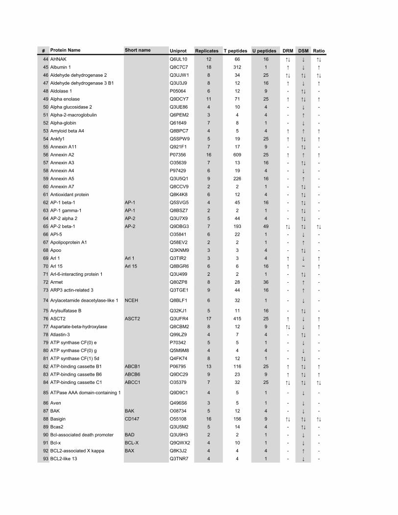
# Protein Name Short name Uniprot Replicates T peptides U peptides DRM DSM Ratio
44 AHNAK Q6UL10 12 66 16 �� � ��
45 Albumin 1 Q8C7C7 18 312 1 � � �
46 Aldehyde dehydrogenase 2 Q3UJW1 8 34 25 �� �� ��
47 Aldehyde dehydrogenase 3 B1 Q3U3J9 8 12 16 � � �
48 Aldolase 1 P05064 6 12 9 - �� -
49 Alpha enolase Q9DCY7 11 71 25 � �� �
50 Alpha glucosidase 2 Q3UE86 4 10 4 - � -
51 Alpha-2-macroglobulin Q6PEM2 3 4 4 - � -
52 Alpha-globin Q61649 7 8 1 - � -
53 Amyloid beta A4 Q8BPC7 4 5 4 � � �
54 Ankfy1 Q5SPW9 5 19 25 � �� �
55 Annexin A11 Q921F1 7 17 9 - �� -
56 Annexin A2 P07356 16 609 25 � � �
57 Annexin A3 O35639 7 13 16 - �� -
58 Annexin A4 P97429 6 19 4 - � -
59 Annexin A5 Q3U5Q1 9 226 16 - � -
60 Annexin A7 Q8CCV9 2 2 1 - �� -
61 Antioxidant protein Q8K4K8 6 12 4 - �� -
62 AP-1 beta-1 AP-1 Q5SVG5 4 45 16 - �� -
63 AP-1 gamma-1 AP-1 Q8BSZ7 2 2 1 - �� -
64 AP-2 alpha 2 AP-2 Q3U7X9 5 44 4 - �� -
65 AP-2 beta-1 AP-2 Q9DBG3 7 193 49 �� �� ��
66 API-5 O35841 6 22 1 - � -
67 Apolipoprotein A1 Q58EV2 2 2 1 - � -
68 Apoo Q3KNM9 3 3 4 - �� -
69 Arl 1 Arl 1 Q3TIR2 3 3 4 � � �
70 Arl 15 Arl 15 Q8BGR6 6 6 16 � ~ �
71 Arl-6-interacting protein 1 Q3U499 2 2 1 - �� -
72 Armet Q80ZP8 8 28 36 - � -
73 ARP3 actin-related 3 Q3TGE1 9 44 16 - � -
74 Arylacetamide deacetylase-like 1 NCEH Q8BLF1 6 32 1 - � -
75 Arylsulfatase B Q32KJ1 5 11 16 - �� -
76 ASCT2 ASCT2 Q3UFR4 17 415 25 � � �
77 Aspartate-beta-hydroxylase Q8CBM2 8 12 9 �� � �
78 Atlastin-3 Q99LZ9 4 7 4 - �� -
79 ATP synthase CF(0) e P70342 5 5 1 - � -
80 ATP synthase CF(0) g Q5M9M8 4 4 4 - � -
81 ATP synthase CF(1) 5d Q4FK74 8 12 1 - �� -
82 ATP-binding cassette B1 ABCB1 P06795 13 116 25 � �� �
83 ATP-binding cassette B6 ABCB6 Q9DC29 9 23 9 � �� �
84 ATP-binding cassette C1 ABCC1 O35379 7 32 25 �� �� ��
85 ATPase AAA domain-containing 1 Q9D9C1 4 5 1 - � -
86 Aven Q496S6 3 5 1 - � -
87 BAK BAK O08734 5 12 4 - � -
88 Basigin CD147 O55108 16 156 9 �� �� ��
89 Bcas2 Q3U5M2 5 14 4 - �� -
90 Bcl-associated death promoter BAD Q3U9H3 2 2 1 - � -
91 Bcl-x BCL-X Q9QWX2 4 10 1 - � -
92 BCL2-associated X kappa BAX Q8K3J2 4 4 4 - � -
93 BCL2-like 13 Q3TNR7 4 4 1 - � -
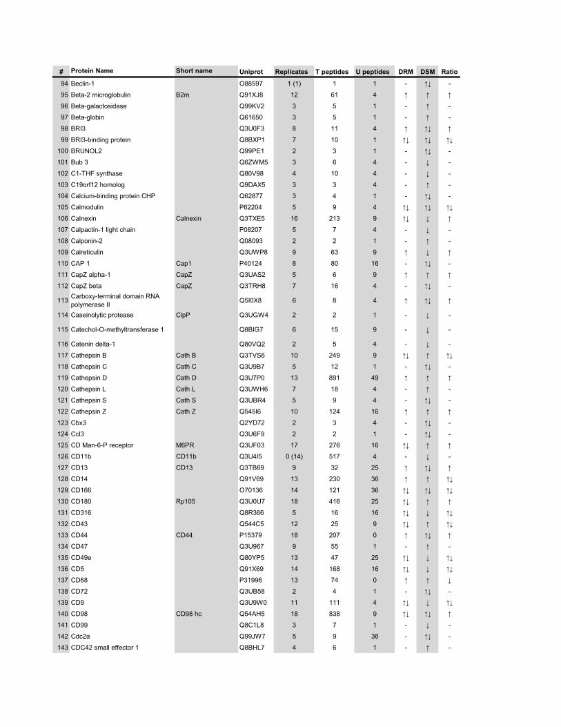
# Protein Name Short name Uniprot Replicates T peptides U peptides DRM DSM Ratio
94 Beclin-1 O88597 1 (1) 1 1 - �� -
95 Beta-2 microglobulin B2m Q91XJ8 12 61 4 � � �
96 Beta-galactosidase Q99KV2 3 5 1 - � -
97 Beta-globin Q61650 3 5 1 - � -
98 BRI3 Q3U0F3 8 11 4 � �� �
99 BRI3-binding protein Q8BXP1 7 10 1 �� �� ��
100 BRUNOL2 Q99PE1 2 3 1 - �� -
101 Bub 3 Q6ZWM5 3 6 4 - � -
102 C1-THF synthase Q80V98 4 10 4 - � -
103 C19orf12 homolog Q9DAX5 3 3 4 - � -
104 Calcium-binding protein CHP Q62877 3 4 1 - �� -
105 Calmodulin P62204 5 9 4 �� �� ��
106 Calnexin Calnexin Q3TXE5 16 213 9 �� � �
107 Calpactin-1 light chain P08207 5 7 4 - � -
108 Calponin-2 Q08093 2 2 1 - � -
109 Calreticulin Q3UWP8 9 63 9 � � �
110 CAP 1 Cap1 P40124 8 80 16 - �� -
111 CapZ alpha-1 CapZ Q3UAS2 5 6 9 � � �
112 CapZ beta CapZ Q3TRH8 7 16 4 - �� -
113 Carboxy-terminal domain RNA polymerase II Q5I0X8 6 8 4 � �� �
114 Caseinolytic protease ClpP Q3UGW4 2 2 1 - � -
115 Catechol-O-methyltransferase 1 Q8BIG7 6 15 9 - � -
116 Catenin delta-1 Q80VQ2 2 5 4 - � -
117 Cathepsin B Cath B Q3TVS6 10 249 9 �� � ��
118 Cathepsin C Cath C Q3U9B7 5 12 1 - �� -
119 Cathepsin D Cath D Q3U7P0 13 891 49 � � �
120 Cathepsin L Cath L Q3UWH6 7 18 4 - � -
121 Cathepsin S Cath S Q3UBR4 5 9 4 - �� -��
122 Cathepsin Z Cath Z Q545I6 10 124 16 � � �
123 Cbx3 Q2YD72 2 3 4 - �� -
124 Ccl3 Q3U6F9 2 2 1 - �� -
125 CD Man-6-P receptor M6PR Q3UF03 17 276 16 �� � �
126 CD11b CD11b Q3U4I5 0 (14) 517 4 - � -
127 CD13 CD13 Q3TB69 9 32 25 � �� �
128 CD14 Q91V69 13 230 36 � � ��
129 CD166 O70136 14 121 36 �� �� ��
130 CD180 Rp105 Q3U0U7 18 416 25 �� � �
131 CD316 Q8R366 5 16 16 �� � ��
132 CD43 Q544C5 12 25 9 �� � ��
133 CD44 CD44 P15379 18 207 0 � �� �
134 CD47 Q3U967 9 55 1 - � -
135 CD49e Q80YP5 13 47 25 �� � ��
136 CD5 Q91X69 14 168 16 �� � ��
137 CD68 P31996 13 74 0 � � �
138 CD72 Q3UB58 2 4 1 - �� -
139 CD9 Q3U9W0 11 111 4 �� � ��
140 CD98 CD98 hc Q54AH5 18 838 9 �� �� �
141 CD99 Q8C1L8 3 7 1 - � -
142 Cdc2a Q99JW7 5 9 36 - �� -
143 CDC42 small effector 1 Q8BHL7 4 6 1 - � -
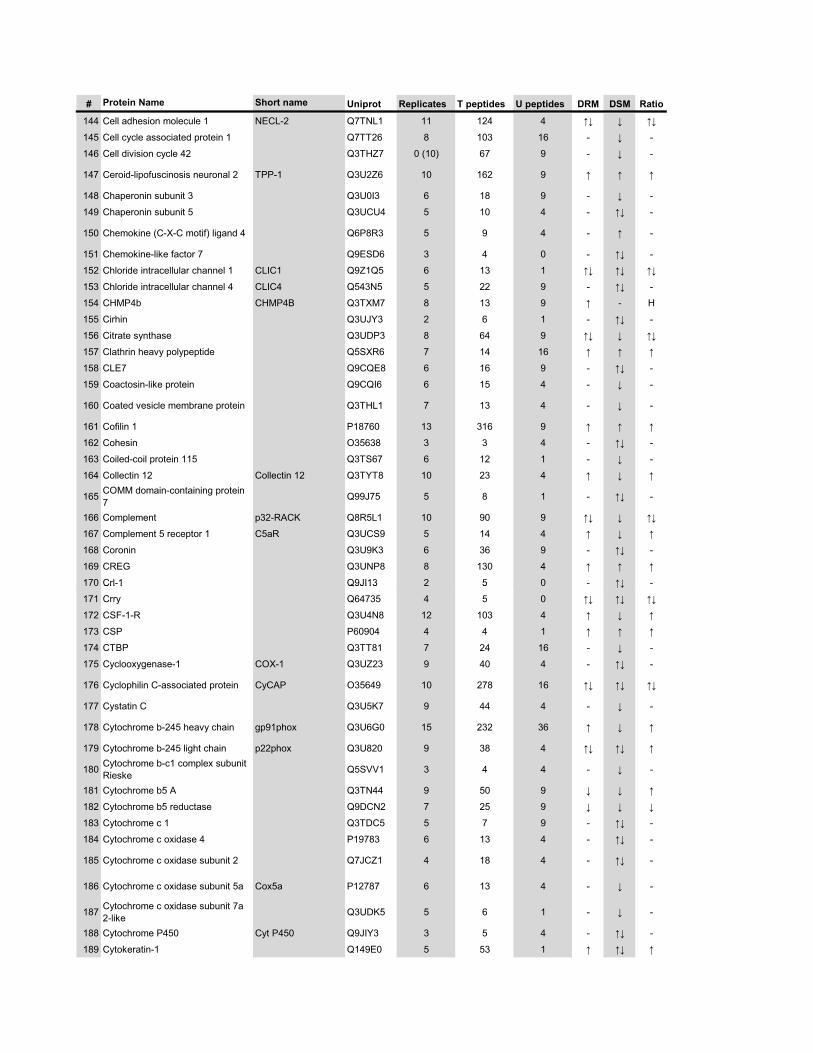
# Protein Name Short name Uniprot Replicates T peptides U peptides DRM DSM Ratio
144 Cell adhesion molecule 1 NECL-2 Q7TNL1 11 124 4 �� � ��
145 Cell cycle associated protein 1 Q7TT26 8 103 16 - � -
146 Cell division cycle 42 Q3THZ7 0 (10) 67 9 - � -
147 Ceroid-lipofuscinosis neuronal 2 TPP-1 Q3U2Z6 10 162 9 � � �
148 Chaperonin subunit 3 Q3U0I3 6 18 9 - � -
149 Chaperonin subunit 5 Q3UCU4 5 10 4 - �� -
150 Chemokine (C-X-C motif) ligand 4 Q6P8R3 5 9 4 - � -
151 Chemokine-like factor 7 Q9ESD6 3 4 0 - �� -
152 Chloride intracellular channel 1 CLIC1 Q9Z1Q5 6 13 1 �� �� ��
153 Chloride intracellular channel 4 CLIC4 Q543N5 5 22 9 - �� -
154 CHMP4b CHMP4B Q3TXM7 8 13 9 � - H
155 Cirhin Q3UJY3 2 6 1 - �� -
156 Citrate synthase Q3UDP3 8 64 9 �� � ��
157 Clathrin heavy polypeptide Q5SXR6 7 14 16 � � �
158 CLE7 Q9CQE8 6 16 9 - �� -
159 Coactosin-like protein Q9CQI6 6 15 4 - � -
160 Coated vesicle membrane protein Q3THL1 7 13 4 - � -
161 Cofilin 1 P18760 13 316 9 � � �
162 Cohesin O35638 3 3 4 - �� -
163 Coiled-coil protein 115 Q3TS67 6 12 1 - � -
164 Collectin 12 Collectin 12 Q3TYT8 10 23 4 � � �
165 COMM domain-containing protein 7 Q99J75 5 8 1 - �� -
166 Complement p32-RACK Q8R5L1 10 90 9 �� � ��
167 Complement 5 receptor 1 C5aR Q3UCS9 5 14 4 � � �
168 Coronin Q3U9K3 6 36 9 - �� -
169 CREG Q3UNP8 8 130 4 � � �
170 Crl-1 Q9JI13 2 5 0 - �� -170 Crl 1 Q9JI13 2 5 0 ��
171 Crry Q64735 4 5 0 �� �� ��
172 CSF-1-R Q3U4N8 12 103 4 � � �
173 CSP P60904 4 4 1 � � �
174 CTBP Q3TT81 7 24 16 - � -
175 Cyclooxygenase-1 COX-1 Q3UZ23 9 40 4 - �� -
176 Cyclophilin C-associated protein CyCAP O35649 10 278 16 �� �� ��
177 Cystatin C Q3U5K7 9 44 4 - � -
178 Cytochrome b-245 heavy chain gp91phox Q3U6G0 15 232 36 � � �
179 Cytochrome b-245 light chain p22phox Q3U820 9 38 4 �� �� �
180 Cytochrome b-c1 complex subunit Rieske Q5SVV1 3 4 4 - � -
181 Cytochrome b5 A Q3TN44 9 50 9 � � �
182 Cytochrome b5 reductase Q9DCN2 7 25 9 � � �
183 Cytochrome c 1 Q3TDC5 5 7 9 - �� -
184 Cytochrome c oxidase 4 P19783 6 13 4 - �� -
185 Cytochrome c oxidase subunit 2 Q7JCZ1 4 18 4 - �� -
186 Cytochrome c oxidase subunit 5a Cox5a P12787 6 13 4 - � -
187 Cytochrome c oxidase subunit 7a 2-like Q3UDK5 5 6 1 - � -
188 Cytochrome P450 Cyt P450 Q9JIY3 3 5 4 - �� -
189 Cytokeratin-1 Q149E0 5 53 1 � �� �
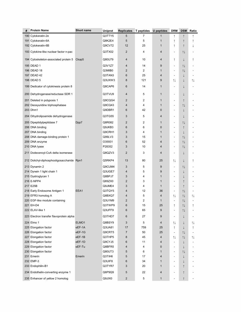
# Protein Name Short name Uniprot Replicates T peptides U peptides DRM DSM Ratio
190 Cytokeratin-2e Q3TTY5 5 7 1 � � �
191 Cytokeratin-6A Q8K2E4 5 5 1 � � �
192 Cytokeratin-6B Q9CV72 12 25 1 � � �
193 Cytokine-like nuclear factor n-pac Q3TX02 2 4 4 - �� -
194 Cytoskeleton-associated protein 5 Ckap5 Q80U79 4 10 4 � � �
195 DEAD 1 Q3V1Z7 4 14 9 - �� -
196 DEAD 18 Q3MIB0 2 2 1 - �� -
197 DEAD 42 Q3TAN3 6 25 4 - � -
198 DEAD 5 Q3UXW3 8 121 9 �� � ��
199 Dedicator of cytokinesis protein 8 Q8CAP6 6 14 1 - � -
200 Dehydrogenase/reductase SDR 1 Q3TVU9 4 5 1 - � -
201 Deleted in polyposis 1 Q9CQG4 2 2 1 - � -
202 Deoxyuridine triphosphatase Q9CQ43 4 4 1 - �� -
203 Dhm1 Q9DBR1 6 42 0 - � -
204 Dihydrolipoamide dehydrogenase Q3TG55 3 5 4 - � -
205 Dipeptidylpeptidase 7 Dpp7 Q8R082 2 2 1 - �� -
206 DNA binding Q9JKB3 6 6 0 - � -
207 DNA binding Q9CRH1 3 4 1 - � -
208 DNA damage-binding protein 1 Q99LV3 3 15 1 - �� -
209 DNA enzyme O35931 6 52 4 - �� -
210 DNA lyase P28352 3 10 4 - �� -
211 Dodecenoyl-CoA delta isomerase Q8QZV3 2 3 4 - � -
212 Dolichyl-diphosphooligosaccharide Rpn1 Q5RKP4 13 80 25 �� � �
213 Dynamin 2 Q9CUM4 3 5 9 - �� -
214 Dynein 1 light chain 1 Q3UGE7 4 5 9 - � -
215 Dystroglycan 1 Q8BPJ7 3 4 1 - � -
216 E-NPP4 Q69ZX0 2 3 1 - � -
217 E25B Q9JME4 3 4 1 - � -
218 Early Endosome Antigen 1 EEA1 Q3TQY5 4 12 36 - �� -
219 EFR3 homolog A Q8BXQ7 4 5 4 �� �� ��
220 EGF-like module containing Q3U1M9 2 2 1 - �� -
221 EH-D4 Q3TWP9 6 15 25 � �� �
222 ELAV-like 1 Q3UFF9 6 65 9 - �� -
223 Electron transfer flavoprotein alpha Q3THD7 6 27 9 - � -
224 Elmo 1 ELMO1 Q8BSY9 3 5 4 �� � ��
225 Elongation factor eEF-1A Q3UA81 17 759 25 � � �
226 Elongation factor eEF-1G Q9CRT5 7 50 25 - �� -
227 Elongation factor eEF-1B Q3THP5 8 45 4 �� �� ��
228 Elongation factor eEF-1D Q9CYJ5 6 11 4 - � -
229 Elongation factor eEF-Tu Q8BFR5 4 4 0 - � -
230 Elongation factor Q80UT3 5 6 1 - �� -
231 Emerin Emerin Q3TIH6 5 17 4 - � -
232 EMP-3 Q3UIF8 6 34 1 - � -
233 Endophilin-B1 Q3TYR7 3 20 1 - � -
234 Endothelin-converting enzyme 1 Q6P9Q9 5 22 4 - � -
235 Enhancer of yellow 2 homolog Q9JIX0 2 5 1 - � -
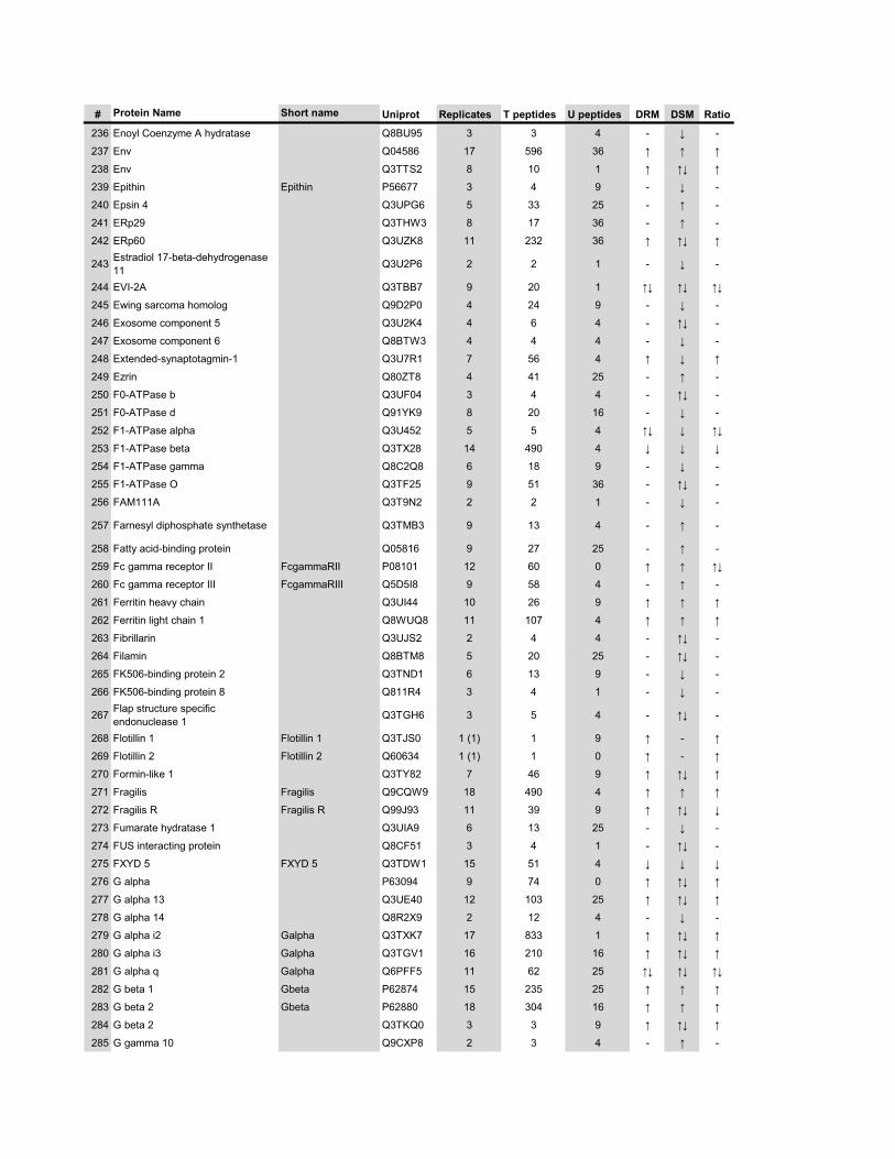
# Protein Name Short name Uniprot Replicates T peptides U peptides DRM DSM Ratio
236 Enoyl Coenzyme A hydratase Q8BU95 3 3 4 - � -
237 Env Q04586 17 596 36 � � �
238 Env Q3TTS2 8 10 1 � �� �
239 Epithin Epithin P56677 3 4 9 - � -
240 Epsin 4 Q3UPG6 5 33 25 - � -
241 ERp29 Q3THW3 8 17 36 - � -
242 ERp60 Q3UZK8 11 232 36 � �� �
243 Estradiol 17-beta-dehydrogenase 11 Q3U2P6 2 2 1 - � -
244 EVI-2A Q3TBB7 9 20 1 �� �� ��
245 Ewing sarcoma homolog Q9D2P0 4 24 9 - � -
246 Exosome component 5 Q3U2K4 4 6 4 - �� -
247 Exosome component 6 Q8BTW3 4 4 4 - � -
248 Extended-synaptotagmin-1 Q3U7R1 7 56 4 � � �
249 Ezrin Q80ZT8 4 41 25 - � -
250 F0-ATPase b Q3UF04 3 4 4 - �� -
251 F0-ATPase d Q91YK9 8 20 16 - � -
252 F1-ATPase alpha Q3U452 5 5 4 �� � ��
253 F1-ATPase beta Q3TX28 14 490 4 � � �
254 F1-ATPase gamma Q8C2Q8 6 18 9 - � -
255 F1-ATPase O Q3TF25 9 51 36 - �� -
256 FAM111A Q3T9N2 2 2 1 - � -
257 Farnesyl diphosphate synthetase Q3TMB3 9 13 4 - � -
258 Fatty acid-binding protein Q05816 9 27 25 - � -
259 Fc gamma receptor II FcgammaRII P08101 12 60 0 � � ��
260 Fc gamma receptor III FcgammaRIII Q5D5I8 9 58 4 - � -
261 Ferritin heavy chain Q3UI44 10 26 9 � � �
262 Ferritin light chain 1 Q8WUQ8 11 107 4 � � �
263 Fibrillarin Q3UJS2 2 4 4 - �� -��
264 Filamin Q8BTM8 5 20 25 - �� -
265 FK506-binding protein 2 Q3TND1 6 13 9 - � -
266 FK506-binding protein 8 Q811R4 3 4 1 - � -
267 Flap structure specific endonuclease 1 Q3TGH6 3 5 4 - �� -
268 Flotillin 1 Flotillin 1 Q3TJS0 1 (1) 1 9 � - �
269 Flotillin 2 Flotillin 2 Q60634 1 (1) 1 0 � - �
270 Formin-like 1 Q3TY82 7 46 9 � �� �
271 Fragilis Fragilis Q9CQW9 18 490 4 � � �
272 Fragilis R Fragilis R Q99J93 11 39 9 � �� �
273 Fumarate hydratase 1 Q3UIA9 6 13 25 - � -
274 FUS interacting protein Q8CF51 3 4 1 - �� -
275 FXYD 5 FXYD 5 Q3TDW1 15 51 4 � � �
276 G alpha P63094 9 74 0 � �� �
277 G alpha 13 Q3UE40 12 103 25 � �� �
278 G alpha 14 Q8R2X9 2 12 4 - � -
279 G alpha i2 Galpha Q3TXK7 17 833 1 � �� �
280 G alpha i3 Galpha Q3TGV1 16 210 16 � �� �
281 G alpha q Galpha Q6PFF5 11 62 25 �� �� ��
282 G beta 1 Gbeta P62874 15 235 25 � � �
283 G beta 2 Gbeta P62880 18 304 16 � � �
284 G beta 2 Q3TKQ0 3 3 9 � �� �
285 G gamma 10 Q9CXP8 2 3 4 - � -
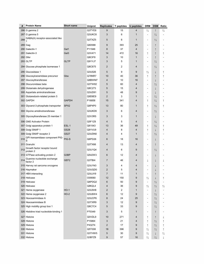
# Protein Name Short name Uniprot Replicates T peptides U peptides DRM DSM Ratio
286 G gamma 2 Q3TYE8 9 15 4 �� � ��
287 G gamma 5 Q3UKC8 3 6 1 - �� -
288 GABA(A) receptor-associated like 1 Q3TXZ5 5 6 1 - �� -
289 Gag Q60588 9 393 25 - � -
290 Galectin-1 Gal1 P11946 8 37 4 - � -
291 Galectin-3 Gal3 Q3V471 14 472 16 � � �
292 Gfer Q8CIF8 3 10 1 - � -
293 GLTP GLTP Q91YJ7 3 5 1 - �� -
294 Glucose phosphate isomerase 1 Q8C675 2 2 4 - �� -
295 Glucosidase 1 Q3U026 5 9 9 �� � �
296 Glucosylceramidase precursor Gba Q78NR7 10 40 36 � � �
297 Glucosyltransferase Q8BWM7 4 10 16 - � -
298 Glucuronidase beta Q3TW82 5 60 4 - �� -
299 Glutamate dehydrogenase Q8C273 5 15 4 - � -
300 Aspartate aminotransferase Q3UD91 5 48 9 - � -
301 Glutaredoxin-related protein 5 Q9D6E9 2 3 1 - �� -
302 GAPDH GAPDH P16858 15 341 4 � �� �
303 Glycerol-3-phosphate transporter SPX2 Q6P4P0 13 80 1 � �� �
304 Glycine amidinotransferase Q3UKD9 3 6 4 - �� -
305 Glycosyltransferase 25 member 1 Q3V3R5 3 3 1 - � -
306 GM2 Activator Protein Q5F1Z8 4 5 4 - �� -
307 Golgi apparatus protein 1 ESL-1 Q61543 13 36 49 � �� �
308 Golgi SNAP 1 GS28 Q91VU9 4 8 4 � � �
309 Golgi SNAP receptor 2 GS27 Q3UDN0 4 4 1 - �� -
310 GPI transamidase component PIG-S PIG-S Q6PD26 6 18 16 - � -
311 Granulin Q3TX66 4 13 4 - � -
312 Growth factor receptor bound protein 2 Q3U1Q4 4 6 9 - �� -
313 GTPase activating protein 2 G3BP Q542W3 6 12 4 - � -
314 Guanine nucleotide exchange factor 2 GEF2 Q3TBI4 7 46 4 � � �
315 Harvey rat sarcoma oncogene Q3U1N3 3 4 4 - � -
316 Haymaker Q3UQD0 2 5 4 - � -
317 HBX-interacting Q3UJV9 7 11 1 - � -
318 Helicase O09060 12 150 9 �� � �
319 Helicase Q6PDQ2 6 50 9 - �� -
320 Helicase Q8K2L4 4 36 9 �� �� ��
321 Heme oxygenase HO-1 Q3U5H8 2 2 1 - � -
322 Heme oxygenase 2 HO-2 Q3U6W4 6 12 9 - �� -
323 Hexosaminidase A Q3UCP6 8 24 25 - �� -
324 Hexosaminidase B Q3TXR9 3 12 9 - �� -
325 High mobility group box 1 Q8C7C4 5 33 9 - �� -
326 Histidine triad nucleotide-binding 1 P70349 3 5 1 - � -
327 Histone Q0VDL9 10 271 4 � � �
328 Histone P15864 3 21 4 � �� �
329 Histone P43274 2 17 9 � �� �
330 Histone Q5T008 18 396 9 �� �� �
331 Histone Q3THW5 5 30 9 �� � ��
332 Histone Q3B7Z8 9 57 16 �� �� �
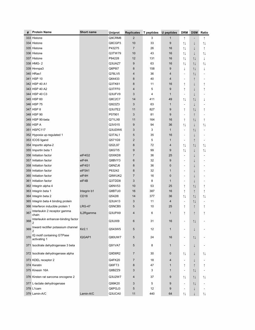
# Protein Name Short name Uniprot Replicates T peptides U peptides DRM DSM Ratio
333 Histone Q9CRM8 2 3 1 � - �
334 Histone Q8CGP3 10 33 9 �� � ��
335 Histone P43275 7 26 16 �� � �
336 Histone Q3TW79 10 43 16 �� � ��
337 Histone P84228 12 131 16 �� �� �
338 HMG- 2 Q3UAZ7 9 63 16 �� �� ��
339 Hnrnpa3 Q6P6I7 8 158 9 � �� �
340 HRas1 Q76LV5 4 36 4 - �� -
341 HSP 10 Q64433 8 40 4 - � -
342 HSP 40 A1 Q3TK61 8 11 16 � � �
343 HSP 40 A2 Q3TFF0 4 5 9 � � �
344 HSP 40 C3 Q3UFV9 3 4 1 - � -
345 HSP 60 Q8C2C7 14 411 49 �� �� �
346 HSP 75 Q922Z3 3 63 1 - � -
347 HSP 8 Q3U7E2 11 827 9 � �� �
348 HSP 90 P07901 3 81 9 - � -
349 HSP 90-beta Q71LX8 11 164 16 � �� �
350 HSP A Q3V015 9 94 36 �� � ��
351 HSPC117 Q3UDW6 3 3 1 - �� -
352 Hypoxia up-regulated 1 Q3TAL1 5 35 16 - � -
353 ICOS ligand Q571G9 2 5 1 - � -
354 Importin alpha-2 Q52L97 8 72 4 �� �� ��
355 Importin beta 1 Q6GTI5 9 99 9 �� � ��
356 Initiation factor eIF4G2 Q5XKD8 7 36 25 - � -
357 Initiation factor eIF4A Q8BVY3 6 32 9 - � -
358 Initiation factor eIF4G1 Q6NZJ6 8 36 0 - � -
359 Initiation factor eIF5A1 P63242 8 32 1 - � -
360 Initiation factor eIF4H Q9WUK2 7 16 0 - � -
361 Initiation factor eIF4B Q3TDD8 3 8 1 - � -
362 Integrin alpha 4 Q6NV53 10 53 25 � �� �
363 Integrin beta 1 Integrin b1 Q8BTU0 16 387 16 � � �
364 Integrin beta 2 CD18 Q542I8 14 377 36 �� �� ��
365 Integrin beta 4 binding protein Q3UA13 3 11 4 - �� -
366 Interferon inducible protein 1 LRG-47 Q5NCB5 5 10 25 � � �
367 Interleukin 2 receptor gamma chain IL2Rgamma Q3UPA9 4 6 1 � � �
368 Interleukin enhancer-binding factor 2 Q3UXI9 6 31 16 - �� -
369 Inward rectifier potassium channel 2 Kir2.1 Q543W5 5 12 1 - � -
370 IQ motif containing GTPase activating 1 IQGAP1 Q80UW7 5 24 16 - �� -
371 Isocitrate dehydrogenase 3 beta Q91VA7 5 8 1 - � -
372 Isocitrate dehydrogenase alpha Q9D6R2 7 30 0 �� � ��
373 KDEL receptor 2 Q4FK20 7 18 4 - � -
374 Keratin Q6IFT3 8 47 1 � � �
375 Kinesin 16A Q8BZZ9 3 3 1 - �� -
376 Kirsten rat sarcoma oncogene 2 Q3U2W7 4 37 9 �� �� ��
377 L-lactate dehydrogenase Q99K20 3 5 9 - �� -
378 L1cam Q6PGJ3 5 12 9 - � -
379 Lamin-A/C Lamin-A/C Q3UCA0 11 440 64 �� � ��
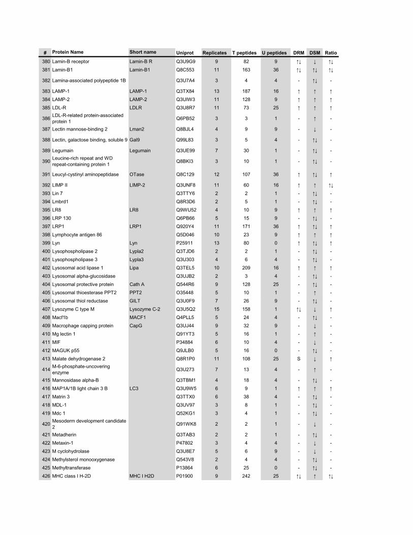
# Protein Name Short name Uniprot Replicates T peptides U peptides DRM DSM Ratio
380 Lamin-B receptor Lamin-B R Q3U9G9 9 82 9 �� � ��
381 Lamin-B1 Lamin-B1 Q8C553 11 163 36 �� �� ��
382 Lamina-associated polypeptide 1B Q3U7A4 3 4 4 - �� -
383 LAMP-1 LAMP-1 Q3TX84 13 187 16 � � �
384 LAMP-2 LAMP-2 Q3UIW3 11 128 9 � � �
385 LDL-R LDLR Q3U8R7 11 73 25 � � �
386 LDL-R-related protein-associated protein 1 Q6PB52 3 3 1 - � -
387 Lectin mannose-binding 2 Lman2 Q8BJL4 4 9 9 - � -
388 Lectin, galactose binding, soluble 9 Gal9 Q99L83 3 5 4 - �� -
389 Legumain Legumain Q3UE99 7 30 1 - �� -
390 Leucine-rich repeat and WD repeat-containing protein 1 Q8BKI3 3 10 1 - �� -
391 Leucyl-cystinyl aminopeptidase OTase Q8C129 12 107 36 � �� �
392 LIMP II LIMP-2 Q3UNF8 11 60 16 � � ��
393 Lin 7 Q3TTY6 2 2 1 - �� -
394 Lmbrd1 Q8R3D6 2 5 1 - �� -
395 LR8 LR8 Q9WU52 4 10 9 � � �
396 LRP 130 Q6PB66 5 15 9 - �� -
397 LRP1 LRP1 Q920Y4 11 171 36 � �� �
398 Lymphocyte antigen 86 Q5D046 10 23 9 � � �
399 Lyn Lyn P25911 13 80 0 � �� �
400 Lysophospholipase 2 Lypla2 Q3TJD6 2 2 1 - �� -
401 Lysophospholipase 3 Lypla3 Q3U303 4 6 4 - �� -
402 Lysosomal acid lipase 1 Lipa Q3TEL5 10 209 16 � � �
403 Lysosomal alpha-glucosidase Q3UJB2 2 3 4 - �� -
404 Lysosomal protective protein Cath A Q544R6 9 128 25 - �� -
405 Lysosomal thioesterase PPT2 PPT2 O35448 5 10 1 - � -
406 Lysosomal thiol reductase GILT Q3U0F9 7 26 9 - �� -
407 Lysozyme C type M Lysozyme C-2 Q3U5Q2 15 158 1 �� � �
408 Macf1b MACF1 Q4PLL5 5 24 4 - �� -
409 Macrophage capping protein CapG Q3UJ44 9 32 9 - � -
410 Mg lectin 1 Q91YT3 5 16 1 - � -
411 MIF P34884 6 10 4 - � -
412 MAGUK p55 Q9JLB0 5 16 0 - �� -
413 Malate dehydrogenase 2 Q8R1P0 11 108 25 S � �
414 M-6-phosphate-uncovering enzyme Q3U273 7 13 4 - � -
415 Mannosidase alpha-B Q3TBM1 4 18 4 - �� -
416 MAP1A/1B light chain 3 B LC3 Q3U9W5 6 9 1 � � �
417 Matrin 3 Q3TTX0 6 38 4 - �� -
418 MDL-1 Q3UV97 3 8 1 - �� -
419 Mdc 1 Q52KG1 3 4 1 - �� -
420 Mesoderm development candidate 2 Q91WK8 2 2 1 - � -
421 Metadherin Q3TAB3 2 2 1 - �� -
422 Metaxin-1 P47802 3 4 4 - � -
423 M cyclohydrolase Q3U8E7 5 6 9 - � -
424 Methylsterol monooxygenase Q543V8 2 4 4 - �� -
425 Methyltransferase P13864 6 25 0 - �� -
426 MHC class I H-2D MHC I H2D P01900 9 242 25 �� � ��
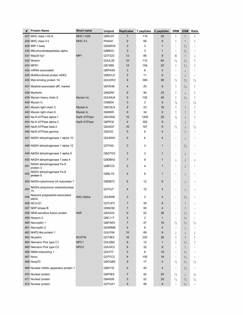
# Protein Name Short name Uniprot Replicates T peptides U peptides DRM DSM Ratio
427 MHC class I H2-K MHC I H2K Q6RJ37 7 119 25 � � �
428 MHC class II li MHC II li P04441 9 68 0 � � �
429 MIP-1-beta Q5QNV9 3 3 1 - �� -
430 Mitochondrialpeptidase alpha Q8BK51 3 3 1 - �� -
431 Map2k1ip1 MP1 Q3TX23 13 88 9 S � �
432 Moesin Q3UL28 10 112 64 �� �� �
433 MPS1 Q61889 14 156 25 � �� �
434 mRNA-associated Q6PAS9 3 6 4 - � -
435 Multifunctional protein ADE2 Q9DCL9 5 11 9 - � -
436 Myb-binding protein 1A Q3U2W2 9 389 36 �� �� ��
437 Myeloid-associated diff. marker Q0VE46 4 25 9 � �� �
438 Myoferlin Q69ZN7 9 90 25 � � �
439 Myosin heavy chain 9 Myosin hc Q3UHU4 9 100 49 � �� �
440 Myosin Ic O08834 3 3 9 �� - ��
441 Myosin light chain 2 Myosin lc Q9CQL8 9 23 16 � � �
442 Myosin light chain 6 Myosin lc Q60605 9 34 0 � � �
443 Na K-ATPase alpha-1 Na/K ATPase Q8VDN2 15 1205 25 �� � �
444 Na K-ATPase alpha-3 Na/K ATPase Q6PIC6 4 392 9 - � -
445 Na/K-ATPase beta-3 Q544Q7 16 167 9 �� � ��
446 Na/K-ATPase gamma Q923I3 5 6 4 - � -
447 NADH dehydrogenase 1 alpha 10 Q3UKK0 5 6 4 - � -
448 NADH dehydrogenase 1 alpha 12 Q3TIA0 3 3 1 - �� -
449 NADH dehydrogenase 1 alpha 9 Q6GTD3 3 3 1 - � -
450 NADH dehydrogenase 1 beta 4 Q9DBH2 7 9 1 � � �
451 NADH dehydrogenase Fe-S protein 3 Q8BTZ3 2 4 1 - � -
452 NADH dehydrogenase Fe-S protein 5 Q99LY9 4 6 1 - � -protein 5 �
453 NADH-cytochrome b5 reductase 1 Q9DB73 6 12 9 - � -
454 NADH-ubiquinone oxidoreductase 75 Q3TIU7 4 10 4 - � -
455 Nascent polypeptide-associated alpha NAC-Alpha Q3UK68 3 3 4 - �� -
456 NCU-G1 Q3TUF5 7 39 9 - � -
457 NDP kinase B Q5NC82 7 95 4 - � -
458 NEM-sensitive fusion protein NSF Q923C6 9 52 36 - �� -
459 Nesprin-3 Q8C117 2 2 1 - �� -
460 Neuropilin 1 Q6PAR3 7 47 16 �� �� ��
461 Neuropilin 2 Q5SRM6 4 6 4 - � -
462 NHP2-like protein 1 Q3UY64 10 89 9 � � �
463 Nicastrin NCSTN Q3T9E5 16 225 36 � � �
464 Niemann Pick type C1 NPC1 Q3U2B2 6 12 1 � �� �
465 Niemann Pick type C2 NPC2 Q3U3C2 8 32 9 - � -
466 NIMA-interacting 1 Q3UTI7 5 8 16 - �� -
467 Nono Q3TFC2 8 105 16 - �� -
468 Noxp70 Q9CQB5 9 17 4 �� �� ��
469 Nuclear mitotic apparatus protein 1 Q80Y35 5 45 4 - �� -
470 Nuclear protein Q6P5E5 7 42 25 �� � ��
471 Nuclear protein Q6A026 5 22 25 �� �� ��
472 Nuclear protein Q3TUA1 4 98 9 - �� -
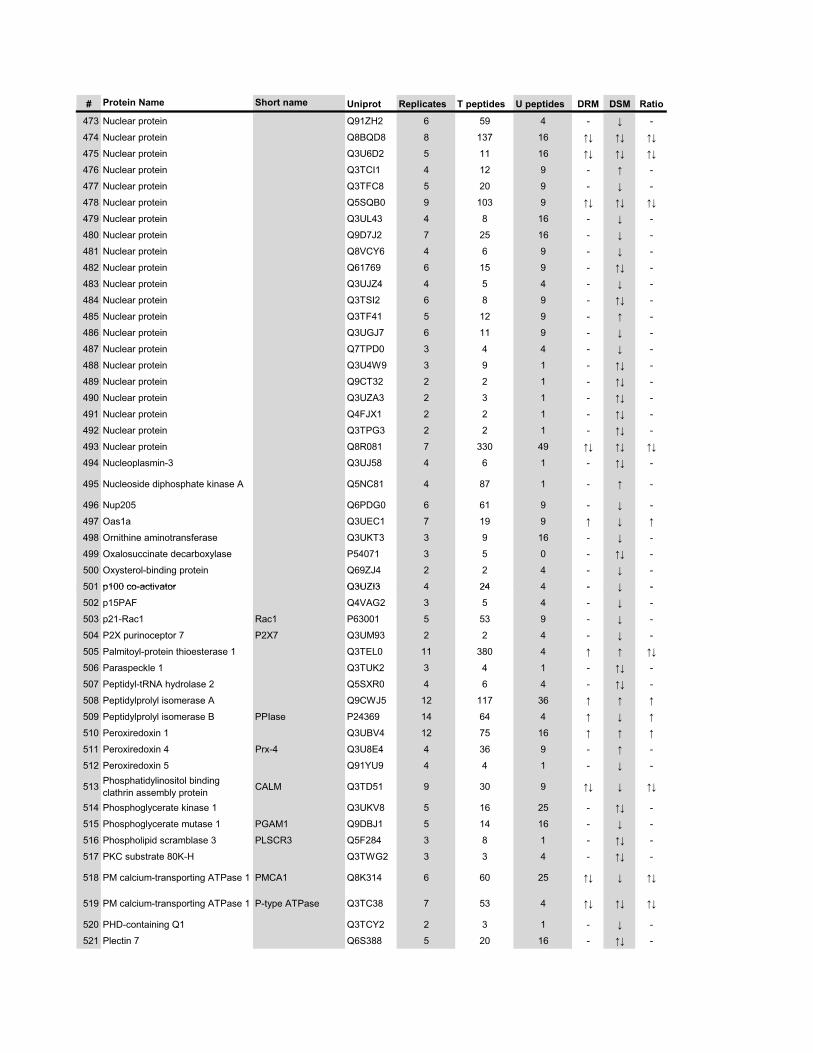
# Protein Name Short name Uniprot Replicates T peptides U peptides DRM DSM Ratio
473 Nuclear protein Q91ZH2 6 59 4 - � -
474 Nuclear protein Q8BQD8 8 137 16 �� �� ��
475 Nuclear protein Q3U6D2 5 11 16 �� �� ��
476 Nuclear protein Q3TCI1 4 12 9 - � -
477 Nuclear protein Q3TFC8 5 20 9 - � -
478 Nuclear protein Q5SQB0 9 103 9 �� �� ��
479 Nuclear protein Q3UL43 4 8 16 - � -
480 Nuclear protein Q9D7J2 7 25 16 - � -
481 Nuclear protein Q8VCY6 4 6 9 - � -
482 Nuclear protein Q61769 6 15 9 - �� -
483 Nuclear protein Q3UJZ4 4 5 4 - � -
484 Nuclear protein Q3TSI2 6 8 9 - �� -
485 Nuclear protein Q3TF41 5 12 9 - � -
486 Nuclear protein Q3UGJ7 6 11 9 - � -
487 Nuclear protein Q7TPD0 3 4 4 - � -
488 Nuclear protein Q3U4W9 3 9 1 - �� -
489 Nuclear protein Q9CT32 2 2 1 - �� -
490 Nuclear protein Q3UZA3 2 3 1 - �� -
491 Nuclear protein Q4FJX1 2 2 1 - �� -
492 Nuclear protein Q3TPG3 2 2 1 - �� -
493 Nuclear protein Q8R081 7 330 49 �� �� ��
494 Nucleoplasmin-3 Q3UJ58 4 6 1 - �� -
495 Nucleoside diphosphate kinase A Q5NC81 4 87 1 - � -
496 Nup205 Q6PDG0 6 61 9 - � -
497 Oas1a Q3UEC1 7 19 9 � � �
498 Ornithine aminotransferase Q3UKT3 3 9 16 - � -
499 Oxalosuccinate decarboxylase P54071 3 5 0 - �� -
500 Oxysterol-binding protein Q69ZJ4 2 2 4 - � -
501 p100 co-activator Q3UZI3 4 24 4 - � -501 p100 co-activator Q3UZI3 4 24 4 - � -
502 p15PAF Q4VAG2 3 5 4 - � -
503 p21-Rac1 Rac1 P63001 5 53 9 - � -
504 P2X purinoceptor 7 P2X7 Q3UM93 2 2 4 - � -
505 Palmitoyl-protein thioesterase 1 Q3TEL0 11 380 4 � � ��
506 Paraspeckle 1 Q3TUK2 3 4 1 - �� -
507 Peptidyl-tRNA hydrolase 2 Q5SXR0 4 6 4 - �� -
508 Peptidylprolyl isomerase A Q9CWJ5 12 117 36 � � �
509 Peptidylprolyl isomerase B PPIase P24369 14 64 4 � � �
510 Peroxiredoxin 1 Q3UBV4 12 75 16 � � �
511 Peroxiredoxin 4 Prx-4 Q3U8E4 4 36 9 - � -
512 Peroxiredoxin 5 Q91YU9 4 4 1 - � -
513 Phosphatidylinositol binding clathrin assembly protein CALM Q3TD51 9 30 9 �� � ��
514 Phosphoglycerate kinase 1 Q3UKV8 5 16 25 - �� -
515 Phosphoglycerate mutase 1 PGAM1 Q9DBJ1 5 14 16 - � -
516 Phospholipid scramblase 3 PLSCR3 Q5F284 3 8 1 - �� -
517 PKC substrate 80K-H Q3TWG2 3 3 4 - �� -
518 PM calcium-transporting ATPase 1 PMCA1 Q8K314 6 60 25 �� � ��
519 PM calcium-transporting ATPase 1 P-type ATPase Q3TC38 7 53 4 �� �� ��
520 PHD-containing Q1 Q3TCY2 2 3 1 - � -
521 Plectin 7 Q6S388 5 20 16 - �� -
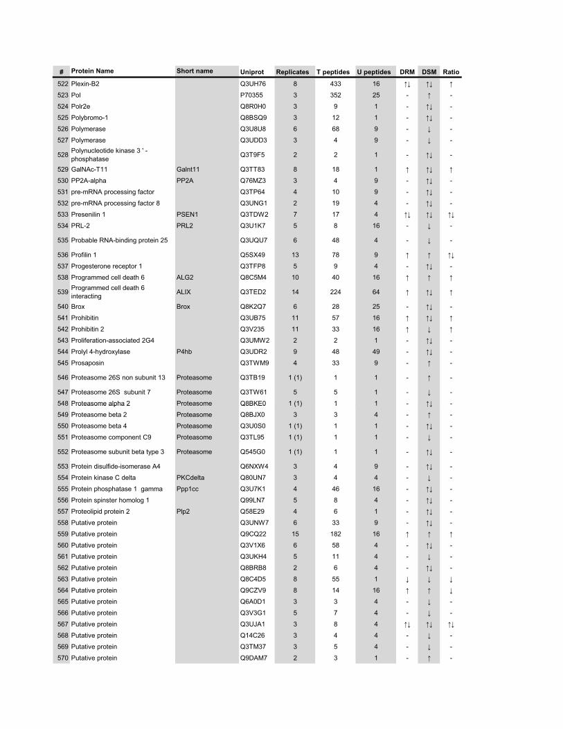
# Protein Name Short name Uniprot Replicates T peptides U peptides DRM DSM Ratio
522 Plexin-B2 Q3UH76 8 433 16 �� �� �
523 Pol P70355 3 352 25 - � -
524 Polr2e Q8R0H0 3 9 1 - �� -
525 Polybromo-1 Q8BSQ9 3 12 1 - �� -
526 Polymerase Q3U8U8 6 68 9 - � -
527 Polymerase Q3UDD3 3 4 9 - � -
528 Polynucleotide kinase 3 ' -phosphatase Q3T9F5 2 2 1 - �� -
529 GalNAc-T11 Galnt11 Q3TT83 8 18 1 � �� �
530 PP2A-alpha PP2A Q76MZ3 3 4 9 - �� -
531 pre-mRNA processing factor Q3TP64 4 10 9 - �� -
532 pre-mRNA processing factor 8 Q3UNG1 2 19 4 - �� -
533 Presenilin 1 PSEN1 Q3TDW2 7 17 4 �� �� ��
534 PRL-2 PRL2 Q3U1K7 5 8 16 - � -
535 Probable RNA-binding protein 25 Q3UQU7 6 48 4 - � -
536 Profilin 1 Q5SX49 13 78 9 � � ��
537 Progesterone receptor 1 Q3TFP8 5 9 4 - �� -
538 Programmed cell death 6 ALG2 Q8C5M4 10 40 16 � � �
539 Programmed cell death 6 interacting ALIX Q3TED2 14 224 64 � �� �
540 Brox Brox Q8K2Q7 6 28 25 - �� -
541 Prohibitin Q3UB75 11 57 16 � �� �
542 Prohibitin 2 Q3V235 11 33 16 � � �
543 Proliferation-associated 2G4 Q3UMW2 2 2 1 - �� -
544 Prolyl 4-hydroxylase P4hb Q3UDR2 9 48 49 - �� -
545 Prosaposin Q3TWM9 4 33 9 - � -
546 Proteasome 26S non subunit 13 Proteasome Q3TB19 1 (1) 1 1 - � -
547 Proteasome 26S subunit 7 Proteasome Q3TW61 5 5 1 - � -
548 Proteasome alpha 2 Proteasome Q8BKE0 1 (1) 1 1 - �� -548 Proteasome alpha 2 Proteasome Q8BKE0 1 (1) 1 1 ��
549 Proteasome beta 2 Proteasome Q8BJX0 3 3 4 - � -
550 Proteasome beta 4 Proteasome Q3U0S0 1 (1) 1 1 - �� -
551 Proteasome component C9 Proteasome Q3TL95 1 (1) 1 1 - � -
552 Proteasome subunit beta type 3 Proteasome Q545G0 1 (1) 1 1 - �� -
553 Protein disulfide-isomerase A4 Q6NXW4 3 4 9 - �� -
554 Protein kinase C delta PKCdelta Q80UN7 3 4 4 - � -
555 Protein phosphatase 1 gamma Ppp1cc Q3U7K1 4 46 16 - �� -
556 Protein spinster homolog 1 Q99LN7 5 8 4 - �� -
557 Proteolipid protein 2 Plp2 Q58E29 4 6 1 - �� -
558 Putative protein Q3UNW7 6 33 9 - �� -
559 Putative protein Q9CQ22 15 182 16 � � �
560 Putative protein Q3V1X6 6 58 4 - �� -
561 Putative protein Q3UKH4 5 11 4 - � -
562 Putative protein Q8BRB8 2 6 4 - �� -
563 Putative protein Q8C4D5 8 55 1 � � �
564 Putative protein Q9CZV9 8 14 16 � � �
565 Putative protein Q6A0D1 3 3 4 - � -
566 Putative protein Q3V3G1 5 7 4 - � -
567 Putative protein Q3UJA1 3 8 4 �� �� ��
568 Putative protein Q14C26 3 4 4 - � -
569 Putative protein Q3TM37 3 5 4 - � -
570 Putative protein Q9DAM7 2 3 1 - � -
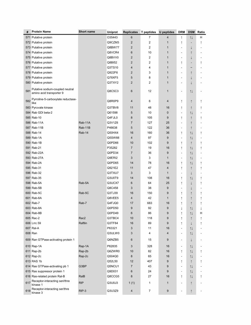
# Protein Name Short name Uniprot Replicates T peptides U peptides DRM DSM Ratio
571 Putative protein O35443 6 7 4 � �� H
572 Putative protein Q9CZM3 2 2 1 � - �
573 Putative protein Q8BW77 2 2 1 - � -
574 Putative protein Q8VCR4 6 10 1 - � -
575 Putative protein Q8BVV0 2 2 1 - � -
576 Putative protein Q8BI52 2 2 1 � - �
577 Putative protein Q3TS10 4 4 1 - ~ -
578 Putative protein Q922F6 2 3 1 - � -
579 Putative protein Q78XF5 5 8 1 - � -
580 Putative protein Q3TXY2 2 2 1 - � -
581 Putative sodium-coupled neutral amino acid transporter 9 Q8C5C3 6 12 1 - �� -
582 Pyrroline-5-carboxylate reductase-like Q8R0P9 4 6 4 � � �
583 Pyruvate kinase Q3TBV8 11 48 16 � � �
584 Rab GDI beta-2 Q61598 5 10 0 - �� -
585 Rab-10 Q4FJL0 8 105 9 � � �
586 Rab-11A Rab-11A Q3V1Z6 7 127 25 - � -
587 Rab-11B Rab-11B P46638 5 122 36 - � -
588 Rab-14 Rab-14 Q50HX4 16 160 36 � �� �
589 Rab-1A Q5SW88 4 97 9 - �� -
590 Rab-1B Q0PD66 10 102 9 � � �
591 Rab-21 P35282 7 19 16 � �� �
592 Rab-22A Q0PD34 7 36 9 - �� -
593 Rab-27A Q9ERI2 3 3 1 - �� -
594 Rab-2A Q0PD65 14 78 16 � �� �
595 Rab-31 Q921E2 11 47 4 � � �
596 Rab-32 Q3TXU7 3 3 1 - � -
597 Rab-35 Q3U0T9 14 108 16 � �� �
598 Rab-5A Rab-5A Q3UCX7 6 64 25 � � �
599 Rab-5B Q8C458 3 38 9 - � -
600 Rab-5C Rab-5C Q3TJ39 16 150 9 � � �
601 Rab-6A Q8VEE5 4 42 1 � � �
602 Rab-7 Rab-7 Q4FJQ0 17 683 16 � � �
603 Rab-8A Q0PD50 9 92 9 � �� �
604 Rab-8B Q0PD49 6 86 9 � �� H
605 Rac-2 Rac2 Q3TBC4 10 118 9 � � �
606 Lrrc 59 Raftlin Q3TF84 16 89 9 � � �
607 Ral-A P63321 3 11 16 - �� -
608 Ran Q3ULW0 3 4 4 - �� -
609 Ran GTPase-activating protein 1 Q6NZB5 6 15 9 - � -
610 Rap-1A Rap-1A P62835 3 328 16 - �� -
611 Rap-2b Rap-2b Q6ZWR0 10 82 16 � �� �
612 Rap-2c Rap-2c Q504Q0 8 65 16 - �� -
613 RAS 1b Q52L50 12 407 9 � � �
614 Ras GTPase-activating pb 1 G3BP Q5NCU1 7 43 9 - �� -
615 Ras suppressor protein 1 Q9D031 6 24 9 - �� -
616 Ras-related protein Ral-B RalB Q8CCG5 8 27 16 � �� �
617 Receptor-interacting seri/thre kinase 1 RIP Q3U0J3 1 (1) 1 1 - � -
618 Receptor-interacting ser/thre kinase 3 RIP-3 Q3U3Z9 4 7 9 - � -
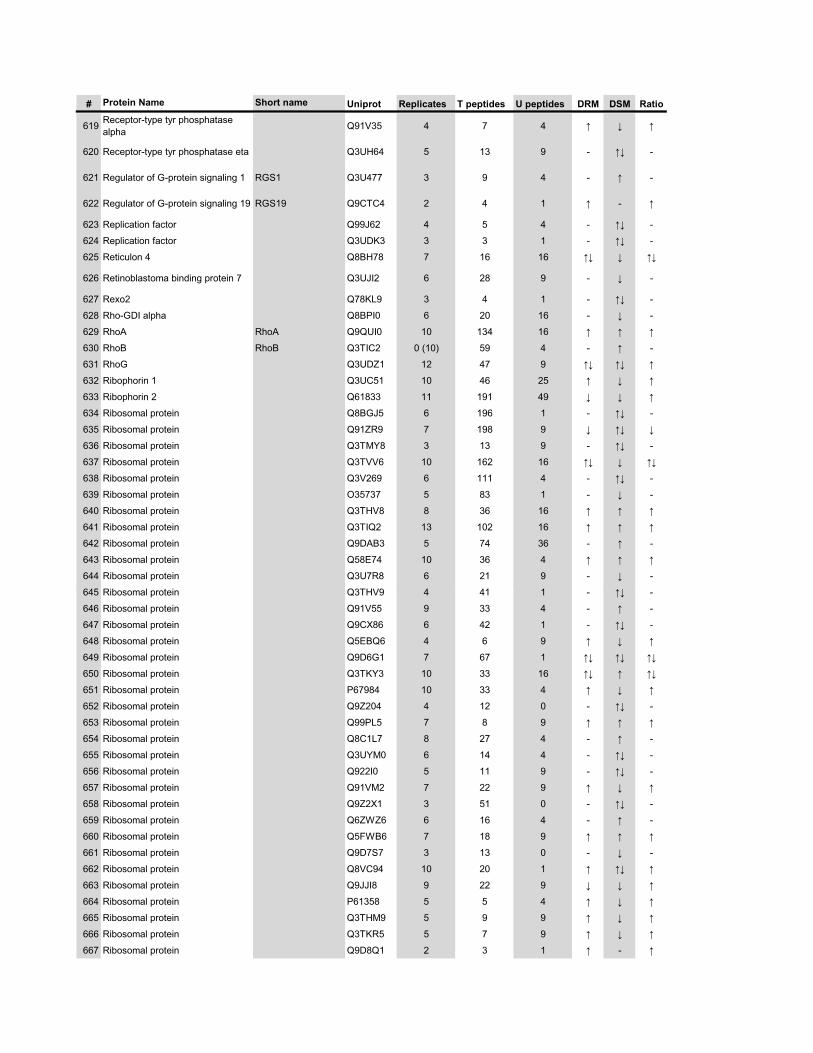
# Protein Name Short name Uniprot Replicates T peptides U peptides DRM DSM Ratio
619 Receptor-type tyr phosphatase alpha Q91V35 4 7 4 � � �
620 Receptor-type tyr phosphatase eta Q3UH64 5 13 9 - �� -
621 Regulator of G-protein signaling 1 RGS1 Q3U477 3 9 4 - � -
622 Regulator of G-protein signaling 19 RGS19 Q9CTC4 2 4 1 � - �
623 Replication factor Q99J62 4 5 4 - �� -
624 Replication factor Q3UDK3 3 3 1 - �� -
625 Reticulon 4 Q8BH78 7 16 16 �� � ��
626 Retinoblastoma binding protein 7 Q3UJI2 6 28 9 - � -
627 Rexo2 Q78KL9 3 4 1 - �� -
628 Rho-GDI alpha Q8BPI0 6 20 16 - � -
629 RhoA RhoA Q9QUI0 10 134 16 � � �
630 RhoB RhoB Q3TIC2 0 (10) 59 4 - � -
631 RhoG Q3UDZ1 12 47 9 �� �� �
632 Ribophorin 1 Q3UC51 10 46 25 � � �
633 Ribophorin 2 Q61833 11 191 49 � � �
634 Ribosomal protein Q8BGJ5 6 196 1 - �� -
635 Ribosomal protein Q91ZR9 7 198 9 � �� �
636 Ribosomal protein Q3TMY8 3 13 9 - �� -
637 Ribosomal protein Q3TVV6 10 162 16 �� � ��
638 Ribosomal protein Q3V269 6 111 4 - �� -
639 Ribosomal protein O35737 5 83 1 - � -
640 Ribosomal protein Q3THV8 8 36 16 � � �
641 Ribosomal protein Q3TIQ2 13 102 16 � � �
642 Ribosomal protein Q9DAB3 5 74 36 - � -
643 Ribosomal protein Q58E74 10 36 4 � � �
644 Ribosomal protein Q3U7R8 6 21 9 - � -
645 Ribosomal protein Q3THV9 4 41 1 - �� -
646 Ribosomal protein Q91V55 9 33 4 - � -
647 Ribosomal protein Q9CX86 6 42 1 - �� -
648 Ribosomal protein Q5EBQ6 4 6 9 � � �
649 Ribosomal protein Q9D6G1 7 67 1 �� �� ��
650 Ribosomal protein Q3TKY3 10 33 16 �� � ��
651 Ribosomal protein P67984 10 33 4 � � �
652 Ribosomal protein Q9Z204 4 12 0 - �� -
653 Ribosomal protein Q99PL5 7 8 9 � � �
654 Ribosomal protein Q8C1L7 8 27 4 - � -
655 Ribosomal protein Q3UYM0 6 14 4 - �� -
656 Ribosomal protein Q922I0 5 11 9 - �� -
657 Ribosomal protein Q91VM2 7 22 9 � � �
658 Ribosomal protein Q9Z2X1 3 51 0 - �� -
659 Ribosomal protein Q6ZWZ6 6 16 4 - � -
660 Ribosomal protein Q5FWB6 7 18 9 � � �
661 Ribosomal protein Q9D7S7 3 13 0 - � -
662 Ribosomal protein Q8VC94 10 20 1 � �� �
663 Ribosomal protein Q9JJI8 9 22 9 � � �
664 Ribosomal protein P61358 5 5 4 � � �
665 Ribosomal protein Q3THM9 5 9 9 � � �
666 Ribosomal protein Q3TKR5 5 7 9 � � �
667 Ribosomal protein Q9D8Q1 2 3 1 � - �
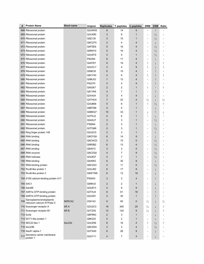
# Protein Name Short name Uniprot Replicates T peptides U peptides DRM DSM Ratio
668 Ribosomal protein Q3UW83 8 14 9 - � -
669 Ribosomal protein Q3UX80 3 6 1 - �� -
670 Ribosomal protein Q9Z130 3 15 1 - �� -
671 Ribosomal protein Q8CCF0 2 4 4 - � -
672 Ribosomal protein Q4FZE6 5 16 4 - �� -
673 Ribosomal protein Q9R0Y0 5 16 4 - �� -
674 Ribosomal protein Q3U9T9 3 5 1 - �� -
675 Ribosomal protein P62264 6 17 4 - � -
676 Ribosomal protein Q497D7 9 19 4 � � �
677 Ribosomal protein Q3UCL7 3 4 9 � � �
678 Ribosomal protein Q58E35 6 15 4 - � -
679 Ribosomal protein Q9CY93 4 6 4 � � �
680 Ribosomal protein Q5BLK2 7 13 4 - � -
681 Ribosomal protein P62270 3 4 4 - � -
682 Ribosomal protein Q542K7 2 2 1 � - �
683 Ribosomal protein Q91YK6 3 7 1 - � -
684 Ribosomal protein Q3V424 3 4 4 - � -
685 Ribosomal protein Q3THU4 7 23 4 �� � ��
686 Ribosomal protein Q3UB68 5 6 1 � �� �
687 Ribosomal protein Q8BTB8 4 5 1 - � -
688 Ribosomal protein Q5BKQ7 10 33 1 � � �
689 Ribosomal protein Q3TKJ3 4 6 1 - � -
690 Ribosomal protein Q542U7 3 3 1 - �� -
691 Ribosomal protein P58064 2 3 1 - � -
692 Ribosomal protein Q3TQ66 2 3 1 - �� -
693 Ring finger protein 149 Q3U2C5 3 3 1 - � -
694 RNA binding Q8CFQ9 6 14 9 - � -
695 RNA binding Q9CWZ3 3 10 0 - �� -
696 RNA binding Q9R0B2 6 13 4 - �� -
697 RNA binding Q64012 3 3 0 - �� -
698 RNA enzyme Q9CZQ0 4 7 9 - �� -
699 RNA helicase Q3U837 3 7 1 - �� -
700 RNA-binding Q545K5 6 30 9 - � -
701 RNA-binding protein Q8C2Q3 4 11 0 - �� -
702 RuvB-like protein 1 Q3UJN2 6 17 9 - � -
703 RuvB-like protein 2 Q9WTM5 6 12 16 - � -
704 S100 calcium-binding protein A11 P50543 3 3 4 - � -
705 SAC1 Q99K33 2 2 1 - � -
706 Sam68 Q3U8T3 4 9 9 - � -
707 SAR1a GTP-binding protein Q3TXJ4 8 61 16 - � -
708 SAR1b GTP-binding protein Q3U281 3 35 1 - � -
709 Sarcoplasmic/endoplasmic reticulum calcium ATPase 2 SERCA2 O55143 9 60 0 �� � ��
710 Scavenger receptor A SR A Q3U2C3 18 300 25 �� � �
711 Scavenger receptor B1 SR B Q3TZ42 13 124 9 �� � �
712 Scrib Q6P9N3 2 3 1 - � -
713 SCY1-like protein 1 Q8K222 2 2 1 - � -
714 SEC22 like 1 Sec22b Q3UZ06 8 16 4 � �� �
715 Sec23B Q8CDS4 3 3 4 - �� -
716 Sec61 alpha-1 Q3TXA5 6 26 9 - � -
717 Secretory carrier membrane protein 1 Q3UY11 4 7 4 - � -
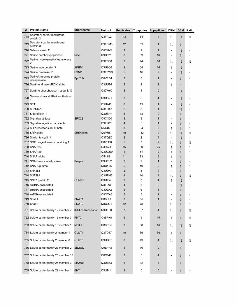
# Protein Name Short name Uniprot Replicates T peptides U peptides DRM DSM Ratio
718 Secretory carrier membrane protein 2 Q3TAL2 13 64 4 �� �� ��
719 Secretory carrier membrane protein 3 Q3TDM8 12 69 1 �� � �
720 Selenoprotein T Q8CHV4 2 2 1 - �� -
721 Serine caroboxypeptidase Risc Q9D625 8 89 16 - � -
722 Serine hydroxymethyl transferase 2 Q3TFD0 7 44 16 �� �� ��
723 Serine incorporator 3 AIGP-1 Q3U7C6 9 38 16 � �� �
724 Serine protease 15 LONP Q3TZW3 5 16 9 - �� -
725 Serine/threonine protein phosphatase Ppp2cb Q8VED4 3 3 1 - � -
726 Ser/thre kinase MRCK alpha Q3UU96 2 2 1 � - �
727 Ser/thre phosphatase 1 subunit 10 Q80W00 3 4 0 - �� -
728 Seryl-aminoacyl-tRNA synthetase 1 Q3U8N1 3 8 4 - �� -
729 SET Q5U445 6 19 1 - �� -
730 SF3b14b Q3TQX7 2 3 1 - �� -
731 Sideroflexin-1 Q3UB44 5 13 9 - � -
732 Signal peptidase SPC22 Q8C1D0 3 3 1 - � -
733 Signal recognition particle 14 Q3TIK2 4 5 1 - � -
734 SRP receptor subunit beta Q544X9 8 34 9 � � �
735 SRP alpha SIRPalpha Q6P6I8 10 142 9 �� �� ��
736 Similar to cyclin I Q3TQZ0 2 2 4 - �� -
737 SMC hinge domain containing 1 Q6P5D8 5 7 9 �� � ��
738 SNAP-23 O35620 15 80 25 � � �
739 SNAP-29 Q3UGN0 4 51 9 � � �
740 SNAP-alpha Q543I3 11 83 9 � � �
741 SNAP-associated protein Snapin Q3UY32 2 2 1 - � -
742 SNAP-gamma Q8C1T5 5 10 9 � � �
743 SNF2L1 Q3UDA4 2 3 4 - � -743 SNF2L1 Q3UDA4 2 3 4 - � -
744 SNF2L4 Q3URH5 4 10 4 �� � ��
745 SNF7 protein 2 CHMP5 Q3UI64 3 3 4 � �� �
746 snRNA-associated Q3TIX3 4 6 9 - �� -
747 snRNA-associated Q3U9X2 5 6 1 - � -
748 snRNA-associated Q9QXA5 5 5 1 - � -
749 Snat 1 SNAT1 Q8BHI3 9 65 1 - � -
750 Snat 2 SNAT2 Q8CD21 12 78 9 �� � �
751 Solute carrier family 12 member 7 K-Cl co-transporter Q3UE50 7 67 4 �� � ��
752 Solute carrier family 15 member 3 PHT2 Q8BPX9 6 9 16 � � ��
753 Solute carrier family 16 member 1 MCT1 Q8BPS5 9 90 16 �� �� ��
754 Solute carrier family 2 member 1 GLUT1 Q3TD17 10 39 36 � � �
755 Solute carrier family 2 member 6 GLUT6 Q3UDF0 8 43 4 �� �� ��
756 Solute carrier family 23 member 2 Slc23a2 Q9EPR4 4 10 0 - � -
757 Solute carrier family 25 member 13 Q8C140 2 5 4 - � -
758 Solute carrier family 25 member 3 Slc25a3 Q3UB63 6 32 4 - � -
759 Solute carrier family 29 member 1 ENT1 Q9JIM1 3 5 0 - � -
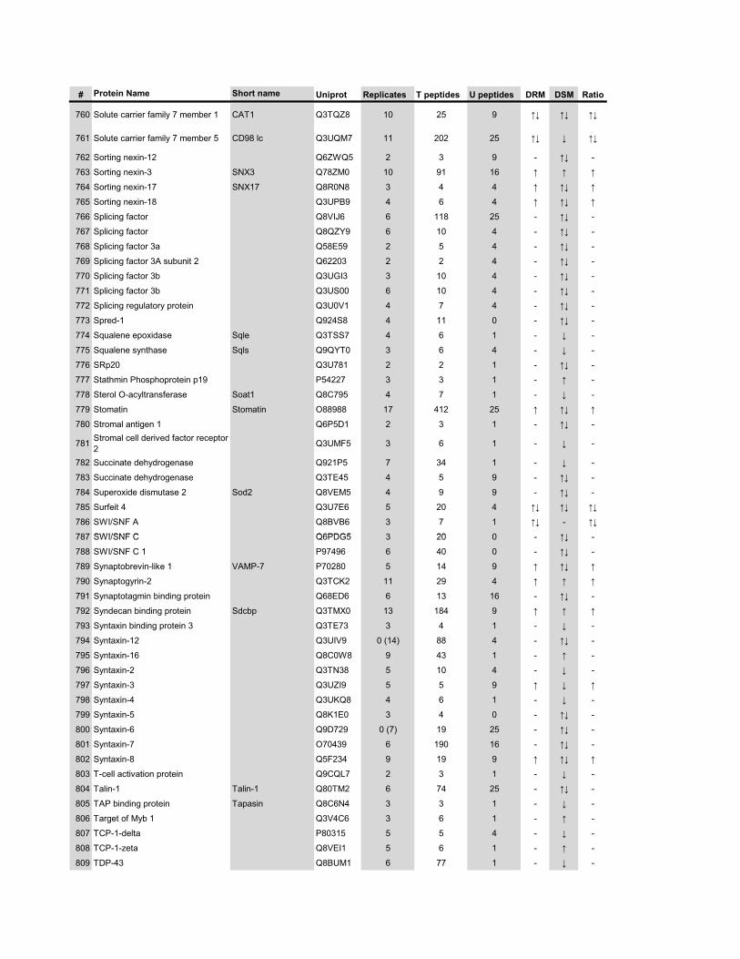
# Protein Name Short name Uniprot Replicates T peptides U peptides DRM DSM Ratio
760 Solute carrier family 7 member 1 CAT1 Q3TQZ8 10 25 9 �� �� ��
761 Solute carrier family 7 member 5 CD98 lc Q3UQM7 11 202 25 �� � ��
762 Sorting nexin-12 Q6ZWQ5 2 3 9 - �� -
763 Sorting nexin-3 SNX3 Q78ZM0 10 91 16 � � �
764 Sorting nexin-17 SNX17 Q8R0N8 3 4 4 � �� �
765 Sorting nexin-18 Q3UPB9 4 6 4 � �� �
766 Splicing factor Q8VIJ6 6 118 25 - �� -
767 Splicing factor Q8QZY9 6 10 4 - �� -
768 Splicing factor 3a Q58E59 2 5 4 - �� -
769 Splicing factor 3A subunit 2 Q62203 2 2 4 - �� -
770 Splicing factor 3b Q3UGI3 3 10 4 - �� -
771 Splicing factor 3b Q3US00 6 10 4 - �� -
772 Splicing regulatory protein Q3U0V1 4 7 4 - �� -
773 Spred-1 Q924S8 4 11 0 - �� -
774 Squalene epoxidase Sqle Q3TSS7 4 6 1 - � -
775 Squalene synthase Sqls Q9QYT0 3 6 4 - � -
776 SRp20 Q3U781 2 2 1 - �� -
777 Stathmin Phosphoprotein p19 P54227 3 3 1 - � -
778 Sterol O-acyltransferase Soat1 Q8C795 4 7 1 - � -
779 Stomatin Stomatin O88988 17 412 25 � �� �
780 Stromal antigen 1 Q6P5D1 2 3 1 - �� -
781 Stromal cell derived factor receptor 2 Q3UMF5 3 6 1 - � -
782 Succinate dehydrogenase Q921P5 7 34 1 - � -
783 Succinate dehydrogenase Q3TE45 4 5 9 - �� -
784 Superoxide dismutase 2 Sod2 Q8VEM5 4 9 9 - �� -
785 Surfeit 4 Q3U7E6 5 20 4 �� �� ��
786 SWI/SNF A Q8BVB6 3 7 1 �� - ��
787 SWI/SNF C Q6PDG5 3 20 0 ��787 SWI/SNF C Q6PDG5 3 20 0 - �� -
788 SWI/SNF C 1 P97496 6 40 0 - �� -
789 Synaptobrevin-like 1 VAMP-7 P70280 5 14 9 � �� �
790 Synaptogyrin-2 Q3TCK2 11 29 4 � � �
791 Synaptotagmin binding protein Q68ED6 6 13 16 - �� -
792 Syndecan binding protein Sdcbp Q3TMX0 13 184 9 � � �
793 Syntaxin binding protein 3 Q3TE73 3 4 1 - � -
794 Syntaxin-12 Q3UIV9 0 (14) 88 4 - �� -
795 Syntaxin-16 Q8C0W8 9 43 1 - � -
796 Syntaxin-2 Q3TN38 5 10 4 - � -
797 Syntaxin-3 Q3UZI9 5 5 9 � � �
798 Syntaxin-4 Q3UKQ8 4 6 1 - � -
799 Syntaxin-5 Q8K1E0 3 4 0 - �� -
800 Syntaxin-6 Q9D729 0 (7) 19 25 - �� -
801 Syntaxin-7 O70439 6 190 16 - �� -
802 Syntaxin-8 Q5F234 9 19 9 � �� �
803 T-cell activation protein Q9CQL7 2 3 1 - � -
804 Talin-1 Talin-1 Q80TM2 6 74 25 - �� -
805 TAP binding protein Tapasin Q8C6N4 3 3 1 - � -
806 Target of Myb 1 Q3V4C6 3 6 1 - � -
807 TCP-1-delta P80315 5 5 4 - � -
808 TCP-1-zeta Q8VEI1 5 6 1 - � -
809 TDP-43 Q8BUM1 6 77 1 - � -
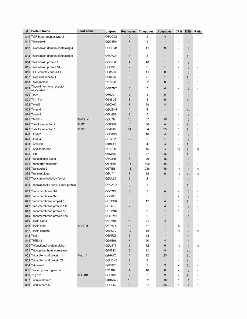
# Protein Name Short name Uniprot Replicates T peptides U peptides DRM DSM Ratio
810 TGF-beta receptor type-2 Q3UG22 2 2 4 � - �
811 Thioredoxin Q9D8R0 7 9 1 - � -
812 Thioredoxin domain containing 4 Q3URM4 6 11 9 - � -
813 Thioredoxin domain containing 5 Q3UWX1 4 8 1 - �� -
814 Thioredoxin protein 1 Q3UCI8 4 10 1 � � �
815 Thioredoxin protein 14 Q8BW13 2 3 1 - � -
816 THO complex subunit 4 O08583 5 11 0 - � -
817 Thymidine kinase 1 Q58EU0 3 6 1 - �� -
818 Thymopoietin Q61029 9 92 0 � � �
819 Thyroid hormone receptor associated 3 Q8BZN7 3 7 4 - � -
820 TIAP O70201 3 3 0 - � -
821 Tim17-A Q545U2 3 4 9 - �� -
822 Tmed9 Q9CX02 7 29 4 � � �
823 Tmem4 Q3UBH8 4 4 1 - �� -
824 Tmem9 Q3U459 2 5 1 - � -
825 TMP21-I TMP21-I Q3U7I7 10 37 16 � � �
826 Toll-like receptor 2 TLR2 Q8K3D9 6 36 4 - �� -
827 Toll-like receptor 7 TLR7 Q548J0 14 84 25 � �� �
828 TOM22 Q9D8D3 6 10 4 - � -
829 TOM34 Q812F2 2 2 1 - � -
830 Tomm20 Q4KL41 3 4 4 - �� -
831 Topoisomerase Q01320 3 15 4 �� �� ��
832 TPR Q7M739 6 37 16 - �� -
833 Transcription factor Q3UJR8 6 29 16 - � -
834 Transferrin receptor Q61560 15 408 36 � � �
835 Transgelin 2 Q3TIB8 11 218 16 �� � ��
836 Transketolase Q3U7Y1 7 15 9 �� �� ��
837 T l ti i iti ti f t Q3ULL5 2 2 1 �837 Translation initiation factor Q3ULL5 2 2 1 - � -
838 Translationally-contr. tumor protein Q3UAG7 2 5 1 - �� -
839 Transmembrane 9-2 Q8C7F9 3 5 4 - � -
840 Transmembrane 9-3 Q8C6F5 3 5 1 - � -
841 Transmembrane emp24 5 Q3TDS6 8 17 4 � �� �
842 Transmembrane protein 111 Q3TIK3 2 2 4 - � -
843 Transmembrane protein 59 Q3TWB4 3 3 1 � - �
844 Transmembrane protein 87A Q8BTV9 2 2 1 - � -
845 TRAP-alpha Q3TV94 10 21 4 � ~ �
846 TRAP-delta TRAP-d Q3TVJ8 10 37 1 S � �
847 TRAP-gamma Q4FK79 10 19 1 �� � ��
848 Tcof 1 Q6PFZ6 6 18 1 - � -
849 TREM-2 Q99NH8 7 90 4 - � -
850 Trifunctional protein alpha Q5U5Y5 6 12 9 �� � ��
851 Triosephosphate isomerase Q64513 6 11 9 - �� -
852 Tripartite motif protein 14 Tmp 14 Q14AR3 4 12 25 � �� �
853 Tripartite motif protein 28 Q3UDN8 2 8 1 - �� -
854 Trk-fused Q9D828 3 9 9 - �� -
855 Tropomyosin 3 gamma P21107 4 10 0 - � -
856 Tsg 101 TSG101 Q3UK08 2 2 9 - �� -
857 Tubulin alpha 2 Q4KMW2 13 62 16 � � �
858 Tubulin beta 5 Q3UF52 5 61 36 � � �
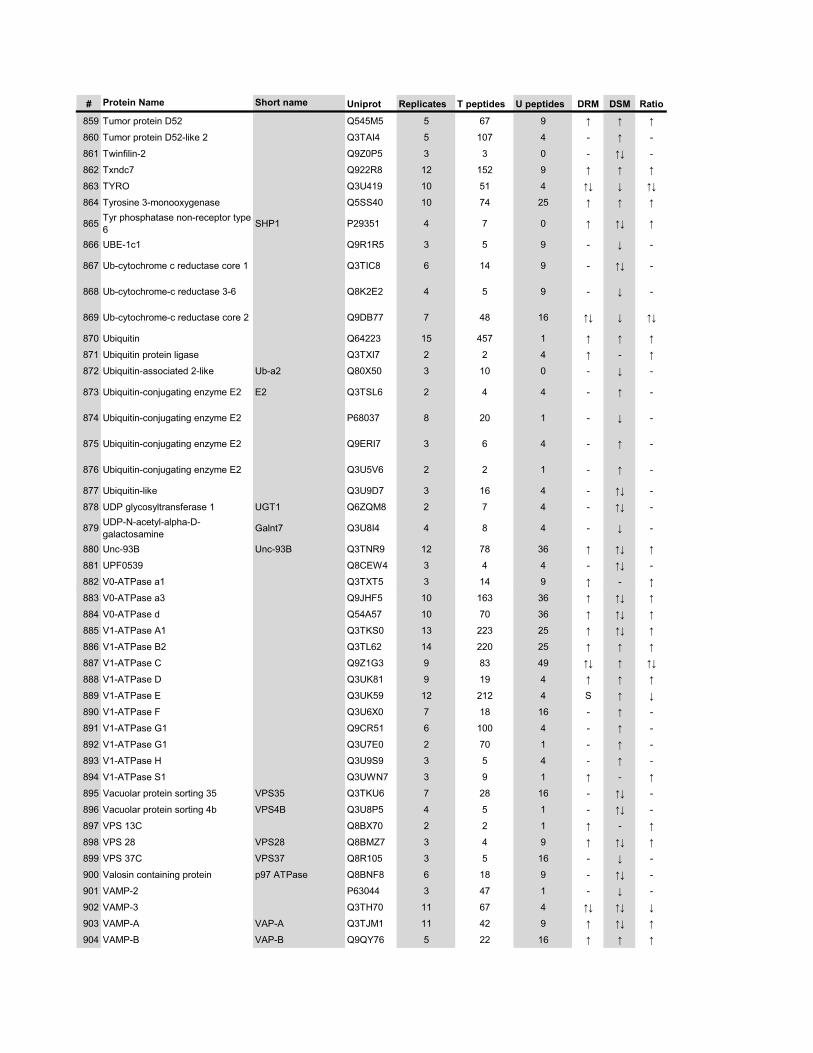
# Protein Name Short name Uniprot Replicates T peptides U peptides DRM DSM Ratio
859 Tumor protein D52 Q545M5 5 67 9 � � �
860 Tumor protein D52-like 2 Q3TAI4 5 107 4 - � -
861 Twinfilin-2 Q9Z0P5 3 3 0 - �� -
862 Txndc7 Q922R8 12 152 9 � � �
863 TYRO Q3U419 10 51 4 �� � ��
864 Tyrosine 3-monooxygenase Q5SS40 10 74 25 � � �
865 Tyr phosphatase non-receptor type 6 SHP1 P29351 4 7 0 � �� �
866 UBE-1c1 Q9R1R5 3 5 9 - � -
867 Ub-cytochrome c reductase core 1 Q3TIC8 6 14 9 - �� -
868 Ub-cytochrome-c reductase 3-6 Q8K2E2 4 5 9 - � -
869 Ub-cytochrome-c reductase core 2 Q9DB77 7 48 16 �� � ��
870 Ubiquitin Q64223 15 457 1 � � �
871 Ubiquitin protein ligase Q3TXI7 2 2 4 � - �
872 Ubiquitin-associated 2-like Ub-a2 Q80X50 3 10 0 - � -
873 Ubiquitin-conjugating enzyme E2 E2 Q3TSL6 2 4 4 - � -
874 Ubiquitin-conjugating enzyme E2 P68037 8 20 1 - � -
875 Ubiquitin-conjugating enzyme E2 Q9ERI7 3 6 4 - � -
876 Ubiquitin-conjugating enzyme E2 Q3U5V6 2 2 1 - � -
877 Ubiquitin-like Q3U9D7 3 16 4 - �� -
878 UDP glycosyltransferase 1 UGT1 Q6ZQM8 2 7 4 - �� -
879 UDP-N-acetyl-alpha-D-galactosamine Galnt7 Q3U8I4 4 8 4 - � -
880 Unc-93B Unc-93B Q3TNR9 12 78 36 � �� �
881 UPF0539 Q8CEW4 3 4 4 - �� -
882 V0-ATPase a1 Q3TXT5 3 14 9 � - �88 0 ase a Q3 5 3 9 � �
883 V0-ATPase a3 Q9JHF5 10 163 36 � �� �
884 V0-ATPase d Q54A57 10 70 36 � �� �
885 V1-ATPase A1 Q3TKS0 13 223 25 � �� �
886 V1-ATPase B2 Q3TL62 14 220 25 � � �
887 V1-ATPase C Q9Z1G3 9 83 49 �� � ��
888 V1-ATPase D Q3UK81 9 19 4 � � �
889 V1-ATPase E Q3UK59 12 212 4 S � �
890 V1-ATPase F Q3U6X0 7 18 16 - � -
891 V1-ATPase G1 Q9CR51 6 100 4 - � -
892 V1-ATPase G1 Q3U7E0 2 70 1 - � -
893 V1-ATPase H Q3U9S9 3 5 4 - � -
894 V1-ATPase S1 Q3UWN7 3 9 1 � - �
895 Vacuolar protein sorting 35 VPS35 Q3TKU6 7 28 16 - �� -
896 Vacuolar protein sorting 4b VPS4B Q3U8P5 4 5 1 - �� -
897 VPS 13C Q8BX70 2 2 1 � - �
898 VPS 28 VPS28 Q8BMZ7 3 4 9 � �� �
899 VPS 37C VPS37 Q8R105 3 5 16 - � -
900 Valosin containing protein p97 ATPase Q8BNF8 6 18 9 - �� -
901 VAMP-2 P63044 3 47 1 - � -
902 VAMP-3 Q3TH70 11 67 4 �� �� �
903 VAMP-A VAP-A Q3TJM1 11 42 9 � �� �
904 VAMP-B VAP-B Q9QY76 5 22 16 � � �
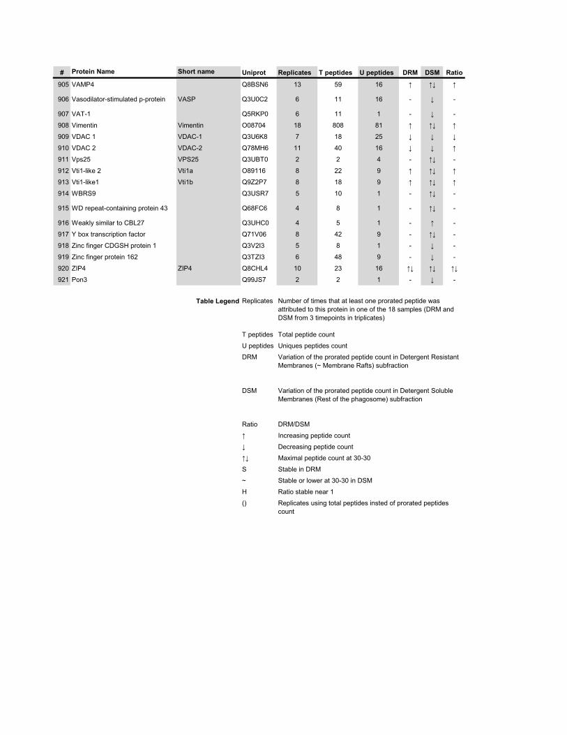
# Protein Name Short name Uniprot Replicates T peptides U peptides DRM DSM Ratio
905 VAMP4 Q8BSN6 13 59 16 � �� �
906 Vasodilator-stimulated p-protein VASP Q3U0C2 6 11 16 - � -
907 VAT-1 Q5RKP0 6 11 1 - � -
908 Vimentin Vimentin O08704 18 808 81 � �� �
909 VDAC 1 VDAC-1 Q3U6K8 7 18 25 � � �
910 VDAC 2 VDAC-2 Q78MH6 11 40 16 � � �
911 Vps25 VPS25 Q3UBT0 2 2 4 - �� -
912 Vti1-like 2 Vti1a O89116 8 22 9 � �� �
913 Vti1-like1 Vti1b Q9Z2P7 8 18 9 � �� �
914 WBRS9 Q3USR7 5 10 1 - �� -
915 WD repeat-containing protein 43 Q68FC6 4 8 1 - �� -
916 Weakly similar to CBL27 Q3UHC0 4 5 1 - � -
917 Y box transcription factor Q71V06 8 42 9 - �� -
918 Zinc finger CDGSH protein 1 Q3V2I3 5 8 1 - � -
919 Zinc finger protein 162 Q3TZI3 6 48 9 - � -
920 ZIP4 ZIP4 Q8CHL4 10 23 16 �� �� ��
921 Pon3 Q99JS7 2 2 1 - � -
Table Legend Replicates
T peptides
U peptides
DRM
DSM
Number of times that at least one prorated peptide was attributed to this protein in one of the 18 samples (DRM and DSM from 3 timepoints in triplicates)
Total peptide count
Uniques peptides count
Variation of the prorated peptide count in Detergent Resistant Membranes (~ Membrane Rafts) subfraction
Variation of the prorated peptide count in Detergent Soluble Membranes (Rest of the phagosome) subfraction
Ratio
�
�
��
S
~
H
() Replicates using total peptides insted of prorated peptides count
Increasing peptide count
Decreasing peptide count
Maximal peptide count at 30-30
Stable in DRM
Stable or lower at 30-30 in DSM
Ratio stable near 1
DRM/DSM
xvi
Supplementary Table 2. List of the 350 proteins present in phagosomal DRMs
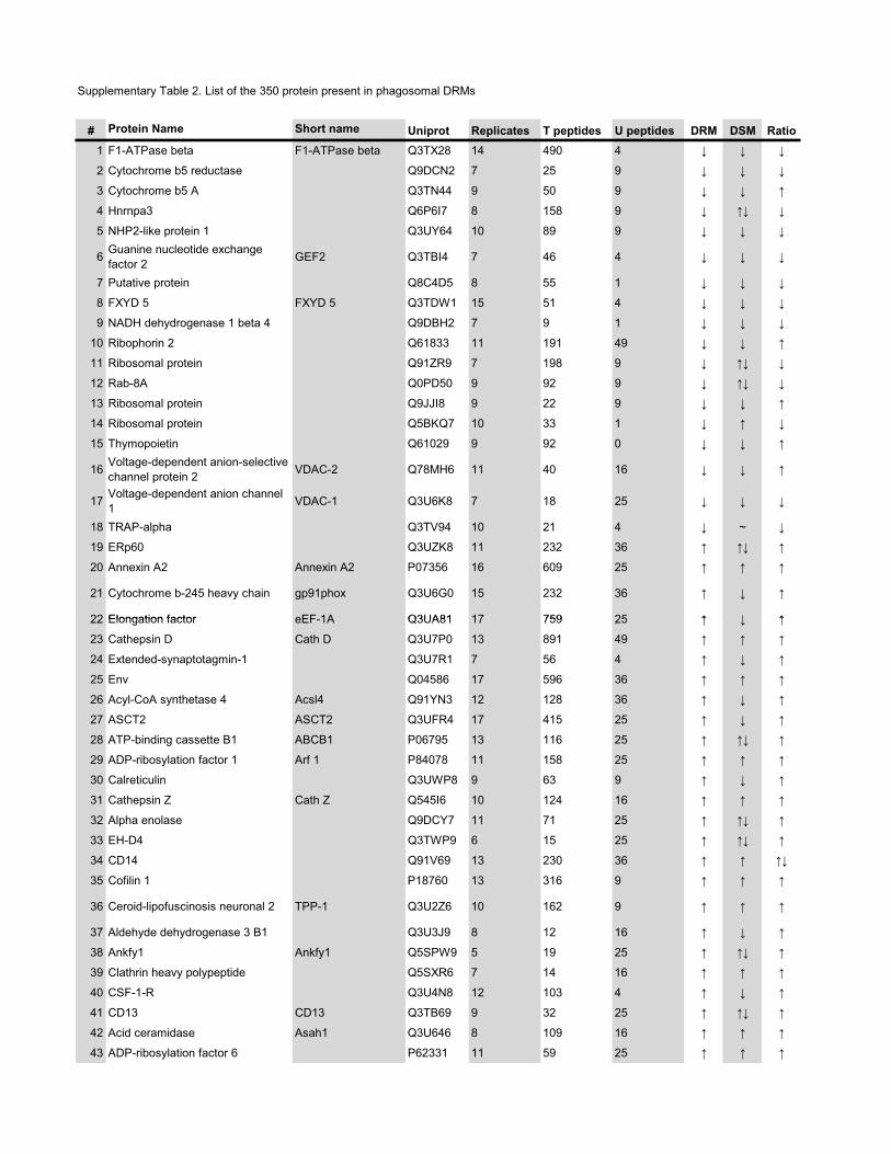
Supplementary Table 2. List of the 350 protein present in phagosomal DRMs
# Protein Name Short name Uniprot Replicates T peptides U peptides DRM DSM Ratio1 F1-ATPase beta F1-ATPase beta Q3TX28 14 490 4 � � �
2 Cytochrome b5 reductase Q9DCN2 7 25 9 � � �
3 Cytochrome b5 A Q3TN44 9 50 9 � � �
4 Hnrnpa3 Q6P6I7 8 158 9 � �� �
5 NHP2-like protein 1 Q3UY64 10 89 9 � � �
6 Guanine nucleotide exchange factor 2 GEF2 Q3TBI4 7 46 4 � � �
7 Putative protein Q8C4D5 8 55 1 � � �
8 FXYD 5 FXYD 5 Q3TDW1 15 51 4 � � �
9 NADH dehydrogenase 1 beta 4 Q9DBH2 7 9 1 � � �
10 Ribophorin 2 Q61833 11 191 49 � � �
11 Ribosomal protein Q91ZR9 7 198 9 � �� �
12 Rab-8A Q0PD50 9 92 9 � �� �
13 Ribosomal protein Q9JJI8 9 22 9 � � �
14 Ribosomal protein Q5BKQ7 10 33 1 � � �
15 Thymopoietin Q61029 9 92 0 � � �
16 Voltage-dependent anion-selective channel protein 2 VDAC-2 Q78MH6 11 40 16 � � �
17 Voltage-dependent anion channel 1 VDAC-1 Q3U6K8 7 18 25 � � �
18 TRAP-alpha Q3TV94 10 21 4 � ~ �
19 ERp60 Q3UZK8 11 232 36 � �� �
20 Annexin A2 Annexin A2 P07356 16 609 25 � � �
21 Cytochrome b-245 heavy chain gp91phox Q3U6G0 15 232 36 � � �
22 Elongation factor eEF 1A Q3UA81 17 759 25 � � �22 Elongation factor eEF-1A Q3UA81 17 759 25 � � �
23 Cathepsin D Cath D Q3U7P0 13 891 49 � � �
24 Extended-synaptotagmin-1 Q3U7R1 7 56 4 � � �
25 Env Q04586 17 596 36 � � �
26 Acyl-CoA synthetase 4 Acsl4 Q91YN3 12 128 36 � � �
27 ASCT2 ASCT2 Q3UFR4 17 415 25 � � �
28 ATP-binding cassette B1 ABCB1 P06795 13 116 25 � �� �
29 ADP-ribosylation factor 1 Arf 1 P84078 11 158 25 � � �
30 Calreticulin Q3UWP8 9 63 9 � � �
31 Cathepsin Z Cath Z Q545I6 10 124 16 � � �
32 Alpha enolase Q9DCY7 11 71 25 � �� �
33 EH-D4 Q3TWP9 6 15 25 � �� �
34 CD14 Q91V69 13 230 36 � � ��
35 Cofilin 1 P18760 13 316 9 � � �
36 Ceroid-lipofuscinosis neuronal 2 TPP-1 Q3U2Z6 10 162 9 � � �
37 Aldehyde dehydrogenase 3 B1 Q3U3J9 8 12 16 � � �
38 Ankfy1 Ankfy1 Q5SPW9 5 19 25 � �� �
39 Clathrin heavy polypeptide Q5SXR6 7 14 16 � � �
40 CSF-1-R Q3U4N8 12 103 4 � � �
41 CD13 CD13 Q3TB69 9 32 25 � �� �
42 Acid ceramidase Asah1 Q3U646 8 109 16 � � �
43 ADP-ribosylation factor 6 P62331 11 59 25 � � �
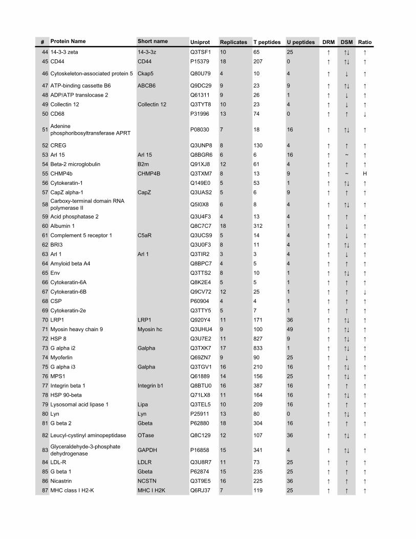
# Protein Name Short name Uniprot Replicates T peptides U peptides DRM DSM Ratio
44 14-3-3 zeta 14-3-3z Q3TSF1 10 65 25 � �� �
45 CD44 CD44 P15379 18 207 0 � �� �
46 Cytoskeleton-associated protein 5 Ckap5 Q80U79 4 10 4 � � �
47 ATP-binding cassette B6 ABCB6 Q9DC29 9 23 9 � �� �
48 ADP/ATP translocase 2 Q61311 9 26 1 � � �
49 Collectin 12 Collectin 12 Q3TYT8 10 23 4 � � �
50 CD68 P31996 13 74 0 � � �
51 Adenine phosphoribosyltransferase APRT P08030 7 18 16 � �� �
52 CREG Q3UNP8 8 130 4 � � �
53 Arl 15 Arl 15 Q8BGR6 6 6 16 � ~ �
54 Beta-2 microglobulin B2m Q91XJ8 12 61 4 � � �
55 CHMP4b CHMP4B Q3TXM7 8 13 9 � ~ H
56 Cytokeratin-1 Q149E0 5 53 1 � �� �
57 CapZ alpha-1 CapZ Q3UAS2 5 6 9 � � �
58 Carboxy-terminal domain RNA polymerase II Q5I0X8 6 8 4 � �� �
59 Acid phosphatase 2 Q3U4F3 4 13 4 � � �
60 Albumin 1 Q8C7C7 18 312 1 � � �
61 Complement 5 receptor 1 C5aR Q3UCS9 5 14 4 � � �
62 BRI3 Q3U0F3 8 11 4 � �� �
63 Arl 1 Arl 1 Q3TIR2 3 3 4 � � �
64 Amyloid beta A4 Q8BPC7 4 5 4 � � �
65 Env Q3TTS2 8 10 1 � �� �
66 Cytokeratin-6A Q8K2E4 5 5 1 � � �
67 Cytokeratin-6B Q9CV72 12 25 1 � � �
68 CSP P60904 4 4 1 � � �
69 Cytokeratin-2e Q3TTY5 5 7 1 � � �
70 LRP1 LRP1 Q920Y4 11 171 36 � �� �
71 Myosin heavy chain 9 Myosin hc Q3UHU4 9 100 49 � �� �
72 HSP 8 Q3U7E2 11 827 9 � �� �
73 G alpha i2 Galpha Q3TXK7 17 833 1 � �� �
74 Myoferlin Q69ZN7 9 90 25 � � �
75 G alpha i3 Galpha Q3TGV1 16 210 16 � �� �
76 MPS1 Q61889 14 156 25 � �� �
77 Integrin beta 1 Integrin b1 Q8BTU0 16 387 16 � � �
78 HSP 90-beta Q71LX8 11 164 16 � �� �
79 Lysosomal acid lipase 1 Lipa Q3TEL5 10 209 16 � � �
80 Lyn Lyn P25911 13 80 0 � �� �
81 G beta 2 Gbeta P62880 18 304 16 � � �
82 Leucyl-cystinyl aminopeptidase OTase Q8C129 12 107 36 � �� �
83 Glyceraldehyde-3-phosphate dehydrogenase GAPDH P16858 15 341 4 � �� �
84 LDL-R LDLR Q3U8R7 11 73 25 � � �
85 G beta 1 Gbeta P62874 15 235 25 � � �
86 Nicastrin NCSTN Q3T9E5 16 225 36 � � �
87 MHC class I H2-K MHC I H2K Q6RJ37 7 119 25 � � �
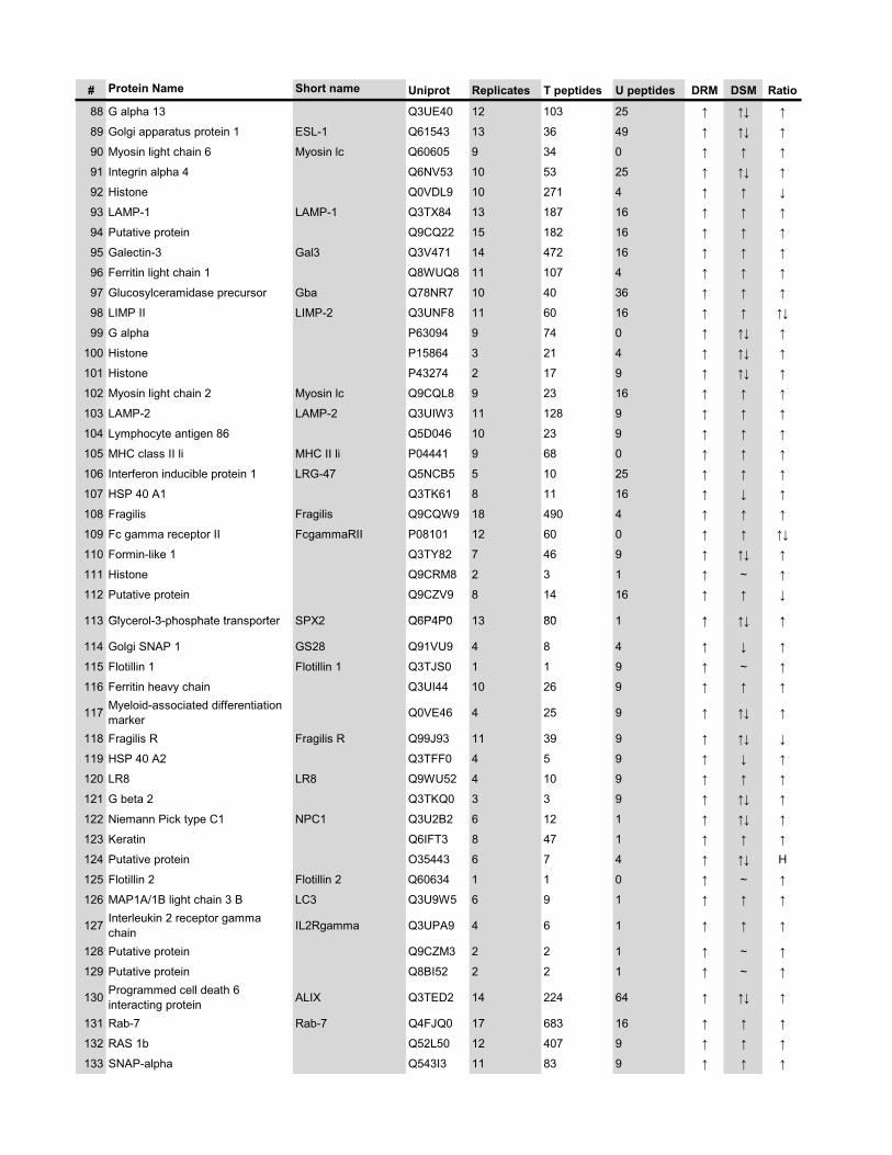
# Protein Name Short name Uniprot Replicates T peptides U peptides DRM DSM Ratio
88 G alpha 13 Q3UE40 12 103 25 � �� �
89 Golgi apparatus protein 1 ESL-1 Q61543 13 36 49 � �� �
90 Myosin light chain 6 Myosin lc Q60605 9 34 0 � � �
91 Integrin alpha 4 Q6NV53 10 53 25 � �� �
92 Histone Q0VDL9 10 271 4 � � �
93 LAMP-1 LAMP-1 Q3TX84 13 187 16 � � �
94 Putative protein Q9CQ22 15 182 16 � � �
95 Galectin-3 Gal3 Q3V471 14 472 16 � � �
96 Ferritin light chain 1 Q8WUQ8 11 107 4 � � �
97 Glucosylceramidase precursor Gba Q78NR7 10 40 36 � � �
98 LIMP II LIMP-2 Q3UNF8 11 60 16 � � ��
99 G alpha P63094 9 74 0 � �� �
100 Histone P15864 3 21 4 � �� �
101 Histone P43274 2 17 9 � �� �
102 Myosin light chain 2 Myosin lc Q9CQL8 9 23 16 � � �
103 LAMP-2 LAMP-2 Q3UIW3 11 128 9 � � �
104 Lymphocyte antigen 86 Q5D046 10 23 9 � � �
105 MHC class II li MHC II li P04441 9 68 0 � � �
106 Interferon inducible protein 1 LRG-47 Q5NCB5 5 10 25 � � �
107 HSP 40 A1 Q3TK61 8 11 16 � � �
108 Fragilis Fragilis Q9CQW9 18 490 4 � � �
109 Fc gamma receptor II FcgammaRII P08101 12 60 0 � � ��
110 Formin-like 1 Q3TY82 7 46 9 � �� �
111 Histone Q9CRM8 2 3 1 � ~ �
112 Putative protein Q9CZV9 8 14 16 � � �
113 Glycerol 3 phosphate transporter SPX2 Q6P4P0 13 80 1 � �� �113 Glycerol-3-phosphate transporter SPX2 Q6P4P0 13 80 1 � �� �
114 Golgi SNAP 1 GS28 Q91VU9 4 8 4 � � �
115 Flotillin 1 Flotillin 1 Q3TJS0 1 1 9 � ~ �
116 Ferritin heavy chain Q3UI44 10 26 9 � � �
117 Myeloid-associated differentiation marker Q0VE46 4 25 9 � �� �
118 Fragilis R Fragilis R Q99J93 11 39 9 � �� �
119 HSP 40 A2 Q3TFF0 4 5 9 � � �
120 LR8 LR8 Q9WU52 4 10 9 � � �
121 G beta 2 Q3TKQ0 3 3 9 � �� �
122 Niemann Pick type C1 NPC1 Q3U2B2 6 12 1 � �� �
123 Keratin Q6IFT3 8 47 1 � � �
124 Putative protein O35443 6 7 4 � �� H
125 Flotillin 2 Flotillin 2 Q60634 1 1 0 � ~ �
126 MAP1A/1B light chain 3 B LC3 Q3U9W5 6 9 1 � � �
127 Interleukin 2 receptor gamma chain IL2Rgamma Q3UPA9 4 6 1 � � �
128 Putative protein Q9CZM3 2 2 1 � ~ �
129 Putative protein Q8BI52 2 2 1 � ~ �
130 Programmed cell death 6 interacting protein ALIX Q3TED2 14 224 64 � �� �
131 Rab-7 Rab-7 Q4FJQ0 17 683 16 � � �
132 RAS 1b Q52L50 12 407 9 � � �
133 SNAP-alpha Q543I3 11 83 9 � � �
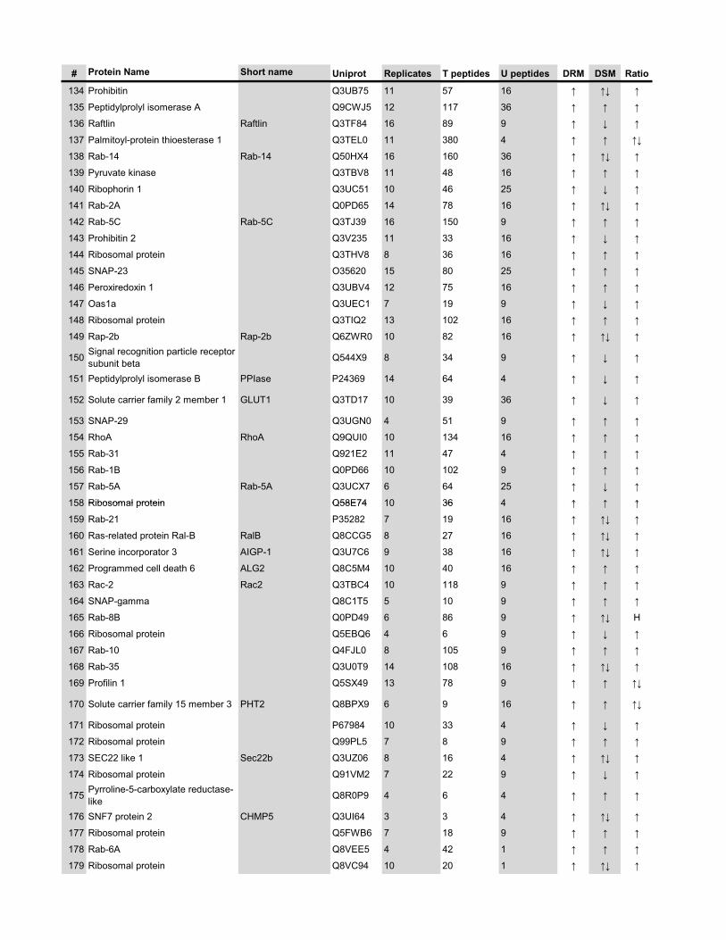
# Protein Name Short name Uniprot Replicates T peptides U peptides DRM DSM Ratio
134 Prohibitin Q3UB75 11 57 16 � �� �
135 Peptidylprolyl isomerase A Q9CWJ5 12 117 36 � � �
136 Raftlin Raftlin Q3TF84 16 89 9 � � �
137 Palmitoyl-protein thioesterase 1 Q3TEL0 11 380 4 � � ��
138 Rab-14 Rab-14 Q50HX4 16 160 36 � �� �
139 Pyruvate kinase Q3TBV8 11 48 16 � � �
140 Ribophorin 1 Q3UC51 10 46 25 � � �
141 Rab-2A Q0PD65 14 78 16 � �� �
142 Rab-5C Rab-5C Q3TJ39 16 150 9 � � �
143 Prohibitin 2 Q3V235 11 33 16 � � �
144 Ribosomal protein Q3THV8 8 36 16 � � �
145 SNAP-23 O35620 15 80 25 � � �
146 Peroxiredoxin 1 Q3UBV4 12 75 16 � � �
147 Oas1a Q3UEC1 7 19 9 � � �
148 Ribosomal protein Q3TIQ2 13 102 16 � � �
149 Rap-2b Rap-2b Q6ZWR0 10 82 16 � �� �
150 Signal recognition particle receptor subunit beta Q544X9 8 34 9 � � �
151 Peptidylprolyl isomerase B PPIase P24369 14 64 4 � � �
152 Solute carrier family 2 member 1 GLUT1 Q3TD17 10 39 36 � � �
153 SNAP-29 Q3UGN0 4 51 9 � � �
154 RhoA RhoA Q9QUI0 10 134 16 � � �
155 Rab-31 Q921E2 11 47 4 � � �
156 Rab-1B Q0PD66 10 102 9 � � �
157 Rab-5A Rab-5A Q3UCX7 6 64 25 � � �
158 Ribosomal protein Q58E74 10 36 4 � � �158 Ribosomal protein Q58E74 10 36 4 � � �
159 Rab-21 P35282 7 19 16 � �� �
160 Ras-related protein Ral-B RalB Q8CCG5 8 27 16 � �� �
161 Serine incorporator 3 AIGP-1 Q3U7C6 9 38 16 � �� �
162 Programmed cell death 6 ALG2 Q8C5M4 10 40 16 � � �
163 Rac-2 Rac2 Q3TBC4 10 118 9 � � �
164 SNAP-gamma Q8C1T5 5 10 9 � � �
165 Rab-8B Q0PD49 6 86 9 � �� H
166 Ribosomal protein Q5EBQ6 4 6 9 � � �
167 Rab-10 Q4FJL0 8 105 9 � � �
168 Rab-35 Q3U0T9 14 108 16 � �� �
169 Profilin 1 Q5SX49 13 78 9 � � ��
170 Solute carrier family 15 member 3 PHT2 Q8BPX9 6 9 16 � � ��
171 Ribosomal protein P67984 10 33 4 � � �
172 Ribosomal protein Q99PL5 7 8 9 � � �
173 SEC22 like 1 Sec22b Q3UZ06 8 16 4 � �� �
174 Ribosomal protein Q91VM2 7 22 9 � � �
175 Pyrroline-5-carboxylate reductase-like Q8R0P9 4 6 4 � � �
176 SNF7 protein 2 CHMP5 Q3UI64 3 3 4 � �� �
177 Ribosomal protein Q5FWB6 7 18 9 � � �
178 Rab-6A Q8VEE5 4 42 1 � � �
179 Ribosomal protein Q8VC94 10 20 1 � �� �
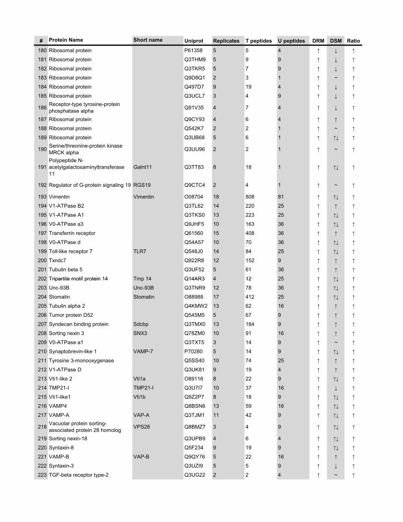
# Protein Name Short name Uniprot Replicates T peptides U peptides DRM DSM Ratio
180 Ribosomal protein P61358 5 5 4 � � �
181 Ribosomal protein Q3THM9 5 9 9 � � �
182 Ribosomal protein Q3TKR5 5 7 9 � � �
183 Ribosomal protein Q9D8Q1 2 3 1 � ~ �
184 Ribosomal protein Q497D7 9 19 4 � � �
185 Ribosomal protein Q3UCL7 3 4 9 � � �
186 Receptor-type tyrosine-protein phosphatase alpha Q91V35 4 7 4 � � �
187 Ribosomal protein Q9CY93 4 6 4 � � �
188 Ribosomal protein Q542K7 2 2 1 � ~ �
189 Ribosomal protein Q3UB68 5 6 1 � �� �
190 Serine/threonine-protein kinase MRCK alpha Q3UU96 2 2 1 � ~ �
191Polypeptide N-acetylgalactosaminyltransferase 11
Galnt11 Q3TT83 8 18 1 � �� �
192 Regulator of G-protein signaling 19 RGS19 Q9CTC4 2 4 1 � ~ �
193 Vimentin Vimentin O08704 18 808 81 � �� �
194 V1-ATPase B2 Q3TL62 14 220 25 � � �
195 V1-ATPase A1 Q3TKS0 13 223 25 � �� �
196 V0-ATPase a3 Q9JHF5 10 163 36 � �� �
197 Transferrin receptor Q61560 15 408 36 � � �
198 V0-ATPase d Q54A57 10 70 36 � �� �
199 Toll-like receptor 7 TLR7 Q548J0 14 84 25 � �� �
200 Txndc7 Q922R8 12 152 9 � � �
201 Tubulin beta 5 Q3UF52 5 61 36 � � �
202 Tripartite motif protein 14 Tmp 14 Q14AR3 4 12 25 � �� �202 Tripartite motif protein 14 Tmp 14 Q14AR3 4 12 25 � �� �
203 Unc-93B Unc-93B Q3TNR9 12 78 36 � �� �
204 Stomatin Stomatin O88988 17 412 25 � �� �
205 Tubulin alpha 2 Q4KMW2 13 62 16 � � �
206 Tumor protein D52 Q545M5 5 67 9 � � �
207 Syndecan binding protein Sdcbp Q3TMX0 13 184 9 � � �
208 Sorting nexin 3 SNX3 Q78ZM0 10 91 16 � � �
209 V0-ATPase a1 Q3TXT5 3 14 9 � ~ �
210 Synaptobrevin-like 1 VAMP-7 P70280 5 14 9 � �� �
211 Tyrosine 3-monooxygenase Q5SS40 10 74 25 � � �
212 V1-ATPase D Q3UK81 9 19 4 � � �
213 Vti1-like 2 Vti1a O89116 8 22 9 � �� �
214 TMP21-I TMP21-I Q3U7I7 10 37 16 � � �
215 Vti1-like1 Vti1b Q9Z2P7 8 18 9 � �� �
216 VAMP4 Q8BSN6 13 59 16 � �� �
217 VAMP-A VAP-A Q3TJM1 11 42 9 � �� �
218 Vacuolar protein sorting-associated protein 28 homolog VPS28 Q8BMZ7 3 4 9 � �� �
219 Sorting nexin-18 Q3UPB9 4 6 4 � �� �
220 Syntaxin-8 Q5F234 9 19 9 � �� �
221 VAMP-B VAP-B Q9QY76 5 22 16 � � �
222 Syntaxin-3 Q3UZI9 5 5 9 � � �
223 TGF-beta receptor type-2 Q3UG22 2 2 4 � ~ �
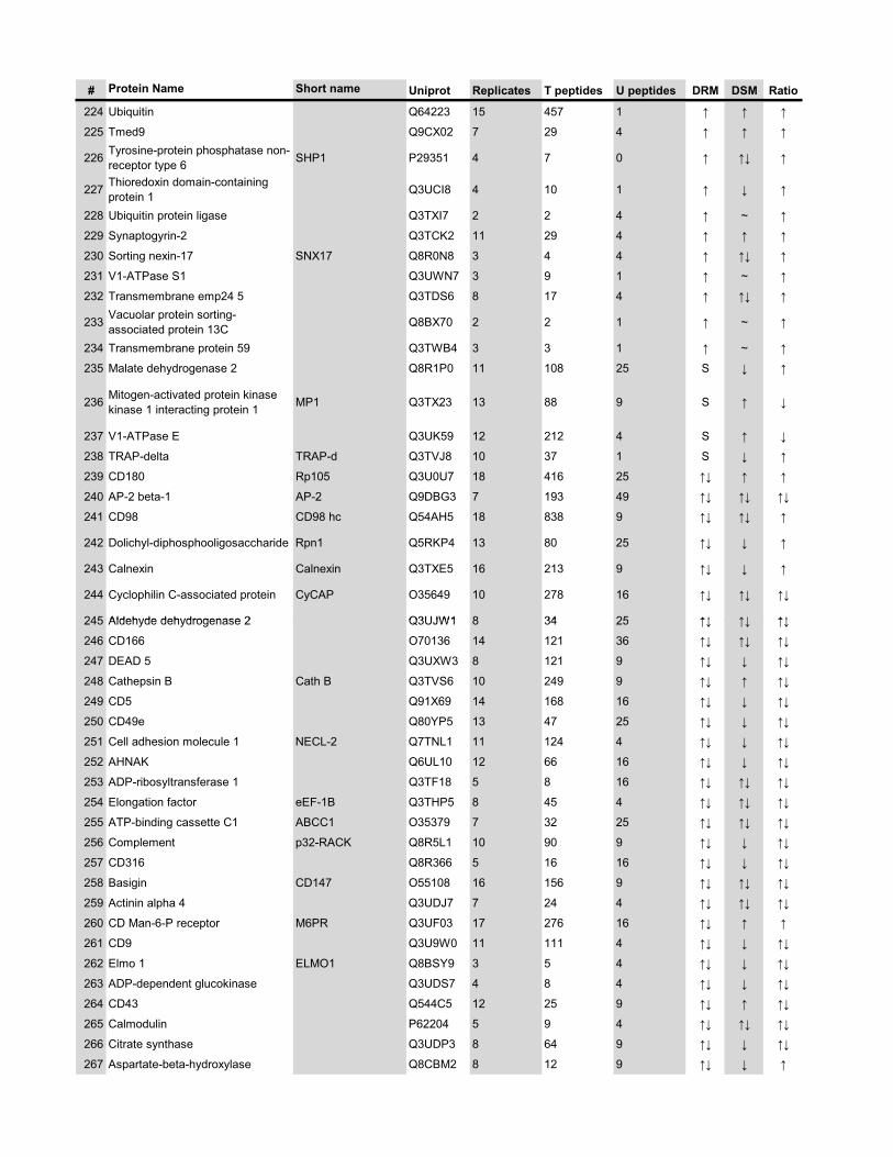
# Protein Name Short name Uniprot Replicates T peptides U peptides DRM DSM Ratio
224 Ubiquitin Q64223 15 457 1 � � �
225 Tmed9 Q9CX02 7 29 4 � � �
226 Tyrosine-protein phosphatase non-receptor type 6 SHP1 P29351 4 7 0 � �� �
227 Thioredoxin domain-containing protein 1 Q3UCI8 4 10 1 � � �
228 Ubiquitin protein ligase Q3TXI7 2 2 4 � ~ �
229 Synaptogyrin-2 Q3TCK2 11 29 4 � � �
230 Sorting nexin-17 SNX17 Q8R0N8 3 4 4 � �� �
231 V1-ATPase S1 Q3UWN7 3 9 1 � ~ �
232 Transmembrane emp24 5 Q3TDS6 8 17 4 � �� �
233 Vacuolar protein sorting-associated protein 13C Q8BX70 2 2 1 � ~ �
234 Transmembrane protein 59 Q3TWB4 3 3 1 � ~ �
235 Malate dehydrogenase 2 Q8R1P0 11 108 25 S � �
236 Mitogen-activated protein kinase kinase 1 interacting protein 1 MP1 Q3TX23 13 88 9 S � �
237 V1-ATPase E Q3UK59 12 212 4 S � �
238 TRAP-delta TRAP-d Q3TVJ8 10 37 1 S � �
239 CD180 Rp105 Q3U0U7 18 416 25 �� � �
240 AP-2 beta-1 AP-2 Q9DBG3 7 193 49 �� �� ��
241 CD98 CD98 hc Q54AH5 18 838 9 �� �� �
242 Dolichyl-diphosphooligosaccharide Rpn1 Q5RKP4 13 80 25 �� � �
243 Calnexin Calnexin Q3TXE5 16 213 9 �� � �
244 Cyclophilin C-associated protein CyCAP O35649 10 278 16 �� �� ��
245 Aldehyde dehydrogenase 2 Q3UJW1 8 34 25 �� �� ��245 Aldehyde dehydrogenase 2 Q3UJW1 8 34 25 �� �� ��
246 CD166 O70136 14 121 36 �� �� ��
247 DEAD 5 Q3UXW3 8 121 9 �� � ��
248 Cathepsin B Cath B Q3TVS6 10 249 9 �� � ��
249 CD5 Q91X69 14 168 16 �� � ��
250 CD49e Q80YP5 13 47 25 �� � ��
251 Cell adhesion molecule 1 NECL-2 Q7TNL1 11 124 4 �� � ��
252 AHNAK Q6UL10 12 66 16 �� � ��
253 ADP-ribosyltransferase 1 Q3TF18 5 8 16 �� �� ��
254 Elongation factor eEF-1B Q3THP5 8 45 4 �� �� ��
255 ATP-binding cassette C1 ABCC1 O35379 7 32 25 �� �� ��
256 Complement p32-RACK Q8R5L1 10 90 9 �� � ��
257 CD316 Q8R366 5 16 16 �� � ��
258 Basigin CD147 O55108 16 156 9 �� �� ��
259 Actinin alpha 4 Q3UDJ7 7 24 4 �� �� ��
260 CD Man-6-P receptor M6PR Q3UF03 17 276 16 �� � �
261 CD9 Q3U9W0 11 111 4 �� � ��
262 Elmo 1 ELMO1 Q8BSY9 3 5 4 �� � ��
263 ADP-dependent glucokinase Q3UDS7 4 8 4 �� � ��
264 CD43 Q544C5 12 25 9 �� � ��
265 Calmodulin P62204 5 9 4 �� �� ��
266 Citrate synthase Q3UDP3 8 64 9 �� � ��
267 Aspartate-beta-hydroxylase Q8CBM2 8 12 9 �� � �
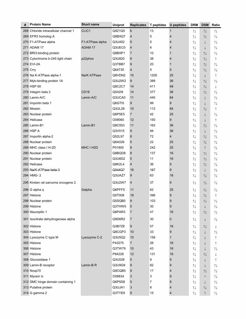
# Protein Name Short name Uniprot Replicates T peptides U peptides DRM DSM Ratio
268 Chloride intracellular channel 1 CLIC1 Q9Z1Q5 6 13 1 �� �� ��
269 EFR3 homolog A Q8BXQ7 4 5 4 �� �� ��
270 F1-ATPase alpha F1-ATPase alpha Q3U452 5 5 4 �� � ��
271 ADAM 17 ADAM 17 Q3UEC0 4 6 4 �� � ��
272 BRI3-binding protein Q8BXP1 7 10 1 �� �� ��
273 Cytochrome b-245 light chain p22phox Q3U820 9 38 4 �� �� �
274 EVI-2A Q3TBB7 9 20 1 �� �� ��
275 Crry Q64735 4 5 0 �� �� ��
276 Na K-ATPase alpha-1 Na/K ATPase Q8VDN2 15 1205 25 �� � �
277 Myb-binding protein 1A Q3U2W2 9 389 36 �� �� ��
278 HSP 60 Q8C2C7 14 411 49 �� �� �
279 Integrin beta 2 CD18 Q542I8 14 377 36 �� �� ��
280 Lamin-A/C Lamin-A/C Q3UCA0 11 440 64 �� � ��
281 Importin beta 1 Q6GTI5 9 99 9 �� � ��
282 Moesin Q3UL28 10 112 64 �� �� �
283 Nuclear protein Q6P5E5 7 42 25 �� � ��
284 Helicase O09060 12 150 9 �� � �
285 Lamin-B1 Lamin-B1 Q8C553 11 163 36 �� �� ��
286 HSP A Q3V015 9 94 36 �� � ��
287 Importin alpha-2 Q52L97 8 72 4 �� �� ��
288 Nuclear protein Q6A026 5 22 25 �� �� ��
289 MHC class I H-2D MHC I H2D P01900 9 242 25 �� � ��
290 Nuclear protein Q8BQD8 8 137 16 �� �� ��
291 Nuclear protein Q3U6D2 5 11 16 �� �� ��
292 Helicase Q8K2L4 4 36 9 �� �� ��
293 Na/K-ATPase beta-3 Q544Q7 16 167 9 �� � ��293 Na/K ATPase beta 3 Q544Q7 16 167 9 �� � ��
294 HMG- 2 Q3UAZ7 9 63 16 �� �� ��
295 Kirsten rat sarcoma oncogene 2 Q3U2W7 4 37 9 �� �� ��
296 G alpha q Galpha Q6PFF5 11 62 25 �� �� ��
297 Histone Q5T008 18 396 9 �� �� �
298 Nuclear protein Q5SQB0 9 103 9 �� �� ��
299 Histone Q3THW5 5 30 9 �� � ��
300 Neuropilin 1 Q6PAR3 7 47 16 �� �� ��
301 Isocitrate dehydrogenase alpha Q9D6R2 7 30 0 �� � ��
302 Histone Q3B7Z8 9 57 16 �� �� �
303 Histone Q8CGP3 10 33 9 �� � ��
304 Lysozyme C type M Lysozyme C-2 Q3U5Q2 15 158 1 �� � �
305 Histone P43275 7 26 16 �� � �
306 Histone Q3TW79 10 43 16 �� � ��
307 Histone P84228 12 131 16 �� �� �
308 Glucosidase 1 Q3U026 5 9 9 �� � �
309 Lamin-B receptor Lamin-B R Q3U9G9 9 82 9 �� � ��
310 Noxp70 Q9CQB5 9 17 4 �� �� ��
311 Myosin Ic O08834 3 3 9 �� ~ ��
312 SMC hinge domain containing 1 Q6P5D8 5 7 9 �� � ��
313 Putative protein Q3UJA1 3 8 4 �� �� ��
314 G gamma 2 Q3TYE8 9 15 4 �� � ��
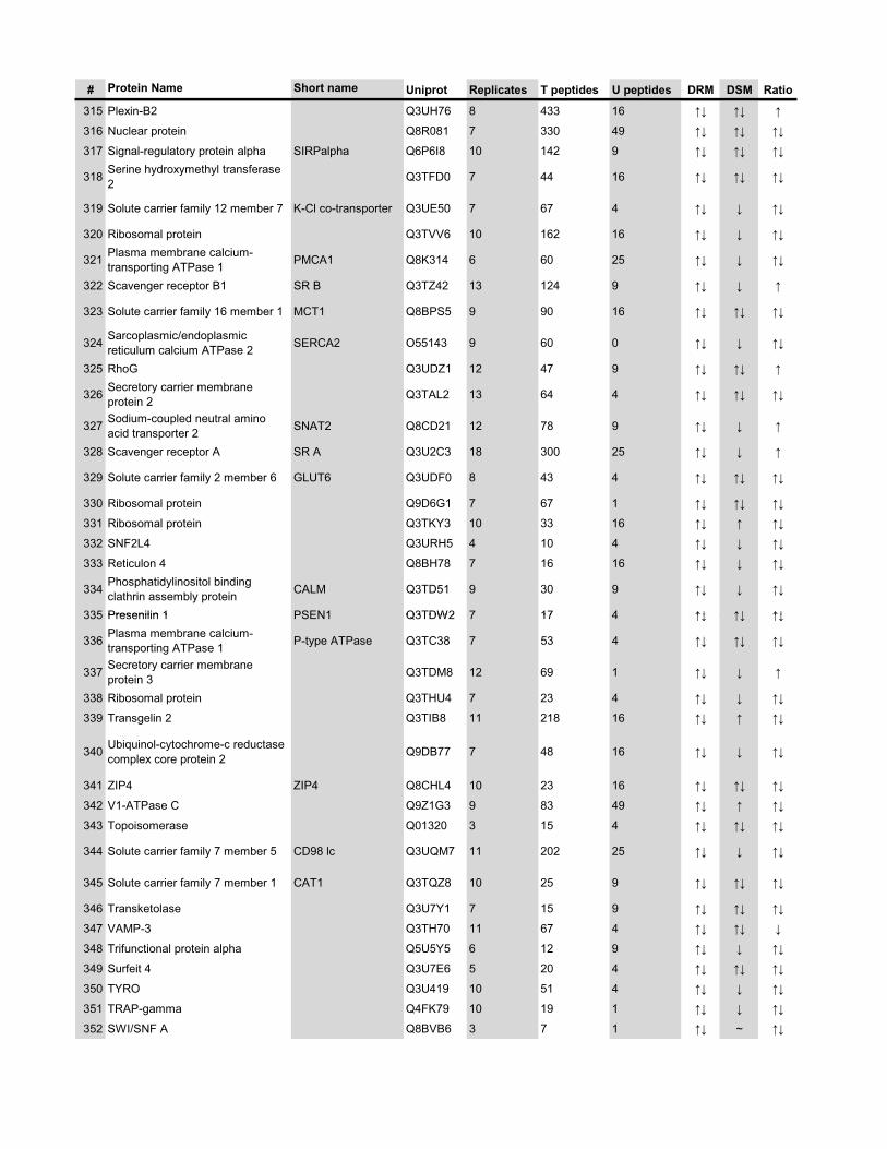
# Protein Name Short name Uniprot Replicates T peptides U peptides DRM DSM Ratio
315 Plexin-B2 Q3UH76 8 433 16 �� �� �
316 Nuclear protein Q8R081 7 330 49 �� �� ��
317 Signal-regulatory protein alpha SIRPalpha Q6P6I8 10 142 9 �� �� ��
318 Serine hydroxymethyl transferase 2 Q3TFD0 7 44 16 �� �� ��
319 Solute carrier family 12 member 7 K-Cl co-transporter Q3UE50 7 67 4 �� � ��
320 Ribosomal protein Q3TVV6 10 162 16 �� � ��
321 Plasma membrane calcium-transporting ATPase 1 PMCA1 Q8K314 6 60 25 �� � ��
322 Scavenger receptor B1 SR B Q3TZ42 13 124 9 �� � �
323 Solute carrier family 16 member 1 MCT1 Q8BPS5 9 90 16 �� �� ��
324 Sarcoplasmic/endoplasmic reticulum calcium ATPase 2 SERCA2 O55143 9 60 0 �� � ��
325 RhoG Q3UDZ1 12 47 9 �� �� �
326 Secretory carrier membrane protein 2 Q3TAL2 13 64 4 �� �� ��
327 Sodium-coupled neutral amino acid transporter 2 SNAT2 Q8CD21 12 78 9 �� � �
328 Scavenger receptor A SR A Q3U2C3 18 300 25 �� � �
329 Solute carrier family 2 member 6 GLUT6 Q3UDF0 8 43 4 �� �� ��
330 Ribosomal protein Q9D6G1 7 67 1 �� �� ��
331 Ribosomal protein Q3TKY3 10 33 16 �� � ��
332 SNF2L4 Q3URH5 4 10 4 �� � ��
333 Reticulon 4 Q8BH78 7 16 16 �� � ��
334 Phosphatidylinositol binding clathrin assembly protein CALM Q3TD51 9 30 9 �� � ��
335 Presenilin 1 PSEN1 Q3TDW2 7 17 4 �� �� ��335 Presenilin 1 PSEN1 Q3TDW2 7 17 4 �� �� ��
336 Plasma membrane calcium-transporting ATPase 1 P-type ATPase Q3TC38 7 53 4 �� �� ��
337 Secretory carrier membrane protein 3 Q3TDM8 12 69 1 �� � �
338 Ribosomal protein Q3THU4 7 23 4 �� � ��
339 Transgelin 2 Q3TIB8 11 218 16 �� � ��
340 Ubiquinol-cytochrome-c reductase complex core protein 2 Q9DB77 7 48 16 �� � ��
341 ZIP4 ZIP4 Q8CHL4 10 23 16 �� �� ��
342 V1-ATPase C Q9Z1G3 9 83 49 �� � ��
343 Topoisomerase Q01320 3 15 4 �� �� ��
344 Solute carrier family 7 member 5 CD98 lc Q3UQM7 11 202 25 �� � ��
345 Solute carrier family 7 member 1 CAT1 Q3TQZ8 10 25 9 �� �� ��
346 Transketolase Q3U7Y1 7 15 9 �� �� ��
347 VAMP-3 Q3TH70 11 67 4 �� �� �
348 Trifunctional protein alpha Q5U5Y5 6 12 9 �� � ��
349 Surfeit 4 Q3U7E6 5 20 4 �� �� ��
350 TYRO Q3U419 10 51 4 �� � ��
351 TRAP-gamma Q4FK79 10 19 1 �� � ��
352 SWI/SNF A Q8BVB6 3 7 1 �� ~ ��
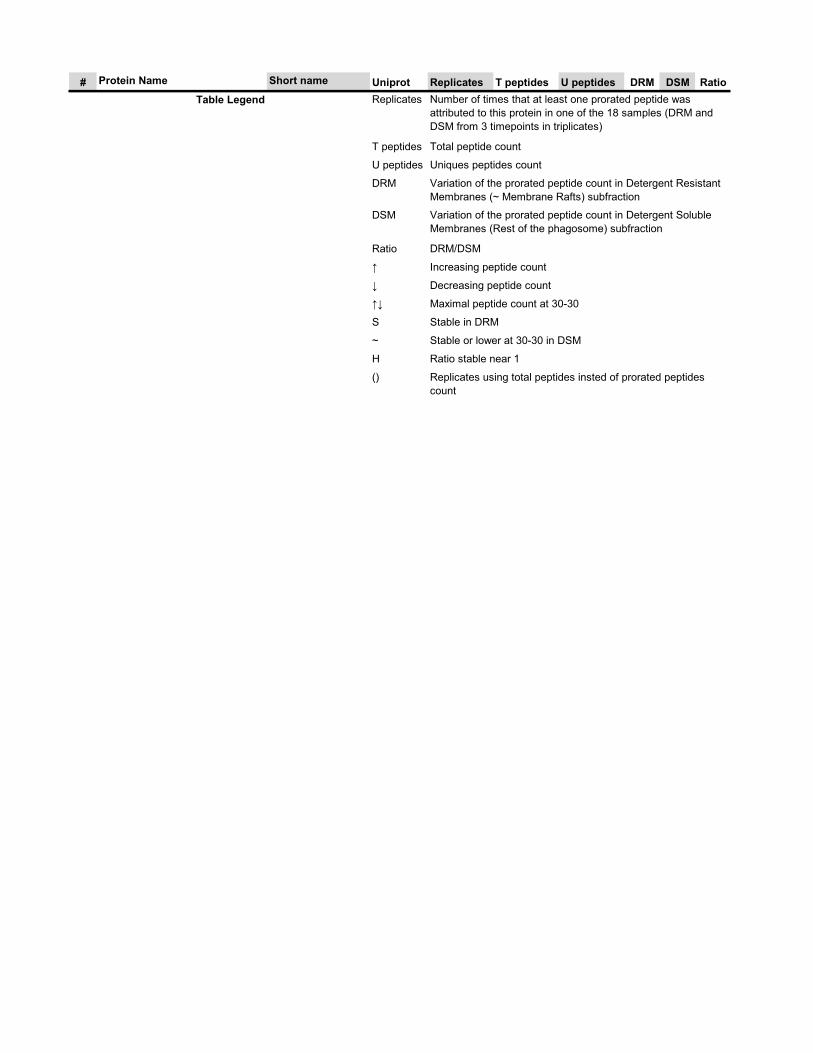
# Protein Name Short name Uniprot Replicates T peptides U peptides DRM DSM RatioTable Legend Replicates
T peptides
U peptides
DRM
DSM
Ratio
�
�
��
S
~
H
() Replicates using total peptides insted of prorated peptides count
Increasing peptide count
Decreasing peptide count
Maximal peptide count at 30-30
Stable in DRM
Stable or lower at 30-30 in DSM
Ratio stable near 1
DRM/DSM
Number of times that at least one prorated peptide was attributed to this protein in one of the 18 samples (DRM and DSM from 3 timepoints in triplicates)
Total peptide count
Uniques peptides count
Variation of the prorated peptide count in Detergent Resistant Membranes (~ Membrane Rafts) subfraction
Variation of the prorated peptide count in Detergent Soluble Membranes (Rest of the phagosome) subfraction

xvii
Supplementary Table 3 Protein-protein interactions from PCA
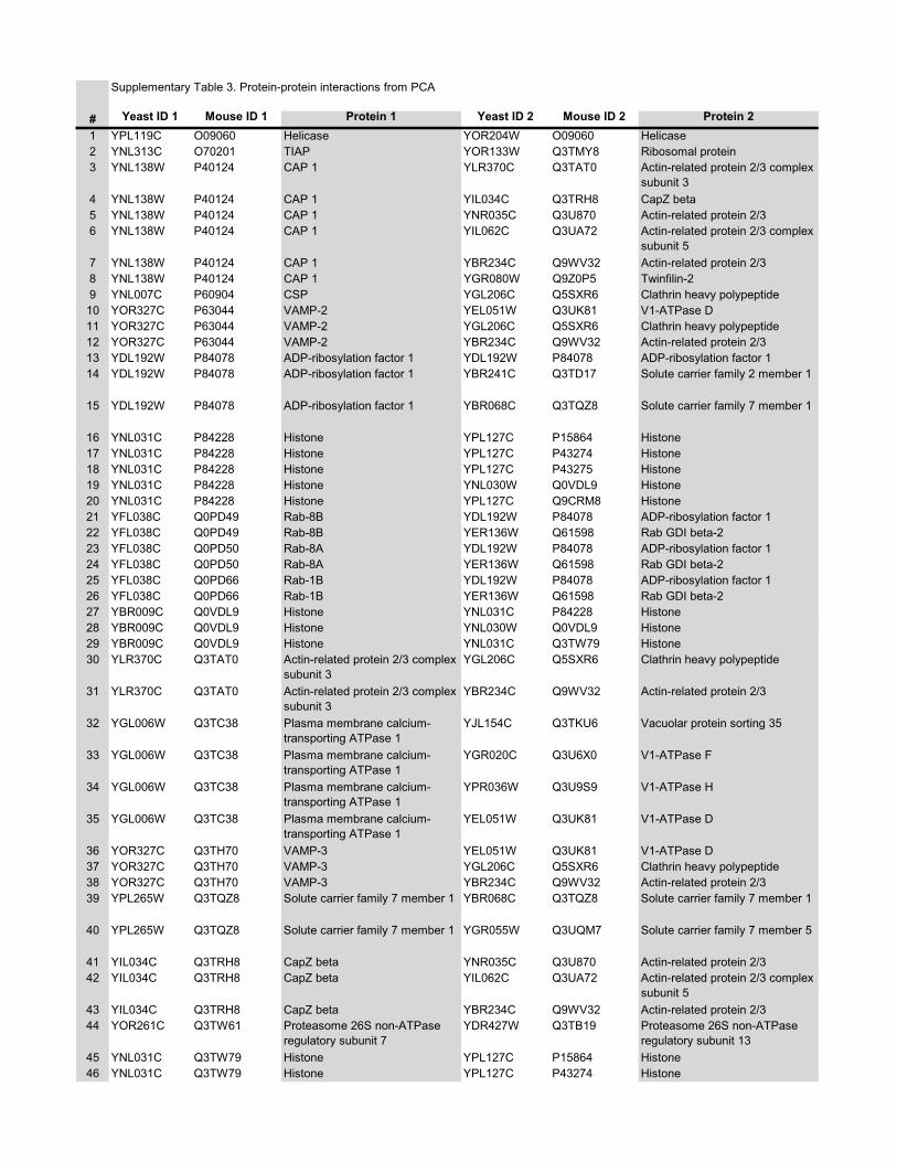
Supplementary Table 3. Protein-protein interactions from PCA
# Yeast ID 1 Mouse ID 1 Protein 1 Yeast ID 2 Mouse ID 2 Protein 21 YPL119C O09060 Helicase YOR204W O09060 Helicase2 YNL313C O70201 TIAP YOR133W Q3TMY8 Ribosomal protein3 YNL138W P40124 CAP 1 YLR370C Q3TAT0 Actin-related protein 2/3 complex
subunit 34 YNL138W P40124 CAP 1 YIL034C Q3TRH8 CapZ beta5 YNL138W P40124 CAP 1 YNR035C Q3U870 Actin-related protein 2/36 YNL138W P40124 CAP 1 YIL062C Q3UA72 Actin-related protein 2/3 complex
subunit 57 YNL138W P40124 CAP 1 YBR234C Q9WV32 Actin-related protein 2/38 YNL138W P40124 CAP 1 YGR080W Q9Z0P5 Twinfilin-29 YNL007C P60904 CSP YGL206C Q5SXR6 Clathrin heavy polypeptide10 YOR327C P63044 VAMP-2 YEL051W Q3UK81 V1-ATPase D11 YOR327C P63044 VAMP-2 YGL206C Q5SXR6 Clathrin heavy polypeptide12 YOR327C P63044 VAMP-2 YBR234C Q9WV32 Actin-related protein 2/313 YDL192W P84078 ADP-ribosylation factor 1 YDL192W P84078 ADP-ribosylation factor 114 YDL192W P84078 ADP-ribosylation factor 1 YBR241C Q3TD17 Solute carrier family 2 member 1
15 YDL192W P84078 ADP-ribosylation factor 1 YBR068C Q3TQZ8 Solute carrier family 7 member 1
16 YNL031C P84228 Histone YPL127C P15864 Histone17 YNL031C P84228 Histone YPL127C P43274 Histone18 YNL031C P84228 Histone YPL127C P43275 Histone19 YNL031C P84228 Histone YNL030W Q0VDL9 Histone20 YNL031C P84228 Histone YPL127C Q9CRM8 Histone21 YFL038C Q0PD49 Rab-8B YDL192W P84078 ADP-ribosylation factor 122 YFL038C Q0PD49 Rab-8B YER136W Q61598 Rab GDI beta-223 YFL038C Q0PD50 Rab-8A YDL192W P84078 ADP-ribosylation factor 124 YFL038C Q0PD50 Rab-8A YER136W Q61598 Rab GDI beta-225 YFL038C Q0PD66 Rab-1B YDL192W P84078 ADP-ribosylation factor 126 YFL038C Q0PD66 Rab-1B YER136W Q61598 Rab GDI beta-227 YBR009C Q0VDL9 Histone YNL031C P84228 Histone28 009C Q0 9 030 Q0 928 YBR009C Q0VDL9 Histone YNL030W Q0VDL9 Histone29 YBR009C Q0VDL9 Histone YNL031C Q3TW79 Histone30 YLR370C Q3TAT0 Actin-related protein 2/3 complex
subunit 3YGL206C Q5SXR6 Clathrin heavy polypeptide
31 YLR370C Q3TAT0 Actin-related protein 2/3 complex subunit 3
YBR234C Q9WV32 Actin-related protein 2/3
32 YGL006W Q3TC38 Plasma membrane calcium-transporting ATPase 1
YJL154C Q3TKU6 Vacuolar protein sorting 35
33 YGL006W Q3TC38 Plasma membrane calcium-transporting ATPase 1
YGR020C Q3U6X0 V1-ATPase F
34 YGL006W Q3TC38 Plasma membrane calcium-transporting ATPase 1
YPR036W Q3U9S9 V1-ATPase H
35 YGL006W Q3TC38 Plasma membrane calcium-transporting ATPase 1
YEL051W Q3UK81 V1-ATPase D
36 YOR327C Q3TH70 VAMP-3 YEL051W Q3UK81 V1-ATPase D37 YOR327C Q3TH70 VAMP-3 YGL206C Q5SXR6 Clathrin heavy polypeptide38 YOR327C Q3TH70 VAMP-3 YBR234C Q9WV32 Actin-related protein 2/339 YPL265W Q3TQZ8 Solute carrier family 7 member 1 YBR068C Q3TQZ8 Solute carrier family 7 member 1
40 YPL265W Q3TQZ8 Solute carrier family 7 member 1 YGR055W Q3UQM7 Solute carrier family 7 member 5
41 YIL034C Q3TRH8 CapZ beta YNR035C Q3U870 Actin-related protein 2/342 YIL034C Q3TRH8 CapZ beta YIL062C Q3UA72 Actin-related protein 2/3 complex
subunit 543 YIL034C Q3TRH8 CapZ beta YBR234C Q9WV32 Actin-related protein 2/344 YOR261C Q3TW61 Proteasome 26S non-ATPase
regulatory subunit 7YDR427W Q3TB19 Proteasome 26S non-ATPase
regulatory subunit 1345 YNL031C Q3TW79 Histone YPL127C P15864 Histone46 YNL031C Q3TW79 Histone YPL127C P43274 Histone
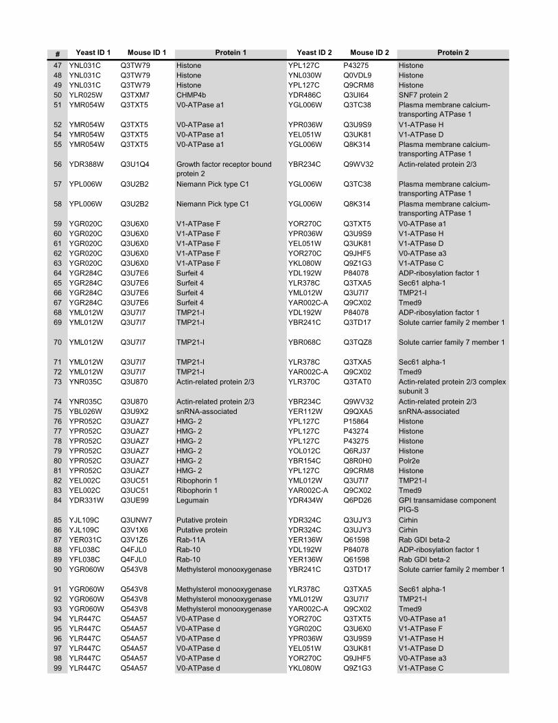
# Yeast ID 1 Mouse ID 1 Protein 1 Yeast ID 2 Mouse ID 2 Protein 2
47 YNL031C Q3TW79 Histone YPL127C P43275 Histone48 YNL031C Q3TW79 Histone YNL030W Q0VDL9 Histone49 YNL031C Q3TW79 Histone YPL127C Q9CRM8 Histone50 YLR025W Q3TXM7 CHMP4b YDR486C Q3UI64 SNF7 protein 251 YMR054W Q3TXT5 V0-ATPase a1 YGL006W Q3TC38 Plasma membrane calcium-
transporting ATPase 152 YMR054W Q3TXT5 V0-ATPase a1 YPR036W Q3U9S9 V1-ATPase H54 YMR054W Q3TXT5 V0-ATPase a1 YEL051W Q3UK81 V1-ATPase D55 YMR054W Q3TXT5 V0-ATPase a1 YGL006W Q8K314 Plasma membrane calcium-
transporting ATPase 156 YDR388W Q3U1Q4 Growth factor receptor bound
protein 2YBR234C Q9WV32 Actin-related protein 2/3
57 YPL006W Q3U2B2 Niemann Pick type C1 YGL006W Q3TC38 Plasma membrane calcium-transporting ATPase 1
58 YPL006W Q3U2B2 Niemann Pick type C1 YGL006W Q8K314 Plasma membrane calcium-transporting ATPase 1
59 YGR020C Q3U6X0 V1-ATPase F YOR270C Q3TXT5 V0-ATPase a160 YGR020C Q3U6X0 V1-ATPase F YPR036W Q3U9S9 V1-ATPase H61 YGR020C Q3U6X0 V1-ATPase F YEL051W Q3UK81 V1-ATPase D62 YGR020C Q3U6X0 V1-ATPase F YOR270C Q9JHF5 V0-ATPase a363 YGR020C Q3U6X0 V1-ATPase F YKL080W Q9Z1G3 V1-ATPase C64 YGR284C Q3U7E6 Surfeit 4 YDL192W P84078 ADP-ribosylation factor 165 YGR284C Q3U7E6 Surfeit 4 YLR378C Q3TXA5 Sec61 alpha-166 YGR284C Q3U7E6 Surfeit 4 YML012W Q3U7I7 TMP21-I67 YGR284C Q3U7E6 Surfeit 4 YAR002C-A Q9CX02 Tmed968 YML012W Q3U7I7 TMP21-I YDL192W P84078 ADP-ribosylation factor 169 YML012W Q3U7I7 TMP21-I YBR241C Q3TD17 Solute carrier family 2 member 1
70 YML012W Q3U7I7 TMP21-I YBR068C Q3TQZ8 Solute carrier family 7 member 1
71 YML012W Q3U7I7 TMP21-I YLR378C Q3TXA5 Sec61 alpha-172 YML012W Q3U7I7 TMP21-I YAR002C-A Q9CX02 Tmed973 YNR035C Q3U870 Actin-related protein 2/3 YLR370C Q3TAT0 Actin-related protein 2/3 complex
subunit 374 YNR035C Q3U870 Actin-related protein 2/3 YBR234C Q9WV32 Actin-related protein 2/375 YBL026W Q3U9X2 snRNA-associated YER112W Q9QXA5 snRNA-associated76 YPR052C Q3UAZ7 HMG- 2 YPL127C P15864 Histone77 YPR052C Q3UAZ7 HMG- 2 YPL127C P43274 Histone78 YPR052C Q3UAZ7 HMG- 2 YPL127C P43275 Histone79 YPR052C Q3UAZ7 HMG- 2 YOL012C Q6RJ37 Histone80 YPR052C Q3UAZ7 HMG- 2 YBR154C Q8R0H0 Polr2e81 YPR052C Q3UAZ7 HMG- 2 YPL127C Q9CRM8 Histone82 YEL002C Q3UC51 Ribophorin 1 YML012W Q3U7I7 TMP21-I83 YEL002C Q3UC51 Ribophorin 1 YAR002C-A Q9CX02 Tmed984 YDR331W Q3UE99 Legumain YDR434W Q6PD26 GPI transamidase component
PIG-S85 YJL109C Q3UNW7 Putative protein YDR324C Q3UJY3 Cirhin86 YJL109C Q3V1X6 Putative protein YDR324C Q3UJY3 Cirhin87 YER031C Q3V1Z6 Rab-11A YER136W Q61598 Rab GDI beta-288 YFL038C Q4FJL0 Rab-10 YDL192W P84078 ADP-ribosylation factor 189 YFL038C Q4FJL0 Rab-10 YER136W Q61598 Rab GDI beta-290 YGR060W Q543V8 Methylsterol monooxygenase YBR241C Q3TD17 Solute carrier family 2 member 1
91 YGR060W Q543V8 Methylsterol monooxygenase YLR378C Q3TXA5 Sec61 alpha-192 YGR060W Q543V8 Methylsterol monooxygenase YML012W Q3U7I7 TMP21-I93 YGR060W Q543V8 Methylsterol monooxygenase YAR002C-A Q9CX02 Tmed994 YLR447C Q54A57 V0-ATPase d YOR270C Q3TXT5 V0-ATPase a195 YLR447C Q54A57 V0-ATPase d YGR020C Q3U6X0 V1-ATPase F96 YLR447C Q54A57 V0-ATPase d YPR036W Q3U9S9 V1-ATPase H97 YLR447C Q54A57 V0-ATPase d YEL051W Q3UK81 V1-ATPase D98 YLR447C Q54A57 V0-ATPase d YOR270C Q9JHF5 V0-ATPase a399 YLR447C Q54A57 V0-ATPase d YKL080W Q9Z1G3 V1-ATPase C
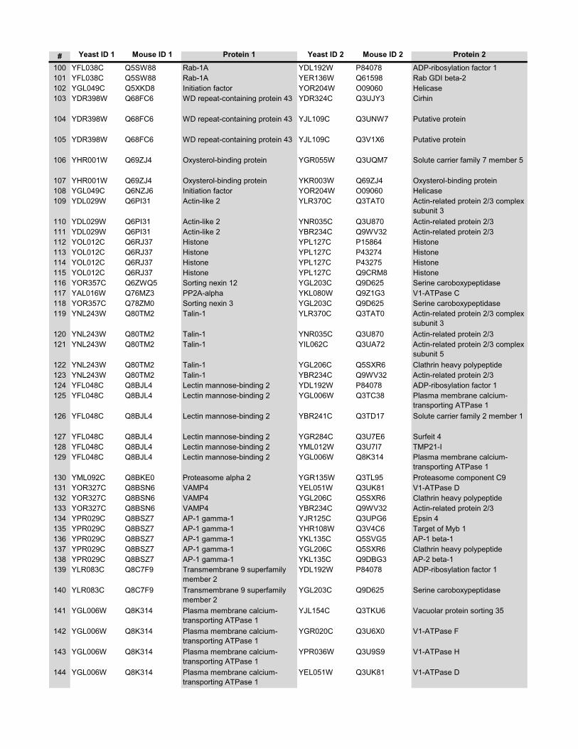
# Yeast ID 1 Mouse ID 1 Protein 1 Yeast ID 2 Mouse ID 2 Protein 2
100 YFL038C Q5SW88 Rab-1A YDL192W P84078 ADP-ribosylation factor 1101 YFL038C Q5SW88 Rab-1A YER136W Q61598 Rab GDI beta-2102 YGL049C Q5XKD8 Initiation factor YOR204W O09060 Helicase103 YDR398W Q68FC6 WD repeat-containing protein 43 YDR324C Q3UJY3 Cirhin
104 YDR398W Q68FC6 WD repeat-containing protein 43 YJL109C Q3UNW7 Putative protein
105 YDR398W Q68FC6 WD repeat-containing protein 43 YJL109C Q3V1X6 Putative protein
106 YHR001W Q69ZJ4 Oxysterol-binding protein YGR055W Q3UQM7 Solute carrier family 7 member 5
107 YHR001W Q69ZJ4 Oxysterol-binding protein YKR003W Q69ZJ4 Oxysterol-binding protein108 YGL049C Q6NZJ6 Initiation factor YOR204W O09060 Helicase109 YDL029W Q6PI31 Actin-like 2 YLR370C Q3TAT0 Actin-related protein 2/3 complex
subunit 3110 YDL029W Q6PI31 Actin-like 2 YNR035C Q3U870 Actin-related protein 2/3111 YDL029W Q6PI31 Actin-like 2 YBR234C Q9WV32 Actin-related protein 2/3112 YOL012C Q6RJ37 Histone YPL127C P15864 Histone113 YOL012C Q6RJ37 Histone YPL127C P43274 Histone114 YOL012C Q6RJ37 Histone YPL127C P43275 Histone115 YOL012C Q6RJ37 Histone YPL127C Q9CRM8 Histone116 YOR357C Q6ZWQ5 Sorting nexin 12 YGL203C Q9D625 Serine caroboxypeptidase117 YAL016W Q76MZ3 PP2A-alpha YKL080W Q9Z1G3 V1-ATPase C118 YOR357C Q78ZM0 Sorting nexin 3 YGL203C Q9D625 Serine caroboxypeptidase119 YNL243W Q80TM2 Talin-1 YLR370C Q3TAT0 Actin-related protein 2/3 complex
subunit 3120 YNL243W Q80TM2 Talin-1 YNR035C Q3U870 Actin-related protein 2/3121 YNL243W Q80TM2 Talin-1 YIL062C Q3UA72 Actin-related protein 2/3 complex
subunit 5122 YNL243W Q80TM2 Talin-1 YGL206C Q5SXR6 Clathrin heavy polypeptide123 YNL243W Q80TM2 Talin-1 YBR234C Q9WV32 Actin-related protein 2/3124 YFL048C Q8BJL4 Lectin mannose-binding 2 YDL192W P84078 ADP-ribosylation factor 1125 YFL048C Q8BJL4 Lectin mannose-binding 2 YGL006W Q3TC38 Plasma membrane calcium-
transporting ATPase 1126 YFL048C Q8BJL4 Lectin mannose-binding 2 YBR241C Q3TD17 Solute carrier family 2 member 1
127 YFL048C Q8BJL4 Lectin mannose-binding 2 YGR284C Q3U7E6 Surfeit 4128 YFL048C Q8BJL4 Lectin mannose-binding 2 YML012W Q3U7I7 TMP21-I129 YFL048C Q8BJL4 Lectin mannose-binding 2 YGL006W Q8K314 Plasma membrane calcium-
transporting ATPase 1130 YML092C Q8BKE0 Proteasome alpha 2 YGR135W Q3TL95 Proteasome component C9131 YOR327C Q8BSN6 VAMP4 YEL051W Q3UK81 V1-ATPase D132 YOR327C Q8BSN6 VAMP4 YGL206C Q5SXR6 Clathrin heavy polypeptide133 YOR327C Q8BSN6 VAMP4 YBR234C Q9WV32 Actin-related protein 2/3134 YPR029C Q8BSZ7 AP-1 gamma-1 YJR125C Q3UPG6 Epsin 4135 YPR029C Q8BSZ7 AP-1 gamma-1 YHR108W Q3V4C6 Target of Myb 1136 YPR029C Q8BSZ7 AP-1 gamma-1 YKL135C Q5SVG5 AP-1 beta-1137 YPR029C Q8BSZ7 AP-1 gamma-1 YGL206C Q5SXR6 Clathrin heavy polypeptide138 YPR029C Q8BSZ7 AP-1 gamma-1 YKL135C Q9DBG3 AP-2 beta-1139 YLR083C Q8C7F9 Transmembrane 9 superfamily
member 2YDL192W P84078 ADP-ribosylation factor 1
140 YLR083C Q8C7F9 Transmembrane 9 superfamily member 2
YGL203C Q9D625 Serine caroboxypeptidase
141 YGL006W Q8K314 Plasma membrane calcium-transporting ATPase 1
YJL154C Q3TKU6 Vacuolar protein sorting 35
142 YGL006W Q8K314 Plasma membrane calcium-transporting ATPase 1
YGR020C Q3U6X0 V1-ATPase F
143 YGL006W Q8K314 Plasma membrane calcium-transporting ATPase 1
YPR036W Q3U9S9 V1-ATPase H
144 YGL006W Q8K314 Plasma membrane calcium-transporting ATPase 1
YEL051W Q3UK81 V1-ATPase D
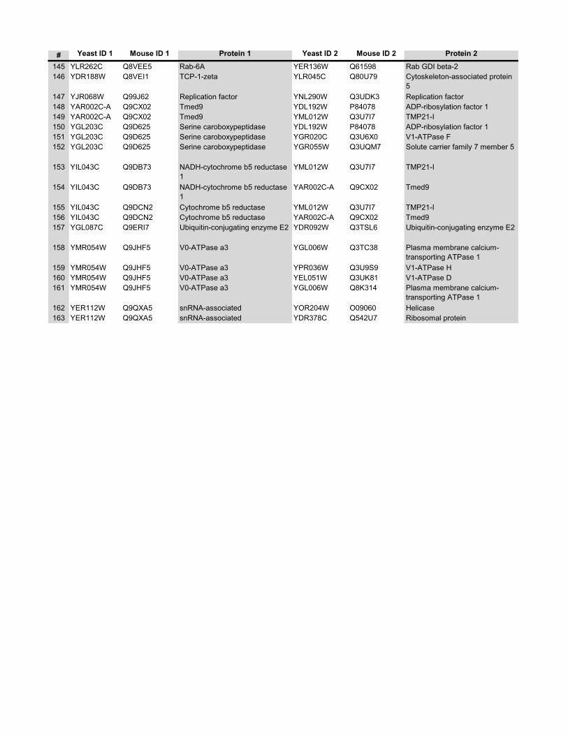
# Yeast ID 1 Mouse ID 1 Protein 1 Yeast ID 2 Mouse ID 2 Protein 2
145 YLR262C Q8VEE5 Rab-6A YER136W Q61598 Rab GDI beta-2146 YDR188W Q8VEI1 TCP-1-zeta YLR045C Q80U79 Cytoskeleton-associated protein
5147 YJR068W Q99J62 Replication factor YNL290W Q3UDK3 Replication factor148 YAR002C-A Q9CX02 Tmed9 YDL192W P84078 ADP-ribosylation factor 1149 YAR002C-A Q9CX02 Tmed9 YML012W Q3U7I7 TMP21-I150 YGL203C Q9D625 Serine caroboxypeptidase YDL192W P84078 ADP-ribosylation factor 1151 YGL203C Q9D625 Serine caroboxypeptidase YGR020C Q3U6X0 V1-ATPase F152 YGL203C Q9D625 Serine caroboxypeptidase YGR055W Q3UQM7 Solute carrier family 7 member 5
153 YIL043C Q9DB73 NADH-cytochrome b5 reductase 1
YML012W Q3U7I7 TMP21-I
154 YIL043C Q9DB73 NADH-cytochrome b5 reductase 1
YAR002C-A Q9CX02 Tmed9
155 YIL043C Q9DCN2 Cytochrome b5 reductase YML012W Q3U7I7 TMP21-I156 YIL043C Q9DCN2 Cytochrome b5 reductase YAR002C-A Q9CX02 Tmed9157 YGL087C Q9ERI7 Ubiquitin-conjugating enzyme E2 YDR092W Q3TSL6 Ubiquitin-conjugating enzyme E2
158 YMR054W Q9JHF5 V0-ATPase a3 YGL006W Q3TC38 Plasma membrane calcium-transporting ATPase 1
159 YMR054W Q9JHF5 V0-ATPase a3 YPR036W Q3U9S9 V1-ATPase H160 YMR054W Q9JHF5 V0-ATPase a3 YEL051W Q3UK81 V1-ATPase D161 YMR054W Q9JHF5 V0-ATPase a3 YGL006W Q8K314 Plasma membrane calcium-
transporting ATPase 1162 YER112W Q9QXA5 snRNA-associated YOR204W O09060 Helicase163 YER112W Q9QXA5 snRNA-associated YDR378C Q542U7 Ribosomal protein

xviii
Supplementary Table 4 VATPase subunits identified on the phagosome
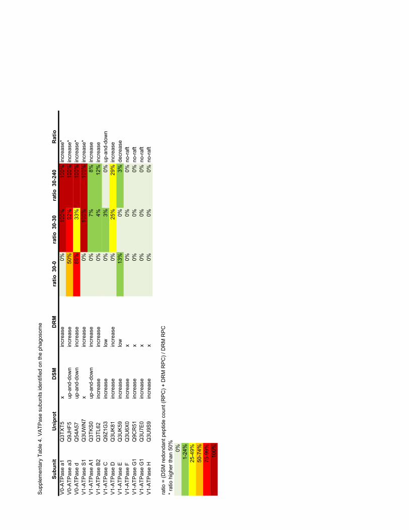
Sup
plem
enta
ry T
able
4. V
ATP
ase
subu
nits
iden
tifie
d on
the
phag
osom
e
Subu
nit
Uni
prot
DSM
DR
Mra
tio 3
0-0
ratio
30-
30ra
tio 3
0-24
0R
atio
V0-
ATP
ase
a1Q
3TX
T5x
incr
ease
0%10
0%10
0%in
crea
se*
V0-
ATP
ase
a3Q
9JH
F5up
-and
-dow
nin
crea
se50
%92
%10
0%in
crea
se*
V0-
ATP
ase
dQ
54A
57up
-and
-dow
nin
crea
se89
%33
%10
0%in
crea
se*
V1-
ATP
ase
S1
Q3U
WN
7x
incr
ease
0%10
0%10
0%in
crea
se*
V1-
ATP
ase
A1
Q3T
KS
0up
-and
-dow
nin
crea
se0%
7%8%
incr
ease
V1-
ATP
ase
B2
Q3T
L62
incr
ease
incr
ease
0%4%
12%
incr
ease
V1-
ATP
ase
CQ
9Z1G
3in
crea
selo
w0%
3%0%
up-a
nd-d
own
V1-
ATP
ase
DQ
3UK
81in
crea
sein
crea
se0%
25%
29%
incr
ease
V1-
ATP
ase
EQ
3UK
59in
crea
selo
w13
%0%
3%de
crea
seV
1-A
TPas
e F
Q3U
6X0
incr
ease
x0%
0%0%
no-r
aft
V1-
ATP
ase
G1
Q9C
R51
incr
ease
x0%
0%0%
no-r
aft
V1-
ATP
ase
G1
Q3U
7E0
incr
ease
x0%
0%0%
no-r
aft
V1-
ATP
ase
HQ
3U9S
9in
crea
sex
0%0%
0%no
-raf
t
ratio
= (D
SM
redo
ndan
t pep
tide
coun
t (R
PC
) + D
RM
RP
C) /
DR
M R
PC
* ra
tio h
ighe
r tha
n 50
%0%
1-24
%25
-49%
50-7
4%75
-99%
100%
xix
Supplementary Table 5 List of the phospholipids of the phagosome identified in the negative ionization mode
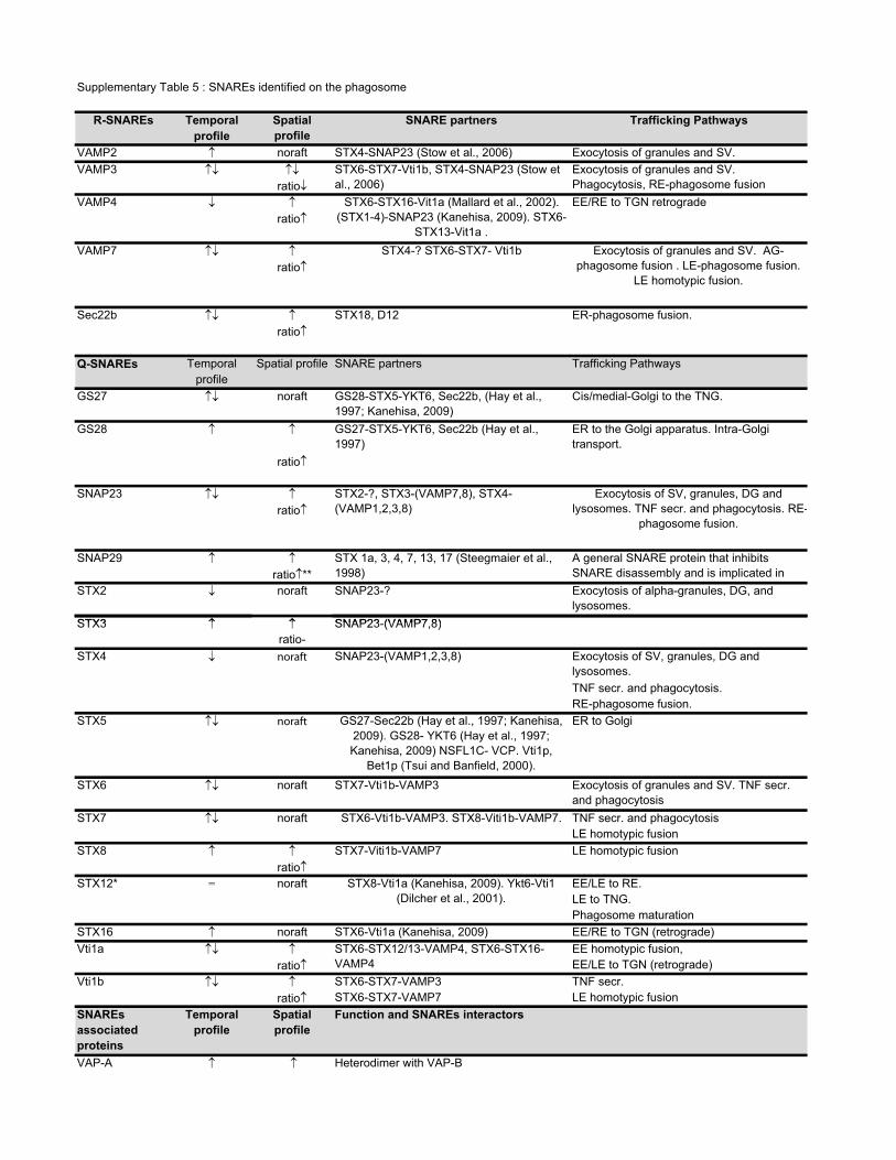
-
� �STX3 SNAP23 (VAMP7 8)
Supplementary Table 5 : SNAREs identified on the phagosome
R-SNAREs Temporal Spatialprofile
SNARE partners Trafficking Pathwaysprofile
VAMP2 � noraft STX4-SNAP23 (Stow et al., 2006) Exocytosis of granules and SV. VAMP3 �� �� STX6-STX7-Vti1b, STX4-SNAP23 (Stow et
al., 2006)Exocytosis of granules and SV. Phagocytosis, RE-phagosome fusionratio�
VAMP4 � � STX6-STX16-Vit1a (Mallard et al., 2002). (STX1-4)-SNAP23 (Kanehisa, 2009). STX6-
STX13-Vit1a .
EE/RE to TGN retrograde ratio�
VAMP7 �� � STX4-? STX6-STX7- Vti1b Exocytosis of granules and SV. AG-phagosome fusion . LE-phagosome fusion.
LE homotypic fusion.ratio�
Sec22b �� � STX18, D12 ER-phagosome fusion.ratio�
Q-SNAREs Temporal Spatial profile SNARE partners Trafficking Pathwaysprofile
GS27 �� noraft GS28-STX5-YKT6, Sec22b, (Hay et al., 1997; Kanehisa, 2009)
Cis/medial-Golgi to the TNG.
GS28 � � GS27-STX5-YKT6, Sec22b (Hay et al., 1997)
ER to the Golgi apparatus. Intra-Golgi transport.
ratio�
SNAP23 �� � STX2-?, STX3-(VAMP7,8), STX4-(VAMP1,2,3,8)
Exocytosis of SV, granules, DG and lysosomes. TNF secr. and phagocytosis. RE
phagosome fusion.ratio�
SNAP29 � � STX 1a, 3, 4, 7, 13, 17 (Steegmaier et al., 1998)
A general SNARE protein that inhibits SNARE disassembly and is implicated in ratio�**
STX2 � noraft SNAP23-? Exocytosis of alpha-granules, DG, and lysosomes.
STX3 � � SNAP23 (VAMP7 8)- ,ratio-
STX4 � noraft SNAP23-(VAMP1,2,3,8) Exocytosis of SV, granules, DG and lysosomes.TNF secr. and phagocytosis.RE-phagosome fusion.
STX5 �� noraft GS27-Sec22b (Hay et al., 1997; Kanehisa, 2009). GS28- YKT6 (Hay et al., 1997; Kanehisa, 2009) NSFL1C- VCP. Vti1p,
Bet1p (Tsui and Banfield, 2000).
ER to Golgi
STX6 �� noraft STX7-Vti1b-VAMP3 Exocytosis of granules and SV. TNF secr. and phagocytosis
STX7 �� noraft STX6-Vti1b-VAMP3. STX8-Viti1b-VAMP7. TNF secr. and phagocytosisLE homotypic fusion
STX8 � � STX7-Viti1b-VAMP7 LE homotypic fusionratio�
STX12* = noraft STX8-Vti1a (Kanehisa, 2009). Ykt6-Vti1 (Dilcher et al., 2001).
EE/LE to RE.LE to TNG.Phagosome maturation
STX16 � noraft STX6-Vti1a (Kanehisa, 2009) EE/RE to TGN (retrograde) Vti1a �� � STX6-STX12/13-VAMP4, STX6-STX16-
VAMP4EE homotypic fusion,
ratio� EE/LE to TGN (retrograde) Vti1b �� � STX6-STX7-VAMP3 TNF secr.
ratio� STX6-STX7-VAMP7 LE homotypic fusionSNAREsassociatedproteins
Temporalprofile
Spatialprofile
Function and SNAREs interactors
VAP-A � � Heterodimer with VAP-B

ratio� VAMP1, VAMP2, STX1A, BET1, SEC22CVAP-B � � Heterodimer with VAP-A
ratio� VAMP1, VAMP2SNAP-alpha � � VAMP8, USE1L, RINT1, STX18
ratio� Couple the energy of ATP binding and hydrolysis to the disassembly of SNARE SNAP-gamma � � Stabilize a 20 S complex consisting of NSF, alpha-SNAP, and SNAREs
ratio� Interacts directly with NSF, as well as Gaf-1/Rip11, but not with SNAREs (Tani et al., Snapin � noraft SNAP23 and SNAP25 (Bao et al., 2008)NSF noraft Couple the energy of ATP binding and hydrolysis to the disassembly of SNARE
Legend Temporal � Increase in phagosome� Decrease in phagosome�� Transient in phagosome
Spacial � Increase in DRM� Decrease in DRM�� Transient in DRM
ratio DSM (RPC) + DRM RPC) / DRM RPC

xx
Supplementary Table 6 List of the phospholipids identified in the negative ion mode of the Fourier Transform mass spectrometer
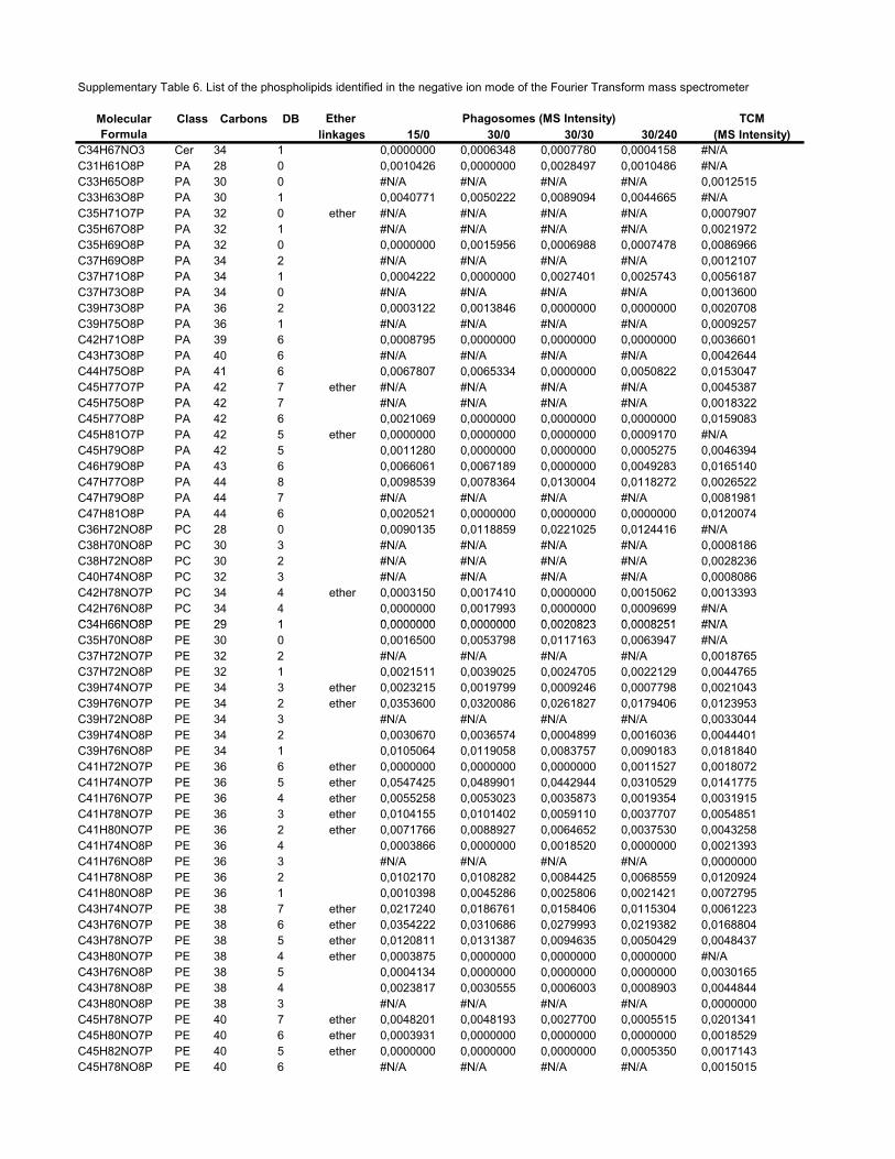
Supplementary Table 6. List of the phospholipids identified in the negative ion mode of the Fourier Transform mass spectrometer
Ether TCMlinkages 15/0 30/0 30/30 30/240 (MS Intensity)
C34H67NO3 Cer 34 1 0,0000000 0,0006348 0,0007780 0,0004158 #N/AC31H61O8P PA 28 0 0,0010426 0,0000000 0,0028497 0,0010486 #N/AC33H65O8P PA 30 0 #N/A #N/A #N/A #N/A 0,0012515C33H63O8P PA 30 1 0,0040771 0,0050222 0,0089094 0,0044665 #N/AC35H71O7P PA 32 0 ether #N/A #N/A #N/A #N/A 0,0007907C35H67O8P PA 32 1 #N/A #N/A #N/A #N/A 0,0021972C35H69O8P PA 32 0 0,0000000 0,0015956 0,0006988 0,0007478 0,0086966C37H69O8P PA 34 2 #N/A #N/A #N/A #N/A 0,0012107C37H71O8P PA 34 1 0,0004222 0,0000000 0,0027401 0,0025743 0,0056187C37H73O8P PA 34 0 #N/A #N/A #N/A #N/A 0,0013600C39H73O8P PA 36 2 0,0003122 0,0013846 0,0000000 0,0000000 0,0020708C39H75O8P PA 36 1 #N/A #N/A #N/A #N/A 0,0009257C42H71O8P PA 39 6 0,0008795 0,0000000 0,0000000 0,0000000 0,0036601C43H73O8P PA 40 6 #N/A #N/A #N/A #N/A 0,0042644C44H75O8P PA 41 6 0,0067807 0,0065334 0,0000000 0,0050822 0,0153047C45H77O7P PA 42 7 ether #N/A #N/A #N/A #N/A 0,0045387C45H75O8P PA 42 7 #N/A #N/A #N/A #N/A 0,0018322C45H77O8P PA 42 6 0,0021069 0,0000000 0,0000000 0,0000000 0,0159083C45H81O7P PA 42 5 ether 0,0000000 0,0000000 0,0000000 0,0009170 #N/AC45H79O8P PA 42 5 0,0011280 0,0000000 0,0000000 0,0005275 0,0046394C46H79O8P PA 43 6 0,0066061 0,0067189 0,0000000 0,0049283 0,0165140C47H77O8P PA 44 8 0,0098539 0,0078364 0,0130004 0,0118272 0,0026522C47H79O8P PA 44 7 #N/A #N/A #N/A #N/A 0,0081981C47H81O8P PA 44 6 0,0020521 0,0000000 0,0000000 0,0000000 0,0120074C36H72NO8P PC 28 0 0,0090135 0,0118859 0,0221025 0,0124416 #N/AC38H70NO8P PC 30 3 #N/A #N/A #N/A #N/A 0,0008186C38H72NO8P PC 30 2 #N/A #N/A #N/A #N/A 0,0028236C40H74NO8P PC 32 3 #N/A #N/A #N/A #N/A 0,0008086C42H78NO7P PC 34 4 ether 0,0003150 0,0017410 0,0000000 0,0015062 0,0013393C42H76NO8P PC 34 4 0,0000000 0,0017993 0,0000000 0,0009699 #N/AC34H66NO8P PE 29 1 0 0000000 0 0000000 0 0020823 0 0008251 #N/A
Phagosomes (MS Intensity)Molecular Formula
Class Carbons DB
C34H66NO8P PE 29 1 0,0000000 0,0000000 0,0020823 0,0008251 #N/AC35H70NO8P PE 30 0 0,0016500 0,0053798 0,0117163 0,0063947 #N/AC37H72NO7P PE 32 2 #N/A #N/A #N/A #N/A 0,0018765C37H72NO8P PE 32 1 0,0021511 0,0039025 0,0024705 0,0022129 0,0044765C39H74NO7P PE 34 3 ether 0,0023215 0,0019799 0,0009246 0,0007798 0,0021043C39H76NO7P PE 34 2 ether 0,0353600 0,0320086 0,0261827 0,0179406 0,0123953C39H72NO8P PE 34 3 #N/A #N/A #N/A #N/A 0,0033044C39H74NO8P PE 34 2 0,0030670 0,0036574 0,0004899 0,0016036 0,0044401C39H76NO8P PE 34 1 0,0105064 0,0119058 0,0083757 0,0090183 0,0181840C41H72NO7P PE 36 6 ether 0,0000000 0,0000000 0,0000000 0,0011527 0,0018072C41H74NO7P PE 36 5 ether 0,0547425 0,0489901 0,0442944 0,0310529 0,0141775C41H76NO7P PE 36 4 ether 0,0055258 0,0053023 0,0035873 0,0019354 0,0031915C41H78NO7P PE 36 3 ether 0,0104155 0,0101402 0,0059110 0,0037707 0,0054851C41H80NO7P PE 36 2 ether 0,0071766 0,0088927 0,0064652 0,0037530 0,0043258C41H74NO8P PE 36 4 0,0003866 0,0000000 0,0018520 0,0000000 0,0021393C41H76NO8P PE 36 3 #N/A #N/A #N/A #N/A 0,0000000C41H78NO8P PE 36 2 0,0102170 0,0108282 0,0084425 0,0068559 0,0120924C41H80NO8P PE 36 1 0,0010398 0,0045286 0,0025806 0,0021421 0,0072795C43H74NO7P PE 38 7 ether 0,0217240 0,0186761 0,0158406 0,0115304 0,0061223C43H76NO7P PE 38 6 ether 0,0354222 0,0310686 0,0279993 0,0219382 0,0168804C43H78NO7P PE 38 5 ether 0,0120811 0,0131387 0,0094635 0,0050429 0,0048437C43H80NO7P PE 38 4 ether 0,0003875 0,0000000 0,0000000 0,0000000 #N/AC43H76NO8P PE 38 5 0,0004134 0,0000000 0,0000000 0,0000000 0,0030165C43H78NO8P PE 38 4 0,0023817 0,0030555 0,0006003 0,0008903 0,0044844C43H80NO8P PE 38 3 #N/A #N/A #N/A #N/A 0,0000000C45H78NO7P PE 40 7 ether 0,0048201 0,0048193 0,0027700 0,0005515 0,0201341C45H80NO7P PE 40 6 ether 0,0003931 0,0000000 0,0000000 0,0000000 0,0018529C45H82NO7P PE 40 5 ether 0,0000000 0,0000000 0,0000000 0,0005350 0,0017143C45H78NO8P PE 40 6 #N/A #N/A #N/A #N/A 0,0015015
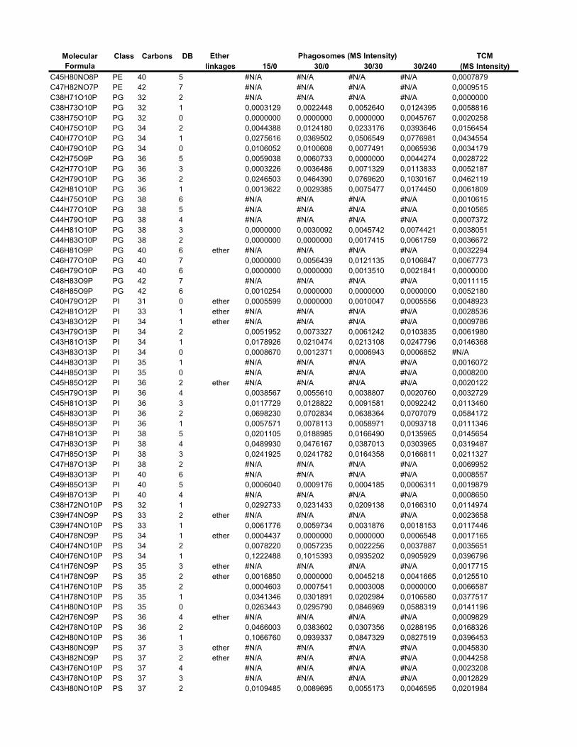
Ether TCMlinkages 15/0 30/0 30/30 30/240 (MS Intensity)
Phagosomes (MS Intensity)Molecular Formula
Class Carbons DB
C45H80NO8P PE 40 5 #N/A #N/A #N/A #N/A 0,0007879C47H82NO7P PE 42 7 #N/A #N/A #N/A #N/A 0,0009515C38H71O10P PG 32 2 #N/A #N/A #N/A #N/A 0,0000000C38H73O10P PG 32 1 0,0003129 0,0022448 0,0052640 0,0124395 0,0058816C38H75O10P PG 32 0 0,0000000 0,0000000 0,0000000 0,0045767 0,0020258C40H75O10P PG 34 2 0,0044388 0,0124180 0,0233176 0,0393646 0,0156454C40H77O10P PG 34 1 0,0275616 0,0369502 0,0506549 0,0776981 0,0434554C40H79O10P PG 34 0 0,0106052 0,0100608 0,0077491 0,0065936 0,0034179C42H75O9P PG 36 5 0,0059038 0,0060733 0,0000000 0,0044274 0,0028722C42H77O10P PG 36 3 0,0003226 0,0036486 0,0071329 0,0113833 0,0052187C42H79O10P PG 36 2 0,0246503 0,0464390 0,0769620 0,1030167 0,0462119C42H81O10P PG 36 1 0,0013622 0,0029385 0,0075477 0,0174450 0,0061809C44H75O10P PG 38 6 #N/A #N/A #N/A #N/A 0,0010615C44H77O10P PG 38 5 #N/A #N/A #N/A #N/A 0,0010565C44H79O10P PG 38 4 #N/A #N/A #N/A #N/A 0,0007372C44H81O10P PG 38 3 0,0000000 0,0030092 0,0045742 0,0074421 0,0038051C44H83O10P PG 38 2 0,0000000 0,0000000 0,0017415 0,0061759 0,0036672C46H81O9P PG 40 6 ether #N/A #N/A #N/A #N/A 0,0032294C46H77O10P PG 40 7 0,0000000 0,0056439 0,0121135 0,0106847 0,0067773C46H79O10P PG 40 6 0,0000000 0,0000000 0,0013510 0,0021841 0,0000000C48H83O9P PG 42 7 #N/A #N/A #N/A #N/A 0,0011115C48H85O9P PG 42 6 0,0010254 0,0000000 0,0000000 0,0000000 0,0052180C40H79O12P PI 31 0 ether 0,0005599 0,0000000 0,0010047 0,0005556 0,0048923C42H81O12P PI 33 1 ether #N/A #N/A #N/A #N/A 0,0028536C43H83O12P PI 34 1 ether #N/A #N/A #N/A #N/A 0,0009786C43H79O13P PI 34 2 0,0051952 0,0073327 0,0061242 0,0103835 0,0061980C43H81O13P PI 34 1 0,0178926 0,0210474 0,0213108 0,0247796 0,0146368C43H83O13P PI 34 0 0,0008670 0,0012371 0,0006943 0,0006852 #N/AC44H83O13P PI 35 1 #N/A #N/A #N/A #N/A 0,0016072C44H85O13P PI 35 0 #N/A #N/A #N/A #N/A 0,0008200C45H85O12P PI 36 2 ether #N/A #N/A #N/A #N/A 0,0020122C45H79O13P PI 36 4 0,0038567 0,0055610 0,0038807 0,0020760 0,0032729C45H81O13P PI 36 3 0 0117729 0 0128822 0 0091581 0 0092242 0 0113460C45H81O13P PI 36 3 0,0117729 0,0128822 0,0091581 0,0092242 0,0113460C45H83O13P PI 36 2 0,0698230 0,0702834 0,0638364 0,0707079 0,0584172C45H85O13P PI 36 1 0,0057571 0,0078113 0,0058971 0,0093718 0,0111346C47H81O13P PI 38 5 0,0201105 0,0188985 0,0166490 0,0135965 0,0145654C47H83O13P PI 38 4 0,0489930 0,0476167 0,0387013 0,0303965 0,0319487C47H85O13P PI 38 3 0,0241925 0,0241782 0,0164358 0,0166811 0,0211327C47H87O13P PI 38 2 #N/A #N/A #N/A #N/A 0,0069952C49H83O13P PI 40 6 #N/A #N/A #N/A #N/A 0,0008557C49H85O13P PI 40 5 0,0006040 0,0009176 0,0004185 0,0006311 0,0019879C49H87O13P PI 40 4 #N/A #N/A #N/A #N/A 0,0008650C38H72NO10P PS 32 1 0,0292733 0,0231433 0,0209138 0,0166310 0,0114974C39H74NO9P PS 33 2 ether #N/A #N/A #N/A #N/A 0,0023658C39H74NO10P PS 33 1 0,0061776 0,0059734 0,0031876 0,0018153 0,0117446C40H78NO9P PS 34 1 ether 0,0004437 0,0000000 0,0000000 0,0006548 0,0017165C40H74NO10P PS 34 2 0,0078220 0,0057235 0,0022256 0,0037887 0,0035651C40H76NO10P PS 34 1 0,1222488 0,1015393 0,0935202 0,0905929 0,0396796C41H76NO9P PS 35 3 ether #N/A #N/A #N/A #N/A 0,0017715C41H78NO9P PS 35 2 ether 0,0016850 0,0000000 0,0045218 0,0041665 0,0125510C41H76NO10P PS 35 2 0,0004603 0,0007541 0,0003008 0,0000000 0,0066587C41H78NO10P PS 35 1 0,0341346 0,0301891 0,0202984 0,0106580 0,0377517C41H80NO10P PS 35 0 0,0263443 0,0295790 0,0846969 0,0588319 0,0141196C42H76NO9P PS 36 4 ether #N/A #N/A #N/A #N/A 0,0009829C42H78NO10P PS 36 2 0,0466003 0,0383602 0,0307356 0,0288195 0,0168326C42H80NO10P PS 36 1 0,1066760 0,0939337 0,0847329 0,0827519 0,0396453C43H80NO9P PS 37 3 ether #N/A #N/A #N/A #N/A 0,0045830C43H82NO9P PS 37 2 ether #N/A #N/A #N/A #N/A 0,0044258C43H76NO10P PS 37 4 #N/A #N/A #N/A #N/A 0,0023208C43H78NO10P PS 37 3 #N/A #N/A #N/A #N/A 0,0012829C43H80NO10P PS 37 2 0,0109485 0,0089695 0,0055173 0,0046595 0,0201984
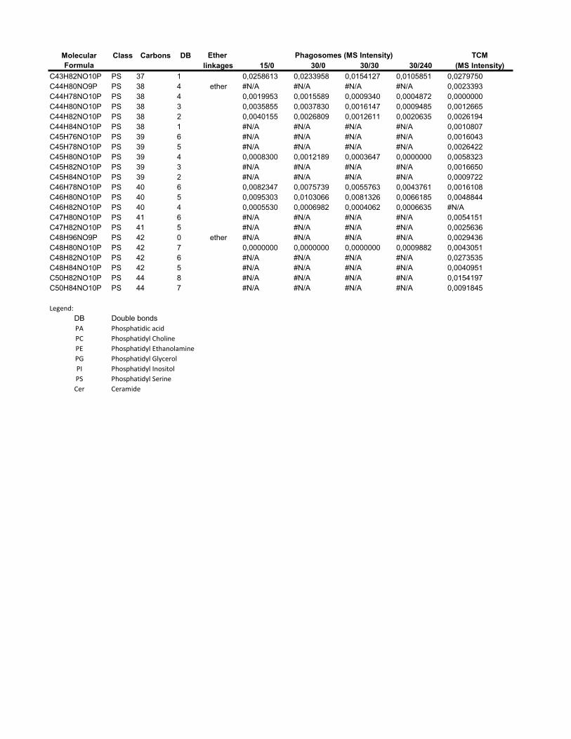
Ether TCMlinkages 15/0 30/0 30/30 30/240 (MS Intensity)
Phagosomes (MS Intensity)Molecular Formula
Class Carbons DB
C43H82NO10P PS 37 1 0,0258613 0,0233958 0,0154127 0,0105851 0,0279750C44H80NO9P PS 38 4 ether #N/A #N/A #N/A #N/A 0,0023393C44H78NO10P PS 38 4 0,0019953 0,0015589 0,0009340 0,0004872 0,0000000C44H80NO10P PS 38 3 0,0035855 0,0037830 0,0016147 0,0009485 0,0012665C44H82NO10P PS 38 2 0,0040155 0,0026809 0,0012611 0,0020635 0,0026194C44H84NO10P PS 38 1 #N/A #N/A #N/A #N/A 0,0010807C45H76NO10P PS 39 6 #N/A #N/A #N/A #N/A 0,0016043C45H78NO10P PS 39 5 #N/A #N/A #N/A #N/A 0,0026422C45H80NO10P PS 39 4 0,0008300 0,0012189 0,0003647 0,0000000 0,0058323C45H82NO10P PS 39 3 #N/A #N/A #N/A #N/A 0,0016650C45H84NO10P PS 39 2 #N/A #N/A #N/A #N/A 0,0009722C46H78NO10P PS 40 6 0,0082347 0,0075739 0,0055763 0,0043761 0,0016108C46H80NO10P PS 40 5 0,0095303 0,0103066 0,0081326 0,0066185 0,0048844C46H82NO10P PS 40 4 0,0005530 0,0006982 0,0004062 0,0006635 #N/AC47H80NO10P PS 41 6 #N/A #N/A #N/A #N/A 0,0054151C47H82NO10P PS 41 5 #N/A #N/A #N/A #N/A 0,0025636C48H96NO9P PS 42 0 ether #N/A #N/A #N/A #N/A 0,0029436C48H80NO10P PS 42 7 0,0000000 0,0000000 0,0000000 0,0009882 0,0043051C48H82NO10P PS 42 6 #N/A #N/A #N/A #N/A 0,0273535C48H84NO10P PS 42 5 #N/A #N/A #N/A #N/A 0,0040951C50H82NO10P PS 44 8 #N/A #N/A #N/A #N/A 0,0154197C50H84NO10P PS 44 7 #N/A #N/A #N/A #N/A 0,0091845
Legend:
DB Double bondsPA Phosphatidic�acid
PC Phosphatidyl�Choline�
PE Phosphatidyl�Ethanolamine
PG Phosphatidyl�Glycerol
PI Phosphatidyl�Inositol
PS Phosphatidyl�Serine�
Cer Ceramide

xxi
Supplementary Table 7 List of the phospholipids identified in the positive ion mode of the Fourier Transform mass spectrometer
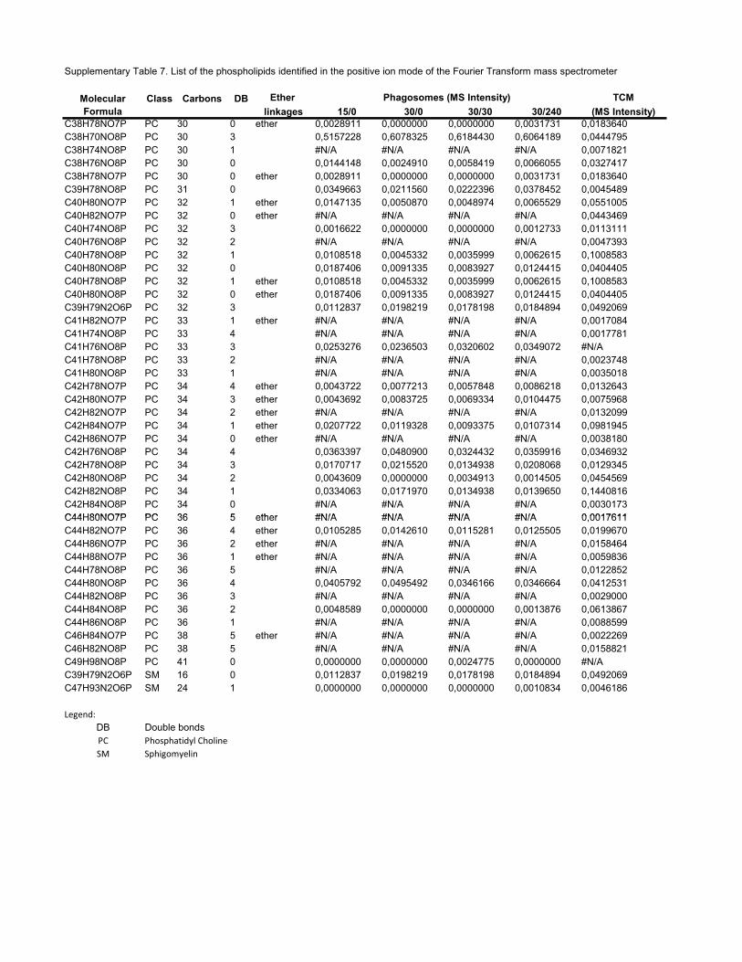
Supplementary Table 7. List of the phospholipids identified in the positive ion mode of the Fourier Transform mass spectrometer
Ether TCMlinkages 15/0 30/0 30/30 30/240 (MS Intensity)
C38H78NO7P PC 30 0 ether 0,0028911 0,0000000 0,0000000 0,0031731 0,0183640C38H70NO8P PC 30 3 0,5157228 0,6078325 0,6184430 0,6064189 0,0444795C38H74NO8P PC 30 1 #N/A #N/A #N/A #N/A 0,0071821C38H76NO8P PC 30 0 0,0144148 0,0024910 0,0058419 0,0066055 0,0327417C38H78NO7P PC 30 0 ether 0,0028911 0,0000000 0,0000000 0,0031731 0,0183640C39H78NO8P PC 31 0 0,0349663 0,0211560 0,0222396 0,0378452 0,0045489C40H80NO7P PC 32 1 ether 0,0147135 0,0050870 0,0048974 0,0065529 0,0551005C40H82NO7P PC 32 0 ether #N/A #N/A #N/A #N/A 0,0443469C40H74NO8P PC 32 3 0,0016622 0,0000000 0,0000000 0,0012733 0,0113111C40H76NO8P PC 32 2 #N/A #N/A #N/A #N/A 0,0047393C40H78NO8P PC 32 1 0,0108518 0,0045332 0,0035999 0,0062615 0,1008583C40H80NO8P PC 32 0 0,0187406 0,0091335 0,0083927 0,0124415 0,0404405C40H78NO8P PC 32 1 ether 0,0108518 0,0045332 0,0035999 0,0062615 0,1008583C40H80NO8P PC 32 0 ether 0,0187406 0,0091335 0,0083927 0,0124415 0,0404405C39H79N2O6P PC 32 3 0,0112837 0,0198219 0,0178198 0,0184894 0,0492069C41H82NO7P PC 33 1 ether #N/A #N/A #N/A #N/A 0,0017084C41H74NO8P PC 33 4 #N/A #N/A #N/A #N/A 0,0017781C41H76NO8P PC 33 3 0,0253276 0,0236503 0,0320602 0,0349072 #N/AC41H78NO8P PC 33 2 #N/A #N/A #N/A #N/A 0,0023748C41H80NO8P PC 33 1 #N/A #N/A #N/A #N/A 0,0035018C42H78NO7P PC 34 4 ether 0,0043722 0,0077213 0,0057848 0,0086218 0,0132643C42H80NO7P PC 34 3 ether 0,0043692 0,0083725 0,0069334 0,0104475 0,0075968C42H82NO7P PC 34 2 ether #N/A #N/A #N/A #N/A 0,0132099C42H84NO7P PC 34 1 ether 0,0207722 0,0119328 0,0093375 0,0107314 0,0981945C42H86NO7P PC 34 0 ether #N/A #N/A #N/A #N/A 0,0038180C42H76NO8P PC 34 4 0,0363397 0,0480900 0,0324432 0,0359916 0,0346932C42H78NO8P PC 34 3 0,0170717 0,0215520 0,0134938 0,0208068 0,0129345C42H80NO8P PC 34 2 0,0043609 0,0000000 0,0034913 0,0014505 0,0454569C42H82NO8P PC 34 1 0,0334063 0,0171970 0,0134938 0,0139650 0,1440816C42H84NO8P PC 34 0 #N/A #N/A #N/A #N/A 0,0030173C44H80NO7P PC 36 5 ether #N/A #N/A #N/A #N/A 0 0017611
Phagosomes (MS Intensity)Molecular Formula
Class Carbons DB
C44H80NO7P PC 36 5 ether #N/A #N/A #N/A #N/A 0,0017611C44H82NO7P PC 36 4 ether 0,0105285 0,0142610 0,0115281 0,0125505 0,0199670C44H86NO7P PC 36 2 ether #N/A #N/A #N/A #N/A 0,0158464C44H88NO7P PC 36 1 ether #N/A #N/A #N/A #N/A 0,0059836C44H78NO8P PC 36 5 #N/A #N/A #N/A #N/A 0,0122852C44H80NO8P PC 36 4 0,0405792 0,0495492 0,0346166 0,0346664 0,0412531C44H82NO8P PC 36 3 #N/A #N/A #N/A #N/A 0,0029000C44H84NO8P PC 36 2 0,0048589 0,0000000 0,0000000 0,0013876 0,0613867C44H86NO8P PC 36 1 #N/A #N/A #N/A #N/A 0,0088599C46H84NO7P PC 38 5 ether #N/A #N/A #N/A #N/A 0,0022269C46H82NO8P PC 38 5 #N/A #N/A #N/A #N/A 0,0158821C49H98NO8P PC 41 0 0,0000000 0,0000000 0,0024775 0,0000000 #N/AC39H79N2O6P SM 16 0 0,0112837 0,0198219 0,0178198 0,0184894 0,0492069C47H93N2O6P SM 24 1 0,0000000 0,0000000 0,0000000 0,0010834 0,0046186
Legend:
DB Double bondsPC Phosphatidyl�Choline�
SM Sphigomyelin
xxii
Annexe III. Article 3
Houde, M., Bertholet, S., Gagnon, E., Brunet, S., Goyette, G., Laplante, A.,
Princiotta, M. F., Thibault, P., Sacks, D., Desjardins, M. (2003) Phagosomes are
competent organelles for antigene cross-presentation. Nature 425, 402-406
CLSMFor CLSM, cells pulsed with 3mm LB (Polyscience, coated with OVA-FITC conjugates)were allowed to adhere on polylysine-coated coverslips. Before fixation (4%paraformaldehyde-PBS, 10min), cells were permeabilized before staining (PBS-saponin0.2%, 10min; except for Fig. 1a, class I MHC staining was performed beforepermeabilization in order to selectively stain cell surface with the anti H-2Kb Y3 antibody)and stained and washed in PBS BSA 0.1% saponine 0.1%. After mounting, coverslips wereanalysed on a Leica SP2 confocal microscope.
In vitro TAP and loading assaysATAP transport assay was performed19. The same procedure was used for HLA-A2 loadingassay, except that HLA-A2 bound peptide was immunoprecipitated on anti-HLA-A2- orcontrol Sepharose. See Supplementary Information SD4.
Cross-presentation assays3mm LB were coated with OVA (or BSA) protein by passive adsorption (incubation in10mgml21 PBS solutions for 18 h at 4 8C) and extensively washed in PBS. DCs werepulsed and chased for indicated times with OVA-beads or BSA-beads. After fixation(0.08% glutaraldehyde 3min, stopped in glycine 0.2M) cells were washed and put inculture (105 per well in 96 flat well plates) for 18 h with the B3Z CD8þ T-cell hybridomaspecific for the H-2Kb/OVA257–264 complex (105 per well)20. B3Z activationwas monitoredby the induction of lacZ reporter under NF-AT elements using the CPRG substrate(Roche)20. For lactacystin inhibition assays, DCs were preincubated for 20minbefore beads pulse with 18 mM lactacystin and lactacystin was kept during the entire pulseperiod.
T-cell activation using purified phagosomal membranesD1 cells were pretreated or not with 18 mM lactacystin and pulsed with OVA-beads orBSA-beads for 30min. After a 2-h chase (in the continuous presence of lactacystin ornot), the cells were homogenized and the phagosomes were purified as before. Thephagosomal membranes were then disrupted by freeze–thaw, separated from the beads bylow-speed centrifugation and pelleted by ultracentrifugation (100,000g, 1 h). Anti-H-2Kb/OVA transgenic OT-1 effector T cells (obtained by in vitro stimulationwith peptide-pulsedDCs and culture for 3 to 15 days in the presence of IL-2) were then added and IFN-gsecretion was measured after an 18-h incubation in supernatants with an ELISA test(Pharmingen).
Cryoelectron microscopy and G-6-Pase stainingD1 cells were incubated with 0.8 mm LB for 1 h (Fig. 1c) or 3 mm LB for 30min (Fig. 1d),before fixation in 2% paraformaldehyde, 0.2% glutaraldehyde. Then cells wereprocessed for ultrathin cryosectioning and immunogold labelling as described21. ForG-6-Pase staining, D1 cells were incubated with 3 mm LB for 1 h and the Epon sectionswere processed as previously described10. When indicated, the cells pretreated with100 nMwortmannin and then pulsed for 1 hwith the LB in the continuous presence of thedrug.
Received 17 March; accepted 10 July 2003; doi:10.1038/nature01911.
1. Guermonprez, P., Valladeau, J., Zitvogel, L. & Amigorena, S. Antigen Presentation and T Cell
Stimulation by Dendritic Cells. Annu. Rev. Immunol. 20, 620–667 (2002).
2. Heath,W. R. & Carbone, F. R. Cross-presentation, dendritic cells, tolerance and immunity.Annu. Rev.
Immunol. 19, 47–64 (2001).
3. Yewdell, J. W., Norbury, C. C. & Bennink, J. R. Mechanisms of exogenous antigen presentation by
MHC class I molecules in vitro and in vivo: implications for generating CD8 þ T cell responses to
infectious agents, tumors, transplants, and vaccines. Adv. Immunol. 73, 1–77 (1999).
4. Kovacsovics-Bankowski, M. & Rock, K. L. A phagosome-to-cytosol pathway for exogenous antigens
presented on MHC class I molecules. Science 267, 243–246 (1995).
5. Norbury, C. C., Chambers, B. J., Prescott, A. R., Ljunggren, H. G. & Watts, C. Constitutive
macropinocytosis allows TAP-dependent major histocompatibility complex class I presentation of
exogenous soluble antigen by bone marrow-derived dendritic cells. Eur. J. Immunol. 27, 280–288
(1997).
6. Rodriguez, A., Regnault, A., Kleijmeer, M., Ricciardi-Castagnoli, P. & Amigorena, S. Selective
transport of internalized antigens to the cytosol forMHC class I presentation in dendritic cells.Nature
Cell Biol. 1, 362–368 (1999).
7. Cresswell, P., Bangia, N., Dick, T. & Diedrich, G. The nature of the MHC class I peptide loading
complex. Immunol. Rev. 172, 21–28 (1999).
8. Gagnon, E. et al. Endoplasmic reticulum-mediated phagocytosis is a mechanism of entry into
macrophages. Cell 110, 119–131 (2002).
9. Ramachandra, L., Sramkoski, R. M., Canaday, D. H., Boom, W. H. & Harding, C. V. Flow analysis of
MHC molecules and other membrane proteins in isolated phagosomes. J. Immunol. Methods 213,
53–71 (1998).
10. Griffiths, G., Quinn, P. & Warren, G. Dissection of the Golgi complex. I. Monensin inhibits the
transport of viral membrane proteins from medial to trans Golgi cisternae in baby hamster kidney
cells infected with Semliki Forest virus. J. Cell Biol. 96, 835–850 (1983).
11. Porgador, A., Yewdell, J. W., Deng, Y., Bennink, J. R. & Germain, R. N. Localization, quantitation, and
in situ detection of specific peptide-MHCclass I complexes using amonoclonal antibody. Immunity 6,
715–726 (1997).
12. Lemonnier, F. A. The utility of H-2 class I knockout mice. Virus Res. 82, 87–90 (2002).
13. Rock, K. L., York, I. A., Saric, T. & Goldberg, A. L. Protein degradation and the generation of MHC
class I-presented peptides. Adv. Immunol. 80, 1–70 (2002).
14. Reits, E. et al. Peptide diffusion, protection, and degradation in nuclear and cytoplasmic
compartments before antigen presentation by MHC class I. Immunity 18, 97–108 (2003).
15. Hogquist, K. A. et al. T cell receptor antagonist peptides induce positive selection. Cell 76, 17–27
(1994).
16. Winzler, C. et al. Maturation stages of mouse dendritic cells in growth factor-dependent long-term
cultures. J. Exp. Med. 185, 317–328 (1997).
17. Thery, C. et al.Molecular characterization of dendritic cell-derived exosomes: selective accumulation
of the heat shock protein hsc73. J. Cell Biol. 147, 599–610 (1999).
18. Desjardins, M., Huber, L. A., Parton, R. G. & Griffiths, G. Biogenesis of phagolysosomes proceeds
through a sequential series of interactions with the endocytic apparatus. J. Cell Biol. 124, 677–688
(1994).
19. Daniel, S., Caillat-Zucman, S., Hammer, J., Bach, J. F. & van Endert, P. M. Absence of functional
relevance of human transporter associated with antigen processing polymorphism for peptide
selection. J. Immunol. 159, 2350–2357 (1997).
20. Karttunen, J., Sanderson, S. & Shastri, N. Detection of rare antigen-presenting cells by the lacZ T-cell
activation assay suggests an expression cloning strategy for T-cell antigens. Proc. Natl Acad. Sci. USA
89, 6020–6024 (1992).
21. Raposo, G., Kleijmeer, M. J., Posthuma, G., Slot, J. W. & Geuze, H. J. in Handbook of Experimental
Immunology 5th edn, Ch. 208, 1–11 (Elsevier, Cambridge, MA, 1997).
22. Houde, M. et al. Phagosomes are competent organelles for antigen cross-presentation. Nature 425,
402–406 (2003).
Supplementary Information accompanies the paper on www.nature.com/nature.
Acknowledgements We want to thank S. Hugues for help with the OT-1 effector cells, J. Griffith
for technical assistance and A. Lennon-Dusmenil for critical reading of the manuscript. P.G. is a
Fellow of the ARC and CNRS. This work was supported by the Institut Curie, the INSERM and
the Ligue de Contre le Cancer.
Competing interests statement The authors declare that they have no competing financial
interests.
Correspondence and requests for materials should be addressed to S.A.
..............................................................
Phagosomes are competentorganelles for antigencross-presentationMathieu Houde1*, Sylvie Bertholet2*, Etienne Gagnon1, Sylvain Brunet1,Guillaume Goyette1, Annie Laplante1, Michael F. Princiotta3,Pierre Thibault4, David Sacks2 & Michel Desjardins1,4
1Departement de pathologie et biologie cellulaire, Universite de Montreal,C.P.6128, Succ centre-ville, Montreal, Quebec, H3C 3J7, Canada2Laboratory of Parasitic Diseases, and 3Laboratory of Viral Diseases, NationalInstitute of Allergy and Infectious Diseases, National Institutes of Health,Bethesda, Maryland 20892, USA4Caprion Pharmaceuticals Inc., Montreal, Quebec, H45 2C8, Canada
* These authors contributed equally to this work
.............................................................................................................................................................................
The ability to process microbial antigens and present them at thesurface of cells is an important aspect of our innate ability to clearinfections. It is generally accepted that antigens in the cytoplasmare loaded in the endoplasmic reticulum and presented at the cellsurface on major histocompatibility complex (MHC) class Imolecules, whereas peptides present in endo/phagocytic com-partments are presented on MHC class II molecules1,2. Despitethe apparent segregation of the class I and class II pathways,antigens from intracellular pathogens including mycobacteria,Escherichia coli, Salmonella typhimurium, Brucella abortus andLeishmania, have been shown to elicit anMHC class-I-dependentCD81 T-cell response3–7, a process referred to as cross-presen-tation2. The cellular mechanisms allowing the cross-presentationpathway are poorly understood. Here we show that phagosomesdisplay the elements and properties needed to be self-sufficientfor the cross-presentation of exogenous antigens, a newlyascribed function linked to phagocytosis mediated by the endo-plasmic reticulum.
Until recently, little was known about the molecular mechanismsand cellular structures enabling the cross-presentation pathway. A
letters to nature
NATURE | VOL 425 | 25 SEPTEMBER 2003 | www.nature.com/nature402 © 2003 Nature Publishing Group
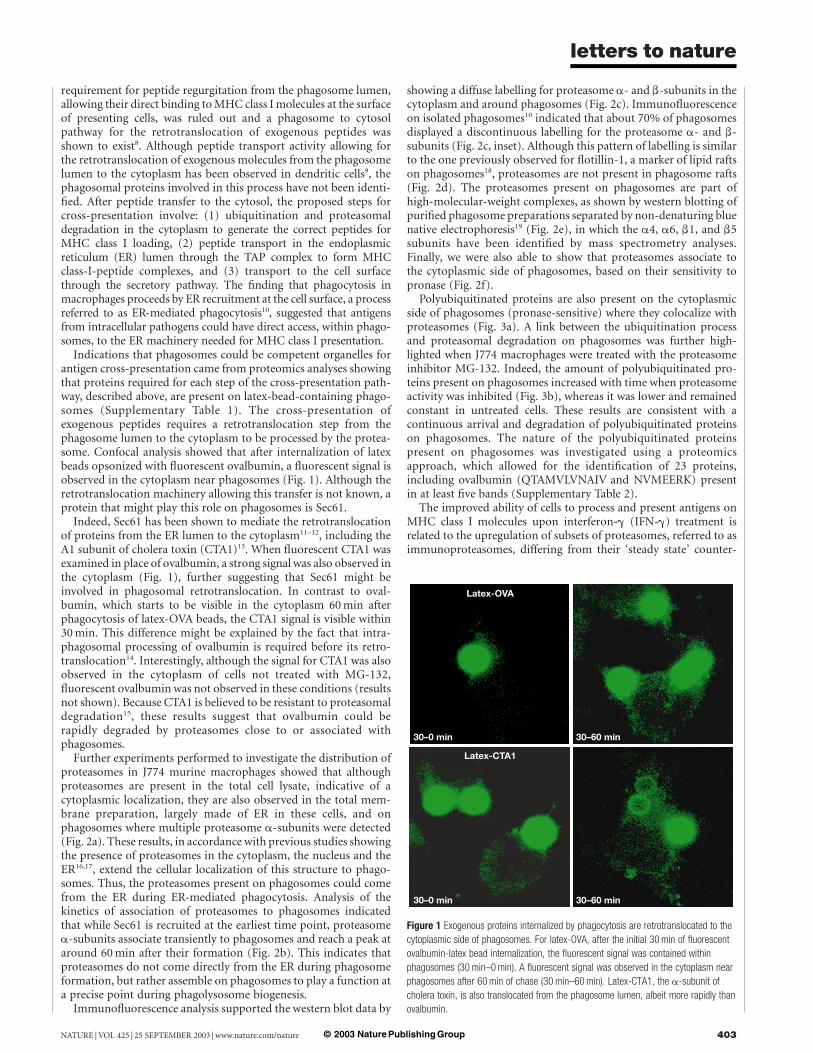
requirement for peptide regurgitation from the phagosome lumen,allowing their direct binding toMHC class Imolecules at the surfaceof presenting cells, was ruled out and a phagosome to cytosolpathway for the retrotranslocation of exogenous peptides wasshown to exist8. Although peptide transport activity allowing forthe retrotranslocation of exogenous molecules from the phagosomelumen to the cytoplasm has been observed in dendritic cells9, thephagosomal proteins involved in this process have not been identi-fied. After peptide transfer to the cytosol, the proposed steps forcross-presentation involve: (1) ubiquitination and proteasomaldegradation in the cytoplasm to generate the correct peptides forMHC class I loading, (2) peptide transport in the endoplasmicreticulum (ER) lumen through the TAP complex to form MHCclass-I-peptide complexes, and (3) transport to the cell surfacethrough the secretory pathway. The finding that phagocytosis inmacrophages proceeds by ER recruitment at the cell surface, a processreferred to as ER-mediated phagocytosis10, suggested that antigensfrom intracellular pathogens could have direct access, within phago-somes, to the ER machinery needed for MHC class I presentation.
Indications that phagosomes could be competent organelles forantigen cross-presentation came from proteomics analyses showingthat proteins required for each step of the cross-presentation path-way, described above, are present on latex-bead-containing phago-somes (Supplementary Table 1). The cross-presentation ofexogenous peptides requires a retrotranslocation step from thephagosome lumen to the cytoplasm to be processed by the protea-some. Confocal analysis showed that after internalization of latexbeads opsonized with fluorescent ovalbumin, a fluorescent signal isobserved in the cytoplasm near phagosomes (Fig. 1). Although theretrotranslocation machinery allowing this transfer is not known, aprotein that might play this role on phagosomes is Sec61.
Indeed, Sec61 has been shown to mediate the retrotranslocationof proteins from the ER lumen to the cytoplasm11–12, including theA1 subunit of cholera toxin (CTA1)13. When fluorescent CTA1 wasexamined in place of ovalbumin, a strong signal was also observed inthe cytoplasm (Fig. 1), further suggesting that Sec61 might beinvolved in phagosomal retrotranslocation. In contrast to oval-bumin, which starts to be visible in the cytoplasm 60min afterphagocytosis of latex-OVA beads, the CTA1 signal is visible within30min. This difference might be explained by the fact that intra-phagosomal processing of ovalbumin is required before its retro-translocation14. Interestingly, although the signal for CTA1 was alsoobserved in the cytoplasm of cells not treated with MG-132,fluorescent ovalbumin was not observed in these conditions (resultsnot shown). Because CTA1 is believed to be resistant to proteasomaldegradation15, these results suggest that ovalbumin could berapidly degraded by proteasomes close to or associated withphagosomes.
Further experiments performed to investigate the distribution ofproteasomes in J774 murine macrophages showed that althoughproteasomes are present in the total cell lysate, indicative of acytoplasmic localization, they are also observed in the total mem-brane preparation, largely made of ER in these cells, and onphagosomes where multiple proteasome a-subunits were detected(Fig. 2a). These results, in accordance with previous studies showingthe presence of proteasomes in the cytoplasm, the nucleus and theER16,17, extend the cellular localization of this structure to phago-somes. Thus, the proteasomes present on phagosomes could comefrom the ER during ER-mediated phagocytosis. Analysis of thekinetics of association of proteasomes to phagosomes indicatedthat while Sec61 is recruited at the earliest time point, proteasomea-subunits associate transiently to phagosomes and reach a peak ataround 60min after their formation (Fig. 2b). This indicates thatproteasomes do not come directly from the ER during phagosomeformation, but rather assemble on phagosomes to play a function ata precise point during phagolysosome biogenesis.
Immunofluorescence analysis supported the western blot data by
showing a diffuse labelling for proteasome a- and b-subunits in thecytoplasm and around phagosomes (Fig. 2c). Immunofluorescenceon isolated phagosomes10 indicated that about 70% of phagosomesdisplayed a discontinuous labelling for the proteasome a- and b-subunits (Fig. 2c, inset). Although this pattern of labelling is similarto the one previously observed for flotillin-1, a marker of lipid raftson phagosomes18, proteasomes are not present in phagosome rafts(Fig. 2d). The proteasomes present on phagosomes are part ofhigh-molecular-weight complexes, as shown by western blotting ofpurified phagosome preparations separated by non-denaturing bluenative electrophoresis19 (Fig. 2e), in which the a4, a6, b1, and b5subunits have been identified by mass spectrometry analyses.Finally, we were also able to show that proteasomes associate tothe cytoplasmic side of phagosomes, based on their sensitivity topronase (Fig. 2f).Polyubiquitinated proteins are also present on the cytoplasmic
side of phagosomes (pronase-sensitive) where they colocalize withproteasomes (Fig. 3a). A link between the ubiquitination processand proteasomal degradation on phagosomes was further high-lighted when J774 macrophages were treated with the proteasomeinhibitor MG-132. Indeed, the amount of polyubiquitinated pro-teins present on phagosomes increased with time when proteasomeactivity was inhibited (Fig. 3b), whereas it was lower and remainedconstant in untreated cells. These results are consistent with acontinuous arrival and degradation of polyubiquitinated proteinson phagosomes. The nature of the polyubiquitinated proteinspresent on phagosomes was investigated using a proteomicsapproach, which allowed for the identification of 23 proteins,including ovalbumin (QTAMVLVNAIV and NVMEERK) presentin at least five bands (Supplementary Table 2).The improved ability of cells to process and present antigens on
MHC class I molecules upon interferon-g (IFN-g) treatment isrelated to the upregulation of subsets of proteasomes, referred to asimmunoproteasomes, differing from their ‘steady state’ counter-
Figure 1 Exogenous proteins internalized by phagocytosis are retrotranslocated to the
cytoplasmic side of phagosomes. For latex-OVA, after the initial 30min of fluorescent
ovalbumin-latex bead internalization, the fluorescent signal was contained within
phagosomes (30min–0min). A fluorescent signal was observed in the cytoplasm near
phagosomes after 60min of chase (30min–60min). Latex-CTA1, the a-subunit of
cholera toxin, is also translocated from the phagosome lumen, albeit more rapidly than
ovalbumin.
letters to nature
NATURE |VOL 425 | 25 SEPTEMBER 2003 | www.nature.com/nature 403© 2003 Nature Publishing Group
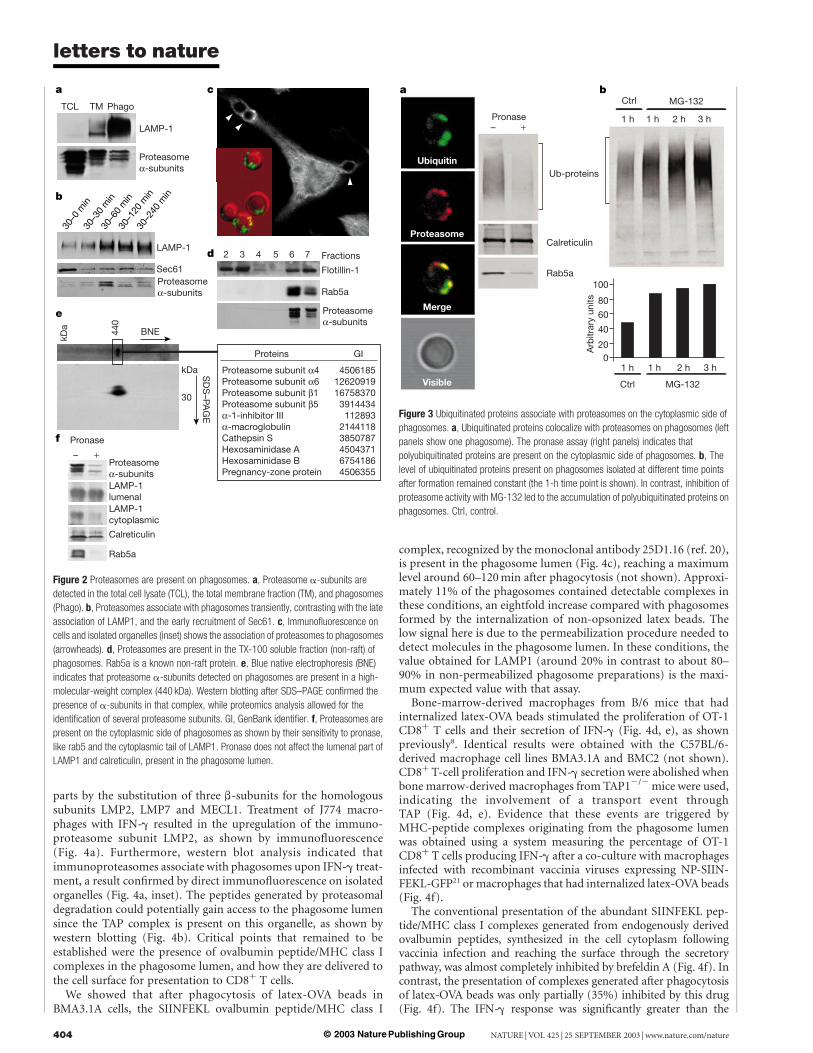
parts by the substitution of three b-subunits for the homologoussubunits LMP2, LMP7 and MECL1. Treatment of J774 macro-phages with IFN-g resulted in the upregulation of the immuno-proteasome subunit LMP2, as shown by immunofluorescence(Fig. 4a). Furthermore, western blot analysis indicated thatimmunoproteasomes associate with phagosomes upon IFN-g treat-ment, a result confirmed by direct immunofluorescence on isolatedorganelles (Fig. 4a, inset). The peptides generated by proteasomaldegradation could potentially gain access to the phagosome lumensince the TAP complex is present on this organelle, as shown bywestern blotting (Fig. 4b). Critical points that remained to beestablished were the presence of ovalbumin peptide/MHC class Icomplexes in the phagosome lumen, and how they are delivered tothe cell surface for presentation to CD8þ T cells.We showed that after phagocytosis of latex-OVA beads in
BMA3.1A cells, the SIINFEKL ovalbumin peptide/MHC class I
complex, recognized by the monoclonal antibody 25D1.16 (ref. 20),is present in the phagosome lumen (Fig. 4c), reaching a maximumlevel around 60–120min after phagocytosis (not shown). Approxi-mately 11% of the phagosomes contained detectable complexes inthese conditions, an eightfold increase compared with phagosomesformed by the internalization of non-opsonized latex beads. Thelow signal here is due to the permeabilization procedure needed todetect molecules in the phagosome lumen. In these conditions, thevalue obtained for LAMP1 (around 20% in contrast to about 80–90% in non-permeabilized phagosome preparations) is the maxi-mum expected value with that assay.
Bone-marrow-derived macrophages from B/6 mice that hadinternalized latex-OVA beads stimulated the proliferation of OT-1CD8þ T cells and their secretion of IFN-g (Fig. 4d, e), as shownpreviously8. Identical results were obtained with the C57BL/6-derived macrophage cell lines BMA3.1A and BMC2 (not shown).CD8þ T-cell proliferation and IFN-g secretionwere abolished whenbone marrow-derived macrophages from TAP12/2mice were used,indicating the involvement of a transport event throughTAP (Fig. 4d, e). Evidence that these events are triggered byMHC-peptide complexes originating from the phagosome lumenwas obtained using a system measuring the percentage of OT-1CD8þ T cells producing IFN-g after a co-culture with macrophagesinfected with recombinant vaccinia viruses expressing NP-SIIN-FEKL-GFP21 or macrophages that had internalized latex-OVA beads(Fig. 4f).
The conventional presentation of the abundant SIINFEKL pep-tide/MHC class I complexes generated from endogenously derivedovalbumin peptides, synthesized in the cell cytoplasm followingvaccinia infection and reaching the surface through the secretorypathway, was almost completely inhibited by brefeldin A (Fig. 4f). Incontrast, the presentation of complexes generated after phagocytosisof latex-OVA beads was only partially (35%) inhibited by this drug(Fig. 4f). The IFN-g response was significantly greater than the
Figure 3 Ubiquitinated proteins associate with proteasomes on the cytoplasmic side of
phagosomes. a, Ubiquitinated proteins colocalize with proteasomes on phagosomes (left
panels show one phagosome). The pronase assay (right panels) indicates that
polyubiquitinated proteins are present on the cytoplasmic side of phagosomes. b, The
level of ubiquitinated proteins present on phagosomes isolated at different time points
after formation remained constant (the 1-h time point is shown). In contrast, inhibition of
proteasome activity with MG-132 led to the accumulation of polyubiquitinated proteins on
phagosomes. Ctrl, control.
Figure 2 Proteasomes are present on phagosomes. a, Proteasome a-subunits are
detected in the total cell lysate (TCL), the total membrane fraction (TM), and phagosomes
(Phago). b, Proteasomes associate with phagosomes transiently, contrasting with the late
association of LAMP1, and the early recruitment of Sec61. c, Immunofluorescence on
cells and isolated organelles (inset) shows the association of proteasomes to phagosomes
(arrowheads). d, Proteasomes are present in the TX-100 soluble fraction (non-raft) of
phagosomes. Rab5a is a known non-raft protein. e, Blue native electrophoresis (BNE)
indicates that proteasome a-subunits detected on phagosomes are present in a high-
molecular-weight complex (440 kDa). Western blotting after SDS–PAGE confirmed the
presence of a-subunits in that complex, while proteomics analysis allowed for the
identification of several proteasome subunits. GI, GenBank identifier. f, Proteasomes are
present on the cytoplasmic side of phagosomes as shown by their sensitivity to pronase,
like rab5 and the cytoplasmic tail of LAMP1. Pronase does not affect the lumenal part of
LAMP1 and calreticulin, present in the phagosome lumen.
letters to nature
NATURE | VOL 425 | 25 SEPTEMBER 2003 | www.nature.com/nature404 © 2003 Nature Publishing Group
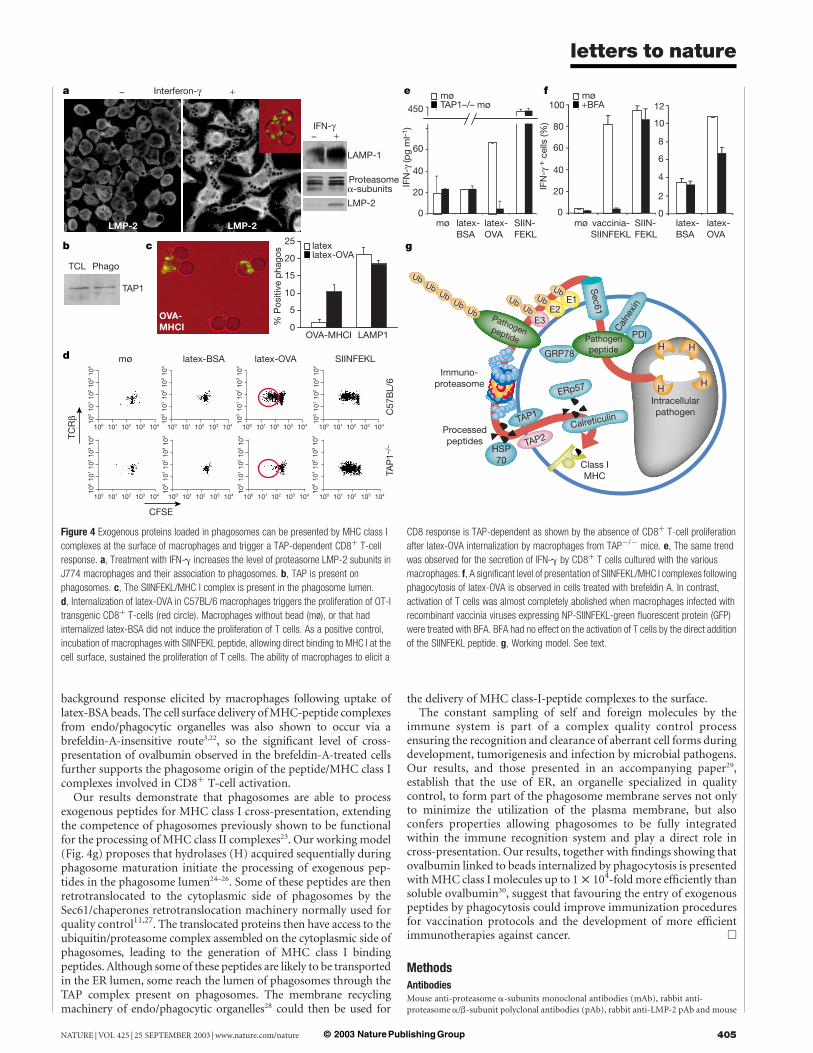
background response elicited by macrophages following uptake oflatex-BSAbeads. The cell surface delivery ofMHC-peptide complexesfrom endo/phagocytic organelles was also shown to occur via abrefeldin-A-insensitive route3,22, so the significant level of cross-presentation of ovalbumin observed in the brefeldin-A-treated cellsfurther supports the phagosome origin of the peptide/MHC class Icomplexes involved in CD8þ T-cell activation.
Our results demonstrate that phagosomes are able to processexogenous peptides for MHC class I cross-presentation, extendingthe competence of phagosomes previously shown to be functionalfor the processing of MHC class II complexes23. Our working model(Fig. 4g) proposes that hydrolases (H) acquired sequentially duringphagosome maturation initiate the processing of exogenous pep-tides in the phagosome lumen24–26. Some of these peptides are thenretrotranslocated to the cytoplasmic side of phagosomes by theSec61/chaperones retrotranslocation machinery normally used forquality control11,27. The translocated proteins then have access to theubiquitin/proteasome complex assembled on the cytoplasmic side ofphagosomes, leading to the generation of MHC class I bindingpeptides. Although some of these peptides are likely to be transportedin the ER lumen, some reach the lumen of phagosomes through theTAP complex present on phagosomes. The membrane recyclingmachinery of endo/phagocytic organelles28 could then be used for
the delivery of MHC class-I-peptide complexes to the surface.The constant sampling of self and foreign molecules by the
immune system is part of a complex quality control processensuring the recognition and clearance of aberrant cell forms duringdevelopment, tumorigenesis and infection by microbial pathogens.Our results, and those presented in an accompanying paper29,establish that the use of ER, an organelle specialized in qualitycontrol, to form part of the phagosome membrane serves not onlyto minimize the utilization of the plasma membrane, but alsoconfers properties allowing phagosomes to be fully integratedwithin the immune recognition system and play a direct role incross-presentation. Our results, together with findings showing thatovalbumin linked to beads internalized by phagocytosis is presentedwithMHC class I molecules up to 1 £ 104-foldmore efficiently thansoluble ovalbumin30, suggest that favouring the entry of exogenouspeptides by phagocytosis could improve immunization proceduresfor vaccination protocols and the development of more efficientimmunotherapies against cancer. A
MethodsAntibodiesMouse anti-proteasome a-subunits monoclonal antibodies (mAb), rabbit anti-proteasome a/b-subunit polyclonal antibodies (pAb), rabbit anti-LMP-2 pAb and mouse
Figure 4 Exogenous proteins loaded in phagosomes can be presented by MHC class I
complexes at the surface of macrophages and trigger a TAP-dependent CD8þ T-cell
response. a, Treatment with IFN-g increases the level of proteasome LMP-2 subunits in
J774 macrophages and their association to phagosomes. b, TAP is present on
phagosomes. c, The SIINFEKL/MHC I complex is present in the phagosome lumen.
d, Internalization of latex-OVA in C57BL/6 macrophages triggers the proliferation of OT-I
transgenic CD8þ T-cells (red circle). Macrophages without bead (mø), or that had
internalized latex-BSA did not induce the proliferation of T cells. As a positive control,
incubation of macrophages with SIINFEKL peptide, allowing direct binding to MHC I at the
cell surface, sustained the proliferation of T cells. The ability of macrophages to elicit a
CD8 response is TAP-dependent as shown by the absence of CD8þ T-cell proliferation
after latex-OVA internalization by macrophages from TAP2/2 mice. e, The same trend
was observed for the secretion of IFN-g by CD8þ T cells cultured with the various
macrophages. f, A significant level of presentation of SIINFEKL/MHC I complexes following
phagocytosis of latex-OVA is observed in cells treated with brefeldin A. In contrast,
activation of T cells was almost completely abolished when macrophages infected with
recombinant vaccinia viruses expressing NP-SIINFEKL-green fluorescent protein (GFP)
were treated with BFA. BFA had no effect on the activation of T cells by the direct addition
of the SIINFEKL peptide. g, Working model. See text.
letters to nature
NATURE |VOL 425 | 25 SEPTEMBER 2003 | www.nature.com/nature 405© 2003 Nature Publishing Group
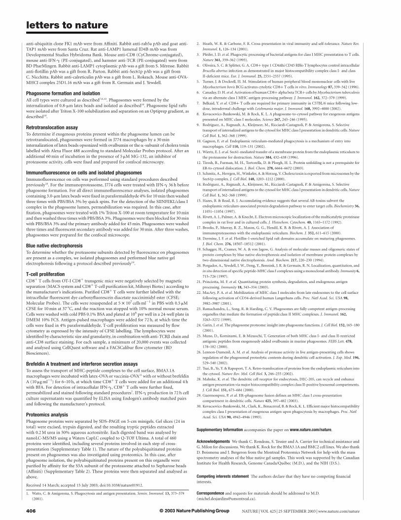
anti-ubiquitin clone FK1 mAb were from Affiniti. Rabbit anti-rab5a pAb and goat anti-TAP1 mAb were from Santa Cruz. Rat anti-LAMP1 lumenal ID4B mAb was fromDevelopmental Studies Hybridoma Bank. Mouse anti-CD8 (CyChrome-conjugated),mouse anti-IFN-g (PE-conjugated), and hamster anti-TCR (PE-conjugated) were fromBD PharMingen. Rabbit anti-LAMP1 cytoplasmic pAb was a gift from S. Meresse. Rabbitanti-flotillin pAb was a gift from R. Parton. Rabbit anti-Sec61p pAb was a gift fromC. Nicchitta. Rabbit anti-calreticulin pAb was a gift from L. Rokeach. Mouse anti-OVA-MHCI complex 25D1.16 mAb was a gift from R. Germain and J. Yewdell.
Phagosome formation and isolationAll cell types were cultured as described7,8,10. Phagosomes were formed by theinternalization of 0.8-mm latex beads and isolated as described10. Phagosome lipid raftswere isolated after Triton X-100 solubilization and separation on an Optiprep gradient, asdescribed18.
Retrotranslocation assayTo determine if exogenous proteins present within the phagosome lumen can beretrotranslocated, phagosomes were formed in J774 macrophages by a 30mininternalization of latex beads opsonized with ovalbumin or the a-subunit of cholera toxinlabelled with Alexa Fluor 488 according to standard Molecular Probes protocol. After anadditional 60min of incubation in the presence of 5 mM MG-132, an inhibitor ofproteasome activity, cells were fixed and prepared for confocal microscopy.
Immunofluorescence on cells and isolated phagosomesImmunofluorescence on cells was performed using standard procedures describedpreviously10. For the immunoproteasome, J774 cells were treated with IFN-g 36 h beforephagosome formation. For all direct immunofluorescence analyses, isolated phagosomescontaining 3.0-mm latex beads were fixed in paraformaldehyde 4% for 10min thenwashedthree times with PBS/BSA 3% by quick spins. For the detection of the SIINFEKL/class Icomplex in the phagosome lumen, permeabilization was required. In this case, afterfixation, phagosomes were treated with 1% Triton X-100 at room temperature for 10minand thenwashed three times with PBS/BSA 3%. Phagosomes were then blocked for 30minwith PBS/BSA 3% and the primary antibody added for 45min. Phagosomes were washedthree times and fluorescent secondary antibody was added for 30min. After three washes,phagosomes were prepared for the confocal microscope.
Blue native electrophoresisTo determine whether the proteasome subunits detected by fluorescence on phagosomesare present as a complex, we isolated phagosomes and performed blue native gelelectrophoresis following a protocol described previously19.
T-cell proliferationCD8þ T cells from OT-I CD8þ transgenic mice were negatively selected by magneticseparation (MACS system and CD8þ T-cell purification kit, Miltenyi Biotec) according tothe manufacturer’s indications. Purified CD8þ T cells were further labelled with theintracellular fluorescent dye carboxyfluorescein diacetate succinimidyl ester (CFSE;Molecular Probes). The cells were resuspended at 5 £ 107 cellsml21 in PBS with 0.5 mMCFSE for 10min at 37 8C, and the reaction was stopped with 10% normal mouse serum.Cells were washed with cold PBS 0.1% BSA and plated at 106 per well in a 24-well plate inDMEM 10% FCS. Antigen-pulsed macrophages were added for 72 h, at which time thecells were fixed in 4% paraformaldehyde. T-cell proliferation was measured by flowcytometry as expressed by the intensity of CFSE labelling. The lymphocytes wereidentified by characteristic size and granularity, in combinationwith anti-TCRb chain andanti-CD8 surface staining. For each sample, a minimum of 20,000 events was collectedand analysed using CellQuest software and a FACSCalibur flow cytometer (BDBiosciences).
Brefeldin A treatment and interferon secretion assaysTo assess the transport of MHC-peptide complexes to the cell surface, BMA3.1Amacrophages were incubated with latex-OVA or vaccinia-OVA21 with or without brefeldinA (10 mgml21) for 6–10 h, at which time CD8þ T cells were added for an additional 4 hwith BFA. For detection of intracellular IFN-g, CD8þ T cells were further fixed,permeabilized and stained following standard procedures7. IFN-g production in 72 h cellculture supernatants was quantified by ELISA using Endogen’s antibody matched pairsand following the manufacturer’s protocol.
Proteomics analysisPhagosome proteins were separated by SDS–PAGE on 5-cm minigels. Gel slices (24 intotal) were excised, trypsin digested, and the resulting tryptic peptides extractedwith 0.2M urea in 50% aqueous acetonitrile. Each digested band was analysed bynanoLC-MS/MS using a Waters CapLC coupled to Q-TOF Ultima. A total of 460proteins were identified, including several proteins involved in each step of cross-presentation (Supplementary Table 1). The nature of the polyubiquitinated proteinspresent on phagosomes was also investigated using proteomics. In this case, afterphagosome isolation, the polyubiquitinated proteins present on this organelle werepurified by affinity for the S5A subunit of the proteasome attached to Sepharose beads(Affiniti) (Supplementary Table 2). These proteins were then separated and analysed asabove.
Received 14 March; accepted 15 July 2003; doi:10.1038/nature01912.
1. Watts, C. & Amigorena, S. Phagocytosis and antigen presentation. Semin. Immunol. 13, 373–379
(2001).
2. Heath, W. R. & Carbone, F. R. Cross-presentation in viral immunity and self-tolerance. Nature Rev.
Immunol. 1, 126–134 (2001).
3. Pfeifer, J. D. et al. Phagocytic processing of bacterial antigens for class I MHC presentation to T cells.
Nature 361, 359–362 (1993).
4. Oliveira, S. C. & Splitter, G. A. CD8þ type 1 CD44hi CD45 RBlo T lymphocytes control intracellular
Brucella abortus infection as demonstrated in major histocompatibility complex class I- and class
II-deficient mice. Eur. J. Immunol. 25, 2551–2557 (1995).
5. Turner, J. & Dockrell, H. M. Stimulation of human peripheral blood mononuclear cells with live
Mycobacterium bovis BCG activates cytolytic CD8þ T cells in vitro. Immunology 87, 339–342 (1996).
6. Canaday, D. H. et al.Activation of humanCD8þ alpha beta TCRþ cells byMycobacterium tuberculosis
via an alternate class I MHC antigen-processing pathway. J. Immunol. 162, 372–379 (1999).
7. Belkaid, Y. et al. CD8þ T cells are required for primary immunity in C57BL/6 mice following low-
dose, intradermal challenge with Leishmania major. J. Immunol. 168, 3992–4000 (2002).
8. Kovacsovics-Bankowski, M. & Rock, K. L. A phagosome-to-cytosol pathway for exogenous antigens
presented on MHC class I molecules. Science 267, 243–246 (1995).
9. Rodriguez, A., Regnault, A., Kleijmeer, M., Ricciardi-Castagnoli, P. & Amigorena, S. Selective
transport of internalized antigens to the cytosol forMHC class I presentation in dendritic cells.Nature
Cell Biol. 1, 362–368 (1999).
10. Gagnon, E. et al. Endoplasmic reticulum-mediated phagocytosis is a mechanism of entry into
macrophages. Cell 110, 119–131 (2002).
11. Wiertz, E. J. et al. Sec61-mediated transfer of a membrane protein from the endoplasmic reticulum to
the proteasome for destruction. Nature 384, 432–438 (1996).
12. Tirosh, B., Furman, M. H., Tortorella, D. & Ploegh, H. L. Protein unfolding is not a prerequisite for
ER-to-cytosol dislocation. J. Biol. Chem. 278, 6664–6672 (2003).
13. Schmitz, A., Herrgen,H.,Winkeler, A. &Herzog, V. Cholera toxin is exported frommicrosomes by the
Sec61p complex. J. Cell Biol. 148, 1203–1212 (2000).
14. Rodriguez, A., Regnault, A., Kleijmeer, M., Ricciardi-Castagnoli, P. & Amigorena, S. Selective
transport of internalized antigens to the cytosol forMHC class I presentation in dendritic cells.Nature
Cell Biol. 1, 362–368 (1999).
15. Hazes, B. & Read, R. J. Accumulating evidence suggests that several AB-toxins subvert the
endoplasmic reticulum-associated protein degradation pathway to enter target cells. Biochemistry 36,
11051–11054 (1997).
16. Rivett, A. J., Palmer, A. & Knecht, E. Electronmicroscopic localization of the multicatalytic proteinase
complex in rat liver and in cultured cells. J. Histochem. Cytochem. 40, 1165–1172 (1992).
17. Brooks, P., Murray, R. Z., Mason, G. G., Hendil, K. B. & Rivett, A. J. Association of
immunoproteasomes with the endoplasmic reticulum. Biochem. J. 352, 611–615 (2000).
18. Dermine, J. F. et al. Flotillin-1-enriched lipid raft domains accumulate on maturing phagosomes.
J. Biol. Chem. 276, 18507–18512 (2001).
19. Schagger, H., Cramer, W. A. & von Jagow, G. Analysis of molecular masses and oligomeric states of
protein complexes by blue native electrophoresis and isolation of membrane protein complexes by
two-dimensional native electrophoresis. Anal. Biochem. 217, 220–230 (1994).
20. Porgador, A., Yewdell, J. W., Deng, Y., Bennink, J. R. & Germain, R. N. Localization, quantitation, and
in situ detection of specific peptide-MHC class I complexes using amonoclonal antibody. Immunity 6,
715–726 (1997).
21. Princiotta, M. F. et al. Quantitating protein synthesis, degradation, and endogenous antigen
processing. Immunity 18, 343–354 (2003).
22. MacAry, P. A. et al. Mobilization of MHC class I molecules from late endosomes to the cell surface
following activation of CD34-derived human Langerhans cells. Proc. Natl Acad. Sci. USA 98,
3982–3987 (2001).
23. Ramachandra, L., Song, R. & Harding, C. V. Phagosomes are fully competent antigen-processing
organelles that mediate the formation of peptide:class II MHC complexes. J. Immunol. 162,
3263–3272 (1999).
24. Garin, J. et al. The phagosome proteome: insight into phagosome functions. J. Cell Biol. 152, 165–180
(2001).
25. Muno, D., Kominami, E. & Mizuochi, T. Generation of both MHC class I- and class II-restricted
antigenic peptides from exogenously added ovalbumin in murine phagosomes. FEBS Lett. 478,
178–182 (2000).
26. Lennon-Dumenil, A. M. et al. Analysis of protease activity in live antigen-presenting cells shows
regulation of the phagosomal proteolytic contents during dendritic cell activation. J. Exp. Med. 196,
529–540 (2002).
27. Tsai, B., Ye, Y. & Rapoport, T. A. Retro-translocation of proteins from the endoplasmic reticulum into
the cytosol. Nature Rev. Mol. Cell Biol. 3, 246–255 (2002).
28. Mahnke, K. et al. The dendritic cell receptor for endocytosis, DEC-205, can recycle and enhance
antigen presentation via major histocompatibility complex class II-positive lysosomal compartments.
J. Cell Biol. 151, 673–684 (2000).
29. Guermonprez, P. et al. ER–phagosome fusion defines an MHC class I cross-presentation
compartment in dendritic cells. Nature 425, 397–402 (2003).
30. Kovacsovics-Bankowski, M., Clark, K., Benacerraf, B. & Rock, K. L. Efficient major histocompatibility
complex class I presentation of exogenous antigen upon phagocytosis by macrophages. Proc. Natl
Acad. Sci. USA 90, 4942–4946 (1993).
Supplementary Information accompanies the paper on www.nature.com/nature.
Acknowledgements We thank C. Rondeau, S. Tessier and A. Carrier for technical assistance and
G. Milon for discussions. We thank K. Rock for the BMA3.1A and BMC2 cell lines. We also thank
D. Boismenu and J. Bergeron from the Montreal Proteomics Network for help with the mass
spectrometry analyses of the blue native gel samples. This work was supported by the Canadian
Institute for Health Research, Genome Canada/Quebec (M.D.), and the NIH (D.S.).
Competing interests statement The authors declare that they have no competing financial
interests.
Correspondence and requests for materials should be addressed to M.D.
letters to nature
NATURE | VOL 425 | 25 SEPTEMBER 2003 | www.nature.com/nature406 © 2003 Nature Publishing Group
xxiii
Annexe IV. Article 4
Stuart, L. M., Boulais, J., Charriere, G. M., Hennessy, E. J., Brunet, S., Jutras, I.,
Goyette, G., Rondeau, C., Letarte, S., Huang, H., Ye, P., Morales, F., Kocks, C.,
Bader, J. S., Desjardins, M., Ezekowitz, R. A. B. (2007) A systems biology analysis
of the Drosophila phagosome. Nature 445, 95-101
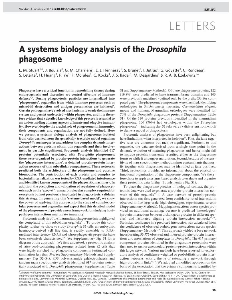
LETTERS
A systems biology analysis of the DrosophilaphagosomeL. M. Stuart1,2, J. Boulais3, G. M. Charriere1, E. J. Hennessy1, S. Brunet3, I. Jutras3, G. Goyette3, C. Rondeau3,S. Letarte3, H. Huang4, P. Ye4, F. Morales5, C. Kocks1, J. S. Bader4, M. Desjardins3 & R. A. B. Ezekowitz1{
Phagocytes have a critical function in remodelling tissues duringembryogenesis and thereafter are central effectors of immunedefence1,2. During phagocytosis, particles are internalized into‘phagosomes’, organelles from which immune processes such asmicrobial destruction and antigen presentation are initiated3.Certain pathogens have evolvedmechanisms to evade the immunesystem and persist undetected within phagocytes, and it is there-fore evident that a detailed knowledge of this process is essential toan understanding of many aspects of innate and adaptive immun-ity. However, despite the crucial role of phagosomes in immunity,their components and organization are not fully defined. Herewe present a systems biology analysis of phagosomes isolatedfrom cells derived from the genetically tractable model organismDrosophila melanogaster and address the complex dynamic inter-actions between proteins within this organelle and their involve-ment in particle engulfment. Proteomic analysis identified 617proteins potentially associated with Drosophila phagosomes;these were organized by protein–protein interactions to generatethe ‘phagosome interactome’, a detailed protein–protein inter-action network of this subcellular compartment. These networkspredicted both the architecture of the phagosome and putativebiomodules. The contribution of each protein and complex tobacterial internalization was tested by RNA-mediated interferenceand identified known components of the phagocyticmachinery. Inaddition, the prediction and validation of regulators of phagocyt-osis such as the ‘exocyst’4, a macromolecular complex required forexocytosis but not previously implicated in phagocytosis, validatesthis strategy. In generating this ‘systems-based model’, we showthe power of applying this approach to the study of complex cel-lular processes and organelles and expect that this detailed modelof the phagosomewill provide a new framework for studying host–pathogen interactions and innate immunity.
Proteomic analysis of the mammalian phagosome has highlightedthe complexity of this subcellular organelle5–7. To probe this com-plexity further we chose to study Drosophila S2 cells, an embryonichaemocyte-derived cell line that is readily amenable to RNA-mediated interference (RNAi) and whose phagocytic properties havebeen extensively characterized8,9 (see Supplementary Fig. S1 for adiagram of the approach). We first undertook a proteomic analysisof latex-bead-containing phagosomes isolated from S2 cells thatwere highly enriched for phagosome components (estimated con-tamination less than 5%; see Supplementary Methods and Supple-mentary Figs S2–S4). SDS polyacrylamide gelelectrophaesis andtandem mass spectrometry analysis identified 617 proteins poten-tially associated with Drosophila phagosomes (Supplementary Table
S1 and Supplementary Methods). Of these phagosome proteins, 122(19.8%) were predicted to have transmembrane domains and 103were previously undefined (defined only by the prefix CG, for com-puted gene). The phagosome components were classified, identifyingorthologues in Saccharomyces cerevisiae, Caenorhabditis elegans,mouse and humans. Mammalian orthologues were identified for70% of the Drosophila phagosome proteins (Supplementary TableS1). Of the 140 proteins previously identified in the mammalianphagosome, 100 (70%) had orthologues within the Drosophilaphagosome5–7, indicating that S2 cells were a valid system fromwhichto derive a model of phagocytosis.
Proteomic analyses of phagosomes have been enlightening buthave limitations when interpreted in isolation10. First, the false nega-tive rates are unknown but may be significant. Pertinent to thisorganelle, the data are derived from a single time point in thedynamic evolution of maturing phagosomes and hence might failto include proteins transiently recruited either as the phagosomeforms or while it undergoes maturation. Second, because of the sens-itivity of mass spectrometry methods, minor contaminants that pur-ify together with phagosomes may be identified as false positives.Third, proteomics provides no information about the physical orfunctional organization of the phagosome components. We there-fore chose to apply a systems-based analysis to evaluate and organizethese proteomic data further (Supplementary Fig. S1).
To place the phagosome proteins in biological context, the pro-teomic data were used to generate a protein–protein interaction net-work of this organelle11–13. A base network of protein–proteininteractions was first generated from confidence-rated interactionsobserved in five large-scale, high-throughput, experimental screens(SupplementaryMethods). Mapping interactions across species pro-vided an additional advantage because it predicted ‘interologues’(protein interactions between orthologous proteins in different spe-cies) and facilitated aligning protein interaction networks14,15.Statistical confidence in a predicted interaction was calculated fromthe confidence of observed orthologous interactions across species(Supplementary Methods)13. This approach yielded a base networkincorporating 53,775 observed and inferred protein–protein interac-tions and associations of complexes in Drosophila. The 617 primarycomponent proteins identified in the phagosome proteomics werethen used to anchor a network of protein–protein interactions withinthis larger network. Various methods have been reported for explor-atory analysis of confidence-weighted or probabilistic protein inter-action networks, with a theme of extending a network throughhigh-probability links12,16. We adopted a modified approach on thebasis of the expectation that the primary components identified by
1Laboratory of Developmental Immunology, Massachusetts General Hospital/ Harvard Medical School, 55 Fruit Street, Boston, Massachusetts 02114, USA. 2MRC Centre forInflammation Research, The University of Edinburgh, The Queen’s Medical Research Institute, 47 Little France Crescent, Edinburgh EH16 4TJ, UK. 3Departement de pathologie etbiologie cellulaire, Universite de Montreal, Montreal, Quebec H3C 3J7, Canada. 4Department of Biomedical Engineering and High-Throughput Biology Center, Johns HopkinsUniversity, 3400 North Charles Street, Baltimore, Maryland 21218, USA. 5Department of Biomedical Engineering, Faculty of Medicine, McGill University, Montreal, Quebec H3A 2B4,Canada. {Present address: Merck Research Laboratories, RY80K-107, PO Box 2000, Rahway, New Jersey 07065, USA.
Vol 445 |4 January 2007 |doi:10.1038/nature05380
95Nature ©2007 Publishing Group
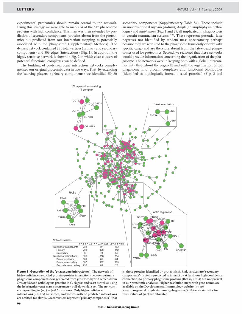
experimental proteomics should remain central to the network.Using this strategy we were able to map 214 of the 617 phagosomeproteins with high confidence. This map was then extended by pre-diction of secondary components, proteins absent from the proteo-mics but predicted from our interaction mapping as potentiallyassociated with the phagosome (Supplementary Methods). Thedensest network contained 281 total vertices (primary and secondarycomponents) and 806 edges (interactions) (Fig. 1). In addition, thehighly sensitive network is shown in Fig. 2 in which clear clusters ofpotential functional complexes can be defined.
The building of protein–protein interaction networks comple-mented our original proteomic data in two ways. First, by extendingthe ‘starting players’ (primary components) we identified 50–80
secondary components (Supplementary Table S7). These includean unconventional myosin (didum), Amph (an amphiphysin ortho-logue) and diaphanous (Figs 1 and 2), all implicated in phagocytosisin certain mammalian systems17–19. These represent potential falsenegatives not identified by tandem mass spectrometry perhapsbecause they are recruited to the phagosome transiently or only withspecific cargo and are therefore absent from the latex-bead phago-somes used for proteomics. Second, we reasoned that these networkswould provide information concerning the organization of the pha-gosome. The networks were in keeping both with a global intercon-nectivity throughout the organelle and with the organization of thephagosome into protein complexes and functional biomodules(identified as topologically interconnected proteins) (Figs 2 and
RAB1
GDI
RAB10 RAB11
RAB7RAB6
RAB8
ORCT
AMPH
14-3-3ε
CG6873
MYO31DF
FIM
ACT87E
CG12740
ZIP
DIDUM
CAM ACT57B
CHIC
ACT88F
EG:114D9.2
CAPTACT42A
TER94
MYO61F
ACT79B
ACT5C
TSR
DIA
SNAP
USNP
SYX1A
RAP
CCT5T-CP1
CG8231
CG7033
CCT-γ
CG16783
RABs
Actin regulation
Chaperonin-containingT complex
Vesicular fusion
Network statistics n = 4, c = 0.5 n = 2, c = 0.75 n = 2, c = 0.8 Number of components 281 214 162
Primary 201 136 112 Secondary 80 78 50
Number of interactions 806 356 204Primary–primary 181 81 64 Primary–secondary 387 192 115 Secondary–secondary 238 83 25
Figure 1 | Generation of the ‘phagosome interactome’. The network ofhigh-confidence predicted protein–protein interactions between primaryphagosome components was generated from yeast two-hybrid screens fromDrosophila and orthologous proteins in C. elegans and yeast as well as usingthe hybrigenics yeast mass spectrometry pull-down data set. The networkcorresponding to (n,c)5 (4,0.5) is shown. Only high-confidenceinteractions (c. 0.5) are shown, and vertices with no predicted interactionsare omitted for clarity. Green vertices represent ‘primary components’ (that
is, those proteins identified by proteomics). Pink vertices are ‘secondarycomponents’ (proteins predicted to interact by at least four high-confidenceconnections to primary phagosome proteins (that is, n5 4) but not presentin our proteomic analysis). Higher-resolution maps with gene names areavailable on the Developmental Immunology website (http://www.massgeneral.org/devimmunol/phagosome/). Network statistics forthree values of (n,c) are tabulated.
LETTERS NATURE |Vol 445 |4 January 2007
96Nature ©2007 Publishing Group
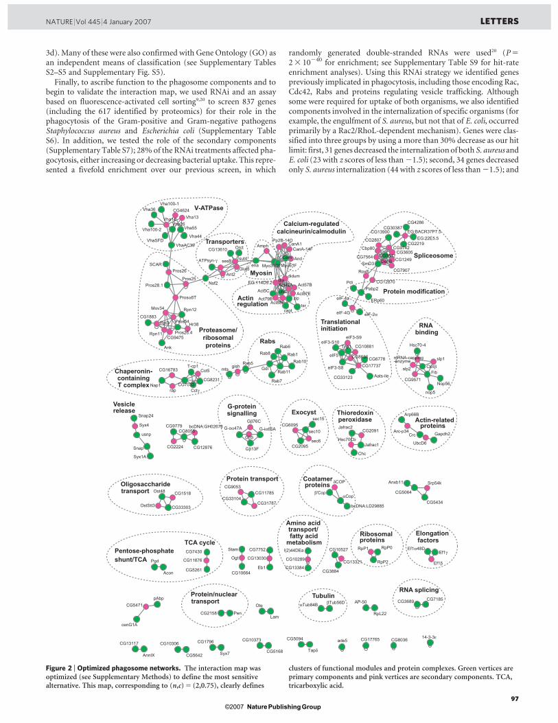
3d). Many of these were also confirmed with Gene Ontology (GO) asan independent means of classification (see Supplementary TablesS2–S5 and Supplementary Fig. S5).
Finally, to ascribe function to the phagosome components and tobegin to validate the interaction map, we used RNAi and an assaybased on fluorescence-activated cell sorting9,20 to screen 837 genes(including the 617 identified by proteomics) for their role in thephagocytosis of the Gram-positive and Gram-negative pathogensStaphylococcus aureus and Escherichia coli (Supplementary TableS6). In addition, we tested the role of the secondary components(Supplementary Table S7); 28% of the RNAi treatments affected pha-gocytosis, either increasing or decreasing bacterial uptake. This repre-sented a fivefold enrichment over our previous screen, in which
randomly generated double-stranded RNAs were used20 (P523 10240 for enrichment; see Supplementary Table S9 for hit-rateenrichment analyses). Using this RNAi strategy we identified genespreviously implicated in phagocytosis, including those encoding Rac,Cdc42, Rabs and proteins regulating vesicle trafficking. Althoughsome were required for uptake of both organisms, we also identifiedcomponents involved in the internalization of specific organisms (forexample, the engulfment of S. aureus, but not that of E. coli, occurredprimarily by a Rac2/RhoL-dependent mechanism). Genes were clas-sified into three groups by using a more than 30% decrease as our hitlimit: first, 31 genes decreased the internalizationof both S. aureus andE. coli (23 with z scores of less than21.5); second, 34 genes decreasedonly S. aureus internalization (44 with z scores of less than21.5); and
T-cp1
bcDNA:LD29885
FibGdi
eIF-4a
Anxb11
CG7185
CG33123
Cctγ
mts
Gapdh2
Nsf2
CG8636
CG11876
EG:BACR37P7.5
pAbp
Lam
Vha44
eIF3-S10
Act87E
Hsc70Cb
Act88F
Syx4
Srp54k
CG10664
Rab7
Ef1γ
CG13117
CG7752
Hsc70-4
OstStt3
Gβ5
ATPsyn-γ
CG10306
CG13900
CG2158
CG7564
CG2807
Act57B
CG17765
CG13030
G-iα65A
CG3542
AnnIX
Ef1α48D
CG30387
Snap24
Nap1
CG8036
dia
Gβ76C
CG33303
EG:114D9.2
And
Pros54
Act5C
CG10289
Myo31DF
CG13384
CG6095
Cam
Ant2
Rab6
CG10373
Ank
Arp66B
eIF3-S8
Vha26
Tapδ
CG12876
CG5471 Ote
CG9475
Gβ13F
CG13321Eb1
CG10527
Ef1β
Syx1A
Rab10
Rpn12 capt
CG5064CG9053
Vha14
αCop
sec10
Rpn11
CG8231
Vha13
CG8055
CG1796
Ost48
G-oα47A
Myo61FSCAR
δCOP
didum
Pgd
SmD3
cenG1A
nop5
Act79B
Crc
Cct5CkIIβ
CG5168
Rox8
Trip1
CG12870
CG31787
Nop56
CG3884
CG1518
Pen
αTub84B
CG1249
CG6778
Syx7
sut4
CG9779
CG5642
Jafrac2
mRNA-capping-enzyme
CG9742
eIF-4G
RpP0
Pros26.4
CG1883
AP-50
Chc
β'Cop
CanA-14F
enaDebB
sec15
ERp60
CG2095
CG4286
tsr
CG7033
CG2219
Rpt3
eIF5
rap
Aats-ile
14-3-3ε
Cbp80
ade5
eIF3-S9
Acon
CG3689
CG7907
slp1
RpL22
Pp2B-14D
CG9571
CanA1
Pros26
bcDNA:GH02678
l(2)44DEa
CG5094
CG10881
βTub56D
Hr38
CG13610 Orct
usnp
slp2
Vha100-2
Ogt
Arc-p34
CG11785
RpP1
VhaSFD
sesB
RpP2
CG7430
CG2091
CG5434
Vha55
VhaAC39
CG33104
Act42A
Rab8
Pros25
zip
Amph
CG4624
Rab11
Vha100-1Vha36
Mov34
gish
Prosα6T
CG3605
CG5261
CG16783
Jafrac1
Rab5
Snap
chic
Pdi
Glut3
sec6
Pabp2
CG17737
Stam
EG:22E5.5
Rab1
UbcD6CG2224
Pros28.1
eIF-2α
V-ATPase
Transporters
Myosin
Actin regulation
Spliceosome
Protein modification
Translational initiation
Rabs
RNA binding
Chaperonin-containingT complex
G-protein signalling Exocyst
Vesicle release Thioredoxin
peroxidase Actin-related proteins
Coatamer proteins
Tubulin
Protein transportOligosaccharide transport
Elongation factors
Ribosomal proteinsTCA cycle
Amino acidtransport/fatty acid
metabolism
RNA splicingProtein/nuclear transport
Pentose-phosphate
shunt/TCA
Proteasome/
ribosomal
proteins
Calcium-regulated
calcineurin/calmodulin
Figure 2 | Optimized phagosome networks. The interaction map wasoptimized (see Supplementary Methods) to define the most sensitivealternative. This map, corresponding to (n,c)5 (2,0.75), clearly defines
clusters of functional modules and protein complexes. Green vertices areprimary components and pink vertices are secondary components. TCA,tricarboxylic acid.
NATURE |Vol 445 |4 January 2007 LETTERS
97Nature ©2007 Publishing Group
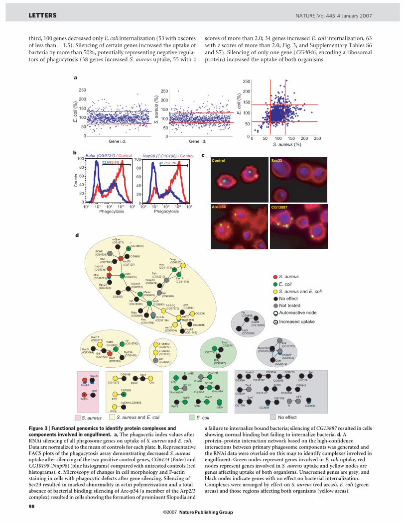
third, 100 genes decreased only E. coli internalization (53with z scoresof less than21.5). Silencing of certain genes increased the uptake ofbacteria by more than 50%, potentially representing negative regula-tors of phagocytosis (38 genes increased S. aureus uptake, 55 with z
scores of more than 2.0; 34 genes increased E. coli internalization, 63with z scores of more than 2.0; Fig. 3, and Supplementary Tables S6and S7). Silencing of only one gene (CG4046, encoding a ribosomalprotein) increased the uptake of both organisms.
bcDNA:LD29885
T-cp1(CG5374)
Gdi (CG4422)
Anxb11
plexB
CG7185
Ef1γ
CG13117
Magi
CG5830lap (CG2520)
AnnIX
Snap24(CG9474)
CG8036
Myo31DF(CG7438)
Cam(CG8472)
TapδVha26
Gβ13FCG10527
Syx1A (CG31136)
CG5064
αCop
sec10 (CG6159)
CG1796
SCAR(CG4636)
polo
CG5168
Nop56(CG13849)
CG3884 Pen
Syx7
kst (CG12008)
CG5642
α-Spec(CG1977)
RpP0
Chc
CG7033
eIF5
14-3-3ε(CG31196)
ade5
RpL22(CG7434)
Khc (CG7765)
βTub56D(CG9277)
VhaSFD
Actn (CG4376)
Rab11 (CG5771)
Snap (CG6625)
14-3-3ζ(CG17870)
Fib (CG9888)
Cctγ(CG8977)
Lam (CG6944)
Moe(CG10701)
r (CG18572)
CG10306
CG2158
G-iα65A
Ef1α48D
Act5C
CG7808
Rala(CG2849)
CG10373
Rab6 (CG6601)
Eb1(CG3265)
Hsp83 (CG1242)
capt
CG9951
CG8055
Syb (CG12210)
G-oα47A
Myo61F(CG9155)
Rac1
exo70 (CG7127)
nop5 (CG10206)
Rap2l
αTub84B(CG1913)
CG9779
sec5 (CG8843)
ena(CG15112)
sec15(CG7034)
CG2095
TSG101 (CG9712)
RpS3A (CG2168)
βSpec(CG5870)
CG3689R
CG5094
usnp (CG11173)
Pax(CG31794)
Hsp23
Hsp26
RpP2
Su(var)3-9
CG31048
zip(CG15792)
Rab5(CG3664)
Rab1(CG3320)
Ced-12(CG5336)
eIF-2α
S. aureus and E. coliS. aureus E. coli No effect
S. aureus and E. coli
S. aureus
E. coli
No effectNot testedAutoreactive node
0
50
100
150
200
250
0 50 100 150 200 2500
50
100
150
200
250
0
50
100
150
200
250
E. c
oli (
%)
E. c
oli (
%)
a
d
b c
Gene i.d. Gene i.d.
100 101 102 103 104
Phagocytosis
Cou
nts
0
20
40
60
80
100
100 101 102 103 1040
20
40
60
80
10052.9/82.9%
Phagocytosis
42.7/82.9%
Eater (CG6124) / Control Nup98 (CG10198) / Control
* *
*
* *
*
* Increased uptake
Control Sec23
CG13887Arc-p34
S. aureus (%)
S. a
ureu
s (%
)
Figure 3 | Functional genomics to identify protein complexes andcomponents involved in engulfment. a, The phagocytic index values afterRNAi silencing of all phagosome genes on uptake of S. aureus and E. coli.Data are normalized to themean of controls for each plate. b, RepresentativeFACS plots of the phagocytosis assay demonstrating decreased S. aureusuptake after silencing of the two positive control genes, CG6124 (Eater) andCG10198 (Nup98) (blue histograms) comparedwith untreated controls (redhistograms). c, Microscopy of changes in cell morphology and F-actinstaining in cells with phagocytic defects after gene silencing. Silencing ofSec23 resulted in marked abnormality in actin polymerization and a totalabsence of bacterial binding; silencing of Arc-p34 (a member of the Arp2/3complex) resulted in cells showing the formation of prominent filopodia and
a failure to internalize bound bacteria; silencing ofCG13887 resulted in cellsshowing normal binding but failing to internalize bacteria. d, Aprotein–protein interaction network based on the high-confidenceinteractions between primary phagosome components was generated andthe RNAi data were overlaid on this map to identify complexes involved inengulfment. Green nodes represent genes involved in E. coli uptake, rednodes represent genes involved in S. aureus uptake and yellow nodes aregenes affecting uptake of both organisms. Unscreened genes are grey, andblack nodes indicate genes with no effect on bacterial internalization.Complexes were arranged by effect on S. aureus (red areas), E. coli (greenareas) and those regions affecting both organisms (yellow areas).
LETTERS NATURE |Vol 445 |4 January 2007
98Nature ©2007 Publishing Group
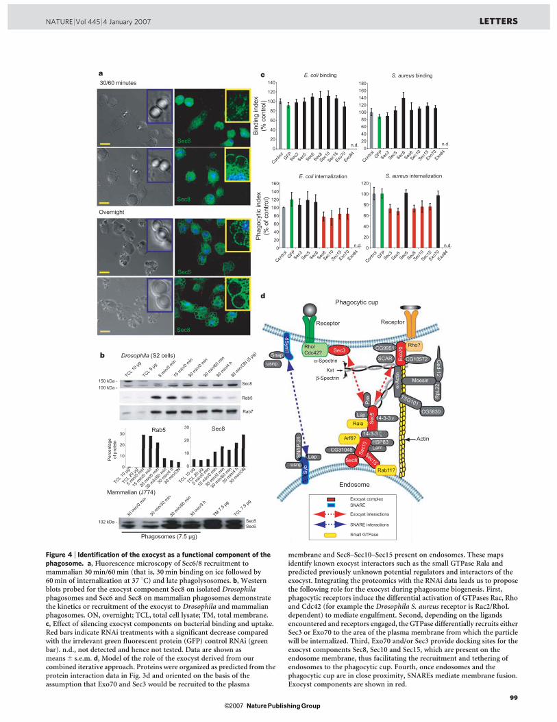
Exocyst complexSNARE
Exocyst interactions
SNARE interactions
Small GTPase
E. coli binding
E. coli internalization
S. aureus binding
S. aureus internalization
e.
Sec6Sec8102 kDa -
Mammalian (J774)
30/60 minutes
Overnight
b
a c
d
Drosophila (S2 cells)
Sec6
Sec6
Sec8
Sec8
20
40
60
80
100
120
140
20
40
60
80
100
120
180160140120100806040200
120
100
80
60
40
20
0
Sec6Sec
8Sec
15Sec
5Sec
10
Contro
lSec
3GFP
Exo70
Exo84
Sec6Sec
8Sec
15Sec
5Sec
10
Contro
lSec
3GFP
Exo70
Exo84
Sec6Sec
8Sec
15Sec
5Sec
10
Contro
lSec
3GFP
Exo70
Exo84
Sec6Sec
8Sec
15Sec
5Sec
10
Contro
lSec
3GFP
Exo70
Exo84
n.d.
n.d. n.d.
n.d.
TCL 10 μ
g
TCL 5 μg
5 min/
0 min
15 m
in/0 m
in
30 m
in/ON (5
μg)
30 m
in/4 h
30 m
in/60
min
30 m
in/0 m
in
150 kDa -100 kDa -
Sec8
Rab5
Rab7
Phagosomes (7.5 μg)
0
10
20
30
0
10
20
30Rab5 Sec8
Per
cent
age
of p
rote
in
Snap
ROP
Syb
SN
AP
-24
usnp
Actin
α-Spectrin
β-SpectrinKst
Sec8
SCAR
Sec15
Sec
5
HSP83
Sec1
0
14-3-3 ε
14-3-3 ζ
Pax
Ced-12
RpL
22
dSynt1
Endosome
Lap
TSG101
Phagocytic cup
CG5830
Lam
Lap
Moesin
usnp
CG31048
Exo
70α-
Act
in
CG9951
CG18572
Sec3Rho/Cdc42?
Rho?
ReceptorReceptor
30 m
in/0 m
in
30 m
in/30
min
30 m
in/60
min
30 m
in/3 h
TM 7.5 μ
g
TCL 7.5
μg
TCL 10 μ
g
TCL 20 μ
g
15 m
in/0 m
in
30 m
in/ON
30 m
in/4 h
30 m
in/60
min
30 m
in/0 m
in
5 min/
0 min
TCL 10 μ
g
TCL 20 μ
g
15 m
in/0 m
in
30 m
in/ON
30 m
in/4 h
30 m
in/60
min
30 m
in/0 m
in
5 min/
0 min Rab11?
Arf6?
Rala
0
160
Pha
gocy
tic in
dex
(% o
f con
trol)
0
140
Bin
ding
inde
x(%
con
trol)
Figure 4 | Identification of the exocyst as a functional component of thephagosome. a, Fluorescence microscopy of Sec6/8 recruitment tomammalian 30min/60min (that is, 30min binding on ice followed by60min of internalization at 37 uC) and late phagolysosomes. b, Westernblots probed for the exocyst component Sec8 on isolated Drosophilaphagosomes and Sec6 and Sec8 on mammalian phagosomes demonstratethe kinetics or recruitment of the exocyst to Drosophila and mammalianphagosomes. ON, overnight; TCL, total cell lysate; TM, total membrane.c, Effect of silencing exocyst components on bacterial binding and uptake.Red bars indicate RNAi treatments with a significant decrease comparedwith the irrelevant green fluorescent protein (GFP) control RNAi (greenbar). n.d., not detected and hence not tested. Data are shown asmeans6 s.e.m. d, Model of the role of the exocyst derived from ourcombined iterative approach. Proteins were organized as predicted from theprotein interaction data in Fig. 3d and oriented on the basis of theassumption that Exo70 and Sec3 would be recruited to the plasma
membrane and Sec8–Sec10–Sec15 present on endosomes. These mapsidentify known exocyst interactors such as the small GTPase Rala andpredicted previously unknown potential regulators and interactors of theexocyst. Integrating the proteomics with the RNAi data leads us to proposethe following role for the exocyst during phagosome biogenesis. First,phagocytic receptors induce the differential activation of GTPases Rac, Rhoand Cdc42 (for example the Drosophila S. aureus receptor is Rac2/RhoLdependent) to mediate engulfment. Second, depending on the ligandsencountered and receptors engaged, the GTPase differentially recruits eitherSec3 or Exo70 to the area of the plasma membrane from which the particlewill be internalized. Third, Exo70 and/or Sec3 provide docking sites for theexocyst components Sec8, Sec10 and Sec15, which are present on theendosome membrane, thus facilitating the recruitment and tethering ofendosomes to the phagocytic cup. Fourth, once endosomes and thephagocytic cup are in close proximity, SNAREs mediate membrane fusion.Exocyst components are shown in red.
NATURE |Vol 445 |4 January 2007 LETTERS
99Nature ©2007 Publishing Group
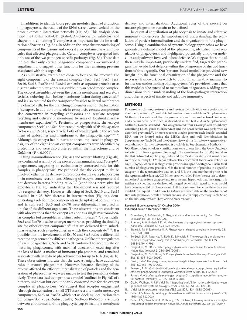
In addition, to identify those protein modules that had a functionin phagocytosis, the results of the RNAi screen were overlaid on theprotein–protein interaction networks (Fig. 3d). This analysis iden-tified the tubulin, Rab–GDI (Rab–GDP-dissociation inhibitor) andchaperonin-containing T-complexes as important for the internali-zation of bacteria (Fig. 3d). In addition the large cluster consisting ofcomponents of the fusome and exocyst also contained several mole-cules that affected phagocytosis. Certain complexes segregated intoonly one of the two pathogen-specific pathways (Fig. 3d). These dataindicate that only certain phagosome components are involved inengulfment and suggest other functions for the remaining proteinsassociated with this organelle.
As an illustrative example we chose to focus on the exocyst4. Theeight components of the exocyst complex (Sec3, Sec5, Sec6, Sec8,Sec10, Sec15, Exo70 and Exo84) can exist as separate proteins or asdiscrete subcomplexes or can assemble into an octodimeric complex.The exocyst assembles between the plasma membrane and secretoryvesicles, tethering them before membrane fusion during exocytosis21
and is also required for the transport of vesicles to lateral membranesin polarized cells, for the branching of neurites and for the formationof synapses. In addition to its role in exocytosis, exocyst componentsalso concentrate in recycling endosomes and regulate receptorrecycling and delivery of membrane to areas of localized plasma-membrane expansion22,23. Pertinent to phagocytosis, the exocystcomponents Sec10 and Sec15 interact directly with ADP-ribosylationfactor 6 and Rab11, respectively, both of which regulate the recruit-ment of endosomes and membrane to the phagocytic cup22,24–26.Although the exocyst had hitherto not been implicated in phagocyt-osis, six of the eight known exocyst components were identified byproteomics and were also clustered within the interactome and byGoMiner (P, 0.0001).
Using immunofluoresence (Fig. 4a) andwestern blotting (Fig. 4b),we confirmed assembly of the exocyst onmammalian andDrosophilaphagosomes, suggesting an evolutionarily conserved role for thiscomplex in phagocytosis. We proposed that the exocyst might beinvolved either in the delivery of receptors during early phagocytosisor in membrane recruitment. Silencing of exocyst components didnot decrease bacteria binding at 27 uC (conditions permissive forexocytosis (Fig. 4c), indicating that the exocyst was not requiredfor receptor delivery. However, silencing of Sec8, Sec10 and Sec15resulted in a 25–30% decrease in internalization (Fig. 4c), dem-onstrating a role for these components in the uptake of both S. aureusand E. coli. Sec3, Sec5 and Exo70 were differentially involved inuptake of the different organisms. These observations are consistentwith observations that the exocyst acts not as a single macromolecu-lar complex but assembles as distinct subcomplexes27,28. Specifically,Sec3 and Exo70 localize to target membranes, providing the dockingsite for other exocyst components21 that are delivered from subcel-lular vesicles, such as endosomes, in which they concentrate22,23. It ispossible that the involvement of Exo70 and Sec3 reflects differentialreceptor engagement by different pathogens. Unlike other regulatorsof early phagocytosis, Sec6 and Sec8 continued to accumulate onmaturing phagosomes, with maximal association occurring afterthe loss of Rab5, a marker of immature phagosomes, and remainedassociated with latex-bead phagolysosomes for up to 16 h (Fig. 4a, b).These observations indicate that the exocyst might have additionalroles in mature phagosomes. However, because silencing of theexocyst affected the efficient internalization of particles and the gen-eration of phagosomes, we were unable to test this possibility defini-tively. These data lead us to propose amodel (outlined in Fig. 4d) of ahitherto unknown but evolutionarily conserved role for the exocystcomplex in phagocytosis. We suggest that receptor engagement(through the activation of small GTPases) recruitsmembrane exocystcomponents, Sec3 or Exo70, which act as docking localization siteson phagocytic cups. Subsequently, Sec8–Sec10–Sec15 assemblesbetween endosomes and the phagocytic cup to facilitate membrane
delivery and internalization. Additional roles of the exocyst onmature phagosomes remain to be defined.
The essential contribution of phagocytosis to innate and adaptiveimmunity underscores the importance of understanding the regu-lation of particle internalization and the organization of the phago-some. Using a combination of systems biology approaches we havegenerated a detailed model of the phagosome, identified novel reg-ulators of phagocytosis and highlighted potentially unknown mole-cules and pathways involved in host defence.We suggest that some ofthese may be important, previously unidentified, targets for patho-gens that evade host defence within the phagosome or disrupt func-tions of this organelle. Our ‘systems-based model’ has provided newinsight into the functional organization of the phagosome and thenecessary framework on which to build, in an iterative manner, tofurther our understanding of phagocytosis.We provide evidence thatthis model can be extended to mammalian phagocytosis, adding newdimensions to our understanding of the host–pathogen interactionand other aspects of innate and adaptive immunity.
METHODS
Phagosome isolation, proteomics and protein identification were performed as
described previously29; and detailed methods are available in Supplementary
Methods. Generation of the phagosome interactome and network inference
and analysis were performed as described in the text and in Supplementary
Methods. Double-stranded RNA was generated from a Drosophila RNAi library
containing 13,000 genes (Geneservice) and the RNAi screen was performed as
described previously20. Primer sequences used to generate each double-stranded
RNA can be located using the MRCg plate and well identifications in
Supplementary Table S8 and the Geneservice website (http://www.geneservice.-
co.uk/home/) (further information is available in Supplementary Methods).
GO Miner. Gene ontology classifications were drawn from the Gene Ontology
website (http://www.geneontology.org). The enrichment factor (Re) found in
the DAG (directed acyclic graph) files and the P value for statistical significance
were calculated by GOMiner as follows. The enrichment factor Re is defined as
(nf/n)/(Nf/N), where nf is phagosome proteins in a specific category, n is the total
number of phagosome proteins, Nf is the total number of proteins in a specific
category in the representative data set, and N is the total number of proteins in
the representative data set. GOMiner uses two-sided Fisher’s exact test to deter-
mine the P value for a category and tests the null hypothesis that the category is
neither enriched in, nor depleted of, flagged genes with regard to what would
have been expected by chance alone. Full data sets used to derive these data are
available on request. In addition, GOMiner generated data on the enrichment of
BioCarta pathways, details of which are available in Supplementary Table S5 or
on the BioCarta website (http://www.biocarta.com).
Received 13 July; accepted 24 October 2006.Published online 6 December 2006.
1. Greenberg, S. & Grinstein, S. Phagocytosis and innate immunity. Curr. Opin.Immunol. 14, 136–145 (2002).
2. Aderem, A. & Underhill, D. M. Mechanisms of phagocytosis in macrophages.Annu. Rev. Immunol. 17, 593–623 (1999).
3. Stuart, L. M. & Ezekowitz, R. A. Phagocytosis: elegant complexity. Immunity 22,539–550 (2005).
4. TerBush, D. R., Maurice, T., Roth, D. & Novick, P. The exocyst is a multiproteincomplex required for exocytosis in Saccharomyces cerevisiae. EMBO J. 15,6483–6494 (1996).
5. Desjardins, M. ER-mediated phagocytosis: a new membrane for new functions.Nature Rev. Immunol. 3, 280–291 (2003).
6. Desjardins, M. & Griffiths, G. Phagocytosis: latex leads the way. Curr. Opin. CellBiol. 15, 498–503 (2003).
7. Garin, J. et al. The phagosome proteome: insight into phagosome functions. J. CellBiol. 152, 165–180 (2001).
8. Pearson, A. M. et al. Identification of cytoskeletal regulatory proteins required forefficient phagocytosis in Drosophila. Microbes Infect. 5, 815–824 (2003).
9. Ramet,M. et al. Drosophila scavenger receptor CI is a pattern recognition receptorfor bacteria. Immunity 15, 1027–1038 (2001).
10. Ge, H.,Walhout, A. J. & Vidal, M. Integrating ‘omic’ information: a bridge betweengenomics and systems biology. Trends Genet. 19, 551–560 (2003).
11. Vidal, M. Interactome modeling. FEBS Lett. 579, 1834–1838 (2005).12. Bader, J. S. Greedily building protein networks with confidence. Bioinformatics 19,
1869–1874 (2003).
13. Bader, J. S., Chaudhuri, A., Rothberg, J. M. & Chant, J. Gaining confidence in high-throughput protein interaction networks. Nature Biotechnol. 22, 78–85 (2004).
LETTERS NATURE |Vol 445 |4 January 2007
100Nature ©2007 Publishing Group
14. Uetz, P. & Finley, R. L. Jr. From protein networks to biological systems. FEBS Lett.579, 1821–1827 (2005).
15. Matthews, L. R. et al. Identification of potential interaction networks usingsequence-based searches for conserved protein–protein interactions or‘interologs’. Genome Res. 11, 2120–2126 (2001).
16. Asthana, S., King, O. D., Gibbons, F. D. & Roth, F. P. Predicting protein complexmembership using probabilistic network reliability. Genome Res. 14, 1170–1175(2004).
17. Cox, D. et al. Myosin X is a downstream effector of PI(3)K during phagocytosis.Nature Cell Biol. 4, 469–477 (2002).
18. Gold, E. S. et al. Amphiphysin IIm, a novel amphiphysin II isoform, is required formacrophage phagocytosis. Immunity 12, 285–292 (2000).
19. Colucci-Guyon, E. et al. A role for mammalian diaphanous-related formins incomplement receptor (CR3)-mediated phagocytosis in macrophages. Curr. Biol.15, 2007–2012 (2005).
20. Ramet, M., Manfruelli, P., Pearson, A., Mathey-Prevot, B. & Ezekowitz, R. A.Functional genomic analysis of phagocytosis and identification of a Drosophilareceptor for E. coli. Nature 416, 644–648 (2002).
21. Boyd, C., Hughes, T., Pypaert, M. & Novick, P. Vesicles carry most exocystsubunits to exocytic sites marked by the remaining two subunits, Sec3p andExo70p. J. Cell Biol. 167, 889–901 (2004).
22. Zhang, X. M., Ellis, S., Sriratana, A., Mitchell, C. A. & Rowe, T. Sec15 is an effectorfor the Rab11 GTPase in mammalian cells. J. Biol. Chem. 279, 43027–43034(2004).
23. Prigent, M. et al.ARF6 controls post-endocytic recycling through its downstreamexocyst complex effector. J. Cell Biol. 163, 1111–1121 (2003).
24. Bajno, L. et al. Focal exocytosis of VAMP3-containing vesicles at sites ofphagosome formation. J. Cell Biol. 149, 697–706 (2000).
25. Niedergang, F., Colucci-Guyon, E., Dubois, T., Raposo, G. & Chavrier, P. ADPribosylation factor 6 is activated and controls membrane delivery duringphagocytosis in macrophages. J. Cell Biol. 161, 1143–1150 (2003).
26. Cox, D., Lee, D. J., Dale, B. M., Calafat, J. & Greenberg, S. A. Rab11-containingrapidly recycling compartment inmacrophages that promotes phagocytosis. Proc.Natl Acad. Sci. USA 97, 680–685 (2000).
27. Clandinin, T. R. Surprising twists to exocyst function.Neuron 46, 164–166 (2005).
28. Mehta, S. Q. et al. Mutations in Drosophila sec15 reveal a function in neuronaltargeting for a subset of exocyst components. Neuron 46, 219–232 (2005).
29. Desjardins, M., Huber, L. A., Parton, R. G. & Griffiths, G. Biogenesis ofphagolysosomes proceeds through a sequential series of interactions with theendocytic apparatus. J. Cell Biol. 124, 677–688 (1994).
Supplementary Information is linked to the online version of the paper atwww.nature.com/nature.
Acknowledgements We thank R. Kearney and J. Bergeron from the MontrealProteomics Network, and Genome-Quebec-Canada for their support. J.S.B., M.D.and R.A.B.E. thank their laboratories for their support. Theworkwas supported by aWellcome Trust Clinician Scientist Award to L.M.S., grants from the WhitakerFoundation andNIH/NIGMS to J.S.B, grants from the Canadian Institute for HealthResearch and Genome-Canada-Quebec to M.D., and NIH grants to R.A.B.E. Thework was conceived through discussions between the Laboratory ofDevelopmental Immunology, Massachusetts General Hospital/Harvard MedicalSchool and the Departement de pathologie et biologie cellulaire, Universite deMontreal. The bioinformatics and RNAi screens were performed in the Laboratoryof Developmental Immunology, Massachusetts General Hospital/HarvardMedical School; the protein–protein networks were generated in the Departmentof Biomedical Engineering and High-Throughput Biology Center, Johns HopkinsUniversity; the proteomics, the annotation of the components and the phagosomeisolation were performed in the Departement de pathologie et biologie cellulaire,Universite de Montreal.
Author Contributions L.M.S. and J.B. contributed equally to this work. J.S.B., M.D.and R.A.B.E. contributed equally to this work. Themanuscript waswritten by L.M.S.and the website linked to this paper was designed by G.M.C.
Author Information Reprints and permissions information is available atwww.nature.com/reprints. The authors declare no competing financial interests.Correspondence and requests for materials should be addressed to L.M.S.([email protected]).
NATURE |Vol 445 |4 January 2007 LETTERS
101Nature ©2007 Publishing Group