utilisation du ct-scan comme mÉthode de …
TRANSCRIPT
ConseilSupérieur de la Santé
UTILISATION DU CT-SCAN COMME MÉTHODE DE DIAGNOSTIC DANS LE CADRE DE L’ÉPIDÉMIE DE CORONAVIRUS EN BELGIQUE
JUIN 2020CSS N° 9587
DROITS D’AUTEUR
Service public Fédéral de la Santé publique, de la Sécurité de la Chaîne alimentaire et de l’Environnement
Conseil Supérieur de la SantéPlace Victor Horta 40 bte 10B-1060 Bruxelles
Tél.: 02/524 97 97 E-mail: [email protected]
Tous droits d’auteur réservés.Veuillez citer cette publication de la façon suivante: Conseil Supérieur de la Santé. Utilisation du CT-scan comme méthode de diagnostic dans le cadre de l’épidémie de coronavirus en Belgique. Bruxelles: CSS; 2020. Avis n° 9587.
La version intégrale de l’avis peut être téléchargés à partir de la page web: www.css-hgr.be
Cette publication ne peut être vendue
Conseil Supérieur de la Santé
www.css-hgr.be
− 1 −
AVIS URGENT DU CONSEIL SUPERIEUR DE LA SANTE N° 9587
Utilisation du CT-scan comme méthode de diagnostic dans le cadre de
l'épidémie de coronavirus en Belgique
In this scientific advisory report, which offers guidance to public health policy-makers,
the Superior Health Council of Belgium provides an expert opinion on the use of chest CT,
in addition to RT-PCR, for hospitalized patients suspected of having Covid-19.
This report aims at providing radiologists and emergency physicians with specific
recommendations on the use of chest CT as a diagnostic and triage tool.
Version 1 validée en urgence par le Bureau du Collège
Le 25 mars 20201
Version 4 validée par le Collège du
3 juin 2020
I INTRODUCTION ET QUESTION
Pour la troisième fois en quelques décennies, le monde se voit confronté à une épidémie à Coronavirus. Dans ce contexte, une identification précoce des patients atteints de ce nouveau virus, contre lequel aucun vaccin ni traitement spécifique n’existe, relève d’une importance capitale afin non seulement de permettre une prise en charge rapide des personnes concernées, mais également la mise en place de mesures adéquates pour prévenir la propagation de ce virus qui s’avère particulièrement contagieux. Le dépistage de la maladie respiratoire Covid-19 et du virus associé SARS-CoV-2 se fait actuellement principalement par réaction en chaîne par polymérase après transcription inverse (RT-PCR) . Cependant, les demandes de tests de dépistage doivent actuellement être priorisées, suite à des problèmes d’approvisionnement de réactifs essentiels à la réalisation de ces tests. Par ailleurs, la littérature affiche de grandes disparités quant à la sensibilité des tests PCR. Ainsi, plusieurs études réalisées en Chine ont affirmé que la RT-PCR a une sensibilité limitée de 60 à 70 % (Bai et al. 2020, Ai et al. 2020, Fang et al., 2020, Kanne et al., 2020), tandis que d’autres, réalisées notamment aux Etats Unis, annoncent une sensibilité de l’ordre de 95 à 97% (Mossa-Basha et al. 2020). Cette pénurie a initié la recherche d’autres procédés de diagnostic. Ainsi, le 17 mars, l’UNamur a présenté un nouveau procédé basé sur des tests chimiques requérant davantage d’opérations manuelles, permettant ainsi de se passer des réactifs en pénurie et d’obtenir un résultat fiable dans les 24 heures. Sur base de la littérature récente, le CSS s’est penché sur le rôle que pouvait jouer le CT scan dans la lutte contre ce virus, au niveau du diagnostic/triage et/ou du suivi du diagnostic de Covid-19 en complément des tests RT-PCR.
1 Le Conseil se réserve le droit de pouvoir apporter, à tout moment, des corrections typographiques mineures à ce document. Par contre, les corrections de sens sont d’office reprises dans un erratum et donnent lieu à une nouvelle version de l’avis.
Conseil Supérieur de la Santé
www.css-hgr.be
− 2 −
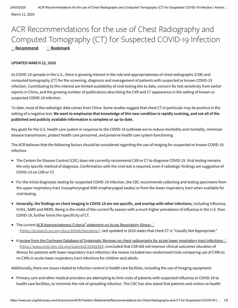
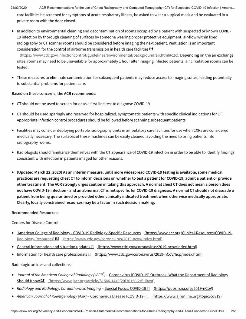
Un questionnaire a également été adressé à des experts radiologues du pays. Celui-ci portait sur l’utilisation actuelle et potentielle du CT dans les services de radiologie. Une étude récente (Ai T. et al., 2020) mentionne une sensibilité du CT pour l'infection par SARS-CoV-2 de l’ordre de 98 %. L’étude portait cependant sur un groupe de patients spécifiques, dans lequel près de 100 % des personnes étaient cliniquement positives. Elle était donc grevée par un certain biais de sélection, ce qui a contribué au score élevé du CT. L’étude relève par ailleurs le risque de faux positifs et la spécificité moins avantageuse du CT-scan. Le CT-scan permet, en outre dans certains cas, d’identifier une infection par le SARS-CoV-2 en avance par rapport au PCR, qui peut se positiver plus tardivement (le lendemain ou dans les jours qui suivent). La précocité du CT thorax par rapport au PCR est cependant discutée (Wang et al, 2020), sa sensibilité augmentant de 84% dans les 5 premiers jours pour atteindre un maximum de 99 % entre 6 et 11 jours ! Dans beaucoup des études disponibles, le timing de la réalisation des tests par rapport au début des symptômes est imprécis. On trouve également dans la littérature des indications que le CT-scan a révélé des anomalies chez des patients asymptomatiques, mais porteurs du SARS-CoV-2 (Heshui, 2020). D’autres études (Xie X et al, 2020 ) mentionnent la possibilité inverse (CT-scan négatifs avec RT-PCR positifs) avec à nouveau un biais possible lié au design utilisé (pas d’information sur le timing d’apparition des symptômes). Il importe de souligner que certains de ces articles portent sur peu de cas et que les valeurs de spécificité et de sensibilité doivent être considérées avec prudence en l’absence de norme diagnostique de référence parfaite (« gold-standard ») par rapport auquel toutes les nouvelles méthodes diagnostiques peuvent être évaluées pour leur sensibilité et leur spécificité. Il est toutefois indéniable que le CT affiche une grande sensibilité dans la détection de lésions chez les patients atteints de difficultés respiratoires et qui présentent un ou plusieurs symptômes (fièvre, nez qui coule, syndrome grippal), voire aucun. A l’inverse, un CT négatif n’est pas une garantie de « non Covid », d’où l’importance de combiner CT-scan et RT- PCR. Un autre avantage du CT scan est la rapidité avec laquelle les résultats de l’examen sont disponibles. Enfin, l’utilisation du CT dans l’évaluation de la gravité et l’ampleur de la pneumonie Covid-19 est évidente. A côté des articles scientifiques, quelques sociétés ont récemment publié des recommandations que nous croyons utiles de porter à votre attention. En premier lieu, l'ACR (American College of Radiology) a, le 11 mars, émis une recommandation très claire de ne pas utiliser le CT en première ligne et de réserver l’usage du CT pour les patients hospitalisés et symptomatiques avec des indications cliniques spécifiques pour le CT. La position de l’ACR repose sur le fait que les constats du CT ne sont pas spécifiques pour la maladie Covid-19 et affichent des points communs avec d’autres infections. L’ACR a précisé, dans un update du 22 mars : “As an interim measure, until more widespread COVID-19 testing is available, some medical practices are requesting chest CT to inform decisions on whether to test a patient for COVID-19, admit a patient or provide other treatment. The ACR strongly urges caution in taking this approach. A normal chest CT does not mean a person does not have COVID-19 infection - and an abnormal CT is not specific for COVID-19 diagnosis. A normal CT should not dissuade a patient from being quarantined or provided other clinically indicated treatment when otherwise medically appropriate. Clearly, locally constrained resources may be a factor in such decision making.” La Société Française de Radiologie (SFR) et la Société Belge de Radiologie (SBR) ont également émis des recommandations qui vont dans le même sens. La SBR a complété ses recommandations ce 31 mars en précisant « When CT is used in specific indications for ’triage’, this should always be followed by PCR testing ». La SBR recommande également la participation active des radiologues dans les équipes décidant du workflow patient.

Conseil Supérieur de la Santé
www.css-hgr.be
− 3 −
La BSTI (British Society for Thoracic Imaging) signale pour sa part que, en cas d’augmentation significative du nombre de patients admis à l’hôpital, le CT scan pourra être utilisé à des fins de stratification du risque. Enfin, un panel d’experts américains (Mossa-Basha et al., 2020) a rédigé une liste de bonnes pratiques pour préparer les services de radiologie dans leur ensemble à la gestion du Covid-19. Ces différentes recommandations figurent en annexe. Bien que le CSS n’ait pas reçu de question spécifique, un échantillon de radiologues belges a été consulté afin d’anticiper le cas du scénario catastrophe (pic d’épidémie avec afflux massif de patients dans les services d’urgence, pénurie de réactifs pour les tests PCR) concernant spécifiquement la détection et le suivi des malades infectés par le SARS-CoV-2 à l’aide de scanners CT.
Conseil Supérieur de la Santé
www.css-hgr.be
− 4 −
II RECOMMANDATIONS – MESSAGES CLES
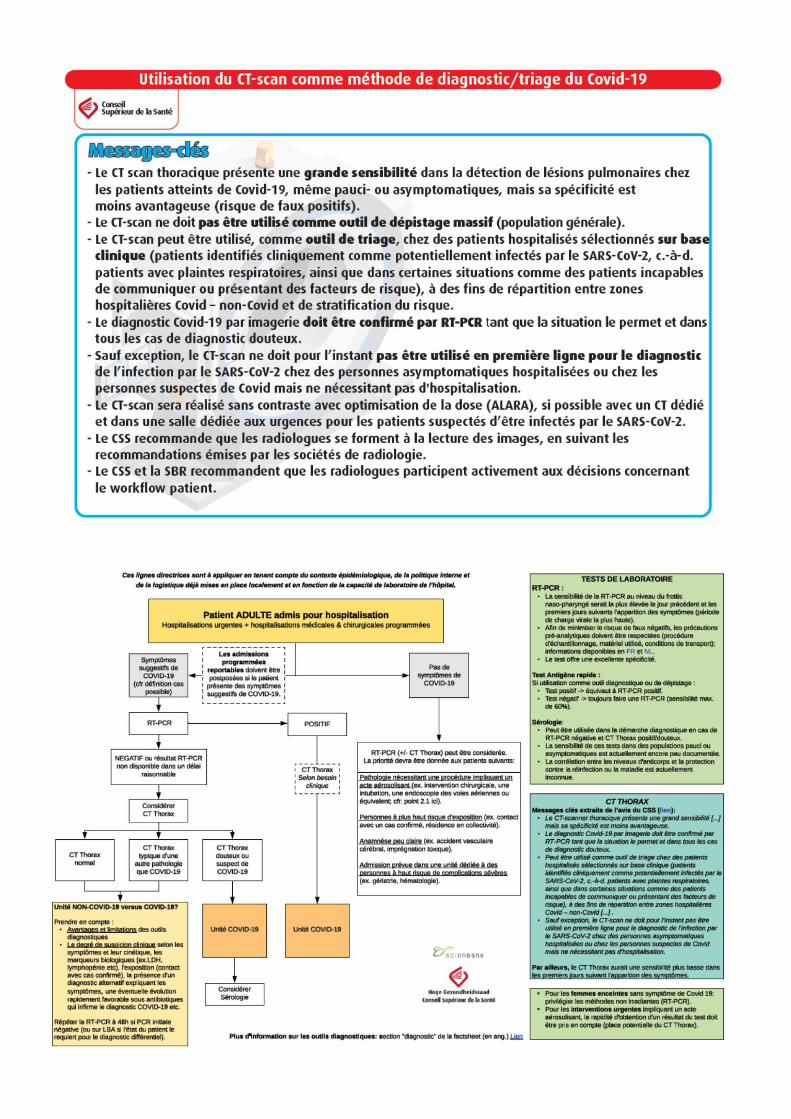
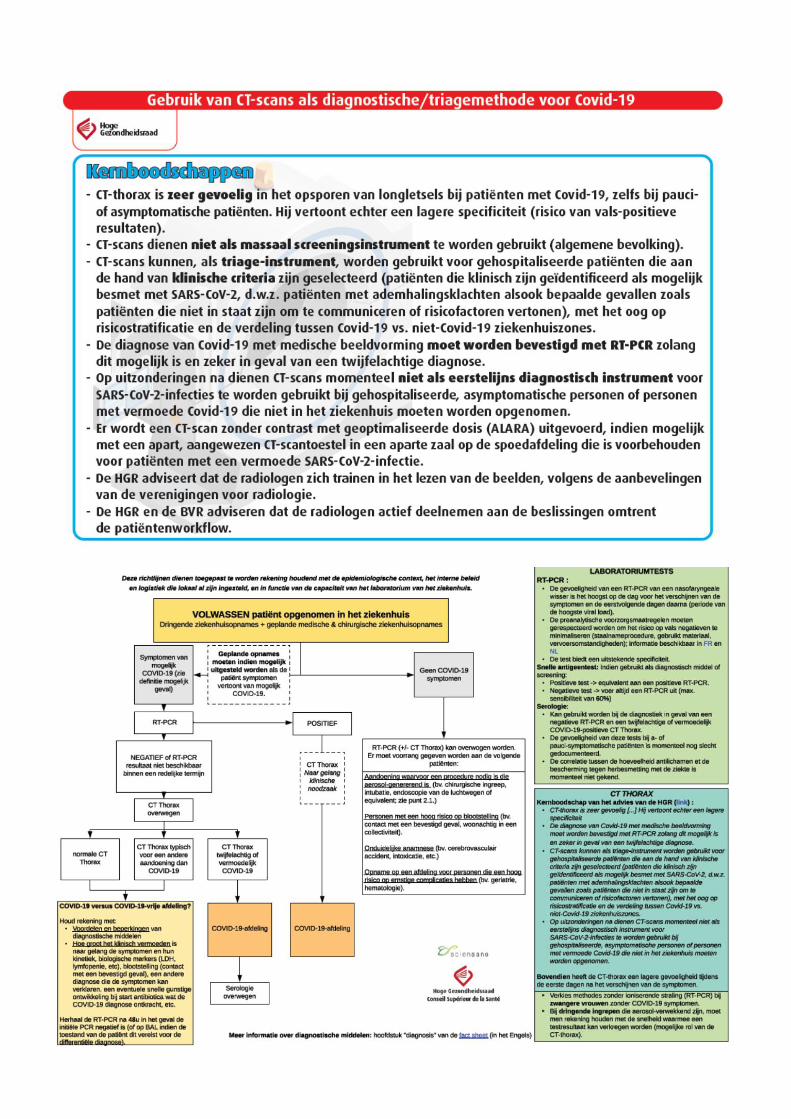
- Le CT scan thoracique présente une grande sensibilité dans la détection de lésions pulmonaires chez les patients atteints de Covid-19, même pauci- ou asymptomatiques mais sa spécificité est moins avantageuse (risque de faux positifs).
- Le CT-scan ne doit pas être utilisé comme outil de dépistage massif (population générale).
- Le CT-scan peut être utilisé, comme outil de triage, chez des patients hospitalisés sélectionnés sur base clinique (patients identifiés cliniquement comme potentiellement infectés par le SARS-CoV-2, c.-à-d. patients avec plaintes respiratoires, ainsi que dans certaines situations comme des patients incapables de communiquer ou présentant des facteurs de risque), à des fins de répartition entre zones hospitalières Covid – non-Covid et de stratification du risque.
- Le diagnostic Covid‐19 par imagerie doit être confirmé par RT‐PCR tant que la situation le permet et dans tous les cas de diagnostic douteux.
- Sauf exception, le CT-scan ne doit pour l’instant pas être utilisé en première ligne pour le diagnostic de l’infection par le SARS-CoV-2 chez des personnes asymptomatiques hospitalisées ou chez les personnes suspectes de Covid mais ne nécessitant pas d'hospitalisation.
- Le CT-scan sera réalisé sans contraste avec optimisation de la dose (ALARA), si possible avec un CT dédié et dans une salle dédiée aux urgences pour les patients suspectés d’être infectés par le SARS-CoV-2.
- Le CSS recommande que les radiologues se forment à la lecture des images, en suivant les recommandations émises par les sociétés de radiologie.
- Le CSS et la SBR recommandent que les radiologues participent activement aux décisions concernant le workflow patient.
Ces recommandations ont été largement reprises par Sciensano dans l’annexe 1 de la procédure pour les hôpitaux2. Le schéma de guide pour l’utilisation des outils diagnostiques de Covid-19 chez les patients adultes hospitalisés est repris en annexe.
2 « Procédure pour les hôpitaux : prise en charge d’un patient possible ou confirmé de Covid-19 » : http://covid-19.sciensano.be/sites/default/files/Covid19/COVID-19_procedure_hospitals_FR.pdf
Conseil Supérieur de la Santé
www.css-hgr.be
− 5 −
Mots clés et MeSH descriptor terms3
MeSH (Medical Subject Headings) is the NLM (National Library of Medicine) controlled vocabulary thesaurus used for indexing
articles for PubMed http://www.ncbi.nlm.nih.gov/mesh.
III METHODOLOGIE
Bien que le CSS n’ait pas reçu de question spécifique sur l’usage du CT dans le diagnostic du Covid-19, un échantillon de radiologues belges a été consulté afin de déterminer l’opportunité ou non d’émettre un avis sur ce sujet. Les présidents du domaine Radiations Ionisantes et les membres du Bureau ont ensuite identifié les expertises nécessaires. Sur cette base, un groupe de travail ad hoc a été constitué, au sein duquel des expertises en radiologie, radioprotection, radiobiologie, radiophysique médicale étaient représentées. Les experts de ce groupe ont rempli une déclaration générale et ad hoc d’intérêts et la Commission de Déontologie a évalué le risque potentiel de conflits d’intérêts. L’avis est basé sur une revue de la littérature scientifique, publiée à la fois dans des journaux scientifiques et des rapports d’organisations nationales et internationales compétentes en la matière (peer-reviewed), ainsi que sur l’opinion des experts. Après approbation de l’avis par le groupe de travail, et vu les circonstances exceptionnelles liées à la crise Covid-19, le Bureau seul a validé l’avis dans un premier temps (validation de la version 1 le 25 mars). Le Collège a toutefois validé la version 4 de l’avis en dernier ressort.
3 Le Conseil tient à préciser que les termes MeSH et mots-clés sont utilisés à des fins de référencement et de définition aisés du scope de l'avis. Pour de plus amples informations, voir le chapitre « méthodologie ».
MeSH terms*
Keywords Sleutelwoorden Mots clés Schlüsselwörter
COVID-19 Covid-19 Covid-19 Covid-19 Covid-19
X Ray, computed tomography
Computed tomography
computertomografie Tomodensitométrie Computertomographie
Diagnostic imaging
Medical imaging
Medische beeldvorming
Imagerie médicale Medizinische Bildgebung
Radiation protection
Dose optimisation
Dosisoptimalisatie Optimisation de dose
Dosisoptimierung
Diagnosis Diagnosis Diagnose Diagnostic Diagnose
Triage Triage Triage Triage Triage
Hospitals Hospital Ziekenhuis Hôpital Krankenhaus
Reverse Transcriptase Polymerase Chain Reaction
Reverse Transcriptase Polymerase Chain Reaction
RT-PCR RT-PCR RT-PCR
Conseil Supérieur de la Santé
www.css-hgr.be
− 6 −
IV ELABORATION ET ARGUMENTATION
1. Déterminer le meilleur usage du CT
Le CSS confirme que l’approche de l’ACR et de la BSR est pertinente, et que le CT-scan ne doit pour l’instant pas être utilisé en première ligne pour le diagnostic, mais être réservé à des patients sélectionnés sur base clinique. Cette approche concorde avec celle de la SFR (Société Française de Radiologie) qui estime que, bien que les auteurs chinois recommandent le scanner comme première technique de diagnostic de l'infection virale, « il n'y a actuellement pas de recommandations quant à la réalisation systématique d'un scanner thoracique, notamment à visée de dépistage de l'infection à Covid-19. » En effet, les transpositions entre la Chine et l’Europe sont hasardeuses, du fait de différences de prévalence de la maladie et de tests PCR utilisés chez nous. Au vu des réponses des experts et en accord avec la SBR, le CSS recommande que les hôpitaux prévoient d’inclure le CT-scan dans leurs procédures pour les patients identifiés cliniquement comme potentiellement infectés par le SARS-CoV-2 (patients avec plaintes respiratoires) en situation de pénurie des tests PCR. Le CT-scan devrait être utilisé, conjointement à la clinique, comme outil de triage rapide à des fins de stratification du risque, et de répartition entre zones hospitalières Covid – non-Covid. De même, lorsque des urgences médicales ou des situations spécifiques au patient (hypoxie, comorbidité, traumatisme, etc.) ne nous permettent pas d'attendre le résultat du test PCR, les scanners peuvent être utilisés comme instrument de triage. Il faut également éviter que des patients atteints du virus, mais qui ont des difficultés à bien exprimer leurs symptômes (patients âgés à comorbidité, patients ayant subi un AVC, etc.) ne soient envoyés dans une zone « non-Covid-19 ». Le CT-scan, en combinaison avec les symptômes cliniques, peut aider à réaliser cette répartition rapidement. S’il semble indéniable que le CT thorax affiche une grande sensibilité dans la détection de lésions chez les patients atteints de Covid-19, sa spécificité est moins avantageuse – le risque de confondre Covid-19 avec d’autres affections étant bien réel. A l’inverse, un CT négatif n’est pas une garantie de « non Covid », d’où l’importance de combiner CT-scan et RT- PCR. Le diagnostic Covid‐19 par imagerie doit être confirmé par
RT‐PCR tant que la situation le permet et dans tous les cas de diagnostic douteux.
Un avantage du CT scan est la rapidité avec laquelle les résultats de l’examen sont disponibles.
Dans tous les cas, le CT scan doit absolument être ciblé, en cas de troubles respiratoires et en cas de détérioration du tableau clinique, ainsi qu’en présence de certains facteurs de risque (voir ci-dessus) Très récemment, un panel multidisciplinaire (radiologues, pneumologues, infectiologues, etc.) d’experts dans la prise en charge des patients Covid-19 et provenant de 10 pays ont évalué l’utilité de l’imagerie à travers 3 scénarios représentant divers facteurs de risque, conditions de travail et ressources disponibles (Rubin et al., 2020). Les recommandations émises ici rejoignent globalement celles figurant dans cet avis.
Conseil Supérieur de la Santé
www.css-hgr.be
− 7 −
Bien évidemment, comme pour tout examen diagnostique, le principe d’optimisation est d’application, une qualité diagnostique suffisante avec une dose aussi faible que possible (scanner thoracique sans contraste, dose optimisée suivant les principes ALARA « As Low As Reasonably Achievable »). Au stade actuel, le RX thorax n'a de place que si le CT n'est pas disponible et dans le suivi des patients aux soins intensifs ou des patients dont l'état clinique se détériore. Plusieurs centres belges recommandent qu’en cas de radiographie du thorax (réalisée éventuellement pour d’autre motifs) atypique ou suggestive du Covid-19 (infiltrats bilatéraux) ou normale avec discordance clinique (auscultation évocatrice d’une pneumonie par exemple), il conviendra de réaliser un CT scan. Les patients sans problèmes respiratoires peuvent dans la plupart des cas rentrer chez eux et rester en quarantaine. La recommandation initiale de la SBR « la radiographie simple du thorax n’a pas sa place pour la détection du Covid-19 (2020) » reste d’application pour le groupe du travail du CSS même si elle a été amendée comme suit par la SBR : « The use of chest radiographs in patients presenting with fever and respiratory complaints should be limited. Radiology departments that have a dedicated chest radiography unit, close to the emergency department or isolation wards, may consider this in certain cases as an alternative for CT. However, the same safety measures (as for CT) need to be taken into account ». Les experts consultés insistent sur le risque de dissémination du virus. En Angleterre, cependant, la radiographie de thorax est toujours présente dans l’arbre décisionnel du rapport « Thoracic imaging in COVID-19 infection- Guidance for the reporting radiologist » du BSTI (British Society of Thoracic Imaging) du 19 mars 2020.
2. Mesures de protection
Les mesures de protection minimales à recommander sont : Au niveau de l’appareillage et des locaux
• Salle dédiée aux urgences pour les patients suspectés d’être infectés par le SARS-CoV-2 ;
• CT scan dédiés aux patients suspects ou positifs (utilisés non seulement pour le CT thorax, mais pour tout le travail diagnostique impliquant ces patients), voire CT mobile externe à l’hôpital ou au moins entrée différenciée de ces patients dans le service ;
• Priorisation d’un ou plusieurs appareils CT-scan en cas d’afflux de patients symptomatiques ;
• Pour les hôpitaux ne disposant que d’un seul scanner, regrouper les patients « Covid-19 » en réalisant les examens successivement, tout en respectant des mesures d’hygiène strictes ;
• Postposer les examens non urgents, afin de garder la priorité aux examens de diagnostic du Covid-19, ainsi que les examens essentiels pour les autres pathologies graves. La continuité des soins des autres patients dans un état grave doit continuer à être assurée ;
• Désinfection de l’appareil avant de réaliser un autre examen.
Conseil Supérieur de la Santé
www.css-hgr.be
− 8 −
• Dans l’éventualité d’un acte aérosolisant dans la salle d’examen, une période d’attente dépendant du taux de renouvellement de l’air est conseillée (1h selon l’American College of Radiology) ;
• Par ailleurs, pour les appareils scanners qui ne sont pas dédiés aux patients Covid-19, appliquer les mêmes mesures de désinfection renforcées par rapport aux standards habituels depuis le début de l’épidémie .
Au niveau du personnel et du patient
• Suivi des procédures habituelles Covid-19 en terme des mesures préventives, tant pour le patient que pour le personnel (cf https://epidemio.wiv-isp.be/ID/Documents/Covid19/COVID-19_procedure_hospitals_FR.pdf) et respecter dans la mesure du possible les mesures de social distancing ;
• Prévoir une protection systématique du personnel soignant de première ligne, que le patient soit suspect positif pour le SARS-CoV-2 ou non ;
• Personnel prévenu avant l’arrivée du patient ;
• Plus spécifiquement pour le CT-scan : o Retrait des gants et désinfection des mains entre l’installation du patient
et la réalisation de l’examen sur la console ; o Redésinfection des mains et port de gants avant de retourner aider le
patient à quitter l’appareil.
3. Organisation du service de radiologie Le très récent article « Radiology Department Preparedness for COVID-19: Radiology Scientific Expert Panel » (Mossa-Basha et al., 2020) figurant en annexe illustre les stratégies et les priorités à mettre en œuvre dans les services de radiologie. Celles-ci peuvent être adaptées aux situations locales. Le CSS recommande
• étant donné la recommandation d’annuler / reporter tous les examens non urgents, de renforcer l’équipe « CT scan des patients Covid-19 » par des technologues médicaux, rendus disponibles et formés récemment au CT ;
• d’éviter la rotation des médecins et du personnel entre plusieurs installations (sur le même site ou sur un autre site).
Pour les hôpitaux où c’est possible :
• un scanner de remplacement dédié pourrait être prévu en cas de panne du « scanner Covid-19 » ;
• pour aller plus loin qu’un scanner spécifique aux patients Covid-19, la piste d’un CT mobile, externe à l’hôpital devrait être investiguée avec les fournisseurs et l’AFCN.
La continuité des soins et examens médicaux urgents d’imagerie médicale doit être assurée, malgré le bouleversement des services. Une permanence par spécialité radiologique doit être assurée. Etant donné l’ampleur du travail, des risques et de la durée prévue de l’épidémie en Belgique, la sécurité et le bien-être du personnel – médecins, infirmiers, technologies - doivent être préservés.
Conseil Supérieur de la Santé
www.css-hgr.be
− 9 −
Enfin, le CSS et la SBR recommandent que les radiologues participent activement aux décisions concernant le workflow patient.
4. Formation des radiologues à la lecture des images Il faut rappeler aux radiologues en formation et aux étudiants que le CT scanner n’est pas spécifique, mais bien sensible. Les anomalies sont très variables, comme pour toutes les pneumonies virales. Le CSS recommande que les radiologues se forment à la lecture des images , en suivant les recommandations émises par les sociétés de radiologie. La Société Belge de Radiologie recommande aux radiologues de se familiariser à l’aspect des lésions liées à la maladie Covid-19 sur les images CT, de manière à être capable de les déceler sur les CT pulmonaires réalisés même pour d’autres raisons. Sur son site, des images significatives sont disponibles. L’analyse des premières images belges semble montrer une similitude par rapport aux cas publiés dans d’autres pays, notamment en Chine. La SFR a résumé sur son site l’aspect des poumons de patients infectés par le SARS-CoV-2 : « L'aspect tomodensitométrique précoce est celui d'opacités en verre dépoli de siège périphérique, sous-pleural, non systématisées, avec un caractère multifocal asymétrique, et une étendue variable, limitée à de petites plages infracentimétriques ou plus étendues. Il n'y a en règle générale pas de nodules, d'adénopathies ou d'épanchement pleural. L'évolution peut se faire vers des aspects de pneumonie organisée, et les formes graves cliniquement se caractérisent par des condensations alvéolaires étendues ». D’autre part, la revue Radiology a mis en ligne les articles reçus ainsi qu'un catalogue d'images, accessible sur ce lien : https://pubs.rsna.org/2019-nCoV#images La BSTI a mis en ligne une base de données d’imagerie anonymisée (https://bit.ly/BSTICovid19_Teaching_Library) et libre d’accès permettant le partage et la consultation de scans de patients avec une infection par le SARS-CoV-2 confirmée ou suspectée. Le but est de fournir un outil permettant d’orienter la gestion future de l’épidémie Covid-19, les protocoles nationaux ainsi que de fournir une aide aux cliniciens et leur permettre d’identifier et de diagnostiquer avec davantage de précision les cas d’infection par le SARS-CoV-2. Le rapport “Thoracic imaging in COVID-19 infection- Guidance for the reporting radiologist” du BSTI présente également une description et une classification des « patterns » rencontrés avec leur intervalle de confiance.
Enfin, il est primordial que les résultats soient communiqués sans délais, de façon écrite au prescripteur ou à la personne en charge du patient. Des exemples de compte-rendu structuré sont proposés par la SFR et la SBR (voir annexes).
Conseil Supérieur de la Santé
www.css-hgr.be
− 10 −
V REFERENCES
• Abraham Kim. CT again tops DNA tests for early coronavirus detection. AuntMinnie, 26/2/2020.(https://www.auntminnie.com/index.aspx?sec=log&itemID=128274). Accessed on: 16/03/2020.
• Abraham Kim. What do radiologists need to know about the coronavirus? AuntMinnie, 27/02/2020. (https://www.auntminnie.com/index.aspx?sec=log&itemID=128285). Accessed on: 16.03.2020.
• Ai Tao*, Zhenlu Yang*, Hongyan Hou, Chenao Zhan, Chong Chen, Wenzhi Lv, Qian Tao, Ziyong Sun, Liming Xia. Correlation of Chest CT and RT-PCR Testing in Coronavirus Disease 2019 (COVID-19) in China: A Report of 1014 Cases. Radiology, 26/02/2020.
• BSR. Belgian Society of Radiology. COVID-19 info from the BSR, 14/03/2020. (https://www.bsr-web.be/docs/COVID.pdf). Accessed on: 20/03/2020.
• BSTI - British Society of Thoracic Imaging. COVID‐19 BSTI statement and guidance. [update 11/03/2020]. (https://www.bsti.org.uk/standards-clinical-guidelines/clinical-guidelines/covid-19-bsti-statement-and-guidance/) . Accessed on: 20/3/2020.
• BSTI - British Society of Thoracic Imaging. Thoracic Imaging in COVID-19 Infection Guidance for the Reporting Radiologist. Version 2, 16/03/2020. (https://www.bsti.org.uk/standards-clinical-guidelines/clinical-guidelines/bsti-covid-19-guidance-for-the-reporting-radiologist/ ). Accessed on: 20/03/2020.
• Fang Y, Zhang H, Xie J, Lin M, Ying L, Pang P, Ji W. Sensitivity of Chest CT for COVID-19:Comparison to RT-PCR. Radiology 2020.
• Fengxiang Song*, Nannan Shi*, Fei Shan, Zhiyong Zhang, Jie Shen, Hongzhou Lu, Yun Ling, Yebin Jiang, Yuxin Shi. Emerging 2019 Novel Coronavirus (2019-nCoV) Pneumonia. Radiology, 06/02/2020.
• Hare S S, Jacob J, Johnstone A, Robinson G. COVID‐19: is CT scanning ready to answer a diagnostic call?. BMJ, 12/03/2020. opinion, 12/03/2020. (https://blogs.bmj.com/bmj/2020/03/12/covid-19-is-ct-scanning-ready-to-answer-a-diagnostic-call/ ). Accessed on: 17/03/2020.
• Harrison X. Bai*, Ben Hsieh*, Zeng Xiong, Kasey Halsey, Ji Whae Choi, Thi My Linh Tran, Ian Pan, Lin-Bo Shi, Dong-Cui Wang, Ji Mei, Xiao-Long Jiang, Qiu-Hua Zeng, Thomas K. Egglin, Ping-Feng Hu, Saurabh Agarwal, Fangfang Xie, Sha Li, Terrance Healey, Michael K. Atalay, Wei-Hua Liao. Performance of radiologists in differentiating COVID-19 from viral pneumonia on chest CT. Radiology, 10/03/2020
• Heshui Shi. Radiological findings from 81 patients with COVID -19 pneumonia from Wuhan, China: a descriptive study. LancetInfectDis. 2020
• Hyungijn Kim. Outbreak of novel coronavirus (COVID 19): What is the role of radiologists? European radiology. 13/02/2020.
• Kanne JP, Little BP, Chung JH, Elicker BM, Ketail LH. Essentials for Radiologists on COVID-19: An Update—Radiology Scientific Expert Panel. Radiology 2020.
• Mossa-Basha M, Meltzer Carolyn C. , Kim Danny C, Tuite, Michael J , Pallav Kolli K., Tan Bien Soo. Radiology Department Preparedness for COVID-19: Radiology Scientific Expert Panel. Radiology, 16/03/2020.
• Rubin GD, Haramati LB, Kanne JP, Schluger NW, Yim JJ, Anderson DJ et al. The Role of Chest Imaging in Patient Management during the COVID-19 Pandemic: A Multinational Consensus Statement from the Fleischner Society. Chest 2020.04.003. CHEST (2020), doi: https://doi.org/10.1016/j.chest.2020.04.003
• SFR – Société française de Radiologie. Épidémie de Covid-19 : ce que les radiologues doivent savoir. (http://communication.radiologie.fr/HM?b=3KH6xt6a-HLmXJSvRIlhjJyRMHdQ_bKoIyG4IPnfc_PXtpQXbtWLRpE5D4iS39DS&c=ZICKgHDWgrxV2oLId_h0TQ). Accessed on: 16/03/2020.
Conseil Supérieur de la Santé
www.css-hgr.be
− 11 −
• SFR - Société française de Radiologie. Épidémie de Covid-19 : TDM Thoracique. (http://www.sfrnet.org/rc/org/sfrnet/nws/News/2020/20200316-155630-175/src/nws_fullText/fr/CR%20TYPE%20COVID-19%20LAST.pdf). Accessed on: 24/03/2020.
• Van Beusekom, Mary. Studies profile lung changes in asymptomatic COVID-19, viral loads in patient samples. University of Minnesota: Center for Infectious Diesease Research and Policy, 25/02/2020. (http://www.cidrap.umn.edu/news-perspective/2020/02/studies-profile-lung-changes-asymptomatic-covid-19-viral-loads-patient). Accessed on: 24/03/2020.
• Wang Y, Dong C, Hu Y, Li C, Ren Q, Zhang X et al. Temporal Changes of CT Findings in 90 Patients with COVID-19 Pneumonia: A Longitudinal Study. Radiology. 2020 Mar 19:200843. doi: 10.1148/radiol.2020200843. [Epub ahead of print]
• Yicheng Fang, Huangqi Zhang, Jicheng Xie, Minjie Lin, Lingjun Ying, Peipei Pang, Wenbin Ji. Sensitivity of Chest CT for COVID-19: Comparison to RT-PCR. 19/2/2020.
• Xie X, Zhong Z, Zhao W, Zheng C, Wang F, Liu J. Chest CT for Typical 2019-nCoV Pneumonia: Relationship to Negative RT-PCR Testing. Radiology. 2020 Feb 12;200343.
VI COMPOSITION DU GROUPE DE TRAVAIL
La composition du Bureau et du Collège ainsi que la liste des experts nommés par arrêté royal se trouvent sur le site Internet du CSS (page : Qui sommes-nous).
Tous les experts ont participé à titre personnel au groupe de travail. Leurs déclarations générales d’intérêts ainsi que celles des membres du Bureau et du Collège sont consultables sur le site Internet du CSS (page : conflits d’intérêts). Les experts suivants ont été interrogés par mail. Ces recommandations émises d’urgence par lettre ont été élaborées sur base de leurs réponses. Le travail a été coordonné par Marie-Thérèse Hoornaert et Patrick Smeesters et le secrétariat scientifique a été assuré par Evelyn Hantson et Sandrine Everaert. COCHE Emmanuel Radiologie UCL St-Luc
EVERARTS Philippe Radiologie CH Jolimont La Louvière
HOORNAERT Marie-Thérése Physique hospitalière, radiophysique médicale
Ex-CH Jolimont La Louvière, ex-Ulg
MEUNIER Paul Radiologie CHU Liège
NIEBOER Koenraad Radiologie UZ Brussel
SMEESTERS Patrick Radiobiologie ex-UCL, ex-AFCN
SMEETS Peter Radiologie UZ Gent
TACK Denis Radiologie EpiCURA
VERSCHAKELEN Johny Radiologie UZ Leuven
Les médecins radiologues thoraciques suivants, experts scientifiques de la SBR, ont été entendus. GOSSELIN Robert Radiologie UZ Gent, SBR
SNOECKX Annemie Radiologie UZA, SBR
ConseilSupérieur de la Santé
VII. ANNEXES • ANNEXE0AETB:CSS:UTILISATIONDUCT-SCANCOMMEMÉTHODEDE DIAGNOSTIC/TRIAGEDUCOVID-19/SCIENSANO//CSS:GUIDEPOUR L’UTILISATIONDESOUTILSDIAGNOSTIQUESDECOVID-19CHEZLESPATIENTS ADULTESHOSPITALISÉS• ANNEXE0CETD:HGR:GEBRUIKVANCT-SCANSALSDIAGNOSTISCHE/ TRIAGEMETHODEVOORCOVID-19//SCIENSANO/HGR:GIDSVOORHET GEBRUIKVANDIAGNOSTISCHEHULPMIDDELENVOORCOVID-19• ANNEXE1:ACRRECOMMENDATIONSFORTHEUSEOFCHEST RADIOGRAPHYANDCOMPUTEDTOMOGRAPHY(CT)FORSUSPECTED COVID-19INFECTION• ANNEXE2A:SOCIÉTÉFRANÇAISEDERADIOLOGIE:EPIDÉMIEDECOVID-19: CEQUELESRADIOLOGUESDOIVENTSAVOIR.5MARS2020.• ANNEXE2B:SOCIÉTÉFRANÇAISEDERADIOLOGIE:EPIDÉMIEDECOVID-19: POINTSURL’IMAGERIE.12MARS2020.• ANNEXE2C:SOCIÉTÉFRANÇAISEDERADIOLOGIE:EPIDÉMIEDECOVID-19: POINTSURL’IMAGERIE.14MARS2020.• ANNEXE2D:SOCIÉTÉFRANÇAISEDERADIOLOGIE:EXEMPLECOMPTE- RENDUTYPECOVID-19• ANNEXE3:BSTI–BRITISHSOCIETYOFTHORACICIMAGING.THORACIC IMAGINGINCOVID-19IMAGING–GUIDANCEFORTHEREPORTING RADIOLOGIST• ANNEXE4A:BSR–BELGIANSOCIETYOFRADIOLOGY:COVID-19INFO FROMBSR.GUIDELINESFORIMAGINGINCOVID-19.14MARS2020.• ANNEXES4BET4C:BSR–BELGIANSOCIETYOFRADIOLOGY:TEMPLATETO REPORTCTINCOVIDCASES.MARS2020.• ANNEXE5:MAHMUDMOSSA-BASHA,CAROLYNC.MELTZER,DANNYCKIM, MICHAELJTUITE,K.PALLAVKOLLI,BIENSOOTAN.RADIOLOGY DEPARTMENTPREPAREDNESSFORCOVID-19:RADIOLOGYSCIENTIFIC EXPERTPANEL.RADIOLOGY,16/3/2020.
ANNEXE 0A – MESSAGES-CLES Utilisation du CT-scan comme méthode de
diagnostic / triage du Covid-19
ANNEXE 0b Guide pour l’utilisation des outils diagnostiques de
Covid-19 chez les patients adultes hospitalisés
ANNEXE 0c – KERNBOODSCHAPPEN Gebruik van CT-scans als diagnostische /
triagemethode voor Covid-19
ANNEXE 0d Gids voor het gebruik van diagnostische
hulpmiddelen voor COVID-19

ANNEXE 1 ACR Recommendations for the use of Chest
Radiography and Computed Tomography (CT) for Suspected COVID-19 Infection
���������� ������ ������������������������������������� ������ ����������������������� !"#$%!�������&� ���'
�����(��)))*���*������+�����#���#,���� �������#-�������#���� ������� ��������#���#����#�����������#���#��#���#�������#�� !"$%#!' $��
./01233456567899:;<==:>?@AB<>CD<EAF:GC:<D8F:CA9@?B<HE@IFJ@>?8<=IGA:?K<=<HE@IFJL8KMD<ENGCI:;A:?8OPQRSTUQ>D:;AB<>VWXYZ[X\Y]_abbcdefghij3kdl0m/ndopq2mrsts4q2m0modu0vwopuopqm0mdqopq2m0vxm/pn/ll0vl0o/qmpmddvy12mdq0/novu0/l2dze{|}/pn1v~l�qmnqv~vu0/l2�ze�}yv0q2md10mmpopu4no/upvdod/pn~/p/um~mpqvyl/qompqdwoq2d�dlm1qmnv0�pvwpefghij3kopym1qovpsevpq0o��qopuqvq2odopqm0mdq/0mxo~oqmn/�/ox/�oxoq�vy�o0/xqmdqopu�oqdqvn/qm41vp1m0pyv0qmdqdmpdoqo�oq�y0v~m/0xom00mlv0qdope2op/4/pnq2mu0vwopup�~�m0vyl��xo1/qovpdnmd10o�opuq2me{|/pne�/llm/0/p1mopq2mdmqqopuvy�pvwpv0d�dlm1qmnefghij3kopym1qovps�vn/qm4~vdqvyq2m0/novxvuo1n/q/1v~mdy0v~e2op/stv~mdq�nomdd�uumdqq2/q12mdqe�opl/0qo1�x/0~/��mlvdoqo�mopq2mdmqqopuvy/pmu/qo�mqmdqs�����������������������������������������������������������������a������������������������������������������������������������������������������m�uv/xdyv0q2mrsts2m/xq21/0md�dqm~op0mdlvpdmqvq2mefghij3kv�q�0m/�/0mqv0mn�1m~v0�onoq�/pn~v0q/xoq�4~opo~o mnodm/dmq0/pd~oddovp4l0vqm1q2m/xq21/0mlm0dvppmx4/pnl0mdm0�m2m/xq21/0md�dqm~y�p1qovpopus�2mce|�mxom�mdq2/qq2myvxxvwopuy/1qv0dd2v�xn�m1vpdonm0mn0mu/0nopuq2m�dmvyo~/uopuyv0d�dlm1qmnv0�pvwpefghij3kopym1qovp¡�2mempqm0dyv0iodm/dmevpq0vxzeie}nvmdpvq1�00mpqx�0m1v~~mpne{|v0e�qvno/upvdmefghij3ksgo0/xqmdqopu0m~/opdq2mvpx�dlm1oyo1~mq2vnvyno/upvdodsevpyo0~/qovpwoq2q2m�o0/xqmdqod0m¢�o0mn4m�mpoy0/novxvuo1yopnopud/0md�uumdqo�mvyefghij3kvpe{|v0e�s£v0q2mopoqo/xno/upvdqo1qmdqopuyv0d�dlm1qmnefghij3kopym1qovp4q2meie0m1v~~mpnd1vxxm1qopu/pnqmdqopudlm1o~mpdy0v~q2m�llm00mdlo0/qv0�q0/1qzp/dvl2/0�pum/xc¤iv0vl2/0�pum/xdw/�d}v0y0v~q2mxvwm00mdlo0/qv0�q0/1qw2mp/�/ox/�xmyv0�o0/xqmdqopus¥��������a���������������������������¦§X�©ª��������������a�����������������������������4op1x�nopuopyx�mp /4«3¤34tc|t/pn.¬|tsmopuopq2m~ondqvyq2m1�00mpqyx�dm/dvpwoq2/~�122ou2m0l0m�/xmp1mvyopyx�mp /opq2mrstsq2/pefghij3k4y�0q2m0xo~oqdq2mdlm1oyo1oq�vye�s�2m1�00mpqce|cll0vl0o/qmpmdde0oqm0o/dq/qm~mpqvpc1�qm|mdlo0/qv0�hxxpmdd®z2qqld¡/1dm/012s/10sv0unv1d°k±±°¤/00/qo�m}4x/dq�ln/qmnop563²dq/qmdq2/q12mdqe�od³rd�/xx�¤vqcll0vl0o/qmsc0m�omwy0v~q2mev120/pmi/q/�/dmvyt�dqm~/qo1|m�omwdvp12mdq0/novu0/l2dyv0/1�qmxvwm00mdlo0/qv0�q0/1qopym1qovpd®z2qqld¡wwwsp1�ospx~spo2suv�l��~mn5±µ°kµ±µ}1vp1x�nmnq2/qe{|nonpvqo~l0v�m1xopo1/xv�q1v~mdzn�0/qovpvyoxxpmdd}yv0l/qompqdwoq2xvwm00mdlo0/qv0�q0/1qopym1qovp¶q2m0m�omwop1x�nmnqwv0/pnv~o mnq0o/xd1v~l/0opu�dmvye{|dqvpve{|dop/1�qmxvwm00mdlo0/qv0�q0/1qopym1qovpdyv012oxn0mp/pn/n�xqdscnnoqovp/xx�4q2m0m/0modd�md0mx/qmnqvopym1qovp1vpq0vxop2m/xq21/0my/1oxoqomd4op1x�nopuq2m�dmvyo~/uopum¢�ol~mpq¡·0o~/0�1/0m/pnvq2m0~mno1/xl0v�onm0d/0m/qqm~lqopuqvxo~oq�odoqdvyl/qompqdwoq2d�dlm1qmnopyx�mp /v0efghij3kqv2m/xq21/0my/1oxoqomd4qv~opo~o mq2m0od�vydl0m/nopuopym1qovps�2meie2/d/xdv/d�mnq2/ql/qompqd/pn�odoqv0dqv2m/xq2
®]�������� ® �������
¹
���������� ������ ������������������������������������� ������ ����������������������� !"#$%!�������&� ���'
�����(��)))*���*������+�����#���#,���� �������#-�������#���� ������� ��������#���#����#�����������#���#��#���#�������#�� !"$%#!' ���
./012/.3435316716.0118192:06;<=5:<6:2/.>51016=30/5:0;3448166?71/6@195:A1/0/6>0B3./4</6@/89711C/4>/51938/=03C/510::<A35D5D19::0.4:619EF8/99353:85:18C30:8<185/4.41/838B/8991.:85/<38/53:8:20::<6:..>=3197;/=/53185A35D6>6=1.519:0@8:A8GHIFJKLM3821.53:87;5D:0:>BD.41/838B:26>02/.167;6:<1:81A1/038B=0:=10=0:51.53C11N>3=<185?/30K24:AA35D3823O190/93:B0/=D;:0GP6./88100::<66D:>4971.:86391019712:013</B38B5D181O5=/53185EI18534/53:836/83<=:05/85.:863910/53:82:05D1.:850:4:2/307:08150/86<3663:838D1/45D./012/.3435316QRD55=6STTAAAE.9.EB:CT3821.53:8.:850:4TB>39143816T18C30:8<185/4T7/.@B0:>89T/30ED5<4U.V.WEJ1=18938B:85D1/301O.D/8B10/516?0::<6</;81195:71>8/C/34/7412:0/==0:O3</514;LD:>0/X103</B38B3821.519=/531856Y/30.30.>4/53:80::<6./871516519EPD161<1/6>0165:143<38/51.:85/<38/53:82:06>761N>185=/531856</;019>.1/..1665:3</B38B6>3516?41/938B=:51853/44;5:6>765/853/4=0:741<62:0=/53185./01EZ[\]_ab]\]c_c]d\eab]fghd]c_ii] \jGP6D:>498:571>6195:6.01182:0:0/6/23065K438151655:93/B8:61GHIFJKLMGP6D:>4971>6196=/038B4;/8901610C192:0D:6=35/43k19?6;<=5:</53.=/531856A35D6=1.323..4383./43893./53:862:0GPEl==0:=03/513821.53:8.:850:4=0:.19>0166D:>49712:44:A19712:016./8838B6>761N>185=/531856Em/.3435316</;.:86391091=4:;38B=:05/7410/93:B0/=D;>835638/<7>4/5:0;./012/.34353162:0>61AD18Gno6/01.:86391019<193./44;81.166/0;EPD16>02/.16:25D161</.D3816./8711/634;.41/819?/C:3938B5D181195:7038B=/531856385:0/93:B0/=D;0::<6Eo/93:4:B36566D:>492/<343/03k15D1<614C16A35D5D1GP/==1/0/8.1:2GHIFJKLM3821.53:838:09105:71/7415:3918532;238938B6.:86365185A35D3821.53:838=/5318563</B192:0:5D1001/6:86Epqr[a]s[dcbttetutuvf\[wa]dwii][\xd]exawyi_d]zw]\rd][g{|}~���a]\aw�w\[�[wy[�y]e\_i]i]wc[yrd[cawc]\[d]d]�x]\aw�cb]\ag�a_w�_di]cw\w_\_zb]ab]da_a]\a[r[aw]a�_dg{|}~���e[iwa[r[aw]a_drd_�w]_ab]dad][ai]a��b]fgh\ad_�y�xd�]\c[xaw_wa[�w�abw\[rrd_[cb�f_di[ycb]\ag�_]\_ai][[r]d\__]\_ab[�]g{|}~���w�]caw_�[ [[�_di[yg�w\_a\r]cw�wc�_dg{|}~���w[�_\w\�f_di[yg�\b_xy_aw\\x[][r[aw]a�d_i�]w��x[d[aw]_drd_�w]_ab]dcywwc[yy�w wc[a]ad][ai]azb]_ab]dzw\]i]wc[yy�[rrd_rdw[a]�gy][dy�ey_c[yy�c_\ad[w]d]\_xdc]\i[��][�[ca_dw\xcb]cw\w_i[�w��h]c_ii] ]h]\_xdc]\jG1851062:0J361/61G:850:4Sl<103./8G:441B1:2o/93:4:B;KGHIFJKLMo/93:4:B;K�=1.323.o16:>0.16RD55=6STTAAAE/.0E:0BTG4383./4Ko16:>0.16TGHIFJKLMKo/93:4:B;Ko16:>0.16WQ RD55=6STTAAAE.9.EB:CT.:0:8/C30>6TV�LMK8.:CT3891OED5<4W�1810/4382:0</53:8/89635>/53:8>=9/516� RD55=6STTAAAE.9.EB:CT.:0:8/C30>6TV�LMK8.:CT3891OED5<4WF82:0</53:82:0D1/45D./01=0:21663:8/46� RD55=6STTAAAE.9.EB:CT.:0:8/C30>6TV�LMK8G:ITD.=T3891OED5<4Wo/93:4:B3./053.416/89.:441.53:86S����������������������������� �¡�����¢£��� ¤¥G:0:8/C30>6RGHIFJKLMWH>5701/@S¦D/55D1J1=/05<185:2o/93:4:B;�D:>49§8:AQ RD55=6STTAAAE/.0E:0BT/053.41T�L©ª«KLªª�RV�W¬�L©�KVT2>4451O5W �¡�����¢��¡ �¡�����¢���¡����������®������¥�=1.3/4m:.>6SGHIFJKLM� RD55=6STT=>76E068/E:0BTV�LMK8G:IW����������������� �����������¢£�� ¤¥G:0:8/C30>6J361/61RGHIFJKLMW� RD55=6STTAAAE/0:84381E:0BT5:=3.T.:CLMW¯
ANNEXE 2a Société Française de Radiologie : Epidémie de
Covid-19 : ce que les radiologues doivent savoir. 5 mars 2020

Épidémie de Covid-19 : ce que les radiologues doivent savoir
Fin décembre 2019, l'organisation mondiale de la santé (OMS) a été informée par les autoritéschinoises de cas groupés de pneumonie dans la région du Hubei en Chine, en lien avec lafréquentation d'un marché d'animaux dans la ville de Wuhan. Un nouveau coronavirus, SARSCoV2 a été identifié comme agent causal de cette maladie, appelée Covid-19. Lors de larédaction de cette Newsletter, le 5 Mars 2020, le nombre de cas mondiaux atteint 93 076,tandis que 285 cas ont été confirmés en France, dont 4 décédés.
Cette épidémie a généré la publication rapide de plusieurs articles radiologiques, décrivant lesaspects tomodensitométriques de l'atteinte pulmonaire qui fait la gravité de cette infection, avecune mortalité estimée 7 à 10 fois supérieure à celle de la grippe.
Quels sont les informations essentielles ?
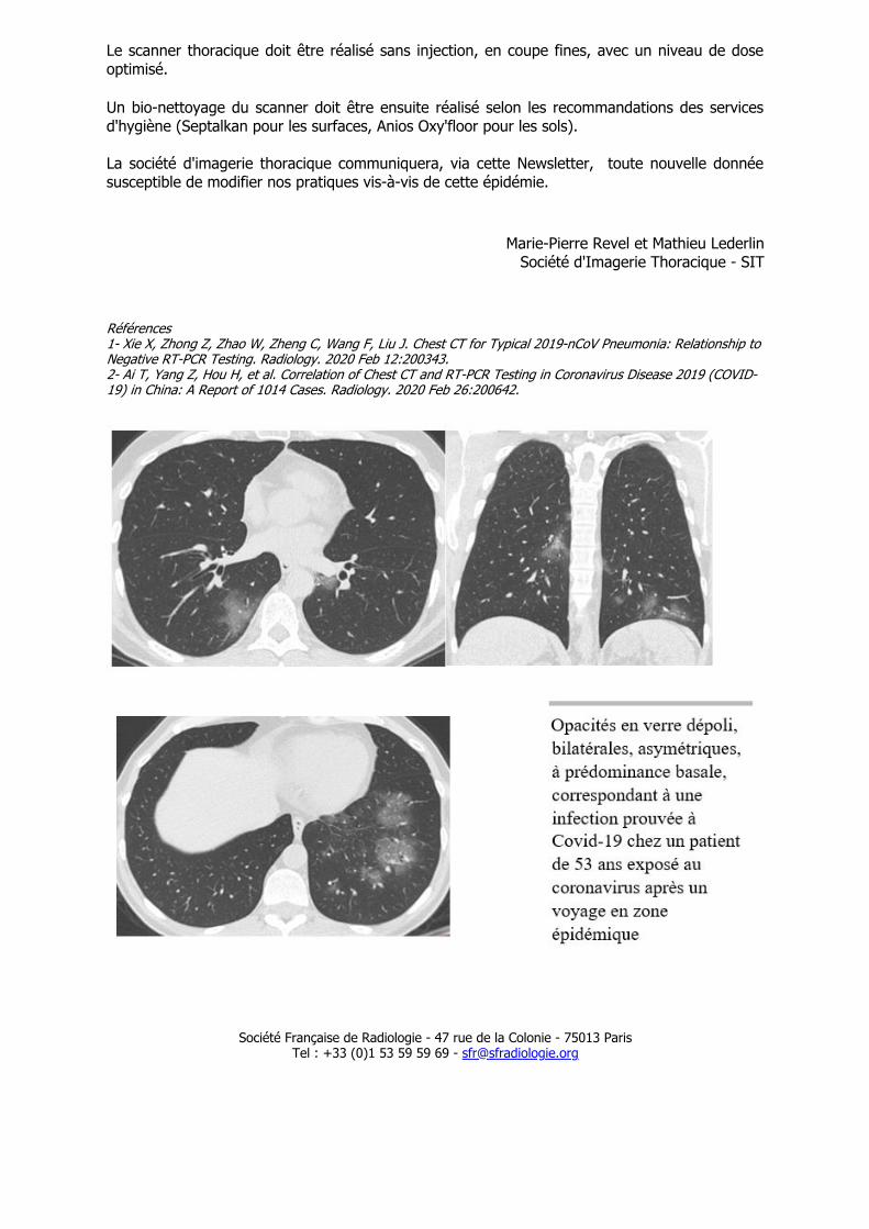
1- L'aspect tomodensitométrique précoce est est celui d'opacités en verre dépoli de siègepériphérique, sous-pleural, non systématisées (Figure), avec un caractère multifocalasymétrique, et une étendue variable, limitée à de petites plages infracentimétriques ou plusétendues. Il n'y a en règle pas de nodules, d'adénopathies ou d'épanchement pleural. L'évolutionpeut se faire vers des aspects de pneumonie organisée, et les formes graves cliniquement secaractérisent par des condensations alvéolaires étendues. La revue Radiology a mis en ligne lesarticles reçus ainsi qu'un catalogue d'images, accessible sur ce lien : https://pubs.rsna.org/2019-nCoV#images
2- Deux publications dans Radiology (1,2), dont la plus large regroupe 1014 cas, font étatd'une sensibilité supérieure du scanner par rapport à la recherche d'ARN viral par RT-PCR, quireste cependant la technique de référence, mais qui peut se positiver de façon retardée parrapport aux premiers signes radiologiques. Par ailleurs, il est démontré que des sujetsasymptomatiques au plan respiratoire peuvent présenter des anomalies tomodensitométriquesanalogues à celles des patients ayant la forme commune, non grave, de l'atteinte respiratoire.Sur la base de ces résultats, les auteurs chinois recommandent la réalisation première d'unscanner pour dépister l'infection virale. Les différences quant à la prévalence de lamaladie et la sensibilité des tests PCR utilisés en France rendent hasardeuse toutetransposition. Il n'y a actuellement pas de recommandations quant à la réalisationsystématique d'un scanner thoracique, notamment à visée de dépistage de l'infection à Covid-19.
3- S'il est décidé de réaliser une imagerie tomodensitométrique, pour les manipulateurssont celles actuellement recommandées pour l'ensemble des soignants prenant en charge lespatients suspects: friction des mains avec produit hydro-alcoolique (PHA), surblouse à mancheslongues à usage unique, masque chirurgical, charlotte, lunettes de protection à usage unique,gants à usage unique. Le patient doit porter un masque chirurgical et effectuer une friction desmains au PHA. Selon les dernières recommandations, le port des masques filtrants FFP2 estactuellement réservé aux seuls personnels hospitaliers en contact étroit et prolongé avec des casconfirmés (soins intensifs).
Le scanner thoracique doit être réalisé sans injection, en coupe fines, avec un niveau de doseoptimisé.
Un bio-nettoyage du scanner doit être ensuite réalisé selon les recommandations des servicesd'hygiène (Septalkan pour les surfaces, Anios Oxy'floor pour les sols).
La société d'imagerie thoracique communiquera, via cette Newsletter, toute nouvelle donnéesusceptible de modifier nos pratiques vis-à-vis de cette épidémie.
Marie-Pierre Revel et Mathieu LederlinSociété d'Imagerie Thoracique - SIT
Références1- Xie X, Zhong Z, Zhao W, Zheng C, Wang F, Liu J. Chest CT for Typical 2019-nCoV Pneumonia: Relationship toNegative RT-PCR Testing. Radiology. 2020 Feb 12:200343. 2- Ai T, Yang Z, Hou H, et al. Correlation of Chest CT and RT-PCR Testing in Coronavirus Disease 2019 (COVID-19) in China: A Report of 1014 Cases. Radiology. 2020 Feb 26:200642.
Société Française de Radiologie - 47 rue de la Colonie - 75013 ParisTel : +33 (0)1 53 59 59 69 - [email protected]
ANNEXE 2b Société Française de Radiologie : Epidémie de Covid-19 : POINT SUR L'IMAGERIE. 12 mars
2020
Epidémie de Covid-19 : POINT SUR L'IMAGERIE
La France est actuellement en situation épidémique de stade 2 vis-à-vis du SARS-Cov-2, et lasollicitation des structures radiologiques, qu'elles soient hospitalières ou non, devient plusimportante.
Cette montée en charge nécessite de clarifier le rôle de l'imagerie dans ce contexte épidémique
Quelles sont les indications d'imagerie et quel type d'examen réaliser ?
1. Il n'y a pas de place pour la radiographie thoracique, si une imagerie est indiquée, il fautréaliser un scanner.
2. Chez des patients sans gravité clinique ni co-morbidités, pour lesquels il existe unehésitation diagnostique entre pneumopathie bactérienne ou bien atteinte Covid-19, lesarguments cliniques (foyer auscultatoire, douleur thoracique) et biologiques(hyperleucocytose) doivent prévaloir, et une PCR peut être indiquée en cas de fièvrerésistant à l'antibiothérapie, plutôt que la prescription d'une imagerie.
3. Il n'y a actuellement pas d'indication à réaliser un scanner thoracique à des fins dedépistage chez des patients sans signes de gravité et sans comorbidités.
4. La réalisation d'un scanner thoracique sans injection en coupes fines estactuellement indiquée chez les patients ayant un diagnostic suspecté ou confirméet des signes de gravité clinique (dyspnée, désaturation...) initiaux ou secondairesrelevant d'une prise en charge hospitalière. Elle peut également se concevoir chezdes patients suspects avec co morbidités, en attente des résultats de PCR, ou bienen première ligne si les délais et disponibilité de PCR deviennent limitants, ce qui semblese profiler.
5. Chez les patients Covid-19 positifs en soins intensifs et réanimation, présentant uneaggravation, l'examen tomodensitométrique doit rechercher une aggravation des lésionsavec évolution vers un tableau de SDRA, mais également un pneumothorax sousventilation ou bien une complication thrombo-embolique et doit donc être réalisé avecinjection.
Quelles précautions prendre pour les manipulateurs et radiologues ?
S'il est décidé de réaliser une imagerie tomodensitométrique, les mesures à prendre sont cellesactuellement recommandées pour l'ensemble des soignants prenant en charge les patientssuspects :- Le patient : doit porter un masque chirurgical et effectuer une friction des mains au PHA.- Les médecins et manipulateurs :
- Friction des mains avec produit hydro-alcoolique (PHA), masque chirurgical.- Si nécessité d'installer le patient sur la table d'examen et/ou de le perfuser :
- Surblouse à manches longues, charlotte et gants à usage unique.- Idéalement, lunettes protectrices réutilisables après désinfection.
- Le port des masques filtrants FFP2 est réservé aux seuls personnels hospitaliers en contactétroit et prolongé avec des cas confirmés (soins intensifs ou nécessité d'un geste de radiologieinterventionnelle).
- Un bio-nettoyage du scanner doit être ensuite réalisé selon les recommandations des servicesd'hygiène (FB spray ou tout autre détergent désinfectant pour les surfaces, Anios Oxy'floor pourles sols).
Les patients doivent venir accompagnés (isolement contact), en tenue permettant uneinstallation directe sur la table de scanner sans déshabillage.Le service de Radiologie doit être prévenu en amont, pour organisation évitant l'attenteau milieu d'autres patients.Des circuits spécifiques doivent être mis en place, avec selon l'affluence et le nombre descanners disponibles, des horaires dédiés sur un scanner ou un scanner totalement dédié àcette activité.
NB : Pour les échographies des patients hospitalisés, il est préférable de les réaliser au lit avecun échographe portatif, pour limiter les allées et venues.
Quels sont les aspects tomodensitométriques rencontrés ?
Ils sont illustrés à partir de ces quelques cas cliniques commentés. Voir les cas cliniques
Il s'agit essentiellement de plages de verre dépoli non systématisées à prédominance souspleurale, et à un stade plus tardif de condensation alvéolaire. Il n'y a en règle pas d'excavation,de nodules, de masses. Les micronodules bronchiolaires, les adénopathies médiastinales etépanchements pleuraux sont rares, en sachant que des épanchements sont possibles en casde décompensation cardiaque.
Pr REVEL, Pr LEDERLIN, Pr BRILLET, Pr KHALIL pour la Société d'Imagerie Thoracique - SIT
Société Française de Radiologie - 47 rue de la Colonie - 75013 ParisTel : +33 (0)1 53 59 59 69 - [email protected]
ANNEXE 2c Société Française de Radiologie : Epidémie de Covid-19 : POINT SUR L'IMAGERIE. 14 mars
2020

Accéder à cette page
Epidémie de Covid-19 : POINT SUR L'IMAGERIE
Le contexte épidémique de Covid-19 entraine une affluence de patients aux urgences de noshôpitaux.
La société d'Imagerie Thoracique tient à rappeler que l'imagerie validée en cas de suspicion depneumonie Covid-19 est le scanner thoracique, indiqué en cas de signes de sévérité(désaturation) ainsi que chez les patients fragiles, avec comorbidités. Il n'est pasactuellement utilisé comme test de dépistage systématique, ce qui pourrait évoluer. Il peut êtrenégatif dans les 3 premiers jours des symptômes.
La radiographie thoracique standard est non indiquée pour explorer les suspicions depneumonie Covid-19 car non sensible pour la détection des opacités en verre dépoli,faussement rassurante voire trompeuse. Elle conserve ses autres indications (suspicion depneumothorax, d'OAP...).
Sa sur prescription entraine une surcharge d'activité et une désorganisation de nos services deradiologie allant à l'encontre de l'efficience requise par la situation épidémique actuelle.
Devant un tableau suggérant une infection respiratoire basse, le contexte actuel doit fairepréférer la réalisation d'un scanner thoracique sans injection en faible dose, plus discriminant,les signes tomodensitométriques de l'atteinte virale étant différent de ceux des pneumoniesbactériennes. Pr MP REVEL , Pr A KHALIL, Pr PY BRILLET, Pr M LEDERLIN, Pr G FERRETTI, Dr L CASSAGNES,
pour la Société d'Imagerie Thoracique
Société Française de Radiologie - 47 rue de la Colonie - 75013 ParisTel : +33 (0)1 53 59 59 69 - [email protected]
Se désabonner
ANNEXE 2d Société Française de Radiologie : Exemple
compte-rendu type Covid-19
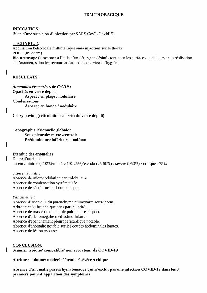
TDM THORACIQUE
INDICATION:
Bilan d’une suspicion d’infection par SARS Cov2 (Covid19)
TECHNIQUE:
Acquisition hélicoïdale millimétrique sans injection sur le thorax
PDL : (mGy.cm)
Bio-nettoyage du scanner à l’aide d’un détergent-désinfectant pour les surfaces au décours de la réalisation
de l’examen, selon les recommandations des services d’hygiène
RESULTATS:
Anomalies évocatrices de CoV19 :
Opacités en verre dépoli
Aspect : en plage / nodulaire
Condensations
Aspect : en bande / nodulaire
Crazy paving (réticulations au sein du verre dépoli)
Topographie lésionnelle globale :
Sous pleurale/ mixte /centrale
Prédominance inférieure : oui/non
Etendue des anomalies
Degré d’atteinte :
absent /minime (<10%)/modéré (10-25%)/étendu (25-50%) / sévère (>50%) / critique >75%
Signes négatifs :
Absence de micronodulation centrolobulaire.
Absence de condensation systématisée.
Absence de sécrétions endobronchiques.
Par ailleurs :
Absence d’anomalie du parenchyme pulmonaire sous-jacent.
Arbre trachéo-bronchique sans particularité.
Absence de masse ou de nodule pulmonaire suspect.
Absence d'adénomégalie médiastino-hilaire.
Absence d'épanchement pleuropéricardique notable.
Absence d'anomalie notable sur les coupes abdominales hautes.
Absence de lésion osseuse.
CONCLUSION:
Scanner typique/ compatible/ non évocateur de COVID-19
Atteinte : minime/ modérée/ étendue/ sévère /critique
Absence d’anomalie parenchymateuse, ce qui n’exclut pas une infection COVID-19 dans les 3
premiers jours d’apparition des symptômes
ANNEXE 3 BSTI – British Society of Thoracic Imaging. Thoracic Imaging in COVID-19 Imaging – Guidance for the Reporting Radiologist
Thoracic Imaging in COVID-19 Infection
Guidance for the Reporting RadiologistBritish Society of Thoracic Imaging
Version 2 16th March 2020
Background COVID-19
• First cases Wuhan City China December 2019
• Large outbreak Northern Italy February 2020
• First UK cases seen February 2020
• WHO Pandemic March 2020
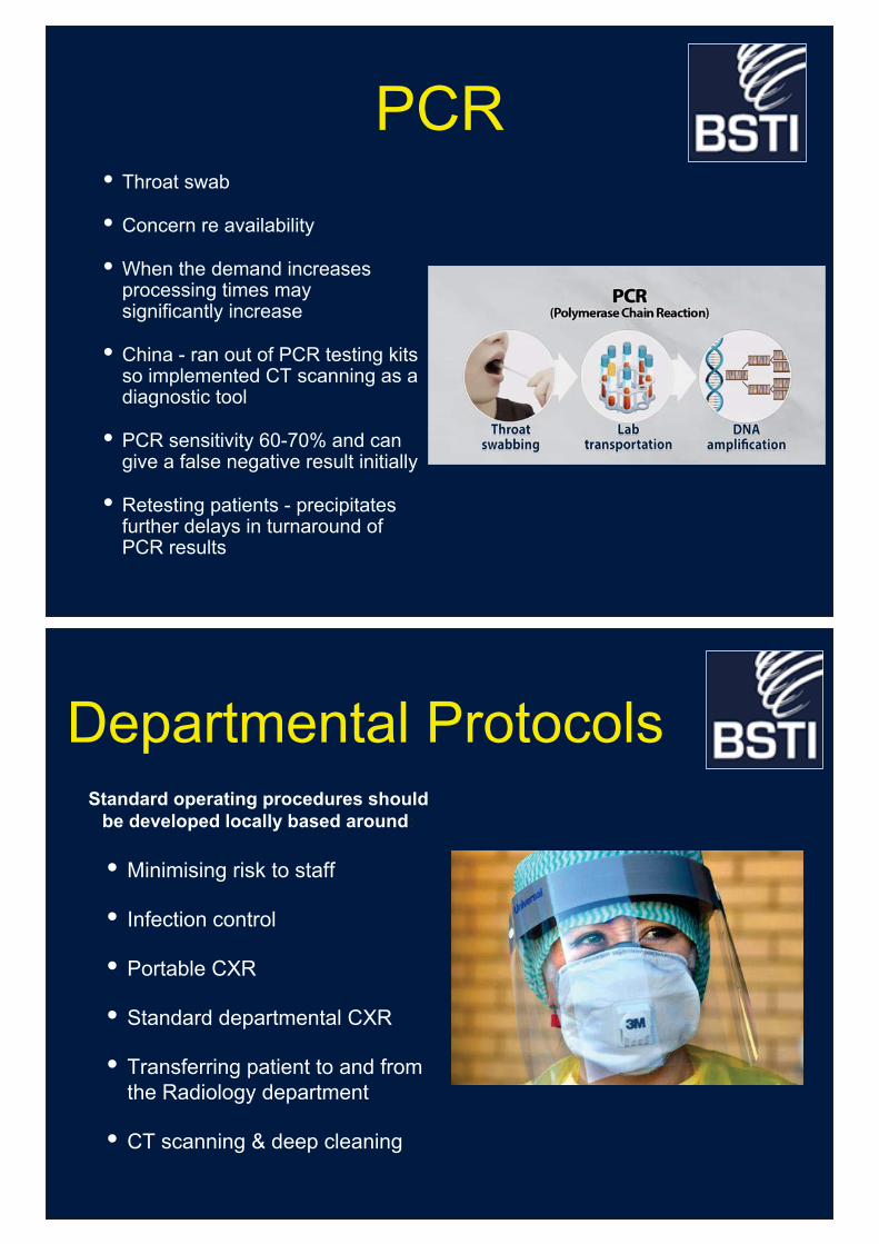
PCR• Throat swab
• Concern re availability
• When the demand increases processing times may significantly increase
• China - ran out of PCR testing kits so implemented CT scanning as a diagnostic tool
• PCR sensitivity 60-70% and can give a false negative result initially
• Retesting patients - precipitatesfurther delays in turnaround of PCR results
Departmental Protocols
• Minimising risk to staff
• Infection control
• Portable CXR
• Standard departmental CXR
• Transferring patient to and from the Radiology department
• CT scanning & deep cleaning
Standard operating procedures should be developed locally based around:
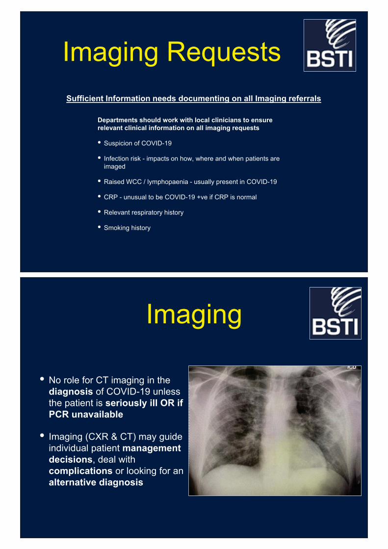
Imaging Requests
Departments should work with local clinicians to ensure relevant clinical information on all imaging requests
• Suspicion of COVID-19
• Infection risk - impacts on how, where and when patients are imaged
• Raised WCC / lymphopaenia - usually present in COVID-19
• CRP - unusual to be COVID-19 +ve if CRP is normal
• Relevant respiratory history
• Smoking history
Sufficient Information needs documenting on all Imaging referrals
Imaging
• No role for CT imaging in the diagnosis of COVID-19 unless the patient is seriously ill OR if PCR unavailable
• Imaging (CXR & CT) may guideindividual patient management decisions, deal with complications or looking for an alternative diagnosis
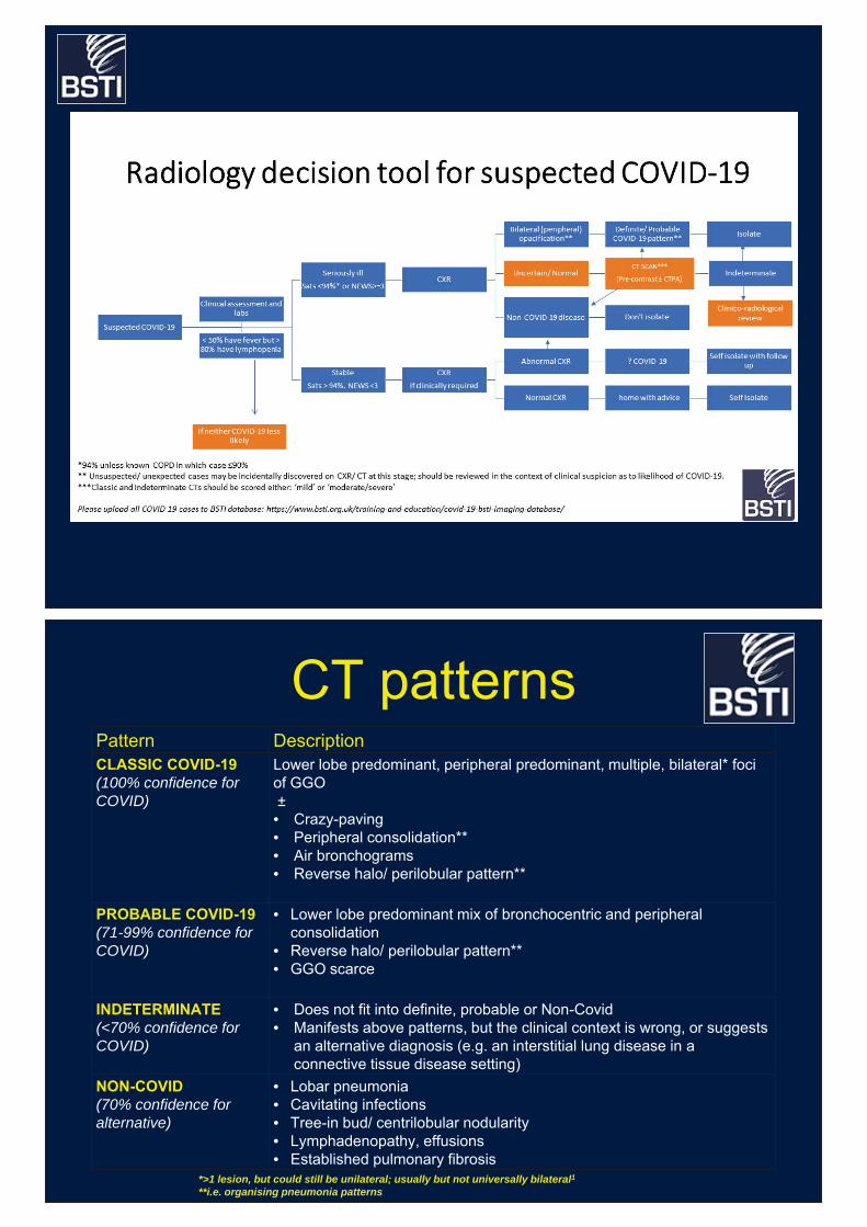
CT patternsPattern DescriptionCLASSIC COVID-19(100% confidence for COVID)
Lower lobe predominant, peripheral predominant, multiple, bilateral* foci of GGO±• Crazy-paving• Peripheral consolidation**• Air bronchograms• Reverse halo/ perilobular pattern**
PROBABLE COVID-19(71-99% confidence for COVID)
• Lower lobe predominant mix of bronchocentric and peripheral consolidation
• Reverse halo/ perilobular pattern**• GGO scarce
INDETERMINATE(<70% confidence for COVID)
• Does not fit into definite, probable or Non-Covid• Manifests above patterns, but the clinical context is wrong, or suggests
an alternative diagnosis (e.g. an interstitial lung disease in a connective tissue disease setting)
NON-COVID(70% confidence for alternative)
• Lobar pneumonia• Cavitating infections• Tree-in bud/ centrilobular nodularity• Lymphadenopathy, effusions• Established pulmonary fibrosis
*>1 lesion, but could still be unilateral; usually but not universally bilateral1**i.e. organising pneumonia patterns
EXAMPLES
• The following examples are from recent UK cases
• Note that the clinical suspicion is IMPERATIVE
• Without the suspicion, the radiology is non-specific and could easily represent so many other processes
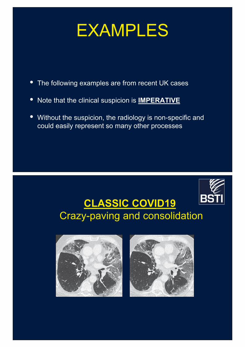
CLASSIC COVID19Crazy-paving and consolidation
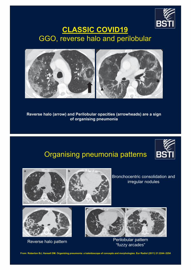
CLASSIC COVID19GGO, reverse halo and perilobular
Reverse halo (arrow) and Perilobular opacities (arrowheads) are a sign of organising pneumonia
Organising pneumonia patterns
From: Roberton BJ, Hansell DM. Organizing pneumonia: a kaleidoscope of concepts and morphologies. Eur Radiol (2011) 21:2244–2254
Reverse halo pattern Perilobular pattern“fuzzy arcades”
Bronchocentric consolidation and irregular nodules

PROBABLE COVID19Bronchocentric and nodular organising pneumonia
patterns, air bronchogram, but no GGO
INDETERMINATE COVID19GGO ?from contrast and/or dependent
Needs clinic-radiology review. Fever, CRP and especially a lymphopaenia, would make COVID19 more likely

NON-COVID19
0.6mm lung recon 8mm MIP lung recon
Burkitt's lymphoma, pancytopenic. febrile 5 days with diarrhoea.tree in bud (MIPs useful) and acinar- COVID negative (initial swab)
Serial change
Chest CT images of a 62-year-old man with fever for 2 weeks, and dyspnea for 1 day. Negative results of RT-PCR assay for the SARS-CoV-2 using a swab samples were obtained on February 3 and 11, 2020, respectively. (column A) Chest CT with multiple axial images shows multiple ground-glass opacities in the bilateral lungs. (column
B) Chest CT with multiple axial images shows enlarged multiple ground-glass opacities. (column C) Chest CT with multiple axial images shows the progression of the disease from ground-glass
opacities to multifocal organizing consolidation. (D column) chest CT with multiple axial images shows partial absorption of the organizing
consolidation.
Ai et al. Radiology. 2020 Feb 26:200642. doi:
10.1148/radiol.2020200642.
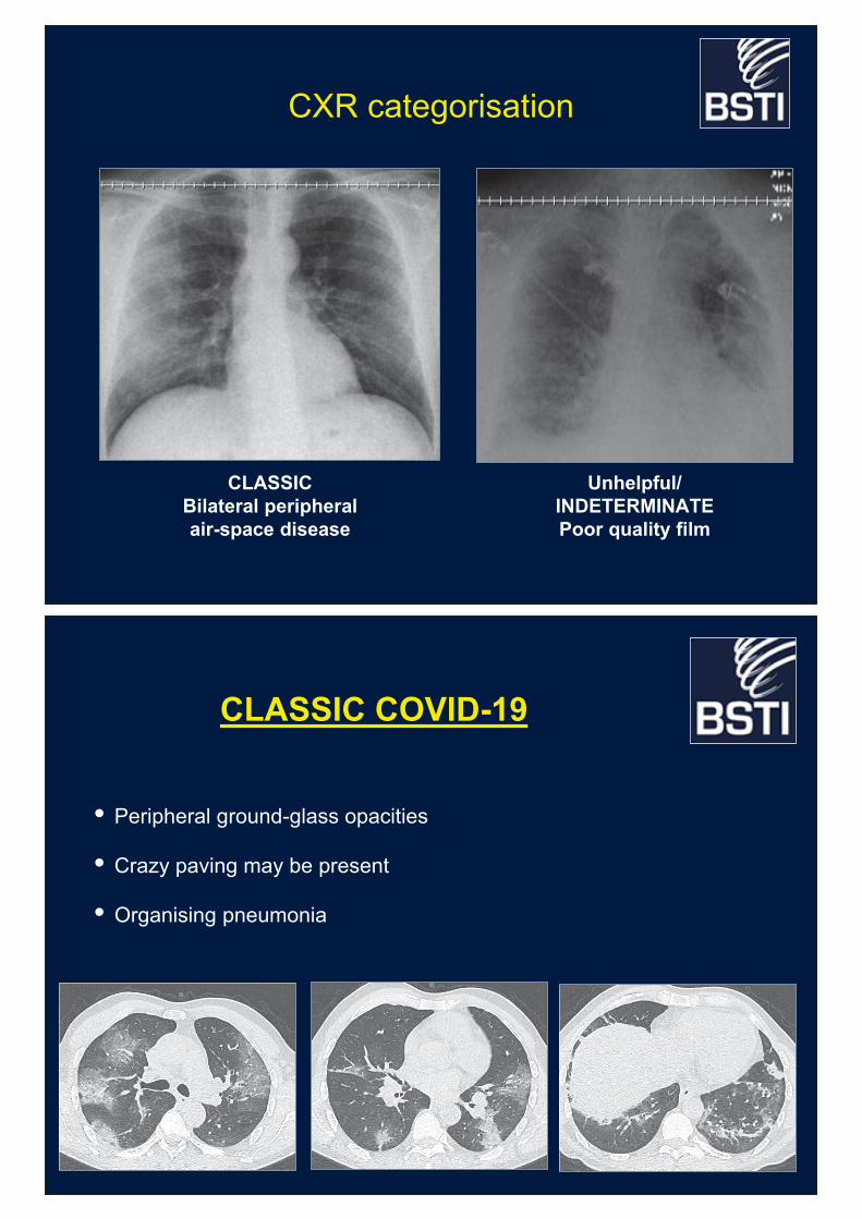
CXR categorisation
CLASSICBilateral peripheral air-space disease
Unhelpful/ INDETERMINATEPoor quality film
CLASSIC COVID-19
• Peripheral ground-glass opacities
• Crazy paving may be present
• Organising pneumonia
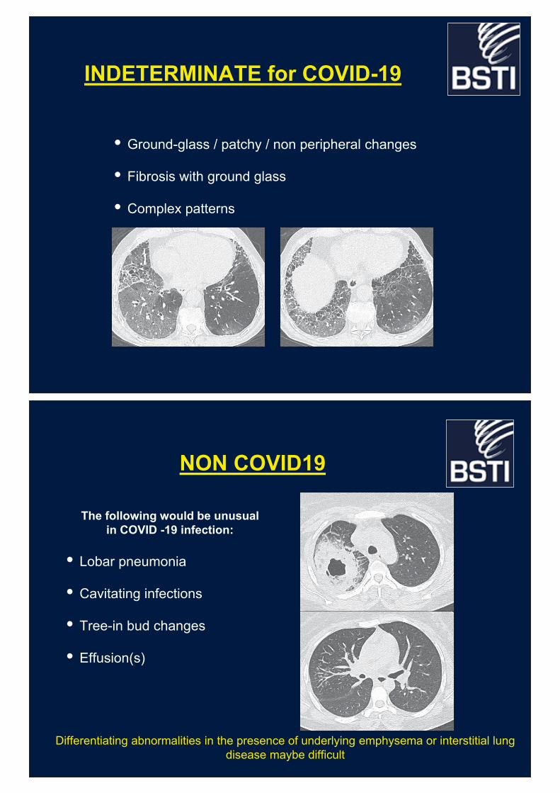
INDETERMINATE for COVID-19
• Ground-glass / patchy / non peripheral changes
• Fibrosis with ground glass
• Complex patterns
NON COVID19
• Lobar pneumonia
• Cavitating infections
• Tree-in bud changes
• Effusion(s)
The following would be unusual in COVID -19 infection:
Differentiating abnormalities in the presence of underlying emphysema or interstitial lung disease maybe difficult
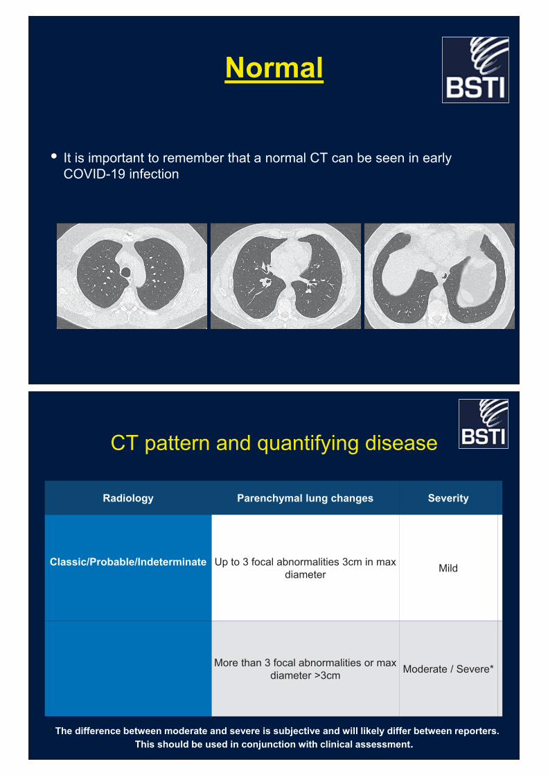
Normal
• It is important to remember that a normal CT can be seen in early COVID-19 infection
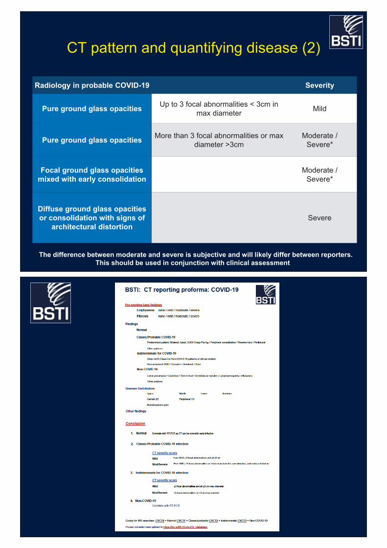
CT pattern and quantifying disease
Radiology Parenchymal lung changes Severity
Classic/Probable/Indeterminate Up to 3 focal abnormalities 3cm in max diameter
Mild
More than 3 focal abnormalities or max diameter >3cm
Moderate / Severe*
* The difference between moderate and severe is subjective and will likely differ between reporters. This should be used in conjunction with clinical assessment.
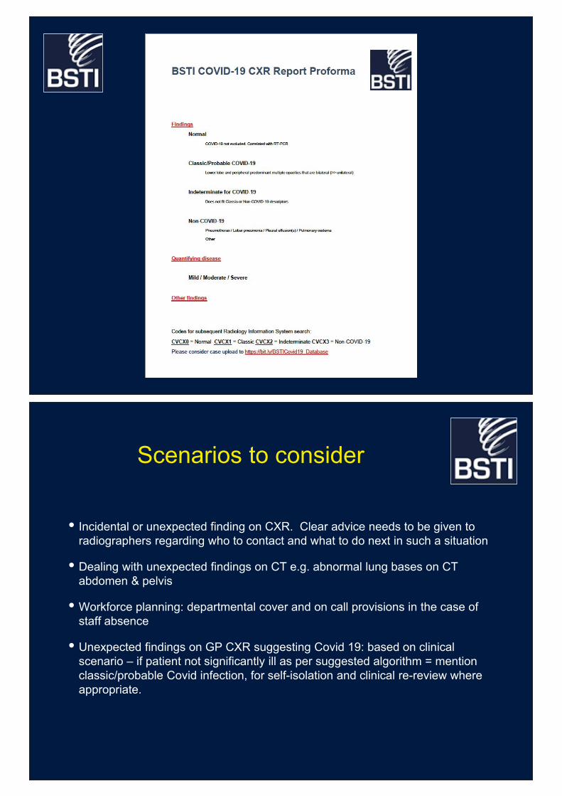
Radiology in probable COVID-19 Severity
Pure ground glass opacities Up to 3 focal abnormalities < 3cm in max diameter
Mild
Pure ground glass opacities More than 3 focal abnormalities or max diameter >3cm
Moderate / Severe*
Focal ground glass opacities mixed with early consolidation
Moderate / Severe*
Diffuse ground glass opacities or consolidation with signs of
architectural distortionSevere
CT pattern and quantifying disease (2)
* The difference between moderate and severe is subjective and will likely differ between reporters. This should be used in conjunction with clinical assessment.
Scenarios to consider
• Incidental or unexpected finding on CXR. Clear advice needs to be given to radiographers regarding who to contact and what to do next in such a situation
• Dealing with unexpected findings on CT e.g. abnormal lung bases on CT abdomen & pelvis
• Workforce planning: departmental cover and on call provisions in the case of staff absence
• Unexpected findings on GP CXR suggesting Covid 19: based on clinical scenario – if patient not significantly ill as per suggested algorithm = mention classic/probable Covid infection, for self-isolation and clinical re-review where appropriate.
Case Database
• Refer a case https://bit.ly/BSTICovid19_Database
• Teaching Library https://bit.ly/BSTICOVID19_Teaching_Library
• Updates can be found on www.bsti.org.uk or via our Facebook(@BSTImaging) or Twitter (@BSTImaging) feeds.
The BSTI would like to thank Prof Nicola Sverzellati and his team in Parma Italy for sharing information and images.
ANNEXE 4a BSR – Belgian Society of Radiology: COVID-19 info from BSR. Guidelines for imaging in COVID-
19. 14 mars 2020
COVID-19 info from the BSR
March 14 2020.
From the BSR Board, Scientific Council and BSR Chest Section.
Concerning COVID-19
1/Guidelines as to the hygienic measures for limiting virus spread in imaging departments are
at the discretion of the authorities and subject to local decisions. The BSR cannot supply exact
guidelines also due to the rapidly changing circumstances.
2/Guidelines how to organize Imaging Departments and their throughput/appropriateness are
also subject to local hospital/network and authority decisions.
3/Guidelines for imaging in COVID-19 are supplied below.
The ACR, SFR and FOD have published very useful information on their websites.
Concerning point 3: Typical indications for imaging Imaging of COVID-19 patients (proven or ‘possible’) is only needed when there are clinical implications.
- CT should NOT be used as screening tool to replace laboratory testing for COVID-19 - If imaging is needed and patients can be transferred to the radiology department:
unenhanced CT is the preferred imaging technique. There is no place for chest radiographs.
- Follow-up imaging of patients who are admitted to the Intensive Care unit or specialized units in isolation: portable radiography
- Patients who have a clinical suspicion of COVID-19 but cannot be tested (due to
shortage of testing) and who are advised to go home: no need for imaging, no chest radiographs, no CT.
- Patients who are hospitalized and tested COVID-19 positive with no clinical deterioration: no imaging
- Patients who are hospitalized and tested COVID-19 positive with clinical deterioration:
o In general: unenhanced CT, thin section o When there are comorbidities, hemoptysis, suspicion of pulmonary embolism,
possibility of other pathology (pleural, pericardial, …) etc.: IV contrast may be needed. This needs to be discussed with the treating clinician on a case per case basis.
- Patients who are hospitalized and tested COVID-19 negative but who have a clinical suspicion of possible COVID-19 infection: should be handled with the same
precautions as COVID-19 positive patients. Imaging only indicated in case of therapeutic consequences.
- In very specific conditions, CT can be considered to confirm or exclude COVID-19 infection. For example patients who need hemodialysis or need to be transferred to other centres (handicapped patients, specific treatment, ….). If patients have negative testing but high clinical suspicion, a negative CT may be used to confirm low likelihood of infection and patients can be transferred safely. These cases should be the exception and should be discussed on a case per case basis with the treating physician.
- Follow-up imaging studies in patients who are clinically improving is not indicated. - There is currently no evidence to perform a CT at the end of treatment.
- If COVID-19 positive patients need an ultrasound: this should be preferably done
bedside.
CT-imaging features: Radiologists should familiarize themselves with the CT appearance of COVID-19 infection, in order to be able to identify possible COVID-19 related infection on imaging studies performed for other reasons. See cases. https://www.bsr-
web.be/docs/Imaging_Coronavirus_BSR_chest.pdf. Preliminary data show that findings in Belgian patients are similar to cases published in literature (mainly Chinese population)
ANNEXE 4b et 4c BSR – Belgian Society of Radiology: template to
report CT in COVID cases. Mars 2020
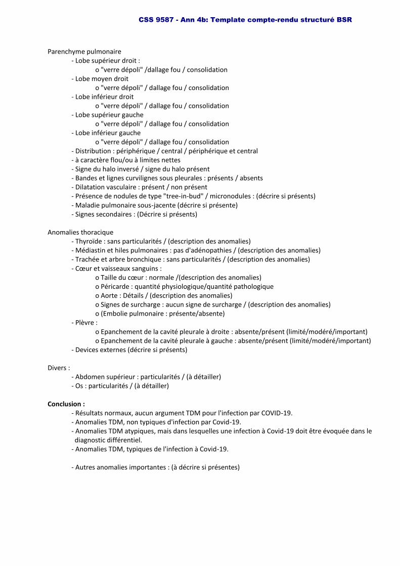
Parenchyme pulmonaire - Lobe supérieur droit :
o "verre dépoli" /dallage fou / consolidation - Lobe moyen droit
o "verre dépoli" / dallage fou / consolidation - Lobe inférieur droit
o "verre dépoli" / dallage fou / consolidation - Lobe supérieur gauche
o "verre dépoli" / dallage fou / consolidation - Lobe inférieur gauche
o "verre dépoli" / dallage fou / consolidation - Distribution : périphérique / central / périphérique et central - à caractère flou/ou à limites nettes - Signe du halo inversé / signe du halo présent - Bandes et lignes curvilignes sous pleurales : présents / absents - Dilatation vasculaire : présent / non présent - Présence de nodules de type "tree-in-bud" / micronodules : (décrire si présents) - Maladie pulmonaire sous-jacente (décrire si présente) - Signes secondaires : (Décrire si présents)
Anomalies thoracique - Thyroïde : sans particularités / (description des anomalies) - Médiastin et hiles pulmonaires : pas d'adénopathies / (description des anomalies) - Trachée et arbre bronchique : sans particularités / (description des anomalies) - Cœur et vaisseaux sanguins :
o Taille du cœur : normale /(description des anomalies) o Péricarde : quantité physiologique/quantité pathologique o Aorte : Détails / (description des anomalies) o Signes de surcharge : aucun signe de surcharge / (description des anomalies) o (Embolie pulmonaire : présente/absente)
- Plèvre : o Epanchement de la cavité pleurale à droite : absente/présent (limité/modéré/important) o Epanchement de la cavité pleurale à gauche : absente/présent (limité/modéré/important)
- Devices externes (décrire si présents)
Divers : - Abdomen supérieur : particularités / (à détailler) - Os : particularités / (à détailler)
Conclusion :
- Résultats normaux, aucun argument TDM pour l'infection par COVID-19. - Anomalies TDM, non typiques d'infection par Covid-19. - Anomalies TDM atypiques, mais dans lesquelles une infection à Covid-19 doit être évoquée dans le diagnostic différentiel. - Anomalies TDM, typiques de l'infection à Covid-19. - Autres anomalies importantes : (à décrire si présentes)
CSS 9587 - Ann 4b: Template compte-rendu structuré BSR

Facultatif*. - Score CT pour évaluer l'étendue du parenchyme pulmonaire affecté, avec évaluation par lobe. % de personnes touchées. 5 points par lobe, soit 25 au total.
0% : 0 point < 5% : 1 point 5-25% : 2 points 25-50% : 3 points 50-75% : 4 points
CSS 9587 - Ann 4b: Template compte-rendu structuré BSR
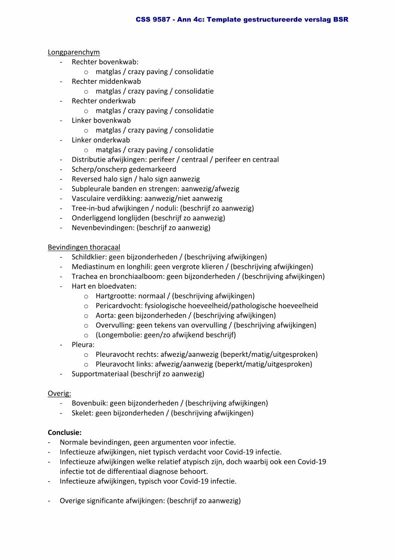
Longparenchym - Rechter bovenkwab:
o matglas / crazy paving / consolidatie - Rechter middenkwab
o matglas / crazy paving / consolidatie - Rechter onderkwab
o matglas / crazy paving / consolidatie - Linker bovenkwab
o matglas / crazy paving / consolidatie - Linker onderkwab
o matglas / crazy paving / consolidatie - Distributie afwijkingen: perifeer / centraal / perifeer en centraal - Scherp/onscherp gedemarkeerd - Reversed halo sign / halo sign aanwezig - Subpleurale banden en strengen: aanwezig/afwezig - Vasculaire verdikking: aanwezig/niet aanwezig - Tree-in-bud afwijkingen / noduli: (beschrijf zo aanwezig) - Onderliggend longlijden (beschrijf zo aanwezig) - Nevenbevindingen: (beschrijf zo aanwezig)
Bevindingen thoracaal
- Schildklier: geen bijzonderheden / (beschrijving afwijkingen) - Mediastinum en longhili: geen vergrote klieren / (beschrijving afwijkingen) - Trachea en bronchiaalboom: geen bijzonderheden / (beschrijving afwijkingen) - Hart en bloedvaten:
o Hartgrootte: normaal / (beschrijving afwijkingen) o Pericardvocht: fysiologische hoeveelheid/pathologische hoeveelheid o Aorta: geen bijzonderheden / (beschrijving afwijkingen) o Overvulling: geen tekens van overvulling / (beschrijving afwijkingen) o (Longembolie: geen/zo afwijkend beschrijf)
- Pleura: o Pleuravocht rechts: afwezig/aanwezig (beperkt/matig/uitgesproken) o Pleuravocht links: afwezig/aanwezig (beperkt/matig/uitgesproken)
- Supportmateriaal (beschrijf zo aanwezig) Overig:
- Bovenbuik: geen bijzonderheden / (beschrijving afwijkingen) - Skelet: geen bijzonderheden / (beschrijving afwijkingen)
Conclusie: - Normale bevindingen, geen argumenten voor infectie. - Infectieuze afwijkingen, niet typisch verdacht voor Covid-19 infectie. - Infectieuze afwijkingen welke relatief atypisch zijn, doch waarbij ook een Covid-19
infectie tot de differentiaal diagnose behoort. - Infectieuze afwijkingen, typisch voor Covid-19 infectie.
- Overige significante afwijkingen: (beschrijf zo aanwezig)
CSS 9587 - Ann 4c: Template gestructureerde verslag BSR
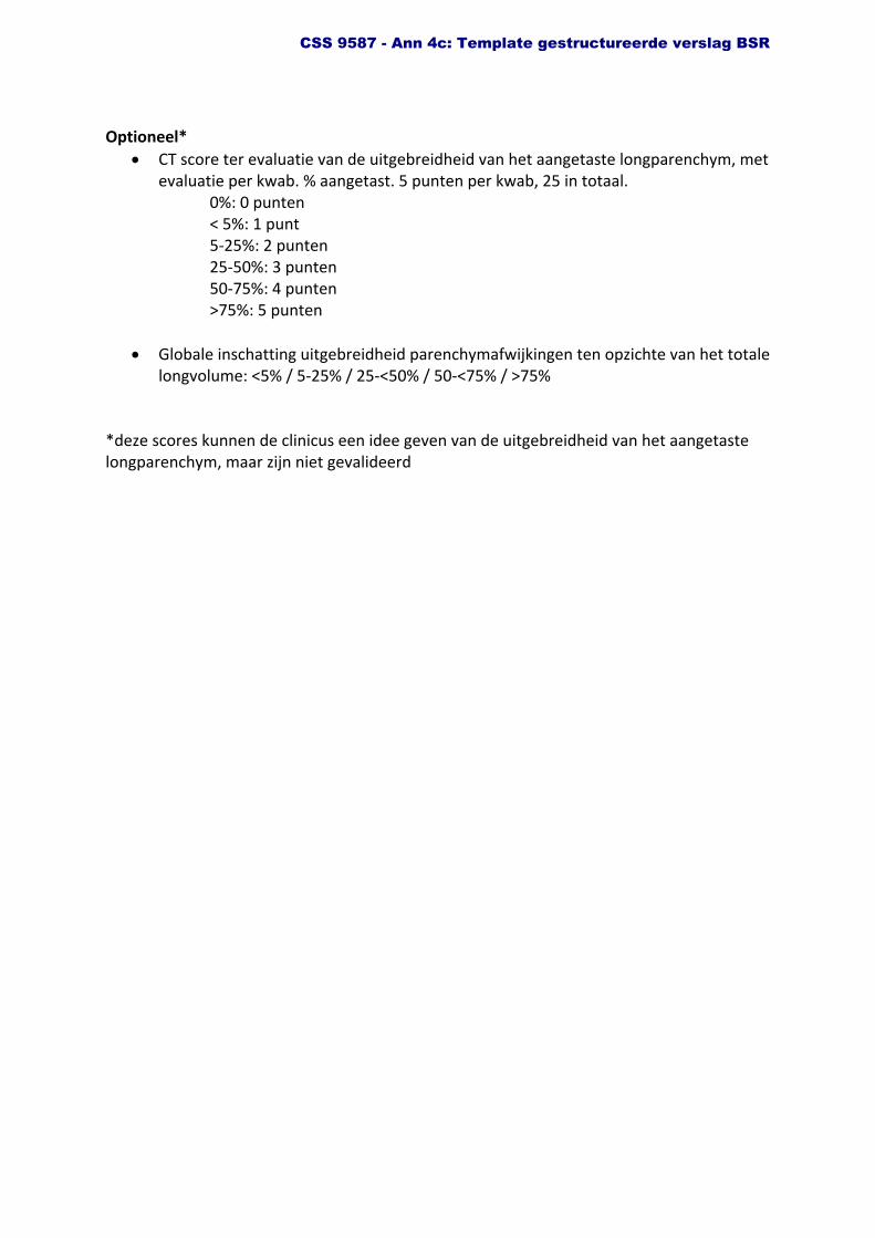
Optioneel*
• CT score ter evaluatie van de uitgebreidheid van het aangetaste longparenchym, met evaluatie per kwab. % aangetast. 5 punten per kwab, 25 in totaal.
0%: 0 punten < 5%: 1 punt 5-25%: 2 punten 25-50%: 3 punten 50-75%: 4 punten >75%: 5 punten
• Globale inschatting uitgebreidheid parenchymafwijkingen ten opzichte van het totale
longvolume: <5% / 5-25% / 25-<50% / 50-<75% / >75% *deze scores kunnen de clinicus een idee geven van de uitgebreidheid van het aangetaste longparenchym, maar zijn niet gevalideerd
CSS 9587 - Ann 4c: Template gestructureerde verslag BSR
ANNEXE 5 Mahmud Mossa-Basha, Carolyn C. Meltzer, Danny
C Kim, Michael J Tuite, K. Pallav Kolli, Bien Soo Tan. Radiology Department Preparedness for COVID-19: Radiology Scientific Expert Panel.
Radiology, 16/3/2020
Inpres
sRadiology Department Preparedness for COVID-19: Radiology Scientific Expert Panel
Mahmud Mossa-Basha, MD Department of Radiology, University of Washington School of Medicine, Seattle, WA Associate professor, radiology Vice chair of operations Email: [email protected] 1959 NE Pacific St, Seattle, WA 98195
Carolyn C. Meltzer, MD FACR (Corresponding Author)[email protected] P. Timmie Professor and Chair of Radiology & Imaging Sciences Executive Associate Dean, Faculty Academic Advancement, Leadership & Inclusion Emory University School of Medicine
Danny C Kim, MD, MMM [email protected] Professor, NYU Grossman School of MedicineAssociate Chair of Quality and Safety, Department of Radiology NYU Langone Health 660 First Avenue, 3rd Floor New York, NY 10016
Michael J Tuite MD Professor, Radiology Vice Chair of Clinical Operations Musculoskeletal Imaging and Intervention [email protected]
K. Pallav Kolli, MD Associate Professor of Clinical Radiology Director of Operations, Interventional RadiologyAssociate Chair for Quality and Safety, Department of Radiology and Biomedical Imaging University of California San Francisco [email protected]
Bien Soo Tan, MD Chair, Division of Radiological Sciences, Singapore General Hospital, Academic Chair, Radiological Sciences Academic Clinical Programme, SingHealth Duke-NUS Academic Medical Centre [email protected] Division of Radiological Sciences, Singapore General Hospital, 20 College Road, Academia Level 4, Singapore 169856
Inpres
sThe Coronavirus Disease 2019 (COVID-19) pandemic began in December 2019 in Wuhan, China. The outbreak is due to severe acute respiratory syndrome coronavirus 2 (SARS-CoV-2) infection (1). Approximately 81,000 patients have been infected in China (2). Although infection rates are said to be controlled in China through severe public health measures, Italy (more than 10,000 cases) and Iran (more than 8000 cases) have seen exponential increases in the number of infected individuals.
Other than China, Italy, and Iran, most countries have had approximately 2 months to prepare their responses to the COVID-19 pandemic. These responses are led by public health authorities of national governments in coordination with local governments and hospitals. Due to the nature of the emergency in China, chest CT findings (eg, peripheral ground-glass infiltrates and/or organizing pneumonia) temporarily became part of official diagnostic criteria of COVID-19 as a surrogate for viral nucleic acid testing (1). With improved disease understanding, chest CT findings are no longer part of the diagnostic criteria for COVID-19. Instead, at present, the focus of most radiology departments outside of China has shifted from diagnostic capability to preparedness.
Radiology preparedness is a set of policies and procedures directly applicable to imaging departments designed (a) to achieve sufficient capacity for continued operation during a health care emergency of unprecedented proportions, (b) to support the care of patients with COVID-19, and (c) to maintain radiologic diagnostic and interventional support for the entirety of the hospital and health system.
Because of varying infection control policies (both nationally and regionally), steps for radiology preparedness for COVID-19 will vary between institutions and clinics. The Radiology Editorial Board has assembled a team of radiologists who are active in coordination, development, and implementation of radiology preparedness policies for COVID-19. Their policies have been developed in conjunction with top infection control experts at their respective world-class healthcare systems. In the sections below, each panel member describes their department’s top priorities for COVID-19 preparedness in their environment. The Editorial Board hopes that readers may find one or more the highlighted healthcare systems to be similar to their own, providing impetus for action or confirmation of your current preparedness activities. See also summary Tables 1-2 and Figures 1-2 for further resources.
Inpres
sUniversity of Washington Medicine (UW) UW is a major metropolitan medical system, with three major urban medical centers and many outpatient clinics and imaging centers spread across Western Washington, at the epicenter of the COVID-19 outbreak in the United States. There have been more than 267 cases of COVID-19 and 24 deaths in Washington state, including approximately 18 patients with confirmed COVID-19 hospitalized at our institutions as of this writing. There is a substantial Asian population that exists in Seattle, including many professionals and students that frequently travel to China and other high infection rate regions. The largest risk remains in older patient populations, as 71% of patients infected in Washington state are over 50 years of age and 57% are over 60 years of age. Radiology leadership has helped in development of policies and guidelines relating to COVID-19 in areas of patient screening, spread precautions and patient triage in coordination with the hospital leadership. Radiology leadership has worked with input from our department membership, especially operations leaders and chest imagers to develop screening-specific guidelines. Top Priorities in Our Environment for COVID-19 Preparedness 1. Early detection and limiting exposure of healthcare workers, employees and patients, especially critically ill patients. The hospitals have implemented screeners at all hospital entrances to check those coming in for symptoms that could be related to SARS-Cov-2 infection or with risk factors related to travel or exposure. The radiology front desk serves as a screening site, with similar screening to that performed at the hospital front door. Patients who present with respiratory symptoms who are undergoing outpatient imaging or procedures have their studies canceled and are asked to follow up with their primary care physician. For inpatients with suspected or confirmed COVID-19, all nonemergent imaging and procedures are delayed until diagnosis is confirmed and they recover from their illness and are considered noncontagious. 2. Use of radiography and chest CT. Despite reports from China (3) and initial concerns from the United States Centers for Disease Control regarding unreliable test performance (4), our current RT-PCR for SARS-CoV-2 viral nucleic acid is estimated to have a sensitivity of 95%-97%. Our lab also has a turnaround time of less than 1 day, making RT-PCR an easy, accurate, and less resource-intensive examination. Our lab has been performing more than 500 tests per day, covering our system but also other regional systems, with approximately 10% positive results. Inconclusive results are seen in a small subset, which are then sent for confirmation to Washington state labs. Sensitivity and specificity of chest CT for COVID-19 are reported to range from 80%-90% and 60%-70%, respectively (3, 5). Thus, imaging is reserved for those cases where it will impact patient management and is clinically indicated or to evaluate for unrelated urgent/emergent indications. This is typically in cases where an alternative diagnosis is being ruled out or being
Inpres
sconsidered for acute symptom worsening. In our current workflow and with the accuracy and rapidity of the RT-PCR testing, there is no need for immediate CT imaging. In addition, if symptom worsening is thought to be secondary to COVID-19, imaging would not change management, as current treatment consists of oxygen and supportive care. When possible, imaging is performed at sites with less foot traffic and with fewer critically ill patients in that area to avoid secondary patient and staff exposure. Considerations are also being made to implement a containment zipper (a room isolation tarp barrier with a zipper for room access) to separate the control area from the CT scanner room. Imaging is performed in the imaging center nearest to the patient and, if possible, at the ambulatory clinic. This approach limits the transit of contagious patients and potential exposure of others. When possible, portable imaging is performed (both portable x-rays and portable CT in patient rooms) to limit equipment, room, and hallway decontamination requirements. 3. Imaging in COVID-19 patients who are suspected positive or RT-PCR positive. For these patients, droplet precaution is employed. Patients are masked during imaging and procedures. Deep cleaning of the room is performed after each patient. Air exchange processes are not employed due to patient masking. After imaging, the room downtime is typically between 30 minutes to 1 hour for room decontamination and passive air exchange. Airborne precautions are reserved for those patients who are critically ill or are undergoing aerosol-generating procedures (bronchoscopy, intubation, nebulization, or open suction). Ventilated patients are not an airborne precaution, as the system is considered a closed system. The decision to remove patients from isolation is determined by hospital infection control staff. Those staff members consider RT-PCR results, imaging findings, clinical characteristics, potential exposures, and risk factors and comorbidities into their decision making. 4. Staff protection. Our hospital systems in coordination with the state have worked to reduce the need to bring patients to major hospitals and clinics. This will help protect our vulnerable patient population and university employees. Harborview Medical Center instituted a team of physicians and nurses that go out to SARS-CoV-2 suspected patient homes to perform testing and evaluation. In coordination with the Seattle Flu Study and Gates Foundation, University of Washington has begun issuing SARS-Cov-2 testing kits for home use. UW Medical Center Northwest Hospital has implemented drive-thru testing for University employees who are symptomatic. The University has placed a moratorium on travel for all employees for 1 month. 5. Maintenance of radiology department operations. In radiology, we have focused on providing the ability for radiology faculty to work from home if needed for isolation. For those that are not suspected of having been exposed or infected but are concerned about potential exposures, we have created radiology outposts and isolated reading rooms across our enterprise, including single station reading rooms in our hospitals as well as in our outpatient imaging centers. Staff that do not need to be on-site and who can work remotely (eg, including coders, billers and schedulers) are directed to work from home. The majority of hospital staff- related meetings now use video conferencing rather than having in-person attendance. For
Inpres
sthose meetings that cannot be virtual, a determination of the necessity of the meeting is made by department leadership and those determined as nonessential are cancelled. Emory University School of Medicine, Atlanta, Georgia Emory is a large urban, academic, research-oriented healthcare system with 10 affiliated hospitals and extensive outpatient facilities covering an urban sprawl of more than 6.5 million persons in the greater Atlanta area. Atlanta is also home to the world’s busiest airport and the Centers for Disease Control and Prevention (CDC). Emory Healthcare has central coordination for COVID-19 preparedness, including daily leadership teleconferences; maintenance of a central stockpile of N-95 masks and other personal protective equipment; policy alignment with CDC, employee health, and infection control personnel. In the radiology department, we are holding regular meetings of the departmental leadership, including division directors and healthcare staff to plan for workforce integrity and the health of our staff. We also work closely with the Emory Office of Critical Event Preparedness and Response to ensure communication between our Emergency Department and the Radiology Department’s Division of Emergency and Trauma Imaging. Email notifications, leader calls, and redistribution of the department’s disaster preparedness escalation policy are the main modes of communication.
Top Priorities in Our Environment for COVID-19 Preparedness
1. Ensuring the health of our workforce so that we can best care for our patients. 2. Planning for sufficient staffing to cover our clinical needs.
Quarantine of clinical radiologists, staff, and trainees following travel to Level 3 countries and/or following exposure to patients with proven or suspected COVID-19 have the potential to quickly overburden our ability to adequately staff critical services. We are working to rapidly obtain and deploy additional home workstations so that asymptomatic radiologists can work from home. While this approach does not help procedural services, it can decompress diagnostic interpretations and provide back up for surge capacity.
3. Arranging for surge potential should the healthcare system be taxed by increased volume of patients and associated imaging needs.
4. Contamination of CT scanners is major concern. CT equipment may be out of commission for several hours for cleaning. Should a patient presenting atypically and/or not triggering suspicion for COVID-19 be scanned, the potential for additional patient and radiology staff exposure is heightened.
Inpres
sOther workforce considerations. The many spring radiology meetings are important venues for professional satisfaction, networking, and sharing meaningful scientific and educational material. With the prospect of major conferences being canceled we are finding opportunities for faculty and trainees to present their work locally to colleagues. During this period our colleagues have significant concerns about the safety of their families and school and daycare closings can impact their ability to get to the hospital.
New York University Langone Health (NYU), New York, NY NYU is a large academic health care center in New York City. The catchment area includes the tri-state region of New York, New Jersey, and Connecticut. With three large airports serving the area, there is a large volume of domestic and international people traffic. Currently, New York has the second the greatest number of COVID-19 cases in the United States. On March 7, 2020, the governor of New York, Andrew Cuomo, declared a state of emergency for New York State. At our institution, guidelines are distributed from the institutional leadership and then implemented by individual departments, including radiology. Top Priorities in Our Environment for COVID-19 Preparedness
1. COVID-19 crisis management team.
A dedicated team from radiology leadership was charged with overseeing the departmental preparedness for COVID-19. A vertical communication network was established to coordinate the activities of four hospitals and multiple ambulatory care offices that comprise our department. Department directors and site managers participated in regular conference calls to standardize the protocols for patient care and operational workflow. The crisis management team share institutional news and sources of information to inform the department leaders. The crisis management team serves as a centralized resource to answer questions and address concerns from individual sites and department members.
2. Implementing protocols for patients with known or suspected COVID-19 exposure
Institutional directives for patient care protocols were adapted to the radiology environment. For outpatients, patients who schedule imaging examinations are screened for pertinent symptoms, travel history and exposure to individuals with known COVID-19. Based upon these answers, patients are scheduled for their imaging examinations or directed to the Virtual Urgent Care program or the Emergency Department. At radiology reception areas, patients are screened for symptoms of fever and cough. Initially, patients with travel history to countries with widespread transmission or contact with individuals with known COVID-19 were identified and consult calls were placed to the institution’s Infection Prevention and Control (IPC) team to determine the need for patient testing and whether the patient will be
Inpres
stransferred to the emergency department or discharged home. Subsequently, as evidence of community spread of COVID-19 appeared in our geographic area, the threshold for calling the IPC team has been lowered with increased emphasis on patient symptoms. For these high-risk patients, the need to perform the scheduled imaging examination was determined by the relative urgency of the examination for patient care management.
In the hospital and emergency department (ED) setting, patients have usually already been identified as known or suspected COVID-19 patients and imaging examinations are provided as clinically warranted.
3. Reducing potential transmission from known or suspected COVID-19 patients
The guiding principle to reduce potential transmission from known or suspected COVID-19 patients is source control. In the ambulatory care setting, a patient presenting with fever and cough is immediately given a mask and directed to an isolation room for further screening and consultation with the IPC team. Healthcare providers who perform this additional screening wear personal protective equipment (PPE) including gloves, mask, and eye-shield. If the scheduled imaging examination is ultimately performed, the healthcare providers, such as the technologist and nurse, wear the same PPE and may wear a gown if there is the potential for close or direct contact with the patient. A census of the other patients and staff in the practice at the time of the patient visit is logged so that they may be contacted should the patient test positive for COVID-19.
In the hospital and ED setting, patients have usually already been identified as known or
suspected COVID-19 patients. A dedicated portable x-ray machine was assigned to image only these patients. If additional advanced imaging examinations are required, the radiology staff wears the necessary PPE and the room is cleaned and disinfected according to hospital protocols.
4. Reducing potential exposure for department members In order to minimize risk and potential for exposure, travel restrictions were instituted for
all employees of our organization. All domestic and international work-related travel and business/academic attendance at meetings, conferences, and similar events were banned for a temporary time period. Large group gatherings were discouraged.
In our department, a significant number of the clinical faculty already have home workstations as part of our clinical wellness program. Prior to the travel ban, radiologists returning from high risk areas were asked to self-quarantine and interpret cases from home. With the possibility of increasing COVID-19 related quarantines involving the clinical faculty, additional home workstations have been ordered to provide quarantined clinical faculty the ability to work from home to meet the clinical demand.
With the increased usage of PPE and the concern for shortages, our institution implemented conservation measures to ensure that supplies are available for radiology staff
Inpres
sand patients who required them. In addition, allocation of PPE has been centralized within out hospital. PPE is distributed to areas of our department based upon clinical need.
University of Wisconsin Hospital
The University of Wisconsin hospital is a 675-bed academic hospital in Madison, WI. Our catchment area is a population of about 600,000 people. The main risk factor in our area is the international nature of our university and area businesses. There are 44,000 students at the University of Wisconsin – Madison, many do a semester abroad; the university research programs thrive on international outreach. Madison is home to Epic Systems; under normal conditions, 4000 employees traveled weekly to work in hospitals throughout the United States and internationally. At the time of writing, the University of Wisconsin hospital has had one patient diagnosed with COVID-19. None of the hospital personnel in contact with the patient developed symptoms or positive real-time reverse transcriptase polymerase chain reaction (RT-PCR) for viral nucleic acid. Our radiology department response is closely coordinated with our hospital infection control team. Hospital infection control personnel have had in-person meetings with our radiography technologists and have given tutorials on infection control procedures for respiratory infections. The hospital infection response workgroup sends out daily emails to all employees. Our department’s Senior Director attends daily hospital COVID-19 command center briefings and meets daily with the radiology Vice Chair of Operations. Department modality managers have policies and procedures developed several years ago to deal with patients who might have EBOLA; existing policies were reviewed, and staff were updated. For the faculty radiologists, our hospital infection response workgroup had our department create a back-up call schedule. The purpose is to cover faculty who become ill or are quarantined. Each radiology section has responsibility for sick-coverage for their daily clinical services and on-call staffing; individual radiology specialty areas have created back-up call schedules. Almost all radiologists have home PACS workstations; use of remote interpretation has been incorporated into our department’s response plan. Top Priorities in Our Environment for COVID-19 Preparedness 1. Insure all department employees are aware of and are performing recommended infection control protocols. This applies not only to interacting with patients but also with fellow employees and working on shared workstations. 2. Arrange infection control tutorials led by hospital infection control experts. Time has been allocated for hospital infection control experts to provide in person tutorials and provide
Inpres
sinformation required protocols for interaction with patients who may have COVID-19. Seminars by our Infection control personal have also been arranged with our CT, ultrasound and interventional technologists. These individuals are anticipated to be on the front lines in interaction with patients with suspected COVID-19 infection. 3. Develop a detailed operational plan for a new, separate urgent care site for COVID-19. In the case of severe community outbreak, hospital infection control staff may take steps to provide care for COVID-19 patients in a separate facility away from our main hospital. We have developed a plan to equip and staff the site with a portable x-ray machine using strict infection control procedures. 4. Review and practice protocols for decontaminating imaging rooms after caring for a COVID-19 patient. This includes a 1 hour downtime for passive air exchange. 5. Work to better define the role of CT and 2-view chest x-rays in patients with COVID-19. While multiple publications in RADIOLOGY and other major journals have described COVID-19 findings on chest CT, the vast majority of patients have been from China. In that environment, lack of RT-PCR test kits, remarkable influxes of patients required detecting and staging of COVID-19 disease patterns with chest CT. The applicability of those results to our environment is not yet know. 6. Continue sharing information with all department employees, including open discussions on ramp-up of stricter infection control procedures if needed. This includes having faculty work from their home PACS or sections splitting their faculty and fellows into separate reading sites, having residents attend conferences remotely from their reading rooms so they do not gather in one location, and having technologists work from one location and not rotate between health care facilities.
University of California San Francisco (UCSF)
UCSF is a tertiary-quaternary, 1000 bed urban referral academic center. San Francisco is an international crossroads and one of the earlier sites of disease in the United States.
The prevalence of COVID-19 cases in our institution’s catchment area has increased rapidly over the last two weeks. UCSF has instituted a Hospital Incident Command System (HICS) structure to guide decisions and practice. The institution has shifted its strategy from containment to risk-mitigation in view of the size of the outbreak and the documented community spread of infection. Until internal laboratory testing had been developed, confirmatory RT-PCR testing represented a significant bottleneck in patient triage.
The response of our radiology department is coordinated with health system leadership, including radiology planning and strategy at the health system level. Policies are developed at
Inpres
sthe health system level, including stay-at-home and return-to-work, faculty travel, and procedures for safe transport, respiratory isolation and treatment of suspected or confirmed COVID-19 patients. We have developed Radiology-specific guidelines for safe imaging of Patients Under Investigation (PUI) with CT and x-ray, contingency staffing planning and reducing risk of nosocomial spread.
Top Priorities in Our Environment for COVID-19 Preparedness 1. Identification of patients at risk for having COVID-19. Patient screening is now undertaken at the time of radiology exam scheduling and in all outpatient imaging settings. Additionally, all patients and visitors are screened at the entrance to clinic buildings and hospitals. Respiratory clinics equipped with portable x-ray units have been established at each of our 3 main hospital sites. Ambulatory patients who screen positive for possible COVID-19 illness are redirected to one of these respiratory clinics for further evaluation. 2. Development of standard operating procedures for safe imaging of patients with suspected or known COVID-19. (see figures) At UCSF, CT is not considered a screening examination for COVID-19. A multidisciplinary team of experts, including thoracic radiologists, is developing a guideline for use of CT imaging in patients with known or suspected disease. Standard operating procedures for safely imaging patients with portable x-ray units and for imaging patients with CT when necessary have been developed, ensuring that all team members are aware of patients’ isolation status and that clinical and imaging teams provide coordinated high-level care in a safe and efficient manner. 3. Staff education regarding COVID-19 prevention. Education of radiology staff regarding stay-at-home policies and infection prevention techniques, such as handwashing and regular cleaning of fomites, was an early focus in our department. 4. Personal protective equipment (PPE) availability and education for health-care workers. Our department has worked with health system leadership to ensure adequate supplies of necessary PPE. Staff and faculty champions were recruited and serve to educate and reinforce appropriate donning and doffing techniques for PPE. 5. Implementation of “social distancing” strategies for staff, trainees, and faculty. All large staff gatherings (e.g. radiology grand rounds) have been cancelled. We have aimed to decrease foot traffic in radiology reading rooms by encouraging remote consultations by video and telephone rather than in-person. Recurring departmental meetings and multiple clinical conferences have been moved to video conferencing. We are exploring strategies to allow diagnostic radiologists to work from home and developing guidance for when this is appropriate.
6. Planning for a potential surge of patients: A surge in patients with known or suspected COVID-19 illness will require re-allocation of resources. Illness or factors such as school closing
Inpres
smay deplete our physician and staff workforce. We have developed contingency plans for maintenance of our clinical activities and staffing in such cases.
In order to maintain hospital bed availability, schedules for interventional radiology and neurointerventional radiology have been frozen (no additional patient slots) for two weeks for procedures that require patients to have overnight observation or hospitalization. Singapore General Hospital (SGH) SGH is a 1773-bed academic tertiary hospital in the city center of Singapore. SGH has one million patient visits annually. Singapore is an international travel hub. The first COVID-19 patient in Singapore was diagnosed in SGH on 23 January 2020. As of March 12, 2020, there were 187 confirmed cases of COVID-19 in Singapore. The activities for infection control in the radiology department are highly coordinated with our institution-wide infection control staff. We have separate imaging facilities for inpatients and outpatients and negative air pressure rooms for radiography, ultrasonography, CT, and MRI to cater for patients with infection control risks. Even prior to the COVID-19 outbreak, radiology had an infection control committee that included infectious disease (ID) physicians. In addition, there is an institutional disease outbreak task force that includes several members from radiology leadership, as there is acknowledgement that radiology is a key component in an outbreak situation (2). At the present time, the national strategy for COVID-19 is that of containment. Our institutional strategy is to minimize any possibility of in-hospital transmission and to achieve zero health care worker transmission to ensure a safe environment for both patients and staff. Top Priorities for Radiology for the Control of COVID-19 in Our Environment 1. Ensure sustainable radiology operations. Our goal is to ensure that every staff member is competent with standard infection control practices and use of appropriate personal protection equipment. At the present time, our priority now is to ensure continuous and consistent practice to avoid staff fatigue or complacency that can otherwise easily result in lapses in infection control. Daily audits of infection control procedures are conducted. We have allocated staff into hybrid teams working in separate physical locations to avoid large numbers of radiology staff members being subject to quarantine. Meetings are conducted via electronic platforms to avoid congregation. Staff are encouraged to practice social distancing. Imaging equipment status is tracked daily to ensure adequate imaging capacity.
Inpres
s2. Monitor and respond to rapid changes in the COVID-19 pandemic. Our existing radiology management structure was too large and cumbersome to effectively respond to the rapid scenario changes. We constituted a smaller radiology disease outbreak task force, incorporating faculty with experience in intense operations (eg, colleagues who have held senior positions in the armed forces). This task force meets daily (eg, 20 minutes each morning) to assess overnight incidents and anticipated changes during the day. Radiology has inserted itself into our infectious disease clinical team due to the key role of imaging in timely diagnosis as well as infection control (nosocomial infection related to imaging tests). The radiology task force actively disseminates information to all staff members via electronic means (eg, website, emails, text messaging). 3. Long range planning for COVID-19: a new norm for radiology operations. At seven weeks of the COVID-19 outbreak, we have minimized in-hospital transmission. Due to the global spread of COVID-19, we are looking at new norms of practice. Our goal is to inculcate an ongoing culture of infection control practice embraced by all staff members. We are re-thinking how radiology can deliver optimal imaging and treatment while reducing unnecessary movement and congregation of patients within our hospital environment. Teleconsultation and electronic smart appointment applications and counselling are being fast tracked for implementation and will have far reaching impact on our future practice.

Inpres
s
Table 1: Relationship between the healthcare institution and radiology department in relationship to the COVID-19 pandemic
� Central coordination for COVID-19 preparedness for messaging between hospital infection control and the radiology department
� Screening (standardized questionnaire) for COVID-19 prior to patient examinations, at the time of scheduling, hospital entrances and at radiology front desks
� Rapid isolation of patients with suspicion of COVID-19 at screening � Training of all employees to follow infection control protocols and to use personal
protective equipment (PPE) � Centralization of PPE supplies to prevent shortages, distributed based on clinical
need � Restricted travel for staff for all domestic and international work –related activities � Use of video-conferencing for hospital/ health system staff meetings.
Table 2: Radiology Preparedness for COVID-19 pandemic � Implementation of standard operating procedures for radiological imaging and
procedures for patients with known or suspected COVID-19 exposure � Imaging only for those COVID-19 patients where imaging will impact management � Performance of imaging at locations with less foot traffic and with fewer critically ill
patients. When possible, portable imaging is performed � Standardized hospital protocols for decontaminating imaging rooms, especially CT
scanners, after caring for a COVID-19 patient � Improving capability for remote interpretations (home, other sites) in the case of
staff isolation or patient surge

Inpres
sFigure 1. Tip sheet posted at the radiology front desk. All front desk locations have been equipped with isolation packets containing instructions for staff roles, N95 masks, gowns (University of California, San Francisco)
An outbreak of respiratory illnesses associated with a novel coronavirus called SARS-CoV-2 was first identified in Wuhan City, Hubei Province, China. The disease caused by this virus has been named coronavirus disease 19 (COVID-19) and cases have been identified in a growing number of other international locations, including the United States.
All Radiology front desk reception areas are screening outpatients asking standardized questions. These are hard stop questions before registration is completed. The current CDC criteria for Persons Under Investigation include:
If a patient presents with fever or cough and meets the criteria above: 1) Ask the patient to put on a surgical mask and place the patient as quickly as possible into an Airborne Infection Isolation (negative pressure) room in the ED or at least a private exam room. Keep the door closed. 2) Institute Airborne and Contact isolation plus eye protection—all healthcare personnel entering the patient’s room should be wearing either an N95 respirator with eye protection (goggles or a face shield) or PAPR, a gown, and gloves. 3) Consider sending a nasopharyngeal swab for routine respiratory viral panel testing to evaluate for community respiratory viral infections (e.g., influenza). 4) Immediately contact Hospital Epidemiology and Infection Prevention (HEIP): phone numbers: XXXX including after-hours numbers.

Inpres
sFigure 2. All radiology sites with rooms that can be used as isolation rooms are identified with appropriate signage.
REFERENCES
1. Zu ZY, Jiang MD, Xu PP, Chen W, Ni QQ, Lu GM, Zhang LJ. Coronavirus Disease 2019 (COVID-19): A Perspective from China. Radiology 2020:200490. doi: 10.1148/radiol.2020200490 2. Coronavirus COVID-19 Global Cases by Johns Hopkins CSSE. Baltimore, MD: Johns Hopkins University. https://gisanddata.maps.arcgis.com/apps/opsdashboard/index.html#/bda7594740fd40299423467b48e9ecf6. Published 2020. Accessed 2020 March 12. 3. Ai T, Yang Z, Hou H, Zhan C, Chen C, Lv W, Tao Q, Sun Z, Xia L. Correlation of Chest CT and RT-PCR Testing in Coronavirus Disease 2019 (COVID-19) in China: A Report of 1014 Cases. Radiology 2020:200642. doi: 10.1148/radiol.2020200642 4. CDC Tests for COVID-19. https://www.cdc.gov/coronavirus/2019-ncov/about/testing.html. Accessed 2020 March 11. 5. Bai HX, Hsieh B, Xiong Z, Halsey K, Choi JW, Tran TML, Pan I, Shi LB, Wang DC, Mei J, Jiang XL, Zeng QH, Egglin TK, Hu PF, Agarwal S, Xie F, Li S, Healey T, Atalay MK, Liao WH. Performance of radiologists in differentiating COVID-19 from viral pneumonia on chest CT. Radiology 2020:200823. doi: 10.1148/radiol.2020200823


service public fédéral
SANTE PUBLIQUESECURITE DE LA CHAINE ALIMENTAIRE ET ENVIRONNEMENT
www.css-hgr.be
Cette publication ne peut être vendue.