the structure and regulation of human muscle α-actinin
TRANSCRIPT

Article
The Structure and Regulationof Human Muscle a-ActininEuripedes de Almeida Ribeiro, Jr.,1,10 Nikos Pinotsis,1,10 Andrea Ghisleni,2,10 Anita Salmazo,1 Petr V. Konarev,3
Julius Kostan,1 Bjorn Sjoblom,1 Claudia Schreiner,1 Anton A. Polyansky,1,8 Eirini A. Gkougkoulia,1 Mark R. Holt,2
Finn L. Aachmann,4 Bojan �Zagrovi�c,1 Enrica Bordignon,5,9 Katharina F. Pirker,6 Dmitri I. Svergun,3 Mathias Gautel,2,*and Kristina Djinovi�c-Carugo1,7,*1Department of Structural and Computational Biology, Max F. Perutz Laboratories, University of Vienna, Campus Vienna Biocenter 5,
1030 Vienna, Austria2British Heart Foundation Centre of Research Excellence, Randall Division for Cell and Molecular Biophysics and Cardiovascular Division,
King’s College London, London SE1 1UL, UK3European Molecular Biology Laboratory, Deutsches Elektronen-Synchrotron, Notkestrasse 85, 22603 Hamburg, Germany4Department of Biotechnology, Norwegian University of Science and Technology, Sem Sælands vei 6/8, 7491 Trondheim, Norway5Laboratory of Physical Chemistry, ETH Zurich, Vladimir-Prelog-Weg 2, 8093 Zurich, Switzerland6Division of Biochemistry, Department of Chemistry, University of Natural Resources and Life Sciences, Muthgasse 18, 1190 Vienna, Austria7Department of Biochemistry, Faculty of Chemistry and Chemical Technology, University of Ljubljana, A�sker�ceva 5, 1000 Ljubljana, Slovenia8M.M. Shemyakin and Yu.A. Ovchinnikov Institute of Bioorganic Chemistry, Russian Academy of Sciences, Moscow 117997, Russia9Fachbereich Physik, Freie Universitat Berlin, Arnimallee 14, 14195 Berlin, Germany10Co-first author
*Correspondence: [email protected] (M.G.), [email protected] (K.D.-C.)
http://dx.doi.org/10.1016/j.cell.2014.10.056
This is an open access article under the CC BY-NC-ND license (http://creativecommons.org/licenses/by-nc-nd/3.0/).
SUMMARY
The spectrin superfamily of proteins plays key rolesin assembling the actin cytoskeleton in various celltypes, crosslinks actin filaments, and acts as scaf-folds for the assembly of large protein complexesinvolved in structural integrity and mechanosensa-tion, as well as cell signaling. a-actinins in particularare the major actin crosslinkers in muscle Z-disks,focal adhesions, and actin stress fibers. We reporta complete high-resolution structure of the 200 kDaa-actinin-2 dimer from striated muscle and exploreits functional implications on the biochemical andcellular level. The structure provides insight into thephosphoinositide-based mechanism controlling itsinteraction with sarcomeric proteins such as titin,lays a foundation for studying the impact of patho-genic mutations at molecular resolution, and is likelyto be broadly relevant for the regulation of spectrin-like proteins.
INTRODUCTION
Mobility is essential to all living organisms, from organelle trans-
port to movement of entire organisms. In many motile systems,
actin and myosin filaments assume ordered arrays organized
by specific actin or myosin ligands. In higher animals, movement
is performed by striated muscle, defined by highly regular
arrangements of visible striations. The minimal contractile unit
of striated muscle is the sarcomere, which is anchored and sta-
bilized by transverse crosslinking structures at the two lateral
Z-disk boundaries, the A-band and the central M-band (Gautel,
2011; Tskhovrebova and Trinick, 2010). In vertebrates, the giant
protein titin (connectin) spans Z-disks to M-bands and may act
as a blueprint for sarcomere assembly (Gautel, 2011; Tskhovre-
bova and Trinick, 2010). Within the vertebrate Z-disk, a compli-
cated network of protein-protein interactions anchors and stabi-
lizes the actin and the elastic titin filaments (Luther, 2009).
a-actinin was originally described as an actin-crosslinking
Z-disk protein in muscle (Masaki et al., 1967), but its four closely
related isogenes (ACTN1–4) fulfil similar functions in all cell types
(Foley and Young, 2014; Sjoblom et al., 2008). a-actinin, in
particular isoform 2 (encoded by ACTN2), is the major Z-disk
component, where it plays a central role crosslinking actin and
titin filaments. a-actinin is an antiparallel homodimer of more
than 200 kDa, comprising an N-terminal actin-binding domain
(ABD), a central domain of four spectrin-like repeats (SRs), and
a C-terminal calmodulin-like domain (CAMD) with two pairs of
EF handmotifs (EFs) (Figure 1A). Because the SR region appears
to have a cylindrical shape, it is also called the rod domain.
The elementary structure of the Z-disk is that of a tetragonal
array of antiparallel actin filaments spaced 240 A apart and
crosslinked by successive layers of filaments at intervals of
z190 A rotated by 90� between each layer along the myofibril
axis (Goldstein et al., 1979). These filaments correlate with
a-actinin crosslinks, but the molecular layout of a-actinin that
allows strict alternating crosslinks between actin filaments re-
mains elusive (reviewed in Luther, 2009).
In striated muscle, a-actinin also binds differentially spliced
titin Z-repeats, possibly regulating the number of crosslinking
a-actinin molecules (Gautel et al., 1996). These titin Z-repeats
contain a short, hydrophobic a-actinin-binding motif, which in-
teracts with the CAMD (Atkinson et al., 2001; Sorimachi et al.,
1997; Young et al., 1998). Additionally, a-actinin binds a plethora
Cell 159, 1447–1460, December 4, 2014 ª2014 The Authors 1447

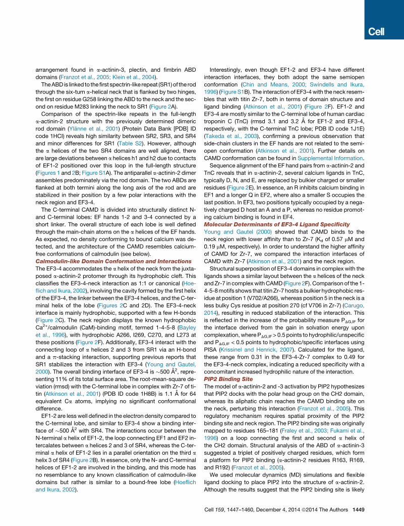
Figure 1. Complete Structure of a-Actinin-2 in Closed Conformation
(A) Domain composition of the a-actinin dimer. Color code, as in all the following figures: ABD, red; neck, yellow; SR1–SR4, green; EF1-2, violet; EF3-4, blue.
(B) The dimeric structure of a-actinin-2 assembled from two halves of the a-actinin-2 protomer (ABD-SR1-SR2/SR3-SR4-CaM) through a crystallographic 2-fold
axis (dashed line; ellipse in C). Overall dimensions are indicated.
(C) Same as in (B), rotated 90� around the horizontal axis.
See also Table S1.
of cytoplasmic and membrane proteins in striated muscle
and nonmuscle tissues (Djinovic-Carugo et al., 2002; Foley and
Young, 2014). To achieve ordered cytoskeletal assemblies,
the binding properties of a-actinin must be spatiotemporally
regulated. Actin binding of nonmuscle isoforms is regulated by
binding of Ca2+ ions to the CAMD (Foley and Young, 2014). In
contrast, muscle a-actinin is calcium insensitive, and its F-actin-
and titin-binding properties are likely regulated by phospholipids
(most notably phosphatidylinositol bisphosphate; PIP2) (Fukami
et al., 1992; Young et al., 1998; Young andGautel, 2000). Despite
being a major integrator of titin and actin in one of the stiffest
structures of the sarcomere, muscle a-actinin shows surprisingly
dynamic association with the Z-disk actin cytoskeleton (Sanger
and Sanger, 2008), suggesting that its actin and titin binding
activity must be dynamically regulated.
Biochemical analysis led us to propose previously that the
a-actinin-titin interaction is regulated by an intramolecular mech-
anismwhere a short sequence in a-actinin between the ABD and
the rod interacts with the CAMD in a pseudoligand mechanism
(Young and Gautel, 2000). A similar mode of interaction has
been found for the a-actinin ligand palladin (Beck et al., 2011).
Here, we report the crystal structure of human a-actinin-2 at
3.5 A resolution. It is a complete high-resolution a-actinin-2
structure, revealing insight into the mechanism that promotes
1448 Cell 159, 1447–1460, December 4, 2014 ª2014 The Authors
the molecular assembly of the Z-disk and the intramolecular
contacts that regulate these interactions. Furthermore, the struc-
ture provides a template for the a-actinin and spectrin super-
family and insight into the impact of disease-associated genetic
variants in ACTN genes.
RESULTS
Closed Structure of a-Actinin-2Overall Architecture
The structure of a-actinin-2 was solved and refined to 3.5 A
resolution to an Rwork/Rfree of 20.5%/25.8% (Table S1 available
online). The a-actinin-2 dimer reveals a cylindrical shape
�360 A long and�60 A wide (Figures 1B and 1C). Each protomer
comprises an N-terminal ABD followed by an a-helical linker
(neck), four spectrin-like repeats (SR1–4), and a C-terminal
CAMD of two pairs of EF hands (EF1-2 and EF3-4). The first 34
and last 2 residues are missing from our model. Two antiparallel
SR1–4s that assemble the core of the extended structure form
the central portion of the dimer (rod). The two ABDs and two
CAMDs flank the elongated assembly at its ends.
As expected, in the absence of actin, the ABD is in a closed
conformation, in which the two calponin homology domains
(CH1 and CH2) are in extensive contact, similar to the
arrangement found in a-actinin-3, plectin, and fimbrin ABD
domains (Franzot et al., 2005; Klein et al., 2004).
TheABD is linked to thefirst spectrin-like repeat (SR1)of the rod
through the six-turn a-helical neck that is flanked by two hinges,
the first on residue G258 linking the ABD to the neck and the sec-
ond on residue M283 linking the neck to SR1 (Figure 2A).
Comparison of the spectrin-like repeats in the full-length
a-actinin-2 structure with the previously determined dimeric
rod domain (Ylanne et al., 2001) (Protein Data Bank [PDB] ID
code 1HCI) reveals high similarity between SR2, SR3, and SR4
and minor differences for SR1 (Table S2). However, although
the a helices of the two SR4 domains are well aligned, there
are large deviations between a helices h1 and h2 due to contacts
of EF1-2 positioned over this loop in the full-length structure
(Figures 1 and 2B; Figure S1A). The antiparallel a-actinin-2 dimer
assembles predominately via the rod domain. The two ABDs are
flanked at both termini along the long axis of the rod and are
stabilized in their position by a few polar interactions with the
neck region and EF3-4.
The C-terminal CAMD is divided into structurally distinct N-
and C-terminal lobes: EF hands 1-2 and 3-4 connected by a
short linker. The overall structure of each lobe is well defined
through the main-chain atoms on the a helices of the EF hands.
As expected, no density conforming to bound calcium was de-
tected, and the architecture of the CAMD resembles calcium-
free conformations of calmodulin (see below).
Calmodulin-like Domain Conformation and Interactions
The EF3-4 accommodates the a helix of the neck from the juxta-
posed a-actinin-2 protomer through its hydrophobic cleft. This
classifies the EF3-4-neck interaction as 1:1 or canonical (Hoe-
flich and Ikura, 2002), involving the cavity formed by the first helix
of the EF3-4, the linker between the EF3-4 helices, and the C-ter-
minal helix of the lobe (Figures 2C and 2D). The EF3-4-neck
interface is mainly hydrophobic, supported with a few H-bonds
(Figure 2C). The neck region displays the known hydrophobic
Ca2+/calmodulin (CaM)-binding motif, termed 1-4-5-8 (Bayley
et al., 1996), with hydrophobic A266, I269, C270, and L273 at
these positions (Figure 2F). Additionally, EF3-4 interact with the
connecting loop of a helices 2 and 3 from SR1 via an H-bond
and a p-stacking interaction, supporting previous reports that
SR1 stabilizes the interaction with EF3-4 (Young and Gautel,
2000). The overall binding interface of EF3-4 is �500 A2, repre-
senting 11% of its total surface area. The root-mean-square de-
viation (rmsd) with the C-terminal lobe in complex with Zr-7 of ti-
tin (Atkinson et al., 2001) (PDB ID code 1H8B) is 1.1 A for 64
equivalent Ca atoms, implying no significant conformational
difference.
EF1-2 are lesswell defined in the electron density compared to
the C-terminal lobe, and similar to EF3-4 show a binding inter-
face of �500 A2 with SR4. The interactions occur between the
N-terminal a helix of EF1-2, the loop connecting EF1 and EF2 in-
tercalates between a helices 2 and 3 of SR4, whereas the C-ter-
minal a helix of EF1-2 lies in a parallel orientation on the third a
helix 3 of SR4 (Figure 2B). In essence, only the N- and C-terminal
helices of EF1-2 are involved in the binding, and this mode has
no resemblance to any known classification of calmodulin-like
domains but rather is similar to a bound-free lobe (Hoeflich
and Ikura, 2002).
Interestingly, even though EF1-2 and EF3-4 have different
interaction interfaces, they both adopt the same semiopen
conformation (Chin and Means, 2000; Swindells and Ikura,
1996) (Figure S1B). The interaction of EF3-4with the neck resem-
bles that with titin Zr-7, both in terms of domain structure and
ligand binding (Atkinson et al., 2001) (Figure 2F). EF1-2 and
EF3-4 are mostly similar to the C-terminal lobe of human cardiac
troponin C (TnC) (rmsd 3.1 and 3.2 A for EF1-2 and EF3-4,
respectively, with the C-terminal TnC lobe; PDB ID code 1J1E)
(Takeda et al., 2003), confirming a previous observation that
side-chain clusters in the EF hands are not related to the semi-
open conformation (Atkinson et al., 2001). Further details on
CAMD conformation can be found in Supplemental Information.
Sequence alignment of the EF hand pairs from a-actinin-2 and
TnC reveals that in a-actinin-2, several calcium ligands in TnC,
typically D, N, and E, are replaced by bulkier charged or smaller
residues (Figure 2E). In essence, an R inhibits calcium binding in
EF1 and a longer Q in EF2, where also a smaller S occupies the
last position. In EF3, two positions typically occupied by a nega-
tively charged D host an A and a P, whereas no residue promot-
ing calcium binding is found in EF4.
Molecular Determinants of EF3-4 Ligand Specificity
Young and Gautel (2000) showed that CAMD binds to the
neck region with lower affinity than to Zr-7 (Kd of 0.57 mM and
0.19 mM, respectively). In order to understand the higher affinity
of CAMD for Zr-7, we compared the interaction interfaces of
CAMD with Zr-7 (Atkinson et al., 2001) and the neck region.
Structural superposition of EF3-4 domains in complex with the
ligands shows a similar layout between the a helices of the neck
andZr-7 in complexwithCAMD (Figure 2F). Comparison of the 1-
4-5-8motifs shows that titin Zr-7 hosts abulkier hydrophobic res-
idue at position 1 (V702/A266), whereas position 5 in the neck is a
less bulky Cys residue at position 270 (cf V706 in Zr-7) (Carugo,
2014), resulting in reduced stabilization of the interaction. This
is reflected in the increase of the probability measure PDG,IF for
the interface derived from the gain in solvation energy upon
complexation, where PDG,IF > 0.5 points to hydrophilic/unspecific
and PDG,IF < 0.5 points to hydrophobic/specific interfaces using
PISA (Krissinel and Henrick, 2007). Calculated for the ligand,
these range from 0.31 in the EF3-4-Zr-7 complex to 0.49 for
the EF3-4-neck complex, indicating a reduced specificity with a
concomitant increased hydrophilic nature of the interaction.
PIP2 Binding Site
The model of a-actinin-2 and -3 activation by PIP2 hypothesizes
that PIP2 docks with the polar head group on the CH2 domain,
whereas its aliphatic chain reaches the CAMD binding site on
the neck, perturbing this interaction (Franzot et al., 2005). This
regulatory mechanism requires spatial proximity of the PIP2
binding site and neck region. The PIP2 binding site was originally
mapped to residues 165–181 (Fraley et al., 2003; Fukami et al.,
1996) on a loop connecting the first and second a helix of
the CH2 domain. Structural analysis of the ABD of a-actinin-3
suggested a triplet of positively charged residues, which form
a platform for PIP2 binding (a-actinin-2 residues R163, R169,
and R192) (Franzot et al., 2005).
We used molecular dynamics (MD) simulations and flexible
ligand docking to place PIP2 into the structure of a-actinin-2.
Although the results suggest that the PIP2 binding site is likely
Cell 159, 1447–1460, December 4, 2014 ª2014 The Authors 1449

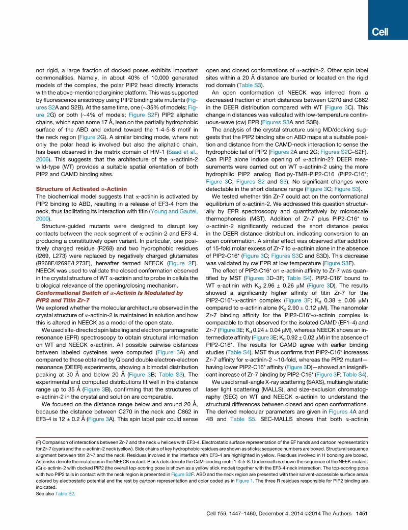
Figure 2. Close-Up of the Functional Domain Interactions
(A) PIP2 binding site on a-actinin-2 and the EF3-4-neck interaction. ABD and EF3-4 are presented with their solvent-accessible surface areas. The R residues
responsible for PIP2 binding are highlighted in blue on the ABD surface.
(B) Detail of EF1-2 interactions with SR4.
(C) Detail of EF3-4 interactions with the neck region and SR1.
(D) Comparison of a-actinin-2 CAMDwith TnC bound to TnI, aligned on EF3-4 and the C-terminal lobe of TnC. Left: cartoon representation of a-actinin EF1-2 and
EF3-4 with the interacting portion of the neck from the juxtaposed subunit (yellow) and a part of SR4 from the same subunit. Right: cartoon representation of TnC
bound to TnI. N-terminal lobe, violet, as in EF1-2, and the C-terminal lobe, blue, as in EF3-4. Bound N-terminal TnI fragment, yellow; C-terminal TnI helix, green.
Calcium ions are shown as black spheres (on TnC). The C-terminal lobe of TnC is aligned to EF3-4. To show the direction of bound helices, residues defining the
neck domain of actinin and TnI fragments are indicated.
(E) Sequence alignment of EF1-2 and EF3-4 and the C-terminal lobe of TnC. The corresponding calcium-binding positions in Ca2+/CaM are indicated by black
dots. Charged residues involved in calcium binding are boxed. Fully conserved residues are highlighted in red.
(legend continued on next page)
1450 Cell 159, 1447–1460, December 4, 2014 ª2014 The Authors
not rigid, a large fraction of docked poses exhibits important
commonalities. Namely, in about 40% of 10,000 generated
models of the complex, the polar PIP2 head directly interacts
with the above-mentioned arginine platform. This was supported
by fluorescence anisotropy using PIP2 binding site mutants (Fig-
ures S2A and S2B). At the same time, one (�35%ofmodels; Fig-
ure 2G) or both (�4% of models; Figure S2F) PIP2 aliphatic
chains, which span some 17 A, lean on the partially hydrophobic
surface of the ABD and extend toward the 1-4-5-8 motif in
the neck region (Figure 2G). A similar binding mode, where not
only the polar head is involved but also the aliphatic chain,
has been observed in the matrix domain of HIV-1 (Saad et al.,
2006). This suggests that the architecture of the a-actinin-2
wild-type (WT) provides a suitable spatial orientation of both
PIP2 and CAMD binding sites.
Structure of Activated a-ActininThe biochemical model suggests that a-actinin is activated by
PIP2 binding to ABD, resulting in a release of EF3-4 from the
neck, thus facilitating its interaction with titin (Young and Gautel,
2000).
Structure-guided mutants were designed to disrupt key
contacts between the neck segment of a-actinin-2 and EF3-4,
producing a constitutively open variant. In particular, one posi-
tively charged residue (R268) and two hydrophobic residues
(I269, L273) were replaced by negatively charged glutamates
(R268E/I269E/L273E), hereafter termed NEECK (Figure 2F).
NEECK was used to validate the closed conformation observed
in the crystal structure of WT a-actinin and to probe in cellula the
biological relevance of the opening/closing mechanism.
Conformational Switch of a-Actinin Is Modulated by
PIP2 and Titin Zr-7
We explored whether the molecular architecture observed in the
crystal structure of a-actinin-2 is maintained in solution and how
this is altered in NEECK as a model of the open state.
Weused site-directed spin labeling and electron paramagnetic
resonance (EPR) spectroscopy to obtain structural information
on WT and NEECK a-actinin. All possible pairwise distances
between labeled cysteines were computed (Figure 3A) and
compared to those obtained by Q band double electron-electron
resonance (DEER) experiments, showing a bimodal distribution
peaking at 30 A and below 20 A (Figure 3B; Table S3). The
experimental and computed distributions fit well in the distance
range up to 35 A (Figure 3B), confirming that the structures of
a-actinin-2 in the crystal and solution are comparable.
We focused on the distance range below and around 20 A,
because the distance between C270 in the neck and C862 in
EF3-4 is 12 ± 0.2 A (Figure 3A). This spin label pair could sense
(F) Comparison of interactions between Zr-7 and the neck a helices with EF3-4. E
for Zr-7 (cyan) and the a-actinin-2 neck (yellow). Side chains of key hydrophobic re
alignment between titin Zr-7 and the neck. Residues involved in the interface wi
Asterisks denote the mutations in the NEECKmutant. Black dots denote the CaM-
(G) a-actinin-2 with docked PIP2 (the overall top-scoring pose is shown as a yello
with two PIP2 tails in contact with the neck region is presented in Figure S2F. ABD
colored by electrostatic potential and the rest by cartoon representation and col
indicated.
See also Table S2.
open and closed conformations of a-actinin-2. Other spin label
sites within a 20 A distance are buried or located on the rigid
rod domain (Table S3).
An open conformation of NEECK was inferred from a
decreased fraction of short distances between C270 and C862
in the DEER distribution compared with WT (Figure 3C). This
change in distances was validated with low-temperature contin-
uous-wave (cw) EPR (Figures S3A and S3B).
The analysis of the crystal structure using MD/docking sug-
gests that the PIP2 binding site on ABD maps at a suitable posi-
tion and distance from the CAMD-neck interaction to sense the
hydrophobic tail of PIP2 (Figures 2A and 2G; Figures S2C–S2F).
Can PIP2 alone induce opening of a-actinin-2? DEER mea-
surements were carried out on WT a-actinin-2 using the more
hydrophilic PIP2 analog Bodipy-TMR-PIP2-C16 (PIP2-C16*;
Figure 3C; Figures S2 and S3). No significant changes were
detectable in the short distance range (Figure 3C; Figure S3).
We tested whether titin Zr-7 could act on the conformational
equilibrium of a-actinin-2. We addressed this question structur-
ally by EPR spectroscopy and quantitatively by microscale
thermophoresis (MST). Addition of Zr-7 plus PIP2-C16* to
a-actinin-2 significantly reduced the short distance peaks
in the DEER distance distribution, indicating conversion to an
open conformation. A similar effect was observed after addition
of 15-fold molar excess of Zr-7 to a-actinin alone in the absence
of PIP2-C16* (Figure 3C; Figures S3C and S3D). This decrease
was validated by cw EPR at low temperature (Figure S3E).
The effect of PIP2-C16* on a-actinin affinity to Zr-7 was quan-
tified by MST (Figures 3D–3F; Table S4). PIP2-C16* bound to
WT a-actinin with Kd 2.96 ± 0.26 mM (Figure 3D). The results
showed a significantly higher affinity of titin Zr-7 for the
PIP2-C16*-a-actinin complex (Figure 3F; Kd 0.38 ± 0.06 mM)
compared to a-actinin alone (Kd 2.90 ± 0.12 mM). The nanomolar
Zr-7 binding affinity for the PIP2-C16*-a-actinin complex is
comparable to that observed for the isolated CAMD (EF1–4) and
Zr-7 (Figure 3E; Kd 0.24 ± 0.04 mM), whereas NEECK shows an in-
termediate affinity (Figure 3E; Kd 0.92± 0.02 mM) in the absence of
PIP2-C16*. The results for CAMD agree with earlier binding
studies (Table S4). MST thus confirms that PIP2-C16* increases
Zr-7 affinity for a-actinin-2 �10-fold, whereas the PIP2 mutant—
having lower PIP2-C16* affinity (Figure 3D)—showed an insignifi-
cant increase of Zr-7 binding by PIP2-C16* (Figure 3F; Table S4).
We used small-angle X-ray scattering (SAXS), multiangle static
laser light scattering (MALLS), and size-exclusion chromatog-
raphy (SEC) on WT and NEECK a-actinin to understand the
structural differences between closed and open conformations.
The derived molecular parameters are given in Figures 4A and
4B and Table S5. SEC-MALLS shows that both a-actinin
lectrostatic surface representation of the EF hands and cartoon representation
sidues are shown as sticks; sequence numbers are boxed. Structural sequence
th EF3-4 are highlighted in yellow. Residues involved in H bonding are boxed.
bindingmotif 1-4-5-8. Underneath is shown the sequence of the NEEKmutant.
w stick model) together with the EF3-4-neck interaction. The top-scoring pose
and the neck region are presented with their solvent-accessible surface areas
or coded as in Figure 1. The three R residues responsible for PIP2 binding are
Cell 159, 1447–1460, December 4, 2014 ª2014 The Authors 1451
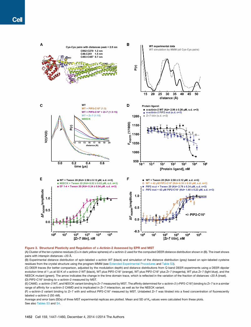
Figure 3. Structural Plasticity and Regulation of a-Actinin-2 Assessed by EPR and MST
(A) Cluster of the ten cysteine residues (Ca in dark yellow spheres) of a-actinin-2 used for the computed DEER distance distribution shown in (B). The inset shows
pairs with interspin distances <20 A.
(B) Experimental distance distribution of spin-labeled a-actinin WT (black) and simulation of the distance distribution (gray) based on spin-labeled cysteine
residues from the crystal structure using the program MMM (see Extended Experimental Procedures and Table S3).
(C) DEER traces (for better comparison, adjusted by the modulation depth) and distance distributions from Q band DEER experiments using a DEER dipolar
evolution time of 1 ms at 50 K of a-actinin-2 WT (black), WT plus PIP2-C16* (orange), WT plus PIP2-C16* plus Zr-7 (magenta), WT plus Zr-7 (light blue), and the
NEECK mutant (green). The arrow indicates the change in the time domain trace, which is reflected in the variation of the fraction of distances <20 A (inset).
(D) PIP2-C16* binding to a-actinin-2 measured by MST.
(E) CAMD, a-actinin-2WT, and NEECK variant binding to Zr-7measured byMST. The affinity determined for a-actinin-2 (+PIP2-C16*) binding to Zr-7 is in a similar
range of affinity for a-actinin-2 CAMD and is implicated in Zr-7 interaction, as well as for the NEECK variant.
(F) a-actinin-2 variant binding to Zr-7 with and without PIP2-C16* measured by MST. Unlabeled Zr-7 was titrated into a fixed concentration of fluorescently
labeled a-actinin-2 (50 nM).
Average and error bars (SDs) of three MST experimental replicas are plotted. Mean and SD of Kd values were calculated from these plots.
See also Tables S3 and S4.
1452 Cell 159, 1447–1460, December 4, 2014 ª2014 The Authors
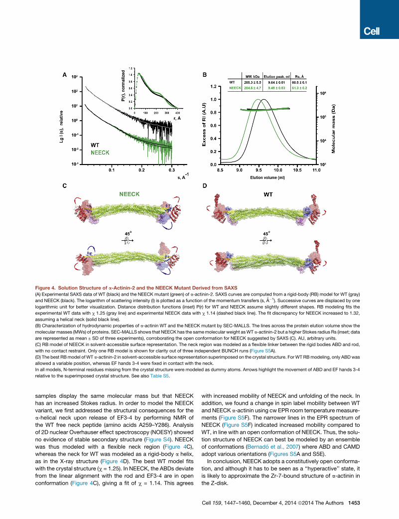
Figure 4. Solution Structure of a-Actinin-2 and the NEECK Mutant Derived from SAXS
(A) Experimental SAXS data of WT (black) and the NEECK mutant (green) of a-actinin-2. SAXS curves are computed from a rigid-body (RB) model for WT (gray)
and NEECK (black). The logarithm of scattering intensity (I) is plotted as a function of the momentum transfers (s, A�1). Successive curves are displaced by one
logarithmic unit for better visualization. Distance distribution functions (inset) P(r) for WT and NEECK assume slightly different shapes. RB modeling fits the
experimental WT data with c 1.25 (gray line) and experimental NEECK data with c 1.14 (dashed black line). The fit discrepancy for NEECK increased to 1.32,
assuming a helical neck (solid black line).
(B) Characterization of hydrodynamic properties of a-actinin WT and the NEECK mutant by SEC-MALLS. The lines across the protein elution volume show the
molecular masses (MWs) of proteins. SEC-MALLS shows that NEECK has the samemolecular weight asWT a-actinin-2 but a higher Stokes radius Rs (inset; data
are represented as mean ± SD of three experiments), corroborating the open conformation for NEECK suggested by SAXS (C). AU, arbitrary units.
(C) RB model of NEECK in solvent-accessible surface representation. The neck region was modeled as a flexible linker between the rigid bodies ABD and rod,
with no contact restraint. Only one RB model is shown for clarity out of three independent BUNCH runs (Figure S5A).
(D) The best RBmodel of WT a-actinin-2 in solvent-accessible surface representation superimposed on the crystal structure. For WT RBmodeling, only ABDwas
allowed a variable position, whereas EF hands 3-4 were fixed in contact with the neck.
In all models, N-terminal residues missing from the crystal structure were modeled as dummy atoms. Arrows highlight the movement of ABD and EF hands 3-4
relative to the superimposed crystal structure. See also Table S5.
samples display the same molecular mass but that NEECK
has an increased Stokes radius. In order to model the NEECK
variant, we first addressed the structural consequences for the
a-helical neck upon release of EF3-4 by performing NMR of
the WT free neck peptide (amino acids A259–Y286). Analysis
of 2D nuclear Overhauser effect spectroscopy (NOESY) showed
no evidence of stable secondary structure (Figure S4). NEECK
was thus modeled with a flexible neck region (Figure 4C),
whereas the neck for WT was modeled as a rigid-body a helix,
as in the X-ray structure (Figure 4D). The best WT model fits
with the crystal structure (c = 1.25). In NEECK, the ABDs deviate
from the linear alignment with the rod and EF3-4 are in open
conformation (Figure 4C), giving a fit of c = 1.14. This agrees
with increased mobility of NEECK and unfolding of the neck. In
addition, we found a change in spin label mobility between WT
and NEECK a-actinin using cw EPR room temperature measure-
ments (Figure S5F). The narrower lines in the EPR spectrum of
NEECK (Figure S5F) indicated increased mobility compared to
WT, in line with an open conformation of NEECK. Thus, the solu-
tion structure of NEECK can best be modeled by an ensemble
of conformations (Bernado et al., 2007) where ABD and CAMD
adopt various orientations (Figures S5A and S5E).
In conclusion, NEECK adopts a constitutively open conforma-
tion, and although it has to be seen as a ‘‘hyperactive’’ state, it
is likely to approximate the Zr-7-bound structure of a-actinin in
the Z-disk.
Cell 159, 1447–1460, December 4, 2014 ª2014 The Authors 1453
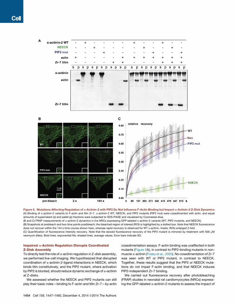
Figure 5. Mutations Affecting Regulation of a-Actinin-2 with PIP2 Do Not Influence F-Actin Binding but Impact a-Actinin-2 Z-Disk Dynamics
(A) Binding of a-actinin-2 variants to F-actin and titin Zr-7. a-actinin-2 WT, NEECK, and PIP2 mutants (PIP2 mut) were cosedimented with actin, and equal
amounts of supernatant (s) and pellet (p) fractions were subjected to SDS-PAGE and visualized by Coomassie blue.
(B and C) FRAP measurements of a-actinin-2 dynamics in live NRCs expressing GFP-labeled a-actinin-2 variants (WT, PIP2 mutants, and NEECK).
(B) Snapshots at prebleach and two time points postbleach; the bleached region of interest (ROI) is highlighted by a dotted box. Note that NEECK fluorescence
does not recover within the 144 s time course shown here, whereas rapid recovery is observed for WT a-actinin. Insets: ROIs enlarged 2-fold.
(C) Quantification of fluorescence intensity recovery. Note that the slowed fluorescence recovery of the PIP2 mutant is mirrored by treatment with 500 mM
neomycin (Neo). Bold lines, exponential fits; shaded lines, average values. Error bars indicate SD.
Impaired a-Actinin Regulation Disrupts CoordinatedZ-Disk AssemblyTo directly test the role of a-actinin regulation in Z-disk assembly,
we performed live-cell imaging. We hypothesized that disrupted
coordination of a-actinin-2-ligand interactions in NEECK, which
binds titin constitutively, and the PIP2 mutant, where activation
by PIP2 is blunted, should reduce dynamic exchange of a-actinin
at Z-disks.
We assessed whether the NEECK and PIP2 mutants can still
play their basic roles—binding to F-actin and titin Zr-7—by actin
1454 Cell 159, 1447–1460, December 4, 2014 ª2014 The Authors
cosedimentation assays. F-actin binding was unaffected in both
mutants (Figure 5A), in contrast to PIP2-binding mutants in non-
muscle a-actinin (Fraley et al., 2003). No cosedimentation of Zr-7
was seen with WT or PIP2 mutant, in contrast to NEECK.
Together, these results suggest that the PIP2 or NEECK muta-
tions do not impair F-actin binding, and that NEECK induces
PIP2-independent Zr-7 binding.
We carried out fluorescence recovery after photobleaching
(FRAP) studies in neonatal rat cardiomyocytes (NRCs) express-
ing the GFP-labeled a-actinin-2 mutants to assess the impact of

a-actinin-2 regulation on spatiotemporal dynamics in sarco-
meres. The exchange of WT a-actinin-2 at Z-disks was rapid,
with a fast component t1/2 25 ± 2 s (Figures 5B and 5C).
In contrast, the NEECK mutant was dramatically slower, with
t1/2 >6,134 s. The PIP2 mutant showed reduced dynamics,
with t1/2 35 ± 4 s (Figure 5B). The slower dynamics of a-actinin-
2 mutants compared to WT observed in cellula seems not to
be due to F-actin binding activity, because both mutants bind
F-actin (Figure 5A). Analysis of FRAP kinetics revealed standard
fast and slow components in the case of WT and the PIP2
mutant, whereas for NEECK only a slow component could be
discriminated. The slow, single-exponential exchange of NEECK
agrees with a dominant, high-affinity interaction. Because the
NEECK interaction with titin is constitutive, the slow cellular
dynamics likely reflect the high affinity of the EF3-4 interaction.
Exchange of the PIP2 mutant was also slower, in agreement
with reduced phospholipid regulation. To probe the role of
PIP2 in regulating a-actinin dynamics independently, we used
the aminoglycoside neomycin, an inhibitor of PIP2 signaling (Li
and Russell, 2013; Schacht, 1976). Neomycin resulted in slower
FRAP recovery of WT a-actinin-2 (z40 ± 4 s), similar to the PIP2
mutant (Figure 5C), supporting the notion that a-actinin-2 Z-disk
dynamics are strongly dependent on PIP2 regulation, in agree-
ment with the Z-disk localization of PIP2 (Figure S6A).
Z-disk morphology in a-actinin-transfected NRCs showed a
striking phenotype for the NEECK mutant but not WT or the
PIP2 mutant. Cells expressing NEECK showed gradual appear-
ance of sarcomeres with wide a-actinin labeling, where titin epi-
topes peripheral (T12 antibody) and more central (Z1Z2) to the
Z-disk (Young et al., 1998) were resolved as doublets flanking
the edge of the a-actinin-labeled central Z-disk. This resulted
in formation of actin/a-actinin bundles resembling nemaline
rods but containing diffusely localized Z-disk titin (Figures 6A–
6C) and ultimately complete disruption of sarcomeres (Fig-
ure 6C). Vinculin localization in NEECK-transfected cardiomyo-
cytes was unaffected (Figure S6B).
Whereas the optically resolvable distance between T12 epi-
topes inWT-transfected cells is�200 nm, in NEECK-transfected
cells this was >600 nm, and >800 nm after 3 days (Figure S6C).
Similar splitting to >200 nm was also seen for Z1Z2, normally at
the limit of optical resolution with a separation of �100 nm
(Young et al., 1998). This suggests that the ordered integration
of titin and a-actinin is severely disrupted by NEECK, raising
the question of whether the spatiotemporal integration of other
titin Z-disk ligands is affected. Current models of titin layout in
the Z-disk predict that Z-disk widening could only be achieved
by relative slipping of the overlapping N termini of titin molecules
entering the Z-disk from two antiparallel sarcomere halves (Gau-
tel, 2011). Titin molecules are crosslinked in an antiparallel palin-
dromic complex of domains Z1-Z2 and the small Z-disk protein
telethonin (Zou et al., 2006). Both telethonin and titin-Z1Z2 epi-
topes strictly colocalize at the Z-disk periphery (Figure 6D) but
also in the wide NEECK Z-disks (Figure 6E). These results
show that intramolecular autoregulation of a-actinin-ligand inter-
actions is crucial for sarcomere integrity regulating the integra-
tion of titin, actin, and a-actinin in Z-disks of controlled width,
without affecting interaction of titin with Z-disk proteins such
as telethonin.
DISCUSSION
The structure of a-actinin-2 shows a modular architecture, yet is
more than just ‘‘the sum of its parts’’: important intra- and inter-
molecular contacts lock the molecule in a closed conformation
that is crucial for dynamic regulation.
Pseudoligand Model ValidationThe structure of a-actinin-2 displays a closed, autoinhibited
conformation, as suggested by Young and Gautel (2000) (Fig-
ure 1B). The closed structure of a-actinin-2 shows, furthermore,
that the PIP2 binding site on the ABD (Franzot et al., 2005) maps
at a suitable position and distance from the CAMD-neck interac-
tion for sensing the hydrophobic tail of PIP2 (Figure 2A), as sup-
ported byMD/docking simulations (Figure 2G; Figures S2C–S2F).
The closed a-actinin-2 conformation shows that a number of
interactions between SR1–4 stabilize the formation of antiparallel
dimers, providing its structural rigidity and stability. Although
addition of PIP2-C16* alone does not promote complete opening
of a-actinin-2, at least asmeasurable by DEER, it promotes bind-
ing of Zr-7 with nanomolar affinity (Figure 3F), suggesting a
positive allosteric modulation for opening and ligand binding.
Furthermore, local structural changes in the CH2 domain, as
observed upon PIP2 binding to nonmuscle a-actinin (Full et al.,
2007), cannot be excluded.
Additional mechanisms might act in cells that cooperate with
PIP2 or offer alternative regulation, including posttranslational
modificationsor protein cofactors.However,wecould not identify
any such plausible sites conserved between muscle a-actinin-2
and -3 in proteomic databases. No protein cofactors regulating
a-actinin-titin interactions have been identified to date.
Structural comparison of interactions between EF3-4, Zr-7,
and the neck reveals the basis for the higher affinity of a-acti-
nin-2 for titin versus the pseudoligand neck (Young and Gautel,
2000) (Figure 2F). Our results suggest amodel for PIP2 regulation
of a-actinin, relying on structural plasticity and conformational
dynamics of a-actinin-2 (Figure 7A): in the absence of PIP2,
a-actinin-2 exists in two conformational states, a highly popu-
lated closed [AC] and a low populated open and active state
[AO]. Addition of PIP2 triggers an activated state [A*:PIP2] with
lower activation energy for opening. Binding of Zr-7 recruits
a-actinin-2 in the open conformation [AO:Zr-7], which is en-
riched, because a-actinin-2 binds Zr-7 with higher affinity than
it does to the pseudoligand neck (Figure 2F; Table S4).
Implications for Binding F-Actin Crosslinking and Z-DiskStructureIn the Z-disk, antiparallel actin filaments are crosslinked by a-ac-
tinin in a paracrystalline tetragonal lattice (Goldstein et al., 1988).
To analyze the structural determinants of a-actinin’s principal
crosslinking function, we mapped the known actin binding sites
(ABSs) to the structure of the a-actinin dimer. ABSs in ABDs of
several actin-binding proteins have been located on the first and
the last a helix of the CH1 domain and on the first a helix of the
CH2 domain (Sjoblom et al., 2008). ABSs in a-actinin-2 are
exposed, andnot blockedby interdomain interactions (Figure7B).
To generate a 3D model of F-actin/a-actinin, we superim-
posed the structure of a-actinin-2 on that of the F-actin-bound
Cell 159, 1447–1460, December 4, 2014 ª2014 The Authors 1455

Figure 6. Constitutively Activated a-Actinin-2 Disrupts Z-Disks and Leads to Myofibril Disassembly
GFP-labeled WT and NEECK a-actinin-2 were transiently expressed in NRCs for 18–48 hr.
(A) WT a-actinin shows normal Z-disk localization, and the titin T12 epitope is resolved as a single line in standard confocal microscopy.
(B) In contrast, NEECK leads to widening of the Z-disk and splitting of the T12 epitope after �18 hr (asterisk); doublet T12 lines are highlighted by arrows.
(C) After 48 hr, Z-disks are completely disrupted and Z-disk titin, actin, and mutant a-actinin are localized in rod-like structures.
(D and E) Superresolutionmicroscopy reveals that epitopes of N-terminal Z1Z2 of titin and their ligand telethonin are unresolvable inWT-transfected cells; NEECK
causes widening of Z-disks. Doublet Z1Z2/telethonin lines are highlighted by arrows and the central a-actinin region is indicated by arrowheads. Insets show
2-fold enlargement. Z-disk titin (T12 epitope) or telethonin, red; mutant a-actinin-GFP, green; actin (Alexa 688-phalloidin) or titin Z1Z2, blue.
1456 Cell 159, 1447–1460, December 4, 2014 ª2014 The Authors
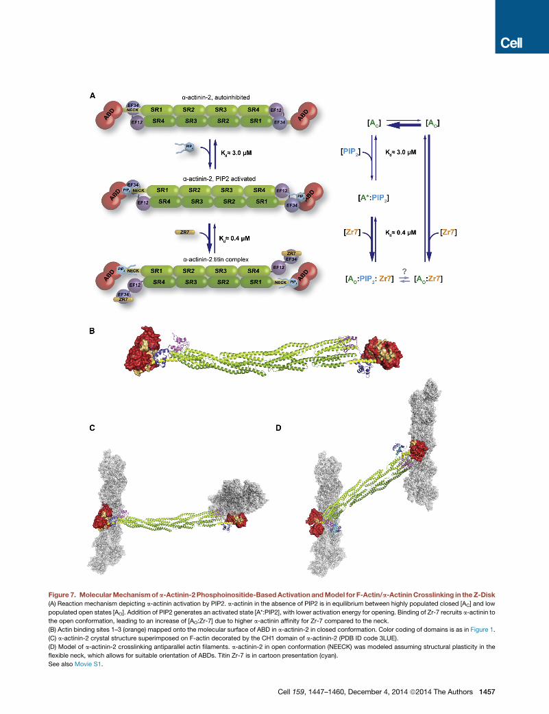
Figure 7. MolecularMechanismofa-Actinin-2Phosphoinositide-BasedActivationandModel for F-Actin/a-ActininCrosslinking in theZ-Disk
(A) Reaction mechanism depicting a-actinin activation by PIP2. a-actinin in the absence of PIP2 is in equilibrium between highly populated closed [AC] and low
populated open states [AO]. Addition of PIP2 generates an activated state [A*:PIP2], with lower activation energy for opening. Binding of Zr-7 recruits a-actinin to
the open conformation, leading to an increase of [AO:Zr-7] due to higher a-actinin affinity for Zr-7 compared to the neck.
(B) Actin binding sites 1–3 (orange) mapped onto the molecular surface of ABD in a-actinin-2 in closed conformation. Color coding of domains is as in Figure 1.
(C) a-actinin-2 crystal structure superimposed on F-actin decorated by the CH1 domain of a-actinin-2 (PDB ID code 3LUE).
(D) Model of a-actinin-2 crosslinking antiparallel actin filaments. a-actinin-2 in open conformation (NEECK) was modeled assuming structural plasticity in the
flexible neck, which allows for suitable orientation of ABDs. Titin Zr-7 is in cartoon presentation (cyan).
See also Movie S1.
Cell 159, 1447–1460, December 4, 2014 ª2014 The Authors 1457
CH1 domain (PDB ID code 3LUE) (Galkin et al., 2010). Notably,
using the a-actinin-2 structure leads to a model with perpendic-
ular actin filaments (Figure 7C), in strong disagreement with
the established antiparallel actin architecture in the Z-disk but
agreeing with the a-actinin-2 dimer architecture. Due to the inter-
nal twist of about 90� of the central rod (Ylanne et al., 2001), the
ABDs in the dimer are rotated by 90�. We next used the structure
of the NEECKmutant as the ‘‘open’’ structure in the Z-disk. Here
the unbound neck is unstructured (Figure S4), allowing the ABD
to explore different orientations, adopting those compatible with
interaction with antiparallel actin filaments. Considering the
angular distribution of F-actin and a-actinin, which centers at
60� and 120�, respectively (Hampton et al., 2007), we generated
a model of two actin filaments crosslinked by an a-actinin-2
dimer (Figure 7D; Movie S1). In this model, the distance between
filaments is �230 A, which is in excellent agreement with the
observed interfilament distances in the tetragonal Z-disk lattice
(240 A; Goldstein et al., 1979).
The structural plasticity of a-actinin-2 has implications for not
only its regulation but also for actin filament binding both in mus-
cle and nonmuscle isoforms, where actin filaments are randomly
oriented, requiring ABDs to adopt variable orientations. Interest-
ingly, muscle a-actinin-2 was found to crosslink antiparallel as
well as parallel actin filaments in a-actinin-F-actin rafts (Hampton
et al., 2007). Assuming the absence of PIP2 in these assays, the
flexibility of the ABD likely resides in the hinge region between
the ABD and the neck (residue G258) (Figure 2A), similar to the
solution structure of closed a-actinin-2 (Figure 4D). Other elec-
tron microscopy studies showed that ABDs in smooth muscle
a-actinin can attain different orientations through movement in
the flexible neck (Taylor and Taylor, 1993; Winkler et al., 1997),
crosslinking both antiparallel and parallel actin filaments in vitro
and in vivo (Meyer and Aebi, 1990; Tang et al., 2001).
Although alternative paths of titin in the Z-disk are conceiv-
able, our results with NEECK show that even in strongly split
Z-disks the Z1Z2 and telethonin epitopes remain in strict coloc-
alization, implying that the two titin molecules crosslinked in the
titin-telethonin complex must come from the same half-sarco-
mere, as suggested previously (Zou et al., 2006). Intriguingly,
these findings also suggest that titin capping (via telethonin)
and barbed-end actin filament capping by CapZ are not directly
correlated, despite the close association of the titin Z1Z2-tele-
thonin complex with the actin barbed end (Zou et al., 2006).
However, actin capping by CapZ and crosslinking by a-actinin
may crosstalk, as indeed a-actinin was reported to interact
with CapZ via a binding site on the rod (Papa et al., 1999) and
both proteins are PIP2 regulated (Figure S6A).
Impact of Pathogenic MutationsGenetic variants in a-actinin genes are associated with several
inherited diseases. Missense variants in nonmuscle actinin 1
(ACTN1 gene) cause autosomal-dominant congenital macro-
thrombocytopenia (Gueguen et al., 2013; Kunishima et al.,
2013), and approximately 4% of autosomal-dominant familial
focal segmental glomerulosclerosis has been linked to non-
muscle ACTN4 mutations (Kaplan et al., 2000). Missense vari-
ants in muscle ACTN2 have been reported in sporadic cases
and a few families with dilated or hypertrophic cardiomyopathy
1458 Cell 159, 1447–1460, December 4, 2014 ª2014 The Authors
(Chiu et al., 2010; Mohapatra et al., 2003; Theis et al., 2006).
Our structure now provides a platform for the analysis of muta-
tional impact on structure, ligand binding, and regulation of
a-actinin in inherited human diseases (see Supplemental Infor-
mation and Figure S7).
Structural Analysis of Selected Genetic VariantsThe genetic variants are spread over all domains of a-actinin.
Several variants are conservative and would not lead to major
structural perturbations, in particular mutations in the CAMD
and the rod domain. Changes of rod surface properties might,
however, abrogate interactions with ligands, because the rod
domain is recognized as the prominent protein interaction plat-
form of a-actinin (Djinovic-Carugo et al., 2002). Interestingly,
the mutations on the rod domain map on the less conserved,
acidic side of the convex surface (Ylanne et al., 2001) (Fig-
ure S7). However, four mutations in the ACTN1 and ACTN4
genes have predicted disruptive potential: E225K (ACTN1
gene, ABD) leads to a loss of a salt bridge and mutation
R738W (ACTN1 gene, CAMD) would disrupt the structure of
the CAMD, whereas the W59R (ACTN4 gene, ABD) and
S262F (ACTN4 gene, ABD) mutations destabilize the domain
structure due to introduction of charged or bulky hydrophobic
residues to the core of the ABD. Most hypertrophic cardiomy-
opathy and dilated cardiomyopathy variants were classified as
structurally neutral.
Implication for Regulation of the a-Actinin Family inGeneralMuscle a-actinin interacts with many proteins via multiple bind-
ing sites. The CAMD EF3-4 site interacts with helical motifs in
the actin- and a-actinin-binding proteins myopalladin, palladin,
and myotilin, highly similar to the a-actinin-titin complex and
the intramolecular neck complex detailed here (Beck et al.,
2011). Dynamic regulation of a-actinin interactions with these
proteins is therefore likely governed by the same principles
as the one with titin. Additionally, the a-actinin-associated
LIM protein (ALP) and ZASP/Cypher bind a-actinin at both
the CAMD (via its PDZ domain) and the SR (Faulkner et al.,
1999; Klaavuniemi et al., 2004). Although the binding sites for
titin Zr-7 and the PDZ domain on CAMD do not coincide, an
open structure might be required to accommodate both bind-
ing partners and prevent steric hindrance by the spatially close
a-actinin domains.
Furthermore, interactions of CAMDs of the structurally related
cytoskeletal actin-binding proteins dystrophin, utrophin, and
spectrin may play important roles in regulating cytoskeletal inter-
actions near the plasmamembrane (Bennett andHealy, 2008), as
suggested by recent studies on spectrin-ankyrin, actin, and pro-
tein 4.2 interactions (Korsgren and Lux, 2010; Korsgren et al.,
2010). Although theseEFhanddomains retain aspects of calcium
regulation (only the N-terminal EF hand binds calcium), the gen-
eral mode of regulation seems highly similar to a-actinin, namely
the CH2-R1 linker region of a/b-spectrin also binds to the CAMD
EF3-4 hands, and this regulates protein interactions.
The mechanism we have detailed here is therefore likely to
be of general relevance for regulating spectrin-like proteins via
intramolecular pseudoligand interactions.
EXPERIMENTAL PROCEDURES
Purification and Crystallization
Proteins were expressed as His fusions in Escherichia coli and purified
via Ni-NTA agarose and size-exclusion chromatography. Protein was lysine
methylated and crystallized in a precipitant containing 0.2 M Mg formate,
5% PEG smear, and 10 mM EDTA by hanging-drop vapor diffusion at 14�C.
Structure Determination
A 3.5 A data set was collected at beamline ID23-2 (European Synchrotron
Radiation Facility [ESRF]). The phase problem was solved by molecular
replacement using structures of the rod domain (PDB ID code 1HCI), the
ABD from a-actinin-3 (PDB ID code 1WKU), and the NMR structure of EF3-4
(PDB ID code 1H8B) as search models.
Residues 34–892 were assigned in the final model. Details on data collec-
tion, processing, structure determination, and refinement are described in
Extended Experimental Procedures and Table S1.
Electron Paramagnetic Resonance
Site-directed spin labeling (SDSL) was performed on native cysteine residues
ofWT and NEECK a-actinin-2. X band cw EPR experiments were carried out at
298 K or 160 K on a Bruker EMX spectrometer. Pulsed EPR measurements
were carried out at 50 K on a Q band power upgraded Bruker ELEXSYS
E580 spectrometer. Details are given in Extended Experimental Procedures.
SAXS Measurements and Modeling
Small-angle X-ray scattering data were collected at beamline X33 at European
Molecular Biology Laboratory (EMBL) Hamburg for WT, NEECK, and PIP2
mutants at three different concentrations and analyzed following standard
procedures. Molecular dynamics simulations were carried out using the
GROMACS 4.0.7 package (Hess et al., 2008), whereas flexible docking was
performed using GOLD version 5.2.2 (Jones et al., 1997). Further details are
described in Extended Experimental Procedures.
Cell Biophysics
Experiments in neonatal rat cardiomyocytes were performed using published
methods and antibodies using live-cell imaging on a Zeiss LSM510 confocal
microscope and superresolution on a Leica TCS STED instrument (see
Extended Experimental Procedures).
ACCESSION NUMBERS
The coordinates and structure factors of the a-actinin-2 structure have been
deposited in the Protein Data Bank under ID code 4D1E.
SUPPLEMENTAL INFORMATION
Supplemental Information includes Extended Experimental Procedures, seven
figures, five tables, and one movie and can be found with this article online at
http://dx.doi.org/10.1016/j.cell.2014.10.056.
AUTHOR CONTRIBUTIONS
Experiments were designed by M.G., K.D.-C., and E.d.A.R. in consultation
with K.F.P., E.B., and D.I.S.; all structural work was performed by E.d.A.R.,
N.P., A.S., B.S., K.F.P., and P.V.K.; E.d.A.R., A.G., and J.K. performed
biochemical work; A.G. and M.R.H. performed cell biophysics, and F.L.A. per-
formed the NMR work; C.S. and E.A.G. assisted with protein purification and
crystallization; A.A.P. and B.�Z. performed MD; and M.G., K.D.-C., E.d.A.R.,
and N.P. wrote the manuscript, with all authors contributing to editing the
manuscript and supporting the conclusions.
ACKNOWLEDGMENTS
We are greatly indebted to Ay Lin Kho for NRC preparations. Thanks also to
Dusan Turk (Institute Jozef Stefan, Ljubljana) for initial help with refinement
and Oliviero Carugo (University of Vienna and University of Pavia) and Bettina
Hartlieb (Baxter Innovations GmbH) for critical reading of the manuscript. We
thank the staff of the MX beamlines at the ESRF in Grenoble, SAXS beamline
X33 (Deutsches Elektronen-Synchrotron, Hamburg), and SWING beamline
(Soleil, Saint-Aubin) for their excellent support. We thank the staff of Campus
Science Support Facilities GmbH (Campus Vienna Biocenter) for technical
assistance. The K.D.-C. group was supported by Austrian Science Fund
(FWF) Projects I525, I1593, P22276, and P19060, by the Federal Ministry of
Economy, Family and Youth through the initiative ‘‘Laura Bassi Centres of
Expertise’’ funding the Center of Optimized Structural Studies (253275), by
theMarie Curie Initial Training Network: MUZIC (238423), and by the University
of Vienna. This researchwas also funded by the EuropeanCommunity Seventh
Framework Programme (FP7/2007-2013) under BioStruct-X (283570). F.L.A.
thanks BioTek2021 Project 217708/O10 and the Research Council of Norway
for financial support. B.�Z. and A.A.P. were supported by the European
Research Council (279408). A.A.P. was supported by the Russian Scientific
Foundation (14-24-00118). M.G. and A.G. were supported by the Leducq
Foundation Transatlantic Network of Excellence: Proteotoxicity (11 CVD 04)
and the Medical Research Council of Great Britain (MR/J010456/1). A.G.
and E.d.A.R. were also supported by the Marie Curie Initial Training Network:
MUZIC (238423). M.G. holds the British Heart Foundation Chair of Molecular
Cardiology (CH/08/001). D.I.S. acknowledges support from the Human Fron-
tier Science Program (RGP0017/2012).
Received: May 7, 2014
Revised: October 1, 2014
Accepted: October 24, 2014
Published: November 26, 2014
REFERENCES
Atkinson, R.A., Joseph, C., Kelly, G., Muskett, F.W., Frenkiel, T.A., Nietlispach,
D., and Pastore, A. (2001). Ca2+-independent binding of an EF-hand domain to
a novel motif in the a-actinin-titin complex. Nat. Struct. Biol. 8, 853–857.
Bayley, P.M., Findlay, W.A., and Martin, S.R. (1996). Target recognition
by calmodulin: dissecting the kinetics and affinity of interaction using short
peptide sequences. Protein Sci. 5, 1215–1228.
Beck, M.R., Otey, C.A., and Campbell, S.L. (2011). Structural characterization
of the interactions between palladin and a-actinin. J. Mol. Biol. 413, 712–725.
Bennett, V., and Healy, J. (2008). Organizing the fluid membrane bilayer:
diseases linked to spectrin and ankyrin. Trends Mol. Med. 14, 28–36.
Bernado, P., Mylonas, E., Petoukhov, M.V., Blackledge, M., and Svergun, D.I.
(2007). Structural characterization of flexible proteins using small-angle X-ray
scattering. J. Am. Chem. Soc. 129, 5656–5664.
Carugo, O. (2014). Wolumes - an algorithm to compute the volume of atoms
and residues in proteins. arXiv, http://arxiv.org/pdf/1406.3242.pdf.
Chin, D., and Means, A.R. (2000). Calmodulin: a prototypical calcium sensor.
Trends Cell Biol. 10, 322–328.
Chiu, C., Bagnall, R.D., Ingles, J., Yeates, L., Kennerson, M., Donald, J.A.,
Jormakka, M., Lind, J.M., and Semsarian, C. (2010). Mutations in a-actinin-2
cause hypertrophic cardiomyopathy: a genome-wide analysis. J. Am. Coll.
Cardiol. 55, 1127–1135.
Djinovic-Carugo, K., Gautel, M., Ylanne, J., and Young, P. (2002). The spectrin
repeat: a structural platform for cytoskeletal protein assemblies. FEBS Lett.
513, 119–123.
Faulkner, G., Pallavicini, A., Formentin, E., Comelli, A., Ievolella, C., Trevisan,
S., Bortoletto, G., Scannapieco, P., Salamon, M., Mouly, V., et al. (1999).
ZASP: a new Z-band alternatively spliced PDZ-motif protein. J. Cell Biol.
146, 465–475.
Foley, K.S., and Young, P.W. (2014). The non-muscle functions of actinins:
an update. Biochem. J. 459, 1–13.
Fraley, T.S., Tran, T.C., Corgan, A.M., Nash, C.A., Hao, J., Critchley, D.R., and
Greenwood, J.A. (2003). Phosphoinositide binding inhibits a-actinin bundling
activity. J. Biol. Chem. 278, 24039–24045.
Cell 159, 1447–1460, December 4, 2014 ª2014 The Authors 1459
Franzot, G., Sjoblom, B., Gautel, M., and Djinovi�c Carugo, K. (2005). The crys-
tal structure of the actin binding domain from a-actinin in its closed conforma-
tion: structural insight into phospholipid regulation of a-actinin. J. Mol. Biol.
348, 151–165.
Fukami, K., Furuhashi, K., Inagaki, M., Endo, T., Hatano, S., and Takenawa, T.
(1992). Requirement of phosphatidylinositol 4,5-bisphosphate for a-actinin
function. Nature 359, 150–152.
Fukami, K., Sawada, N., Endo, T., and Takenawa, T. (1996). Identification of a
phosphatidylinositol 4,5-bisphosphate-binding site in chicken skeletal muscle
a-actinin. J. Biol. Chem. 271, 2646–2650.
Full, S.J., Deinzer, M.L., Ho, P.S., and Greenwood, J.A. (2007). Phosphoinosi-
tide binding regulates a-actinin CH2 domain structure: analysis by hydrogen/
deuterium exchange mass spectrometry. Protein Sci. 16, 2597–2604.
Galkin, V.E., Orlova, A., Salmazo, A., Djinovi�c-Carugo, K., and Egelman, E.H.
(2010). Opening of tandem calponin homology domains regulates their affinity
for F-actin. Nat. Struct. Mol. Biol. 17, 614–616.
Gautel, M. (2011). The sarcomeric cytoskeleton: who picks up the strain? Curr.
Opin. Cell Biol. 23, 39–46.
Gautel, M., Goulding, D., Bullard, B., Weber, K., and Furst, D.O. (1996). The
central Z-disk region of titin is assembled from a novel repeat in variable
copy numbers. J. Cell Sci. 109, 2747–2754.
Goldstein, M.A., Schroeter, J.P., and Sass, R.L. (1979). The Z lattice in canine
cardiac muscle. J. Cell Biol. 83, 187–204.
Goldstein, M.A., Michael, L.H., Schroeter, J.P., and Sass, R.L. (1988). Struc-
tural states in the Z band of skeletal muscle correlate with states of active
and passive tension. J. Gen. Physiol. 92, 113–119.
Gueguen, P., Rouault, K., Chen, J.M., Raguenes, O., Fichou, Y., Hardy, E., Go-
bin, E., Pan-Petesch, B., Kerbiriou, M., Trouve, P., et al. (2013). A missense
mutation in the a-actinin 1 gene (ACTN1) is the cause of autosomal dominant
macrothrombocytopenia in a large French family. PLoS ONE 8, e74728.
Hampton, C.M., Taylor, D.W., and Taylor, K.A. (2007). Novel structures for
a-actinin:F-actin interactions and their implications for actin-membrane
attachment and tension sensing in the cytoskeleton. J. Mol. Biol. 368, 92–104.
Hess, B., Kutzner, C., van der Spoel, D., and Lindahl, E. (2008). GROMACS 4:
algorithms for highly efficient, load-balanced, and scalable molecular simula-
tion. J. Chem. Theory Comput. 4, 435–447.
Hoeflich, K.P., and Ikura, M. (2002). Calmodulin in action: diversity in target
recognition and activation mechanisms. Cell 108, 739–742.
Jones, G., Willett, P., Glen, R.C., Leach, A.R., and Taylor, R. (1997). Develop-
ment and validation of a genetic algorithm for flexible docking. J. Mol. Biol.
267, 727–748.
Kaplan, J.M., Kim, S.H., North, K.N., Rennke, H., Correia, L.A., Tong, H.Q.,
Mathis, B.J., Rodrıguez-Perez, J.C., Allen, P.G., Beggs, A.H., and Pollak,
M.R. (2000). Mutations in ACTN4, encoding a-actinin-4, cause familial focal
segmental glomerulosclerosis. Nat. Genet. 24, 251–256.
Klaavuniemi, T., Kelloniemi, A., and Ylanne, J. (2004). The ZASP-like motif in
actinin-associated LIM protein is required for interaction with the a-actinin
rod and for targeting to the muscle Z-line. J. Biol. Chem. 279, 26402–26410.
Klein, M.G., Shi, W., Ramagopal, U., Tseng, Y., Wirtz, D., Kovar, D.R., Staiger,
C.J., and Almo, S.C. (2004). Structure of the actin crosslinking core of fimbrin.
Structure 12, 999–1013.
Korsgren, C., and Lux, S.E. (2010). The carboxyterminal EF domain of erythroid
a-spectrin is necessary for optimal spectrin-actin binding. Blood 116, 2600–
2607.
Korsgren, C., Peters, L.L., and Lux, S.E. (2010). Protein 4.2 binds to the
carboxyl-terminal EF-hands of erythroid a-spectrin in a calcium- and calmod-
ulin-dependent manner. J. Biol. Chem. 285, 4757–4770.
Krissinel, E., and Henrick, K. (2007). Inference of macromolecular assemblies
from crystalline state. J. Mol. Biol. 372, 774–797.
Kunishima, S., Okuno, Y., Yoshida, K., Shiraishi, Y., Sanada, M., Muramatsu,
H.,Chiba,K., Tanaka,H.,Miyazaki, K.,Sakai,M., et al. (2013).ACTN1mutations
cause congenital macrothrombocytopenia. Am. J. Hum. Genet. 92, 431–438.
1460 Cell 159, 1447–1460, December 4, 2014 ª2014 The Authors
Li, J., and Russell, B. (2013). Phosphatidylinositol 4,5-bisphosphate regulates
CapZb1 and actin dynamics in response to mechanical strain. Am. J. Physiol.
Heart Circ. Physiol. 305, H1614–H1623.
Luther, P.K. (2009). The vertebrate muscle Z-disc: sarcomere anchor for struc-
ture and signalling. J. Muscle Res. Cell Motil. 30, 171–185.
Masaki, T., Endo, M., and Ebashi, S. (1967). Localization of 6S component of
a-actinin at Z-band. J. Biochem. 62, 630–632.
Meyer, R.K., and Aebi, U. (1990). Bundling of actin filaments by a-actinin
depends on its molecular length. J. Cell Biol. 110, 2013–2024.
Mohapatra, B., Jimenez, S., Lin, J.H., Bowles, K.R., Coveler, K.J., Marx, J.G.,
Chrisco, M.A., Murphy, R.T., Lurie, P.R., Schwartz, R.J., et al. (2003). Muta-
tions in the muscle LIM protein and a-actinin-2 genes in dilated cardiomyopa-
thy and endocardial fibroelastosis. Mol. Genet. Metab. 80, 207–215.
Papa, I., Astier, C., Kwiatek, O., Raynaud, F., Bonnal, C., Lebart, M.C., Rou-
stan, C., and Benyamin, Y. (1999). aactinin-CapZ, an anchoring complex for
thin filaments in Z-line. J. Muscle Res. Cell Motil. 20, 187–197.
Saad, J.S., Miller, J., Tai, J., Kim, A., Ghanam, R.H., and Summers, M.F.
(2006). Structural basis for targeting HIV-1 Gag proteins to the plasma mem-
brane for virus assembly. Proc. Natl. Acad. Sci. USA 103, 11364–11369.
Sanger, J.M., and Sanger, J.W. (2008). The dynamic Z bands of striated mus-
cle cells. Sci. Signal. 1, pe37.
Schacht, J. (1976). Inhibition by neomycin of polyphosphoinositide turnover in
subcellular fractions of guinea-pig cerebral cortex in vitro. J. Neurochem. 27,
1119–1124.
Sjoblom, B., Salmazo, A., and Djinovi�c-Carugo, K. (2008). a-actinin structure
and regulation. Cell. Mol. Life Sci. 65, 2688–2701.
Sorimachi, H., Freiburg, A., Kolmerer, B., Ishiura, S., Stier, G., Gregorio, C.C.,
Labeit, D., Linke, W.A., Suzuki, K., and Labeit, S. (1997). Tissue-specific
expression and a-actinin binding properties of the Z-disc titin: implications
for the nature of vertebrate Z-discs. J. Mol. Biol. 270, 688–695.
Swindells, M.B., and Ikura, M. (1996). Pre-formation of the semi-open confor-
mation by the apo-calmodulin C-terminal domain and implications for binding
IQ-motifs. Nat. Struct. Biol. 3, 501–504.
Takeda, S., Yamashita, A., Maeda, K., and Maeda, Y. (2003). Structure of the
core domain of human cardiac troponin in the Ca(2+)-saturated form. Nature
424, 35–41.
Tang, J., Taylor, D.W., and Taylor, K.A. (2001). The three-dimensional structure
of a-actinin obtained by cryoelectron microscopy suggests a model for
Ca(2+)-dependent actin binding. J. Mol. Biol. 310, 845–858.
Taylor, K.A., and Taylor, D.W. (1993). Projection image of smooth muscle
a-actinin from two-dimensional crystals formed on positively charged lipid
layers. J. Mol. Biol. 230, 196–205.
Theis, J.L., Bos, J.M., Bartleson, V.B., Will, M.L., Binder, J., Vatta, M., Towbin,
J.A., Gersh, B.J., Ommen, S.R., and Ackerman, M.J. (2006). Echocardio-
graphic-determined septal morphology in Z-disc hypertrophic cardiomyopa-
thy. Biochem. Biophys. Res. Commun. 351, 896–902.
Tskhovrebova, L., and Trinick, J. (2010). Roles of titin in the structure and elas-
ticity of the sarcomere. J. Biomed. Biotechnol. 2010, 612482.
Winkler, J., Lunsdorf, H., and Jockusch, B.M. (1997). Flexibility and fine struc-
ture of smooth-muscle a-actinin. Eur. J. Biochem. 248, 193–199.
Ylanne, J., Scheffzek, K., Young, P., and Saraste, M. (2001). Crystal structure
of the a-actinin rod reveals an extensive torsional twist. Structure 9, 597–604.
Young, P., and Gautel, M. (2000). The interaction of titin and a-actinin is
controlled by a phospholipid-regulated intramolecular pseudoligand mecha-
nism. EMBO J. 19, 6331–6340.
Young, P., Ferguson, C., Banuelos, S., and Gautel, M. (1998). Molecular struc-
ture of the sarcomeric Z-disk: two types of titin interactions lead to an asym-
metrical sorting of a-actinin. EMBO J. 17, 1614–1624.
Zou, P., Pinotsis, N., Lange, S., Song, Y.H., Popov, A., Mavridis, I., Mayans,
O.M., Gautel, M., and Wilmanns, M. (2006). Palindromic assembly of the giant
muscle protein titin in the sarcomeric Z-disk. Nature 439, 229–233.