risque rythmique et insuffisance cardiaque: role de …
TRANSCRIPT
Pr. Hubert CochetPr. Hubert CochetUniversity of Bordeaux, FranceUniversity of Bordeaux, France
Email: hubert.cochet@chuEmail: [email protected]
RISQUE RYTHMIQUE ET INSUFFISANCE CARDIAQUE: ROLE DE L’IMAGERIE
RISQUE RYTHMIQUE ET INSUFFISANCE CARDIAQUE: ROLE DE L’IMAGERIE

DISCLOSURES
Shareholder: Co-founder of inHEART
Grant/Research Support: Siemens HealthineersGuerbetMedtronic
Speaker/Consultant fees: Siemens HealthineersBiosense WebsterBoston ScientificAbbottFineheartFarapulse
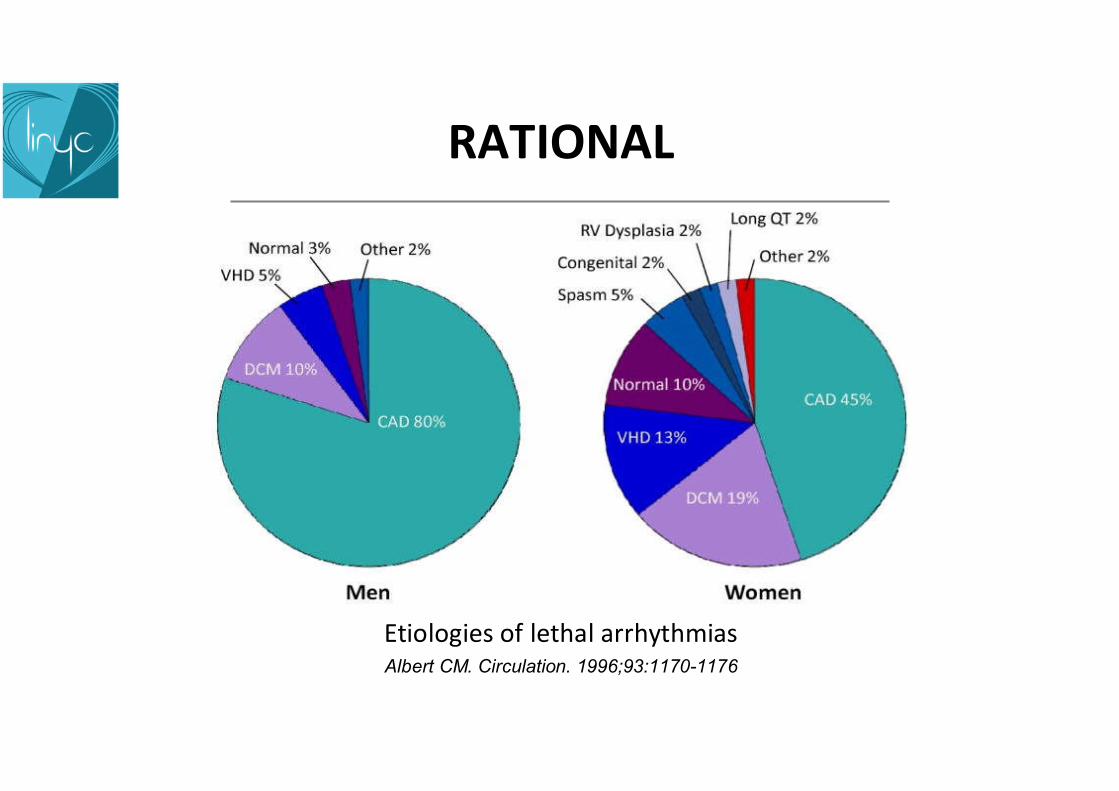
RATIONAL
Albert CM. Circulation. 1996;93:1170-1176
Etiologies of lethal arrhythmias

RATIONAL
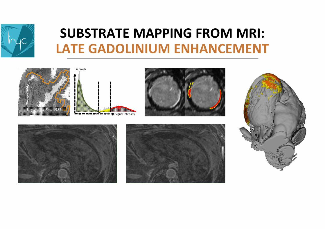
Ursell PC et al. Circ Res. 1985;56:436-51
Wagner A et al. Lancet. 2003;361:374-9
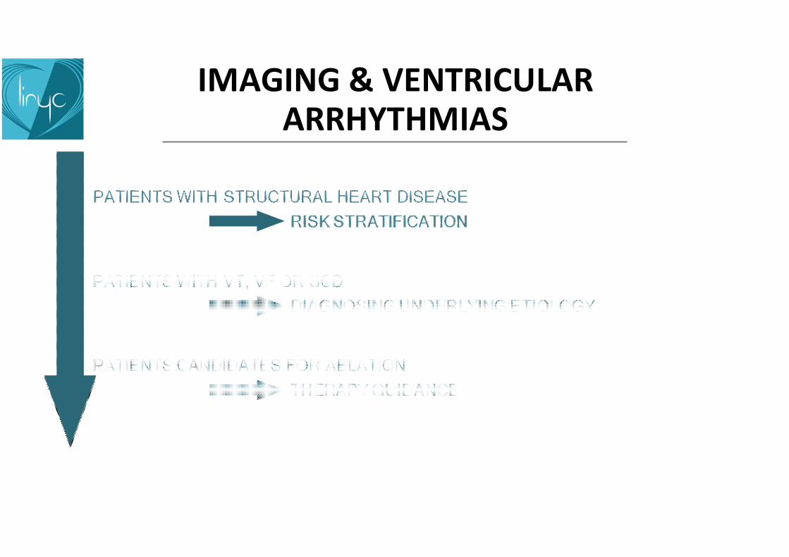
IMAGING & VENTRICULAR ARRHYTHMIAS

• > 4 millions/yr worldwide (1/5 of all deaths…)• 70% due to arrhythmias• 90% of arrhythmias occur on a substrate visible on CMR
CURRENT PREVENTIONICDs in LVEF <35%
Covers only a small minority of SCDs (<10%)
Most implanted pts do not need ICDs (2/3)
CLINICAL ISSUE OF SUDDEN CARDIAC DEATH

PROGNOSTIC ROLE OF LGE CMRIN ISCHEMIC HEART DISEASE
Watanabe, E., et al. Circ. Cardiovasc. Imaging. 2014; 7: 887–894 Signal intensity
n pixels
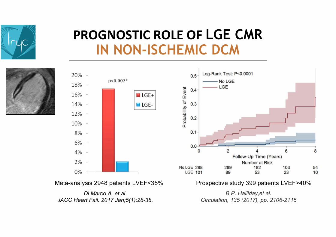
PROGNOSTIC ROLE OF LGE CMRIN NON-ISCHEMIC DCM
Di Marco A, et al. JACC Heart Fail. 2017 Jan;5(1):28-38.
Meta-analysis 2948 patients LVEF<35% Prospective study 399 patients LVEF>40%
B.P. Halliday,et al.Circulation, 135 (2017), pp. 2106-2115
PROGNOSTIC ROLE OF LGE CMRIN HCM
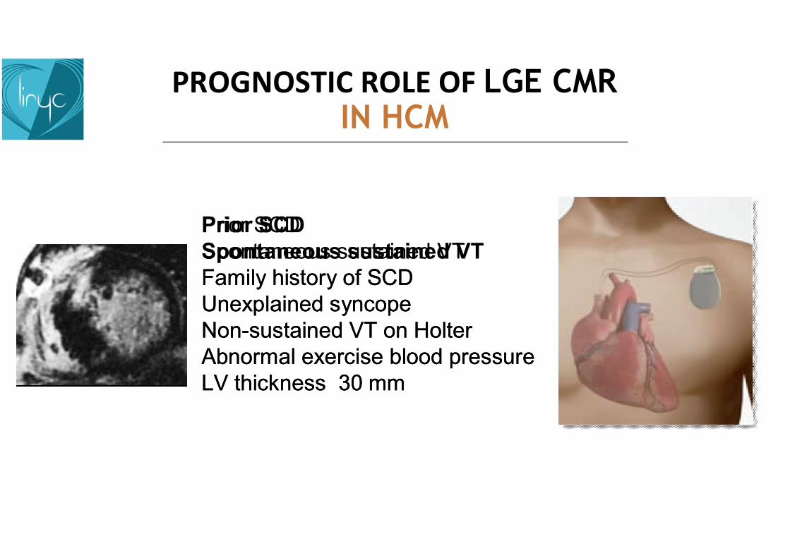
Prior SCDSpontaneous sustained VTFamily history of SCDUnexplained syncopeNon-sustained VT on HolterAbnormal exercise blood pressureLV thickness 30 mm
Prior SCDSpontaneous sustained VTFamily history of SCDUnexplained syncopeNon-sustained VT on HolterAbnormal exercise blood pressureLV thickness 30 mm

PROGNOSTIC ROLE OF LGE CMRIN HCM
NPV overestim
ated
by retro
spective studies
PPV to lo
w
to justify
costs & complicatio
ns NPV overestim
ated
by retro
spective studies
PPV to lo
w
to justify
costs & complicatio
ns
Prior SCDSpontaneous sustained VTFamily history of SCDUnexplained syncopeNon-sustained VT on HolterAbnormal exercise blood pressureLV thickness >30 mm

PROGNOSTIC ROLE OF LGE CMRIN HCM
Prospective study 1293 patients
Chan RH, et al.Circulation. 2014 Aug 5;130(6):484-95.
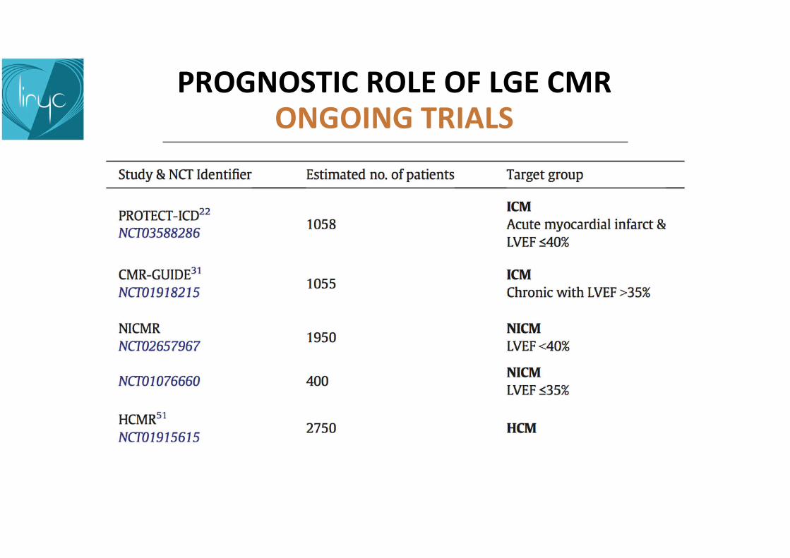
PROGNOSTIC ROLE OF LGE CMRONGOING TRIALS
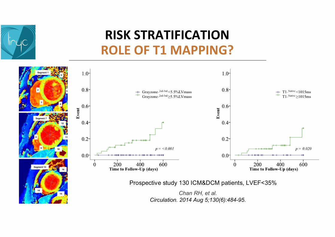
RISK STRATIFICATIONROLE OF T1 MAPPING?
Prospective study 130 ICM&DCM patients, LVEF<35%
Chan RH, et al.Circulation. 2014 Aug 5;130(6):484-95.
IMAGING & VENTRICULAR ARRHYTHMIAS

157 pts with VT or VFCMR alters diagnosis in 38% of pts with no history of SHD
negative echo & angio
Hennig et al. Eur Heart J Cardiovasc Imaging 2017
IMAGING TO IDENTIFY THE UNDERLYING VT/VF ETIOLOGY

44 yo man with monomorphic sustained VT of RBBB morphology Negative TTE & coronary angiography
ED ES LGE
CARDIAC SARCOID
IMAGING TO IDENTIFY THE UNDERLYING VT/VF ETIOLOGY
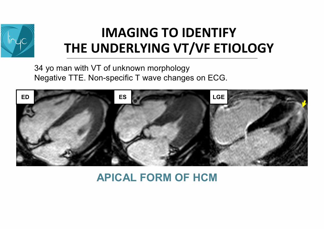
34 yo man with VT of unknown morphologyNegative TTE. Non-specific T wave changes on ECG.
APICAL FORM OF HCM
ED ES LGE
IMAGING TO IDENTIFY THE UNDERLYING VT/VF ETIOLOGY

58 yo women with sustained VT of RBBB morphologyNegative TTE & coronary angiography
CHURG-STRAUSS VASCULITIS
ED ES LGE
IMAGING TO IDENTIFY THE UNDERLYING VT/VF ETIOLOGY
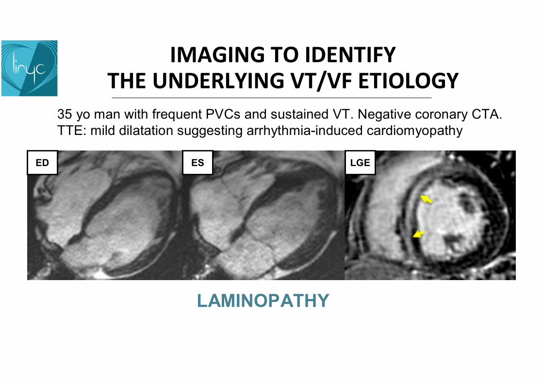
35 yo man with frequent PVCs and sustained VT. Negative coronary CTA. TTE: mild dilatation suggesting arrhythmia-induced cardiomyopathy
LAMINOPATHY
ED ES LGE
IMAGING TO IDENTIFY THE UNDERLYING VT/VF ETIOLOGY
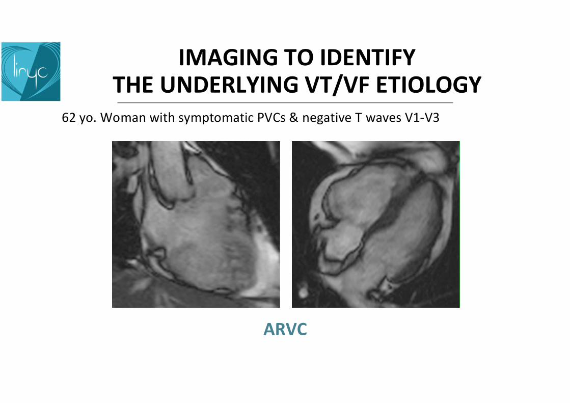
62 yo. Woman with symptomatic PVCs & negative T waves V1-V3
ARVC
IMAGING TO IDENTIFY THE UNDERLYING VT/VF ETIOLOGY
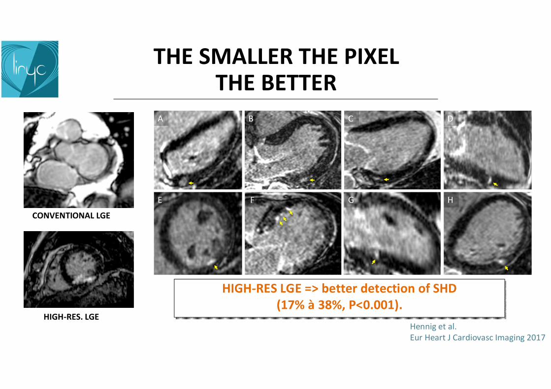
HIGH-RES LGE => better detection of SHD(17% à 38%, P<0.001).
HIGH-RES LGE => better detection of SHD(17% à 38%, P<0.001).
THE SMALLER THE PIXEL THE BETTER
CONVENTIONAL LGE
HIGH-RES. LGEHennig et al. Eur Heart J Cardiovasc Imaging 2017

IMAGING & VENTRICULAR ARRHYTHMIAS
Signal intensity
n pixels
Ursell. Circ Res. 1985
SUBSTRATE MAPPING FROM MRI: LATE GADOLINIUM ENHANCEMENT
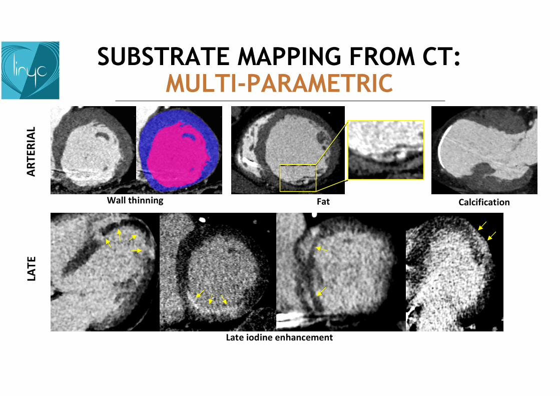
FatWall thinning Calcification
AR
TER
IAL
Late iodine enhancement
LAT
E
SUBSTRATE MAPPING FROM CT: MULTI-PARAMETRIC
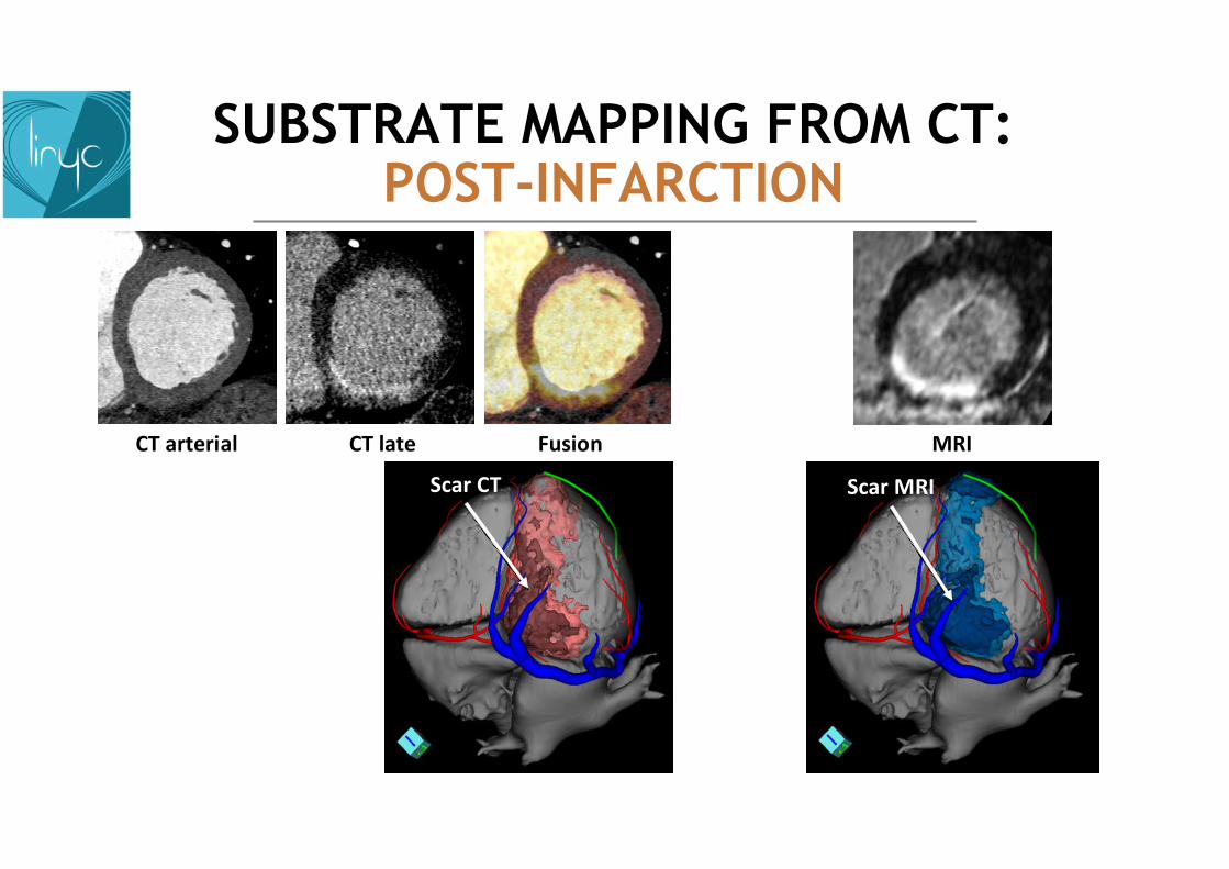
CT arterial CT late Fusion
Scar CT
MRI
Scar MRI
SUBSTRATE MAPPING FROM CT: POST-INFARCTION
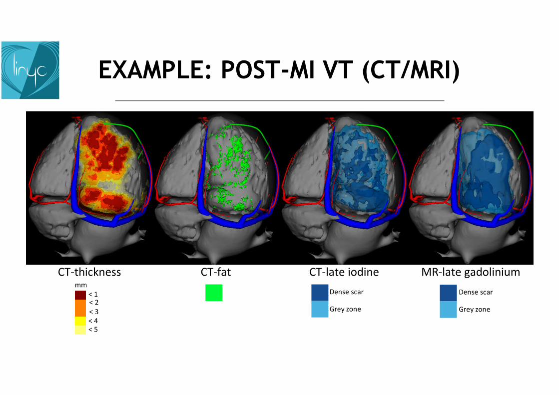
CT-thickness CT-fat CT-late iodine MR-late gadolinium
< 5< 4< 3< 2< 1
mmDense scar
Grey zone
Dense scar
Grey zone
EXAMPLE: POST-MI VT (CT/MRI)

PROCEDURAL INTEGRATION
Improved efficacyShorter proceduresSimpler & more standardized procedures

Hendriks. EP Europace. 2017 Jun;19(3):483.
VT-
FREE
SU
RV
IVA
L
OV
ERA
LL S
UR
VIV
AL
IMPACT ON OUTCOME

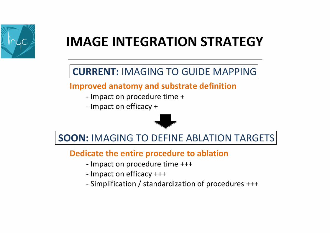
IMAGE INTEGRATION STRATEGY
Improved anatomy and substrate definition- Impact on procedure time +- Impact on efficacy +
CURRENT: IMAGING TO GUIDE MAPPING
Dedicate the entire procedure to ablation- Impact on procedure time +++- Impact on efficacy +++- Simplification / standardization of procedures +++
SOON: IMAGING TO DEFINE ABLATION TARGETS

57 yo man with prior infarct in LAD territoryMultiple ICD shocks on monomorphic VT
Nicolas Cedilnik et al. Europace 2018
HD VT map Simulation endo Simulation epi
Thic
knes
s
5mm
0mm
PERSPECTIVEIMAGE-BASED SIMULATION
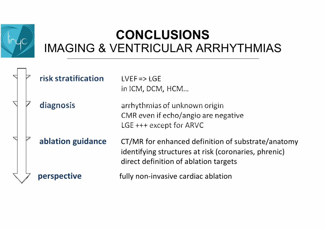
CONCLUSIONSIMAGING & VENTRICULAR ARRHYTHMIAS
ablation guidance CT/MR for enhanced definition of substrate/anatomy
identifying structures at risk (coronaries, phrenic)direct definition of ablation targets
perspective fully non-invasive cardiac ablation
TAKE HOME MESSAGES
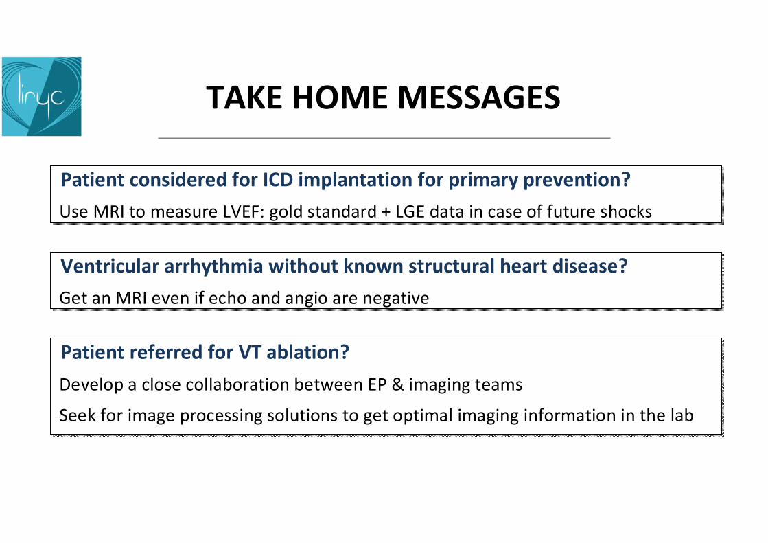
Ventricular arrhythmia without known structural heart disease?
Get an MRI even if echo and angio are negative
Ventricular arrhythmia without known structural heart disease?
Get an MRI even if echo and angio are negative
Patient considered for ICD implantation for primary prevention?
Use MRI to measure LVEF: gold standard + LGE data in case of future shocks
Patient considered for ICD implantation for primary prevention?
Use MRI to measure LVEF: gold standard + LGE data in case of future shocks
Patient referred for VT ablation?
Develop a close collaboration between EP & imaging teams
Seek for image processing solutions to get optimal imaging information in the lab
Patient referred for VT ablation?
Develop a close collaboration between EP & imaging teams
Seek for image processing solutions to get optimal imaging information in the lab