prévalence et signification fonctionnelle des … syndrome... · le deuxième objectif de cette...
TRANSCRIPT
Université de Montréal
Prévalence et signification fonctionnelle des
mouvements périodiques des jambes
par
Marie-Hélène Pennestri
Département de Psychologie
Faculté des Arts et Sciences
Thèse présentée à la Faculté des études supérieures et postdoctorales
en vue de l’obtention du grade de PhD
en psychologie clinique
option recherche et intervention
Septembre, 2010
© Marie-Hélène Pennestri, 2010
Université de Montréal
Faculté des études supérieures et postdoctorales
Cette thèse intitulée :
Prévalence et signification fonctionnelle des mouvements périodiques des jambes
Présentée par :
Marie-Hélène Pennestri
a été évaluée par un jury composé des personnes suivantes :
Antonio Zadra, président-rapporteur
Jacques Montplaisir, directeur de recherche
Paola A Lanfranchi, co-directrice
Jean-François Gagnon, membre du jury
Dominique Lorrain, examinatrice externe
Pierre Rainville, représentant du doyen de la FES
i
Résumé Les mouvements périodiques des jambes sont de courts mouvements involontaires qui
surviennent de façon périodique au cours du sommeil ou de l’éveil. Ils sont présents dans
certains troubles du sommeil, mais également chez des sujets sans plainte reliée au
sommeil.
Le premier objectif de cette thèse visait une meilleure description de la prévalence de ces
mouvements. Nous avons montré que chez les sujets sans plainte de sommeil, la prévalence
des mouvements périodiques des jambes en sommeil augmentait de façon importante à
partir d’environ 40 ans, tandis que l’index des mouvements périodiques des jambes à l’éveil
évoluait avec l’âge selon une courbe en U. Chez les sujets atteints de narcolepsie, on
retrouvait davantage de mouvements périodiques des jambes que chez les sujets témoins,
mais leur patron d’évolution avec l’âge était similaire.
Le deuxième objectif de cette thèse visait l’étude des mouvements périodiques des jambes
en relation avec le système nerveux autonome cardiovasculaire. Nous avons non seulement
confirmé la présence d’une tachycardie suivie d’une bradycardie lors des mouvements
périodiques des jambes durant le sommeil chez les patients atteints du syndrome
d’impatiences musculaires à l’éveil et chez les sujets sans plainte de sommeil, mais nous
avons également décrit ces mêmes changements de la fréquence cardiaque, quoiqu’avec
une plus faible amplitude, chez les sujets atteints de narcolepsie.

ii
Finalement, nous avons montré pour la première fois que les mouvements périodiques des
jambes en sommeil des sujets atteints du syndrome d’impatiences musculaires à l’éveil et
des sujets sans plainte de sommeil étaient aussi associés à des augmentations importantes et
significatives de la pression artérielle.
Mots-clés : Sommeil, mouvements périodiques des jambes, syndrome d’impatiences
musculaires à l’éveil, narcolepsie, sujets sans plainte de sommeil, système nerveux
autonome, fréquence cardiaque, pression artérielle
iii
Abstract Periodic leg movements are short involuntary movements occurring periodically during
sleep or wakefulness. They occur in some sleep disorders, but also in healthy subjects not
complaining of sleep problems.
The first objective of this thesis was to provide a better description of the prevalence of
these movements. In healthy non-complaining subjects, the prevalence of periodic leg
movements during sleep increased dramatically from about age 40, whereas the age-related
evolution of periodic leg movements during wakefulness followed a U curve. In narcoleptic
patients there were more periodic leg movements than in control subjects, but their
evolution with age showed the same pattern.
The second objective of this thesis was to study periodic leg movements in relationship
with cardiovascular autonomic nervous system. We not only confirmed that periodic leg
movements during sleep were associated with a tachycardia followed by a bradycardia in
restless legs syndrome patients and in healthy non-complaining subjects, but that these
heart rate changes were also present in narcoleptic patients, albeit of a lower amplitude.
Finally, we showed for the first time that periodic leg movements during sleep in restless
legs syndrome patients and in healthy non-complaining subjects were also associated with
significant and important rises of blood pressure.

iv
Keywords : Sleep, periodic leg movements, restless legs syndrome, narcolepsy, healthy
non-complaining subjects, autonomic nervous system, heart rate, blood pressure

v
Table des matières
Résumé …………………………………………...…………………………………………i
Abstract ……………………………………………………………………………………iii
Table des matières …………….………………….……………………………………..…v
Liste des tableaux …………………………………..……………………………………..ix
Liste des figures ………………………...………………………………………………….x
Liste des abréviations ……………………………………………………………………xii
Remerciements …………………………..……………………………………….……...xvi
1. Introduction .................................................................................................................. 1
1.1. Introduction générale ..................................................................................................... 2
1.2. Mouvements périodiques des jambes............................................................................. 2
1.2.1. Tableau clinique ................................................................................................. 2
1.2.2. Historique ........................................................................................................... 3
1.2.3. Enregistrement en laboratoire ............................................................................ 5
1.2.3.1. Mouvements périodiques des jambes en sommeil (MPJS) ............................. 5
1.2.3.2. Mouvements périodiques des jambes à l’éveil (MPJE) ................................. 6
1.2.3.3. Paramètres de sommeil .................................................................................. 7
1.2.4. Prévalence .......................................................................................................... 8
1.2.5. Trouble des mouvements périodiques des jambes (PLMD) .............................. 9
1.3. MPJ et syndrome d’impatiences musculaires à l’éveil (SIME) ................................... 11
1.3.1. Tableau clinique et prévalence du SIME ......................................................... 11
1.3.2. Diagnostic du SIME ......................................................................................... 12
1.3.3. MPJ dans le SIME ............................................................................................ 13
1.3.3.1. MPJS dans le SIME ...................................................................................... 13
1.3.3.2. MPJE dans le SIME ..................................................................................... 15
1.3.3.3. Mouvements périodiques des bras dans le SIME ......................................... 15
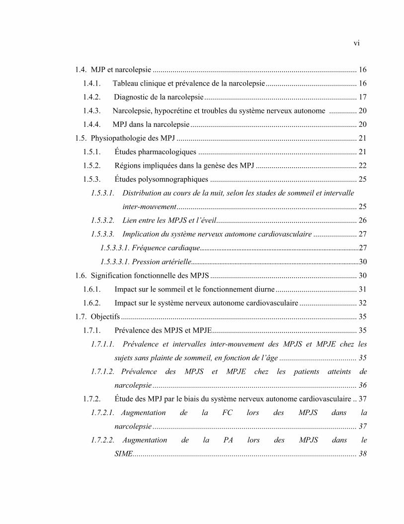
vi
1.4. MJP et narcolepsie ....................................................................................................... 16
1.4.1. Tableau clinique et prévalence de la narcolepsie .............................................. 16
1.4.2. Diagnostic de la narcolepsie ............................................................................. 17
1.4.3. Narcolepsie, hypocrétine et troubles du système nerveux autonome .............. 20
1.4.4. MPJ dans la narcolepsie .................................................................................... 20
1.5. Physiopathologie des MPJ ........................................................................................... 21
1.5.1. Études pharmacologiques ................................................................................ 21
1.5.2. Régions impliquées dans la genèse des MPJ ................................................... 22
1.5.3. Études polysomnographiques .......................................................................... 25
1.5.3.1. Distribution au cours de la nuit, selon les stades de sommeil et intervalle
inter-mouvement ........................................................................................... 25
1.5.3.2. Lien entre les MPJS et l’éveil....................................................................... 26
1.5.3.3. Implication du système nerveux automone cardiovasculaire ...................... 27
1.5.3.3.1. Fréquence cardiaque……….………………………………………………………………………….27
1.5.3.3.1. Pression artérielle……….…………………………………………..………………………………….30
1.6. Signification fonctionnelle des MPJS .......................................................................... 30
1.6.1. Impact sur le sommeil et le fonctionnement diurne ......................................... 31
1.6.2. Impact sur le système nerveux autonome cardiovasculaire ............................. 32
1.7. Objectifs ....................................................................................................................... 35
1.7.1. Prévalence des MPJS et MPJE ......................................................................... 35
1.7.1.1. Prévalence et intervalles inter-mouvement des MPJS et MPJE chez les
sujets sans plainte de sommeil, en fonction de l’âge ....................................... 35
1.7.1.2. Prévalence des MPJS et MPJE chez les patients atteints de
narcolepsie ....................................................................................................... 36
1.7.2. Étude des MPJ par le biais du système nerveux autonome cardiovasculaire .. 37
1.7.2.1. Augmentation de la FC lors des MPJS dans la
narcolepsie ....................................................................................................... 37
1.7.2.2. Augmentation de la PA lors des MPJS dans le
SIME ................................................................................................................. 38
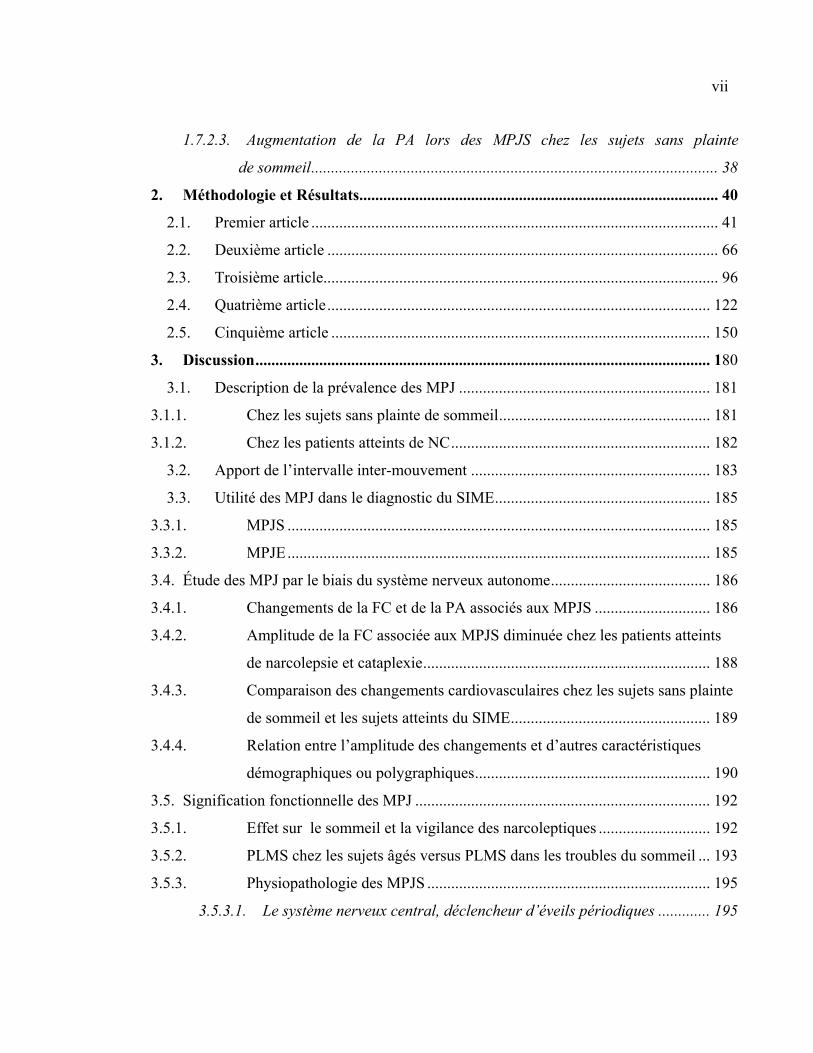
vii
1.7.2.3. Augmentation de la PA lors des MPJS chez les sujets sans plainte
de sommeil ...................................................................................................... 38
2. Méthodologie et Résultats.......................................................................................... 40
2.1. Premier article ...................................................................................................... 41
2.2. Deuxième article .................................................................................................. 66
2.3. Troisième article................................................................................................... 96
2.4. Quatrième article ................................................................................................ 122
2.5. Cinquième article ............................................................................................... 150
3. Discussion .................................................................................................................. 180
3.1. Description de la prévalence des MPJ ............................................................... 181
3.1.1. Chez les sujets sans plainte de sommeil ..................................................... 181
3.1.2. Chez les patients atteints de NC ................................................................. 182
3.2. Apport de l’intervalle inter-mouvement ............................................................ 183
3.3. Utilité des MPJ dans le diagnostic du SIME ...................................................... 185
3.3.1. MPJS .......................................................................................................... 185
3.3.2. MPJE .......................................................................................................... 185
3.4. Étude des MPJ par le biais du système nerveux autonome ........................................ 186
3.4.1. Changements de la FC et de la PA associés aux MPJS ............................. 186
3.4.2. Amplitude de la FC associée aux MPJS diminuée chez les patients atteints
de narcolepsie et cataplexie ........................................................................ 188
3.4.3. Comparaison des changements cardiovasculaires chez les sujets sans plainte
de sommeil et les sujets atteints du SIME .................................................. 189
3.4.4. Relation entre l’amplitude des changements et d’autres caractéristiques
démographiques ou polygraphiques ........................................................... 190
3.5. Signification fonctionnelle des MPJ .......................................................................... 192
3.5.1. Effet sur le sommeil et la vigilance des narcoleptiques ............................ 192
3.5.2. PLMS chez les sujets âgés versus PLMS dans les troubles du sommeil ... 193
3.5.3. Physiopathologie des MPJS ....................................................................... 195
3.5.3.1. Le système nerveux central, déclencheur d’éveils périodiques ............. 195
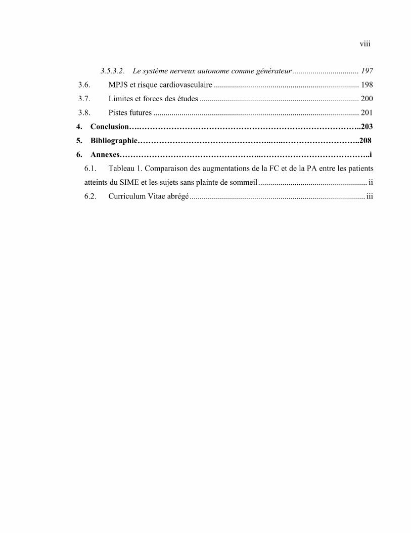
viii
3.5.3.2. Le système nerveux autonome comme générateur ................................. 197
3.6. MPJS et risque cardiovasculaire ........................................................................ 198
3.7. Limites et forces des études ............................................................................... 200
3.8. Pistes futures ...................................................................................................... 201
4. Conclusion….………………………………………………………………………..203
5. Bibliographie…………………………………………..…..………………………..208
6. Annexes……………………………………………..…………………………………..i
6.1. Tableau 1. Comparaison des augmentations de la FC et de la PA entre les patients
atteints du SIME et les sujets sans plainte de sommeil ...................................................... ii
6.2. Curriculum Vitae abrégé ....................................................................................... iii
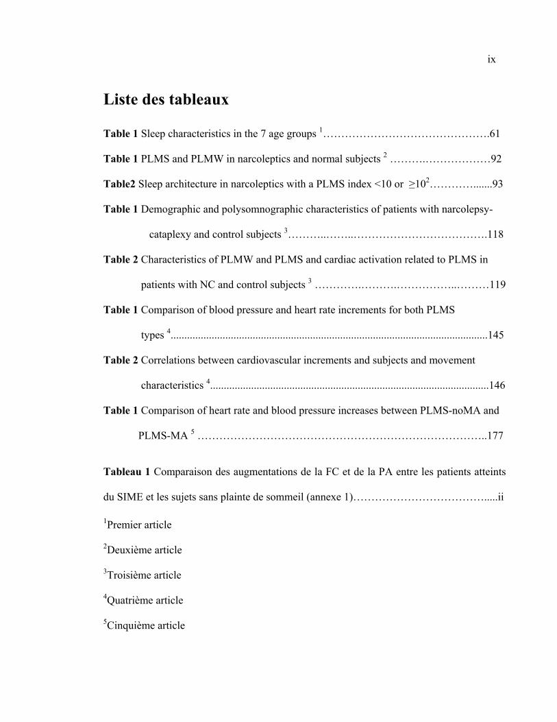
ix
Liste des tableaux
Table 1 Sleep characteristics in the 7 age groups 1……………………………………….61
Table 1 PLMS and PLMW in narcoleptics and normal subjects 2 ……….………………92
Table2 Sleep architecture in narcoleptics with a PLMS index <10 or ≥102………….......93
Table 1 Demographic and polysomnographic characteristics of patients with narcolepsy-
cataplexy and control subjects 3………..……..……………………………….118
Table 2 Characteristics of PLMW and PLMS and cardiac activation related to PLMS in
patients with NC and control subjects 3 ………….……….……………..………119
Table 1 Comparison of blood pressure and heart rate increments for both PLMS
types 4....................................................................................................................145
Table 2 Correlations between cardiovascular increments and subjects and movement
characteristics 4......................................................................................................146
Table 1 Comparison of heart rate and blood pressure increases between PLMS-noMA and
PLMS-MA 5 ……………………………………………………………………..177
Tableau 1 Comparaison des augmentations de la FC et de la PA entre les patients atteints
du SIME et les sujets sans plainte de sommeil (annexe 1)……………………………….....ii
1Premier article
2Deuxième article
3Troisième article
4Quatrième article
5Cinquième article
x
Liste des figures
Figure 1 Mean periodic leg movements during sleep (PLMS) index for healthy subjects
according to 7 age groups 1……………………………………………………...62
Figure 2 Mean periodic leg movements during wakefulness (PLMW) index for healthy
subjects according to 7 age groups 1…………......……………………………...63
Figure 3 Duration of mean periodic leg movements during sleep (PLMS) intermovement
intervals among 7 age groups 1…………........………………….……………….64
Figure 4 Duration of mean periodic leg movements during wakefulness (PLMW)
intermovement intervals among 7 age groups1......................................................65
Figure 1 Curves of mean periodic leg movements in sleep indices distribution for patients
with narcolepsy and normal controls according to five age groups for (a) NREM
sleep only and (b) REM sleep only 2…………….……………………..………..94
Figure 2 Curves of mean periodic leg movements while awake indices distribution for
patients with narcolepsy and normal controls according to five age
groups 2...………….…..…………………………………………………………95
Figure 1 Heart rate changes associated with periodic limb movements during sleep
(PLMS) without microarousal (MA) in patients with narcolepsy-cataplexy (NC)
and in control subjects 3...........................................................................................120
Figure 2 Heart rate changes associated with periodic limb movements during sleep
(PLMS) with microarousal (MA) in patients with narcolepsy-cataplexy (NC) and
in control subjects 3..............................................................................................121
xi
Figure 1 PLMS-related beat-to-beat blood pressure, EKG and EEG signals, presented in a
compact window and wider temporal windows 4.................................................147
Figure 2 Mean SBP, DBP and HR changes associated with PLMS-MA and
PLMS-noMA 4.....................................................................................................148
Figure 3 Mean SBP and DBP increments associated with PLMW and PLMS-noMA 4.. 149
Figure 1 PLMS-MA-related EEG, ECG, beat-to-beat BP, and respiratory signals 5…….178
Figure 2 Mean SBP, DBP and HR changes associated with PLMS-MA and
PLMS-noMA 5 …………………………………………………………………179
1Premier article
2Deuxième article
3Troisième article
4Quatrième article
5Cinquième article

xii
Liste des abréviations En français :
EEG : Électroencéphalogramme
EOG : Électrooculogramme
EMG : Électromyogramme
FC : Fréquence cardiaque
FRSQ : Fonds de la recherche en santé du Québec
Hz : hertz
IRMf : Imagerie par résonance magnétique fonctionnelle
IRSC : Instituts de recherche en santé du Canada
ME : Micro-éveil
mmHg : Millimètres de mercure
MPJ : Mouvements périodiques des jambes
MPJE : Mouvements périodiques des jambes à l’éveil
MPJS : Mouvements périodiques des jambes durant le sommeil
µV : microvolts
NC : Narcolepsie avec cataplexie
PA : Pression artérielle
PSG : Polysomnographie
SIME : Syndrome d’impatiences musculaires à l’éveil
SNA : Système nerveux autonome
SP : Sommeil paradoxal
TCSP : Trouble comportemental en sommeil paradoxal
TIDE : Test itératif de délai d’endormissement
TIS : Test d’immobilisation suggérée
xiii
En anglais :
ANOVA : Analysis of variance
ASDA: American Sleep Disorders Association
bpm : Beats per minute
BP : Blood pressure
CAP: cyclic alternating pattern
DA : Dopamine
DBP : Diastolic blood pressure
DSM-IV : Diagnostic and statistical manual of mental disorders, 4th edition
EEG : Electroencephalogram
EKG : Electrocardiogram
EOG : Electrooculogram
EMG : Electromyogram
ESRS: European Sleep Research Society
HR : Heart rate
HSD : Honest significant difference
ICSD : International classification of sleep disorders
IMI : Intermovement interval
MA : Microarousal
mmHg : Millimeters of mercury
MSLT : Multiple sleep latency test
n.a. : non applicable
NC : Narcolepsy-cataplexy
n.s. : non significant
NREM : Non rapied eye movement
OSA : Obstructive sleep apnea syndrome
PET : Positron emission tomography
PLM : Periodic leg movements
PLMD : Periodic leg movements disorder
xiv
PLMS : Periodic leg movements during sleep
PLMW : Periodic leg movements during wakefulness
RBD : Rapid eye movement sleep behavior disorder
REM : Rapid eye movement
RLS : Restless legs syndrome
SBP : Systolic blood pressure
SOREMP : Sleep-onset rapid eye movement period
SPECT : Single photon emission computed tomography

xv
À ma famille,
à Pascal,
à Delphine

xvi
Remerciements
Après ce long voyage, me voilà maintenant au moment d’écrire ces lignes… Je
comprends déjà qu’il me sera difficile de choisir les bons mots pour exprimer toute la
gratitude et l’affection que j’éprouve envers vous tous et toutes qui m’avez entourée et
appuyée tout au long de ces années…
Mon cher Jacques, je me rappellerai toujours cette rencontre avec toi au printemps 2001,
alors que je terminais mes études collégiales. Je me souviens de la passion qui t’animait
lorsque tu me parlais du sommeil, passion que tu as su me transmettre instantanément et qui
m’a aussitôt convaincue d’orienter autrement mes choix de carrière. L’année suivante, tu
m’accueillais dans ton équipe comme stagiaire durant mon baccalauréat. Merci de m’avoir
fait confiance, déjà à cette époque. Grâce à toi j’ai eu la chance de découvrir le monde du
sommeil et de la recherche dans un laboratoire hors du commun, où l’entraide,
l’enthousiasme et la rigueur scientifique vont de pair. Merci pour tout ce que tu m’as appris
et pour tout ce que tu as rendu possible. Travailler à tes côtés est une source de motivation
et d’inspiration constante. Merci pour ta grande générosité, je te serai toujours
reconnaissante!
Chère Paola, nos routes se sont croisées quelques années plus tard, ce qui m’a permis
d’ajouter un peu de « cœur » à mes travaux de recherche. Partager avec toi les cadeaux de
la vie et les moments difficiles a toujours été une expérience précieuse pour moi. J’ai eu
beaucoup de plaisir à discuter avec toi de tous les sujets possibles et à imaginer avec toi
toutes sortes de projets psycho-physio-cardio!
Merci à Dominique pour ta présence toujours discrète, mais tellement essentielle. Tu as
vraiment un talent incroyable pour améliorer notre travail, tout en nous donnant
l’impression que « tu n’as presque rien fait ». Merci Gaétan, pour tous les programmes qui
m’ont si souvent facilité la vie et plus particulièrement pour ton ingéniosité avec mon

xvii
nouvel ordinateur violet. Merci Jean, avec toi faire des statistiques c’est non seulement
facile, mais tout aussi amusant. Merci Sylvie pour toutes les analyses que nous avons
partagées… et pour les fous rires inclus. Merci à Mireille pour tous ces détails qui
deviennent tellement faciles à tes côtés. Tu as toujours la solution à tout et en plus, avec le
sourire. Merci aussi à Carmen pour ta complicité et ta bonne humeur.
Merci à tous les techniciens du laboratoire qui au fil des années ont contribué énormément
à tous les projets auxquels j’ai participé : Benoît, Nancy, Hélène, Sophie, Karine, Danielle,
Sonia, Maryse, Geneviève, Christian, Jimmy, Dominique et Sonia. C’est toujours agréable
et formateur de travailler à vos côtés.
Merci à tous mes collègues, amis et chercheurs du Centre d’étude du sommeil, Mélanie
pour la complicité depuis le Honor, Shirley, Annie, Valérie, Alex, Martin, Mathieu, Livia,
Jessica, Lorraine, Maud, Évelyne, Régine, Christiane, Geneviève et Jessica. J’ai la tête
pleine de beaux souvenirs avec vous. Merci à Valérie, Jean-François et Nadia, votre
parcours est inspirant et me donne envie de vous suivre. Un merci particulier à Nadia pour
toutes nos discussions inépuisables et pour tes judicieux conseils. Merci à Julie Carrier,
Marie Dumont, Toré Nielsen et Gilles Lavigne, j’ai appris beaucoup en travaillant à vos
côtés. Merci à Guy Rousseau pour tes invitations régulières à participer aux activités du
Centre de recherche. Un merci tout spécial à tous ceux et celles qui avez participé à la
chorale du Centre d’étude du sommeil et avec qui j’ai eu beaucoup de plaisir musical!
Merci à Yves Dauvilliers pour toutes nos collaborations et discussions et surtout pour ton
accueil chaleureux durant l’été 2010. Merci aussi à toute ton équipe, particulièrement,
Valérie, Sabine et Monica.
Merci à tous mes collègues du doctorat en psychologie et mes collègues d’internat. Partager
ces moments avec vous a toujours été amusant et soutenant. Un coucou spécial à « l’autre »
Marie-Hélène pour tous les moments de complicité.

xviii
Merci à tous les organismes subventionnaires qui m’ont apporté un support financier non
négligeable durant mes études, les Fonds de la Recherche en Santé du Québec, les Instituts
de Recherche en Santé du Canada, la Sleep Research Society, l’European Sleep Research
Society et la Faculté des études supérieures et postdoctorales de l’Université de Montréal.
Merci à tous les amis de ma vie qui m’accompagnent depuis bien longtemps… un petit clin
d’œil à Phuong Vy qui a su avant moi que je serais psychologue… Un merci plein de
gratitude à mon amie clinicienne Geneviève avec qui les élaborations sont sans fin, mais
qui m’ont grandement aidée à mettre en acte cette fin de rédaction. Merci aussi à Danielle,
tu as fait une différence énorme pour moi et tu as su m’aider à développer mon identité de
clinicienne.
Merci à mes quatre grands-parents que je suis choyée d’avoir tout près de moi, Réjeanne,
Clément, ma nonna et mon nonno, ainsi qu’à toute ma famille. Merci à mon petit frère,
Philippe, pour sa pensée critique qui m’a obligée maintes fois à restructurer ma pensée en
croyant à tort le convaincre. Merci à mes parents Johanne et Dominique pour votre soutien
et votre amour inconditionnel. La certitude que j’ai de vous savoir près de moi est si forte
qu’elle me permet de tout accomplir sans crainte.
À Pascal, mon chum devenu mon mari, puis le papa de ma fille à travers ce voyage. Merci
pour ton authenticité, ton sens de la justice, pour ta musique et pour tout ce bonheur. Merci
de partager ma vie et toutes les montagnes-russes qu’elle nous réserve. Tu es mon
inspiration et mon équilibre, je t’aime tellement…
À ma belle Delphine, ma grande-petite fille. Ma petite boule d’amour, mon petit miracle tu
es toujours là pour me ramener à l’essentiel. Merci pour tes sourires, tes chansons et pour la
vie que tu sèmes partout autour de toi. Te regarder exister est un privilège. Tu apportes
tellement de bonheur dans ma vie de maman. Merci d’être là, je t’aime!

1. Introduction
2
1.1. Introduction générale
Les mouvements périodiques des jambes (MPJ) sont de courts mouvements involontaires
qui surviennent périodiquement au cours du sommeil ou de l’éveil. Bien qu’ils aient surtout
été étudiés auprès des patients atteints du syndrome d’impatiences musculaires à l’éveil
(SIME), ils n’en demeurent pas moins une entité clinique distincte. En effet, ils sont
également présents dans certains troubles du sommeil et même chez des sujets sans plainte
reliée au sommeil. Malgré leur prévalence importante dans différentes populations, on ne
connaît toujours pas la signification fonctionnelle de ces mouvements. Le premier volet de
cette thèse vise une meilleure description de la prévalence des MPJ dans deux populations
différentes, soit les sujets sans plainte de sommeil et les patients atteints de narcolepsie. Un
deuxième volet se penchera sur la signification fonctionnelle de ces phénomènes moteurs,
par le biais de l’étude du système nerveux autonome cardiovasculaire. Cette deuxième
partie portera sur des patients atteints de narcolepsie, des patients atteints du SIME et des
sujets sans plainte de sommeil.
1.2. Mouvements périodiques des jambes
1.2.1. Tableau clinique
Les MPJ sont des mouvements involontaires survenant de façon répétée au cours du
sommeil (MPJS) ou de l’éveil (MPJE). On décrit les MPJS comme une extension du gros
orteil et du pied, une dorsiflexion de la cheville, parfois accompagnée d’une flexion du
3
genou et de la hanche, durant le sommeil. La plupart du temps, les sujets n’ont pas
conscience de ces mouvements. Les MPJS sont de courte durée, soit d’environ 2 à 4
secondes, et surviennent périodiquement, dans un intervalle se situant typiquement entre 20
et 40 secondes. Ils peuvent se présenter sur une jambe à la fois, ou sur les deux de façon
simultanée.
Les MPJE sont également des mouvements involontaires, présents au cours de la période
précédant l’endormissement ou durant les éveils nocturnes. Ils possèdent les mêmes
caractéristiques que les MPJS, mais leur durée est souvent un peu plus longue, compte tenu
de la composante sensorielle désagréable qui les accompagne la plupart du temps
(Michaud, Poirier, Lavigne, & Montplaisir, 2001).
1.2.2. Historique
C’est en 1953 que Charles Symonds a rapporté pour la première fois la présence de
mouvements involontaires au niveau des jambes durant le sommeil. Il donna le nom de
« nocturnal myoclonus » (ou myoclonies nocturnes) à ces manifestations motrices
(Symonds, 1953). Provenant du grec ancien klonos, signifiant « agitation », une myoclonie
est une « contraction musculaire brève et involontaire ». N’ayant pas accès à des
enregistrements polygraphiques, Symonds postula à tort que ces mouvements étaient
d’origine épileptique. Lorsqu’on revoit aujourd’hui la description clinique des cinq cas de
Symonds, il semble que ces patients présentaient diverses manifestations motrices
4
nocturnes, incluant à la fois des MPJS et d’autres types de mouvements (Coleman, Pollak,
& Weitzman, 1980).
C’est à l’équipe de Lugaresi que l’on doit les premiers enregistrements
polysomnographiques (PSG) et la description détaillée des MPJS, dans les années 60. Leur
but était alors de documenter l’insomnie provoquée par les paresthésies et l’agitation
motrice des patients atteints du SIME, ainsi que l’efficacité des traitements
pharmacologiques. Cependant, les enregistrements ont mis en évidence un phénomène
moteur auquel les auteurs ne s’attendaient pas. En effet, les patients présentaient des
mouvements involontaires toutes les 20 ou 30 secondes au moment de s’endormir
(Lugaresi, Coccagna, Tassinari, & Ambrosetto, 1965). Lugaresi et ses collaborateurs ont
décrit le fait que les mouvements pouvaient se présenter de façon symétrique sur les deux
jambes, ou encore alterner d’une jambe à l’autre (Coccagna, Lugaresi, Tassinari, &
Ambrosetto, 1966). Ils ont également documenté que ces mouvements pouvaient apparaître
indépendamment du SIME, montrant déjà à cette époque qu’il s’agissait d’une entité
clinique distincte (Lugaresi, Coccagna, Gambi, Ceroni, & Poppi, 1966). Lugaresi a repris
l’appellation de myoclonie nocturne, en l’honneur de Symonds. Néanmoins, il a apporté
une nuance importante, distinguant d’une part les myoclonies épileptiques, qui surviennent
de façon sporadique et qui sont accompagnées de pointes épileptiques au niveau cortical et
d’autre part, les myoclonies nocturnes, qui sont au contraire périodiques et non-

5
accompagnées d’activité électroencéphalographique (EEG) épileptique (Lugaresi 1968,
dans (Bixler et al., 1982)).
En 1980, Coleman a proposé le terme « mouvements périodiques des jambes en sommeil »
(MPJS), reprenant l’argument que ces mouvements n’étaient pas réellement myocloniques,
présentant généralement un potentiel d’action plus long (Coleman et al., 1980). C’est
également Coleman qui a par la suite officialisé les critères d’enregistrement et de cotation
des MPJS qui sont toujours utilisés aujourd’hui, malgré de légères modifications (Coleman,
1982).
1.2.3. Enregistrement en laboratoire
1.2.3.1. Mouvements périodiques des jambes en sommeil (MPJS)
Bien que l’on puisse les observer à l’œil nu, les MPJS sont étudiés en laboratoire de
sommeil, afin de les quantifier. À cette fin, les critères proposés par Coleman en 1982
(Coleman, 1982) ont été repris en 1993 par l’American Sleep Disorders Association
(ASDA, 1993) et ont récemment été revus en 2006 par the International Restless Legs
Sydrome Study Group (Zucconi et al., 2006). Ce sont ces derniers critères qui seront
présentés ici, mais il convient de souligner que les modifications amenées au fil des
différentes publications sont somme toute minimes. La tendance actuelle est d’inclure un
6
plus grand nombre de mouvements, en augmentant notamment le critère de durée maximale
des MPJS.
Lors d’une évaluation PSG en laboratoire de sommeil, on pose deux électrodes à la surface
de la peau, pour enregistrer l’activité électrique du muscle tibialis antérieur de chacune des
jambes. On considère le début de l’événement moteur lorsque l’amplitude du signal
électromyographique (EMG) excède le niveau de base de 8 µV et la fin de l’événement
lorsque l’amplitude revient à 2 µV au-dessus du niveau de base pendant au moins 0,5
secondes. Pour être considérés comme des MPJS, les mouvements doivent avoir une durée
entre 0,5 à 10,0 secondes et l’intervalle les séparant doit se situer entre 5 et 90 secondes. De
plus, ils doivent se présenter dans une série de quatre ou plus. Le nombre de MPJS est
calculé pour toute la nuit et il est ensuite possible d’en calculer un index, soit le nombre
total de mouvements, divisé par le nombre d’heures de sommeil. Un index plus élevé que
cinq a longtemps été considéré pathologique (Coleman, 1982), mais ce critère a depuis été
revu et nuancé, comme cela sera exposé ultérieurement.
1.2.3.2. Mouvements périodiques des jambes à l’éveil (MPJE)
Même s’il n’existe pas de critères standardisés, les MPJE sont généralement enregistrés et
identifiés selon les mêmes critères que les MPJS (Michaud, Paquet, Lavigne, Desautels, &
Montplaisir, 2002). Ces mouvements ont essentiellement été étudiés chez les sujets atteints
du SIME (Montplaisir et al., 1997; Nicolas, Michaud, Lavigne, & Montplaisir, 1999).
7
D’ailleurs, en plus d’être mesurés durant la période d’endormissement et au cours des
éveils nocturnes, les MPJE peuvent aussi être enregistrés durant un test développé
spécifiquement pour cette population, soit le test d’immobilisation suggérée (TIS)
(Montplaisir et al., 1998). Durant le TIS, les sujets sont en position assise à 45°, les jambes
allongées et reçoivent comme consigne de ne pas bouger les jambes. Les patients doivent
demeurer éveillés durant tout le test, d’une durée d’une heure. Les mouvements
involontaires sont alors enregistrés et quantifiés de la même façon que durant le sommeil.
Comme les symptômes du SIME augmentent durant la soirée et la nuit, ce test doit être
administré au cours de la soirée (Michaud et al., 2005).
1.2.3.3. Paramètres de sommeil
Différents paramètres de sommeil sont enregistrés en même temps que l’évaluation des
MPJ lors d’une investigation en laboratoire de sommeil. Une évaluation PSG comprend
minimalement l’enregistrement des ondes cérébrales (électroencéphalogramme; EEG), des
mouvements des yeux (électrooculogramme; EOG) et du tonus musculaire
(électromyogramme; EMG). Ces paramètres permettent de distinguer différents états, ou
stades de sommeil. Le sommeil non paradoxal, appelé en anglais « Non Rapid Eye
Movement » (NREM), formé des stades 1 à 4 et le sommeil paradoxal (SP), appelé en
anglais « Rapid Eye Movement » (REM) constituent les deux états principaux du sommeil.
Le sommeil non paradoxal se subdivise également en deux catégories, soit le sommeil léger
(stades 1 et 2) et le sommeil lent profond (stades 3 et 4). Durant un cycle de sommeil
8
normal, on s’attend d’abord à la présence du sommeil léger, puis du sommeil lent profond.
Il y a ensuite un retour vers le sommeil léger, puis le cycle se termine par une période de
SP. Ce cycle d’environ 90 minutes survient trois à cinq fois au cours de la nuit, à la
différence que le sommeil lent profond est davantage présent au début de la nuit, tandis que
le SP occupe plus de temps en fin de nuit.
Différents indices peuvent être calculés, ce qui permet d’évaluer la quantité et la qualité du
sommeil des sujets. On évalue notamment la latence au sommeil (délai d’endormissement
après la fermeture des lumières), le temps total dormi, le temps total d’éveil, la proportion
du temps passé dans les différents stades de sommeil, l’efficacité du sommeil (proportion
du temps dormi sur le temps passé au lit depuis l’endormissement), le nombre d’éveils et le
nombre et l’index de micro-éveils (ME).
1.2.4. Prévalence
Récemment, une étude comprenant 592 participants sélectionnés au hasard dans la
population générale et chez qui le sommeil a été enregistré durant une nuit de PSG a évalué
la prévalence des MPJS à 7,6%, selon un critère de plus de 15 mouvements par heure de
sommeil (Scofield, Roth, & Drake, 2008). Dans cette étude, les patients présentant un index
élevé, rapportaient davantage une plainte reliée à l’insomnie ou au SIME que les autres
sujets. En effet, les MPJS sont particulièrement associés au SIME, même s’ils sont
également présents dans d’autres troubles du sommeil (Coleman et al., 1980; Montplaisir,
9
Michaud, Denesle, & Gosselin, 2000). On les retrouve notamment dans la narcolepsie
(Coleman et al., 1980; Dauvilliers, Billiard, & Montplaisir, 2003; Montplaisir et al., 2000),
le trouble comportemental en sommeil paradoxal (TCSP) (Coleman et al., 1980; Fantini,
Michaud, Gosselin, Lavigne, & Montplaisir, 2002), le syndrome d’apnées du sommeil
(Carelli, Krieger, Calvi-Gries, & Macher, 1999; Coleman et al., 1980; Fry, DiPhillipo, &
Pressman, 1989; Morisson et al., 2001), l’insomnie (Coleman et al., 1980; Montplaisir et
al., 2000) et l’hypersomnie (Coleman et al., 1980; Montplaisir et al., 2000).
Les MPJS sont également présents chez des sujets en santé, ne présentant pas de plainte
spécifique reliée au sommeil. La prévalence est particulièrement élevée chez les sujets plus
âgés (Ancoli-Israel et al., 1991; Ancoli-Israel, Kripke, Mason, & Kaplan, 1985; Bixler et
al., 1982; Carrier et al., 2005; Scofield et al., 2008; Silber, 2001). À l’inverse, chez les
enfants et les adolescents sans trouble de sommeil, on retrouve très peu de MPJS (Arens et
al., 1998; Bixler et al., 1982; Picchietti, England, Walters, Willis, & Verrico, 1998).
1.2.5. Trouble des mouvements périodiques des jambes (PLMD)
Dans la première édition de l’International Classification of Sleep Disorders (ICSD)
(ASDA, 1990) publiée en 1990, on retrouve le « periodic limb movement disorder
(PLMD) » soit, le trouble des MPJ (traduction libre). Ce diagnostic reposait sur la présence
d’un index de MPJS plus grand ou égal à cinq par heure de sommeil, associé à une
insomnie ou une hypersomnolence, ne pouvant être expliquée par une autre condition
10
médicale. Cette entité diagnostique a été critiquée et remise en question, autant en ce qui a
trait à sa validité qu’à son utilité clinique. En effet, bien qu’on retrouve des MPJS chez les
sujets atteints d’insomnie ou d’hypersomnie, cette prévalence est souvent la même que dans
d’autres troubles du sommeil ou chez des sujets en santé (Nicolas, Lesperance, &
Montplaisir, 1998). D’autres auteurs ont argumenté que pour poser un diagnostic de
PLMD, il devrait y avoir une perception claire des MPJS ou encore des effets des MPJS sur
le sommeil ou le fonctionnement diurne, ainsi que la présence d’un index plus élevé que ce
qui serait attendu chez une personne du même âge (Silber, 2001). À l’heure actuelle, de
telles normes ne sont cependant pas disponibles.
Ainsi, la deuxième version de l’ICSD (American Academy of Sleep Medicine, 2005)
propose des changements majeurs quant à ce diagnostic. On pose d’abord que l’index de
MPJS doit être plus grand que 15 par heure de sommeil pour être considéré comme
pathologique et que les mouvements associés à la fin d’un événement respiratoire devraient
être exclus de cet index. Finalement, on souligne l’importance de se baser avant tout sur le
contexte clinique pour juger de la pertinence d’un tel diagnostic, plutôt que sur la présence
d’un index prédéterminé de MPJS.
11
1.3. MPJ et syndrome d’impatiences musculaires à l’éveil (SIME)
1.3.1. Tableau clinique et prévalence du SIME
Le SIME est une maladie neurologique qui provoque chez les patients des sensations
désagréables au niveau des jambes, particulièrement lorsqu’ils sont au repos. Ces
sensations sont décrites comme un engourdissement, des fourmis dans les jambes, des
picotements ou même de la douleur. Ces paresthésies sont associées à un besoin irrésistible
de bouger les jambes, d’où l’expression du « syndrome des jambes sans repos ». Cette
activation motrice semble effectivement soulager les symptômes, mais uniquement de
façon temporaire. La symptomatologie des patients augmente en soirée et durant la nuit, ce
qui entraîne souvent des difficultés d’endormissement, ainsi que des éveils durant la nuit.
Au Canada, une seule étude a évalué la prévalence du SIME dans la population générale, et
ce, à partir de deux questions posées à 2019 participants. Lorsqu’on ciblait la sensation
d’impatiences au coucher, la prévalence était évaluée à 15%, alors que 10% des répondants
rapportaient la présence de sensations désagréables dans les jambes au cours du sommeil,
pouvant amener un éveil ainsi qu’un besoin irrésistible de bouger (Lavigne & Montplaisir,
1994). Cette étude rapportait également une prévalence deux fois plus élevée chez les
patients canadiens francophones d’origine française, suggérant une influence génétique.
La publication de critères diagnostiques standardisés du SIME par le « International
Restless Legs Syndrome Study Group » en 1995 (Walters, 1995) et leur révision en 2003
12
(Allen, Picchietti, Hening, Trenkwalder, Walters, & Montplaisir, 2003) ont permis
d’uniformiser les questions utilisées dans les études épidémiologiques. Au cours des
dernières années, plusieurs études ont utilisé ces critères standardisés pour évaluer la
prévalence du SIME, principalement en Europe et aux États-Unis. Elles ont trouvé des
prévalences variant entre 6 et 12% (Allen et al., 2005; Berger, Luedemann, Trenkwalder,
John, & Kessler, 2004; Hogl et al., 2005; Rothdach, Trenkwalder, Haberstock, Keil, &
Berger, 2000; Sevim et al., 2003; Tan et al., 2001; Ulfberg, Nystrom, Carter, & Edling,
2001a, 2001b). On observe néanmoins une prévalence très faible en Asie, soit de moins
de 1% à Singapour (Tan et al., 2001), ce qui appuie à nouveau l’hypothèse d’une influence
génétique.
La plupart des études ayant examiné l’association entre l’âge et la prévalence du SIME
montre une augmentation des cas avec l’âge (Allen et al., 2005; Berger et al., 2004; Hogl et
al., 2005; Lavigne & Montplaisir, 1994; Ohayon & Roth, 2002; Phillips et al., 2000;
Rothdach et al., 2000; Sevim et al., 2003). Une étude effectuée chez 250 patients atteints du
SIME, a montré une distribution bimodale de l’âge de début de la maladie, avec un premier
pic vers 20 ans et un deuxième pic au milieu de la quarantaine (Whittom et al., 2007).
1.3.2. Diagnostic du SIME
Le diagnostic du SIME est de nature clinique, s’appuyant sur la présence de quatre
caractéristiques essentielles, soit: (1) un besoin irrésistible de bouger les jambes, le plus
13
souvent associé à la présence de paresthésies, (2) l’apparition ou l’aggravation des
symptômes au repos et en particulier au moment du coucher, (3) le soulagement des
symptômes par l’activité motrice, en particulier par la marche, (4) l’aggravation des
symptômes en soirée et au cours de la nuit (Allen, Picchietti, Hening, Trenkwalder,
Walters, & Montplaisir, 2003). Bien que ces quatre critères soient suffisants pour poser le
diagnostic, d’autres caractéristiques peuvent le supporter et s’avèrent particulièrement utiles
lorsque le diagnostic clinique demeure incertain. C’est le cas notamment de l’efficacité du
traitement dopaminergique (Stiasny-Kolster, Kohnen, Moller, Trenkwalder, & Oertel,
2006) et de la présence d’une histoire familiale de la maladie (Allen, La Buda, Becker, &
Earley, 2002). De plus, la présence des MPJS et des MPJE s’avère souvent un indice
moteur du SIME.
1.3.3. MPJ dans le SIME
1.3.3.1. MPJS dans le SIME
Bien que les MPJ soient présents dans d’autres pathologies, c’est principalement par l’étude
des sujets atteints du SIME qu’ils ont été décrits et investigués, ces deux conditions étant
étroitement liées. En effet, l’étude des MPJ en laboratoire constitue souvent une façon
objective d’appuyer le diagnostic du SIME (Montplaisir, Michaud, Pennestri, & Lanfranchi
A, 2009).
14
Dans une étude comprenant 131 patients atteints du SIME, environ 80% des patients
avaient un index de MPJS plus élevé que cinq par heure de sommeil (Montplaisir et al.,
1997). Dans la même étude, un sous-groupe de 49 patients a été étudié durant deux nuits
consécutives. Dans ce sous-groupe, 82% des patients avaient un index supérieur à cinq au
cours d’une des deux nuits, alors que 76% atteignait ce critère pour les deux nuits. Cette
étude souligne donc la prévalence élevée de MPJS auprès des sujets atteints du SIME.
La présence des MPJS permet également de discriminer adéquatement les sujets atteints du
SIME des sujets témoins sans trouble du sommeil. En effet, un index de sept mouvements
par heure de sommeil a montré une sensibilité de 78%, ce seuil ayant la capacité
d’identifier le SIME lorsqu’il est présent. Ce même index présentait une spécificité de 76%,
ce qui réfère cette fois à la capacité d’exclure le diagnostic du SIME lorsque la condition
est absente (Michaud, Paquet, et al., 2002). Dans cette étude, les sujets avaient un âge
moyen d’environ 48 ans; la capacité de discrimination de l’index de MPJS n’a pas été
évaluée chez des sujets plus jeunes ou plus âgés.
Si on considère la plainte subjective des patients, on a aussi montré une association entre la
sévérité des symptômes sensoriels du SIME et l’index de MPJS au cours de la nuit (Garcia-
Borreguero, Larrosa, de la Llave, Granizo, & Allen, 2004; Montplaisir et al., 1997).
Finalement, contrairement aux résultats obtenus chez les sujets en santé ou atteints de
différents troubles du sommeil, il ne semble pas y avoir de corrélation entre l’index de
15
MPJS et l’âge chez les sujets atteints du SIME (Nicolas et al., 1999). Néanmoins, la durée
de l’intervalle séparant les mouvements diminue avec l’âge (Nicolas et al., 1999).
1.3.3.2. MPJE dans le SIME
L’index de MPJE précédant la période d’endormissement et durant les périodes d’éveil
nocturne, s’est avéré un outil encore plus efficace que l’index de MPJS pour discriminer les
patients atteints du SIME des sujets témoins. Dans la même étude citée précédemment, un
index de 15 MPJE par heure a montré une sensibilité de 87% et une spécificité de 80%
(Michaud, Paquet, et al., 2002). L’index de MPJE, est quant à lui corrélé positivement avec
l’âge chez les sujets atteints du SIME (Nicolas et al., 1999).
Lorsqu’on considère les MPJE enregistrés durant le TIS, les sujets atteints du SIME
présentent aussi plus de mouvements que les sujets témoins (Michaud, Lavigne, Desautels,
Poirier, & Montplaisir, 2002; Montplaisir et al., 1998). De plus, on a montré une
association positive entre les symptômes sensoriels rapportés durant le TIS et l’index de
MPJE obtenu durant le test (Garcia-Borreguero et al., 2004).
1.3.3.3. Mouvements périodiques des bras dans le SIME
Bien qu’ils aient été moins étudiés, il est également possible d’observer des mouvements
périodiques involontaires au niveau des membres supérieurs. Les mouvements périodiques
16
des bras sont observés chez environ 50% des patients atteints du SIME et semblent
davantage présents dans les cas de SIME plus sévère (Chabli, Michaud, & Montplaisir,
2000; Michaud, Chabli, Lavigne, & Montplaisir, 2000; Montplaisir et al., 2000).
1.4. MJP et narcolepsie
1.4.1. Tableau clinique et prévalence de la narcolepsie
La narcolepsie est un trouble caractérisé par la présence d’une somnolence excessive durant
la journée, un sommeil nocturne perturbé, ainsi que des intrusions anormales de certaines
composantes du SP durant l’éveil, créant des états dissociés. Ces manifestations peuvent
inclure la cataplexie, la paralysie du sommeil et la présence d’hallucinations.
La cataplexie, présente chez plus des deux tiers des patients narcoleptiques, est un
relâchement soudain du tonus musculaire, le plus souvent provoqué par des émotions
intenses telles que la surprise, le rire, la fierté ou la colère. Cette perte de tonus n’est
cependant pas accompagnée d’une altération de la conscience et est généralement de courte
durée, soit de quelques secondes, ou plus rarement, de quelques minutes. La cataplexie peut
être partielle, souvent localisée au niveau du cou et de la tête ou encore totale, provoquant
alors la chute du patient. La paralysie de sommeil consiste en une intrusion de l’atonie
musculaire, habituellement associée au SP, durant l’éveil. Le patient devient alors incapable
de bouger, alors qu’il est pourtant réveillé. On peut également observer une incorporation
17
de l’imagerie du rêve durant l’éveil, qui se manifeste par des hallucinations hypnagogiques
(à l’endormissement) ou hypnopompiques (à l’éveil). Les patients atteints de narcolepsie ne
présentent pas nécessairement tous ces symptômes. De plus, ces symptômes, mis à part la
cataplexie, ne sont pas uniques à la narcolepsie.
La narcolepsie avec cataplexie (NC) présente une très faible prévalence, soit environ 0,013
à 0,18% de la population générale (pour une revue, Dauvilliers et al., 2003). La prévalence
varie légèrement d’un pays à l’autre, mais également selon la méthode employée pour
l’évaluer. Certaines études appuient le diagnostic avec un enregistrement PSG, tandis que
d’autres utilisent uniquement des questionnaires. La narcolepsie peut être observée à
n’importe quel âge, mais elle est rarement diagnostiquée avant cinq ans. Dans deux
populations indépendantes, la distribution de l’âge de début s’est révélée bimodale, avec un
premier pic vers 15 ans et un deuxième autour de 36 ans (Dauvilliers et al., 2001).
1.4.2. Diagnostic de la narcolepsie
Selon la deuxième édition de l’ICSD (American Academy of Sleep Medicine, 2005), les
critères minimaux pour diagnostiquer la NC consistent en une plainte de somnolence diurne
excessive, presque toute la journée, durant au moins trois mois et une histoire de cataplexie.
Un troisième critère suggère d’obtenir, lorsque cela est possible, une confirmation par un
enregistrement PSG nocturne et un test itératif de délai d’endormissement (TIDE, connu en
18
anglais sous le nom de « Multiple Sleep Latency Test » (MSLT)) ou encore, une
vérification du niveau d’hypocrétine.
Le TIDE est considéré comme une mesure physiologique de la tendance à s’endormir, en
l’absence de stimulations extérieures pouvant favoriser l’éveil (Carskadon & Dement,
1982). Ce test consiste en une série de quatre ou cinq occasions de siestes, programmées
aux deux heures au cours de la journée, durant lesquelles le patient est étendu dans une
chambre confortable, obscure et sans bruit, alors qu’on mesure par PSG le temps
d’endormissement (Carskadon et al., 1986). Pour appuyer le diagnostic de narcolepsie, la
latence au sommeil doit être en moyenne de huit minutes ou moins. De plus, tel que
mentionné précédemment, on s’attend à ce qu’un sujet en santé s’endorme d’abord en
sommeil non paradoxal. Toutefois, compte tenu des manifestations anormales du SP chez
les sujets atteints de la NC, il arrive que ceux-ci s’endorment directement dans cet état. Ce
phénomène est appelé en anglais « sleep-onset REM period » (SOREMP). L’ICSD suggère
qu’il y ait présence d’au moins deux SOREMP au cours des siestes du TIDE, pour appuyer
le diagnostic de narcolepsie. Il convient néanmoins de souligner l’existence d’une
controverse quand aux différents critères PSG concernant le diagnostic de narcolepsie,
certains auteurs considérant ces critères trop sévères (Dauvilliers et al., 2004).
Quant au niveau d’hypocrétine, on sait que la NC est associée à une diminution importante
du niveau d’hypocrétine dans le liquide céphalo-rachidien, ainsi que du nombre de
19
neurones à hypocrétine lors d’analyses post-mortem (Dauvilliers, Arnulf, & Mignot, 2007;
Mignot et al., 2002; Peyron et al., 2000). Ainsi, on peut effectuer une ponction lombaire
pour vérifier le niveau d’hypocrétine chez les patients possiblement atteints de narcolepsie.
L’ICSD propose comme critère un niveau inférieur ou égal à 110 pg/mL, soit environ un
tiers des valeurs attendues chez les sujets témoins. Une diminution du niveau d’hypocrétine
est une mesure très spécifique (99%) et sensible (89%) de la présence de cataplexie
(Dauvilliers et al., 2003; Mignot et al., 2002).
1.4.3. Narcolepsie, hypocrétine et troubles du système nerveux autonome
Différents auteurs ont décrit chez les patients atteints de narcolepsie des anomalies au
niveau du système nerveux autonome, notamment la fonction pupillaire, érectile et
cardiovasculaire (Hublin, Matikainen, & Partinen, 1994; Karacan, 1986; Sachs & Kaijser,
1980). Plus récemment, certains auteurs ont examiné l’analyse spectrale de la variabilité
cardiaque durant le sommeil et l’éveil dans cette population, mais les résultats demeurent
peu concluants (Ferini-Strambi et al., 1997; Fronczek et al., 2008).
Néanmoins, l’association documentée entre la narcolepsie avec cataplexie et la diminution
du niveau d’hypocrétine constitue un argument supplémentaire pour appuyer l’hypothèse
d’un dysfonctionnement du système nerveux autonome dans cette population. En effet, les
neurones à hypocrétine, bien que localisés exclusivement dans l’hypothalamus dorsolatéral,
projettent dans de nombreuses régions, incluant les structures hypothalamiques et
20
pontiques, structures connues notamment pour leur rôle dans la régulation cardiovasculaire
centrale (Samson, Taylor, & Ferguson, 2005; Shirasaka, Kunitake, Takasaki, & Kannan,
2002). Par exemple, chez le rat, l’administration d’hypocrétine augmente la fréquence
cardiaque et la pression artérielle.
L’ensemble de ces résultats suggèrent une association entre la narcolepsie, la diminution du
niveau d’hypocrétine et une dysfonction du système nerveux autonome. Néanmoins,
certains résultats modestes ou contradictoires soulèvent la nécessité d’effectuer d’autres
études avant de conclure définitivement à une dysfonction du système nerveux autonome
dans la narcolepsie.
1.4.4. MPJ dans la narcolepsie
Le sommeil des patients atteints de narcolepsie est très instable. On remarque des
changements fréquents de stades de sommeil, ainsi que de nombreux éveils (Dauvilliers,
Billiard, et al., 2003; Dauvilliers et al., 2004; Montplaisir et al., 1978). De plus, l’activité
motrice est généralement augmentée durant leur sommeil (Montplaisir et al., 2000; Mosko,
Shampain, & Sassin, 1984; van den Hoed et al., 1981; Wittig, Zorick, Piccione, Sicklesteel,
& Roth, 1983). Quelques études ont rapporté la présence de MPJS chez des patients atteints
de narcolepsie (Boivin, Montplaisir, & Poirier, 1989; Coleman et al., 1980; Mendelson,
1996; Mosko et al., 1984; Wittig et al., 1983), tandis qu’aucune étude n’a évalué la
présence des MPJE dans cette population. Certaines études ont montré un index de MPJS
21
plus élevé chez les patients atteints de narcolepsie que chez les patients atteints d’autres
types d’hypersomnie (Baker, Guilleminault, Nino-Murcia, & Dement, 1986; van den Hoed
et al., 1981). Finalement, une étude a révélé un index de MPJS de 18,6 chez des patients
atteints de narcolepsie versus 6,9 chez des sujets témoins (Montplaisir et al., 2000), et une
autre un index de 35,5 chez les patients atteints de narcolepsie versus 9,7 chez les sujets
témoins (Dauvilliers et al., 2007) Bien que ces études suggèrent que l’index de MPJS soit
plus élevé chez les patients atteints de narcolepsie que chez les sujets témoins, elles ont été
effectuées avec de petits échantillons.
1.5. Physiopathologie des MPJ
1.5.1. Études pharmacologiques
La levodopa, un précurseur de la dopamine, est couramment utilisée pour traiter la
composante sensorielle du SIME et s’est également avérée efficace pour diminuer le
nombre de MPJS dans plusieurs types de populations. On a ainsi rapporté une réduction des
MPJS suite à l’administration de la levodopa chez des patients atteints d’urémie (Walker,
Fine, & Kryger, 1996), du SIME (Benes et al., 1999; Brodeur, Montplaisir, Godbout, &
Marinier, 1988; Collado-Seidel et al., 1999; Montplaisir, Boucher, Gosselin, Poirier, &
Lavigne, 1996; Trenkwalder et al., 2007) et de narcolepsie (Boivin et al., 1989).
Différents agonistes dopaminergiques amènent également une diminution des MPJS. C’est
le cas de la bromocriptine (Boivin, Lorrain, & Montplaisir, 1993; Walters, Hening, Kavey,
22
Chokroverty, & Gidro-Frank, 1988), de la pergolide (Wetter et al., 1999) et de la
cabergoline (Oertel et al., 2006; Stiasny, Robbecke, Schuler, & Oertel, 2000), trois
agonistes dérivés de l’ergoline. Plus récemment, des agonistes dopaminergiques non-
dérivés de l’ergoline, tels que le pramipexole (Montplaisir, Nicolas, Denesle, & Gomez-
Mancilla, 1999; Partinen et al., 2006) et le ropinirole (Allen et al., 2004) ont également
démontré leur efficacité à diminuer l’index de MPJS, tout en produisant moins d’effets
secondaires que les agonistes dérivés de l’ergoline.
Bien que d’autres molécules aient également été utilisées, celles-ci sont les plus
documentées. L’ensemble de ces résultats convergent et supportent l’hypothèse d’une
déficience de la transmission dopaminergique associée au SIME et aux MPJS.
1.5.2. Régions impliquées dans la genèse des MPJ
Compte tenu de l’efficacité des agents dopaminergiques pour traiter le SIME et les MPJ,
plusieurs équipes ont effectué des études en imagerie cérébrale pour investiguer le système
dopaminergique des sujets atteints de cette condition. Néanmoins, les résultats des études
en imagerie cérébrale se sont avérés différents et parfois même contradictoires, tant au
niveau présynaptique que postsynatique. Cela fait en sorte qu’il est difficile d’en tirer une
conclusion générale, mais s’il existe une atteinte au niveau des neurones dopaminergiques
chez les sujets atteints du SIME, elle est probablement assez faible, compte tenu ce ces
résultats divergents (Trenkwalder & Earley, 2009).
23
De plus, la plupart des études ayant recruté des patients présentant les deux conditions à la
fois, il est difficile de départager ce qui relève de la composante sensorielle du SIME, soit
les paresthésies, de ce qui a trait à la composante motrice, donc les MPJ. Pourtant, une
équipe a effectué une étude d’imagerie par résonance magnétique fonctionnelle
(IRMf) pour tenter de localiser chez les sujets atteints du SIME les régions générant d’une
part la composante sensorielle et d’autre part, la composante motrice. Ainsi, ces auteurs ont
montré une activation du cervelet, ainsi qu’une activation thalamique controlatérale durant
la présence d’inconfort au niveau des jambes, alors que l’activation se situait plutôt au
niveau du noyau rouge et du tronc cérébral lors des MPJE (Bucher, Seelos, Oertel, Reiser,
& Trenkwalder, 1997). Dans les deux cas, aucune activation corticale n’a été observée. Par
ailleurs, une condition témoin a également été utilisée, lors de laquelle on a demandé aux
sujets d’effectuer des mouvements volontaires au niveau des jambes pour les distinguer des
MPJE. Ces mouvements étaient effectivement associés à une activation bien différente des
MPJE et se situait plutôt au niveau du globus pallidus et du cortex moteur.
Des études électrophysiologiques suggèrent également que les MPJ ne sont pas d’origine
corticale. En effet, aucun potentiel EEG n’a été détecté avant l’apparition de MPJE,
contrairement à ce qui a été observé lors de mouvements volontaires chez les mêmes sujets,
menant à la conclusion que les MPJE sont générés à un niveau sous-cortical (Trenkwalder
et al., 1993). De plus, on a rapporté chez des sujets présentant des MPJS des différences
24
quant au mécanisme d’habituation du réflexe de fermeture des paupières comparativement
à des sujets témoins, ce qui suggère à nouveau une atteinte sous-corticale, possiblement au
niveau du tronc cérébral (Briellmann, Rosler, & Hess, 1996; Wechsler, Stakes, Shahani, &
Busis, 1986).
Finalement, on a déjà observé la présence de MPJ suite à une lésion partielle ou totale de la
moelle épinière (Dickel, Renfrow, Moore, & Berry, 1994; Yokota, Hirose, Tanabe, &
Tsukagoshi, 1991), ce qui laisse croire que la moelle épinière est impliquée dans la genèse
des MPJS. D’ailleurs, lors de la stimulation du nerf plantaire médial, on a montré une
hyperexcitabilité spinale chez les sujets atteints du SIME avec MPJS, comparativement à
des sujets témoins (Bara-Jimenez, Aksu, Graham, Sato, & Hallett, 2000). De plus, les
auteurs ont noté une similarité entre les composantes tardives de ce réflexe et les MPJS,
tant au niveau de la durée qu’au niveau des muscles recrutés. L’ensemble de ces résultats
supportent l’hypothèse d’un générateur spinal qui pourrait favoriser la présence de MPJS,
particulièrement lorsqu’il y a une absence ou une diminution de l’inhibition supraspinale,
ou encore une hyperexcitabilité spinale.
En somme, les régions impliquées dans le SIME et les MPJ demeurent incertaines et il est
souvent difficile de départager les structures impliquées dans l’un ou l’autre de ces troubles.
Malgré certains résultats contradictoires, un déficit fonctionnel du système dopaminergique
est probablement impliqué dans la physiopathologie des MPJ. De plus, l’ensemble des
25
études rapportées suggèrent que les MPJ ne sont pas générés au niveau cortical, mais plutôt
au niveau sous-cortical. Plus précisément, les régions du diencéphale et du tronc cérébral
semblent impliquées dans la physiopathologie des MPJ.
1.5.3. Études polysomnographiques
1.5.3.1. Distribution au cours de la nuit, selon les stades de sommeil et intervalle inter-
mouvement
Encore une fois, la plupart des études ayant étudié la distribution des MPJS au cours de la
nuit, ont été effectuées sur des sujets atteints du SIME. Bien que les MPJS peuvent survenir
durant toute la nuit, ils sont généralement plus présents durant les premiers cycles de
sommeil (Montplaisir, Godbout, Poirier, & Bedard, 1986; Sforza, Jouny, & Ibanez, 2003).
Les stades de sommeil influencent également la fréquence des mouvements. De façon
générale, plus le sommeil devient profond, moins les MPJS sont fréquents et plus
l’intervalle les séparant augmente (Nicolas et al., 1999; Pollmacher & Schulz, 1993). Ainsi,
les mouvements sont davantage présents en sommeil léger et sont plus rares en sommeil
lent profond (Bixler et al., 1982; Pollmacher & Schulz, 1993). Durant le SP, ils sont absents
ou peu nombreux (Nicolas et al., 1999; Pollmacher & Schulz, 1993), sauf chez les patients
atteints du TCSP, chez qui on observe une absence de l’atonie musculaire habituellement
présente en SP (Fantini et al., 2002). Durant l’éveil, l’index et l’intervalle inter-mouvement
sont comparables à ceux observés durant le sommeil léger (Nicolas et al., 1999). Lorsque
les MPJS sont enregistrés durant plusieurs nuits, on note beaucoup de variabilité au niveau
26
de l’index entre les nuits chez les mêmes sujets et ce, dans différentes populations cliniques
ou non-cliniques (Ancoli-Israel et al., 1991; Bliwise, Carskadon, & Dement, 1988; Dickel
& Mosko, 1990; Edinger et al., 1992; Mosko, Dickel, & Ashurst, 1988).
1.5.3.2. Lien entre les MPJS et l’éveil
Les MPJS peuvent survenir de façon isolée, ou encore être associés à des éveils corticaux
ou sous-corticaux, visibles au niveau de l’EEG (Coleman, 1982; Lugaresi et al., 1966;
Lugaresi et al., 1965; Sforza et al., 2003). Les complexes K, les complexes K-alpha, les
bouffées d’ondes alpha, les bouffées d’ondes delta et les micro-éveils (ME) sont les types
d’éveils ayant été le plus étudiés en relation avec les MPJS.
Une étude a rapporté que 34% des MPJS étaient associés à des micro-éveils (ME) chez les
sujets atteints du SIME (Sforza et al., 1999). Les ME sont définis comme un changement
abrupt dans la fréquence du signal EEG, pouvant inclure des fréquences thêta, alpha ou des
fréquences de plus de 16 Hz, dont la durée doit se situer entre trois et dix secondes (ASDA,
1992). Lorsqu’un mouvement et un ME se produisent à un intervalle de moins de deux
secondes, on considère qu’ils sont associés. C’est durant le sommeil léger, que les MPJS
sont le plus fréquemment associés à des ME (Pollmacher & Schulz, 1993; Sforza et al.,
1999).
27
Les MPJS sont associés à des K-alpha, c’est-à-dire un complexe K suivi d’une bouffée
d’ondes alpha, dans 49% des cas. Au niveau de la temporalité, la relation entre les
mouvements et les éveils n’est pas constante, ce qui rend difficile l’établissement d’un lien
de causalité de l’un sur l’autre. En effet, dans 65% des cas, ces K-alpha suivent les MPJS,
alors qu’ils les précèdent ou se présentent en même temps dans 35% des cas (Montplaisir,
et al., 1996).
Même lorsque les MPJS ne sont pas accompagnés par des signes d’éveils visibles au niveau
de l’EEG, des études ont pu montrer qu’ils étaient tout de même associés à des
modifications subtiles, à l’aide de l’analyse spectrale de l’EEG. Dans ces études, on
observe le patron temporel suivant: une augmentation de la fréquence cardiaque (FC),
suivie d’une augmentation de l’activité corticale lente delta, suivie de l’activité motrice
(MPJS), puis d’activité EEG plus rapide (Ferrillo et al., 2004; Guggisberg, Hess, & Mathis,
2007; Sforza, Juony, & Ibanez, 2002).
1.5.3.3. Implication du système nerveux autonome cardiovasculaire
1.5.3.3.1. Fréquence cardiaque
Ainsi, certaines études se sont intéressées à d’autres changements physiologiques liés aux
MPJS, tels que des changements cardiovasculaires. Plusieurs auteurs ont décrit les
changements du rythme cardiaque associés aux MPJS. En effet, il est maintenant bien
connu que les MPJS sont associés à une brève accélération de la FC, c’est-à-dire une
28
tachycardie, suivie d’un retour à la ligne de base, puis d’un ralentissement de la FC
(bradycardie) (Fantini et al., 2002; Sforza et al., 1999; Winkelman, 1999). La tachycardie,
d’une durée de 10 secondes, débute environ une seconde avant le début du mouvement et
est suivie par une bradycardie de la même durée. Contrairement à la présence de ME, ce
changement est présent lors de 99% des MPJS chez les sujets atteints du SIME et semble
donc constituer un indice d’activation plus sensible que les ME (Sforza et al., 1999). Les
changements de la FC ont été décrits chez des sujets atteints du SIME (Gosselin et al.,
2003; Sforza et al., 1999), du PLMD (Winkelman, 1999), du TCSP (Fantini et al., 2002) et
chez des enfants présentant des MPJS (Walter et al., 2009). Une seule étude a comparé les
changements de la FC associés aux MPJS dans deux populations différentes, montrant une
amplitude moins élevé chez les sujets atteints du TCSP, comparativement à des sujets
atteints du SIME (Fantini et al., 2002).
L’augmentation de la FC est présente que les MPJS soient associés ou non à des ME, bien
que l’amplitude soit légèrement plus élevée en présence de ME (Sforza, et al., 1999;
Winkelman, 1999). Chez les sujets atteints du SIME, une diminution significative de
l’amplitude des changements de la FC a été rapporté avec l’âge, autant pour la tachycardie
que la bradycardie (Gosselin et al., 2003).
Plus récemment, certaines études ont décrit les changements de la variabilité cardiaque liés
aux MPJS en utilisant l’analyse spectrale, technique permettant de quantifier les
29
composantes oscillatoires de la variabilité de la FC. À l’aide de différents algorithmes,
telles que la transformée rapide de Fourier ou encore la transformée à ondelettes, des
auteurs ont montré une augmentation des basses fréquences (0,04 à 0,15 Hz) et des très
basses fréquences (<0,04 Hz) et une diminution des hautes fréquences (0,15 à 0,4 Hz) en
association avec les MPJS. (Sforza, Pichot, Barthelemy, Haba-Rubio, & Roche, 2005;
Walter et al., 2009). Bien que ces résultats puissent être considérés comme le reflet d’une
augmentation de l’activité autonomique sympathique et d’une diminution de l’activité
autonomique parasympathique, ces changements pourraient aussi être induits par la
fréquence d’apparition des MPJS.
Peu d’études ont considéré les changements cardiaques associés à des mouvements des
jambes durant l’éveil. Une première étude a mesuré la réponse cardiaque lors de
mouvements volontaires durant l’éveil (Winkelman, 1999). L’auteur rapporte que
l’augmentation de la FC associée aux mouvements volontaires représentait environ 40% du
changement présent lors des mouvements en sommeil. Cependant, dans cette étude, les
MPJS et les mouvements volontaires n’ont pas été mesurés chez les mêmes sujets. Les
MPJS ont été mesurés chez des patients présentant un index de MPJS élevé, tandis que les
mouvements volontaires à l’éveil ont été quantifiés chez des sujets témoins. Une autre
étude a évalué la FC durant des MPJE, donc durant des mouvements involontaires, chez des
sujets atteints du SIME et/ou ayant un index de MPJS élevé. Cette étude a rapporté une
tachycardie qui débute en même temps que les MPJE, mais cette activation était de moins
30
grande amplitude et était plus prolongée que celle associée aux MPJS (Lavoie, de Bilbao,
Haba-Rubio, Ibanez, & Sforza, 2004).
1.5.3.3.2. Pression artérielle
La plupart des études ayant mesuré l’activation cardiovasculaire associée à des MPJS se
sont intéressées à la FC. Une seule étude de cas portant sur un sujet narcoleptique a montré
une augmentation d’environ 20 mmHG de la pression artérielle (PA) systolique et une
augmentation similaire de la PA diastolique lors des MPJS associés à des ME (Ali, Davies,
Fleetham, & Stradling, 1991). Dans un deuxième volet de cette étude, les auteurs ont
administré du témazépam (une benzodiazépine) au participant. Suite à l’administration du
médicament, on note toujours une augmentation de la PA associée aux MPJS, malgré une
diminution considérable des ME. Cela souligne encore une fois l’importance de
l’association entre les changements cardiovasculaires et les MPJS, changements qui
demeurent présents malgré l’inhibition des ME.
1.6. Signification fonctionnelle des MPJS
Puisque les MPJS sont présents dans plusieurs troubles du sommeil et même chez des
sujets sans plainte de sommeil, on peut se questionner sur leur signification fonctionnelle et
leur valeur au niveau clinique. Cette absence de spécificité amène une réflexion quant à
l’impact possible de ces mouvements.
31
1.6.1. Impact sur le sommeil et le fonctionnement diurne
Plusieurs études ont évalué l’impact des MPJS sur la qualité du sommeil et le
fonctionnement diurne, auprès de populations cliniques et chez les sujets sans plainte de
sommeil. Les résultats de ces études demeurent contradictoires. On peut même parfois
observer des résultats divergents au sein d’une même étude, que ce soit entre différentes
variables, ou bien entre différentes nuits chez les mêmes sujets.
Chez des patients atteints de divers troubles de sommeil, certaines études ont montré une
association entre un index élevé de MPJS et une diminution de la qualité objective du
sommeil, telle que mesurée par la PSG (Bastuji & Garcia-Larrea, 1999; Hilbert &
Mohsenin, 2003; Saskin, Moldofsky, & Lue, 1985). Certains auteurs ont également
rapporté une corrélation entre un index élevé de MPJS et une diminution de la durée
subjective du sommeil, bien que cette association n’était pas nécessairement présente lors
de toutes les nuits d’enregistrement (Bliwise, Petta, Seidel, & Dement, 1985; Hornyak,
Riemann, & Voderholzer, 2004).
À l’inverse, des études ont montré que l’index de MPJS n’était pas associé aux variables
PSG (Bliwise et al., 1985; Boivin et al., 1993; Carrier et al., 2005; Karadeniz, Ondze,
Besset, & Billiard, 2000a; Mendelson, 1996; Nicolas et al., 1998; Scofield et al., 2008;
Youngstedt, Kripke, Klauber, Sepulveda, & Mason, 1998), à la somnolence diurne
subjective ou objective (Bastuji & Garcia-Larrea, 1999; Mendelson, 1996; Nicolas et al.,
32
1998; Scofield et al., 2008), ni à la qualité subjective du sommeil (Hornyak et al., 2004;
Youngstedt et al., 1998). D’ailleurs, une étude portant sur des sujets d’âge moyen sans
plainte de sommeil, n’a trouvé aucune différence au niveau des variables PGS entre les
participants ayant un index de MPJS faible (<5) ou élevé (>10) (Carrier et al., 2005).
Compte tenu de ces résultats, certains auteurs ont proposé que la fragmentation du sommeil
liée aux MPJS reposait peut-être sur l’association entre les mouvements et les ME
(Pollmacher & Schulz, 1993). D’ailleurs, on a montré une corrélation positive entre l’index
de MPJS et de ME (Montplaisir, Lapierre, & Lavigne, 1994). Toutefois, non seulement le
niveau d’impact des MPJS associés à des ME sur la somnolence diurne est lui aussi
controversé (Mendelson, 1996), mais il faut également considérer que seulement 34 % des
MPJS sont associés à des ME chez les sujets atteints du SIME (Sforza et al., 1999). En
somme, bien que certains résultats demeurent divergents, l’ensemble de ces études
suggèrent que si les MPJS ont un effet sur le sommeil ou la somnolence diurne, cet effet est
probablement assez faible.
1.6.2. Impact sur le système nerveux autonome cardiovasculaire
Alors que l’impact des MPJS sur le sommeil et la somnolence diurne demeure incertain,
certaines études épidémiologiques se sont intéressées au lien entre le SIME et les maladies
cardiovasculaires. Comme le SIME est étroitement lié à la présence de MPJS, cette
association pourrait se révéler utile dans l’étude de la physiopathologie des MPJS.
33
En effet, plusieurs études épidémiologiques ont rapporté une association entre la présence
du SIME et des maladies cardiovasculaires (Ohayon & Roth, 2002; Ulfberg, et al., 2001a;
Winkelman, Finn, & Young, 2006; Winkelman, Shahar, Sharief, & Gottlieb, 2008).
Notamment, une étude prospective portant sur 2821 sujets a montré une association entre le
fait de rapporter les symptômes du SIME une à six fois par semaine et la présence d’une
maladie cardiovasculaire (rapport de cotes :1,61; intervalles de confiance : 0,82-3,13) et
une association encore plus importante lorsque les patients rapportaient des symptômes
tous les jours (rapport de cotes : 2,58; intervalles de confiance : 1,38-4,84) (Winkelman et
al., 2006). Une autre étude plus récente a également montré chez les patients atteints du
SIME une augmentation du risque de maladies coronariennes (rapport de cotes : 2,22;
intervalles de confiance : 1,40-3,53), et également de l’ensemble des maladies
cardiovasculaires (incluant les maladies coronariennes, les accidents vasculaires-cérébraux
et l’insuffisance cardiaque (rapport de cotes : 2,38; intervalles de confiance : 1,55-3,65)
(Winkelman et al., 2008). Il est possible que la faible qualité du sommeil chez les sujets
atteints du SIME soit responsable de cette association. Néanmoins, il est également
possible que les changements cardiovasculaires associés aux MPJS, omniprésents dans
cette population, soient en partie responsable de cette association. En faveur de cette
hypothèse, une étude a montré, suite à une enquête téléphonique, une association entre la
présence du PLMD et des maladies cardiovasculaires, en l’absence des plaintes subjectives
associées au SIME (Ohayon & Roth, 2002). Il importe toutefois de nuancer les résultats de
34
cette étude, puisqu’elle n’incluait pas l’évaluation des MPJS en laboratoire pour confirmer
leur présence. Finalement, on a également montré suite à des évaluations PSG que chez les
patients atteints d’insuffisance cardiaque, la prévalence des MPJS est de 19 à 39%,
lorsqu’on considère un index ≥ 5, ce qui est manifestement plus élevée que dans la
population générale (Yumino, Wang & Floras, 2008; Hanly & Zuberi, 1992; Hanly &
Zuberi-Khokhar, 1996; Javaheri, 2006; Javaheri et al., 2004; Skomro, Silva, Alves,
Figueiredo, & Lorenzi-Filho, 2009).
Ces nouvelles données amènent un éclairage différent quant à l’importance clinique des
MPJS, malgré le fait qu’ils semblent avoir un impact plus modéré sur la qualité du
sommeil. D’ailleurs, des données préliminaires suggèrent que les patients atteints
d’insuffisance cardiaque présentant un index élevé de MPJS pourraient avoir un taux de
mortalité plus élevé que ceux qui n’ont pas de MPJS (Yumino, Wang & Floras, 2008).
En somme, compte tenu de la prévalence élevée des MPJS au sein de la population générale
et plus particulièrement chez les sujets vieillissants ou atteints de certains troubles du
sommeil et de l’association possible entre ces mouvements et différentes maladies
cardiovasculaires, une meilleure compréhension de la signification fonctionnelle des MPJS
s’impose, malgré un effet sur le sommeil qui demeure controversé.
35
1.7. Objectifs
Ce travail se situe dans un programme de recherche visant une meilleure description de la
prévalence, ainsi qu’une meilleure compréhension de la signification fonctionnelle des
MPJS et des MPJE. À cette fin, ces mouvements seront étudiés dans différentes populations
et à travers deux angles de travail distincts. Le premier volet de cette thèse est plutôt de
nature descriptive, tandis qu’un deuxième volet se penchera sur la signification
fonctionnelle des MPJ, par le biais de l’étude du système nerveux autonome
cardiovasculaire.
1.7.1. Prévalence des MPJS et MPJE
1.7.1.1. Prévalence et intervalles inter-mouvement des MPJS et MPJE chez les sujets sans
plainte de sommeil, en fonction de l’âge
Le premier objectif de ce volet est d’évaluer la prévalence et la distribution des intervalles
inter-mouvements des MPJS et des MPJE chez des sujets en santé, sans plainte relative au
sommeil, selon leur groupe d’âge. Selon notre hypothèse, l’index de MPJS augmentera
avec l’âge. Néanmoins, bien que plusieurs études rapportent une plus grande présence de
MPJS chez les sujets âgés que chez les sujets jeunes, aucune étude n’a comparé l’index de
MPJS entre des sujets de différents groupes d’âge. Une telle étude permettra d’établir des
normes de référence quant à la notion d’index pathologique de MPJ dans des populations
cliniques de différents groupes d’âge. Quant aux MPJE, bien que l’on sache que leur
présence est plus élevée chez les sujets atteints du SIME que chez les sujets non-atteints, on
36
ne connaît pas leur prévalence, ni leur évolution avec l’âge, dans une population sans
plainte de sommeil. Selon notre hypothèse, l’index de MPJE augmentera avec l’âge chez
les sujets sans plainte de sommeil, comme cela a été rapporté dans le SIME. Finalement,
aucune étude n’a évalué la distribution des intervalles des MPJS et des MPJE dans cette
même population, en fonction de l’âge. Compte tenu du fait que les critères d’inclusion des
MPJ considèrent un intervalle très large, se situant entre cinq et 90 secondes, on peut se
demander si tous les mouvements inclus sont réellement des mouvements périodiques.
Cette dernière question est de nature exploratoire.
1.7.1.2. Prévalence des MPJS et MPJE chez les patients atteints de narcolepsie
Le deuxième objectif de ce volet est d’évaluer la fréquence des MPJ durant les différents
stades de sommeil et durant l’éveil, chez les sujets atteints de narcolepsie. Il vise également
la comparaison de cette prévalence par rapport à des sujets témoins, ainsi qu’une meilleure
compréhension de l’impact fonctionnel des MPJS sur le sommeil et le fonctionnement
diurne dans la narcolepsie. Selon notre hypothèse, les index de MPJS et de MPJE seront
plus élevés chez les patients atteints de narcolepsie que chez les sujets témoins. Bien que
l’on ait déjà rapporté la présence de MPJS dans plusieurs troubles du sommeil, il existe
uniquement de petites études soulignant la présence de MPJS chez des patients atteints de
narcolepsie. De surcroit, aucune étude n’a investigué le lien entre les MPJS, les stades de
sommeil et l’âge dans cette population. Quant aux MPJE, aucune étude n’a spécifiquement
évalué leur présence chez les narcoleptiques. Finalement, aucune étude n’a évalué l’impact
37
des MPJS sur le sommeil et la somnolence au cours de la journée dans la narcolepsie,
question également de nature exploratoire, compte tenu des résultats contradictoires
rapportés à cet effet au sein de différentes populations.
1.7.2. Étude des MPJ par le biais du système nerveux autonome cardiovasculaire
Un deuxième volet de cette thèse se penchera sur l’implication du système nerveux
autonome (SNA) cardiovasculaire dans les MPJS.
1.7.2.1. Augmentation de la FC lors des MPJS dans la narcolepsie
Le premier objectif de ce volet consiste en la comparaison de l’augmentation de la FC
associée aux MPJS chez les narcoleptiques, à celle observée chez des sujets sans plainte de
sommeil. En effet, plusieurs études rapportent une tachycardie, suivie d’une bradycardie
liée aux MPJS dans différentes populations, mais aucune n’a évalué ces changements chez
les narcoleptiques.
Considérant d’une part le lien unissant la narcolepsie, la diminution du taux d’hypocrétine
et la régulation du système nerveux autonome et d’autre part les études ayant proposé une
dysfonctionnement du SNA dans la narcolepsie, nous posons l’hypothèse que les
changements de la FC lors de MPJS chez les narcoleptiques seront de plus faible amplitude
que chez les sujets témoins.
38
1.7.2.2. Augmentation de la PA lors des MPJS dans le SIME
Le deuxième objectif de ce volet, consiste à évaluer les changements de PA liés aux MPJS
chez des sujets atteints du SIME. Bien que plusieurs études rapportent la présence de
changements de la FC liés aux MPJS dans différentes populations, une seule étude portant
sur un seul sujet a rapporté des augmentations de PA liées à des MPJS, paramètre qui
représente pourtant une mesure du SNA d’une grande importance clinique. Selon notre
hypothèse, nous retrouverons des augmentations de PA lors des MPJS chez les sujets
atteints de SIME. Si cette augmentation est confirmée, cela pourrait modifier le débat sur
l’importance clinique des MPJS. En effet, considérant que les MPJS se produisent de façon
répétée chez les sujets atteints du SIME et ce, nuit après nuit, les augmentations périodiques
et répétées de la PA associées aux MPJS pourraient avoir un impact à long terme sur le
système cardiovasculaire et ainsi expliquer en partie les résultats des études
épidémiologiques montrant une association entre le SIME et les risques cardiovasculaires.
1.7.2.3. Augmentation de la PA lors des MPJS chez les sujets sans plainte de sommeil
Finalement, le dernier objectif de ce travail est de vérifier si les augmentations de PA
associées aux MPJS sont également présentes chez des sujets sans plainte de sommeil.
Puisque plusieurs études ont montré une augmentation de la prévalence des MPJS avec
l’âge, la présence d’augmentation de PA liée aux MPJS dans cette population pourrait aussi

39
contribuer en partie à l’augmentation des risques cardiovasculaires présente avec le
vieillissement. Selon notre hypothèse, des augmentations de PA lors des MPJS seront
également présentes dans cette population.

40
2. Méthodologie et Résultats
41
2.1. Premier article
PLMS and PLMW in Healthy Subjects as a Function of Age: Prevalence and Interval
Distribution
Marie-Hélène Pennestri 1,2, Shirley Whittom 1,2, Benoît Adam 1, Dominique Petit 1, Julie
Carrier 1,2, Jacques Montplaisir MD, PhD 1,3
1Sleep Disorders Center, Hôpital du Sacré-Cœur
2Department of Psychology
3Department of Psychiatry, Université de Montréal, Canada
Article publié dans : Sleep (2006) 29(9), 1183-1187

42
Contribution des auteurs
Marie-Hélène Pennestri : Élaboration du protocole, recueil des données, analyse des
données, analyses statistiques, interprétation des résultats, rédaction du manuscrit.
Shirley Whittom : Recueil des données
Benoît Adam : Recueil des données, analyse des données
Dominique Petit : Élaboration du protocole, interprétation des résultats, révision du
manuscrit
Julie Carrier : Interprétation des résultats, révision du manuscrit
Jacques Montplaisir : Élaboration du protocole, interprétation des résultats, révision du
manuscrit
43
ABSTRACT
Study Objectives: Several studies have demonstrated a positive correlation between
periodic leg movements during sleep (PLMS) and age in healthy subjects. However, little is
known about periodic leg movements during wakefulness (PLMW) in this population.
Although the definitions of PLMS and PLMW specify a typical intermovement interval of
20 to 40 seconds, scoring criteria allow an intermovement interval of 4 to 90 seconds. The
aim of the present study was to look at the prevalence and interval distribution of PLMS
and PLMW in relationship with age in a population of healthy subjects.
Design: Periodic leg movements were recorded during 1 night.
Setting: Sleep laboratory, Hôpital du Sacré-Coeur de Montréal.
Participants: Sixty-seven healthy subjects aged between 5 and 76 years (32 F, 35 M).
Interventions: N/A
Measurements and Results: The presence of PLMS was rare before the age of 40, but
then the index increased dramatically. PLMW index was higher in younger subjects
compared with middle-aged subjects. Interval histograms of PLMS did not revealed a clear
peak in younger subjects. With advancing age, PLMS interval histograms show a peak
around 15 to 35 seconds, which is not observed in younger subjects. On the other hand,
despite high indexes, PLMW interval histograms do not show a clear peak for any age
group.
44
Conclusion: These results illustrate that interval evaluation is an important feature of the
calculation of periodic movements to discriminate spontaneous motor activity from PLMS
or PLMW.
Keywords: PLMS, PLMW, age, interval
45
INTRODUCTION
Periodic leg movements during sleep (PLMS) are described as extensions of the big toe and
dorsiflexions of the ankle with occasional flexions of the knee and hip, occurring
repetitively during sleep. These movements last 0.5 to 5 seconds and they typically show a
frequency of about 20 to 40 seconds. PLMS were first described in patients with restless
legs syndrome (RLS).1 However, PLMS are also reported in other sleep disorders such as
narcolepsy,2-5 rapid eye movement sleep behavior disorder,6 obstructive sleep apnea, 2,3,7,8
insomnia,2,4 and hypersomnia.2,4 Moreover, PLMS are also documented in patients without
any sleep complaint.4,9,10 Several studies have shown that PLMS increase with age. The
presence of PLMS is rare in children,11 whereas it is common in middle and older age.9,10,12-
15
Patients with RLS also present similar movements during wakefulness (periodic leg
movements during wakefulness, PLMW).16 It is possible that, in addition to the involuntary
movement, a voluntary contraction occurs in order to better relieve the dysesthesia. This
would thus produce longer movements when patients are awake. Therefore, a duration
criterion of 0.5 to 10 seconds is used when scoring PLMW.17 In patients with RLS, the
PLMW index increases with age.18 Little is known about PLMW in subjects without RLS.
One study showed more PLMW in healthy teenagers, compared with healthy young
adults.19 However, the evolution of PLMW with age in nonclinical populations remains
unclear.
46
Although the definition of PLMS and PLMW specify a typical interval of 20 to 40 seconds,
scoring criteria allow an intermovement interval (IMI) of 4 to 90 seconds.20,21 We can thus
wonder whether all PLMS or PLMW are truly periodic. If the IMI criteria are not
sufficiently restrictive, nonperiodic motor activity can be identified wrongly as PLMS or
PLMW.
The aim of the present study was to look at the prevalence and the interval distribution of
PLMS and PLMW in relationship with age in a population of healthy subjects.
METHODS
Subjects
Sixty-seven healthy subjects who have participated in different research projects were
randomly selected for this study. They were between 5 and 76 years old (mean age: 35.81 ±
20.56 years; 32 females, 35 males). All subjects were studied in the sleep laboratory
between 1999 and 2005. None of the subjects had any medical, psychiatric, or sleep
disorders. In the different protocols, subjects (except children aged 5 to 9 years) had to fill
out a screening questionnaire in which they were asked about sleep problems. They were
specifically asked whether they experience urges to move associated with unpleasant leg
sensations. Subjects were excluded if they reported any sleep problems or if they answered
47
positively to the RLS question, regardless of the presence of other diagnostic criteria.
Subjects with an index of respiratory events (apneas + hypopneas) greater than 5 were
excluded from the study. Subjects with a sleep efficiency lower than 80% were excluded
(percentage of the time spent asleep over the recording time from sleep onset to the last
awakening). No selection criteria concerning PLMS or PLMW index were applied. They
were not taking any medication known to influence sleep or motor activity. All participants
gave written informed consent before the beginning of their respective protocol.
Procedures
Subjects were recorded for 1 or 2 nights in the sleep laboratory. Only the first night was
used for analyses. Sleep was recorded and scored by the standard method.22 Sleep was
monitored using 2 electroencephalogram leads (C3-A2 and O2-A1), right and left
electrooculograms and chin electromyogram. Oral and nasal airflow were recorded with a
thermistor, thoracic and/or abdominal respiratory movements were recorded with strain
gauges, and oximetry was performed to detect apneas and hypopneas, although these
sensors do not allow the exclusion of subjects with upper airway resistance syndrome.
Respiratory events were not recorded in children. Surface electromyogram electrodes,
placed 3 to 4 cm apart on the right and left anterior tibialis muscles, were used to record leg
movements during sleep (PLMS) and during periods of nocturnal wakefulness, including
the presleep onset period (PLMW). An electrocardiogram was recorded from a standard D1
lead.
48
PLMS were scored according to standard criteria.20,21 Only movements lasting 0.5 to 5
seconds, separated by intervals of 4 to 90 seconds and occurring in series of at least 4
consecutive movements were counted. PLMW were scored following modified Coleman
criteria, allowing for movement duration up to 10 seconds.17 The amplitude criterion for
detecting both types of movements was at least 25% of the amplitude of the voluntary
movement performed at the beginning of the recording.
Statistical Analyses
Several PLMS and PLMW characteristics were measured: number, index, mean duration,
and mean IMI. First, the relationship between age and movement parameters was assessed
by Pearson product-moment correlations. Subjects were then divided into 7 age groups: 5
to 9 years, 10 to 19 years, 20 to 29 years, 30 to 39 years, 40 to 49 years, 50 to 59 years, and
60 years and older. Sleep characteristics for the 7 age groups are presented in Table 1. In
addition, Kruskal-Wallis analyses of variance followed by multiple comparisons (by ranks)
were performed on the PLMS and PLMW indexes. Finally, the mean of the individual
distributions of PLMS and PLMW IMI durations was illustrated with histograms for each
age group.
49
RESULTS
PLMS and PLMW Characteristics in Relationship With Age
Pearson correlation coefficients were calculated between age and both PLMS and PLMW
characteristics. With advancing age, there was a significant increase in the PLMS index (r =
0.50; p < .001) and a significant decrease of the PLMS duration (r = -0.28; P <0.05). The
mean PLMS IMI was not correlated with age (r = 0.18; p = .19). However, during
wakefulness, the PLMW index was negatively correlated with age (r = -0.34; p = .005).
With advancing age, there was also a significant increase in the mean PLMW IMI (r =
0.25; p < .05), whereas the mean PLMW duration was not correlated with age (r = -0.06; p
= .64).
The distribution of mean PLMS index values as a function of age is illustrated in Figure 1.
The Kruskal-Wallis analysis of variance showed a significant between-age group difference
(H = 17.4; p = .008). Multiple comparisons revealed a significantly higher PLMS index in
the older age group (60 years and older) compared with the 30- to 39-years group. The
distribution of the mean index (Figure 1) rather suggests that PLMS increase linearly with
age.
The distribution of mean PLMW index values as a function of age is illustrated in Figure 2.
The Kruskal-Wallis analysis of variance also revealed a significant between-age group
difference (H = 18.5; p = .005). Multiple comparisons revealed a significantly higher
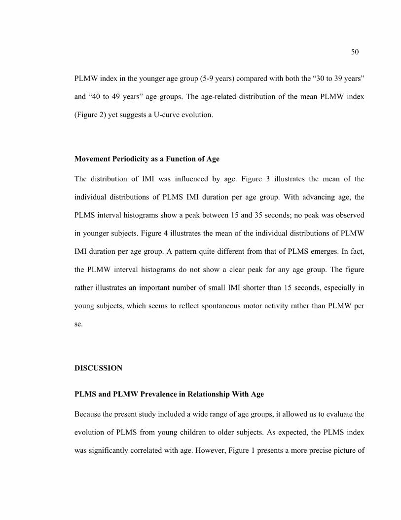
50
PLMW index in the younger age group (5-9 years) compared with both the “30 to 39 years”
and “40 to 49 years” age groups. The age-related distribution of the mean PLMW index
(Figure 2) yet suggests a U-curve evolution.
Movement Periodicity as a Function of Age
The distribution of IMI was influenced by age. Figure 3 illustrates the mean of the
individual distributions of PLMS IMI duration per age group. With advancing age, the
PLMS interval histograms show a peak between 15 and 35 seconds; no peak was observed
in younger subjects. Figure 4 illustrates the mean of the individual distributions of PLMW
IMI duration per age group. A pattern quite different from that of PLMS emerges. In fact,
the PLMW interval histograms do not show a clear peak for any age group. The figure
rather illustrates an important number of small IMI shorter than 15 seconds, especially in
young subjects, which seems to reflect spontaneous motor activity rather than PLMW per
se.
DISCUSSION
PLMS and PLMW Prevalence in Relationship With Age
Because the present study included a wide range of age groups, it allowed us to evaluate the
evolution of PLMS from young children to older subjects. As expected, the PLMS index
was significantly correlated with age. However, Figure 1 presents a more precise picture of
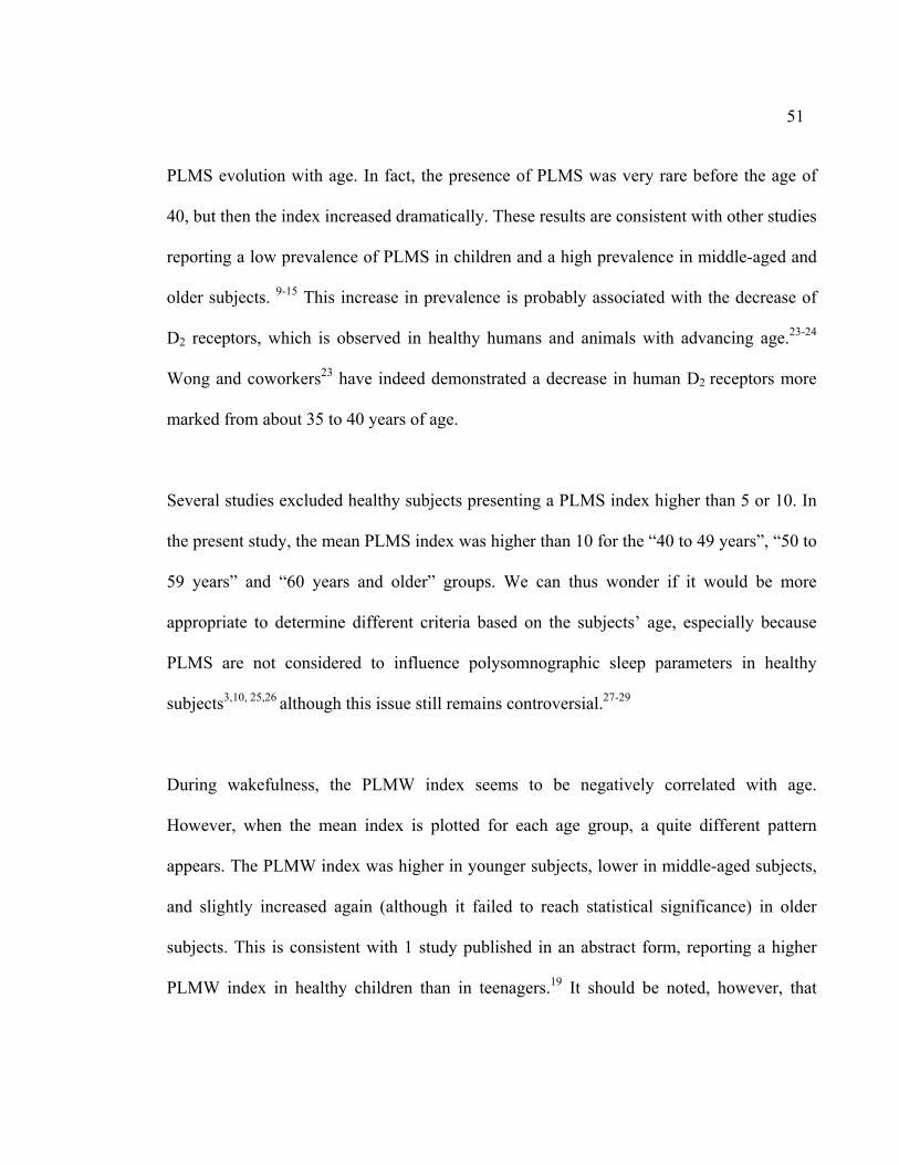
51
PLMS evolution with age. In fact, the presence of PLMS was very rare before the age of
40, but then the index increased dramatically. These results are consistent with other studies
reporting a low prevalence of PLMS in children and a high prevalence in middle-aged and
older subjects. 9-15 This increase in prevalence is probably associated with the decrease of
D2 receptors, which is observed in healthy humans and animals with advancing age.23-24
Wong and coworkers23 have indeed demonstrated a decrease in human D2 receptors more
marked from about 35 to 40 years of age.
Several studies excluded healthy subjects presenting a PLMS index higher than 5 or 10. In
the present study, the mean PLMS index was higher than 10 for the “40 to 49 years”, “50 to
59 years” and “60 years and older” groups. We can thus wonder if it would be more
appropriate to determine different criteria based on the subjects’ age, especially because
PLMS are not considered to influence polysomnographic sleep parameters in healthy
subjects3,10, 25,26 although this issue still remains controversial.27-29
During wakefulness, the PLMW index seems to be negatively correlated with age.
However, when the mean index is plotted for each age group, a quite different pattern
appears. The PLMW index was higher in younger subjects, lower in middle-aged subjects,
and slightly increased again (although it failed to reach statistical significance) in older
subjects. This is consistent with 1 study published in an abstract form, reporting a higher
PLMW index in healthy children than in teenagers.19 It should be noted, however, that
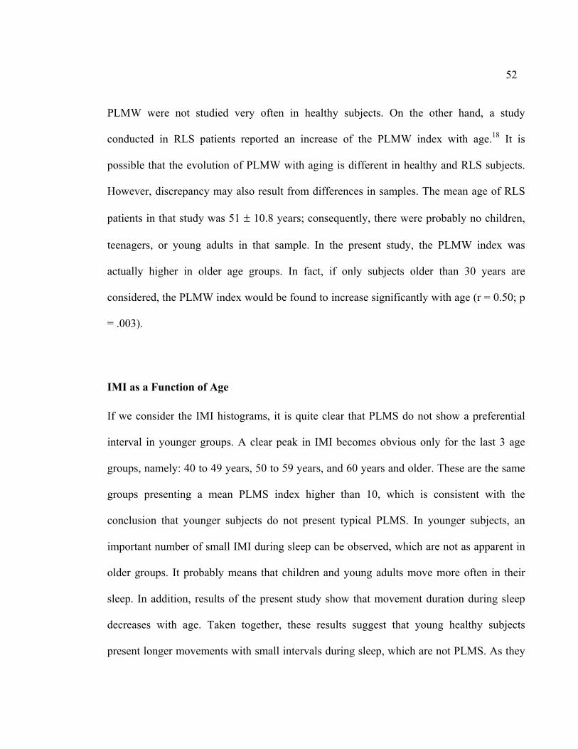
52
PLMW were not studied very often in healthy subjects. On the other hand, a study
conducted in RLS patients reported an increase of the PLMW index with age.18 It is
possible that the evolution of PLMW with aging is different in healthy and RLS subjects.
However, discrepancy may also result from differences in samples. The mean age of RLS
patients in that study was 51 ± 10.8 years; consequently, there were probably no children,
teenagers, or young adults in that sample. In the present study, the PLMW index was
actually higher in older age groups. In fact, if only subjects older than 30 years are
considered, the PLMW index would be found to increase significantly with age (r = 0.50; p
= .003).
IMI as a Function of Age
If we consider the IMI histograms, it is quite clear that PLMS do not show a preferential
interval in younger groups. A clear peak in IMI becomes obvious only for the last 3 age
groups, namely: 40 to 49 years, 50 to 59 years, and 60 years and older. These are the same
groups presenting a mean PLMS index higher than 10, which is consistent with the
conclusion that younger subjects do not present typical PLMS. In younger subjects, an
important number of small IMI during sleep can be observed, which are not as apparent in
older groups. It probably means that children and young adults move more often in their
sleep. In addition, results of the present study show that movement duration during sleep
decreases with age. Taken together, these results suggest that young healthy subjects
present longer movements with small intervals during sleep, which are not PLMS. As they
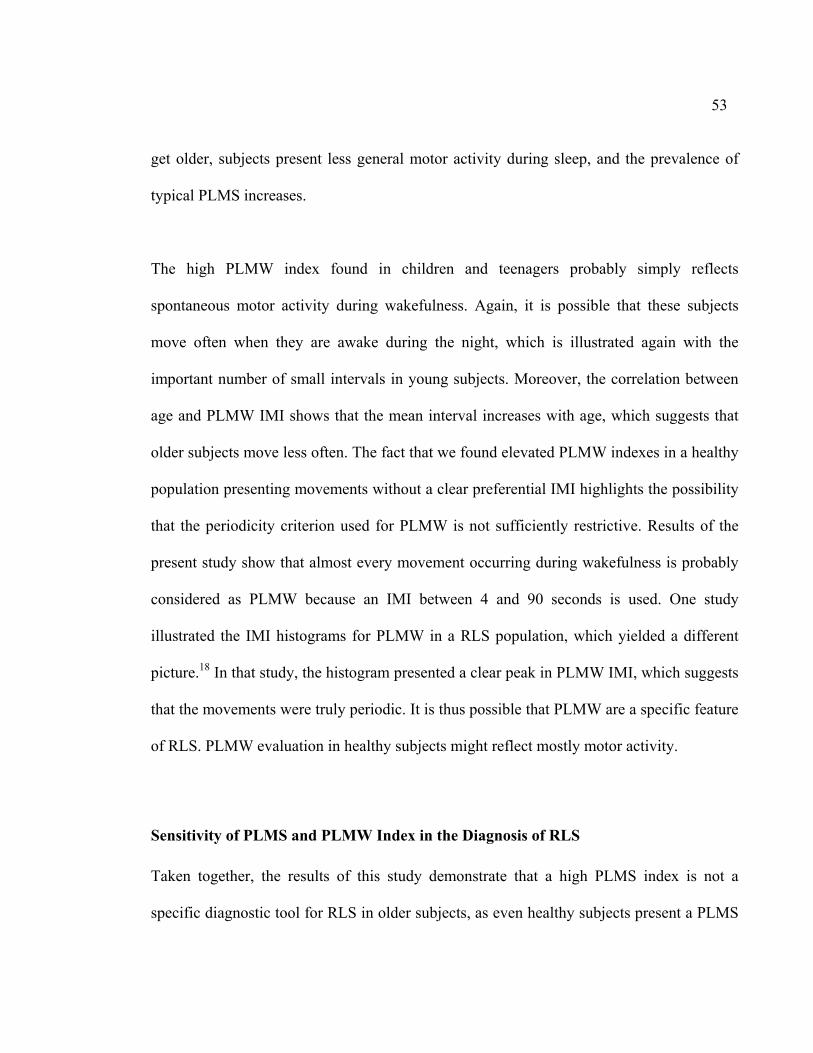
53
get older, subjects present less general motor activity during sleep, and the prevalence of
typical PLMS increases.
The high PLMW index found in children and teenagers probably simply reflects
spontaneous motor activity during wakefulness. Again, it is possible that these subjects
move often when they are awake during the night, which is illustrated again with the
important number of small intervals in young subjects. Moreover, the correlation between
age and PLMW IMI shows that the mean interval increases with age, which suggests that
older subjects move less often. The fact that we found elevated PLMW indexes in a healthy
population presenting movements without a clear preferential IMI highlights the possibility
that the periodicity criterion used for PLMW is not sufficiently restrictive. Results of the
present study show that almost every movement occurring during wakefulness is probably
considered as PLMW because an IMI between 4 and 90 seconds is used. One study
illustrated the IMI histograms for PLMW in a RLS population, which yielded a different
picture.18 In that study, the histogram presented a clear peak in PLMW IMI, which suggests
that the movements were truly periodic. It is thus possible that PLMW are a specific feature
of RLS. PLMW evaluation in healthy subjects might reflect mostly motor activity.
Sensitivity of PLMS and PLMW Index in the Diagnosis of RLS
Taken together, the results of this study demonstrate that a high PLMS index is not a
specific diagnostic tool for RLS in older subjects, as even healthy subjects present a PLMS
54
index higher than 10. However, for children, teenagers, and young adults, a high PLMS
index is quite rare and could be used as a strong supportive feature for a diagnosis of RLS.
On the other hand, the PLMW index alone seems less useful as a tool for diagnosing RLS.
In the present study, high PLMW indexes were observed in all age groups, in the absence
of typical IMI.
Limitations and Conclusion
The present study has a few limitations that are important to mention. First, the number of
subjects per age group is modest. Increasing the number of subjects would allow more
robust statistical analyses and trend analyses. Second, the uneven men-to-women ratios
between the youngest and oldest age group could potentially influence the data. Adjusting
for a similar men-to-women ratio per age group would also allow sex-specific analyses.
Finally, the present study assessed preferential intermovement intervals of PLMS and
PLMW rather than periodicity per se. In order to rigorously study PLM periodicity, a
different study design (e.g., autocorrelation, entropy) with only subjects with high PLMS or
PLMW indexes should be used. Since the first objective of the paper was to investigate the
prevalence of PLMS and PLMW in normal subjects of different age groups, subjects with
few or no periodic leg movements were included in the present study.
In conclusion, it becomes clear that the periodicity evaluation is an important feature of
periodic-movement calculation during sleep and wakefulness. In fact, if only the index is

55
taken into account, important information is missing, and it becomes difficult to
discriminate spontaneous motor activity from periodic movements. Finally, it could be
appropriate to define different periodicity criteria in different age groups in order to better
identify movements that are truly PLMS and PLMW.
56
REFERENCES
1. Lugaresi E, Tassinari CA, Coccagna D, Ambrosetto C. Particularités cliniques et
polygraphiques du syndrome d’impatience des membres inférieurs. Rev Neurol (Paris),
1965;113:545-55.
2. Coleman RM, Pollak CP, Weitzman ED. Periodic movements in sleep (Nocturnal
myoclonus) : Relation to sleep disorders. Ann Neurol 1980;8:416-21.
3. Mendelson WB. Are periodic leg movements associated with clinical sleep disturbance?
Sleep 1996;19:219-23.
4. Montplaisir J, Michaud M, Denesle R, Gosselin A. Periodic leg movements are not more
prevalent in insomnia or hypersomnia but are specifically associated with sleep disorders
involving a dopaminergic impairment. Sleep Med 2000;1:163-7.
5. Montplaisir J, Whittom S, Michaud M, Dang-Vu T, Dauvilliers Y, Rompré S. Periodic
leg movements during sleep and waking in narcolepsy. Neurology 2005;64(Suppl.):A43.
6. Fantini ML, Michaud M, Gosselin N, Lavigne G, Montplaisir J. Periodic leg movements
in REM sleep behavior disorder and related autonomic and EEG activation. Neurology
2002;59:1889-94.
7. Fry JM, DiPhillipo MA, Pressman MR. Periodic leg movements in sleep following
treatment of obstructive sleep apnea with nasal continuous positive airway pressure. Chest
1989;96:89-91.
57
8. Carelli G, Krieger J, Calvi-Gries F, Macher JP. Periodic limb movements and obstructive
sleep apneas before and after continuous positive airway treatment. J Sleep Res
1999;8:211-6.
9. Bixler EO, Kales A, Vela-Bueno A, Jacoby JA, Scarone S, Soldatos CR. Nocturnal
myoclonus and nocturnal myoclonic activity in the normal population. Res Commun Chem
Pathol Pharmacol 1982;36:129-40.
10. Carrier J, Frenette S, Monplaisir J, Paquet J, Drapeau C, Morettini J. Effects of periodic
leg movements during sleep in middle-aged subjects without sleep complaints. Mov Disord
2005;20:1127-32.
11. Arens R, Wright B, Elliott J, et al. Periodic limb movements in sleep in children with
Williams syndrome. J Pediatr 1998;133:670-4.
12. Ancoli-Israel S, Kripke DF, Mason W, Kaplan OJ. Sleep apnea and periodic
movements in an aging sample. J Gerontol 1985;40:419-25.
13. Mosko SS, Dickel MJ, Paul T, et al. Sleep apnea and sleep-related periodic leg
movements in community resident seniors. J Am Geriatr Soc 1988;36:502-8.
14. Dickel MJ, Mosko SS. Morbidity cut-offs for sleep apnea and periodic leg movements
in predicting subjective complaints in seniors. Sleep 1990;13:155-66.
15. Ancoli-Israel S, Kripke DF, Klauber MR, Mason WJ, Fell R, Kaplan O. Periodic limb
movements in sleep in community-dwelling elderly. Sleep 1991;14:496-500.
58
16. Montplaisir J, Boucher S, Poirier G, Lavigne G, Lapierre O, Lesperance P. Clinical,
polysomnographic, and genetic characteristics of restless legs syndrome: a study of 133
subjects diagnosed with new standard criteria. Mov Disord 1997;12:61-5.
17. Michaud M, Poirier G, Lavigne G, Montplaisir J. Restless legs syndrome: scoring
criteria for leg movements recorded during the suggested immobilization test. Sleep Med
2001;2:317-21.
18. Nicolas A, Michaud M, Lavigne G, Montplaisir J. The influence of sex, age and
sleep/wake state on characteristics of periodic leg movements in restless legs syndrome
patients. Clin Neurophysiol 1999;110:1168-74.
19. Adam B, Michaud M, Laberge L, Lavigne G, Montplaisir J. Periodic leg movements
during sleep in normal teenagers and young adults. Sleep 2002;25:A490-1.
20. Coleman RM. Periodic movements in sleep (nocturnal myoclonus) and restless legs
syndrome. In: Guilleminault C, ed. Sleeping and Waking Disorders: Indications and
Techniques. Menlo Park: Addison-Wesley; 1982:265-295.
21. American Sleep Disorders Association. Recording and scoring leg movements. The
atlas task force. Sleep 1993;16:749-59.
22. Rechtschaffen A, Kales A. A Manual of Standardized Terminology, Techniques and
Scoring System for Sleep Stages of Human Subjects. Los Angeles: Brain Information
Service;1968.
59
23. Wong DF, Wagner HN, Dannals RF, et al. Effects of age on dopamine and serotonin
receptors measured by positron tomography in the living human brain. Science
1984;226:1393-96.
24. Zhang Z, Andersen A, Grondin R, et al. Pharmacological MRI mapping of age-
associated changes in basal ganglia circuitry of awake rhesus monkeys. Neuroimage
2001;14:1159-67.
25. Youngstedt SD, Kripke DF, Klauber MR, Sepulveda, RS, Mason WJ. Periodic leg
movements during sleep and sleep disturbances in elders. J Gerontol 1998;53A:M391-4.
26. Karadeniz D, Ondze B, Besset A, Billiard M. Are periodic leg movements during sleep
(PLMS) responsible for sleep disruption in insomnia patients. Eur J Neurol 2000;7:331-6.
27. Saskin P, Moldofsky H, Lue FA. Periodic movements in sleep and sleep-
wake complaint. Sleep 1985;8:319-24.
28. Bastuji H, Garcia-Larrea L. Sleep/wake abnormalities in patients with periodic leg
movements during sleep: factor analysis on data from 24-hour ambulatory polygraphy. J
Sleep Res 1999;8:217-23.
29. Hilbert J, Mohsenin V. Can periodic limb movement disorder be diagnosed
without polysomnography? A case-control study. Sleep Med 2003;4:35-41.
60
FIGURE LEGENDS
Figure 1—Mean periodic leg movements during sleep (PLMS) index for healthy subjects
according to 7 age groups (5-9 y, 10-19 y, 20-29 y, 30-39 y, 40-49 y, 50-59 y, and 60 y and
older). Vertical bars represent the SEM.
Figure 2—Mean periodic leg movements during wakefulness (PLMW) index for healthy
subjects according to 7 age groups (5-9 y, 10-19 y, 20-29 y, 30-39 y, 40-49 y, 50-59 y, and
60 y and older). Vertical bars represent the SEM.
Figure 3—Duration of mean periodic leg movements during sleep (PLMS) intermovement
intervals (IMI) among 7 age groups (5-9 y, 10-19 y, 20-29 y, 30-39 y, 40-49 y, 50-59 y, and
60 y and older). The distribution of IMI duration is calculated using a 1 second bin-width.
Vertical bars represent the SD.
Figure 4—Duration of mean periodic leg movements during wakefulness (PLMW)
intermovement intervals (IMI) among 7 age groups (5-9 y, 10-19 y, 20-29 y, 30-39 y, 40-49
y, 50-59 y, and 60 y and older). The distribution of IMI duration is calculated using a 1
second bin-width. Vertical bars represent the SD.
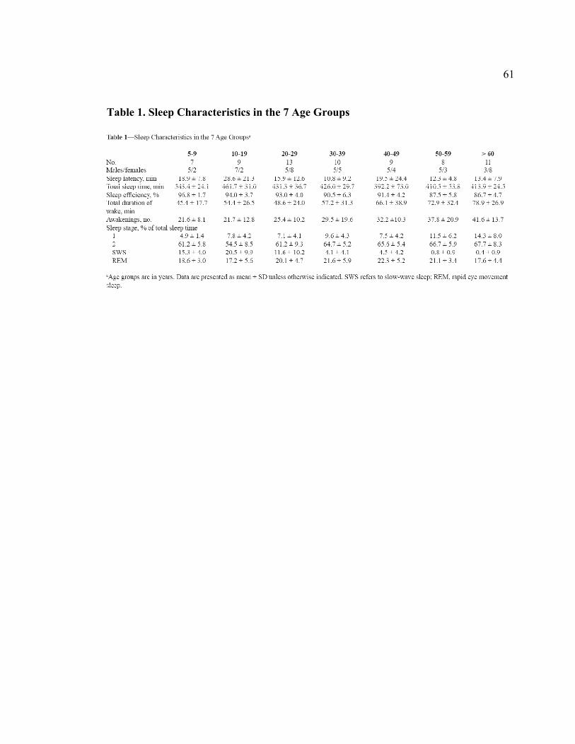
61
Table 1. Sleep Characteristics in the 7 Age Groups
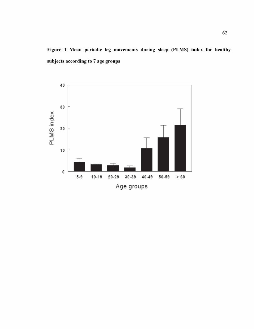
62
Figure 1 Mean periodic leg movements during sleep (PLMS) index for healthy
subjects according to 7 age groups
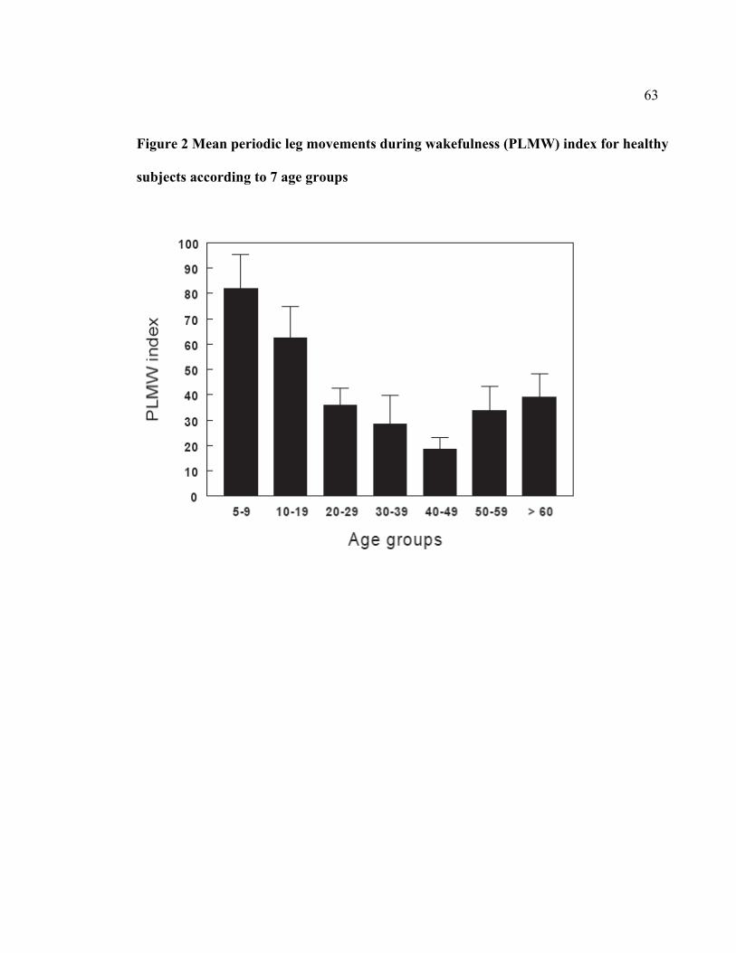
63
Figure 2 Mean periodic leg movements during wakefulness (PLMW) index for healthy
subjects according to 7 age groups
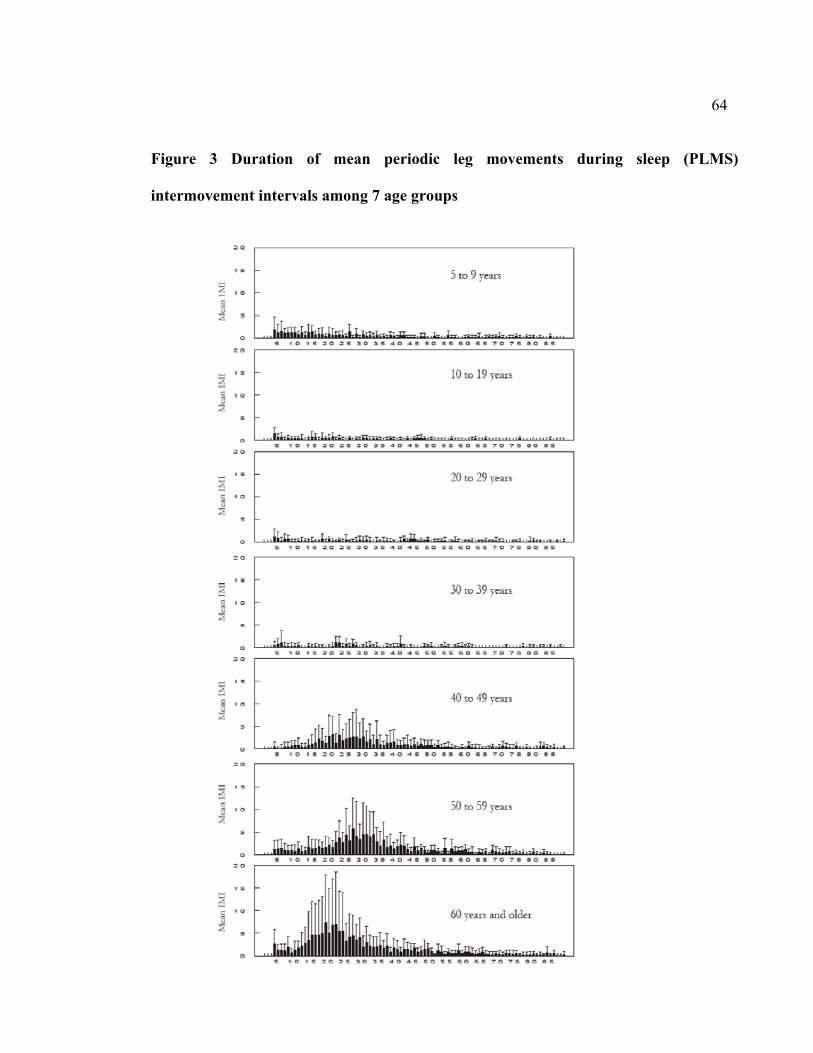
64
Figure 3 Duration of mean periodic leg movements during sleep (PLMS)
intermovement intervals among 7 age groups
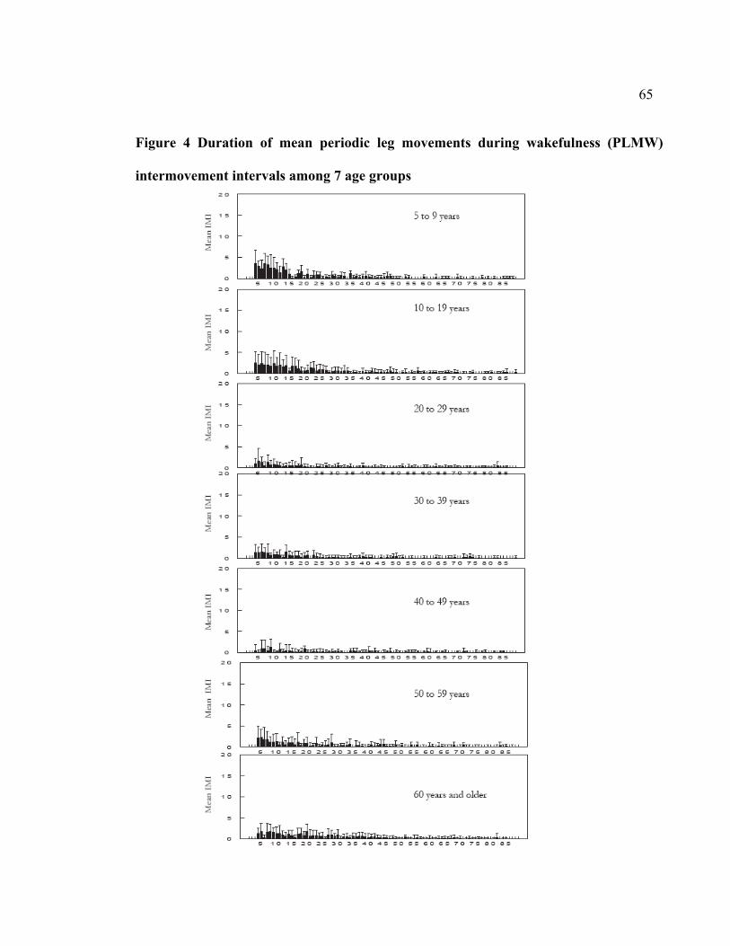
65
Figure 4 Duration of mean periodic leg movements during wakefulness (PLMW)
intermovement intervals among 7 age groups
66
2.2. Deuxième article
Periodic leg movements during sleep and wakefulness in narcolepsy
Yves Dauvilliers1, Marie-Hélène Pennestri2, Dominique Petit2, Thanh Dang-Vu3, Gilles
Lavigne2, Jacques Montplaisir2
1 Department of Neurology, Hôpital Gui de Chauliac, INSERM 0361, Université de
Montpellier, France
2 Sleep Disorders Center, Hôpital du Sacré-Cœur, Université de Montréal, Canada
3Department of Neurology, Université de Liège, Belgium
Article publié dans : Journal of Sleep Research (2007) 16, 333-339

67
Contribution des auteurs
Yves Dauvilliers : Élaboration du protocole, recueil des données, analyses des données,
interprétation des résultats, rédaction du manuscrit
Marie-Hélène Pennestri : Recueil des données, analyses des données, analyses
statistiques, interprétation des résultats, participation à la rédaction du manuscrit, révision
du manuscrit
Dominique Petit : Interprétation des résultats, participation à la rédaction du manuscrit,
révision du manuscrit
Thanh Dang-Vu : Recueil des données, analyses des données
Gilles Lavigne : Interprétation des résultats, révision du manuscrit
Jacques Montplaisir : Élaboration du protocole, interprétation des résultats, participation à
la rédaction du manuscrit, révision du manuscrit
68
ABSTRAT
The objectives of the study were to measure the prevalence of periodic leg movements
during NREM and REM sleep (PLMS) and while awake (PLMW) and to assess the impact
of PLMS on nocturnal sleep and daytime functioning in patients with narcolepsy. One
hundred and sixty-nine (169) patients with narcolepsy and 116 normal controls matched for
age and gender were included. Narcoleptics with high and low PLMS indices were
compared to assess the impact of PLMS on sleep and Multiple Sleep Latency Test (MSLT)
variables. More narcoleptics than controls had a PLMS index greater than 5 per hour of
sleep (67% versus 37%) and an index greater than 10 (53% versus 21%). PLMS indices
were higher both in NREM and REM sleep in narcoleptic patients, but the between-group
difference was greater for REM sleep. A significant increase of PLMS index was also
found with aging in both narcoleptic patients and controls. PLMW indices were also
significantly higher in narcoleptic patients. Patients with an elevated index of PLMS had a
higher percentage of stage 1 sleep, a lower percentage of REM sleep, a lower REM
efficiency and a shorter MSLT latency. The present study demonstrates a high frequency of
PLMS and PLMW in narcolepsy, an association between the presence of PLMS and
measures of REM sleep and daytime functioning disruption. These results suggest that
PLMS represent an intrinsic feature of narcolepsy.
Keywords: age, multiple sleep latency test, narcolepsy, non-rapid eye movement sleep,
periodic leg movements in sleep, periodic leg movements while awake, rapide eye
movement sleep
69
INTRODUCTION
Narcolepsy is characterized by the presence of excessive daytime sleepiness and abnormal
REM sleep manifestations including cataplexy and sleep paralysis (REM sleep atonia
intruding into wakefulness), impaired maintenance of REM sleep atonia, and dream
imagery intruding into wakefulness (e.g. hypnagogic and hypnopompic hallucinations)
(Dauvilliers et al., 2003). Major recent findings revealed that deficient hypocretin
transmission causes narcolepsy (Dauvilliers et al., 2003; Peyron et al., 2000; Mignot et al.,
2002). As hypocretin system is involved in thalamocortical arousal pathways, an alteration
in arousals systems in narcolepsy with modifications of cerebral cortex excitability has
been suggested (Sakurai 2005; Terzano et al., 2006; Oliviero et al., 2005). Indeed, night-
time sleep of patients with narcolepsy was remarkable for its instability with frequent stage
shifts and arousals (Dauvilliers et al., 2003, Dauvilliers et al., 2004; Montplaisir et al., 1978)
and increased motor activity in sleep (Wittig et al., 1983; Montplaisir et al., 2000; Mosko et
al., 1984; van den Hoed et al., 1981) such as periodic leg movements in sleep (PLMS).
PLMS are best defined as rhythmical extension of the big toe and dorsiflexion of the ankle
with occasional flexion of the knee and hip (Coleman et al., 1980; Lugaresi et al., 1986).
PLMS are frequently associated with arousals and awakenings and they occur in a wide
range of sleep disorders including restless legs syndrome (RLS) (Lugaresi et al., 1986),
REM sleep behavior disorder (RBD) (Fantini et al., 2002), obstructive sleep apnea
syndrome (OSAS) (Fry et al., 1989), insomnia (Coleman et al., 1980) and hypersomnia
70
including narcolepsy. However, only small or uncontrolled studies had previously reported
the presence of PLMS in patients with narcolepsy (Wittig et al., 1983; Boivin et al., 1989;
Montplaisir et al., 2000; Mosko et al., 1984; Baker et al., 1986; van den Hoed et al., 1981).
PLMS are also reported in subjects without any sleep complaint and while they are rare in
young individuals, they are relatively common in the old (Carrier et al., 2005). In normal
subjects as in RLS patients, PLMS are more frequent in stages 1 and 2 NREM sleep.
Changes in PLMS with age and stages of sleep have never been specifically studied in
narcolepsy.
Periodic leg movements may also occur while awake (PLMW). Little is known about
PLMW in non-RLS subjects; in RLS patients, PLMW index increases with age (Nicolas et
al., 1999). By contrast, a recent study reported a different PLMW distribution in normal
controls where PLMW index was high in children and young adults, very low in middle-
aged individuals, with a slight increase in older subjects (Pennestri et al., 2006). To our
knowledge, there is no published data on the prevalence and characteristics of PLMW in
narcolepsy.
The aims of the present study are (1) to look at the frequency of PLMS in REM and NREM
sleep and of PLMW in narcolepsy; (2) to study the PLMS and PLMW distribution with age
in comparison with sex- and age- matched normal controls; and (3) to measure the
71
functional impact of PLMS on nocturnal sleep and daytime functioning in a large sample of
narcoleptics.
METHODS
Subjects
One hundred and sixty-nine (169) narcoleptic patients, (89 males and 80 females, mean age
at 45.8 ± 14.2, range from 18 to 82 years old) were included in the study. Inclusion criteria
for narcolepsy were the presence of both excessive daytime sleepiness and cataplexy, the
presence of HLA-DR2 antigen and of at least one sleep onset REM period (SOREMP)
during the Multiple Sleep Latency Test (MSLT). The frequency of cataplectic attacks was
assessed on a scale (Dauvilliers et al., 2001; Dauvilliers et al., 2004) from 1 to 5 where 1
represents one or fewer cataplectic attack per year, 2 represents more than one cataplectic
attack per year but fewer than one per month, 3 represents more than one cataplectic attack
per month but fewer than one per week, 4 represents more than one cataplectic attack per
week but fewer than one per day and 5 represents severe cases with at least one cataplectic
attack per day. None of the patients had a diagnosis of RLS or any other condition known
to be associated with PLMS such as anemia, renal insufficiency, rheumatoid arthritis,
diabetes, polyneuropathies or RBD based on medical history and clinical examination.
None of the patients had any psychiatric disorder based on the DSM-IV criteria. Subjects
with an index of respiratory events (apneas + hypopneas) greater than 10 were excluded
from the study. Narcoleptic patients were not taking psychostimulants for at least 1 week
72
prior to the sleep laboratory recording, or anticataplectic medications (tricyclics or 5HT
reuptake inhibitors) or any other medication known to influence sleep or motor activity for
at least 1 month prior to the sleep recording. Patients were divided into five age groups,
where group 1 was 18-29 years old, group 2 was 30-39 yrs, group 3 was 40-49 yrs, group 4
was 50-59 yrs, and group 5 was older than 60 years, at the time of the sleep evaluation.
One hundred and sixteen normal subjects matched for age and gender (59 males and 57
females, mean age at 45.9 ± 13.7, range from 18 to 76 years old) were recruited from the
Montreal community by newspaper advertisement. Exclusion criteria for the normal
controls were the same as for narcoleptics. In addition, none of the controls had any
symptoms of the narcoleptic tetrad.
Procedures
All subjects underwent one night of polysomnographic recording in the sleep laboratory. In
patients with narcolepsy, all-night recording was followed by the MSLT consisting of five
naps scheduled at 2-h intervals starting at 10:00 hours (Carskadon et al., 1986). Sleep was
recorded and scored based on the standard method (Rechtschaffen and Kales 1968). The
following polysomnographic variables were measured: total sleep time, sleep latency, sleep
efficiency, duration of wake after sleep onset, number of awakenings, percentage of stages
1, 2, 3, 4 and REM sleep, REM latency and REM efficiency. The mean sleep latency and
number of SOREMP on the MSLT was also calculated. REM efficiency is defined by the
73
percentage of time spent in REM sleep over the duration of the REM period (which can
contain epochs of wakefulness or of other sleep stages).
Periodic leg movements in sleep were scored according to Coleman’s criteria (Coleman
1982): only movements lasting 0.5-5 s, separated by intervals of 4-90 s and occurring in
series of at least four consecutive movements were counted. Indices of PLMS were
calculated separately in REM and NREM sleep, but only in subjects with a PLMS index
greater than 5 per hour of sleep in order to avoid biased indices. The same criteria were
used to score PLMW during the nocturnal recording, but allowing for movement duration
up to 10 s. The amplitude criterion for both PLMS and PLMW was at least 25% of the
EMG potential recorded at the time of a voluntary dorsal extension movement of the foot
performed prior to nocturnal PSG recording.
Microarousals (MA) were scored according to the criteria developed by a task force of the
ASDA (American Sleep Disorders Association, 1992). A PLMS was considered associated
with a MA if the MA started within 2 s before or after the onset of PLMS.
Data analysis
Statistical analyses were performed to compare patients with narcolepsy to normal controls
and to compare narcoleptic patients with high and low PLMS indices using Student t-tests
or non-parametric equivalents. Two-way analyses of variance (ANOVAS) with two
74
independent factors (group: narcoleptics and controls; age group: 1-5), were used to
measure the effect of age on the frequency of PLMS, PLMS in NREM and in REM sleep
and PLMW. Contrast analyses were carried out with Tukey HSD test. Pearson’s correlation
coefficients were also calculated to assess the relationship between PLMS, PLMW and age.
RESULTS
Prevalence of PLMS and PLMW
Results presented in Table 1 show that patients with narcolepsy had significantly more
PLMS, and more PLMW than control subjects. Between-group comparison clearly revealed
an overall higher index of PLMS in narcoleptic population (F [4,275] =18.5, p<0.00003). A
greater proportion of patients with narcolepsy had a PLMS index greater than 5 than
controls (67% versus 37%) (p<0.0001) or a PLMS index greater than 10 (53% versus 21%)
(p<0.0001). In addition, PLMS associated with MAs were more frequent in the narcoleptic
group (Table 1). The distribution of PLMS per thirds of night was also investigated in both
groups. There was no third of night effect as the PLMS index decreased across the night for
controls but not in patients with narcolepsy yielding a nearly significant interaction
(p=0.06).
The mean PLMW indices was significantly higher in patients with narcolepsy than in
controls (F [4,255] =8.4, p<0.004). In addition, a positive correlation was noted between
75
PLMS and PLMW indices in narcoleptics (r=0.49; p<0.000001) as well as in controls
(r=0.29, p<0.001).
Frequency of PLMS in REM and NREM sleep
PLMS indices in both NREM and REM sleep were higher in narcoleptics than in controls;
however, the difference was more obvious in REM sleep (Table 1). Post-hoc analysis (t-
test) revealed that PLMS indices in REM and NREM sleep were significantly different in
controls (3.2 ± 7.4 versus 8.2 ± 15.1, p=0.0013), but not in the narcoleptic population (18.3
± 33.6 vs 21.2 ± 31.0; NS). In addition, two-thirds of patients with narcolepsy had PLMS
index in REM sleep greater than 5 compared to only 27.5% of the controls (p = 0.00015).
Finally, we noted that gender had no significant effect on PLMW, total PLMS, and PLMS
in NREM and REM sleep in narcoleptics or controls.
Effects of age on PLMS and PLMW
A positive correlation was found between PLMS index and age in the entire narcoleptic
population (r= 0.35, p=0.000003) and in normal controls (r= 0.39, p<0.00002). The group
by age group ANOVA revealed both group F [1,275] = 18.5, p = 0.00002 and age group F
[4,275] = 6.8, p = 0.00003) effects. Tukey HSD post-hoc analysis showed that subjects
under age 39 years (age groups 1 and 2) had a lower PLMS index than subjects above 50
years (age groups 4 and 5) (p<0.025). In addition, subjects from group 3 showed a lower
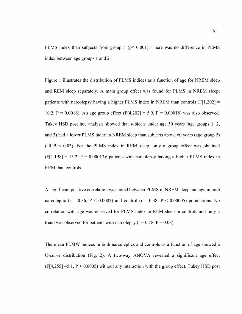
76
PLMS index than subjects from group 5 (p≤ 0.001). There was no difference in PLMS
index between age groups 1 and 2.
Figure 1 illustrates the distribution of PLMS indices as a function of age for NREM sleep
and REM sleep separately. A main group effect was found for PLMS in NREM sleep;
patients with narcolepsy having a higher PLMS index in NREM than controls (F[1,202] =
10.2, P = 0.0016). An age group effect (F[4,202] = 5.9, P = 0.00018) was also observed.
Tukey HSD post hoc analysis showed that subjects under age 50 years (age groups 1, 2,
and 3) had a lower PLMS index in NREM sleep than subjects above 60 years (age group 5)
(all P < 0.03). For the PLMS index in REM sleep, only a group effect was obtained
(F[1,198] = 15.2, P = 0.00013); patients with narcolepsy having a higher PLMS index in
REM than controls.
A significant positive correlation was noted between PLMS in NREM sleep and age in both
narcoleptic (r = 0.36, P < 0.0002) and control (r = 0.38, P < 0.00005) populations. No
correlation with age was observed for PLMS index in REM sleep in controls and only a
trend was observed for patients with narcolepsy (r = 0.18, P = 0.08).
The mean PLMW indices in both narcoleptics and controls as a function of age showed a
U-curve distribution (Fig. 2). A two-way ANOVA revealed a significant age effect
(F[4,255] =5.1, P ≤ 0.0005) without any interaction with the group effect. Tukey HSD post
77
hoc analysis showed that subjects from age group 1 had a significantly higher PLMW index
than subjects from age group 3 (P ≤ 0.008). Subjects from age groups 2 and 3 had a
significantly lower PLMW index than subjects from age group 5 (P ≤ 0.02).
Functional significance of PLMS
To assess the influence of PLMS on nocturnal sleep parameters, sleep latency on the MSLT
and severity of cataplexy, patients with narcolepsy were divided in two subgroups 61
patients matched for age and gender with a high (≥ 10) and a low (<10) PLMS index
respectively. Patients with high PLMS indices have also higher PLMW. Table 2 also shows
slight differences in sleep architecture: a higher percentage of stage 1 sleep, a lower
percentage of REM sleep, a lower REM efficiency, and a shorter latency on the MSLT in
the high PLMS index subgroup. No difference was found for the frequency of cataplectic
attacks. A negative correlation was found between PLMS index in REM sleep and REM
sleep efficiency (r= - 0.26, p= 0.043).
DISCUSSION
Prevalence of PLMS and PLMW in narcolepsy
The results of the present study show in a large cohort of subjects, that narcoleptic patients
with cataplexy have a higher frequency of PLMS associated or not with MAs compared
with age-matched normal controls. This is consistent with results of previous uncontrolled
studies, performed in a limited number of patients, who reported the presence of PLMS in
78
narcolepsy (Wittig et al., 1983; Boivin et al., 1989; Mosko et al., 1984; Baker et al., 1986;
van den Hoed et al., 1981) and with one publication from our laboratory comparing 20
narcoleptic patients to 20 age-matched controls (Montplaisir et al., 2000). However this
study was a controversial paper for which no methodology or statistical assessment was
reported.
Increased frequency of PLMS has also been reported in other specific sleep disorders,
namely RLS and idiopathic RBD, two conditions where there are evidence, as in
narcolepsy, of central dopaminergic (DA) transmission impairment (Montplaisir et al.,
2000; Fantini et al., 2002; Nightingale et al., 2005; Mayer and Meier-Ewert, 1993),
therefore, DA abnormalities may be responsible for the presence of PLMS in these three
conditions. The DA hypothesis of PLMS is supported by pharmacological and to some
extent by brain imaging and neuroendocrine studies. It is well known that the treatment of
RLS-PLMS patients with levodopa or with DA agonists (Boivin et al., 1989; Wetter et al.,
1999; Benes et al., 2004; Montplaisir et al., 1999; Allen et al., 2004; Stiasny et al., 2001;
Boivin et al., 1993) not only alleviates symptoms of RLS in the waking state but also
strongly suppresses PLMS. Similar results were obtained in two studies of narcoleptic
patients where levodopa decreased the mean PLMS index from 21.3 to 9.9 (Boivin et al.,
1989) and bromocriptine decreased the mean PLMS index from 43.4 to 15.9 (Boivin et al.,
1993). Brain imaging studies, conducted both in SPECT and in PET partially support this
hypothesis; some studies (Michaud et al., 2002 ; Turjansky et al., 1999; Staedt et al., 1993)
79
showed a decreased binding to D2 postsynaptic receptors in patients with RLS-PLMS and
patients with PLMS alone, a result that was not found in two other SPECT studies
(Eisensehr et al., 2001; Tribl et al., 2004). Results of a recent neuro-endocrine study
(Garcia-Borreguero et al., 2004) suggested a hypersensitivity of DA receptors at night in
the physiopathology of PLMS in patients with both RLS-PLMS.
Modifications in DA pathways were also reported in human narcolepsy-cataplexy with
lowered metabolism of dopamine (Faull et al., 1983), genetic differences (through catechol-
O-methyltransferase gene) (Dauvilliers et al., 2001b), and altered striatal postsynaptic D2
receptors SPECT findings (Eisensehr et al., 2003). DA abnormalities are critical
downstream mediators of the hypocretin deficiency (Dauvilliers et al., 2003; Sakurai 2005;
Nishino et al., 2000). Dysfunctions in the hypocretin/DA system are likely to be the most
important mechanism involved in the pathophysiology of narcolepsy, with alterations in
arousal systems but also in sleep-related motor activation with a large amount of PLMS. In
the genetically natural canine narcolepsy model, the frequency of PLMS is also important
(Okura et al., 2001). In narcoleptic dogs, PLMS are characterized by dorsi-flexion of the
ankle lasting 0.5-1.5 s and recurring at intervals of 3-20 s (Okura et al., 2001).
Prevalence of PLMS in REM and NREM sleep
This is the first study that characterizes PLMS in NREM sleep and REM sleep in a large
cohort of patients with narcolepsy. We noted a higher PLMS index in both REM and
80
NREM sleep when compared with controls, without any difference between PLMS indices
in REM and NREM sleep in the narcoleptic population. In fact, PLMS were as numerous in
REM than in NREM sleep in patients with narcolepsy in contrast to controls. This result
may be due to impaired mechanism responsible for motor inhibition during REM sleep in
narcolepsy. A similar increase in the frequency of PLMS in REM sleep was also found in
RBD, a condition associated with a loss of REM sleep muscle atonia. There are several
evidence of disrupted REM sleep motor inhibition in narcolepsy and indeed RBD is often
found in association with narcolepsy (Nightingale et al., 2005; Mayer and Meier-Ewert,
1993). In fact, it is likely that the high rate of PLMS in REM sleep (which is more prevalent
later in the night) precluded to the decrease in PLMS index across the night in narcoleptic
patients. We also found a correlation between PLMS in REM sleep and REM efficiency.
REM efficiency has been previously reported to be altered in narcolepsy (Dauvilliers et al.,
2001a; Dauvilliers et al., 2004). It is however difficult to determine whether PLMS in REM
sleep are directly responsible for the decreased REM sleep efficiency or whether both
phenomena are consequences of the REM sleep instability characteristics in this condition.
Effect of age on PLMS and PLMW
A progressive increase in the index of PLMS with age was observed independently in the
narcoleptic and control populations. However no interaction was seen, suggesting that the
same mechanism may be responsible for age effect in both groups. The reason for the age
effect on PLMS is unknown. One possibility is that age-related effect on PLMS would be
81
the result of decreased D2 receptor density that found in both animals and humans with
advancing age (Wong et al., 1984; Zhang et al ., 2001).
The age effect on PLMW was quite different with elevated values in young individuals and
the old. The distribution of PLMW in narcoleptics was the same than in normal controls.
These results taken together indicate that there is no specific PLM-related age effect in
narcolepsy.
Functional significance of PLMS in narcolepsy
Little difference was seen in NREM sleep between narcoleptic patients with or without
PLMS except for an increase in stage 1 sleep. By contrast, indicators of some REM sleep
disruption were found in narcoleptics with PLMS, e.g. a slight decrease in REM sleep
percent and REM efficiency. We believe that PLMS are part of the REM sleep dissociation
phenomena characteristic of narcolepsy with frequent shift from REM to NREM sleep and
the presence of intermediate stages of sleep where features of REM and NREM sleep are
present simultaneously (Dauvilliers et al., 2003). The negative correlation between PLMS
index and REM efficiency suggest that PLMS may be an indicator of the severity of REM
disruption in narcolepsy or alternatively may contribute to REM sleep disruption in this
condition. The increase in stage 1 NREM sleep may be seen as a consequence of this
process rather than a sign of specific NREM sleep disruption. It is also possible that, as
82
REM sleep has not been scored with a method allowing for REM sleep without atonia,
some epochs of REM sleep were scored as stage 1 sleep.
Except for specific changes in REM sleep, PLMS had little effect on nocturnal sleep
parameters like sleep efficiency, wake time after sleep onset or total sleep time. This is
quite different from what was previously reported in patients with RLS. One reason for this
discrepancy is the low index of PLMS associated with MAs in narcoleptics compared to
RLS patients (Sforza et al., 1999). A small but significant decrease of sleep latency on the
MSLT was also found in patients with narcolepsy with high PLMS index. This result
suggests that REM sleep associated with PLMS may play a role in diurnal symptoms of
narcolepsy. It could also reflect a more severe degree of the condition which could be the
origin of both more severe daytime symptoms and more sleep disruption, inducing events
such as PLMS in REM and NREM sleep. Further research will be needed to elucidate the
reasons why PLMS are less often associated with MAs in narcolepsy, and in RBD
compared to RLS patients. One possibility is that sympathetic activation would play a role
in EEG arousal associated with PLMS and that patients with narcolepsy or RBD have
dysautonomia (Fantini et al., 2002; Ferini-Strambi et al., 1997).
In summary, the present study shows a high frequency of PLMW and PLMS in both
NREM and REM sleep in patients with narcolepsy and a major increase in mean PLMS
index with age. Patients with narcolepsy also show a specific increase of PLMS during
REM sleep and provide additional evidence of REM sleep disruption in this condition.

83
These results also demonstrate an association between PLMS and daytime somnolence, as
assessed by the MSLT, in patients with narcolepsy.
84
REFERENCES
Allen, R., Becker, PM., Bogan, R., et al. Ropinirole decreases periodic leg movements
and improves sleep parameters in patients with Restless legs syndrome. Sleep
2004; 5:907-914.
American Sleep Disorders Association. EEG arousal: scoring rules and examples. Sleep
1992; 15:174-184
Baker, TL., Guilleminault, C., Nino-Murcia, G., et al. Comparative polysomnographic
study of narcolepsy and idiopathic central nervous system hypersomnia. Sleep.
1986; 9(1 Pt 2):232-42.
Benes, H., Heinrich, CR., Ueberall, MA., Kohnen, R. Long-term safety and efficacy of
cabergoline for the treatment of idiopathic restless legs syndrome: results from an
open-label 6-month clinical trial. Sleep 2004; 27:674-682
Boivin, DB., Montplaisir, J., Poirier G. The effects of L-dopa on periodic leg
movements and sleep organization in narcolepsy. Clin Neuropharmacol 1989;
12:339-345.
Boivin, DB., Lorrain, D., Montplaisir J. The effects of bromocriptine on periodic limb
movements in human narcolepsy. Neurology 1993; 43:2134-21-36
Carrier, J., Frenette, S., Montplaisir, J., Paquet, J., Drapeau, C., Morettini J. Effects of
periodic leg movements during sleep in middle-aged subjects without sleep
complaints. Mov Disord 2005; 20:1127-1132.
85
Carskadon, MA., Dement, WC., Mitler, MM., Roth, T., Westbrook, PR., Keenan S.
Guidelines for the Multiple Sleep Latency Test (MSLT): a standard measure of
sleepiness. Sleep 1986; 9:519-524.
Coleman, RM., Pollak, CP., Weitzman, ED. Periodic movements in sleep (nocturnal
myoclonus): relation to sleep disorders. Ann Neurol 1980; 8:416-421.
Coleman, RM. Periodic movements in sleep (nocturnal myoclonus) and restless legs
syndrome. In: C Guilleminault (ed), Sleeping and waking disorders: indications
and techniques. Menlo Park (CA): Addison-Wesley; 1982; 265-295.
Dauvilliers, Y., Montplaisir, J., Molinari, N., et al. Age at onset of narcolepsy in two
large populations of patients in France and Quebec. Neurology 2001; 57:2029-33.
Dauvilliers, Y., Neidhart, E., Lecendreux, M., Billiard, M., Tafti, M. MAO-A and
COMT polymorphisms and gene effects in narcolepsy. Mol Psychiatry. 2001;
6:367-72.
Dauvilliers, Y., Billiard, M., Montplaisir, J. Clinical aspects and pathophysiology of
narcolepsy. Clin Neurophysiol 2003;114: 2000-2017.
Dauvilliers, Y., Gosselin, A., Paquet, J., Touchon, J, Billiard, M, Montplaisir, J. Effect
of age on MSLT results in patients with narcolepsy-cataplexy. Neurology 2004;
62:46-50.
Eisensehr, I., Wetter, TC., Linke, R., et al. Normal IPT and IBZM SPECT in drug-naive
and levodopa-treated idiopathic restless legs syndrome. Neurology 2001; 57:1307-
1309
86
Eisensehr, I., Linke, R., Tatsch, K., et al. Alteration of the striatal dopaminergic system
in human narcolepsy. Neurology. 2003; 60:1817-9
Fantini, L., Michaud, M., Gosselin, N., et al. Periodic leg movements in REM sleep
behavior disorder and related autonomic and EEG activation. Neurology 2002;
59:1889-1894.
Faull, KF., Guilleminault, C., Berger, PA, Barchas, JD. Cerebrospinal fluid monoamine
metabolites in narcolepsy and hypersomnia. Ann Neurol. 1983; 13:258-63.
Ferini-Strambi, L., Spera, A., Oldani, A., et al. Autonomic function in narcolepsy: power
spectrum analysis of heart rate variability. J Neurol. 1997; 244:252-5
Fry, JM., DiPhillipo, MA., Pressman, MR. Periodic leg movements in sleep following
treatment of obstructive sleep apnea with nasal continuous positive airway pressure.
Chest 1989; 96:89-91.
Garcia-Borreguero, D., Larrosa, O., Granizo, JJ., de la Llave, Y., Hening, WA.
Circadian variation in neuroendocrine response to L-dopa in patients with restless
legs syndrome. Sleep 2004; 27:669-673
Lugaresi, E., Cirignotta, F., Coccagna, G, et al. Nocturnal myoclonus and restless legs
syndrome. In: S Fahn et al. (eds), Advances in Neurology, New York: Raven Press;
1986; 43:295-307.
Mayer G., Meier-Ewert K. Motor dyscontrol in sleep of narcoleptic patients (a lifelong
development?). J Sleep Res. 1993; 2:143-148
87
Michaud, M., Soucy, JP., Chabli, A., Lavigne, G., Montplaisir, J. SPECT imaging of
striatal pre- and postsynaptic dopaminergic status in restless legs syndrome. J
Neurol 2002; 249:164-170.
Mignot, E., Lammers, GJ., Ripley, B., et al. The role of cerebrospinal fluid hypocretin
measurement in the diagnosis of narcolepsy and other hypersomnias. Arch Neurol
2002; 59:1553-62.
Montplaisir, J., Billiard, M., Takahashi, S., et al. Twenty-four-hour recording in REM-
narcoleptics with special reference to nocturnal sleep disruption. Biol
Psychiatry.1978; 13:73-89
Montplaisir, J., Nicolas, A., Denesle, R., Gomez-Mancilla, B. RLS improved by
pramipexole: a double-blind randomized trial. Neurology 1999; 52: 938-943.
Montplaisir, J., Michaud, M., Denesle, R., Gosselin A. Periodic leg movements are not
more prevalent in insomnia or hypersomnia but are specifically associated with
sleep disorders involving a dopaminergic impairment. Sleep Med 2000; 1:163-
167.
Mosko, SS., Shampain, DS., Sassin, JF. Nocturnal REM latency and sleep disturbance in
narcolepsy. Sleep. 1984; 7:115-25
Nicolas, A., Michaud, M., Lavigne, G., et al The influence of sex, age and sleep/wake
state on characteristics of periodic leg movements in restless legs syndrome
patients. Clin Neurophysiol. 1999; 110:1168-74
88
Nightingale, S., Orgill, JC., Ebrahim, IO., et al. The association between narvolepsy and
REM behavior disorder (RBD) Sleep Med. 2005; 6:253-8
Nishino, S., Okura M., Mignot E. Narcolepsy: genetic predisposition and
neuropharmacological mechanisms. Sleep Med Rev. 2000 ; 4:57-99
Okura, M., Fujiki, N., Ripley, B., et al. Narcoleptic canines display periodic leg movements
during sleep. Psychiatr Clin Neurosci 2001; 55:243-244.
Oliviero, A., Della Marca, G., Tonali, PA, et al Functional involvement of cerebral
cortex in human narcolepsy. J Neurol. 2005; 252:56-61
Pennestri, MH., Shirley, W., Benoit, A. et al . PLMS and PLMW in healthy subjects as a
function of age; Prevalence and interval distribution. Sleep 2006 Epub
Peyron, C., Faraco, J., Rogers, W., et al. A mutation in a case of early onset narcolepsy
and a generalized absence of hypocretin peptides in human narcoleptic brains. Nat
Med. 2000; 6:991-7.
Rechtschaffen, A., Kales, A (editors). A manual of standardized terminology, techniques
and scoring system for sleep stages of humans subjects. BIS/BRI, UCLA, 1968. Los
Angeles.
Sakurai, T. Roles of orexin/hypocretin in regulation of sleep/wakefulness and energy
homeostasis. Sleep Med Rev. 2005; 9:231-41
Sforza, E., Nicolas, A., Lavigne, G., Gosselin, A.. Petit, D., Montplaisir, J. EEG and
cardiac activation during periodic leg movements in sleep. Neurology 1999;
52:786-791
89
Staedt, J., Stoppe, G., Kogler, A., et al. Dopamine D2 receptor alteration in patients with
periodic movements in sleep (nocturnal myoclonus). J Neural Transm Gen Sect.
1993; 93:71-74.
Stiasny, K., Wetter, TC., Winkelmann, J., et al. Long-term effects of pergolide in the
treatment of restless legs syndrome. Neurology 2001; 56:1399-1402.
Terzano, MG., Smerieri, A., Del Felice, A., Giglia F., Palomba V., Parrino L. Cyclic
alternating pattern (CAP) alterations in narcolepsy. Sleep Med. 2006 30; Epub
Tribl, GG., Asenbaum, S., Happe, S., et al. Normal striatal D2 receptor binding in
idiopathic restless legs syndrome with periodic leg movements in sleep. Nucl Med
Commun. 2004; 25:55-60
Turjansky, N., Lees, A., Brooks, DJ. Striatal dopaminergic function in restless legs
syndrome: 18F-dopa and 11C-raclopride PET. Neurology 1999; 52:932-937.
van den Hoed, J., Kraemer, H., Guilleminault, C., et al Disorders of excessive daytime
somnolence: polygraphic and clinical data for 100 patients. Sleep. 1981; 4:23-37
Wetter, TC., Stiasny, K., Winkelmann, J., et al. A randomized controlled study of
pergolide in patients with restless legs syndrome. Neurology 1999; 52:944-950.
Wittig, R,. Zorick, F., Piccione P. et al Narcolepsy and disturbed nocturnal sleep. Clin
electroencephalogr. 1983;14:130-4.
Wong, DF., Wagner, HN., Dannals, RF., et al. Effects of age on dopamine and serotonin
receptors measured by positron tomography in the living human brain. Science
1984; 226:1393-1396.

90
Zhang, Z., Andersen, A., Grondin, R., et al. Pharmacological MRI mapping of age-
associated changes in basal ganglia circuitry of awake rhesus monkeys.
Neuroimage 2001; 14:1159-1167.
91
FIGURE LEGENDS
Figure 1:
Curves of mean periodic leg movements in sleep indices distribution for patients with
narcolepsy and normal controls according to five age groups for (a) NREM sleep only and
(b) REM sleep only. Vertical bars represent the standard error of the mean.
Figure 2:
Curves of mean periodic leg movements while awake indices distribution for patients with
narcolepsy and normal controls according to five age groups. Vertical bars represent the
standard error of the mean.
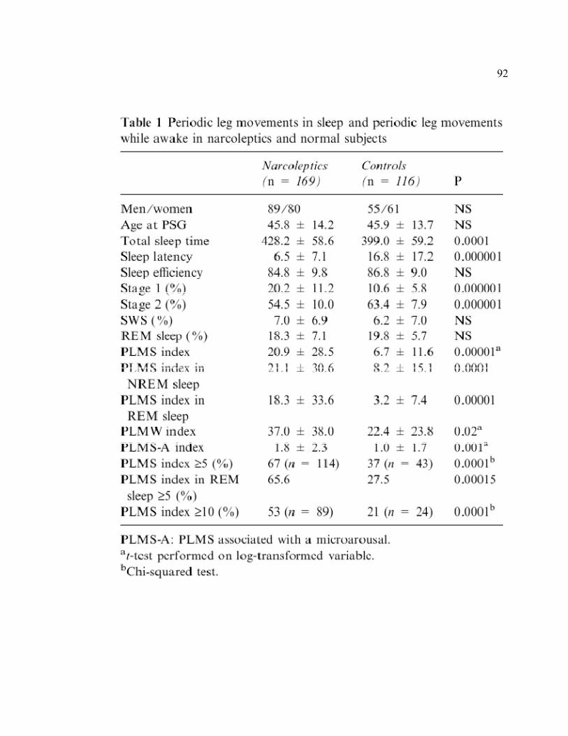
92
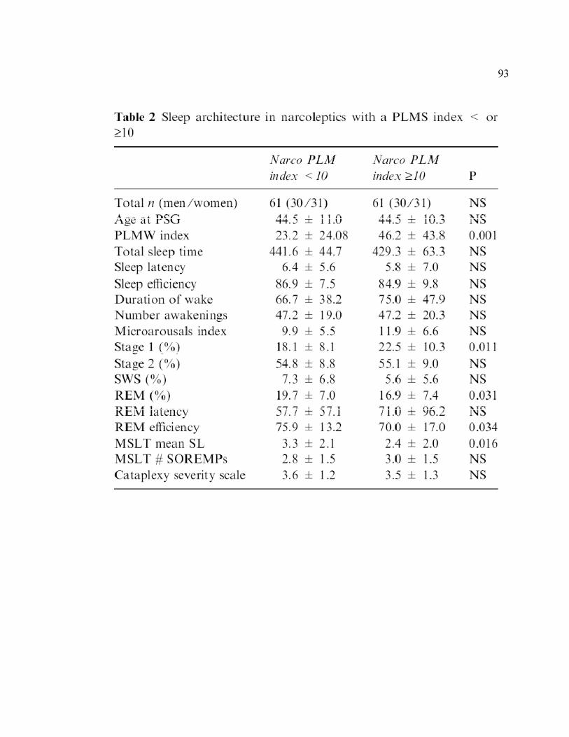
93
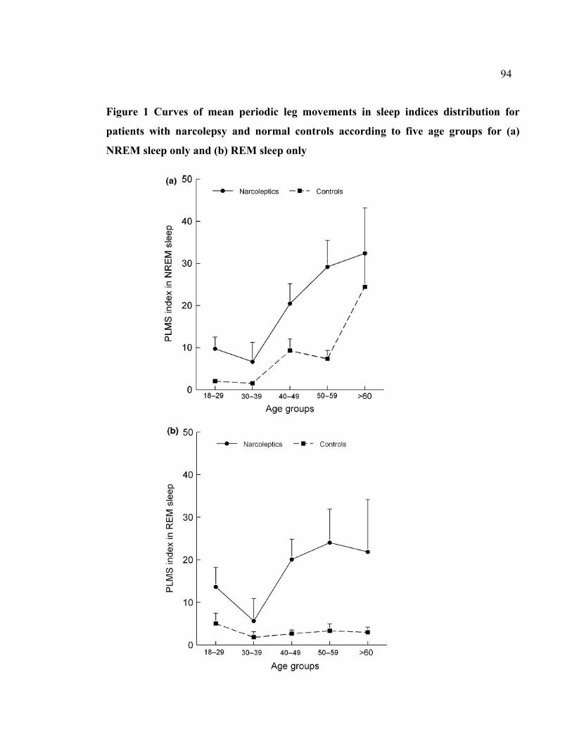
94
Figure 1 Curves of mean periodic leg movements in sleep indices distribution for
patients with narcolepsy and normal controls according to five age groups for (a)
NREM sleep only and (b) REM sleep only
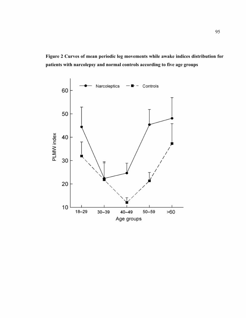
95
Figure 2 Curves of mean periodic leg movements while awake indices distribution for
patients with narcolepsy and normal controls according to five age groups
96
2.3. Troisième article
Autonomic response to periodic leg movements during sleep in narcolepsy-cataplexy
Yves Dauvilliers, MD, PhD1;Marie-Hélène Pennestri, MPs2,3; Shirley Whittom, BSc2;
Paola A Lanfranchi, MD, MSc2,4,5; Jacques Y Montplaisir, MD, PhD2,6
1Department of Neurology, National Reference Network for Orphan Diseases (Narcolepsy
and Idiopathic Hypersomnia), Hôpital Gui de Chauliac, INSERM U888, Montpellier,
France; 2Sleep Disorders Center, Hôpital du Sacré-Coeur, Université de Montréal, Montréal,
Canada;
3Department of Psychology, Université de Montréal, Montréal, Canada; 4Cardiology division, Hôpital du Sacré-Coeur, Université de Montréal, Montréal, Canada;
5Department of Medicine, Université de Montréal, Montréal, Canada; 6Department of Psychiatry, Université de Montréal, Montréal, Canada
Sous presse dans Sleep

97
Contribution des auteurs
Yves Dauvilliers : Élaboration du protocole, recueil des données, analyse des données,
interprétation des résultats, rédaction du manuscrit
Marie-Hélène Pennestri : Élaboration du protocole, recueil des données, analyse des
données, analyses statistiques, interprétation des résultats, rédaction du manuscrit
Shirley Whittom : Recueil des données
Paola A Lanfranchi : Analyse des données, interprétation des résultats, révision du
manuscrit
Jacques Y Montplaisir : Élaboration du protocole, interprétation des résultats, révision du
manuscrit
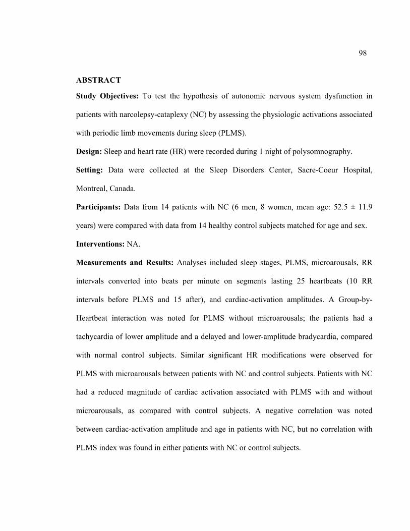
98
ABSTRACT
Study Objectives: To test the hypothesis of autonomic nervous system dysfunction in
patients with narcolepsy-cataplexy (NC) by assessing the physiologic activations associated
with periodic limb movements during sleep (PLMS).
Design: Sleep and heart rate (HR) were recorded during 1 night of polysomnography.
Setting: Data were collected at the Sleep Disorders Center, Sacre-Coeur Hospital,
Montreal, Canada.
Participants: Data from 14 patients with NC (6 men, 8 women, mean age: 52.5 ± 11.9
years) were compared with data from 14 healthy control subjects matched for age and sex.
Interventions: NA.
Measurements and Results: Analyses included sleep stages, PLMS, microarousals, RR
intervals converted into beats per minute on segments lasting 25 heartbeats (10 RR
intervals before PLMS and 15 after), and cardiac-activation amplitudes. A Group-by-
Heartbeat interaction was noted for PLMS without microarousals; the patients had a
tachycardia of lower amplitude and a delayed and lower-amplitude bradycardia, compared
with normal control subjects. Similar significant HR modifications were observed for
PLMS with microarousals between patients with NC and control subjects. Patients with NC
had a reduced magnitude of cardiac activation associated with PLMS with and without
microarousals, as compared with control subjects. A negative correlation was noted
between cardiac-activation amplitude and age in patients with NC, but no correlation with
PLMS index was found in either patients with NC or control subjects.
99
Conclusion: Our study pinpointed a significant reduction in the amplitude of PLMS-related
HR responses in both tachycardia and bradycardia in patients with NC. These findings
favor the physiologic relevance of the action of hypocretin on autonomic function that may
be of clinical significance, ie, increasing the risk of cardiovascular diseases.
Keywords: Narcolepsy, periodic leg movements, heart rate, cardioreactivity, hypocretin
100
INTRODUCTION
Narcolepsy with cataplexy (NC) is characterized by excessive daytime sleepiness,
cataplexy, and disturbed nocturnal sleep, including parasomnias, dissociated rapid eye
movement (REM) sleep, and periodic leg movements during sleep (PLMS) (Dauvilliers,
Arnulf, et al., 2007; Dauvilliers, Pennestri, et al., 2007; Dauvilliers, Rompre, et al., 2007;
Ferri, Zucconi, Manconi, Bruni, et al., 2006). Recent advances in our understanding of the
pathophysiology of NC have demonstrated a marked decrease in hypocretin-1 levels in the
cerebrospinal fluid, and in the number of hypocretin neurons (Dauvilliers, Arnulf, et al.,
2007; Mignot, et al., 2002; Peyron, et al., 2000). Hypocretin neurons are exclusively
located in the lateral hypothalamus but project widely throughout the central nervous
system, including to the hypothalamic and brainstem structures that are known to play a
role in central cardiovascular regulation (Samson, et al., 2005; Shirasaka, et al., 2002).
Animal studies have revealed that pharmacologic administration of hypocretin stimulates
arousal and elevates arterial blood pressure, heart rate (HR), oxygen consumption, body
temperature, and plasma catecholamine levels (Chen, Hwang, Chang, & Dun, 2000; de
Oliveira, Rosas-Arellano, Solano-Flores, & Ciriello, 2003; Samson, et al., 2005; Shirasaka,
Nakazato, Matsukura, Takasaki, & Kannan, 1999; Smith, Connolly, & Ferguson, 2002).
The increased blood pressure (BP) and HR effects have been demonstrated to be mediated
by activation of the sympathetic arm of the autonomic nervous system (Chen, et al., 2000;
de Oliveira, et al., 2003; Samson, et al., 2005; Shirasaka, et al., 1999; Smith, et al., 2002).
101
Studies have explored the cardiovascular status of hypocretin-deficient mice, with results
showing that these mice have lower arterial BP (Kayaba et al., 2003).
Sleep fragmentation is frequently reported in patients with NC, who show a high frequency
of PLMS (Dauvilliers, Pennestri, et al., 2007; Ferri, Zucconi, Manconi, Bruni, et al., 2006).
Physiologic activation associated with PLMS represents a model to study the responses of
the autonomic nervous system. Hence, studies conducted in patients with restless legs
syndrome (RLS) and in healthy control subjects without RLS have reported that HR
changes occurring with PLMS are sensitive markers of autonomic responses, characterized
by a tachycardia followed by a bradycardia (Gosselin, et al., 2003; Sforza, et al., 1999). To
our knowledge, the arousal responses associated with PLMS have never been studied in
subjects with NC. Studying interactions between sleep and the cardiovascular system
through physiologic activations associated with PLMS is of interest in NC, which is
characterized by the absence of hypocretin.
The aim of this study was to measure HR changes associated with PLMS in the presence or
absence of a microarousal in patients with NC and in age- and sex- matched healthy control
subjects.
102
METHODS
Subjects
Fourteen patients with sporadic NC (6 men, 8 women) aged between 29 and 67 years (mean
age, 52.4 ± 12.0 years) were included in the study. Inclusion criteria for narcolepsy were
the presence of excessive daytime sleepiness, cataplexy, HLA DQB1*0602 positivity, at
least 2 sleep-onset REM periods, and a mean sleep latency of less than 8 minutes during the
Multiple Sleep Latency Test. An additional inclusion criterion was that the subjects have a
PLMS index of at least 10 per hour. Exclusion criteria for NC were the presence of any
other sleep disorder, especially RLS, REM sleep behavior disorder (based on clinical
interview), and sleep apnea syndrome (as defined by an index of respiratory events > 5).
Nine patients were drug naïve, and 5 had stopped their medication (psychostimulant,
anticataplectic drugs, and any drug known to influence sleep, motor, or autonomic nervous
system activities) for at least 1 month prior to the polysomnographic recording in the sleep
laboratory.
Data from 14 control subjects (6 men, 8 women) aged between 20 and 65 years (mean age,
51.3 ± 10.8 yrs) matched for age and sex were compared with data from the patients with
NC. These subjects were recruited from the general population by announcements placed in
newpapers. None of the control subjects had any complaint of daytime sleepiness,
insomnia, RLS, or REM sleep behavior disorder. Control subjects were also required to
have a PLMS index of at least 10 per hour. Exclusion criteria for healthy normal subjects
103
were the same as for patients with NC. In addition, none of the control subjects reported
any symptom of NC.
All participants were free of coronary artery disease, stroke, heart failure, hypertension, or
diabetes and none smoked. All subjects signed a consent form approved by the ethics
committee of the Sacré-Coeur Hospital.
Procedures
All subjects underwent 1 night of polysomnographic recording in the sleep laboratory.
Sleep recording included 4 electroencephalographic leads (C3, C4, O1 and O2), 2 bilateral
electrooculograms, 1 electrocardiogram (lead II), and 1 chin electromyogram (EMG).
Respiration was assessed by nasal cannula, thoracoabdominal strain gauges, and finger
pulse oximetry. Respiratory events were scored according to American Academy of Sleep
Medicine recommendations (Iber, Ancoli-Israel, Chesson, & Quan, 2007). Surface EMG
electrodes placed 3 cm apart on the right and left anterior tibialis muscles were used to
record PLMS. Sleep was scored by the standard method (Iber, et al., 2007), and PLMS
were scored following the criteria set by the International RLS Study Group (Zucconi, et
al., 2006). Only movements lasting 0.5 to 10 seconds, separated by intervals of 5 to 90
seconds and occurring in a series of at least 4 consecutive movements, were counted. The
amplitude criterion for detecting leg movements was an increase in EMG to at least 8 µV
above the resting baseline for the onset of the movement and a decrease in EMG to less
104
than 2 µV above the resting level for the offset of movement. Microarousals were scored
according to standard American Sleep Disorders Association and American Academy of
Sleep Medicine criteria (Sleep Disorders Atlas Task Force of the American Sleep Disorders
Association, 1992; Iber, et al., 2007).
In addition to the standard criteria for PLMS, only movements separated by at least 20
seconds were selected for cardiovascular analysis in order to avoid overlapping HR
response to successive leg movements. Movements were selected in stage 2 sleep to avoid
sleep-stage interaction. Only movements free of any physiologic (rhythmic masticatory
muscle activity, flow limitation at the nasal cannula, apneas and hypopneas, arrhythmias) or
technical factors were selected. Finally, a total of 210 PLMS with microarousals and 490
PLMS without microarousals were selected for cardiovascular analysis in control subjects;
117 PLMS with and 418 without microarousals were selected in patients with NC. HR was
measured on segments lasting 25 heartbeats, comprising 10 RR intervals before the
movement (-10 to -1) and 15 RR intervals after the movement (+1 to +15). The RR
intervals were converted into beats per minute. For each movement, the baseline was
defined as the mean value from beat -10 to -6. Changes in HR were then calculating by
subtracting baseline from each HR value. In addition, the mean cardiac-activation
amplitude was calculated in each subject for PLMS with and without microarousals
(amplitude = maximal value during tachycardia – minimal value during bradycardia).
105
Statistical Analyses
Mann-Whitney tests were performed to compare sleep architecture and PLMS in
narcoleptics patients and normal control subjects. Beat-to-beat HR changes associated with
PLMS with and without microarousals in patients with NC and control subjects were
assessed independently by 2-way analyses of variance with 1 factor (group) and 1 repeated
measure (heartbeat) followed by planned comparisons. Greenhouse-Geisser correction for
sphericity was applied. Mann-Whitney tests were also performed to assess between-group
differences for cardiac-activation amplitude associated with PLMS with and without
microarousals. Spearman rank-order correlation coefficients were calculated to assess the
relationships among cardiac activation amplitude, age, and PLMS. Differences were
considered significant at p < 0.05.
RESULTS
Clinical and Polysomnographic Data
Polysomnographic data from patients and control subjects are shown in Table 1. The mean
age of patients and control subjects was similar at the time that polysomnography was
performed (52.4 ± 12.0 y vs 51.3 ± 10.8). Patients reported a long duration of disease at the
time of the study, mean of 27.8 ± 15.6 years. We found some differences in sleep
architecture between patients and control subjects, with a higher percentage of stage 1
sleep, a higher number of awakenings, a lower percentage of stage 2 sleep, a slightly higher
106
percentage of slow wave sleep, and shorter nighttime and REM sleep latencies in patients
with NC (Table 1).
The PLM index during wakefulness was higher in patients with NC, compared with control
subjects; however, total PLMS and PLMS in non-rapid eye movement (NREM) sleep
indexes were higher in control subjects (Table 2). No difference was seen between NC and
control subjects for PLMS duration (2.0 s ± 0.4 vs 2.1 s ± 0.5) and for PLMS
intermovement durations (31.9 s ± 5.6 vs 33.1 s ± 3.7). Finally, no between-group
difference was reported for microarousals and PLMS with microarousal indexes (Tables 1
and 2).
HR Changes Associated With PLMS
Figure 1 shows the distribution of HR changes associated with PLMS without
microarousals in patients with NC, compared with control subjects, for 10 RR intervals
before the movement (-10 to -1) and 15 RR intervals after the onset of the movement (+1 to
+15). A Group-by-Heartbeat interaction was noted for PLMS without microarousals, with a
tachycardia of lower amplitude (significant for beats +3 to +5) and a delayed and lower
amplitude of bradycardia (significant for beats +9 to +12) in patients with NC (F19, 494 =
6.01, p = 0.001).
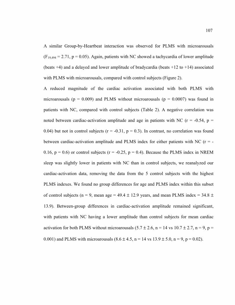
107
A similar Group-by-Heartbeat interaction was observed for PLMS with microarousals
(F19,494 = 2.71, p = 0.05). Again, patients with NC showed a tachycardia of lower amplitude
(beats +4) and a delayed and lower amplitude of bradycardia (beats +12 to +14) associated
with PLMS with microarousals, compared with control subjects (Figure 2).
A reduced magnitude of the cardiac activation associated with both PLMS with
microarousals (p = 0.009) and PLMS without microarousals (p = 0.0007) was found in
patients with NC, compared with control subjects (Table 2). A negative correlation was
noted between cardiac-activation amplitude and age in patients with NC (r = -0.54, p =
0.04) but not in control subjects (r = -0.31, p = 0.3). In contrast, no correlation was found
between cardiac-activation amplitude and PLMS index for either patients with NC (r = -
0.16, p = 0.6) or control subjects (r = -0.25, p = 0.4). Because the PLMS index in NREM
sleep was slightly lower in patients with NC than in control subjects, we reanalyzed our
cardiac-activation data, removing the data from the 5 control subjects with the highest
PLMS indexes. We found no group differences for age and PLMS index within this subset
of control subjects (n = 9, mean age = 49.4 ± 12.9 years, and mean PLMS index = 34.8 ±
13.9). Between-group differences in cardiac-activation amplitude remained significant,
with patients with NC having a lower amplitude than control subjects for mean cardiac
activation for both PLMS without microarousals (5.7 ± 2.6, n = 14 vs 10.7 ± 2.7, n = 9, p =
0.001) and PLMS with microarousals (8.6 ± 4.5, n = 14 vs 13.9 ± 5.0, n = 9, p = 0.02).
108
DISCUSSION
Our study reports for the first time the autonomic activation associated with PLMS in
patients with NC, with a lower magnitude of the changes reported, compared with healthy
matched control subjects. The pattern of cardiac acceleration and deceleration was
substantially similar for PLMS with and without microarousals, although the changes were
greater when microarousals occurred. The increase in HR started before PLMS with or
without microarousals in patients with NC and in control subjects.
Reduced PLMS-associated autonomic changes in patients with NC favor a lower sensitivity
of the common brainstem generator of cardiac activations (Berlucchi, 1997; Sforza et al.,
2005). Similar findings were previously reported in patients with RLS and with the aging
process (Gosselin et al., 2003; Sforza et al., 1999; Winkelman, 1999), as also pinpointed in
our present study in both patients with NC and control populations. However, we noted in
the present study that cardiac-activation amplitude associated with PLMS was independent
of the PLMS index in both patients with NC and control subjects.
Reduced autonomic activation associated with PLMS, together with a lower HR variability
during sleep and an impairment in both sympathetic and parasympathetic activities in
wakefulness, have been reported in patients with idiopathic REM sleep behavior disorder
(Fantini et al., 2002; Ferini-Strambi, Oldani, Zucconi, & Smirne, 1996). These findings
were of major interest in terms of pathophysiology, since an increased frequency of REM
sleep without atonia, phasic EMG activity, and clinical REM sleep behavior disorder had
109
been previously reported in patients with NC (Dauvilliers, Rompre, et al., 2007;
Nightingale et al., 2005). We hypothesize that common abnormalities in REM sleep motor
regulation and autonomic function exist in both conditions.
Old clinical studies in patients with NC, before the discovery of hypocretins, reported some
abnormalities in the autonomic nervous system, including pupillary, erection, and
cardiovascular functions (Hublin, Matikainen, & Partinen, 1994; Karacan, 1986; Sachs &
Kaijser, 1980). Hence, 1 study exploring the cardiovascular system in patients with NC
revealed a reduced vegetative reactivity in response to muscle contraction and the Valsalva
maneuver (Sachs & Kaijser, 1980), findings that have not been further confirmed (Hublin,
et al., 1994). Later on, power spectrum analysis studies of HR and BP variability have
suggested only minor changes in NC that failed to find a clear autonomic dysfunction
(Ferini-Strambi et al., 1997; Fronczek et al., 2008).
Our findings, therefore, have demonstrated, for the first time, a primary change in
autonomic function in patients with NC. Autonomic imbalance may be characterized by an
impairment in both sympathetic and parasympathetic activities. Recent studies have
highlighted the nonsleep actions of hypocretin, including roles in controlling central
pathways involved in the regulation of cardiovascular functions (Date et al., 1999;
Shirasaka, et al., 2002). Pharmacologic studies have revealed that intrathecal hypocretin
administration results in increased BP and HR in conscious, as well as anesthetized,
110
animals, an effect that is mediated by enhancement of the sympathetic output (Chen, et al.,
2000; de Oliveira, et al., 2003; Shirasaka, et al., 1999). In addition to acting on brainstem
sites related to autonomic function, hypocretin also acts in the hypothalamic paraventricular
nucleus and sympathetic preganglionic neurons, increasing the sympathetic tone (Ciriello &
de Oliveira, 2003; Davis, Williams, Xu, Glatzer, & Smith, 2003). Moreover, cardiovascular
studies in orexin-knockout mice have revealed lower arterial BP, as compared with in wild-
type mice, differences that are abolished by α-adrenergic or ganglionic blockade (Kayaba,
et al., 2003). Altogether, these results corroborate that the hypocretin system acts as an
essential modulator for coordinating autonomic-system circuits, with the predominant
sympathetic excitatory effect targeting the autonomic centers of the brain. We hypothesize
that the generalized attenuation of PLM-related HR changes observed in human narcolepsy
is explained by a lower sympathetic response due to the hypocretin-deficient condition.
Although the significance of PLMS and the lower amplitude of the PLMS-related cardiac
activations remain unclear, it may be of clinical importance (Pennestri, Montplaisir,
Colombo, Lavigne, & Lanfranchi, 2007). Hence, several studies have shown that a decrease
in HR variability, a tool that is frequently used to assess autonomic imbalance, is associated
with a higher risk of mortality due to cardiovascular diseases (Fagard et al., 2008; Kleiger,
Miller, Bigger, & Moss, 1987). These findings require further attention, since NC is often
associated with obesity, type 2 diabetes, and the metabolic syndrome (Poli et al., 2009;
Schuld, Hebebrand, Geller, & Pollmacher, 2000). We also emphasize that patients with NC
111
will be treated with psychostimulants for the rest of their lives, therapy that has a well-
known impact on autonomic nervous and cardiovascular systems (Auger et al., 2005).
Several limitations in our study need to be addressed. First, the sample size is small,
although sufficient to demonstrate a significant reduction in the amplitude of the PLMS-
related HR response. The small sample size is the result of the strict inclusion criteria,
which required the presence of enough PLMS to be further analyzed and compared with the
PLMS index of a carefully age- and sex-matched population that had no comorbid disease
or was taking any medication that may impact the results. Second, we focused on PLMS-
related cardiac activations only in stage 2 non-REM sleep. Because most of the PLMS
existed in stage 2 sleep, especially in control subjects, we were unable to score enough
PLMS to perform between-group HR changes by comparing PLMS in various sleep stages.
Finally, no cerebrospinal fluid hypocretin-1 measurement was available in patients.
However, we assume that patients were almost all hypocretin deficient because they all had
sporadic NC and had severe daytime sleepiness, clear-cut cataplexy, typical results on the
Multiple Sleep Latency Test, and HLA DQB1*0602 positivity.
CONCLUSION
Our study pinpointed a significant reduction in the amplitude of PLMS-related HR
responses in both tachycardia and bradycardia in patients with NC. This result suggests that
112
the physiologic relevance of the action of hypocretin on autonomic function may be of
clinical significance with regard to increasing the risk of cardiovascular diseases.
ACKNOWLEDGMENT
The authors thank Sylvie Rompré and Jean Paquet for helping with data and statistical
analysis.
REFERENCES
1. Dauvilliers Y, Arnulf I, Mignot E. Narcolepsy with cataplexy. Lancet
2007;369:499-511.
2. Dauvilliers Y, Pennestri MH, Petit D, Dang-Vu T, Lavigne G, Montplaisir J.
Periodic leg movements during sleep and wakefulness in narcolepsy. J Sleep Res
2007;16:333-9.
3. Dauvilliers Y, Rompre S, Gagnon JF, Vendette M, Petit D, Montplaisir J. REM
sleep characteristics in narcolepsy and REM sleep behavior disorder. Sleep 2007;30:844-9.
4. Ferri R, Zucconi M, Manconi M, et al. Different periodicity and time structure of
leg movements during sleep in narcolepsy/cataplexy and restless legs syndrome. Sleep
2006;29:1587-94.
5. Peyron C, Faraco J, Rogers W, et al. A mutation in a case of early onset narcolepsy
and a generalized absence of hypocretin peptides in human narcoleptic brains. Nat Med
2000;6:991-7.
113
6. Mignot E, Lammers GJ, Ripley B, et al. The role of cerebrospinal fluid hypocretin
measurement in the diagnosis of narcolepsy and other hypersomnias. Arch Neurol
2002;59:1553-62.
7. Shirasaka T, Kunitake T, Takasaki M, Kannan H. Neuronal effects of orexins:
relevant to sympathetic and cardiovascular functions. Regul Pept 2002;104:91-5.
8. Samson WK, Taylor MM, Ferguson AV. Non-sleep effects of hypocretin/orexin.
Sleep Med Rev 2005;9:243-52.
9. Smith PM, Connolly BC, Ferguson AV. Microinjection of orexin into the rat
nucleus tractus solitarius causes increases in blood pressure. Brain Res 2002;950:261-7.
10. de Oliveira CV, Rosas-Arellano MP, Solano-Flores LP, Ciriello J. Cardiovascular
effects of hypocretin-1 in nucleus of the solitary tract. Am J Physiol Heart Circ Physiol
2003;284:H1369-77.
11. Chen CT, Hwang LL, Chang JK, Dun NJ. Pressor effects of orexins injected
intracisternally and to rostral ventrolateral medulla of anesthetized rats. Am J Physiol Regul
Integr Comp Physiol 2000;278:R692-7.
12. Shirasaka T, Nakazato M, Matsukura S, Takasaki M, Kannan H. Sympathetic and
cardiovascular actions of orexins in conscious rats. Am J Physiol 1999;277:R1780-5.
13. Kayaba Y, Nakamura A, Kasuya Y, et al. Attenuated defense response and low
basal blood pressure in orexin knockout mice. Am J Physiol Regul Integr Comp Physiol
2003;285:R581-93.
114
14. Sforza E, Nicolas A, Lavigne G, Gosselin A, Petit D, Montplaisir J. EEG and
cardiac activation during periodic leg movements in sleep: support for a hierarchy of
arousal responses. Neurology 1999;52:786-91.
15. Gosselin N, Lanfranchi P, Michaud M, et al. Age and gender effects on heart rate
activation associated with periodic leg movements in patients with restless legs syndrome.
Clin Neurophysiol 2003;114:2188-95.
16. Iber C, Ancoli-Israel S, Chesson A, Quan S. The AASM Manual for the Scoring of
Sleep and Associated Events; Rules, Terminology and Technical Specifications, 1st ed.
Westchester, IL: Amercian Academy of Sleep Medicine; 2007.
17. Zucconi M, Ferri R, Allen R, et al. The official World Association of Sleep
Medicine (WASM) standards for recording and scoring periodic leg movements in sleep
(PLMS) and wakefulness (PLMW) developed in collaboration with a task force from the
International Restless Legs Syndrome Study Group (IRLSSG). Sleep Med 2006;7:175-83.
18. EEG arousals: scoring rules and examples: a preliminary report from the Sleep
Disorders Atlas Task Force of the American Sleep Disorders Association. Sleep
1992;15:173-84.
19. Sforza E, Pichot V, Barthelemy JC, Haba-Rubio J, Roche F. Cardiovascular
variability during periodic leg movements: a spectral analysis approach. Clin Neurophysiol
2005;116:1096-104.
115
20. Berlucchi G. One or many arousal systems? Reflections on some of Giuseppe
Moruzzi's foresights and insights about the intrinsic regulation of brain activity. Arch Ital
Biol 1997;135:5-14.
21. Winkelman JW. The evoked heart rate response to periodic leg movements of sleep.
Sleep 1999;22:575-80.
22. Ferini-Strambi L, Oldani A, Zucconi M, Smirne S. Cardiac autonomic activity
during wakefulness and sleep in REM sleep behavior disorder. Sleep 1996;19:367-9.
23. Fantini ML, Michaud M, Gosselin N, Lavigne G, Montplaisir J. Periodic leg
movements in REM sleep behavior disorder and related autonomic and EEG activation.
Neurology 2002;59:1889-94.
24. Nightingale S, Orgill JC, Ebrahim IO, de Lacy SF, Agrawal S, Williams AJ. The
association between narcolepsy and REM behavior disorder (RBD). Sleep Med
2005;6:253-8.
25. Hublin C, Matikainen E, Partinen M. Autonomic nervous system function in
narcolepsy. J Sleep Res 1994;3:131-7.
26. Karacan I. Erectile dysfunction in narcoleptic patients. Sleep 1986;9:227-31.
27. Sachs C, Kaijser L. Autonomic control of cardiovascular reflexes in narcolepsy. J
Neurol Neurosurg Psychiatry 1980;43:535-9.
28. Ferini-Strambi L, Spera A, Oldani A, et al. Autonomic function in narcolepsy:
power spectrum analysis of heart rate variability. J Neurol 1997;244:252-5.
116
29. Fronczek R, Overeem S, Reijntjes R, Lammers GJ, van Dijk JG, Pijl H. Increased
heart rate variability but normal resting metabolic rate in hypocretin/orexin-deficient human
narcolepsy. J Clin Sleep Med 2008;4:248-54.
30. Date Y, Ueta Y, Yamashita H, et al. Orexins, orexigenic hypothalamic peptides,
interact with autonomic, neuroendocrine and neuroregulatory systems. Proc Natl Acad Sci
U S A 1999;96:748-53.
31. Davis SF, Williams KW, Xu W, Glatzer NR, Smith BN. Selective enhancement of
synaptic inhibition by hypocretin (orexin) in rat vagal motor neurons: implications for
autonomic regulation. J Neurosci 2003;23:3844-54.
32. Ciriello J, de Oliveira CV. Cardiac effects of hypocretin-1 in nucleus ambiguus. Am
J Physiol Regul Integr Comp Physiol 2003;284:R1611-20.
33. Pennestri MH, Montplaisir J, Colombo R, Lavigne G, Lanfranchi PA. Nocturnal
blood pressure changes in patients with restless legs syndrome. Neurology 2007;68:1213-8.
34. Thayer J, Yamamoto S, Brosschot J. The relationship of autonomic imbalance, heart
rate variability and cardiovascular disease risk factors. Int J Cardiology 2010; 141:122-31.
35. Kleiger RE, Miller JP, Bigger JT, Jr., Moss AJ. Decreased heart rate variability and
its association with increased mortality after acute myocardial infarction. Am J Cardiol
1987;59:256-62.
36. Poli F, Plazzi G, Di Dalmazi G, et al. Body mass index-independent metabolic
alterations in narcolepsy with cataplexy. Sleep 2009;32:1491-7.
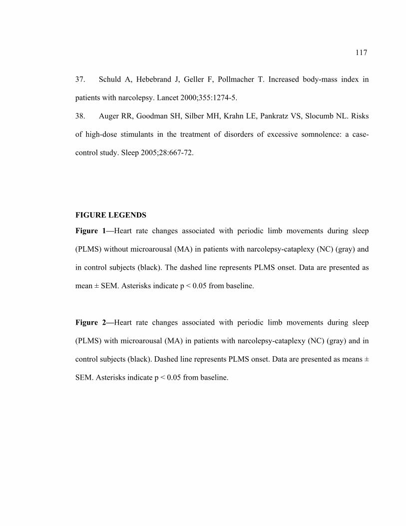
117
37. Schuld A, Hebebrand J, Geller F, Pollmacher T. Increased body-mass index in
patients with narcolepsy. Lancet 2000;355:1274-5.
38. Auger RR, Goodman SH, Silber MH, Krahn LE, Pankratz VS, Slocumb NL. Risks
of high-dose stimulants in the treatment of disorders of excessive somnolence: a case-
control study. Sleep 2005;28:667-72.
FIGURE LEGENDS
Figure 1—Heart rate changes associated with periodic limb movements during sleep
(PLMS) without microarousal (MA) in patients with narcolepsy-cataplexy (NC) (gray) and
in control subjects (black). The dashed line represents PLMS onset. Data are presented as
mean ± SEM. Asterisks indicate p < 0.05 from baseline.
Figure 2—Heart rate changes associated with periodic limb movements during sleep
(PLMS) with microarousal (MA) in patients with narcolepsy-cataplexy (NC) (gray) and in
control subjects (black). Dashed line represents PLMS onset. Data are presented as means ±
SEM. Asterisks indicate p < 0.05 from baseline.
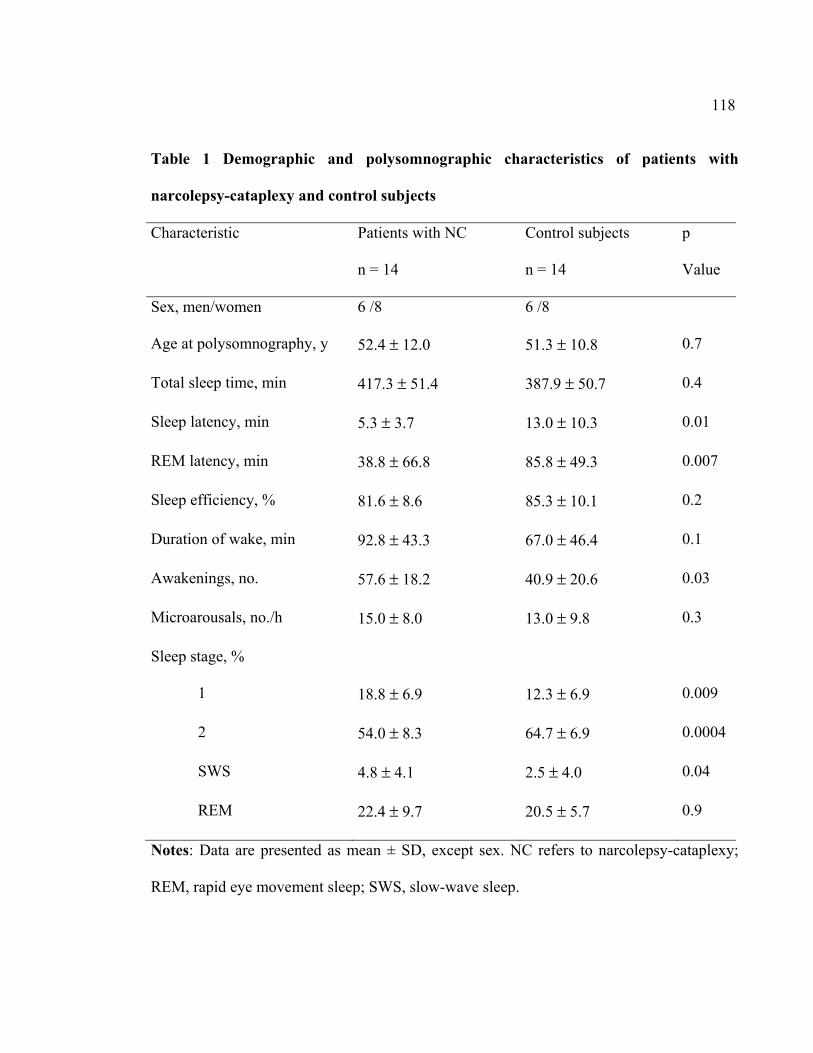
118
Table 1 Demographic and polysomnographic characteristics of patients with
narcolepsy-cataplexy and control subjects
Characteristic Patients with NC
n = 14
Control subjects
n = 14
p
Value
Sex, men/women 6 /8 6 /8
Age at polysomnography, y 52.4 ± 12.0 51.3 ± 10.8 0.7
Total sleep time, min 417.3 ± 51.4 387.9 ± 50.7 0.4
Sleep latency, min 5.3 ± 3.7 13.0 ± 10.3 0.01
REM latency, min 38.8 ± 66.8 85.8 ± 49.3 0.007
Sleep efficiency, % 81.6 ± 8.6 85.3 ± 10.1 0.2
Duration of wake, min 92.8 ± 43.3 67.0 ± 46.4 0.1
Awakenings, no. 57.6 ± 18.2 40.9 ± 20.6 0.03
Microarousals, no./h 15.0 ± 8.0 13.0 ± 9.8 0.3
Sleep stage, %
1 18.8 ± 6.9 12.3 ± 6.9 0.009
2 54.0 ± 8.3 64.7 ± 6.9 0.0004
SWS 4.8 ± 4.1 2.5 ± 4.0 0.04
REM 22.4 ± 9.7 20.5 ± 5.7 0.9
Notes: Data are presented as mean ± SD, except sex. NC refers to narcolepsy-cataplexy;
REM, rapid eye movement sleep; SWS, slow-wave sleep.
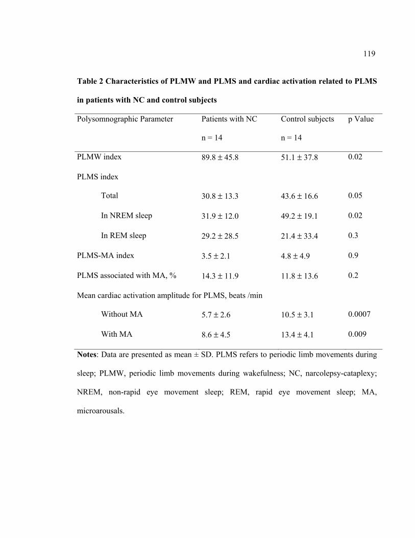
119
Table 2 Characteristics of PLMW and PLMS and cardiac activation related to PLMS
in patients with NC and control subjects
Polysomnographic Parameter Patients with NC
n = 14
Control subjects
n = 14
p Value
PLMW index 89.8 ± 45.8 51.1 ± 37.8 0.02
PLMS index
Total 30.8 ± 13.3 43.6 ± 16.6 0.05
In NREM sleep 31.9 ± 12.0 49.2 ± 19.1 0.02
In REM sleep 29.2 ± 28.5 21.4 ± 33.4 0.3
PLMS-MA index 3.5 ± 2.1 4.8 ± 4.9 0.9
PLMS associated with MA, % 14.3 ± 11.9 11.8 ± 13.6 0.2
Mean cardiac activation amplitude for PLMS, beats /min
Without MA 5.7 ± 2.6 10.5 ± 3.1 0.0007
With MA 8.6 ± 4.5 13.4 ± 4.1 0.009
Notes: Data are presented as mean ± SD. PLMS refers to periodic limb movements during
sleep; PLMW, periodic limb movements during wakefulness; NC, narcolepsy-cataplexy;
NREM, non-rapid eye movement sleep; REM, rapid eye movement sleep; MA,
microarousals.
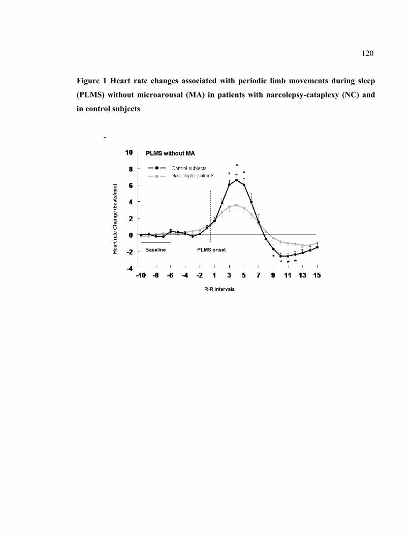
120
Figure 1 Heart rate changes associated with periodic limb movements during sleep
(PLMS) without microarousal (MA) in patients with narcolepsy-cataplexy (NC) and
in control subjects
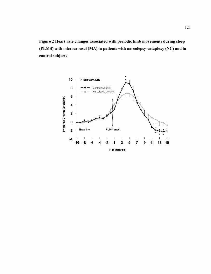
121
Figure 2 Heart rate changes associated with periodic limb movements during sleep
(PLMS) with microarousal (MA) in patients with narcolepsy-cataplexy (NC) and in
control subjects
122
2.4. Quatrième article
Nocturnal blood pressure changes in patients with restless legs syndrome
Marie-Hélène Pennestri BSc,1,2 Jacques Montplaisir MD, PhD, 1,3 Roberto Colombo,4 Gilles Lavigne DMD,1,5 Paola A Lanfranchi MD, MSc1,6,7
1Sleep Disorders Center, Sacré-Cœur Hospital, University of Montreal 2Department of Psychology, University of Montreal 3Department of Psychiatry, University of Montreal
4Department of Bioengineering, Salvatore Maugeri Foundation, Veruno, Italy 5Faculty of Dentistry, University of Montreal
6Cardiology division, Sacré-Cœur Hospital, University of Montreal 7Department of Medicine, University of Montreal
Article publié dans : Neurology (2007) 68(15), 1213-1218

123
Contribution des auteurs
Marie-Hélène Pennestri : Élaboration du protocole, recrutement des participants,
participation à l’évaluation des participants, analyse des données, analyse statistiques,
interprétation des résultats, rédaction du manuscrit.
Jacques Montplaisir : Élaboration du protocole, interprétation des résultats, révision du
manuscrit.
Roberto Colombo : Analyse des données
Gilles Lavigne : Interprétation des résultats, révision du manuscrit
Paola A Lanfranchi : Élaboration du protocole, interprétation des résultats, rédaction du
manuscrit
124
ABSTRACT
Objectives: To assess heart rate (HR) and blood pressure (BP) changes associated with
periodic leg movements during sleep (PLMS) with or without EEG signs of arousal in
subjects with primary restless legs syndrome (RLS).
Methods: Ten patients with RLS (4 women, aged 47.3 ± 13.5 yrs) underwent one night of
polysomnography along with non-invasive beat-to-beat BP monitoring. Ten PLMS with
micro-arousals (PLMS-MA) and 10 PLMS without microarousals (PLMS-noMA) were
analyzed in each subject. Systolic and diastolic BP (SBP, DBP) were measured within a 25-
beat temporal window comprising 10 beats before and 15 beats after onset of each
movement. PLMS-related BP changes were assessed by repeated measures one-way
analysis of variance. BP changes associated with PLMS-MA and PLMS-noMA were
compared by paired t-tests. Pearson correlation coefficients were used to assess the
relationship between cardiovascular changes and clinical and polysomnographic variables.
Results: BP increased significantly in association with all PLMS (on average, SBP 22
mmHg, DBP 11 mmHg). BP changes associated with PLMS-MA were greater vs those
associated with PLMS-noMA (p<0.05). SBP and DBP changes increased with age and the
duration of illness.
Conclusions: Periodic leg movements-related repetitive nocturnal blood pressure
fluctuations could contribute to the risk of cardiovascular diseases in patients with restless
legs syndrome, especially in the elderly.
125
INTRODUCTION
Population-based studies have found that restless legs syndrome (RLS) is associated with
an increased risk for hypertension and coronary artery disease.1,2,3 However, the
mechanisms linking RLS and cardiovascular diseases are unknown.
Sleep laboratory investigations have revealed that at least 80% of patients with RLS
experience periodic leg movements (PLMS) during their sleep.4 PLMS are described as a
rhythmic extension of the big toe and dorsiflexion of the ankle, with occasional flexion at
the knee and hip. They typically recur at approximately 20- to 40-second intervals, and
about one-third of these movements are associated with EEG signs of arousal, also called
microarousals (MA).5 Even though PLMS represent a supportive criterion for an RLS
diagnosis, PLMS occur in other sleep disorders such as narcolepsy,6 REM sleep behavior
disorder (RBD),7 sleep apnea8 and even in healthy, non-complaining subjects with
advancing age.6,9,10
Studies from our and other groups have consistently shown that PLMS, whether or not
associated with MA, are accompanied by significant heart rate (HR) changes, i.e. a
tachycardia followed by a bradycardia.5,7,11,12 However, greater HR changes were observed
when PLMS were associated with MA.5 BP, a functional parameter of high clinical
relevance,13 is also greatly affected by autonomic influences. Hence, BP responses are also
part of the circulatory response to arousal activation, whether or not they are associated
126
with respiratory events.14,15,16 Clinical studies indicate that not only average BP but also the
magnitude of BP variability and time rate of BP variation over 24 hours correlate linearly
with indirect indices of atherosclerosis.17,18 A possibility that emerges from these studies is
the potential clinical relevance of PLMS in RLS if the presence of repetitive BP changes
associated with PLMS is confirmed.
In this study we sought to assess HR and BP changes associated with PLMS with or
without MA and to correlate these changes with clinical characteristics of the disease in
patients with primary RLS.
METHODS
Subjects
We studied 10 consecutive patients with RLS (4 women and 6 men) aged 29 to 69 (mean
age = 47.3 ± 13.5 years), who met the following criteria. A diagnosis of RLS was based on
the presence of the four standard clinical criteria developed and recently revised by the
International RLS Study Group.19 An additional inclusion criterion was a PLMS index
(number of PLM per hour of sleep) ≥ 10. Exclusion criteria were: 1) presence of other
psychiatric or medical conditions known to be associated with RLS such as uraemia,
peripheral neuropathy, rheumatoid arthritis or anaemia; 2) serum ferritin level < 10 ug/L; 3)
presence of other sleep disorders associated with PLMS or with cardiovascular changes
127
during sleep such as narcolepsy, RBD or sleep bruxism; 4) presence of sleep apnea
syndrome as defined by an apnea-hypopnea index ≥ 5 per hour; 5) use of psychotropic
medications or other drugs known to influence sleep, autonomic nervous system function or
motor activity. Subjects reported a mean duration of illness of 25.7 ± 19.0 years.
All subjects signed the consent form, which was approved by the ethics committee of the
Sacré-Coeur Hospital.
Data Collection
Subjects spent one night in the sleep laboratory. In the evening preceding
polysomnographic (PSG) sleep recordings, brachial cuff arterial BP was measured at rest in
the sitting position in the non-dominant arm to provide a baseline clinical value of systolic
and diastolic BP (SBP and DBP). All subjects had normal BP (SBP: 124.3 ± 15.0 mmHg,
DPB: 72.9 ± 10.8 mmHg).
Sleep recording included four EEG leads (C3, C4, O1 and O2), two bilateral electro-
oculograms (EOG), and one chin EMG. Respiration was assessed by nasal-cannula,
thoraco-abdominal strain gauges and finger pulse oxymetry. Surface EMG electrodes
placed 3 cm apart on the right and left anterior tibialis muscles were used to record PLM
during sleep and wakefulness. The electrocardiogram (EKG) included leads I, II and III.
Continuous BP recordings were obtained using a TNO Portapres Model-2 (TNO TPD
128
Biomedical Instrumentation, The Netherlands), which provides non-invasive beat-to-beat
BP recording through a continuous finger arterial pressure waveform. Comparative studies
of finger BP and invasive brachial arterial BP monitoring have shown that the finger
method, while presenting a consistent underestimation of the values measured in the
brachial artery, is highly accurate in measuring sudden BP changes occurring in response to
various stimuli. It therefore offers a reliable alternative to invasive measurement of beat-to-
beat BP.20
Data analyses
Sleep was scored by the standard method21 and PLMS were scored according to Coleman’s
criteria.22 Only movements lasting 0.5 to 5 seconds, separated by intervals of 4 to 90
seconds and occurring in a series of at least four consecutive movements were counted. The
amplitude criterion for detecting leg movements was the presence of EMG activity with at
least 25% the amplitude of the EMG signal recorded during a voluntary movement
performed at the beginning of the recording. MAs were scored according to standard
American Sleep Disorders Association (ASDA) criteria.23 Respiratory events were scored
according to the ASDA and the Academy of Sleep Medicine (AASM) recommendations.24
PLMS selection and cardiovascular analyses
In each subject, 10 PLMS with MA (PLMS-MA) and 10 PLMS without MA (PLMS-
noMA) were selected for cardiovascular analysis. In addition to the standard criteria for
129
PLMS, only movements separated by at least 20 seconds were selected for analysis in order
to avoid overlapping HR and BP responses to successive leg movements. Movements were
selected in stage 2 to avoid sleep stage interaction. Only movements free of any
physiological (rhythmic masticatory muscle activity, flow limitation at the nasal cannula,
apneas and hypopneas, arrhythmias), or technical factors that would potentially affect BP
waveform were selected for further analysis. Since one subject did not have any PLMS
associated with MA, the influence of MA on BP response was assessed in only 9 patients.
HR, SBP and DBP were measured automatically on segments of EKG and BP signals
lasting 25 heartbeats, comprising 10 beats before the movement (-10 to -1) and 15 beats
after the movement (+1 to +15) (Figure 1). Beat-to-beat values of HR, SBP and DBP were
calculated. For each movement, the baseline was defined as the mean value from beats -10
to -4. Since heart rate changes were found to start before the onset of movement,12 beats -3
to -1 were excluded from the baseline measures. The highest value obtained after the onset
of movement was taken as the peak value. Increments (delta) from baseline to peak values
were calculated for HR, SBP and DBP during PLMS-MA and PLMS-noMA in each
subject. HR, SBP and DBP increments associated with both types of movement were then
averaged for each subject ([delta PLMS-MA + delta PLMS-noMA]/2) to obtain overall
individual HR, SBP and DBP increments during sleep.
130
BP changes associated with 10 PLM occurring during wakefulness (PLMW) were also
measured and compared to those associated with PLMS, in order to distinguish the effects
of exercise from the central activation component on the pressure response observed during
PLMS. PLMW were scored according to modified Coleman’s criteria, including
movements lasting 0.5 to 10 seconds.25 This analysis was performed in 8 of the 10 subjects,
who had a sufficient number of PLMW during pre-sleep onset period and nocturnal wake,
according to the criteria described above.
Statistical analyses
HR, SBP and DBP responses associated with PLMS were assessed by repeated measures
one-way analysis of variance followed by planned comparisons. Each value of HR, SBP
and DBP (beat -10 to +15) was compared to baseline (beat -10 to -4). Greenhouse-Geisser
correction for sphericity was applied to all repeated measures.
The comparison of increments of HR, SBP and DBP, associated with PLMS-MA and
PLMS-noMA, was assessed within subjects by paired t-tests.
Pearson correlation coefficients were calculated between HR, SBP and DBP increments
associated with both types of PLMS pooled together and the following clinical and
polygraphic characteristics: age, duration of illness, index and duration of PLMS, and
duration of MA associated with PLMS.
131
Finally, in order to compare SBP and DBP increments associated with PLMW and PLMS-
noMA, paired-t-tests were calculated.
RESULTS
PLMS index was 29.5 ± 13.7 /hour (range: 10.4 to 47.7/hour). MA index was 10.7 ± 4.8
and PLMS-MA index was 3.1 ± 1.4. Duration of movements used for cardiovascular
analysis was 2.5 ± 0.6 seconds for PLMS-noMA versus 2.7 ± 0.5 seconds for PLMS-MA
(p=NS). Duration of MA associated with PLMS was 5.3 ± 0.9.
Changes in HR, SBP and DBP in association with PLMS are illustrated in Figure 2. PLMS-
noMA were associated with HR changes (F(25, 200)=15.6, p<0.0001), e.g., a tachycardia
(beats -1 and +2 to +7) and a bradycardia (beats +11 to +15). PLMS-noMA were also
associated with SBP (F(25, 200)=14.4, p=0.0001 and DBP increments (F(25, 200)=13.5,
p=0.0001). Planned comparisons revealed an increase in SBP starting from beats +5 to +14
and in DBP from beats + 4 to +12 (p<0.05).
Changes in HR were also associated with PLMS-MA (F(25, 200)=24.6, p<0.000001) with
a tachycardia (beat -1 to +9), while bradycardia was not significant. PLMS-MA were
associated with SBP (F(25, 200)=29.2, p=0.000001) and DBP increments (F(25,
200)=30.1, p=0.000001). Planned comparisons revealed an increase in SBP from beat +4 to
beat +14 and in DBP from beat +3 to beat +12 (p<0.05).
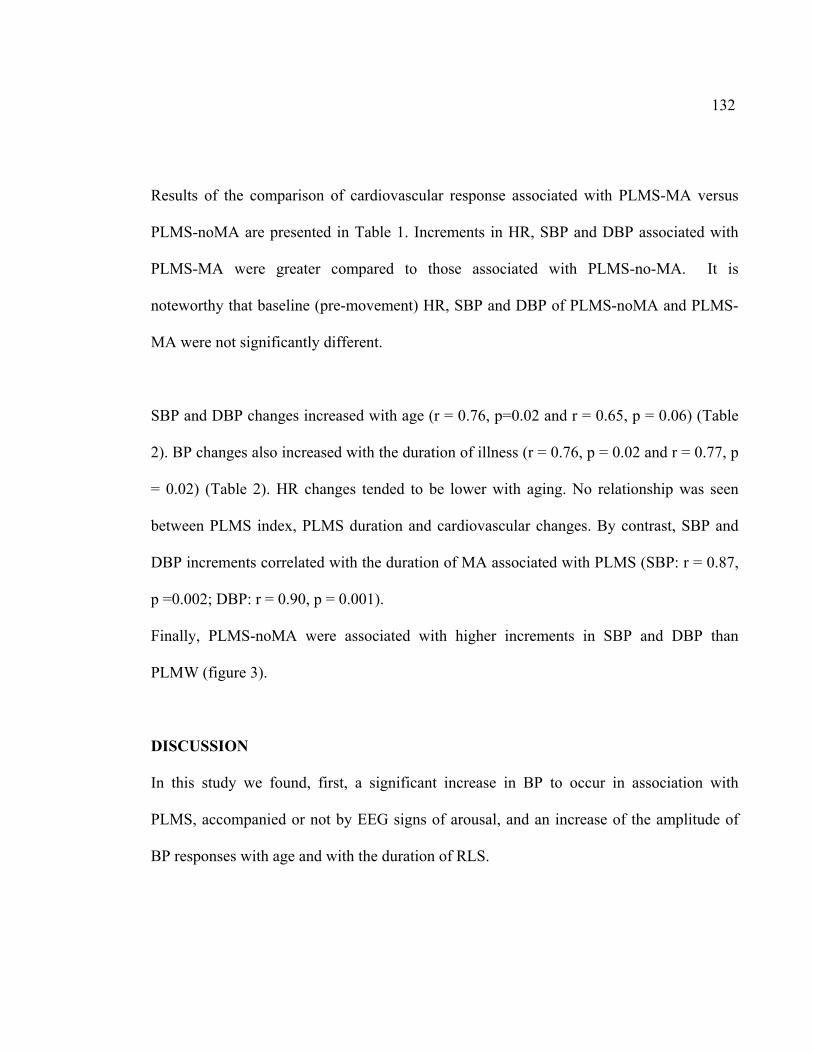
132
Results of the comparison of cardiovascular response associated with PLMS-MA versus
PLMS-noMA are presented in Table 1. Increments in HR, SBP and DBP associated with
PLMS-MA were greater compared to those associated with PLMS-no-MA. It is
noteworthy that baseline (pre-movement) HR, SBP and DBP of PLMS-noMA and PLMS-
MA were not significantly different.
SBP and DBP changes increased with age (r = 0.76, p=0.02 and r = 0.65, p = 0.06) (Table
2). BP changes also increased with the duration of illness (r = 0.76, p = 0.02 and r = 0.77, p
= 0.02) (Table 2). HR changes tended to be lower with aging. No relationship was seen
between PLMS index, PLMS duration and cardiovascular changes. By contrast, SBP and
DBP increments correlated with the duration of MA associated with PLMS (SBP: r = 0.87,
p =0.002; DBP: r = 0.90, p = 0.001).
Finally, PLMS-noMA were associated with higher increments in SBP and DBP than
PLMW (figure 3).
DISCUSSION
In this study we found, first, a significant increase in BP to occur in association with
PLMS, accompanied or not by EEG signs of arousal, and an increase of the amplitude of
BP responses with age and with the duration of RLS.
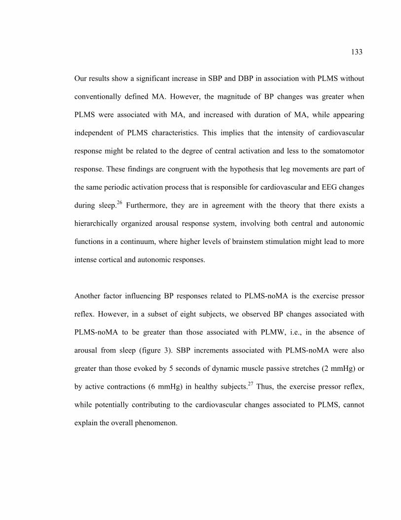
133
Our results show a significant increase in SBP and DBP in association with PLMS without
conventionally defined MA. However, the magnitude of BP changes was greater when
PLMS were associated with MA, and increased with duration of MA, while appearing
independent of PLMS characteristics. This implies that the intensity of cardiovascular
response might be related to the degree of central activation and less to the somatomotor
response. These findings are congruent with the hypothesis that leg movements are part of
the same periodic activation process that is responsible for cardiovascular and EEG changes
during sleep.26 Furthermore, they are in agreement with the theory that there exists a
hierarchically organized arousal response system, involving both central and autonomic
functions in a continuum, where higher levels of brainstem stimulation might lead to more
intense cortical and autonomic responses.
Another factor influencing BP responses related to PLMS-noMA is the exercise pressor
reflex. However, in a subset of eight subjects, we observed BP changes associated with
PLMS-noMA to be greater than those associated with PLMW, i.e., in the absence of
arousal from sleep (figure 3). SBP increments associated with PLMS-noMA were also
greater than those evoked by 5 seconds of dynamic muscle passive stretches (2 mmHg) or
by active contractions (6 mmHg) in healthy subjects.27 Thus, the exercise pressor reflex,
while potentially contributing to the cardiovascular changes associated to PLMS, cannot
explain the overall phenomenon.
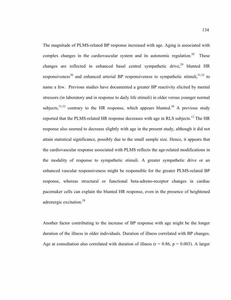
134
The magnitude of PLMS-related BP response increased with age. Aging is associated with
complex changes in the cardiovascular system and its autonomic regulation.28 These
changes are reflected in enhanced basal central sympathetic drive,29 blunted HR
responsiveness30 and enhanced arterial BP responsiveness to sympathetic stimuli,31,32 to
name a few. Previous studies have documented a greater BP reactivity elicited by mental
stressors (in laboratory and in response to daily life stimuli) in older versus younger normal
subjects,31,32 contrary to the HR response, which appears blunted.30 A previous study
reported that the PLMS-related HR response decreases with age in RLS subjects.12 The HR
response also seemed to decrease slightly with age in the present study, although it did not
attain statistical significance, possibly due to the small sample size. Hence, it appears that
the cardiovascular response associated with PLMS reflects the age-related modifications in
the modality of response to sympathetic stimuli. A greater sympathetic drive or an
enhanced vascular responsiveness might be responsible for the greater PLMS-related BP
response, whereas structural or functional beta-adreno-receptor changes in cardiac
pacemaker cells can explain the blunted HR response, even in the presence of heightened
adrenergic excitation.28
Another factor contributing to the increase of BP response with age might be the longer
duration of the illness in older individuals. Duration of illness correlated with BP changes.
Age at consultation also correlated with duration of illness (r = 0.86, p = 0.003). A larger
135
population would be needed to independently assess the effects of these two factors on BP
changes.
The clinical significance of PLMS in RLS and other sleep disorders is controversial.
Several studies have shown that PLMS have little impact on nocturnal sleep or daytime
functioning,9,33,34 leading to the hypothesis that PLMS are not clinically significant. Even
the observed 7 to 10 beats per minute increase in HR12 is unlikely to prove harmful to the
patient. By contrast, the repetitive raise in BP associated with PLMS, as high as 40 mmHg
in some individuals (table 1), could be harmful to the cardiovascular system, especially in
severe cases and older subjects and in those with a long history of the disease. Repetitive
increases in BP associated with the hyperventilatory phases of sleep apneas are believed to
contribute to the pathogenesis of cardiovascular disease in this condition. 35 Striking is the
similarity between the average magnitude of the PLMS-related BP increments observed in
our population (22 ± 10 mmHg) and those associated with the hyperventilatory phases of
apneas and hypopneas (23 ± 10 mmHg) in subjects with sleep apneas.36 Large clinical
studies have shown clearly that enhanced BP variability (as seen in our RLS subjects with
frequent PLMS) is associated with the development of vascular and cardiac damage.17,18,37
Furthermore, selectively enhanced nocturnal BP variability is associated with a higher
incidence of stroke in hypertensive elderly persons.38 Experimental models have shown that
pulsatile blood flow, which may occur in association with frequent PLMS, modulates
several biochemical stimuli, which may in turn promote inflammation and oxidative stress,
136
both precursors of atherosclerosis.39,40 All these findings strongly suggest that PLMS-
related BP surges might exert a deleterious impact on the cardiovascular system in
otherwise healthy subjects, and are potentially implicated in the association reported in
several epidemiological studies between RLS subjects and cardiovascular disease.1,2,3
Longitudinal epidemiological studies as well as the assessment of cardiovascular functions
before and after treatment of RLS with dopaminergic agonists, known to suppress PLMS,
would be needed to clarify this issue. Further studies are also needed to investigate
autonomic changes associated with PLMS in an asymptomatic aged population.
Our findings also have implications for patients with pre-existing cardiovascular disease,
particularly those with systolic heart failure and cardiac transplantation. Studies showed
these patients to have very frequently PLMS and RLS.41,42 The repetitive increase in
afterload as result of PLMS, could adversely affect an already impaired cardiac function
and contribute to the progression of the disease.
The main limitation of the present study is the small sample size. Although sufficient to
demonstrate increase in BP associated with PLMS, it considerably limits the power of the
correlation analyses. These correlations should therefore be interpreted with caution. The
small number of patients results from the stringent selection criteria to eliminate
confounding factors for the cardiovascular response. Only untreated patients with primary
RLS and PLMS and without any associated conditions were included in the study.
137
Restrictive selection criteria were also applied to PLMS and PLMW, and only movements
separated by intervals of at least 20 seconds were analyzed in order to assess individual
responses and avoid overlapping responses associated with PLMS that were separated by
short intervals.
ACKNOWLEDGMENTS
The authors thank Dominique Petit and Margaret McKeys for linguistic revision. The
authors also thank Sylvie Rompré for helping with the analyses.
138
REFERENCES
1Ulfberg J, Nyström B, Carter N, Edling, C. Prevalence of restless legs syndrome among
men aged 18 to 64 years: an association with somatic disease and neuropsychiatric
symptoms. Mov Disord 2001;16:1159-1163.
2Ohayon MM, Roth T. Prevalence of restless legs syndrome and periodic limb movement
disorder in the general population. J Psychosom Res 2002;53: 547-554.
3Winkelman JW, Finn L, Young T. Prevalence and correlates of restless legs syndrome
symptoms in the Wisconsin Sleep Cohort. Sleep Med 2006; 7: 545-552.
4Montplaisir J, Boucher S, Poirier G, Lavigne G, Lapierre O, Lesperance P. Clinical,
polysomnographic, and genetic characteristics of restless legs syndrome: a study of 133
patients diagnosed with new standard criteria. Mov Disord 1997;12:61-65.
5Sforza E, Nicolas A, Lavigne G, Gosselin A, Petit D, Montplaisir J. EEG and cardiac
activation during periodic leg movements in sleep: support for a hierarchy of arousal
responses. Neurology 1999; 2:786-791.
6Montplaisir J, Michaud M, Denesle R, Gosselin A. Periodic leg movements are not more
prevalent in insomnia or hypersomnia but are specifically associated with sleep disorders
involving a dopaminergic impairment. Sleep Med 2000;1:163-167.
7Fantini ML, Michaud M, Gosselin N, Lavigne G, Montplaisir J. Periodic leg movements
in REM sleep behavior disorder and related autonomic and EEG activation. Neurology
2002;59:1889-1894.
139
8Fry JM, DiPhillipo MA, Pressman MR. Periodic leg movements in sleep following treatment
of obstructive sleep apnea with nasal continuous positive airway pressure. Chest 1989;96:89-
91.
9Carrier J, Frenette S, Monplaisir J, Paquet J, Drapeau C, Morettini J. Effects of periodic
leg movements during sleep in middle-aged subjects without sleep complaints. Mov Disord
2005;20:1127-1132.
10Pennestri MH, Whittom S, Adam B, Petit D, Carrier J, Montplaisir J. PLMS and PLMW
in healthy subjects as a function of age: prevalence and interval distribution. Sleep
2006;29:1174-1178.
11Winkelman JW. The evoked heart rate response to periodic leg movements of sleep.
Sleep 1999;22:575-580.
12Gosselin N, Lanfranchi P, Michaud M, et al. Age and gender effects on heart rate
activation associated with periodic leg movements in patients with restless legs syndrome.
Clin Neurophysiol 2003;114:2188-1195.
13World Health Organization-International Society of Hypertension Guidelines for the
Management of Hypertension. Guidelines Sub-Committee. Blood Press Suppl. 1999;1:9-
43.
14Schroeder JS, Motta J, Guilleminault C. Hemodynamic studies in sleep apnea. In:
Guilleminault C & Dement WC, Editors. Sleep apnea syndromes. New York: Liss;
1978:177-196.
140
15 Davies RJ, Belt PJ, Roberts SJ, Ali NJ, Stradling JR. Arterial blood pressure responses to
graded transient arousal from sleep in normal humans. J Appl Physiol 1993;74:1123-1130.
16Morgan BJ, Crabtree DC, Puleo DS, Badr MS, Toiber F, Skatrud JB. Neurocirculatory
consequences of abrupt chane in sleep state in humans. J Appl Physiol 1996; 80:1627-1636.
17Frattola A, Parati G, Cuspidi C, Albini F, Mancia G. Prognostic value of 24-hour blood
pressure variability. J Hypertens 1993;11:1133-1137.
18Zakopoulos NA, Tsivgoulis G, Barlas G, et al. Time rate of blood pressure variation is
associated with increased common carotid artery intima-media thickness. Hypertension.
2005;45:505-512.
19Allen RP, Picchietti D, Hening WA, Trenkwalder C, Walters AS, Montplaisir J. Restless
legs syndrome: diagnostic criteria, special considerations, and epidemiology. A report from
the restless legs syndrome diagnosis and epidemiology workshop at the National Institutes
of Health. Sleep Med 2003;4:101-119.
20Imholz BPM, Wieling W, van Montfrans GA, Wesseling KH. Fifteen years experience
with finger arterial pressure monitoring: assessment of the technology. Cardiovasc Res
1998;38:605-616.
21Rechtschaffen A, Kales A. A manual of standardized terminology, techniques and
scoring system for sleep stages of human subjects. Los Angeles: Brain Information Service,
1968.
141
22Coleman RM. Periodic movements in sleep (nocturnal myoclonus) and restless legs
syndrome. In: Guilleminault C, ed. Sleeping and Waking Disorders. Menlo Park: Addison-
Wesley; 1982: 265-295.
23American Sleep Disorders Association. EEG arousals: scoring rules and examples: a
preliminary report from the Sleep Disorders Atlas Task Force of the American Sleep
Disorders Association. Sleep 1992;15:173-184.
24American Academy of Sleep Medicine. Sleep-related breathing disorders in adults:
recommendations for syndrome definition and measurement techniques in clinical research.
The Report of an American Academy of Sleep Medicine Task Force. Sleep 1999; 22: 667-
689.
25Michaud M, Poirier G, Lavigne G, Montplaisir J. Restless legs syndrome: scoring criteria
for leg movements recorded during the suggested immobilization test. Sleep Med
2001;2:317-321.
26Ferrillo F, Beelke M, Canovaro P, et al. Changes in cerebral and autonomic activity
heralding periodic limb movements in sleep. Sleep Med 2004;5:407-412.
27Cui J, Blaha C, Moradkhan R, et al. Muscle sympathetic nerve activity responses to
dynamic passive muscle stretch in humans, J Physiol 2006; 576: 625-634.
28Lakatta EG. Cardiovascular regulatory mechanisms in advanced age. Physiol Rev
1993;73:413-467.
142
29Esler M, Hastings J, Lambert G, Kaye D, Jennings G, Seals DR. The influence of aging
on the human sympathetic nervous system and brain norepinephrine turnover. Am J Physiol
Regul Integr Comp Physiol 2002; 282: R909-R916.
30Stratton JR, Cerqueira MD, Schwartz RS, et al. Differences in cardiovascular responses to
isoproterenol in relation to age and exercise training in healthy men. Circulation
1992;86:504-512.
31Uchino BN, Uno D, Holt-Lunstad J, Flinders JB. Age-related differences in
cardiovascular reactivity during acute psychological stress in men and women. J Geronol B
Psychol Sci Soc Sci 1999;54:P339-P346.
32Uchino BN, Berg CA, Smith TW, Pearce G, Skinner M. Age-related differences in
ambulatory blood pressure during daily stress: evidence for greater blood pressure
reactivity with age. Psychol Aging 2006;21:231-239.
33Nicolas A, Lesperance P, Montplaisir J. Is excessive daytime sleepiness with
periodic leg movements during sleep a specific diagnostic category ? Eur Neurol
1998;40:22-26.
34Mendelson WB. Are periodic leg movements associated with clinical sleep disturbance?
Sleep 1996;19:219-223.
35Somers VK, Dyken ME, Clary MP, Abboud FM. Sympathetic neural mechanisms in
obstructive sleep apnea. 1995;96:1897-1904.
36Morgan BJ, Dempsey JA, Pegelow DF et al. Blood pressure perturbations caused by
subclinical sleep-disordered breathing. Sleep 1998;21:737-746.
143
37Roman MJ, Pickering TG, Schwartz JE, Pini R, Devereux RB. Relation of blood pressure
variability to carotid atherosclerosis and carotid artery and left ventricular hypertrophy.
Arterioscler Thromb Vasc Biol 2001;21:1507-511.
38Pringle E, Phillips C, Thijs L, et al. Systolic blood pressure variability as a risk factor for
stroke and cardiovascular mortality in the elderly hypertensive population. J Hypertens
2003;21:251-257.
39De Keulenaer GW, Chappell DC, Ishizaka N, Nerem RM, Alexander RW, Griendling
KK. Oscillatory and steady laminar shear stress differentially affect human endothelial
redox state: role of a superoxide-producing NADH oxidase. Circ Res 1998;82: 1094-1101.
40Chappell DC, Varner SE, Nerem RM, Medford RM, Alexander RW. Oscillatory shear
stress stimulates adhesion molecule expression in cultured human endothelium. Circ Res
1998;82:532-539.
41Javaheri S, Abraham WT, Brown C, Nishiyama H, Giesting R, Wagoner LE. Prevalence
of obstructive sleep apnoea and periodic limb movements in 45 subjects. Eur heart J
2004;25:260-266.
42Javaheri S. Sleep disorders in systolic heart failure: a prospective study of 100 male
patients. The final report. Int J Cardiol 2006;106:21-28.
144
FIGURE LEGENDS
Figure 1.
PLMS-related beat-to-beat blood pressure, EKG and EEG signals, presented in a compact
window (Panel A), and wider temporal windows (Panel B). The portion of signals shown in
Panel B represents the temporal window used for the analyses. Panel C illustrates the
corresponding measurements of SBP, DBP and HR.
Figure 2.
Mean SBP, DBP and HR changes associated with PLMS-MA (black curve) and PLMS-
noMA (grey curve). Dashed line represents the onset of leg movement. Data are presented
as means ± SEM. Asterisks indicate p< 0.05 from baseline.
Figure 3.
Mean SBP and DBP increments associated with PLMW and PLMS-noMA. Data are
presented as means ± SEM. Asterisks indicate p< 0.05.
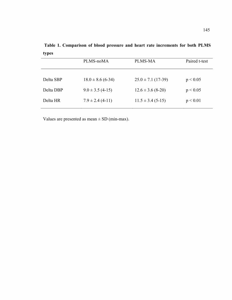
145
Table 1. Comparison of blood pressure and heart rate increments for both PLMS
types
PLMS-noMA
PLMS-MA
Paired t-test
Delta SBP
18.0 ± 8.6 (6-34)
25.0 ± 7.1 (17-39)
p < 0.05
Delta DBP 9.0 ± 3.5 (4-15) 12.6 ± 3.6 (8-20) p < 0.05
Delta HR 7.9 ± 2.4 (4-11) 11.5 ± 3.4 (5-15) p < 0.01
Values are presented as mean ± SD (min-max).
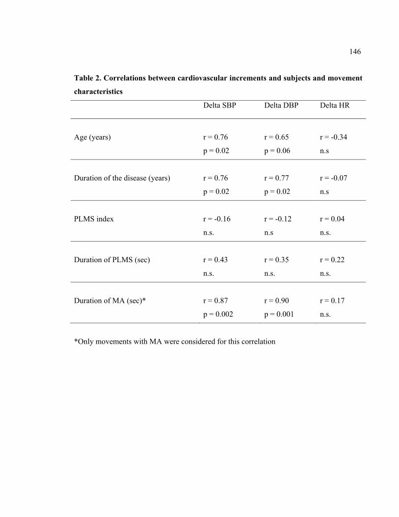
146
Table 2. Correlations between cardiovascular increments and subjects and movement
characteristics
Delta SBP Delta DBP Delta HR
Age (years)
r = 0.76
p = 0.02
r = 0.65
p = 0.06
r = -0.34
n.s
Duration of the disease (years)
r = 0.76
p = 0.02
r = 0.77
p = 0.02
r = -0.07
n.s
PLMS index
r = -0.16
n.s.
r = -0.12
n.s
r = 0.04
n.s.
Duration of PLMS (sec)
r = 0.43
n.s.
r = 0.35
n.s.
r = 0.22
n.s.
Duration of MA (sec)*
r = 0.87
p = 0.002
r = 0.90
p = 0.001
r = 0.17
n.s.
*Only movements with MA were considered for this correlation
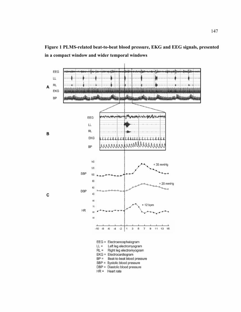
147
Figure 1 PLMS-related beat-to-beat blood pressure, EKG and EEG signals, presented
in a compact window and wider temporal windows
Figure 2
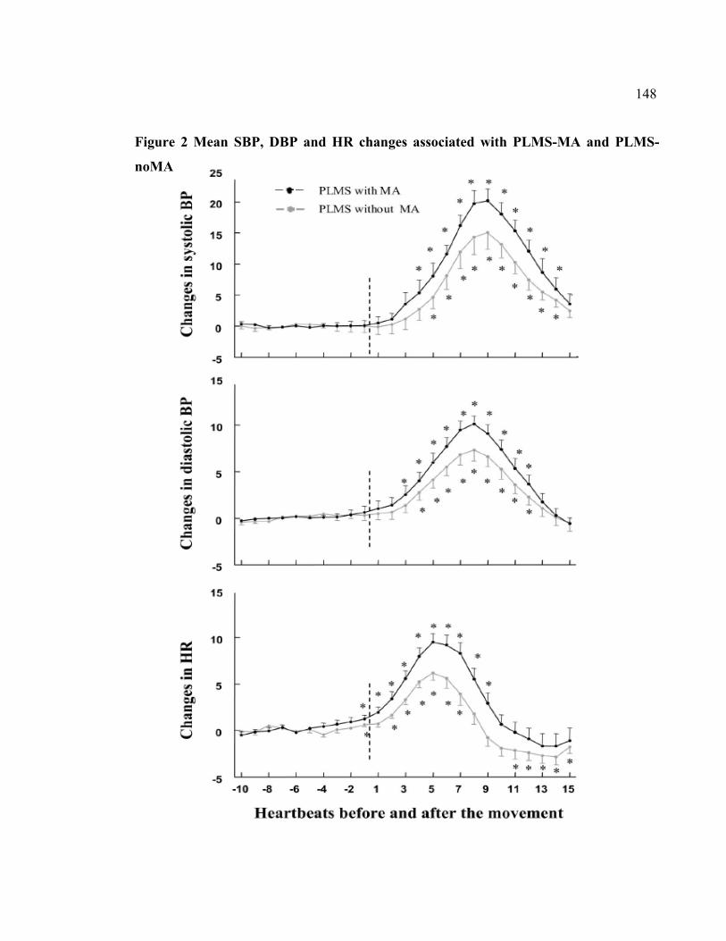
148
Figure 2 Mean SBP, DBP and HR changes associated with PLMS-MA and PLMS-
noMA
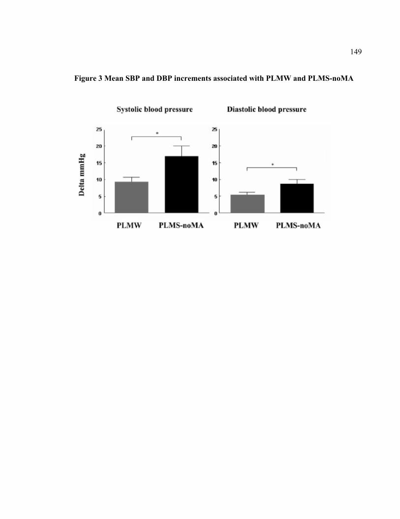
149
Figure 3 Mean SBP and DBP increments associated with PLMW and PLMS-noMA
150
2.5. Cinquième article
Blood pressure changes associated with periodic leg movements during sleep in healthy
subjects not complaining of sleep disorders
Marie-Hélène Pennestri BSc1,2, Jacques Montplaisir MD, PhD1,3, Lorraine Fradette1, Gilles
Lavigne DMD, PhD1,4, Roberto Colombo5, Paola A Lanfranchi MD, MSc1,6,7
1Sleep Disorders Center, Sacre-Cœur Hospital, University of Montreal 2Department of Psychology, University of Montreal
3Department of Psychiatry, University of Montreal 4Faculty of Dentistry, University of Montreal
5Department of Bioengineering, Salvatore Maugeri Foundation, Veruno, Italy 6Cardiology division, Sacré-Cœur Hospital, University of Montreal
7Department of Medicine, University of Montreal
Article soumis, Journal of Clinical Neurophysiology

151
Contribution des auteurs
Marie-Hélène Pennestri : Élaboration du protocole, recrutement des participants,
participation à l’évaluation des participants, analyse des données, analyse statistiques,
interprétation des résultats, rédaction du manuscrit.
Jacques Montplaisir : Élaboration du protocole, interprétation des résultats, révision du
manuscrit.
Lorraine Fradette : Participation à l’évaluation des participants, analyse des données
Gilles Lavigne: Interprétation des résultats, révision du manuscrit
Roberto Colombo: Particiaption à l’analyse des données
Paola A Lanfranchi: Élaboration du protocole, interprétation des résultats, révision du
manuscrit.
152
ABSTRACT
Study Objectives: To assess heart rate (HR) and blood pressure (BP) changes associated
with periodic leg movements during sleep (PLMS) with or without electroencephalographic
micro-arousals (MA) in healthy subjects not complaining of sleep disorders.
Design: Beat-to-beat noninvasive blood pressure (Portapres) was continuously recorded
during one night of polysomnography.
Setting: Data were collected at the Sleep Disorders Center, Sacre-Coeur Hospital,
Montreal,Canada.
Participants: Fifteen non-complaining healthy subjects (7 men, 8 women; mean age = 46.7
± 9.3 yrs) were studied.
Interventions: NA
Measurements and Results: Ten PLMS with micro-arousals (PLMS-MA) and 10 PLMS
without micro-arousals (PLMS-noMA) were analyzed for each subject. Systolic and
diastolic BP (SBP, DBP) were measured within a 30-beat temporal window comprising 10
beats before and 20 beats after onset of each movement. Significant increments in HR and
BP were associated with all PLMS (average: Delta HR = 8 bpm; Delta SBP = 18 mm Hg;
Delta DBP = 9 mm Hg). HR and BP increments associated with PLMS-MA were greater
than those associated with PLMS-noMA (p<0.05). HR changes decrease with age but no
correlation was observed between BP changes and age.

153
Conclusions: Significant heart rate and blood pressure increases are associated with
periodic leg movements during sleep in healthy subjects not complaining of sleep disorders.
Keywords: Sleep, healthy subjects, periodic leg movements, blood pressure, sympathetic
nervous system.
154
INTRODUCTION
Periodic leg movements (PLMS) are short, involuntary movements occurring repetitively
during sleep at about 20- to 40-second intervals. They are described as a rhythmic
extension of the big toe and dorsiflexion of the ankle, with occasional flexion at the knee
and hip. They have been extensively studied in patients with restless legs syndrome (RLS),
where they are considered a supportive criterion for diagnosis (Allen, Picchietti, Hening,
Trenkwalder, Walters, & Montplaisi, 2003), but they also occur in other sleep disorders
such as narcolepsy (Dauvilliers, Pennestri, et al., 2007), rapid eye movement (REM) sleep
behavior disorder (Fantini, et al., 2002), and sleep apnea (Baran, Richert, Douglass, May, &
Ansarin, 2003; Fry, et al., 1989). Several studies have also shown a high prevalence of
PLMS in healthy non-complaining subjects, especially after the age of 40 years (Carrier, et
al., 2005; Pennestri et al., 2006). A recent study determined that the prevalence of PLMS in
a population-based sample of 592 participants was 7.6%, considering a PLMS index > 15
as a criterion (Scofield, et al., 2008).
It is now well established that PLMS are associated with significant heart rate (HR)
changes, namely a tachycardia followed by a bradycardia, with larger amplitude changes
when PLMS are accompanied by micro-arousals (MA) (Sforza, et al., 1999) or by
synchronized EEG activity (burst of delta waves or K-complex) (Sforza, et al., 2002). This
PLMS-associated cardiac activation has been observed in patients with RLS (Gosselin, et
al., 2003; Sforza, et al., 1999), REM sleep behavior disorders (RBD) (Fantini et al., 2002),
155
and in adults (Ferrillo, et al., 2004; Winkelman, 1999) and children (Walter, et al., 2009)
with PLMS. Using various algorithms of analysis including fast Fourier transformation
(FFT) and wavelet HR variability analysis, it has been shown that the sympathetic activity
associated with PLMS is higher during stage 2 of NREM sleep as compared to slow wave
sleep (Sforza, et al., 2005).
Recently we found that, in addition to HR changes, PLMS are associated with significant
blood pressure (BP) increase in RLS patients whether or not movements are associated with
MA. Mean increases were 22 mm Hg for systolic BP (SBP) and 11 mm Hg for diastolic BP
(DBP), with greater activation when PLMS were associated with MA (Pennestri, et al.,
2007). These findings have been replicated in treated RLS patients (who therefore
presented a lower PLMS index), albeit the amplitude of cardiovascular activation
associated with PLMS response seemed to be lower (Siddiqui et al., 2007). The aim of the
present study was to assess HR and BP changes associated with PLMS in healthy non-
complaining subjects and to assess the relationship between these changes and age and
polysomnographic variables.
METHODS
Subjects
Thirty-two non-complaining healthy subjects were recruited through newspaper
advertisements to participate in this study. Subjects with symptoms of RLS or a family
156
history of RLS or with a current diagnosis of cardiovascular diseases, including
hypertension, motor disorders, sleep disorders, or psychiatric disorders, were excluded from
the study. Subjects using any medication known to influence sleep, motor activity, or
autonomic nervous system activity and those with an index of respiratory events (apneas +
hypopneas) > 10 were also excluded. None of the subjects included had participated in
previous studies on PLMS-related cardiovascular changes conducted in our laboratory.
Fifteen of these subjects (7 men, 8 women), had a PLMS index > than 5 per hour of sleep
and were included in the study. They were aged from 30 to 62 years (mean age = 46.7 ± 9.3
yrs). All subjects signed a consent form approved by the ethics committee of the Sacré-
Coeur Hospital.
Data collection
Subjects spent one night in the sleep laboratory. In the evening preceding
polysomnographic (PSG) sleep recordings, brachial arterial BP was measured at rest in the
sitting position with a cuff on the non-dominant arm to provide a baseline clinical SBP and
DBP. All subjects had normal BP (SBP: 112.8 ± 10.1 mmHg, DPB: 69.4 ± 8.6 mmHg).
Sleep recording was performed using four electroencephalographic (EEG) leads (C3, C4,
O1, and O2), two bilateral electrooculograms (EOG), and one chin (electromyogram)
EMG. Respiration was assessed by nasal-cannula and thoracoabdominal strain gauges and
finger pulse oxymetry. Surface EMG electrodes placed 3 cm apart on the right and left
anterior tibialis muscles were used to record PLMS and voluntary movements. The
157
electrocardiogram (ECG) included leads I, II, and III. Continuous BP recordings were
obtained using a TNO Portapres Model-2 (TNO TPD Biomedical Instrumentation, The
Netherlands), which provides non-invasive beat-to-beat BP recording through a continuous
finger arterial pressure waveform. Although this method has shown consistent
underestimation compared to direct measurements with the brachial artery, it provides an
accurate measure of sudden BP changes in response to various stimuli (Imholz, Wieling,
van Montfrans, & Wesseling, 1998). It is therefore an appropriate non-invasive alternative
method to measure beat-to-beat BP changes associated with episodic events during sleep.
Data analyses
Sleep was scored by the standard method (Rechtschaffen & Kales, 1968) and PLMS were
scored using the criteria set by the International RLS Study Group (Zucconi, et al., 2006).
Only movements lasting 0.5 to 10 seconds, separated by intervals of 5 to 90 seconds and
occurring in a series of at least 4 consecutive movements, were counted. The amplitude
criterion for detecting leg movements was an increase in EMG to ≥ 8 µV above the resting
baseline for movement onset and a decrease in EMG to < 2 µV above the resting level for
movement offset. MAs were scored according to standard ASDA criteria (American Sleep
Disorders Association, 1992). Respiratory events were scored according to ASDA and the
Academy of Sleep Medicine recommendations (Report of an American Academy of Sleep
Medicine Task Force, 1999).
158
PLMS selection and cardiovascular analyses
For each subject, 20 PLMS (10 PLMS with MA [PLMS-MA] and 10 PLMS without MA
[PLMS-noMA]) were selected for cardiovascular analysis. In addition to the standard
criteria for PLMS, only movements separated by at least 20 seconds were selected for
analysis in order to avoid overlapping HR and BP responses to successive leg movements.
Movements were selected in stage 2 to avoid sleep stage interaction. Only movements free
of any physiological (rhythmic masticatory muscle activity, flow limitation at the nasal
cannula, apneas and hypopneas, arrhythmias) or technical factors that would potentially
affect HR or BP signals were selected.
HR, SBP, and DBP were measured automatically on segments lasting 30 heartbeats,
comprising 10 beats before the movement (-10 to -1) and 20 beats after the movement (+1
to +20) (figure 1). Beat-to-beat values of HR, SBP, and DBP were calculated. For each
movement, baseline was defined as the mean value from beats -10 to -4. The highest value
obtained after movement onset was taken as the peak value. Increments (delta) from
baseline to peak values were calculated for HR, SBP, and DBP during PLMS-MA and
PLMS-noMA in each subject. HR, SBP, and DBP increments associated with both
movement types were averaged for each subject ((delta PLMS-MA + delta PLMS-
noMA)/2) to obtain overall individual HR, SBP, and DBP increments during sleep.
159
Statistical analyses
HR, SBP, and DBP responses associated with PLMS-noMA and PLMS-MA were assessed
by Friedman ANOVA followed by Wilcoxon matched pairs tests. Each value of HR, SBP,
and DBP (beat -3 to +20) was compared to baseline (beat -10 to -4), where differences were
considered statistically significant at p < 0.01, considering the multiple comparisons.
A within-subject comparison of HR, SBP, and DBP increments associated with PLMS-MA
and PLMS-noMA was performed using paired t-tests. Pearson correlation coefficients were
calculated between HR, SBP, and DBP increments associated with both PLMS types
pooled together and the following variables: age, PLMS index, PLMS duration, and MA
duration associated with PLMS.
RESULTS
Mean PLMS index was 30.5 ± 23.2 (range 5.5 to 81.2) and positively correlated with age (r
= 0.68; p = 0.006). Duration of the PLMS-MA selected for cardiovascular analysis were
longer than PLMS-noMA (2.7 ± 1.0 vs. 1.9 ± 0.5, p = 0.02). Mean duration of MA
associated with PLMS used for analysis was 5.9 ± 1.1 sec.
Changes in SBP, DBP, and HR in association with PLMS are illustrated in Figure 2.
PLMS-noMA were associated with an increase in SBP [χr2 =264.0, p < 0.00001]. Wilcoxon
matched pairs tests revealed significant changes for beats + 5 to + 14. An increase in DBP
160
was associated with PLMS-noMA [χr2 =226.0, p < 0.00001], with significant changes for
beats +4 to +11. PLMS-noMA were associated with HR changes [χr2 = 214.8, p <
0.00001], with a tachycardia (beats +1 to +6) and a bradycardia (beats +10 to +13).
Even higher SBP increments were found for PLMS-MA [χr2 =236.1, p < 0.00001] and DBP
[χr2 =256.4, p < 0.00001]. Wilcoxon matched pairs tests revealed significant increases in
SBP for beats +3 to +13 and significant increases in DBP for beats +3 to + 11, followed by
a decrease for beats + 18 to + 20. PLMS-MA were also associated with significant HR
changes [χr2 =199.3, p < 0.00001]. Increased HR was significant for beats +2 to +8.
Comparisons of cardiovascular changes associated with PLMS-noMA and PLMS-MA are
presented in Table 1. All increments in SBP, DBP and HR were greater for PLMS-MA than
for PLMS-noMA.
HR changes decreased with age (r = -0.51; p = 0.05), but no relationship was observed
between SBP changes (r = 0.004; p = 1) or DBP changes (r = -0.27; p = 0.3) and age. No
relationship was observed between PLMS index, PLMS, or MA duration and
cardiovascular changes, although the small sample size limits the correlation analysis.
161
DISCUSSION
1. Autonomic changes associated with PLMS in healthy subjects
To our knowledge, this is the first study to report significant BP increase associated with
PLMS in healthy non-complaining subjects. The cardiovascular changes observed in the
present study are very similar to those found recently in patients with RLS (Pennestri, et al.,
2007). It is noteworthy that age and mean PLMS index were similar in the two studies.
Further studies, conducted in large populations of RLS and normal subjects, will be needed
to directly assess between-group differences in PLMS-related cardiovascular changes.
As in previous studies (Pennestri, et al., 2007; Sforza, et al., 1999; Siddiqui, et al., 2007),
PLMS-related BP and HR changes were of higher magnitude when PLMS were associated
with MA. One possibility would be that PLMS associated with MA have different
characteristics, such as increased duration of movement, but no correlation was found
between the duration of PLMS and the magnitude of blood pressure changes (SBP: r =
0.32; p = 0.24; DBP: r = 0.32; p = 0.25). However, these results should be interpreted
cautiously, considering the small sample size for correlation analysis.
The present study confirms the decrease in PLMS-related HR changes observed in RLS
patients with advancing age (Gosselin, et al., 2003), but found no relationship between the
magnitude of PLMS-related BP responses and age. In contrast, in a previous study
conducted with RLS patients, age was strongly correlated with increased BP associated
162
with PLMS (Pennestri, et al., 2007). It is worth mentioning that, in the RLS study, the
magnitude of BP responses was also correlated with the duration of the illness. However,
the small sample did not allow an independent assessment of the two factors. The present
results indirectly suggest that illness duration may in fact be more important than age per se
to explain BP increases in these patients.
It is possible that a portion of the PLMS-related BP activation is secondary to leg muscle
activation. However, previous studies showed a higher increase in PLMS-related HR or BP
than changes associated with voluntary movements (Siddiqui, et al., 2007; Winkelman,
1999), involuntary periodic leg movements during wakefulness (PLMW) (Lavoie, et al.,
2004; Pennestri, et al., 2007; Siddiqui, et al., 2007), or passive ankle movement (Cui,
Blaha, Moradkhan, Gray, & Sinoway, 2006). Therefore, autonomic changes observed in
association with PLMS are not caused by the leg movement itself.
2. Physiopathology of PLMS and relationship with arousals
The physiopathology of PLMS is still unclear. Several studies have shown that cortical
arousals such as MA or specific EEG pattern, called K-alpha, where the K-complex is
followed by a burst of alpha activity, may sometimes occur before, during, or after a leg
movement (Karadeniz, Ondze, Besset, & Billiard, 2000b; Montplaisir, et al., 1996). This
inconsistent temporal relation suggests that cortical activation is not simply a response
triggered by motor activity.
163
Moreover, either PLMS or arousals can be observed in the absence of the other
phenomenon. In a case report, cortical arousals have been observed independently of PLMS
in the middle of PLMS-MA sequences (El-Ad & Chervin, 2000). Other evidence comes
from pharmacological studies. One study showed the persistence of K-alpha when PLMS
were suppressed by L-Dopa (Montplaisir, et al., 1996), whereas another report showed that
MA were specifically suppressed by temazepam (Ali, et al., 1991). Taken together, these
studies suggest that cortical and motor activities are not generated by one another, but
rather that the two phenomena could be part of a global arousal mechanism.
Some authors have proposed that a central generator could periodically trigger one or
several arousal responses, which could explain why the leg movement or arousal could be
occasionally absent (El-Ad & Chervin, 2000; Montplaisir, et al., 1996). To this effect, a
study reported a correlation between PLMS and cyclic alternating pattern (CAP), defined as
sequences of transient arousals, which are distinct from background
electroencephalographic activity in NREM sleep. This correlation suggests that CAP
operates as a gate-control rhythm for PLMS (Parrino et al., 1996). It has also been
postulated that a hierarchy in the arousal response was present and followed a continuum
(Ferrillo, et al., 2004; Sforza, Jouny, & Ibanez, 2000; Sforza, et al., 1999). In the present
study, the association between PLMS and MA would represent a higher degree of arousal,
which could explain the higher increase in BP in this case.
164
An alternate hypothesis would be that the autonomic activation is responsible for arousals
and PLMS. To this effect, patients with RBD who present an autonomic dysfunction
(Lanfranchi, Fradette, Gagnon, Colombo, & Montplaisir, 2007) and a loss of sympathetic
terminals (Miyamoto et al., 2006) have a suppression of MA associated with PLMS and an
attenuation of the PLMS-associated HR increase (Fantini et al., 2002).
3. Clinical significance
The clinical significance of PLMS remains unclear. In different clinical populations, some
authors found an association between PLMS and polysomnographic sleep variables
(Bastuji & Garcia-Larrea, 1999; Hilbert & Mohsenin, 2003; Saskin, et al., 1985), while
other studies showed no relationship between PLMS and objective or subjective quality of
sleep, or daytime sleepiness (Hornyak, et al., 2004; Karadeniz, et al., 2000a; Mendelson,
1996; Nicolas, et al., 1998; Youngstedt, et al., 1998). In healthy non-complaining subjects,
it was recently shown that a high PLMS index does not influence polysomnographic sleep
(Carrier, et al., 2005). The impact of PLMS on sleep and daytime functioning therefore
remains controversial.
Some epidemiological studies have found an association between severe RLS and an
increased risk for hypertension (Phillips, Hening, Britz, & Mannino, 2006; Ulfberg, et al.,
2001a). A more convincing relationship was reported between RLS and cardiovascular
165
disease (Ohayon & Roth, 2002; Ulfberg, et al., 2001a; Winkelman, et al., 2006;
Winkelman, et al., 2008). While in one hand RLS-related insomnia and poor sleep could be
factors implicated in this relationship, it is also possible that rapid and transient BP
increments associated with PLMS, present in approximately 80% of RLS patients
(Montplaisir, et al., 1997), contribute to this association. Two studies showed that the
likelihood of hypertension increases with PLMS severity (Billars et al., 2007; Espinar-
Sierra, Vela-Bueno, & Luque-Otero, 1997). Other studies point to the potential link
between PLMS and cardiovascular disease in non RLS populations. One phone survey
found such an association between periodic leg movements disorder and heart disease
(where periodic leg movements disorder was defined as a complaint of insomnia or
excessive sleepiness and the presence of PLMS), but as no laboratory evaluation was
performed, it is impossible to evaluate the specific association between PLMS and
cardiovascular disease in this condition (Ohayon & Roth, 2002). Studies performing
polysomnography in patients with congestive heart failure found that 19 to 39% of these
patients have an index of PLMS ≥5/hour (Hanly & Zuberi, 1992; Hanly & Zuberi-Khokhar,
1996; Javaheri, 2006; Javaheri, et al., 2004; Skomro, et al., 2009; Yumino, Wang, & Floras,
2008). The repetitive PLMS related BP surges in these patients could result in repetitive
increase in afterload and then have the potential to adversely affect an already impaired
cardiac function. Indeed, a preliminary report conducted in 193 patients with congestive
heart failure suggests that a high PLMS index in this population is associated with more
166
profound clinical and functional impairment, and higher mortality rates (Yumino, et al.,
2008).
A number of studies have shown consistently that PLMS index increases with age (Dickel
& Mosko, 1990; Hilbert & Mohsenin, 2003; Mendelson, 1996; Nicolas, et al., 1998;
Pennestri, et al., 2006), as confirmed in the present study. Therefore, although no
correlation was observed between age and the magnitude of BP increases, the rise in BP is
repeated more often during the night in older subjects, which could increase nocturnal BP
variability. It is well documented that an augmentation in BP variability is associated with
the development of vascular and cardiac damage (Frattola, Parati, Cuspidi, Albini, &
Mancia, 1993; Roman, Pickering, Schwartz, Pini, & Devereux, 2001; Zakopoulos et al.,
2005). Thus, the PLMS-related repeated cardiovascular changes could contribute to
increase cardiovascular risk especially in elderly populations. Finally, preliminary data
suggest that as effect of dopaminergic treatment not only there is a significant reduction in
PLMS rate but there might be also an attenuation in HR response associated with the
residual PLMS.(Zucconi et al., 2010) It would be important to establish as whether
dopaminergic agents can also control PLMS related BP changes.
4. Limitations
Some limitations should be considered in the present study. First, whereas all subjects
showed a clear increase in BP associated with PLMS, the sample was too small to perform
167
a reliable correlation analysis between changes in cardiovascular parameters and movement
characteristics. Second, a small number of movements were studied in each subject, as the
20-second criterion between movements limited the number of analyzed PLMS. However,
this criterion allowed the maximal BP increase to be determined, as the signal was able to
return to baseline level before the next movement.
Conclusion
PLMS are associated with significant HR and BP changes in healthy non-complaining
subjects. This increase is even greater when PLMS are associated with MA. As the PLMS
index increases with age and aging is associated with high cardiovascular risk, further
studies should investigate the impact of PLMS-related BP changes on the development of
cardiovascular disease in elderly populations.
ACKNOWLEDMNENT
The authors thank Dominique Petit and Margaret McKeys for linguistic revision.
168
REFERENCES
1. Allen RP, Picchietti D, Hening WA, Trenkwalder C, Walters AS, Montplaisi J.
Restless legs syndrome: diagnostic criteria, special considerations, and epidemiology. A
report from the restless legs syndrome diagnosis and epidemiology workshop at the
National Institutes of Health. Sleep Med 2003;4:101-19.
2. Dauvilliers Y, Pennestri MH, Petit D, Dang-Vu T, Lavigne G, Montplaisir J.
Periodic leg movements during sleep and wakefulness in narcolepsy. J Sleep Res
2007;16:333-9.
3. Fantini ML, Michaud M, Gosselin N, Lavigne G, Montplaisir J. Periodic leg
movements in REM sleep behavior disorder and related autonomic and EEG activation.
Neurology 2002;59:1889-94.
4. Baran AS, Richert AC, Douglass AB, May W, Ansarin K. Change in periodic limb
movement index during treatment of obstructive sleep apnea with continuous positive
airway pressure. Sleep 2003;26:717-20.
5. Fry JM, DiPhillipo MA, Pressman MR. Periodic leg movements in sleep following
treatment of obstructive sleep apnea with nasal continuous positive airway pressure. Chest
1989;96:89-91.
6. Carrier J, Frenette S, Montplaisir J, Paquet J, Drapeau C, Morettini J. Effects of
periodic leg movements during sleep in middle-aged subjects without sleep complaints.
Mov Disord 2005;20:1127-32.
169
7. Pennestri MH, Whittom S, Adam B, Petit D, Carrier J, Montplaisir J. PLMS and
PLMW in healthy subjects as a function of age: prevalence and interval distribution. Sleep
2006;29:1183-7.
8. Scofield H, Roth T, Drake C. Periodic limb movements during sleep: population
prevalence, clinical correlates, and racial differences. Sleep 2008;31:1221-7.
9. Sforza E, Nicolas A, Lavigne G, Gosselin A, Petit D, Montplaisir J. EEG and
cardiac activation during periodic leg movements in sleep: support for a hierarchy of
arousal responses. Neurology 1999;52:786-91.
10. Sforza E, Juony C, Ibanez V. Time-dependent variation in cerebral and autonomic
activity during periodic leg movements in sleep: implications for arousal mechanisms. Clin
Neurophysiol 2002;113:883-91.
11. Gosselin N, Lanfranchi P, Michaud M, et al. Age and gender effects on heart rate
activation associated with periodic leg movements in patients with restless legs syndrome.
Clin Neurophysiol 2003;114:2188-95.
12. Winkelman JW. The evoked heart rate response to periodic leg movements of sleep.
Sleep 1999;22:575-80.
13. Ferrillo F, Beelke M, Canovaro P, et al. Changes in cerebral and autonomic activity
heralding periodic limb movements in sleep. Sleep Med 2004;5:407-12.
14. Walter LM, Foster AM, Patterson RR, et al. Cardiovascular variability during
periodic leg movements in sleep in children. Sleep 2009;32:1093-9.
170
15. Sforza E, Pichot V, Barthelemy JC, Haba-Rubio J, Roche F. Cardiovascular
variability during periodic leg movements: a spectral analysis approach. Clin Neurophysiol
2005;116:1096-104.
16. Pennestri MH, Montplaisir J, Colombo R, Lavigne G, Lanfranchi PA. Nocturnal
blood pressure changes in patients with restless legs syndrome. Neurology 2007;68:1213-8.
17. Siddiqui F, Strus J, Ming X, Lee IA, Chokroverty S, Walters AS. Rise of blood
pressure with periodic limb movements in sleep and wakefulness. Clin Neurophysiol
2007;118:1923-30.
18. Imholz BP, Wieling W, van Montfrans GA, Wesseling KH. Fifteen years
experience with finger arterial pressure monitoring: assessment of the technology.
Cardiovasc Res 1998;38:605-16.
19. Rechtschaffen A, Kales A. A manual of standardized terminology, techniques and
scoring system for sleep stages of human subjects. Los Angeles: Brain Information Service,
1968.
20. Zucconi M, Ferri R, Allen R, et al. The official World Association of Sleep
Medicine (WASM) standards for recording and scoring periodic leg movements in sleep
(PLMS) and wakefulness (PLMW) developed in collaboration with a task force from the
International Restless Legs Syndrome Study Group (IRLSSG). Sleep Med 2006;7:175-83.
21. EEG arousals: scoring rules and examples: a preliminary report from the Sleep
Disorders Atlas Task Force of the American Sleep Disorders Association. Sleep
1992;15:173-84.
171
22. Sleep-related breathing disorders in adults: recommendations for syndrome
definition and measurement techniques in clinical research. The Report of an American
Academy of Sleep Medicine Task Force. Sleep 1999;22:667-89.
23. Lavoie S, de Bilbao F, Haba-Rubio J, Ibanez V, Sforza E. Influence of sleep stage
and wakefulness on spectral EEG activity and heart rate variations around periodic leg
movements. Clin Neurophysiol 2004;115:2236-46.
24. Cui J, Blaha C, Moradkhan R, Gray KS, Sinoway LI. Muscle sympathetic nerve
activity responses to dynamic passive muscle stretch in humans. J Physiol 2006;576:625-
34.
25. Montplaisir J, Boucher S, Gosselin A, Poirier G, Lavigne G. Persistence of
repetitive EEG arousals (K-alpha complexes) in RLS patients treated with L-DOPA. Sleep
1996;19:196-9.
26. Karadeniz D, Ondze B, Besset A, Billiard M. EEG arousals and awakenings in
relation with periodic leg movements during sleep. J Sleep Res 2000;9:273-7.
27. El-Ad B, Chervin RD. The case of a missing PLM. Sleep 2000;23:450-1.
28. Ali NJ, Davies RJ, Fleetham JA, Stradling JR. Periodic movements of the legs
during sleep associated with rises in systemic blood pressure. Sleep 1991;14:163-5.
29. Parrino L, Boselli M, Buccino GP, Spaggiari MC, Di Giovanni G, Terzano MG.
The cyclic alternating pattern plays a gate-control on periodic limb movements during non-
rapid eye movement sleep. J Clin Neurophysiol 1996;13:314-23.
172
30. Sforza E, Jouny C, Ibanez V. Cardiac activation during arousal in humans: further
evidence for hierarchy in the arousal response. Clin Neurophysiol 2000;111:1611-9.
31. Lanfranchi PA, Fradette L, Gagnon JF, Colombo R, Montplaisir J. Cardiac
autonomic regulation during sleep in idiopathic REM sleep behavior disorder. Sleep
2007;30:1019-25.
32. Miyamoto T, Miyamoto M, Inoue Y, Usui Y, Suzuki K, Hirata K. Reduced cardiac
123I-MIBG scintigraphy in idiopathic REM sleep behavior disorder. Neurology
2006;67:2236-8.
33. Saskin P, Moldofsky H, Lue FA. Periodic movements in sleep and sleep-wake
complaint. Sleep 1985;8:319-24.
34. Bastuji H, Garcia-Larrea L. Sleep/wake abnormalities in patients with periodic leg
movements during sleep: factor analysis on data from 24-h ambulatory polygraphy. J Sleep
Res 1999;8:217-23.
35. Hilbert J, Mohsenin V. Can periodic limb movement disorder be diagnosed without
polysomnography? A case-control study. Sleep Med 2003;4:35-41.
36. Mendelson WB. Are periodic leg movements associated with clinical sleep
disturbance? Sleep 1996;19:219-23.
37. Nicolas A, Lesperance P, Montplaisir J. Is excessive daytime sleepiness with
periodic leg movements during sleep a specific diagnostic category? Eur Neurol
1998;40:22-6.
173
38. Youngstedt SD, Kripke DF, Klauber MR, Sepulveda RS, Mason WJ. Periodic leg
movements during sleep and sleep disturbances in elders. J Gerontol A Biol Sci Med Sci
1998;53:M391-4.
39. Karadeniz D, Ondze B, Besset A, Billiard M. Are periodic leg movements during
sleep (PLMS) responsible for sleep disruption in insomnia patients? Eur J Neurol
2000;7:331-6.
40. Hornyak M, Riemann D, Voderholzer U. Do periodic leg movements influence
patients' perception of sleep quality? Sleep Med 2004;5:597-600.
41. Ulfberg J, Nystrom B, Carter N, Edling C. Prevalence of restless legs syndrome
among men aged 18 to 64 years: an association with somatic disease and neuropsychiatric
symptoms. Mov Disord 2001;16:1159-63.
42. Phillips B, Hening W, Britz P, Mannino D. Prevalence and correlates of restless legs
syndrome: results from the 2005 National Sleep Foundation Poll. Chest 2006;129:76-80.
43. Winkelman JW, Finn L, Young T. Prevalence and correlates of restless legs
syndrome symptoms in the Wisconsin Sleep Cohort. Sleep Med 2006;7:545-52.
44. Winkelman JW, Shahar E, Sharief I, Gottlieb DJ. Association of restless legs
syndrome and cardiovascular disease in the Sleep Heart Health Study. Neurology
2008;70:35-42.
45. Ohayon MM, Roth T. Prevalence of restless legs syndrome and periodic limb
movement disorder in the general population. J Psychosom Res 2002;53:547-54.
174
46. Montplaisir J, Boucher S, Poirier G, Lavigne G, Lapierre O, Lesperance P. Clinical,
polysomnographic, and genetic characteristics of restless legs syndrome: a study of 133
patients diagnosed with new standard criteria. Mov Disord 1997;12:61-5.
47. Espinar-Sierra J, Vela-Bueno A, Luque-Otero M. Periodic leg movements in sleep
in essential hypertension. Psychiatry Clin Neurosci 1997;51:103-7.
48. Billars L, Hicks A, Bliwise D, et al. Hypertension risk and PLMS in restless legs
syndrome. Sleep 2007;30:A297-A8.
49. Hanly P, Zuberi N. Periodic leg movements during sleep before and after heart
transplantation. Sleep 1992;15:489-92.
50. Hanly PJ, Zuberi-Khokhar N. Periodic limb movements during sleep in patients
with congestive heart failure. Chest 1996;109:1497-502.
51. Javaheri S. Sleep disorders in systolic heart failure: a prospective study of 100 male
patients. The final report. Int J Cardiol 2006;106:21-8.
52. Javaheri S, Abraham WT, Brown C, Nishiyama H, Giesting R, Wagoner LE.
Prevalence of obstructive sleep apnoea and periodic limb movement in 45 subjects with
heart transplantation. Eur Heart J 2004;25:260-6.
53. Skomro R, Silva R, Alves R, Figueiredo A, Lorenzi-Filho G. The prevalence and
significance of periodic leg movements during sleep in patients with congestive heart
failure. Sleep Breath 2009;13:43-7.
175
54. Yumino D, Wang H, Floras J. Association between periodic limb movements in
sleep and increased mortality risk in patients with heart failure. Circulation 2008;118
(Suppl)(S_796).
55. Dickel MJ, Mosko SS. Morbidity cut-offs for sleep apnea and periodic leg
movements in predicting subjective complaints in seniors. Sleep 1990;13(2):155-66.
56. Frattola A, Parati G, Cuspidi C, Albini F, Mancia G. Prognostic value of 24-hour
blood pressure variability. J Hypertens 1993;11:1133-7.
57. Roman MJ, Pickering TG, Schwartz JE, Pini R, Devereux RB. Relation of blood
pressure variability to carotid atherosclerosis and carotid artery and left ventricular
hypertrophy. Arterioscler Thromb Vasc Biol 2001;21:1507-11.
58. Zakopoulos NA, Tsivgoulis G, Barlas G, et al. Time rate of blood pressure variation
is associated with increased common carotid artery intima-media thickness. Hypertension
2005;45:505-12.
59. Zucconi M, Manconi M, Ferri R, Rundo F, Oldani A, Ferini-Strambi L.
Pramipexole normalizes the heart rate response to periodic leg movements during sleep in
restless legs syndrome. Sleep 2010;33 (Abstract supplement):A251.
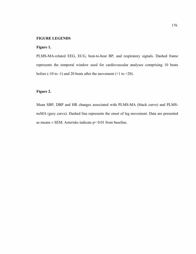
176
FIGURE LEGENDS
Figure 1.
PLMS-MA-related EEG, ECG, beat-to-beat BP, and respiratory signals. Dashed frame
represents the temporal window used for cardiovascular analyses comprising 10 beats
before (-10 to -1) and 20 beats after the movement (+1 to +20).
Figure 2.
Mean SBP, DBP and HR changes associated with PLMS-MA (black curve) and PLMS-
noMA (grey curve). Dashed line represents the onset of leg movement. Data are presented
as means ± SEM. Asterisks indicate p< 0.01 from baseline.
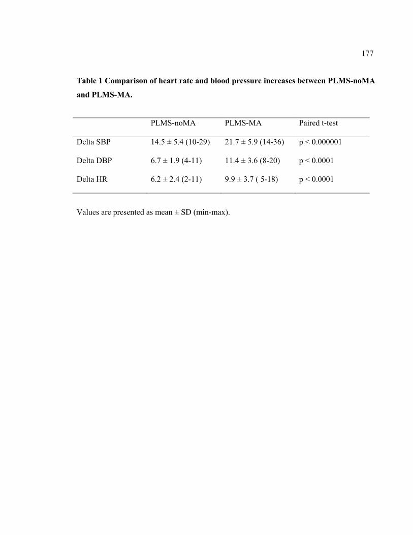
177
Table 1 Comparison of heart rate and blood pressure increases between PLMS-noMA
and PLMS-MA.
PLMS-noMA PLMS-MA Paired t-test
Delta SBP 14.5 ± 5.4 (10-29) 21.7 ± 5.9 (14-36) p < 0.000001
Delta DBP 6.7 ± 1.9 (4-11) 11.4 ± 3.6 (8-20) p < 0.0001
Delta HR 6.2 ± 2.4 (2-11) 9.9 ± 3.7 ( 5-18) p < 0.0001
Values are presented as mean ± SD (min-max).
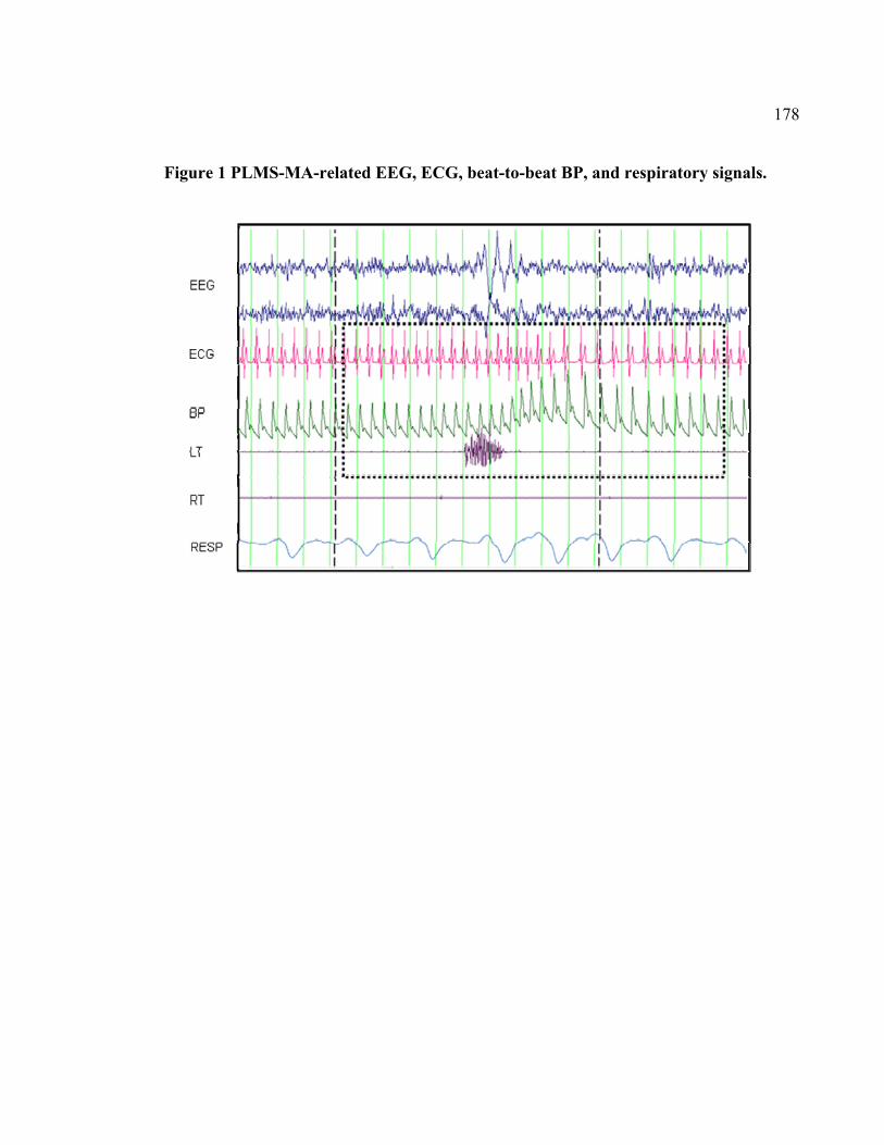
178
Figure 1 PLMS-MA-related EEG, ECG, beat-to-beat BP, and respiratory signals.
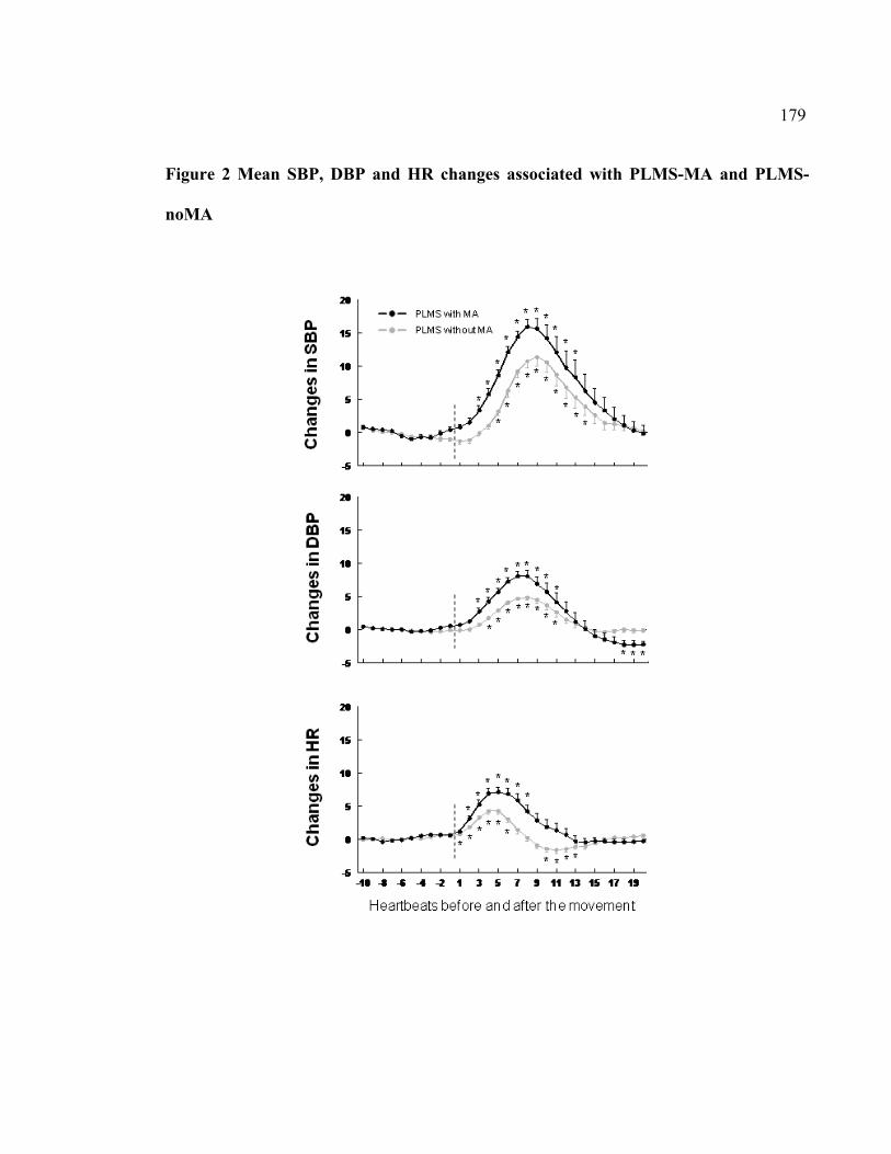
179
Figure 2 Mean SBP, DBP and HR changes associated with PLMS-MA and PLMS-
noMA

180
3. Discussion
181
3.1. Description de la prévalence des MPJ
3.1.1. Chez les sujets sans plainte de sommeil
Le premier article de cette thèse corrobore les données de plusieurs études ayant montré un
index de MPJS élevé chez les sujets âgés sans trouble du sommeil. Toutefois, le fait d’avoir
inclus des sujets de plusieurs groupes d’âge différents permet de dresser un portrait
beaucoup plus précis de l’évolution de la prévalence des MPJS avec l’âge. Chez les sujets
plus jeunes, soit entre 5 et 39 ans, l’index de MPJS était très faible, se situant en bas de 5
mouvements par heure. Ainsi, une étude corrélationnelle chez des sujets de moins de 40 ans
sans plainte de sommeil ne montrerait sans doute pas de relation entre l’index de MPJS et
l’âge. La figure 1 du premier article de la thèse montre clairement que l’index de MPJS
devient plus élevé à partir de 40 ans et que cette prévalence continue à augmenter
graduellement dans les groupes de sujets plus âgés.
L’index de MPJE montre quant à lui une corrélation négative avec l’âge. Encore une fois,
le fait d’étudier l’évolution des MPJE au sein de différents groupes d’âge a permis de
nuancer ce résultat. En effet, la figure 2 du premier article montre que l’index de MPJE
évolue plutôt sous la forme d’une courbe en U. Bien que statistiquement les différences
entre les groupes ne soient pas toutes significatives, une inspection visuelle révèle que les
enfants et les adolescents présentent des index très élevés de MPJE, ce qui est suivi d’une
diminution dans les groupes de 30-39 ans et de 40-49 ans, puis d’une légère augmentation à
partir de 50 ans. Ces résultats soulignent que l’âge des cohortes étudiées peut faire varier
182
drastiquement les résultats si on ne les décompose pas en groupes d’âge. En effet, une étude
chez des sujets plus jeunes montrerait toujours une corrélation négative, alors qu’une étude
chez des sujets plus âgés (ce qui est le plus souvent le cas dans l’étude des MPJ) montrerait
au contraire une corrélation positive. Il importe donc de garder en tête un portrait plus
global de l’évolution de l’index de MPJE avec l’âge, particulièrement lors de comparaisons
entre différentes populations.
3.1.2. Chez les patients atteints de NC
Tel qu’attendu, le deuxième article de cette thèse montre un index de MPJS plus élevé chez
les sujets atteints de NC que chez les sujets sans plainte de sommeil, soit de 21
mouvements par heure pour une population dont l’âge moyen était de 46 ans. Cette
différence était présente dans tous les stades de sommeil, mais était particulièrement
importante en SP, stade dans lequel on retrouve habituellement très peu de MPJS. En fait,
chez les sujets atteints de NC, l’index de MPJS était très similaire en SP et en sommeil non
paradoxal. Cette particularité a déjà été observée chez les sujets atteints du TCSP (Fantini
et al., 2002) et cette similitude sera discutée ultérieurement.
On retrouve également une augmentation de l’index de MPJS avec l’âge chez les sujets
atteints de NC. Cette évolution est analogue à celle observée chez les sujets en santé, soit
une prévalence plus faible chez les sujets plus jeunes et une augmentation prononcée à
partir de l’âge de 40 ans. L’évolution de la prévalence des MPJE des patients NC avec l’âge
183
était aussi comparable à celle des témoins. On retrouvait encore une fois une courbe en U,
bien que l’étendue d’âge de la population recrutée ne permettait pas d’évaluer la prévalence
chez des patients plus jeunes que 18 ans. En somme, bien que les index de MPJ soient
généralement plus élevés chez les patients atteints de NC que chez les sujets sans plainte de
sommeil, l’évolution de leur prévalence avec l’âge ne semble pas différente.
3.2. Apport de l’intervalle inter-mouvement
Cette thèse montre chez les sujets sans plainte de sommeil de tous les groupes d’âge un
index particulièrement élevé de MPJE. Néanmoins, lorsqu’on considère l’intervalle inter-
mouvement, on comprend que ce nombre élevé contient probablement différents types de
mouvements. En effet, puisque l’intervalle défini dans les critères standardisés présente une
grande étendue, soit entre 5 et 90 secondes, la plupart des mouvements présents à l’éveil
peuvent satisfaire le critère de périodicité. Dans la figure 4 du premier article, on peut voir
que les histogrammes d’intervalles inter-mouvements ne dévoilent aucun intervalle
préférentiel entre les MPJE au sein des différents groupes d’âge, si ce n’est qu’un nombre
important de courts intervalles, surtout chez les sujets très jeunes. Cette absence de
périodicité soulève un questionnement sur la réelle signification de la courbe en U,
illustrant l’évolution de l’index des MPJE avec l’âge. En effet, on peut penser que les
enfants et les adolescents ont tendance à bouger davantage au cours de la période
d’endormissement et durant les éveils nocturnes et que ces mouvements sont probablement
comptabilisés comme des MPJE, alors qu’ils représentent des mouvements volontaires.
184
Une analyse vidéo synchronisée à l’enregistrement PSG des sujets aurait pu permettre de
mieux distinguer les MPJE des autres types de mouvements.
On peut se demander si le large intervalle utilisé comme critère de périodicité fait
également en sorte d’inclure à tort des mouvements survenant au cours de sommeil, qui ne
sont pas réellement des MPJS. Les histogrammes d’intervalles inter-mouvements de la
figure 3 du premier article montrent clairement la présence d’une périodicité située entre 15
et 35 secondes chez les sujets plus âgés, soit les mêmes sujets chez qui on a observé un
index élevé de MPJS. Au contraire, aucune périodicité n’est observée dans les
histogrammes des sujets plus jeunes, suggérant que les mouvements identifiés comme des
MPJS dans cette population sont peut-être simplement le reflet de l’activité motrice
générale durant le sommeil.
Ces résultats portent à croire qu’en présence de critères d’intervalles inter-mouvements qui
ne sont pas suffisamment restreints, des mouvements non périodiques sont inclus dans les
index de MPJ à la fois en sommeil et à l’éveil. C’est d’ailleurs dans cette ligne de pensée
que Ferri et ses collaborateurs ont proposé dernièrement le concept d’index de périodicité.
Cet index représente la proportion de tous les intervalles inter-mouvements compris entre
10 et 90 secondes, sur l’ensemble de tous les intervalles (Ferri, Zucconi, Manconi, Plazzi,
et al., 2006). On peut en effet penser que le fait de quantifier la périodicité à l’aide d’un
185
histogramme d’intervalles et de l’index de périodicité ajoute un niveau de précision dans
l’évaluation des MPJ au sein de différentes populations.
3.3. Utilité des MPJ dans le diagnostic du SIME
3.3.1. MPJS
Bien que l’on ait montré que l’index de MPJS était un outil sensible et spécifique pour
discriminer les sujets atteints du SIME des sujets témoins chez des populations d’âge
moyen (Michaud, Paquet, et al., 2002), les résultats de cette thèse montrent que ce n’est
sans doute pas le cas chez les sujets plus âgés. En effet, dans les groupes d’âge de 40 ans et
plus, l’index moyen de MPJS était généralement plus élevé que 10 et ce, dans deux
échantillons de sujets sans plainte de sommeil. Néanmoins, les présents résultats montrent
qu’un index élevé de MPJS chez les jeunes adultes, les adolescents et les enfants n’est pas
commun chez les sujets en santé et confirment l’utilité de l’index de MPJS pour discriminer
les sujets atteints du SIME des sujets témoins dans ces groupes d’âge.
3.3.2. MPJE
À première vue, on peut s’étonner de la présence d’index de MPJE aussi élevés chez les
sujets sans plainte de sommeil, respectivement de 41,1 et 22,4 dans les deux premiers
articles. Il convient de souligner qu’un index élevé de MPJE ne signifie pas nécessairement
un nombre élevé de mouvements, particulièrement chez les sujets en santé, sans plainte de
186
sommeil. En effet, il faut garder en tête que l’index représente toujours le nombre de
mouvements divisé par le nombre d’heures d’éveil. Puisque ces sujets ne présentent pas de
trouble du sommeil, leur temps d’éveil est relativement court, ce qui a pour effet de gonfler
l’index de MPJE, particulièrement chez les sujets plus jeunes.
Pourtant, dans l’étude de Michaud citée précédemment (Michaud, Paquet, et al., 2002), un
index de MPJE plus élevé que 15 discriminait adéquatement les sujets SIME des sujets
témoins, ce qui d’emblée, ne semble pas concorder avec nos résultats. Néanmoins,
lorsqu’on considère l’âge des patients de l’étude de Michaud, soit en moyenne 48 ans, on
constate que les résultats ne sont pas si divergents. En effet, dans les deux premiers articles
de cette thèse, le groupe de sujets sans plainte de sommeil présentant l’index de MPJE le
plus faible est celui des 40-49 ans et on peut penser que dans cette tranche d’âge, un index
de MPJE > 15 pourrait classifier adéquatement la majorité de ces sujets. On peut ainsi
conclure que l’index de MPJE n’est probablement pas un outil assez spécifique pour
discriminer les sujets SIME des sujets témoins chez les enfants, les adolescents, les jeunes
adultes et les sujets plus âgés, mais uniquement chez les sujets d’âge moyen.
3.4. Étude des MPJ par le biais du système nerveux autonome
3.4.1. Changements de la FC et de la PA associés aux MPJS
Les résultats de cette thèse confirment que les MPJS sont associés à des changements de la
FC chez les sujets sans plainte de sommeil et les patients atteints du SIME. De plus, le
187
troisième article de cette thèse montre des changements similaires, bien que de plus faible
amplitude, chez les patients atteints de NC. Dans ces trois populations, on retrouve le même
patron d’activité, soit une tachycardie qui débute peu avant le mouvement, suivie d’une
bradycardie.
Pour la première fois, les quatrième et cinquième articles de cette thèse montrent également
des augmentations significatives de la PA associées aux MPJS, à la fois chez des sujets
atteints du SIME, mais également chez des sujets sans plainte de sommeil. Cela confirme
les résultats de l’étude de cas effectuée en 1991 (Ali et al., 1991). La présence de cette
augmentation de PA a d’ailleurs été répliquée par un autre groupe de chercheurs, chez des
patients atteints du SIME (Siddiqui et al., 2007). Il est à noter que les patients de l’article de
Siddiqui recevaient différents médicaments au moment de l’étude, dont des agonistes
dopaminergiques. Il est possible que cette prise de médication ait pu influencer les
augmentations de PA qui sont en effet légèrement moins élevées dans cet article que dans
le nôtre. Cette hypothèse concernant l’effet possible de la médication devra néanmoins être
vérifiée avec de plus grands échantillons de patients et une prise de médicament plus
uniforme.
188
3.4.2. Amplitude de la FC associée aux MPJS diminuée chez les patients atteints de
narcolepsie et cataplexie
Malgré le fait que le patron de changements de la FC était similaire au sein des différents
groupes, les MPJS des sujets atteints de NC étaient associés à des activations de la FC de
moins grande amplitude, comparativement à celles des sujets sans trouble du sommeil.
Cette diminution de l’amplitude est comparable à celle observée chez les patients atteints
du TCSP (Fantini et al., 2002).
La diminution de l’amplitude de l’activation cardiaque associée aux MPJS n’est pas la
seule similitude entre la NC et le TCSP. Nous avons déjà souligné la présence élevée de
MPJS durant le SP dans ces deux populations. Cela s’ajoute au fait que certaines
manifestations cliniques du TCSP ont déjà été rapportées chez les narcoleptiques
(Dauvilliers, Rompre, et al., 2007; Nightingale et al., 2005). De surcroit, bien que certains
résultats demeurent contradictoires ou montrent des effets modérés, différentes études ont
suggéré la présence d’une dysautonomie, à la fois chez les patients atteints de narcolepsie
(Ferini-Strambi et al., 1997; Fronczek et al., 2008; Hublin et al., 1994; Karacan, 1986;
Sachs & Kaijser, 1980) et chez les patients atteints du TCSP (Ferini-Strambi et al., 1996;
Lanfranchi et al., 2007). Des études futures pourront mieux établir si les manifestations
cliniques et motrices communes à la NC et au TCSP, ainsi que leurs irrégularités au niveau
de la fonction autonomique, sont causées par une atteinte aux mêmes mécanismes. En effet,
la NC et le TCSP présentent des liens au niveau de certaines structures et réseaux
neuronaux. Comme cela a été mentionné, une diminution de l’hypocrétine est associée à la
189
NC. Les neurones à hypocrétine, situés au niveau de l’hypothalamus dorsolatéral, projettent
notamment au niveau de structures du tronc cérébral impliquées dans la régulation motrice
du SP (Mileykovskiy, Kiyashchenko & Siegel, 2005). Ainsi, une diminution de
l’hypocrétine pourrait contribuer à la perte de l’atonie musculaire en SP chez les patients
atteints du TCSP.
3.4.3. Comparaison des changements cardiovasculaires chez les sujets sans plainte
de sommeil et les sujets atteints du SIME
Dans le cinquième article de cette thèse, on peut observer que les changements
cardiovasculaires associés aux MPJS chez les sujets sans plainte de sommeil sont
légèrement moins élevés que ceux observés chez les sujets atteints du SIME présentés dans
le quatrième article. Il est à noter que l’âge et l’index de MPJS sont similaires dans ces
deux groupes de sujets. Toutefois, si on compare les augmentations de ces deux groupes à
l’aide de tests t de Student pour échantillons indépendants, on constate que les moyennes
des augmentations de la FC et de la PA ne sont pas statistiquement différentes (voir tableau
1, annexe 1). De plus, les valeurs maximales des augmentations sont souvent les mêmes au
sein des deux groupes. Il demeure néanmoins difficile de conclure définitivement à
l’absence d’une différence entre l’activation cardiovasculaire associée aux MPJS entre ces
groupes, compte tenu du petit nombre de sujets.
190
L’étude de Fantini avait bien montré une plus grande activation de la FC lors des MPJS
chez les sujets atteints du SIME que chez les sujets atteints du TCSP (Fantini et al., 2002).
Cependant, en l’absence d’un groupe témoin sans trouble de sommeil dans cette étude, il
est impossible de conclure à une activation amplifiée chez les sujets atteints du SIME, à une
diminution de l’activation chez les sujets atteints du TCSP, ou encore aux deux réalités à la
fois. Les différentes études portant sur la dysfonction autonomique dans le TCSP citées
précédemment portent à croire qu’il pourrait effectivement y avoir une diminution de
l’amplitude des changements cardiovasculaires associés aux MPJS dans cette pathologie,
mais cela n’exclut pas la possibilité qu’il y ait également une activation amplifiée chez les
patients atteints du SIME.
3.4.4. Relation entre l’amplitude des changements et d’autres caractéristiques
démographiques ou polygraphiques
Bien que tous les MPJS aient été associés à des augmentations de la FC et de la PA,
l’amplitude de l’activation était systématiquement plus élevée lorsque les MPJS étaient
associés à des ME. Par contre, dans tous les articles de cette thèse, l’intensité de l’activation
de la FC ou de la PA ne semblait pas reliée à d’autres caractéristiques liées aux
mouvements, telles que l’index ou la durée des MPJS, et ce pour toutes les populations
étudiées. Encore une fois, il est nécessaire de rappeler les petites tailles d’échantillon et de
ne pas considérer cette absence de relation comme définitive.
191
Cette thèse corrobore le fait que les changements de la FC associés aux MPJS diminuent
avec l’âge. Ces résultats sont concordants avec la réduction de la réactivité de la FC à des
stimulations externes rapportée avec l’âge (Stratton et al., 1992). D’ailleurs, une diminution
générale de la variabilité de la FC est observée chez les individus plus âgés, alors qu’une
grande variabilité de la FC démontre au contraire une bonne capacité du système nerveux
autonome cardiovasculaire à s’adapter aux différents changements (Antelmi et al., 2004).
Au contraire, les changements de la PA associés aux MPJS augmentaient avec l’âge chez
les sujets atteints du SIME. Ces résultats sont également attendus, puisque contrairement à
la FC, une augmentation de la variabilité de la PA est un facteur associé à l’âge et à des
problèmes cardiovasculaires (Mancia et al., 1983; Seals, Stevenson, Jones, DeSouza, &
Tanaka, 1999). De plus, des études ont montré une augmentation de la réponse de la PA
lors de stimulations en laboratoire et durant les activités quotidiennes chez des sujets plus
âgés, comparativement à des sujets plus jeunes (Uchino, Berg, Smith, Pearce, & Skinner,
2006; Uchino, Uno, Holt-Lunstad, & Flinders, 1999).
Toutefois, chez les sujets sans plainte de sommeil, nous n’avons pu retrouver de corrélation
entre l’âge et l’amplitude de la PA. Bien entendu, il est possible que le petit nombre de
sujets soit ici en cause. Une autre possibilité réside dans le fait que l’association entre
l’augmentation de la PA et l’âge chez les sujets atteints du SIME soit en fait le reflet de la
durée de la maladie. En effet, la durée de la maladie du SIME était également corrélée
192
positivement avec l’augmentation de PA. Des études sur des échantillons plus grands
permettront d’évaluer indépendamment l’effet de l’âge et de la durée de la maladie sur
l’amplitude de la réponse de la PA lors des MPJS chez les sujets atteints du SIME.
3.5. Signification fonctionnelle des MPJ
3.5.1. Effet sur le sommeil et la somnolence des narcoleptiques
Tel que discuté dans la section 1.6.1, l’impact des MPJS sur le sommeil et la somnolence
n’est pas bien établi et demeure controversé. Le deuxième article de cette thèse s’est penché
sur l’effet possible des MPJS sur le sommeil, en comparant les variables PSG reliées au
sommeil, ainsi qu’à la somnolence diurne, chez une population de patients avec NC
présentant un index élevé (≥ 10) ou faible (< 10) de MPJS. Dans cet article, presque toutes
les mesures reliées au sommeil non paradoxal étaient comparables entre ces deux groupes,
mis à part un peu plus de temps passé en stade 1 chez les patients ayant un index élevé. Par
contre, le SP des NC présentant un index élevé de MPJS était davantage affecté et
présentait une diminution du pourcentage de SP et une baisse de l’efficacité du SP,
montrant donc un SP moins bien consolidé. De plus, non seulement l’index de MPJS était
anormalement élevé durant le SP chez les patients atteints de NC, mais un index élevé était
également associé avec une moins bonne efficacité du SP. Cette étude ne permet cependant
pas de savoir si c’est la présence élevée de MPJS qui affecte la consolidation du SP des
193
patients NC, ou si l’index élevé de MPJS durant le SP de ces patients est plutôt le reflet
d’une difficulté à maintenir le SP et l’atonie qui y est normalement associée.
Ainsi, à part les différences liées au SP qui sont déjà caractéristiques des patients avec NC,
la comparaison des paramètres de sommeil des patients NC avec et sans MPJS est similaire
à celle observée chez les sujets sans trouble de sommeil et ne montre pas d’effet particulier
des MPJS (Carrier et al., 2005).
Finalement, les patients narcoleptiques ayant un index plus élevé de MPJS présentaient tout
de même une légère diminution de la latence au sommeil durant les TIDE. On peut penser
que la présence d’un index élevé de MPJS pourrait contribuer à expliquer en partie la
somnolence diurne rapportée par les narcoleptiques, mais il devient ici difficile de
départager l’effet des MPJS de celui du SP fragmenté. Ainsi, l’effet des MPJS sur la
somnolence des patients avec NC demeure à investiguer.
3.5.2. PLMS chez les sujets âgés versus PLMS dans les troubles du sommeil
Les résultats de cette thèse ainsi que de nombreuses études soulignent la présence d’un
index de MPJS élevé à la fois chez les sujets vieillissants sans trouble du sommeil et chez
des sujets atteints de certains troubles du sommeil, particulièrement le SIME, la narcolepsie
et le TCSP. Cette constatation amène un questionnement quant à savoir si les MPJS
représentent le même phénomène et sont générés par les mêmes mécanismes dans ces deux
194
cas. Autrement dit, les MPJS des sujets âgés en santé sont-ils de même nature que ceux des
sujets ayant des troubles du sommeil?
Si on suppose qu’il s’agit du même phénomène, un facteur commun à toutes ces situations
pourrait se situer au niveau d’une altération de la transmission dopaminergique. Bien que
certains résultats soient contradictoires ou montrent un effet prudent, on peut
raisonnablement penser qu’il existe une diminution de la transmission dopaminergique
centrale, à la fois dans le SIME, la narcolepsie et le TCSP (Albin et al., 2000; Eisensehr et
al., 2000; Eisensehr et al., 2003; Faull, Guilleminault, Berger, & Barchas, 1983; Michaud,
Soucy, Chabli, Lavigne, & Montplaisir, 2002; Montplaisir, Lorrain, & Godbout, 1991;
Montplaisir, et al., 2000). Quant à l’augmentation des MPJS avec l’âge, cette thèse montre
qu’elle est présente chez les sujets en santé, chez les narcoleptiques et un autre groupe a
montré qu’elle était également présente chez les sujets atteints du SIME (Ferri et al., 2008).
Cette association avec l’âge peut aussi être appuyée par l’hypothèse dopaminergique. En
effet, on a rapporté une diminution de la densité des récepteurs D2 à la fois chez l’animal et
chez l’humain âgés (Kaasinen & Rinne, 2002; van Dyck et al., 2002; Wong et al., 1984;
Zhang et al., 2001). De surcroit, cette diminution semble débuter vers 35-40 ans, ce qui est
précisément l’âge auquel on voit augmenter rapidement les index de MPJS et ce, dans
différentes populations.
195
Par contre, bien que toutes les populations mentionnées présentent un index élevé de
MPJS, on constate certaines particularités différentes, notamment au niveau de la
périodicité, ce qui soulève un doute quant à l’étiologie commune de ces mouvements. En
effet, les MPJS des sujets atteints du SIME (Ferri et al., 2008) et des sujets en santé
vieillissants présentent une périodicité élevée (premier article de cette thèse). Au contraire,
les MPJS des sujets atteints de narcolepsie (Ferri, Zucconi, Manconi, Bruni, et al., 2006) et
de TCSP (Manconi et al., 2007) présentent une périodicité plus faible et plus variable. De
plus, l’activation cardiaque associée aux MPJS dans ces deux populations est moins élevée.
Bien que cela peut relever d’une dysautonomie dans la NC et le TCSP, il n’est pas exclu
que les MPJS constituent une entité différente au sein de ces deux pathologies.
3.5.3. Physiopathologie des MPJS
3.5.3.1. Le système nerveux central, déclencheur d’éveils périodiques
Tel que mentionné dans l’introduction de cette thèse, la physiopathologie des MPJS n’est
pas encore bien connue. En présence de l’activation cardiovasculaire et des réactions
d’éveils corticaux ou sous-corticaux qui sont associés aux MPJS, on peut se demander
lequel de ces phénomènes constitue le stimulus déclencheur et lesquels sont en fait des
réponses.
196
Nous avons déjà discuté du fait que différents types d’éveils présentent une relation
temporelle inconstante avec les MPJS. S’ajoute à cette constatation le fait qu’une des deux
entités peut être présente sans l’autre phénomène (El-Ad & Chervin, 2000) et que des
études pharmacologiques ont montré la suppression spécifique possible du mouvement ou
de l’éveil (Ali et al., 1991; Montplaisir et al., 1996). Le fait que la relation temporelle n’est
pas constante et qu’en plus un des phénomènes peut être présent alors que l’autre est absent
suggère fortement que les mouvements et les signes d’éveil corticaux ne sont pas causés
l’un par l’autre, mais qu’ils font plutôt partie d’un mécanisme global d’éveil, dont les
changements cardiovasculaires, l’activité motrice et les éveils transitoires pourraient
représenter des manifestations. Ainsi, on a proposé la présence d’un générateur du système
nerveux central, probablement situé au niveau du tronc cérébral, qui pourrait déclencher un
ou plusieurs de ces phénomènes d’éveil de façon périodique (El-Ad & Chervin, 2000;
Karadeniz et al., 2000b; Montplaisir et al., 1996).
Dans cet ordre d’idée, l’équipe de Terzano et ses collaborateurs ont proposé et développé
au cours des dernières années un modèle décrivant un patron de cycles périodiques des
ondes cérébrales au cours du sommeil, connu en anglais sous le nom de « cyclic alternating
pattern » (CAP). Concrètement, les CAP sont des éveils transitoires et périodiques qui se
distinguent de l’activité EEG de fond au cours du sommeil non paradoxal. Ces épisodes
sont connus pour amener une instabilité au cours du sommeil, ce qui pourrait favoriser la
présence de réactions d’éveil, dont la fréquence d’oscillation est d’environ toutes les 20 à
197
40 secondes (Terzano et al., 1985). Il est à noter que cette fréquence est similaire à celle des
MPJS. D’ailleurs, une étude a montré une corrélation entre la présence de MPJS et des
CAP, suggérant que ceux-ci pourraient favoriser l’apparition des MPJS, ce qui expliquerait
leur périodicité (Parrino et al., 1996).
3.5.3.2. Le système nerveux autonome comme générateur
Une autre possibilité serait de plutôt considérer l’activation autonomique comme étant le
phénomène primaire des réactions d’éveil et des MPJS. Différents résultats peuvent
appuyer cette hypothèse. D’abord, trois articles de cette thèse, ainsi que d’autres études
dans différents laboratoires ont montré des augmentations significatives de la FC et de PA
associés avec des MPJS. De façon constante dans toutes ces études, l’activation
autonomique précède toujours les mouvements et les réactions d’éveil. De plus, une équipe
ayant étudié le lien temporel entre l’analyse spectrale de l’EEG, de l’ECG et les MPJS a
montré qu’une augmentation des basses fréquences, représentant une activation
sympathique, précédait le mouvement et les changements de l’activité EEG de plusieurs
secondes (Guggisberg et al., 2007).
Par ailleurs, deux populations chez qui on suspecte une dysautonomie, c’est-à-dire les
patients atteints de narcolepsie et de TCSP, montrent une diminution des ME associés avec
les MPJS et une atténuation de l’augmentation de la FC associée aux MPJS. Finalement,
une étude rapportant des résultats préliminaires, montre une diminution marquée des MPJS
198
chez deux sujets, suite à l’administration d’un bloqueur des récepteurs adrénergiques alpha
(Ware, Blumoff, & Pittard, 1988). Bien que ces résultats doivent être répliqués sur un plus
grand nombre de sujets, cela suggère que dans certains cas du moins, agir sur le SNA
sympathique a un effet sur la présence des MPJS.
3.6. MPJS et risque cardiovasculaire
Compte tenu des études épidémiologiques qui lient le SIME à des risques
cardiovasculaires, ainsi que nos résultats montrant des augmentations importantes de la PA
lors de ces mouvements, on peut se demander si les MPJS constituent un réel facteur de
risque cardiovasculaire. Pour appuyer cette hypothèse, on peut considérer plusieurs études
qui ont souligné qu’une augmentation de la variabilité de la PA était associée à un risque
cardiovasculaire (Mancia et al., 1983; Seals et al., 1999). En effet, on peut imaginer que les
augmentations répétées de la PA au cours de la nuit peuvent contribuer à augmenter la
variabilité moyenne de la PA. De plus, l’index de MPJS est plus élevé chez les sujets âgés
et on sait que le vieillissement est également associé à une augmentation des risques
cardiovasculaires.
Par contre, une étude récente dans notre laboratoire a montré chez une cohorte de sujets
atteints du SIME une absence de relation entre l’index de MPJS et différents marqueurs
cardiovasculaires, dont la diminution attendue de la PA durant la nuit (dipping) (Pennestri,
Lanfranchi A, Amyot, Petit, & Montplaisir, 2010). En fait, c’étaient plutôt les variables
199
démontrant une mauvaise qualité et une diminution de la quantité de sommeil qui
semblaient avoir un effet sur les paramètres cardiovasculaires. Ainsi, lorsque les sujets
dormaient moins bien, leur PA ne diminuait pas au cours de la nuit. Ces résultats portent à
croire que le sommeil altéré des sujets atteints du SIME pourrait aussi contribuer à
expliquer le lien entre le SIME et les risques cardiovasculaires, par le biais d’une
augmentation de la PA durant la nuit. Ce même raisonnement pourrait s’appliquer aux
sujets vieillissants qui ont souvent un sommeil de moins bonne qualité et en quantité
moindre. La contribution du sommeil perturbé versus celle des MPJS dans cette association
demeure à explorer.
Une nuance doit néanmoins être considérée au niveau de ces résultats préliminaires. En
effet, les critères d’exclusion des sujets dans cette étude étaient très stricts et faisaient en
sorte que les sujets avec une histoire de troubles cardiovasculaires ou toute autre condition
pouvant être associée à des maladies cardiovasculaires (notamment l’obésité, le diabète, le
cholestérol, l’hypertension) étaient exclus de cette étude. Cela a peut-être fait en sorte que
tous les sujets inclus présentaient une excellente santé cardiovasculaire et, par le fait même,
étaient moins représentatifs de la population générale des sujets atteints du SIME, tandis
que les cas plus sévères qui auraient pu montrer un effet ont été éliminés dès le
recrutement.
200
En somme, les augmentations répétées de la PA lors des MPJS ont certainement un grand
intérêt au niveau de la recherche et méritent d’être étudiées davantage. Il faut toutefois
demeurer prudent quant à l’interprétation des conséquences possibles de ce phénomène,
particulièrement d’un point de vue clinique.
3.7. Limites et forces des études
La principale limite des différentes études de cette thèse est le petit échantillon utilisé dans
certains des articles. Bien sûr, cela limite les possibilités au niveau des analyses statistiques,
que ce soit lors de comparaisons inter-groupes, ou dans les analyses corrélationnelles.
Néanmoins, les sujets choisis étaient homogènes et non médicamentés, ce qui a permis
d’obtenir plusieurs effets clairs, malgré les petites tailles de groupes.
De plus, dans certains articles mesurant les changements cardiovasculaires associés aux
MPJS, il est arrivé que le nombre de mouvements analysés soit restreint. Il s’agit ici d’un
choix méthodologique. En effet, nous avons préféré utiliser un petit nombre d’événements
qui soient équivalents et dénués d’artefacts physiologiques ou techniques. Un point
important réside également dans le choix d’utiliser un intervalle d’au moins 20 secondes
entre les mouvements. Ce critère limite bien entendu le nombre total d’événements
analysés. Néanmoins, cela permettait à la FC et à la PA de revenir à la ligne de base et ainsi
de pouvoir observer la réelle augmentation de ces variables.
201
La force principale de cette thèse est de s’inscrire dans un contexte d’interdisciplinarité, ce
qui a permis d’étudier des phénomènes reliés au sommeil dans un cadre cardiovasculaire.
Ainsi, les articles de cette thèse utilisent différentes mesures liées à la fois au domaine du
sommeil et de la cardiologie, permettant un portrait plus global des pathologies étudiées.
Deux articles de cette thèse utilisent notamment un appareil mesurant la PA, le Portapres,
qui avait peu été utilisé auparavant. Cet appareil a permis à la fois une mesure de la PA
non-invasive durant le sommeil, mais également une mesure battement par battement, qui a
pu montrer l’évolution précise dans le temps de la PA durant les événements moteurs
périodiques étudiés dans ces articles. Notons finalement que plusieurs résultats obtenus
dans cette thèse sont constants à travers les différents articles et les différentes populations
étudiées, ce qui solidifie les conclusions qui peuvent en être tirées.
3.8. Pistes futures
Différentes études pourraient faire suite à ce travail sur les MPJS. D’abord, il pourrait être
pertinent d’effectuer un suivi longitudinal des patients avec et sans MPJS, afin d’en évaluer
l’évolution dans le temps. Cela permettrait d’évaluer l’incidence des MPJS, en plus de leur
prévalence. De plus, un suivi longitudinal d’un grand nombre de patients présentant un
index élevé de MPJS avec ou sans SIME permettrait également d’évaluer plus clairement le
lien entre cette condition et le développement de l’hypertension et des maladies
cardiovasculaires.
202
Une autre voie pourrait également consister en la vérification de l’effet de l’amplitude de la
réactivité cardiovasculaire associée aux MPJS sur le sommeil et la vigilance. Il est possible
que les résultats divergents concernant l’effet des MPJS sur le sommeil et la vigilance
soient dus en partie à une différence au niveau de l’activation autonomique liée à ces
événements.
Finalement, une piste intéressante serait l’évaluation de différents traitements sur les
changements cardiovasculaires associés aux MPJS, afin de mieux comprendre leur
physiopathologie. Des résultats préliminaires ont rapporté que les agonistes
dopaminergiques semblaient diminuer la réponse de la FC lors des MPJS en plus de
diminuer l’index de MPJS (Zucconi et al., 2010), mais leur effet sur la PA n’a pas été
évalué. L’effet d’un bêtabloquant pourrait également être investigué, à savoir s’il diminue
l’augmentation de la PA associée aux MPJS et/ou les MPJS eux-mêmes, ce qui pourrait
clarifier le rôle du système nerveux autonome dans la genèse des mouvements.

203
4. Conclusion
204
Le premier objectif de cette thèse était de mieux décrire la prévalence des MPJ chez les
sujets sans plainte de sommeil et chez les sujets atteints de NC. Nous avons montré que
chez les sujets sans trouble de sommeil, la prévalence des MPJS augmentait de façon
importante à partir de l’âge de 40 ans et qu’elle continuait ensuite à progresser avec l’âge.
Quant aux MPJE, leur évolution avec l’âge montrait plutôt une courbe en U. Ces résultats
portent à croire que les MPJS permettent de bien discriminer les sujets atteints du SIME des
sujets témoins, mais uniquement dans les groupes de sujets plus jeunes. Par contre, les
MPJE permettent cette discrimination seulement chez les sujets d’âge moyen, soit entre 40
et 50 ans, puisque les index des autres groupes d’âge sont très élevés, même chez une
population de sujets sans trouble de sommeil.
Chez les sujets atteints de NC, les index de MPJS et MPJE étaient plus élevés que chez les
sujets témoins, particulièrement en SP. Toutefois, l’évolution de leur prévalence avec l’âge
montrait le même patron que celui observé chez les sujets sans plainte de sommeil. L’effet
des MPJS sur le sommeil et sur la somnolence des patients atteints de NC semblait modéré
et affectait davantage le SP. En effet, une augmentation de l’index de MPJS était associée à
une plus grande instabilité du SP, mais il est difficile de savoir quel phénomène influence
l’autre. De plus, une augmentation de l’index de MPJS était aussi associée à une légère
augmentation de la propension à s’endormir durant le jour, telle que mesurée en laboratoire
par le TIDE, mais il est encore une fois difficile de départager l’effet des MPJS du SP
altéré.
205
Finalement, nous avons montré dans ce premier volet l’importance de considérer non
seulement les index de MPJ, mais également l’intervalle inter-mouvements. En effet, la
grande étendue du critère d’intervalles inter-mouvements (entre cinq et 90 secondes) fait en
sorte que certains mouvements sont calculés comme des MPJ, alors qu’ils constituent plutôt
des mouvements volontaires durant l’éveil ou le reflet de l’activité motrice spontanée
durant le sommeil.
Le deuxième objectif de cette thèse visait l’étude des MPJS en relation avec le système
nerveux autonome cardiovasculaire. Nous avons d’abord montré que les MPJS des patients
atteints de NC étaient associés à des changements de la FC similaires à ceux rapportés dans
d’autres populations, soit une tachycardie suivie d’une bradycardie. Néanmoins, ces
changements étaient de plus faible amplitude, ce qui appuie l’hypothèse d’une
dysautonomie dans la NC, suggérée par différents auteurs.
Nous avons ensuite non seulement confirmé les changements de la FC liés aux MPJS chez
les sujets atteints du SIME et chez les sujets sans trouble de sommeil, mais également
montré qu’ils étaient associés à des augmentations significatives de la PA. Ces
augmentations étaient légèrement plus élevées chez les sujets atteints du SIME, mais cette
différence n’était pas statistiquement significative et reste à évaluer chez des groupes de
plus grande taille.
206
Les augmentations de la FC et de la PA étaient présentes lors de tous les MPJS, mais
étaient plus importantes lorsque ceux-ci étaient associés à des ME. Les changements de la
FC étaient moins importants chez les sujets plus âgés, dans toutes les populations étudiées.
Au contraire, les changements de la PA étaient plus grands chez les sujets atteints de SIME
qui étaient plus âgés et présentaient des symptômes depuis plus longtemps. Cependant,
cette association entre l’âge et l’augmentation de l’amplitude de la PA lors des MPJS n’a
pu être répliquée chez les sujets sans trouble du sommeil.
Bien que cette thèse ait permis de bien répondre aux deux objectifs principaux énoncés,
plusieurs questions concernant les MPJ devront être investiguées dans des études futures.
Nous avons d’ailleurs abordé certaines de ces questions dans la discussion de cette thèse.
D’abord, il est encore difficile de savoir si les MPJS présents chez les sujets vieillissants
sont de même nature que les MPJS des patients atteints de troubles du sommeil. Nonobstant
l’hypothèse d’une altération de la transmission dopaminergique, d’autres études seront
nécessaires pour répondre à cette question.
De plus, la physiopathologie de ces mouvements demeure encore inconnue. Nous avons
présenté la possibilité que ces mouvements soient d’origine centrale ou encore influencés
par le système nerveux autonome, mais d’autres études devront être effectuées pour
répondre à cette question, probablement des études pharmacologiques.
207
Finalement, un enjeu très important demeure la signification fonctionnelle de ces
mouvements. L’ensemble des résultats disponibles à l’heure actuelle suggère que
l’influence des MPJS sur le sommeil et la somnolence est somme toute assez faible. Nous
avons proposé que cet effet puisse être influencé par le SNA, ce qui reste néanmoins à
investiguer. Bien entendu, les augmentations importantes de la PA liées aux MPJS, ainsi
que les études épidémiologiques liant le SIME aux risques cardiovasculaires demeurent une
piste intéressante. Il serait en effet attrayant d’établir ici un lien de causalité entre ces
différentes réalités. Néanmoins, tel que mentionné dans la discussion, ce lien semble pour
l’instant prématuré et il sera nécessaire d’effectuer davantage d’études longitudinales avant
de pouvoir interpréter les conséquences cliniques de ces augmentations répétées de la PA.
En somme, cette thèse révèle une prévalence importante des MPJS chez les sujets en santé
vieillissants et chez les sujets atteints de narcolepsie. Elle souligne également une
association entre ces mouvements et l’implication du système nerveux autonome
cardiovasculaire. Des études futures permettront de préciser la signification fonctionnelle et
l’importance clinique de ces phénomènes moteurs, qui rappelons-le surviennent la plupart
du temps à l’insu des individus!
208
5. Bibliographie
Albin, R. L., Koeppe, R. A., Chervin, R. D., Consens, F. B., Wernette, K., Frey, K. A., et
al. (2000). Decreased striatal dopaminergic innervation in REM sleep behavior
disorder. Neurology, 55(9), 1410-1412.
Ali, N. J., Davies, R. J., Fleetham, J. A., & Stradling, J. R. (1991). Periodic movements of
the legs during sleep associated with rises in systemic blood pressure. Sleep, 14(2),
163-165.
Allen, R., Becker, P. M., Bogan, R., Schmidt, M., Kushida, C. A., Fry, J. M., et al. (2004).
Ropinirole decreases periodic leg movements and improves sleep parameters in
patients with restless legs syndrome. Sleep, 27(5), 907-914.
Allen, R. P., La Buda, M. C., Becker, P., & Earley, C. J. (2002). Family history study of the
restless legs syndrome. Sleep Med, 3 Suppl, S3-7.
Allen, R. P., Picchietti, D., Hening, W. A., Trenkwalder, C., Walters, A. S., & Montplaisi,
J. (2003). Restless legs syndrome: diagnostic criteria, special considerations, and
epidemiology. A report from the restless legs syndrome diagnosis and epidemiology
workshop at the National Institutes of Health. Sleep Med, 4(2), 101-119.
Allen, R. P., Walters, A. S., Montplaisir, J., Hening, W., Myers, A., Bell, T. J., et al.
(2005). Restless legs syndrome prevalence and impact: REST general population
study. Arch Intern Med, 165(11), 1286-1292.
209
American Academy of Sleep Medicine. (2005). International Classification of Sleep
Disorders: Diagnostic and Coding Manual, rev. ed., ed 2. Westchester, IL:
American Academy of Sleep Medicine.
Ancoli-Israel, S., Kripke, D. F., Klauber, M. R., Mason, W. J., Fell, R., & Kaplan, O.
(1991). Periodic limb movements in sleep in community-dwelling elderly. Sleep,
14(6), 496-500.
Ancoli-Israel, S., Kripke, D. F., Mason, W., & Kaplan, O. J. (1985). Sleep apnea and
periodic movements in an aging sample. J Gerontol, 40(4), 419-425.
Antelmi, I., de Paula, R. S., Shinzato, A. R., Peres, C. A., Mansur, A. J., & Grupi, C. J.
(2004). Influence of age, gender, body mass index, and functional capacity on heart
rate variability in a cohort of subjects without heart disease. Am J Cardiol, 93(3),
381-385.
Arens, R., Wright, B., Elliott, J., Zhao, H., Wang, P. P., Brown, L. W., et al. (1998).
Periodic limb movement in sleep in children with Williams syndrome. J Pediatr,
133(5), 670-674.
ASDA. (1990). The International Classification of Sleep Diosrders: Diagnostic and
Coding Manual. Rochester, MN: American Sleep Disorders Association.
Auger, R. R., Goodman, S. H., Silber, M. H., Krahn, L. E., Pankratz, V. S., & Slocumb, N.
L. (2005). Risks of high-dose stimulants in the treatment of disorders of excessive
somnolence: a case-control study. Sleep, 28(6), 667-672.
210
Baker, T. L., Guilleminault, C., Nino-Murcia, G., & Dement, W. C. (1986). Comparative
polysomnographic study of narcolepsy and idiopathic central nervous system
hypersomnia. Sleep, 9(1 Pt 2), 232-242.
Bara-Jimenez, W., Aksu, M., Graham, B., Sato, S., & Hallett, M. (2000). Periodic limb
movements in sleep: state-dependent excitability of the spinal flexor reflex.
Neurology, 54(8), 1609-1616.
Baran, A. S., Richert, A. C., Douglass, A. B., May, W., & Ansarin, K. (2003). Change in
periodic limb movement index during treatment of obstructive sleep apnea with
continuous positive airway pressure. Sleep, 26(6), 717-720.
Bastuji, H., & Garcia-Larrea, L. (1999). Sleep/wake abnormalities in patients with periodic
leg movements during sleep: factor analysis on data from 24-h ambulatory
polygraphy. J Sleep Res, 8(3), 217-223.
Benes, H., Kurella, B., Kummer, J., Kazenwadel, J., Selzer, R., & Kohnen, R. (1999).
Rapid onset of action of levodopa in restless legs syndrome: a double-blind,
randomized, multicenter, crossover trial. Sleep, 22(8), 1073-1081.
Berger, K., Luedemann, J., Trenkwalder, C., John, U., & Kessler, C. (2004). Sex and the
risk of restless legs syndrome in the general population. Arch Intern Med, 164(2),
196-202.
Berlucchi, G. (1997). One or many arousal systems? Reflections on some of Giuseppe
Moruzzi's foresights and insights about the intrinsic regulation of brain activity.
Arch Ital Biol, 135(1), 5-14.
211
Billars, L., Hicks, A., Bliwise, D., Sigmundsson, T., Sigurdsson, A., Kristjansson, K., et al.
(2007). Hypertension risk and PLMS in restless legs syndrome. Sleep, 30, A297-
A298.
Bixler, E. O., Kales, A., Vela-Bueno, A., Jacoby, J. A., Scarone, S., & Soldatos, C. R.
(1982). Nocturnal myoclonus and nocturnal myoclonic activity in the normal
population. Res Commun Chem Pathol Pharmacol, 36(1), 129-140.
Bliwise, D., Petta, D., Seidel, W., & Dement, W. (1985). Periodic leg movements during
sleep in the elderly. Arch Gerontol Geriatr, 4(3), 273-281.
Bliwise, D. L., Carskadon, M. A., & Dement, W. C. (1988). Nightly variation of periodic
leg movements in sleep in middle aged and elderly individuals. Arch Gerontol
Geriatr, 7(4), 273-279.
Boivin, D. B., Lorrain, D., & Montplaisir, J. (1993). Effects of bromocriptine on periodic
limb movements in human narcolepsy. Neurology, 43(10), 2134-2136.
Boivin, D. B., Montplaisir, J., & Poirier, G. (1989). The effects of L-dopa on periodic leg
movements and sleep organization in narcolepsy. Clin Neuropharmacol, 12(4), 339-
345.
Briellmann, R. S., Rosler, K. M., & Hess, C. W. (1996). Blink reflex excitability is
abnormal in patients with periodic leg movements in sleep. Mov Disord, 11(6), 710-
714.
212
Brodeur, C., Montplaisir, J., Godbout, R., & Marinier, R. (1988). Treatment of restless legs
syndrome and periodic movements during sleep with L-dopa: a double-blind,
controlled study. Neurology, 38(12), 1845-1848.
Bucher, S. F., Seelos, K. C., Oertel, W. H., Reiser, M., & Trenkwalder, C. (1997). Cerebral
generators involved in the pathogenesis of the restless legs syndrome. Ann Neurol,
41(5), 639-645.
Carelli, G., Krieger, J., Calvi-Gries, F., & Macher, J. P. (1999). Periodic limb movements
and obstructive sleep apneas before and after continuous positive airway pressure
treatment. J Sleep Res, 8(3), 211-216.
Carrier, J., Frenette, S., Montplaisir, J., Paquet, J., Drapeau, C., & Morettini, J. (2005).
Effects of periodic leg movements during sleep in middle-aged subjects without
sleep complaints. Mov Disord, 20(9), 1127-1132.
Carskadon, M. A., & Dement, W. C. (1982). The multiple sleep latency test: what does it
measure? Sleep, 5 Suppl 2, S67-72.
Carskadon, M. A., Dement, W. C., Mitler, M. M., Roth, T., Westbrook, P. R., & Keenan, S.
(1986). Guidelines for the multiple sleep latency test (MSLT): a standard measure
of sleepiness. Sleep, 9(4), 519-524.
Chabli, A., Michaud, M., & Montplaisir, J. (2000). Periodic arm movements in patients
with the restless legs syndrome. Eur Neurol, 44(3), 133-138.
213
Chen, C. T., Hwang, L. L., Chang, J. K., & Dun, N. J. (2000). Pressor effects of orexins
injected intracisternally and to rostral ventrolateral medulla of anesthetized rats. Am
J Physiol Regul Integr Comp Physiol, 278(3), R692-697.
Ciriello, J., & de Oliveira, C. V. (2003). Cardiac effects of hypocretin-1 in nucleus
ambiguus. Am J Physiol Regul Integr Comp Physiol, 284(6), R1611-1620.
Coccagna, G., Lugaresi, E., Tassinari, C. A., & Ambrosetto, C. (1966). [The restless leg
syndrome (restless legs)]. Omnia Med Ther, 44(4), 619-667.
Coleman, R. M. (1982). Periodic movements in sleep (nocturnal myoclonus) and restless
legs syndrome. In C. Guilleminault (Ed.), Sleeping and Waking Disorders:
Indications and Techniques (pp. 265-295). Menlo Park, CA: Addison-Wesley.
Coleman, R. M., Pollak, C. P., & Weitzman, E. D. (1980). Periodic movements in sleep
(nocturnal myoclonus): relation to sleep disorders. Ann Neurol, 8(4), 416-421.
Collado-Seidel, V., Kazenwadel, J., Wetter, T. C., Kohnen, R., Winkelmann, J., Selzer, R.,
et al. (1999). A controlled study of additional sr-L-dopa in L-dopa-responsive
restless legs syndrome with late-night symptoms. Neurology, 52(2), 285-290.
Cui, J., Blaha, C., Moradkhan, R., Gray, K. S., & Sinoway, L. I. (2006). Muscle
sympathetic nerve activity responses to dynamic passive muscle stretch in humans.
J Physiol, 576(Pt 2), 625-634.
214
Date, Y., Ueta, Y., Yamashita, H., Yamaguchi, H., Matsukura, S., Kangawa, K., et al.
(1999). Orexins, orexigenic hypothalamic peptides, interact with autonomic,
neuroendocrine and neuroregulatory systems. Proc Natl Acad Sci U S A, 96(2), 748-
753.
Dauvilliers, Y., Arnulf, I., & Mignot, E. (2007). Narcolepsy with cataplexy. Lancet,
369(9560), 499-511.
Dauvilliers, Y., Baumann, C. R., Carlander, B., Bischof, M., Blatter, T., Lecendreux, M., et
al. (2003). CSF hypocretin-1 levels in narcolepsy, Kleine-Levin syndrome, and
other hypersomnias and neurological conditions. J Neurol Neurosurg Psychiatry,
74(12), 1667-1673.
Dauvilliers, Y., Billiard, M., & Montplaisir, J. (2003). Clinical aspects and pathophysiology
of narcolepsy. Clin Neurophysiol, 114(11), 2000-2017.
Dauvilliers, Y., Gosselin, A., Paquet, J., Touchon, J., Billiard, M., & Montplaisir, J. (2004).
Effect of age on MSLT results in patients with narcolepsy-cataplexy. Neurology,
62(1), 46-50.
Dauvilliers, Y., Montplaisir, J., Molinari, N., Carlander, B., Ondze, B., Besset, A., et al.
(2001). Age at onset of narcolepsy in two large populations of patients in France
and Quebec. Neurology, 57(11), 2029-2033.
Dauvilliers, Y., Pennestri, M. H., Petit, D., Dang-Vu, T., Lavigne, G., & Montplaisir, J.
(2007). Periodic leg movements during sleep and wakefulness in narcolepsy. J
Sleep Res, 16(3), 333-339.
215
Dauvilliers, Y., Rompre, S., Gagnon, J. F., Vendette, M., Petit, D., & Montplaisir, J.
(2007). REM sleep characteristics in narcolepsy and REM sleep behavior disorder.
Sleep, 30(7), 844-849.
Davis, S. F., Williams, K. W., Xu, W., Glatzer, N. R., & Smith, B. N. (2003). Selective
enhancement of synaptic inhibition by hypocretin (orexin) in rat vagal motor
neurons: implications for autonomic regulation. J Neurosci, 23(9), 3844-3854.
de Oliveira, C. V., Rosas-Arellano, M. P., Solano-Flores, L. P., & Ciriello, J. (2003).
Cardiovascular effects of hypocretin-1 in nucleus of the solitary tract. Am J Physiol
Heart Circ Physiol, 284(4), H1369-1377.
Dickel, M. J., & Mosko, S. S. (1990). Morbidity cut-offs for sleep apnea and periodic leg
movements in predicting subjective complaints in seniors. Sleep, 13(2), 155-166.
Dickel, M. J., Renfrow, S. D., Moore, P. T., & Berry, R. B. (1994). Rapid eye movement
sleep periodic leg movements in patients with spinal cord injury. Sleep, 17(8), 733-
738.
Edinger, J. D., McCall, W. V., Marsh, G. R., Radtke, R. A., Erwin, C. W., & Lininger, A.
(1992). Periodic limb movement variability in older DIMS patients across
consecutive nights of home monitoring. Sleep, 15(2), 156-161.
EEG arousals: scoring rules and examples: a preliminary report from the Sleep Disorders
Atlas Task Force of the American Sleep Disorders Association. (1992). Sleep,
15(2), 173-184.
216
Eisensehr, I., Linke, R., Noachtar, S., Schwarz, J., Gildehaus, F. J., & Tatsch, K. (2000).
Reduced striatal dopamine transporters in idiopathic rapid eye movement sleep
behaviour disorder. Comparison with Parkinson's disease and controls. Brain, 123 (
Pt 6), 1155-1160.
Eisensehr, I., Linke, R., Tatsch, K., von Lindeiner, H., Kharraz, B., Gildehaus, F. J., et al.
(2003). Alteration of the striatal dopaminergic system in human narcolepsy.
Neurology, 60(11), 1817-1819.
El-Ad, B., & Chervin, R. D. (2000). The case of a missing PLM. Sleep, 23(4), 450-451.
Espinar-Sierra, J., Vela-Bueno, A., & Luque-Otero, M. (1997). Periodic leg movements in
sleep in essential hypertension. Psychiatry Clin Neurosci, 51(3), 103-107.
Fagard, R. H., Celis, H., Thijs, L., Staessen, J. A., Clement, D. L., De Buyzere, M. L., et al.
(2008). Daytime and nighttime blood pressure as predictors of death and cause-
specific cardiovascular events in hypertension. Hypertension, 51(1), 55-61.
Fantini, M. L., Michaud, M., Gosselin, N., Lavigne, G., & Montplaisir, J. (2002). Periodic
leg movements in REM sleep behavior disorder and related autonomic and EEG
activation. Neurology, 59(12), 1889-1894.
Faull, K. F., Guilleminault, C., Berger, P. A., & Barchas, J. D. (1983). Cerebrospinal fluid
monoamine metabolites in narcolepsy and hypersomnia. Ann Neurol, 13(3), 258-
263.
217
Ferini-Strambi, L., Oldani, A., Zucconi, M., & Smirne, S. (1996). Cardiac autonomic
activity during wakefulness and sleep in REM sleep behavior disorder. Sleep, 19(5),
367-369.
Ferini-Strambi, L., Spera, A., Oldani, A., Zucconi, M., Bianchi, A., Cerutti, S., et al.
(1997). Autonomic function in narcolepsy: power spectrum analysis of heart rate
variability. J Neurol, 244(4), 252-255.
Ferri, R., Manconi, M., Lanuzza, B., Cosentino, F. I., Bruni, O., Ferini-Strambi, L., et al.
(2008). Age-related changes in periodic leg movements during sleep in patients with
restless legs syndrome. Sleep Med, 9(7), 790-798.
Ferri, R., Zucconi, M., Manconi, M., Bruni, O., Ferini-Strambi, L., Vandi, S., et al. (2006).
Different periodicity and time structure of leg movements during sleep in
narcolepsy/cataplexy and restless legs syndrome. Sleep, 29(12), 1587-1594.
Ferri, R., Zucconi, M., Manconi, M., Plazzi, G., Bruni, O., & Ferini-Strambi, L. (2006).
New approaches to the study of periodic leg movements during sleep in restless legs
syndrome. Sleep, 29(6), 759-769.
Ferrillo, F., Beelke, M., Canovaro, P., Watanabe, T., Arico, D., Rizzo, P., et al. (2004).
Changes in cerebral and autonomic activity heralding periodic limb movements in
sleep. Sleep Med, 5(4), 407-412.
Frattola, A., Parati, G., Cuspidi, C., Albini, F., & Mancia, G. (1993). Prognostic value of
24-hour blood pressure variability. J Hypertens, 11(10), 1133-1137.
218
Fronczek, R., Overeem, S., Reijntjes, R., Lammers, G. J., van Dijk, J. G., & Pijl, H. (2008).
Increased heart rate variability but normal resting metabolic rate in
hypocretin/orexin-deficient human narcolepsy. J Clin Sleep Med, 4(3), 248-254.
Fry, J. M., DiPhillipo, M. A., & Pressman, M. R. (1989). Periodic leg movements in sleep
following treatment of obstructive sleep apnea with nasal continuous positive
airway pressure. Chest, 96(1), 89-91.
Garcia-Borreguero, D., Larrosa, O., de la Llave, Y., Granizo, J. J., & Allen, R. (2004).
Correlation between rating scales and sleep laboratory measurements in restless legs
syndrome. Sleep Med, 5(6), 561-565.
Gosselin, N., Lanfranchi, P., Michaud, M., Fantini, L., Carrier, J., Lavigne, G., et al.
(2003). Age and gender effects on heart rate activation associated with periodic leg
movements in patients with restless legs syndrome. Clin Neurophysiol, 114(11),
2188-2195.
Guggisberg, A. G., Hess, C. W., & Mathis, J. (2007). The significance of the sympathetic
nervous system in the pathophysiology of periodic leg movements in sleep. Sleep,
30(6), 755-766.
Hanly, P., & Zuberi, N. (1992). Periodic leg movements during sleep before and after heart
transplantation. Sleep, 15(6), 489-492.
Hanly, P. J., & Zuberi-Khokhar, N. (1996). Periodic limb movements during sleep in
patients with congestive heart failure. Chest, 109(6), 1497-1502.
219
Hilbert, J., & Mohsenin, V. (2003). Can periodic limb movement disorder be diagnosed
without polysomnography? A case-control study. Sleep Med, 4(1), 35-41.
Hogl, B., Kiechl, S., Willeit, J., Saletu, M., Frauscher, B., Seppi, K., et al. (2005). Restless
legs syndrome: a community-based study of prevalence, severity, and risk factors.
Neurology, 64(11), 1920-1924.
Hornyak, M., Riemann, D., & Voderholzer, U. (2004). Do periodic leg movements
influence patients' perception of sleep quality? Sleep Med, 5(6), 597-600.
Hublin, C., Matikainen, E., & Partinen, M. (1994). Autonomic nervous system function in
narcolepsy. J Sleep Res, 3(3), 131-137.
Iber, C., Ancoli-Israel, S., Chesson, A., & Quan, S. (2007). The AASM manual for the
scoring of sleep and associated events; rules, terminology and technical
specifications. 1 st ed. Wetchester, IL: Amercian Academy of Sleep Medicine.
Imholz, B. P., Wieling, W., van Montfrans, G. A., & Wesseling, K. H. (1998). Fifteen years
experience with finger arterial pressure monitoring: assessment of the technology.
Cardiovasc Res, 38(3), 605-616.
Javaheri, S. (2006). Sleep disorders in systolic heart failure: a prospective study of 100
male patients. The final report. Int J Cardiol, 106(1), 21-28.
Javaheri, S., Abraham, W. T., Brown, C., Nishiyama, H., Giesting, R., & Wagoner, L. E.
(2004). Prevalence of obstructive sleep apnoea and periodic limb movement in 45
subjects with heart transplantation. Eur Heart J, 25(3), 260-266.
220
Kaasinen, V., & Rinne, J. O. (2002). Functional imaging studies of dopamine system and
cognition in normal aging and Parkinson's disease. Neurosci Biobehav Rev, 26(7),
785-793.
Karacan, I. (1986). Erectile dysfunction in narcoleptic patients. Sleep, 9(1 Pt 2), 227-231.
Karadeniz, D., Ondze, B., Besset, A., & Billiard, M. (2000a). Are periodic leg movements
during sleep (PLMS) responsible for sleep disruption in insomnia patients? Eur J
Neurol, 7(3), 331-336.
Karadeniz, D., Ondze, B., Besset, A., & Billiard, M. (2000b). EEG arousals and
awakenings in relation with periodic leg movements during sleep. J Sleep Res, 9(3),
273-277.
Kayaba, Y., Nakamura, A., Kasuya, Y., Ohuchi, T., Yanagisawa, M., Komuro, I., et al.
(2003). Attenuated defense response and low basal blood pressure in orexin
knockout mice. Am J Physiol Regul Integr Comp Physiol, 285(3), R581-593.
Kleiger, R. E., Miller, J. P., Bigger, J. T., Jr., & Moss, A. J. (1987). Decreased heart rate
variability and its association with increased mortality after acute myocardial
infarction. Am J Cardiol, 59(4), 256-262.
Lanfranchi, P. A., Fradette, L., Gagnon, J. F., Colombo, R., & Montplaisir, J. (2007).
Cardiac autonomic regulation during sleep in idiopathic REM sleep behavior
disorder. Sleep, 30(8), 1019-1025.
Lavigne, G. J., & Montplaisir, J. Y. (1994). Restless legs syndrome and sleep bruxism:
prevalence and association among Canadians. Sleep, 17(8), 739-743.
221
Lavoie, S., de Bilbao, F., Haba-Rubio, J., Ibanez, V., & Sforza, E. (2004). Influence of
sleep stage and wakefulness on spectral EEG activity and heart rate variations
around periodic leg movements. Clin Neurophysiol, 115(10), 2236-2246.
Lugaresi, E., Coccagna, G., Gambi, D., Ceroni, G. B., & Poppi, M. (1966). [Apropos of
some nocturnal myoclonic manifestations. (Symonds' nocturnal myoclonus)]. Rev
Neurol (Paris), 115(3), 547-555.
Lugaresi, E., Coccagna, G., Tassinari, C. A., & Ambrosetto, C. (1965). [Polygraphic data
on motor phenomena in the restless legs syndrome]. Riv Neurol, 35(6), 550-561.
Mancia, G., Ferrari, A., Gregorini, L., Parati, G., Pomidossi, G., Bertinieri, G., et al. (1983).
Blood pressure and heart rate variabilities in normotensive and hypertensive human
beings. Circ Res, 53(1), 96-104.
Manconi, M., Ferri, R., Zucconi, M., Fantini, M. L., Plazzi, G., & Ferini-Strambi, L.
(2007). Time structure analysis of leg movements during sleep in REM sleep
behavior disorder. Sleep, 30(12), 1779-1785.
Mileykovskiy, BY., Kiyashchenko, L.I., & Siegel, J.M. (2005). Behavioral correlates of
activity in identified hypocretin/orexin neurons. Neuron, 46 (5),787-98.
Mendelson, W. B. (1996). Are periodic leg movements associated with clinical sleep
disturbance? Sleep, 19(3), 219-223.
Michaud, M., Chabli, A., Lavigne, G., & Montplaisir, J. (2000). Arm restlessness in
patients with restless legs syndrome. Mov Disord, 15(2), 289-293.
222
Michaud, M., Dumont, M., Paquet, J., Desautels, A., Fantini, M. L., & Montplaisir, J.
(2005). Circadian variation of the effects of immobility on symptoms of restless
legs syndrome. Sleep, 28(7), 843-846.
Michaud, M., Lavigne, G., Desautels, A., Poirier, G., & Montplaisir, J. (2002). Effects of
immobility on sensory and motor symptoms of restless legs syndrome. Mov Disord,
17(1), 112-115.
Michaud, M., Paquet, J., Lavigne, G., Desautels, A., & Montplaisir, J. (2002). Sleep
laboratory diagnosis of restless legs syndrome. Eur Neurol, 48(2), 108-113.
Michaud, M., Poirier, G., Lavigne, G., & Montplaisir, J. (2001). Restless Legs Syndrome:
scoring criteria for leg movements recorded during the suggested immobilization
test. Sleep Med, 2(4), 317-321.
Michaud, M., Soucy, J. P., Chabli, A., Lavigne, G., & Montplaisir, J. (2002). SPECT
imaging of striatal pre- and postsynaptic dopaminergic status in restless legs
syndrome with periodic leg movements in sleep. J Neurol, 249(2), 164-170.
Mignot, E., Lammers, G. J., Ripley, B., Okun, M., Nevsimalova, S., Overeem, S., et al.
(2002). The role of cerebrospinal fluid hypocretin measurement in the diagnosis of
narcolepsy and other hypersomnias. Arch Neurol, 59(10), 1553-1562.
Miyamoto, T., Miyamoto, M., Inoue, Y., Usui, Y., Suzuki, K., & Hirata, K. (2006).
Reduced cardiac 123I-MIBG scintigraphy in idiopathic REM sleep behavior
disorder. Neurology, 67(12), 2236-2238.
223
Montplaisir, J., Billiard, M., Takahashi, S., Bell, I. R., Guilleminault, C., & Dement, W. C.
(1978). Twenty-four-hour recording in REM-narcoleptics with special reference to
nocturnal sleep disruption. Biol Psychiatry, 13(1), 73-89.
Montplaisir, J., Boucher, S., Gosselin, A., Poirier, G., & Lavigne, G. (1996). Persistence of
repetitive EEG arousals (K-alpha complexes) in RLS patients treated with L-DOPA.
Sleep, 19(3), 196-199.
Montplaisir, J., Boucher, S., Nicolas, A., Lesperance, P., Gosselin, A., Rompre, P., et al.
(1998). Immobilization tests and periodic leg movements in sleep for the diagnosis
of restless leg syndrome. Mov Disord, 13(2), 324-329.
Montplaisir, J., Boucher, S., Poirier, G., Lavigne, G., Lapierre, O., & Lesperance, P.
(1997). Clinical, polysomnographic, and genetic characteristics of restless legs
syndrome: a study of 133 patients diagnosed with new standard criteria. Mov
Disord, 12(1), 61-65.
Montplaisir, J., Godbout, R., Poirier, G., & Bedard, M. A. (1986). Restless legs syndrome
and periodic movements in sleep: physiopathology and treatment with L-dopa. Clin
Neuropharmacol, 9(5), 456-463.
Montplaisir, J., Lapierre, O., & Lavigne, G. (1994). [The restless leg syndrome: a condition
associated with periodic or aperiodic slowing of the EEG]. Neurophysiol Clin,
24(2), 131-140.
224
Montplaisir, J., Lorrain, D., & Godbout, R. (1991). Restless legs syndrome and periodic leg
movements in sleep: the primary role of dopaminergic mechanism. Eur Neurol,
31(1), 41-43.
Montplaisir, J., Michaud, M., Denesle, R., & Gosselin, A. (2000). Periodic leg movements
are not more prevalent in insomnia or hypersomnia but are specifically associated
with sleep disorders involving a dopaminergic impairment. Sleep Med, 1(2), 163-
167.
Montplaisir, J., Michaud, M., Pennestri, M., & Lanfranchi A, P. (2009). Sleep laboratory
diagnosis of restless legs syndrome. In R. A. WA Hening, S Chokroverty, CJ Early
(Ed.), Restless Legs Syndrome (pp. 134-141).
Montplaisir, J., Nicolas, A., Denesle, R., & Gomez-Mancilla, B. (1999). Restless legs
syndrome improved by pramipexole: a double-blind randomized trial. Neurology,
52(5), 938-943.
Morisson, F., Decary, A., Petit, D., Lavigne, G., Malo, J., & Montplaisir, J. (2001).
Daytime sleepiness and EEG spectral analysis in apneic patients before and after
treatment with continuous positive airway pressure. Chest, 119(1), 45-52.
Mosko, S. S., Dickel, M. J., & Ashurst, J. (1988). Night-to-night variability in sleep apnea
and sleep-related periodic leg movements in the elderly. Sleep, 11(4), 340-348.
Mosko, S. S., Shampain, D. S., & Sassin, J. F. (1984). Nocturnal REM latency and sleep
disturbance in narcolepsy. Sleep, 7(2), 115-125.
225
Nicolas, A., Lesperance, P., & Montplaisir, J. (1998). Is excessive daytime sleepiness with
periodic leg movements during sleep a specific diagnostic category? Eur Neurol,
40(1), 22-26.
Nicolas, A., Michaud, M., Lavigne, G., & Montplaisir, J. (1999). The influence of sex, age
and sleep/wake state on characteristics of periodic leg movements in restless legs
syndrome patients. Clin Neurophysiol, 110(7), 1168-1174.
Nightingale, S., Orgill, J. C., Ebrahim, I. O., de Lacy, S. F., Agrawal, S., & Williams, A. J.
(2005). The association between narcolepsy and REM behavior disorder (RBD).
Sleep Med, 6(3), 253-258.
Oertel, W. H., Benes, H., Bodenschatz, R., Peglau, I., Warmuth, R., Happe, S., et al.
(2006). Efficacy of cabergoline in restless legs syndrome: a placebo-controlled
study with polysomnography (CATOR). Neurology, 67(6), 1040-1046.
Ohayon, M. M., & Roth, T. (2002). Prevalence of restless legs syndrome and periodic limb
movement disorder in the general population. J Psychosom Res, 53(1), 547-554.
Parrino, L., Boselli, M., Buccino, G. P., Spaggiari, M. C., Di Giovanni, G., & Terzano, M.
G. (1996). The cyclic alternating pattern plays a gate-control on periodic limb
movements during non-rapid eye movement sleep. J Clin Neurophysiol, 13(4), 314-
323.

226
Partinen, M., Hirvonen, K., Jama, L., Alakuijala, A., Hublin, C., Tamminen, I., et al.
(2006). Efficacy and safety of pramipexole in idiopathic restless legs syndrome: a
polysomnographic dose-finding study--the PRELUDE study. Sleep Med, 7(5), 407-
417.
Pennestri, M. H., Lanfranchi A, P., Amyot, R., Petit, D., & Montplaisir, J. (2010).
Nocturnal blood pressure dipping in restless legs syndrome. Sleep, 33(Abstract
Supplement), A257.
Pennestri, M. H., Montplaisir, J., Colombo, R., Lavigne, G., & Lanfranchi, P. A. (2007).
Nocturnal blood pressure changes in patients with restless legs syndrome.
Neurology, 68(15), 1213-1218.
Pennestri, M. H., Whittom, S., Adam, B., Petit, D., Carrier, J., & Montplaisir, J. (2006).
PLMS and PLMW in healthy subjects as a function of age: prevalence and interval
distribution. Sleep, 29(9), 1183-1187.
Peyron, C., Faraco, J., Rogers, W., Ripley, B., Overeem, S., Charnay, Y., et al. (2000). A
mutation in a case of early onset narcolepsy and a generalized absence of hypocretin
peptides in human narcoleptic brains. Nat Med, 6(9), 991-997.
Phillips, B., Hening, W., Britz, P., & Mannino, D. (2006). Prevalence and correlates of
restless legs syndrome: results from the 2005 National Sleep Foundation Poll.
Chest, 129(1), 76-80.
227
Phillips, B., Young, T., Finn, L., Asher, K., Hening, W. A., & Purvis, C. (2000).
Epidemiology of restless legs symptoms in adults. Arch Intern Med, 160(14), 2137-
2141.
Picchietti, D. L., England, S. J., Walters, A. S., Willis, K., & Verrico, T. (1998). Periodic
limb movement disorder and restless legs syndrome in children with attention-
deficit hyperactivity disorder. J Child Neurol, 13(12), 588-594.
Poli, F., Plazzi, G., Di Dalmazi, G., Ribichini, D., Vicennati, V., Pizza, F., et al. (2009).
Body mass index-independent metabolic alterations in narcolepsy with cataplexy.
Sleep, 32(11), 1491-1497.
Pollmacher, T., & Schulz, H. (1993). Periodic leg movements (PLM): their relationship to
sleep stages. Sleep, 16(6), 572-577.
Rechtschaffen, A., & Kales, A. (1968). A manual of standardized terminology, techniques
and scoring system for sleep stages of human subjects. Los Angeles: Brain
Information Service.
Recording and scoring leg movements. The Atlas Task Force. (1993). Sleep, 16(8), 748-
759.
Roman, M. J., Pickering, T. G., Schwartz, J. E., Pini, R., & Devereux, R. B. (2001).
Relation of blood pressure variability to carotid atherosclerosis and carotid artery
and left ventricular hypertrophy. Arterioscler Thromb Vasc Biol, 21(9), 1507-1511.
228
Rothdach, A. J., Trenkwalder, C., Haberstock, J., Keil, U., & Berger, K. (2000). Prevalence
and risk factors of RLS in an elderly population: the MEMO study. Memory and
Morbidity in Augsburg Elderly. Neurology, 54(5), 1064-1068.
Sachs, C., & Kaijser, L. (1980). Autonomic control of cardiovascular reflexes in
narcolepsy. J Neurol Neurosurg Psychiatry, 43(6), 535-539.
Samson, W. K., Taylor, M. M., & Ferguson, A. V. (2005). Non-sleep effects of
hypocretin/orexin. Sleep Med Rev, 9(4), 243-252.
Saskin, P., Moldofsky, H., & Lue, F. A. (1985). Periodic movements in sleep and sleep-
wake complaint. Sleep, 8(4), 319-324.
Schuld, A., Hebebrand, J., Geller, F., & Pollmacher, T. (2000). Increased body-mass index
in patients with narcolepsy. Lancet, 355(9211), 1274-1275.
Scofield, H., Roth, T., & Drake, C. (2008). Periodic limb movements during sleep:
population prevalence, clinical correlates, and racial differences. Sleep, 31(9), 1221-
1227.
Seals, D. R., Stevenson, E. T., Jones, P. P., DeSouza, C. A., & Tanaka, H. (1999). Lack of
age-associated elevations in 24-h systolic and pulse pressures in women who
exercise regularly. Am J Physiol, 277(3 Pt 2), H947-955.
Sevim, S., Dogu, O., Camdeviren, H., Bugdayci, R., Sasmaz, T., Kaleagasi, H., et al.
(2003). Unexpectedly low prevalence and unusual characteristics of RLS in Mersin,
Turkey. Neurology, 61(11), 1562-1569.
229
Sforza, E., Jouny, C., & Ibanez, V. (2000). Cardiac activation during arousal in humans:
further evidence for hierarchy in the arousal response. Clin Neurophysiol, 111(9),
1611-1619.
Sforza, E., Jouny, C., & Ibanez, V. (2003). Time course of arousal response during periodic
leg movements in patients with periodic leg movements and restless legs syndrome.
Clin Neurophysiol, 114(6), 1116-1124.
Sforza, E., Juony, C., & Ibanez, V. (2002). Time-dependent variation in cerebral and
autonomic activity during periodic leg movements in sleep: implications for arousal
mechanisms. Clin Neurophysiol, 113(6), 883-891.
Sforza, E., Nicolas, A., Lavigne, G., Gosselin, A., Petit, D., & Montplaisir, J. (1999). EEG
and cardiac activation during periodic leg movements in sleep: support for a
hierarchy of arousal responses. Neurology, 52(4), 786-791.
Sforza, E., Pichot, V., Barthelemy, J. C., Haba-Rubio, J., & Roche, F. (2005).
Cardiovascular variability during periodic leg movements: a spectral analysis
approach. Clin Neurophysiol, 116(5), 1096-1104.
Shirasaka, T., Kunitake, T., Takasaki, M., & Kannan, H. (2002). Neuronal effects of
orexins: relevant to sympathetic and cardiovascular functions. Regul Pept, 104(1-3),
91-95.
Shirasaka, T., Nakazato, M., Matsukura, S., Takasaki, M., & Kannan, H. (1999).
Sympathetic and cardiovascular actions of orexins in conscious rats. Am J Physiol,
277(6 Pt 2), R1780-1785.
230
Siddiqui, F., Strus, J., Ming, X., Lee, I. A., Chokroverty, S., & Walters, A. S. (2007). Rise
of blood pressure with periodic limb movements in sleep and wakefulness. Clin
Neurophysiol, 118(9), 1923-1930.
Silber, M. H. (2001). Commentary on controversies in sleep medicine. Montplaisir et al.:
Periodic leg movements are not more prevalent in insomnia or hypersomnia but are
specifically associated with sleep disorders involving a dopaminergic mechanism.
Sleep Med, 2(4), 367-369.
Skomro, R., Silva, R., Alves, R., Figueiredo, A., & Lorenzi-Filho, G. (2009). The
prevalence and significance of periodic leg movements during sleep in patients with
congestive heart failure. Sleep Breath, 13(1), 43-47.
Sleep-related breathing disorders in adults: recommendations for syndrome definition and
measurement techniques in clinical research. The Report of an American Academy
of Sleep Medicine Task Force. (1999). Sleep, 22(5), 667-689.
Smith, P. M., Connolly, B. C., & Ferguson, A. V. (2002). Microinjection of orexin into the
rat nucleus tractus solitarius causes increases in blood pressure. Brain Res, 950(1-2),
261-267.
Stiasny-Kolster, K., Kohnen, R., Moller, J. C., Trenkwalder, C., & Oertel, W. H. (2006).
Validation of the "L-DOPA test" for diagnosis of restless legs syndrome. Mov
Disord, 21(9), 1333-1339.
231
Stiasny, K., Robbecke, J., Schuler, P., & Oertel, W. H. (2000). Treatment of idiopathic
restless legs syndrome (RLS) with the D2-agonist cabergoline--an open clinical
trial. Sleep, 23(3), 349-354.
Stratton, J. R., Cerqueira, M. D., Schwartz, R. S., Levy, W. C., Veith, R. C., Kahn, S. E., et
al. (1992). Differences in cardiovascular responses to isoproterenol in relation to
age and exercise training in healthy men. Circulation, 86(2), 504-512.
Symonds, C. P. (1953). Nocturnal myoclonus. J Neurol Neurosurg Psychiatry, 16(3), 166-
171.
Tan, E. K., Seah, A., See, S. J., Lim, E., Wong, M. C., & Koh, K. K. (2001). Restless legs
syndrome in an Asian population: A study in Singapore. Mov Disord, 16(3), 577-
579.
Terzano, M. G., Mancia, D., Salati, M. R., Costani, G., Decembrino, A., & Parrino, L.
(1985). The cyclic alternating pattern as a physiologic component of normal NREM
sleep. Sleep, 8(2), 137-145.
Trenkwalder, C., Benes, H., Grote, L., Happe, S., Hogl, B., Mathis, J., et al. (2007).
Cabergoline compared to levodopa in the treatment of patients with severe restless
legs syndrome: results from a multi-center, randomized, active controlled trial. Mov
Disord, 22(5), 696-703.
Trenkwalder, C., Bucher, S. F., Oertel, W. H., Proeckl, D., Plendl, H., & Paulus, W. (1993).
Bereitschaftspotential in idiopathic and symptomatic restless legs syndrome.
Electroencephalogr Clin Neurophysiol, 89(2), 95-103.
232
Trenkwalder, C., & Earley, C. J. (2009). Neuroimaging in Restless Legs Syndrome. In R.
A. WA Hening, S Chokroverty, CJ Early (Ed.), Restless Legs Syndrome
(pp. 78-82). Philadelphia.
Uchino, B. N., Berg, C. A., Smith, T. W., Pearce, G., & Skinner, M. (2006). Age-related
differences in ambulatory blood pressure during daily stress: evidence for greater
blood pressure reactivity with age. Psychol Aging, 21(2), 231-239.
Uchino, B. N., Uno, D., Holt-Lunstad, J., & Flinders, J. B. (1999). Age-related differences
in cardiovascular reactivity during acute psychological stress in men and women. J
Gerontol B Psychol Sci Soc Sci, 54(6), P339-346.
Ulfberg, J., Nystrom, B., Carter, N., & Edling, C. (2001a). Prevalence of restless legs
syndrome among men aged 18 to 64 years: an association with somatic disease and
neuropsychiatric symptoms. Mov Disord, 16(6), 1159-1163.
Ulfberg, J., Nystrom, B., Carter, N., & Edling, C. (2001b). Restless Legs Syndrome among
working-aged women. Eur Neurol, 46(1), 17-19.
van den Hoed, J., Kraemer, H., Guilleminault, C., Zarcone, V. P., Jr., Miles, L. E., Dement,
W. C., et al. (1981). Disorders of excessive daytime somnolence: polygraphic and
clinical data for 100 patients. Sleep, 4(1), 23-37.
van Dyck, C. H., Seibyl, J. P., Malison, R. T., Laruelle, M., Zoghbi, S. S., Baldwin, R. M.,
et al. (2002). Age-related decline in dopamine transporters: analysis of striatal
subregions, nonlinear effects, and hemispheric asymmetries. Am J Geriatr
Psychiatry, 10(1), 36-43.
233
Walker, S. L., Fine, A., & Kryger, M. H. (1996). L-DOPA/carbidopa for nocturnal
movement disorders in uremia. Sleep, 19(3), 214-218.
Walter, L. M., Foster, A. M., Patterson, R. R., Anderson, V., Davey, M. J., Nixon, G. M., et
al. (2009). Cardiovascular variability during periodic leg movements in sleep in
children. Sleep, 32(8), 1093-1099.
Walters, A. S. (1995). Toward a better definition of the restless legs syndrome. The
International Restless Legs Syndrome Study Group. Mov Disord, 10(5), 634-642.
Walters, A. S., Hening, W. A., Kavey, N., Chokroverty, S., & Gidro-Frank, S. (1988). A
double-blind randomized crossover trial of bromocriptine and placebo in restless
legs syndrome. Ann Neurol, 24(3), 455-458.
Ware, J. C., Blumoff, R., & Pittard, J. T. (1988). Peripheral vasoconstriction in patients
with sleep related periodic leg movements. Sleep, 11(2), 182-186.
Wechsler, L. R., Stakes, J. W., Shahani, B. T., & Busis, N. A. (1986). Periodic leg
movements of sleep (nocturnal myoclonus): an electrophysiological study. Ann
Neurol, 19(2), 168-173.
Wetter, T. C., Stiasny, K., Winkelmann, J., Buhlinger, A., Brandenburg, U., Penzel, T., et
al. (1999). A randomized controlled study of pergolide in patients with restless legs
syndrome. Neurology, 52(5), 944-950.
Whittom, S., Dauvilliers, Y., Pennestri, M. H., Vercauteren, F., Molinari, N., Petit, D., et al.
(2007). Age-at-onset in restless legs syndrome: a clinical and polysomnographic
study. Sleep Med, 9(1), 54-59.
234
Winkelman, J. W. (1999). The evoked heart rate response to periodic leg movements of
sleep. Sleep, 22(5), 575-580.
Winkelman, J. W., Finn, L., & Young, T. (2006). Prevalence and correlates of restless legs
syndrome symptoms in the Wisconsin Sleep Cohort. Sleep Med, 7(7), 545-552.
Winkelman, J. W., Shahar, E., Sharief, I., & Gottlieb, D. J. (2008). Association of restless
legs syndrome and cardiovascular disease in the Sleep Heart Health Study.
Neurology, 70(1), 35-42.
Wittig, R., Zorick, F., Piccione, P., Sicklesteel, J., & Roth, T. (1983). Narcolepsy and
disturbed nocturnal sleep. Clin Electroencephalogr, 14(3), 130-134.
Wong, D. F., Wagner, H. N., Jr., Dannals, R. F., Links, J. M., Frost, J. J., Ravert, H. T., et
al. (1984). Effects of age on dopamine and serotonin receptors measured by positron
tomography in the living human brain. Science, 226(4681), 1393-1396.
Yokota, T., Hirose, K., Tanabe, H., & Tsukagoshi, H. (1991). Sleep-related periodic leg
movements (nocturnal myoclonus) due to spinal cord lesion. J Neurol Sci, 104(1),
13-18.
Youngstedt, S. D., Kripke, D. F., Klauber, M. R., Sepulveda, R. S., & Mason, W. J. (1998).
Periodic leg movements during sleep and sleep disturbances in elders. J Gerontol A
Biol Sci Med Sci, 53(5), M391-394.
Yumino, D., Wang, H., & Floras, J. (2008). Association between periodic limb movements
in sleep and increased mortality risk in patients with heart failure. Circulation, 118
(Suppl)(S_796).
235
Zakopoulos, N. A., Tsivgoulis, G., Barlas, G., Papamichael, C., Spengos, K., Manios, E., et
al. (2005). Time rate of blood pressure variation is associated with increased
common carotid artery intima-media thickness. Hypertension, 45(4), 505-512.
Zhang, Z., Andersen, A., Grondin, R., Barber, T., Avison, R., Gerhardt, G., et al. (2001).
Pharmacological MRI mapping of age-associated changes in basal ganglia circuitry
of awake rhesus monkeys. Neuroimage, 14(5), 1159-1167.
Zucconi, M., Ferri, R., Allen, R., Baier, P. C., Bruni, O., Chokroverty, S., et al. (2006). The
official World Association of Sleep Medicine (WASM) standards for recording and
scoring periodic leg movements in sleep (PLMS) and wakefulness (PLMW)
developed in collaboration with a task force from the International Restless Legs
Syndrome Study Group (IRLSSG). Sleep Med, 7(2), 175-183.
Zucconi, M., Manconi, M., Ferri, R., Rundo, F., Oldani, A., & Ferini-Strambi, L. (2010).
Pramipexole normalizes the heart rate response to periodic leg movements during
sleep in restless legs syndrome. Sleep, 33 (Abstract supplement), A251.

i
6. Annexes
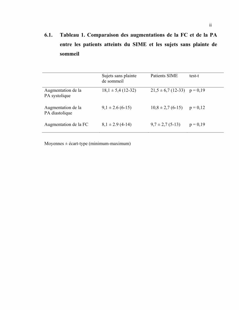
ii
6.1. Tableau 1. Comparaison des augmentations de la FC et de la PA
entre les patients atteints du SIME et les sujets sans plainte de
sommeil
Sujets sans plainte de sommeil
Patients SIME test-t
Augmentation de la PA systolique
18,1 ± 5,4 (12-32) 21,5 ± 6,7 (12-33) p = 0,19
Augmentation de la PA diastolique
9,1 ± 2.6 (6-15) 10,8 ± 2,7 (6-15) p = 0,12
Augmentation de la FC 8,1 ± 2.9 (4-14) 9,7 ± 2,7 (5-13) p = 0,19
Moyennes ± écart-type (minimum-maximum)
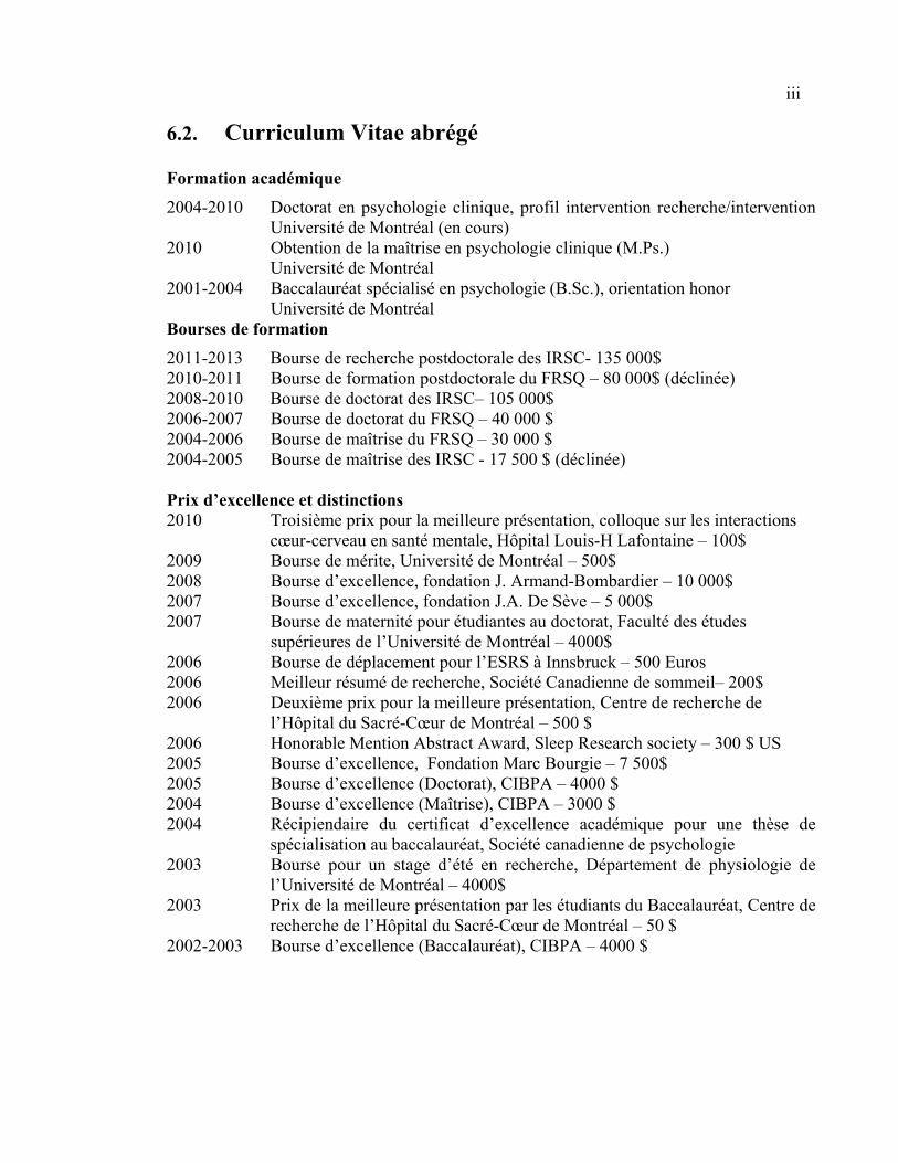
iii
6.2. Curriculum Vitae abrégé
Formation académique 2004-2010 Doctorat en psychologie clinique, profil intervention recherche/intervention
Université de Montréal (en cours) 2010 Obtention de la maîtrise en psychologie clinique (M.Ps.)
Université de Montréal 2001-2004 Baccalauréat spécialisé en psychologie (B.Sc.), orientation honor Université de Montréal Bourses de formation 2011-2013 Bourse de recherche postdoctorale des IRSC- 135 000$ 2010-2011 Bourse de formation postdoctorale du FRSQ – 80 000$ (déclinée) 2008-2010 Bourse de doctorat des IRSC– 105 000$ 2006-2007 Bourse de doctorat du FRSQ – 40 000 $ 2004-2006 Bourse de maîtrise du FRSQ – 30 000 $ 2004-2005 Bourse de maîtrise des IRSC - 17 500 $ (déclinée) Prix d’excellence et distinctions 2010 Troisième prix pour la meilleure présentation, colloque sur les interactions
cœur-cerveau en santé mentale, Hôpital Louis-H Lafontaine – 100$ 2009 Bourse de mérite, Université de Montréal – 500$ 2008 Bourse d’excellence, fondation J. Armand-Bombardier – 10 000$ 2007 Bourse d’excellence, fondation J.A. De Sève – 5 000$ 2007 Bourse de maternité pour étudiantes au doctorat, Faculté des études supérieures de l’Université de Montréal – 4000$ 2006 Bourse de déplacement pour l’ESRS à Innsbruck – 500 Euros 2006 Meilleur résumé de recherche, Société Canadienne de sommeil– 200$ 2006 Deuxième prix pour la meilleure présentation, Centre de recherche de l’Hôpital du Sacré-Cœur de Montréal – 500 $ 2006 Honorable Mention Abstract Award, Sleep Research society – 300 $ US 2005 Bourse d’excellence, Fondation Marc Bourgie – 7 500$ 2005 Bourse d’excellence (Doctorat), CIBPA – 4000 $ 2004 Bourse d’excellence (Maîtrise), CIBPA – 3000 $ 2004 Récipiendaire du certificat d’excellence académique pour une thèse de
spécialisation au baccalauréat, Société canadienne de psychologie 2003 Bourse pour un stage d’été en recherche, Département de physiologie de
l’Université de Montréal – 4000$ 2003 Prix de la meilleure présentation par les étudiants du Baccalauréat, Centre de
recherche de l’Hôpital du Sacré-Cœur de Montréal – 50 $ 2002-2003 Bourse d’excellence (Baccalauréat), CIBPA – 4000 $
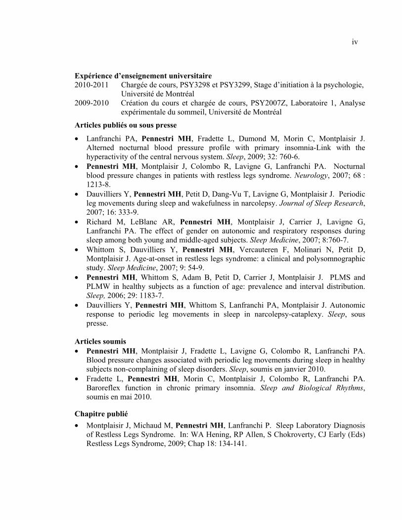
iv
Expérience d’enseignement universitaire 2010-2011 Chargée de cours, PSY3298 et PSY3299, Stage d’initiation à la psychologie, Université de Montréal 2009-2010 Création du cours et chargée de cours, PSY2007Z, Laboratoire 1, Analyse
expérimentale du sommeil, Université de Montréal
Articles publiés ou sous presse
• Lanfranchi PA, Pennestri MH, Fradette L, Dumond M, Morin C, Montplaisir J. Alterned nocturnal blood pressure profile with primary insomnia-Link with the hyperactivity of the central nervous system. Sleep, 2009; 32: 760-6.
• Pennestri MH, Montplaisir J, Colombo R, Lavigne G, Lanfranchi PA. Nocturnal blood pressure changes in patients with restless legs syndrome. Neurology, 2007; 68 : 1213-8.
• Dauvilliers Y, Pennestri MH, Petit D, Dang-Vu T, Lavigne G, Montplaisir J. Periodic leg movements during sleep and wakefulness in narcolepsy. Journal of Sleep Research, 2007; 16: 333-9.
• Richard M, LeBlanc AR, Pennestri MH, Montplaisir J, Carrier J, Lavigne G, Lanfranchi PA. The effect of gender on autonomic and respiratory responses during sleep among both young and middle-aged subjects. Sleep Medicine, 2007; 8:760-7.
• Whittom S, Dauvilliers Y, Pennestri MH, Vercauteren F, Molinari N, Petit D, Montplaisir J. Age-at-onset in restless legs syndrome: a clinical and polysomnographic study. Sleep Medicine, 2007; 9: 54-9.
• Pennestri MH, Whittom S, Adam B, Petit D, Carrier J, Montplaisir J. PLMS and PLMW in healthy subjects as a function of age: prevalence and interval distribution. Sleep, 2006; 29: 1183-7.
• Dauvilliers Y, Pennestri MH, Whittom S, Lanfranchi PA, Montplaisir J. Autonomic response to periodic leg movements in sleep in narcolepsy-cataplexy. Sleep, sous presse.
Articles soumis • Pennestri MH, Montplaisir J, Fradette L, Lavigne G, Colombo R, Lanfranchi PA.
Blood pressure changes associated with periodic leg movements during sleep in healthy subjects non-complaining of sleep disorders. Sleep, soumis en janvier 2010.
• Fradette L, Pennestri MH, Morin C, Montplaisir J, Colombo R, Lanfranchi PA. Baroreflex function in chronic primary insomnia. Sleep and Biological Rhythms, soumis en mai 2010.
Chapitre publié • Montplaisir J, Michaud M, Pennestri MH, Lanfranchi P. Sleep Laboratory Diagnosis
of Restless Legs Syndrome. In: WA Hening, RP Allen, S Chokroverty, CJ Early (Eds) Restless Legs Syndrome, 2009; Chap 18: 134-141.