tomotherapy: when the patient is a child or an adolescent: hopes, … · 2012. 12. 7. · helical...
TRANSCRIPT
- 21 -
Rivista Medica Vol.13, No. 3, 2007
Correspondence: Dr. Maurizio Mascarin, S.O. di Radioterapia Pediatrica, Centro di Riferimento Oncologico, via Franco Gallini 2,33081 Aviano (PN), tel. 0434-659523, fax: 0434-659524, e-mail: [email protected] Medica 2007; 13 (3): 21-28. ISSN: 1127-6339. Fascicolo monografico: ISBN: 978-88-8041-075-1.Comunicazione presentata al “1° Convegno Nazionale di TomoTerapia”, 25 maggio 2007, Aviano (Pordenone). Copyright © 2007by new Magazine edizioni s.r.l., via dei Mille 69, 38100 Trento, Italia. Tutti i diritti riservati. Indexed in EMBASE/Excerpta Medica.www.rivistamedica.it
Tomotherapy: when the patient is a child
or an adolescent: hopes, results and issues
M. MASCARIN, G. FRANCHIN*, M. GIGANTE*, E. MINATEL*, A. DRIGO**, A. DASSIE**,G. SARTOR**, I. ABU RUMEILEH*, R. INNOCENTE*, M. AVANZO**, C. CAPPELLETTO**,E. CAPRA*, E. BORSATTI***, M. DE CICCO****, M.G. TROVÒ*
Pediatric Radiotherapy Unit,
* Department of Radiation Oncology,
** Department of Radiation Physics,
*** Department of Nuclear Medicine,
**** Department of Anesthesia,
Centro di Riferimento Oncologico (CRO), Aviano (Pordenone)
SUMMARY: We describe our experience in 31 pediatric or adolescent oncological patients (median age 14
years old; range 2-24 years) treated with intensity modulation radiation therapy (IMRT) delivered with linear
accelerator (11 patients) or tomotherapy (20 patients). Tomotherapy represents a significant advance in the
ability to deliver the high radiation doses that appear to be required to improve the local control of several pe-
diatric tumors. It demonstrated excellent target coverage, homogeneity and organ sparing compared with con-
ventional radiotherapy. Possible disadvantages of tomotherapy in children are also discussed such as increased
low dose to non-target tissue, prolonged treatment planning and set-up time, increased anesthesia time and in-
creased overall integral dose.
KEY WORDS: Adolescent tumors, Brain tumors, Cranio-spinal irradiation, IMRT, Nasopharynx cancer,
Pediatric tumors, Radiotherapy, Sarcoma, Tomotherapy, Whole abdominal irradiation.
Tomoterapia: quando il paziente è un bambino o un adolescente:
speranze, risultati e problematiche
RIASSUNTO: Riportiamo la nostra esperienza in 31 bambini o adolescenti (età mediana 14 anni; range 2-24)
affetti da neoplasie. I pazienti sono tutti stati trattati con radioterapia ad intensità modulata (IMRT) erogata
con acceleratore lineare (11 pazienti) o con tomoterapia (20 pazienti). La tomoterapia rappresenta un signifi-
cativo miglioramento nella capacità di erogare dosi più alte di radioterapia, con l’obiettivo di migliorare il
controllo locale di diversi tumori pediatrici. Nella nostra esperienza la tomoterapia, se confrontata con la ra-
dioterapia convenzionale, si è dimostrata in grado di comprendere bene ed in modo omogeneo il target.
Vengono discussi anche i possibili svantaggi circa l’utilizzo della tomoterapia in età pediatrica: il possibile au-
mento delle basse dosi a tessuti non-target, i tempi prolungati di trattamento e di set-up, l’aumentato tempo di
anestesia ed infine l’aumento della dose integrale all’intero corpo.
Proceedings Article

INTRODUCTION
In children, radiation is one of the most effectivetreatments for solid tumors, yet the threat of its ef-fects on cognition, growth and development has fordecades led physicians to seek alternatives to thisform of therapy. 3D conformal radiation therapy (3D-CRT) promises high precision in dose delivery andallows approximately 30-40% reduction in the vol-ume of normal tissue included within the high dosevolume compared with conventional 2D planning(5).In recent years, the further advancement in confor-mation has renewed interest in the use of radiationtherapy, also in very young children.Tomotherapy seems one of the most promising meth-ods of treatment. IMRT can achieve an extremelyconformal distribution of radiation to the target vol-ume while sparing critical, surrounding normal tis-sue(6). Because of this ability, the potential for dose es-calation exists, which may translate to a better localcontrol without increasing complication rates.Application of these techniques in children is a for-midable challenge(7,8,9). Guidelines are developing thatensure appropriate volume for each specific type oftumor, and assessment of outcome is essential to en-sure that benefits of the new techniques outweigh therisks. Helical Tomotherapy delivers only IMRTbeams with a potential increase in integral dose tonormal structures or to the whole body(11). The pur-pose is to report our preliminary experience with to-motherapy in children with cancer.
MATERIALS AND METHODS
At Centro Riferimento Oncologico (CRO) in Aviano,we implemented in children computerized assistedplanning with acquisition of volumetric imaging datain 1995, no-coplanar conformal radiation therapy in2000, stereotactic irradiation in 2001, IMRT in 2005and tomotherapy in 2006. A Clinac 2100CD (Varian),equipped with a 120-leaf dynamic multileaf collima-tor, was used for IMRT planning and 3D conformalradiation therapy. 6 MV X-rays were used for all pa-tients. During every week of treatment, a daily set ofisocenter verification portal films were acquired.Helical radiotherapy was delivered with a tomothe-
rapy Hi-Art System, in which a 6 MV linear acce-lerator and CT technology are integrated. That resultsin a helical form of radiation delivery, without junc-tional problems. Through mega voltage CT, imagesof the patient’s anatomy, including tumor characte-ristics and clinical structures, were acquired daily.This allowed an on-line update of the treatment planfor any change in the patient’s anatomy or position.■■ SIMULATION. The simulation process was donewith helical CT-simulator and took 40-90 min. Theupper time limit referred to younger patients thatneeded sedation or images of the cranio-spinal axis.In order to immobilize the brain we used a thermo-plastic mask (sometimes with a dental bite) and forthe chest wall and abdomen a combination of a per-sonalized “cradle” bag and extended wing board(Figure 1). After the introduction of tomotherapy thepatients were all aligned and immobilized in supineposition, also for cranio-spinal irradiation (CSI). TheCT images were acquired to a slice thickness andspacing of 5 mm and a pith of 1.5 mm (obtained dur-ing quiet respiration).■■ CONTOURING. According to ICRU definitions wedefined gross tumor volume (GTV), clinical targetvolume (CTV) referred to the tissue potentially con-taining a sub-clinical tumor and planning target vol-ume (PTV) to include geometric and set-up uncer-tainties and the accuracy of the immobilization deviceused. In some cases we defined the tumor extensiondetermined by CT, NMR and/or PET merged for plan-ning to obtain a metabolic target volume and a dosemodulation inside the volume of interest (Figure 2).An extra structure was generated (“tune structure”) toobtain a better optimization around the target.■■ PLANNING. The IMRT plan was performed on anEclipse-Varian treatment planning station. The dosewas prescribed to a volume (PTV) and not to a singlepoint. The treatment plans consisted preferentially ofcoplanar fields. The directions of the fields were cho-sen to avoid normal tissues. For brain tumor we paidspecial attention to the lenses, cochlea and pituitaryarea. For other sites we paid special attention to thespine, lungs, heart, bowel, growing bones, glandularand endocrine function. The tomotherapy plan wasgenerated by tomotherapy planning workstation.Prior to optimization, dose volume constraints, prece-dence, importance and penalty factors were used to
- 22 -
Tomotherapy: when the patient is a child or an adolescent: hopes, results and issues M. Mascarin
PAROLE CHIAVE: Tumori dell’adolescenza, Tumori cerebrali, Irradiazione cranio-spinale, IMRT, Tumori
del rinofaringe, Tumori pediatrici, Radioterapia, Tomoterapia, Irradiazione addominale.

improve target dose homogeneities and reduce dosesto normal structure. The dose limits for the criticalstructure were the standard values used in clinicalpractice for pediatric tumors. Parameters specified aspart of the optimization/dose calculation process werepitch, beam thickness and modulation factor. Filmanalysis, dose profile comparison and ion chambermeasurements were used to verify the intensity mapsfor each plan and the absolute dose for each patient.
RESULTS
Since the introduction of the intensity modulationprogram in our Department, we have treated 31 chil-dren or adolescent patients with IMRT delivered withlinear accelerator (11 patients) or tomotherapy (20 pa-tients) (Table 1).The median age of patients was 14 years (range 2-24years). In all cases radiotherapy was part of the mul-timodality program according to Italian or European
pediatric protocol. Young patients affected by tumorsthat were complex, large, or close to critical areas,were selected for this kind of therapy.With regard to IMRT and tomotherapy the time re-quired from simulation to the first day of treatmentwas 1-4 weeks. The time for contouring was 3-10hours, depending on the necessity to use merged im-ages. From 3 to more than 10 optimization iterationswere required. IMRT planning generally requiredmore interactions than tomotherapy. The patientswere set-up and treated within a 20-45 minutes peri-od, depending on the volume of the tumor and theneed for anesthesia. From the beginning of the tomo-therapy program in children we applied a step by stepadaptive therapy process including: radiological andmetabolic diagnostic imaging, patient position and si-mulation, target and structure countering, treatmentplanning optimization, radiotherapy imaging “on bo-ard”, planning and treatment images co-registration,modified patients position, treatment delivery and amodified treatment plan, if necessary.
- 23 -
Rivista Medica Vol.13, No. 3, 2007
Figure 2. Tumor extension in a frontal malignant gliomas determined by CT, NMR (blue line), spectroscopic NMR (fuchsia line) andPET-CT (red line) merged for planning.
Figure 1. Immobilization devices: thermoplastic mask for brain tumor, thermoplastic mask with a dental bite for nasopharynx can-cer and a personalized “cradle” bag for abdominal tumor in a child requiring sedation.
❒❒ ADVANTAGES
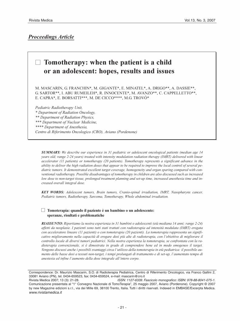
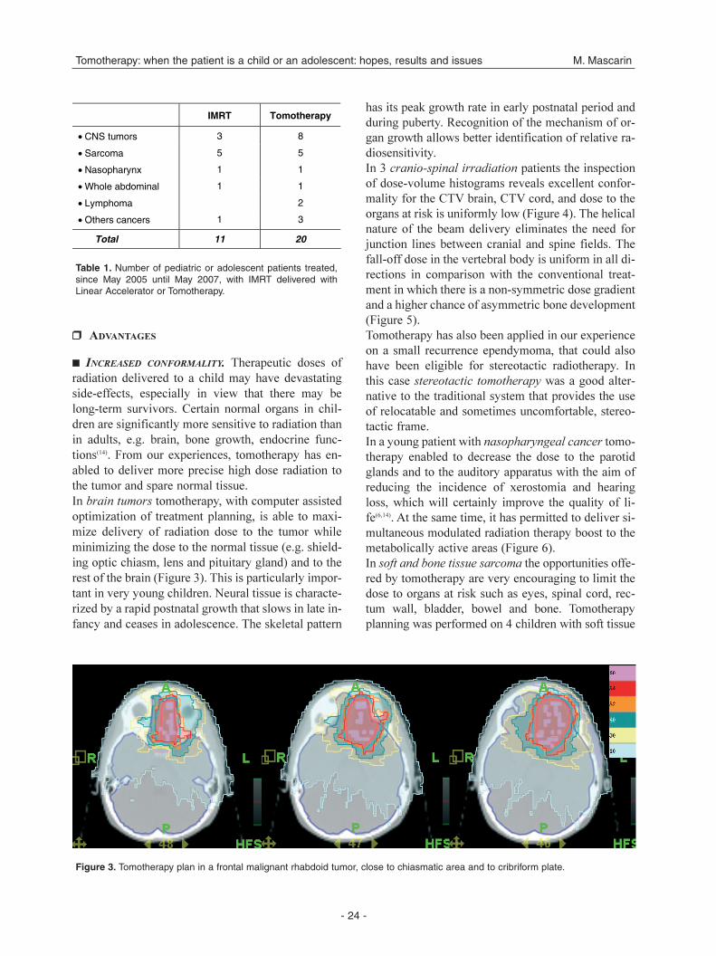
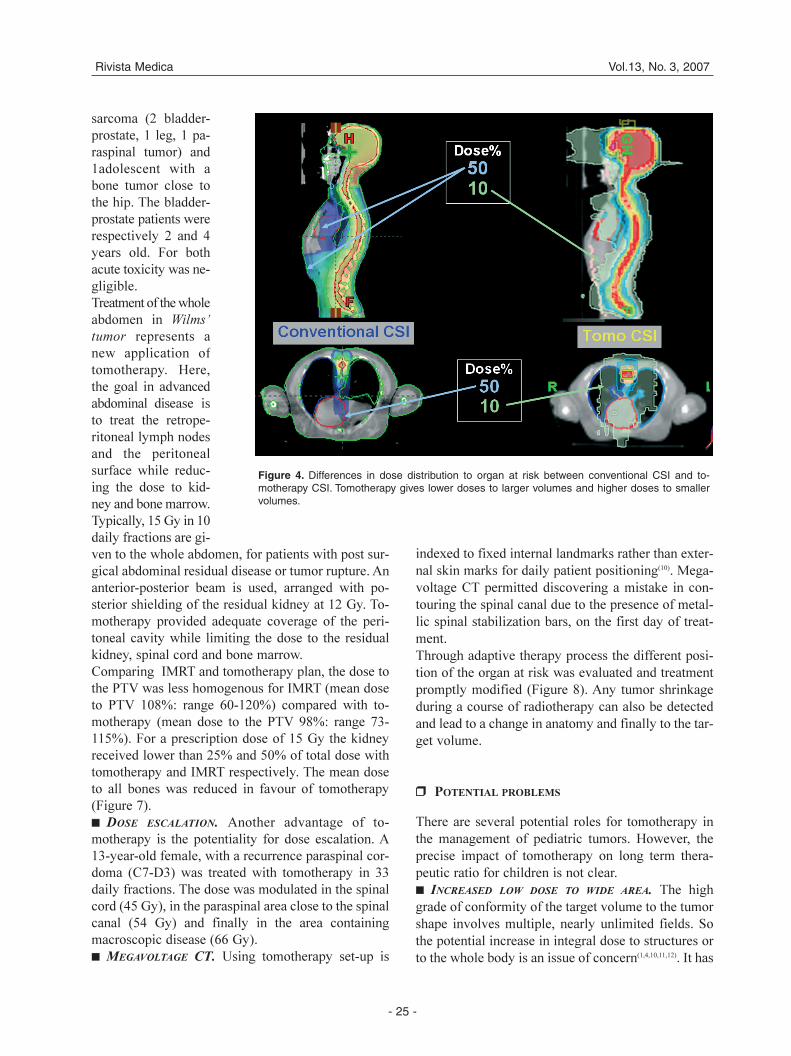
■■ INCREASED CONFORMALITY. Therapeutic doses ofradiation delivered to a child may have devastatingside-effects, especially in view that there may belong-term survivors. Certain normal organs in chil-dren are significantly more sensitive to radiation thanin adults, e.g. brain, bone growth, endocrine func-tions(14). From our experiences, tomotherapy has en-abled to deliver more precise high dose radiation tothe tumor and spare normal tissue.In brain tumors tomotherapy, with computer assistedoptimization of treatment planning, is able to maxi-mize delivery of radiation dose to the tumor whileminimizing the dose to the normal tissue (e.g. shield-ing optic chiasm, lens and pituitary gland) and to therest of the brain (Figure 3). This is particularly impor-tant in very young children. Neural tissue is characte-rized by a rapid postnatal growth that slows in late in-fancy and ceases in adolescence. The skeletal pattern
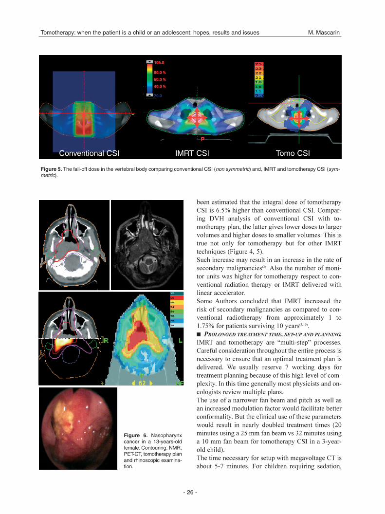
has its peak growth rate in early postnatal period andduring puberty. Recognition of the mechanism of or-gan growth allows better identification of relative ra-diosensitivity.In 3 cranio-spinal irradiation patients the inspectionof dose-volume histograms reveals excellent confor-mality for the CTV brain, CTV cord, and dose to theorgans at risk is uniformly low (Figure 4). The helicalnature of the beam delivery eliminates the need forjunction lines between cranial and spine fields. Thefall-off dose in the vertebral body is uniform in all di-rections in comparison with the conventional treat-ment in which there is a non-symmetric dose gradientand a higher chance of asymmetric bone development(Figure 5).Tomotherapy has also been applied in our experienceon a small recurrence ependymoma, that could alsohave been eligible for stereotactic radiotherapy. Inthis case stereotactic tomotherapy was a good alter-native to the traditional system that provides the useof relocatable and sometimes uncomfortable, stereo-tactic frame.In a young patient with nasopharyngeal cancer tomo-therapy enabled to decrease the dose to the parotidglands and to the auditory apparatus with the aim ofreducing the incidence of xerostomia and hearingloss, which will certainly improve the quality of li-fe(6,14). At the same time, it has permitted to deliver si-multaneous modulated radiation therapy boost to themetabolically active areas (Figure 6).In soft and bone tissue sarcoma the opportunities offe-red by tomotherapy are very encouraging to limit thedose to organs at risk such as eyes, spinal cord, rec-tum wall, bladder, bowel and bone. Tomotherapyplanning was performed on 4 children with soft tissue
- 24 -
Tomotherapy: when the patient is a child or an adolescent: hopes, results and issues M. Mascarin
IMRT Tomotherapy
• CNS tumors 3 8
• Sarcoma 5 5
• Nasopharynx 1 1
• Whole abdominal 1 1
• Lymphoma 2
• Others cancers 1 3
Total 11 20
Table 1. Number of pediatric or adolescent patients treated,since May 2005 until May 2007, with IMRT delivered withLinear Accelerator or Tomotherapy.
Figure 3. Tomotherapy plan in a frontal malignant rhabdoid tumor, close to chiasmatic area and to cribriform plate.
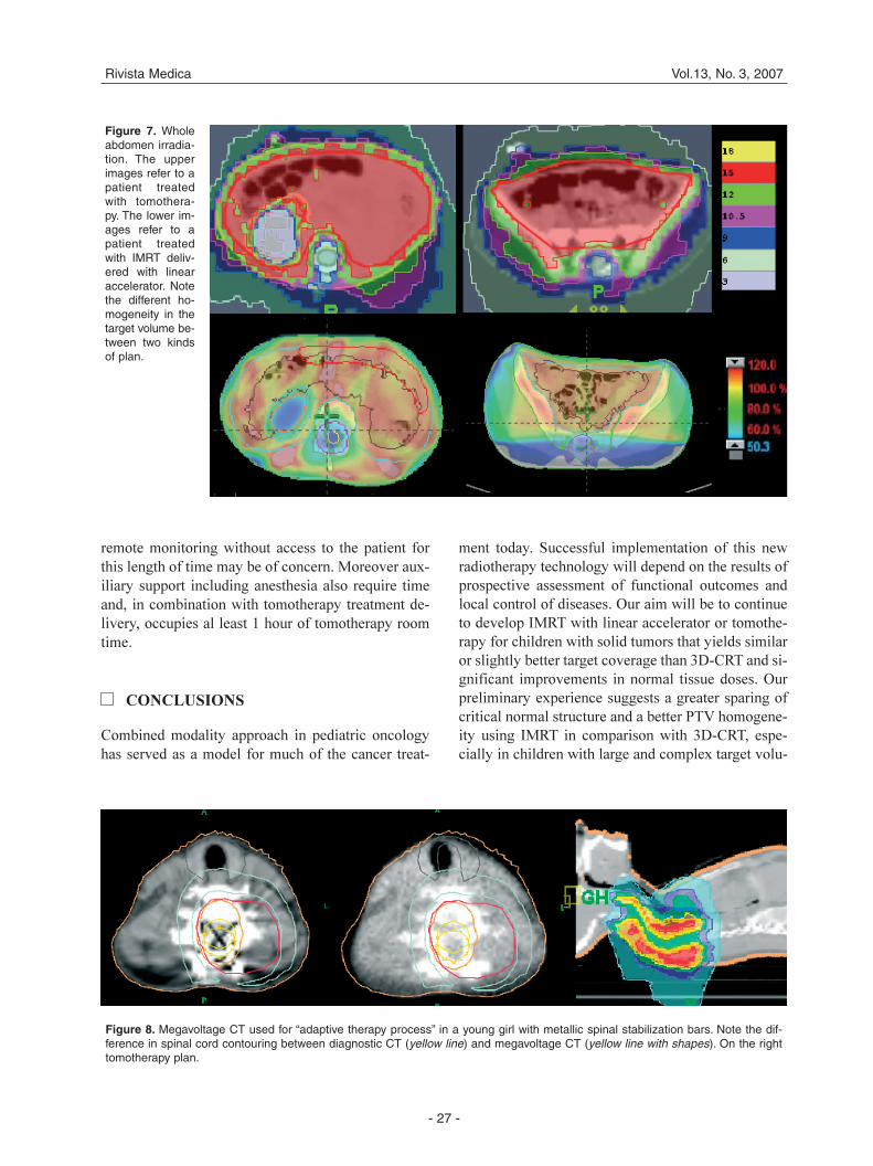
sarcoma (2 bladder-prostate, 1 leg, 1 pa-raspinal tumor) and1adolescent with abone tumor close tothe hip. The bladder-prostate patients wererespectively 2 and 4years old. For bothacute toxicity was ne-gligible.Treatment of the wholeabdomen in Wilms’
tumor represents anew application oftomotherapy. Here,the goal in advancedabdominal disease isto treat the retrope-ritoneal lymph nodesand the peritonealsurface while reduc-ing the dose to kid-ney and bone marrow.Typically, 15 Gy in 10daily fractions are gi-ven to the whole abdomen, for patients with post sur-gical abdominal residual disease or tumor rupture. Ananterior-posterior beam is used, arranged with po-sterior shielding of the residual kidney at 12 Gy. To-motherapy provided adequate coverage of the peri-toneal cavity while limiting the dose to the residualkidney, spinal cord and bone marrow.Comparing IMRT and tomotherapy plan, the dose tothe PTV was less homogenous for IMRT (mean doseto PTV 108%: range 60-120%) compared with to-motherapy (mean dose to the PTV 98%: range 73-115%). For a prescription dose of 15 Gy the kidneyreceived lower than 25% and 50% of total dose withtomotherapy and IMRT respectively. The mean doseto all bones was reduced in favour of tomotherapy(Figure 7).■■ DOSE ESCALATION. Another advantage of to-motherapy is the potentiality for dose escalation. A13-year-old female, with a recurrence paraspinal cor-doma (C7-D3) was treated with tomotherapy in 33daily fractions. The dose was modulated in the spinalcord (45 Gy), in the paraspinal area close to the spinalcanal (54 Gy) and finally in the area containingmacroscopic disease (66 Gy).■■ MEGAVOLTAGE CT. Using tomotherapy set-up is
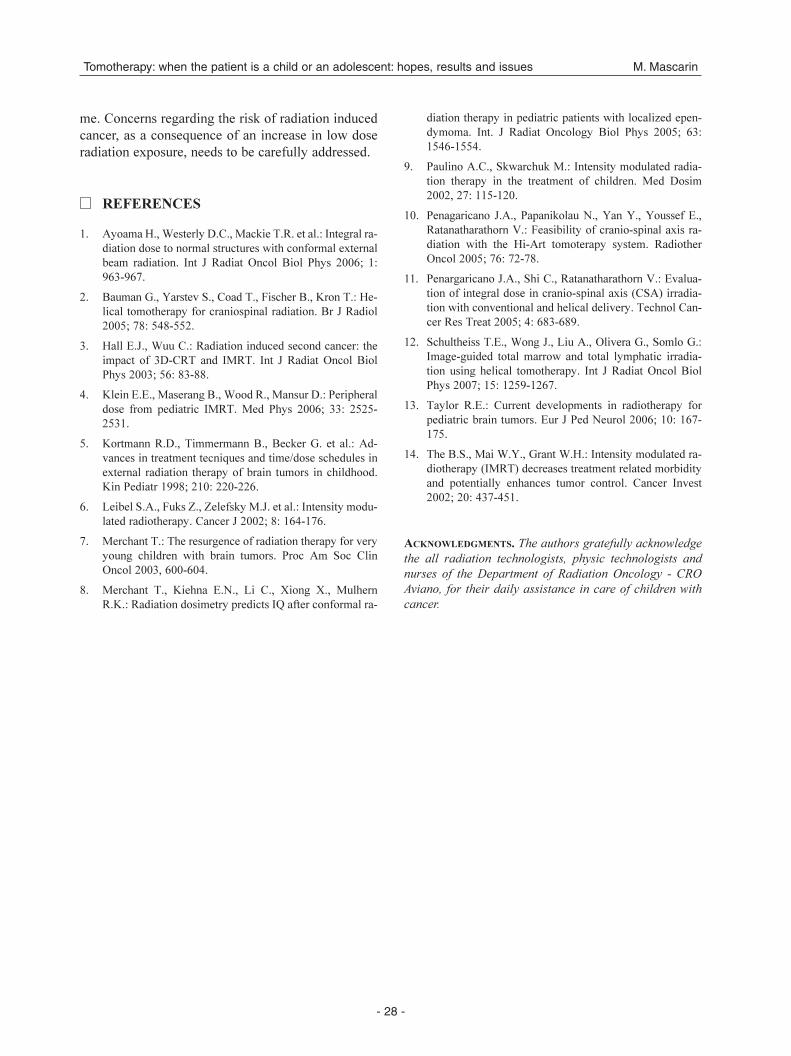
indexed to fixed internal landmarks rather than exter-nal skin marks for daily patient positioning(10). Mega-voltage CT permitted discovering a mistake in con-touring the spinal canal due to the presence of metal-lic spinal stabilization bars, on the first day of treat-ment.Through adaptive therapy process the different posi-tion of the organ at risk was evaluated and treatmentpromptly modified (Figure 8). Any tumor shrinkageduring a course of radiotherapy can also be detectedand lead to a change in anatomy and finally to the tar-get volume.
❒❒ POTENTIAL PROBLEMS
There are several potential roles for tomotherapy inthe management of pediatric tumors. However, theprecise impact of tomotherapy on long term thera-peutic ratio for children is not clear.■■ INCREASED LOW DOSE TO WIDE AREA. The highgrade of conformity of the target volume to the tumorshape involves multiple, nearly unlimited fields. Sothe potential increase in integral dose to structures orto the whole body is an issue of concern(1,4,10,11,12). It has
- 25 -
Rivista Medica Vol.13, No. 3, 2007
Figure 4. Differences in dose distribution to organ at risk between conventional CSI and to-motherapy CSI. Tomotherapy gives lower doses to larger volumes and higher doses to smallervolumes.
been estimated that the integral dose of tomotherapyCSI is 6.5% higher than conventional CSI. Compar-ing DVH analysis of conventional CSI with to-motherapy plan, the latter gives lower doses to largervolumes and higher doses to smaller volumes. This istrue not only for tomotherapy but for other IMRTtechniques (Figure 4, 5).Such increase may result in an increase in the rate ofsecondary malignancies(2). Also the number of moni-tor units was higher for tomotherapy respect to con-ventional radiation therapy or IMRT delivered withlinear accelerator.Some Authors concluded that IMRT increased therisk of secondary malignancies as compared to con-ventional radiotherapy from approximately 1 to1.75% for patients surviving 10 years(3,10).■■ PROLONGED TREATMENT TIME, SET-UP AND PLANNING.
IMRT and tomotherapy are “multi-step” processes.Careful consideration throughout the entire process isnecessary to ensure that an optimal treatment plan isdelivered. We usually reserve 7 working days fortreatment planning because of this high level of com-plexity. In this time generally most physicists and on-cologists review multiple plans.The use of a narrower fan beam and pitch as well asan increased modulation factor would facilitate betterconformality. But the clinical use of these parameterswould result in nearly doubled treatment times (20minutes using a 25 mm fan beam vs 32 minutes usinga 10 mm fan beam for tomotherapy CSI in a 3-year-old child).The time necessary for setup with megavoltage CT isabout 5-7 minutes. For children requiring sedation,
- 26 -
Tomotherapy: when the patient is a child or an adolescent: hopes, results and issues M. Mascarin
Figure 6. Nasopharynxcancer in a 13-years-oldfemale. Contouring, NMR,PET-CT, tomotherapy planand rhinoscopic examina-tion.
Conventional CSI IMRT CSI Tomo CSI
Figure 5. The fall-off dose in the vertebral body comparing conventional CSI (non symmetric) and, IMRT and tomotherapy CSI (sym-metric).
remote monitoring without access to the patient forthis length of time may be of concern. Moreover aux-iliary support including anesthesia also require timeand, in combination with tomotherapy treatment de-livery, occupies al least 1 hour of tomotherapy roomtime.
CONCLUSIONS
Combined modality approach in pediatric oncologyhas served as a model for much of the cancer treat-
ment today. Successful implementation of this newradiotherapy technology will depend on the results ofprospective assessment of functional outcomes andlocal control of diseases. Our aim will be to continueto develop IMRT with linear accelerator or tomothe-rapy for children with solid tumors that yields similaror slightly better target coverage than 3D-CRT and si-gnificant improvements in normal tissue doses. Ourpreliminary experience suggests a greater sparing ofcritical normal structure and a better PTV homogene-ity using IMRT in comparison with 3D-CRT, espe-cially in children with large and complex target volu-
- 27 -
Rivista Medica Vol.13, No. 3, 2007
Figure 7. Wholeabdomen irradia-tion. The upperimages refer to apatient treatedwith tomothera-py. The lower im-ages refer to apatient treatedwith IMRT deliv-ered with linearaccelerator. Notethe different ho-mogeneity in thetarget volume be-tween two kindsof plan.
Figure 8. Megavoltage CT used for “adaptive therapy process” in a young girl with metallic spinal stabilization bars. Note the dif-ference in spinal cord contouring between diagnostic CT (yellow line) and megavoltage CT (yellow line with shapes). On the righttomotherapy plan.
me. Concerns regarding the risk of radiation inducedcancer, as a consequence of an increase in low doseradiation exposure, needs to be carefully addressed.
REFERENCES
1. Ayoama H., Westerly D.C., Mackie T.R. et al.: Integral ra-diation dose to normal structures with conformal externalbeam radiation. Int J Radiat Oncol Biol Phys 2006; 1:963-967.
2. Bauman G., Yarstev S., Coad T., Fischer B., Kron T.: He-lical tomotherapy for craniospinal radiation. Br J Radiol2005; 78: 548-552.
3. Hall E.J., Wuu C.: Radiation induced second cancer: theimpact of 3D-CRT and IMRT. Int J Radiat Oncol BiolPhys 2003; 56: 83-88.
4. Klein E.E., Maserang B., Wood R., Mansur D.: Peripheraldose from pediatric IMRT. Med Phys 2006; 33: 2525-2531.
5. Kortmann R.D., Timmermann B., Becker G. et al.: Ad-vances in treatment tecniques and time/dose schedules inexternal radiation therapy of brain tumors in childhood.Kin Pediatr 1998; 210: 220-226.
6. Leibel S.A., Fuks Z., Zelefsky M.J. et al.: Intensity modu-lated radiotherapy. Cancer J 2002; 8: 164-176.
7. Merchant T.: The resurgence of radiation therapy for veryyoung children with brain tumors. Proc Am Soc ClinOncol 2003, 600-604.
8. Merchant T., Kiehna E.N., Li C., Xiong X., MulhernR.K.: Radiation dosimetry predicts IQ after conformal ra-
diation therapy in pediatric patients with localized epen-dymoma. Int. J Radiat Oncology Biol Phys 2005; 63:1546-1554.
9. Paulino A.C., Skwarchuk M.: Intensity modulated radia-tion therapy in the treatment of children. Med Dosim2002, 27: 115-120.
10. Penagaricano J.A., Papanikolau N., Yan Y., Youssef E.,Ratanatharathorn V.: Feasibility of cranio-spinal axis ra-diation with the Hi-Art tomoterapy system. RadiotherOncol 2005; 76: 72-78.
11. Penargaricano J.A., Shi C., Ratanatharathorn V.: Evalua-tion of integral dose in cranio-spinal axis (CSA) irradia-tion with conventional and helical delivery. Technol Can-cer Res Treat 2005; 4: 683-689.
12. Schultheiss T.E., Wong J., Liu A., Olivera G., Somlo G.:Image-guided total marrow and total lymphatic irradia-tion using helical tomotherapy. Int J Radiat Oncol BiolPhys 2007; 15: 1259-1267.
13. Taylor R.E.: Current developments in radiotherapy forpediatric brain tumors. Eur J Ped Neurol 2006; 10: 167-175.
14. The B.S., Mai W.Y., Grant W.H.: Intensity modulated ra-diotherapy (IMRT) decreases treatment related morbidityand potentially enhances tumor control. Cancer Invest2002; 20: 437-451.
ACKNOWLEDGMENTS. The authors gratefully acknowledge
the all radiation technologists, physic technologists and
nurses of the Department of Radiation Oncology - CRO
Aviano, for their daily assistance in care of children with
cancer.
- 28 -
Tomotherapy: when the patient is a child or an adolescent: hopes, results and issues M. Mascarin