tk news 2|04
DESCRIPTION
Minimally invasive techniques in trauma surgery, New MIS products, New long bone products, New foot and ankle products, New power tools, New spine products, New craniomaxillofacial products, Development and validation of AO fracture classification, Database of Human CT Data, Portrait: Frankie Leung, Hong Kong, TK Prize 2003, AO Courses, ImprintTRANSCRIPT
News—Number 2 | 2004
New Productsfrom AO Development
News 2 | 04
Table of contents
Minimally invasive techniques in trauma surgery 3
New MIS productsChisel for plate extraction 5
New long bone productsLocking Trochanter Stabilization Plate 5Basic Percutaneous Instrumentation Set for
Locking Condylar Plate 6Large Distractor/Compressor Instrument 6Drive Adaptors with Quick Connect for Schanz Screws 7Medium External Fixation System: additional
clamps, MR safe 7Small External Fixation System: Wire Cutter 84.0 mm Schanz Screw, spade point, with 38 mm
thread length 8
New foot and ankle productsSpherical Washer 8
New power toolsE-Pen 9Torque Limiter for Quick Coupling and
Mini Quick Coupling 9
New spine productsAnterior Cervical Locking Plate (ACLP) 10Anterior Telescoping Fixation System (TeleFix) 10Anterior Tension Band (ATB) 11Axon System 11SynCage-LR 45º/90º 12Additional T-PLIF Minimal Invasive Instruments 12Guide Rods for Cervical SynFrame 13Ti USS Variable Axis Screw, different lengths
and diameters 13USS Pedicle Screw and additional instruments 13USS II Washers and USS II Anterior Vertebral
Body Screws 14Click’X: additional instruments 145.0 mm Ti Replacement Setscrew 14
New craniomaxillofacial products2.0 mm Mandible Locking Plate: line extension 152.0 mm UniLOCK Screw, TAV and 2.4 mm
Emergency Screw 15Lock Zygomatic Plate 16Stardrive Screwdriver Shafts, dynamic 16Stardrive Blade with Holding Sleeve 162.0 mm Ti Curved Sagittal Split Plate, 6-hole 16Craniofacial Plates: line extensions: 17
1.3 mm Double Y-Plate, 6-hole, 22 mm
1.3 mm Maxillary T-Plates, 8–14 mm
1.5 mm pre-bent Maxillary Plates, 0 mm
advancement
1.5 mm Chin Plates, Double Bend, 4–10 mm
2.0 mm Y-Plate, 110º
2.0 mm Double Adaption Plate, 40-hole
1.5 mm Resorbable Tap, self-drilling: line extension 181.5 mm Resorbable Orbital Floor Plate: line extension 181.3 mm Drill Bits with Stop (3 mm) 18
Development and validation of AO fracture classification 19
Database of Human CT Data 21
Portrait: Frankie Leung, Hong Kong 22
TK Prize 2003 23
AO Courses 23
Imprint 26
Due to varying countries’ legal and regulatory approval requirements please consult the appropriate local product labeling for approved intended use of the products decsribed in this brochure.
and this issue of TK News features quite a few of these. Many of these
improvements result directly from the feedback of surgeons and OR
personnel. If you have any proposals or critique on existing devices,
please tell us.
The AO Foundation is not only searching for technical perfection but
also trying to consider regional differences worldwide. As a truly global
organization, AO Development involves surgeons from Asia and Latin
America to incorporate differences in mentality or anatomy, eg, the
Small PFN for Asia. Reflecting the dynamics of these regions, AO De-
velopment participated in the AO Alumni meetings of AO East Asia and
AO Latin America in 2004. The case discussions showed a high level of
orthopedic trauma care equivalent to European and North American
standards. The spirit, engagement, and dedication of the AO Alumni
members must be similar to one of the founding days of the AO. To share
this experience with you, the column Portrait features Frankie Leung,
MD, from Hong Kong.
Once again, I would like to stress that none of the product descriptions
in this publication is a substitute for the AO’s surgical techniques or
AO teaching tools. You can obtain more detailed information on these
products from AO or your local SYNTHES® representative.
If you have any comments or questions on the articles or the new prod-
ucts, please don’t hesitate to contact me.
Yours faithfully,
Norbert P Haas
Dear reader,
Minimally invasive surgery (MIS) is some-
thing everybody talks about—but not in the
same language, indicating very different inter-
pretations. The AO Foundation has a very clear
philosophy which Dr Dankward Höntzsch, the
chairman of AO’s MIS group, will present to
you in the lead article. He clearly shows how
the evolution of new approaches in patient
treatment is based on research results and the
collective clinical experience of AO surgeons.
AO solutions are special because of its sound
scientific background and its consistent phi-
losophy, as well as the tremendous teaching
efforts in which they are embedded. At the
moment AO is working hard to adapt all its
teaching materials to the Locking Compres-
sion Plate (LCP) concept. A new book and
long-distance learning materials will be avail-
able soon.
The LCP concept is the “state-of-the-art”
worldwide. Almost all plates are now available
with the combination hole. In the last issues
of TK News, you received information on these
new angular stable implants. But in the OR,
little things often make a huge difference in
the outcome and influence the stress factor on
us surgeons and our OR personnel. Therefore,
AO pays a lot of attention to its instruments,
their ergonomics, power tools, and a huge vari-
ety of line extensions. Even little devices, tem-
plates etc make procedures more convenient
Intramedullary nailingIntramedullary nailing is basically the mini-
mally invasive method par excellence: a small
incision far away from the injured region, in-
direct reduction and bridging, providing rela-
tive stability.
The intramedullary methods include stabili-
zation of the proximal and distal femur and
proximal humerus. Work is going on to im-
prove these methods. This is also true with
regard to further indications and implants:
Titanium Elastic Nails (TENs), elastic wiring
of the clavicle and arthrodesis nails, to name
just a few.
External fixationBasically, external fixation can be taken as a
prime example of a minimally invasive sta-
bilization method. This means that the tissue
damaged by the injury is preserved as much
as possible and is not subjected to additional
trauma. This is one reason why the method
is applied in particularly critical situations,
namely, when the soft tissues and the bone
have been traumatized and have sustained se-
rious damage, or in severely injured patients
(polytrauma). Discussion of “Damage Con-
trolled Surgery“ indicates the value of exter-
nal fixation. The external fixation method
Dankward Höntzsch
Minimally invasive techniques in trauma surgery
In all surgical disciplines, the operative methods that are described as
minimally invasive are those methods that effectively reduce any addi-
tional trauma caused by the approach and the surgical procedures. Less
intervention means that the integrity of the body’s structures suffers
the least possible additional damage.
It is important to realize that the abbreviation MIPO does not stand
for “minimally invasive plate” but rather “percutaneous” osteosynthe-
sis. This needs to be borne in mind since the expression “minimally
invasive surgery” (MIS) is being increasingly used. Thus the goals are
underlined as: minimization of input or load for all procedures from
positioning, operation with reduction, retention, and intraoperative as-
sessments, eg, by radiographic means, through to aftercare.
reaches its limitations during the course of the healing and treatment
process so that we, as many others, advocate a two-stage procedure
whereby conversion to another minimally invasive method, eg, in-
tramedullary nailing or a percutaneous insertion of a plate, is per-
formed as soon as possible.
AO modular external fixators are available in various sizes. With these
fixators primary, secondary, and intraoperative reduction manoeuvers
are possible. The latter are achieved with the so-called External Reduc-
tion Device (ERD) that is a part of the MIPO/MIS instrument set.
Percutaneously inserted platesThe development and application of minimally invasive plate osteo-
synthesis has rocketed in the last few years. Due to two major develop-
ments these innovations are here to stay. One thing we have learnt is
that it is advantageous for plate osteosynthesis of diaphyseal fractures
to be performed using the bridging technique. A further development
and complementary aspect of this method is that these plate systems
can now, in many cases, be slid into position in a semi-closed proce-
dure. This technique means that not only the operative trauma (bridg-
ing plate) is reduced and kept to a minimum, but also the trauma of
the approach.
Minimally invasive plate osteosynthesis was and still is realized using
conventional plate systems. However, thanks to the internal fixator
principle with locking head screws, plates and/or stabilization systems
have been developed that are especially suited to this operative meth-
od, eg, the Less Invasive Stabilization System = LISS and the whole
spectrum of Locking Compression Plates (LCP). Instruments such as
tunneling aids, plate holders, spreaders etc are a great help in achiev-
ing minimally invasive plate insertion with accuracy and performing
2 | Minimally invasive techniques in trauma surgery
Minimally invasive techniques in trauma surgery | 3
gentle reduction manoeuvers in this osteosyn-
thesis technique. It is not the size of the skin
incision but rather the extent of manipulation
actually applied internally that is critical for
the additional trauma.
Screws and tension band fixationOther operative methods such as isolated
screws, tension band plates, and mixed
osteosynthesis can now be performed through
a minimal approach and using a minimally
invasive technique. For example, single screw
osteosynthesis and tension band wire fixation
can be performed using a minimally invasive
technique. New instruments have also been
designed and developed for these procedures.
Spine surgeryFor operations on the spine from a dorsal ap-
proach, development is still in its infancy with
regard to further minimization of approach
and surgical trauma. For anterior operative
methods, minimally invasive methods became
standard procedures.
ArthroscopyArthroscopic interventions are basically mini-
mally invasive surgical methods. Arthroscopy
of the knee and shoulder joints is the most
popular and most explored procedure.
Other arthroscopic operations on the joints
include those for the ankle, wrist, and elbow
joints. Arthroscopic techniques for the hip
joint are in the development phase.
Arthroscopic techniques can be combined to
advantage with conventional open and new
minimally invasive surgical techniques.
Computer-assisted navigationComputer-assisted navigation increasingly
permits minimization of surgical trauma and
facilitates, for example, minimally invasive
operations on the axial skeleton and the ex-
tremities. An important innovative leap for-
ward in the field of computer-assisted naviga-
tion is expected within the next few years.
Instruments, surgical methods, and teachingSpecial instruments for minimally invasive procedures have been
launched over the last 3 years and others are still being developed.
Some of the highlights and the contents of a basic set are presented in
the figures and the descriptive text. One purpose of these instruments
is to enable minimally invasive surgical techniques to be used also for
the application of conventional osteosynthesis implants.
At the same time, teaching modules are being developed that have al-
ready proven valuable when first presented at AO Courses and which
will gradually become available to the teachers and participants at AO
Courses.
AO and its TK-System are particularly dedicated to this task.
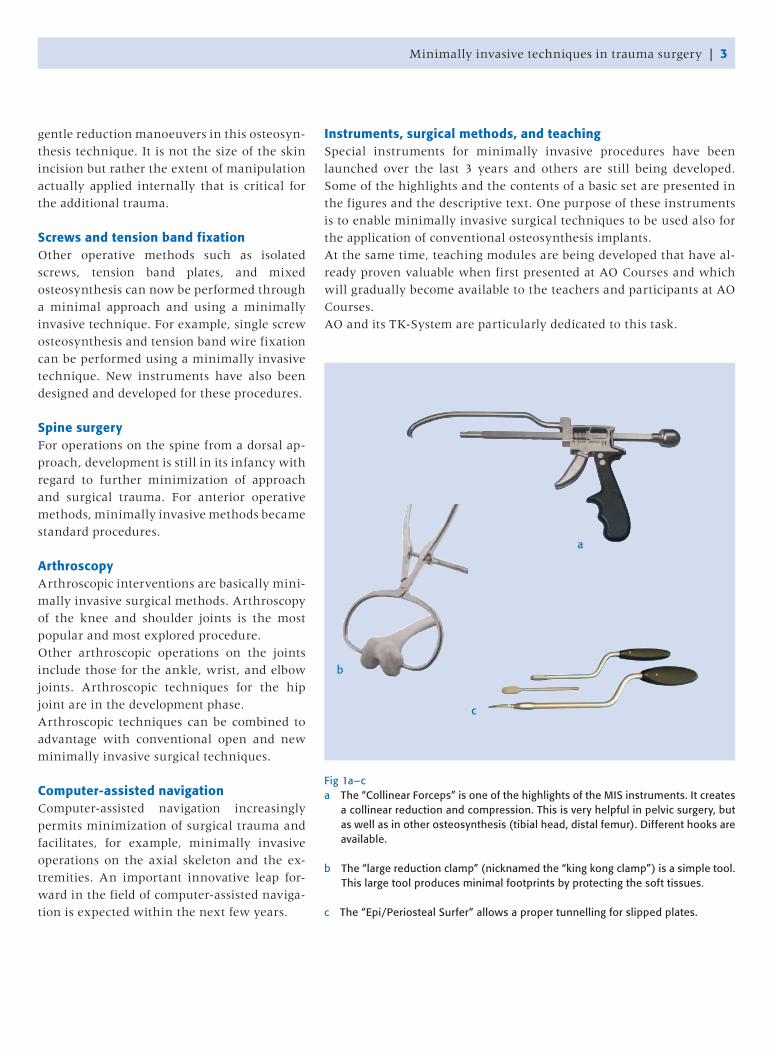
Fig 1a–ca The “Collinear Forceps” is one of the highlights of the MIS instruments. It creates
a collinear reduction and compression. This is very helpful in pelvic surgery, but as well as in other osteosynthesis (tibial head, distal femur). Different hooks are available.
b The “large reduction clamp” (nicknamed the “king kong clamp”) is a simple tool. This large tool produces minimal footprints by protecting the soft tissues.
c The “Epi/Periosteal Surfer” allows a proper tunnelling for slipped plates.
c
b
a
SummaryIn summary, it can be stated that with the introduction of minimally
invasive techniques as the result of modifying long established meth-
ods (arthroscopy, intramedullary nailing, and external fixation) and
the development of new surgical procedures and implants (plates based
on the internal fixator principle, indirect manipulation, spine surgery,
etc) a new age has dawned in traumatology. Every procedure in osteo-
synthesis should take minimally invasive surgery into account.
In addition to the new instruments and implants, the attitude and
training of the present and future generation of surgeons is the most
important aspect. AO, in keeping with its tradition and mission, will
pave the way for this whole spectrum of basic research, development,
documentation, and education and set the future trend.
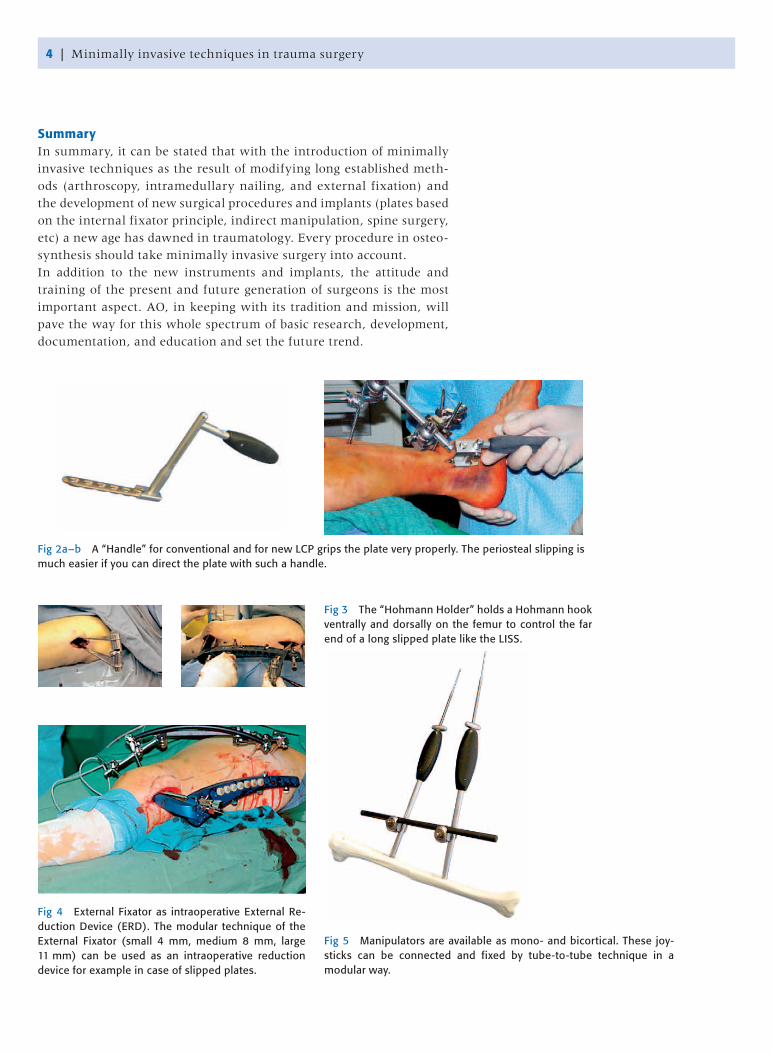
Fig 2a–b A “Handle” for conventional and for new LCP grips the plate very properly. The periosteal slipping is much easier if you can direct the plate with such a handle.
Fig 3 The “Hohmann Holder” holds a Hohmann hook ventrally and dorsally on the femur to control the far end of a long slipped plate like the LISS.
Fig 4 External Fixator as intraoperative External Re-duction Device (ERD). The modular technique of the External Fixator (small 4 mm, medium 8 mm, large 11 mm) can be used as an intraoperative reduction device for example in case of slipped plates.
Fig 5 Manipulators are available as mono- and bicortical. These joy-sticks can be connected and fixed by tube-to-tube technique in a modular way.
4 | Minimally invasive techniques in trauma surgery
New MIS/long bone products | 5
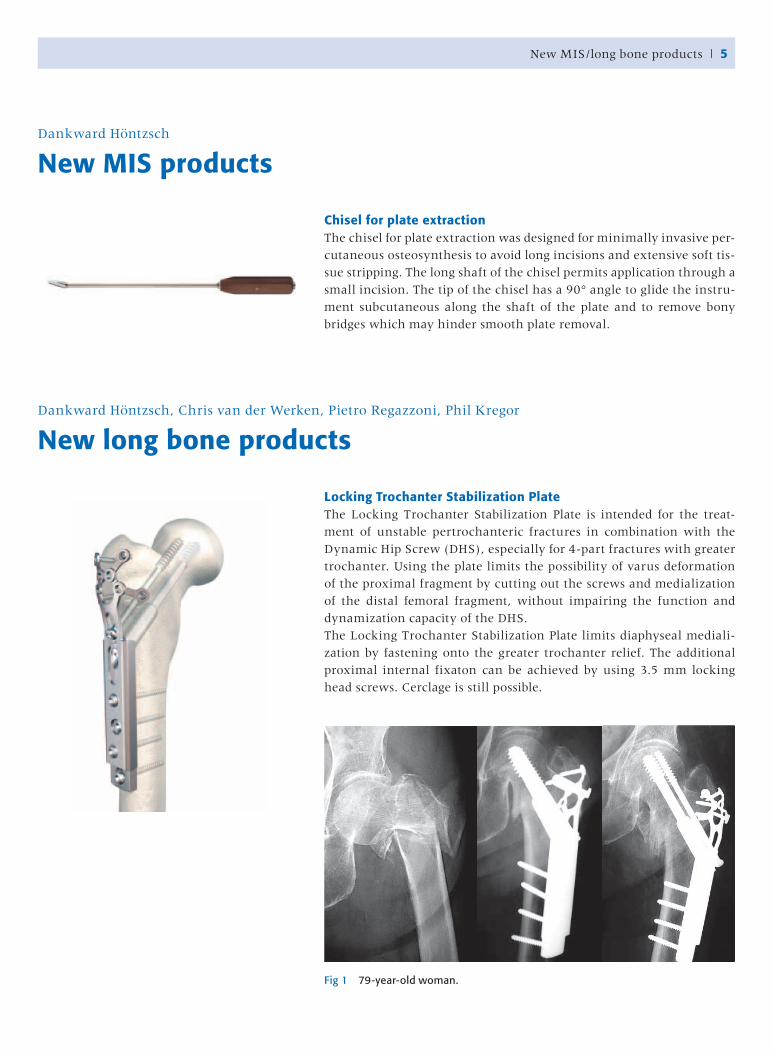
Chisel for plate extractionThe chisel for plate extraction was designed for minimally invasive per-
cutaneous osteosynthesis to avoid long incisions and extensive soft tis-
sue stripping. The long shaft of the chisel permits application through a
small incision. The tip of the chisel has a 90° angle to glide the instru-
ment subcutaneous along the shaft of the plate and to remove bony
bridges which may hinder smooth plate removal.
Dankward Höntzsch
New MIS products
Locking Trochanter Stabilization PlateThe Locking Trochanter Stabilization Plate is intended for the treat-
ment of unstable pertrochanteric fractures in combination with the
Dynamic Hip Screw (DHS), especially for 4-part fractures with greater
trochanter. Using the plate limits the possibility of varus deformation
of the proximal fragment by cutting out the screws and medialization
of the distal femoral fragment, without impairing the function and
dynamization capacity of the DHS.
The Locking Trochanter Stabilization Plate limits diaphyseal mediali-
zation by fastening onto the greater trochanter relief. The additional
proximal internal fixaton can be achieved by using 3.5 mm locking
head screws. Cerclage is still possible.
Dankward Höntzsch, Chris van der Werken, Pietro Regazzoni, Phil Kregor
New long bone products
Fig 1 79-year-old woman.
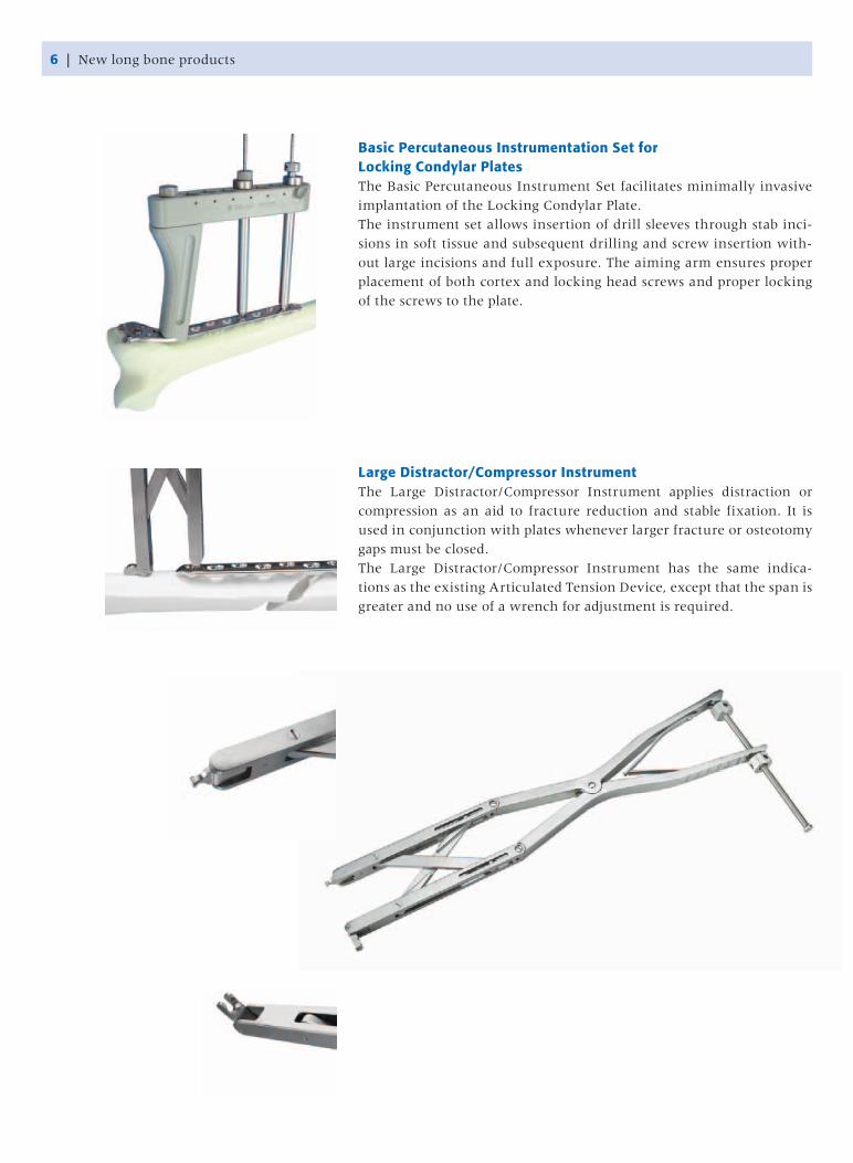
Basic Percutaneous Instrumentation Set for Locking Condylar PlatesThe Basic Percutaneous Instrument Set facilitates minimally invasive
implantation of the Locking Condylar Plate.
The instrument set allows insertion of drill sleeves through stab inci-
sions in soft tissue and subsequent drilling and screw insertion with-
out large incisions and full exposure. The aiming arm ensures proper
placement of both cortex and locking head screws and proper locking
of the screws to the plate.
Large Distractor/Compressor InstrumentThe Large Distractor/Compressor Instrument applies distraction or
compression as an aid to fracture reduction and stable fixation. It is
used in conjunction with plates whenever larger fracture or osteotomy
gaps must be closed.
The Large Distractor/Compressor Instrument has the same indica-
tions as the existing Articulated Tension Device, except that the span is
greater and no use of a wrench for adjustment is required.
6 | New long bone products
New long bone products | 7
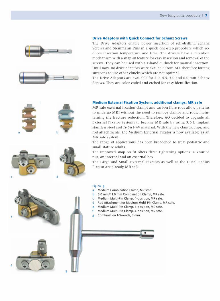
Drive Adaptors with Quick Connect for Schanz ScrewsThe Drive Adaptors enable power insertion of self-drilling Schanz
Screws and Steinmann Pins in a quick one-step procedure which re-
duces insertion temperature and time. The drivers have a retention
mechanism with a snap-in feature for easy insertion and removal of the
screws. They can be used with a T-handle Chuck for manual insertion.
Until now, no drive adaptors were available from AO, therefore forcing
surgeons to use other chucks which are not optimal.
The Drive Adaptors are available for 4.0, 4.5, 5.0 and 6.0 mm Schanz
Screws. They are color-coded and etched for easy identification.
Medium External Fixation System: additional clamps, MR safeMR safe external fixation clamps and carbon fibre rods allow patients
to undergo MRI without the need to remove clamps and rods, main-
taining the fracture reduction. Therefore, AO decided to upgrade all
External Fixator Systems to become MR safe by using 3/6 L implant
stainless steel and Ti-6A1-4V material. With the new clamps, clips, and
rod attachments, the Medium External Fixator is now available as an
MR safe system.
The range of applications has been broadened to treat pediatric and
small stature adults.
The improved snap-on fit offers three tightening options: a knurled
nut, an internal and an external hex.
The Large and Small External Fixators as well as the Distal Radius
Fixator are already MR safe.
Fig 2a–ga Medium Combination Clamp, MR safe.b 8.0 mm/11.0 mm Combination Clamp, MR safe.c Medium Multi-Pin Clamp, 4-position, MR safe.d Rod Attachment for Medium Multi-Pin Clamp, MR safe.e Medium Multi-Pin Clamp, 6-position, MR safe.f Medium Multi-Pin Clamp, 4-position, MR safe.g Combination T-Wrench, 8 mm.
a b
dc
e
f
g
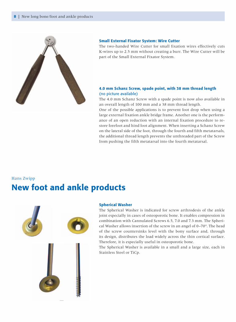
Small External Fixator System: Wire CutterThe two-handed Wire Cutter for small fixation wires effectively cuts
K-wires up to 2.5 mm without creating a burr. The Wire Cutter will be
part of the Small External Fixator System.
4.0 mm Schanz Screw, spade point, with 38 mm thread length(no picture available)The 4.0 mm Schanz Screw with a spade point is now also available in
an overall length of 100 mm and a 38 mm thread length.
One of the possible applications is to prevent foot drop when using a
large external fixation ankle bridge frame. Another one is the perform-
ance of an open reduction with an internal fixation procedure to re-
store forefoot and hind foot alignment. When inserting a Schanz Screw
on the lateral side of the foot, through the fourth and fifth metatarsals,
the additional thread length prevents the unthreaded part of the Screw
from pushing the fifth metatarsal into the fourth metatarsal.
Spherical WasherThe Spherical Washer is indicated for screw arthrodesis of the ankle
joint especially in cases of osteoporotic bone. It enables compression in
combination with Cannulated Screws 6.5, 7.0 and 7.3 mm. The Spheri-
cal Washer allows insertion of the screw in an angel of 0–70°. The head
of the screw countersinks level with the bony surface and, through
its design, distributes the load widely across the thin cortical surface.
Therefore, it is especially useful in osteoporotic bone.
The Spherical Washer is available in a small and a large size, each in
Stainless Steel or TiCp.
Hans Zwipp
New foot and ankle products
8 | New long bone/foot and ankle products
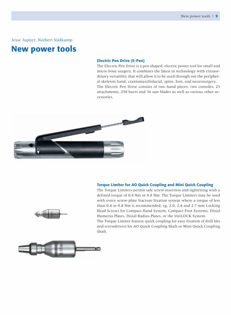
Electric Pen Drive (E-Pen)The Electric Pen Drive is a pen shaped, electric power tool for small and
micro bone surgery. It combines the latest in technology with extraor-
dinary versatility that will allow it to be used through out the peripher-
al skeleton hand, craniomaxillofacial, spine, foot, and neurosurgery.
The Electric Pen Drive consists of two hand pieces, two consoles, 23
attachments, 250 burrs and 36 saw blades as well as various other ac-
cessories.
Jesse Jupiter, Norbert Südkamp
New power tools
New power tools | 9
Torque Limiter for AO Quick Coupling and Mini Quick CouplingThe Torque Limiters permit safe screw insertion and tightening with a
defined torque of 0.4 Nm or 0.8 Nm. The Torque Limiters may be used
with every screw-plate fracture fixation system where a torque of less
than 0.4 or 0.8 Nm is recommended, eg, 2.0, 2.4 and 2.7 mm Locking
Head Screws for Compact Hand System, Compact Foot Systems, Distal
Humerus Plates, Distal Radius Plates, or the UniLOCK System.
The Torque Limiter feature quick coupling for easy fixation of drill bits
and screwdrivers for AO Quick Coupling Shaft or Mini Quick Coupling
Shaft.
10 | New spine products
Mike Janssen, Paul Pavlov
New spine products
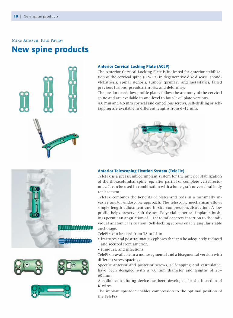
Anterior Cervical Locking Plate (ACLP)The Anterior Cervical Locking Plate is indicated for anterior stabiliza-
tion of the cervical spine (C2–C7) in degenerative disc disease, spond-
ylolisthesis, spinal stenosis, tumors (primary and metastatic), failed
previous fusions, pseudoarthrosis, and deformity.
The pre-lordosed, low profile plates follow the anatomy of the cervical
spine and are available in one-level to four-level plate versions.
4.0 mm and 4.5 mm cortical and cancellous screws, self-drilling or self-
tapping are available in different lengths from 6–12 mm.
Anterior Telescoping Fixation System (TeleFix)TeleFix is a preassembled implant system for the anterior stabilization
of the thoracolumbar spine, eg, after partial or complete vertebrecto-
mies. It can be used in combination with a bone graft or vertebral body
replacement.
TeleFix combines the benefits of plates and rods in a minimally in-
vasive and/or endoscopic approach. The telescopic mechanism allows
simple length adjustment and in-situ compression/distraction. A low
profile helps preserve soft tissues. Polyaxial spherical implants bush-
ings permit an angulation of ± 15° to tailor screw insertion to the indi-
vidual anatomical situation. Self-locking screws enable angular stable
anchorage.
TeleFix can be used from T8 to L5 in
• fractures and posttraumatic kyphoses that can be adequately reduced
and secured from anterior,
• tumours, and infections.
TeleFix is available in a monosegmental and a bisegmental version with
different screw spacings.
Specific anterior and posterior screws, self-tapping and cannulated,
have been designed with a 7.0 mm diameter and lengths of 25–
60 mm.
A radiolucent aiming device has been developed for the insertion of
K-wires.
The implant spreader enables compression to the optimal position of
the TeleFix.
New spine products | 11
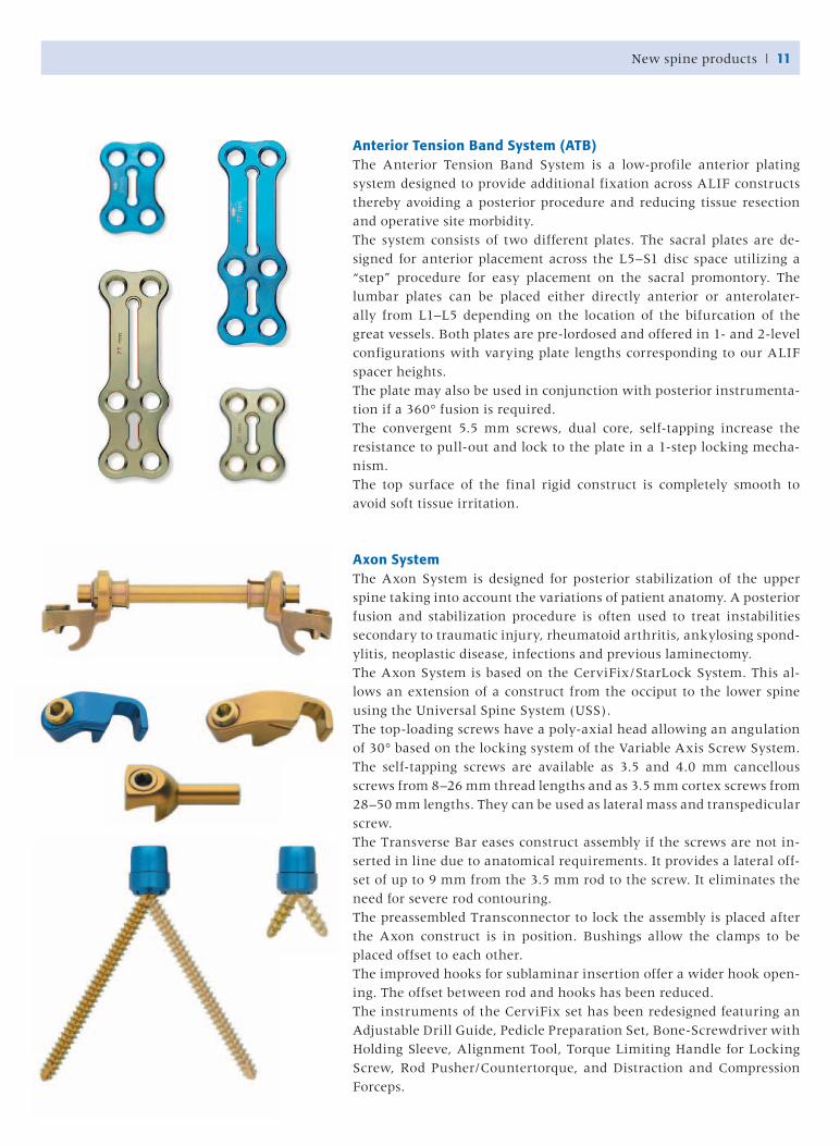
Anterior Tension Band System (ATB)The Anterior Tension Band System is a low-profile anterior plating
system designed to provide additional fixation across ALIF constructs
thereby avoiding a posterior procedure and reducing tissue resection
and operative site morbidity.
The system consists of two different plates. The sacral plates are de-
signed for anterior placement across the L5–S1 disc space utilizing a
“step” procedure for easy placement on the sacral promontory. The
lumbar plates can be placed either directly anterior or anterolater-
ally from L1–L5 depending on the location of the bifurcation of the
great vessels. Both plates are pre-lordosed and offered in 1- and 2-level
configurations with varying plate lengths corresponding to our ALIF
spacer heights.
The plate may also be used in conjunction with posterior instrumenta-
tion if a 360° fusion is required.
The convergent 5.5 mm screws, dual core, self-tapping increase the
resistance to pull-out and lock to the plate in a 1-step locking mecha-
nism.
The top surface of the final rigid construct is completely smooth to
avoid soft tissue irritation.
Axon SystemThe Axon System is designed for posterior stabilization of the upper
spine taking into account the variations of patient anatomy. A posterior
fusion and stabilization procedure is often used to treat instabilities
secondary to traumatic injury, rheumatoid arthritis, ankylosing spond-
ylitis, neoplastic disease, infections and previous laminectomy.
The Axon System is based on the CerviFix/StarLock System. This al-
lows an extension of a construct from the occiput to the lower spine
using the Universal Spine System (USS).
The top-loading screws have a poly-axial head allowing an angulation
of 30° based on the locking system of the Variable Axis Screw System.
The self-tapping screws are available as 3.5 and 4.0 mm cancellous
screws from 8–26 mm thread lengths and as 3.5 mm cortex screws from
28–50 mm lengths. They can be used as lateral mass and transpedicular
screw.
The Transverse Bar eases construct assembly if the screws are not in-
serted in line due to anatomical requirements. It provides a lateral off-
set of up to 9 mm from the 3.5 mm rod to the screw. It eliminates the
need for severe rod contouring.
The preassembled Transconnector to lock the assembly is placed after
the Axon construct is in position. Bushings allow the clamps to be
placed offset to each other.
The improved hooks for sublaminar insertion offer a wider hook open-
ing. The offset between rod and hooks has been reduced.
The instruments of the CerviFix set has been redesigned featuring an
Adjustable Drill Guide, Pedicle Preparation Set, Bone-Screwdriver with
Holding Sleeve, Alignment Tool, Torque Limiting Handle for Locking
Screw, Rod Pusher/Countertorque, and Distraction and Compression
Forceps.
12 | New spine products
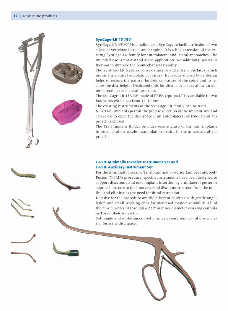
SynCage-LR 45°/90°SynCage-LR 45°/90° is a radiolucent SynCage to facilitate fusion of two
adjacent vertebrae in the lumbar spine. It is a line extension of the ex-
isting SynCage-LR family for anterolateral and lateral approaches. The
intended use is not a stand alone application. An additional posterior
fixation to improve the biomechanical stability.
The SynCage-LR features convex superior and inferior surfaces which
mimic the natural endplate curvature. Its wedge-shaped body design
helps to restore the natural lordotic curvature of the spine and to re-
store the disc height. Dedicated rails for distractor blades allow an an-
terolateral or true lateral insertion.
The SynCage-LR 45°/90° made of PEEK Optima LT3 is available in two
footprints with sizes from 12–19 mm.
The existing instruments of the SynCage-LR family can be used.
New Trail Implants permit the precise selection of the implant size and
can serve to open the disc space if an anterolateral or true lateral ap-
proach is chosen.
The Trail Implant Holder provides secure grasp of the trail implants
in order to allow a safe manipulation in-situ in the anterolateral ap-
proach.
T-PLIF Minimally Invasive Instrument Set andT-PLIF Auxiliary Instrument SetFor the minimally invasive Tansforaminal Posterior Lumbar Interbody
Fusion (T-PLIF) procedure, specific instruments have been designed to
support discetomy and ease implant insertion by a unilateral posterior
approach. Access to the intervertebral disc is more lateral from the mid-
line and eliminates the need for dural retraction.
Decisive for the procedure are the different curettes with gentle angu-
lation and small working ends for increased manoeuverability. All of
the new curettes fit through a 22 mm inner diameter working cannula
or Three Blade Retractor.
Soft angle and up-biting curved pituitaries ease removal of disc mate-
rial form the disc space.
New spine products | 13
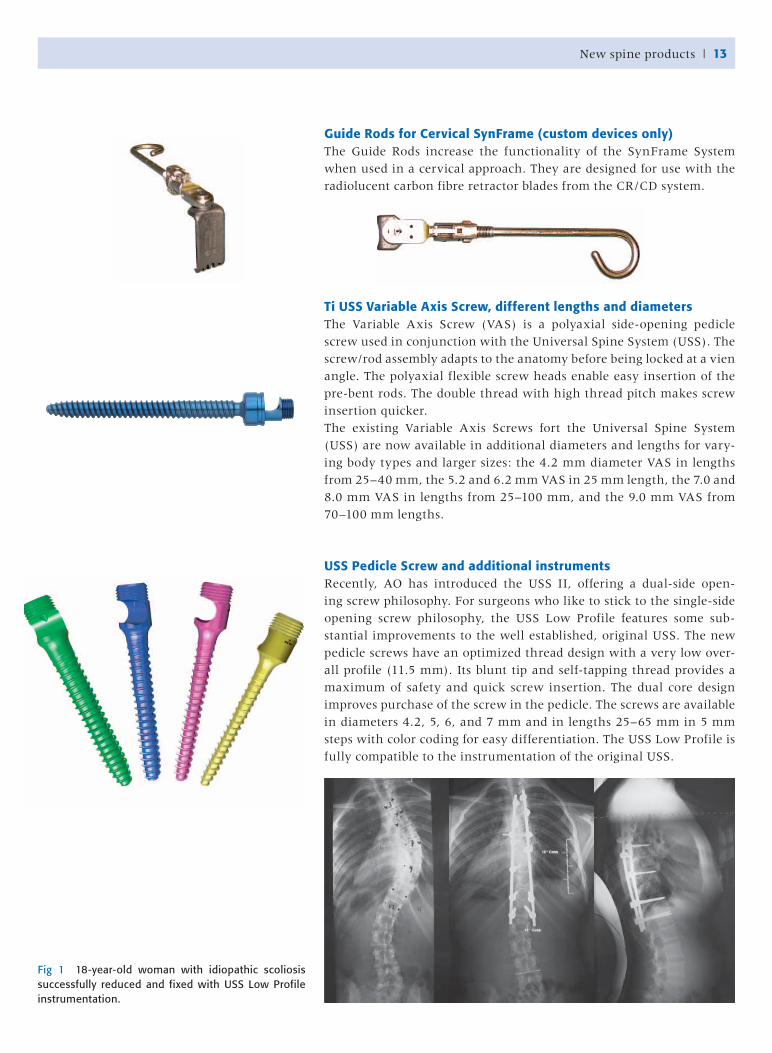
Guide Rods for Cervical SynFrame (custom devices only)The Guide Rods increase the functionality of the SynFrame System
when used in a cervical approach. They are designed for use with the
radiolucent carbon fibre retractor blades from the CR/CD system.
Ti USS Variable Axis Screw, different lengths and diametersThe Variable Axis Screw (VAS) is a polyaxial side-opening pedicle
screw used in conjunction with the Universal Spine System (USS). The
screw/rod assembly adapts to the anatomy before being locked at a vien
angle. The polyaxial flexible screw heads enable easy insertion of the
pre-bent rods. The double thread with high thread pitch makes screw
insertion quicker.
The existing Variable Axis Screws fort the Universal Spine System
(USS) are now available in additional diameters and lengths for vary-
ing body types and larger sizes: the 4.2 mm diameter VAS in lengths
from 25–40 mm, the 5.2 and 6.2 mm VAS in 25 mm length, the 7.0 and
8.0 mm VAS in lengths from 25–100 mm, and the 9.0 mm VAS from
70–100 mm lengths.
USS Pedicle Screw and additional instrumentsRecently, AO has introduced the USS II, offering a dual-side open-
ing screw philosophy. For surgeons who like to stick to the single-side
opening screw philosophy, the USS Low Profile features some sub-
stantial improvements to the well established, original USS. The new
pedicle screws have an optimized thread design with a very low over-
all profile (11.5 mm). Its blunt tip and self-tapping thread provides a
maximum of safety and quick screw insertion. The dual core design
improves purchase of the screw in the pedicle. The screws are available
in diameters 4.2, 5, 6, and 7 mm and in lengths 25–65 mm in 5 mm
steps with color coding for easy differentiation. The USS Low Profile is
fully compatible to the instrumentation of the original USS.
Fig 1 18-year-old woman with idiopathic scoliosis successfully reduced and fixed with USS Low Profile instrumentation.
14 | New spine products
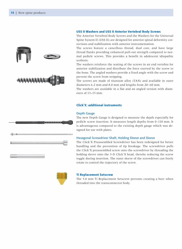
USS II Washers and USS II Anterior Vertebral Body ScrewsThe Anterior Vertebral Body Screws and the Washers for the Universal
Spine System II (USS II) are designed for anterior spinal deformity cor-
rections and stabilization with anterior instrumentation.
The screws feature a cancellous thread, dual core, and have large
thread flanks providing enhanced pull-out strength compared to nor-
mal pedicle screws. This provides a benefit in adolescent idiopathic
scoliosis.
The washers reinforce the seating of the screws in an end vertebra for
anterior stabilization and distribute the force exerted by the screw to
the bone. The angled washers provide a fixed angle with the screw and
prevent the screw from stripping.
The screws are made of titanium alloy (TAN) and available in outer
diameters 6.2 mm and 8.0 mm and lengths from 20–60 mm.
The washers are available in a flat and an angled version with diam-
eters of 13–15 mm.
Click’X: additional instruments
Depth GaugeThe new Depth Gauge is designed to measure the depth especially for
pedicle screw insertion. It measures length depths from 0–110 mm. It
is advantageous compared to the existing depth gauge which was de-
signed for use with plates.
Hexagonal Screwdriver Shaft, Holding Sleeve and SleeveThe Click’X Preassembled Screwdriver has been redesigned for better
handling and the prevention of tip breakage. The screwdriver pulls
the Click’X preassembled screw onto the screwdriver by threading the
holding sleeve onto the 3-D Click’X head, thereby reducing the screw
toggle during insertion. The outer sleeve of the screwdriver can freely
rotate to control the trajectory of the screw.
Ti Replacement SetscrewThe 5.0 mm Ti Replacement Setscrew prevents creating a burr when
threaded into the transconnector body.
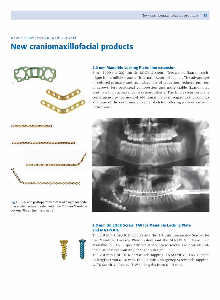
2.0 mm Mandible Locking Plate: line extensionSince 1999 the 2.0 mm UniLOCK System offers a new fixation tech-
nique in mandible trauma (internal fixator principle). The advantages
of reduced primary and secondary loss of reduction, reduced pull-out
of screws, less periosteal compression and more stable fixation had
lead to a high acceptance in osteosynthesis. The line extension is the
consequence to the need of additional plates in regard to the complex
anatomy of the craniomaxillofacial skeleton offering a wider range of
indications.
2.0 mm UniLOCK Screw, TAV for Mandible Locking Plate and MAXPLATEThe 2.0 mm UniLOCK Screws and the 2.4 mm Emergency Screws for
the Mandible Locking Plate System and the MAXPLATE have been
available in TAN. Especially for Japan, these screws are now also of-
fered in TAV without any change in design.
The 2.0 mm UniLOCK Screw, self-tapping, T6 Stardrive, TAV is made
in lengths from 6–18 mm, the 2.4 mm Emergency Screw, self-tapping,
w/T6 Stardrive Recess, TAV in lengths from 6–12 mm.
Rainer Schmelzeisen, Ralf Gutwald
New craniomaxillofacial products
Fig 1 Pre- and postoperative x-rays of a right mandib-ular angle fracture treated with two 2.0 mm Mandible Locking Plates (mini and extra).
New craniomaxillofacial products | 15
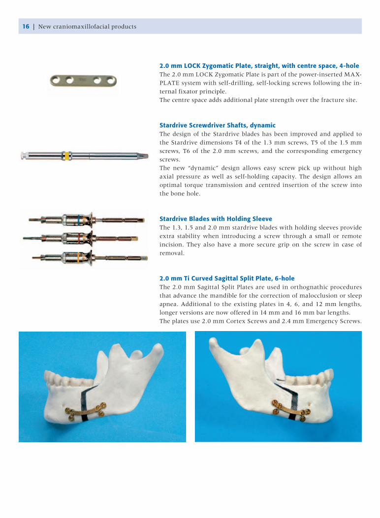
2.0 mm LOCK Zygomatic Plate, straight, with centre space, 4-holeThe 2.0 mm LOCK Zygomatic Plate is part of the power-inserted MAX-
PLATE system with self-drilling, self-locking screws following the in-
ternal fixator principle.
The centre space adds additional plate strength over the fracture site.
Stardrive Screwdriver Shafts, dynamicThe design of the Stardrive blades has been improved and applied to
the Stardrive dimensions T4 of the 1.3 mm screws, T5 of the 1.5 mm
screws, T6 of the 2.0 mm screws, and the corresponding emergency
screws.
The new “dynamic” design allows easy screw pick up without high
axial pressure as well as self-holding capacity. The design allows an
optimal torque transmission and centred insertion of the screw into
the bone hole.
Stardrive Blades with Holding SleeveThe 1.3, 1.5 and 2.0 mm stardrive blades with holding sleeves provide
extra stability when introducing a screw through a small or remote
incision. They also have a more secure grip on the screw in case of
removal.
2.0 mm Ti Curved Sagittal Split Plate, 6-holeThe 2.0 mm Sagittal Split Plates are used in orthognathic procedures
that advance the mandible for the correction of malocclusion or sleep
apnea. Additional to the existing plates in 4, 6, and 12 mm lengths,
longer versions are now offered in 14 mm and 16 mm bar lengths.
The plates use 2.0 mm Cortex Screws and 2.4 mm Emergency Screws.
16 | New craniomaxillofacial products
New craniomaxillofacial products | 17
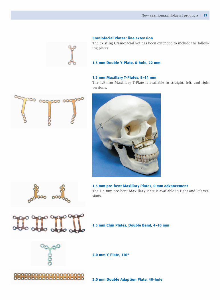
Craniofacial Plates: line extensionThe existing Craniofacial Set has been extended to include the follow-
ing plates:
1.3 mm Double Y-Plate, 6-hole, 22 mm
1.3 mm Maxillary T-Plates, 8–14 mmThe 1.3 mm Maxillary T-Plate is available in straight, left, and right
versions.
1.5 mm pre-bent Maxillary Plates, 0 mm advancementThe 1.5 mm pre-bent Maxillary Plate is available in right and left ver-
sions.
1.5 mm Chin Plates, Double Bend, 4–10 mm
2.0 mm Y-Plate, 110º
2.0 mm Double Adaption Plate, 40-hole
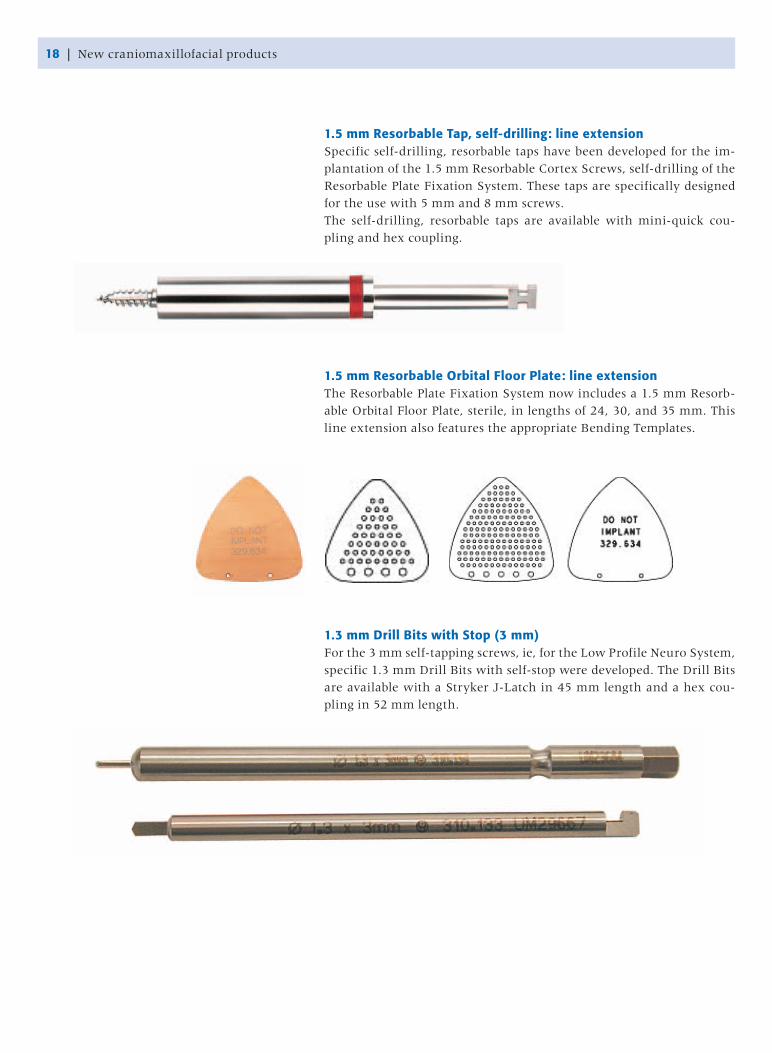
1.5 mm Resorbable Tap, self-drilling: line extensionSpecific self-drilling, resorbable taps have been developed for the im-
plantation of the 1.5 mm Resorbable Cortex Screws, self-drilling of the
Resorbable Plate Fixation System. These taps are specifically designed
for the use with 5 mm and 8 mm screws.
The self-drilling, resorbable taps are available with mini-quick cou-
pling and hex coupling.
1.5 mm Resorbable Orbital Floor Plate: line extensionThe Resorbable Plate Fixation System now includes a 1.5 mm Resorb-
able Orbital Floor Plate, sterile, in lengths of 24, 30, and 35 mm. This
line extension also features the appropriate Bending Templates.
1.3 mm Drill Bits with Stop (3 mm)For the 3 mm self-tapping screws, ie, for the Low Profile Neuro System,
specific 1.3 mm Drill Bits with self-stop were developed. The Drill Bits
are available with a Stryker J-Latch in 45 mm length and a hex cou-
pling in 52 mm length.
18 | New craniomaxillofacial products
Development and validation of AO fracture classifications | 19
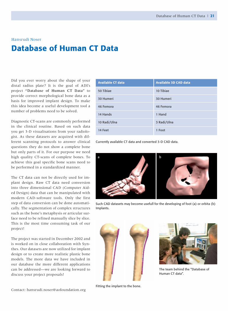
The CMF classification project is currently in it‘s first phase of deve-
lopment and two pilot studies were conducted in 2004. CT-scans are
used for cranio-midface and condylar fractures, while radiographs are
used for other mandibular fractures. To assist the diagnosis and clas-
sification of complex fractures, a specific software is being developed
Laurent Audigé
Development and validation of AO fracture classifications
Following in the footsteps of Prof M Müller
and as a continuation of a long history of frac-
ture classification at the AO Foundation, the
AO Classification Supervisory Committee and
AOCID are implementing projects aimed at
the development and validation of fracture
classification systems. Three projects are be-
ing conducted regarding pediatric long bone
fractures, craniomaxillofacial fractures, and
foot fractures. A fourth project about scapula
fractures will be initiated in 2005 in collabo-
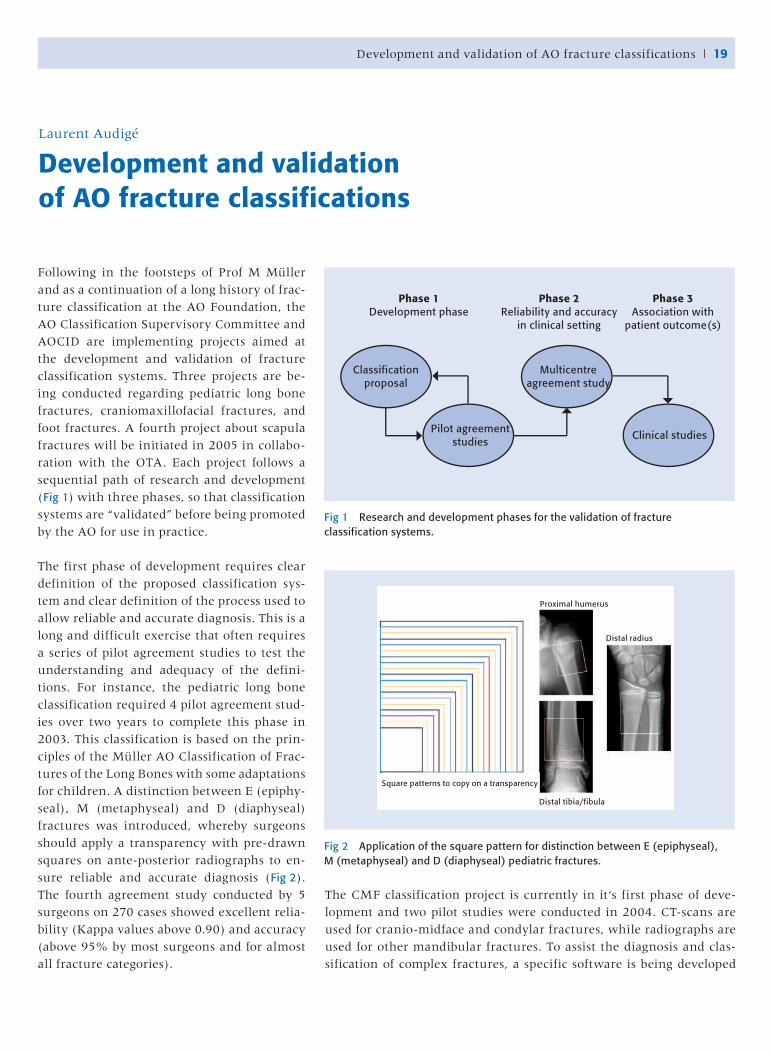
ration with the OTA. Each project follows a
sequential path of research and development
(Fig 1) with three phases, so that classification
systems are “validated” before being promoted
by the AO for use in practice.
The first phase of development requires clear
definition of the proposed classification sys-
tem and clear definition of the process used to
allow reliable and accurate diagnosis. This is a
long and difficult exercise that often requires
a series of pilot agreement studies to test the
understanding and adequacy of the defini-
tions. For instance, the pediatric long bone
classification required 4 pilot agreement stud-
ies over two years to complete this phase in
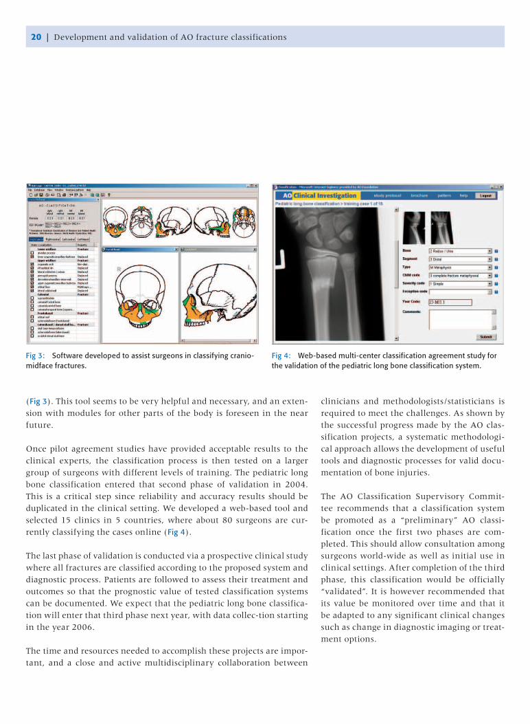
2003. This classification is based on the prin-
ciples of the Müller AO Classification of Frac-
tures of the Long Bones with some adaptations
for children. A distinction between E (epiphy-
seal), M (metaphyseal) and D (diaphyseal)
fractures was introduced, whereby surgeons
should apply a transparency with pre-drawn
squares on ante-posterior radiographs to en-
sure reliable and accurate diagnosis (Fig 2).
The fourth agreement study conducted by 5
surgeons on 270 cases showed excellent relia-
bility (Kappa values above 0.90) and accuracy
(above 95% by most surgeons and for almost
all fracture categories).
Fig 1 Research and development phases for the validation of fracture classification systems.
Fig 2 Application of the square pattern for distinction between E (epiphyseal), M (metaphyseal) and D (diaphyseal) pediatric fractures.
Phase 1Development phase
Phase 2Reliability and accuracy
in clinical setting
Phase 3Association with
patient outcome(s)
Classificationproposal
Multicentreagreement study
Pilot agreement studies
Clinical studies
Proximal humerus
Distal radius
Distal tibia/fibula
Square patterns to copy on a transparency
(Fig 3). This tool seems to be very helpful and necessary, and an exten-
sion with modules for other parts of the body is foreseen in the near
future.
Once pilot agreement studies have provided acceptable results to the
clinical experts, the classification process is then tested on a larger
group of surgeons with different levels of training. The pediatric long
bone classification entered that second phase of validation in 2004.
This is a critical step since reliability and accuracy results should be
duplicated in the clinical setting. We developed a web-based tool and
selected 15 clinics in 5 countries, where about 80 surgeons are cur-
rently classifying the cases online (Fig 4).
The last phase of validation is conducted via a prospective clinical study
where all fractures are classified according to the proposed system and
diagnostic process. Patients are followed to assess their treatment and
outcomes so that the prognostic value of tested classification systems
can be documented. We expect that the pediatric long bone classifica-
tion will enter that third phase next year, with data collec-tion starting
in the year 2006.
The time and resources needed to accomplish these projects are impor-
tant, and a close and active multidisciplinary collaboration between
clinicians and methodologists/statisticians is
required to meet the challenges. As shown by
the successful progress made by the AO clas-
sification projects, a systematic methodologi-
cal approach allows the development of useful
tools and diagnostic processes for valid docu-
mentation of bone injuries.
The AO Classification Supervisory Commit-
tee recommends that a classification system
be promoted as a “preliminary” AO classi-
fication once the first two phases are com-
pleted. This should allow consultation among
surgeons world-wide as well as initial use in
clinical settings. After completion of the third
phase, this classification would be officially
“validated”. It is however recommended that
its value be monitored over time and that it
be adapted to any significant clinical changes
such as change in diagnostic imaging or treat-
ment options.
Fig 3: Software developed to assist surgeons in classifying cranio-midface fractures.
Fig 4: Web-based multi-center classification agreement study for the validation of the pediatric long bone classification system.
20 | Development and validation of AO fracture classifications
Database of Human CT Data | 21
Did you ever worry about the shape of your
distal radius plate? It is the goal of ADI’s
project “Database of Human CT Data” to
provide correct morphological bone data as a
basis for improved implant design. To make
this idea become a useful development tool a
number of problems need to be solved.
Diagnostic CT-scans are commonly performed
in the clinical routine. Based on such data
you get 3-D visualisations from your radiolo-
gist. As these datasets are acquired with dif-
ferent scanning protocols to answer clinical
questions they do not show a complete bone
but only parts of it. For our purpose we need
high quality CT-scans of complete bones. To
achieve this goal specific bone scans need to
be performed in a standardized manner.
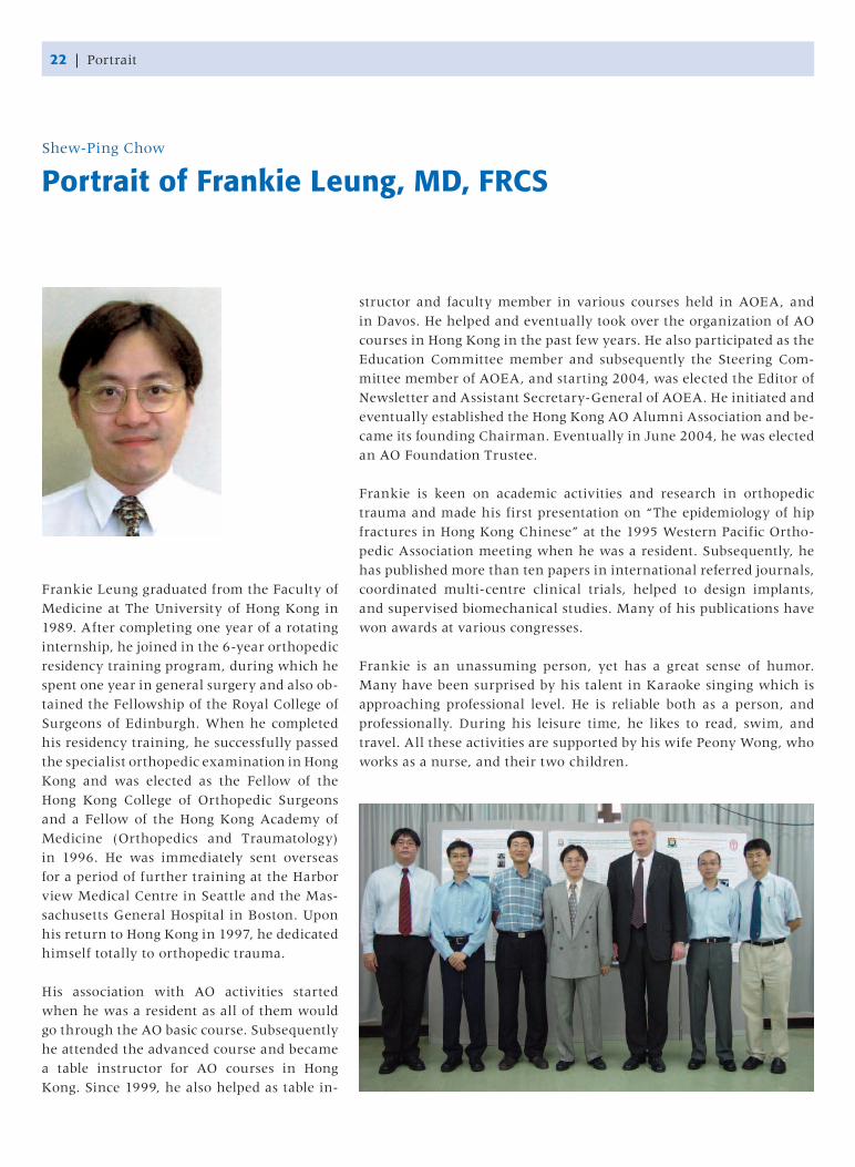
The CT data can not be directly used for im-
plant design. Raw CT data need conversion
into three dimensional CAD (Computer Aid-
ed Design) data that can be manipulated with
modern CAD-software tools. Only the first
step of data conversion can be done automati-
cally. The segmentation of complex structures
such as the bone’s metaphysis or articular sur-
face need to be refined manually slice by slice.
This is the most time consuming task of our
project!
The project was started in December 2002 and
is worked on in close collaboration with Syn-
thes. Our datasets are now utilized for implant
design or to create more realistic plastic bone
models. The more data we have included in
our database the more different applications
can be addressed—we are looking forward to
discuss your project proposals!
Contact: [email protected]
Hansrudi Noser
Database of Human CT Data
Currently available CT data and converted 3-D CAD data.
Available CT data Available 3D CAD data
50 Tibiae 10 Tibiae
30 Humeri 30 Humeri
46 Femora 46 Femora
14 Hands 1 Hand
10 Radi/Ulna 3 Radi/Ulna
14 Feet 1 Foot
Such CAD datasets may become usefull for the developing of foot (a) or orbita (b) Implants.
Fitting the implant to the bone.
The team behind the “Database of Human CT data”.
a b
Frankie Leung graduated from the Faculty of
Medicine at The University of Hong Kong in
1989. After completing one year of a rotating
internship, he joined in the 6-year orthopedic
residency training program, during which he
spent one year in general surgery and also ob-
tained the Fellowship of the Royal College of
Surgeons of Edinburgh. When he completed
his residency training, he successfully passed
the specialist orthopedic examination in Hong
Kong and was elected as the Fellow of the
Hong Kong College of Orthopedic Surgeons
and a Fellow of the Hong Kong Academy of
Medicine (Orthopedics and Traumatology)
in 1996. He was immediately sent overseas
for a period of further training at the Harbor
view Medical Centre in Seattle and the Mas-
sachusetts General Hospital in Boston. Upon
his return to Hong Kong in 1997, he dedicated
himself totally to orthopedic trauma.
His association with AO activities started
when he was a resident as all of them would
go through the AO basic course. Subsequently
he attended the advanced course and became
a table instructor for AO courses in Hong
Kong. Since 1999, he also helped as table in-
Shew-Ping Chow
Portrait of Frankie Leung, MD, FRCS
structor and faculty member in various courses held in AOEA, and
in Davos. He helped and eventually took over the organization of AO
courses in Hong Kong in the past few years. He also participated as the
Education Committee member and subsequently the Steering Com-
mittee member of AOEA, and starting 2004, was elected the Editor of
Newsletter and Assistant Secretary-General of AOEA. He initiated and
eventually established the Hong Kong AO Alumni Association and be-
came its founding Chairman. Eventually in June 2004, he was elected
an AO Foundation Trustee.
Frankie is keen on academic activities and research in orthopedic
trauma and made his first presentation on “The epidemiology of hip
fractures in Hong Kong Chinese” at the 1995 Western Pacific Ortho-
pedic Association meeting when he was a resident. Subsequently, he
has published more than ten papers in international referred journals,
coordinated multi-centre clinical trials, helped to design implants,
and supervised biomechanical studies. Many of his publications have
won awards at various congresses.
Frankie is an unassuming person, yet has a great sense of humor.
Many have been surprised by his talent in Karaoke singing which is
approaching professional level. He is reliable both as a person, and
professionally. During his leisure time, he likes to read, swim, and
travel. All these activities are supported by his wife Peony Wong, who
works as a nurse, and their two children.
22 | Portrait
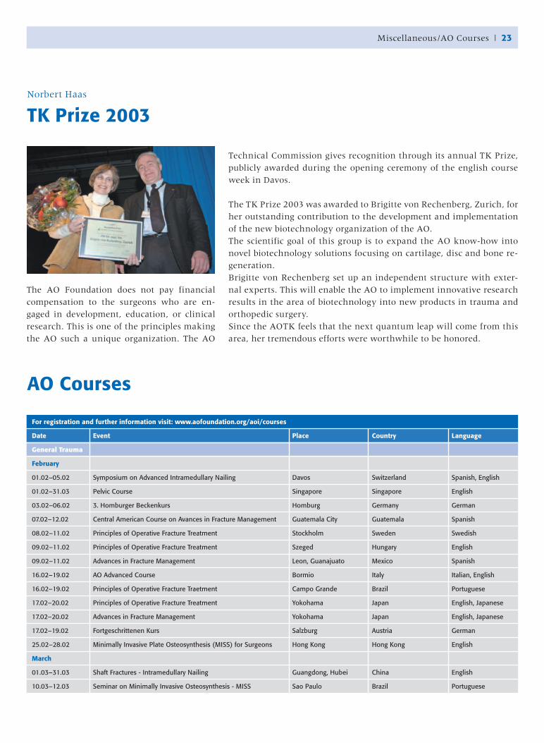
Norbert Haas
TK Prize 2003
The AO Foundation does not pay financial
compensation to the surgeons who are en-
gaged in development, education, or clinical
research. This is one of the principles making
the AO such a unique organization. The AO
Technical Commission gives recognition through its annual TK Prize,
publicly awarded during the opening ceremony of the english course
week in Davos.
The TK Prize 2003 was awarded to Brigitte von Rechenberg, Zurich, for
her outstanding contribution to the development and implementation
of the new biotechnology organization of the AO.
The scientific goal of this group is to expand the AO know-how into
novel biotechnology solutions focusing on cartilage, disc and bone re-
generation.
Brigitte von Rechenberg set up an independent structure with exter-
nal experts. This will enable the AO to implement innovative research
results in the area of biotechnology into new products in trauma and
orthopedic surgery.
Since the AOTK feels that the next quantum leap will come from this
area, her tremendous efforts were worthwhile to be honored.
Miscellaneous/AO Courses | 23
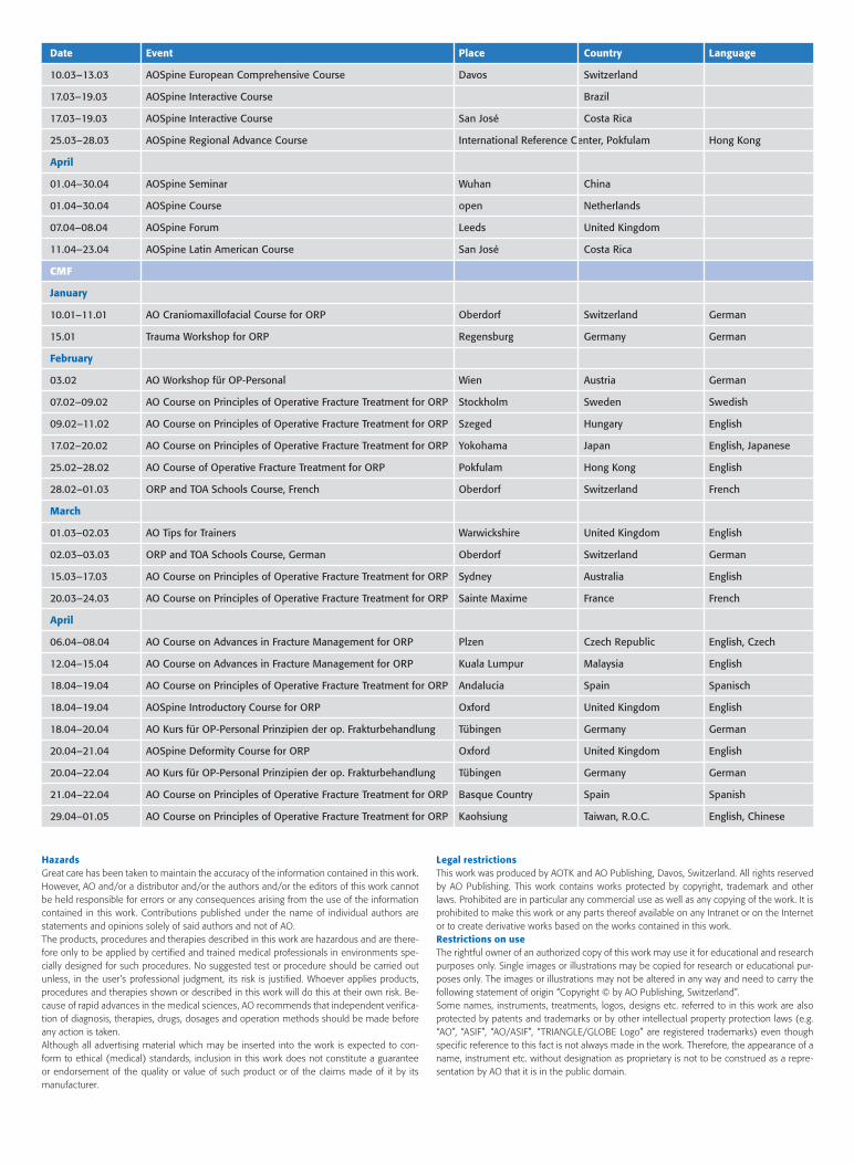
AO Courses
For registration and further information visit: www.aofoundation.org/aoi/courses
Date Event Place Country Language
General Trauma
February
01.02–05.02 Symposium on Advanced Intramedullary Nailing Davos Switzerland Spanish, English
01.02–31.03 Pelvic Course Singapore Singapore English
03.02–06.02 3. Homburger Beckenkurs Homburg Germany German
07.02–12.02 Central American Course on Avances in Fracture Management Guatemala City Guatemala Spanish
08.02–11.02 Principles of Operative Fracture Treatment Stockholm Sweden Swedish
09.02–11.02 Principles of Operative Fracture Treatment Szeged Hungary English
09.02–11.02 Advances in Fracture Management Leon, Guanajuato Mexico Spanish
16.02–19.02 AO Advanced Course Bormio Italy Italian, English
16.02–19.02 Principles of Operative Fracture Traetment Campo Grande Brazil Portuguese
17.02–20.02 Principles of Operative Fracture Treatment Yokohama Japan English, Japanese
17.02–20.02 Advances in Fracture Management Yokohama Japan English, Japanese
17.02–19.02 Fortgeschrittenen Kurs Salzburg Austria German
25.02–28.02 Minimally Invasive Plate Osteosynthesis (MISS) for Surgeons Hong Kong Hong Kong English
March
01.03–31.03 Shaft Fractures - Intramedullary Nailing Guangdong, Hubei China English
10.03–12.03 Seminar on Minimally Invasive Osteosynthesis - MISS Sao Paulo Brazil Portuguese

Date Event Place Country Language
11.03 The Upper Limb Brüxelles Belgium English
11.03 AO Seminar Fractures in Childhood Tehran Iran English
12.03 5.Celler AO- Seminar Celle Germany German
13.03–18.03 Advances in Fracture Management Are Sweden Swedish
14.03–17.03 Principles of Operative Fracture Treatment Edinburgh United Kingdom English
14.03–17.03 38. AO Kurs Trauma I – Prinzipien Kurs Freiburg Germany German
15.03–18.03 Principles of Operative Fracture Treatment Sydney Australia English
16.03–26.03 Principles of Operative Fracture Treatment Moscow Russian Federation English, Russian
17.03–20.03 Advanced Course Bangalore India English
20.03–24.03 Cours de base AO pour jeunes chirurgiens ? France French
21.03–24.03 Principles of Operative Frcature Treatment Dublin Ireland English
April
01.04–30.04 Principles Seminar (LCP) Chengdu, Chongqing China English
06.04–08.04 Advances in Fracture Mangement Plzen Czech Republic Czech, English
08.04 AO Seminar on Periprosthetic Fractures after Knee Arthroplasty
+ Pediatric Fractures (Upper Extremity) Plzen Czech Republic English, Czech
08.04–09.04 Seminar on Intramedullary Fixation Santiago Chile Spanish
11.04–16.04 Advances in Fracture Management Punta del Este Uruguay Spanish
12.04–15.04 Advances in Fracture Management Kuala Lumpur Malaysia English
13.04–17.04 Principles of Operative Fracture Treatment San Juan Spain Spanish
15.04–16.04 Seminar of Complex Trauma on Upper Limb Sao Paulo Brazil Portuguese
24.04–29.04 Lecture Tour on LPHP, PLT Metaphyseal Plate, Distal femur Delhi, Mumbai, Chennai India English
29.04–01.05 Principles of Operative Fracture Management Kaohsiung Taiwan, R.O.C. English
Hand
March
10.03–12.03 Handkurs Trauma II Münster Germany German
CMF
February
07.02 - 09.02 Maxillofacial Course Bangkok Thailand English
March
09.03–11.03 Cranio-maxillofacial Endoscopic Course—New Technologies Cadiz Spain English
12.03–14.03 Advanced Cranio-Maxillofacial Course Hong Kong Hong Kong English
31.03–02.04 Central European CMF Course Prague Czech Republic English
April
22.04 Cranio Maxillofacial Seminar Bruxelles Belgium English
Spine
February
01.02–31.03 AOSpine Comprehensive Course Singapore Singapore
17.02–18.02 AOSpine Course for Residents of Universitys of Milan and Turin Galeazzi Institute, Milan Italy
21.02–23.02 AOSpine Masterclass Innsbruck Austria
March
01.03–31.03 AOSpine Comprehensive Course Qingdao China
01.03–01.12 AOSpine Comprehensive Course Lille Belgium
04.03–05.03 Kurzstreckige und Langstreckige Fusionen Düsseldorf Germany
24 | AO Courses
HazardsGreat care has been taken to maintain the accuracy of the information contained in this work. However, AO and/or a distributor and/or the authors and/or the editors of this work cannot be held responsible for errors or any consequences arising from the use of the information contained in this work. Contributions published under the name of individual authors are statements and opinions solely of said authors and not of AO.The products, procedures and therapies described in this work are hazardous and are there-fore only to be applied by certified and trained medical professionals in environments spe-cially designed for such procedures. No suggested test or procedure should be carried out unless, in the user’s professional judgment, its risk is justified. Whoever applies products, procedures and therapies shown or described in this work will do this at their own risk. Be-cause of rapid advances in the medical sciences, AO recommends that independent verifica-tion of diagnosis, therapies, drugs, dosages and operation methods should be made before any action is taken.Although all advertising material which may be inserted into the work is expected to con-form to ethical (medical) standards, inclusion in this work does not constitute a guarantee or endorsement of the quality or value of such product or of the claims made of it by its manufacturer.
Legal restrictionsThis work was produced by AOTK and AO Publishing, Davos, Switzerland. All rights reserved by AO Publishing. This work contains works protected by copyright, trademark and other laws. Prohibited are in particular any commercial use as well as any copying of the work. It is prohibited to make this work or any parts thereof available on any Intranet or on the Internet or to create derivative works based on the works contained in this work. Restrictions on useThe rightful owner of an authorized copy of this work may use it for educational and research purposes only. Single images or illustrations may be copied for research or educational pur-poses only. The images or illustrations may not be altered in any way and need to carry the following statement of origin “Copyright © by AO Publishing, Switzerland”. Some names, instruments, treatments, logos, designs etc. referred to in this work are also protected by patents and trademarks or by other intellectual property protection laws (e.g. “AO”, “ASIF”, “AO/ASIF”, “TRIANGLE/GLOBE Logo” are registered trademarks) even though specific reference to this fact is not always made in the work. Therefore, the appearance of a name, instrument etc. without designation as proprietary is not to be construed as a repre-sentation by AO that it is in the public domain.
Date Event Place Country Language
10.03–13.03 AOSpine European Comprehensive Course Davos Switzerland
17.03–19.03 AOSpine Interactive Course Brazil
17.03–19.03 AOSpine Interactive Course San José Costa Rica
25.03–28.03 AOSpine Regional Advance Course International Reference Center, Pokfulam Hong Kong
April
01.04–30.04 AOSpine Seminar Wuhan China
01.04–30.04 AOSpine Course open Netherlands
07.04–08.04 AOSpine Forum Leeds United Kingdom
11.04–23.04 AOSpine Latin American Course San José Costa Rica
CMF
January
10.01–11.01 AO Craniomaxillofacial Course for ORP Oberdorf Switzerland German
15.01 Trauma Workshop for ORP Regensburg Germany German
February
03.02 AO Workshop für OP-Personal Wien Austria German
07.02–09.02 AO Course on Principles of Operative Fracture Treatment for ORP Stockholm Sweden Swedish
09.02–11.02 AO Course on Principles of Operative Fracture Treatment for ORP Szeged Hungary English
17.02–20.02 AO Course on Principles of Operative Fracture Treatment for ORP Yokohama Japan English, Japanese
25.02–28.02 AO Course of Operative Fracture Treatment for ORP Pokfulam Hong Kong English
28.02–01.03 ORP and TOA Schools Course, French Oberdorf Switzerland French
March
01.03–02.03 AO Tips for Trainers Warwickshire United Kingdom English
02.03–03.03 ORP and TOA Schools Course, German Oberdorf Switzerland German
15.03–17.03 AO Course on Principles of Operative Fracture Treatment for ORP Sydney Australia English
20.03–24.03 AO Course on Principles of Operative Fracture Treatment for ORP Sainte Maxime France French
April
06.04–08.04 AO Course on Advances in Fracture Management for ORP Plzen Czech Republic English, Czech
12.04–15.04 AO Course on Advances in Fracture Management for ORP Kuala Lumpur Malaysia English
18.04–19.04 AO Course on Principles of Operative Fracture Treatment for ORP Andalucia Spain Spanisch
18.04–19.04 AOSpine Introductory Course for ORP Oxford United Kingdom English
18.04–20.04 AO Kurs für OP-Personal Prinzipien der op. Frakturbehandlung Tübingen Germany German
20.04–21.04 AOSpine Deformity Course for ORP Oxford United Kingdom English
20.04–22.04 AO Kurs für OP-Personal Prinzipien der op. Frakturbehandlung Tübingen Germany German
21.04–22.04 AO Course on Principles of Operative Fracture Treatment for ORP Basque Country Spain Spanish
29.04–01.05 AO Course on Principles of Operative Fracture Treatment for ORP Kaohsiung Taiwan, R.O.C. English, Chinese
26 | New spine products
For further information please contact:
AOTK Office
Clavadelerstrasse
CH-7270 Davos Platz
Phone: +41 81 4142-471
Fax: +41 81 4142-290
Editors:
Univ.-Prof. Dr. Norbert P. Haas
AOTK Chairman
Philip Schreiterer
AOTK Assistant
Number of copies: 20,000
Issued: December 2004
Photos and illustrations courtesy of Synthes partners and authors
Copyright © 2004 by AO Publishing, Switzerland