these 2010 guerin 1 · pdf file1 université de la méditerranée...
TRANSCRIPT

1
Université de la Méditerranée
Aix-Marseille II Ecole Doctorale des Sciences de la Vie et de la Santé
THESE
Présentée pour obtenir le titre de Docteur ès Sciences de l’Université de la Méditerranée
Aix-Marseille II
Mention Pathologies Humaines
Spécialité Oncologie, Pharmacologie et Thérapeutique
Olivier Guérin
Intérêts en Thérapeutique du ciblage des récepteurs à
l’Epidermal Growth Factor (EGFR) et des récepteurs au
Vascular Endothelial Growth Factor (VEGFR) dans le
cancer de prostate hormono-résistant et docetaxel-
résistant. Etudes précliniques. Laboratoire d’oncopharmacologie, Centre Antoine Lacassagne, Nice.
Directeur : Dr Gérard Milano
Soutenue publiquement le 25 juin 2010 à Marseille
Jury Monsieur le Professeur Athanassios Iliadis Président
Monsieur le Professeur Milou-Daniel Drici Rapporteur
Monsieur le Professeur Laurent Teillet Rapporteur
Monsieur le Professeur Jean-Marc Ferrero Examinateur
Monsieur le Docteur Gérard Milano Directeur de Thèse
2
Remerciements
Monsieur le Professeur Athanassios Iliadis, nous vous remercions d’avoir accepté de juger
ce travail, c’est un très grand honneur pour nous que vous présidiez ce jury de thèse.
Monsieur le Professeur Milou Drici, nous vous sommes très reconnaissants pour votre aide
et vos conseils. Nous espérons sincèrement pouvoir collaborer à vos côtés au développement
de la Thérapeutique Clinique en harmonie et complémentarité avec la Pharmacologie. C’est
un grand plaisir pour nous que vous jugiez ce travail.
Monsieur le Professeur Laurent Teillet, conscient de votre compétence et de votre
investissement dans le domaine de l’oncogériatrie, c’est un honneur pour nous de vous
compter parmi les membres de ce jury et nous vous en sommes sincèrement reconnaissants.
Monsieur le Professeur Jean-Marc Ferrero, votre expertise est reconnue dans la maladie
cancéreuse prostatique. C’est un très grand privilège de vous soumettre notre travail.
Monsieur le Docteur Gérard Milano, je vous suis très reconnaissant d’avoir dirigé cette
thèse, votre compétence et votre envie de transmettre vos connaissances m’ont permis

3
d’avancer. Votre patience et votre tolérance m’ont permis d’aller au terme de ce travail.
J’espère très sincèrement que je pourrai continuer à cheminer à vos côtés.
Je remercie tous les membres du laboratoire d’Oncopharmacologie du Centre Antoine
Lacassagne, et bien évidemment tout particulièrement Monsieur Jean-Louis Fischel sans qui
rien n’aurait été possible, et Madame Patricia Formento pour sa patience et son encadrement
constant lors de notre travail de paillasse commun.

4
A ma Famille, Simone, Hélène, Roger, Louis, Roland, c’est votre exemple qui est ma
motivation.
A mes Parents, mon unique ambition est de mettre mes pas dans les vôtres, au service des
autres comme vous savez si bien le faire, parce que c’est à l’aune de cet engagement que l’on
juge la qualité d’un homme. Merci de m’avoir donné les armes pour avancer dans cette voie.
A mon Epouse, Valentine, ta présence à mes côtés est un trésor inestimable, une richesse
incomparable. Rien ne serait possible sans toi.
A mes Enfants, Elouan et Thibault, vous êtes la motivation profonde de mes engagements, et
vous voir grandir est un enchantement permanent. Mon bonheur n’est fait que du vôtre.
A mon Frère, Nicolas, compagnon de mon enfance, compère et complice des grands
moments, mon immense affection t’est acquise à jamais.

5
TABLE DES MATIERES
Introduction………………………………………………………p.9
1 La prise en charge du cancer de la prostate…………………..……….p.11
1.1 Prise en charge actuelle……………………………………………………….p.11
1.2 Innovations thérapeutiques dans le CPHR…………………………………p.11
2 Quelques éléments de réflexion sur la problématique oncogériatrique
autour du cancer de la prostate, et l’importance de cette approche en
thérapeutique……………………………………………………………….p.13
2.1 Elements épidémiologiques généraux………………………………………..p.13
2.2 Présentation de l’approche oncogériatrique……………………………...…p.13
2.3 Intérêts dans le cancer de prostate du sujet âgé……………………………p.14
3 Le ciblage de EGFR et de VEGFR dans le cancer de prostate……….p.16
3.1 Le ciblage du VEGFR………………………………………………………...p.16
3.1.1 Généralités sur le VEGFR………………………………………….p.16
3.1.2 VEGFR et cancer de prostate………………………………………p.19
3.2 Le ciblage de l’EGFR………………………………………………………….p.20
3.2.1 Généralités sur l’EGFR……………………………………………..p.20
3.2.2 EGFR dans le cancer de prostate et son évolution………………...p.23
3.2.3 Principaux résultats des études ciblant EGFR dans le CPHR en
phases précliniques……………………………………………………….p.24

6
3.2.4 Résultats des études cliniques principales des thérapies ciblant EGFR
dans le CPHR………………………………………………………………p.26
3.2.4.1 En monothérapies
3.2.4.2 En combinaisons de traitements
3.2.5 Synthèse et conclusion………………………………………………p.28
4 Travail de thèse expérimental…………………………………………….p.31
4.1 Objectifs et déroulé du travail………………………………………………………..p.31
4.2 Résultats………………………………………………………………………………..p.33
4.2.1 Effet anti-tumoral supra-additif du sunitinib malate (SU11248, Sutent®)
combiné au docetaxel. Une nouvelle perspective thérapeutique dans le cancer de
prostate hormono-résistant……………………………………………………….p.33
4.2.1.1 Introduction et objectifs
4.2.1.2 Matériels et méthodes
4.2.1.3 Résultats
4.2.1.4 Conclusion
4.2.2 Vandetanib, inhibiteur de tyrosine-kinases multicibles, dans le traitement
du cancer de prostate hormono-résistant : comparaison de son effet en
association au docetaxel sur des lignées cellulaires tumorales docetaxel-sensibles
et docetaxel-résistantes. Etude in vivo préclinique……………………………..p.35
4.2.2.1 Introduction et objectifs
4.2.2.2 Matériels et méthodes
4.2.2.3 Résultats

7
4.2.2.4 Conclusion
4.3 Synthèse et perspectives………………………………………………………………p.38
Bibliographie…………………………………………………………………………p.41
Annexes (articles soumis ou publiés dans le cadre du travail de thèse)………..……..p.51
Annexe 1 EGFR targeting in hormone-resistant prostate cancer: current appraisal
and prospects for treatment......................................................................................p.52
Annexe 2 Supra-additive antitumor effects of sunitinib malate combined with
docetaxel. A new therapeutic perspective in hormone refractory prostate cancer..p.70
Annexe 3 The multi-targeted tyrosine kinase inhibitor Vandetanib in the treatment of
hormone refractory prostate cancer: comparison of its effects in association with
docetaxel on docetaxel-sensitive and docetaxel-resistant tumor cell lines. A preclinical
in vivo study.............................................................................................................p.77

8
Prinicipales abréviations utilisées dans le texte (par ordre d’apparition)
VEGFR Vascular Endothelial Growth Factor Receptor
EGFR Epidermal Growth Factor Receptor
CPHR Cencer de Prostate Hormono-Résistant
TKI Tyrosine kinases inhibiteurs
SIOG Société Internationale d’Oncogériatrie
CR Ratio de combinaison
PDGFR Platelet-derived growth factor receptor
KIT CD117 (récepteur cytokine)
FLT3 FMS-like Tyrosine Kinase 3
p27 Inhibiteur de kinase cycline-dépendant
Ki67 Antigène marqueur de prolifération
vWF Facteur Von Willebrandt
PSA Prostate Specific Antigene
EGS Evaluation Gérontologique Standardisée
RCP Réunion de Concertation Pluridisciplinaire
SIOG Société Internationale d’OncoGériatrie

9
Introduction
Le cancer de la prostate, premier cancer chez l’homme en France, a bénéficié largement des
avancées thérapeutiques de la chirurgie, de la chimiothérapie, de la radiothérapie et, depuis
une dizaine d’années, de l’hormonothérapie. Aux Etats-Unis en 2008, le cancer de prostate
représentait 25% des nouveaux cas de cancer chez l’homme (1) et 60000 nouveaux cas en
France.
Ce cancer concerne souvent les hommes âgés et toute stratégie thérapeutique se doit de tenir
compte de leur qualité de vie, en se basant notamment sur une évaluation oncogériatrique.
Cette dimension est de plus en plus importante du fait du vieillissement de la population et de
l’exigence croissante des hommes âgés de voir leur cancer traité efficacement. Cela nécessite
une approche personnalisée car la population âgée est très hétérogène, et une évaluation de
toutes les fragilités gériatriques afin de minimiser leur impact sur la tolérance aux traitements
et leur efficacité, et d’éviter leur décompensation en cours de traitement.
Lorsque le cancer devient hormono-résistant (CPHR), un nouveau palier dans la progression
de la maladie est franchi et il faut bien garder à l’esprit que les chimiothérapies doivent être
alors discutées. En effet, il s’agit alors d’En effet, il s’agit alors d’une maladie agressive dont
le pronostic est mauvais. Le traitement historique associant mitoxantrone et prednisolone sans
augmenter la survie des patients, diminuait les douleurs et augmentait leur qualité de vie (2).
Désormais, le traitement de référence du cancer de prostate hormono-résistant combine le
docétaxel et la prednisolone (3) puisqu’il est le seul traitement à avoir montré un bénéfice en
survie. Ce traitement n’augmente cependant la médiane de survie que de 2,9 mois (survie
médiane de 19,2 mois vs 16,3 mois avec la mitoxantrone, p=0.004). Les stratégies futures
visant à améliorer la prise en charge du cancer de prostate avancé hormono-résistant (CPHR)
devront examiner outre l’efficacité de nouveaux médicaments, les bénéfices de leur
10
association au docétaxel. En l’état actuel des connaissances, les traitements de deuxième ligne
sont décevants et tout reste à faire. Dans ce cadre clinique très défavorable, notre travail a eu
pour but d’évaluer l’impact de thérapies ciblées visant EGFR et VEGFR, seules ou associées
au docetaxel, sur un modèle préclinique de tumeur prostatique humaine PC3 xénogreffée sur
la souris nude. Le choix de ces récepteurs est lié à leur surexpression lors de l’évolution
hormono-résistante inéluctable des cancers de prostate en progression (voir chap 3). Nous
avons choisi le modèle PC3, lignée cellulaire la mieux connue et la plus utilisée pour les
études précliniques de cancer de prostate hormono-résistant humain. Les souris nude
constituent le modèle animal de choix, car leur immunodéficience complète leur permet de
développer des tumeurs xénogreffées d’autres espèces, ce qui est impossible pour des
animaux immunocompétants.

11
1 La prise en charge du cancer de la prostate
1.1 Prise en charge actuelle
Le cancer de la prostate est le premier cancer chez l’homme en France de par sa fréquence et
le deuxième de par sa mortalité (après le cancer du poumon et devant le cancer colo-rectal). Il
s’agit d’un adénocarcinome caractérisable par trois paramètres : le stade clinique TNM, le
taux initial de PSA et le grade histologique de Gleason. Le traitement curatif est décidé en
fonction de ces trois paramètres. Actuellement, quatre approches curatives sont définitivement
validées : la prostatectomie chirurgicale, la radiothérapie, la curiethérapie et l’irradiation
transuréthrale par hyperfréquences. Une part importante de la décision est prise en fonction du
rapport bénéfice-risque et du désir du malade. Les métastases les plus fréquentes sont
osseuses. C’est un cancer sensible aux hormones mâles d’origine testiculaire (testostérone) et
surrénalienne. En présence de métastase, une hormonosuppression androgénique (par
castration ou par agoniste LH-RH) est effectuée, parfois associée au traitement local.
Néanmoins, l’apparition d’une hormono-résistance est toujours observée. Le traitement
standard est alors la chimiothérapie par docetaxel. Il a été montré que ce traitement avait le
même impact, quel que soit l’âge du patient, sur la survie, la sédation des douleurs, la
diminution de consommation d’antalgiques et plus généralement sur la qualité de vie (4).
1.2 Innovations thérapeutiques dans le cancer de prostate hormono-résistant
Dans ce contexte de cancer hormono-résistant, la recherche de nouvelles thérapeutiques revêt
une importance capitale, d’autant que le docetaxel n’est pas d’un usage très facile chez le
sujet âgé, a fortiori chez le sujet âgé fragile. Actuellement, les nouvelles thérapeutiques dont
le développement est le plus avancé, ciblent l’inhibition de la voie des récepteurs des
androgènes par des médicaments appropriés (abiratérone, MDV3100), qui demeure capitale

12
même en phase de résistance à la castration. Les thérapies visant à agir sur le processus de
développement des métastases osseuses (inhibiteurs du récepteur de l’endothéline-1, de
RANK-L, associations de la chimiothérapie à l’irradiation métabolique osseuse) sont
particulièrement prometteuses, et des résultats de phases III et en cours de publication sont
attendus très prochainement. La vaccination autologue dirigée contre la phosphatase acide
prostatique semble augmenter la survie globale, et d’autres stratégies de vaccination ou de
modulation de l’immunité (anticorps anti-CTLA4) sont en cours d’évaluation (5). Suite aux
bons résultats obtenus avec la thalidomide, médicament cependant mal toléré pour cette
population âgée et souvent vulnérable, un analogue potentiellement plus efficace, le
lénalidomide, est en cours de développement (6). Dans ce contexte, l’inhibition de
l’angiogénèse par ciblage de VEGFR et/ou de EGFR a montré une efficacité clinique variable
mais globalement assez décevante. Des résultats cliniques de grande ampleur seront bientôt
disponibles. Il n’y a que très peu de résultats cliniques concernant les maladies cancéreuses
prostatiques hormono et docetaxel résistantes.

13
2 Quelques éléments de réflexion sur la problématique
oncogériatrique autour du cancer de la prostate, et importance de
cette approche en thérapeutique
2.1 Eléments épidémiologiques généraux
En France, 70% des décès par cancer surviennent après 65 ans. Le cancer est la première
cause de mortalité entre 65 et 79 ans et la deuxième après 80 ans, âge à partir duquel les
maladies cardio-vasculaires représentent la cause principale de décès. C’est également la
seule cause de mortalité qui progresse actuellement dans ces tranches d’âges.
Par ailleurs, le vieillissement de la population française est une réalité désormais bien connue.
Ainsi, il y aura en France 18 millions de personnes âgées de plus de 60 ans et 6 millions de
plus de 75 ans en 2020 (source DREES 2009). L’espérance de vie à la naissance sera de 90
ans en 2020 (source DREES 2009). Elle aura ainsi doublée en 150 ans. La moitié des
nouveaux-nés de sexe féminin qui naissent aujourd’hui sera centenaire. De plus, l’espérance
de vie par tranche d’âge, de même que l’espérance de vie sans incapacité augmentent, y
compris pour les tranches d’âge les plus avancées. Ainsi, de nos jours, un français sur quatre
âgé de 80 ans atteindra l’âge de 100 ans.
2.2 Présentation de l’approche oncogériatrique
L’une des difficultés actuelles rencontrées par les oncologues, devant la proportion toujours
croissante de patients âgés se présentant en consultation, est de pouvoir les faire bénéficier
d’une évaluation gériatrique multidimentionnelle et pluridisciplinaire médico-psycho-sociale,
permettant d’aider à la décision thérapeutique prise en réunion de concertation pluri-
disciplinaire (RCP). Cette évaluation permet de mieux cerner une population âgée très

14
hétérogène (7)(8)(9). L’évaluation gériatrique standardisée (EGS) (10) permet l’identification
des grands syndromes gériatriques, d’estimer la qualité de vie ainsi que l’espérance de vie qui
doivent êtres connues de l’oncologue pour mieux les confronter aux caractéristiques de la
maladie néoplasique. Cette EGS permet d’individualiser 3 groupes de patients, d’après les
travaux de l’équipe de Tampa : un premier groupe de patients autonomes sans critères de
fragilité qui pourraient recevoir un traitement identique aux patients plus jeunes, un second
groupe de patients vulnérables présentant une dépendance relative avec une ou deux
comorbidités et enfin un troisième groupe de patients fragiles avec au moins une dépendance
au niveau des activités de la vie quotidienne associée à la présence d’au moins trois
comorbidités ou présence d’un des grands syndromes gériatriques (troubles de la marche,
démence ou confusion mentale, incontinence et dénutrition) (11)(12)(13). Ainsi, l’évaluation
gériatrique est un processus diagnostique permettant d’appréhender de façon systématique les
problèmes et les ressources tant sur le plan médical que fonctionnel ou psycho-social du sujet
âgé. Elle améliore la prise en charge en identifiant des interventions à mener pour éviter les
décompensations de fragilités retrouvées à l’EGS, en proposant des schémas thérapeutiques
tenant compte du pronostic et des modalités de suivi. L’évaluation gériatrique vient renforcer
celle de l’oncologue dans l’établissement d’un schéma thérapeutique adapté et réaliste.
Cependant, l’une des grandes difficultés actuellement, pour de nombreux centres de prise en
charge des patients âgés présentant une pathologie néoplasique, est de pouvoir disposer dans
un délai raisonnable de cette évaluation gériatrique pour aider à définir la meilleure stratégie
thérapeutique.
2.3 Intérêts dans le cancer de prostate du sujet âgé
Concernant le cancer de la prostate, l’aide à la stratégie thérapeutique que le gériatre peut
amener à l’oncologue est très différente selon le stade de la maladie. Chez le sujet âgé,

15
l’existence d’un cancer de prostate localisé (T1-3N0M0) pose la question assez complexe de
l’estimation de son espérance de vie. En effet, le plus souvent, la maladie prostatique ne sera
pas à l’origine du décès du patient, qui décèdera pour une toute autre raison, justifiant une
simple surveillance clinique et éventuellement biologique. Il existe également une autre
situation où l’évaluation de l’espérance de vie est nécessaire, c’est lors de la récidive de la
maladie (le plus souvent après radiothérapie chez le sujet âgé). Dans ce cas, le temps de
doublement du PSA parait être un très bon critère d’évolutivité pour aider à la décision de
traitement prise en commun par les acteurs de la cancérologie et le gériatre (14).
Lors de l’utilisation dans les années à venir des thérapies ciblant VEGFR et EGFR, champ des
présentes investigations précliniques, l’approche oncogériatrique occupera certainement une
place importante, le but étant de proposer un traitement adapté et personnalisé au patient âgé
présentant un cancer de prostate en échappement basé sur l’EGS afin de déterminer le
traitement présentant un juste équilibre entre efficacité et tolérance selon l’objectif adéquat à
l’évaluation gérontologique effectuée (entre qualité de vie et objectifs carcinologiques). Les
thérapies ciblées ne sont pas exemptes de toxicité, et celles-ci doivent être anticipées. Ainsi, le
Sunitinib induit des asthénies majeures pouvant entraîner chez le sujet âgé des risques de
perte assez rapide d’autonomie, par une réduction importante de l’activité physique et une
sarcopénie consécutive à l’alitement. Thérapie ciblée n’est donc pas synonyme de bonne
tolérance. L’obstacle principal actuel à l’usage de ces nouvelles molécules chez le sujet âgé,
est que les essais thérapeutiques menés pour l’obtention de l’autorisation de mise sur le
marché excluent le plus souvent ces sujets âgés de plus de 75 ou 80 ans, et systématiquement
les sujets âgés polypathologiques, qui seront pourtant ceux qui poseront des difficultés de
définition d’une stratégie thérapeutique cohérente.
Cette approche intégrant l’EGS pour le choix thérapeutique est désormais recommandée, suite
à la publication des propositions de la Société Internationale d’OncoGériatrie (SIOG) (15).

16
3. Le ciblage de EGFR et de VEGFR dans le cancer de prostate
3.1 Le ciblage de VEGFR
3.1.1 Généralités sur VEGFR
Un système vasculaire fonctionnel est essentiel à la croissance des tumeurs solides et à
l’apparition de métastases. En effet, en l’absence d’un processus de néoangiogénèse, une
tumeur est incapable de se développer au-delà de quelques millimètres de diamètre et
demeure quiescente (16)(17). Ce phénomène de néoangiogénèse est très complexe et fait
intervenir différents signaux biochimiques et divers types cellulaires. Il est initié par la
sécrétion de facteurs pro-angiogéniques par les cellules tumorales. Parmi ces facteurs, le
vascular endothelial growth factor (VEGF) joue un rôle majeur dans la prolifération des
cellules endothéliales et la formation de néovaisseaux. Il est également capable d’induire un
certain degré de perméabilité vasculaire et joue un rôle clé dans l’activation des voies de
survie des cellules endothéliales composant les vaisseaux néoformés (18). L’intérêt
thérapeutique potentiel d’agents ciblant la vascularisation des tumeurs est donc évident, et
explique le grand nombre de médicaments qui sont actuellement en cours d’investigation dans
ce domaine, dont certaines déjà parvenues au stade de développement clinique (19). Alors que
les agents cytotoxiques conventionnels agissent directement sur les cellules tumorales, les
médicaments ciblant la vascularisation des tumeurs exercent une activité antitumorale de
façon indirecte, en privant les cellules tumorales de l’oxygène et des nutriments dont elles ont
besoin. Ces différences de cibles et de mode d’action laissent entrevoir une grande
complémentarité dans les effets de ces diverses thérapeutiques. Ainsi, il existe une synergie
d’action potentielle entre les agents ciblant la vascularisation tumorale et les autres agents
cytotoxiques. De plus, les cellules composant le réseau vasculaire tumoral se distinguent des

17
cellules tumorales elles-mêmes, par une plus grande stabilité génétique, et par voie de
conséquence, par un risque quasi nul d’acquisition de résistance au traitement (20), ce qui en
fait des cibles oncologiques privilégiées.
Deux grands groupes d’agents ciblant la vascularisation des tumeurs ont été développés: les
agents anti-angiogéniques et les agents de ciblage vasculaire (21). Les agents anti-
angiogéniques inhibent la formation de nouveaux vaisseaux, c'est-à-dire le processus de
néoangiogénèse. Ils ont donc une action préventive, nécessitent une administration chronique,
et sont surtout susceptibles d’apporter un bénéfice au stade précoce de la maladie ou encore
devant des métastases asymptomatiques (21). Du fait du rôle majeur du VEGF dans le
processus de néoangiogénèse, le ciblage des voies de signalisation qui y sont liées apparaît
comme une approche prometteuse dans le cadre des thérapeutiques anti-angiogéniques.
Différents agents ont été ainsi développés, permettant soit le blocage du VEGF lui-même,
c’est le cas d’anticorps monoclonaux comme le bevacizumab (Avastin�
) (22)(23), soit de ses
récepteurs (VEGFR), c’est le cas des petites molécules inhibitrices de tyrosine kinases comme
l’AZD2171, le ZD6474, le sunitinib ou le sorafenib (21)(24).
A l’inverse des thérapeutiques anti-angiogéniques proprement dites, les agents de ciblage
vasculaire ont la capacité d’agir sur le réseau vasculaire préétabli de masses tumorales déjà
constituées, engendrant ischémie et nécrose tumorale. Par conséquent, ces agents, administrés
de façon aiguë, entraînent des effets quasi-immédiats, et sont susceptibles d’être actifs devant
des tumeurs avancées, qui sont habituellement résistantes aux agents cytotoxiques
conventionnels (21)(25)(26). Différentes stratégies sont actuellement en cours
d’investigations dans ce domaine. La première approche consiste à utiliser des anticorps ou
des peptides capables de se lier spécifiquement à une cible (VEGFR, endogline, intégrine αν,
domaine EDB de la fibronectine…) présente sur le réseau vasculaire des tumeurs, afin d’y
délivrer une molécule effectrice (toxine, agent pro-thrombotique…) entraînant des lésions

18
vasculaires et secondairement la thrombose du réseau vasculaire tumoral (21). La deuxième
approche est celle des petites molécules capables d’induire un collapsus vasculaire et une
nécrose tumorale secondaire, parmi lesquelles on retrouve les flavonoïdes (5,6-
dimethylxanthenone-4-acetic acid ou DMXAA) et les agents se liant à la tubuline (ZD6126,
CA4P…) (21)(27). Si le mécanisme d’action des flavonoïdes, faisant intervenir une sécrétion
de cytokines et d’agents vaso-actifs, demeure mal compris, celui des agents se liant à la
tubuline apparait plus clair. Ces agents ciblent les vaisseaux sanguins tumoraux, qui diffèrent
nettement de ceux des tissus sains. En effet, les cellules endothéliales qui tapissent leurs
parois vasculaires conservent les caractéristiques des cellules en division, immatures et
nouvellement formées, et dépendent fortement d'un cytosquelette à tubuline pour conserver
leur forme. Le ZD6126, par exemple, perturbe ce cytosquelette, ce qui conduit les cellules
endothéliales à passer d'une forme aplatie à une forme arrondie, causant la détérioration de
l'endothélium (25). Ce phénomène génère une congestion vasculaire et l'interruption du flux
sanguin. La tumeur est ainsi privée d'oxygène et de nutriments, tandis que ses déchets
s’accumulent pour atteindre des niveaux toxiques. Il en résulte, comme c’est le cas d’ailleurs
avec l’ensemble des agents de ciblage vasculaire, une nécrose tumorale centrale, laissant une
fine couche de cellules tumorales viables en périphérie de la tumeur. Ce dernier phénomène
peut être expliqué par la simple diffusion de l’oxygène et des nutriments vers les cellules
tumorales périphériques par l’intermédiaire des tissus normaux environnant (21).
Le ciblage de la vascularisation tumorale (agents anti-angiogéniques et agents de ciblage
vasculaire) constitue donc une piste thérapeutique innovante et particulièrement intéressante
en cancérologie. Les effets de l’association de ces différents agents entre eux et combinés aux
autres thérapeutiques ciblées et notamment aux anti-EGFR, de même qu’aux agents
cytotoxiques conventionnels et à l’irradiation, font l’objet d’une intense activité de recherche,
qui débouche actuellement sur des résultats cliniques convaincants. Ainsi, par exemple, le

19
bevacizumab est actuellement utilisé chez des patients atteints de cancers colorectaux
métastatiques ou de cancers du sein métastatiques, en association à une chimiothérapie
conventionnelle. Le sunitinib et le sorafenib ont quant à eux démontré leur efficacité chez des
patients atteints de formes métastatiques de cancer du rein.
3.1.2 VEGFR et cancer de prostate
Si les inhibiteurs de l’angiogénèse ont montré des résultats cliniques intéressants pour les
cancers du rein, du colon, et dans une moindre mesure pour le cancer du sein et du poumon,
nous n’avons que peu de résultats cliniques sur le cancer de prostate évolué, mais des essais
sont en cours. Ils concernent à la fois les anticorps mnonclonaux tels que le bevacizumab ou
des leurres dérivés de ces anticorps tels le VEGF-trap, et les inhibiteurs tyrosine kinase
donnés per os comme le sunitinib ou le sorafenib.
Le bevacizumab n’a pas d’activité antitumorale en monothérapie. En association au docetaxel
et à l’estramustine, il a été noté une réponse partielle pour 9 des 17 patients inclus dans un
essai de phase II (avec une réponse biochimique sur les PSA de 80%) (28). Une autre étude de
phase II a étudié l’association du bevacizumab au docetaxel, à la prednisone et la thalidomide,
chez 60 patients (29). Ils obtenaient un taux de réponse élevé (87%) sur le PSA avec 59% des
patients ayant une cible mesurable à l’imagerie présentant une réponse partielle. Cependant,
des problèmes de tolérance importants ont été notés (neutropénies fébriles, hémorragies et
thromboses). Une autre étude de phase II sur 20 patients a ciblé l’association bevacizumab (10
mg/kg J1-J21) et docetaxel (60 mg/m² J1-J21) déjà traités initialement par docetaxel, et a
montré une réponse sur le PSA chez 11 patients et une réponse objective chez 7 patients (30).
4 de ces patients n’avaient pas eu de réponse lors d’un premier traitement par docetaxel seul.
Il y aurait donc ici un bénéfice au blocage angiogénique pour la réponse au docetaxel. Une
étude de phase III étudiant l’association bevacizumab-docetaxel est en cours.

20
Une étude récente a porté sur l’usage du sorafenib. Elle montre une efficacité modérée en
réponse biologique sur le PSA, avec un effet paradoxal de diminution du PSA à l’arrêt du
traitement (31).
Un autre inhibiteur de tyrosine-kinases, ciblant l’ensemble des récepteurs au VEGF,
l’AZD2171, a montré une activité antitumorale in vitro et in vivo sur des modèles de tumeurs
de prostate xénogreffées (32). Pour cette molécule, une étude de phase I a permis de
déterminer la dose maximale tolérée (20 mg/j), et montré (même si ça n’est pas l’objectif
d’une phase I) une réponse objective et quatre stabilisations (33). La phase II est en cours.
Enfin, le VEGF-trap est un pseudo-anticorps monoclonal qui reconnaît VEGF et le piège par
son extrémité de fixation au ligand identique aux récepteurs VEGFR2 et 3. Un essai de phase
III qui compare en première ligne métastatique le docétaxel avec ou sans VEGF-trap est en
cours.
3.2 Le ciblage de EGFR
3.2.1 Généralités sur EGFR
Le récepteur au facteur de croissance épidermique (EGFR ou HER-1) est le premier membre
connu des récepteurs de la famille HER. A l’état physiologique, EGFR est exprimé dans de
nombreux épithéliums (peau, col utérin, canaux biliaires, hépatocytes, glandes sébacées,
bronches, vessie, cellules myoépithéliales du sein). De même, EGFR est surexprimé dans de
nombreux cancers (poumon non à petites cellules, tête et cou, ovaires, côlon, vessie, rein et
prostate) (34).
EGFR est un récepteur avec activité tyrosine-kinase. Il détient un rôle clé dans les processus
de transduction du signal, régulant des fonctions cellulaires majeures, telles que la survie et la
prolifération. L’activation de la signalisation EGFR passe par la fixation sur ce récepteur de

21
facteurs de croissance tels que EGF, TGFα, amphiréguline, heparin-binding EGF, et
bêtacelluline (même si EGF et TGFα restent les ligands préférentiels) (35), aboutissant à sa
dimérisation ou à son hétérodimérisation avec d’autres récepteurs de la famille HER (HER-2
notamment, mais également HER 3 et HER 4). L’autophosphorylation et la
transphosphorylation des récepteurs par l’intermédiaire de leurs domaines tyrosine kinase
conduisent alors au recrutement d’effecteurs intracellulaires et à l’activation des voies de
prolifération et de survie (36).
Les thérapeutiques ciblées en cancérologie font actuellement principalement appel à 2 grandes
catégories de molécules, les anticorps monoclonaux (Acm) et les inhibiteurs de tyrosine
kinase (TKI) (37). Parmi les produits ciblant le EGFR, le cetuximab (Erbitux®
) (38)(39) pour
les Acm, le géfitinib (Iressa®
) (40)(41) et l’erlotinib (Tarceva®
) (42)(43) pour les TKI, font
partie des agents les mieux connus et les plus avancés dans leur développement clinique. De
nombreuses autres molécules sont également en développement clinique ou préclinique,
notamment concernant les TKI désormais assez souvent multicibles (le EGFR n’est pas le
seul récepteur impliqué alors dans les mécanismes d’action). Acm et TKI diffèrent clairement
de par leur mode d’action sur leur cible. Le cetuximab est un antagoniste compétitif de l’EGF
sur son récepteur. Indépendamment de la phosphorylation du récepteur, le complexe
cetuximab-EGFR est consécutivement internalisé (44)(45). Au contraire, les TKI agissent sur
la portion cytosolique de EGFR, en compétition avec l’ATP au niveau de son domaine de
liaison, empêchant ainsi l’autophosphorylation du récepteur. En fonction de la nature du TKI,
l’inhibition du EGFR peut être réversible comme c’est le cas avec le géfitinib et l’erlotinib, ou
bien irréversible comme par exemple avec le PD-183805 (46)(47)(48). A l’inverse des Acm,
les TKI ne sont pas strictement spécifiques de leur cible présumée (EGFR par exemple). En
effet, les TKI étant des antagonistes compétitifs de l’ATP au niveau de son site de fixation sur

22
les tyrosines kinases (47), il peut exister, de façon variable, une réactivité croisée des TKI
avec les autres membres de la famille des récepteurs HER, comme HER-2 (49).
Les effets du ciblage de EGFR témoignent du rôle physiologique de ce récepteur dans les
voies de transduction du signal impliquées dans les phénomènes de division cellulaire,
d’apoptose et de néoangiogenèse. Sur le plan de la prolifération cellulaire d’abord, on observe
un ralentissement du processus de division cellulaire, avec blocage des cellules en phase G1,
faisant intervenir des modifications moléculaires au niveau des principaux points de contrôle
du cycle cellulaire (50)(51). Par ailleurs, il a été également rapporté une modification de
l’équilibre entre les niveaux intracellulaires de Bax et de Bcl-2, ce qui souligne l’effet pro-
apoptotique du ciblage du EGFR (52). Quant à l’effet anti-angiogénique du ciblage du EGFR,
il a été mis en évidence pour les Acm et pour les TKI, notamment par inhibition de la
sécrétion tumorale de facteurs pro-angiogéniques, tels que le VEGF et le facteur VIII
(53)(54).
Les études réalisées in vivo sur xénogreffes tumorales révèlent une efficacité du cetuximab
très supérieure à ce que l’on observe sur lignées cellulaires in vitro (55). En effet, une part
importante de l’activité antitumorale des Acm pourrait provenir de processus immunologiques
tels que la cytotoxicité cellulaire dépendante des anticorps (ADCC) (56)(57). Ces différences
d’activité in vitro et in vivo notées avec le cetuximab ne sont pas aussi marquées avec les TKI.
Ainsi, le géfitinib de même que d’autres TKI de développement plus récent, comme le
sunitinib (Sutent®
), entraînent une inhibition de la prolifération cellulaire aussi bien in vitro
que in vivo, sur de nombreuses lignées cellulaires (58)(59).
Différentes études ont étudié les effets de la combinaison des anti-EGFR aux agents
cytotoxiques conventionnels et à l’irradiation. Il en ressort généralement, qu’il s’agisse d’Acm
ou de TKI, des effets cytotoxiques additifs ou supra-additifs. Au niveau expérimental, il ne
semble pas y avoir de distinction entre Acm et TKI, quant à la possibilité d’obtenir une

23
synergie des effets cytotoxiques, lorsqu’ils sont associés aux agents de chimiothérapie
conventionnelle ou à l’irradiation. Cette synergie peut être attribuée en grande partie aux
effets connus du ciblage du EGFR sur la prolifération cellulaire, l’apoptose, l’angiogenèse et
la réparation de l’ADN (58)(60)(61)(62).
Sur le plan clinique, le géfitinib et l’erlotinib, pour les TKI, sont utilisés dans le traitement du
cancer broncho-pulmonaire non à petites cellules, où ils se sont révélés particulièrement
efficaces chez les patients présentant certaines mutations du EGFR (63)(64). Concernant les
Acm, le cetuximab a actuellement prouvé son intérêt dans le traitement des cancers
colorectaux, en association à l’irinotécan (65). Il a également démontré son efficacité dans le
traitement des carcinomes épidermoïdes des voies aérodigestives supérieures (66)(67).
3.2.2 EGFR dans le cancer de prostate et son évolution
Dans le cadre du cancer de prostate, l’approche semble particulièrement intéressante. En effet,
de nombreux mécanismes moléculaires sont liés à la progression vers l'hormono-résistance
(68). Mais l'augmentation de l'expression des protéines de signalisation de HER semble être
un facteur important (69).
Dans une étude récente, Schlomm et al (70) ont analysé des échantillons tissulaires provenant
de 2497 tumeurs prostatiques au niveau de l'ADN et au niveau protéique. Une expression
détectable d'EGFR a été retrouvée dans 18% des cancers et s'est révélée corrélée aux hauts
grades, aux stades avancés et à un risque élevé de récidive du PSA en analyse univariée
(p<0.0001 dans chaque cas). L'utilité potentielle des traitements anti-EGFR pourrait ainsi être
analysée dans le cancer de prostate exprimant EGFR.
Par ailleurs, après prostatectomie totale, l’expression d’EGFR est corrélée au risque de
récidive et à la progression vers l’hormono-résistance. En analyse multivariée, EGFR est un
facteur pronostique indépendant de la survie sans progression. 100% des métastases des

24
cancers de prostate hormono-résistants expriment EGFR, suggérant que ce récepteur est une
voie de transduction capitale pour la croissance tumorale (71).
Par ailleurs, dans une autre étude (72), l'amplification et la mutation d'EGFR ont été analysées
dans le cancer soit hormono-sensible soit hormone-résistant, chez 10 patients. Aucune
corrélation significative n'a été trouvée entre l'évolution des mutations et le statut hormono-
sensible ou hormono-résistant. Toutefois, le temps nécessaire pour passer au CPHR a été
significativement plus court chez les patients avec une mutation du gène EGFR (p=0.017).
Ainsi, dans cette étude, les mutations d'EGFR ne semblent pas jouer un rôle important dans la
progression vers l'hormono-résistance mais elles sont corrélées au pronostic. Il faut considérer
dans cette étude également le faible nombre de sujets.
3.2.3 Principaux résultats des études ciblant EGFR dans le CPHR en phases
précliniques
Des études récentes suggèrent que l'involution prostatique induite par la castration puisse être
due à des effets initialement dans les vaisseaux prostatiques. Hammarsten et al (73) ont
récemment examiné si des traitements antiangiogéniques pouvaient mimer les effets de la
castration. Le plan expérimental a utilisé des cellules AT-1 de cancer de prostate androgéno-
indépendant. Ces cellules ont été inoculées dans la prostate ventrale de rats adultes. Les
animaux porteurs de tumeurs ont été traités par un inhibiteur de VEGFR 2 et d'EGFR, le
ZD6474 (Astra Zeneca), et les auteurs ont comparé les effets à court terme (3 jours) de ce
traitement avec ceux induits par la castration. Les résultats ont montré que la castration
entraînait une diminution de la densité vasculaire dans le tissu sain entourant la tumeur et
donc une augmentation de l'hypoxie tissulaire et de l'apoptose, ainsi qu'une diminution
modérée de la croissance tumorale. Le traitement par le ZD6474 a abouti à une diminution de
la densité vasculaire tumorale accompagnée d'une augmentation de l'hypoxie tumorale et de
25
l'apoptose, ainsi qu'à une diminution de la croissance tumorale, suggérant que la castration et
la thérapie antiangiogénique agissent selon un mécanisme similaire. Il est intéressant de noter
que le traitement combinant la castration et le ZD 6474 a été plus efficace que la castration et
le ZD 6474 administrés seuls pour induire l'hypoxie tumorale, l'apoptose, la nécrose et
diminuer la densité vasculaire tumorale. Les auteurs ont conclu que ce traitement combiné
pourrait être un moyen particulièrement efficace de traiter les tumeurs prostatiques.
Notre équipe a testé chez l’animal (74) la combinaison d’un inhibiteur de tyrosine-kinase
(Sunitinib) à action principalement antiangiogénique, du cetuximab (anti-EGFR) et du
docétaxel. Chaque médicament, administré en monothérapie, a démontré des effets
comparables et modérés sur la croissance tumorale, avec une inhibition d'environ 50% à la fin
du cycle d'administration de 3 semaines. Les valeurs du rapport de combinaison (CR)
informatisé pour la croissance tumorale indiquent des effets supra-additifs pour la
combinaison sunitinib-docétaxel et sunitinib-cetuximab et suggèrent des effets seulement
additifs pour la combinaison sunitinib-cetuximab-docétaxel. Les effets sur la croissance
tumorale se sont accompagnés d'une diminution parallèle de la prolifération des cellules
tumorales (Ki 67) et de la vascularisation tumorale (facteur Von Willebrand). Des effets pro-
apoptotiques (clivage de la caspase-3) significativement plus importants ont été observés avec
les combinaisons sunitinib-docétaxel et sunitinib-docétaxel-cetuximab qu'avec les autres
combinaisons. Nous concluons donc que les effets antitumoraux supra-additifs observés avec
la combinaison sunitinib-docétaxel pourraient étayer des stratégies novatrices dans la prise en
charge du cancer de prostate avancé, utilisant simultanément des traitements ciblant EGFR.
Dans le cadre de ces approches combinées, des résultats récents sur des lignées cellulaires
(DU145) et des modèles in vivo murins ont montré l’intérêt d’une radioimmunothérapie
(associant du lutétium 177 couplée à un anticorps anti-prostate hu3S193) cytotoxique et pro-
apoptotique. Dans cet article (75), la dose maximale tolérée de la radioimmunothérapie a été

26
identifiée et un résultat très intéressant est que l’association de ce traitement à un TKI
(AG1478) à dose sub-thérapeutique a montré une amélioration significative de l’efficacité sur
les modèles in vivo, comme d’ailleurs l’association au docétaxel.
3.2.4 Résultats des études cliniques principales des thérapies ciblant EGFR dans le
CPHR
3.2.4.1 Essais en monothérapie
Dans l’étude de Agus et al (76), un agent appartenant à une nouvelle classe d'agents
anticancéreux ciblés a été évalué, l'inhibiteur de la dimérisation d'EGFR humain
(pertuzumab). Le but de cette étude clinique de phase II à un seul bras était d'évaluer
l'efficacité et la tolérance du pertuzumab en monothérapie chez des patients avec un CPHR
ayant présenté une progression de la maladie après une chimiothérapie antérieure à base de
docétaxel. 42 patients ont été inclus dans l'étude. Le pertuzumab a été bien toléré et n'a induit
aucune réponse objective, mais plusieurs patients ont présenté une stabilisation de la maladie
supérieure à 23 semaines alors qu'il s'agissait d'une population lourdement prétraitée. Les
auteurs ont comparé les patients à des témoins historiques afin d'identifier un allongement de
la survie médiane avec le pertuzumab, ce qui est bien sûr discutable.
Gravis et al (77) ont réalisé une étude de phase II pour 30 patients dont 29 CPHR. 23 patients
avaient déjà reçu une première ligne de chimiothérapie. Ils avaient un PSA médian de 102
ng/ml (range 3-1213 ng/ml). Les patients ont reçu 150 mg/j d’erlotinib les 4 premières
semaines, puis 200 mg/j si toxicité acceptable. Le critère principal de jugement était une
diminution ou une stabilisation du PSA sans évolution clinique. La toxicité a été modérée,
mais seuls 14% des patients ont eu au moins une stabilisation du PSA. Par contre, un bénéfice
clinique a été retrouvé pour 40% des patients (défini par une amélioration de l’index de

27
Karnofsky ou des douleurs), ce qui est un résultat intéressant à ce stade de la maladie, mais çà
n’était pas le critère principal de jugement.
Le géfitinib est une autre molécule ciblant EGFR comme nous l’avons vu initialement. Dans
un essai de phase II qui a inclus à peu près 100 patients présentant un CPHR, il n’y a pas eu
de réponse observée et une activité anticancéreuse très faible (78).
Cette résistance au géfitinib est peut-être due à une suractivité de la voie PI3K/Akt dans le
cancer de prostate (79) et donc des essais avec des stratégies de combinaisons semblent plus
intéressants à priori.
3.2.4.2 Essais de combinaisons de traitements
Les essais cliniques des anti-REGF en association sont peu nombreux à l’heure actuelle.
Le docétaxel est, nous l’avons vu, le traitement standard de première intention dans le cadre
du CPHR. Une association aux thérapies ciblées anti-EGFR peut laisser espérer une efficacité
et une diminution des toxicités en permettant de réduire les doses de l’antimitotique.
L'eerlotinib est un inhibiteur actif par voie orale et réversible de l'enzyme tyrosine kinase
d'EGFR qui bloque la progression du cycle cellulaire en phase G1. Chiorean et al (80) ont
réalisé une étude afin de définir la dose maximale tolérée de docétaxel hebdomadaire associé
à l'erlotinib quotidien. 25 patients ont été inclus. Des réponses ont été observées dans le
cancer de prostate (mais pas dans le cancer bronchopulmonaire non à petites cellules ni dans
le carcinome hépatocellulaire) et la dose recommandée pour la phase II a été de 30 mg/m² de
docétaxel par semaine et de 150 mg d'erlotinib par jour.
Gross et al ont ainsi utilisé l'erlotinib dans le cadre du CPHR (81). L'erlotinib est un inhibiteur
spécifique de l'activité tyrosine kinase d'EGFR. Il s'agissait d'une étude de phase II
multicentrique chez des patients présentant un CPHR et âgés de plus de 65 ans. 22 patients
ont été inclus (âge médian : 73.5 ans). Il n’y a pas eu de réponse objective lorsqu’il existait

28
des cibles radiologiques mesurables (8 patients), mais 5 patients ont eu une diminution du
PSA de plus de 50%. Il y a eu deux neutropénies fébriles et les toxicités les plus fréquentes
étaient la fatigue, l'anorexie et la diarrhée. L'activité anticancéreuse semble généralement
comparable à celle du docétaxel utilisé en monothérapie. Les toxicités hématologiques et non
hématologiques pourraient être augmentées par rapport au docétaxel en monothérapie. Dans
cette population âgée, il ne semble donc pas exister de bénéfice à une telle association vu cette
étude de phase II.
De nouvelles molécules agissent sur plusieurs cibles, dont EGFR. C’est le cas du vandetanib,
un anticancéreux oral à prendre une fois par jour, qui inhibe VEGF et EGFR. Horti J et al (82)
ont réalisé une étude de phase II, randomisée, en double aveugle, expérimentant le vandetanib
(100 mg/j) vs placebo en association au docétaxel (75 mg/m² toutes les 3 semaines) et à la
prednisolone (10 mg/j). Cette étude a inclus 86 patients avec un CPHR métastatique. Le
critère principal de jugement était la réponse du PSA (réduction de plus de 50% du taux
plasmatique). Les résultats ont montré une augmentation des événements indésirables
(progression de la maladie ou décès) dans le bras vandetanib (65% vs 60%, HR=1.13,
p=0.67), cependant de manière non significative. L'incidence globale des événements
indésirables a été similaire dans les deux groupes. Le profil de sécurité et de tolérance du
vandetanib a été similaire à celui rapporté précédemment. Dans cette étude, le vandetanib
associé au docétaxel/prednisolone n'a pas montré de bénéfice d'efficacité sur la base du taux
de PSA vs placebo associé au docétaxel/prednisolone.
3.2.5 Synthèse et conclusion EGFR et CPHR
Le ciblage de EGFR dans le CPHR est une piste logique puisque HER1 est surexprimé très
fréquemment dans cette maladie et que l’activation d'EGFR est à l’origine d’une prolifération
cellulaire, d’une néoangiogenèse et d’une augmentation de l’invasion tumorale par une

29
augmentation de la mobilité de ces cellules tumorales. De plus, les agents pharmaceutiques
dans cette catégorie sont maintenant bien connus et ont pour la plupart montré des bénéfices
cliniques dans le cadre de tumeurs solides. Si des éléments précliniques paraissaient assez
convaincants sur l’usage de molécules anti-EGFR (soit anticorps monoclonaux soit TKI), le
passage aux phases cliniques en monothérapie est assez décevant. Cependant, les approches
cliniques associant des anti-EGFR aux antimitotiques (et notamment au docétaxel) sont
certainement à poursuivre. Cette approche par traitements combinés associant des thérapies
ciblées comme celles ciblant EGFR peuvent nous permettre d’adapter au mieux les doses des
cytotoxiques systémiques dans une population le plus souvent âgée et polypathologique, avec
une maladie cancéreuse agressive. Il faudrait également, vu les résultats précliniques, réfléchir
à associer également différentes cibles au REGF, notamment les anti-angiogéniques. Les TKI
multicibles peuvent se révéler certainement intéressants. La limitation sera cependant la
toxicité de ces polytraitements, d’autant que le CPHR concerne des populations souvent âgées
et vulnérables. Ainsi, le cancer de prostate est un bon modèle pour les approches
oncogériatriques. Les études cliniques devraient donc intégrer des paramètres d’évaluation
gériatrique, afin de définir au mieux les patients pour qui le traitement sera réellement
bénéfique. Ces paramètres concernent l’état cognitif, l’état nutritionnel, la recherche d’un
trouble anxio-dépressif, l’évaluation du nombre et de la sévérité des comorbidités, du risque
iatrogène par la polymédication, de l’autonomie pour les activités quotidiennes, du risque de
chute, de la qualité du soutien socio-environnemental des patients. La survie dans ce contexte
n’est peut-être pas le paramètre essentiel pour le jugement des études, mais cela demande une
révolution culturelle que prône la vision oncogériatrique. Cette approche est désormais
recommandée par la Société Internationale d’Oncogériatrie (SIOG) qui propose que les
stratégies thérapeutiques soient décidées après une évaluation gériatrique pour les patients
vulnérables (15).

30
Remarque : nous avons réalisé une revue de la littérature soumise au journal
« Pharmaceuticals » sur l’implication d’EGFR dans la progression de la maladie cancéreuse
prostatique et sur les résultats actuels des essais thérapeutiques précliniques et cliniques
utilisant des anti-EGFR dans le cancer de prostate hormono-résistant. Le texte complet est en
Annexes (Annexe 1).

31
4 Travail de thèse expérimental
4.1 Objectifs et déroulé du travail
La néo-angiogénèse est indispensable au développement des tumeurs solides, et notamment
des tumeurs de prostate très riches en vaisseaux. Dans ce type de tumeurs, comme nous
l’avons vu plus haut, le rôle de EGFR dans le processus de cancérogénèse est clairement
établi, de même que son impact en terme de pronostic (70).
Les agents anti-EGFR offrent, de par leur action sur les cellules tumorales, une perspective
nouvelle pour le traitement de ces cancers. Ils ont en outre une activité anti-angiogénique qui
s’explique d’une part par une inhibition de la sécrétion de facteurs pro-angiogéniques par les
cellules tumorales et d’autre part par une action directe sur les cellules endothéliales qui
expriment aussi EGFR (voir 3.2). Cependant, comme pour d’autres tumeurs solides et malgré
le rationnel fort sous-tendant cette catégorie de médicaments antitumoraux, les essais en
monothérapie se sont avérés décevants (voir 3.2). Les stratégies actuelles envisagent donc très
souvent des associations de ces agents avec des agents antimitotiques conventionnels, et
notamment avec le docetaxel, qui est la drogue de référence pour cette pathologie (83).
En outre, au vu des arguments ci-dessus, l’association d’un traitement antiangiogénique et
d’un traitement anti-EGFR semble pouvoir prendre une place dans une stratégie thérapeutique
contre le cancer de prostate hormono-résistant sur le plan mécanistique. L’intérêt d’une
trithérapie (antiangiogénique, anti-EGFR et docetaxel) est aussi à envisager.
C’est pourquoi la première partie de notre travail, initiée pendant le Master 2 et poursuivie et
finalisée durant la thèse, nous a amené à étudier les effets anti-tumoraux du sunitinib
(médicament oral présentant une activité intracellulaire multicible inhibitrice de tyrosine
kinases, avec activité antiangiogénique. Les cibles sont le PDGFR, le VEGFR, KIT et FLT3

32
(84)(85). Nous l’avons associé au Cetuximab (C225) qui est un anticorps monoclonal
humanisé reconnaissant la partie extracellulaire de EGFR (HER1) dont l’ action conduit à
l’arrêt du cycle cellulaire en phase G1 associé à une augmentation de p27 et à une diminution
des CDK 2, 4, 6 (86), et au docetaxel, poison du fuseau par stabilisation des microtubules qui
est le médicament de référence dans le cancer de prostate hormono-résistant (voir plus haut).
Nous avons étudié les effets de ces médicaments seuls ou en association in vivo sur la lignée
cellulaire PC3 xénogreffée chez la souris nude. En effet, la lignée PC3 est la lignée de cellules
de cancer de prostate humain hormono-résistant la plus utilisée dans les modèles précliniques
et dont les caractéristiques sont les lieux connues.
La seconde partie de notre travail a concerné un autre inhibiteur de tyrosine-kinases
multicibles, le vandetanib. Ces cibles sont VEGFR et EGFR (87). Nous l’avons également
associé au docetaxel. L’apparition fréquente de résistances secondaires à cette dernière drogue
nous a amené à étudier l’action du vandetanib, associé ou non, au docetaxel sur une lignée
cellulaire PC3 sauvage, mais également sur une lignée PC3 résistante au docetaxel. Nous
avons également, comme pour le premier travail, utilisé un modèle xénogreffé in vivo sur
souris nude.

33
4.2 Résultats
4.2.1 Effet anti-tumoral supra-additif du sunitinib malate (SU11248, Sutent®) combiné
au docetaxel. Une nouvelle perspective thérapeutique dans le cancer de prostate
hormono-résistant. (74) (Article en Annexe 2)
Guerin O, Formento P, Lo Nigro C, et al. Supre-additive antitumor effect of sunitinib malate
(SU11248, Sutent®) combined with docetaxel. A new therapeutic perspective in hormone
refractory prostate cancer. J Cancer Res Clin Oncol 2008;134:51-57
4.2.1.1 Introduction et objectifs
Les résultats des approches physiopathologiques et biomoléculaires indiquent qu’il existe une
dysrégulation de la néo-angiogénèse et des voies de signalisation liées aux protéines HER
lors de la progression du cancer de prostate hormono-résistant. Le but de notre étude a été de
tester une nouvelle approche thérapeutique rationnelle combinant le docetaxel avec un agent
ciblant REGF (le cetuximab) et un inhibiteur de tyrosine-kinases multicibles à action
principalement anti-angiogénique, le sunitinib.
4.2.1.2 Matériels et méthodes
Nous avons utilisé une lignée cellulaire connue et bien documentée de cancer de prostate
humain hormono-résistant, la lignée PC3. Les cellules ont été xénogreffées chez des souris
nude, et les traitements ont débutés lorsque le volume tumoral moyen atteignait 250 mm³.
Nous avons traité 8 groupes de souris. Le premier groupe a été un groupe témoin traité par le
solvant seul, les autres par sunitinib (40 mg/kg/j, 5j/sem, pendant 3 semaines – 0.2 ml per os
photo 1 p.45), par cetuximab (0.2 mg/kg/j, 5j/sem, pendant 3 semaines – 0.2 ml
intrapéritonéal), par docetaxel (10 mg/kg/j, 1j/sem, pendant 3 semaines – 0.2 ml

34
intrapéritonéal, photo 2 p.45) en monothérapie ou en association deux par deux, et enfin un
dernier groupe en trithérapie. Les tumeurs ont été collectées (photo 3 p.45) au 84ème
jour ou
lorsque le volume tumoral dépassait 2500 mm³. Les paramètres suivants ont été évalués :
- les volumes tumoraux
- Par Western Blot:
o Voies de signalisation de EGFR et VEGFR: phosphoAKT, phosphoMAPK
o Cycle cellulaire: p21, p27
o Apoptose: Bax, Bcl2, PARP clivée - caspase 3
- Par immuno-précipitation:
o Expression de KIT et phosphoKIT
- Par tissue microarray:
o vWF (facteur Von Willebrandt) et Ki67
4.2.1.3 Résultats
Chaque médicament, administrée en monothérapie, a montré un effet modéré et comparable
sur la croissance tumorale avec environ 50% d’inhibition à la fin des trois semaines de
traitement. Nous avons utilisé un Rapport de Combinaison (Computed Combination Ratio,
CR) afin de déterminer le type d’interactions sur la croissance tumorale des différentes
combinaisons d’agents à J61, J68 et J75. Ces CR ont montré un effet supra-additif pour
l’association sunitinib-docetaxel (1.53, 1.15 et 1.47 pour les trois temps respectivement), et
pour l’association sunitinib-cetuximab (1.20, 1.32, et 1.14). Ce CR suggère un simple effet
additif pour la triple association (CR=1). L’effet sur la taille des tumeurs a été accompagné
d’une diminution de la prolifération cellulaire (Ki67) et de la vascularisation tumorale (vWF).
Nous avons également constaté un effet pro-apoptotique significativement plus élevé (caspase

35
3 clivée) pour les associations sunitinib-docetaxel et sunitinib-docetaxel-cetuximab comparé
aux autres associations.
4.2.1.4 Conclusion
L’effet supra-additif observé avec la combinaison sunitinib-docetaxel pourrait être une
approche intéressante et nouvelle parmi les pistes d’innovation thérapeutique pour le cancer
de prostate hormono-résistant.
4.2.2 Vandetanib, inhibiteur de tyrosine-kinases multicibles, dans le traitement du
cancer de prostate hormono-résistant : comparaison de son effet en association au
docetaxel sur des lignées cellulaires tumorales docetaxel-sensibles et docetaxel-
résistantes. Etude in vivo préclinique.
Cet article a été soumis en mai 2010 à J Cancer Res Clin Oncol. Article en Annexe 3.
4.2.2.1 Introduction et objectifs
Au cours de la progression d’un cancer de prostate vers l’hormono-résistance on retrouve une
surexpression de l’EGFR et une dysrégulation de la néo-angiogénèse. De nombreuses tumeurs
de prostate traitées à ce stade par docetaxel deviennent par la suite résistantes à la
chimiothérapie par taxane. Le but de cette étude est de tester une nouvelle approche
thérapeutique combinant le docetaxel au vandetanib qui cible à la fois le VEGFR et l’EGFR.
4.2.2.2 Matériels et méthodes
Nous avons utilisé une lignée cellulaire connue et bien documentée de cancer de prostate
humain hormono-résistant, la lignée PC3 (PC3 Wt). Nous avons également utilisé une lignée
rendue résistante au docetaxel (PC3R) dérivée de cette dernière. Les cellules ont été

36
xénogreffées chez des souris nude, et les traitements ont commencé lorsque le volume
tumoral moyen atteignait 250 mm³. Les souris ont été traitées soit avec le solvant seul (groupe
contrôle), soit avec le vandetanib seul (25 ou 50 mg/kg/j, 5j/sem, pendant 3 semaines, 0.2 ml
per os, photo 1 p.45), soit le docetaxel seul (10 ou 30 mg/kg/j, 1j/sem, pendant 3 semaines,
0.2 ml intrapéritonéal, photo 2 p.45), soit une combinaison des deux drogues (vandetanib 25
mg/kg + docetaxel 10 mg/kg, ou vandetanib 50 mg/kg + docetaxel 30 mg/kg). Ces traitements
ont été réalisés avec les deux lignées cellulaires décrites ci-dessus (PC3Wt et PC3R).
4.2.2.3 Résultats
Docetaxel en monothérapie a un effet anti-tumoral plus puissant sur la lignée PC3Wt.
Vandetanib a un effet de stimulation de la croissance tumorale pour la lignée PC3Wt à dose
faible, et un effet inhibiteur dose-dépendant sur la lignée PC3R. L’association vandetanib –
docetaxel à faible comme à forte dose n’a pas un effet supérieur au docetaxel seul sur PC3Wt
et a un effet au mieux additif sur PC3R. La prolifération qui est supérieure pour les cellules
PC3R par rapport aux cellules PC3Wt (avant traitement) est diminuée par le vandetanib mais
seulement pour PC3R. Les associations montrent une réduction importante de la
vascularisation tumorale, sachant que cette vascularisation était plus importante initialement
pour la lignée résistante, mais pas supérieur à celle induite par le seul vandetanib. Par
ailleurs, nous n’avons pas retrouvé in vitro d’action modulatrice du vandetanib sur MDR1
(multi drug resistance 1, MDR GP 170) pour la lignée PC3R.
4.2.2.4 Conclusion
L’utilisation possible du vandetanib en monothérapie, ne peut s’envisager dans le traitement
d’un cancer de prostate hormono-résistant que dans le cas où la tumeur est en échappement au
docetaxel.

37
Photo 1
Photo 2
Photo 3

38
4.3 Synthèse et perspectives
Le docetaxel, par ses effets pro-apoptotiques importants et son efficacité anti-tumorale,
constitue le traitement de référence dans le cadre des cancers de prostate hormono-résistants
(3). Cette hormono-résistance s’accompagnant d’une surexpression d’EGFR et d’une
sécrétion accrue de VEGF (68).
C’est pourquoi nous avons envisagé une combinaison de traitements associant un anti-EGFR
(cetuximab ou C225) et un inhibiteur de l’angiogénèse (sunitinib ou SU11248) dans le cadre
de notre premier travail.
Nous avons montré que l’association du docetaxel au sunitinib diminue l’expression de
pospho-p42-44 et donc inhibe les voies de la prolifération et de la survie. Elle présente une
efficacité évidente sur les volumes tumoraux chez la souris. De plus le ratio de combinaison
(CR) sunitinib- docetaxel, largement supérieur à 1, est en faveur d’un effet supra-additif des
deux médicaments, que l’on ne retrouve pas pour la triple association, même si les effets anti-
tumoraux restent tout de même supérieurs dans cette configuration. La triple association
induit un effet pro-apoptotique important, mais du même ordre de grandeur que celui induit
par la bithérapie. Les analyses immuno-histochimiques retrouvent également une diminution
importante de la prolifération (mesurée par Ki67) et un effet anti-angiogénique par diminution
de l’expression de vWF. La triple association est active non seulement sur la tumeur elle-
même mais également sur son environnement. Le cœur d’impact de la triple association reste
l’effet supra-additif sunitinib – docetaxel, le cetuximab n’apporte qu’un effet additif à la
bithérapie sunitinib - docetaxel, même si l’on constate qu’il n’altère pas la tolérance du
traitement, évaluée par la mesure du poids des souris. Cette tolérance est très bonne pour le
sunitinib en monothérapie, le docetaxel en monothérapie, et leur association. Nos résultats
sont concordants avec ceux de la littérature (88).

39
Notre deuxième travail a porté sur une approche utilisant le vandetanib, inhibiteur de tyrosine-
kinases ciblant VEGFR et EGFR puisque les tumeurs de prostate évoluant vers l’hormono-
résistance surexpriment ces deux récepteurs. En complément de notre premier travail, nous
avons évalué l’efficacité du vandetanib associé au docetaxel, sur des cellules PC3 docetaxel-
sensibles (PC3Wt) mais aussi sur les cellules PC3 résistantes (PC3R) au docetaxel. Nos
résultats montrent clairement que les effets du vandetanib, du docetaxel et de leur association
varient considérablement selon le modèle utilisé (PC3Wt ou PC3R). En effet, pour la lignée
PC3Wt, si le docetaxel diminue la croissance des tumeurs, çelà n’est pas le cas du vandetanib.
A faible concentration, le vandetanib a même un effet inverse puisque l’association docetaxel-
vandetanib est moins efficace que le docetaxel seul. Cela pourrait expliquer les résultats
cliniques négatifs, en première ligne, récemment publiées obtenus avec cette association (82).
Sur le modèle PC3R, si le docetaxel a un effet plus faible (par définition), le vandetanib à
posologie forte a un effet inhibiteur supérieur à son effet sur PC3Wt. Ce résultat peut être
expliqué par la surexpression des deux cibles du vandetanib pour PC3R par rapport à PC3Wt :
- tout d’abord une augmentation de la densité de vaisseaux associée à une
augmentation de l’expression de VEGF. Cette relation a déjà été mise en évidence
par notre équipe dans le cadre des tumeurs de la face et du cou (89) concernant le
ZD6126 (agent de cibles identiques) ou par d’autres concernant le vandetanib (90).
- Ensuite, par une surexpression d’EGFR par les cellules tumorales. Si cette
surexpression est controversée pour certaines tumeurs, de nombreux travaux
retrouvent une amplification génique, de l’ARNm ou de la protéine dans le cadre
des cancers de prostate hormono-résistants (voir 3.2.2)
La protéine MDR GP 170 est surexprimée par PC3R. In vitro, nous n’avons retrouvé aucune
modulation de cette protéine par le vandetanib, contrairement à ce que notre équipe a montré
avec le lanreotide sur cette même lignée PC3R et (91). Ces résultats sont également en
40
contradiction avec ceux publiés pour le vandetanib mais en association avec d’autres
médicaments et sur d’autres types de tumeurs (92). Ils demeurent cependant cohérants avec
nos résultats expérimentaux in vivo qui montrent une absence de potentialisation de l’action
du docetaxel par le vandetanib. Bien que nos résultats ne soient pas à la hauteur de nos
espérances, l’extrapolation de résultats précliniques vers une situation clinique potentielle
peuvent être hasardeux. Voici nos conclusions basées sur nos études expérimentales:
- le vandetanib ne devrait pas être associé au docetaxel pour le traitement de tumeurs
naives
- la résistance tumorale induite au docetaxel pourrait rendre ces tumeurs plus
sensibles au vandetanib
- le vandetanib ne peut réverser la résistance au docetaxel dans notre modèle.
Cela implique que l’usage de vandetanib ne serait rationnel qu’en monothérapie sur les
tumeurs devenues résistantes au docetaxel.

41
BIBLIOGRAPHIE
1. Jemal A, Siegel R, Ward E. Cancer statistics. Cancer J Clin 2008;58:71-96
2. Tannock IF, Osoba D, Stockler MR, et al. Chemotherapy with mitoxantrone plus
prednisone or prednisone alone for symptomatic hormone-resistant prostate cancer : a
canadian randomized trial with palliative end points. J Clin Oncol 1996;14:1756-64
3. Khan MA, Carducci MA, Partin AW, et al. The evolving role of docetaxel in the
management of androgen-independent prostate cancer. J Urol 2003;170:1709-16
4. Berthold DR, Pond GR, Soban F, et al. Docetaxel plus prednisone or mitoxantrone plus
prednisone for avdanced prostate cancer : updated survival in the TAX 327 study. J Clin
Oncol 2008;26:242-5
5. Small EJ, Sacks N, Nemunaitis J, et al. A pilot trial of CTLA-4 blockade with human anti-
CTLA-4 in patients with hormone-refractory prostate cancer. Clin Cancer Res 2007;13:1810-
5
6. Petrylac DP. A phase I open-label study using lenalidomide and docetaxel in castration-
resistant prostate cancer. J Clin Oncol 2009;27:Abstr 5156
7. Droz JP. Propositions pour l’établissement d’algorithmes décisionnels dans le cadre du
traitement du sujet âgé présentant un cancer et une pathologie associée. Oncologie 2001;
3:100-106.
8. Basso U, Monfardini S. Multidimensional geriatric evaluation in elderly cancer patients: a
practical approach. Eur J Cancer Care 2004;13:424-433
9. Ghiringhelli F, Ladoire S, Manckoundia P et al. Prise en charge des cancers solides et des
hémopathies malignes du sujet âgé : l’oncogériatrie une discipline en devenir.Rev Med Int
2005;26:216-225
10. Wieland D, Hirth V. Comprehensive geriatric assessment. Cancer Control 2003;10:454-62

42
11. Extermann M, Aapro M, Bernabei R, et al. Use of comprehensive geriatric assessment in
older cancer patients recommendations from the task force on CGA of the International
Society of Geriatric Oncology (SIOG). Crit Rev Oncol Hematol 2005;55:241-252.
12. Balducci L. New paradings for treating elderly patients with cancer: the comprehensive
geriatric assessment and guidelines for supportive care. J Support Oncol 2003;1:30-37.
13. Saliba D, Elliott M, Rubenstein LZ, et al. The Vulnerable Elders Survey: a tool for
identifying vulnerable older people in the community. J Am Geriatr Soc. 2001
Dec;49(12):1691-9.
14. D’Amico AV, Cote K, Loffredo M, et al. Determinants of prostate cancer specific survival
after radiation therapy for patients with clinically localized prostate cancer. J Clin Oncol
2002;20:4567-73
15. Droz JP, Balducci L, Bolla M, et al. Background for the proposal of SIOG guidelines for
the management of prostate cancer in senior adults. Crit Rev Oncol Hematol 2010;73:68-91
16. Folkman J. Tumor angiogenesis: therapeutic implications. N Engl J Med 1971;285:1182-
1186
17. Folkman J. What is the evidence that tumors are angiogenesis dependent ? J Natl Cancer
Inst 1990;82:4-6
18. Hicklin DJ, Ellis LM. Role of the vascular endothelial growth factor pathway in tumor
growth and angiogenesis. J Clin Oncol 2005;23(5):1011-1027
19. Carter SK. Clinical strategy for the development of angiogenesis inhibitors. Oncologist
2000;5 Suppl 1:51-4
20. Kerbel R, Folkman J. Clinical translation of angiogenesis inhibitors. Nat Rev Cancer
2002;2:727-739
21. Siemann DW, Bibby MC, Dark GG, et al. Differentiation and definition of vascular-
targeted therapies. Clin Cancer Res 2005;11(2 Pt 1):416-20

43
22. Caponigro F, Formato R, Caraglia M, Normanno N, Iaffaioli RV. Monoclonal antibodies
targeting epidermal growth factor receptor and vascular endothelial growth factor with a focus
on head and neck tumors. Curr Opin Oncol 2005;17(3):212-7
23. Collins TS, Hurwitz HI. Targeting vascular endothelial growth factor and angiogenesis for
the treatment of colorectal cancer. Semin Oncol 2005;32(1):61-8
24. Ryan AJ, Wedge SR. ZD6474--a novel inhibitor of VEGFR and EGFR tyrosine kinase
activity. Br J Cancer 2005;92 Suppl 1:S6-13
25. Thorpe PE. Vascular targeting agents as cancer therapeutics. Clin Cancer Res
2004;10(2):415-27
26. Siemann DW, Chaplin DJ, Horsman MR. Vascular-targeting therapies for treatment of
malignant disease. Cancer 2004;100(12):2491-9
27. Gaya AM, Rustin GJ. Vascular disrupting agents: a new class of drug in cancer therapy.
Clin Oncol (R Coll Radiol) 2005;17(4):277-90
28. Picus J. The use of bevacizumab with docetaxel and extramustine in hormone-refractory
prostate cancer : initial results of CALGB 90006. Proc Am Soc Clin Oncol 2003 ;22 (ASCO
Annual Meeting) : Abstr 1578
29. Ning YM. A phase II trial of docetaxel, thalidomude, bevacizumab, and prednisone in
patients with metastatic androgen-independant prostate cancer. ASCO Prostate Cancer
Symposium 2006;Abstr 224. Atlanta, 2-6 juin 2006.
30. Di Lorenzo G, Figg WD, Fossa SD, et al. Combination of bevacizumab and docetaxel in
docetaxel-pretreated hormone-refractory prostate cancer : a phase II study. Eur Urol
2008;54:1089-94
31. Dahut WL, Scripture C, Posadas E, et al. A phase II clinical trial of sorafenib in androgen-
independant prostate cancer. Clin Cancer Res 2008;14:209-14

44
32. Wedge SR, Kendrew J, Hennequin LF, et al. AZD2171 : a highly potent, orally
bioavailable, vascular endothelial growth factor receptor-2 tyrosine-kinase inhibitor for the
treatment of prostate cancer. Cancer Res 2005;65:4389-400
33. Ryan CJ, Stadler WM, Roth B, et al. Phase I dose escalation and pharmacokinetic study of
AZD2171, an inhibitor of the vascular endothelial growth factor receptor tyrosine-kinase, in
patients with hormone-refractory prostate cancer. Invest New Drugs 2007;25:445-51
34. Paule B, Brion N. EGF receptors in urological cancer. Molecular basis and therapeutic
involvements. Ann Med Intern 2003 ;154(7) :448-56.
35. Salomon DS, Brandt R, Ciardello F, et al. Epidermal growth factor-related peptides and
their receptors in human malignancies. Crit Rev Oncol Hematol 1995 ;19:183-232.
36. Bianco R, Melisi D, Ciardiello F, et al. Key cancer cell signal transduction pathways as
therapeutic targets. Eur J Cancer 2006;42(3):290-4.
37. Modi S, Seidman AD. An update on epidermal growth factor receptor inhibitors. Curr
Oncol Rep 2002;4(1):47-55.
38. Baselga J. The EGFR as a target for anticancer therapy--focus on cetuximab. Eur J Cancer
2001;37 Suppl 4:S16-22.
39. Herbst RS, Kim ES, Harari PM. IMC-C225, an anti-epidermal growth factor receptor
monoclonal antibody, for treatment of head and neck cancer. Expert Opin Biol Ther
2001;1(4):719-32.
40. Fukuoka M, Yano S, Giaccone G, et al. Multi-institutional randomized phase II trial of
gefitinib for previously treated patients with advanced non-small-cell lung cancer (The
IDEAL 1 Trial). J Clin Oncol 2003;21(12):2237-46.
41. Ranson M, Hammond LA, Ferry D, et al. ZD1839, a selective oral epidermal growth
factor receptor-tyrosine kinase inhibitor, is well tolerated and active in patients with solid,
malignant tumors: results of a phase I trial. J Clin Oncol 2002;20(9):2240-50.

45
42. Bonomi P. Erlotinib: a new therapeutic approach for non-small cell lung cancer. Expert
Opin Investig Drugs 2003;12(8):1395-401.
43. Herbst RS. Erlotinib (Tarceva): an update on the clinical trial program. Semin Oncol
2003;30(3 Suppl 7):34-46.
44. Fan Z, Lu Y, Wu X, et al. Antibody-induced epidermal growth factor receptor
dimerization mediates inhibition of autocrine proliferation of A431 squamous carcinoma
cells. J Biol Chem 1994;269(44):27595-602.
45. Prewett M, Rockwell P, Rockwell RF, et al. The biologic effects of C225, a chimeric
monoclonal antibody to the EGFR, on human prostate carcinoma. J Immunother Emphasis
Tumor Immunol 1996;19(6):419-27.
46. Ciardiello F, Tortora G. A novel approach in the treatment of cancer: targeting the
epidermal growth factor receptor. Clin Cancer Res 2001;7(10):2958-70.
47. Denny WA. Irreversible inhibitors of the erbB family of protein tyrosine kinases.
Pharmacol Ther 2002;93(2-3):253-61.
48. Noonberg SB, Benz CC. Tyrosine kinase inhibitors targeted to the epidermal growth
factor receptor subfamily: role as anticancer agents. Drugs 2000;59(4):753-67.
49. Arteaga CL. The epidermal growth factor receptor: from mutant oncogene in nonhuman
cancers to therapeutic target in human neoplasia. J Clin Oncol 2001;19(18 Suppl):32S-40S.
50. Fan Z, Shang BY, Lu Y, et al. Reciprocal changes in p27(Kip1) and p21(Cip1) in growth
inhibition mediated by blockade or overstimulation of epidermal growth factor receptors. Clin
Cancer Res 1997;3(11):1943-8.
51. Kiyota A, Shintani S, Mihara M, et al. Anti-epidermal growth factor receptor monoclonal
antibody 225 upregulates p27(KIP1) and p15(INK4B) and induces G1 arrest in oral squamous
carcinoma cell lines. Oncology 2002;63(1):92-8.

46
52. Huang SM, Bock JM, Harari PM. Epidermal growth factor receptor blockade with C225
modulates proliferation, apoptosis, and radiosensitivity in squamous cell carcinomas of the
head and neck. Cancer Res 1999;59(8):1935-40.
53. Huang SM, Harari PM. Modulation of radiation response after epidermal growth factor
receptor blockade in squamous cell carcinomas: inhibition of damage repair, cell cycle
kinetics, and tumor angiogenesis. Clin Cancer Res 2000;6(6):2166-74.
54. Tortora G, Caputo R, Damiano V, et al. Oral administration of a novel taxane, an
antisense oligonucleotide targeting protein kinase A, and the epidermal growth factor receptor
inhibitor Iressa causes cooperative antitumor and antiangiogenic activity. Clin Cancer Res
2001;7(12):4156-63.
55. Goldstein NI, Prewett M, Zuklys K, et al. Biological efficacy of a chimeric antibody to the
epidermal growth factor receptor in a human tumor xenograft model. Clin Cancer Res
1995;1(11):1311-8.
56. Bleeker WK, Lammerts van Bueren JJ, van Ojik HH, et al. Dual mode of action of a
human anti-epidermal growth factor receptor monoclonal antibody for cancer therapy. J
Immunol 2004;173(7):4699-707.
57. Milano G. [Pharmacological skills for targeting EGFR and VEGF]. Bull Cancer
2005;92(Spec no):S17-20.
58. Ciardiello F, Caputo R, Bianco R, et al. Antitumor effect and potentiation of cytotoxic
drugs activity in human cancer cells by ZD-1839 (Iressa), an epidermal growth factor
receptor-selective tyrosine kinase inhibitor. Clin Cancer Res 2000;6(5):2053-63.
59. Magné N, Fischel JL, Dubreuil A, et al. Influence of epidermal growth factor receptor
(EGFR), p53 and intrinsic MAP kinase pathway status of tumour cells on the antiproliferative
effect of ZD1839 ("Iressa"). Br J Cancer 2002;86(9):1518-23.

47
60. Magné N, Fischel JL, Dubreuil A, et al. Sequence-dependent effects of ZD1839 ('Iressa')
in combination with cytotoxic treatment in human head and neck cancer. Br J Cancer
2002;86(5):819-27.
61. Prewett MC, Hooper AT, Bassi R, et al. Enhanced antitumor activity of anti-epidermal
growth factor receptor monoclonal antibody IMC-C225 in combination with irinotecan (CPT-
11) against human colorectal tumor xenografts. Clin Cancer Res 2002;8(5):994-1003.
62. Bruns CJ, Harbison MT, Davis DW, et al. Epidermal growth factor receptor blockade with
C225 plus gemcitabine results in regression of human pancreatic carcinoma growing
orthotopically in nude mice by antiangiogenic mechanisms. Clin Cancer Res 2000;6(5):1936-
48.
63. Han SW, Kim TY, Hwang PG, et al. Predictive and prognostic impact of epidermal
growth factor receptor mutation in non-small-cell lung cancer patients treated with gefitinib. J
Clin Oncol 2005;23(11):2493-501.
64. Buter J, Giaccone G. EGFR inhibitors in lung cancer. Oncology (Williston Park)
2005;19(13):1707-11; discussion 1711-2, 1720-3.
65. Folprecht G, Lutz MP, Schoffski P, et al. Cetuximab and irinotecan/5-fluorouracil/folinic
acid is a safe combination for the first-line treatment of patients with epidermal growth factor
receptor expressing metastatic colorectal carcinoma. Ann Oncol 2006;17(3):450-6.
66. Bonner JA, Harari PM, Giralt J, et al. Radiotherapy plus cetuximab for squamous-cell
carcinoma of the head and neck. N Engl J Med 2006;354(6):567-78.
67. Bourhis J, Rivera F, Mesia R, et al. Phase I/II study of cetuximab in combination with
cisplatin or carboplatin and fluorouracil in patients with recurrent or metastatic squamous cell
carcinoma of the head and neck. J Clin Oncol 2006;24(18):2866-2872.

48
68. Amler LC, Agus DB, LeDuc C, et al. Dysregulated expression of androgen-responsive
and nonresponsive genes in the androgen-independent prostate cancer xenograft model
CWR22-R1. Cancer Res 2000;60:6134-41
69. Di Lorenzo G, Tortora G, D’Armiento FP, et al. Expression of epidermal growth factor
receptor is correlated with disease relapse and progression to androgen-independence in
human prostate cancer. Clin Cancer Res 2002;8:3438-44.
70. Schlomm T, Kirstein P, Iwers L, et al. Clinical Significance of Epidermal Growth Factor
Receptor protein overexpression and gene copy number gains in prostate cancer. Clin Cancer
Res 2007;13:6579-84.
71. Zhau HE, Wan DS, Zhou J, et al. Expression of c-erbB2/neu protooncogene in human
prostatic cancer tissues and cell line. Mol Carcinol 1992;5:320-7.
72. Cho KS, Lee JS, Cho NH, et al. Gene amplification and mutation analysis of Epidermal
Growth Factor Receptor in hormone refractory prostate cancer. The Prostate 2008;68:803-8.
73. Hammarsten P, Halin S, Wikstöm P, et al. Inhibitory effects of castration in an orthotopic
model of androgen-independent prostate cancer can be mimicked and enhanced by
angiogenesis inhibition. Clin Cancer Res 2006;12(24):7431-6.
74. Guérin O, Formento P, Lo Nigro C, et al. Supra-additive antitumor effects of sunitinib
malate combined with docetaxel. A new therapeutic perspective in hormone refractory
prostate cancer. J Cancer Res Clin Oncol 2008;134:51-7.
75. Kelly MP, Lee ST, Lee FT, et al. Therapeutic efficacy of 177Lu-CHX-A-DTPA-hu3S193
radioimmunotherapy in prostate cancer is enhanced by EGFR inhibition or docetaxel
chemotherapy. The Prostate 2009; 69: 926104.
76. Agus DB, Sweeny CJ, Morris MJ, et al. Efficacy and safety of single-agent Pertuzumab, a
human epidermal growth factor receptor dimerization inhibitor, in castration-resistant prostate
cancer after progression from taxane-based therapy. J Clin Oncol 2007;25:675-81.

49
77. Gravis G, Bladou F, Salem N, et al. Results from a monocentric phase II trial of erlotinib
in patients with metastatic prostate cancer. Ann Oncol 2008;19:1624-8.
78. Canil CM, Moore MJ, Winquist E, et al. Randomized phase II study of two doses of
gefitinib in hormone-refractory prostate cancer: a trial of the National Cancer Institute of
Canada-Clinical Trials Group. J Clin Oncol 2005;23:455-60.
79. She QB, Solit D, Basso A, et al. Resistance to gefitinib in PTEN-null HER-
overexpressing tumor cells can be overcome through restoration of PTEN function or
pharmacologic modulation of constitutive PI3K/Akt pathway signaling. Clin Cancer Res
2003;9:4340-6.
80. Chiorean EG, Porter JM, Foster AE, et al. A phase I and pharmacokinetic trial of erlotinib
in combination with weekly docetaxel in patients with taxane-naive malignancies. Clin
Cancer Res 2008;14:1131-7.
81. Gross M, Higano C, Pantuck A, et al. A phase II trial of docetaxel and erlotinib as first-
line therapy for elderly patients with androgen-independent prostate cancer. BMC Cancer
2007;7:142-8.
82. Horti J, Widmark A, Stenzi A, et al. A randomized, double-blind, placebo-controlled
phase II study of vandetanib plus docetaxel/prednisolone in patients with hormone-refractory
prostate cancer. Cancer Biother Radiopharm 2009;24:175-80.
83. Retter AS, Figg WD, Dahut WL. The combination of angiogenic and cytotoxic agents in
the treatment of prostate cancer. Clin Prostate Cancer. 2003; 3:153-159
84. O’Farrell AM, Abrams TJ, Yuen HA. SU11248 is a novel FLT3 tyrosine kinase receptor
inhibitor with potent activity in vitro and in vivo. Blood. 2003; 101:3597-3605
85. Mendel DB, Laird AD, Xin X. In vivo antitumor activity of SU11248, a novel tyrosine
kinase inhibitor targeting vascular endothelial growth factor and platelet-derived growth

50
factor receptors: determination of pharmacokinetic/pharmacodynamic relationship. Clin
Cancer Res. 2003; 9:327-337
86. Ye D, Mendelsohn J, Fan Z. Augmentation of a humanized anti-HER2 mAb 4D5 induced
growth inhibition by a human-mouse chimeric anti-EGF receptor mAb C225. Oncogene.
1999; 18:731-738
87. Wedge SR, Ogilvie DJ, Dukes M, et al. ZD6474 inhibits vascular endothelial growth
factor signaling, angiogenesis, and tumor growth following oral administration. Cancer Res
2002, 62 : 4645-4655
88. Kim SJ, Uehara H, Karashima T, et al. Blockade of epidermal growth factor receptor
signaling in tumor cells and tumor-associated endothelial cells for therapy of androgen-
independent human prostate cancer growing in the bone of nude mice. Clin Cancer Res
2003;9:1200-10
89. Bozec A, Lassalle S, Gugenheim J, et al. Enhanced tumour antiangiogenic effects when
combining gefitinib with the antivascular agent ZD6126. Br J Cancer, 2006, 95 : 722-728
90. Siemann DW, Norris CM, Ryan A, et al. Impact of tumor cell VEGF expression on the In
vivo efficacy of vandetanib (zactima ; ZD6474). Anticancer Res 2009, 29: 1987-1992
91. Lo Nigro C, Maffi M, Fischel JL, et al. The combination of docetaxel and the somatostatin
analogue lanreotide on androgen-independent docetaxel-resistant prostate cancer :
experimental data. BJU Int.2008, 102 : 622-627
92. Zheng LS, Wang F, Li YH, et al. Vandetanib (Zactima, ZD6474) antagonizes ABCC1-
and ABCG2-mediated multidrug resistance by inhibition of their transport function. PloS One
2009, 4 : e5172

51
Annexes

52
Annexe 1
EGFR targeting in hormone-resistant prostate cancer: current appraisal and prospects for
treatment
O.Guérin (1,2,3), JL.Fischel (1), JM Ferrero (1,3), A.Bozec (1), G.Milano (1)
(1) Oncopharmacology Unit, Centre Antoine Lacassagne, 33 ave Valombrose, 06100 Nice,
France.
(2) Department of Gerontology, Centre Hospitalier et Universitaire de Nice, Hôpital de
Cimiez, 4 ave Reine Victoria, 06000 Nice, France. E-Mail : [email protected]
(3) University of Nice Sophia-Antipolis, Nice, France
Key words: chemotherapy, targeted therapy, EGFR, kinase inhibitors, prostate cancer,
hormone-resistant prostate cancer, geriatric oncology, review
Abstract:
The incidence of prostate cancer increases with age and because of its high prevalence this
disease has a major public health concern. Despite advances in our understanding of the
biological mechanisms responsible for the development of this cancer, the transition to the
hormone refractory stage (HRPC) and metastatic progression pose real problems of clinical
management. Currently, docetaxel chemotherapy has been shown to have a slight but
significant impact on survival though the gain in median survival is still less than three
months. Research is therefore continuing to improve treatment outcomes. The progression of
prostate cancer is accompanied by the overexpression of EGFR (epidermal growth factor

53
receptor) in a very large majority of cases, suggesting that this may play a mechanistic role.
Unfortunately, although preclinical findings seem to be promising for therapies targeting the
EGFR in HRPC, current clinical results are disappointing. These results should however
encourage us to look for different ways of using anti-EGFR agents or combining them with
other targeted therapies.
Introduction
Prostate cancer which is the first male cancer in France, has benefited from advances in
surgery, chemotherapy, radiotherapy and hormone therapy during the last ten years. In the
United States in 2008, prostate cancer in man accounted for 25% of new cases of cancer (1),
and 60,000 new cases in France. As this cancer often occurs in senior adult men, any
therapeutic strategy must take quality of life into account. However, when the cancer becomes
hormone-refractory (HRPC), a new stage in disease progression has been crossed, and
chemotherapies must be reassessed. Although past therapy combining mitoxantrone and
prednisone did not improve patient survival it does relieve pain and improve quality of life
(2). Currently, the reference treatment for hormone-resistant prostate cancer combines
docetaxel and prednisone (3). However the median increase in survival induced by this
treatment is only 2.9 months (median survival of 19.2 months vs 16.3 months with
mitoxantrone, p=0.004). Future strategies aimed at improving management of hormone-
refractory advanced prostate cancer should examine the benefits of combining other agents
with docetaxel. The current responses to second line-treatments are disappointing and
considerable progress remains to be made. This literature review summarizes our
understanding of the merits of treatments targeting EGFR in this setting, as current data
about these agents require our attention.

54
1/ EGFR: a particularly interesting target in oncology
Epidermal growth factor receptor (EGFR or HER-1) was the first known member of the HER
receptor family. In the physiological state, EGFR is expressed by many epithelia (skin, cervix,
bile ducts, hepatocytes, sebaceous glands, bronchi, bladder, breast myoepithelial cells).
Likewise, EGFR is overexpressed in many cancers (non-small cell lung cancer, head and neck
cancer, ovarian, colon, bladder, kidney and prostate cancers) (4).
EGFR is a receptor with a tyrosine kinase activity. It has a key function in the processes of
signal transduction, controlling major cell functions, such as survival and proliferation.
Activation of EGFR signaling requires the binding on this receptor of growth factors such as
EGF, TGFα, amphiregulin, heparin-binding EGF, and betacellulin (although EGF and
TGFα are the preferred ligands) (5), leading to its dimerization or heterodimerization with
other receptors of HER family (HER-2 in particular, but also HER 3 and HER 4). The
autophosphorylation and transphosphorylation of receptors via their tyrosine kinase domains
then leads to the recruitment of intracellular effectors and to the activation of the
proliferation and survival pathways (6).
Targeted therapies in oncology currently include 2 main categories of molecules: monoclonal
antibodies (Acm) and tyrosine kinase inhibitors (TKI) (7). The best known agents targeting
EGFR, with the most advanced clinical development include cetuximab (Erbitux®
) (8)(9) for
Acm and gefitinib (Iressa®
) (10)(11) or erlotinib (Tarceva®
) (12)(13) for TKI. Many other
molecules are also under clinical or preclinical development and, in particular for TKI, these
are now also multitargeted as EGFR is not the only receptor involved in their action
mechanisms. Acm and TKI clearly differ by their action mechanism on their target.
Cetuximab is a competitive antagonist of EGF on its receptor. Independently of the
phosphorylation of the receptor, the cetuximab-EGFR complex is then internalized (14)(15).
On the contrary, TKI act on the cytosolic portion of EGFR, in competition with ATP at the

55
level of its binding domain, thereby preventing the autophosphorylation of the receptor.
Depending on the nature of the TKI, the inhibition of EGFR may be reversible as is the case
with gefitinib and erlotinib, or irreversible as for example with PD-183805 (16)(17)(18).
Unlike Acm, TKI are not strictly specific for their supposed target (EGFR for example). As
TKI are competitive antagonists of ATP at the level of its tyrosine kinase binding sites (17),
there may be a variable cross-reactivity of TKI with other members of the HER receptor
family, such as HER-2 (19).
The effects of EGFR targeting testify to the physiological role of this receptor in the signal
transduction pathways involved in cell division, apoptosis and neoangiogenesis. At the level
of cell proliferation first of all, a slowing of cell division is observed with blocking of cells in
the G1 phase, involving molecular changes at the main points of control of the cell cycle
(20)(21). In addition, a change in the equilibrium between intracellular Bax and Bcl-2 levels
has been reported, underlining the pro-apoptotic effect of EGFR targeting (22). The anti-
angiogenic effect of EGFR targeting was demonstrated for Acm and TKI, in particular by
inhibition of tumor secretion of pro-angiogenic factors such as VEGF and factor VIII
(23)(24).
Studies performed in vivo on tumor xenografts show that cetuximab has a much greater
efficacy than that observed on cell lines in vitro (25). A significant part of the antitumor
activity of Acm may be due to immunological processes such as antibody-dependant cell
cytotoxicity (ADCC) (26)(27). These differences between the in vitro and in vivo activity noted
with cetuximab are not as marked with TKI. Hence gefitinib and other more recently
developed TKI such as sunitinib (Sutent®
), cause an inhibition of cell proliferation both in
vitro and in vivo, on many cell lines (28)(29).
Different studies have investigated the effects of combining anti-EGFR with conventional
cytotoxic agents and radiotherapy. Additive or supra-additive cytotoxic effects are usually

56
obtained with both Acm or TKI. At experimental level, there seems to be no difference
between Acm and TKI concerning the possibility of obtaining synergic cytotoxic effects, when
they are combined with conventional chemotherapy agents or radiation. This synergy may be
mainly assigned to the known effects of EGFR targeting on cell proliferation, apoptosis,
angiogenesis and DNA repair (28)(30)(31)(32).
At clinical level, gefitinib and erlotinib for TKI, are used in the treatment of non-small cell
lung cancer, where they were found to be particularly effective in patients with certain EGFR
mutations (33)(34). Concerning Acm, cetuximab has currently been shown to be beneficial in
the treatment of colorectal cancers, in combination with irinotecan (35). Its efficacy has also
been demonstrated in the treatment of squamous cell carcinomas of the upper respiratory and
digestive tracts (36)(37).
2/ EGFR in prostate cancer and its outcome
This approach seemed particularly interesting for the treatment of prostate cancer. Indeed,
numerous molecular mechanism are linked to the transition to hormone-refractory prostate
cancer (38). However, the increased expression of HER signaling family proteins seems to be
one important factor (39).
In a recent study, Schlomm et al. (40) analyzed DNA and protein levels in tissue samples from
2497 prostate tumors. Detectable EGFR expression was found in 18% of cancers and was
associated with high grade, advanced stages, and high risk for prostate-specific antigen
recurrence by univariate analysis (p<0.0001, each). EGFR copy number gains were mostly
due to an overrepresentation of the whole chromosome and were associated with EGFR
protein expression (p<0.0001), high grade (p<0.0001), and advanced stage (p=0.0056). This
significant association of EGFR copy number gains with protein expression argues for the

57
significant role of minimal gene copy number changes for protein expression. The potential
utility of anti-EGFR treatments could be analyzed in EGFR-expressing prostate cancer.
In addition, after total prostatectomy, the expression of EGFR is correlated with the risk of
recurrence and progression to hormone resistance. By multivariate analysis, EGFR is an
independent prognostic factor of progression-free survival. 100% of metastases of hormone-
refractory prostate cancers express EGFR, suggesting that this receptor is a major
transduction pathway for tumor growth (41).
In another study (42), EGFR amplification and mutation were analyzed in 10 patients with
either hormone-sensitive or hormone-refractory prostate cancer. No significant correlation
was found between mutation status and the hormone sensitive or refractory status. However,
the time to convert to HRPC was significantly shorter in patients with a mutation in the EGFR
gene (p=0.017). In this study, EGFR mutation did not appear to play a significant role in the
hormone refractory pathway but was associated with prognosis. The small number of subjects
in this study must also be taken into account.
3/ Main results of preclinical studies targeting EGFR in HRPC
Recent studies suggest that castration-induced prostate involution may be caused by primary
effects in the prostate vasculature. Hammarsten et al. (43), recently investigated if
antivascular treatments may mimic the effects of castration. The experimental design used
androgen-independent AT-1 prostate cancer cells. These cells were grown inside the ventral
prostate of adult rats. Tumor-bearing animals were treated with a VEGFR 2 and EGFR
inhibitor: ZD6474 (Astra Zeneca), and authors compared short-term effects (after 3 days) of
this treatment with those induced by castration. The results showed that castration caused by
decreased vascular density in the normal tissue surrounding the tumor increased tumor
hypoxia and apoptosis, and moderately decreased tumor growth. ZD6474 treatment resulted
58
in decreased tumor vascular density accompanied by increased tumor hypoxia, apoptosis, and
decreased tumor growth, suggesting that castration and antiangiogenic therapy work through
a similar mechanism. Interestingly, combined treatment by castration + ZD 6474 was more
effective than castration and ZD 6474 alone in inducing tumor hypoxia, apoptosis, necrosis,
and decreasing tumor vascular density. They concluded that this combined treatment could be
a particularly effective way to treat prostate tumors.
Our team tested in experimental animals (44) the combination of an anti-EGFR tyrosine
kinase inhibitor (SU11248) having an antiangiogenic action, cetuximab (anti-EGFR) and
docetaxel. Each drug, administered as a single-agent, has comparable and moderate effects
on tumor growth with approximately 50% inhibition at the end of the 3-week dosing schedule.
Computed combination ratio (CR) values for tumor growth indicated supra-additive effects
for the sunitinib-docetaxel and sunitinib-cetuximab combinations, and suggested additive
effects only for the sunitinib-cetuximab-docetaxel combination. The effects on tumor growth
were accompanied by a parallel reduction in tumor cell proliferation (Ki 67) and tumor
vascularization (Von Willebrand factor). The sunitinib-docetaxel and sunitinib-docetaxel-
cetuximab combinations had significantly higher pro-apoptotic effects (caspase-3 cleavage)
than the other conditions. We therefore concluded that the supra-additive anti-tumor effects
observed with the sunitinib-docetaxel combination might support innovative strategies in the
management of advanced prostate cancer, using simultaneous treatments targeting EGFR.
Concerning these combined approaches, recent results on cell lines (DU145) and in vivo
murine models showed the benefit of cytotoxic drug associated to pro-apoptotic
radioimmunotherapy (combining lutetium 177 and an anti-prostate antibody hu3S193). In
this article (45), the maximum tolerated dose of radioimmunotherapy was determined. One
very interesting result was that the combination of this treatment with a TKI (AG1478) at

59
subtherapeutic doses gave a significant improvement in efficacy on in vivo models as after
combination with docetaxel.
4/ Results of the main clinical studies of therapies targeting EGFR in HRPC
4-1/ Single agent therapy trials
In the study of Agus et al., (46), a new class of targeted anticancer agent was assessed,
human EGFR dimerization inhibitor (Pertuzumab). The aim of this single-arm phase II
clinical study was to assess the efficacy and safety of single-agent pertuzumab in HRPC
patients who had experienced progression after prior chemotherapy using docetaxel. 42
patients were enrolled. Pertuzumab was well tolerated and resulted in no objective responses,
but several patients in a heavily pretreated population had stable disease for more than 23
weeks. They were compared with historical controls to identify a prolonged median survival
time with pertuzumab, which is of course criticable.
Gravis et al (47) performed a Phase II study on 30 patients including 29 HRPC. 23 patients
had already received a first line of chemotherapy. They had a median PSA of 102 ng/ml
(range 3-1213 ng/ml). The patients received 150 mg/day of erlotinib for the first 4 weeks and
then 200 mg/day if toxicity was acceptable. The primary endpoint was a reduction or
stabilization of PSA with no clinical progression. Toxicity was moderate, but only 14% of
patients had at least a stabilization of PSA. On the other hand, a clinical benefit was obtained
in 40% of patients (defined by an improvement in the Karnofsky index or pain), which was an
interesting result at this stage of the disease, though this was not the primary endpoint.
As mentioned above, Gefitinib is another molecule targeting EGFR. In a phase II study which
included about 100 HRPC patients, no response and a very low anti-cancer activity was
observed (48).

60
This resistance to gefitinib was perhaps due to an overactivity of the PI3K/Akt pathway in
prostate cancer (49), and trials with combination strategies are therefore a priori more
interesting.
4-2 Trials of treatment combinations
Only few clinical trials have currently been performed using combinations with anti-EGFR
agents.
As we have seen Docetaxel is the standard first-line treatment for HRPC. Combination with
targeted anti-EGFR therapies may improve efficacy and reduce toxicity and make it possible
to reduce the doses of the antimitotic agent.
Erlotinib is an orally active and reversible inhibitor of EGFR tyrosine kinase that blocks the
cell cycle in the G1 phase. Chiorean et al. (50) performed a study to define the maximum
tolerated weekly dose of docetaxel combined with daily erlotinib. 25 patients were enrolled.
Responses were seen in prostate cancer (and in non-small cell lung cancer and hepatocellular
carcinoma), and the recommended doses for phase II were a weekly dose of 30 mg/m² of
docetaxel and a daily dose of 150 mg of erlotinib.
Gross et al. used Erlonitib in HRPC (51). Erlonitib is a specific inhibitor of the tyrosine
kinase activity of EGFR. This was a multi-institutional phase II study in patients with HRPC
aged > 65 years. 22 patients were enrolled (median age 73.5 years). There was no objective
response when there were measurable radiological targets (8 patients), but 5 patients had a >
50% reduction in PSA. There were two febrile neutropenias and the most frequent toxicities
were fatigue, anorexia and diarrhea. The anti-cancer disease activity was generally
comparable to docetaxel when used as monotherapy. Hematologic and non-hematologic
toxicity may be increased over than docetaxel monotherapy. This combination there did not
have any benefit during this phase II study in an elderly population.

61
New molecules act on several targets, including EGFR. This is the case for Vandetanib, a
once-daily oral anticancer drug that inhibits VEGF and REGF. Horti J et al. (52). This was a
randomized, double-blind, phase II study, investigating vandetanib (100 mg/day) vs placebo,
in combination with docetaxel (75 mg/m² every 3 weeks) and prednisolone (10 mg/day). This
study included 86 patients with metastatic HRPC. The primary endpoint was PSA response (>
50 % reduction in plasma levels). The results showed an increase in adverse events (disease
progression or death) in the vandetanib arm (65% vs 60%, HR=1.13, p=0.67), though this
was not statistically significant. The overall incidence of adverse events was similar in both
groups. The safety and tolerability profile for vandetanib was similar to that previously
reported. In this study, vandetanib associated with docetaxel/prenisolone gave no increase in
efficacy based on the PSA level, vs placebo combined with docetaxel/prednisolone.
Synopsis and conclusion
EGFR targeting in HRPC is a logical approach as HER1 is very frequently overexpressed in
this disease, and activation of EGFR leads to cell proliferation, neoangiogenesis, and an
increased tumor invasion due to the greater mobility of these tumor cells. Moreover,
pharmaceutical agents in this category are now well known and most have been shown to
induce clinical benefits in the treatment of solid tumors. Although preclinical findings on the
use of anti-EGFR molecules (either monoclonal antibodies or TKI) were promising, the
transition to clinical phases as monotherapy has been rather disappointing. However, clinical
approaches in which anti-EGFRs are combined with antimitotics (and in particular with
docetaxel) must be continued. This use of combined treatments with targeted therapies such
as those targeting EGFR may improve dose adjustment of the systemic cytotoxic drugs
administered in a usually elderly population with multiple comorbid diseases and an

62
aggressive cancerous disease. The preclinical results also suggest that combination of other
targeted therapies with EGFR and in particular antiangiogenics should be considered. Multi-
target TKI may also be useful. However, the main limitation will be the toxicity of this multi-
agent therapy, all the more as HRPC often affects elderly and vulnerable populations.
Prostate cancer is therefore a good model for geriatric oncology approaches. Clinical studies
must therefore integrate geriatric evaluation parameters, in order to better define those
patients for whom treatment will be really beneficial. These parameters concern cognitive
status, nutritional status, signs of anxiety/depression, the evaluation of the number and
severity of comorbidities, drug misadventures related to polymedication, autonomy during
daily activities, the risk of falls and socioenvironmental factors determining the quality of
patient support and care. In this setting, survival should perhaps not be the main parameter
used to assess studies, although this requires a cultural change extolling a geriatric oncology
approach. This approach is now recommended by the International Society of Geriatric
Oncology (SIOG) which recommends that therapeutic strategies are decided after a geriatric
assessment for vulnerable patients (53).
References
1. Jemal A, Siegel R, Ward E. Cancer statistics. Cancer J Clin 2008;58:71-96.
2. Tannock IF, Osoba D, Stockler MR, Ernst DS, Neville AJ, Moore MJ, Armitage GR, Wilson
JJ, Venner PM, Coppin CM, Murphy KC. Chemotherapy with mitoxantrone plus prednisone
or prednisone alone for symptomatic hormone-resistant prostate cancer: a Canadian
randomized trial with palliative end points. J Clin Oncol 1996;14:1756-64.
3. Tannock IF, De Wit R, Berry WR, Horti J, Pluzanska A, Chi KN, Oudard S, Theodore C,
James ND, Turesson I, Rosenthal MA, Eisenberg MA; TAX 327 investigators. Docetaxel plus

63
prednisone or mitoxantrone plus prednisone fot advanced prostate cancer. N Eng J Med
2004;351:1502-12.
4. Paule B, Brion N. EGF receptors in urological cancer. Molecular basis and therapeutic
involvements. Ann Med Intern 2003 ;154(7) :448-56.
5. Salomon DS, Brandt R, Ciardello F, Normanno N. Epidermal growth factor-related
peptides and their receptors in human malignancies. Crit Rev Oncol Hematol 1995 ;19:183-
232.
6. Bianco R, Melisi D, Ciardiello F, Tortora G. Key cancer cell signal transduction pathways
as therapeutic targets. Eur J Cancer 2006;42(3):290-4.
7. Modi S, Seidman AD. An update on epidermal growth factor receptor inhibitors. Curr
Oncol Rep 2002;4(1):47-55.
8. Baselga J. The EGFR as a target for anticancer therapy--focus on cetuximab. Eur J Cancer
2001;37 Suppl 4:S16-22.
9. Herbst RS, Kim ES, Harari PM. IMC-C225, an anti-epidermal growth factor receptor
monoclonal antibody, for treatment of head and neck cancer. Expert Opin Biol Ther
2001;1(4):719-32.
10. Fukuoka M, Yano S, Giaccone G, Tamura T, Nakagawa K, Douillard JY, Nishiwaki Y,
Vansteenkiste J, Kudoh S, Rischin D, Eek R, Horai T, Noda K, Takata I, Smit E, Averbuch S,
Macleod A, Feyereislova A, Dong RP, Baselga J. Multi-institutional randomized phase II trial
of gefitinib for previously treated patients with advanced non-small-cell lung cancer (The
IDEAL 1 Trial). J Clin Oncol 2003;21(12):2237-46.
11. Ranson M, Hammond LA, Ferry D, Kris M, Tullo A, Murray PI, Miller V, Averbuch S,
Ochs J, Morris C, Feyereislova A, Swaisland H, Rowinsky EK. ZD1839, a selective oral
epidermal growth factor receptor-tyrosine kinase inhibitor, is well tolerated and active in

64
patients with solid, malignant tumors: results of a phase I trial. J Clin Oncol
2002;20(9):2240-50.
12. Bonomi P. Erlotinib: a new therapeutic approach for non-small cell lung cancer. Expert
Opin Investig Drugs 2003;12(8):1395-401.
13. Herbst RS. Erlotinib (Tarceva): an update on the clinical trial program. Semin Oncol
2003;30(3 Suppl 7):34-46.
14. Fan Z, Lu Y, Wu X, Mendelsohn J. Antibody-induced epidermal growth factor receptor
dimerization mediates inhibition of autocrine proliferation of A431 squamous carcinoma
cells. J Biol Chem 1994;269(44):27595-602.
15. Prewett M, Rockwell P, Rockwell RF, Giorgio NA, Mendelsohn J, Scher HI, Goldstein NI.
The biologic effects of C225, a chimeric monoclonal antibody to the EGFR, on human
prostate carcinoma. J Immunother Emphasis Tumor Immunol 1996;19(6):419-27.
16. Ciardiello F, Tortora G. A novel approach in the treatment of cancer: targeting the
epidermal growth factor receptor. Clin Cancer Res 2001;7(10):2958-70.
17. Denny WA. Irreversible inhibitors of the erbB family of protein tyrosine kinases.
Pharmacol Ther 2002;93(2-3):253-61.
18. Noonberg SB, Benz CC. Tyrosine kinase inhibitors targeted to the epidermal growth
factor receptor subfamily: role as anticancer agents. Drugs 2000;59(4):753-67.
19. Arteaga CL. The epidermal growth factor receptor: from mutant oncogene in nonhuman
cancers to therapeutic target in human neoplasia. J Clin Oncol 2001;19(18 Suppl):32S-40S.
20. Fan Z, Shang BY, Lu Y, Chou JL, Mendelsohn J. Reciprocal changes in p27(Kip1) and
p21(Cip1) in growth inhibition mediated by blockade or overstimulation of epidermal growth
factor receptors. Clin Cancer Res 1997;3(11):1943-8.
21. Kiyota A, Shintani S, Mihara M, Nakahara Y, Ueyama Y, Matsumura T, Tachikawa T,
Wong DT. Anti-epidermal growth factor receptor monoclonal antibody 225 upregulates

65
p27(KIP1) and p15(INK4B) and induces G1 arrest in oral squamous carcinoma cell lines.
Oncology 2002;63(1):92-8.
22. Huang SM, Bock JM, Harari PM. Epidermal growth factor receptor blockade with C225
modulates proliferation, apoptosis, and radiosensitivity in squamous cell carcinomas of the
head and neck. Cancer Res 1999;59(8):1935-40.
23. Huang SM, Harari PM. Modulation of radiation response after epidermal growth factor
receptor blockade in squamous cell carcinomas: inhibition of damage repair, cell cycle
kinetics, and tumor angiogenesis. Clin Cancer Res 2000;6(6):2166-74.
24. Tortora G, Caputo R, Damiano V, Bianco R, Fontanini G, Cuccato S, De Placido S,
Bianco AR, Ciardiello F. Oral administration of a novel taxane, an antisense oligonucleotide
targeting protein kinase A, and the epidermal growth factor receptor inhibitor Iressa causes
cooperative antitumor and antiangiogenic activity. Clin Cancer Res 2001;7(12):4156-63.
25. Goldstein NI, Prewett M, Zuklys K, Rockwell P, Mendelsohn J. Biological efficacy of a
chimeric antibody to the epidermal growth factor receptor in a human tumor xenograft
model. Clin Cancer Res 1995;1(11):1311-8.
26. Bleeker WK, Lammerts van Bueren JJ, van Ojik HH, Gerritsen AF, Pluyter M, Houtkamp
M, Halk E, Goldstein J, Schuurman J, van Dijk MA, van de Winkel JG, Parren PW. Dual
mode of action of a human anti-epidermal growth factor receptor monoclonal antibody for
cancer therapy. J Immunol 2004;173(7):4699-707.
27. Milano G. [Pharmacological skills for targeting EGFR and VEGF]. Bull Cancer
2005;92(Spec no):S17-20.
28. Ciardiello F, Caputo R, Bianco R, Damiano V, Pomatico G, De Placido S, Bianco AR,
Tortora G. Antitumor effect and potentiation of cytotoxic drugs activity in human cancer cells
by ZD-1839 (Iressa), an epidermal growth factor receptor-selective tyrosine kinase inhibitor.
Clin Cancer Res 2000;6(5):2053-63.

66
29. Magné N, Fischel JL, Dubreuil A, Formento P, Poupon MF, Laurent-Puig P, Milano G.
Influence of epidermal growth factor receptor (EGFR), p53 and intrinsic MAP kinase
pathway status of tumour cells on the antiproliferative effect of ZD1839 ("Iressa"). Br J
Cancer 2002;86(9):1518-23.
30. Magné N, Fischel JL, Dubreuil A, Formento P, Marcié S, Lagrange JL, Milano G.
Sequence-dependent effects of ZD1839 ('Iressa') in combination with cytotoxic treatment in
human head and neck cancer. Br J Cancer 2002;86(5):819-27.
31. Prewett MC, Hooper AT, Bassi R, Ellis LM, Waksal HW, Hicklin DJ. Enhanced antitumor
activity of anti-epidermal growth factor receptor monoclonal antibody IMC-C225 in
combination with irinotecan (CPT-11) against human colorectal tumor xenografts. Clin
Cancer Res 2002;8(5):994-1003.
32. Bruns CJ, Harbison MT, Davis DW, Portera CA, Tsan R, McConkey DJ, Evans DB,
Abbruzzese JL, Hicklin DJ, Radinsky R. Epidermal growth factor receptor blockade with
C225 plus gemcitabine results in regression of human pancreatic carcinoma growing
orthotopically in nude mice by antiangiogenic mechanisms. Clin Cancer Res 2000;6(5):1936-
48.
33. Han SW, Kim TY, Hwang PG, Jeong S, Kim J, Choi IS, Oh DY, Kim JH, Kim DW, Chung
DH, Im SA, Kim YT, Lee JS, Heo DS, Bang YJ, Kim NK. Predictive and prognostic impact of
epidermal growth factor receptor mutation in non-small-cell lung cancer patients treated with
gefitinib. J Clin Oncol 2005;23(11):2493-501.
34. Buter J, Giaccone G. EGFR inhibitors in lung cancer. Oncology (Williston Park)
2005;19(13):1707-11; discussion 1711-2, 1720-3.
35. Folprecht G, Lutz MP, Schoffski P, Seufferlein T, Nolting A, Pollert P, Kohne CH.
Cetuximab and irinotecan/5-fluorouracil/folinic acid is a safe combination for the first-line

67
treatment of patients with epidermal growth factor receptor expressing metastatic colorectal
carcinoma. Ann Oncol 2006;17(3):450-6.
36. Bonner JA, Harari PM, Giralt J, Azarnia N, Shin DM, Cohen RB, Jones CU, Sur R,
Raben D, Jassem J, Ove R, Kies MS, Baselga J, Youssoufian H, Amellal N, Rowinsky EK, And
KK. Radiotherapy plus cetuximab for squamous-cell carcinoma of the head and neck. N Engl
J Med 2006;354(6):567-78.
37. Bourhis J, Rivera F, Mesia R, Awada A, Geoffrois L, Borel C, Humblet Y, Lopez-Pousa A,
Hitt R, Vega ME, Duck L, Rosine D, Amellal N, Schueler A, Harstrick A. Phase I/II study of
cetuximab in combination with cisplatin or carboplatin and fluorouracil in patients with
recurrent or metastatic squamous cell carcinoma of the head and neck. J Clin Oncol
2006;24(18):2866-2872.
38. Amler LC, Agus DB, LeDuc C, Sapinoso ML, Fox WD, Lee D, Wang V, Leysens M,
Higgins B, Martin J, Gerald W, Dracopoli N, Cordon-Cardo C, Scher HI, Hampton GM.
Dysregulated expression of androgen-responsive and nonresponsive genes in the androgen-
independent prostate cancer xenograft model CWR22-R1. Cancer Res 2000;60:6134-41
39. Di Lorenzo G, Tortora G, D’Armiento FP, De Rosa G, Staibano S, Autorino R,
D’Armiento M, De Laurentiis M, De Placido S, Catalano G, Bianco AR, Ciardiello F.
Expression of epidermal growth factor receptor is correlated with disease relapse and
progression to androgen-independence in human prostate cancer. Clin Cancer Res
2002;8:3438-44.
40. Schlomm T, Kirstein P, Iwers L, Daniel B, Steuber T, Walz J, Chun FH, Haese A,
Kollermann J, Graefen M, Huland H, Sauter G, Simon R, Erbersdobler A. Clinical
Significance of Epidermal Growth Factor Receptor protein overexpression and gene copy
number gains in prostate cancer. Clin Cancer Res 2007;13:6579-84.

68
41. Zhau HE, Wan DS, Zhou J, Miller GJ, Von Eschenbach AC. Expression of c-erbB2/neu
protooncogene in human prostatic cancer tissues and cell line. Mol Carcinol 1992;5:320-7.
42. Cho KS, Lee JS, Cho NH, Park K, Ham WS, Choi YD; Gene amplification and mutation
analysis of Epidermal Growth Factor Receptor in hormone refractory prostate cancer. The
Prostate 2008;68:803-8.
43. Hammarsten P, Halin S, Wikstöm P, Henriksson R, Haggstrom Rudolfsson S, Bergh A.
Inhibitory effects of castration in an orthotopic model of androgen-independent prostate
cancer can be mimicked and enhanced by angiogenesis inhibition. Clin Cancer Res
2006;12(24):7431-6.
44. Guérin O, Formento P, Lo Nigro C, Hofman P, Fischel JL, Etienne-Grimaldi MC,
Merlano M, Ferrero JM, Milano G. Supra-additive antitumor effects of sunitinib malate
combined with docetaxel. A new therapeutic perspective in hormone refractory prostate
cancer. J Cancer Res Clin Oncol 2008;134:51-7.
45. Kelly MP, Lee ST, Lee FT, Smyth FE, Davis ID, Brechbiel MW, Scott AM. Therapeutic
efficacy of 177Lu-CHX-A-DTPA-hu3S193 radioimmunotherapy in prostate cancer is
enhanced by EGFR inhibition or docetaxel chemotherapy. The Prostate 2009; 69: 926104.
46. Agus DB, Sweeny CJ, Morris MJ, Mendelson DS, McNeel DG, Ahmann FR, Wang J,
Derynck MK, Ng K, Lyons B, Allison DE, Kattan MW, Scher HI. Efficacy and safety of single-
agent Pertuzumab, a human epidermal growth factor receptor dimerization inhibitor, in
castration-resistant prostate cancer after progression from taxane-based therapy. J Clin
Oncol 2007;25:675-81.
47. Gravis G, Bladou F, Salem N, Gonçalves A, Esterni B, Walz J, Bagattini S, Marcy M,
Brunelle S, Viens P. Results from a monocentric phase II trial of erlotinib in patients with
metastatic prostate cancer. Ann Oncol 2008;19:1624-8.

69
48. Canil CM, Moore MJ, Winquist E, Baetz T, Pollak M, Chi KN. Randomized phase II study
of two doses of gefitinib in hormone-refractory prostate cancer: a trial of the National Cancer
Institute of Canada-Clinical Trials Group. J Clin Oncol 2005;23:455-60.
49. She QB, Solit D, Basso A, et al. Resistance to gefitinib in PTEN-null HER-overexpressing
tumor cells can be overcome through restoration of PTEN function or pharmacologic
modulation of constitutive PI3K/Akt pathway signaling. Clin Cancer Res 2003;9:4340-6.
50. Chiorean EG, Porter JM, Foster AE, Al Omari AS, Yoder CA, Fife KL, Strother RM,
Murry DJ, Yu M, Jones DR, Sweeny CJ. A phase I and pharmacokinetic trial of erlotinib in
combination with weekly docetaxel in patients with taxane-naive malignancies. Clin Cancer
Res 2008;14:1131-7.
51. Gross M, Higano C, Pantuck A, Castellanos O, Green E, Nguyen K, Agus AB. A phase II
trial of docetaxel and erlotinib as first-line therapy for elderly patients with androgen-
independent prostate cancer. BMC Cancer 2007;7:142-8.
52. Horti J, Widmark A, Stenzi A, Federico MH, Abratt RP, Sanders N, Pover GM, Bodrogi I.
A randomized, double-blind, placebo-controlled phase II study of vandetanib plus
docetaxel/prednisolone in patients with hormone-refractory prostate cancer. Cancer Biother
Radiopharm 2009;24:175-80.
53. Droz JP, Balducci L, Bolla M, Emberton M, Fitzpatrick JM, Joniau S, Kattan MW,
Monfardini S, Moul JW, Naeim A, Van Poppel H, Saad F, Sternberg CN. Background for the
proposal of SIOG guidelines for the management of prostate cancer in senior adults. Crit Rev
Oncol Hematol 2010;73:68-91.

70
Annexe 2
71

72

73

74
75

76

77
Annexe 3
The multi-targeted tyrosine kinase inhibitor Vandetanib in the treatment of hormone
refractory prostate cancer: comparison of its effects in association with docetaxel on
docetaxel-sensitive and docetaxel-resistant tumor cell lines. A preclinical in vivo study.
Olivier Guérin1, Martino Monteverde
2, Anne Sudaka
3, Patricia Formento
4, Marie-Christine
Etienne4, Cristiana Lo Nigro
2, L Lattanzio
2, Monica Maffi
2, F Tonissi
2, Jean-louis
Fischel4,5
, Marco Merlano2 and Gérard Milano
4.
1) Nice General Hospital Nice France
2) Laboratorio Oncologia Tranlazionale, Oncology Dept, ASO S.Croce e Carle, Cuneo
Italy
3) Pathology Laboratory, Centre Antoine Lacassagne, Nice France.
4) Oncopharmacology Laboratory, Centre Antoine Lacassagne, Nice France.
5) To whom requests for reprints should be addressed :
Oncopharmacology Unit
Centre Antoine Lacassagne
33, Avenue de Valombrose
06189 Nice Cedex 2, France
Phone: 33.(4).92.03.15.52
Fax: 33.(4).93.81.71.31
E-mail : [email protected]

78
Key words : prostate cancer, docetaxel, vandetanib, drug
combinations, animal model.
ABSTRACT
Purpose : Overexpression of EGFR and dysregulation of neoangiogenesis appeared during
progression of advanced prostate cancer.Docetaxel remains the treatment of choice but most
tumors relapse after an initial answer. The aim of this study was to test a novel therapeutic
approach by combining docetaxel with vandetanib a dual EGFR and VEGFR targeting agent.
Methods : Mice bearing docetaxel-sensitive-or-resistant PC3 (Wt-or-R) prostatic tumors,
were treated for three weeks, with vehicle alone (controls), vandetanib alone (25 or 50
mg/kg/day, 5 days a week, 0.2ml p.o.), docetaxel alone (10 or 30 mg/kg/day, 1 day a week,
0.2ml i.p.) and the combinations of either the lowest or highest concentrations of each drug.
Results : The effects of docetaxel are shorter lasting on PC3R than on PC3Wt. Vandetanib
had growth-stimulating effects at the smallest concentration on PC3Wt and growth-inhibiting
effects on PC3R. The low and high concentrations combinations were not superior to
docetaxel alone for PC3Wt and had additive effects on PC3R. Proliferation, higher in PC3R
than in PC3Wt, was diminished by vandetanib in PC3R only. EGFR was highly overexpressed
in PC3R as well as VEGF secretion which was enhanced by vandetanib and diminished by
docetaxel in both cell lines with drug combinations exhibiting a clear effect on tumor
vasculature.
MDR1 was expressed in PC3R only, but was neither modulated nor its action inhibited, by
vandetanib in vitro.
79
Conclusion : Use of the sole vandetanib for treating hormone-resistant prostatic cancer
could then be a rational choice only after relapse under docetaxel.
INTRODUCTION
Prostate cancer remains one of the leading causes of cancer-related deaths in the USA and
Europe (Weir et al, 2003). It has very recently been chosen by the French National Cancer
Institute (INCA) as the target of an integrated research program (PAIR).The progression of
prostate cancer to androgen-independence which happens after a median of 18-36 months
(Figg et al, 1997; Sharifi et al, 2005) has been shown to be linked with the tumoral
overexpression of epidermal growth factor receptor (EGFR) (Di Lorenzo et al, 2002).
Docetaxel-containing chemotherapy prolongs survival for men with metastatic androgen-
independent prostate cancer (mAIPC) and has become the standard treatment in this setting
(Petrylak et al, 2004; Tannock et al, 2004) with a median survival of 18.9 months when
combined to prednisone versus 16.5 months for mitroxantrone combined with prednisone and
this action has been explained, at least partly, very recently (Gan et al. 2009) as taxanes
could inhibit the signalling of androgen receptor.
Second-line chemotherapy containing satraplatin, a novel platinum analog, provided a mere
additional 1.5 week progression-free survival ( Sternberg et al 2005, Petrylak et al 2007).

80
There is clearly a need to develop new drugs that would improve survival of AIPC patients
after docetaxel failure (Ohlmann and Stöckle 2009 ). The main angiogenesis-related growth
factor, VEGF, appears to influence prostate tumor progression, mainly through its effect on
microvessel density (Strohmeyer et al, 2004). In addition, gene expression of angiogenic
factors correlates with metastatic potential of prostate cancer cells (Aalinkeel et al, 2004).
Thus, in addition to the presence of EGFR overexpression, there are both physiological and
molecular arguments supporting a role of neoangiogenesis in the progression of advanced
prostate cancer. The ongoing clinical studies using angiogenesis inhibitors displayed limited,
if any, improvement (mainly bevacizumab, thalidomide and its analogue lenalidomide,
sorafenib and AZD2171) and have been recently reviewed (Aragon-Ching and Dahut. 2009,
Shelley et al. 2009). Even worse, Ebos et al, 2009 and Paez-Ribes et al, 2009 have recently,
put into evidence a possible facilitation of metastasis by antiangiogenic drugs of the TKI type
such as sorafenib and sunitinib and questions about the risk/benefit ratio of using anti-
angiogenic drugs began to arise (Schneider et Sledge. 2009).
Vandetanib is an oral multi-targeted tyrosine kinase inhibitor (TKI) with antitumor and
antiangiogenic activities targeting VEGFR-2 (and to a lesser degree VEGFR-3), with
additional activity against EGFR receptor (Wedge et al, 2002). Thus vandetanib seemed to be
a good candidate for treating those tumors which overexpress its two targets. Pre-clinical
data have put into evidence a markedly enhanced cytotoxic impact when vandetanib was
combined with anti-androgen therapy in mice bearing androgen sensitive human prostate
xenografts (Nicholson et al, 2004) and anti-tumor efficacy of vandetanib against the PC3
model of human hormone-refractory prostate cancer (Checkley et al, 2003). Moreover
reversal of multidrug resistance by direct inhibition of P-glycoprotein function by vandetanib
has been demonstrated (Mi and Lou 2007) and confirmed recently by others (Zheng et al,
2009) auguring well of the possibility for that drug to overcome docetaxel-induced resistance.

81
Nevertheless the encouraging clinical results obtained in a phase II study when adding
vandetanib to docetaxel (18 months PFS for vandetanib plus docetaxel versus 12 months for
docetaxel alone) in the treatment of previously platinum-treated NSCLC patients (Heymach et
al, 2007) were followed by disappointing ones, with enhanced toxicity without clinical benefit,
when adding vandetanib to docetaxel-plus-prednisone classical scheme for the treatment of
metastatic AIPC docetaxel-naive patients (Horti et al, 2009). In the following article we tried
to explain these unexpected clinical results examining the antitumor effects of vandetanib
combined with docetaxel on the hormone refractory human prostate cancer cells PC3,
naturally sensitive (Wt) or made resistant (R), to docetaxel, growing as a xenograft in the
nude mice model.
MATERIAL AND METHODS
Cell lines
Human androgen insensitive prostate carcinoma cell line PC3, derived from bone metastasis,
was originally obtained from CNRS UMR 6543 (PC3wt). Characteristics of this cell line are
extensively described (Uzgare et al. 2004): PTEN mutated, p53 mutated, pRB wild type.
Cells were made resistant to docetaxel (PC3R) by culturing them under the continuous
pressure of the drug (10 nM). Resistance factor varied between 4 (“low resistance” cells) and
400 (“high resistance” cells) according to the passage, since successive passages of the cells
attenuate the resistance factor (from 0 to 21 days of culture, which corresponded to passages
1 through 6).
The cell lines were maintained as mono-layer cultures in DMEM supplemented with 10% FBS
v/v, 2mM glutamic acid, 50000 units/l penicillin and 80µM streptomycin in a humidified
incubator (Sanyo, Japan) at 37°C in an atmosphere containing 8% CO2. Batches of 15x106

82
cells were frozen in FBS supplemented with 5% DMSO v/v in advance for injection into mice.
Shortly before injection, cells were thawed and suspended in Ringer lactate.
In vivo experiments
Chemicals
Vandetanib was kindly provided by AstraZeneca. Docetaxel was a marketed drug and was
provided by our institution pharmacy. Working solutions were prepared extemporaneously as
follows: docetaxel (1.5 gl-1
) was diluted in 0.9% NaCl and vandetanib (7.5 gl-1
) was
suspended in 0.9% NaCl, 0.01% Tween 80, according to manufacturer’s indications. For all
drugs the concentrations were adjusted to include the daily dose in 0.2 ml of drug
solution/suspension. Dulbecco’s modified Eagle’s medium (DMEM), penicillin, streptomycin
and glutamine were purchased from Whittaker (Verviers, Belgium). Fetal bovine serum (FBS)
was obtained from Dutscher (Brumath, France).
Mice
Animal experiments were performed in accordance with institutional guidelines: European
Directive 86/609/EEC. Six-week old male NMRI nude mice were purchased from Janvier
laboratories (Le Genet sur Isle, France) and received s.c. inoculation in the right flank of
6.7x10⁶
and 20x106
cells, for PC3wt and PC3R respectively, dissolved in 200µl of Ringer
lactate (10 animals per treatment condition).
Tumors
Tumor length and width were measured weekly using a caliper. Tumor volume was calculated
as π/6 x length x width2 until animal sacrifice. At that time, animals were sacrificed by spinal

83
cord dislocation and tumors were subsequently removed surgically; half of the tumor was
directly frozen in liquid nitrogen for protein analysis and the other half fixed in para-
formaldehyde overnight for immuno - histo - chemistry analyses.
Treatments
Mice bearing well-established tumors (mean tumor volume/treatment group ~ 250 mm3, at
day 30 and 41 after tumor cells injection for PC3wt and PC3R respectively) were treated
every week with vehicle alone (controls), vandetanib alone (25 or 50 mg/kg/day 5 days/week
during three weeks, 0.2 ml p.o.), docetaxel alone (10 or 30 mg/kg, 1 day/week during three
weeks, 0.2 ml i.p.) and the combinations of both drugs (either vandetanib 25 mg/kg +
docetaxel 10 mg/kg, or vandetanib 50 mg/kg + docetaxel 30 mg/kg). The doses of docetaxel
and vandetanib were chosen according to preliminary experiments to ensure that each drug
given alone exerts moderate effects on tumor growth.
Interaction between vandetanib and docetaxel was evaluated for tumor growth inhibition.
The effects of the treatments were examined as previously described (Guérin et al 2008). The
effect on tumor growth was measured by taking the mean tumor volume (MTV) on different
given days for the different treatment groups: controls, vandetanib, docetaxel and docetaxel +
vandetanib. Combination Ratio (CR) was defined as MTVdocetaxel x MTVvandetanib / MTV
docetaxel + vandetanib x MTV control. If CR >1, there are supra-additive effects and if CR
<1 infra-additive ones. Strictly additive effects are observed if CR = 1.
At the end of treatment at day 50 and 61 after cell injection for PC3wt and PC3R respectively,
half of the mice of each treatment group were sacrificed and tumors were removed
surgically.The remaining mice were used for follow-up of tumor growth. For each tumor, half
was directly frozen in liquid nitrogen for analysis of VEGF by ELISA method and the other

84
half was fixed in paraformaldehyde for EGFR expression, number of vessels determination
(VEGFR-2) and tumour cells proliferation (Ki67) examination using micro-tissue array.
Analysis of tumor markers
Frozen tumours were pulverised in a liquid nitrogen-cooled Thermovac pulveriser. The
resulting powders were homogenised in 10 volumes of a 10 mM Tris-HCl buffer pH 7.4
containing 1 mM EDTA, 0.5 mM dithiothreitol and 10 mM sodium molybdate. The
homogenates were centrifuged for 1 hr at 105 000 g (+ 4°C) and the supernatants (cytosols)
were used for VEGF determination. Total protein content was measured using the
bicinchoninic acid assay (BCA). Human VEGF secreted by the PC3Wt and PC3R cells was
quantified by ELISA using DVE 00 from Quantikine.
The expressions of EGFR, the micro-vessel marker VEGFR2 and the proliferation marker
Ki67 were evaluated using immunohistochemistry (DBS mouse monoclonal antibody MOB
461-05, Cell Signalling Technology rabbit monoclonal antibody 55B11 and DAKO
monoclonal antibody ref: M7240 MIB-1 respectively). The analysis of VEGFR2 took into
account both vessel loss and reduction in vessel area. The analysis of Ki67 took into account
the proportion of labelled cells. Forty x 10 magnification images of regions of high vascular
density within the tumor were analysed in order to quantify tumor angiogenesis. Whatever the
studied marker (EGFR, Ki67 or VEGFR2), the final value was the result of the examination
by a pathologist of 2 to 10 fields per tumor (mean = 5) for each treatment group.
In vitro experiments

85
Exposure to drugs
Docetaxel (Docetaxel ®) was obtained from Aventis Pharmaceuticals.
Prolonged-release and long-acting formulation of Vandetanib (ZACTIMATM
) microparticle
depot formulation (Vandetanib 30 mg) was kindly provided by Astra Zeneca. Before being
applied to cells, an accurate dissolution of the active drug contained in the microparticle
formulation was made in the same 2 ml solvent vial (DMSO), leading a stock concentration of
10 mM. A 90% recovery after dissolution was assumed. Cells were treated with docetaxel or
vandetanib or various combinations; the concentration of each reagent is specified in Table
1.
Western blotting
Experimental conditions were as follows: at day 0, one tube of frozen PC3wt and PC3R cells
was put in culture; at day 3, cells were seeded in a 6 well plates (0.4x10⁶ and 0.5x10
6
cells/well respectively); at day 6, DMEM without serum + vandetanib 10-5
M and/or 10-11
M
< [docetaxel] < 10-9
M was added; at day 8, cells were scraped, pelletted and rinsed in ice-
cold PBS-buffered saline, then lysed in an ice-cold cell lysis buffer containing a battery of
protease and phosphatase inhibitors. In this way, drug exposure lasted 48 h.
Protein concentration was determined using the Bio-Rad protein assay kit. For
immunoblotting, �-mercapto-ethanol (100 mM) and bromophenol blue (0.01%) were added to
equal amounts of proteins (10 µg per well), separated by electrophoresis on SDS-PAGE
(12%) and transferred to a PolyVinylidene DiFluoride membrane overnight at 4°C in a buffer
containing Tris 25 mM, glycine 192 mM and ethanol 20%. Prestained molecular weight
markers were included in each gel. Membranes were blocked for 1 hour in TBS (Tris-HCl -
pH 7.5- 25 mM, NaCl 137 mM, KCl2.68 mM) with 5% milk. After blocking, membranes were
incubated overnight at 4°C with the appropriate primary antibody in TBS 5% milk. After
washing the membranes three times with TBS + 0.05% Tween, they were incubated for 1 hour

86
at room temperature with HRP peroxidase-conjugated secondary antibodies. After three
washes in TBS, protein bands were visualized by VersadocTM
imaging system according to the
manufacturer’s instructions (Biorad, Hercules, California, USA.). �-actin was used as a
loading control protein in the experiments.
Primary antibodies against VEGFR, EGFR, MDR1, are listed in Table 2. Secondary
antibodies (DAKO) were used as follows: antibody antimouse HRP-conjugated, dilution
1/1000; antibody antirabbit HRP-conjugated, dilution 1/3000.
Quantification of Western blots was done by imaging the autoradiograms with VersadocTM
imaging system (Biorad) and quantifying relative band densities using Quantity One 1D
analysis software (Biorad).
Real-Time quantitative RT-PCR
Total RNA was isolated using the Qiagen RNA isolation kit (Qiagen) and the two-step
GeneAmp RT-PCR kit (Applied Biosystem) was used for RT-PCR, according to the
manufacturer’s instructions. For Real Time RT-PCR, triplicates of 25- l PCR reactions were
performed on 2 µl of cDNA obtained by the retrotranscription of 2 µg of RNA in a 100 µl
reaction. Amplification and analysis were performed, according to the manufacturer’s
instructions, using ABI PRISM 7000 Sequence Detection System (Applied Biosystems) and the
precasted Assay on Demand (Applera, https://products.appliedbiosystems.com/). PC3wt and
PC3R cells were analysed for the differential expression of ABCB1, ABCC1, EGFR.
Quantification of target transcripts was performed in comparison to the reference transcript
Eukaryotic 18S rRNA, implementing the “delta-delta Ct method for comparing relative
expression results between treatments in real-time PCR” as outlined by PE Applied
Biosystems (Perkin Elmer, Forster City, CA, USA).

87
MDR1 functional test
We used the functional test (Altenberg et al. 1994), to measure the unidirectional fluxes of
rhodamine 123 (R123), a Pgp-transported fluorescent dye that accumulates in mitochondria.
The unidirectional efflux of R123 was proportional to the level of MDR1/P-glycoprotein
(Pgp) expression and was reduced by Pgp inhibitors. Briefly, at day 0, one frozen tube of
PC3R cells was put in culture with 10 nM docetaxel and one tube of PC3wt was put in culture
with complete medium; at day 3, cells were seeded 20,000/well (PC3wt) and 30,000/well
(PC3R) in a 96-well black plate (Corning) in DMEM without serum. At confluence, cells were
treated in 200 µl of PBS (with calcium and magnesium) and 1 µM of Rhodamine 123 for 1
hour at 37°C with: PBS (as negative control), Verapamil (as positive control, 100 µM) or
vandetanib at 10-5
M (concentration at which it had enhancing effects on docetaxel-induced
toxicity). Wells were then washed with 3x200 µl cold PBS and fluorescence was analysed with
a reader (BMG Fluostar Galaxy) at 480-500 nm (emission 510-530 nm).
Statistical analysis
Data are presented as representative experiments or as arithmetic means with standard error.
The influence of treatments on tumor volumes was tested by means of analysis of variance.
The dependent variable was the logarithm 10 of the tumor volume, which fits a Gaussian
distribution. To this end, 2 separate analyses were performed on each cell line : one analysis
included data obtained in control mice and mice receiving vandetanib 25 mg/kg/day 5
days/week during three weeks, taxotere 10 mg/kg, 1 day/week during three weeks, or both; the
second one included data obtained in control mice and mice receiving vandetanib 50
mg/kg/day 5 days/week during three weeks, taxotere 30 mg/kg, 1 day/week during three
weeks, or both. The tested factors were : the presence of vandetanib, the presence of taxotere,

88
the possible interaction between vandetanib and taxotere, and the time effect (considered as a
nominal variable). Plot of estimated marginal means of log10 tumor volumes according to
treatment allowed the interpretation of the interaction observed between vandetanib and
taxotere. Statistics were performed on SPSS software (version 15.0).
Statistical significance was calculated by means of the non-parametric Kruskall-Wallis test.
RESULTS
In vivo experiments
Effects of treatments on tumour growth
The effects of the different treatments on PC3Wt and PC3R tumor growth are displayed on
Figures 1a and 1b respectively. Each figure represents tumor growth during treatment (main
graph) and follow-up (insert graph). Vandetanib given alone has paradoxical stimulating
effect, at the smallest dose only, on PC3Wt tumor growth (Figure 1a, main graph and insert,
p<0.0001 versus control); on PC3R its growth-inhibiting effect was dose-dependant, only
moderate during treatment (Figure 1b main graph), but more pronounced during follow-up
(Figure 1b insert, p> 0.05 and p = 0.037 versus control for the smallest and the highest dose
of vandetanib respectively). As expected, docetaxel had long-lasting, growth-inhibiting effects
on the PC3Wt (Figure 1a, p< 0.0001 versus control for docetaxel whatever the dose). Its
effects were still significative (p < 0.0001 versus control whatever the dose) on resistant
PC3R but they were short-lasting (Fig 1b). The low concentration drug combination was less
efficient than docetaxel given alone (Figure 1a) on PC3Wt and had additive short-lasting
effects on PC3R (Figure 1b, p >0.05 for the interaction). The high concentration
combination was not superior to docetaxel given alone on PC3Wt (Figure 1a) and had infra-
additive short-lasting effects on PC3R (Figure 1b, p = 0.017).

89
Effects of treatments on proliferation (Ki67)
As shown on Figure 2 PC3R has a higher proliferation index than PC3Wt (p<0.0001).
As shown on Figure 3a, whatever their concentrations, neither vandetanib nor docetaxel had
a significative effect on PC3Wt; nevertheless this apparent absence of effect could be due to
the dispersion of control values as the highest dose of docetaxel has a clear inhibiting effect
when compared to the smallest one.Vandetanib at high concentration diminished
proliferation on PC3R (Figure 3b). These effects on PC3Wt as well as on PC3R tumor cell
proliferation corroborate those observed on tumor growth.
Effects of treatments on free VEGF concentration in the tumour
We (Bozec et al, 2006) and others (Siemann et al, 2009) previously described a possible
correlation between tumor sensitivity to antiangiogenic agents and VEGF expression. In
agreement with this hypothesis, if vandetanib increased and docetaxel decreased VEGF
secretion by the tumor both in PC3Wt and PC3R, the basic level of VEGF was nevertheless 4
times more elevated in PC3R as compared to PC3Wt (p< 0.001). The adding of docetaxel
down-regulates to control levels the vandetanib-induced increase for PC3R only (Figure 4).
Effects of treatments on tumour vessels (VEGFR2 labelling)
Vandetanib and docetaxel given alone decreased in a concentration-dependent manner the
number of tumor vessels in both tumors. Paralleling the above results concerning VEGF,
number of tumor vessels (Figure 5) were 1.5 more elevated in PC3R as compared to PC3Wt
(p = 0.027). On PC3Wt both Vandetanib and Docetaxel diminish significantly the number of
tumor vessels in a dose dependant way; on PC3R it was also the case but only treatment

90
containing vandetanib at the highest concentration reach significativity. The combinations
were more efficient than each individual drug (Figure 6).
In vitro experiments
The effects of each single drug and their combination and/or sequence were examined for the
impact on molecular factors representative of EGFR signaling (VEGFR, which labels
endothelial cells only, was not a relevant target for in vitro experiments on tumor cells).
Modulation of MDR1 by vandetanib
Our previous data demonstrated that the PC3R cells have a marked increase in the
expression of MDR1 in comparison with PC3wt (Lo Nigro et al, 2008).
We first investigated the role of vandetanib in regulating docetaxel-efflux pumps expression at
mRNA and protein levels.
In PC3Wt and PC3R cells, no significant regulation of ABCC1 mRNA could be seen for any
treatment (Figure 7).
As expected, MDR1 pump was not expressed in PC3wt cells, while it was highly
overexpressed in PC3R cells but no modulation was seen neither at a mRNA nor at a protein
level after any treatment (figure 8).
Second, we investigated with a functional test whether vandetanib could reverse resistance to
docetaxel by modulating the Pgp pump. Different (10-6
M, 10-5
M and 5x10⁻⁵
M)
concentrations of vandetanib (with verapamil as a positive control) were tested: no increase

91
in the level of intracellular fluorescence with the rhodamine test could be put into evidence
(data not shown).
Molecular targets of vandetanib
The expression of EGFR mRNA and protein levels were determined to ascertain the
biological mechanisms of the higher effect of vandetanib and docetaxel in combination
previously observed in vivo, on the PC3R as compared to PC3Wt. Basal EGFR mRNA and
protein were expressed at higher levels in PC3R as compared to PC3wt (3-fold higher mRNA
level, 28-fold higher protein level, Figure 9, p<0.001). This was also the case in vivo but the
difference did not reach significativity (Figure 10).
DISCUSSION
It appears clearly from the present results, that the effects of vandetanib, docetaxel and their
combinations differ greatly between docetaxel-sensitive PC3Wt and docetaxel–resistant
PC3R.
On PC3Wt, if docetaxel diminished tumor growth, as could have been expected, it was not
the case for vandetanib; the less concentrated combination was less efficient than docetaxel
given alone, demonstrating an antagonistic effect of vandetanib on docetaxel action. These
rather unexpected experimental results nevertheless agree with the negative clinical results of
the docetaxel-vandetanib combination on prostatic cancer patients recently reported (Horti et
al, 2009).
On PC3R, if docetaxel had a shorter lasting effect, as compared to PC3Wt, as could have
been expected, vandetanib at the highest dose had a clear inhibiting effect on tumor growth,

92
at variance with its effects on PC3Wt; nevertheless, as for PC3Wt, their combinations had no
better effect than vandetanib given alone.
The rather stimulating effects of vandetanib on PC3Wt tumor growth was not noted by other
investigators (Checkley et al, 2003): this effect being most important at the smallest dose of
vandetanib (inversed bell-shape dose-effect curve), could reflect the now well-known vessel-
normalizing effect at low doses, promoting tumor growth, followed by vessel necrosis effect
at higher doses, which has been described for antiangiogenic drugs in general (Jain, 2005),
and for vandetanib in particular (Claes et al, 2008).
The surprising enhanced sensitivity to vandetanib of the docetaxel-resistant tumors could be
explained by the enhanced expression, as compared to PC3Wt, of the two targets of
vandetanib : First, enhancement of vessel density (put into evidence by anti-VEGFR-labelling
of endothelial cells) and concomitant enhanced expression of VEGF, the homologous growth
factor : these changes in vascularity of the tumor signals a higher dependency of docetaxel-
resistant tumors to vascular integrity. This could be put in perspective with the relation
established previously by us (Bozec et al, 2006), for head and neck tumors, between VEGF
expression and ZD6126 (a vascular disrupting agent) efficiency, and by others for
vandetanib, (Siemann et al, 2009), between drug efficiency and tumor VEGF expression.
Second, enhancement of tumor cells EGFR expression : the relative importance of EGFR
gene amplification, protein or mRNA expression and / or mutation state for predicting
spontaneous tumor sensitivity to EGFR targeting drugs is still controversial, depending
greatly of tumor types and the method of measurement (Magné et al, 2002; Parra et al 2004;
Dei Tos and Ellis 2005; Helfrich et al, 2005; Liu et al, 2007; Taguchi et al, 2008; Hirsch et
al, 2008). Nevertheless two different experimental works give credit to a possible role for
EGFR expression level in the modulation of sensitivity to EGFR-targeting drugs : on one
hand, EGFR expression was diminished, among other effects, in PC3 induced-resistance to
93
gefitinib, an EGFR-dependant TKI (Festuccia et al, 2007); on the other hand, sensitization to
erlotinib, another EGFR-dependant TKI, followed interferon-induced EGFR-overexpression
in human colon cancer cell lines (Yang et al, 2005). Moreover enhanced EGFR expression
means enhanced EGFR signalling and consequently the observed overproduction of VEGF,
the indirect product of EGF signalling (Wedge et al, 2002), in PC3R as compared with
PC3Wt.
If MDR GP 170 protein was, as expected, over-expressed in PC3R, in vitro experiments
indicated that vandetanib neither modulated its expression nor inhibited its action contrary to
what was observed by us, with this same PC3R cell line, with lanreotide (Lo Nigro et al,
2005). These in vitro results, contradict what has been obtained by others before with
vandetanib but in association with other drugs and on different types of tumors (Mi and Lou,
2007 ; Zheng et al, 2009). They are nevertheless in full agreement with the observed absence
of potentialisation of docetaxel action by vandetanib in the in vivo experiences. Keeping in
mind the great difficulties in extrapolating preclinical results to the clinical setting, three
main conclusions can nevertheless be drawn from the present experiments :
First, vandetanib which counteracts or, to the best, does not add to, the effects of docetaxel
should clearly not be added to docetaxel, for the treatment of docetaxel-naïve AIPC.
Second, docetaxel-induced resistance to docetaxel renders tumors more sensitive to
vandetanib.
Third, vandetanib does not reverse docetaxel-resistance in this tumor model.
These in vivo and in vitro results may explain the failure of docetaxel-vandetanib association
observed in the clinic in the treatment of AIPC, but, nevertheless, maintain open the
possibility for vandetanib, given alone, to be a reasonable therapeutic choice after
progression of AIPC under docetaxel treatment.

94

95
REFERENCES
Aalinkeel R, Nair MP, Sufrin G, Mahajan SD, Chadha KC, Chawda RP, Schwartz SA
Gene expression of angiogenic factors correlates with metastatic potential of prostate cancer
cells.. Cancer Res 2004, 64 :5311-5321
Altenberg GA, Vanoye CG, Han ES, Deitmer JW, Reuss L Relationship between rhodamine
123 transport, cell volume, and ion-channel function of P-glycoprotein. J Biol Chem 1994,
269: 7145-7149
Aragon-Ching JB, Dahut W Chemotherapy in Androgen-independent prostate cancer
(AIPC) : What’s next after taxane progression ? Cancer Ther. 2007, 5 :151-160
Bozec A, Lassalle S, Gugenheim J, Fischel JL, Formento P, Hofman P, Milano G.
Enhanced tumour antiangiogenic effects when combining gefitinib with the antivascular agent
ZD6126. Br J Cancer, 2006, 95 : 722-728
Checkley D, Tessier JJ, Kendrew J, Waterton JC, Wedge SR Use of dynamic contrast-
enhanced MRI to evaluate acute treatment with ZD6474, a VEGF signalling inhibitor, in PC-
3 prostate tumours. Br J Cancer, 2003, 89 :1889-1895
Claes A, Wesselring P, Jeuken J, Maass G, Heershap A, Leenders WP Antiangiogenic
compounds interfere with chemotherapy of brain tumors due to vessel normalization. Mol
Cancer Ther, 2008, 7 : 71-78
96
Dei Tos AP, Ellis I Assessing epidermal growth factor receptor expression in tumours : what
is the value of current test mmethods ? Eur J Cancer 2005, 41 :1383-1392
Di Lorenzo G, Tortora G, D’Armieno FP, De Rosa G, Staibano S, Autorino R, D’Armieno
M, De Laurentiis M, De Placido S, Catalano G, Bianco AR, Ciardello F. Expression of
epidermal growth factor receptor correlates with disease relapse and progression to
androgen-independence in human prostate cancer. Clin Cancer Res. 2002, 8 : 3438-3444
Ebos JM, Lee CR, Cruz-Munoz W, Bjarnason GA, Christensen JG, Kerbel RS. Accelerated
metastasis after short-term treatment with a potent inhibitor of tumor angiogenesis. Cancer
Cell 2009, 15 : 232-239
Festuccia C, Gravina GL, Millimaggi D, Muzi P, Specia S, Ricevuto E, Vicentini C,
Bologna M. Uncoupling of the epidermal growth factor receptor from downstream signal
transduction molecules guides the acquired resistance to gefitinib in prostate cancer. Oncol
Rep 2007, 18 : 503-511
Figg WD, Feuer JA, Bauer KS. Management of hormone-sensitive metastatic prostate
cancer. Update of hormonal therapy. Cancer Pract 1997, 5 : 258-263
Freshney RI. ZD6474 reverses multidrug resistance by directly inhibiting the function of P-
glycoprotein. Author reply. Br J Cancer 2007, 97 : 1714
Gan L, Chen S, Wang Y, Watahiki A, Bohrer L, Sun Z, Wang Y, Huang H.

97
Inhibition of the androgen receptor as a novel mechanism of taxol chemotherapy in prostate
cancer. Cancer Res 2009, 69 : 8386-8394
Guérin O, Formento P, Lo Nigro C, Hofman P, Fischel JL, Etienne-Grimaldi MC,
Merlano M, Ferrero JM, Milano G. Supra-additive antitumor effect of sunitinib malate
(SU11248, Sutent) combined with docetaxel. A new therapeutic perspective in hormone
refractory prostate cancer. J Cancer Res Clin Oncol 2008, 134 : 51-57
Helfrich BA, Raben D, Varella-Garcia M, Gustafson D, Chan DC, Bernis L, Coldren C,
Baron A, Zeng C, Franklin WA, Hirsch FR, Gazdar A, Minna j, Bunn PA Jr. Antitumor
activity of the epidermal growth factor receptor (EGFR) tyrosine kinase inhibitor gefitinib
(ZD1839, Iressa) in non-small cell lung cancer cell lines correlates with gene copy number
and EGFR mutations but not EGFR protein levels. Clin Cancer Res 2006, 12 : 7117-7125
Heymach JV, Johnson BE, Prager D, Csada E, Roubec J, Pesek M, Spàsovà I, Belani CP,
Bodrogi I, Gadgeel S, Kennedy SJ, Hou J, Herbst RS. Randomized, placebo-controlled
phase II study of vandetanib plus docetaxel in previously treated non small-cell lung cancer. J
Clin Oncol. 2007, 25 : 4270-4277
Hirsch FR, Dziadziuszko R, Thatcher N, Mann H, Walkins C, Parums DV, Speake G,
Holloway B, Bunn PA Jr, Franklin WA. Epidermal growth factor receptor
immunohitochemistry : comparison of antibodies and cutoff points to predict benefit from
gefitinib in a phase 3 placebo-controlled study in advanced nonsmall-cell lung cancer.
Cancer 2008, 112 : 1114-1121

98
Horti J, Widmark A, Stenzl A, Federico MH, Abratt RP, Sanders N, Pover GM, Bodrogi I.
A randomized, double-blind, placebo-controlled phase II study of vandetanib plus
docetaxel/prednisolone in patients with hormone-refractory prostate cancer. Cancer Biother
Radiopharm. 2009, 24 : 175-180
Jain RK. Normalization of tumor vasculature : an emerging concept in antiangiogenic
therapy. Science. 2005, 307 : 58-62
Liu W, Wu X, Zhang W, Montenegro RC, Fackenthal DL, Spitz JA, Huff LM, of ZD1839
(Iressa), Das S, Cook EH Jr, Cox HJ, Bates SE, Ratain MJ. Relationship of EGFR
mutations, expression, amplification, and polymorphisms to epidermal growth factor receptor
inhibitors in the NCI60 cell lines. Clin Cancer Res 2007, 13 : 6788-6795
Lo Nigro C, Maffi M, Fischel JL, Formento P, Milano G, Merlano M. The combination of
docetaxel and the somatostatin analogue lanreotide on androgen-independent docetaxel-
resistant prostate cancer : experimental data. BJU Int.2008, 102 : 622-627
Magné N, Fischel JL, Dubreuil A, Formento P, Poupon M-F, Laurent-Puig P, Milano G.
Influence of epidermal growth factor receptor (EGFR), p53 and intrinsic MAP kinase
pathway status of tumour cells on the antiproliferative effect of ZD1839 (Iressa). Br J Cancer
2002, 86 : 1518-1523
Mi Y, Lou L. ZD6474 reverses multidrug resistance by directly inhibiting the function of P-
glycoprotein. Br J Cancer 2007, 97 : 934-940

99
McCarty MF, Wey J, Stoeltzing O, Liu W, Fan F, Bucana C, Mansfield PF, Ryan AJ, Ellis
LM. ZD6474, a vascular endothelial growth factor receptor tyrosine kinase inhibitor with
additional activity against epidermal growth factor receptor tyrosine kinase, inhibits
orthotopic growth and angiogenesis of gastric cancer Mol Cancer Ther 2004, 3 : 1041-1048
Nicholson B, Gulding K, Conaway M, Wedge SR, Theodorescu D. Combination
antiangiogenic and androgen deprivation therapy for prostate cancer : a promising
therapeutic approach. Clin Cancer Res 2004 : 10 :8728-8734
Ohlmann C-H, Stöckle M. Was kommt nach Docetaxel ? Urologe 2009, 1-5
Paez-Ribes M, Allen E, Hudock J, Takeda T, Okuyama H, Vinals F, Inoue M, Bergers G,
Hanahan D, Casanovas O. Antiangiogenic therapy elicits malignant progression of tumors
to increase local invasion and distant metastasis. Cancer Cell 2009, 15 : 220-231
Parra HS, Cavina R, Latteri F, Zucali PA, Campagnoli E, Morenghi E, Grimaldi GC,
Roncalli M, Santoro A. Analysis of epidermal growth factor receptor expression as a
p^redictive factor for response to gefitinib (Iressa, ZD1839) in non-small-cell lung cancer. Br
J Cancer 2004, 91 : 208-212
Petrylak DP, Tangen CM, Hussain MH, Lara PN Jr, Jones JA, Taplin ME, Burch PA,
Berry D, Moinpour C, Kohl M, Benson MC, Small EJ, Raghavan. Docetaxel and
estramustine compared with mitoxantrone and prednisone for advanced refractory prostate
cancer. N Engl J Med 2004 : 351 : 1513-1520

100
Petrylak DP. New paradigms for advanced prostate cancer. Rev Urol 2007, 9(suppl 2) : S3-
S12
Sharifi N, Dahut WL, Steinberg SM, Figg WD, Tarassof C, Arlen P, Gulley JL. A
retrospective study of the time to clinical endpoints for advanced prostate cancer. BJU Int
2005, 96 : 985-989
Sharifi N, Gulley JL, Dahut WL. Androgen deprivation therapy for prostate cancer. JAMA
2005, 294 : 238-244
Shelley M, Harrison C, Coles B, Stafforth J, Wilt T, Mason M. Chemotherapy for hormone-
refractory prostate cancer. 2009, Cochrane Database of Systemic Review 2006, Issue 4. Art.
No : CD005247
Siemann DW, Norris CM, Ryan A, Shi W. Impact of tumor cell VEGF expression on the In
vivo efficacy of vandetanib (zactima ; ZD6474). Anticancer Res 2009, 29: 1987-1992
Sternberg CN, Petrylak DP, Sartor O, Witjes JA, Demkow T, Ferrero JM, Eymard JC,
Falcon S, Calabro F, James N, Bodrogi I, Harper P, Wirth M, Berry W, Petrone ME,
McKean TJ, Noursalehi M, George M, Rozencweig M. Multinational, double-blind, phase
III study of prednisone and either satraplatin or placebo in patients with castrate-refractory
prostate cancer progressing after prior chemotherapy : the SPARC trial. J Clin Oncol 2005,
27 : 5431-5438

101
Strohmeyer D, Strauss F, Rössing C, Roberts C, Kaufmann O, Bartsch G, Effert P.
Expression of bFGF, VEGF and c-met and their correlation with microvessel density and
progression in prostate carcinoma. Anticancer Res 2004, 24: 1797-1804
Taguchi T, Tsukuda M, Imagawa-Ishiguro Y, Kato Y, Sano D. Involvement of EGFR in the
response of squamous cell carcinoma of the head and neck cell lines to gefitinib. Oncol Rep
2008, 19: 65-71
Tannock IF, de Wit R, Berry WR, Horti J, Pluzanska A, Chi KN, Oudard S, Théodore C,
James ND, Turesson I, Rosenthal MA, Eisenberger MA ; TAX 327 Investigators Docetaxel
plus prednisone or mitoxantrone plus prednisone for advanced prostate cancer. N Engl J Med
2004, 351 : 1502-1512
Uzgare AR, Isaacs JT. Enhanced redundancy in Akt and mitogen-activated protein kinase-
induced survival of malignant versus normal prostate epithelial cells. Cancer Res 2004, 64 :
6190-6199
Wedge SR, Ogilvie DJ, Dukes M, Kendrew J, Chester R, Jackson JA, Boffey SJ, Valentine
PJ, Curwen JO, Musgrove HL, Graham GA, Hughes GD, Thomas AP, Stokes ESE, Curry
B, Richmond GHP, Wadsworth PF, Bigley AL, Hennequin LF. ZD6474 inhibits vascular
endothelial growth factor signaling, angiogenesis, and tumor growth following oral
administration. Cancer Res 2002, 62 : 4645-4655
Weir HK , Thun MJ, Hankey BF, Ries LA, Howe HL, Wingo PA, Jemal A, Ward E,
Anderson RN, Edwards BK. Annual report to the nation on the status of cancer, 1975-2000,

102
featuring the uses of surveillance data for cancer prevention and control. J Natl Cancer Inst
2003, 95 : 1276-99.
Yang JL, Qu XJ, Russell PJ, Goldstein D. Interferon-alpha promotes the antiproliferative
effect of Erlotinib (OSI-774) on human colon cancer cell lines. Cancer Lett 2005, 225: 61-74
Zheng LS, Wang F, Li YH, Zhang X, Chen LM, Liang YJ, Dai CL, Yan YY, Tao LY, Mi
YJ, Yang AK, To KK, Fu LW. Vandetanib (Zactima, ZD6474) antagonizes ABCC1- and
ABCG2-mediated multidrug resistance by inhibition of their transport function. PloS One
2009, 4 : e5172

103
Table 1. Five-days exposure to drugs for MTT test and IC50 (concentration leading to 50%
cell survival) in the wild type (wt) and docetaxel-resistant (R) PC3 cells.
Treatment Dose (M) IC50 values (M)
PC3wt PC3R
docetaxel 2day 10-11
-10-7
* 4,25 10-10
4,87
10-8
vandetanib 2day 10-7
-10-4
1,33 10-5
1,15 10
-5
*10-11
-10-9
in PC3wt and 10-9
-10-7
in PC3R
Table 2 . Characterization of primary and secondary antibodies. MW: molecular weight.
Antibody MW (kDa) Source Supplier Dilution
Actin � 42 Mouse Sigma 1/2500
pEGFR (Tyr 1173) 175 Mouse Upstate 1/1000
EGFR 170 Mouse Neomarkers 1/1000
MDR1 (PgP) 170/180 Mouse Sigma 1/500
Polyclonal anti-mouse immunoglobulins/HRP
Rabbit DAKO 1/3000

104
LEGENDS TO FIGURES
Figure 1: Effect of vandetanib (25 and 50mg/kg), docetaxel (10 and 30 mg/kg) given alone,
and of the low concentration combination vandetanib 25mg/kg + docetaxel 10 mg/kg, or of
the high concentration combination vandetanib 50mg/kg + docetaxel 30mg/kg on PC3Wt
(Figure 1a) and PC3R (Figure 1b) tumor growth. Main graph represents tumor growth
during treatment and insert graph tumor growth during follow-up. Bars = SD
Figure 2: Comparison between Ki67 value-distribution between PC3Wt and PC3R untreated
tumors (controls). The proliferation indexes are higher for PC3R than for PC3Wt (p<
0.0001).
Figure 3: Effect (mean and SEM) of vandetanib (25 and 50mg/kg), docetaxel (10 and 30
mg/kg) given alone and of the low concentration combination vandetanib 25mg/kg +
docetaxel 10 mg/kg or of the high concentration combination vandetanib 50mg/kg +
docetaxel 30mg/kg on PC3Wt (Figure 3a) and PC3R (Figure 3b) cell proliferation (Ki67).
The p value on each treatment group corresponds to the comparison with the control group,
and above a bracket to the comparison of the two concerned groups of treatment. *, **, ***
p<0.05, p<0.01 and p<0.001 respectively.
Figure 4: Effect (mean and SEM) of vandetanib (25 and 50mg/kg), docetaxel (10 and 30
mg/kg) given alone and of the low concentration combination vandetanib 25mg/kg +
docetaxel 10 mg/kg or of the high concentration combination vandetanib 50mg/kg +
docetaxel 30mg/kg on PC3Wt (Figure 4a) and PC3R (Figure 4b) VEGF tumor content. The p
value on each treatment group corresponds to the comparison with the control group, and
above a bracket to the comparison of the two concerned groups of treatment. NS, *, **, *** ,
not significant, p<0.05, p<0.01 and p<0.001 respectively.

105
Figure 5: Comparison between endothelial cell density value-distribution between PC3Wt
and PC3R untreated tumors (controls). The values were higher for PC3R than for PC3Wt (p
= 0.027).
Figure 6: Effect (mean and SEM) of vandetanib (25 and 50mg/kg), docetaxel (10 and 30
mg/kg) given alone and of the low concentration combination vandetanib 25mg/kg +
docetaxel 10 mg/kg or of the high concentration combination vandetanib 50mg/kg +
docetaxel 30mg/kg on PC3Wt (Figure 6a) and PC3R (Figure 6b) endothelial cells density
(VEGFR2) The p value on each treatment group corresponds to the comparison with the
control group. NS, *, **, *** , not significant, p<0.05, p<0.01 and p<0.001 respectively.
.
Figure 7: Effect of docetaxel and vandetanib on ABCC1 mRNA in PC3R in vitro (1 = control
PC3Wt, 2 = Vandetanib PC3Wt, 3 = Docetaxel PC3Wt, 4 = control PC3R, 5 = Vandetanib
PC3R, 6 = Docetaxel PC3R).
Figure 8: Effect of docetaxel and vandetanib on MDR1 mRNA (8a) and protein (8b) levels in
PC3R in vitro(1 = control PC3Wt, 2 = Vandetanib PC3Wt, 3 = Docetaxel PC3Wt, 4 =
control PC3R, 5 = Vandetanib PC3R, 6 = Docetaxel PC3R).
Figure 9: Effect of docetaxel and vandetanib on mRNA (9a) and protein EGFR expression
(9b) of PC3Wt and PC3R in vitro(1 = control PC3Wt, 2 = Vandetanib PC3Wt, 3 = Docetaxel
PC3Wt, 4 = control PC3R, 5 = Vandetanib PC3R, 6 = Docetaxel PC3R).
Figure 10: Comparison between EGFR value-distribution between PC3Wt and PC3R
untreated tumors (controls). The values are higher for PC3R than for PC3Wt.

106
PC3 wt tumor growth
20 30 40 50 600
500
1000
1500
2000control
vandetanib 25 mg/kg
vandetanib 50 mg/kg
docetaxel 10 mg/kg
docetaxel 30 mg/kg
docetaxel 10 + vandetanib 25 mg/kg
docetaxel 30 + vandetanib 50 mg/kg
treatment
days after tumor cells injection
tum
or
vo
lum
e m
m3
tumor growth follow up
20 30 40 50 60 70 800
500
1000
1500
2000
2500
3000control
vandetanib 25 mg/kg
vandetanib 50 mg/kg
docetaxel 10 mg/kg
docetaxel 30 mg/kg
docetaxel 10 + vandetanib 25 mg/kg
docetaxel 30 + vandetanib 50 mg/kg
days after tumor cells injection
tum
or
vo
lum
e m
m3
Figure 1a
PC3R tumor growth
28 35 42 49 56 630
500
1000vandetanib 25 mg/kg
vandetanib 50 mg/kg
vandetanib 50 mg/kg + docetaxel 30 mg/kg
vandetanib 25 mg/kg + docetaxel 10 mg/kg
docetaxel 10 mg/kg
control
docetaxel 30 mg/kg
treatment
days after tumor cells injection
tum
or
vo
lum
e m
m3
tumor growth follow-up
30 50 70 90 110 1300
1000
2000
3000 vandetanib 25 mg/kg
vandetanib 50 mg/kg
vandetanib 50 mg/kg + docetaxel 30 mg/kg
vandetanib 25 mg/kg + docetaxel 10 mg/kg
docetaxel 10 mg/kg
control
docetaxel 30 mg/kg
days after tumor cell injection
tu
mo
r vo
lum
e m
m3
Figure 1b

107
Ki67
PC3 Wt PC3 R0
50
1001
2
3
labelling intensity
Figure 2
tumor cells
% r
ela
tive l
ab
ell
ing

108
Ki67 PC3Wt
cont
rol
vand
etan
ib25
vand
etan
ib50
doce
taxe
l 10
doce
taxe
l 30
doce
t 10
vand
e25
doce
30
vand
e50
0
25
50
75control
vandetanib 25
vandetanib 50
docetaxel 10
docetaxel 30
vandetanib 25 + docetaxel 10
vandetanib 50 + docetaxel 30
*
***
Figure 3a
% l
ab
ell
ing
Ki67 PC3R
cont
rol
vand
etan
ib25
vand
etan
ib50
doce
taxe
l10
doce
taxe
l30
doce
10va
nde2
5
doce
30va
nde5
00
25
50
75
100control
vandetanib 25
vandetanib 50
docetaxel 10
docetaxel 30
docetaxel10 + vandetanib 25
docetaxel 30 + vandetanib 50
*
*
Figure 3b
% l
ab
ell
ing

109
VEGF PC3wt
Con
trol
Tax10
Zac25
Tax10
Zac25
Tax30
Zac50
Tax30
Zac50
0
250
500
750
1000
1250 PC3wt
Tax10
Zac25
Tax10Zac25
Tax30
Zac50
Tax30Zac50
VE
GF
pg
/mg
NS
NS
NS
NS
NS*
**
VEGF PC3R
Con
trol
Tax10
Zac25
Tax10
Zac25
Tax30
Zac50
Tax30
Zac50
0
1000
2000
3000
4000Control
Tax10
Zac25
Tax10Zac25
Tax30
Zac50
Tax30Zac50
VE
GF
pg
/mg
NS NS
NSNS
***
**
Figure 4a
Figure 4b
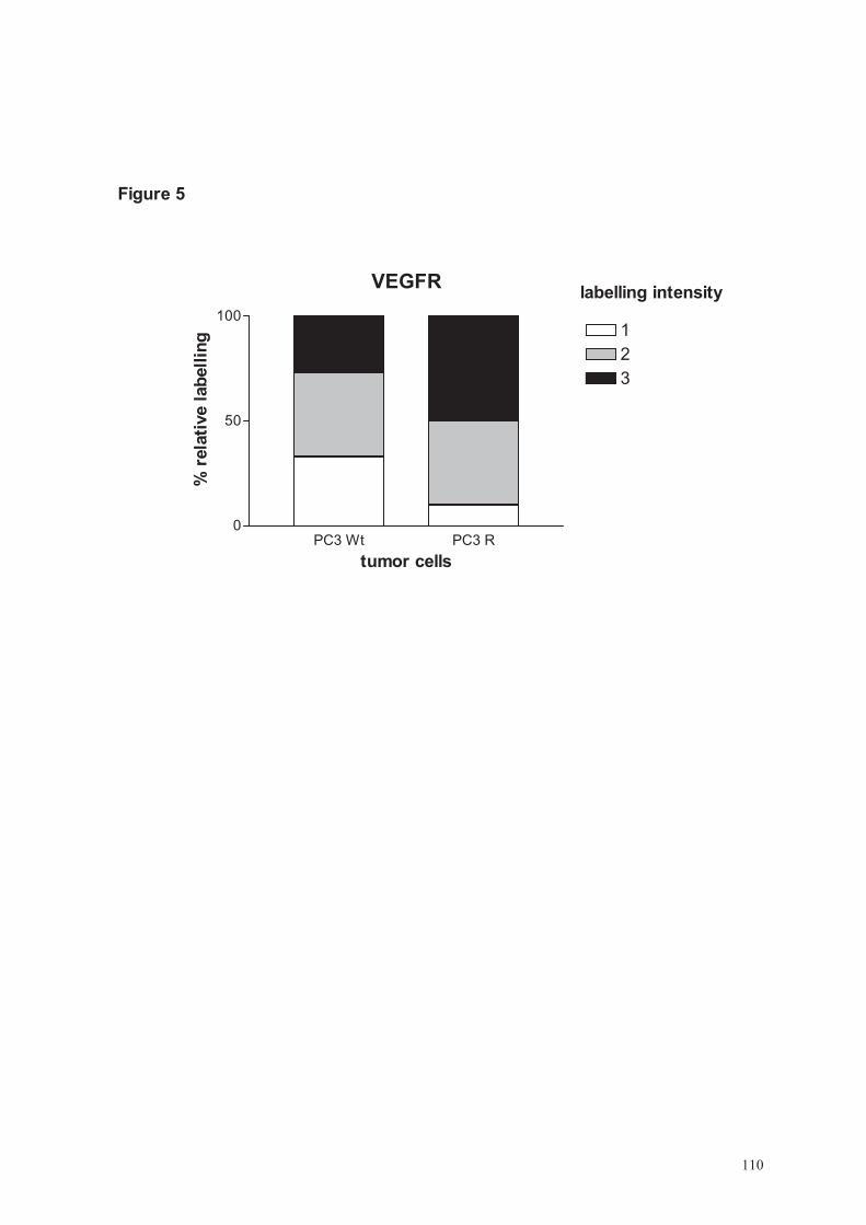
110
VEGFR
PC3 Wt PC3 R0
50
1001
2
3
labelling intensity
Figure 5
tumor cells
% r
ela
tive l
ab
ell
ing

111
vascular density PC3 Wt
cont
rol
vand
etan
ib25
vand
etan
ib50
doce
taxe
l 10
doce
taxe
l 30
vand
etan
ib 2
5+
doce
taxe
l 10
vand
etan
ib 5
0+
doce
taxe
l 30
0
50
1001
2
3
4
Figure 6a
** *** * *** *** ***
rela
tive %
vascular density PC3R
cont
rol
vand
etan
ib25
vand
etan
ib50
doce
taxe
l 10
doce
taxe
l 30
vand
etan
ib 2
5+
doce
taxe
l 10
vand
etan
ib 5
0+
doce
taxe
l 30
0
50
100
1
2
3
4
Figure 6b
% l
ab
ell
ing
NS NS NS NS ***

112
Figure 7 : ABCC1 mRNA expression
mRNA expression
0,0
0,2
0,4
0,6
0,8
1,0
1,2
1,4
1,6
1 2 3 4 5 6
arb
itra
ry u
nit
s

113
Figure 8a : MDR1 (ABCB1) mRNA expression
mRNA expression
0,0
0,5
1,0
1,5
2,0
2,5
1 2 3 4 5 6
arb
itra
ry u
nit
s

114
Figure 8b : MDR1 (ABCB1) protein expression
protein expression
0,0
0,2
0,4
0,6
0,8
1,0
1,2
1,4
1 2 3 4 5 6
arb
itra
ry u
nit
s
ACTIN
MDR1
ACTIN
MDR1

115
Figure 9a: EGFR mRNA expression
mRNA expression
0,0
1,0
2,0
3,0
4,0
5,0
6,0
1 2 3 4 5 6
arb
itra
ry u
nit
s

116
Figure 9b: EGFR protein expression
Protein expression
0
1
2
3
4
5
6
7
8
1 2 3 4 5 6
arb
itra
ry u
nits
Protein expression
0
2
4
6
8
10
12
14
16
18
20
1 2 3 4 5 6
arb
itra
ry u
nits
ACTIN
EGFR
pEGFR
ACTIN
EGFR
pEGFR

117
EGFR
PC3 Wt PC3 R0
50
1001
2
3
labelling intensity
Figure 10
tumor cells
% r
ela
tive l
ab
ell
ing

118

119
RESUME
Contexte et objectif Le cancer de la prostate, premier cancer chez l’homme en France, a bénéficié largement des avancées
thérapeutiques de la chirurgie, de la chimiothérapie, de la radiothérapie et, depuis une dizaine
d’années, de l’hormonothérapie. Lorsque le cancer devient hormono-résistant, la chimiothérapie par
docetaxel et prednisone est désormais le traitement de référence. Actuellement les traitements de
deuxième ligne sont décevants et tout reste à faire. Notre travail a eu pour but d’évaluer l’impact de
thérapies ciblées visant EGFR et VEGFR dans cette situation clinique actuellement très défavorable,
données seules ou en association au docetaxel. Nous avons pour celà utilisé un modèle cellulaire PC3
xénogreffé sur la souris nude.
Matériels et méthodes Un premier travail a consisté à tester l’efficacité anti-tumorale du sunitinib (agent anti-angiogénique),
du cetuximab (agent anti-EGFR) et du docetaxel en monothérapies, bithérapies ou trithérapie in vivo,
à partir d’une lignée cellulaire PC3 xénogreffée à la souris nude. Un deuxième travail a consisté à
tester l’efficacité anti-tumorale du vandetanib (agent anti-VEGFR et anti-EGFR) associé ou non au
docetaxel in vivo, à partir de deux lignées PC3 xénogreffées à la souris nude. Une lignée sauvage
sensible au docetaxel, et une lignée résistante au docetaxel.
Résultats : Dans notre premier travail, nous avons montré un effet supra-additif de l’association sunitinib –
docetaxel et sunitinib - cetuximab, avec une bonne tolérance du traitement évaluée sur le poids des
souris. Dans notre deuxième travail, le vandetanib n’a pas montré d’efficacité sur la souche sauvage, et
même un effet activateur de croissance tumorale à faible dose. Sur la souche résistante, il n’a pas
montré d’effet de resensibilisation au docetaxel. In vitro, il n’a pas montré d’effet modulateur sur
MDR1.
Conclusion Si le sunitinib a présenté des résultats intéressants dans le cadre de notre modèle préclinique pour le
cancer de prostate hormono-résistant, il semble que le vandetanib soit à réserver éventuellement aux
cancers de prostate hormono-résistants devenus docetaxel résistants, en deuxième ligne et en
monothérapie, dans le cadre d’essais cliniques à venir.
ABSTRACT
PurposeProstate cancer is the first male cancer men in France. Clinical benefits were realized during the ten
last year’s concerning surgery, cytotoxic chemotherapy, hormonotherapy and radiotherapy. The
reference treatment for homone-refractory prostate cancer combines docetaxel and prednisone. The
current responses to second-line treatments are disappointing and considerable progress remains to be
made. The aim of our studies was to test novel therapeutics approaches by combining docetaxel with
EGFR and VEGFR targeting agents. Mice bearing well-established PC3 prostate tumors were used.
Methods The aim of our first study was to test a rational therapeutic approach by combining docetaxel with an
EGFR-targeting agent (cetuximab) and with an anti-angiogenic agent (sunitinib), using mice bearing
PC3 prostate tumors.
The aim of our second study was to test a novel therapeutic approach by combining docetaxel with
vandetanib, a dual EGFR and VEGFR targeting agent. Mice bearing docetaxel-sensitive-or-resistant
PC3 were used.
Results In our first study, supra-additive effects were observed with the sunitinib-docetaxel combination.
Stable mean mouse weight suggested that no drug-induced toxicity was evident.
In our second study, vandetanib had growth-stimulation effects at the smallest concentration on PC3
wild type, and no effect concerning the other conditions on PC3 wild type. MDR1 was expressed in
PC3 resistant only, but was neither modulated nor its action inhibited, by vandetanib in vitro.
Conclusion Use of sunitinib might support innovative strategies in the management of hormone-refractory prostate
cancer, in our preclinical model. Concerning vandetanib, it would be use for hormone-refractory
prostate cancer only after relapse under docetaxel, as a second-line treatment.