short-form paper: jcm00236-13 comparison of restriction
TRANSCRIPT
Comparison of Restriction Enzymes for Pulsed-Field GelElectrophoresis Typing of Moraxella catarrhalis
Sara Marti,a,b Carmen Puig,a,b Arnau Domenech,a,b Josefina Liñares,a,b Carmen Ardanuya,b
Microbiology Department, Hospital Universitari Bellvitge, IDIBELL, University of Barcelona, Barcelona, Spaina; CIBER de Enfermedades Respiratorias, ISCIII, Madrid, Spainb
NotI, the most prevalent restriction enzyme used for typing Moraxella catarrhalis, failed to digest genomic DNA from respira-tory samples. An improved pulsed-field gel electrophoresis (PFGE) methodology determined SpeI as the best choice for typingthis bacterial species, with a good restriction of clinical samples and a good clustering correlation with NotI.
Moraxella catarrhalis is a Gram-negative diplococcus com-monly found colonizing the upper respiratory tract of chil-
dren (1, 2). This mucosal pathogen is also a significant cause ofrecurrent otitis media in young children (3) and of acute exacer-bations in chronic obstructive pulmonary disease (COPD) pa-tients (3, 4), a syndrome associated with abnormal inflammatoryimmune responses of the lungs (5–7).
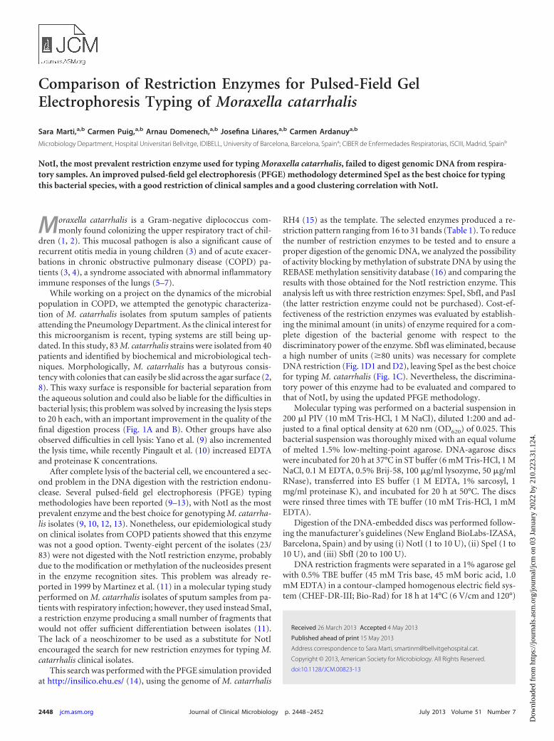
While working on a project on the dynamics of the microbialpopulation in COPD, we attempted the genotypic characteriza-tion of M. catarrhalis isolates from sputum samples of patientsattending the Pneumology Department. As the clinical interest forthis microorganism is recent, typing systems are still being up-dated. In this study, 83 M. catarrhalis strains were isolated from 40patients and identified by biochemical and microbiological tech-niques. Morphologically, M. catarrhalis has a butyrous consis-tency with colonies that can easily be slid across the agar surface (2,8). This waxy surface is responsible for bacterial separation fromthe aqueous solution and could also be liable for the difficulties inbacterial lysis; this problem was solved by increasing the lysis stepsto 20 h each, with an important improvement in the quality of thefinal digestion process (Fig. 1A and B). Other groups have alsoobserved difficulties in cell lysis: Yano et al. (9) also incrementedthe lysis time, while recently Pingault et al. (10) increased EDTAand proteinase K concentrations.
After complete lysis of the bacterial cell, we encountered a sec-ond problem in the DNA digestion with the restriction endonu-clease. Several pulsed-field gel electrophoresis (PFGE) typingmethodologies have been reported (9–13), with NotI as the mostprevalent enzyme and the best choice for genotyping M. catarrha-lis isolates (9, 10, 12, 13). Nonetheless, our epidemiological studyon clinical isolates from COPD patients showed that this enzymewas not a good option. Twenty-eight percent of the isolates (23/83) were not digested with the NotI restriction enzyme, probablydue to the modification or methylation of the nucleosides presentin the enzyme recognition sites. This problem was already re-ported in 1999 by Martinez et al. (11) in a molecular typing studyperformed on M. catarrhalis isolates of sputum samples from pa-tients with respiratory infection; however, they used instead SmaI,a restriction enzyme producing a small number of fragments thatwould not offer sufficient differentiation between isolates (11).The lack of a neoschizomer to be used as a substitute for NotIencouraged the search for new restriction enzymes for typing M.catarrhalis clinical isolates.
This search was performed with the PFGE simulation providedat http://insilico.ehu.es/ (14), using the genome of M. catarrhalis
RH4 (15) as the template. The selected enzymes produced a re-striction pattern ranging from 16 to 31 bands (Table 1). To reducethe number of restriction enzymes to be tested and to ensure aproper digestion of the genomic DNA, we analyzed the possibilityof activity blocking by methylation of substrate DNA by using theREBASE methylation sensitivity database (16) and comparing theresults with those obtained for the NotI restriction enzyme. Thisanalysis left us with three restriction enzymes: SpeI, SbfI, and PasI(the latter restriction enzyme could not be purchased). Cost-ef-fectiveness of the restriction enzymes was evaluated by establish-ing the minimal amount (in units) of enzyme required for a com-plete digestion of the bacterial genome with respect to thediscriminatory power of the enzyme. SbfI was eliminated, becausea high number of units (�80 units) was necessary for completeDNA restriction (Fig. 1D1 and D2), leaving SpeI as the best choicefor typing M. catarrhalis (Fig. 1C). Nevertheless, the discrimina-tory power of this enzyme had to be evaluated and compared tothat of NotI, by using the updated PFGE methodology.
Molecular typing was performed on a bacterial suspension in200 �l PIV (10 mM Tris-HCl, 1 M NaCl), diluted 1:200 and ad-justed to a final optical density at 620 nm (OD620) of 0.025. Thisbacterial suspension was thoroughly mixed with an equal volumeof melted 1.5% low-melting-point agarose. DNA-agarose discswere incubated for 20 h at 37°C in ST buffer (6 mM Tris-HCl, 1 MNaCl, 0.1 M EDTA, 0.5% Brij-58, 100 �g/ml lysozyme, 50 �g/mlRNase), transferred into ES buffer (1 M EDTA, 1% sarcosyl, 1mg/ml proteinase K), and incubated for 20 h at 50°C. The discswere rinsed three times with TE buffer (10 mM Tris-HCl, 1 mMEDTA).
Digestion of the DNA-embedded discs was performed follow-ing the manufacturer’s guidelines (New England BioLabs-IZASA,Barcelona, Spain) and by using (i) NotI (1 to 10 U), (ii) SpeI (1 to10 U), and (iii) SbfI (20 to 100 U).
DNA restriction fragments were separated in a 1% agarose gelwith 0.5% TBE buffer (45 mM Tris base, 45 mM boric acid, 1.0mM EDTA) in a contour-clamped homogenous electric field sys-tem (CHEF-DR-III; Bio-Rad) for 18 h at 14°C (6 V/cm and 120°)
Received 26 March 2013 Accepted 4 May 2013
Published ahead of print 15 May 2013
Address correspondence to Sara Marti, [email protected].
Copyright © 2013, American Society for Microbiology. All Rights Reserved.
doi:10.1128/JCM.00823-13
2448 jcm.asm.org Journal of Clinical Microbiology p. 2448–2452 July 2013 Volume 51 Number 7
Dow
nloa
ded
from
http
s://j
ourn
als.
asm
.org
/jour
nal/j
cm o
n 03
Jan
uary
202
2 by
210
.223
.31.
124.
and increasing pulse time intervals from 1 to 30 s (NotI) or 0.5 to35 s (SpeI and SbfI). PFGE band patterns were analyzed using theFingerprinting-II software, version 3.0 (Bio-Rad, Madrid), withoptimization and tolerance for the Dice coefficient of 0.75%.
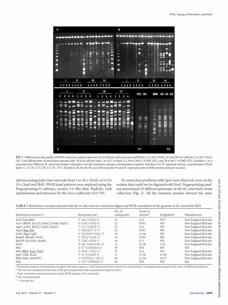
No restriction problems with SpeI were observed, even on theisolates that could not be digested with NotI. Fingerprinting anal-ysis determined 20 different genotypes in the M. catarrhalis straincollection (Fig. 2). All the repeated samples showed the same
FIG 1 Differences in the quality of PFGE restriction analysis between 5 h of cell lysis with lysozyme and RNase (1 to 10 U NotI) (A) and 20 h of cell lysis (1 to 10 U NotI)(B). Cost-effectiveness of restriction enzymes after 20 h of cell lysis with 1 to 10 U of SpeI (C), 80 to 100 U of SbfI (D1), and 20 to 60 U of SbfI (D2). Numbers 1 to 4represent four different M. catarrhalis isolates selected to test the minimum enzyme concentration required. Numbers I to IV represent enzyme concentration (NotI/SpeI): I, 1 U; II, 2.5 U; III, 5 U; IV, 10 U. Numbers 20, 40, 60, 80, and 100 in panels D1 and D2 represent units of SbfI enzyme used per reaction.
TABLE 1 Restriction enzymes selected with the in silico tool for restriction digest and PFGE simulation of the genome of M. catarrhalis RH4
Restriction enzyme(s)a Restriction siteeNo. ofcutting sites
$/unit ofenzymeb $/digestionb Manufacturer
AclI (Psp1406I) 5=-AA^CGTT-3= 19 0.21 NDd New England BioLabsAcvI (BbrPI, Eco72I, PmaCI, PmlI, PspCI) 5=-CAC^GTG-3= 27 0.031 ND New England BioLabsAgeI (AsiGI, BshTI, CspAI, PinAI) 5=-A^CCGGT-3= 23 0.22 ND New England BioLabsApaI (Bsp120I) 5=-GGGCC^C-3= 22 0.013 ND New England BioLabsAsiSI (RgaI, SgfI) 5=-GCGAT^CGC-3= 18 0.126 ND New England BioLabsBsp68I (BtuMI, NruI) 5=-TCG^CGA-3= 16 0.055 ND New England BioLabsBstSNI (Eco105I, SnaBI) 5=-TAC^GTA-3= 16 0.11 ND New England BioLabsNotIc 5=-GC^GGCCGC-3= 12 0.136 1.36 New England BioLabsPfoI 5=-T^CCNGGA-3= 19 ND ND NDSfoI (BbeI, KasI, NarI) 5=-GGC^GCC-3= 27 0.12 ND New England BioLabsSpeI (AhlI, BcuI) 5=-A^CTAGT-3= 31 0.126 0.126 New England BioLabsSbfI (SdaI, Sse8387I) 5=-CCTGCA^GG-3= 28 0.134 10.72 New England BioLabsPasI 5=-CC^CWGGG-3= 24 ND ND NDa Restriction enzymes in parentheses recognize the same sequence. Underlined restriction enzymes are neoschizomer (recognize same sequence but cleave at different positions).b The cost was calculated on the basis of the prices indicated by the manufacturer (listed in 2012).c NotI, restriction enzyme previously used in PFGE analysis of M. catarrhalis.d ND, not determined.e ^, cleavage site.
PFGE Typing of Moraxella catarrhalis
July 2013 Volume 51 Number 7 jcm.asm.org 2449
Dow
nloa
ded
from
http
s://j
ourn
als.
asm
.org
/jour
nal/j
cm o
n 03
Jan
uary
202
2 by
210
.223
.31.
124.
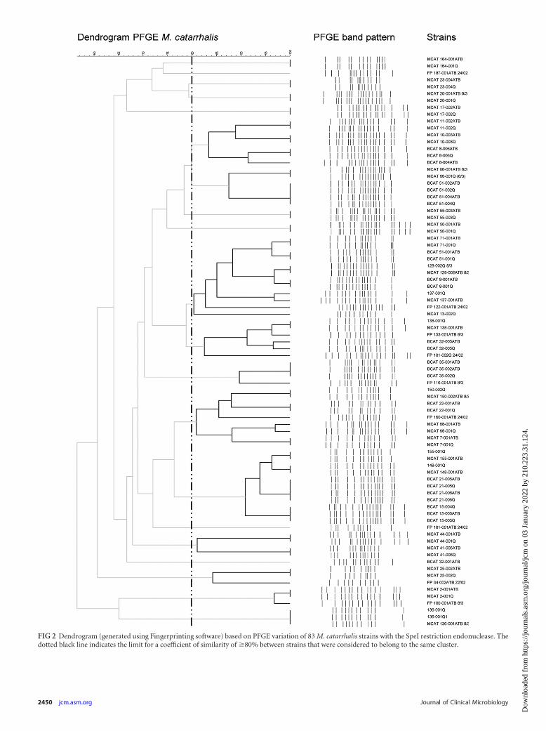
FIG 2 Dendrogram (generated using Fingerprinting software) based on PFGE variation of 83 M. catarrhalis strains with the SpeI restriction endonuclease. Thedotted black line indicates the limit for a coefficient of similarity of �80% between strains that were considered to belong to the same cluster.
2450 jcm.asm.org Journal of Clinical Microbiology
Dow
nloa
ded
from
http
s://j
ourn
als.
asm
.org
/jour
nal/j
cm o
n 03
Jan
uary
202
2 by
210
.223
.31.
124.
PFGE pattern, which corroborates the reproducibility of this up-dated methodology.
The evaluation of the discriminatory power of SpeI with respect tothat of NotI was performed on 35 nonrepeated isolates from differentepisodes of 31 patients with COPD that presented a complete diges-tion for NotI. As shown in Fig. 3, the discriminatory power of NotI(19 genotypes) is slightly superior to that of SpeI (14 genotypes). Aprevious study published by Vu-Thien et al. (13) on 11 M. catarrhalisisolates suggested that although NotI had the most discriminatorypower, SpeI patterns were also reliable. Our study, performed on 35independent isolates, proves that the difference between both restric-tion enzymes is minimal (Fig. 3) and, probably, the importance of acorrect restriction of all the samples outweighs the small difference indiscriminatory power.
A methodology for PFGE should be cost-effective and stan-dardized to allow the comparison of fingerprints between labora-tories. So far, genotyping of M. catarrhalis involved different re-striction enzymes, and the epidemiological results could not becompared between centers. The NotI restriction enzyme provedto be inefficient, at least for typing M. catarrhalis isolated fromrespiratory samples. The evaluation indicated that PFGE withSpeI is almost as discriminatory as the one performed with NotI,
and so far it avoids the restriction problems encountered in respi-ratory samples.
ACKNOWLEDGMENTS
We thank S. Santos from the Pneumology Department of the HospitalUniversitari de Bellvitge, who contributed to this project by collecting thesputum samples from COPD patients.
This study was supported by a grant from the Fondo de Investigacio-nes Sanitarias de la Seguridad Social (PI0901904) and by CIBER de En-fermedades Respiratorias (CIBERES; CB06/06/0037), run by the Institutode Salud Carlos III (ISCIII; Madrid, Spain).
C.P. and A.D. were supported by grants from Formación de Profeso-rado Universitario (FPU; Ministerio de Educación, Spain). S.M. was sup-ported by a Sara Borrell postdoctoral contract, CD10/00298, from theISCIII, Madrid, Spain.
We report no transparency declarations.
REFERENCES1. de Vries SP, Bootsma HJ, Hays JP, Hermans PW. 2009. Molecular
aspects of Moraxella catarrhalis pathogenesis. Microbiol. Mol. Biol. Rev.73:389 – 406.
2. Verduin CM, Hol C, Fleer A, van Dijk H, van Belkum A. 2002. Moraxellacatarrhalis: from emerging to established pathogen. Clin. Microbiol. Rev. 15:125–144.
FIG 3 Comparison of the main genotype clusters obtained after NotI digestion (A) and SpeI digestion (B). Strains selected: 35 nonrepeated strains with acomplete digestion for the NotI restriction endonuclease. Gray-colored squares indicate the clusters obtained only with one of the restriction enzymes. Dottedblack lines indicate the limit for a coefficient of similarity of �80% between strains that were considered to belong to the same cluster. Crossed-out strain namesrepresent differences between clusters.
PFGE Typing of Moraxella catarrhalis
July 2013 Volume 51 Number 7 jcm.asm.org 2451
Dow
nloa
ded
from
http
s://j
ourn
als.
asm
.org
/jour
nal/j
cm o
n 03
Jan
uary
202
2 by
210
.223
.31.
124.
3. Karalus R, Campagnari A. 2000. Moraxella catarrhalis: a review of animportant human mucosal pathogen. Microbes Infect. 2:547–559.
4. Enright MC, McKenzie H. 1997. Moraxella (Branhamella) catarrhalis—clinical and molecular aspects of a rediscovered pathogen. J. Med. Micro-biol. 46:360 –371.
5. Murphy TF, Sethi S, Niederman MS. 2000. The role of bacteria inexacerbations of COPD. A constructive view. Chest 118:204 –209.
6. Sethi S, Murphy TF. 2001. Bacterial infection in chronic obstructivepulmonary disease in 2000: a state-of-the-art review. Clin. Microbiol. Rev.14:336 –363.
7. Sethi S. 2010. Infection as a comorbidity of COPD. Eur. Respir. J. 35:1209 –1215.
8. Murphy TF, Parameswaran GI. 2009. Moraxella catarrhalis, a humanrespiratory tract pathogen. Clin. Infect. Dis. 49:124 –131.
9. Yano H, Suetake M, Kuga A, Irinoda K, Okamoto R, Kobayashi T,Inoue M. 2000. Pulsed-field gel electrophoresis analysis of nasopharyn-geal flora in children attending a day care center. J. Clin. Microbiol. 38:625– 629.
10. Pingault NM, Lehmann D, Riley TV. 2008. Improved pulsed field gelelectrophoresis method for Moraxella catarrhalis. J. Microbiol. Methods75:344 –345.
11. Martinez G, Ahmed K, Zheng CH, Watanabe K, Oishi K, Nagatake T.1999. DNA restriction patterns produced by pulsed-field gel electropho-resis in Moraxella catarrhalis isolated from different geographical areas.Epidemiol. Infect. 122:417– 422.
12. Pingault NM, Lehmann D, Bowman J, Riley TV. 2007. A comparison ofmolecular typing methods for Moraxella catarrhalis. J. Appl. Microbiol.103:2489 –2495.
13. Vu-Thien H, Dulot C, Moissenet D, Fauroux B, Garbarg-Chenon A.1999. Comparison of randomly amplified polymorphic DNA analysis andpulsed-field gel electrophoresis for typing of Moraxella catarrhalis strains.J. Clin. Microbiol. 37:450 – 452.
14. Bikandi J, San Millan R, Rementeria A, Garaizar J. 2004. In silicoanalysis of complete bacterial genomes: PCR, AFLP-PCR and endonu-clease restriction. Bioinformatics 20:798 –799.
15. de Vries SP, van Hijum SA, Schueler W, Riesbeck K, Hays JP, HermansPW, Bootsma HJ. 2010. Genome analysis of Moraxella catarrhalis strainRH4, a human respiratory tract pathogen. J. Bacteriol. 192:3574 –3583.
16. Roberts RJ, Vincze T, Posfai J, Macelis D. 2010. REBASE—a database forDNA restriction and modification: enzymes, genes and genomes. NucleicAcids Res. 38:D234 –D236.
Marti et al.
2452 jcm.asm.org Journal of Clinical Microbiology
Dow
nloa
ded
from
http
s://j
ourn
als.
asm
.org
/jour
nal/j
cm o
n 03
Jan
uary
202
2 by
210
.223
.31.
124.