influence of the occlusal alteration in the mandible
TRANSCRIPT

1151
Int. J. Morphol.,28(4):1151-1157, 2010.
Influence of the Occlusal Alterationin the Mandible Morphology of the Gerbil
Influencia de la Alteración Oclusal en la Morfología Mandibular del Jerbo
Fernando José Dias; Richard Honorato de Oliveira; João Paulo Mardegan Issa; Simone Cecílio Hallak Regalo;Valéria O. Pagnano de Souza; Alma Blásida C. E. B. Catirse & Mamie Mizusaki Iyomasa
DIAS, F. J.; OLIVEIRA, R. H.; ISSA, J. P. M.; REGALO, S. C. H.; SOUZA, V. O. P.; CATIRSE, A. B. C. E. B. & IYOMASA, M.M. Influence of the occlusal alteration in the mandible morphology of the gerbil. Int. J. Morphol., 28(4):1151-1157, 2010.
SUMMARY: Considering the biomechanical aspects, many facts need to be understood on the mandible, to know which effectsunilateral occlusal changes may cause on the stomatognathic system. The aim of this study was to analyze the malocclusion by unilateralteeth extraction on the mandible morphology in gerbil. We used 10 gerbils Meriones unguiculatus, young male, weighing around 50-60g,divided into two groups (n=5), an experimental group and control, which evaluated the two hemi-mandibles, with a total of 20 sampleswhich were measured by digital pachymeter. The measures were taken: (1) length and (2) width of the mandible condyle and (3) bodyheight in the region of mandibular 1st molar. Data from these measurements were analyzed using ANOVA and Mann-Whitney test. Theresults of this study showed a statistically significant difference in the three measures between experimental and control groups. Thewidth of the mandible head (condyle) showed statistical difference between the ipsilateral and contralateral sides to teeth extraction. Itwas concluded that the mastication modification by unilateral teeth extraction caused an imbalance, promoting not only a modificationin the craniofacial growth pattern, but also a harmful effect on the stomatognathic system of the gerbil used as an experimental model inthis study.
KEY WORDS: Malocclusion; Morphology; Gerbil.
INTRODUCTION
The stomatognathic system consists of aheterogeneous group of organs and tissues whose biology istotally interdependent on the performance of their functions.Over the years, the relationship between muscle activity andocclusal changes concerns have been expressed by manyprofessionals involved in oral rehabilitation. Changesobserved in the bone belonged to the craniofacial complexin growing rats, caused by masticatory function modification(Beecher & Corruccini, 1981; Kiliaridis et al., 1985, 1999)and in dental occlusion can promote biomechanicaldysfunction on the morphofunctional structure of the head(Baba et al., 1996, 2000).
Muscles play an important role in the normal activityof the stomatognathic system complex, due to the directrelationship with the muscles origin and bone insertions.These locations represent different regions of functionaldemand, as the shape and area ratio with muscle. However,
it will only be possible to understand the biomechanics ofthe craniofacial skeleton considering it as a dynamicphenomenon.
Studies carried out in experimental animal modelsrevealed that the reduction in masticatory muscles demandcan cause interferences in jaws development (Ulgen et al.,1997; Corruccini & Lee, 1984; Poikela et al., 1997). Inagreement, the Bresin et al. (1999) observations showed thatthe diet consistency change may induce significantdifferences in the mandible morphology.
Bone studies revealed the onset of bone remodelingby mechanical factors (Bouvier & Hylander, 1996),compressive loads at specific bone surface exerted by mus-cular contraction (Teng & Herring, 1998) and deformationcomponents of the temporomandibular joint masticatorystrain (Liu & Herring, 2000). However, a need exists for in
Departamento de Morfologia, Estomatologia e Fisiologia, Faculdade de Odontologia de Ribeirão Preto - Universidade de São Paulo, Brazil.
1152
vivo studies to understand the biomechanics of occlusalalteration effects on the craniofacial skeleton.
Thus, the purpose of this study was to analyze theeffect of unilateral extraction of left upper molars on themandible morphology (both sides) in the gerbil.
MATERIAL AND METHOD
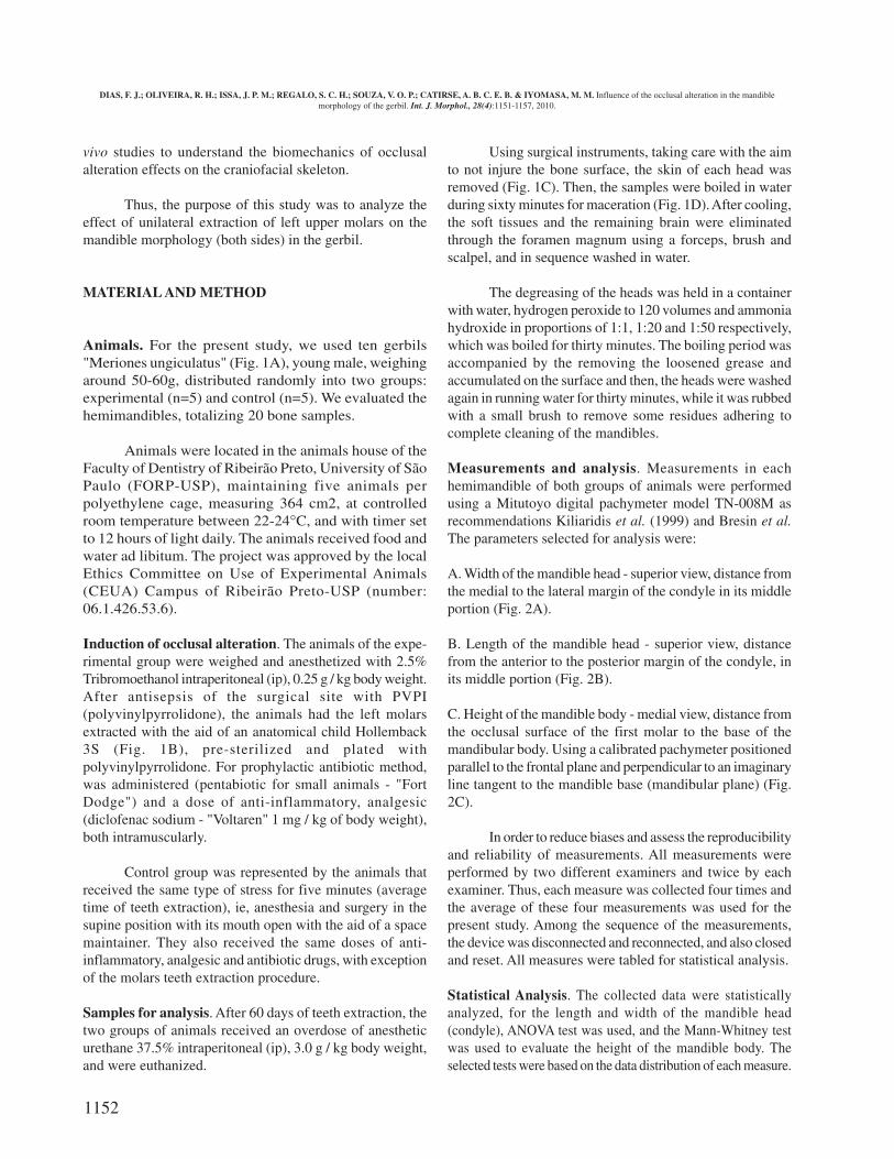
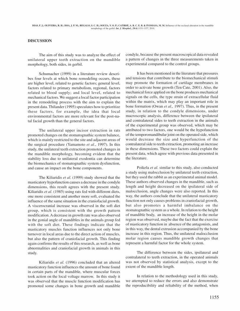
Animals. For the present study, we used ten gerbils"Meriones ungiculatus" (Fig. 1A), young male, weighingaround 50-60g, distributed randomly into two groups:experimental (n=5) and control (n=5). We evaluated thehemimandibles, totalizing 20 bone samples.
Animals were located in the animals house of theFaculty of Dentistry of Ribeirão Preto, University of SãoPaulo (FORP-USP), maintaining five animals perpolyethylene cage, measuring 364 cm2, at controlledroom temperature between 22-24°C, and with timer setto 12 hours of light daily. The animals received food andwater ad libitum. The project was approved by the localEthics Committee on Use of Experimental Animals(CEUA) Campus of Ribeirão Preto-USP (number:06.1.426.53.6).
Induction of occlusal alteration. The animals of the expe-rimental group were weighed and anesthetized with 2.5%Tribromoethanol intraperitoneal (ip), 0.25 g / kg body weight.After antisepsis of the surgical site with PVPI(polyvinylpyrrolidone), the animals had the left molarsextracted with the aid of an anatomical child Hollemback3S (Fig. 1B), pre-sterilized and plated withpolyvinylpyrrolidone. For prophylactic antibiotic method,was administered (pentabiotic for small animals - "FortDodge") and a dose of anti-inflammatory, analgesic(diclofenac sodium - "Voltaren" 1 mg / kg of body weight),both intramuscularly.
Control group was represented by the animals thatreceived the same type of stress for five minutes (averagetime of teeth extraction), ie, anesthesia and surgery in thesupine position with its mouth open with the aid of a spacemaintainer. They also received the same doses of anti-inflammatory, analgesic and antibiotic drugs, with exceptionof the molars teeth extraction procedure.
Samples for analysis. After 60 days of teeth extraction, thetwo groups of animals received an overdose of anestheticurethane 37.5% intraperitoneal (ip), 3.0 g / kg body weight,and were euthanized.
Using surgical instruments, taking care with the aimto not injure the bone surface, the skin of each head wasremoved (Fig. 1C). Then, the samples were boiled in waterduring sixty minutes for maceration (Fig. 1D). After cooling,the soft tissues and the remaining brain were eliminatedthrough the foramen magnum using a forceps, brush andscalpel, and in sequence washed in water.
The degreasing of the heads was held in a containerwith water, hydrogen peroxide to 120 volumes and ammoniahydroxide in proportions of 1:1, 1:20 and 1:50 respectively,which was boiled for thirty minutes. The boiling period wasaccompanied by the removing the loosened grease andaccumulated on the surface and then, the heads were washedagain in running water for thirty minutes, while it was rubbedwith a small brush to remove some residues adhering tocomplete cleaning of the mandibles.
Measurements and analysis. Measurements in eachhemimandible of both groups of animals were performedusing a Mitutoyo digital pachymeter model TN-008M asrecommendations Kiliaridis et al. (1999) and Bresin et al.The parameters selected for analysis were:
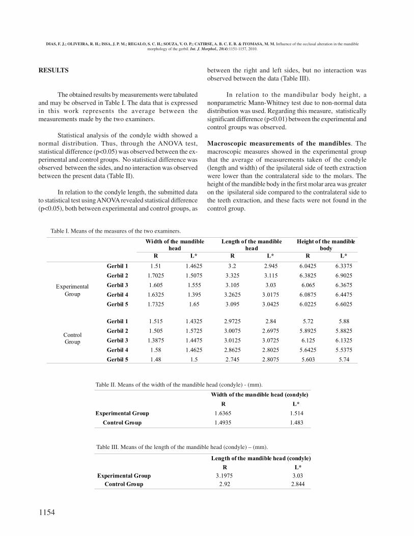
A. Width of the mandible head - superior view, distance fromthe medial to the lateral margin of the condyle in its middleportion (Fig. 2A).
B. Length of the mandible head - superior view, distancefrom the anterior to the posterior margin of the condyle, inits middle portion (Fig. 2B).
C. Height of the mandible body - medial view, distance fromthe occlusal surface of the first molar to the base of themandibular body. Using a calibrated pachymeter positionedparallel to the frontal plane and perpendicular to an imaginaryline tangent to the mandible base (mandibular plane) (Fig.2C).
In order to reduce biases and assess the reproducibilityand reliability of measurements. All measurements wereperformed by two different examiners and twice by eachexaminer. Thus, each measure was collected four times andthe average of these four measurements was used for thepresent study. Among the sequence of the measurements,the device was disconnected and reconnected, and also closedand reset. All measures were tabled for statistical analysis.
Statistical Analysis. The collected data were statisticallyanalyzed, for the length and width of the mandible head(condyle), ANOVA test was used, and the Mann-Whitney testwas used to evaluate the height of the mandible body. Theselected tests were based on the data distribution of each measure.
DIAS, F. J.; OLIVEIRA, R. H.; ISSA, J. P. M.; REGALO, S. C. H.; SOUZA, V. O. P.; CATIRSE, A. B. C. E. B. & IYOMASA, M. M. Influence of the occlusal alteration in the mandiblemorphology of the gerbil. Int. J. Morphol., 28(4):1151-1157, 2010.

1153
Fig. 2. A. Width of the mandible head; B. Length of the mandible Head; Height of the mandible body
Fig. 1. A. Gerbils used in the study; B. Molar extraction; C. Head without skin; D. Samples Boiled
DIAS, F. J.; OLIVEIRA, R. H.; ISSA, J. P. M.; REGALO, S. C. H.; SOUZA, V. O. P.; CATIRSE, A. B. C. E. B. & IYOMASA, M. M. Influence of the occlusal alteration in the mandiblemorphology of the gerbil. Int. J. Morphol., 28(4):1151-1157, 2010.
1154
RESULTS
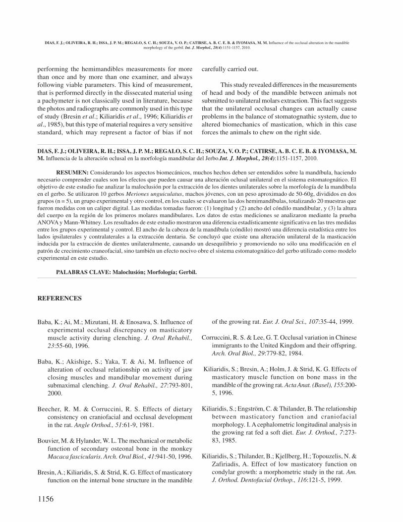
The obtained results by measurements were tabulatedand may be observed in Table I. The data that is expressedin this work represents the average between themeasurements made by the two examiners.
Statistical analysis of the condyle width showed anormal distribution. Thus, through the ANOVA test,statistical difference (p<0.05) was observed between the ex-perimental and control groups. No statistical difference wasobserved between the sides, and no interaction was observedbetween the present data (Table II).
In relation to the condyle length, the submitted datato statistical test using ANOVA revealed statistical difference(p<0.05), both between experimental and control groups, as
between the right and left sides, but no interaction wasobserved between the data (Table III).
In relation to the mandibular body height, anonparametric Mann-Whitney test due to non-normal datadistribution was used. Regarding this measure, statisticallysignificant difference (p<0.01) between the experimental andcontrol groups was observed.
Macroscopic measurements of the mandibles. Themacroscopic measures showed in the experimental groupthat the average of measurements taken of the condyle(length and width) of the ipsilateral side of teeth extractionwere lower than the contralateral side to the molars. Theheight of the mandible body in the first molar area was greateron the ipsilateral side compared to the contralateral side tothe teeth extraction, and these facts were not found in thecontrol group.
Width of the mandible head (condyle)
R L*
Experimental Group 1.6365 1.514
Control Group 1.4935 1.483
Width of the mandible
head
Length of the mandible
head
Height of the mandible
body
R L* R L* R L*
Gerbil 1 1.51 1.4625 3.2 2.945 6.0425 6.3375
Gerbil 2 1.7025 1.5075 3.325 3.115 6.3825 6.9025
Gerbil 3 1.605 1.555 3.105 3.03 6.065 6.3675
Gerbil 4 1.6325 1.395 3.2625 3.0175 6.0875 6.4475
Gerbil 5 1.7325 1.65 3.095 3.0425 6.0225 6.6025
Experimental
Group
Gerbil 1 1.515 1.4325 2.9725 2.84 5.72 5.88
Gerbil 2 1.505 1.5725 3.0075 2.6975 5.8925 5.8825
Gerbil 3 1.3875 1.4475 3.0125 3.0725 6.125 6.1325
Gerbil 4 1.58 1.4625 2.8625 2.8025 5.6425 5.5375
ControlGroup
Gerbil 5 1.48 1.5 2.745 2.8075 5.603 5.74
Length of the mandible head (condyle)
R L*
Experimental Group 3.1975 3.03
Control Group 2.92 2.844
Table III. Means of the length of the mandible head (condyle) – (mm).
Table I. Means of the measures of the two examiners.
Table II. Means of the width of the mandible head (condyle) - (mm).
DIAS, F. J.; OLIVEIRA, R. H.; ISSA, J. P. M.; REGALO, S. C. H.; SOUZA, V. O. P.; CATIRSE, A. B. C. E. B. & IYOMASA, M. M. Influence of the occlusal alteration in the mandiblemorphology of the gerbil. Int. J. Morphol., 28(4):1151-1157, 2010.
1155
DISCUSSION
The aim of this study was to analyze the effect ofunilateral upper teeth extraction on the mandiblemorphology, both sides, in gerbil.
Schumacher (1999) in a literature review descri-bes four levels at which bone remodeling occurs, theseare higher level, related to genetic factors; general level,factors related to primary metabolism, regional, factorsrelated to blood supply; and local level, related tomechanical factors. We suggest a local factor participationin the remodeling process with the aim to explain thepresent data. Thilander (1995) speculates how to prioritizethese factors, for example, the idea that localenvironmental factors are more relevant for the post-na-tal facial growth than the general factors.
The unilateral upper incisor extraction in ratspromoted changes on the stomatognathic system balance,which is mainly restricted to the site and adjacent areas ofthe surgical procedure (Yamamoto et al., 1997). In thisstudy, the unilateral teeth extraction promoted changes inthe mandible morphology, becoming evident that thestability loss due to unilateral exodontia can determinethe biomechanics of stomatognathic system dysfunction,and cause an impact on the bone components.
The Kiliaridis et al. (1999) study showed that themasticatory hypofunction causes a decrease in the condyledimensions, this result agrees with the present study.Kiliaridis et al. (1985) using rats fed with different diets,one more consistent and another more soft, evaluated theinfluence of the same situation in the craniofacial growth.A viscerocranial increase was observed in the soft dietgroup, which is consistent with the growth patternmodification. A decrease in growth rate was also observedin the gonial angle of mandibles in the animals group fedwith the soft diet. These findings indicate that themasticatory muscles function influences not only boneturnover in local areas due to the direct action of muscles,but also the pattern of craniofacial growth. This findingagain confirms the results of this research, as well as boneabnormalities and craniofacial growth in animals in thisstudy.
Kiliaridis et al. (1996) concluded that an alteredmasticatory function influences the amount of bone foundin certain parts of the mandible, where muscular forcestook action on the local voltage marrow. In this study itwas observed that the muscle function modification haspromoted some changes in bone growth and mandible
condyle, because the present macroscopical data revealeda pattern of changes in the three measurements taken inexperimental compared to the control groups.
It has been mentioned in the literature that pressuresand tensions that contribute to the biomechanical stimulimay promote the formation of cartilage membranes inorder to activate bone growth (Ten Cate, 2001). Also, themechanical force applied on the bone produces mechanicalsignals on the cells, the type strain of extracellular fluidwithin the matrix, which may play an important role inbone formation (Owan et al., 1997). Thus, in the presentstudy, in relation to the condyle dimensions, undermacroscopic analysis, difference between the ipsilateraland contralateral sides to teeth extraction in the animalsof the experimental group was observed, which may beattributed to two factors, one would be the hypofunctionof the temporomandibular joint on the operated side, whichwould decrease the size and hyperfunction of thecontralateral side to teeth extraction, promoting an increasein these dimensions. These two factors could explain thepresent data, which agree with previous data presented inthe literature.
Poikela et al. similar to this study, also conducteda study using malocclusion by unilateral teeth extraction,but they used the rabbit as an experimental animal model.These authors observed changes in the mandible, such aslength and height decreased on the ipsilateral side ofmalocclusion, angle changes were also reported. In thisway, the authors conclude that the unilateral masticatoryfunction not only causes problems in craniofacial growth,but also promotes a harmful imbalance on thestomatognathic system as a whole. In relation to the heightof mandible body, an increase of the height in the molarregion was observed, maybe due the fact that the exerciseof masticatory function in absence of the antagonists, andin this way, the dental extrusion accompanied by the boneincrease in this region. Thus, the unilateral malocclusionmolar region causes mandible growth changes thatrepresent a harmful factor for the whole system.
The difference between the sides, ipsilateral andcontralateral to teeth extraction, in the operated animalswas not observed by statistical analysis, except to theextent of the mandible length.
In relation to the methodology used in this study,we attempted to reduce the errors and also demonstratethe reproducibility and reliability of the method, when
DIAS, F. J.; OLIVEIRA, R. H.; ISSA, J. P. M.; REGALO, S. C. H.; SOUZA, V. O. P.; CATIRSE, A. B. C. E. B. & IYOMASA, M. M. Influence of the occlusal alteration in the mandiblemorphology of the gerbil. Int. J. Morphol., 28(4):1151-1157, 2010.
1156
DIAS, F. J.; OLIVEIRA, R. H.; ISSA, J. P. M.; REGALO, S. C. H.; SOUZA, V. O. P.; CATIRSE, A. B. C. E. B. & IYOMASA, M.M. Influencia de la alteración oclusal en la morfología mandibular del Jerbo.Int. J. Morphol., 28(4):1151-1157, 2010.
RESUMEN: Considerando los aspectos biomecánicos, muchos hechos deben ser entendidos sobre la mandíbula, haciendonecesario comprender cuales son los efectos que pueden causar una alteración oclusal unilateral en el sistema estomatognático. Elobjetivo de este estudio fue analizar la maloclusión por la extracción de los dientes unilaterales sobre la morfología de la mandíbulaen el gerbo. Se utilizaron 10 gerbos Meriones unguiculatus, machos jóvenes, con un peso aproximado de 50-60g, divididos en dosgrupos (n = 5), un grupo experimental y otro control, en los cuales se evaluaron las dos hemimandíbulas, totalizando 20 muestras quefueron medidas con un caliper digital. Las medidas tomadas fueron: (1) longitud y (2) ancho del cóndilo mandibular, y (3) la alturadel cuerpo en la región de los primeros molares mandibulares. Los datos de estas mediciones se analizaron mediante la pruebaANOVA y Mann-Whitney. Los resultados de este estudio mostraron una diferencia estadísticamente significativa en las tres medidasentre los grupos experimental y control. El ancho de la cabeza de la mandíbula (cóndilo) mostró una diferencia estadística entre loslados ipsilaterales y contralaterales a la extracción dentaria. Se concluyó que existe una alteración unilateral de la masticacióninducida por la extracción de dientes unilateralmente, causando un desequilibrio y promoviendo no sólo una modificación en elpatrón de crecimiento craneofacial, sino también un efecto nocivo obre el sistema estomatognático del gerbo utilizado como modeloexperimental en este estudio.
PALABRAS CLAVE: Maloclusión; Morfología; Gerbil.
performing the hemimandibles measurements for morethan once and by more than one examiner, and alwaysfollowing viable parameters. This kind of measurement,that is performed directly in the dissecated material usinga pachymeter is not classically used in literature, becausethe photos and radiographs are commonly used in this typeof study (Bresin et al.; Kiliaridis et al., 1996; Kiliaridis etal., 1985), but this type of material requires a very sensitivestandard, which may represent a factor of bias if not
carefully carried out.
This study revealed differences in the measurementsof head and body of the mandible between animals notsubmitted to unilateral molars extraction. This fact suggeststhat the unilateral occlusal changes can actually causeproblems in the balance of stomatognathic system, due toaltered biomechanics of mastication, which in this caseforces the animals to chew on the right side.
REFERENCES
Baba, K.; Ai, M.; Mizutani, H. & Enosawa, S. Influence ofexperimental occlusal discrepancy on masticatorymuscle activity during clenching. J. Oral Rehabil.,23:55-60, 1996.
Baba, K.; Akishige, S.; Yaka, T. & Ai, M. Influence ofalteration of occlusal relationship on activity of jawclosing muscles and mandibular movement duringsubmaximal clenching. J. Oral Rehabil., 27:793-801,2000.
Beecher, R. M. & Corruccini, R. S. Effects of dietaryconsistency on craniofacial and occlusal developmentin the rat. Angle Orthod., 51:61-9, 1981.
Bouvier, M. & Hylander, W. L. The mechanical or metabolicfunction of secondary osteonal bone in the monkeyMacaca fascicularis. Arch. Oral Biol., 41:941-50, 1996.
Bresin, A.; Kiliaridis, S. & Strid, K. G. Effect of masticatoryfunction on the internal bone structure in the mandible
of the growing rat. Eur. J. Oral Sci., 107:35-44, 1999.
Corruccini, R. S. & Lee, G. T. Occlusal variation in Chineseimmigrants to the United Kingdom and their offspring.Arch. Oral Biol., 29:779-82, 1984.
Kiliaridis, S.; Bresin, A.; Holm, J. & Strid, K. G. Effects ofmasticatory muscle function on bone mass in themandible of the growing rat. Acta Anat. (Basel), 155:200-5, 1996.
Kiliaridis, S.; Engström, C. & Thilander, B. The relationshipbetween masticatory function and craniofacialmorphology. I. A cephalometric longitudinal analysis inthe growing rat fed a soft diet. Eur. J. Orthod., 7:273-83, 1985.
Kiliaridis, S.; Thilander, B.; Kjellberg, H.; Topouzelis, N. &Zafiriadis, A. Effect of low masticatory function oncondylar growth: a morphometric study in the rat. Am.J. Orthod. Dentofacial Orthop., 116:121-5, 1999.
DIAS, F. J.; OLIVEIRA, R. H.; ISSA, J. P. M.; REGALO, S. C. H.; SOUZA, V. O. P.; CATIRSE, A. B. C. E. B. & IYOMASA, M. M. Influence of the occlusal alteration in the mandiblemorphology of the gerbil. Int. J. Morphol., 28(4):1151-1157, 2010.

1157
Liu, Z. J. & Herring, S. W. Bone surface strains and internalbony pressures at the jaw joint of the miniature pig duringmasticatory muscle contraction. Arch. Oral Biol., 45:95-112, 2000.
Owan, I.; Burr, D. B.; Turner, C. H.; Qiu, J.; Tu, Y.; Onyia,J. E.; et al. Mechanotransduction in bone: osteoblastsare more responsive to fluid forces than mechanicalstrain. Am. J. Physiol., 273:C810-5, 1997.
Poikela, A.; Kantomaa, T. & Pirttiniemi, P. Craniofacialgrowth after a period of unilateral masticatory functionin young rabbits. Eur. J. Oral Sci., 105:331-7, 1997.
Schumacher, G. H. Regulative and adaptive factors incraniofacial growth. Ann. Anat., 181:9-13, 1999.
Ten Cate, A. R. Histologia Bucal. Desenvolvimento, estruturae função. 5ª ed. Rio de Janeiro, Guanabara Koogan, 1998.
Teng, S. & Herring, S. W. Compressive loading on bonesurfaces from muscular contraction: an in vivo study inthe miniature pig, Sus scrofa. J. Morphol., 238:71-80,1998.
Thilander, B. Basic mechanisms in craniofacial growth. ActaOdontol. Scand., 53:144-51, 1995.
Ulgen, M.; Baran, S.; Kaya, H. & Karadede, I. The influenceof the masticatory hypofunction on the craniofacialgrowth and development in rats. Am. J. Orthod.Dentofacial Orthop., 111:189-98, 1997.
Yamamoto, M. K.; Novelli, M. D. & Luz, J. G. Effects ofunilateral upper incisor extraction on facial growth ofyoung rats. J. Nihon. Univ. Sch. Dent., 39:191-5, 1997.
Correspondence to:Prof. Dra. Mamie Mizusaki Iyomasa
Departamento de Morfologia Estomatologia e Fisiologia /
FORP-USP
Avenida do Café, s/n
Bairro - Monte Alegre
CEP:14040-904
Ribeirão Preto
SP – BRASIL
Email: [email protected]
Received: 10-07-2010
Accepted: 22-09-2010
DIAS, F. J.; OLIVEIRA, R. H.; ISSA, J. P. M.; REGALO, S. C. H.; SOUZA, V. O. P.; CATIRSE, A. B. C. E. B. & IYOMASA, M. M. Influence of the occlusal alteration in the mandiblemorphology of the gerbil. Int. J. Morphol., 28(4):1151-1157, 2010.

1158