enregistrement de l’occlusion occlusal recording contrôle
TRANSCRIPT
Controlling occlusion with new technologies
MOTS CLÉS /KEYWORDS
Mouvements mandibulairesEmpreinte optiqueEnregistrement de l’occlusion
Mandibular movementsOptical impressionOcclusal recording
G. DUMINIL
Contrôle de l’occlusion avec de nouvelles technologies
GÉRARD DUMINIL. Docteur en chirurgie dentaire, Docteur en science odontologiques. CES Parodontologie, Prothèse fixée. DU Occlusodontologie, Implantologie. Exercice libéral à Nice.
ROS – OCTOBRE 2017197 Rev Odont Stomat 2017;46:197-210 OCTOBER 2017
RÉSUMÉLe contrôle de l’occlusion est un acte quotidiennement réalisé, de la simple obturation occlusale à la mise en place d’unerestauration prothétique. Des nouvelles technologies permettent d’aller au-delà du marquage coloré en incorporant lespossibilités de visualiser, soit directement, soit sous forme graphique, les contacts occlusaux statiques et dynamiques.Nous évoquons les possibilités de l’empreinte optique et de ses extensions logicielles, du capteur occlusal Occlusense® etdu très performant système ModJaw® d’enregistrement des mouvements physiologiques par caméra stéréoscopique.
ABSTRACTThe control of the occlusion is a procedure performed every day, from the simple occlusal filling to the placement of anextended prosthetic restoration. Beyond the colored marking methods, new technologies provide the possibilities ofvisualizing either directly or graphically the static and dynamic occlusal contacts.We will evoke the optical impression technique and its software extensions, the occlusal sensor OccluSense® and the veryefficient ModJaw system® that records the physiological movements with a stereoscopic camera.
L ’ O C C L U S I O N : A S P E C T S C L I N I Q U E S
Demande de tirés-à-part : [email protected]

L ’ O C C L U S I O N : A S P E C T S C L I N I Q U E S
Le contrôle de l’occlusion se fait classiquement en utilisant des indicateursocclusaux qui se présentent sous plusieurs formes :– les marqueurs sur supports (papiers et rubans colorés) ;– les rubans sans marqueur (films d’aluminium) ;– les marqueurs sans support (poudre en spray ou encre) ;– les élastomères et les cires.Utilisés avec succès depuis des décennies pour localiser les contactsocclusaux, ces divers moyens sont très largement répandus.Les évolutions technologiques actuelles ouvrent de nouvelles perspectivesdans le domaine de l’occlusion. Mais que peuvent nous apporter ces nou-velles technologies par rapport aux moyens classiques ?Le but de cet article est de présenter trois options modernes pour contrôlerl’occlusion statique et dynamique : les empreintes optiques, un appareil demordu électronique sur capteur sensible à la pression (l’OccluSense®) etun système de modélisation de la cinématique mandibulaire (ModJaw®).
LES EMPREINTES OPTIQUESLes indications prothétiques de l’empreinte optique sont bien connues,mais ses applications pour l’analyse de l’occlusion et ses vertus pédago-giques restent à explorer.Classiquement, l’enregistrement de l’occlusion se fait en OIM, mais il estpossible d’enregistrer d’autres positions occlusales en utilisant éventuel-lement des cales pour stabiliser une position lors de l’enregistrement.
Quel que soit le système d’empreinte utilisé, on effectue la numérisationcomplète des deux arcades, ainsi que l’enregistrement de la position d’OIM.L’affichage immédiat offre la possibilité d’informer le patient et de l’instruireà ce sujet (fig. 1).
Several types of occlusal indicators are conventionallyused to control the occlusion:� Marking materials (colored papers and strips).� Strips with no markers (articulating foil).� Markers with no support (spray powder or ink).� Elastomers and wax.
Successfully used for decades to localize the occlusalcontacts, these various methods are very widely spread.The current technological developments are openingnew perspectives in the field of occlusion. Still, what arethe advantages of these new technologies comparedwith the conventional techniques?This article will describe three new techniques to controlthe static and dynamic occlusion: the optical impression,a digital device connected to a sensor measuring thepressure (OccluSense®) and a modeling system of themandibular kinetics (ModJaw®).
OpTiCal iMpRESSiOnSThe prosthetic indications for optical impressions arewell known. However, their applications in the analysisof the occlusion and their educational virtues still needto be explored.Occlusion is generally recorded in iCp, but it is possibleto record other occlusal positions by using rims tomaintain a position during the recording.
Whatever the system of impression, both arches arefully digitalized, as well as the recording of the iCpposition. The immediate visualization allows to describethe status to the patient and to give him/her someexplanations (fig. 1).
ROS – OCTOBRE/OCTOBER 2017198
CONTRÔLE DE L’OCCLUSION AVEC DE NOUVELLES TECHNOLOGIES
1
Fig. 1. La présentation de l’image de ses arcades au patient aun fort impact pédagogique lors de la présentation du plan detraitement (caméra 3Shape).
Fig. 1. Showing the image of his/her arches to the patient hasa strong educational impact during the presentation of thetreatment plan (intra oral scanner).
L ’ O C C L U S I O N : A S P E C T S C L I N I Q U E S
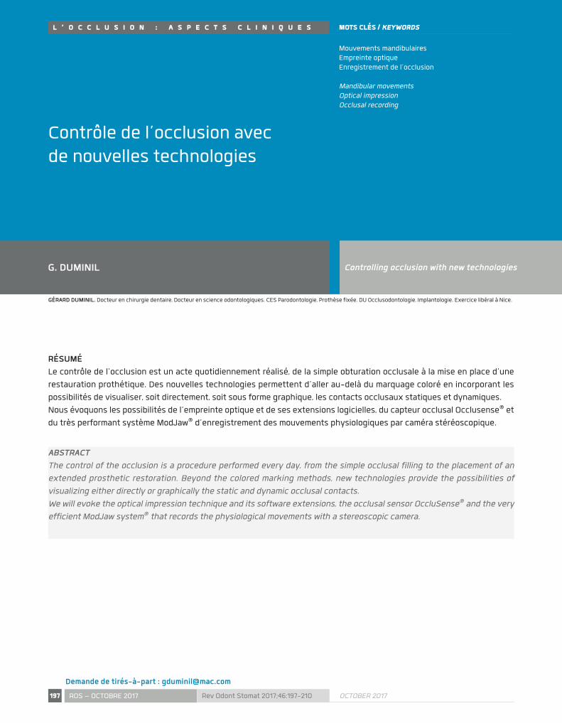
Il est ainsi possible d’observer cet engrènement sous toutes ses faces,comme si l’on tenait les moulages à la main (fig. 2a-d).
Chaque arcade peut être isolée et examinée séparément ou en positiond’engrènement.
it is thus possible to observe the occlusion from all sides,as if we held the casts in our hand (fig. 2 a, b, c, d).
Each arch can be isolated and examined separately orin iCp.
ROS – OCTOBRE/OCTOBER 2017199
CONTRÔLE DE L’OCCLUSION AVEC DE NOUVELLES TECHNOLOGIES
2a
Fig. 2a. Engrènement en OIM d’arcades présentant de bonnesrelations occlusales.
Fig. 2a. Engagement in iCp with proper occlusal relationships.
2b
2c
Fig. 2b. Les images peuvent être orientées sous tous les angles(ici, la vue linguale).
Fig. 2b. images can be directed from all angles, here the lingualview.
Fig. 2c. En vue latérale, la classe d’angle est facilement iden-tifiable.
Fig. 2c. in lateral view, the angle class is easy to identify.
L ’ O C C L U S I O N : A S P E C T S C L I N I Q U E S
Une commande permet d’ajouter sur les faces occlusales un gradient decouleur objectivant l’intensité des contacts occlusaux (fig. 3a-b et 4a-b).Lors de l’enregistrement, il est important que le patient maintienne ferme-ment la position d’engrènement, la présence de la caméra contre la jouepouvant entraîner un léger desserrement.
a function allows to add a colored gradient on theocclusal faces, objectifying the intensity of the occlusalcontacts (fig. 3 a, b), (fig. 4 a, b). During the recording,it is important that the patient firmly holds the occlusalposition as the presence of the camera against thecheek may result in a slight relaxation.
ROS – OCTOBRE/OCTOBER 2017200
CONTRÔLE DE L’OCCLUSION AVEC DE NOUVELLES TECHNOLOGIES
2d
Fig. 2d. Avec la possibilité de zoomer, l’engrènement lingualpeut être plus facilement contrôlé.
Fig. 2d. With the zoom, the lingual engagement can be moreeasily controlled.
3a 3b
Fig. 3a. Activation de la commande de la distance à l’arcade opposée. Lescontacts sont identifiés par les points bleus.
Fig. 3a. activation of the command for the distance to the opposite arch, thecontacts are identified with blue points.
Fig. 3b. Vue de l’arcade antagoniste.
Fig. 3b. View of the antagonist arch.
4a 4b
Fig. 4a. Appréciation des rapports occlusaux après empreinte avec caméra DentalWings. Des mesures peuvent être prises pour la distance à l’antagoniste.
Fig. 4a. assessment of occlusal relationships after impression with Dental wingsscanner. The distance to the antagonist can be measured.
Fig. 4b. L’enregistrement monochrome est parfaitement compatible avec l’utili-sation de l’échelle de couleurs.
Fig. 4b. The monochrome recording is perfectly compatible with the use of thecolor scale.
L ’ O C C L U S I O N : A S P E C T S C L I N I Q U E S
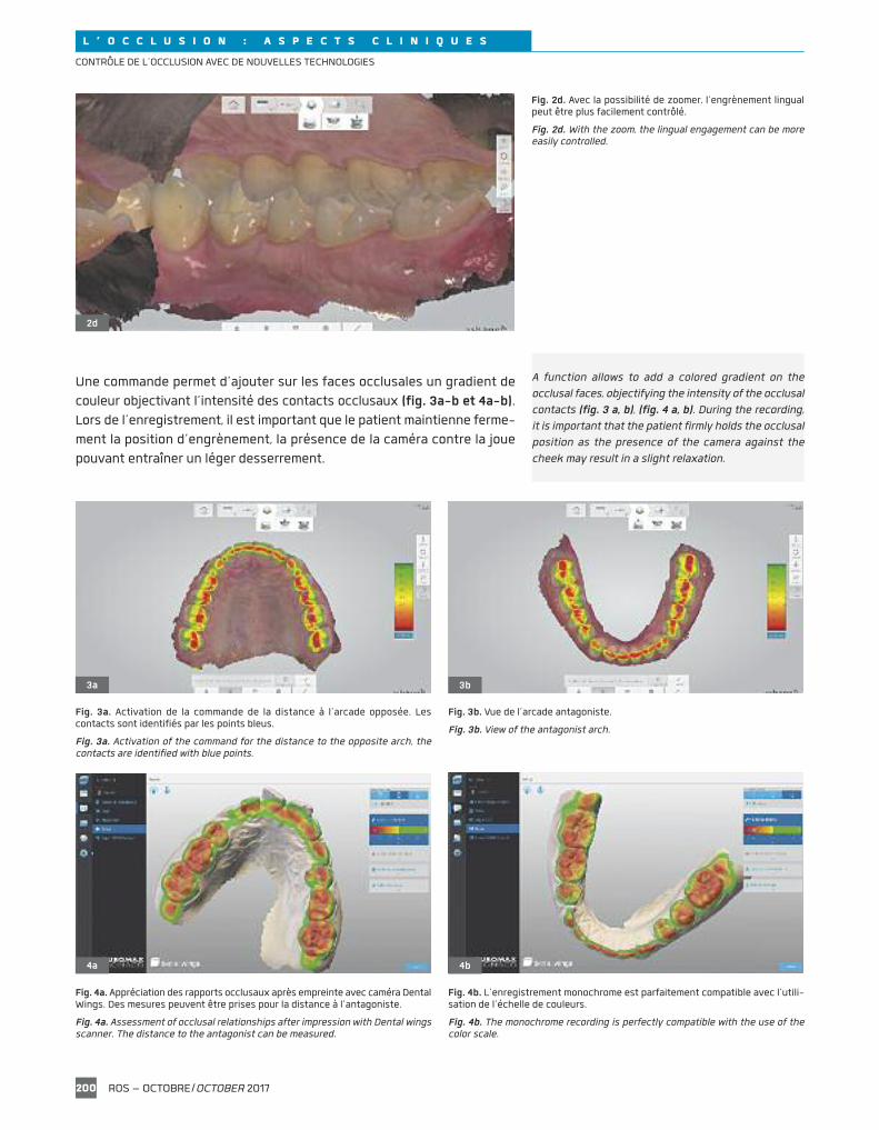
L’échelle de valeurs colorées est ajustable pour discriminer plusprécisément la position des contacts interarcades (fig. 5). Ceci concentrel’observation sur les zones de contact et peut s’assimiler à l’utilisation d’unmarqueur occlusal très fin.
Le passage de l’image en mode monochrome améliore l’observation desfacettes d’usures ou des éclats d’émail (fig. 6). La présence de facettesimportantes peut être un indicateur de parafonctions ; l’utilisation combinéede la fonction de zoom permet l’examen dans des zones où l’examenclinique est quasiment impossible.
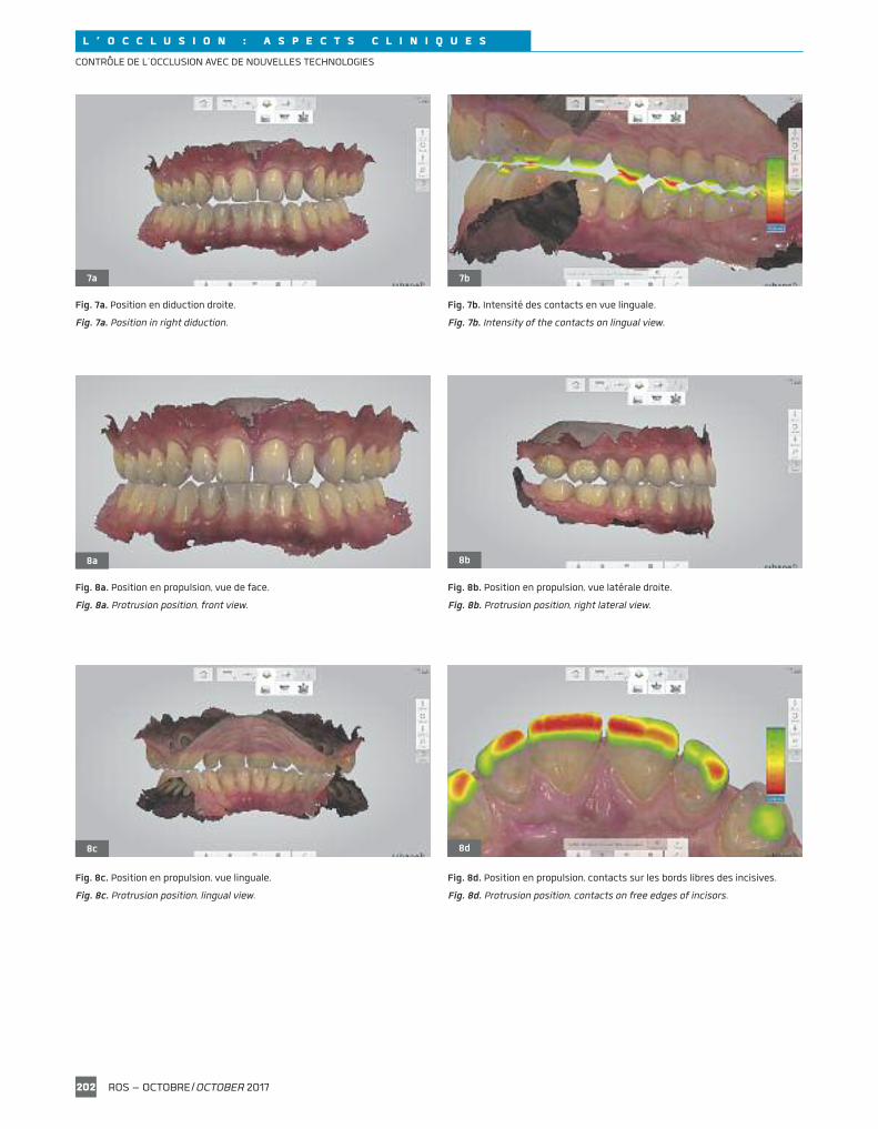
Il est aussi possible d’enregistrer des positions excentrées, diduction(fig. 7a-b) ou propulsion (fig. 8a-d). Pour cela, la confection de cales encire ou en élastomère permet le maintien de la position par le patient lorsde l’enregistrement. La cale est interposée du côté controlatéral àl’enregistrement, puis interchangée pour réaliser l’enregistrement du côtéopposé.L’examen de l’OIM et de positions excentrées est intéressant, mais neconcerne que des situations figées.
The scale of colored values is adjustable in order todiscriminate more accurately the position of theinterarch contacts (fig. 5). The observation is thusfocused on the contact zones and might be similar tothe use of a very thin occlusal marking indicator.
The image in monochrome mode improves the observationof wear facets or chipped enamel (fig. 6). a great numberof facets may indicate the presence of parafunctions;the combined use of the zoom function enables toexamine zones where the clinical examination is almostimpossible.
it is also possible to record off-centered positions(diduction (fig. 7 a, b) or protrusion (fig. 8 a, b, c, d)) andto this purpose, wax or elastomer rims allow the patientto keep the same position during the recording. The rimis placed on the contralateral side of the recording, thenchanged to make the recording on the opposite side.Examining the iCp and the off-centered positions isinteresting, but only concerns static situations.
ROS – OCTOBRE/OCTOBER 2017201
CONTRÔLE DE L’OCCLUSION AVEC DE NOUVELLES TECHNOLOGIES
Fig. 5. Système 3Shape. Le gradient de couleurs est adaptéafin de mieux individualiser les zones de contact.
Fig. 5. 3Shape system, the gradient of colors is adapted toaccurately indicate the contact zones.
5
Fig. 6. Système 3Shape. La suppression de la couleur permetde mieux identifier la facette d’usure (flèche a) et l’éclatd’émail (flèche B).
Fig. 6. 3Shape system, Removing the color allows to identifymore easily the wear facet (arrow a) and chipped enamel(arrow B).
6
L ’ O C C L U S I O N : A S P E C T S C L I N I Q U E S
ROS – OCTOBRE/OCTOBER 2017202
CONTRÔLE DE L’OCCLUSION AVEC DE NOUVELLES TECHNOLOGIES
7a 7b
Fig. 7a. Position en diduction droite.
Fig. 7a. position in right diduction.
Fig. 7b. Intensité des contacts en vue linguale.
Fig. 7b. intensity of the contacts on lingual view.
Fig. 8a. Position en propulsion, vue de face.
Fig. 8a. protrusion position, front view.
Fig. 8b. Position en propulsion, vue latérale droite.
Fig. 8b. protrusion position, right lateral view.
8a 8b
Fig. 8c. Position en propulsion, vue linguale.
Fig. 8c. protrusion position, lingual view.
Fig. 8d. Position en propulsion, contacts sur les bords libres des incisives.
Fig. 8d. protrusion position, contacts on free edges of incisors.
8c 8d

L ’ O C C L U S I O N : A S P E C T S C L I N I Q U E S
Une évolution récente des logiciels 3Shape permet l’enregistrement desmouvements dynamiques et, instantanément, leur reproduction à l’écransous tous les angles (fig. 9a-b).
L’affichage des zones de contact reflète alors les trajets des dents encontact lors des excursions, et cela devient bien plus intéressant, surtoutdans la perspective d’une restauration prothétique. Nous observons ici unefonction de groupe en diduction, la présence de contacts interférents auniveau du couple 17-47 et des contacts non travaillants entre 27 et 37(fig. 10a-b).
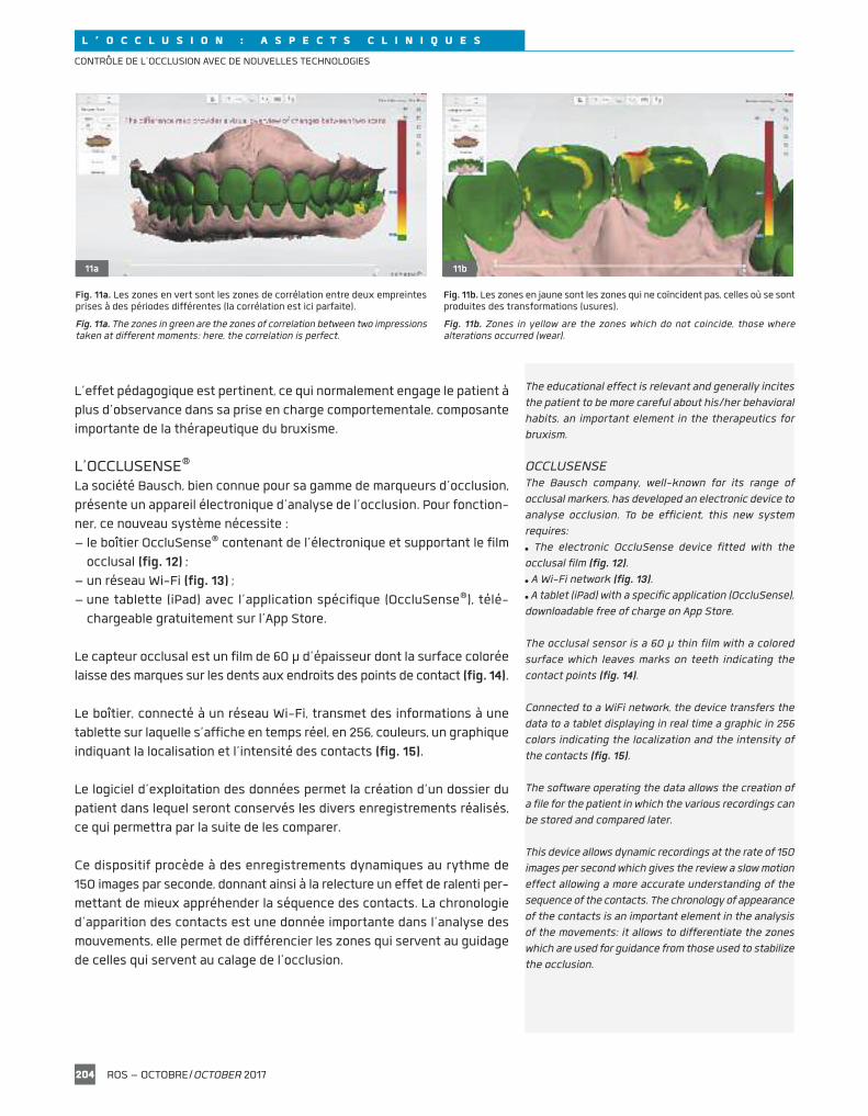
Une autre fonction appelée « monitoring » est destinée à contrôler leschangements intervenant au fil du temps sur les dents du patient. Sonapplication principale est la surveillance des sujets bruxeurs afin de ren-forcer leur prise en charge comportementale.Le logiciel superpose des empreintes prises à des dates différentes et meten évidence de manière colorée les modifications intervenues dansl’intervalle de temps (fig. 11a-b).
Recent developments of the 3Shape softwares allow therecording of dynamic movements and their immediatedisplay on screen from all angles (fig. 9 a, b).
The display of the contact zones shows the pathways ofteeth in contact during the excursions, which is muchmore interesting, especially when a prostheticrestoration is planned. We can observe here a groupfunction in diduction, the presence of interferingcontacts in the couple 17/47 and nonworking contactsbetween 27 and 37 (fig. 10 a, b).
another feature called “monitoring” is intended to checkthe changes occurring over time on the patient’s teeth.This feature mostly concerns patients suffering frombruxim to enhance the behavorial follow-up.The software superimposes the impressions taken atdifferent dates and highlights with colors themodifications which occurred over time (fig. 11 a, b).
ROS – OCTOBRE/OCTOBER 2017203
CONTRÔLE DE L’OCCLUSION AVEC DE NOUVELLES TECHNOLOGIES
9a 9b
Fig. 9a. Après enregistrement des mouvements dynamiques, une fonction dulogiciel permet de relire et donc d’observer les déplacements mandibulaires dentsau contact.
Fig. 9a. after recording the dynamic movements, a function of the software allowsto review and observe the mandibular movements teeth in the contact.
Fig. 9b. La relecture est saisissante lorsqu’elle est observée sous tous les angles.
Fig. 9b. The reviewing is striking when it is observed from all angles.
10a 10b
Fig. 10a. Les contacts durant les trajets sont colorés en bleu.
Fig. 10a. Contacts during the movements are colored in blue.
Fig. 10b. Ici, on visualise parfaitement les interférences au niveau du couple 47-17.
Fig. 10b. Here, we can perfectly see the interferences for the couple 47/17.
L ’ O C C L U S I O N : A S P E C T S C L I N I Q U E S
L’effet pédagogique est pertinent, ce qui normalement engage le patient àplus d’observance dans sa prise en charge comportementale, composanteimportante de la thérapeutique du bruxisme.
L’OCCLUSENSE®
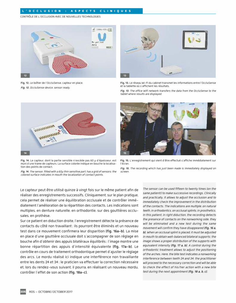
La société Bausch, bien connue pour sa gamme de marqueurs d’occlusion,présente un appareil électronique d’analyse de l’occlusion. Pour fonction -ner, ce nouveau système nécessite :– le boîtier OccluSense® contenant de l’électronique et supportant le filmocclusal (fig. 12) ;
– un réseau Wi-Fi (fig. 13) ;– une tablette (iPad) avec l’application spécifique (OccluSense®), télé -chargeable gratuitement sur l’App Store.
Le capteur occlusal est un film de 60 µ d’épaisseur dont la surface coloréelaisse des marques sur les dents aux endroits des points de contact (fig. 14).
Le boîtier, connecté à un réseau Wi-Fi, transmet des informations à unetablette sur laquelle s’affiche en temps réel, en 256, couleurs, un graphiqueindiquant la localisation et l’intensité des contacts (fig. 15).
Le logiciel d’exploitation des données permet la création d’un dossier dupatient dans lequel seront conservés les divers enregistrements réalisés,ce qui permettra par la suite de les comparer.
Ce dispositif procède à des enregistrements dynamiques au rythme de150 images par seconde, donnant ainsi à la relecture un effet de ralenti per-mettant de mieux appréhender la séquence des contacts. La chronologied’apparition des contacts est une donnée importante dans l’analyse desmouvements, elle permet de différencier les zones qui servent au guidagede celles qui servent au calage de l’occlusion.
The educational effect is relevant and generally incitesthe patient to be more careful about his/her behavioralhabits, an important element in the therapeutics forbruxism.
OCCluSEnSEThe Bausch company, well-known for its range ofocclusal markers, has developed an electronic device toanalyse occlusion. To be efficient, this new systemrequires: � The electronic OccluSense device fitted with theocclusal film (fig. 12). � a Wi-Fi network (fig. 13). � a tablet (ipad) with a specific application (OccluSense),downloadable free of charge on app Store.
The occlusal sensor is a 60 µ thin film with a coloredsurface which leaves marks on teeth indicating thecontact points (fig. 14).
Connected to a WiFi network, the device transfers thedata to a tablet displaying in real time a graphic in 256colors indicating the localization and the intensity ofthe contacts (fig. 15).
The software operating the data allows the creation ofa file for the patient in which the various recordings canbe stored and compared later.
This device allows dynamic recordings at the rate of 150images per second which gives the review a slow motioneffect allowing a more accurate understanding of thesequence of the contacts. The chronology of appearanceof the contacts is an important element in the analysisof the movements: it allows to differentiate the zoneswhich are used for guidance from those used to stabilizethe occlusion.
ROS – OCTOBRE/OCTOBER 2017204
CONTRÔLE DE L’OCCLUSION AVEC DE NOUVELLES TECHNOLOGIES
11a 11b
Fig. 11a. Les zones en vert sont les zones de corrélation entre deux empreintesprises à des périodes différentes (la corrélation est ici parfaite).
Fig. 11a. The zones in green are the zones of correlation between two impressionstaken at different moments; here, the correlation is perfect.
Fig. 11b. Les zones en jaune sont les zones qui ne coïncident pas, celles où se sontproduites des transformations (usures).
Fig. 11b. Zones in yellow are the zones which do not coincide, those wherealterations occurred (wear).
L ’ O C C L U S I O N : A S P E C T S C L I N I Q U E S
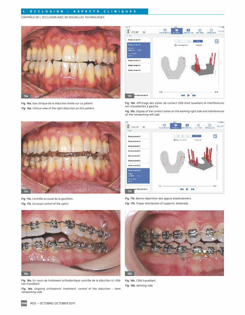
Le capteur peut être utilisé quinze à vingt fois sur le même patient afin deréaliser des enregistrements successifs. Cliniquement, sur le plan pratique,cela permet de réaliser une équilibration occlusale et de contrôler immé-diatement l’amélioration de la répartition des contacts. Les indications sontmultiples, en denture naturelle, en orthodontie, sur des gouttières occlu-sales, en prothèse.Sur ce patient en diduction droite, l’enregistrement détecte la présence decontacts du côté non travaillant ; ils pourront être éliminés et un nouveautest dans ce mouvement confirmera leur disparition (fig. 16a-b). La miseen place d’une gouttière occlusale doit s’accompagner de son réglage enbouche afin d’obtenir des appuis bilatéraux équilibrés ; l’image montre unebonne répartition des appuis d’intensité équivalente (fig. 17a-b). Lecontrôle en cours de traitement orthodontique permet d’ajuster le réglagedes arcs. Le mordu réalisé ici indique une interférence non travaillanteentre les dents 24 et 34 ; le praticien va effectuer la correction nécessaireet, lors du rendez-vous suivant, il pourra, en réalisant un nouveau mordu,contrôler l’effet de son action (fig. 18a-c).
The sensor can be used fifteen to twenty times (on thesame patient!) to make successive recordings. Clinicallyand practically, it allows to adjust the occlusion and toimmediately check the improvement in the distributionof the contacts. The indications are multiple, on naturalteeth, in orthodontics, on occlusal splints, in prosthetics.in this patient, in right diduction, the recording detectsthe presence of contacts on the nonworking side, theywill be eliminated and a new test during the samemovement will confirm they have disappeared (fig. 16 a,b). When an occlusal splint is placed, it must be adjustedin mouth to obtain well-balanced bilateral supports; theimage shows a proper distribution of the supports withequivalent intensity (fig. 17 a, b). a control during theorthodontic treatment allows to adjust the positioningof the arches. Here, the bite test indicates a nonworkinginterference between teeth 24 and 34; the practitionerwill proceed to the necessary correction and will be ableto check the effect of his/her action with a new bitetest during the next appointment (fig. 18 a, b, c).
ROS – OCTOBRE/OCTOBER 2017205
CONTRÔLE DE L’OCCLUSION AVEC DE NOUVELLES TECHNOLOGIES
12
Fig. 12. Le boîtier de l’OccluSense, capteur en place.
Fig. 12. OccluSense device, sensor ready.
13
Fig. 13. Le réseau Wi-Fi du cabinet transmet les informations entre l’OccluSenseet la tablette où s’affichent les résultats.
Fig. 13. The office Wifi network transfers the data from the OccluSense to thetablet where results are displayed.
14
Fig. 14. Le capteur, dont la partie sensible n’excède pas 60 µ d’épaisseur, estmuni d’une trame de capteurs. La surface colorée indique en bouche la localisa-tion des points de contact.
Fig. 14. The sensor, fitted with a 60µ thin sensitive part, has a grid of sensors; thecolored surface indicates in mouth the localization of contact points.
15
Fig. 15. L’enregistrement qui vient d’être effectué s’affiche immédiatement surl’écran.
Fig. 15. The recording which has just been made is immediately displayed onscreen.
L ’ O C C L U S I O N : A S P E C T S C L I N I Q U E S
ROS – OCTOBRE/OCTOBER 2017206
CONTRÔLE DE L’OCCLUSION AVEC DE NOUVELLES TECHNOLOGIES
16a
Fig. 16a. Vue clinique de la diduction droite sur ce patient.
Fig. 16a. Clinical view of the right diduction on this patient.
16b
Fig. 16b. Affichage des zones de contact côté droit tavaillant et interférencesnon travaillantes à gauche.
Fig. 16b. Display of the contact zones on the working right side and interferenceson the nonworking left side.
17a
Fig. 17a. Contrôle occlusal de la gouttière.
Fig. 17a. Occlusal control of the splint.
17b
Fig. 17b. Bonne répartition des appuis bilatéralement.
Fig. 17b. proper distribution of supports, bilaterally.
18a
Fig. 18a. En cours de traitement orthodontique contrôle de la diduction ici côténon travaillant.
Fig. 18a. Ongoing orthodontic treatment, control of the diduction - herenonworking side.
18b
Fig. 18b. Côté travaillant.
Fig. 18b. Working side.
L ’ O C C L U S I O N : A S P E C T S C L I N I Q U E S
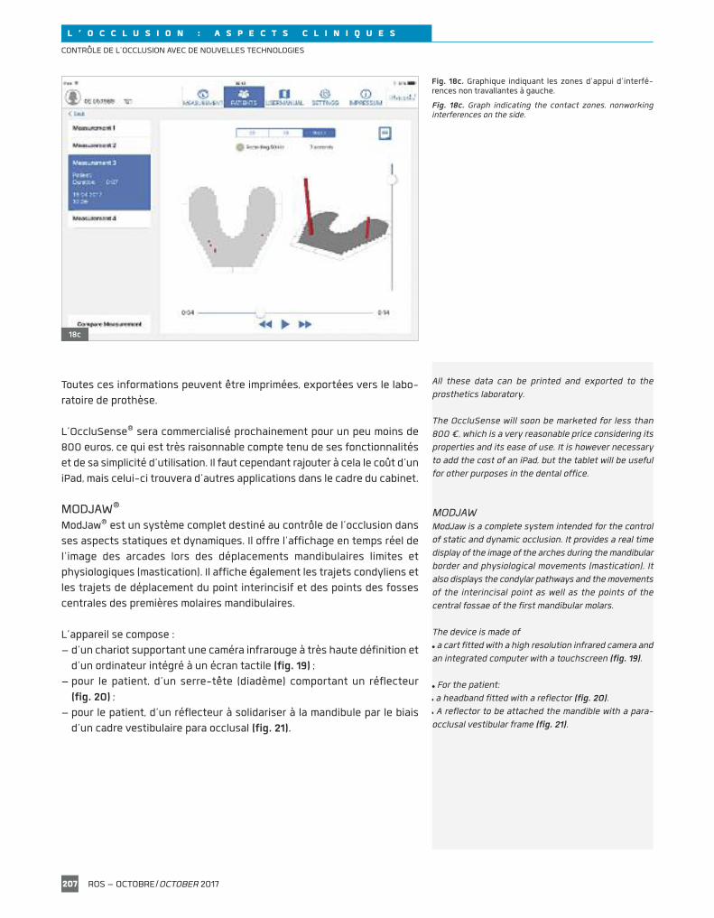
Toutes ces informations peuvent être imprimées, exportées vers le labo-ratoire de prothèse.
L’OccluSense® sera commercialisé prochainement pour un peu moins de800 euros, ce qui est très raisonnable compte tenu de ses fonctionnalitéset de sa simplicité d’utilisation. Il faut cependant rajouter à cela le coût d’uniPad, mais celui-ci trouvera d’autres applications dans le cadre du cabinet.
MODJAW®
ModJaw® est un système complet destiné au contrôle de l’occlusion dansses aspects statiques et dynamiques. Il offre l’affichage en temps réel del’image des arcades lors des déplacements mandibulaires limites etphysiologiques (mastication). Il affiche également les trajets condyliens etles trajets de déplacement du point interincisif et des points des fossescentrales des premières molaires mandibulaires.
L’appareil se compose :– d’un chariot supportant une caméra infrarouge à très haute définition etd’un ordinateur intégré à un écran tactile (fig. 19) ;
– pour le patient, d’un serre-tête (diadème) comportant un réflecteur(fig. 20) ;
– pour le patient, d’un réflecteur à solidariser à la mandibule par le biaisd’un cadre vestibulaire para occlusal (fig. 21).
all these data can be printed and exported to theprosthetics laboratory.
The OccluSense will soon be marketed for less than800 €, which is a very reasonable price considering itsproperties and its ease of use. it is however necessaryto add the cost of an ipad, but the tablet will be usefulfor other purposes in the dental office.
MODJaWModJaw is a complete system intended for the controlof static and dynamic occlusion. it provides a real timedisplay of the image of the arches during the mandibularborder and physiological movements (mastication). italso displays the condylar pathways and the movementsof the interincisal point as well as the points of thecentral fossae of the first mandibular molars.
The device is made of� a cart fitted with a high resolution infrared camera andan integrated computer with a touchscreen (fig. 19).
� For the patient: � a headband fitted with a reflector (fig. 20).� a reflector to be attached the mandible with a para-occlusal vestibular frame (fig. 21).
ROS – OCTOBRE/OCTOBER 2017207
CONTRÔLE DE L’OCCLUSION AVEC DE NOUVELLES TECHNOLOGIES
18c
Fig. 18c. Graphique indiquant les zones d’appui d’interfé-rences non travallantes à gauche.
Fig. 18c. Graph indicating the contact zones, nonworkinginterferences on the side.
L ’ O C C L U S I O N : A S P E C T S C L I N I Q U E S
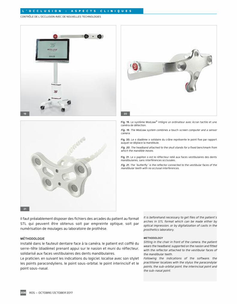
Il faut préalablement disposer des fichiers des arcades du patient au formatSTL qui peuvent être obtenus soit par empreinte optique, soit parnumérisation de moulages au laboratoire de prothèse.
MÉTHODOLOGIEInstallé dans le fauteuil dentaire face à la caméra, le patient est coiffé duserre-tête (diadème) prenant appui sur le nasion et muni du réflecteur,solidarisé aux faces vestibulaires des dents mandibulaires.Le praticien, en suivant les indications du logiciel, localise avec son styletles points paracondyliens, le point sous-orbital, le point interincisif et lepoint sous-nasal.
it is beforehand necessary to get files of the patient’sarches in STl format which can be made either byoptical impression, or by digitalization of casts in theprosthetics laboratory.
METHODOLOGYSitting in the chair in front of the camera, the patientwears the headband, supported on the nasion and fittedwith the reflector attached to the vestibular faces ofthe mandibular teeth. Following the indications of the software, thepractitioner localizes with the stylus the paracondylarpoints, the sub-orbital point, the interincisal point andthe sub-nasal point.
ROS – OCTOBRE/OCTOBER 2017208
CONTRÔLE DE L’OCCLUSION AVEC DE NOUVELLES TECHNOLOGIES
19
Fig. 19. Le système ModJaw® intègre un ordinateur avec écran tactile et unecaméra de détection.
Fig. 19. The ModJaw system combines a touch-screen computer and a sensorcamera.
Fig. 20. Le « diadème » solidaire du crâne représente le point fixe par rapportauquel se déplace la mandibule.
Fig. 20. The headband attached to the skull stands for a fixed benchmark fromwhich the mandible moves.
Fig. 21. Le « papillon » est le réflecteur relié aux faces vestibulaires des dentsmandibulaires, sans interférences occlusales.
Fig. 21. The “butterfly” is the reflector connected to the vestibular faces of themandibular teeth with no occlusal interferences.
20
21
L ’ O C C L U S I O N : A S P E C T S C L I N I Q U E S
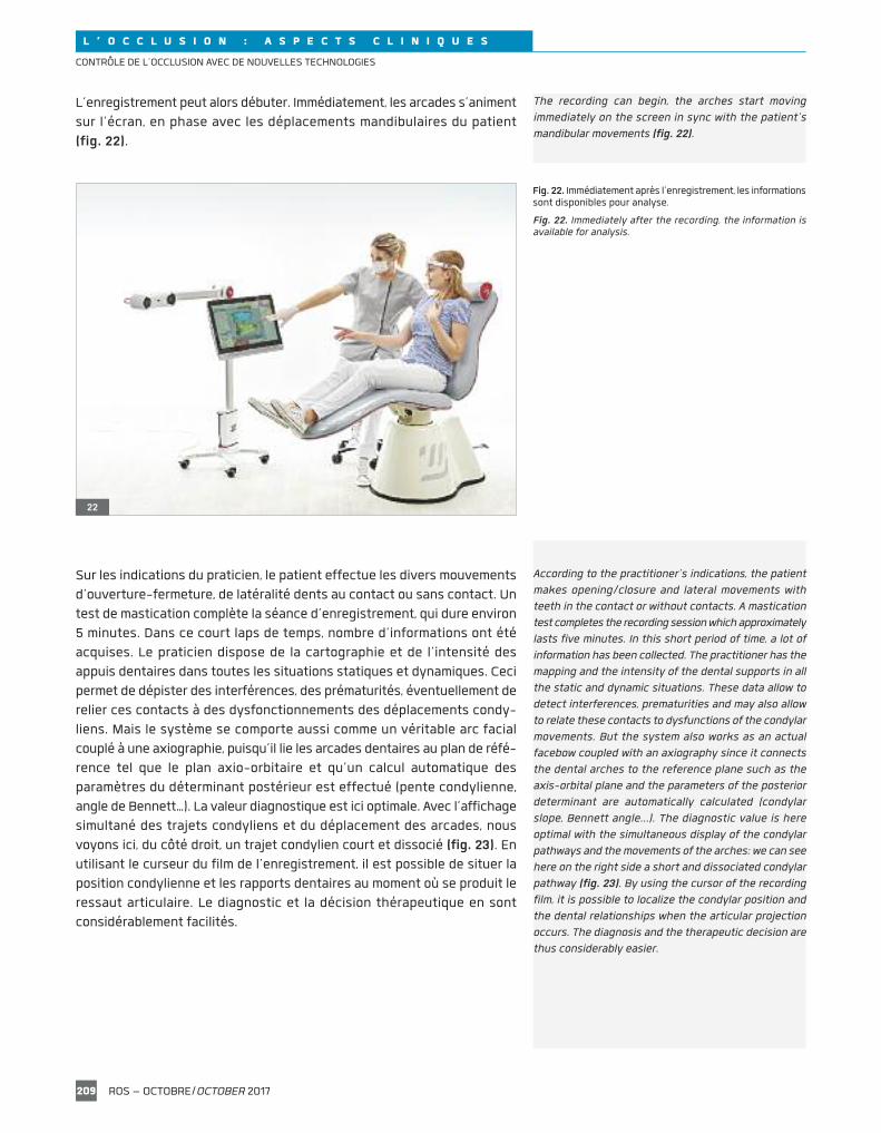
L’enregistrement peut alors débuter. Immédiatement, les arcades s’animentsur l’écran, en phase avec les déplacements mandibulaires du patient(fig. 22).
Sur les indications du praticien, le patient effectue les divers mouvementsd’ouverture-fermeture, de latéralité dents au contact ou sans contact. Untest de mastication complète la séance d’enregistrement, qui dure environ5 minutes. Dans ce court laps de temps, nombre d’informations ont étéacquises. Le praticien dispose de la cartographie et de l’intensité desappuis dentaires dans toutes les situations statiques et dynamiques. Cecipermet de dépister des interférences, des prématurités, éventuellement derelier ces contacts à des dysfonctionnements des déplacements condy-liens. Mais le système se comporte aussi comme un véritable arc facialcouplé à une axiographie, puisqu’il lie les arcades dentaires au plan de réfé-rence tel que le plan axio-orbitaire et qu’un calcul automatique desparamètres du déterminant postérieur est effectué (pente condylienne,angle de Bennett…). La valeur diagnostique est ici optimale. Avec l’affichagesimultané des trajets condyliens et du déplacement des arcades, nousvoyons ici, du côté droit, un trajet condylien court et dissocié (fig. 23). Enutilisant le curseur du film de l’enregistrement, il est possible de situer laposition condylienne et les rapports dentaires au moment où se produit leressaut articulaire. Le diagnostic et la décision thérapeutique en sontconsidérablement facilités.
The recording can begin, the arches start movingimmediately on the screen in sync with the patient’smandibular movements (fig. 22).
according to the practitioner’s indications, the patientmakes opening/closure and lateral movements withteeth in the contact or without contacts. a masticationtest completes the recording session which approximatelylasts five minutes. in this short period of time, a lot ofinformation has been collected. The practitioner has themapping and the intensity of the dental supports in allthe static and dynamic situations. These data allow todetect interferences, prematurities and may also allowto relate these contacts to dysfunctions of the condylarmovements. But the system also works as an actualfacebow coupled with an axiography since it connectsthe dental arches to the reference plane such as theaxis-orbital plane and the parameters of the posteriordeterminant are automatically calculated (condylarslope, Bennett angle...). The diagnostic value is hereoptimal with the simultaneous display of the condylarpathways and the movements of the arches: we can seehere on the right side a short and dissociated condylarpathway (fig. 23). By using the cursor of the recordingfilm, it is possible to localize the condylar position andthe dental relationships when the articular projectionoccurs. The diagnosis and the therapeutic decision arethus considerably easier.
ROS – OCTOBRE/OCTOBER 2017209
CONTRÔLE DE L’OCCLUSION AVEC DE NOUVELLES TECHNOLOGIES
22
Fig. 22. Immédiatement après l’enregistrement, les informationssont disponibles pour analyse.
Fig. 22. immediately after the recording, the information isavailable for analysis.
L ’ O C C L U S I O N : A S P E C T S C L I N I Q U E S
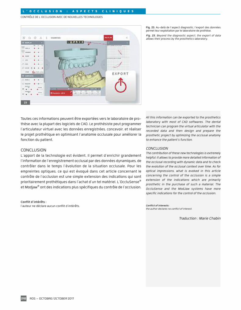
Toutes ces informations peuvent être exportées vers le laboratoire de pro-thèse avec la plupart des logiciels de CAO. Le prothésiste peut programmerl’articulateur virtuel avec les données enregistrées, concevoir, et réaliserle projet prothétique en optimisant l’anatomie occlusale pour améliorer lafonction du patient.
CONCLUSIONL’apport de la technologie est évident. Il permet d’enrichir grandementl’information de l’enregistrement occlusal par des données dynamiques, decontrôler dans le temps l’évolution de la situation occlusale. Pour lesempreintes optiques, ce qui est évoqué dans cet article concernant lecontrôle de l’occlusion est une simple extension des indications qui sontprioritairement prothétiques dans l’achat d’un tel matériel. L’OccluSense®
et Modjaw® ont des indications plus spécifiques du contrôle de l’occlusion.
Conflit d’intérêts :l’auteur ne déclare aucun conflit d’intérêts.
all this information can be exported to the prostheticslaboratory with most of CaD softwares. The dentaltechnician can program the virtual articulator with therecorded data and then design and prepare theprosthetic project by optimizing the occlusal anatomyto enhance the patient’s function.
COnCluSiOnThe contribution of these new technologies is extremelyhelpful: it allows to provide more detailed information ofthe occlusal recording with dynamic data and to checkthe evolution of the occlusal context over time. as foroptical impressions, what is evoked in this articleconcerning the control of the occlusion is a simpleextension of the indications which are primarilyprosthetic in the purchase of such a material. TheOccluSense and the ModJaw systems have morespecific indications for the control of the occlusion.
Conflict of interests: the author declares no conflict of interest.
Traduction : Marie Chabin
ROS – OCTOBRE/OCTOBER 2017210
CONTRÔLE DE L’OCCLUSION AVEC DE NOUVELLES TECHNOLOGIES
23
Fig. 23. Au-delà de l’aspect diagnostic, l’export des donnéespermet leur exploitation par le laboratoire de prothèse.
Fig. 23. Beyond the diagnostic aspect, the export of dataallows their process by the prosthetics laboratory.