henrik paavilainen – inhibition of herpes simplex virus
TRANSCRIPT

TURUN YLIOPISTON JULKAISUJA – ANNALES UNIVERSITATIS TURKUENSISSarja - ser. D osa - tom. 1276 | Medica - Odontologica | Turku 2017
Henrik Paavilainen
INHIBITION OF HERPES SIMPLEX VIRUS INFECTION WITH RNA INTERFERENCE

Supervised by
Professor Veijo Hukkanen, MD, PhDDepartment of VirologyUniversity of TurkuTurku, Finland
Reviewed by
Professor Igor Jurak, PhDDepartment of BiotechnologyUniversity of RijekaRijeka, Croatia
Docent Peter Sarin, PhDDepartment of BiosciencesUniversity of HelsinkiHelsinki, Finland
Opponent
Professor Krystyna Bieńkowska-Szewczyk, PhDDepartment of Molecular Biology of VirusesUniversity of GdanskGdansk, Poland
The originality of this thesis has been checked in accordance with the University of Turku quality assurance system using the Turnitin OriginalityCheck service.
ISBN 978-951-29-6765-0 (PRINT)ISBN 978-951-29-6766-7 (PDF)ISSN 0355-9483 (Print)ISSN 2343-3213 (Online)Painosalama Oy - Turku, Finland 2017
University of Turku
Faculty of MedicineDepartment of VirologyandDrug Research Doctoral Programme (DRDP)FinPharma Doctoral Program Drug Discovery Section (FPDP-D)

To my family!
“…herpes is to say a vocation-…”
P G Unna 1883

Abstract
ABSTRACT
HENRIK PAAVILAINEN Inhibition of herpes simplex virus infection with RNA interference
University of Turku, Faculty of Medicine, Department of Virology, Drug Re-search Doctoral Programme (DRDP)
Annales Universitatis Turkuensis Turku, Finland, 2017
Herpes simplex virus (HSV) is a common pathogen. Approximately half of the human population carries the virus. Of clinical symptoms, the most common one is a blister on the lip. This manifestation is often at the initial site of infection. However, in primary infection, before epithelial eradication of HSV by the im-mune system, the virus infects sensory neurons. In these neurons the virus hides and forms latency. From this latency, the virus can reactivate and travel via axons back to the epithelium to form a new lytic infection. In addition, the virus can upon reactivation travel to the eye and cause HSV keratitis. HSV is the leading cause of blindness due to infectious origin in the developed world. In addition, antiviral resistant HSV strains are relatively abundant in immunocompromised patients and in eye infections of HSV. Thus, there is need for novel drugs against HSV.
In this study, the goal was to develop a new drug against HSV, especially for the treatment of HSV keratitis. RNA interference, based on enzymatically produced and cleaved antiviral small interfering RNA (siRNA) pools (swarms of siRNAs) were studied. The drug development of this therapy started from in silico analysis of the target sequences in the viral genome. From there, drugs produced via vari-ous methods were studied. Swarms targeting different parts of the viral genome were studied for their innate immunity induction profile and antiviral efficacy. Clinical field isolates of HSV were used in addition to laboratory strains. For an in vivo keratitis model, a single siRNA swarm was selected. This swarm, target-ing HSV gene UL29 showed broad effectiveness in vitro and in vivo and had a limited innate immunity induction profile, being the best candidate for further development.
This study shows, that an siRNA swarm approach against herpes simplex virus infection is feasible.
Keywords: herpes simplex virus, antiviral, RNA interference, innate immunity, drug development

Tiivistelmä
TIIVISTELMÄ
HENRIK PAAVILAINEN Herpes simplex -virusinfektion esto RNA-parvella
Turun yliopisto, Lääketieteellinen tiedekunta, Virusoppi, Lääketutkimuksen toh-toriohjelma (DRDP)
Annales Universitatis Turkuensis Turku, Suomi, 2017
Herpes simplex -virus (HSV; yskänrokkovirus) on yleinen ihmisen taudinaiheut-taja. Noin puolet ihmisistä on viruksen kantajia maassamme. Kaikille ei kuiten-kaan koskaan tule näkyviä, kliinisiä, oireita. Yleisin oire on huuliherpes. Tällöin virus ja immuunijärjestelmä kilpailevat toisiaan vastaan ja normaalitilanteessa immuunijärjestelmä voittaa. Mutta virus ei ole hävinnyt elimistöltä, vaan se on piiloutunut. HSV infektoi hermosoluja, joiden tumaan virus kulkeutuu ja muo-dostaa niissä piilevän, latentin infektion. Virus voi aktivoitua uudelleen piilos-taan, kulkeutua takaisin epiteelille ja aiheuttaa uuden solutuhoa aiheuttavan in-fektion. Suun alueen epiteelin sijaan HSV voi kulkeutua myös toisaalle, esimer-kiksi silmään ja aiheuttaa HSV-keratiitin. Tällainen silmäinfektio on suhteellisen yleinen ja HSV-keratiitti onkin yleisin infektioperäisen sokeuden aiheuttaja län-simaissa. Hoitoa hankaloittaa se, että nimenomaan HSV-silmäinfektiossa lääke-resistentit viruskannat ovat yleisiä. On siis tarvetta uusille lääkehoidoille.
Tässä tutkimuksessa kehitettiin uutta lääkettä herpesinfektiota vastaan. Erityisenä kysymyksenä oli silmäinfektion hoitoon tähtäävän lääkkeen kehitys. Tutkitut lääkkeet perustuivat RNA-häirinnässä käytettäviin, HSV:n geeneihin kohdistu-viin, RNA-parviin. Nämä pieniä häiritseviä RNA molekyylejä sisältävät parvet valmistettiin entsymaattisesti. Lääkekehitysprosessi alkoi sopivien kohteiden seu-lomisella virusgenomin sekvenssitietoihin perustuen. Eri valmistusmenetelmillä tehtyjä parvia tutkittiin niiden antiviraalisen tehon sekä luonnollisen immunitee-tin vaikutusten osalta. In vitro -kokeiden perusteella löysimme RNA-parven, joka oli tehokas sekä laboratorio- että kliinisiä HSV-kantoja vastaan. HSV-geeniin UL29 kohdistuva RNA-parvi aiheutti vain lieviä luonnollisen immuniteetin vas-teita ja osoittautui tehokkaaksi myös in vivo HSV-keratiittimallissa.
Tämä lääkekehitysprojekti osoittaa, että herpesinfektion estoon suunniteltu RNA-parvi on potentiaalinen lääkekehityssuunta HSV-infektioita vastaan.
Avainsanat: herpes simplex -virus, viruslääke, RNA-häirintä, luonnollinen im-muniteetti, lääkekehitys

Table of contents
TABLE OF CONTENTS
ABSTRACT ........................................................................................................... 4
TIIVISTELMÄ ....................................................................................................... 5
ABBREVIATIONS ................................................................................................ 8
LIST OF ORIGINAL PUBLICATIONS ............................................................. 10
1 INTRODUCTION ....................................................................................... 11
2 REVIEW OF LITERATURE ...................................................................... 12
2.1 Herpesviruses ...................................................................................... 12 2.2 Herpes simplex virus (HSV) and us ................................................... 13
2.2.1 HSV prevalence ....................................................................... 13 2.2.2 HSV diseases ........................................................................... 14 2.2.3 Antiviral medication against HSV .......................................... 17
2.3 The structure of herpes simplex virus ................................................. 18 2.3.1 HSV virion ............................................................................... 18 2.3.2 Genome of HSV and its role in drug development ................. 19
2.4 Infection cycle ..................................................................................... 21 2.4.1 Lytic infection ......................................................................... 22 2.4.2 Latent infection ........................................................................ 23
2.5 HSV and the immune system ............................................................. 23 2.5.1 Innate immunity against HSV ................................................. 24 2.5.2 Adaptive immunity responses to HSV .................................... 25
2.6 HSV infection models ......................................................................... 25 2.6.1 In vitro infection ...................................................................... 25 2.6.2 In vivo infection models .......................................................... 26
2.7 HSV as a tool ...................................................................................... 27 2.7.1 Gene therapy applications of HSV and their impact on antiviral
drug development .................................................................... 27 2.8 Drug development against HSV ......................................................... 29 2.9 RNA interference ................................................................................ 29
2.9.1 RNA interference in drug development .................................. 31 2.9.2 Use of RNAi against herpes simplex virus ............................. 33
3 AIMS OF THE STUDY .............................................................................. 34
4 MATERIALS AND METHODS ................................................................ 35
4.1 Quantitative PCR (I-IV) ..................................................................... 35 4.1.1 RNA extraction and subsequent reverse transcription ............ 35 4.1.2 Standards for qPCR runs ......................................................... 35 4.1.3 qPCR primers .......................................................................... 36

Table of contents
4.2 Viruses (I-IV) ...................................................................................... 38 4.2.1 Clinical isolates ........................................................................ 39 4.2.2 Plaque titration ......................................................................... 39
4.3 Cell lines (I-III and IV) ....................................................................... 39 4.4 Small interfering RNA products (I-IV) ............................................... 39
4.4.1 RNA products used .................................................................. 40 4.4.2 In vitro siRNA delivery ........................................................... 42
4.5 Corneal infection model (IV) .............................................................. 43 4.5.1 Anesthesia, infection and treatment ......................................... 43 4.5.2 Follow-up and sampling .......................................................... 44
4.6 Statistical analyses (I-IV) .................................................................... 45 4.7 Microscopy .......................................................................................... 45
5 RESULTS .................................................................................................... 46
5.1 Effects of RNAs on cells (I, II) ........................................................... 46 5.2 HSV infection inhibition in vitro with siRNA swarms (I-III) ............ 50
5.2.1 Comparison of siRNAs of different lengths ............................ 50 5.2.2 Comparison of the anti-HSV-siRNA swarms .......................... 51
5.3 Corneal infection treatment with enzymatically created siRNA swarms (IV) ......................................................................................... 55 5.3.1 Encephalitis model ................................................................... 55 5.3.2 Peripheral infection model ....................................................... 56
6 DISCUSSION .............................................................................................. 58
6.1 Cells treated with siRNA swarms (I-II) .............................................. 58 6.1.1 Interplay of treatment and infection (II-III) ............................. 60
6.2 Efficacy of the antiviral swarm in vitro (I-III) .................................... 62 6.3 In vivo efficacy of the swarms against HSV (IV) ............................... 63
7 CONCLUSIONS .......................................................................................... 65
ACKNOWLEDGEMENTS .................................................................................. 66
REFERENCES ..................................................................................................... 68
ORIGINAL PUBLICATIONS ............................................................................. 87

Abbreviations
ABBREVIATIONS
ACV acyclovir BAC bacterial artificial chromosome bp base pair cDNA complementary DNA CMV cytomegalovirus CNS central nervous system DNA double stranded deoxyribonucleic acid dpi days post infection EBV Epstein-Barr virus EMA European Medicines Agency FBS fetal bovine serum FDA U.S. Food and Drug Administration GAPDH Glyceraldehyde 3-phosphate dehydrogenase gC / D / E / I … glycoprotein C / D / E / I … GD Giardia intestinalis Dicer HSCT Hematopoietic stem cell transplantation hpi hours post infection hpt hours post transfection HSV herpes simplex virus HSV-1 herpes simplex virus type 1 HSV-2 herpes simplex virus type 2 ICP infected cell protein IFN interferon IgG immunoglobulin G ip intraperitoneally ISG interferon stimulated gene LAT latency associated transcript MHC Major histocompatibility complex miRNA microRNA mRNA messenger RNA NK natural killer (cell)

Abbreviations
nm nanometer nt nucleotide PBS phosphate-buffered saline PCR polymerase chain reaction PFU plaque forming unit qPCR real-time quantitative PCR RCF relative centrifugal force RISC RNA-induced silencing complex (ds)RNA (double stranded) ribonucleic acid RNAi RNA interference sc subcutaneously SEM standard error of mean siRNA small interfering RNA TG trigeminal ganglia tk / TK thymidine kinase gene / protein TLR3 toll-like receptor 3 UL unique sequence long segment (part of HSV genome) UL27 Unique sequence long segment gene 27 UL29 Unique sequence long segment gene 29 UL54 Unique sequence long segment gene 54 UL27 RNA swarm targeting UL27 UL29 RNA swarm targeting UL29 UL54 RNA swarm targeting UL54 US unique sequence short segment (part of HSV genome)
List of original publications
LIST OF ORIGINAL PUBLICATIONS
This thesis is based on the following original publications, which are referred to in the text by the Roman numerals (I-IV), and on data presented in this thesis.
I Romanovskaya A*, Paavilainen H*, Nygårdas M, Bamford DH, Hukka-nen V and Poranen MM. 2012. Enzymatically Produced Pools of Canoni-cal and Dicer-Substrate siRNA Molecules Display Comparable Gene Si-lencing and Antiviral Activities against Herpes Simplex Virus. PLoS ONE 7(11): e51019. *Equal contribution.
II Paavilainen H, Romanovskaya A, Nygårdas M, Bamford DH, Poranen MM, and Hukkanen V. 2015. Innate responses to small interfering RNA pools inhibiting herpes simplex virus infection in astrocytoid and epitheli-al cells. Innate Immunity 21(4) 349–357.
III Paavilainen H, Lehtinen J, Romanovskaya A, Nygårdas M, Bamford DH, Poranen MM, Hukkanen V. 2016. Inhibition of clinical pathogenic herpes simplex virus 1 strains with enzymatically created siRNA pools. Journal of Medical Virology 88(12):2196-2205.
IV Paavilainen H, Lehtinen J, Romanovskaya A, Nygårdas M, Bamford DH, Poranen MM, and Hukkanen V. 2017. Topical treatment of herpes sim-plex virus infection with enzymatically created siRNA swarm. Antiviral Therapy, In press.
The original publications have been reprinted with the permission of the copy-right holders.

Introduction 11
1 INTRODUCTION
Herpes simplex virus (HSV) causes diseases in humans. The most common symptom is labial herpes - the most common form of HSV infections of the skin and mucosa. HSV also causes corneal infections and, rarely, HSV encephalitis or meningitis. Approximately every second human is a carrier of HSV, but of them, only a portion gets symptoms. Upon initial infection HSV hides in neurons, caus-ing latency. From this latency, recurrent infection can happen even without symptoms. Antiviral drugs are available against HSV but there is need for im-provement, especially as there are drug resistant HSV strains.
Drug development approaches against HSV are broad: from small interfering RNA swarms to monoclonal antibodies to vaccines. The use of small molecule drugs against HSV has not resulted in widespread prevalence of drug resistant HSV strains. However, immunocompromised patients have higher incidence of drug resistant HSV and some current medical treatments result in attenuation of the immune system. Hence, as this type of immunosuppressant treatments are on the increase, it would be likely that more drug resistant HSV cases are going to emerge. Moreover, the drug resistant strains are frequent in HSV keratitis, which is the most common form of HSV eye disease.
RNA interference is a promising approach against infectious diseases. The target of the small interfering (si)RNA can be one or several viral genes. Especially with viruses, who hijack the host cells and rely on the host cell translational ma-chinery for replication, there are less available drug development targets in com-parison with bacteria which have a host organism-independent cellular machin-ery. However, one drawback of single site siRNAs is the risk of drug resistance. A single mutation in the viral genome can render the siRNA ineffective. To cir-cumvent this problem, we have developed enzymatically created siRNA swarms that have a target area of hundreds of nucleotides. These swarms cover a large portion of their target gene. In comparison, the target area is approximately 20 nucleotides for canonical siRNAs.
This thesis focuses on the drug development of an antiviral siRNA swarm against HSV. The long term goal is the treatment of HSV keratitis.

12 Review of literature
2 REVIEW OF LITERATURE
Viruses are organisms that have a DNA or RNA genome. The can only replicate in living cells using the cellular synthetic machinery to form new particles that have the viral genome. These new particles are then to be transferred to new cells to produce further progeny.
It is easy to answer, whether or not virus is part of the realm of the living; it is. Without life there is no viral activity. But it appears to be more of a philosophical questions whether or not a virus is alive. A virus is fully dependent on its host for replication. Virus is a parasite and is inanimate on its own - the virus particles are not alive outside of the cell
2.1 Herpesviruses
Herpesviruses constitute a family of evolutionary ancient viruses whose origin can be traced back to Pangaean times, when there was only one continent on earth [Grose, 2012]. Members of human herpesviruses, herpes simplex virus (HSV), Epstein-Barr virus (EBV) and cytomegalovirus (CMV), have been found from isolated tribes in South America [Black, 1975], suggesting that these virus-es have been there already before the arrival of Europeans. The global distribu-tion of herpesviral genetic clades is in line with the human origins from Africa [Grose, 2012; Hayward, 1999]. For HSV, which is the focus of this thesis, the evolution can be traced for millions of years [Norberg et al., 2011].
Over 200 different herpesviruses belong to the virus family herpesviridae. The host range among viruses is wide, with only a few being capable of infecting more than one host organism [Pellett and Roizman, 2013]. Different herpesvirus-es share the genome type and have a similar virion structure. In the world of vi-ruses, the herpesvirus genome is large, consisting of approximately 152 kb of double stranded DNA. Herpesviruses encode virus-specific enzymes involved in the metabolism of their DNA. Herpesviruses can form lytic and latent infections. In the case of lytic infection, the host cell dies of the infection, with the tendency of producing a high amount of progeny viruses before cell death. As for latent infection, the dormant virus can reactivate to cause a new lytic infection to spread to new hosts. Herpes is a friend for life, as the latent state can last over a lifetime of the host [Forghani et al., 1977], and sometimes even further, as reac-tivating virus can be isolated from cadavers [Bastian et al., 1972; Ouwendijk et al., 2012].

Review of literature 13
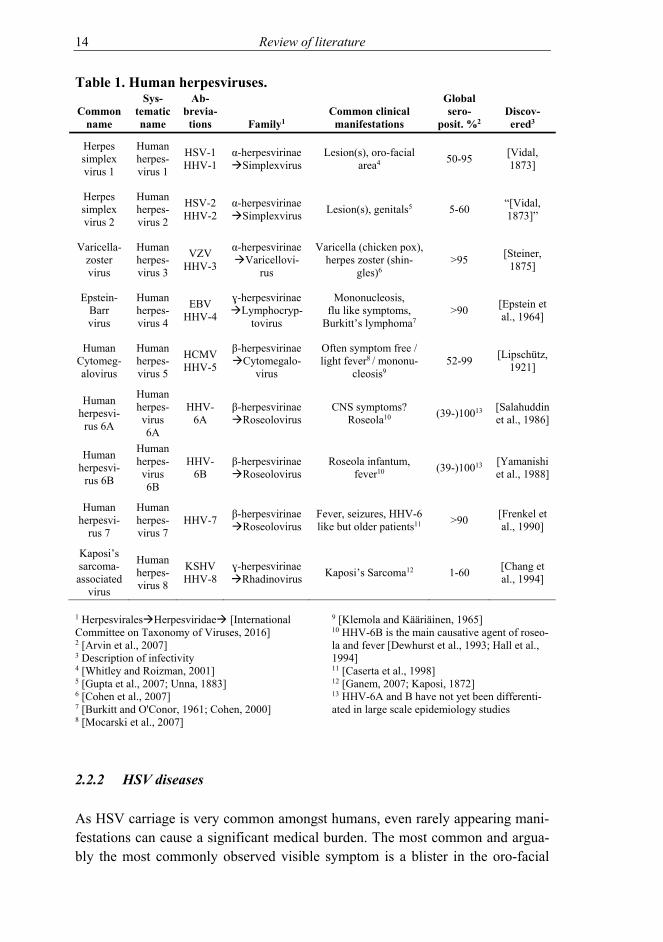
The herpesviruses are highly adapted to their hosts as both have evolved side by side. Herpesviruses are widely spread, common and rarely cause serious disease in an immunocompetent host. Keeping the host live, well and capable of infect-ing subsequent hosts is a good survival plan for a virus. However, there are ex-ceptions, and partly due to the sheer magnitude of hosts infected, serious illness-es do occur. The human herpesviruses and the most typical illnesses they cause are presented in Table 1.
2.2 Herpes simplex virus (HSV) and us
The first reports of HSV and indeed, the name of the virus, can be traced back to ancient Greeks [Roizman and Whitley, 2001]. Back then, the word “herpes” most likely described many various causative agents of similar disorders. In the 1920’s HSV was characterized as an infectious agent in animal studies – in humans, au-toinoculation and transmission of HSV was studied and understood already in the 19th century [Flexner and Amoss, 1925; Goodpasture and Teague, 1923a; Roizman and Whitley, 2001; Unna, 1883; Vidal, 1873].
2.2.1 HSV prevalence
Herpes simplex virus is a common human pathogen with approximately half to two thirds of the human population as carriers of the virus [Looker et al., 2015a; Pebody et al., 2004]. However, seroprevalence is decreasing [Bradley et al., 2014]. At any given moment, HSV DNA shedding can be detected from oral scrapings generally in 2-5% of the world population [Roizman et al., 2013]. For example in Finland, a steep drop of HSV seroprevalence from 69.5% to 45% has happened in merely twenty years in pregnant women according to a recent study [Puhakka et al., 2016]. However, in a similar study cohort, over 2% of adults in Finland have HSV DNA positive oral scrapings at a given moment [Mäki et al., 2015]. There are two types of HSV: HSV-1 and HSV-2. HSV-1, which is the main causative agent of oro-labial herpes, and HSV-2, which is considered as the causative agent of genital herpes with an estimated global prevalence of over 11% [Looker et al., 2015b]. HSV took a non-human primate detour 6 million years ago to return as human HSV-2 some 1.6 million years ago whereas HSV-1 has evolved alongside human evolution [Wertheim et al., 2014]. For genital HSV infections, it seems that HSV-1 is becoming more prevalent as the cause of geni-tal herpes in young women [Löwhagen et al., 2000; Roberts et al., 2003; Tuokko et al., 2014].
14 Review of literature
Table 1. Human herpesviruses.
Common name
Sys-tematic name
Ab-brevia-tions Family1
Common clinical manifestations
Global sero-
posit. %2 Discov-ered3
Herpes simplex virus 1
Human herpes-virus 1
HSV-1 HHV-1
α-herpesvirinae Simplexvirus
Lesion(s), oro-facial area4 50-95 [Vidal,
1873]
Herpes simplex virus 2
Human herpes-virus 2
HSV-2 HHV-2
α-herpesvirinae Simplexvirus Lesion(s), genitals5 5-60 “[Vidal,
1873]”
Varicella-zoster virus
Human herpes-virus 3
VZV HHV-3
α-herpesvirinae Varicellovi-
rus
Varicella (chicken pox), herpes zoster (shin-
gles)6 >95 [Steiner,
1875]
Epstein-Barr virus
Human herpes-virus 4
EBV HHV-4
ɣ-herpesvirinae Lymphocryp-
tovirus
Mononucleosis, flu like symptoms,
Burkitt’s lymphoma7 >90 [Epstein et
al., 1964]
Human Cytomeg-alovirus
Human herpes-virus 5
HCMV HHV-5
β-herpesvirinae Cytomegalo-
virus
Often symptom free / light fever8 / mononu-
cleosis9 52-99 [Lipschütz,
1921]
Human herpesvi-
rus 6A
Human herpes-virus 6A
HHV-6A
β-herpesvirinae Roseolovirus
CNS symptoms? Roseola10 (39-)10013 [Salahuddin
et al., 1986]
Human herpesvi-
rus 6B
Human herpes-virus 6B
HHV-6B
β-herpesvirinae Roseolovirus
Roseola infantum, fever10 (39-)10013 [Yamanishi
et al., 1988]
Human herpesvi-
rus 7
Human herpes-virus 7
HHV-7 β-herpesvirinae Roseolovirus
Fever, seizures, HHV-6 like but older patients11 >90 [Frenkel et
al., 1990]
Kaposi’s sarcoma-associated
virus
Human herpes-virus 8
KSHV HHV-8
ɣ-herpesvirinae Rhadinovirus Kaposi’s Sarcoma12 1-60 [Chang et
al., 1994]
1 HerpesviralesHerpesviridae [International Committee on Taxonomy of Viruses, 2016] 2 [Arvin et al., 2007] 3 Description of infectivity 4 [Whitley and Roizman, 2001] 5 [Gupta et al., 2007; Unna, 1883] 6 [Cohen et al., 2007] 7 [Burkitt and O'Conor, 1961; Cohen, 2000] 8 [Mocarski et al., 2007]
9 [Klemola and Kääriäinen, 1965] 10 HHV-6B is the main causative agent of roseo-la and fever [Dewhurst et al., 1993; Hall et al., 1994] 11 [Caserta et al., 1998] 12 [Ganem, 2007; Kaposi, 1872] 13 HHV-6A and B have not yet been differenti-ated in large scale epidemiology studies
2.2.2 HSV diseases
As HSV carriage is very common amongst humans, even rarely appearing mani-festations can cause a significant medical burden. The most common and argua-bly the most commonly observed visible symptom is a blister in the oro-facial

Review of literature 15
area. Even though not necessarily dangerous, a visible blister can be a nuisance both physically and socially. HSV can travel through neurons [Goodpasture and Teague, 1923b] and is capable of forming latency in these neurons (please see below Latency chapter 2.4.2) where it hides from the immune system. From this latency HSV can reactivate and cause a new lytic infection – a lifelong cycle. These reactivations can lead to clinical or subclinical infection, usually near the initial site of infection and can thus transmit the virus to a new susceptible host(s) [Roizman et al., 2013].
However, sometimes this back and forth track is not followed and an alternative site is challenged with a lytic infection. One possibility is that HSV continues onwards to the central nervous system (CNS) where it can cause severe, poten-tially fatal HSV encephalitis [Steiner, 2011; Whitley and Roizman, 2001]. In en-cephalitis, lytic viral infection and immune responses both can lead to dire con-sequences. Moreover, enteric nervous system may be involved as well, as the virus might spread and cause digestive tract damage and peristaltic stop, which has been shown to be the cause of lethality following CNS infection in certain animal models [Khoury-Hanold et al., 2016]. Encephalitis is usually caused by HSV-1 whereas HSV-2 is the more likely causative agent in HSV meningitis. Even though rare, HSV is the most common sporadic cause of encephalitis [Bradshaw and Venkatesan, 2016; Välimaa et al., 2013; Whitley and Roizman, 2001; Whitley et al., 1998].
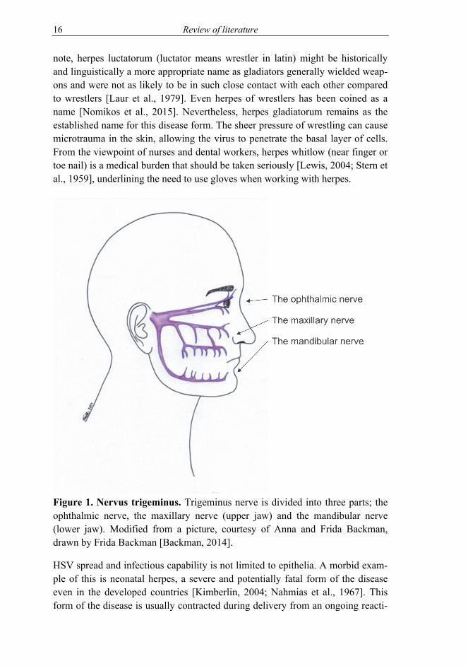
Another side track HSV can take is the one to the eye. The eye is a somewhat immunoprivileged organ with a constant tear rinsing and a tight protective outer layer. Even though this barrier can be breached, HSV can reach this organ from behind as well, through the nerves [Labetoulle et al., 2000; Labetoulle et al., 2003], as trigeminal ganglia innervate both the eye and the skin of the face [Kuo et al., 2014] (Figure 1). In addition to local damage to the eye, HSV infection can result in changes in the trigeminal fibres connected to the area [Rousseau et al., 2015]. The estimated amount of herpes keratitis is in the hundreds of thou-sands in developed countries (prevalence of approximately 20 out of 100 000 persons/year) and over million in developing countries per annum [Farooq and Shukla, 2012; Labetoulle et al., 2005]. The infection can be persistent and recur-rent and HSV is the leading cause of blindness due to infectious origin [Roizman et al., 2013], even with the current medication available in the developed coun-tries.
Hippocrates used the term herpes as lesions that appeared to “creep along the skin” [Roizman and Whitley, 2001] – HSV can cause infections anywhere in the skin or mucosal tissue. A good example of this, still tying to the ancient times, is herpes gladiatorum [Selling and Kibrick, 1964; Wheeler and Cabaniss, 1965]. Of
16 Review of literature
note, herpes luctatorum (luctator means wrestler in latin) might be historically and linguistically a more appropriate name as gladiators generally wielded weap-ons and were not as likely to be in such close contact with each other compared to wrestlers [Laur et al., 1979]. Even herpes of wrestlers has been coined as a name [Nomikos et al., 2015]. Nevertheless, herpes gladiatorum remains as the established name for this disease form. The sheer pressure of wrestling can cause microtrauma in the skin, allowing the virus to penetrate the basal layer of cells. From the viewpoint of nurses and dental workers, herpes whitlow (near finger or toe nail) is a medical burden that should be taken seriously [Lewis, 2004; Stern et al., 1959], underlining the need to use gloves when working with herpes.
Figure 1. Nervus trigeminus. Trigeminus nerve is divided into three parts; the ophthalmic nerve, the maxillary nerve (upper jaw) and the mandibular nerve (lower jaw). Modified from a picture, courtesy of Anna and Frida Backman, drawn by Frida Backman [Backman, 2014].
HSV spread and infectious capability is not limited to epithelia. A morbid exam-ple of this is neonatal herpes, a severe and potentially fatal form of the disease even in the developed countries [Kimberlin, 2004; Nahmias et al., 1967]. This form of the disease is usually contracted during delivery from an ongoing reacti-

Review of literature 17
vation of the mother’s genital herpes and especially in the case of primary infec-tion [Brown et al., 2003]. The infection can become systemic and this can be fa-tal, even with medication. As the amount of HSV-1 genital infections is on the rise [Tuokko et al., 2014], it is worrying that the systemic infections of the new-born are more likely due to HSV-1 than HSV-2 [Brown et al., 2007; Välimaa et al., 2013].
As an alphaherpesvirus, HSV has a wide variety of host cells. It is in a way an ectodermotopic virus, preferring the epithelium and nervous system, which both originate from the ectoderm. However, HSV can infect many cell types within the body. This facilitates in vitro laboratory studies of the virus, where infection and viral replication are desired.
2.2.3 Antiviral medication against HSV
There is currently no herpes simplex vaccine available despite the substantial investments by the pharmaceutical industry. A vaccine candidate in Phase III clinical trial failed recently [Belshe et al., 2012]. This HSV-2 vaccine had some efficacy against HSV-1 genital disease but the efficacy against HSV-2 genital herpes was a disappointment and the vaccine did not reach the market. The scien-tific community did not stop there, and the amount of HSV vaccine research in-creased by a third in the following years as measured by amount of publications. There is, fortunately, antiviral medication available against HSV. Acyclovir (ACV), a nucleoside analogue, is a selective drug against HSV [Männistö and Tuominen, 2012; Schaeffer et al., 1978]. To become active, ACV needs HSV gene thymidine kinase (the gene product of UL23/tk), and thus the effect is very selective. There are derivatives of ACV, ie. valacyclovir, famciclovir, ganciclo-vir, which have some effect against other herpesviruses as well. Despite usage around the globe for decades, the resistance prevalence increase is not a highly alarming issue. Prophylactic usage of nucleoside analogues on patients with a risk of contracting the virus has yielded favorable results, but unfortunately not full protection [Anderson et al., 2016]. ACV is a well-tolerated drug, but there is a very low risk that ACV can result in side effects in patients with underlying renal disease; a state called Cotard’s syndrome [Lindén and Helldén, 2013]. In addition, prophylactic usage of ACV against HSV cannot entirely prevent viral reactivation [Johnston et al., 2012] and prophylactic usage can promote the emergence of ACV resistant strains [Duan et al., 2009; van Velzen et al., 2013].
As HSV tk is a non-essential gene, HSV can replicate without a functional thy-midine kinase [Coen and Schaffer, 1980]. A virus negative for tk is, however, attenuated, and this can manifest in reduction of spread and replication and reac-

18 Review of literature
tivation capabilities as well as temperature sensitivity [Coen et al., 1989; Darby et al., 1981; Shimada et al., 2007]. Rarely, HSV DNA polymerase mutation can also lead to ACV resistance in addition to tk mutants [Burrel et al., 2013; Chibo et al., 2004; Coen et al., 1985; Larder et al., 1987; Suzutani et al., 2003]. As these strains are often attenuated their detection might be underestimated. ACV re-sistant HSV usually emerges in immunopriviledged sites such as in the eye, or in immunocompromised patients [Frobert et al., 2014; Stránská et al., 2005]. When an ACV resistant disease emerges, the drug options are few. Foscarnet is availa-ble [Männistö and Tuominen, 2012], but the drug is not well tolerated, and over half of the treated patient’s HSVs generate resistance to it [Danve-Szatanek et al., 2004]. Moreover, TK can remain (somewhat) functional but at the same time drug resistant [Darby et al., 1981]. Even with all the available antivirals, HSV remains a significant medical burden and there is a need for treatment modalities with differing mechanisms of action.
2.3 The structure of herpes simplex virus
2.3.1 HSV virion
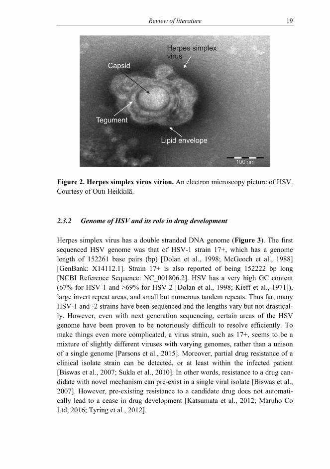
A description of an HSV virion in an electron micrograph is a sunny side up fried egg (Figure 2). The 3D shape is roughly a sphere, but a flexible one. The size of a virion is 225 nanometers (nm) with glycoprotein spikes protruding from the lipid envelope and 186 nm without [Grünewald et al., 2003].
The structure is from center outwards: the genome, capsid, tegument, and lipid envelope (Figure 2). The outer layer of the virion is the lipid envelope harboring viral glycoproteins. Underneath lays the tegument. Its structure is not rigid and it harbors numerous HSV proteins, including virion host shutoff protein, which upon release to the host cells starts to degrade host messenger RNAs (mRNAs) [Kwong et al., 1988; Read and Frenkel, 1983]. HSV capsid is an icosahedral pro-tein structure [Schrag et al., 1989]. Its size is one-third of the lipid enveloped volume [Grünewald et al., 2003]. The capsid shelters the viral DNA, which is released to the nucleus of the cell once the capsid has been transported there via microtubules [Marozin et al., 2004; Ojala et al., 2000]. Viral DNA is held in the core of the virion inside the capsid. Here the DNA is held in a toroid form [Furlong et al., 1972; Kieff et al., 1971] and the negative charge of the DNA is balanced by the presence of polyamins, spermidine and spermine [Gibson and Roizman, 1971]. The DNA is in linear form within the capsid, but upon entry to nucleus, the DNA takes a circular episomal form and associates with histones [Garber et al., 1993; Kent et al., 2004].
Review of literature 19
Figure 2. Herpes simplex virus virion. An electron microscopy picture of HSV. Courtesy of Outi Heikkilä.
2.3.2 Genome of HSV and its role in drug development
Herpes simplex virus has a double stranded DNA genome (Figure 3). The first sequenced HSV genome was that of HSV-1 strain 17+, which has a genome length of 152261 base pairs (bp) [Dolan et al., 1998; McGeoch et al., 1988] [GenBank: X14112.1]. Strain 17+ is also reported of being 152222 bp long [NCBI Reference Sequence: NC_001806.2]. HSV has a very high GC content (67% for HSV-1 and >69% for HSV-2 [Dolan et al., 1998; Kieff et al., 1971]), large invert repeat areas, and small but numerous tandem repeats. Thus far, many HSV-1 and -2 strains have been sequenced and the lengths vary but not drastical-ly. However, even with next generation sequencing, certain areas of the HSV genome have been proven to be notoriously difficult to resolve efficiently. To make things even more complicated, a virus strain, such as 17+, seems to be a mixture of slightly different viruses with varying genomes, rather than a unison of a single genome [Parsons et al., 2015]. Moreover, partial drug resistance of a clinical isolate strain can be detected, or at least within the infected patient [Biswas et al., 2007; Sukla et al., 2010]. In other words, resistance to a drug can-didate with novel mechanism can pre-exist in a single viral isolate [Biswas et al., 2007]. However, pre-existing resistance to a candidate drug does not automati-cally lead to a cease in drug development [Katsumata et al., 2012; Maruho Co Ltd, 2016; Tyring et al., 2012].

20 Review of literature
Figure 3. Schematic representation of the HSV genome. UL = unique se-quence long segment, US = unique sequence short segment, B/B’ = repeat and its inversion flanking UL, C/C’ repeat and its inversion flanking US, a/a’ = short few hundred base pair repeat.
Thus far, at least 90 transcriptional units have been found from HSV genome and of these at least 84 encode proteins (www.viprbrc.org/). The genes are tran-scribed in opposite directions and can be overlapping. Latency associated tran-script, LAT, a long non-coding RNA with an initial length of approximately 9 kb and with the subsequent shorter stable 1.5 and 2.0 kb LAT introns and micro (mi)RNAs all have a role in maintenance of HSV latency (and please see 2.4.2) [Cliffe et al., 2009; Stevens et al., 1987; Umbach et al., 2008]. HSV miRNAs are expressed during lytic infection cycle as well [Jurak et al., 2010]. No known pro-teins are expressed from LAT region in latency, even though there are a handful of short open reading frames that can be expressed during lytic infection [Lagunoff and Roizman, 1994].
In a lytic infection, HSV genes are expressed in a cascade fashion with α genes first, followed by β and then ɣ genes [Honess and Roizman, 1974]. In a latent infection, these genes are silenced. Upon reactivation the gene expression is not regulated in expression cascade at first as all gene classes are expressed simulta-neously and then followed by a more classical expression cascade [Camarena et al., 2010; Du et al., 2011; Mattila et al., 2015]. Some of HSV genes are not im-portant/necessary for its replication in cells in vitro, however, a loss of gene func-tion usually results in attenuation in virulence in a host organism. Noteworthy is the ɣ34.5 gene known as the neurovirulence gene [Bolovan et al., 1994; Chou et al., 1990], coding for multifunctionary infected cell protein (ICP)34.5 [Ackermann et al., 1986; Alexander et al., 2007; He et al., 1997], as its deletion leads to a virus that does not harm the CNS but can still selectively (ie. cancer) be a lytic virus [Andreansky et al., 1998; Andtbacka et al., 2015; Broberg et al., 2001; Markert et al., 2000; Nygårdas et al., 2013].

Review of literature 21
2.4 Infection cycle
There are two major phases in herpes simplex virus life cycle; the lytic and latent infection. In lytic infection, the virus replicates and produces new virions with the result of lysis of the infected cell. Hallmark of viral spreading in vitro is plaque formation (Figure 4), based on lateral spread of the virus with the help of viral glycoproteins [Dingwell et al., 1994].
Figure 4. HSV plaque formation. The developing HSV-1 plaque is indicated by an arrow in the panels representing 30 and 42 hours post infection (hpi). The black bar depicts the width of the plaque. All pictures are to scale and the scale bar is shown in the 30 hpi panel. Vero cells were infected at a low MOI and the developing HSV-1 plaque was analyzed at 30, 42, 54 and 66 hpi.
An in vivo clinical example of the spread is a blister. When this infection is ongo-ing, the virus is detectable by the immune system (please see section 2.5). Thus, if an immune system is present and active, the spread at lytic infection site is halted. The relatively quick inhibition of the viral spread is an important safety feature for the virus, as it is advantageous for the virus to keep the host alive and capable of infecting new individuals. To start a new virus production cycle, there are two possibilities, infect a new host or hide from the immune system. HSV is a master of stealth, and in latent infection the virus cannot be eradicated by the

22 Review of literature
immune system and from this quiescent form, the virus can reactivate to cause a new lytic infection.
2.4.1 Lytic infection
The chain of events triggered by lytic infection is the reason for symptoms, which occur during HSV infection. Thus, this phase of the infection is a natural target for drug design, but there are, however, some approaches trying to eradi-cate latent virus from the host as well (as pointed out in Table 2, 2.8). The virus enters the cell through fusion of lipid envelopes of HSV and the host cell [Morgan et al., 1968]. An alternative pathway involving endocytosis exists as well [Nicola et al., 2003]. HSV glycoproteins play an indispensable role in entry pathways and this complex and variable setting, with both the virus and the host cells playing their parts, is still unfolding, as reviewed by [Campadelli-Fiume et al., 2012; Heldwein and Krummenacher, 2008]. Upon entry, the contents of the viral tegument are released into the cell. Elements from the tegument are accom-panied by HSV infection-related exosome-delivered factors [Kalamvoki et al., 2014]. These factors have an effect on the antiviral responses by the cell and on the expression of the viral genes later on in the infection. The viral DNA, encap-sulated in the capsid, is transported to the nucleus by microtubules [Kristensson et al., 1986] with dynein where the linear DNA is released [Döhner et al., 2002; Ojala et al., 2000; Sodeik et al., 1997]. The DNA takes a circular form [Strang and Stow, 2005; Su et al., 2002], and provided that the host cell and viral factors are suitably present, starts to express viral genes, resulting in a complete take-over of the cell for viral production.
As mentioned above, the viral genes are expressed in a cascade. In a simplifica-tion, α gene expression is required for β gene expression and β gene expression is required for ɣ gene expression. Already two hours post infection (hpi), α genes are expressed, whereas β gene expression begins 4 to 8 hpi. After viral DNA syn-thesis has commenced, the ɣ genes start to be expressed. Finally all the parts of a new virion are produced, and the egress of new infective viruses can commence. When infection is elaborated more closely, HSV genes are “leaky” for the differ-ent gene classes [Powell et al., 1975]. In addition, the α genes are defined by their ability to be expressed in the presence of cycloheximide (ie. without de no-vo protein synthesis), and the β genes are not expressed prior to α gene expres-sion after cycloheximide removal [Honess and Roizman, 1974]. In the presence of phosphonoacetic acid, α and β genes are readily expressed whereas ɣ genes are not [Honess and Watson, 1977]. Moreover, reactivation from a latent infection to productive infection is proceeded with non-cascade type of gene expression [Camarena et al., 2010; Mattila et al., 2015].

Review of literature 23
2.4.2 Latent infection
In latent infection, the virus hides from its host. Upon infecting latently a new host cell, the viral genome is repressed. This repression could also be viewed as a natural state of the virus. Two neuronal populations are found upon acute infec-tion; one in which viral genes are expressed and another, where lytic infection associated-genes are repressed [Margolis et al., 1992]. While in hiding, only a part of the viral genome is active, the latency associated RNA, LAT, is expressed [Stevens et al., 1987; Wagner et al., 1988]. In addition to LAT and its splice vari-ants, also miRNA are expressed from the latency region of the virus [Jurak et al., 2010; Umbach et al., 2008]. The viral DNA stays in the nucleus as a non-integrated episome. The DNA is bound to histones with the LAT area to acety-lated [Kubat et al., 2004] and lytic genes to dimethylated histones [Wang et al., 2005]. This results in the activity of the LAT area and the inactivity of the lytic genes. Latency forms in sensory neurons in which the capsid is transported via retrograde transport into the soma of the neuron, where the viral DNA stays (non-integrated) in a latent state in the nucleus. The requirement for definition of genuine latency is that the virus can reactivate from this state. Upon reactivation the repressing locks open and the anterograde transport takes the infection to, or near to, the original infection site. There can be deviations from this, as in rare cases the virus travels the wrong way to the CNS. In addition, infection can man-ifest at another peripheral site than the initial site of infection – for example from a primary lip infection, a recurrent infection in the eye can ensue upon viral reac-tivation.
The complexity of the latency-reactivation cycle and equilibrium of immune sys-tem and the virus is baffling. A good example of the equilibrium is that an adapted T cell population is waiting for HSV to reactivate at the previous site(s) of infection in vivo [Gebhardt et al., 2009]. Despite this alertness, HSV can form a new lytic cycle.
Understanding latency is a daunting task, but could indeed open up new possible targets for treatment of HSV infection. However, for example, cutting the (latent) genome with various methods has not thus far led to desired results [van Diemen et al., 2016], but not without some preliminary success as well [Aubert et al., 2014].
2.5 HSV and the immune system
Immune system is crucial for human survival. It consists of many barriers that a pathogen must overcome. Parts of it we are born with (the innate immunity), and

24 Review of literature
parts adapt to pathogens (the adaptive immunity). These two parts of immunity are integrated to one another. The first barriers of the immune system are physi-cal, such as the skin and the low pH of the stomach. On a tissue level, the im-mune cells can autonomously detect pathogens and, additionally, adapt to them. Both pathogens and immune system adapt to counteract the other’s countermeas-ures. On a subcellular level, antibodies and the complement system play an inte-gral part in the detection and destruction of pathogens. Some cells are solely ded-icated to immune responses, but cells in general have a possibility to react to pathogens. Interferons help the (infected) cell to counteract viral infections, alert neighboring cells and call for help from other cell types.
Many immune system processes such as complement system, natural killer (NK) cells and T- and B-cell responses all try to eradicate HSV. Functioning immune system can clear the lytic infection and thus it will not spread too much in the host. However, latent HSV remains even with the efforts of a functional immune system.
Even though immune evasion is important for the virus survival, in a way HSV relies on many innate immunity systems to slow it down enough not to cause too much havoc on the host. Even viruses where the neurovirulence gene has been deleted remain virulent in nude mice, with partly deficient immune system [Lasner et al., 1998]. Toll-like receptor 3 (TLR3), part of the innate immunity pathogen pattern associated recognition, for one, is important in the HSV en-cephalitis prevention [Zhang et al., 2007]. NK cells can locate the viruses so that adaptive immunity can step in line, but not before the virus has had an opportuni-ty in the production of new virions. Immunocompetent hosts rarely get serious infections.
2.5.1 Innate immunity against HSV
The first and perhaps the best barrier against HSV infection is the intact skin. Other factors, such saliva, also have antiviral effect on HSV [Välimaa et al., 2002]. To replicate, HSV needs access to the basal layer cells. Upon infection (entry to the host cell), the host cell responses are shut off. Host gene expression is inhibited, cellular proteins are degraded and programmed cell death is blocked, to a point. Complement alternative (and classical) pathway activation is inhibited by HSV glycoprotein C (gC) blocking C3b component of complement system [Fries et al., 1986; Kostavasili et al., 1997]. On the other hand, however, antibod-ies can target gC [Adamiak et al., 2010]. Major histocompatibility complex (MHC) class I presentation of virus is inhibited to hide from T-cell mediated re-sponses (see below) but this inhibition leads to susceptibility to NK cell respons-

Review of literature 25
es. NK response is not that effectively inhibited by HSV. However, (at least) HSV-2 has the ability to deceive NK cells’ function with secreted glycoprotein G-2 [Bellner et al., 2005]. The importance of innate immunity against HSV is underlined by the fact that TLR3 deficiency subjects children to encephalitis [Zhang et al., 2007].
2.5.2 Adaptive immunity responses to HSV
Adaptive immunity is blocked in multiple ways by HSV. Antigen presentation is inhibited by ICP47. ICP47 prevents peptide loading to MHC and prevents their transport for CD8+ T cell presentation [Früh et al., 1995; Hill et al., 1995; York et al., 1994]. HSV can prevent T cell mediated cell death by other ways as well, but these mechanisms are not as clearly elucidated. Antibody-mediated adaptive immunity is blocked by gE and gI that form a Fc receptor that, through binding to Fc part of immunoglobulin G (IgG), blocks antibody-dependent antiviral re-sponses [Johnson et al., 1988]. In addition, gC and gE block HSV recognition via antibodies by sheltering other viral glycoproteins [Hook et al., 2008]. An indirect evidence of HSV’s capability to evade adaptive immunity is the fact that there is no vaccine available. This is the case despite the investments in and devotion to creating one by the industry and academic research.
2.6 HSV infection models
It has been nearly hundred years since the beginning of experimental HSV infec-tion models as reviewed by [Roizman and Whitley, 2001]. Even though HSV is a human virus, it can, through inoculation, infect various animals and animal cells from mouse to monkey through zebrafish to chicken, to name a few [Burgos et al., 2008; Flexner and Amoss, 1925; Hukkanen et al., 2002; Hunter et al., 1999; Scott et al., 1953; Wertheim et al., 2014]. A wide range of hosts offer different models in which the virus can be studied. On the other hand, this creates a neces-sity of choosing the most suitable model for each experimental need.
2.6.1 In vitro infection
An in vitro setting is the method of choice for viral production. For HSV, many factors such as temperature can have an effect on viral production [Hoggan and Roizman, 1959] and spread [Hoggan et al., 1960], different cells produce differ-ent amounts of virus [Kaplan, 1957] and different viral stocks have different

26 Review of literature
spread and replication properties [Roizman and Roane, 1961]. Moreover, cell lines can lack the capability to produce interferons, and for that matter be more suitable for viral (stock) production [Desmyter et al., 1968]. More recently, ad-vanced experimental settings can model more complex infections than lytic in-fections, such as latency [Camarena et al., 2010; Hafezi et al., 2012; Mattila et al., 2015; Wilcox and Johnson, 1987]. Thus the knowledge and/or the explora-tion, of the in vitro system used, is very important.
In the context of drug development, a good lead in in vitro phase is important. There is not, however, a need for hundred percent inhibition of viral replication to be a promising lead. In addition, broad spectrum antiviral molecule might have cell type specific activity [Denisova et al., 2012]. As discussed above, acyclovir is a good drug against HSV. Nevertheless, its in vitro efficacy in viral inhibition (decrease in viral replication) was 2-4 logs compared to untreated cells [Rosenwirth et al., 1987] and combination treatment with interferon (IFN)-α in-creased the inhibition of viral replication by ACV from 2 to over 3 logs [Taylor et al., 1989]. This seeming discrepancy with these results merely demonstrates the difference between experimental settings and the subsequent findings, not the reduced activity of the drug. Another factor, which needs to be taken into ac-count, is the possible interaction of a drug with cellular defenses, such as apopto-sis, which can lead to destruction of infected cells. If a drug is working in such a way, but it is intended against cancerous cells, not infected cells, the results can be potentially very harmful for the host as cell destruction can happen at an un-desired location [Kakkola et al., 2013].
2.6.2 In vivo infection models
In comparison with in vitro models, in vivo models face even more complica-tions; each factor contributes to overall pathogenesis of the virus. The strain and production method of the virus, as well as the strain, age and sex of the animal are just a few factors contributing to the infection course [Lopez, 1975]. For ex-ample the Swiss Jim Lambert (SJL) mice are more susceptible to latent infection through the intranasal infection route rather than the more commonly used ocular route [Nygårdas, 2013]. In addition to animal strain, also the age of the animal has an effect on the susceptibility to HSV as older mice can tolerate much higher viral doses than younger ones as the pups do not develop an NK cell response [Zawatzky et al., 1982]. Mouse is perhaps the most used animal for HSV infec-tion. Major drawbacks of the mouse as a model animal for HSV infection is the lack of (detectable) spontaneous reactivation and that the HSV immunoevasion factor ICP47 is not active in mice. Reactivation can be induced for example with

Review of literature 27
another infection [Stevens et al., 1975], heat shock [Sawtell and Thompson, 1992] and with severe organ stress, such as death of host followed by explant culture of ganglia [Baringer, 1976; Stevens and Cook, 1971]. Mouse does, how-ever, offer an extensively studied and robust setting for HSV infections. Corneal HSV infections are studied mostly in rabbits, or C57BL/6 and BALB/c mice [Hill et al., 2012; Nygårdas, 2013]. In comparison to mouse models, spontaneous reactivation happens in rabbits after corneal infection and in guinea pigs after genital inoculation [Wagner and Bloom, 1997]. For genital herpes models, guin-ea pig is often used [Da Costa et al., 1997], but mouse [Palliser et al., 2006] and rat models are used as well [Boukhvalova et al., 2015; Yim et al., 2005]. Non-human primates are used as well, albeit more rarely, for HSV related studies, but they do, however, have their role in stepping from pre-clinical to clinical research and different animal models are used to meet and represent various experimental settings [Meignier et al., 1987; Meignier et al., 1988; Meignier et al., 1990; Patel et al., 2016; Roth et al., 2014]. Newest member of host species in HSV infection studies in vivo is the tree shrew, with the interesting distinction of ICP0 transcript expression during latency [Li et al., 2016]. Should this or similar expression be true in humans as well, there might be great deal of impact on antiviral develop-ment against latent HSV. For lytic HSV infections, however, the current models offer a good and wide selection for drug development.
2.7 HSV as a tool
2.7.1 Gene therapy applications of HSV and their impact on antiviral drug development
In the gene therapy and virotherapy field, HSV has played a key role. Most nota-bly, replication competent HSV was “the first FDA-approved oncolytic virus therapy” [FDA, 2015] (U.S. Food and Drug Administration, FDA) and European Medicines Agency (EMA): “Imlygic is a first-in-class advanced therapy medici-nal product (ATMP) derived from a virus” [EMA, 2015]. Deliberate attenuation of HSV, and addition of therapy genes, has led to first oncolytic virus drug on the western market [Andtbacka et al., 2015; Duodecim-drug-database, 2016; Hukkanen and Vihinen, 2016]. In addition to cancer, replication competent HSV vectors are in development against autoimmune disease as well [Broberg et al., 2001; Nygårdas et al., 2011; Nygårdas et al., 2013].
Manipulation of HSV genome has been done for over 30 years [Post et al., 1981]. The original tandem transfection method is still used, with the bacterial artificial

28 Review of literature
chromosome (BAC) en passant technique as a current alternative [Brunnemann et al., 2016; Nagel, 2006; Nygårdas et al., 2013; Tischer et al., 2006]. With the BAC technique, there is no need to work with a live virus while modifying the genome. The genome modifications are based on homologous recombination. HSV genome (Figure 3) with its repeat areas forms homologous recombination within itself in a eukaryotic cell. In the gene therapy drug development field, this natural and laboratory-induced recombination can be exploited and the methods designed so that there is virtually no risk of increasing virulence mutations. In addition, the natural drug sensitivity gene is present. However, the modifications to the tk gene in the gene therapy applications would be in some cases desirable [Manservigi et al., 2010; Wilson et al., 1999]. Due to (current) lack of other suit-able drugs, the drug sensitivity gene deletion is not a favorable choice in vector development. In addition, a situation where one needs to medicate against HSV and let HSV-based therapy vector function at the same time is quite likely faced in the future. Therefore, there is a need for HSV drugs with varying methods of action.
Parts of HSV have been exploited for other gene therapy applications, namely the tk gene. An obvious choice has been the incorporation of the tk gene to a heterol-ogous oncolytic viral vector and, once the tumor cells are infected, a tk-based drug is given to the patient with the result of the infected cells dying [Sangro et al., 2010; Stedt et al., 2013; Su et al., 1996]. This short term exposure to tk-based drug is unlikely to significantly influence serious emergence of drug-resistant herpes strains [Mitterreiter et al., 2016].
Another gene therapy approach, with more relevance to anti-HSV drug develop-ment, is the use of tk-positive lymphocytes in (haplo-identical hematopoietic) stem-cell transplantation (HSCT). The idea is to reserve a safety switch for graft-versus-host disease as the given modified-to-be-tk-positive-cells can be destroyed with drug (ie. ganciclovir) treatment in the case of adverse effects [Ciceri et al., 2009; Hashimoto et al., 2015a]. Unfortunately, these patients are susceptible for herpes infections; giving the tk-based drug against manifestation of HSV would also trigger the off-switch of the HSCT treatment. Moreover, HSCT patients are at greatest risk of tk-resistant HSV prevalence [Piret and Boivin, 2011]. This leads to the problematic phase where treatment of viral infection might be unsuc-cessful due to resistance but at the same time the safety switch is triggered un-necessarily. In addition, tk-positive lymphocyte-treated patients that need to be treated with tk-based drug, can alter the remaining tk-positive lymphocytes [Hashimoto et al., 2015b]. Furthermore, tk-based drug prophylaxis is considered a standard procedure for herpes(simplex virus) seropositive individuals [Tomblyn et al., 2009], and this again can result in the emergence of new re-sistant viruses [Duan et al., 2009; van Velzen et al., 2013].
Review of literature 29
All in all, there are numerous reasons closely knitted to current and future gene therapy applications, why drugs with new mechanisms of action are needed against HSV.
2.8 Drug development against HSV
As a virus that has spread ubiquitously around the globe in the human popula-tion, HSV has remained as an attractive target for drug development (Table 2). Big pharma, amongst others, has put a lot of effort in HSV vaccine development. An HSV-2 vaccine [Belshe et al., 2012], was found to be slightly active against HSV-1 in its phase III trial. Alas, the result was disappointing as it did not have an effect on HSV-2 genital herpes. Genital herpes infection control is important, since in addition to HSV medical burden, genital HSV infection increases the risk of contraction of HIV [Freeman et al., 2006; Wald and Link, 2002].
As discussed previously, there is a good drug against HSV, acyclovir and its de-rivatives, which all base their effect on the tk-gene. However, tk, being an im-portant yet dispensable gene, allows for escape mutants to emerge [Duan et al., 2009; Piret and Boivin, 2011]. Even slightly active thymidine kinase allows for reactivation of the virus [Pan and Coen, 2012] and thus a new recurrent infection to which new drugs are needed. There are various approaches to combat HSV infection and a set of these approaches is listed in Table 2.
2.9 RNA interference
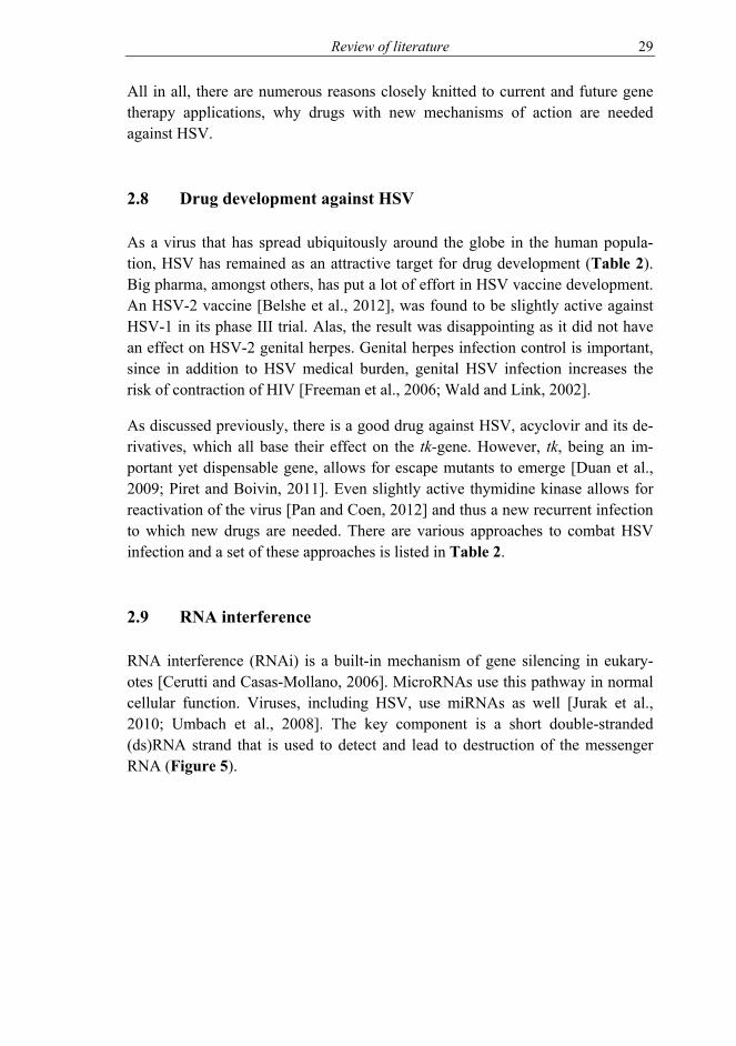
RNA interference (RNAi) is a built-in mechanism of gene silencing in eukary-otes [Cerutti and Casas-Mollano, 2006]. MicroRNAs use this pathway in normal cellular function. Viruses, including HSV, use miRNAs as well [Jurak et al., 2010; Umbach et al., 2008]. The key component is a short double-stranded (ds)RNA strand that is used to detect and lead to destruction of the messenger RNA (Figure 5).

30 Review of literature
Table 2. Examples of drug development approaches against herpes simplex virus.
Targeted virus Mechanism Targeted
disease phase Research status Reference
HSV Enzymatically created siRNA
swarm Lytic In vivo This thesis
HSV-1 Chemically synthe-sized siRNA Lytic In vivo
[da Silva et al., 2016; Li et al., 2014; Palliser et al.,
2006]
HSV-1 Monoclonal anti-body Lytic In vivo [Krawczyk et al., 2015]
HSV-1 Homing me-gaendonuclease Latent (lytic) In vitro (in vi-
vo?) [Aubert et al., 2014; Grosse et al., 2011]
HSV-1 Adenovirus mediat-ed shRNA Lytic In vitro [Song et al., 2016]
HSV Crispr/CAS9 Lytic In vitro [van Diemen et al., 2016]
HSV Small molecule (microbicide)
Lytic / prophylaxis In vitro [Chamoun-Emanuelli et
al., 2014]
HSV-2 HSV-1 (amplicon) shRNA vector
Lytic, recur-rence In vivo [Liu et al., 2013]
HSV-2 Replication defec-tive HSV-2
Prophylactic, lytic, latent –
vaccine Phase I [Bernard et al., 2015; Da
Costa et al., 2000]
HSV-2 Attenuated HSV-1 (ICP0 mutant) Vaccine In vivo / Phase
0? [Halford et al., 2010]
HSV-2 DNA Vaccine Phase I [Dutton et al., 2013]
HSV-2 (-1) Subunit (gD) Vaccine Phase III, dis-continued [Belshe et al., 2012]
HSV-2 ∆gD HSV-2 Vaccine In vivo [Petro et al., 2016]
To create these short RNAs, commonly referred to as small interfering RNAs (siRNA), miRNAs are loaded into Dicer. Dicer is an enzyme that cleaves the long dsRNA to siRNA [Bernstein et al., 2001]. Length of the siRNA depends on the Dicer. Human Dicer cleaves 21-23 bp long siRNAs [Provost et al., 2002], while for example Giardia intestinalis Dicer cleaves 25-27 bp long siRNAs [Macrae et al., 2006]. These longer siRNAs are considered Dicer-substrate siR-NAs, as they are loaded to (human) Dicer upon entering cells of human origin. After siRNA is available, it enters the RNA-induced silencing complex (RISC) [Hammond et al., 2000]. There the two strands are separated and the guide strand is associated with Argonaute 2 protein. The Argonaute 2 mediates sequence-specific cleavage of the complementary mRNA [Rand et al., 2004].
Review of literature 31
Figure 5. RNA-interference in a human cell. Endogenous or exogenous RNA molecules are cleaved by Dicer (Dicer-substrate RNA) and then loaded to RISC complex via which sequence specifically mRNA translation is inhibited. Short siRNA molecules can bypass Dicer loading. RISC = RNA-induced silencing complex, siRNA = short interfering RNA, miRNA = microRNA, mRNA = mes-senger RNA.
2.9.1 RNA interference in drug development
Exogenous siRNAs can trigger the RNAi pathway as well [Elbashir et al., 2001]. There are multiple approaches in drug development field to combat various dis-eases with an siRNA approach, ranging from viral infections to cancers to age-related macular degeneration to name a few from currently ongoing or finished clinical trials [ClinicalTrials.gov, 2016]. In preclinical phase the choices are even more numerous.
As a biological drug, the manufacturing of siRNAs is relatively simple. Chemical synthesis can be used of a chosen sequence and such molecules can be ordered from commercial vendors. Another approach is to enzymatically create a swarm

32 Review of literature
of siRNAs, siRNA pools [Aalto et al., 2007; Donzé and Picard, 2002; Myers et al., 2003; Nygårdas et al., 2009; Romanovskaya et al., 2013; Yang et al., 2002]. A part of a gene is chosen as a template and from this, hundreds of base pairs long target area, a long dsRNA molecule is synthesized. Depending on Dicer used to cleave siRNAs from the long dsRNA, the final swarm of siRNAs can be of varying lengths. When aiming to treat viral diseases with siRNAs, especially with siRNA products targeting single site of the target gene, the risk of emer-gence of resistance is a problem [Geisbert et al., 2006; Gitlin et al., 2005; McDonagh et al., 2015; Shah et al., 2012; Wilson and Richardson, 2005]. When the target area covers hundreds of base pairs, the risk of resistance mutants is un-likely to occur quickly. In the pools, the amount of single siRNA molecules with identical sequence is low, and thus off-target effects due to siRNA sequence are very low. Unintended effects due to exogenous RNA can be also due to the in-troduction of foreign dsRNA into the cell. The length of the dsRNA is an im-portant factor in the response, and, to point, generally the longer the dsRNA is, the stronger the toxic effect is. For example, 88 bp long dsRNA is considered toxic, and even more toxic than hundreds to over thousand bp long dsRNAs [Jiang et al., 2011]. It induces lethal cellular responses, by being detected by cel-lular sensors of dsRNA. For shorter siRNAs (<30 bp), the increase of size does not necessarily lead to intensifying cellular responses. In some cases, for chemi-cally synthesized siRNAs, the increase in length of the siRNA adds to toxicity [Reynolds et al., 2006]. However, the sequence itself can have an effect on the non-target specific responses [Fedorov et al., 2006]. In human cells, the differ-ence between a shorter and longer siRNA is the route to RISC complex; longer siRNA first goes through Dicer before being loaded to RISC whereas shorter siRNAs are directly loaded to RISC [Kim et al., 2005] (Figure 5). This factor could contribute to the different induction of innate immunity in various size RNA swarms.
The delivery of siRNA drugs is often the main question in clinical development, as the target tissue can be hard to reach. The siRNAs are also somewhat delicate and large molecules, compared to canonical small molecule drugs, and thus per os administration is unlikely to render good bioavailability for siRNAs. Many approaches are under development to help with the homing of the RNAi based drugs including protein and other linker conjugations, lipid envelopment, deliv-ery vectors such as viruses, transfection, nanoparticles, and of course plain naked siRNA [da Silva et al., 2016; Kanasty et al., 2013; Kari et al., 2007; Kim et al., 2016]. No blockbuster RNAi based drugs have made it to the market, but there has been an antiviral antisense oligonucleotide drug approved by the FDA [Crooke, 1998], paving the way for new RNAi biologicals in the future.

Review of literature 33
2.9.2 Use of RNAi against herpes simplex virus
There are a few efforts pursuing to concept of HSV infection inhibition via chemically synthesized small interfering RNAs [Duan et al., 2012; Jin et al., 2014; Palliser et al., 2006; Silva et al., 2014]. RNA interference is a promising antiviral approach against HSV. The mechanism of action differs clearly from current available treatments. Most common manifestation of the disease is at the periphery. When planning the delivery of siRNA, a biological drug with a rela-tively large molecular mass compared to small molecule drugs, skin, eye, muco-sal membranes are all sites that are easier to reach than internal organs. A simple administration of naked siRNA is possible and the coupling with reagents that help the siRNAs to cross the lipid envelope of the cell is possible.
There are obstacles to overcome in the development of anti-HSV siRNA drugs. Nevertheless, HSV infection treatment is feasible with siRNAs and there is a burning need to treat the billions infected and to treat with novel drugs the hun-dreds of thousands to millions with antiviral resistant HSV.

34 Aims of the study
3 AIMS OF THE STUDY
Herpes simplex virus (HSV) causes a medical burden. In addition, novel drugs are needed to tackle drug resistance of the virus. The aim of this work was to in-troduce a novel approach against HSV infection.
Our antiviral approach was RNA interference using swarm of enzymatically cre-ated small interfering RNAs. Even though most epithelial HSV infections could be potentially treated with the new drug, the chosen treatment target was the HSV infection of the eye – a well established model of HSV keratitis, a disease form which has a high prevalence of drug resistant cases.
The specific aims of study were:
I Design and test for the proof of principle of the biological drug;
II Explore the innate immunity effects of the drug and HSV infection;
III Test the feasibility of the antiviral against clinical isolates of HSV;
IV Study of suitability of the biological drug in a corneal in vivo infection model.

Materials and methods 35
4 MATERIALS AND METHODS
4.1 Quantitative PCR (I-IV)
To measure innate immunity responses, viral gene expression and DNA load, real-time quantitative polymerase chain reaction (qPCR) was used. Messenger RNA (mRNA) was first extracted and then converted into complementary DNA (cDNA). The efficacy was measured with a house-keeping gene glyceraldehyde 3-phosphate dehydrogenase (GAPDH), which was additionally used for normali-zation. The subsequent analysis of mRNA quantities was done from the sample. As for viral DNA amount analysis, the DNA from the sample was extracted. Please see 4.1.1 and 4.5.2 for the RNA and DNA extraction protocols, respec-tively. To measure the copy numbers of analyzed genes, a real-time instrument Rotorgene (Qiagen, Hilden, Germany) was used. A standard curve was supplied for every run.
4.1.1 RNA extraction and subsequent reverse transcription
RNA extraction was done from cell cultures. The medium was removed and the cells were lysed in TRI Reagent (Molecular Research, Cincinnati, OH) and sub-sequently the total RNA was isolated as instructed by the manufacturer. The RNA was treated with DNase (ThermoFisher Scientic, Waltham, MA, USA) to rid the samples from possible viral DNA contaminants. For cDNA synthesis, random hexamer primers were used (ThermoFisher Scientic) with RevertAid H Minus Reverse Transcriptase enzyme (ThermoFisher Scientific). The cycle was 60’ +42°C, 10’ 70°C, hold +4°C. A total of 2% of the original cell sample was used in each qPCR run. The GAPDH count in such a run had to be over 1000 for the extraction and cDNA synthesis to be considered successful.
4.1.2 Standards for qPCR runs
All qPCR runs were performed with a six to eight step (log) dilutions of the cor-responding standard, which was either a larger PCR product, a plasmid or viral DNA. Viral DNA was used as a standard when viral DNA amount was meas-ured. For mRNA analysis a PCR product served as a standard for all but one test (α-TIF), where a plasmid served as a standard template. For the primers used to create standards and the standards used, please see Table 3.

36 Materials and methods
Table 3. Standards and primers used to create standards for qPCR analyses.
Gene Primers used (5’-3’, upper forward, lower re-verse) to create, or template used as, standard Gene id or equivalent
US1 AAG CCC AAA TGC AAT GCT AC CAG ACA CTT GCG GTC TTC TG
NC_001806.2 (132101..133963)
UL54 GTG CCC CCA GAA CCA ATC CGG CAA AAG TGC GAT AGA G
NC_001806.2 (113735..115283)
UL29 GGT GCG GTC AAA AAT AAG GA CCT ACC AGA AGC CCG ACA AG
NC_001806.2 (58410..62054, complement)
UL27 CAC TTG GTC ATG GTG CAG AC CAC CAC CGA CCT CAA GTA CA
NC_001806.2 (53059..55795, complement)
α-TIF / VP16 / UL48 α-TIF plasmid pRB3717 [McKnight et al., 1987] BamHI fragment F;
GU734771.1
gD for HSV-1 DNA isolated from HSV-1 strain 17+ NC_001806.2
gD for HSV-2 DNA isolated from HSV-2 strain H1224 Not available
IFN-α TGG CTG TGA AGA AAT ACT TCC G TGT TTT CAT GTT GGA CCA GAT G
NM_024013.2 NM_006900.3
IFN-β AGA CTG CTC ATG CGT TTT CC TCC TCC AAA TTC CTC TCC TG NM_002176.3
IFN-λ1 GAC TTT GGT GCT AGG CTT GG AAG GTG ACA GAT GCC TCC AG NM_172140.1
IFN-λ2/3 CAG TGC TGG TGC TGA TGG GAT ATG GTG CAG GGT GTG AA
NM_172138.1 NM_172139.2
ISG54 AAG CCA CAA TGT GCA ACC TA GAG CCT TCT CAA AGC ACA CC NM_001547.4
TLR3 ATG AAA TGT CTG GAT TTG GAC TA GTT AGC TGG CTA TAC CTT GTG A NM_003265.2
β-actin CCC TGG AGA AGA GCT ACG A TAA AGC CAT GCC AAT CTC ATT NM_001101.3
GAPDH AAT CCC ATC ACC ATC TTC CA TGA GTC CTT CCA CGA TAC CA NM_002046
4.1.3 qPCR primers
The primers used for qPCR are presented in Table 4.

Materials and methods 37
Table 4. Primers used for qPCR detection.
Target gene Organism
5’-3’ sequences upper sequence forward and
lower reverse primer Reference
US1 HSV-1 CAT GCG CCA GTG TAT CAA TC CGG CAG TAT CCC ATC AGG TA III
UL54 HSV-1 GTC CTG CGC TCC ATC TCC GTC GTG CAT GAC CTG TGC II
UL29 HSV-1 AAG CTG GTT GCG TTG GAG TTT CTG CTG AAG CAG TTC CA I
UL27 HSV-1 TAG CTG GTG TGT TCG GTG TG GGA CGA CGG TAA ACT GCA TC This study
α-TIF / VP16 / UL48
HSV-1 TTT GAC CCG CGA GAT CCT AT GCT CCG TTG ACG AAC ATG AA [Broberg et al., 2003]
gD HSV-1 CGG TAG CCC GGC CGT GTG CAT ACC GGA ACG CAC CAC ACA A
[Hukkanen et al., 2000; Mäki et al., 2015]
gD HSV-2 ACC CAC CGC ACC ACC ATA CTC GCG ACT AGT GGT TCG CAA TGC A
[Hukkanen et al., 2000; Mäki et al., 2015]
IFN-α¤ Human TGG CTG TGA AGA AAT ACT TCC G TGT TTT CAT GTT GGA CCA GAT G [Peri et al., 2008]
IFN-β Human TCT CCA CGA CAG CTC TTT CCA ACA CTG ACA ATT GCT GCT TCT TTG [Peri et al., 2008]
IFN-λ1 Human GAC GCC TTG GAA GAG TCA C CTC ACC TGG AGA AGC CTC A III
IFN-λ2/3 Human GCC ACA TAG CCC AGT TCA AG TCC TTC AGC AGA AGC GAC TC II
ISG54¤ Human ACT ATC ACA TGG GCC GAC TC TTT AAC CGT GTC CAC CCT TC I
TLR3 Human TAG CAG TCA TCC AAC AGA ATC AT AAT CTT CTG AGT TGA TTA TGG GTA A [Peri et al., 2008]
β-actin Human TTG CCG ACA GGA TGC AGA A TCA GGA GGA GCA ATG ATC ATT TGA T [Mäkelä et al., 2006]
GAPDH¤ Human GAG AAG GCT GGG GCT CAT TGC TGA TGA TCT TGA GGC TG [Nygårdas et al., 2009]
¤ Annealing temperature 55 °C, others at 60 °C.

38 Materials and methods
4.2 Viruses (I-IV)
The herpes simplex viruses used in the study can be divided into three categories, wild type (wt), clinical isolate and recombinant. All viruses used are presented in Table 5.
Table 5. Viruses used in the study. Name of strain HSV type Category Study
17+ HSV-1 Wild type I-IV
F HSV-1 Wild type III
KOS HSV-1 Wild type III
LoxLUC HSV-1 Recombinant, Luciferase under human CMV promoter IV
H1211 HSV-1 Clinical isolate III
H1215 HSV-1 Clinical isolate III
H12114 HSV-1 Clinical isolate III
H12115 HSV-1 Clinical isolate III
H12117 HSV-1 Clinical isolate III
H12118 HSV-1 Clinical isolate III
H12119 HSV-1 Clinical isolate III
G HSV-2 Wild type This study
H1224 HSV-2 Clinical isolate This study
H1226 HSV-2 Clinical isolate This study
H1227 HSV-2 Clinical isolate This study
H1228 HSV-2 Clinical isolate This study
H1229 HSV-2 Clinical isolate This study
H12211 HSV-2 Clinical isolate This study
H12212 HSV-2 Clinical isolate This study
The viruses were propagated on Vero cells (African green monkey kidney cells; ATCC, Manassas, VA). For in vitro studies, shed viruses were used (III). Viruses in the supernatant were clarified via brief low speed (3000 relative centrifugal force [RCF]) spin and when required, subsequently concentrated further by pel-leting at higher speed (20000 RCFmax) (I-II). For in vivo work, high titer viral stock was prepared from pelleted infected cells, freeze-thawed thrice, sonicated and stored in sterile 9% fat free milk in water, as described earlier [Nygårdas et al., 2013; Roizman and Spear, 1968; Syrjänen et al., 1996].

Materials and methods 39
4.2.1 Clinical isolates
The clinical HSV isolates originated from anonymously archived clinical diag-nostic samples obtained from herpes lesions (Department of Virology). The type of the HSV isolate was first demonstrated by an immunoperoxidase-rapid culture assay [Ziegler et al., 1988] and subsequently confirmed by a type-specific HSV DNA-PCR [Hukkanen et al., 2000]. The primers were presented in Table 3. These viruses were propagated on Vero cells and the stocks used in the studies were from second passage of each virus.
4.2.2 Plaque titration
Plaque titration was performed in Vero cells (I-IV). Cells in wells were infected with a dilution of the virus. After 1-2 h incubation, the viral dilution was re-moved and replaced with a growth medium containing human IgG. The IgG lim-ited the spread of the virus to the cell-to-cell level. After 3(-4) days incubation in +37°C 5% CO2 the cells were fixed with cold methanol and stained with crystal violet. Herpes plaques were visible to naked eye and counted.
4.3 Cell lines (I-III and IV)
Vero cells were used for viral propagation and titer analysis (I-IV), kept in DMEM with heat-inactivated fetal bovine serum (FBS). Cell lines derived from various human tissues were used to study in vitro infection inhibition. Human glioblasma-astrocytoma U373MG cells (U-251 MG; ATCC) (I-III) were main-tained in high glucose DMEM supplemented with 10% FBS and 2 mM L-glutamin. Epithelial HaCaT cells (Department of Dentistry, University of Turku, Finland [Boukamp et al., 1988]) (I-III) were kept in DMEM with 7% FBS. Hu-man telomerase reverse transcriptase-immortalized retinal pigment epithelial (hTERTRPE1, Clontech) (RPE) cells (III), were maintained in DMEM : Ham’s F12 (1:1), 10% FBS, 2 mM L-glutamine, and 0.348% sodium bicarbonate. All cells were maintained in +37°C with 5% CO2 and humidified atmosphere.
4.4 Small interfering RNA products (I-IV)
The production and concept of the siRNA swarms and how they correlate to sin-gle site siRNAs is depicted in Figure 6. In essence, a swarm is a pool of siRNAs

40 Materials and methods
enzymatically cleaved from a hundreds of base pairs long dsRNA. Thus the amount of identical siRNAs / swarm is very low.
Figure 6. Schematic presentation of siRNA swarm production. The lengths of the sequence elements are in scale and each zooming step is 10x. UL29 = HSV gene coding for ICP8. ds = double stranded. UL = unique sequence long, US = unique sequence short, B/B’ = repeat and its inversion flanking UL, C/C’ repeat and its inversion flanking US, a/a’ = short few hundred base pair repeat. CDS = coding sequence.
4.4.1 RNA products used
The RNAs used in the studies are presented in Table 6. Throughout the studies, the enzymatically cleaved anti-HSV-UL29 and the unspecific eGFP swarms were used. To create these pools, Giardia intestinalis Dicer was used for cleaving. Representing the exact same sequence, a swarm consisting of shorter siRNAs was created using human Dicer, defined by an H in the swarm name. Giardia intestinalis Dicer was used to cleave the other swarms, UL54 and UL27 (and the swarms in ULMIX, consisting of equimolar mixture of UL27, UL29 and UL54 swarms).The synthetic anti-HSV-siRNAs were ordered from Dharmacon (Lafa-yette, CO, USA) and anti-GAPDH siRNA from Qiagen (Hilden, Germany).

Materials and methods 41
Table 6. RNAs and RNA swarms used in the study. (si)RNA product
Product type Study Target gene and its protein product
Corresponding HSV sequencea
UL54 Swarm III HSV α-gene UL54 coding for ICP27, multi-
functionary, essential. Cleaved with Giardia intestinalis Dicer (GD).
113947-114715
UL29 Swarm I-IV HSV β-gene UL29 coding for ICP8, essential
role in HSV DNA replication. Cleaved with GD.
59301-59953
UL29H Swarm I-II As above, cleaved with human Dicer. As above
UL27 Swarm III HSV ɣ-gene UL27 coding for glycoprotein
gB, essential for virion entry. Cleaved with GD.
54689-55207
ULMIX Swarm III 1:1:1 mixture of UL27, UL29 and UL54. Cleaved with GD. All of the above
eGFP Swarm I-IV No HSV or host cell targets. Cleaved with GD. None
UL29S Synthetic I-II UL29 [Palliser et al., 2006] 59930-59948
UL29L Synthetic I-II UL29 59925-59949
siGFP Synthetic I-II No HSV or host cell targets [Caplen et al., 2001] None
hGAPDH Synthetic I Human GAPDH None
88bp Long dsRNA I-II Long dsRNA product, toxic to cells [Jiang et
al., 2011] None
GFP-tagged siRNA
Synthetic I Label IT RNAi Delivery Control, with no
intended target and a GFP-tag attached to the siRNA
None
a HSV-1 strain 17+ (GenBank NC_001806.1)
The sequences had to be suitable for PCR amplification (verified with Primer3 software [Koressaar and Remm, 2007; Untergasser et al., 2012]), no known mRNA hits were allowed from human and mouse genomes (search performed with BLAST [Altschul et al., 1990]) and no matches to known miRNAs (search performed with MirBase [Griffiths-Jones, 2004]). On the other hand, the highest similarity between sequenced HSV strains was chosen. There were some similar-ities with other herpesviruses as well, but these were limited.
The siRNA swarms were produced by first amplifying the target sequences from HSV-1 strain 17+ purified DNA. This PCR product was then cloned into a plas-mid from which the long dsRNA was produced with Replicator RNAi kit (Ther-moFisher Scientific) according to the manufacturer’s instructions. The dsRNAs were then sliced to siRNAs, creating a swarm, using Dicer (human or Giardia intestinalis) enzyme. Subsequently the swarm was purified. At first, siRNAs were purified by an anion-exchange Gen-Pak FAX column (Waters, Milford, MA, USA) (I-II) and later on (III-IV) with a more high throughput CIMac QA

42 Materials and methods
column (BIA Separations, Ajdovščina, Slovenia) [Romanovskaya et al., 2013], connected to the ÄKTA purifier (GE Healthcare, Chicago, IL, USA). The se-quences of the primers used to create the swarms and the single site siRNAs are shown in Table 7.
Table 7. Primers used for the production of the siRNA swarms and long dsRNA and the RNA molecules used.
Name 5’-3’ sequencesa Reference
UL54-T7 sense TAA TAC GAC TCA CTA TAG GGC CGT CTC GTC CAG AAG ACC III UL54-phi6 anti-
sense GGA AAA AAA CGG CAA AAG TGC GAT AGA G
UL29-T7 sense TAA TAC GAC TCA CTA TAG GGA TGA TGG CCG TAA GGG TGT I UL29-phi6 anti-
sense GGA AAA AAA CGC AAC TTT CGC AAT CAA T
UL27-T7 sense TAA TAC GAC TCA CTA TAG GGC ACT TGG TCA TGG TGC AGAC III UL27-phi6 anti-
sense GGA AAA AAA GGT GAT CGA CAA GAT CAA CG
eGFP-T7 sense TAA TAC GAC TCA CTA TAG GGA TGG TGA GCA AGG GCG AGG AG [Aalto et al., 2007] eGFP-phi6 anti-
sense GGA AAA AAA CTT GTA CAG CTC GTC CAT GCC
G
88bp-T7 sense CGC GTA ATA CGA CTC ACT ATA GAT AAA CAA GTC CTT GTA [Jiang et al., 2011] 88bp-phi6 anti-
sense GGA AAA AAA GAG AGA GAG CCC CCG AAG G
UL29S/UL29.2 siRNA
5’-CUUUCGCAAUCAAUUCCAAUU-3’ 3’-UUGAAAGCGUUAGUUAAGGUU-5’ [Palliser et al., 2006]
UL29L siRNA 5’-ACUUUCGCAAUCAAUUCCAACCGGUGC-3’ 3’-UGAAAGCGUUAGUUAAGGUUGGCCACG-5’ I
eGFPS siRNA 5’-GCAAGCUGACCCUGAAGUUCAU-3’ 3’-GCCGUUCGACUGGGACUUCAAG-5’
[Caplen et al., 2001; Nygårdas et al., 2009]
a The sequences corresponding to T7 and Phi6 promoter sequences are in italics.
4.4.2 In vitro siRNA delivery
After initial studies, a transfection reagent modified especially for siRNA was chosen: RNAiMAX (Invitrogen). Its suitability for transfection was tested with fluorescein-labeled siRNA (Label IT RNAi Delivery Control, Mirus Madison, WI, USA) at various cell confluency levels and various cell cultures. HaCaT cell line was used for majority of the initial tests. Being an epithelial cell line, HaCaT served both robust test setting for transfection and suitable cell type to study HSV infection and its treatment.
Experiments were performed on 60-80% confluency on 96-well plates. Toxicity of the transfection was monitored by microscopy and assessed by cell viability measurement with CellTiter-Glo luminescent assay (Promega, Madison, WI,

Materials and methods 43
USA) with the measurement done with Wallac 1420 Multilabel Counter VIC-TOR3 (PerkinElmer, Turku, Finland). A decrease in cell viability of at least 20% was regarded as toxicity. Doses ranging from 1 to 50 pmol of RNA were meas-ured for changes they caused in cell viability. Cells were initially infected with various viral doses (0.001-0.1 MOI) to study the spread and replication as a func-tion of time. A 1000 plaque forming units (PFU)/well was chosen for the exper-iments (0.01-0.1 MOI). Antiviral efficacy of siRNAs during various time points relative to infection, was tested. A transfection time of 4 hours prior to infection was chosen for the experiments.
4.5 Corneal infection model (IV)
For the corneal HSV infection model, young, 4-6 week-old, female Balb/c mice from the Central Animal Laboratory of the University of Turku were used under the permits ESAVI 1169 and 3029 approved by National Animal Experiment Board in Finland. The mice were kept at a 12 hours dark-light cycle with normal chow and water available ad libitum.
4.5.1 Anesthesia, infection and treatment
As an anesthetic, a cocktail of medetomidine (1 mg/kg) and ketamine (75 mg/kg) was given intraperitoneally (ip) with a 26G needle. After injection, the mice were placed in a new heated cage were they fell asleep. Surgical anesthesia, verified by leg pinch, followed in minutes. Subsequently, the analgesic buprenorphine dose of 0.075 mg/kg was given subcutaneously (sc) with a 27G needle to the loose skin at the back of the neck. The mice eyes were scarified with a 26G nee-dle’s open side with 15 strokes in intersecting directions (#). Following scarifica-tion, the mice were placed back into their own cage which was placed over a heating pad. Infection was performed thereafter by pipetting 10 µl of the viral (17+ or LoxLUC, 106 or 107 PFU/eye, respectively [Nygårdas et al., 2013]) dilu-tion on each eye. One hour into the anesthesia, the mice were woken up with atipamezole hydrochloride (0.5 mg/kg) injected sc into a flank. This lower-than-textbook dose was used to enable the re-anesthesia in four hours. Despite the lower dose, the mice were up and moving about within a few minutes. The cage was removed from the heating pad at this point.
Four hours post infection, the mice were anesthetized as above and the drug was pipetted in a 7.5 µl volume on each eye. Each mouse received the same infection
44 Materials and methods
and treatment on both of its eyes. After 1 h, the mice were roused as described above.
4.5.2 Follow-up and sampling
The mice were monitored daily or more frequently, when necessary due to symp-toms. The health of the mice was assessed, the weight was measured and swab samples were taken from both eyes. In case of weight loss, wet food was added to the floor of the cage and the mice were checked upon more frequently. In case of encephalitis signs and/or severe weight drop (over 20%), the mice were eu-thanized. Please see the following paragraphs for the procedure and sampling from euthanized animals.
The swab was taken with a sterile cotton swab that was slightly moistened in cold sterile phosphate-buffered saline (PBS). Swabbing was done in a one rotat-ing motion (of the cotton swab) over the eye in ear-to-nose direction. The swab was placed in a snap cap tube with cold 2 ml of DMEM with 5% FBS with gen-tamycin and amphotericin. The shaft of the swab was twisted in half so that the tube could be shut. The swabs were then processed for viral culture and HSV DNA analyses.
For mice infected with LoxLUC marker virus, luciferase signal was detected via IVIS (In vivo Imaging System apparatus, Xenogen, Caliper Life Sciences, Af-fligem, Belgium) [Nygårdas et al., 2013]. Mice were injected ip with 150 mg/kg D-Luciferin (PerkinElmer, Waltham, MA) with a 26G needle. The mice were anesthetized with 3.5-4% isoflurane and imaged with in the IVIS dark chamber within 5-10 minutes from luciferin injection. During imaging, a 2-2.5% isoflu-rane anesthesia was used. Images were acquired for 1-5 minutes.
Virus culture from the fresh swab samples. The medium-containing swab tube was vortexed and aliquoted for subsequent titration (please refer to section 4.2.2), DNA extraction (storage -20°C), and long-term storage at -70°C. The virus titra-tion cultures were fixed 3-4 days post infection (dpi) (please see 4.2.2). Outliers were reanalyzed from long term storage samples. If continuing viral activity was suspected, a new swab sample was always obtained until the end of the experi-ment.
HSV DNA analysis from the swab samples was performed by first extracting DNA with a NucliSENS easyMag (bioMérieux, Marcy l'Etoile, France) machine according to the manufacturer’s instructions. After DNA isolation, qPCR meas-urement was performed with a Rotor-Gene Q real-time instrument (Qiagen). A

Materials and methods 45
1/20th of the original sample was used for the analysis. If a clear test specific DNA melting peak was observed, even a single HSV DNA copy was considered as a positive sample and used for subsequent analysis.
Sacrificing was performed with CO2 and subsequent exsanguination via heart puncture with a 24G needle. The eyes, brain and trigeminal ganglia (TG) were removed from the dead animal. The tissue samples were placed to a tissue culture tube filled with a 5 mm stainless steel bead (Qiagen) and topped with DMEM with 5% FBS and antibiotics. The tissue was dissociated with a TissueLyser LT (Qiagen) with 50 oscillations per second for 30 seconds, spun down, and freshly titrated. To detect latent virus from the TG samples, a 5 day incubation in +37°C with 5% CO2 was performed prior to (adding the bead for) dissociation and titra-tion. In case of an unscheduled symptom-based euthanization, an eye swab was taken prior to terminal anesthesia and all samples, including TG, were freshly titrated.
4.6 Statistical analyses (I-IV)
All statistical tests were performed with SPSS Statistics 20 (IBM, Armonk, NY, USA) software by comparing two groups at a time using non-parametric Mann-Whitney U-test. In in vitro work, siRNA treatments and/or infections were com-pared to groups of non-transfected and H2O-transfected cells.
4.7 Microscopy
For microscopy, Olympus Tokyo CK (Olympus Corporation Shinjuku, Tokyo, Japan) inverted microscope was used. To obtain pictures for Figure 4, EVOS FL Cell Imaging System was used (ThermoFisher Scientific).

46 Results
5 RESULTS
5.1 Effects of RNAs on cells (I, II)
The sequences chosen for the siRNA swarm development were first checked for possible similarities in human (and mouse) genomes. Although similarities with other herpesvirus family members were found, the hits were of low similarity with only very short coverage.
The first step was to see that the cells used were capable of being transfected. A green fluorescent protein (GFP-) tagged siRNA (see Table 6) was used to visual-ly detect the siRNA entry into the cells. The transfection efficiency was addition-ally measured by the inhibition of GAPDH expression after GAPDH-specific siRNA (study I, supplementary Figure 2). These results were standardized to β-actin expression. When it was confirmed that the transfection protocol itself was working, various doses were tested.
After initial testing, the cells were subjected to low and high doses of siRNAs to test for cell viability (Figure 7). As a quality control of (non-)toxicity, all siRNA batches were tested for their effect on cell viability with CellTiter-Glo assay. A long 88bp dsRNA was used as a positive control for toxic reaction. Even very high doses (50 pmols/well) of siRNAs were tolerated (relative cell viability over 0.8, with 1=non-transfected/water transfected). However, a slight drop was ob-served at 10 pmols/well for U373MG cells (Figure 7A) whereas HaCaT cells did not show this phenomenon (Figure 7B).
To study dose dependent reactions, 1 pmol and 10 pmol doses were studied fur-ther for their innate immunity gene expression induction at 8, 24 and 48 hours post transfection (hpt) (Figures 8 and 9). The 1 pmol represented a low dose whereas 10 pmols was slightly higher than standard pmol amount in siRNA screens. The 88bp long dsRNA induced robust changes in type I and III interfer-ons (IFN-β and -λ1 [IL-29], respectively) and interferon stimulated gene 54 (ISG54) gene expression. IFN-α responses were modest all around in both U373MG (Figure 8A) and HaCaT (Figure 9A) cells. Treatment with the swarm created with human Dicer (UL29H) resulted in strongest, dose-dependent, innate immunity responses of the antiviral siRNA molecules. Longer siRNAs (25-27 nt; Giardia intestinalis Dicer cleaved siRNA swarm UL29, and UL29L siRNA) had similar profiles compared to each other. The short siRNA UL29S (UL29.2) had little effect on the innate responses.

Results 47
Figure 7. Cell viability changes due to siRNA treatment. A) U373MG and B) HaCaT cell viability relative to no transfection/H2O transfection 48 hours post transfection with various RNAs and doses given to cells on 96-well plates. UL29S 21 nt siRNA, UL29L 27 nt siRNA, UL29H 21-23 nt siRNA swarm, UL29 25-27 nt siRNA swarms and 88bp an 88 nt dsRNA. 10-50 pmol dose for 88bp, and 20-50 pmol dose for UL29H not done. Mean±SEM shown. Part of the data has been published before [Romanovskaya et al 2012, study I].
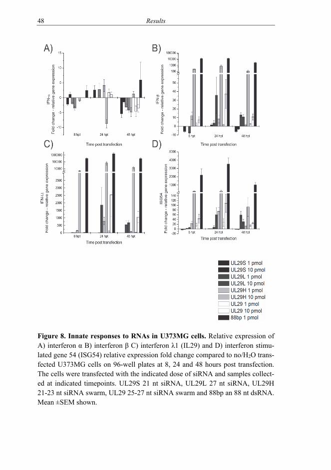
48 Results
Figure 8. Innate responses to RNAs in U373MG cells. Relative expression of A) interferon α B) interferon β C) interferon λ1 (IL29) and D) interferon stimu-lated gene 54 (ISG54) relative expression fold change compared to no/H2O trans-fected U373MG cells on 96-well plates at 8, 24 and 48 hours post transfection. The cells were transfected with the indicated dose of siRNA and samples collect-ed at indicated timepoints. UL29S 21 nt siRNA, UL29L 27 nt siRNA, UL29H 21-23 nt siRNA swarm, UL29 25-27 nt siRNA swarm and 88bp an 88 nt dsRNA. Mean ±SEM shown.
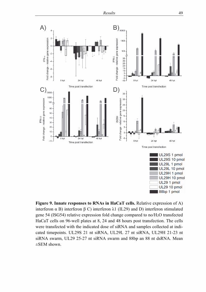
Results 49
Figure 9. Innate responses to RNAs in HaCaT cells. Relative expression of A) interferon α B) interferon β C) interferon λ1 (IL29) and D) interferon stimulated gene 54 (ISG54) relative expression fold change compared to no/H2O transfected HaCaT cells on 96-well plates at 8, 24 and 48 hours post transfection. The cells were transfected with the indicated dose of siRNA and samples collected at indi-cated timepoints. UL29S 21 nt siRNA, UL29L 27 nt siRNA, UL29H 21-23 nt siRNA swarm, UL29 25-27 nt siRNA swarm and 88bp an 88 nt dsRNA. Mean ±SEM shown.

50 Results
In U373MG cells (Figure 8), over 1000-fold increase in type I and III IFN gene expression was detected for human Dicer cleaved siRNA swarm. This effect last-ed for over 48 hours, whereas ISG54 gene expression started to subside by two days post transfection. However, comparing human Dicer created swarm to the toxic 88bp dsRNA, the long dsRNA did have a ten to hundred times higher gene expression induction with responses being at high levels even at late time points. The HaCaT cells (Figure 9) mirrored the U373MG cells for type I and III IFN responses. For ISG54 the responses were more modest and subsided faster.
Empty transfection, water transfection, and no transfection had no detectable ef-fect on studied cells.
5.2 HSV infection inhibition in vitro with siRNA swarms (I-III)
5.2.1 Comparison of siRNAs of different lengths
After establishing safety features, for example the lack of possible off-target and unexpected toxic effects, of the siRNA products, their antiviral effect was studied (Figure 10). A non-specific siRNA swarm eGFP(Giardia intestinalis Dicer) was included into the testing. This swarm had no corresponding RNA matches in the host or the virus. A non-specific single site siRNA siGFP was included into the studied siRNA product list as well.
The non-specific swarm eGFP(G) had some effect in U373MG cells (Figure 10A) whereas in HaCaT cells (Figure 10B) there was no inhibition of viral shed-ding. The single site nonsense-siRNA siGFP had no antiviral effect regardless of the cell line used. As for the various HSV-specific siRNA products, the siRNA swarms created with both human and Giardia intestinalis Dicer had the strongest antiviral effect. By visual inspection (data not shown) and cell viability (Figure 7) the 88bp dsRNA killed the cells quicker than HSV was able to replicate.
As swarms created with human Dicer and Giardia intestinalis Dicer both had comparable antiviral properties (Figure 10), but the human Dicer swarms had a higher decrease on the cell viability and higher induction of innate responses (Figures 7-9), Giardia intestinalis Dicer was chosen as the method of choice for the production of novel target sequences against HSV.

Results 51
Figure 10. Viral shedding proportion from treated and HSV-1 infected A) U373MG and B) HaCaT cells. Viral shedding inhibition (%) in relation to no /H2O treatment. Viral shedding was measured by plaque titration of the superna-tant two days post 1000 PFU infection. UL29S 21 nt siRNA, UL29L 27 nt siR-NA, UL29H 21-23 nt siRNA swarm, UL29 25-27 nt siRNA swarms and 88bp an 88 nt dsRNA. *=p<0.05 and **=p<0.01 compared to no/H2O treatment. Mean±SEM shown.
5.2.2 Comparison of the anti-HSV-siRNA swarms
After showing the antiviral efficacy of UL29, new anti-HSV-siRNA swarms tar-geting other HSV genes expressed at different phases of infection were studied. HSV genes UL54 and UL27 were chosen as additional targets against HSV; the siRNA swarms being named UL54 and UL27, respectively. UL54 is an immedi-
52 Results
ate early gene coding for ICP27, an essential multifunctional regulatory protein. UL27 is expressed at the late stages of infection and encodes for glycoprotein B (gB), which is an essential component in virion entry. In addition, a mixture of these HSV-specific swarms, including the UL29, was also used (ULMIX) (Table 6). Type I and III interferon and TLR3 gene expression in U373MG, HaCaT and RPE cells were studied for their responses to siRNA swarms and HSV infection (see Table 12 in Discussion section 6.1.1).
The above tested swarms were subjected for proof-of-principle antiviral testing. Each swarm was tested for its capability to inhibit its own target gene(s) (Table 8). The proportion of inhibition of viral mRNA production was over 99.9% for the swarm targets. All anti-HSV siRNAs were effective (p<0.001) against HSV-1 strain 17+ and were significantly (p<0.05) more effective than eGFP in U373MG cells.
Table 8. Inhibition of target gene(s) in U373MG cells. U373MG cells were treated with 10 pmol/well of the indicated swarm and subsequently infected with HSV-1 strain 17+. Samples were collected 44 hpi/48 hpt and mRNA expression was analysed.
Swarm / measured target gene
Target gene inhibition (%) HSV-specific vs eGFP-pool
UL54 99.54 UL29 99.98 UL27 99.90
ULMIX 99.65 – 99.83 – 99.89a a Inhibition of UL54, UL29 and UL27, respectively.
For a drug to be suitable in antiviral development, it is of importance to test the candidates against viral strains from clinical isolates. This is especially important for RNAi based drugs, since there is genetic variation between viral strains. When challenged against multiple HSV-1 strains, including wild type and clini-cal isolates, the most effective swarm was UL29 with an average inhibition rate of over 92% (Table 9). The other swarms were effective as well but with UL-MIX being to only other drug to reach overall inhibition of over 90%. There was some antiviral effect in the unspecific swarm, but this was surpassed by all anti-HSV-swarms.

Results 53
Tabl
e 9.
The
ant
ivir
al e
ffica
cy (i
nhib
ition
of v
iral
she
ddin
g) o
f the
siR
NA
sw
arm
s. Th
e ef
fect
of
the
swar
ms
was
mea
sure
d in
co
mpa
rison
to n
o/H
2O tr
ansf
ectio
n co
ntro
ls. A
ll sh
own
strai
ns a
re o
f HSV
-1.
U37
3MG
ce
lls
HSV
-1 1
7+
HSV
-1 F
H
SV-1
KO
S H
1211
H
1215
H
1211
4 H
1211
5 H
1211
7 H
1211
8 H
1211
9 A
vera
ge
inhi
bitio
n (%
) eG
FP
68.7
±8.7
91
.5±3
.3*
97.3
±0.8**
77
.2±6
.8*
56.6
±13.
0 18
.6±3
1 81
.0±3
.7
53.7
±13
69.0
±6.0*
45.0
±28
65.9
±13
UL5
4 90
.9±3
.4**
99
.3±0
.2**
98.
0±1.
2*
97.1
±1.2**
97
.6±1
.1**
98
.0±0
.7**
94
.3±1
.9**
97
.9±1
.2**
94
.4±2
.8**
83
.6±9
.3*
95.1
±2.3
UL2
9 99
.7±.
2**
99.9
±0.1**
99.
99±0
.0 *
* 99.
8±0.
1**
99.4
±0.5**
99
.5±0
.2**
99
.8±0
.1**
98
.8±0
.8**
98
.3±1
.0**
98
.1±1
.0**
99
.3±0
.4
UL2
7 97
.1±0
.8**
99
.4±0
.2**
99.
4±0.
2**
84.6
±5.2*
93.9
±1.2**
98
.7±0
.2**
97
.2±0
.8**
96
.9±1
.1**
96
.9±1
.0**
75
.6±4
.7
94.0
±1.5
ULM
IX 9
5.7 ±
1.5*
* 99
.7±0
.1**
99.
7±0.
1**
96.3
±1.0**
92
.2±3
.6*
98.4
±0.7**
93
.4±3
.3**
93
.1±3
.2**
97
.9±0
.7**
93
.1±2
.1**
96
.0± 1
.6
RPE
cel
ls H
SV-1
17+
H
SV-1
F
HSV
-1 K
OS
H12
11
H12
15
H12
114
H12
115
H12
117
H12
118
H12
119
Ave
rage
in
hibi
tion
(%)
eGFP
72
.5±9
.6
67.2
±12
58.3
±16
-262
.8±8
8 -2
5.1±
34
-13.
0±36
16
.0±2
8 69
.2±7
.7
13.9
±22
-35.
5±37
-4
.0±2
9 U
L54
74.1
±7.1**
94
.5±2
.4**
99
.1±0
.3**
46
.2±1
2 76
.8±9
.4*
84.9
±2.1**
85
.5±2
.1
77.0
±11
60.2
±14
-114
.7±6
9 58
.4±1
2 U
L29
99.3
±0.2**
99
.8±0
.1**
96
.2±1
.2**
86
.5±4
.7**
95
.5±1
.3**
76
.2±8
.3*
96.5
±0.9**
99
.4±0
.1**
86
.6±6
.0**
61
.5±1
1 89
.8±3
.4 U
L27
83.0
±4.1**
99
.3±0
.2**
81
.6±8
.6
83.8
±5.4**
85
.1±5
.4**
97
.8±0
.5**
89
.0±1
.2*
81.6
±8.8
90.2
±4.2**
26
.4±2
1 81
.8±5
.9
ULM
IX 8
6.2 ±
3.4*
* 99
.6±0
.1**
99
.5±0
.1**
92
.0±1
.4**
98
.1±0
.3**
95
.9±0
.5**
95
.6±1
.6**
94
.1±1
.8*
95.3
±1.7**
62
.7±1
0 91
.9±2
.1
HaC
aT ce
lls H
SV-1
17+
H
SV-1
F
HSV
-1 K
OS
H12
11
H12
15
H12
114
H12
115
H12
117
H12
118
H12
119
Ave
rage
in
hibi
tion
(%)
eGFP
64
.7±1
2*
74.7
±4.7
-8.5
±55
20.5
±18
95.8
±0.8*
56.6
±17
55.0
±19
72.8
±3.2
84.1
±7.1
80.5
±4.0
61.3
±14
UL5
4 97
.5±0
.9**
78
.5±9
.0
58.3
±20
-2.2
±51
92.0
±3.6
55.3
±23
73.8
±14 *
77
.5±9
.4
78.8
±16*
81
.5±6
.4
69.1
±23
UL2
9 99
.1±0
.3**
94
.7±3
.1**
94
.3±3
.3*
87.0
±3.5**
95
.7±2
.4**
60
.3±1
5 82
.0±1
0**
93.6
±1.4*
87.0
±6.5*
91.2
±2.0*
88.5
±4.8
UL2
7 97
.4±0
.6**
77
.1±9
.4
84.9
±9.5*
66.3
±20
96.6
±1.1*
57.3
±18
3.1±
75
93.2
±1.7*
34.5
±33
66.8
±5.7
67.7
±27
ULM
IX 9
7.1 ±
1.2*
* 96
.3±1
.3**
87
.6±6
.7
59.0
±20
97.3
±0.9**
74
.4±1
0 *
86.8
±4.4**
88
.3±6
.3*
84.2
±7.6*
78.6
±8.6
85.0
±6.7
* p≤
0.01
**
p≤0
.001
Lo
wer
effi
cacy
indi
cate
d by
ital
ics a
nd u
nder
linin
g (p
<0.0
5)
Ove
r 80%
inhi
bitio
n bo
lded
±S
EM is
show
n

54 Results
All HSV specific swarms remained effective against clinical HSV-1 isolates. There were, however, some viral strains capable of replicating to some extent even in the presence of certain HSV specific swarms. This depended on the cell line in question. UL29 and ULMIX were the most effective swarms. UL29, with the most overlap with HSV-2 sequences, was studied against HSV-2 strains, both wild type (HSV-2 G) and clinical isolates (Figure 11). All clinical isolates were not readily replicating in the given 2-day time frame (Figure 11A), resulting in a couple of non-significant findings in UL29 HSV-2 inhibition capabilities as there was little to inhibit. However, the HSV-1 specific UL29 remained highly active against HSV-2 as well.
Figure 11. Antiviral properties of UL29 swarm against HSV-2. UL29 siRNA pool against 1000 PFU infection of laboratory wt strain G and clinical isolates (H1224, H1226, H1227, H1228, H1229, H12211 and H12212) of HSV-2 in A) U373MG and B) RPE cells. Supernatant samples titered on Vero cells 48 h post treatment, 44 h post infection * = p<0.05 and ** = p<0.01 against no treatment (NT)/H2O treatment, ns = non-significant. UL29 and eGFPG 25-27 nt siRNA swarms. Average ±SEM shown. The y-axes have the same range in A) and B).

Results 55
5.3 Corneal infection treatment with enzymatically created siRNA swarms (IV)
In the in vitro studies (I-III) we found potential antiviral swarms against HSV. These swarms targeted HSV genes UL27, UL29 and UL54. Anti-UL29 swarm had the highest antiviral effect combined with the low innate immunity gene ex-pression induction. These qualities made it the top candidate for in vivo testing.
5.3.1 Encephalitis model
Corneal HSV infections were done in young female Balb/c mice. After infection, the mice were treated with swarms or vehicle (PBS). A dose of 106 PFU / eye of HSV-1 strain 17+ resulted in severe infection. Only 20% of the mice were not subjected to euthanization due to weight drop and encephalitis signs by day 8 post infection (and please see later Figure 13). The unspecific swarm eGFP did not increase survival. When treated with UL29, the survival clearly increased being 60% (p=0.063 compared to vehicle treated mice, please see study IV Fig-ure 1). The overall condition (measured by weight % compared to start of the experiment) of the animals was additionally improved when treated with UL29 compared to vehicle and unspecific siRNA swarm treatments (Table 10 and IV Figure 1B).
Table 10. Changes in weight after HSV-1 corneal infection. Balb/c mice were infected with 106 PFU / eye and 4 hours post infection treated once with vehicle (PBS), unspecific eGFP or HSV specific UL29 swarm (250 pmol/eye). Days 1-2 Days 3-5 Days 6-7 post infection
Treatment PBS eGFP UL29 PBS eGFP UL29 PBS eGFP UL29
PBS - ns ns - ns x - ns x
eGFP - ns ns - x ns - x
UL29 ns ns - x x - x x -
ns = non-significant x = p<0.05
No changes were seen in viable viral shedding from the eye at early, intermediate and late stages of the disease. However, the UL29 treated mice had significantly lower HSV DNA load in the eye swabs at 6 dpi of infection compared to PBS treated mice (study IV Figure 2B). Upon euthanasia, the eyes and trigeminal ganglia (TG) were freshly dissociated and titered on Vero cells to determine the replicating viral load in the tissue. The unspecific swarm was able to inhibit TG viral load, but there was no difference in the peripheral sample, as the viral load

56 Results
in the eye was comparable to PBS treated mice. However, the UL29 strongly inhibited viral load both in the eye and TG (p<0.01 and p<0.05 respectively, study IV Figure 2C-D). In the eyes, UL29 significantly inhibited replication competent viral load compared to the unspecific swarm as well (p<0.01). All brains collected from euthanized mice had detectable viral replication (Table 11). As for explant cultures, UL29 was able to inhibit viral spread to the TG in the wt infected mice. However, in the LoxLUC infected mice, there was no re-duction in the viral TG penetrance with the UL29 swarm. The unspecific swarm had a reductive effect on the positive TGs. There was, however, no differences seen in the viral titer loads between treatments (study IV, Figure 3).
Table 11. Replication competent virus from tissue samples. Replication competent virus (positive / tested samples)
Virus HSV-1 wt (17+) LoxLUC Treatment PBS eGFP UL29 PBS eGFP UL29
Brains of mice with encephalitis symptoms 4/4 4/4 2/2 n/a n/a n/a
TG explant culture 2/2 2/2 4/6 5/10 3/10 6/10 n/a = not applicable; all mice in the group survived and did not display signs of enceph-alitis.
5.3.2 Peripheral infection model
To study the infection with live imaging, we used HSV-1 17+ based LoxLUC virus [Nygårdas et al., 2013] constructed with BAC-technique and having a lu-ciferase phCMV cassette (Table 5, please see 4.2). This virus was slightly atten-uated and did not invade CNS as readily as HSV-1 strain 17+. Active viral pres-ence was detected from the periphery, but not from the CNS with IVIS imaging (Figure 12). Viral activity was detected usually for 5 dpi. In rare cases (see pan-els on the left in Figure 12), peripheral spreading was detected. There was, how-ever, no detectable viral shedding from the new areas. New lesions as such were not visible, but swelling was present.
None of the mice succumbed to the LoxLUC infection (Figure 13A). The mice did not experience weight drop either (Figure 13B) as they were comparable to non-infected mock mice. In addition to luciferin-luciferase signal (Figure 12), there was viral replication detected from the eye swabs. When treated with UL29 the viral shedding (PFU) and viral DNA load were downregulated at the peak of the disease (3-5 dpi; p<0.05, study IV, Figure 3). The unspecific eGFP pool had some effect as at 4 dpi there was a significant drop in viral shedding (please see IV Figure 3 for details). There was not, however, difference detected in the daily viral DNA load when treated with eGFP swarm. All in all the UL29 was broadly effective in inhibiting viral shedding and symptoms caused by HSV.

Results 57
Figure 12. LoxLUC corneal infection followup with IVIS imaging. The HSV infec-tion of mice was illustrated by the activity of the viral transgene luciferase (LUC). The procedure involved ip injection of luciferin and measurement of light signal from LUC oxidization of luciferin with ATP. Measurement and photography were done by IVIS imaging under isoflurane anesthesia (3-10 dpi). The viral activity can be seen as a light signal (intensity increasing from purple to red).
Figure 13. Survival after corneal HSV challenge. A) Mice were subjected to HSV strain 17+ (106 PFU/eye) or LoxLUC (107 PFU/eye) corneal infection and their survival was followed. B) The weight %, relative to the weight of day 0, of the mice was record-ed daily and followed for the duration of the experiment. The non-infected mock mice received the same treatments (ie. anesthesia, scarification) as the infected, but without the virus.

58 Discussion
6 DISCUSSION
6.1 Cells treated with siRNA swarms (I-II)
The proof of principle of applicability of the siRNA swarm in HSV infection control was investigated in study I. The siRNA production kinetics and purity were studied (study I Figure 2 and data not shown). The purity was considered crucial, as residual amounts of longer dsRNA molecules could result in a steep cell viability drop. As reported by [Jiang et al., 2011], longer dsRNAs, and espe-cially the 88 bp dsRNA, cause a strong innate immunity reaction. We saw in our experimental models, that already 1 pmol of the 88bp caused a drop in cell via-bility, which was considered toxic. The effect was also seen in microscope in-spection. Thus after production of each drug batch, the possible toxicity was measured via cell viability. Somewhat dose-dependent reaction was observed (Figure 7). However, even high doses of the specific siRNA swarm, up to 50 pmols/well, were well tolerated. There was no significant difference between the various siRNA products, investigated in our cell viability assays.
To study the effects of the siRNAs more closely, mRNA expression of innate immunity markers were studied. Here, a clear dose-dependent response was seen (Figures 8 and 9). In concordance with previous findings by others [Reynolds et al., 2006], the longer single site (anti-HSV-)siRNA caused higher innate immuni-ty responses. However, on the contrary, the length of the siRNA was not a factor with the siRNA swarms. The shorter siRNA containing swarm created with hu-man Dicer (UL29H; 21-23 nt) induced much higher production of type I and III and ISG genes compared to Giardia intestinalis Dicer cleaved UL29 (25-27 nt). Approximately equal responses were seen to 1 pmol of human Dicer cleaved swarm and 10 pmols of Giardia intestinalis swarm. The 10 pmol dose of swarm cleaved with human Dicer led to high increase in the type I and III interferons and interferon stimulated gene expression responses (Figures 8 and 9). Different cell lines can have different reaction to RNA products [Reynolds et al., 2006], and similarly the siRNA products used here had variable (off-target) gene ex-pression induction profiles depending on the cell lines. Dicer has been suggested to be a chaperone-like protein, guiding single strand RNA for annealing with complementary RNA [Kurzynska-Kokorniak et al., 2016]. It is feasible that the Dicer loading of longer siRNAs could play a major role in the low interferon in-duction. Moreover, as the amount of siRNAs containing the same sequence with-in a swarm is low, the amount of possible toxic siRNAs due to sequence [Fedorov et al., 2006], is low as well. Moreover, the Dicer-substrate UL29 swarm and swarm of canonical siRNA length, UL29H, covered the same sequence but
Discussion 59
had different innate immunity gene expression induction profiles. This interest-ing discrepancy between these swarms cannot therefore be sequence specific but is caused by another factor(s), of which Dicer loading is very likely candidate. It would be of interest to pursue this phenomenon further. However, from a drug development point of view, these results already tipped the scales in favor of the Dicer-substrate UL29 swarm rather than the UL29H. Of these two candidates both had same antiviral effect whereas the non-Dicer-substrate UL29H swarm induced interferon and interferon stimulated gene expressions much more clear-ly.
The findings with the synthetically produced single site siRNAs UL29S and UL29L (21 and 27 nt long respectively) were in canon with previous findings by others [Bhuyan et al., 2004; Palliser et al., 2006] as their induction of interferons and interferon stimulated genes was relatively low to non-existent. The toxic 88bp dsRNA -induced innate immunity gene expression was on a different level when compared to other RNA products. They were, however, in line with previ-ous and following findings [Jiang et al., 2011; Jiang et al., 2015]. The amount of induced gene expression of IFN-α, IFN-β, IFN-λ1 and ISG54, was cell type-dependent, but nevertheless both U373MG and HaCaT cells reacted in the same manner (Figures 8 and 9, respectively). The responses (the measured gene ex-pression) were detected already at 8 hpt. However, they were detectable even at 48 hpt. This paved way for studies of the interplay of the virus and the biological drugs. Interferons as such are used against viral infections, especially when there are no other drugs available, for example against certain hepatitis viruses [Lappalainen and Färkkilä, 2016]. Interferon induction caused by the RNA prod-ucts could facilitate antiviral responses. And, indeed, in some cases there was an antiviral effect observed at the same time as interferon gene expression increase. However, without question, the highest specific anti-herpes effect was seen from sequence based RNA-interference reactions from the HSV-specific siRNAs.
To study the antiviral effect, an in vitro setting that resembled an actual in vivo infection was used. In a natural infection, only a small portion of cells are infect-ed with the virus spreading from one cell to another. At the same time the cells are communicating with each other. Depending on the virus, the amount of in-formation and priming to infection can most likely vary. Priming with immune system molecules, such as interleukin 27, leads to inhibition of infection [Heikkilä et al., 2016a]. Moreover, HSV infection leads to exosome production, which in turn has an effect on the infection [Han et al., 2016; Heikkilä et al., 2016b; Kalamvoki et al., 2014]. In our in vitro experimental setting, the drug would be in both cells that have and in those that do not have the virus. Therefore a relatively low initial viral dose and a time point of 2 dpi was chosen. This was to mimic the microenvironment of an actual infection. In addition, the drug dose

60 Discussion
(pmol) was set to mimic same situation and the dose was set for per well, not per cell. The drawback of this experimental setting was that all of the cells were not in uniform situation and viral replication was at various stages at a given time.
All in all, the siRNA products were found to efficiently inhibit viral shedding (p<0.05) (Figure 10). The strongest inhibitor of HSV was the 88bp dsRNA. However, it cannot be taken as a suitable candidate for drug development as it is a desirable feature for the drug to leave the host alive, but the 88bp dsRNA did kill virus-infected cells non-selectively. This finding points out another reason why it is important that an RNA-product does not cause too high innate immuni-ty induction, as for example with UL29 compared to UL29H. For some target viruses a need might arise to use an RNA swarm-drug simultaneously with ad-ministered interferon. Here, a too strong of an innate immunity inducer swarm might alongside with the interferon cause undesired side effects.
6.1.1 Interplay of treatment and infection (II-III)
HSV induced innate responses, namely type I and III IFN and IFN -stimulated gene (here TLR3) expressions, in the host cells. This response was cell type-specific (Table 12). For example U373MG cells had very low response to HSV infection in this setting whereas HaCaT and RPE cells had strong responses, both up and downregulation of genes. However, the treatment of infection with siRNA swarms had an effect on the responses in U373MG cells. This was important to know, as some cell lines have very limited ability to react with and transcribe IFN-genes [Desmyter et al., 1968]. However, these U373MG cells had the capa-bility to react with and transcribe these innate response genes. The siRNAs had an effect on the other cell lines as well. Type I and III interferons were upregulat-ed. TLR3 gene expression was upregulated in unison to swarm treatments, being an interferon-induced component of the innate immune system. These differ-ences in innate immunity gene expression profiles are likely causative factors on the variable effects of the siRNA swarms, especially the unspecific eGFP swarm. Here the HSV effect was studied for strain 17+, but different clinical isolates have different effects on cells (Lehtinen et al. unpublished results).
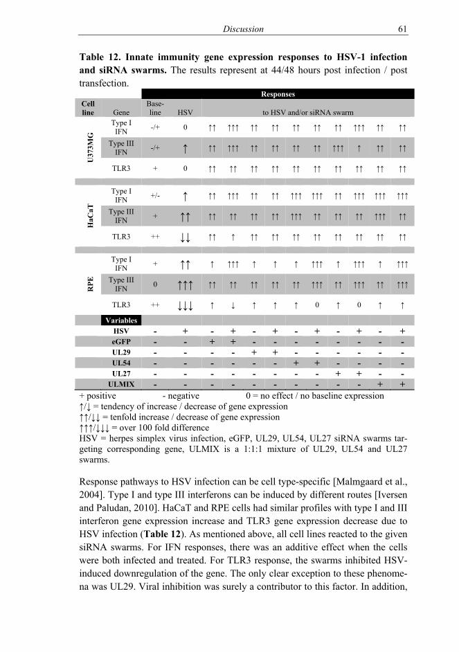
Discussion 61
Table 12. Innate immunity gene expression responses to HSV-1 infection and siRNA swarms. The results represent at 44/48 hours post infection / post transfection.
Responses Cell line Gene
Base-line HSV to HSV and/or siRNA swarm
U37
3MG
Type I IFN -/+ 0 ↑↑ ↑↑↑ ↑↑ ↑↑ ↑↑ ↑↑ ↑↑ ↑↑↑ ↑↑ ↑↑
Type III IFN -/+ ↑ ↑↑ ↑↑↑ ↑↑ ↑↑ ↑↑ ↑↑ ↑↑↑ ↑ ↑↑ ↑↑
TLR3 + 0 ↑↑ ↑↑ ↑↑ ↑↑ ↑↑ ↑↑ ↑↑ ↑↑ ↑↑ ↑↑
HaC
aT
Type I IFN +/- ↑ ↑↑ ↑↑↑ ↑↑ ↑↑ ↑↑↑ ↑↑↑ ↑↑ ↑↑↑ ↑↑↑ ↑↑↑
Type III IFN + ↑↑ ↑↑ ↑↑ ↑↑ ↑↑ ↑↑↑ ↑↑ ↑↑ ↑↑ ↑↑↑ ↑↑
TLR3 ++ ↓↓ ↑↑ ↑ ↑↑ ↑↑ ↑↑ ↑↑ ↑↑ ↑↑ ↑↑ ↑↑
RPE
Type I IFN + ↑↑ ↑ ↑↑↑ ↑ ↑ ↑ ↑↑↑ ↑ ↑↑↑ ↑ ↑↑↑
Type III IFN 0 ↑↑↑ ↑↑ ↑↑ ↑↑ ↑↑ ↑↑ ↑↑↑ ↑↑ ↑↑↑ ↑↑ ↑↑↑
TLR3 ++ ↓↓↓ ↑ ↓ ↑ ↑ ↑ 0 ↑ 0 ↑ ↑
Variables HSV - + - + - + - + - + - + eGFP - - + + - - - - - - - - UL29 - - - - + + - - - - - - UL54 - - - - - - + + - - - - UL27 - - - - - - - - + + - - ULMIX - - - - - - - - - - + + + positive - negative 0 = no effect / no baseline expression ↑/↓ = tendency of increase / decrease of gene expression ↑↑/↓↓ = tenfold increase / decrease of gene expression ↑↑↑/↓↓↓ = over 100 fold difference HSV = herpes simplex virus infection, eGFP, UL29, UL54, UL27 siRNA swarms tar-geting corresponding gene, ULMIX is a 1:1:1 mixture of UL29, UL54 and UL27 swarms.
Response pathways to HSV infection can be cell type-specific [Malmgaard et al., 2004]. Type I and type III interferons can be induced by different routes [Iversen and Paludan, 2010]. HaCaT and RPE cells had similar profiles with type I and III interferon gene expression increase and TLR3 gene expression decrease due to HSV infection (Table 12). As mentioned above, all cell lines reacted to the given siRNA swarms. For IFN responses, there was an additive effect when the cells were both infected and treated. For TLR3 response, the swarms inhibited HSV-induced downregulation of the gene. The only clear exception to these phenome-na was UL29. Viral inhibition was surely a contributor to this factor. In addition,

62 Discussion
it is likely that the targeted viral gene and its expression phase in a lytic infection played a role in the innate immunity gene expression responses. Inhibition of UL29 encoding protein, the ICP8, the protein responsible in part for DNA repli-cation, should lead to halt/delay in viral DNA accumulation. This in turn could result in lighter cellular response. Why would this lead into different profiles compared to the other HSV targeting swarms? UL54 siRNA swarm had an addi-tive effect on the innate responses, but then the swarm did not inhibit viral repli-cation as strongly as UL29. On the other hand, UL27 siRNA swarm target, UL27, is expressed later than UL29 thus not inhibiting viral DNA replication as such.
In UL29 treated cells, the infection influenced only little the interferon gene re-sponse. The induction of interferon and TLR3 gene expression due to siRNA swarms, including non-specific eGFP, could influence infection inhibition bene-ficially in a whole organism. TLR3 plays an important role in HSV infection [Peri et al., 2008; Reinert et al., 2012; Zhang et al., 2007]. An immunostimulant like mechanism of action of an unspecific swarm was seen, with some inhibitory effect on viral shedding in some settings against certain virus strains. Im-munostimulus of an siRNA product has been found to have a favorable effect on antiviral activity, at least against Semliki Forest virus [Gantier et al., 2010]. However, the immunostimulus of a drug could also result in an increase of ad-verse effects.
In general, the siRNA effect and the HSV effect were additive on the innate im-munity gene expression. If HSV and RNA product both induced IFN gene ex-pression, the expression was higher for the combination than either individually. In some cases, if HSV response caused a downregulation and the swarm caused an upregulation of certain gene expression, the detected net effect remained near zero. When the virus and the swarm caused a positive reaction, the reaction was additive. The only exception to this was UL29, where infection and the swarm did not cause an additive effect. Swarm targeting UL29 had the highest likeli-hood of not causing adverse effects and at the same time had the strongest antivi-ral effect (Table 9), being the most favorable combination. These features of the UL29 swarm made it a top candidate for further drug development.
6.2 Efficacy of the antiviral swarm in vitro (I-III)
The HSV-specific siRNA swarms were produced using HSV-1 17+ DNA as a template. Thus, the swarms were effective against HSV-1 strain 17+ (Table 8). As the swarms had an effect on the innate responses and the treatment was given prior to infection, it was interesting to see that the unspecific siRNA swarm had

Discussion 63
some effect against HSV. The antiviral effect of interferon-inducing siRNA is even more prominent, the earlier the treatment is given even, especially in Ha-CaT cells [Backman, 2014]. To place the swarms to actual test, various HSV strains, both clinical field isolates and wild type HSVs were used (Table 9). When studying the antiviral effect against clinical isolates of HSV, two swarms emerged: the UL29 and ULMIX were most broadly effective against HSV in the various cell lines. UL29 had the most resemblance of the swarms to HSV-2 ge-nome, and was found effective against HSV-2 strains, both wt and clinical iso-lates (Figure 11). Moreover, there was no evidence of quick resistance building against UL29 (study III, Figure 4). Furthermore, it is unlikely that a virus can form resistance even in the long run to all of the siRNAs in the swarm, as the targeted area is hundreds of bps long.
The UL29 swarm had a more beneficial innate immunity gene expression induc-tion profile than ULMIX. At same pmol-dose, the UL29 caused much milder innate immunity induction profiles than ULMIX. Compared to HSV infection, the ULMIX treatment caused an significant upregulation of interferon gene ex-pression (Table 12 and from study III Figures 1-2), raising concerns about possi-ble side effects in whole organisms.
6.3 In vivo efficacy of the swarms against HSV (IV)
Based on the in vitro findings, anti-UL29 swarm was chosen for the in vivo treatment approach. As HSV eye infection causes serious medical burden, with the additional problem of frequent antiviral chemotherapy-resistant HSV infec-tion cases, HSV keratitis was used as a model for infection and treatment. The fact that the first antiviral RNA interference like drug to reach the market was against CMV eye infection (Vitravene, DNA oligo) [Crooke, 1998], is also an encouragement. In contrast to our in vitro experiments, in the in vivo model, the treatment was given post infection and without transfection reagent. The siRNAs swarms were delivered as eye drops in PBS.
A single 250 pmol/eye dose of UL29 swarm was able to increase mouse survival by three fold. This topical siRNA swarm treatment was effective, while, in con-trast, in an antibody-mediated antiviral approach against corneal HSV, no effect of topical treatment was found [Krawczyk et al., 2015]. The increase of survival was similar to that of an HSV-2 intravaginal challenge treated with a synthetic siRNA product [Palliser et al., 2006], and could perhaps be increased further with the combination of ACV to the treatment [da Silva et al., 2016]. In addition to survival, symptoms were alleviated with the swarm (Table 10) and viral load in TG was limited (Table 11). The unspecific swarm did also somewhat inhibit vi-

64 Discussion
ral load in TG. Immune system activation has shown to be beneficial against HSV in previous studies [Taylor et al., 1989] and in our in vitro work. However, eGFP swarm did not increase survival nor did it alleviate symptoms (Table 10).
The Balb/c mice were susceptible to corneal HSV challenge with the wt virus whereas the attenuated marker virus resulted in an attenuated, peripherally lim-ited, infection (Figures 12 and 13). Even though rare, an interesting peripheral viral spread without site-specific viral shedding was observed (Figure 12) for the marker virus LoxLUC. The spread was most likely via neurons as has been demonstrated previously by others [Balliet et al., 2007; Halford et al., 2004].
The further the infection proceeded from the periphery, the more the weight of the mice started to plummet by 5 dpi. Brains of these mice had detectable repli-cating virus. For the surviving mice the weight drop was not so drastic but ob-servable. For the UL29 treated mice, the weight loss was significantly reduced (Table 10), and the tissue viral load was also reduced in the UL29 treated, eu-thanized mice (Table 11). Not all mice survived the viral challenge even though they were treated. A more frequent dosing might be able to protect the mice bet-ter. However, when the virus reaches the nervous system, a peripheral admin-istration is unlikely to be effective. Thus, there is a need to develop novel deliv-ery methods for the siRNA swarms. However, in the case of human corneal HSV disease, the virus is most likely already a resident in the patient nerves and the treatment of local viral reactivations is of paramount importance.
In the peripherally limited infection (Tables 10 and 11 and Figures 12 and 13), the UL29 swarm was able to inhibit viral replication and shedding significantly. The eGFP swarm had an effect at a point during the follow-up period. At the end of the study, the eyes and brains were measured for viral replication, of which there was none, in both wt and marker virus infected mice. TG explant culture showed no significant differences between different treatment groups (Table 11). In LoxLUC infected UL29-treated mice the amount of latently infected ganglia was slightly elevated but in the strain 17+ infected UL29-treated mice the latent-ly infected ganglia were reduced. The peripheral dosing seemed to have little ef-fect on latent virus.
All in all, UL29 swarm inhibited HSV-1 infection and alleviated symptoms in corneal HSV infection models. Moreover, UL29 protected the mice from a lethal corneal HSV challenge. Anti-HSV-UL29 swarm is a good lead for drug devel-opment against HSV, therefore to be developed further.
Conclusions 65
7 CONCLUSIONS
There is a need for new remedies against herpes simplex virus (HSV) which is a widespread pathogen. Despite the fact that the immune system of the host and available antivirals can inhibit the virus, there are situations where the viral dis-ease takes over. In these cases, neither the immune system nor the drugs are ca-pable of preventing damage caused by the infection. Such cases are common in immunocompromised patients and in HSV keratitis.
There are multiple approaches in antiviral drug development against HSV and there is a need for development of new treatment modalities against HSV. In ad-dition, new and novel treatment methods, involving tk gene usage, of various diseases, such as cancer with HSV or HSV tk gene, can lead to situations where the usage of standard tk-based anti-HSV medication would be unwanted.
RNA interference (RNAi) is a promising tool in drug research. As a novel type of treatment, it is especially suitable for topical treatments. Standard, canonical, single site targeting small interfering (si)RNAs are susceptible to mutations in the viral genome, as a single nucleotide change could potentially render the virus resistant to the siRNA treatment. Moreover, the commercialization and patenta-bility of widely used methods and very short nucleotide sequences could be diffi-cult. These pose a problem to the potential drug development pipeline.
There is, however, a non-canonical way of producing siRNAs that are not sus-ceptible to small mutations in the viral genome. Our enzymatically created novel siRNA swarms targeting large segments of target genes is a novel approach in antiviral drug research. Our studies show how anti-HSV-siRNA swarms are ef-fective against both wild type and clinical field isolate strains of HSV. Moreover, a swarm targeting HSV gene UL29 has proven wide efficacy, low innate im-munity activation, shown no hint of antiviral resistance formation and has proven efficient in a corneal model as well. These results elucidate favorably the suita-bility of siRNA swarms in anti-HSV drug development.
In conclusion, the anti-HSV-siRNA swarm approach is feasible against HSV in-fection and harbours great potential for subsequent clinical implementation.
66 Acknowledgements
ACKNOWLEDGEMENTS
This study was carried out at the Department of Virology, University of Turku. Professors Ilkka Julkunen, Timo Hyypiä and Veijo Hukkanen are thanked for allowing me to work and use the facilities of the Department during this project.
Deepest bow of gratitude goes towards my awesome supervisor Veijo Hukkanen. It has been a privilege to be your student! Your great enthusiasm towards science is contagious and your expertise is awe-inspiring. Thank you for your time, sup-port and encouragement, all of which have been plenty, to put it mildly.
My thesis follow-up committee members Professor Markku Koulu and Associate Professor Eeva Broberg are warmly thanked for all their advice and encourage-ment during the years.
The expert reviewers of this thesis, Professor Igor Jurak and Docent Peter Sarin, are sincerely thanked for their comments and suggestions that did indeed im-prove the book.
Michaela Nygårdas, thank you for your mentorship and friendship. I believe that without your help, I would have been lost with laboratory work, planning and around the world at scientific meetings. Your sunshine attitude and patience when teaching a very green novice pipeter are something I look up to.
This work would not have been possible without the help of co-authors at Hel-sinki and Turku, tremendous thanks. Alesia Romanosvkaya, Minna M Poranen and Dennis H Bamford, swarmful of thankyous for everything. Jenni Lehtinen, Michaela and Veijo, thank you for the great effort, especially for the stable help.
Thank you to Tytti Vuorinen and the team of virus growers for making intriguing strains available. Beate Sodeik is thanked for viral mutants and backbones. Mar-ja-Leena Mattila and Tiina Laihinen are thanked for the countless cells. To Ritva Kajander, Merja Kleme and Liisa Lund, with Terhi Helander, Outi Rauta and Mervi Laanti, I owe great thanks for all the expert technical assistance. Central Animal Laboratory of the University of Turku and Turku Centre for Biotechnol-ogy, and their personel, thank you for expert help with mice and machines.
Thank you to all past and present members and visitors of the Hukkanen labora-tory for creating a joyful working atmosphere and superb support. The emeritas of HSV group Eeva B, Piritta Peri, Michaela, Riikka Arffman and Outi Heikkilä, my students Sonja Häkli, Anna Backman and Jenni Lehtinen and the rare young male scientist Jussi Palomäki, to name a few. Thank you all for the laughs and

Acknowledgements 67
good times in and out of the laboratory, it really has been fun working in the laaturyhmä!
Everybody at the Department of Virology, you make the Virusoppi a wonderful place to work at. Virusoppi is truly a place where outstanding science and diag-nostics is done. Thank you all for the privilege to work with you.
It has been inspirational to work with a wide range of people and projects. The spin-ins and spin-offs of this thesis and the, at least seemingly, out-of-the-blue-experiments have kept things even more fun and interesting, even if in the end they did not fit in the scope of this thesis. The gene therapy with Michaela, the spheroids with Aaro and the question of mouthfuls of herpes with Johanna with Stina, the heart-warming gravely smiles under the stereomicroscope with Riikka, the intense pace of Dennis K et al., the long distance letter exploration with Mo-riah S et al., the wire tangling with Laura A. et al. and the nerve tingling collabo-ration with Mahesh and Ari have made the thesis project great.
Jussi Heinonkoski, Kaj Rindell and Hannu Pesonen are thanked for their help with interpreting “old” science languages. The National Repository Library, Va-rastokirjasto, is acknowledged for outstanding (free!) service and for importance in making (a tad older) publications available.
The Drug Research Doctoral Programme (DRDP), its predecessor Drug Discov-ery Graduate School (DDGS), FinPharma Doctoral Program Drug Discovery Section (FPDP-D) and their coordinator Eeva Valve are kindly thanked for great study atmosphere. The funding from the programs has been indispensable, thank you. Foundation for Research on Viral Diseases, The Scientific Socie-ty/Foundation of Microbiologists in Turku, Program for Infection Biology and Infectious Diseases, Valto Takala´s fund and Finnish Society for Study of Infec-tious Diseases are kindly acknowledged for financial support of travels to scien-tific meetings and a pleasant medium to be part of. The Academy of Finland and Turku University Foundation are acknowledged for funding to Professor Veijo Hukkanen (SA grants no: 128915 and 308446). Great thanks are also extended to Alfred Kordelin Foundation, Instrumentarium Foundation and Finnish Cultural Foundation for essential and much appreciated financial support.
A very many thanks to my family and friends. A long time ago, the aftermath of a rodent in a mousetrap sparked my curiosity for science – thank you for feeding that hunger. Most importantly, I thank my wife Jenni and daughter Lyydia. Be-cause of you two, I know what is actually significant, my stars. Thank you for your understanding, support and love.
Turku, winter of 2016-2017

68 Acknowledgements
REFERENCES
Aalto A, Sarin L, van Dijk A, Saarma M, Poranen M, Arumäe U, Bamford D. 2007. Large-scale production of dsRNA and siRNA pools for RNA interference utilizing bacteriophage phi6 RNA-dependent RNA polymerase. RNA 13(3):422-429.
Ackermann M, Chou J, Sarmiento M, Lerner RA, Roizman B. 1986. Identification by antibody to a synthetic peptide of a protein specified by a diploid gene located in the terminal repeats of the L component of herpes simplex virus genome. J Virol 58(3):843-850.
Adamiak B, Trybala E, Mardberg K, Johansson M, Liljeqvist JA, Olofsson S, Grabowska A, Bienkowska-Szewczyk K, Szewczyk B, Bergstrom T. 2010. Human antibodies to herpes simplex virus type 1 glycoprotein C are neutralizing and target the heparan sulfate-binding domain. Virology 400(2):197-206.
Alexander DE, Ward SL, Mizushima N, Levine B, Leib DA. 2007. Analysis of the role of autophagy in replication of herpes simplex virus in cell culture. J Virol 81(22):12128-12134.
Altschul SF, Gish W, Miller W, Myers EW, Lipman DJ. 1990. Basic local alignment search tool. J Mol Biol 215(3):403-410.
Anderson BJ, McGuire DP, Reed M, Foster M, Ortiz D. 2016. Prophylactic Valacyclovir to Prevent Outbreaks of Primary Herpes Gladiatorum at a 28-Day Wrestling Camp: A 10-Year Review. Clin J Sport Med 26(4):272-278.
Andreansky S, He B, van Cott J, McGhee J, Markert J, Gillespie G, Roizman B, Whitley R. 1998. Treatment of intracranial gliomas in immunocompetent mice using herpes simplex viruses that express murine interleukins. Gene Ther 5(1):121-130.
Andtbacka RH, Kaufman HL, Collichio F, Amatruda T, Senzer N, Chesney J, Delman KA, Spitler LE, Puzanov I, Agarwala SS, Milhem M, Cranmer L, Curti B, Lewis K, Ross M, Guthrie T, Linette GP, Daniels GA, Harrington K, Middleton MR, Miller WH, Zager JS, Ye Y, Yao B, Li A, Doleman S, VanderWalde A, Gansert J, Coffin RS. 2015. Talimogene Laherparepvec Improves Durable Response Rate in Patients With Advanced Melanoma. J Clin Oncol 33(25):2780-2788.
Arvin A, Cohen J, Ganem D, Kieff E, Knipe D, Mocarski E, Pass R, Pellet P, Rickinson A, Roizman B, Thomas S, Straus S, Whitley R, Yamanishi K, Yasuko M, Authors-in-alphabetical-order. 2007. Herpesviridae. In: Knipe D, Howley P, editors. Fields Virology. 5th ed. Philadelphia, PA, USA: Lippincott Williams & Wilkins.
Aubert M, Boyle NM, Stone D, Stensland L, Huang ML, Magaret AS, Galetto R, Rawlings DJ, Scharenberg AM, Jerome KR. 2014. In vitro Inactivation of Latent HSV by Targeted Mutagenesis Using an HSV-specific Homing Endonuclease. Mol Ther Nucleic Acids 3:e146.
Backman A. 2014. Herpes simplex -infektion hoito RNA-interferenssillä in vitro. Pro Gradu. Turku: University of Turku. 71 p.
Balliet JW, Kushnir AS, Schaffer PA. 2007. Construction and characterization of a herpes simplex virus type I recombinant expressing green fluorescent protein: acute phase replication and reactivation in mice. Virology 361(2):372-383.
Baringer JR. 1976. The biology of herpes simplex virus infection in humans. Surv Ophthalmol 21(2):171-174.
Bastian FO, Rabson AS, Yee CL, Tralka TS. 1972. Herpesvirus hominis: isolation from human trigeminal ganglion. Science 178(4058):306-307.

References 69
Bellner L, Thorén F, Nygren E, Liljeqvist JA, Karlsson A, Eriksson K. 2005. A proinflammatory peptide from herpes simplex virus type 2 glycoprotein G affects neutrophil, monocyte, and NK cell functions. J Immunol 174(4):2235-2241.
Belshe RB, Leone PA, Bernstein DI, Wald A, Levin MJ, Stapleton JT, Gorfinkel I, Morrow RL, Ewell MG, Stokes-Riner A, Dubin G, Heineman TC, Schulte JM, Deal CD, Women HTf. 2012. Efficacy results of a trial of a herpes simplex vaccine. N Engl J Med 366(1):34-43.
Bernard MC, Barban V, Pradezynski F, de Montfort A, Ryall R, Caillet C, Londono-Hayes P. 2015. Immunogenicity, protective efficacy, and non-replicative status of the HSV-2 vaccine candidate HSV529 in mice and guinea pigs. PLoS One 10(4):e0121518.
Bernstein E, Caudy AA, Hammond SM, Hannon GJ. 2001. Role for a bidentate ribonuclease in the initiation step of RNA interference. Nature 409(6818):363-366.
Bhuyan PK, Karikò K, Capodici J, Lubinski J, Hook LM, Friedman HM, Weissman D. 2004. Short interfering RNA-mediated inhibition of herpes simplex virus type 1 gene expression and function during infection of human keratinocytes. J Virol 78(19):10276-10281.
Biswas S, Swift M, Field HJ. 2007. High frequency of spontaneous helicase-primase inhibitor (BAY 57-1293) drug-resistant variants in certain laboratory isolates of HSV-1. Antivir Chem Chemother 18(1):13-23.
Black F. 1975. Infectious diseases in primitive societies. Science 187(4176):515-518.
Bolovan CA, Sawtell NM, Thompson RL. 1994. ICP34.5 mutants of herpes simplex virus type 1 strain 17syn+ are attenuated for neurovirulence in mice and for replication in confluent primary mouse embryo cell cultures. J Virol 68(1):48-55.
Boukamp P, Petrussevska RT, Breitkreutz D, Hornung J, Markham A, Fusenig NE. 1988. Normal keratinization in a spontaneously immortalized aneuploid human keratinocyte cell line. J Cell Biol 106(3):761-771.
Boukhvalova M, McKay J, Mbaye A, Sanford-Crane H, Blanco JC, Huber A, Herold BC. 2015. Efficacy of the Herpes Simplex Virus 2 (HSV-2) Glycoprotein D/AS04 Vaccine against Genital HSV-2 and HSV-1 Infection and Disease in the Cotton Rat Sigmodon hispidus Model. J Virol 89(19):9825-9840.
Bradley H, Markowitz LE, Gibson T, McQuillan GM. 2014. Seroprevalence of herpes simplex virus types 1 and 2--United States, 1999-2010. J Infect Dis 209(3):325-333.
Bradshaw MJ, Venkatesan A. 2016. Herpes Simplex Virus-1 Encephalitis in Adults: Pathophysiology, Diagnosis, and Management. Neurotherapeutics 13(3):493-508.
Broberg E, Setala N, Roytta M, Salmi A, Eralinna JP, He B, Roizman B, Hukkanen V. 2001. Expression of interleukin-4 but not of interleukin-10 from a replicative herpes simplex virus type 1 viral vector precludes experimental allergic encephalomyelitis. Gene Ther 8(10):769-777.
Broberg EK, Nygårdas M, Salmi AA, Hukkanen V. 2003. Low copy number detection of herpes simplex virus type 1 mRNA and mouse Th1 type cytokine mRNAs by Light Cycler quantitative real-time PCR. J Virol Methods 112(1-2):53-65.
Brown EL, Gardella C, Malm G, Prober CG, Forsgren M, Krantz EM, Arvin AM, Yasukawa LL, Mohan K, Brown Z, Corey L, Wald A. 2007. Effect of maternal herpes simplex virus (HSV) serostatus and HSV type on risk of neonatal herpes. Acta Obstet Gynecol Scand 86(5):523-529.

70 References
Brown ZA, Wald A, Morrow RA, Selke S, Zeh J, Corey L. 2003. Effect of serologic status and cesarean delivery on transmission rates of herpes simplex virus from mother to infant. JAMA 289(2):203-209.
Brunnemann AK, Liermann K, Deinhardt-Emmer S, Maschkowitz G, Pohlmann A, Sodeik B, Fickenscher H, Sauerbrei A, Krumbholz A. 2016. Recombinant herpes simplex virus type 1 strains with targeted mutations relevant for aciclovir susceptibility. Sci Rep 6:29903.
Burgos JS, Ripoll-Gomez J, Alfaro JM, Sastre I, Valdivieso F. 2008. Zebrafish as a new model for herpes simplex virus type 1 infection. Zebrafish 5(4):323-333.
Burkitt D, O'Conor GT. 1961. Malignant lymphoma in African children. I. A clinical syndrome. Cancer 14:258-269.
Burrel S, Boutolleau D, Azar G, Doan S, Deback C, Cochereau I, Agut H, Gabison EE. 2013. Phenotypic and genotypic characterization of acyclovir-resistant corneal HSV-1 isolates from immunocompetent patients with recurrent herpetic keratitis. J Clin Virol 58(1):321-324.
Camarena V, Kobayashi M, Kim JY, Roehm P, Perez R, Gardner J, Wilson AC, Mohr I, Chao MV. 2010. Nature and duration of growth factor signaling through receptor tyrosine kinases regulates HSV-1 latency in neurons. Cell Host Microbe 8(4):320-330.
Campadelli-Fiume G, Menotti L, Avitabile E, Gianni T. 2012. Viral and cellular contributions to herpes simplex virus entry into the cell. Curr Opin Virol 2(1):28-36.
Caplen NJ, Parrish S, Imani F, Fire A, Morgan RA. 2001. Specific inhibition of gene expression by small double-stranded RNAs in invertebrate and vertebrate systems. Proc Natl Acad Sci U S A 98(17):9742-9747.
Caserta MT, Hall CB, Schnabel K, Long CE, D'Heron N. 1998. Primary human herpesvirus 7 infection: a comparison of human herpesvirus 7 and human herpesvirus 6 infections in children. J Pediatr 133(3):386-389.
Cerutti H, Casas-Mollano JA. 2006. On the origin and functions of RNA-mediated silencing: from protists to man. Curr Genet 50(2):81-99.
Chamoun-Emanuelli AM, Bobardt M, Moncla B, Mankowski MK, Ptak RG, Gallay P, Chen Z. 2014. Evaluation of PD 404,182 as an anti-HIV and anti-herpes simplex virus microbicide. Antimicrob Agents Chemother 58(2):687-697.
Chang Y, Cesarman E, Pessin MS, Lee F, Culpepper J, Knowles DM, Moore PS. 1994. Identification of herpesvirus-like DNA sequences in AIDS-associated Kaposi's sarcoma. Science 266(5192):1865-1869.
Chibo D, Druce J, Sasadeusz J, Birch C. 2004. Molecular analysis of clinical isolates of acyclovir resistant herpes simplex virus. Antiviral Res 61(2):83-91.
Chou J, Kern ER, Whitley RJ, Roizman B. 1990. Mapping of herpes simplex virus-1 neurovirulence to gamma 134.5, a gene nonessential for growth in culture. Science 250(4985):1262-1266.
Ciceri F, Bonini C, Stanghellini MT, Bondanza A, Traversari C, Salomoni M, Turchetto L, Colombi S, Bernardi M, Peccatori J, Pescarollo A, Servida P, Magnani Z, Perna SK, Valtolina V, Crippa F, Callegaro L, Spoldi E, Crocchiolo R, Fleischhauer K, Ponzoni M, Vago L, Rossini S, Santoro A, Todisco E, Apperley J, Olavarria E, Slavin S, Weissinger EM, Ganser A, Stadler M, Yannaki E, Fassas A, Anagnostopoulos A, Bregni M, Stampino CG, Bruzzi P, Bordignon C. 2009. Infusion of suicide-gene-engineered donor lymphocytes after family haploidentical haemopoietic stem-cell transplantation for leukaemia (the TK007 trial): a non-randomised phase I-II study. Lancet Oncol 10(5):489-500.

References 71
Cliffe A, Garber D, Knipe D. 2009. Transcription of the herpes simplex virus latency-associated transcript promotes the formation of facultative heterochromatin on lytic promoters. J Virol 83(16):8182-8190.
ClinicalTrials.gov. 2016. http://clinicaltrials.gov/ siRNA search pharse.
Coen DM, Fleming HE, Leslie LK, Retondo MJ. 1985. Sensitivity of arabinosyladenine-resistant mutants of herpes simplex virus to other antiviral drugs and mapping of drug hypersensitivity mutations to the DNA polymerase locus. J Virol 53(2):477-488.
Coen DM, Kosz-Vnenchak M, Jacobson JG, Leib DA, Bogard CL, Schaffer PA, Tyler KL, Knipe DM. 1989. Thymidine kinase-negative herpes simplex virus mutants establish latency in mouse trigeminal ganglia but do not reactivate. Proc Natl Acad Sci U S A 86(12):4736-4740.
Coen DM, Schaffer PA. 1980. Two distinct loci confer resistance to acycloguanosine in herpes simplex virus type 1. Proc Natl Acad Sci U S A 77(4):2265-2269.
Cohen J, Straus S, Arvin A. 2007. Varicella-Zoster Virus Replication, Pathogenesis, and Management. In: Knipe D, Howley P, editors. Fields Virology. 5th ed. Philadelphia, PA, USA: Lippincott Williams & Wilkins. p 2773-2818.
Cohen JI. 2000. Epstein-Barr virus infection. N Engl J Med 343(7):481-492.
Crooke ST. 1998. Vitravene--another piece in the mosaic. Antisense Nucleic Acid Drug Dev 8(4):vii-viii.
Da Costa X, Kramer MF, Zhu J, Brockman MA, Knipe DM. 2000. Construction, phenotypic analysis, and immunogenicity of a UL5/UL29 double deletion mutant of herpes simplex virus 2. J Virol 74(17):7963-7971.
Da Costa XJ, Bourne N, Stanberry LR, Knipe DM. 1997. Construction and characterization of a replication-defective herpes simplex virus 2 ICP8 mutant strain and its use in immunization studies in a guinea pig model of genital disease. Virology 232(1):1-12.
da Silva AS, Raposo JV, Pereira TC, Pinto MA, de Paula VS. 2016. Effects of RNA interference therapy against herpes simplex virus type 1 encephalitis. Antivir Ther 21(3):225-235.
Danve-Szatanek C, Aymard M, Thouvenot D, Morfin F, Agius G, Bertin I, Billaudel S, Chanzy B, Coste-Burel M, Finkielsztejn L, Fleury H, Hadou T, Henquell C, Lafeuille H, Lafon ME, Le Faou A, Legrand MC, Maille L, Mengelle C, Morand P, Morinet F, Nicand E, Omar S, Picard B, Pozzetto B, Puel J, Raoult D, Scieux C, Segondy M, Seigneurin JM, Teyssou R, Zandotti C. 2004. Surveillance network for herpes simplex virus resistance to antiviral drugs: 3-year follow-up. J Clin Microbiol 42(1):242-249.
Darby G, Field HJ, Salisbury SA. 1981. Altered substrate specificity of herpes simplex virus thymidine kinase confers acyclovir-resistance. Nature 289(5793): 81-83.
Denisova OV, Kakkola L, Feng L, Stenman J, Nagaraj A, Lampe J, Yadav B, Aittokallio T, Kaukinen P, Ahola T, Kuivanen S, Vapalahti O, Kantele A, Tynell J, Julkunen I, Kallio-Kokko H, Paavilainen H, Hukkanen V, Elliott RM, De Brabander JK, Saelens X, Kainov DE. 2012. Obatoclax, saliphenylhalamide, and gemcitabine inhibit influenza a virus infection. J Biol Chem 287(42):35324-35332.
Desmyter J, Melnick JL, Rawls WE. 1968. Defectiveness of interferon production and of rubella virus interference in a line of African green monkey kidney cells (Vero). J Virol 2(10):955-961.
72 References
Dewhurst S, McIntyre K, Schnabel K, Hall CB. 1993. Human herpesvirus 6 (HHV-6) variant B accounts for the majority of symptomatic primary HHV-6 infections in a population of U.S. infants. J Clin Microbiol 31(2):416-418.
Dingwell K, Brunetti C, Hendricks R, Tang Q, Tang M, Rainbow A, Johnson D. 1994. Herpes simplex virus glycoproteins E and I facilitate cell-to-cell spread in vivo and across junctions of cultured cells. J Virol 68(2):834-845.
Dolan A, Jamieson FE, Cunningham C, Barnett BC, McGeoch DJ. 1998. The genome sequence of herpes simplex virus type 2. J Virol 72(3):2010-2021.
Donzé O, Picard D. 2002. RNA interference in mammalian cells using siRNAs synthesized with T7 RNA polymerase. Nucleic Acids Res 30(10):e46.
Du T, Zhou G, Roizman B. 2011. HSV-1 gene expression from reactivated ganglia is disordered and concurrent with suppression of latency-associated transcript and miRNAs. Proc Natl Acad Sci U S A 108(46):18820-18824.
Duan F, Ni S, Nie Y, Huang Q, Wu K. 2012. Small interfering RNA targeting for infected-cell polypeptide 4 inhibits herpes simplex virus type 1 replication in retinal pigment epithelial cells. Clin Experiment Ophthalmol 40(2):195-204.
Duan R, de Vries RD, van Dun JM, van Loenen FB, Osterhaus AD, Remeijer L, Verjans GM. 2009. Acyclovir susceptibility and genetic characteristics of sequential herpes simplex virus type 1 corneal isolates from patients with recurrent herpetic keratitis. J Infect Dis 200(9):1402-1414.
Duodecim-drug-database. 2016. Duodecim's drug database. 16.12.2015 ed. www.terveysportti.fi: Kustannus Oy Duodecim, Kela, Suomen Apteekkariliitto, Lääkealan turvallisuus ja kehittämiskeskus Fimea, Lääketietokeskus Oy, Suomen Lääkeohje Oy.
Dutton JL, Li B, Woo WP, Marshak JO, Xu Y, Huang ML, Dong L, Frazer IH, Koelle DM. 2013. A novel DNA vaccine technology conveying protection against a lethal herpes simplex viral challenge in mice. PLoS One 8(10):e76407.
Döhner K, Wolfstein A, Prank U, Echeverri C, Dujardin D, Vallee R, Sodeik B. 2002. Function of dynein and dynactin in herpes simplex virus capsid transport. Mol Biol Cell 13(8):2795-2809.
Elbashir SM, Harborth J, Lendeckel W, Yalcin A, Weber K, Tuschl T. 2001. Duplexes of 21-nucleotide RNAs mediate RNA interference in cultured mammalian cells. Nature 411(6836): 494-498.
EMA. 2015. First oncolytic immunotherapy medicine recommended for approval. In: Benstetter M, editor. 23/10/201 ed. http://www.ema.europa.eu/ema/index.jsp?curl=pages/news_and_events/news/2015/10/news_detail_002421.jsp&mid=WC0b01ac058004d5c1: European Medicines Agency.
Epstein MA, Achong BG, Barr YM. 1964. Virus particles in cultured lymphoblasts from Burkitt's lymphoma. Lancet 1(7335):702-703.
Farooq AV, Shukla D. 2012. Herpes simplex epithelial and stromal keratitis: an epidemiologic update. Surv Ophthalmol 57(5):448-462.
FDA. 2015. FDA approves first-of-its-kind product for the treatment of melanoma. In: Goodin T, editor. October 27, 2015 ed. http://www.fda.gov/NewsEvents/ Newsroom/PressAnnouncements/ucm469571.htm: U.S. Food and Drug Administration.
Fedorov Y, Anderson EM, Birmingham A, Reynolds A, Karpilow J, Robinson K, Leake D, Marshall WS, Khvorova A. 2006. Off-target effects by siRNA can induce toxic phenotype. RNA 12(7):1188-1196.

References 73
Flexner S, Amoss H. 1925. Contributions to the pathology of experimental virus encephalitis : II. Herpetic strains of encephalitogenic virus. J Exp Med 41(2):233-244.
Forghani B, Klassen T, Baringer JR. 1977. Radioimmunoassay of herpes simplex virus antibody: correlation with ganglionic infection. J Gen Virol 36(3):371-375.
Freeman EE, Weiss HA, Glynn JR, Cross PL, Whitworth JA, Hayes RJ. 2006. Herpes simplex virus 2 infection increases HIV acquisition in men and women: systematic review and meta-analysis of longitudinal studies. AIDS 20(1):73-83.
Frenkel N, Schirmer EC, Wyatt LS, Katsafanas G, Roffman E, Danovich RM, June CH. 1990. Isolation of a new herpesvirus from human CD4+ T cells. Proc Natl Acad Sci U S A 87(2):748-752.
Fries LF, Friedman HM, Cohen GH, Eisenberg RJ, Hammer CH, Frank MM. 1986. Glycoprotein C of herpes simplex virus 1 is an inhibitor of the complement cascade. J Immunol 137(5):1636-1641.
Frobert E, Burrel S, Ducastelle-Lepretre S, Billaud G, Ader F, Casalegno JS, Nave V, Boutolleau D, Michallet M, Lina B, Morfin F. 2014. Resistance of herpes simplex viruses to acyclovir: an update from a ten-year survey in France. Antiviral Res 111:36-41.
Früh K, Ahn K, Djaballah H, Sempé P, van Endert P, Tampé R, Peterson P, Yang Y. 1995. A viral inhibitor of peptide transporters for antigen presentation. Nature 375(6530):415-418.
Furlong D, Swift H, Roizman B. 1972. Arrangement of herpesvirus deoxyribonucleic acid in the core. J Virol 10(5):1071-1074.
Ganem D. 2007. Kaposi's Sarcoma-associated Herpesvirus. In: Knipe DM, Howley PM, editors. Fields Virology. 5th ed. Philadelphia, PA, USA: Lippincott Williams & Wilkins. p 2847-2888.
Gantier MP, Tong S, Behlke MA, Irving AT, Lappas M, Nilsson UW, Latz E, McMillan NA, Williams BR. 2010. Rational design of immunostimulatory siRNAs. Mol Ther 18(4):785-795.
Garber DA, Beverley SM, Coen DM. 1993. Demonstration of circularization of herpes simplex virus DNA following infection using pulsed field gel electrophoresis. Virology 197(1):459-462.
Gebhardt T, Wakim L, Eidsmo L, Reading P, Heath W, Carbone F. 2009. Memory T cells in nonlymphoid tissue that provide enhanced local immunity during infection with herpes simplex virus. Nat Immunol 10(5):524-530.
Geisbert TW, Hensley LE, Kagan E, Yu EZ, Geisbert JB, Daddario-DiCaprio K, Fritz EA, Jahrling PB, McClintock K, Phelps JR, Lee AC, Judge A, Jeffs LB, MacLachlan I. 2006. Postexposure protection of guinea pigs against a lethal ebola virus challenge is conferred by RNA interference. J Infect Dis 193(12):1650-1657.
Gibson W, Roizman B. 1971. Compartmentalization of spermine and spermidine in the herpes simplex virion. Proc Natl Acad Sci U S A 68(11):2818-2821.
Gitlin L, Stone JK, Andino R. 2005. Poliovirus escape from RNA interference: short interfering RNA-target recognition and implications for therapeutic approaches. J Virol 79(2):1027-1035.
Goodpasture EW, Teague O. 1923a. Experimental Production of Herpetic Lesions in Organs and Tissues of the Rabbit. J Med Res 44(2):121-138.125.
Goodpasture EW, Teague O. 1923b. Transmission of the Virus of Herpes Febrilis along Nerves in experimentally infected Rabbits. J Med Res 44(2):139-184.137.
Griffiths-Jones S. 2004. The microRNA Registry. Nucleic Acids Res 32(Database issue):D109-111.

74 References
Grose C. 2012. Pangaea and the Out-of-Africa Model of Varicella-Zoster Virus Evolution and Phylogeography. J Virol 86(18):9558-9565.
Grosse S, Huot N, Mahiet C, Arnould S, Barradeau S, Clerre DL, Chion-Sotinel I, Jacqmarcq C, Chapellier B, Ergani A, Desseaux C, Cédrone F, Conseiller E, Pâques F, Labetoulle M, Smith J. 2011. Meganuclease-mediated Inhibition of HSV1 Infection in Cultured Cells. Mol Ther 19(4):694-702.
Grünewald K, Desai P, Winkler D, Heymann J, Belnap D, Baumeister W, Steven A. 2003. Three-dimensional structure of herpes simplex virus from cryo-electron tomography. Science 302(5649):1396-1398.
Gupta R, Warren T, Wald A. 2007. Genital herpes. Lancet 370(9605):2127-2137.
Hafezi W, Lorentzen EU, Eing BR, Müller M, King NJ, Klupp B, Mettenleiter TC, Kühn JE. 2012. Entry of herpes simplex virus type 1 (HSV-1) into the distal axons of trigeminal neurons favors the onset of nonproductive, silent infection. PLoS Pathog 8(5):e1002679.
Halford WP, Balliet JW, Gebhardt BM. 2004. Re-evaluating natural resistance to herpes simplex virus type 1. J Virol 78(18):10086-10095.
Halford WP, Püschel R, Rakowski B. 2010. Herpes simplex virus 2 ICP0 mutant viruses are avirulent and immunogenic: implications for a genital herpes vaccine. PLoS One 5(8):e12251.
Hall CB, Long CE, Schnabel KC, Caserta MT, McIntyre KM, Costanzo MA, Knott A, Dewhurst S, Insel RA, Epstein LG. 1994. Human herpesvirus-6 infection in children. A prospective study of complications and reactivation. N Engl J Med 331(7):432-438.
Hammond SM, Bernstein E, Beach D, Hannon GJ. 2000. An RNA-directed nuclease mediates post-transcriptional gene silencing in Drosophila cells. Nature 404(6775):293-296.
Han Z, Liu X, Chen X, Zhou X, Du T, Roizman B, Zhou G. 2016. miR-H28 and miR-H29 expressed late in productive infection are exported and restrict HSV-1 replication and spread in recipient cells. Proc Natl Acad Sci U S A 113(7):E894-901.
Hashimoto H, Kitano S, Ueda R, Ito A, Tada K, Fuji S, Yamashita T, Tomura D, Nukaya I, Mineno J, Fukuda T, Mori S, Takaue Y, Heike Y. 2015a. Infusion of donor lymphocytes expressing the herpes simplex virus thymidine kinase suicide gene for recurrent hematologic malignancies after allogeneic hematopoietic stem cell transplantation. Int J Hematol 102(1):101-110.
Hashimoto H, Kitano S, Yamagata S, Miyagi Maeshima A, Ueda R, Ito A, Tada K, Fuji S, Yamashita T, Tomura D, Nukaya I, Mineno J, Fukuda T, Mori S, Takaue Y, Heike Y. 2015b. Donor lymphocytes expressing the herpes simplex virus thymidine kinase suicide gene: detailed immunological function following add-back after haplo-identical transplantation. Cytotherapy 17(12):1820-1830.
Hayward GS. 1999. KSHV strains: the origins and global spread of the virus. Semin Cancer Biol 9(3):187-199.
He B, Gross M, Roizman B. 1997. The gamma(1)34.5 protein of herpes simplex virus 1 complexes with protein phosphatase 1alpha to dephosphorylate the alpha subunit of the eukaryotic translation initiation factor 2 and preclude the shutoff of protein synthesis by double-stranded RNA-activated protein kinase. Proc Natl Acad Sci U S A 94(3):843-848.
Heikkilä O, Nygårdas M, Paavilainen H, Ryödi E, Hukkanen V. 2016a. Interleukin-27 Inhibits Herpes Simplex Virus Type 1 Infection by Activating STAT1 and 3, Interleukin-6, and Chemokines IP-10 and MIG. J Interferon Cytokine Res 36(11):617-629.
Heikkilä O, Ryödi E, Hukkanen V. 2016b. γ134.5 neurovirulence gene of herpes simplex virus type 1 modifies the exosome secretion profile in epithelial cells. J Virol 90(23):10981-10984.

References 75
Heldwein EE, Krummenacher C. 2008. Entry of herpesviruses into mammalian cells. Cell Mol Life Sci 65(11):1653-1668.
Hill A, Jugovic P, York I, Russ G, Bennink J, Yewdell J, Ploegh H, Johnson D. 1995. Herpes simplex virus turns off the TAP to evade host immunity. Nature 375(6530):411-415.
Hill JM, Nolan NM, McFerrin HE, Clement C, Foster TP, Halford WP, Kousoulas KG, Lukiw WJ, Thompson HW, Stern EM, Bhattacharjee PS. 2012. HSV-1 latent rabbits shed viral DNA into their saliva. Virol J 9:221.
Hoggan MD, Roizman B. 1959. The effect of the temperature of incubation on the formation and release of herpes simplex virus in infected FL cells. Virology 8:508-524.
Hoggan MD, Roizman B, Turner TB. 1960. The effect of the temperature of incubation on the spread of Herpes simplex virus in an immune environment in cell culture. J Immunol 84:152-159.
Honess R, Roizman B. 1974. Regulation of herpesvirus macromolecular synthesis. I. Cascade regulation of the synthesis of three groups of viral proteins. J Virol 14(1):8-19.
Honess RW, Watson DH. 1977. Herpes simplex virus resistance and sensitivity to phosphonoacetic acid. J Virol 21(2):584-600.
Hook LM, Huang J, Jiang M, Hodinka R, Friedman HM. 2008. Blocking antibody access to neutralizing domains on glycoproteins involved in entry as a novel mechanism of immune evasion by herpes simplex virus type 1 glycoproteins C and E. J Virol 82(14):6935-6941.
Hukkanen V, Broberg E, Salmi A, Erälinna JP. 2002. Cytokines in experimental herpes simplex virus infection. Int Rev Immunol 21(4-5):355-371.
Hukkanen V, Rehn T, Kajander R, Sjöroos M, Waris M. 2000. Time-resolved fluorometry PCR assay for rapid detection of herpes simplex virus in cerebrospinal fluid. J Clin Microbiol 38(9):3214-3218.
Hukkanen V, Vihinen P. 2016. Onkolyyttinen geenihoitovirus tulossa osaksi ihomelanooman lääkehoitoa. Lääkärilehti 11:818-820.
Hunter W, Martuza R, Feigenbaum F, Todo T, Mineta T, Yazaki T, Toda M, Newsome J, Platenberg R, Manz H, Rabkin S. 1999. Attenuated, replication-competent herpes simplex virus type 1 mutant G207: safety evaluation of intracerebral injection in nonhuman primates. J Virol 73(8):6319-6326.
International Committee on Taxonomy of Viruses ICTV. 2016. http://ictvonline.org/.
Iversen MB, Paludan SR. 2010. Mechanisms of type III interferon expression. J Interferon Cytokine Res 30(8):573-578.
Jiang M, Osterlund P, Sarin LP, Poranen MM, Bamford DH, Guo D, Julkunen I. 2011. Innate immune responses in human monocyte-derived dendritic cells are highly dependent on the size and the 5' phosphorylation of RNA molecules. J Immunol 187(4):1713-1721.
Jiang M, Österlund P, Fagerlund R, Rios DN, Hoffmann A, Poranen MM, Bamford DH, Julkunen I. 2015. MAP kinase p38α regulates type III interferon (IFN-λ1) gene expression in human monocyte-derived dendritic cells in response to RNA stimulation. J Leukoc Biol 97(2):307-320.
Jin F, Li S, Zheng K, Zhuo C, Ma K, Chen M, Wang Q, Zhang P, Fan J, Ren Z, Wang Y. 2014. Silencing herpes simplex virus type 1 capsid protein encoding genes by siRNA: a promising antiviral therapeutic approach. PLoS One 9(5):e96623.

76 References
Johnson D, Frame M, Ligas M, Cross A, Stow N. 1988. Herpes simplex virus immunoglobulin G Fc receptor activity depends on a complex of two viral glycoproteins, gE and gI. J Virol 62(4):1347-1354.
Johnston C, Saracino M, Kuntz S, Magaret A, Selke S, Huang ML, Schiffer JT, Koelle DM, Corey L, Wald A. 2012. Standard-dose and high-dose daily antiviral therapy for short episodes of genital HSV-2 reactivation: three randomised, open-label, cross-over trials. Lancet 379(9816):641-647.
Jurak I, Kramer MF, Mellor JC, van Lint AL, Roth FP, Knipe DM, Coen DM. 2010. Numerous conserved and divergent microRNAs expressed by herpes simplex viruses 1 and 2. J Virol 84(9):4659-4672.
Kakkola L, Denisova OV, Tynell J, Viiliäinen J, Ysenbaert T, Matos RC, Nagaraj A, Ohman T, Kuivanen S, Paavilainen H, Feng L, Yadav B, Julkunen I, Vapalahti O, Hukkanen V, Stenman J, Aittokallio T, Verschuren EW, Ojala PM, Nyman T, Saelens X, Dzeyk K, Kainov DE. 2013. Anticancer compound ABT-263 accelerates apoptosis in virus-infected cells and imbalances cytokine production and lowers survival rates of infected mice. Cell Death Dis 4:e742.
Kalamvoki M, Du T, Roizman B. 2014. Cells infected with herpes simplex virus 1 export to uninfected cells exosomes containing STING, viral mRNAs, and microRNAs. Proc Natl Acad Sci U S A 111(46):E4991-4996.
Kanasty R, Dorkin JR, Vegas A, Anderson D. 2013. Delivery materials for siRNA therapeutics. Nat Mater 12(11):967-977.
Kaplan AS. 1957. A study of the herpes simplex virus-rabbit kidney cell system by the plaque technique. Virology 4(3):435-457.
Kaposi. 1872. Idiopathisches multiples Pigmentsarkom der Haut. Archiv Für Dermatologie und Syphilis II:265-273.
Kari I, Syrjanen S, Johansson B, Peri P, He B, Roizman B, Hukkanen V. 2007. Antisense RNA directed to the human papillomavirus type 16 E7 mRNA from herpes simplex virus type 1 derived vectors is expressed in CaSki cells and downregulates E7 mRNA. Virol J 4:47.
Katsumata K, Weinberg A, Chono K, Takakura S, Kontani T, Suzuki H. 2012. Susceptibility of herpes simplex virus isolated from genital herpes lesions to ASP2151, a novel helicase-primase inhibitor. Antimicrob Agents Chemother 56(7):3587-3591.
Kent JR, Zeng PY, Atanasiu D, Gardner J, Fraser NW, Berger SL. 2004. During lytic infection herpes simplex virus type 1 is associated with histones bearing modifications that correlate with active transcription. J Virol 78(18):10178-10186.
Khoury-Hanold W, Yordy B, Kong P, Kong Y, Ge W, Szigeti-Buck K, Ralevski A, Horvath TL, Iwasaki A. 2016. Viral Spread to Enteric Neurons Links Genital HSV-1 Infection to Toxic Megacolon and Lethality. Cell Host Microbe 19(6):788-799.
Kieff E, Bachenheimer S, Roizman B. 1971. Size, composition, and structure of the deoxyribonucleic acid of herpes simplex virus subtypes 1 and 2. J Virol 8(2):125-132.
Kim DH, Behlke MA, Rose SD, Chang MS, Choi S, Rossi JJ. 2005. Synthetic dsRNA Dicer substrates enhance RNAi potency and efficacy. Nat Biotechnol 23(2):222-226.
Kim HJ, Kim A, Miyata K, Kataoka K. 2016. Recent progress in development of siRNA delivery vehicles for cancer therapy. Adv Drug Deliv Rev 104:61-77.
Kimberlin D. 2004. Neonatal herpes simplex infection. Clin Microbiol Rev 17(1):1-13.
Klemola E, Kääriäinen L. 1965. Cytomegalovirus as a possible cause of a disease resembling infectious mononucleosis. Br Med J 2(5470):1099-1102.

References 77
Koressaar T, Remm M. 2007. Enhancements and modifications of primer design program Primer3. Bioinformatics 23(10):1289-1291.
Kostavasili I, Sahu A, Friedman HM, Eisenberg RJ, Cohen GH, Lambris JD. 1997. Mechanism of complement inactivation by glycoprotein C of herpes simplex virus. J Immunol 158(4):1763-1771.
Krawczyk A, Dirks M, Kasper M, Buch A, Dittmer U, Giebel B, Wildschütz L, Busch M, Goergens A, Schneweis KE, Eis-Hübinger AM, Sodeik B, Heiligenhaus A, Roggendorf M, Bauer D. 2015. Prevention of herpes simplex virus induced stromal keratitis by a glycoprotein B-specific monoclonal antibody. PLoS One 10(1):e0116800.
Kristensson K, Lycke E, Röyttä M, Svennerholm B, Vahlne A. 1986. Neuritic transport of herpes simplex virus in rat sensory neurons in vitro. Effects of substances interacting with microtubular function and axonal flow [nocodazole, taxol and erythro-9-3-(2-hydroxynonyl)adenine]. J Gen Virol 67 ( Pt 9):2023-2028.
Kubat N, Amelio A, Giordani N, Bloom D. 2004. The herpes simplex virus type 1 latency-associated transcript (LAT) enhancer/rcr is hyperacetylated during latency independently of LAT transcription. J Virol 78(22):12508-12518.
Kuo T, Wang C, Badakhshan T, Chilukuri S, BenMohamed L. 2014. The challenges and opportunities for the development of a T-cell epitope-based herpes simplex vaccine. Vaccine 32(50):6733-6745.
Kurzynska-Kokorniak A, Pokornowska M, Koralewska N, Hoffmann W, Bienkowska-Szewczyk K, Figlerowicz M. 2016. Revealing a new activity of the human Dicer DUF283 domain in vitro. Sci Rep 6:23989.
Kwong A, Kruper J, Frenkel N. 1988. Herpes simplex virus virion host shutoff function. J Virol 62(3):912-921.
Labetoulle M, Auquier P, Conrad H, Crochard A, Daniloski M, Bouée S, El Hasnaoui A, Colin J. 2005. Incidence of herpes simplex virus keratitis in France. Ophthalmology 112(5):888-895.
Labetoulle M, Kucera P, Ugolini G, Lafay F, Frau E, Offret H, Flamand A. 2000. Neuronal propagation of HSV1 from the oral mucosa to the eye. Invest Ophthalmol Vis Sci 41(9):2600-2606.
Labetoulle M, Maillet S, Efstathiou S, Dezelee S, Frau E, Lafay F. 2003. HSV1 latency sites after inoculation in the lip: assessment of their localization and connections to the eye. Invest Ophthalmol Vis Sci 44(1):217-225.
Lagunoff M, Roizman B. 1994. Expression of a herpes simplex virus 1 open reading frame antisense to the gamma(1)34.5 gene and transcribed by an RNA 3' coterminal with the unspliced latency-associated transcript. J Virol 68(9):6021-6028.
Lappalainen M, Färkkilä M. 2016. Virushepatiitit. 7.7.2016. Read 19.1.2017 ed. Lääkärin käsikirja: Duodecim Oy.
Larder BA, Kemp SD, Darby G. 1987. Related functional domains in virus DNA polymerases. EMBO J 6(1):169-175.
Lasner T, Tal-Singer R, Kesari S, Lee V, Trojanowski J, Fraser N. 1998. Toxicity and neuronal infection of a HSV-1 ICP34.5 mutant in nude mice. J Neurovirol 4(1):100-105.
Laur WE, Posey RE, Waller JD. 1979. Herpes gladiatorum. Arch Dermatol 115(6):678.
Lewis MA. 2004. Herpes simplex virus: an occupational hazard in dentistry. Int Dent J 54(2):103-111.
Li L, Li Z, Wang E, Yang R, Xiao Y, Han H, Lang F, Li X, Xia Y, Gao F, Li Q, Fraser NW, Zhou J. 2016. Herpes Simplex Virus 1 Infection of Tree Shrews Differs from That of Mice in the Severity of Acute Infection and Viral Transcription in the Peripheral Nervous System. J Virol 90(2):790-804.
78 References
Li Z, Duan F, Lin L, Huang Q, Wu K. 2014. A new approach of delivering siRNA to the cornea and its application for inhibiting herpes simplex keratitis. Curr Mol Med 14(9):1215-1225.
Lindén T, Helldén A. 2013. Cotard's syndrome as an adverse effect of acyclovir treatment in renal failure. Journal of the Neurological Sciences 333, Supplement 1(0):e650.
Lipschütz B. 1921. Untersuchungen über die Ätiologie der Krankheiten der Herpesgruppe (Herpes zoster, Herpes genitalis, Herpes febrilis). Archiv für Dermatologie und Syphilis 136(3):428-482.
Liu Z, Xiang Y, Wei Z, Yu B, Shao Y, Zhang J, Yang H, Li M, Guan M, Wan J, Zhang W. 2013. Application of shRNA-containing herpes simplex virus type 1 (HSV-1)-based gene therapy for HSV-2-induced genital herpes. J Virol Methods 193(2):353-358.
Looker KJ, Magaret AS, May MT, Turner KM, Vickerman P, Gottlieb SL, Newman LM. 2015a. Global and Regional Estimates of Prevalent and Incident Herpes Simplex Virus Type 1 Infections in 2012. PLoS One 10(10):e0140765.
Looker KJ, Magaret AS, Turner KM, Vickerman P, Gottlieb SL, Newman LM. 2015b. Global estimates of prevalent and incident herpes simplex virus type 2 infections in 2012. PLoS One 10(1):e114989.
Lopez C. 1975. Genetics of natural resistance to herpesvirus infections in mice. Nature 258(5531):152-153.
Löwhagen GB, Tunbäck P, Andersson K, Bergström T, Johannisson G. 2000. First episodes of genital herpes in a Swedish STD population: a study of epidemiology and transmission by the use of herpes simplex virus (HSV) typing and specific serology. Sex Transm Infect 76(3):179-182.
Macrae IJ, Zhou K, Li F, Repic A, Brooks AN, Cande WZ, Adams PD, Doudna JA. 2006. Structural basis for double-stranded RNA processing by Dicer. Science 311(5758):195-198.
Malmgaard L, Melchjorsen J, Bowie AG, Mogensen SC, Paludan SR. 2004. Viral activation of macrophages through TLR-dependent and -independent pathways. J Immunol 173(11):6890-6898.
Manservigi R, Argnani R, Marconi P. 2010. HSV Recombinant Vectors for Gene Therapy. Open Virol J 4:123-156.
Margolis TP, Sedarati F, Dobson AT, Feldman LT, Stevens JG. 1992. Pathways of viral gene expression during acute neuronal infection with HSV-1. Virology 189(1):150-160.
Markert J, Medlock M, Rabkin S, Gillespie G, Todo T, Hunter W, Palmer C, Feigenbaum F, Tornatore C, Tufaro F, Martuza R. 2000. Conditionally replicating herpes simplex virus mutant, G207 for the treatment of malignant glioma: results of a phase I trial. Gene Ther 7(10):867-874.
Marozin S, Prank U, Sodeik B. 2004. Herpes simplex virus type 1 infection of polarized epithelial cells requires microtubules and access to receptors present at cell-cell contact sites. J Gen Virol 85(Pt 4):775-786.
Maruho Co Ltd. 2016. Phase III Study of ASP2151 in Herpes Simplex Patients. NCT01959295 ed. Internet: ClinicalTrials.gov March 2016.
Mattila RK, Harila K, Kangas SM, Paavilainen H, Heape AM, Mohr IJ, Hukkanen V. 2015. An investigation of herpes simplex virus type 1 latency in a novel mouse dorsal root ganglion model suggests a role for ICP34.5 in reactivation. J Gen Virol 96:2304–2313.
McDonagh P, Sheehy PA, Norris JM. 2015. Combination siRNA therapy against feline coronavirus can delay the emergence of antiviral resistance in vitro. Vet Microbiol 176(1-2):10-18.
McGeoch D, Dalrymple M, Davison A, Dolan A, Frame M, McNab D, Perry L, Scott J, Taylor P. 1988. The complete DNA sequence of the long unique region in the genome of herpes simplex virus type 1. J Gen Virol 69 ( Pt 7):1531-1574.

References 79
McKnight JL, Kristie TM, Roizman B. 1987. Binding of the virion protein mediating alpha gene induction in herpes simplex virus 1-infected cells to its cis site requires cellular proteins. Proc Natl Acad Sci U S A 84(20):7061-7065.
Meignier B, Jourdier TM, Norrild B, Pereira L, Roizman B. 1987. Immunization of experimental animals with reconstituted glycoprotein mixtures of herpes simplex virus 1 and 2: protection against challenge with virulent virus. J Infect Dis 155(5):921-930.
Meignier B, Longnecker R, Roizman B. 1988. In vivo behavior of genetically engineered herpes simplex viruses R7017 and R7020: construction and evaluation in rodents. J Infect Dis 158(3):602-614.
Meignier B, Martin B, Whitley RJ, Roizman B. 1990. In vivo behavior of genetically engineered herpes simplex viruses R7017 and R7020. II. Studies in immunocompetent and immunosuppressed owl monkeys (Aotus trivirgatus). J Infect Dis 162(2):313-321.
Mitterreiter JG, Titulaer MJ, van Nierop GP, van Kampen JJ, Aron GI, Osterhaus AD, Verjans GM, Ouwendijk WJ. 2016. Prevalence of Intrathecal Acyclovir Resistant Virus in Herpes Simplex Encephalitis Patients. PLoS One 11(5):e0155531.
Mocarski E, Shenk T, Pass R. 2007. Cytomegaloviruses. In: Knipe D, Howley P, editors. Fields Virology. 5th ed. Philadelphia, PA, USA: Lippincott Williams & Wilkins. p 2701-2772.
Morgan C, Rose HM, Mednis B. 1968. Electron microscopy of herpes simplex virus. I. Entry. J Virol 2(5):507-516.
Myers JW, Jones JT, Meyer T, Ferrell JE. 2003. Recombinant Dicer efficiently converts large dsRNAs into siRNAs suitable for gene silencing. Nat Biotechnol 21(3):324-328.
Mäkelä M, Oling V, Marttila J, Waris M, Knip M, Simell O, Ilonen J. 2006. Rotavirus-specific T cell responses and cytokine mRNA expression in children with diabetes-associated autoantibodies and type 1 diabetes. Clin Exp Immunol 145(2):261-270.
Mäki J, Paavilainen H, Grénman S, Syrjänen S, Hukkanen V. 2015. Carriage of herpes simplex virus and human papillomavirus in oral mucosa is rare in young women: A long-term prospective follow-up. Journal of Clinical Virology 70:58-62.
Männistö P, Tuominen R. 2012. Virustautien hoitoon tarkoitetut mikrobilääkkeet. In: Koulu M, Mervaala E, Tuomisto J, editors. Farmakologia ja toksikologia. 8 ed. Kuopio: Medicina. p 961-974.
Nagel C-H. 2006. Fluorescence tagging of herpes simplex virus type 1 proteins by mutagenesis of a bacterial artificial chromosome. Hannover: University of Hannover. 166 p. Dissertation.
Nahmias AJ, Josey WE, Naib ZM. 1967. Neonatal herpes simplex infection. Role of genital infection in mother as the source of virus in the newborn. JAMA 199(3):164-168.
Nicola AV, McEvoy AM, Straus SE. 2003. Roles for endocytosis and low pH in herpes simplex virus entry into HeLa and Chinese hamster ovary cells. J Virol 77(9):5324-5332.
Nomikos NN, Chounta-Karatza EK, Nomikos GN, Fragkiadaki M. 2015. Is the Naming of Herpes Simplex Gladiatorum Correct? : British Journal of Medicine and Medical Research. p 1441-1446.
Norberg P, Tyler S, Severini A, Whitley R, Liljeqvist J, Bergström T. 2011. A genome-wide comparative evolutionary analysis of herpes simplex virus type 1 and varicella zoster virus. PLoS One 6(7):e22527.

80 References
Nygårdas M. 2013. Herpes simplex virus type 1 (HSV-1) pathogenesis and HSV gene therapy of experimental autoimmune encephalomyelitis. Turku: University of Turku. 210 p. Annales Universitatis Turkuensis D 1077.
Nygårdas M, Aspelin C, Paavilainen H, Röyttä M, Waris M, Hukkanen V. 2011. Treatment of experimental autoimmune encephalomyelitis in SJL/J mice with a replicative HSV-1 vector expressing interleukin-5. Gene Ther 18(7):646-655.
Nygårdas M, Paavilainen H, Müther N, Nagel C-H, Röyttä M, Sodeik B, Hukkanen V. 2013. A Herpes Simplex Virus-Derived Replicative Vector Expressing LIF Limits Experimental Demyelinating Disease and Modulates Autoimmunity. PLoS ONE 8(5):e64200.
Nygårdas M, Vuorinen T, Aalto A, Bamford D, Hukkanen V. 2009. Inhibition of coxsackievirus B3 and related enteroviruses by antiviral short interfering RNA pools produced using phi6 RNA-dependent RNA polymerase. J Gen Virol 90(Pt 10):2468-2473.
Ojala P, Sodeik B, Ebersold M, Kutay U, Helenius A. 2000. Herpes simplex virus type 1 entry into host cells: reconstitution of capsid binding and uncoating at the nuclear pore complex in vitro. Mol Cell Biol 20(13):4922-4931.
Ouwendijk WJ, Choe A, Nagel MA, Gilden D, Osterhaus AD, Cohrs RJ, Verjans GM. 2012. Restricted varicella-zoster virus transcription in human trigeminal ganglia obtained soon after death. J Virol 86(18):10203-10206.
Palliser D, Chowdhury D, Wang Q, Lee S, Bronson R, Knipe D, Lieberman J. 2006. An siRNA-based microbicide protects mice from lethal herpes simplex virus 2 infection. Nature 439(7072):89-94.
Pan D, Coen DM. 2012. Quantification and analysis of thymidine kinase expression from acyclovir-resistant G-string insertion and deletion mutants in herpes simplex virus-infected cells. J Virol 86(8):4518-4526.
Parsons LR, Tafuri YR, Shreve JT, Bowen CD, Shipley MM, Enquist LW, Szpara ML. 2015. Rapid genome assembly and comparison decode intrastrain variation in human alphaherpesviruses. MBio 6(2).
Patel DM, Foreman PM, Nabors LB, Riley KO, Gillespie GY, Markert JM. 2016. Design of a Phase I Clinical Trial to Evaluate M032, a Genetically Engineered HSV-1 Expressing IL-12, in Patients with Recurrent/Progressive Glioblastoma Multiforme, Anaplastic Astrocytoma, or Gliosarcoma. Hum Gene Ther Clin Dev 27(2):69-78.
Pebody R, Andrews N, Brown D, Gopal R, De Melker H, François G, Gatcheva N, Hellenbrand W, Jokinen S, Klavs I, Kojouharova M, Kortbeek T, Kriz B, Prosenc K, Roubalova K, Teocharov P, Thierfelder W, Valle M, Van Damme P, Vranckx R. 2004. The seroepidemiology of herpes simplex virus type 1 and 2 in Europe. Sex Transm Infect 80(3):185-191.
Pellett PE, Roizman B. 2013. Herpesviridae. In: Knipe DM, Howley PM, editors. Fields Virology. 6th ed. Philadelphia, PA, USA: Lippincott Williams & Wilkins. p 1802-1822.
Peri P, Mattila RK, Kantola H, Broberg E, Karttunen HS, Waris M, Vuorinen T, Hukkanen V. 2008. Herpes simplex virus type 1 Us3 gene deletion influences toll-like receptor responses in cultured monocytic cells. Virol J 5:140.
Petro CD, Weinrick B, Khajoueinejad N, Burn C, Sellers R, Jacobs WR, Herold BC. 2016. HSV-2 ΔgD elicits FcγR-effector antibodies that protect against clinical isolates. JCI Insight 1(12).

References 81
Piret J, Boivin G. 2011. Resistance of herpes simplex viruses to nucleoside analogues: mechanisms, prevalence, and management. Antimicrob Agents Chemother 55(2):459-472.
Post L, Mackem S, Roizman B. 1981. Regulation of alpha genes of herpes simplex virus: expression of chimeric genes produced by fusion of thymidine kinase with alpha gene promoters. Cell 24(2):555-565.
Powell KL, Purifoy DJ, Courtney RJ. 1975. The synthesis of herpes simplex virus proteins in the absence of virus DNA synthesis. Biochem Biophys Res Commun 66(1):262-271.
Provost P, Dishart D, Doucet J, Frendewey D, Samuelsson B, Radmark O. 2002. Ribonuclease activity and RNA binding of recombinant human Dicer. EMBO J 21(21):5864-5874.
Puhakka L, Sarvikivi E, Lappalainen M, Surcel HM, Saxen H. 2016. Decrease in seroprevalence for herpesviruses among pregnant women in Finland: cross-sectional study of three time points 1992, 2002 and 2012. Infect Dis (Lond) 48(5):406-410.
Rand TA, Ginalski K, Grishin NV, Wang X. 2004. Biochemical identification of Argonaute 2 as the sole protein required for RNA-induced silencing complex activity. Proc Natl Acad Sci U S A 101(40):14385-14389.
Read G, Frenkel N. 1983. Herpes simplex virus mutants defective in the virion-associated shutoff of host polypeptide synthesis and exhibiting abnormal synthesis of alpha (immediate early) viral polypeptides. J Virol 46(2):498-512.
Reinert LS, Harder L, Holm CK, Iversen MB, Horan KA, Dagnæs-Hansen F, Ulhøi BP, Holm TH, Mogensen TH, Owens T, Nyengaard JR, Thomsen AR, Paludan SR. 2012. TLR3 deficiency renders astrocytes permissive to herpes simplex virus infection and facilitates establishment of CNS infection in mice. J Clin Invest 122(4):1368-1376.
Reynolds A, Anderson EM, Vermeulen A, Fedorov Y, Robinson K, Leake D, Karpilow J, Marshall WS, Khvorova A. 2006. Induction of the interferon response by siRNA is cell type- and duplex length-dependent. RNA 12(6):988-993.
Roberts CM, Pfister JR, Spear SJ. 2003. Increasing proportion of herpes simplex virus type 1 as a cause of genital herpes infection in college students. Sex Transm Dis 30(10):797-800.
Roizman B, Knipe DM, Whitley RW. 2013. Herpes Simplex Viruses. In: Knipe DM, Howley PM, editors. Fields Virology. 6 ed. Philadelphia, PA, USA: Lippincott Williams; Wilkins. p 1823-1897.
Roizman B, Roane PR. 1961. A physical difference between two strains of herpes simplex virus apparent on sedimentation in cesium chloride. Virology 15:75-79.
Roizman B, Spear PG. 1968. Preparation of herpes simplex virus of high titer. J Virol 2(1):83-84.
Roizman B, Whitley R. 2001. The nine ages of herpes simplex virus. Herpes 8(1):23-27.
Romanovskaya A, Sarin LP, Bamford DH, Poranen MM. 2013. High-throughput purification of double-stranded RNA molecules using convective interaction media monolithic anion exchange columns. J Chromatogr A 1278:54-60.
Rosenwirth B, Streicher W, De Clercq E, Wanek E, Schwarz W, Griengl H. 1987. In vitro and in vivo antiviral activity of 2'-fluorinated arabinosides of 5-(2-haloalkyl)uracil. Antiviral Res 7(5):271-287.
Roth JC, Cassady KA, Cody JJ, Parker JN, Price KH, Coleman JM, Peggins JO, Noker PE, Powers NW, Grimes SD, Carroll SL, Gillespie GY, Whitley RJ, Markert JM. 2014. Evaluation of the safety and biodistribution of M032, an attenuated herpes simplex virus type 1 expressing hIL-12, after intracerebral administration to aotus nonhuman primates. Hum Gene Ther Clin Dev 25(1):16-27.

82 References
Rousseau A, Nasser G, Chiquet C, Barreau E, Gendron G, Kaswin G, M'Garrech M, Benoudiba F, Ducreux D, Labetoulle M. 2015. Diffusion tensor magnetic resonance imaging of trigeminal nerves in relapsing herpetic keratouveitis. PLoS One 10(4):e0122186.
Salahuddin SZ, Ablashi DV, Markham PD, Josephs SF, Sturzenegger S, Kaplan M, Halligan G, Biberfeld P, Wong-Staal F, Kramarsky B. 1986. Isolation of a new virus, HBLV, in patients with lymphoproliferative disorders. Science 234(4776):596-601.
Sangro B, Mazzolini G, Ruiz M, Ruiz J, Quiroga J, Herrero I, Qian C, Benito A, Larrache J, Olagüe C, Boan J, Peñuelas I, Sádaba B, Prieto J. 2010. A phase I clinical trial of thymidine kinase-based gene therapy in advanced hepatocellular carcinoma. Cancer Gene Ther 17(12):837-843.
Sawtell NM, Thompson RL. 1992. Rapid in vivo reactivation of herpes simplex virus in latently infected murine ganglionic neurons after transient hyperthermia. J Virol 66(4):2150-2156.
Schaeffer HJ, Beauchamp L, de Miranda P, Elion GB, Bauer DJ, Collins P. 1978. 9-(2-hydroxyethoxymethyl) guanine activity against viruses of the herpes group. Nature 272(5654):583-585.
Schrag J, Prasad B, Rixon F, Chiu W. 1989. Three-dimensional structure of the HSV1 nucleocapsid. Cell 56(4):651-660.
Scott TF, Coriell LL, Blank H, Gray A. 1953. The growth curve of the virus of herpes simplex on the choriollantoic membrane of the embryonated hen's egg. J Immunol 71(3):134-144.
Selling B, Kibrick S. 1964. An outbreak of herpes simplex among wrestlers (herpes gladiatorum). N Engl J Med 270:979-982.
Shah PS, Pham NP, Schaffer DV. 2012. HIV develops indirect cross-resistance to combinatorial RNAi targeting two distinct and spatially distant sites. Mol Ther 20(4):840-848.
Shimada Y, Suzuki M, Shirasaki F, Saito E, Sogo K, Hasegawa M, Takehara K, Phromjai J, Chuhjo T, Shiraki K. 2007. Genital herpes due to acyclovir-sensitive herpes simplex virus caused secondary and recurrent herpetic whitlows due to thymidine kinase-deficient/temperature-sensitive virus. J Med Virol 79(11):1731-1740.
Silva AP, Lopes JF, Paula VS. 2014. RNA interference inhibits herpes simplex virus type 1 isolated from saliva samples and mucocutaneous lesions. Braz J Infect Dis 18(4):441-444.
Sodeik B, Ebersold MW, Helenius A. 1997. Microtubule-mediated transport of incoming herpes simplex virus 1 capsids to the nucleus. J Cell Biol 136(5):1007-1021.
Song B, Liu X, Wang Q, Zhang R, Yang T, Han Z, Xu Y. 2016. Adenovirus-mediated shRNA interference against HSV-1 replication in vitro. J Neurovirol.
Stedt H, Samaranayake H, Pikkarainen J, Määttä AM, Alasaarela L, Airenne K, Ylä-Herttuala S. 2013. Improved therapeutic effect on malignant glioma with adenoviral suicide gene therapy combined with temozolomide. Gene Ther 20(12):1165-1171.
Steiner. 1875. Zur Inokulation der Varicellen. Wiener Medizinische Wochenschrift(16):305-308.
Steiner I. 2011. Herpes simplex virus encephalitis: new infection or reactivation? Curr Opin Neurol 24(3):268-274.
Stern H, Elek SD, Millar DM, Anderson HF. 1959. Herpetic whitlow, a form of cross-infection in hospitals. Lancet 2(7108):871-874.
Stevens J, Wagner E, Devi-Rao G, Cook M, Feldman L. 1987. RNA complementary to a herpesvirus alpha gene mRNA is prominent in latently infected neurons. Science 235(4792):1056-1059.
Stevens JG, Cook ML. 1971. Latent herpes simplex virus in spinal ganglia of mice. Science 173(3999):843-845.

References 83
Stevens JG, Cook ML, Jordan MC. 1975. Reactivation of latent Herpes simplex virus after pneumococcal pneumonia in mice. Infect Immun 11(4):635-639.
Strang BL, Stow ND. 2005. Circularization of the herpes simplex virus type 1 genome upon lytic infection. J Virol 79(19):12487-12494.
Stránská R, Schuurman R, Nienhuis E, Goedegebuure IW, Polman M, Weel JF, Wertheim-Van Dillen PM, Berkhout RJ, van Loon AM. 2005. Survey of acyclovir-resistant herpes simplex virus in the Netherlands: prevalence and characterization. J Clin Virol 32(1):7-18.
Su H, Chang JC, Xu SM, Kan YW. 1996. Selective killing of AFP-positive hepatocellular carcinoma cells by adeno-associated virus transfer of the herpes simplex virus thymidine kinase gene. Hum Gene Ther 7(4):463-470.
Su YH, Moxley MJ, Ng AK, Lin J, Jordan R, Fraser NW, Block TM. 2002. Stability and circularization of herpes simplex virus type 1 genomes in quiescently infected PC12 cultures. J Gen Virol 83(Pt 12):2943-2950.
Sukla S, Biswas S, Birkmann A, Lischka P, Zimmermann H, Field HJ. 2010. Mismatch primer-based PCR reveals that helicase-primase inhibitor resistance mutations pre-exist in herpes simplex virus type 1 clinical isolates and are not induced during incubation with the inhibitor. J Antimicrob Chemother 65(7):1347-1352.
Suzutani T, Ishioka K, De Clercq E, Ishibashi K, Kaneko H, Kira T, Hashimoto K, Ogasawara M, Ohtani K, Wakamiya N, Saijo M. 2003. Differential mutation patterns in thymidine kinase and DNA polymerase genes of herpes simplex virus type 1 clones passaged in the presence of acyclovir or penciclovir. Antimicrob Agents Chemother 47(5):1707-1713.
Syrjänen S, Mikola H, Nykänen M, Hukkanen V. 1996. In vitro establishment of lytic and nonproductive infection by herpes simplex virus type 1 in three-dimensional keratinocyte culture. J Virol 70(9):6524-6528.
Taylor JL, Casey MS, O'Brien WJ. 1989. Synergistic antiherpes virus activity of acyclovir and interferon in human corneal stromal cells. Invest Ophthalmol Vis Sci 30(3):365-370.
Tischer BK, von Einem J, Kaufer B, Osterrieder N. 2006. Two-step red-mediated recombination for versatile high-efficiency markerless DNA manipulation in Escherichia coli. Biotechniques 40(2):191-197.
Tomblyn M, Chiller T, Einsele H, Gress R, Sepkowitz K, Storek J, Wingard JR, Young JA, Boeckh MJ, Boeckh MA, Research CfIBaM, program NMD, Group EBaM, Transplantation ASoBaM, Group CBaMT, America IDSo, America SfHEo, Canada AoMMaID, Prevention CfDCa. 2009. Guidelines for preventing infectious complications among hematopoietic cell transplantation recipients: a global perspective. Biol Blood Marrow Transplant 15(10):1143-1238.
Tuokko H, Bloigu R, Hukkanen V. 2014. Herpes simplex virus type 1 genital herpes in young women: current trend in Northern Finland. Sex Transm Infect 90(2):160.
Tyring S, Wald A, Zadeikis N, Dhadda S, Takenouchi K, Rorig R. 2012. ASP2151 for the treatment of genital herpes: a randomized, double-blind, placebo- and valacyclovir-controlled, dose-finding study. J Infect Dis 205(7):1100-1110.
Umbach JL, Kramer MF, Jurak I, Karnowski HW, Coen DM, Cullen BR. 2008. MicroRNAs expressed by herpes simplex virus 1 during latent infection regulate viral mRNAs. Nature 454(7205):780-783.

84 References
Unna P, G. 1883. On Herpes Progenitalis, Especially in Women. Journal of Cutaneous and Venereal Diseases 1(11):321-334.
Untergasser A, Cutcutache I, Koressaar T, Ye J, Faircloth BC, Remm M, Rozen SG. 2012. Primer3--new capabilities and interfaces. Nucleic Acids Res 40(15):e115.
van Diemen FR, Kruse EM, Hooykaas MJ, Bruggeling CE, Schürch AC, van Ham PM, Imhof SM, Nijhuis M, Wiertz EJ, Lebbink RJ. 2016. CRISPR/Cas9-Mediated Genome Editing of Herpesviruses Limits Productive and Latent Infections. PLoS Pathog 12(6):e1005701.
van Velzen M, van de Vijver DA, van Loenen FB, Osterhaus AD, Remeijer L, Verjans GM. 2013. Acyclovir prophylaxis predisposes to antiviral-resistant recurrent herpetic keratitis. J Infect Dis 208(9):1359-1365.
Vidal E. 1873. Inoculabilité des pustules d'ecthyma. Annales de Dermatologie et de Syphiligraphie II(23):350-358.
Välimaa H, Seppänen M, Hukkanen V. 2013. [Herpes simplex]. Duodecim 129(1):31-40.
Välimaa H, Waris M, Hukkanen V, Blankenvoorde M, Nieuw Amerongen A, Tenovuo J. 2002. Salivary defense factors in herpes simplex virus infection. J Dent Res 81(6):416-421.
Wagner E, Devi-Rao G, Feldman L, Dobson A, Zhang Y, Flanagan W, Stevens J. 1988. Physical characterization of the herpes simplex virus latency-associated transcript in neurons. J Virol 62(4):1194-1202.
Wagner EK, Bloom DC. 1997. Experimental investigation of herpes simplex virus latency. Clin Microbiol Rev 10(3):419-443.
Wald A, Link K. 2002. Risk of human immunodeficiency virus infection in herpes simplex virus type 2-seropositive persons: a meta-analysis. J Infect Dis 185(1):45-52.
Wang Q, Zhou C, Johnson K, Colgrove R, Coen D, Knipe D. 2005. Herpesviral latency-associated transcript gene promotes assembly of heterochromatin on viral lytic-gene promoters in latent infection. Proc Natl Acad Sci U S A 102(44):16055-16059.
Wertheim JO, Smith MD, Smith DM, Scheffler K, Kosakovsky Pond SL. 2014. Evolutionary origins of human herpes simplex viruses 1 and 2. Mol Biol Evol 31(9):2356-2364.
Wheeler CE, Cabaniss WH. 1965. Epidemic cutaneous herpes simplex in wrestlers (herpes gladiatorum). JAMA 194(9):993-997.
Whitley R, Roizman B. 2001. Herpes simplex virus infections. Lancet 357(9267):1513-1518.
Whitley RJ, Kimberlin DW, Roizman B. 1998. Herpes simplex viruses. Clin Infect Dis 26(3):541-553; quiz 554-545.
Wilcox CL, Johnson EM. 1987. Nerve growth factor deprivation results in the reactivation of latent herpes simplex virus in vitro. J Virol 61(7):2311-2315.
Wilson JA, Richardson CD. 2005. Hepatitis C virus replicons escape RNA interference induced by a short interfering RNA directed against the NS5b coding region. J Virol 79(11):7050-7058.
Wilson SP, Yeomans DC, Bender MA, Lu Y, Goins WF, Glorioso JC. 1999. Antihyperalgesic effects of infection with a preproenkephalin-encoding herpes virus. Proc Natl Acad Sci U S A 96(6):3211-3216.
Yamanishi K, Okuno T, Shiraki K, Takahashi M, Kondo T, Asano Y, Kurata T. 1988. Identification of human herpesvirus-6 as a causal agent for exanthem subitum. Lancet 1(8594):1065-1067.
Yang D, Buchholz F, Huang Z, Goga A, Chen CY, Brodsky FM, Bishop JM. 2002. Short RNA duplexes produced by hydrolysis with Escherichia coli RNase III mediate effective RNA interference in mammalian cells. Proc Natl Acad Sci U S A 99(15):9942-9947.

References 85
Yim KC, Carroll CJ, Tuyama A, Cheshenko N, Carlucci MJ, Porter DD, Prince GA, Herold BC. 2005. The cotton rat provides a novel model to study genital herpes infection and to evaluate preventive strategies. J Virol 79(23):14632-14639.
York I, Roop C, Andrews D, Riddell S, Graham F, Johnson D. 1994. A cytosolic herpes simplex virus protein inhibits antigen presentation to CD8+ T lymphocytes. Cell 77(4):525-535.
Zawatzky R, Engler H, Kirchner H. 1982. Experimental infection of inbred mice with herpes simplex virus. III. Comparison between newborn and adult C57BL/6 mice. J Gen Virol 60(Pt 1):25-29.
Zhang SY, Jouanguy E, Ugolini S, Smahi A, Elain G, Romero P, Segal D, Sancho-Shimizu V, Lorenzo L, Puel A, Picard C, Chapgier A, Plancoulaine S, Titeux M, Cognet C, von Bernuth H, Ku CL, Casrouge A, Zhang XX, Barreiro L, Leonard J, Hamilton C, Lebon P, Héron B, Vallée L, Quintana-Murci L, Hovnanian A, Rozenberg F, Vivier E, Geissmann F, Tardieu M, Abel L, Casanova JL. 2007. TLR3 deficiency in patients with herpes simplex encephalitis. Science 317(5844):1522-1527.
Ziegler T, Waris M, Rautiainen M, Arstila P. 1988. Herpes simplex virus detection by macroscopic reading after overnight incubation and immunoperoxidase staining. J Clin Microbiol 26(10):2013-2017.