département de - université de montréal
TRANSCRIPT

Université de Montréal
Lysosomal sialidase, Neul: The new role in ce!! immune response.
Par
FengLiang
Département de Biochimie
Faculté de Médecine
Thèse présentée à la Faculté des études supérieures
en vue de l’obtention du grade de
Philosophiae Doctoral (Ph. D.)
En biochimie
Janvier, 2007
© Feng Liang, 2007

Dcfl
o

Universitéde Montréal
Direction des bibliothèques
AVIS
L’auteur a autorisé l’Université de Montréal à reproduire et diffuser, en totalitéou en partie, par quelque moyen que ce soit et sur quelque support que cesoit, et exclusivement à des fins non lucratives d’enseignement et derecherche, des copies de ce mémoire ou de cette thèse.
L’auteur et les coauteurs le cas échéant conservent la propriété du droitd’auteur et des droits moraux qui protègent ce document. Ni la thèse ou lemémoire, ni des extraits substantiels de ce document, ne doivent êtreimprimés ou autrement reproduits sans l’autorisation de l’auteur.
Afin de se conformer à la Loi canadienne sur la protection desrenseignements personnels, quelques formulaires secondaires, coordonnéesou signatures intégrées au texte ont pu être enlevés de ce document. Bienque cela ait pu affecter la pagination, il n’y a aucun contenu manquant.
NOTICE
The author of this thesis or dissertation has granted a nonexclusive licenseallowing Université de Montréal to reproduce and publish the document, inpart or in whole, and in any format, solely for noncommercial educational andresearch purposes.
The author and co-authors if applicable retain copyright ownership and moralrights in this document. Neither the whole thesis or dissertation, norsubstantial extracts from it, may be printed or otherwise reproduced withoutthe author’s permission.
In compliance with the Canadian Privacy Act some supporting forms, contactinformation or signatures may have been removed from the document. Whilethis may affect the document page count, it does flot represent any loss ofcontent from the document.
Université de Montréal
Faculté des études supérieures
Ce thesis intitulé:
Lysosomal sialidase, Neul: The new role in ceil immune response.
présenté par:
Feng Liang
a été évalué par un jury composé des personnes suivantes
Dr. Stephen Michni ck, président-rapporteur
Dr. Alexey Pshezhetsky, directeur de recherche
Dr. Nikolaus Heveker, membre du jury
Dr. Carlos Morales, examinateur externe
li’
SUMMARY
Sialidases are enzymes that influence cellular activity by removing terminal
sïalic acid from glycolipids and glycoproteins. Four genetically distinct sialidases
have been identified in mammalian cells, each with a predominant cellular
localizatïon (lysosomal, cytosolic or plasma membrane-associated) and substrate
specificity. In the lysosome, the Neul sialidase exists as a component of the
multienzyme complex also containing the lysosomal carboxypeptidase A
(CathAlprotective protein), -galactosidase and N-acetylgalactosamine-6-sulfate
sulfatase. The deficiency of Neul causes autosornal recessive diseases of chiidren,
sialidosis and galactosialidosis. In addition to its role in the intralysosomal catabolism
of sialylated glycoconjugates. Neul is also involved in cellular signaling duhng the
immune response. In these studies, I showed that during the differentiation of
monocytes and the monocytic ceit une, THP-1, into macrophages, the majority of
Neul changes its localization from the lysosomal to the ceil surface-associated. In
contrast to other cellular sialidases Neu2, Neu3. and Neu4 whose expression either
remains unchanged or are down-regulated, the Neul mRNA, protein and activity are
specifically increased during the differentiation, consistent with a significant
induction of the transcriptional activity of the Neul gene promoter. CathA, which
forms a complex with and activates NeuÏ in the lysosome, is sorted to the plasma
membrane of the differentiating cells similarly to Neul. Both proteins are first
targeted to the lysosome and then are sorted to the LAMP-2-negative. major
histocompatibility complex II-positive vesicles, which later merge with the plasma
membrane. The inhibition of Neul expression with small interfering RNA or with
anti-Neul antibodies significantly reduced the ability of macrophages to enguif
bacteria or to produce cytokines.
iv
To clarify the bioÏogical roles ofNeul and CathA in the immune celis we have
further developed animal models of a single CathA deficiency and a double CathA/Neul
deficiency by gene targeting in mice. Macrophages derived from the spienocytes or the
peripheral blood monocytes of the CatÏiA90”° mice as well as their immature DCs
showed significantly reduced capacity to enguif bacteria. The mature DCs also had
lower effect on T ceils proliferation, while properties of the celis derived from the
CathA’’90’1 mice were indistinct from those of the wiÏd type controls. Macrophages
from the Neul-deficient mice showed an increased affinity to FITC-labeled Maackia
aniurensis lectin, consistent with a compromised processing of sialylated sugar
residues of ceIl surface molecules. Altogether our data suggest that the celI surface
Neu 1 plays an important role in the regulation of affinity of the immune celis towards
each other and extemal pathogens.
We also show that the upregutation of the Neul expression is important for
the primary function of immune cells and establish the link between Neul and the
cellular immune response.
Key words: Neul, sialidase, CathA, monocyte differentiation, ceil signaling, immune
response, lysosomes

V
RÉSUMÉ
Les sialidases sont des enzymes qui agissent sur l’activité cellulaire en
enlevant l’acide sialique terminal des glycolipides et des glycoprotéines. Quatre
sialidases génétiquement distinctes on été identifiées dans les cellules de
mammifères, chacune ayant une localisation cellulaire (lysosomale, cytosolique ou
associée à la membrane plasmique) et une spécificité de substrat prédominantes.
Dans le Ïysosome, la siaÏidase Neul fait partie d’un complexe muÏtienzymatique
auquel appartiennent aussi la carboxypeptidase A lysosomale (CathA/protéine
protectrice), la -galactosidase et la N-acetylgalactosarnine-6-sulfate sulfatase. La
déficience en Neul cause des maladies autosomales récessives chez l’enfant : la
sialidose et la galactosialidose. En plus de son rôle dans le catabolisme
intralysosornal des composés glycoconjugués sialilés, Neul est aussi impliqué dans la
signalisation cellulaire pendant la réponse immunitaire. Dans ce projet, j’ai démontré
que lors de la différenciation en macrophages des monocytes et de la lignée monocyte
THP-l, la majeure partie de Neul devient associée à la surface de la cellule plutôt
que lysosomale. Contrairement à d’autres sialidases cellulaires, Neu2, Neu3 et Neu4,
dont l’expression reste soit inchangée ou est régulée à la baisse, l’ARNrn, la protéine
et l’activité de Neul sont spécifiquement augmentés pendant la différenciation, allant
de pair avec une induction significative de l’activité transcriptionnelle à partir du
promoteur Neul. CathA, qui fait partie du même complexe et qui active Neul dans le
lysosorne, est aussi envoyé vers la membrane plasmique des cellules en
différenciation, tout comme Neul. Les deux protéines sont d’abord envoyées vers le
lysosome puis sont triées dans des vésicules LAMP-2 négatif / complexe majeur
d’histocornpatibilité II positif, qui se fusionnent ensuite à la membrane plasmique.
L’inhibition de l’expression de Neul avec de petits ARN d’interférence ou avec des
anticorps anti-Neul réduit de façon significative la capacité des macrophages à
engloutir des bactéries ou à produire des cytokines.
Afin de clarifie;- les rôles de Neul et CathA dans les cellules immunitaires,
nous avons développé par ciblage de gènes des modèles de souris possédant une
vi
déficience simple pour CathA et une double déficience CathA/Neul. Les
macrophages dérivés des splénocytes ou des monocytes du sang périphérique des
souris tout comme leurs cellules dendritiques immatures, montrent
une réduction significative de leur capacité à engtoutir les bactéries. Les cellules
dendritiques matures ont aussi montré un effet diminué sur la prolifération des
lymphocytes T, alors que les propriétés des cellules dérivées des souris CathA”90’
sont indistinctes de celles des contrôles de type sauvage. Les macrophages provenant
de souris déficientes en Neul ont montré une affinité accrue envers la lectine de
Ivfaackia ainurensis couplée au FITC, ce qui est conséquent avec une entrave au
traitement des résidus de sucre sialylés des molécules de la surface cellulaire. En
somme, nos données suggèrent que le Neul situé à la surface de la cellule joue un
rôle important dans la régulation de l’affinité des cellules immunitaires entre elles et
envers les pathogènes externes.
Nous avons aussi démontré que la régulation à la hausse de l’expression de
Neul est importante pour la fonction primaire des cellules immunitaires et établi le
lien entre Neul et la réponse immunitaire cellulaire.
Mots clés Neul, sialidase, cathepsine A, différenciation des monocytes,
signalisation cellulaire, réponse immunitaire, lysosomes.
vii
TABLE 0F CONTENTS
Summary iii
Resume y
Table of contents vii
List of figures X
List of tables xiii
List of abbreviations xiv
Amino acid codes XV
Acknowledgements xvi
Chapter 1: General introduction 1
1.1 Lysosomes: biogenesis and bïological role 2
1.1.1 Lysosomal membrane 4
1.1.2 Biosynthesis of lysosomal enzymes 5
1.1.2.1 Sorting of lysosomal membrane proteins 5
1.1.2.2 Sorting of soluble tysosomal proteins 6
1.2 Mammalian neuraminidases (sialidases) 8
1.2.1 Cytosolic sialidase (Neu2) 9
1.2.2 Plasma membrane sialidase (Neu3) 10
1.2.3 Human Ïysosomal sialidase (Neul) 11
1 2.3.1 Biochemical properties 11
1.2.3.2 Activity and specificity 12
1.2.3.3 Cloning and characterization of human Neul 13
1.2.3.4 Mutations in the Neul gene 14
1.2.3.5 Processing and lysosomal targeting of human Neul 15
1.3 Lysosomal storage diseases 16
1.3.1 GalactosiaÏidosis 18
1.3.2 Siatidosis 21
1.4 Biological role of Neul in the immune response 23
1.5 Research hypotheses and objectives 27
viii
Chapter 2: article 1
Differential expression of endogenous sialidases of human
monocytes during cellular differentiation into macrophages 29
Abstract 31
Introduction 32
Experimental procedures 33
Results 39
Discussion 43
Acknowledgernents 47
References 48
Tables and figures 54
Chapter 3: article 2
Monocyte differentiation upregulates the expression of the lysosornal
sialidase, Neul and triggers its targeting to the plasma membrane via
MHC class II-positive compartments 63
Abstract 65
Introduction 66
Experimental procedures 67
Results 74
Discussion 82
Footnotes 86
References 86
Tables and figures 91
Chapter 4: article 3
Neul sialidase but not cathepsin A activity on the surface ofthe
immune cells is important for their flrnctional integrity 112
Abstract 114
Introduction 115

ix
Experimental procedures .116
Resuits 121
Discussion 126
Acknowledgernents 128
References 128
Tables and figures 133
Chapter 5: General discussion and conclusion 143
5.1 General discussion 144
5.2 Conclusion 155
References 156
X
LIST 0F FIGURES
Chapter 1
figure 1 Biogenesis of lysosornes 3
Chapter 2
Figure 1 Differentiation of monocytes into macrophages is associated
with increased expression of endogenous sialidase 54
figure 2 Immunoprecipitation ofNeul from celi extracts removes
sialidase activity using 4-MU-NANA as substrate 56
figure 3 Differential regulation of genes encoding Neu 1, Neu3 and
Neu4 during monocyte differentiation 58
Figure 4 The arnount ofNeul and Neu3 proteins increases
during monocyte differentiation 60
Chapter 3
f igure 1 Induction ofNeul during the differentiation of
THP-1 celis into macrophages 91
figure 2 Transcriptional activity ofthe Neul gene
promoter and its activation during the
differentiation of THP-1 celis 93
figure 3 Effect of TNf-u, NAC and curcurnin on
Neul promoter-driven CAT expression in
COS-7 (A) and THP-1 (B) celis 95
figure 4 Immunohistochemical localization of Neu 1
during the differentiation of THP- 1 ceils
(A, B), and primary monocytes (C, D) 97

xi
figure 5 Immunohistochernical localization of cathepsin A
(CathA) during the differentiation of THP- 1
celis (A, B), and primary monocytes (C) 99
f igure 6 Biochernical confirmation of the ceils surface
localization ofNeul and cathepsin A in
the THP-1-derived macrophages 101
f igure 7 Differentiation ofTHP-1 ceits induces the targeting
of internatized Texas Red-Iabeled dextran to the celi
surface 103
Figure 8 Immunohistochemical localization ofNeul, cathepsin A,
MHC II and the intemalized antigen, horseradish peroxidase
during the differentiation of THP-1 ceils 103
figure 9 Reduced phagocytosis in THP-1-derived macrophages
with suppressed Neul 105
f igure 10 Effect ofanti-Neul antibodies on the ionomycin-induced
production of IL-10, IL-12(p40), IL-lb, IL-6 and
TNf-a in THP-1-derived macrophages 107
Supplemental figure 1 Nucleotide sequence ofthe 5’-flanking
region of the human Neul gene 109
Chapter 4
figure 1 FACS calibre immunophenotypic profiles of total
spienocytes obtained from the WT, and CathASl9 C0 mice 133
Figure 2 Neul and CathA enzyme activity assay 135
Figure 3 Reduced phagocytosis in CathA 90A-Ne0 mice
splenocytes-derived macrophages and immature DC 137
xii
Figure 4 Proliferation rate of lymphocytes induced
by the mature splenocyte-derived DC measured
by ceil counting (A) and [methyl-3H]-thymidine
incorporation (B) 139
Figure 5 Induced ccli surface staining of CathA5’ 90A-Neo
mice splenocyte-derived macrophages by
the lectin from Maackia amurensis 141

xiii
LIST 0F TABLES
Chapter 2
Table 1 Specific activity and arnount of select proteins
in monocytes and macrophages 62
Chapter 3
Supplemental table PCR primers and conditions
used to measure the concentration ofNeul
and Neu3 mRNA in THP-Ï celis during
PMA-induced differentiation 111

xiv
LIST 0F ABBREVIATIONS
APC Antigen presenting ceil
4MU-NeuAc 4 methylurnbellifeeryl-alpha-D-N-acetylneuramic acid
CathA Cathepsin A
DAPI 4, 6-diamidino-2-phenylindole
DMSO Dimethylsulfoxid
DNA Dinucleic acid
DTT Dithiothreitol
EDTA Ethylenediaminetetraacetic acid
GALNS N-acetylgalactosamine-6-sulfatase
GAPDH Glyceraldehyde-3 -phosphate dehydrogenase
GS Galactosialidosis
HEX Hexosaminidase
KDa Kilo Dalton
LAMP Lysosomal associated membrane protein
LAP Lysosomal acid phosphate
LIMP Lysosomal integral membrane protein
LSD Lysosornal storage disease
Neul Lysosornal sialidase
Neu2 Cytosolic sialidase
Neu3 Plasma-membrane-associated sialidase
PAGE Polyacrylamide gel electrophores is
PMA 1 2-O-tetradecanoylphorbol- 1 3-acetate
PBS Phosphate-buffered-saline
SDS Sodium dodecyl sulfate
SL Sialidosis
TBS Tris buffered saline
xv
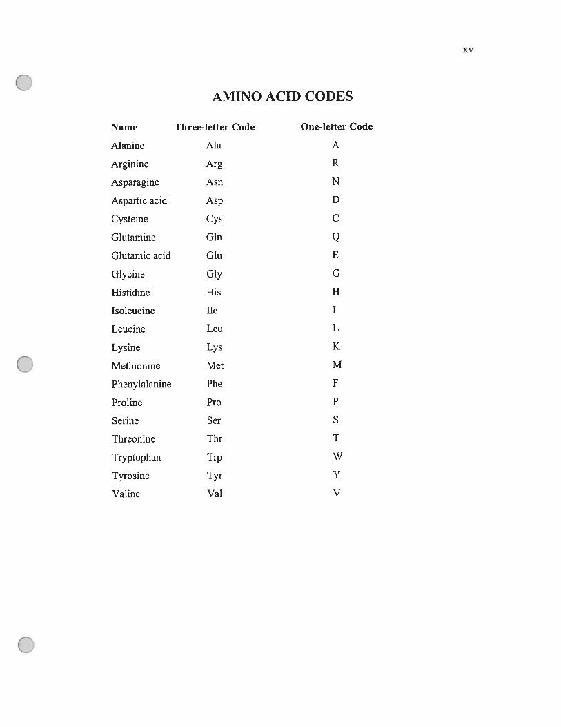
AMINO ACID CODES
Name Three-letter Code One-letter Code
Alanine Ala A
Arginine Arg R
Asparagine Asn N
Aspartic acid Asp D
Cysteine Cys C
Glutamine Gin QGlutamic acid Giu E
Glycine Gly G
Histidine His H
Isoleucine 11e I
Leucine Leu L
Lysine Lys K
Methionine Met M
Phenylalanine Phe f
Proline Pro P
Serine Ser S
Threonine Thr T
Tryptophan Trp W
Tyrosine Tyr Y
Valine Val V

xvi
ACKNOWLEDGEMENTS
I am very grateful to my director Dr. Alexey Pshezhetsky for the opportunity
to work in his laboratory and for supporting me relentlessly through this project. I am
also indebted to Dr. Ah Ahrnad who gave me the confidence and inspiration to
resume research after a long interval.
I thank Dr. Nicolas Stamatos for his indispensable role in providing the idea
of the role of Neul in human monocytic differentiation and brought me the first
paper. I am also grateful to ail our laboratory colleagues Dr. Voikan Seyrantepe,
Karine Landry, Dr. Sheila Ernst and Stéphanie Durand, Panojot Bifsha, Stéphanie
Trudel for their close collaboration, especially the technical supervision from Dr.
Voikan Seyrantepe and Karine Landry.
I wouid hike to thank the members of my jury for reading, correcting and
evaluating this thesis.
finally, I would like to thank ahi the members of rny entire family especiaiiy
rny wife and my parents for standing by me through thick and thin in making my
dream corne truc.
This work was supported by the operating grants from Canadian histitutes of
Health Research (MOP 15079 and GOP 38107), and by the equiprnent grant from
Canadian foundation for Innovation to Dr. Alexey Pshezhetsky.

CHAPTER 1: GENERAL INTRODUCTION
1.1 Lysosomes: biogenesis and biologicai foie
Lysosomes are cytoplasmic organelles discovered in 1949 by a Belgian
biologist Christian de Duve. They harbor over 100 hydrolytic enzymes such as
nucleases, proteases, lipases. glycosidases, phosphatases, sulfatases and
phiospholipases. Ail of these enzymes have an acidic pH optimum and are essentially
involved in the degradation of ail types of biological macromolecules including lipids.
potysaccharides, proteins, nucleic acids, and even whole bacteria and worn out and
nonfunctioning organelies like the mitochondria.
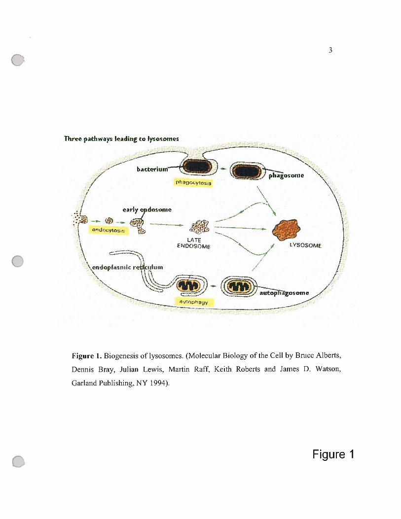
The main pathways in the biogenesis of lysosomes are shown in Figure 1.
The first pathway is followed by macromolecules from receptor-mediated
endocytosis. Initially, the endocytosed material, including the receptors, hgands and
associated membrane, is dehvered into smail irregularly shaped intracellular vesicles,
so-called early endosomes. Some of the ingested molecules are then seiectively
retrieved and recycied to the plasma membrane, while the rest of the vesicles become
late endosomes. Next, late endosomes fuse with the vesicles containing lysosomal
hydrolases from the Gotgi apparatus to form mature lysosomes. At a lower pH,
lysosomes favor the release and the hydrolytic digestion of the endocytosed
molecules. A second pathway can degrade worn-out organelles such as mitochondria
in a process called autophagy. In this process, an organelle is enciosed by membranes
derived from the ER, producing an autophagosome which then fuses with vesicles
containing lysosomal enzymes and become mature lysosome. The third pathway
(called phagocytosis) involves the uptake and degradation of large particies and
microorganisms hke bacteria by specialized celis (phagocytes). These phagocytes
(macrophages and neutrophiles in vertebrates) engulf objects to form phagosomes. a
counterpart to autophagosomes. The phagosomes are later converted to lysosomes.
As described above, the biogenesis of lysosomes is a complex process that
requires that specific sets of soluble hydrolases and membrane proteins, synthesized
in the rough endoplasmic reticulum (RER), be segregated from proteins with other
sub-cellular destinations and be transferred to form mature lysosomes. Any failure in
the biogenesis of lysosomes due to genetic mutation can lead to a severe inherited
j
-4.
Thrce p.athways Ieadi’ig ta Jysawrnes
—
- :: umt))agos
N
/‘///
/ earlyepdosonie
r;
j
-t.-- «--z:-,
\ eiiJopbiiiic re-iitiêuin
-
e
-.
Figure 1
s-’
J
IJ
Figure Î. Biogeriesis oflysosomes. (Molecular Biology ofthe Ce!! by Brnce Alberts,
Dennis Bray. Julian Lewis. Mai-tin Raff, Keith Roberts and James D. Watson,
Garland Pub!ishing, NY !994).
4
disorder, featured by the absence of one or more lysosomal enzymes. These disorders
are called lysosomal storage disorders because they result from the accumulation of
undigested macromolecules in the lysosomes of the affected tissues.
In 1967, Leroy and DeMars identified large, phase-dense inclusions in the
fibroblasts of a patient with a disorder harboring a clinical resemblance to a Hurler
Syndrome (Leroy et al., 1967). Since these celis were called inclusion ceils (1-celis),
the disorder later became known as 1-celi disease (mucolipidosis II). In 1972
Hickman and Neufeld found that multiple deficiency of lysosomal enzymes in 1-ceil
disease results from a deficiency in a recognition marker, which is common to all
soluble lysosomal enzymes and is required for their targeting to the lysosomes. This
discovery eventually lcd to the identification of the lysosomal targeting marker of the
soluble lysosomal proteins, mannose-6-phosphate (Kaplan et al., 1977) and its two
distinct receptors of 275-300 kDa and 46 kDa, respectively. Intrinsically different
mechanisms have later been identified for the targeting of membrane-bound
lysosomal enzymes (reviewed in Neufeld, 1991; Natowicz et al, 1979).
1.1.1 Lysosomal membrane
The lysosomal membrane, equipped with carriers, transport systems and a
proton pump that maintaïns the pH of the lumen, is serving as a selective
permeability barrier between the lysosomal lumen and the cytoplasm. It prevents
macromolecules that enter the lysosomes by endocytosis or autophagy from egress,
but allows exit of certain small molecules and degradation products.
Monosaccharides seem to cross the membrane by carrier-mediated systems. For
example, sialic acid transfer is mediated by a specific carrier, sialic acid transporter
protein (Havelaar et al., 1999). A deficiency in this carrier causes Salla disease,
characterized by sialic acid accumulation within the lysosomes (Martin et al. 2003).
Sanfilippo C syndrome is another disease caused by a defect in a lysosomal
membrane transporter, acetyl-CoA: alpha glycosaminide N-acetyltransferase
(Hrebicek et al., 2006), which is normally involved in the transmembrane acetylation
of heparan sulfate. Since acetylation is necessary for further heparan sulfate
5
degradation in Sanfilippo C syndrome, heparan sulfate accumulates in the lysosomes
(Bame and Rome. 1985).
Using polyclonal and monoclonal antibodies prepared against the lysosomal
membrane. several other integral or associated membrane proteins have been
identified, including lysosomal-associated membrane proteins (LAMPs) and
lysosomal integral membrane proteins (LIMPs) (reviewed in Hunziker and Geuze,
1996). These proteins are mostly found on the lumenal side of the lysosomal
membrane and contain many complex oligosaccharides bearing sialic acid residues,
which are thought to protect the membrane from the attack of lysosomal hydrolases.
The sialic acid moieties may also contribute to the establishment of a Donnan
potential for protons that maintains to the low pH of the lysosomes (Cretin, 1982).
1.1.2 Biosynthesis of lysosomal enzymes
1.1.2.1 Sorting of lysosomal membrane proteins
Membrane-bound lysosomal proteins are normally glycoproteins nch in N
linked oligosaccharides, most of which are complex. These proteins are synthesized
and glycosylated in the RER and their oligosaccharides modified in the Golgi
apparatus.
The sorting of membrane-bound lysosomal proteins, such as LIMPs and
LAMPs (Barriocanal et al., 1986), involves their transportation to the lysosomes by a
mechanism requiring the association of Gly-Tyr-X-X-hydrophobic Leu-leu amino
acid motifs at the carboxyl terminus of their cytoplasmic domain with li-subunit of
HA adaptor complexes (Guamien et aï., 1993; Pearse et al., 2000). It is independent
of mannose-6-phosphate receptors whïch mediate transport of soluble lysosomal
enzymes. The same pathway is used by the lysosomal acid phosphatase (LAP), which
is synthesized and transported to lysosomes as a transmembrane protein (Braun et al.,
1989). In lysosomes, LAP is released from the membrane by proteolytic processing,
which involves at Ieast two cleavages at the C-terminus of LAP. One cleavage is
catalyzed by a thiol proteinase outside the lysosomal membrane, removing the bulk
of the cytoplasmic tail of LAP. Another cleavage is cata]yzed by an aspartyl

6
proteinase inside the lysosomes and releases the luminal part of LAP from the
membrane-spanning domain (Gottschalk et al., 1989).
Existence of a distinct sorting mechanism for the membrane-associated
lysosomal proteins is further supported by the presence of normal levels of-
glucocerebrosidase and acid phosphatase in I-ce!! fibroblasts. Moreover. pulse
labeling and ce!! fractionation experiments of rat kidney cultured celis treated with
the g!ycosylation inhibitor, tunicamycin, have indicated that newly synthesized
plasma membrane proteins lacking N-linked oligosaccharides were nonetheless
rapidly transported to the lysosome (Barriocanal et al., 1986). These studies with
tunicamycin confirmed that the sorting of lysosomal membrane proteins is different
from the transport of soluble lysosomal enzymes.
1.1.2.2 Sorting of soluble lysosornal proteins
Significant progress lias been made towards an understanding of the targeting
mechanisms of newly synthesized soluble lysosomal enzymes. Lysosomal proteins as
well as most secretory proteins are glycoproteins synthesized in polyribosomes bound
to the RER membranes. Each protein contains an amphyphilic signal peptide at the
N-terminus which interacts with a signa! recognition particle, an 11 S
ribonucleoprotein, thereby initiating the vectoral transfer of the nascent protein across
the RER membrane into the lumen of this organelle (Erickson et al., 1981). In the
initial stage of glycoprotein synthesis, a large precursor oligosaccharide
Glc3Man9GlcNAc2 (three glucose. nine mannose and two N-acetylglucosamine
residues), is assembled with a pyrophosphate link to a ligand carrier, dolichol (Dol),
and then transferred to the target asparagine residue of the precursor glycoprotein.
The signal peptide is cleaved stili in the RER, followed by processing of the
asparagine-linked (or N-linked) oligosaccharide. Glucosidases I and II excise three
glucoses, and mannosidase cleaves one of the mannose residues from the sugar chain.
Then the proteins move to the Golgi stack by vesicular transport where they
undergo a number of post-translational modifications and are sorted for targeting to
the further destination: the lysosome. secretory granule or plasma membrane. The
7
Golgi complex consists of a stack of flat cisterns functionally divided into three
regions: cis, media] and trans Golgi. The cis part normally receives products of
biosynthesis from the RER. Dunng the passage through the Golgi apparatus, the
oligosaccharide chain residues on secretory and membrane glycoproteins are
processed stepwise to complex-type units containing sialic acid. This processing
removes five mannose residues and adds 3 molecules: N-acetylglucosamine,
galactose and sialic acid.
Almost ail of the oligosaccharides on lysosomal enzymes undergo a different
series of modifications that introduce the Man-6-P marker. The Man-6-P
distinguishes lysosomal from secretory glycoproteins and is responsible for
addressing them via one of two Man-6-P receptors to their lysosomal destinations.
The Man-6-P marker is ïncorporated ïnto lysosomal enzymes by a two-step process
that requires the sequential actions of the two enzymes. The first step involves the
enzyme UDP-G1cNAc :lysosomai enzyme N-acetylglucosamine 1 -phosphotransferase
(phosphotransferase), a multi-subunit protein (Bao et al., 1996; Raas-Rothschild et al.,
2000) which transfers N-acetylglucosamine 1-phosphate (GIcNAc-1-P) from the
nucleotide sugar uridine diphosphate-N-acetylglucosamine (UDP-G1cNAc) to the C-6
hydroxyl position of specific mannose residues on the high mannose oligosaccharides
of newly synthesized lysosomal enzymes, to produce a phosphodiester intermediate.
This modification of mannose residues in the cis-Golgi protects them from cleavage
by mannosidases in the media] Golgi. In the media] Golgi. the second enzyme, a
specific phosphodiesterase called N-acetylglucosamine-1-phosphodiester u-N
acetylgiucosaminidase (phosphodiesterase) (Kornfeld et al.. 1999) removes the
terminal N-acetylglucosamine (GIcNAc) residue uncovering the phosphate, thus
exposing the phosphomannosyl signal. Subsequently, the lysosomal hydrolases bind
to the mannose-6-phosphate receptors in the trans-Golgi network, thus remaining
intracellularly.
Two known Man-6-P receptors differ in their binding properties and divalent
cation requirements (Hoflack et al., 1985). The first identified one is a 275-300 kDa
cation-independent Man-6-P receptor (CI-MPR, MPR300), which is a type I

$
transmembrane glycoprotein with a segment of 17 kDa exposed on the cytoplasmic
side of the membrane. Detected in the Golgi complex, coated vesicles, endosomes,
and the plasma membrane, CI-MPR participates in the endocytosis of extra-ceÏÏuÏar
Man-6-P-containing proteins and has an insulin-like growth factor type II receptor
function. The importance of CI-MPR for lysosomal enzyme trafficking was
demonstrated by the massive mistargeting of lysosomal enzyme precursors in
homozygote embryos of chimeric mice lacking CI-MPR (Sahagian et aI., 1984).
The second receptor is a glycoprotein composed of three subunits of 46 kDa
each (Hoflack et al., 1985 and Robbins et al., 1981). As the cation-dependent Man-6-
P receptor (CD-MPR, MPR46), CD-MPR was first discovered in the endothelial celis.
While the CI-MPR appears to have a dominant role in lysosomal targeting. the
function of CD-MPR remains unclear. Like CI-MPR. CD-MPR is a transmembrane
protein with a cytoplasmic domain of 69 residues. Both CI-MPR and CD-MPR, like
other integral membrane proteins and endocytosed receptors, have sorting
determinants in their cytoplasmic domains. The trafficking of CI-MPR is mediated
by the tyrosine- and di-leucine-based motifs, while the CD-MPR has two distinct
targeting peptides: a weak YRGV sequence and a dominant FPHLAF sequence
(Collawn et al., 1991; reviewed in Le Borgne and Hoflack, 1998; Schweizer et al.,
2000). The affinity of these receptors to Man-6-P moieties of lysosomal hydrolases is
pH dependent: the strongly acidic lysosomal medium favors dissociation of the ligand
(Dahms et al., 1989), whereas at neutral or slightly acidic pH, the receptors bind
strongly to their ligand. Both receptors are recycled back to the Golgi complex
through the interaction with so-called tail-interacting 47-kDa protein (T1P47), which
binds the cytoplasmic domains of the CD-MPR and CI-MPR and mediates their
transport from endosomes to the Golgi complex (Orsel et aI. 2000).
1.2 Mammalian neuraminidases (sialidases)
Sialidases are widely distributed in vertebrates as well as in microorganisms
such as viruses, bacteria. fungi and protozoa, most of whïch are unable to produce
sialic acid themselves, but must process sialic acids on their hosts in order to infect
9
and replicate (Miyagi et al., 1993; Warner et aI., 1993, Colman 1994; Schenkman et
al., 1994; Chou et aI., 1996). Sialidases are glycosidases that hydrolyze the alpha-2,3-,
alpha-2,6- and alpha-2,8 linkages of terminal siaÏic acid residues in various
sialoglycoconjugates including oligosaccharides, glycoprotei ns, glycoiipids,
colominic acid and synthetic substrates. They are also known as exo-aipha sialidases,
N-acylneuraminate glycohydrolases, N-acetyl neuramyl hydrolases or
neuraminidases (EC 3.2.1.18)
In vertebrates, sialidases modulate cellular events such as activation,
differentiation, maturation and growth, ail of which involve the changes of sialic acid
level (Schauer T, 1985). In microorganisms, sialidases are believed to be important in
nutrition, using liberated sialic acid as a source of energy (Corfield, 1992). In other
situations, the bacterial sialidases are involved in pathogenesis. For example, Vibrio
cholerae sialidase removes sialic acid from the gangliosides on the surface of
interstinal celis to produce GMI-gangiioside, the binding site for choiera toxin (Gaten,
1992; Corfield, 1992; Tang et al., 1996).
In mammals, 4 distinct sialidases encoded by Neu]-Neu4 genes have been
identified. They differ in their subcellular localization, substrate preference and pH
optimum (Carrillo et al., 1997, Monti et al., 1999. Monti et al.. 2000 and Seyrantepe
et aI., 2004).
1.2.1 Cytosolic sialidase (Neu2)
Cytosolic sialidase was first isolated from rat skeletal muscles and Chinese
hamster ovary ceils and found to be optimalty active at pH 6.0 (Miyagi et al., 1990.
1993; Sato and Miyagi, 1995). Monti et al. (1999) identified the human cytosolic
sialidase gene, named Neu2 and located at human chromosome 2 (2q37), using a
sequence homoiogy-based approach. The gene encodes a 380-aa polypeptide with a
deduced molecular mass of 42 kDa, contaïning two Asp biocks, a conserved amino
acid consensus sequences (G-X-D-X-G-X-X-W/F) that are called Asp boxes and
found in ail sialidases. Another conserved sequence YRIP that contains an active site
nucleophyl is found in the amino terminal part of the peptide. Neu2 has a broad
10
substrate specificity and is active against u4 —* -sialylated oligosaccharides.
glycopeptides, and some gangliosides including G,13, GDIa and GDfb-gangtiosides, but
flot G11 and GM2-gangliosides as well as the artificial fluorogenic substrate, 4
methylumbel liferyl-alpha-D-N-acetylneuramic acid (4MU-NeuAc) (Miyagi and
Tsuiki, 1985). The exact biologïcal role of Neu2, which is expressed mostly in
muscle cells especially during their diffentiation, is stiil unknown (Sato and Miyagi,
1996; Akita et al., 1997), however, it was suggested that this enzyme may lead to the
alteration of cytoskeletal functions, through cleaving GM3-ganglioside that is
associated with the cytoskeleton. The Neu2 activity of melanoma ceils inversely
correlates with their invasive and metastatic potential (Tokuyama et al., 1997).
1.2.2 Plasma membrane sîalidase (Neu3)
Plasma membrane sialidase, also refelTed to as GMI-ganglioside sialidase. GMI
sialidase and ganglioside sialidase. is an integral membrane protein with a strict
specificity for gangliosides such as GML, GDIa and other polysialogangliosides
(Schneider-Jakob and Cantz, 1991) but not glycoproteins or oligosaccharides (Kopitz
et al., 1997; Miyagi et al., 1999). Miyagi et al. (1999) cloned and expressed a
ganglioside sialidase cDNA, isolated from a bovine brain cDNA library. The enzyme
encodes a 428-aa protein with a deduced molecular mass of 47.9 kDa and contaïns a
transmembrane domain and three Asp boxes. Neu3 has a pH optimum of 4.6 and is
activated by non-ionic detergents such as Triton X-100. Neu3 bas been recognized as
an important modulator of cellular functions including ceil-celi and celi-matrix
interactions, celi proliferation, differentiation and oncogenic transformation
(Hakomori and Igarashi., 1993; Kopitz et al., 1996, 1998). In ganglions, the enzyme
is probably involved in neuritogenesis, synaptogenesis and neuronal survival
(Tettamanti and Robin, 1993; Kopitz et al., 1994, 1997). Monti et al. (2000) cloned
cDNA of human plasma-membrane-associated sialidase (Neu3), which bas a 78%
sequence homology to the bovine protein (Miyagi et al., 1999). Human Neu3 is a
428-aa protein highly active against ganglioside substrates, and bas a pH optimum of
3.8.
11
1.2.3 Human lysosomal sialidase (Neuf)
1.2.3.1 Bïochemical properties
Lysosomal sialidase. Neul has pH optimum of 5.0 and catalyzes the removal
of terminal sialic acid residues from oligosaccharides, some glycolipids and short
glycopeptides, but is inactive against glycoproteins such as fetuin or submaxillary
mucin (Miyagi and Tsuiki, 1985, Schneider-Jakob and Cantz, 1991, Hiraiwa et aï.
1987, 1988). In the lysosomes the release of the sialic acid triggers further
degradation of the sugar moiety by other glycosidases. The human enzyme
preferentially cleaves cL4-3 and a4-6 sialyl bonds in sialoconjugates (Frisch and
Neufeld, 1979). Early reports suggested that in addition to soluble (luminal) sialidase,
lysosomes also contain another enzyme, associated with their membranes. Although
this enzyme has neyer been isolated, Miyagi et al. (1990) reported that the lysosomal
membrane sialidase from rat liver has an acidic pH optimum like its plasma
membrane counterpart and a broad specificity in hydrolyzing gangliosides as well as
fetuin, sialytïactose and 4MU-NeuAc (Kopitz et al., 1996). Since Neul is often
completely deficient in the ceils of patients affected by the inherited disease,
sialidosis caused by the mutations in the Neul gene (Bonten et al., 1996; Pshezhetsky
et al., 1997), both lysosomal membrane and intralysosomal sialidases are probably
encoded by the Nen] gene.
Multiple attempts. to purify and characterizate Neul did not succeed because
of a low tissue content and an extreme liability of the enzyme. Verheijen et al. (1983,
1985) indicated that the lysosomal carboxydase A (CathA. protective protein) forms a
complex with and activates Neul in bovine testis. Further experiments showed that in
the lysosomes of mammaÏian tissues three gïycoproteins: CathA, 3-gaIactosidase and
Neul form a multi-enzyme complex. Moffeau et al. (1992) suggested that f3-
galactosidase and CathA associate soon after synthesis in the ER and co-migrate to
the lysosomes, where they acquire their active and stable conformations. AIl 3
components of the complex can be co-purified on both f3-galactosidase-binding or
CathA-binding affinity matrices (Potier et al., 1990; Pshezhetsky and Potier, 1994).

12
In partïcular, Pshezhetsky and Potier (1994, 1996) purified human placental CathA
on agarose-Phe-Leu affinity column and concluded that CathA forms 1,270-kDa
complex with CathA, 3-ga1actosidase and the lysosomal N-acetyigalactosamine-6-
sulfatase (GALNS). Only a small percentage of total CathA and 3-galactosidase
occur in the complex, which however contains ail Neul. This in turn explains why it
is impossible to isoiate Neul activity separately from the complex (Hoogeveen et al.,
1983; Wamer et al., 1993; Hubbes et ai., 1992).
1.2.3.2 Activity and Specificity
The invoivement of Neul in the hydroiysis of sialilated gangliosides has been
a subject of debate for a long time. Characterization of the storage products in urine
and cultured fibroblasts from siaiidosis patients reveaied that sialylated
oligosaccharides are a major natural substrate for Neul (Strecker et al.. 1977;
Dorland et aI., 1978, van Peit et. al., 1988), whereas the resuits on the analysis of the
storage products in the autopsy materiais from sialidosis patients contradicted these
findings. For some patients, several foid increase of GM3 and GD3 gangliosides was
observed in systemic organs (Ulrich-Bott et al., 1987) and brain (Yoshino et al.,
1990), but other studies performed on the ceils from sialidosis patients (Sakuraba et
aI., 1983) or in the knock-out mouse model (Zhou et al., 1995) did flot confirm
storage of gangliosides. The fact that the cuitured fibrobiasts of sialidosis and
galactosialidosis patients treated with radioactiveiy iabeled GMI-ganghoside
accumulated GM3-ganglioside (Mancini et al., 1986) also suggested that Neul is
involved in degradatïon of this glycolipid.
Further studies (Schneider-Jakob and Cantz, 1991, Hiraiwa et ai. 1987, 1988)
showed that the hydrolysis of gangliosides by Neul depends on presence of ionic
detergent, such as sodium cholate or taurodeoxycholate (Triton X-100 that activated
Neu3 does not have effect on Neul), suggesting that in vivo this reaction requires
activator proteins that play in the lysosome a role of natural detergents (Conzelmann,
1987). This idea was confirmed by Fingerhut et al. (1992), who showed that Neul
cieaved GM3, GDIa, and GTIb-ganghosides in the presence of Saposin B (aiso calied
13
sulfatide activator protein). The complete hydrolysis of GD1b-gangiioside to
lactosylceramide by glycoprotein fraction from human placenta containing essentially
ail soluble lysosomal enzymes required the presence of two activators, Saposin B that
activated reactions catalyzed by 3-ga1actosidase and Neul and GM2-activator that
activated reaction of GM2 to GM3 conversion by hexosaminidase A. Gangliosides
GDIb, GM1 and GM2 were extremely poor substrates for the Neul (Fingerhut et al.,
1992). The iast conclusion was however later reconsidered when asialylated GMI and
GM2 gangliosides, GAI and GA2, respectively were found among major storage
products in knock-out mouse models of GM1-gangliosidosis (Matsuda et al., 1997)
and Sandhoff disease (combined deficiency of hexosaminidases (HEX) A and B)
(Huang et al., 1997).
Sandhoff mice succumbed to a profound neurodegenerative disease by 4-6
months of age, resembling human phenotype, while Tay-Sachs mice depleted of
hexosaminidase A remained asymptomatic to at least 1 year of age because GA2-
gangiioside was further efficientiy cleaved by hexosaminidase B (Huang et al., 1997).
Therefore, mouse models of Tay-Sachs disease have reveaied a metabolic bypass of
the genetic defect based on the more potent activity of Neul towards GM2. In order to
determine whether a similar effect would be produced by increasing the ievel of Neul
in human Tay-Sachs cells, Igdoura et al (1999) introduced a human Netti cDNA into
neuroglia celis derived from a Tay-Sachs fetus and demonstrated a dramatic
reduction in the accumuiated GM2. These studies proved invoivement of Neul in the
hydrolysis of GM2, suggesting a new method for the treatment of human Tay-Sachs
disease.
1.2.3.3 Cloning and characterization of hurnan Neul
Using the bacterial conserved sequences and taking advantage of the rapid
progress of the Human Genome Project, several groups simultaneously cloned and
sequenced human (Bonten et ai., 1996; Pshezhetsky et. al., 1997; Mimer et al., 1997)
and mouse (Cariilo et ai., 1997; Igdoura et aI., 1998, Rottier et al., 1998) Neul.
Bonten et ai. (1996) identified an expressed sequence tag (EST) clone containing a
14
full-length Neul cDNA. The mRNA was highly expressed in the pancreas, kidney
and skeletal muscle, but showed low expression level in the brain. Similarly,
Pshezhetsky et al. (1997) searched the expressed sequences tags database (UbEST) for
human analogues of bacterial sialidases and found several overlapping clones from
human fetal brain, spleen and placenta. Using primer complementary to the
sequences of two overlapping clones, they obtained a complete cDNA by RT-PCR
amplification. Both Bonten et al. (1996) and Pshezhetsky et al. (1997) localized the
gene by in situ hybridization on chromosome 6p21.3 within the human major
histocompatibility complex as previously suggested by Oohira et al. (1985).
The Netti cDNA contains an open reading frame (ORF) of 1245 nucleotides,
encoding a protein of 415 amino acids. The first 47 amino acids represent the signal
peptide consisting of a positively charged region, a central hydrophobic core and a
polar carboxy-terminal domain. The protein contains a F/YRIP domain and four Asp
boxes characteristic of bacterial and rodent Neu2 (Roggentin et al., 1993; Carillo et
al., 1997). This region is highly conserved between the human and bacterial sialidases
with sequence similarity from 32% to 38%.
The human Neul has three potential N-glycosylation sites at positions 185,
343 and 352. The positions of the active site residues are also conserved in bacterial,
rodent and human Neul. The conserved residues are Arg 37, 246 and 309 in
Salmonella typhymurium, Arg 72, 274 and 341 in mice and Arg 7$, 280 and 347 in
humans (Crenneil et al., 1993; Pshezhetsky et al., 1997; Igdoura et al., 199$). The
predicted molecular mass of the Neul precursor is 45.4 kDa (Bonten et al., 1996) and
after the cleavage of the signal peptide and glycosylation, the mature and active form
of the protein lias a mass of 48.3 kDa (Vinogradova et al., 1998).
1.2.3.4 Mutations in the Netti gene
Since the identification of the Neu] cDNA (Bonten et al., 1996; Pshezhetsky
et aI. 1997; Milner et al., 1997), considerable progress bas been made in the
understanding of the molecular defects and biochemical mechanism of sialidosis. In
two siblings with sialidosis type I, Bonten et aI. (1996) identified a heterozygous

15
1258G—*ii transversion that generated a premature TAG termination codon at amino
acid 377 and caused a C-terminal truncation of 38 amino acids. In sialidosis type II
patients Pshezhetsky et al., (1997) identified two missense mutations: a 779 T—+1
(Phe26OTyr) transversion and a 1088 T—*d transition (Leu363Pro) in a ceil une.
GMO1718A, obtained from a 2-month old infant with sialidosis type II. Another
mutation was a frameshift caused by an ACTG duplication after nucleotide 7
(7insACTG) in the GMI 1604 cell line from a sialidosis type II patient. In another
patient with sialidosis type II, Bonten et al. found a compound heterozygosity for a
401T—*1 transversion (Leu9lArg) in one allele and the other allele contained a
1337de1G deletion that caused a frameshift at amino acid 403 and a 69-aa extension
of the protein. The extension produced a 53-kDa protein immunoprecipitated from
the patient’s fifroUaas In the S MI rhce th a defidency cf the 1’èul
neuraminidase, Rottier et ai. (1998) identified a 625C—+d (Leu2O9Ile) transversion,
which accounts for the partial deficiency of Neul.
1.2.3.5 Processing and lysosomal targeting of human Neul
Several mechanisms have been proposed for sorting of the Neul precursor to
the lysosome. Van der Spoel et al. (1998) compared the intracellular distribution of
human Neul expressed in COS-l celis transfected with Netil cDNA alone or co
transfected with Neul and human CathA cDNA, and suggested that Neul associates
with CathA precursor shortiy after synthesis and that this complex is targeted to the
lysosome using a mannose-6-phosphate receptor-dependent pathway. In the absence
of CathA, Neul is partiaiiy secreted and partially segregates to endosomal
compartment (van der Spoel et al., 1998). In contrast, numerous data demonstrated
that there are two pools of Neul in the lysosome, soluble and membrane associated.
Both forms are absent in cultured ceits of sialidosis patients and are encoded by the
same gene (Miyagi et al., 1990, 1992, 1993; Verheijen et ai, 1983). Furthermore, the
activation of T lymphocytes is associated with several-fold increase of siahdase
activïty on the cell surface. The activation does not occur in T-ceHs obtained form
SMJJ or SMIBÏO mouse strains with a mutation in Neul gene (Landolfi et al., 1985;

16
Naraparaju and Yamamoto, 1994). This fact also demonstrates that sialidases
expressed on the ce!! surface and in the lysosome are the products of the same gene.
Analysis of the deduced amino acid sequence of Neul (Pshezhetsky et aI., 1997)
revealed that C-terminal tetrapeptide, 412YG1L415 bas similarity to the intemalization
signais of severai endocyted surface receptors and lysosomal membrane proteins. As
described above the Tyr-X-X-0 (hydrophobic residue) internalization signal have
been previously determined in cytoplasmic domains of several intemalized
membrane proteins, such as glucocerebrosidase LAMP-I, LAMP-II, lysosomal
membrane glycoprotein (LGP-85), Iow density lipoprotein (LDL), transferrin,
asyaloglycoprotein, polymeric immunoglobulin and cation-independent mannose-6-
phosphate receptor, (Peters and figura, 1994, Pearse et al., 2000; Hirst and Robinson,
1998).
1.3 Lysosomal storage diseases
In 1972, Hers introduced the concept of lysosomal storage diseases (LSD) to
explain how the deficiency of a lysosomai enzyme, Œ-glucosidase, couid be lethal in
Pompe disease. The undegraded substrate would gradually accumulate within the
lysosomes, increasing the size and number of organelles and thus ieading to celluiar
death and eventually to the malfunctioning of the affected organ (Hers, 1972; Van
Hoof, 1976). Later other disorders have been described caused by the molecular
defects that resulted in deficiency of lysosomal enzymes affecting the enzyme’s
synthesis, processing, routing, folding, maturation, activation, stability and
oligomerization or complex formation. LSD may manifest as several clinical forms
with different onset (infantile, juvenile or aduit) or severity. Aithough clinical
features generally vary between different disorders, the most common characteristics
include hepatosplenomegaly, neuronal deterioration, skeletal complications, coarse
faces, blindness and growth retardation.
At ieast 41 genetically distinct and biochemically related LSD caused by the
absence of one or more lysosomal hydrolases have been described (reviewed in
17
Neufeld, 1991). Based on the clinical and biochemical manifestations of the disease,
LSD are grouped into different classes: sphingolipidosis, mucopolysaccharidosis,
mucolipidosis, glycoprotein storage diseases, lysosomal membrane transport
disorders and others types. LSD are consïdered rare and each one typically affects
fewer than 10,000 people in the world, although high prevalence values have been
reported in some populations (Meikie et al., 1999) Most LSD are inherited in an
autosomal recessive manner, with the exception of Fabry disease and
mucopolysaccharidosis (MPS) type II, which show X-linked recessive inheritance.
The biggest group of lysosomal storage diseases is related to deficiencies of
glycosidases involved in the catabolism of sugar chains of glycolipids,
oligosaccharides and glycoproteins. This group includes single enzyme deficiencies
as in Gaucher, Tay-Sachs and Sandhoff diseases, as weII as the functional
deficiencies of multiple enzymes caused by genetic mutations in sphingolipid
activator proteins (saposins, GM2-activator protein) which stimulate the activity of
glycosidases against gangliosides and glycoshpingolipids.
Both Tay-Sachs and Sandhoff diseases are caused by a failure in the
catabolism of GM2-ganglioside. Protein products of three genes are required for this
process: HEXA and HEXB genes encoding the akancJ3Â subuits d hexos aElini da A
and the gene encoding the GM2-activator, a lipid-binding protein that presents the GM2
substrate to the enzyme. A defect in the HEXA gene causes Tay-Sachs disease, an
autosomal recessive, progressive neurodegenerative disorder. Although less than 0.3
% of the general populations are carriers for the disease, the frequency is ten foÏd
higher (3 %) in Ashkenazi Jews (Petersen et al., 1983). This disease is characterized
by the developmental retardation, followed by paralysis, dementia and blindness and
usually causes death at the age of 2 or 3 years. A defect in the HEXB gene leads to the
clinically related Sandhoff disease.
Gaucher disease, another sphingolipidosis, is the most common lysosomal
storage disease with an estimated carrier frequency of 4.6% among Ashkenazi Jews
in Israel (Matoth et al., 1987). It is caused by deficiency of glucocerebrosidase,
leading to the lysosomal accumulation of glucosylceramide.

18
Disorders caused by defects in the lysosomal membrane transporters include
Nieman-Pick type C involving a cholesterol transport defect, sialic acid storage
disease (sialic acid transport protein deficiency) and cystinosis (cystine transport
protein deficiency).
A distinct group of disorders: galactosialidosis, f3-galactosidosis and sialidosis
involve the components of the 1.27 MDa lysosomal multi-enzyme complex of CathA,
-ga1actosidase and Neul,
1.3.1 Galactosialidosis
Galactosialidosis (GS) is an autosomal recessive disease caused by a primary
defect of protective protein/CathA which resuits in a combined secondary deficiency
of f3-galactosidase and Neul (Goldberg et al., 1971; Suzuki et al., 1988, Zhang and
Callahan, 1996; D’ Pzo i., 1995). Œ is charadŒized by tyc I yo niI
storage disease manifestations including coarse facies, macular cherry-red spots,
mental retardation, corneal opacities, vertebral changes, bone marrow foam ceils and
vacuolated lymphocytes.
According to the severity and age of onset, GS can be classified into three
phenotypically distinct forms: an early infantile, late infantile and juvenile/adult type.
The early infantile onset manifest prenatally as non-immunologic hydrops fetalis, the
excessive accumulation of serious fluid in the subcutaneous tissues and serous
cavities of the fetus (Stone and Sidransky, 1999), or postnatally as ascites, massive
edema, proteinuria, viceromegaly, skeletal dysplasia and early death from cardiac
failure, kidney failure or airway obstruction (Wenger et al., 1978; Lowden and
O’ Ri er 1979). So n earl -infantile GS patients have demonstrated cytopenias
(abnormal decrease in number of ceils in the bone marrow) (Olcay et al., 1998) and
punctuate epïphyses ofthe femora, calcanei, and sacrum (Patel et al., 1999). The late
infantile onset is associated with hepatosplenomegaly, growth retardation, cardiac
involvement and absence of relevant neurologic signs. The juvenile/adult onset,
patients may survive to adulthood, often without mental retardation or viceromegaly,
but with ataxia, myoclonus and angiokeratoma. About 70% of GS cases can be
19
classified in this group and the majority of reported patients are of Japanese origin
(Suzuki et al., 1988; Takano et al., 1991). Parental consanguinity lias been reported in
haif of the families.
First OS patients reported by Pinsky et al. (1974) and Loonen et al. (1974)
had 3-ga1actosidase deficiency but showed a clinical phenotype different from that of
patients affected with -ga1actosidosis (GM i-gangliosidosis) with normal intelligence
and late development of psychomotor deterioration. Galjaard et al. (1975) performed
somatic ceils hybridization studies, showing that these variants are caused by the
defects in a gene other than 3-ga1actosidase. Similarly, Wenger et al. (1978) found a
combined deficiency of two lysosomal enzymes, f3-galactosidase and Neul. AIl
reported patients belonged to the same complementation group, aliowing designating
the condition as a distinct disorder called galactosialidosis (GS) (Andria et al., 1981).
For several years, primary molecular defect in GS was thought to be same as that in
single Neul deficiency, sialidosis (Cantz et al., 1977). However, Hoogeveen et al.
(1980, 1981) showed that hybridization and even co-culturing of fibroblasts from OS
and sialidosis patients resulted in partial correction of Neul and -ga1actosidase
activity. These studies suggested a protein corrective facto?’ exists in normal,
sialidosis or GM1-gangliosidosis celis, but absent in those of GS patients. Further
studies showed a 10-fold enhanced cellular degradation of -galactosidase in
galactosialidosis fibroblasts (van Diggelen et al., 1982) that could be prevented by the
addition to the celi medium of either a fraction containing the corrective factor” or
by the inhibition of lysosomal proteases (Suzuki, et al. 1988, d’Azzo et al, 1982).
CathA precursor is a 54 kDa protein which is processed in the lysosomes to
the mature form containing two protein chains of 32 and 20 kDa. D’Azzo et al. (1982)
showed that both the 32 kDa protein and its 54 kDa precursor are genetically absent
in the ceils of ail OS patients and that addition of the 54 kDa precursor to these cells
restores the normal level of 3-galactosidase protein. Moreover, the 32-kDa subunit
was identified as the “°ccrrecti vefact d’± nhsi ‘E in S cals ‘DA7oet t, 1E2
It became possible to understand the molecular defects in OS only after the
isolation and characterization of the gene encoding human CathA. The CathA gene
20
consists of 15 exons, spanning 7.5 kb on human chromosome 20 (20q13.1) (Gaijart et
al., 1988; Shimmoto et al., 1996). The protein is synthesized as a 542 amino acid
precursor, glycosylated at Asn 117 in the ER, transported via the mannose-6-
phosphate receptor (Morreau et al., 1992) to the lysosomes where it is activated by
the proteolytic cleavage (Bonten et al., 1995).
A number of molecular lesions in the CathA gene in ail subtypes of
galactosialidosis have been identified. For instance, Zhou et al. (1996) found three
mutations, VIO4M, L208P and G41IS, in patients with earÏy infantile GS. These
mutations prevented the phosphorylation of the CathA precursor and thereby its
transport to the lysosome. Examples of late infantile GS mutations include M378T,
which generates a new Asn-hnked glycosylation site, and Y221N, which decreases
the stabiiity of CathA in the lysosome (Zhou et ai.. 1996). A 2-nucleotide deletion,
C5l7deflT, and an intronic mutation, IVS$+9C-G, which resuits in splice defect,
generated frameshifts and a protein truncation (Richard et al., 1998). Among
Japanese patients with juvenile/aduit onset the most common mutations are a Y249N
change (Fukuhara et ai., 1992), and an IVS7, A-G, +3, EX7DEL mutation, resuiting
in skipping of exon 7 in the mRNA (Shimmoto et al., 1996).
Because the CathA protein has about 30% amino acid sequence identity with
the yeast carboxypeptidase Y and the wheat carboxypeptidase-il (Gaijart et aI., 1988,
Elsliger and Potier, 1994), the X-ray atomic coordinates of the wheat enzyme were
used to build the CathA structure model (Elsiiger and Potier, 1994). Later, the X-ray
structure of the CathA precursor expressed in a baculovirus system was deterrnined
with a 2.2-2.4 resolution (Rudenko et al., 1995). The structure is similar to those of
plant and yeast serine carboxypeptidases, showing that CathA belongs to a so-calied
Œ-hydrolase family (Remington, 1993). The protein is composed of a cap domain
and a core domain. The cap domain consists of three u-helixes and three-stranded
mixed -sheet. The core domain consists of a central ten-stranded -sheet which is
flanked by ten Œ-helixes and two small -strands on both sides. The catalytic triad in
the active site is forrned by the 5cr’50, His429 and Asp372 residues. At acidic pH,

21
the enzyme forms 95-98 kDa homodimers with known X-ray structure (Rudenko et
al., 1995). The pI for the human enzyme is 5.4.
The resolution of the X-ray structure of CathA precursor allowed a
comprehensive analysis of structural changes induced by the mutations in CathA
molecules (Rudenko et al., 199$). The analysis revealed a correlation between the
effects of mutation on the protein structure and the clinical phenotype of the affected
patients. None of the mutations occurred in the active site or at the protein surface.
Among II amino acid substitutions modeÏed, 9 found in patients affected with severe
early or laie infantile type of GS (Q21R, S23Y, W37R, S62L, V1O4M, L208P,
Y367C, M378T, and G411S) were located in the central core domain of CathA.
These substitutions introduced unbalanced charged groups, hydrogen bonds or bulkier
side chains to the protein core; or created cavities in protein interiors and interfaces.
AI! these changes would dramatically change the folding of mutant CathA, resulting
in impaired sorting and rapid degradation. In contrast, the other two mutations
(F412V and Y221N) associated with a more moderate clinical effect were located in
the Œ-helical cap domain of the enzyme and predicted to have a milder effect on
protein structure. For several mutations (Q21R, W37R, S62L, Y221N, Y367C and
F4 12V), homologous modeling of CathA structure (Elsiiger and Potier, 1994) also
made similar predictions.
1.3.2 Sialidosis
Sialidosis (SL) is a very rare inherited metabolic disorder characterized by a
single Neul deficiency leading to the abnormal accumulation of complex
carbohydrates (mucopolysaccharides) and mucolipids in many tissues of the body.
Previously known as Mucolipidosis I (Cantz et al., 1977), the cherry-red spot
myclonus syndrome (O’ Ri en ., 196), the Goldberg syndrome (Thomas et al.,
197$) and nephrosialidosis (Maroteaux et al., 1978) SL belongs to a subgroup of
lysosomal diseases known as mucotipidoses. Lowden and O’ Bi en ( 197 poi ded
the nosology, sialidosis, and its classification into two phenotypically distinct
subtypes: sialidosis type I and sialidosis type II, based on dysmorphic features,
22
severity and age of onset. Sialidosis type I (Goldberg syndrome or non-dysmorphic
type) is a mild and late-onset form characterized by bilateral macular cherry-red spots,
debilitating myoclonus (sudden involuntary muscle contractions) and progressive
visual impairment (Durand et al., 1977; Soggs et al., 1979; Rapin et al., 197$;
O’ lieu 197g Federico d L, 1980). Ihese sy nJto i usuUy dom; appear urtil
the second decade of life. O’ Ri en and ner (1980) nrt edt ha dosis type I was
more frequent in Italians. Sialidosis type II (mucolipidosis I,
lipomucopolysaccharidosis or dysmorphic type) is a severe and infantile onset form
associated with skeletal dysphasia, Hurleroïd features (mildly coarse facial features),
dysostosis multiplex (bone malformations), mental retardation, hepatosplenomegaly
and death in the first decade of life (Kelly et al, 1977; Winter et al., 1980; Oohira et
al., 1985). Earlier reports indicated that most type II patients were of Japanese origin
(O’ Rien and Vrner. 1980). In a recert udy Nsh ya na L (1997) repated a
cerebral blood llow and glucose metabolism decrease in the occipital lobe region of
Japanese patients with the aduit onset sialidosis. Other symptoms may include
abdominal swelling; Ioss of muscle mass (atrophy); in-egular, involuntary spastic
muscle movements (choreoathetosis); Iack of muscle tone (hypotonïa); the protrusion
of a portion of the intestines through an abnormal opening in the muscular waIl of the
abdomen (inguinal hernia) (Provenzale et al., 1995) Nephrosialidosis (congenital
sialidosïs), a phenotypic variant of sialidosis type II, presents type II symptoms as
well as severe hydrops fetalïs, ascites and proteinuria. Death usually resuits from
edema, dysproteinuria and bleeding (Maroteaux et al., 1978; Tylki-Szymanska et al.
1996). Besides the above clinical features, Buchholz et al. reported severe dilated
coronary arteries, excessive retinal tortuosity and an erythematous. macular rash in an
infant with congenital sialidosis.
Sialidosis is a very rare lysosomal storage disorder with insignificant
prevalence in a selected population. Since Tipton et al. noted at least 11 cases in 1978,
there have been no published reports on the global incidence of sialidosis. However,
according to the Mucopolysaccharadosis (MPS) society, sialidosis occurrence is
between 1 in 250,000 and 1 in 4.2 million (www.mpssociety.caldiseases).
23
Various studies based on geographic and!or ethnic distribution of lysosomal
storage diseases have been conducted. In the Netherlands, only 49 cases of
mucolipidoses and oligosaccharidoses were diagnosed between 1970 and 1996, with
a combined birth prevalence is 1.0 per 100.000 live births (Poorthuis et al., 1999). In
Australia, 27 different lysosomal storage diseases were diagnosed in 545 patients and
a prevalence ranging from 1 in 57,000 live births for Gaucher disease to 1 in 4.2
million live births for sialidosis was reported (Meikie et al., 1999). The overali
incidence for ail types of LSD in this study was approximately 1 in 7,700 live births.
Since the Australian population has mainly a British ancestry, with a minor
contribution from other European countries and Asia, Meikie et al. believed their
results could be extrapolated to white non-Hispanic populations in the United States,
Canada and the United Kingdom. At least fifteen cases of sialidosis type II have been
reported in Japan (Nishiyama et al., 1997; Naganawa et al., 2000).
1.4 Biological role of Neul in the immune response
As discussed above, the role of Neul in the ïntratysosomal catabolism of
sialilated glycolipids and glycoproteins has been well-established. Several studies
have shown that theNeul-encoded sialidase, in addition to its role in intralysosomal
catabolism of sialylated glycoconjugates, is also involved in cellular signaling during
the immune response. Endogenous sialidase activity increases in celis of the immune
system following ceIl activation (Chen et al., 1997 and Katoh et al., 1999). The
enhanced sialidase activity and consequent desialylation of surface glycoconjugates
in activated cells induced production of interleukin-4 by lymphocytes. In particular, T
celis require Neul for both early production of IL-4 and the IL-4 priming of
conventional T ceÏÏs to become active IL-4 producers (Chen et aÏ., 1997). During the
activation of T celis, Neul is expressed on the plasma membrane where it participates
in desialylation of surface antigen-presenting molecules such as myosin heavy chain
class I, required to render T cells responsive to APCs (Landolfi et al., 1986), and
GM3-ganglioside, which modulates Ca2 immobilization and regulates IL-4
production (Chen et al., 2000). In addition, Neul of T lymphocytes converts the

24
group specific component or Gc protein into a factor necessary for the inflammation
primed activation of macrophages (Naraparaju et al., 1994). T-cells derived from
SM/Jor B10.SM strains ofmice, partially deficient in Neul (Carrillo et al., 1997), fail
to convert Gc and synthesize IL-4, and B celis of these mice cannot produce IgGi and
IgE after immunization with pertussis toxin. Since binding of Neul to CathA is
required for activation it is possible to suggest that these two enzymes remain
associated on the plasma membrane. Therefore, Neul may interact with CathA
precursor in the Golgi and stay associated with it on the root to the plasma membrane.
In accordance with this hypothesis recent studies demonstrated the presence of
enzymatically nonactive CathA precursor on the plasma membrane (Hinek et al.,
1996).
Lukong et al., (2001) have shown that in the activated T-cells, Neul-encoded
sialidase is increased on the ccli surface in good agreement with the data of Chen and
Landolfi. Lukong et al., further investigated the mechanism by which Neul reaches
the cell surface and showed that it contains the internalization signal found in
lysosomal membrane proteins targeted to endosomes via clathrin-coated pits. The
signal consists of a C-terminal tetrapeptide 412YGTL415, with Tyr412 and Leu415
essential for the endocytosis of the enzyme. In tum, the redistribution of Neul from
lysosomes to the cdl surface of activated lymphocytes was accompanied by increased
reactivity of the enzyme towards anti-phosphotyrosine antibodies. Lukong et al.,
suggested that a specific mechanism regulates sorting and retention of newly
synthesized Neul on the surface of activated T celis. They proposed that the
interaction of Neul with adaptor complexes and targeting to the lysosome can be
blocked by phosphorylation ofthe essential tyrosine in the intemalization motif. Such
a mechanism has been recently described for cytotoxic T Ïymphocyte-associated
antigen (CTLA-4), which is transiently expressed on the surface of activated T ceils
and is involved in their down-regulation where it may influence immune function
(van der Horst et al., 1989; Lukong et al., 2000). In activated T cells, phosphorylation
of the essential Tyr’65 residue by the T-lymphocyte associated tyrosine Src family and
25
Jak2 kinases prevents its interaction with the 12 ubunit of AP2 and resuits in its
expression on the celi surface (Bradshaw et ai., 1999 and Chikuma et ai., 2000).
Several unes of evidence also suggest that Neul plays a pivotai roie in
celluiar signaling during monocytes differentiation and activation. For example,
activation of monocytes caused significant induction of sialidase activity which
enhanced binding of CD44 on the surface of monocytes to hyaiuronic acid, a
component of the extraceiluiar matrix (Gee et ai., 2003). In this study, they showed
that LPS-induced CD44-mediated HA (CD44-HA) binding in monocytes is reguiated
by endogenously produced tumor necrosis factor (TNF)-a and IL-10. Furthermore,
p38 mitogen- activated protein kinase (MAPK) activation was required for LPS- and
TNF-Œ-induced, but flot for the IL-10-induced CD44-HA-binding in normai
monocytes. It suggested that Neul activation may be required for the acquisition of
the HA-binding form of CD44 in LPS- and TNF-stimuiated monocytic celis.
In previous studies, LPS stimulation increased the Neul activity in monocytes,
but it is flot known whether it was due to the gene induction or post-transcriptional
activation (Katoh et ai., 1999). The Neul induction is not oniy specific for T-cells and
monocytes. The induction of the Neul gene by IL-1 was also observed in human
biood neutrophuis, where it was proposed to be invoived in tissue recruitment (Cross
et al., 2002). IL-1 is known to have a specific stimuiatory effect on neutrophil
recruitment (Lin et al., 2002), but whether or flot the Neul induction plays a role in
the IL-1-induced neutrophil recruitment remains to be elucidated.
In the SMJJ mice, partial Neul deficiency is specifically associated with the
suppressed expression of Th2, but not Thi, cytokines (Chen et al., 1997). However,
in cultured human T-cells, overexpression of Neul resuited in enhanced production
of both Thl and Th2 cytokines (Wang et al., 2004). There are two possible
explanations for this apparent discrepancy. One possibility relates to species
difference, as suggested by the difference of Neul regulation in mice and humans
(Landolfi et al., 1986 and Lukong et al., 2001). Aiternativeiy, and more plausibiy, the
specific association of Neul with Th2 cytoldne expression may happen in vivo only.
In fact, it is reported that in mouse T-celis and DCs treatment with a virai Neul
26
resulted in enhanced production of multiple cytokines, including both Th2 and Thi
(Oh et al., 2000). Taken together these resuits suggest that Neul may indeed be
involved in the expression ofThl and Th2 cytoldnes in both humans and mice.
Previously identified (Monti et al., 2000) plasma membrane-associated
sialidase Neu3 promotes cell adhesion to laminins, integrin-mediated signaling to
ERK and subsequent activation of celi proliferation, but attenuates adhesion to
fibronectin and the related signaling (Kato et al., 2006). There is also evidence that
Neu3 overexpression may contribute to sustained ERK activation through Shc and
FAK signaling, when ceils are attached to laminin-5, teading to adhesion-dependent
cell proliferation. Neu3 overexpression in T-cells resulted in enhanced production of
several cytokines of both Thl and Th2 types, but not of IL-4. These results suggest
that gangliosides blocked Thi cytokine expression without affecting IL-4 (Wang et
al., 2004). On the other hand, the differential effects of Neul and Neu3 on IL-4
expression may be due to the fact that the two siaÏidases have different substrates on
the ceil surface.
Neul and Neu3 may regulate cytokine genes expression by processing certain
gangliosides on the cell surface. Gangliosides are known to be involved in a variety
of biological functions in most (if not ah) celis, including cytokine gene regulation in
T-cells (Kanda et al., 2001; Irani et al., 1996) and antigen-presenting cells (Shen et al.,
2002). It is possible that Neul and Neu3 may hydrolyse different gangliosides in vivo,
which may explain why they have differential effects on cytokine genes expression.
Neul and Neu3 may also activate intracellular signaling, which leads to cytokine
production by desialylating ceIl surface glycoproteins, such as TCR, CD4, CD8,
CD43, CD45, IFN-yÀrece1i œ, andigands for siglecs (Pappu et al., 2004 and Ikehara
et al., 2004). Future experiments using lymphocytes from individuals affected with
sialidosis (primary Neul deficiency) and galactosialidosis (secondary Neul
deficiency) may help to establish the specific substrates for Neul and Neu3 and
define the contribution of each enzyme to the production of WN-yÂIn addition to
influencing the production of IFN-y4 I’èu andJor Neu3 may play a role in some of
the other immune functions of human lymphocytes. This prospect raises the

27
possibility that inhibition of the activity of cellular sialidases with anti-sialidase
antibodies or pharmacologic inhibitors may have a therapeutic value in treating
inflammation and infection.
1.5 Research hypotheses and objectives
The previous studies from our lab and published data summarized above
showed that Neul, in addition to its role in intralysosomal catabolism of sialylated
glycoconjugates, is also involved in cellular signaling during the immune response
participating in the production of cytokines by T celis. There is also indirect
evidence that Neul may also play an important role in other types of immune ceils, in
particular in monocytes, macrophages and dendritic celis. These celis play a central
role in innate and adaptive immune responses and their activation is central to the
outcome of virtually eveiy infectious disease. In addition a failure to properly
regulate macrophage function during infection is often itself a major cause of disease.
For example, chronic infections, such as tuberculosis, are often characterized by the
inability of macrophages to eliminate persistent infections. On the other hand, in
septic shock, caused by acute infections, hyperactivated macrophages initiate
cascades of immune deregulation often resulting in death. Therefore understanding of
the signaling pathways in macrophages holds great promise for future development of
therapeutic targets for a plethora of inflammatory diseases, but in contrast to the
relatively well-understood pathways in innate immune responses, the signaling
pathways that are necessary to counter and temper aggressive macrophages are
poorly characterized.
Our major hypothesis is that the lysosomal sialidase Neul and /or CathA,
which forms with Neul the lysosomal multi-enzyme complex and activates it in the
lysosome play a crucial role in signaling, during the differentiation and activation of
macrophages through transcriptional induction and mobilization to the celi surface in
stimulated celis. In order to test this hypothesis, the specific aims are based on the
analysis of ceil unes and ceils from two mouse models generated in our laboratory.
The mouse models are: the CathAS9OA point mutation with single CathA deficiency,
28
and the CathA9O0 hypomorph with a double CathA/Neul deficiency. These
models are keys to our studies because they link biochemical deficits in the protein
components of the lysosomal multienzyme complex to end-stage phenotypes only
amenable through the functional organism.
Our specific aims are:
1. To smdy the expression ofNeul and other sialidases during the activation ofblood
monocytes and monocytic celi lines.
2. To study the regulation ofNeu] gene transcription in monocytes and macrophages.
3. To study the targeting ofNeul and CathA in monocytes and macrophages.
4. To study the biological role of Neul in ceil signaling during the maturation and
activation of immune celis using siRNA and the ceils of transgenic mice with a
single CathA and double CathAINeul deficiency.

CHAPTER 2: DIFFERENTIAL EXPRESSION 0F
ENDOGENOUS SIALIDASES 0F HUMAN
MONOCYTES DURING CELLULAR
DIFFERENTIATION INTO MACROPHAGES
ARTICLE 1
Publié dans:
The fEBS Journal, 2005 May; 272(10):2545-56
30
DIFFERENTIAL EXPRESSION 0F ENDOGENOUS
SIALIDASES 0F HUMAN MONOCYTES DURING
CELLULAR DIFFERENTIATION INTO
MACROPHAGE S
Nicholas M. Stamatos1’2, Feng Liang3, Xinli Nan’, Karine Landry3, Alan S. Cross2,
Lai-Xi Wang’ and Alexey V. Pshezhetsky3
1 Institute ofHuman Virology, University ofMaiyland, Baltimore, MD, USA
2 Division of Infectious Diseases, Department ofMedicine, University of Maryland
Medical Center, Baltimore, MD, USA
3 Hôpital Sainte-Justine and Département de Pédiatrie, Université de Montréal,
Montréal, Quebec, Canada
Correspondence: N. M. Stamatos, 725 West Lombard St.,
Institute of Human Virology, University of
Maryland Medical System, Baltimore,
MD 21201, USA
Fax: +1 410 7064619
Tel: +1 410 7062645
E-mail:

31
ABSTRACT
Sialidases are enzymes that influence cellular activity by removing terminal
sialic acid from glycoÏipids and glycoproteins. Four genetically distinct sialidases
have been identified in mammalian celis. In this study, we demonstrate that three of
these sialidases, lysosomal Neul and Neu4 and plasma mernbrane-associated Neu3,
are expressed in human monocytes. When measured using the artificial substrate 2’-
(4 methylumbelliferyl)-a-d-Nacetylneuraminic acid (4-MU-NANA), sialidase activity
of monocytes increased up to 14-fold per milligram of total protein afier cells had
differentiated into macrophages. In these same ceils, the specific activity of other
cellular proteins (e.g. f3-galactosidase, cathepsin A and aikaline phosphatase)
increased only two- to fourfold during differentiation of monocytes. Sialidase activity
measured with 4-MU-NANA resultcd from increased expression ofNcul, as removal
of Neul from the ceil lysate by immunoprecipitation eliminated more than 99% of
detectable sialidase activity.
When exogenous mixed bovine gangliosides were used as substrates, there
was a twofold increase in sialidase activity per milligram of total protein in
monocyte-derived macrophages in comparison to monocytes. The increased activity
measured with mixed gangliosides was not affected by removal of Neu I, suggesting
that the expression of a sialidase other than Neul was present in macrophages. The
amount of Neu] and Neit3 RNAs detected by real time RT-PCR increased as
monocytes differentiated into macrophages, whereas the amount of Neu4 RNA
decreased. No RNA encoding the cytosolic sialidase (Neu2) was detected in
monocytes or macrophages. Western blot analysis using specific antibodies showed
that the amount of Neul and Neu3 proteins increased during monocyte
differentiation. Thus, the differentiation of monocytes into macrophages is associated
with regulation of the expression of at least three distinct cellular sialidases, with
specific up-regulation of the enzyme activhy of only Neul.

32
INTRODUCTION
Sialic acid is present on glycoproteins and glycolipids that are widely
distributed throughout nature. Removal of sialic acid from these glycoconjugates on
the surface of mammalian ceils changes the functional capacity of the ceils [1—8].
Sialidases comprise a famiiy of enzymes that remove terminal sialyl residues from
glycoconjugates. Four genetically distinct forms of mammalian sialidase have been
characterized, each with a predominant cellular localization (lysosomal, cytosolic or
plasma membrane-associated) and substrate specificity [9—17]. Lysosomal sialidase
(Neul) has a catabolic role in desialylating glycoproteins and glycolipids in
lysosornes [18], but is also present on the surface of activated T ceils [19], where it
may influence immune function [2,20]. Plasma membrane sialidase (Neu3) localizes
on the ccli surface [13, 14] and, by preferentiaily desialylating gangliosides, is
believed to have a regulatory role in cellular activation, differentiation and
transformation [4,2 1—23]. The cytosolic sialidase (Neu2) can desialylate both
glycoproteins and gangliosides [12], but its function remains to be determined. The
function of the recentiy characterized Neu4 sialidase also has not been established.
Neu4 sialidase is expressed in a wide range of cdl types [15—17], has broad substrate
specificity, and is iocalized in lysosomes [17].
Endogenous sialidase activity increases in cells of the immune system
following cdl activation [2,5,6,20,24—27]. The enhanced sialidase activity and
consequent desialyiation of surface glycoconjugates in activated celis induced
production of interleukin-4 by lymphocytes [2], enhanced binding of CD44 on the
surface of monocytes to hyaluronic acid, a component of the extracellular matrix
[5,27], and promoted the transendothelial migration of polymorphonuclear ieukocytes
(PMNs) [7]. In activated lymphocytes [2,20] and PMNs [7], the effect on ceils was
attributed to the activity of Neul sialidase, some of which was translocated from
lysosomes to the cdl surface [7,19]. The role of the other forms of sialidase in the
activation ofthese cells has not been deterrnined.
Circuiating peripheral blood monocytes play a key roie in potentiating diverse
immune activities and can differentiate into either macrophages or dendritic cells by

33
exposure to specific stimuli [2$]. The function of monocytes changes from antigen
recognition and processing to antigen presentation in macrophages and dendritic
celis. We have previously shown that desialylation of glycoconjugates on the surface
of freshly isolated monocytes using an exogenous bacterial neuraminidase activated
the extracellular signal-related kinase 1 /2 (ERK 1 / 2), enhanced the production of
specific cytokines, and promoted the responsiveness of monocytes to bacterial
lipopolysaccharide [29]. In this paper, we demonstrate that endogenous sialidase
activity of freshly isolated human monocytes is up-regulated as they differentiate into
macrophages. We show that (a) Neul and Neu3 are present in both monocytes and
macrophages, and that the specific activity of only Neul is up-regulated in
comparison to other lysosomal proteins during differentiation; (b) Neu4 is also
expressed in monocytes as evidenced by the presence of Nezt4 RNA, but that the
amount of this RNA declines during monocyte differentiation; and (c) Neu2 is not
detected at the RNA level in either monocytes or macrophages.
EXPERIMENTAL PROCEDURES
Isolation of peripheral blood mononuclear ceils and purification of monocytes
Peripheral blood mononuclear cells were isolated by leukophoresis of blood
from HIV- 1 and hepatitis B and C seronegative donors followed by centrifugation
over Ficoil-Paque Plus (Amersham Biosciences, Uppsala, Sweden) gradients using
standard procedures. Monocytes were purified from peripheral blood mononuclear
ceils by an additional centrifugation over Percoll (Amersham Biosciences, Uppsala,
Sweden) gradients and then by negative selection using StemSep separation columns
(Stem Cdl Technologies, Vancouver, BC, Canada) as per the manufacturer’s
suggested protocol. The purity of monocytes exceeded 95% as determined by flow
cytometry after staining ceils with phycoerythrin (PE)-, allophycocyanin (APC)-, or
fluorescein isothiocyanate (fITC)-conjugated monoclonal antibodies to CD3, CD 14,
CD 19, CD206 and isotypic control IgGs (ah mAbs from BD PharMingen, San Diego,
CA, USA). Briefly, lx 106 cells were resuspended in 0.5 mL of a solution containing
NaCI / Pi pH 7.4, 2% human serum AB and anti-CD32 Fc receptor Abs (1.5 tg)

34
(Stem Ce!! Technologies) and incubated at 4°C for 15 min to minimize non-specific
binding ofreagents. Ceils were then stained at 4°C for 30 min with the fluorochrome
conjugated monoclonal antibodies, washed with 2 mL of NaCY / Pi and fixed with
1.0% (y / y) paraformaldehyde. CeÏls were analyzed using a Becton-Dickinson
FACScaliber (Mountain View, CA, USA) and data were analyzed using FLOWJO
data analysis software. The viability of monocytes was greater than 97% as
determined by trypan blue dye exclusion.
Culture conditions for purified monocytes
To obtain monocyte-derived macrophages, purified monocytes were
suspended at 2x10⁶ cells/mL in RPMI medium 1640 (Gibco, Grand Island, NY,
USA) containing 10% heat-inactivated human AB serum (Gemini Bioproducts,
Calabasas, CA, USA) and recombinant human macrophage colony stimulating factor
(rhM-CSF; R&D Systems, Inc., Minneapolis, Ivfl’l, USA) at 10 ng/mL and were
maintained at 2.5x10⁶ cells per well in six-well tissue culture plates (Costar, Coming
Inc., Coming, NY, USA) at 37°C in a 5% (v/v) humidified C02 incubator. At the
indicated times, nonadherent celis were removed by two washes with NaC1 / P pH
7.4 and the adherent, differentiating macrophages (larger and more granular than
monocytes as seen by light microscopy) were harvested in NaC1/Pi pH 7.4 by gentle
scraping with a polyethylene ce!! scraper (Nalge Nunc International, Rochester, NY,
USA). The harvested cells were confirmed to have characteristic macrophage ccl!
surface phenotypic markers (CD 14+, CD206+) by flow cytometry that was
performed as described above.
Measurement of sialidase activities
Cells were collected on the indicated days and 2x10⁶ monocytes (day 0) or 5
x105 cells on days 3 and 7 were suspended in 0.20 mL ofa solution containing 0.5%
(v/v) Triton X-100, 0.05 M sodium acetate pH 4.4, and 0.125 mM 4-MU-NANA
(Sigma-Aldrich, St. Louis, MO, USA) and incubated at 37°C for 1 h. The reaction
was terminated by the addition of 1.0 mL of a solution containing 0.133 M glycine,

35
0.06 M NaC1 and 0.083 M Na2CO3 pH 10.7. Liberated 4-methylumbelliferone was
measured with a Victor2 1420 spectrofluorometer (Wallac, Turku, Finland) with
excitation at 355 nm and emission at 460 nm. The amount of 4-methylumbelliferone
that was liberated from 4-MU-NANA during the 1 h reaction was determined by
comparison to a standard curve of increasing amounts of 4-methylumbelliferone
(Sigma-Aldrich). In this assay, 1 nmol of liberated 4-methylumbelliferone signified
the release of 1 nmol of sialic acid, and a unit of sialidase activity was defined as the
amount of enzyme that released 1 nmol of sialic acid per hour at 37°C. Protein
concentration was measured by the Bradford method using a protein assay kit (Bio
Rad, Hercules, CA, USA) and the amount of activity measured in each sample was
conected based on protein concentration to represent activity per milligram ofprotein
as seen in fig. 1.
Sialidase activity was also determined against mixed bovine brain
gangliosides (Calbiochem, La Jolla, CA, USA) and in the absence of exogenous
substrate (i.e. where activity reflects the release of sialic acid from endogenous
cellular sialylconjugates). In these assays, cells were collected on the indicated days
and 2x10⁶ cells were suspended in 0.20 mL of a solution containing 0.1% (y / y)
Triton X-100, 0.05 M sodium acetate pH 4.4, 0.1% (w / y) BSA (Pentex bovine
albumin fraction V, Miles Inc., Kankakee, IL, USA) and 0.250 mlvi mixed bovine
brain gangliosides. Altematively, the gangliosides were omitted from the reaction
mixture such that any detected free sialic acid would be that released from cellular
sialylconjugates. After a 60 min incubation at 37°C, the reaction mixture was
microfuged to remove cellular debris and 0.02 mL of each supernatant was analyzed
for sialic acid content using a Dionex DX600 chromatography system (Dionex
Corporation, Suimyvale, CA, USA) equipped with an electrochemical detector
(ED5O, Dionex Corporation), as described previously [7]. Material from each 0.02
mL sample was injected into a CarboPac-PA1 column (4 x 250 mm) in the presence
of 0.1 M NaOH, and sialic acid was eluted using a gradient of 5—20% (w/v) 1 M
sodium acetate in 0.1 M NaOH over 15 min at a rate of 1 mL / min. Under this
condition, sialic acid was eluted at 8.7 min and was quantified by integration of the

36
peak area using a standard solution of sialic acid as the reference. One unit of
sialidase activity was defined as the amount of enzyme that liberated 1 nmol of sialic
acid per hour at 37°C. The amount of activity measured in each sample was corrected
based on protein concentration to represent activity per milligram of protein as seen
in fig. 1.
Quantitation of other lysosomal and cellular proteins
Freshly isolated monocytes and macrophages after 7 days in culture were
collccted and homogenized in H20 by sonication. Hexosaminidase and J3-
galactosidase activity were measured separately by incubating 5jig of ccli
homogenate in 0. lmL of a solution containing 40 mM sodium acetate pH 4.6 and
either 1.25 mM 4-methylumbellifeiyl-2-acetamido-2-deoxy-J3-d-glucopyranoside or
1.5 mlvi 4-rnethylurnbelliferyl-f3-d-galactoside as previously described [51,52]. After
incubation at 37°C for 15 or 30 mm, the reactions were terminated with 1.9 mL of
0.4 M glycine buffer pH 10.4 and the arnount of fluorescence of the liberated 4-
methylumbelliferone was measured with a Shimadzu Rf-5301 spectrofluorometer.
Alkaline phosphatase, glutamate dehydrogenase and cathepsin A activities in 5 jig of
cdl homogenate were measured as described elsewhere [34,53,54]. The amount of
lysosome-associated membrane protein-2 (LAMP-2) in monocytes and macrophages
was detennined by separating cellular proteins by SDS / PAGE, electrotransferring
them to polyvinyldifluoride membranes, and reacting the proteins that were
transferred to the biots with monoclonal antihuman LAMP-2 antibodies (Washington
Biotechnology Inc., Baltimore, MD, USA). Antibody-bound LAMP-2 was detected
using the BM chemiluminescence kit (Roche Diagnostics, Mannheim, Germany) in
accordance with the manufacturer’s protocol.
Immunoprecipitation ofNeul multienzyme complex
Neul exists in a multienzyme complex with -d-ga1actosidase and cathepsin
A [18,31—34] and can be immunoprecipitated selectively from ccli lysates using anti
cathepsin A antibodies [34]. Neither Neu2 nor Neu3 form oligomeric structures when
37
purifïed from tissues [55,56]. In addition, when COS-7 celis were transfected with
plasmids that expressed Neu3 or Neu4 and celi lysates were reacted with anti
cathepsin immune serum, neither Neu3 nor Neu4 sialidases were immunoprecipitated
[K. Landry, unpublished resuits]. Freshly isolated monocytes or monocyte-derived
macrophages (106 celis) were homogenized in 0.55 mL of a solution containing 100
mM NaCÏ, 0.5% (w / y) of sodium desoxycholate, and 50 mM sodium phosphate
buffer, pH 6.0. After centrifugation of the homogenate at 12000 g for 10 mm, 0.20
mL ofthe supematant was mixed with 0.10 mL ofa solution containing 10 mg/mL
BSA, 100 mM NaC1, and 50 mM sodium phosphate buffer, pH 6.0 with 5 tg of
rabbit anti-human cathepsin A immune serum or preimmune serum and incubated at
4°C for 1 h as described elsewhere [34]. The pellet from 0.300 ml ofPansorbin Ceils
(Calbiochem, La Joua, CA, USA) was added to the reaction mixture after the 1 h
incubation and the sample was incubated for an additional 1 h at 4°C with constant
shaking. The im;uune complexes were rernoved from the supernatant by
centrifugation at 13000 g for 10 min. The supematants were assayed for f3-
galactosidase (GAL), f3-hexosaminidase (HEX), and sialidase activities as described
above.
Isolation of RNA and real lime RT-PCR
Monocytes and monocyte-derived macrophages were harvested as previously
described and total RNA was isolated using an RNeasy mini kit (Qiagen, Valencia,
CA, USA) following the protocol suggested by the manufacturer. The RNA
preparation was treated with DNase I (Invitrogen, Carlsbad, CA, USA) at 37°C for 30
min to remove contaminating DNA. DNase was then removed by binding to Blue
Sorb DNase affinity slurry (Clonogene, St. Petersburg, Russia).
Semi-quantitative real-time RT-PCR was performed using a QuantiTect
SYBR green RT-PCR Kit (Qiagen, Valencia, CA, USA) with an ABI Sequence
Detection System (ABI PRISM 5700) to detect gene expression of Neul (GenBank
accession NM 000434), Neu2 (GenBank Accession NM 005383), Neu3 (GenBank
accession AB008185), and Neu4 (GenBank accession NM 080741) using RNAs

38
generated as described above. Gene expression of 18S rRNA (GenBank accession
X03205) was also measured as an internai control. The following primers were
selected using Primer Express vl.0 (Applied Biosystems, Foster City, CA, USA) or
DNAsis Max (Hitachi, Japan) software and were synthesized by Qiagen
(Germantown, MD, USA): Neït] (forward; nt 1047—1066) 5’-
TGTGACCTTCGACCCTGAGC-3’ and (reverse; nt 115 1—1170) 3’-
CTCACTTGGACTGGGACGCT-5’ yielding a 123 base product; Neu2 (forward; nt
45$—477) 5’-AGTGGTCCACCTTTGCAGTG-3’ and (reverse; nt 581—600) 3’-
GGAAGACGAAGGAGTCGGTA-5’ yielding a 142 base product; Neit3 (forward; nt
$44—864) 5’-AATGTGAAGTGGCAGAGGTGA-3’ and (reverse; nt 971—991) 3’-
GGACTCAGCTGTCGAGACACT-5’ yielding a 147 base product; Neït4 (forward;
nt 1002—1020) 5’-TGCTGGTACCCGCCTACAC-3’ and (reverse; nt 1085—1104) 3’-
AAGATGTCGCTACTGGTGCC-5’ yielding a 103 base product; and 18S rRNA
(forward: nt 1279—1298) 5’ -CGGACAGGATTGACAGATTG-3’ and (reverse; nt
137$—1397) 3’-TTGCTTGCTCTGAGACCGTA-S’ yielding a 119 base product. Ten
nanograms (10 ng) of total RNA was added to a 25 j.iL final reaction mixture
containing 0.5 .iM of each primer pair, 1 x QuantiTect SYBR-green RT-PCR Master
Mix and 0.25 jiL of QuantiTect RT Mix. To synthesize cDNA, reverse transcription
was performed at 50°C for 30 min. Following a 15 min hot start at 95°C, DNA
amplification was allowed to proceed for 40 cycles (15 s at 95°C, 30 s at 57 oc and
30 s at 72°c). Ail reactions were mn in triplicate. Semi-quantitative analysis was
based on the cycle number (CT) at which the SYBR-green fluorescent signal crossed
a threshoid in the log-linear range of RT-PCR, indicating the relative amount of
starting template in each sample. The fold change in expression of Neul, Neu3, and
Neu4 RNAs in macrophages compared to monocytes was normalized to the
expression of 185 rRNA and was calculated by equation where AACi= (CT
Neul,2 or 3 — Ci iss rRNA)macrophages — (CT Neul,2 or 3 — CT iss rRNA) monocytes. The
accuracy of each reaction was monitored by analysis of melting curves and product
size on gel electrophoresis.
39
Western blot analysis of cellular proteins
Monocytes and macrophages were collected at the indicated times and
proteins from 2 x10⁶ celis were solubilized in 0.1 mL ofa solution containing 50 mM
Tris/HCI pH 7.4, 100 mlvi NaC1, 0.5% (v/v) Triton X-100, 0.5% (w/v) sodium
desoxycholate, 0.1% (w/v) SDS and protease inhibitors (1 250 dilution ofprotease
inhibitor cocktail from Sigma-Aldrich). Protein concentration was measured by the
Bradford method using a Bio-Rad protein assay kit (Bio-Rad). Proteins (5 tg) from
each celi lysate were resolved by electrophoresis on a 10% SDS/polyacrylamide gel
using Tris / glycine /SDS rulming buffer (gel and mnning buffer from Invitrogen,
Carlsbad, CA, USA), electrotransferred by a semi-wet method to a Sequi-Blot
polyvinyldifluoride membrane (Bio-Rad) and probed with polyclonal rabbit
antibodies to either Neul or Neu3 at 0.5 jig/mL The polyclonal anti-Neul Igs were
generated by immunizing rabbits with recombinant human Neul sialidase and were
characterized as described elsewhere [38]. Rabbit polyclonal anti-Neu3 Igs were
generated by immunizing rabbits with a synthetic peptide conesponding to amino
acids 109—12$ of the human Neu3 sialidase and were affinity-purified using the
immunogen that was coupled to a column. These anti-Neu3 Igs detected a single 47
kDa band in COS-7 ceils that were transfected with the Neu3 gene. The respective
blots were incubated with a 1 10 000 dilution of goat HRP-conjugated anti-rabbit
IgGs (Santa Cruz Biotechnology, Inc., Santa Cruz, CA, USA), developed using an
ECL chemilurninescence substrate kit (Amersham Biosciences, Piscataway, NJ,
USA), and exposed to Kodak X-ray film.
RESULTS
Differentiation of monocytes into macrophages resuits in increased expression of
endogenous sialidase(s)
To determine whether differentiation of monocytes into monocyte-derived
macrophages is associated with changes in the level of endogenous sialidase activity,
monocytes were purified from the peripheral blood of human donors and maintained
in culture conditions that promoted differentiation into macrophages. The amount of

40
sialidase activity in freshly isolated monocytes (CD 14+, CD206—) and in monocyte
derived macrophages (CDY4+, CD206+) after 3 and 7 days in culture was determined
using the exogenous sialidase substrates 2’-(4-methylumbelliferyl)-Œ-d-N-
acetylneuraminic acid (4-MU-NANA) and mixed bovine gangliosides. These
substrates are utilized with different efficiencies in vitro by the four genetically
distinct mammalian sialidases [10,13,14,30]. Sialidase activity of ceils was also
evaluated in the absence of exogenous substrates to determine whether any of the
cellular sialidases was able to desialylate endogenous sialylconjugates under the
conditions that were used. Sialidase activity from solubilized ceils in each assay
reflected the amount of sialic acid that was released from glycoconjugates (one unit
of activity was defined as the amount of enzyme that liberated 1 nmol of sialic acid
per hour at 3 7°C) and was measured either fluorometrically when 4-MU-NANA was
used or by HPLC when gangliosides or endogenous sialylconjugates were used. In
the absence of 4-MU-NANA and exogenous gangliosides, 3.9 + 1.0 nmol of sialic
acid were liberated per hour by the sialidase activity in 1 mg of total protein from
freshly isolated monocytes (day 0, Fig. lA). The amount of this activity against
endogenous substrates per milligram of protein rose to 17.2 + 3.7 units when these
celis had differentiated into macrophages after 7 days in culture (day 7, Fig. lA). The
22.2 + 2.3 units of sialidase activity in freshly isolated monocytes detected when
exogenous gangliosides were used as substrate increased to 48.1 ± 4.4 units after 7
days in culture (Fig. lB). With 4-MU-NANA as substrate, 4.7 + 1.2 units ofsialidase
activity in freshly isolated monocytes rose to 64.0 + 9.7 units after 7 days in culture
(Fig. 1C). Sialidase activity was not detected in monocytes or monocyte-derived
macrophages when the assay measuring activity against endogenous sialylconjugates
(i.e. in the absence of 4-MU-NANA or exogenous gangliosides) was performed at
4°C, making it unlikely that the liberated sialic acid that was measured in this
condition (Fig. lA) was simply the resuit of free intracellular sialic acid being
released from solubilized ceils (data not shown). These resuits using different
substrates demonstrate that the endogenous sialidase activity of monocytes increases
as they differentiate in vitro into macrophages.

41
The increase in activity of lysosomal sialidase Neul during monocyte
differentiation is greater than the change in activity of other lysosomal enzymes
Neu 1 exists in a multienzyrne complex with f3-d-galactosidase and cathepsin
A in the lysosorne and when isolated from solubilized celis (reviewed in [18,3 1—34]).
To determine whether Neul was responsible for most ofthe activity seen with 4-MU-
NANA in F ig. 1C, antibodies to human cathepsin A were used to
coimmunoprecipitate Neul from the ccli lysate prior to evaluating sialidase activity.
The anti-cathepsin A Igs immunoprecipitated most of the f3-galactosidase (GAL)
activity from both monocytes and macrophages, whereas f3-hexosaminidase (HEX)
activity, that i not associated with the Neul multienzyme complex, was not changed
(Fig. 2). These antibodies precipitated from both monocyte and macrophage extracts
more than 99% of sialidase activity against 4-MU-NANA at pH 4.4 (Fig. 2). When
celi extracts were incubated in the presence of preimmune Igs prior to
immunoprecipitation, there was no change in the amount of sialidase activity against
4-MU-NANA (data flot shown). The anti-cathepsin antibodies did not remove the
sialidase activity against mixed gangliosides (MG, Fig. 2), suggesting that the
sialidase activity measured with rnixed bovine gangliosides was flot due to the
activity ofNeul. Thus, the activity ofNeul and at least one other sialidase increased
during monocyte differentiation into macrophages.
To determine whether the activity of Neul was specifically up-regulated
during monocyte differentiation, changes in activity of other lysosomal enzymes and
in the amount of a specific lysosomal protein (LAMP-2) were also measured as
freshly isolated monocytes differentiated into macrophages. The specific activity of
sialidase using 4-MU-NANA as substrate increased 12- to 14-fold during monocyte
differentiation into macrophages (Fig. 1C and Table 1). In contrast, the specific
activity of other lysosomal enzymes (J3-hexosaminidase, -ga1actosidase and
cathepsin A) and the amount of the lysosomal membrane protein LAMP-2 increased
only two to four fold during differentiation of monocytes to macrophages (Table 1).
In addition, the specific activity of the mitochondrial enzyme glutamate
dehydrogenase and plasma membrane aikaline phosphatase increased 3.8- and 3.2-
42
foid, respectiveiy, as monocytes differentiated into macrophages. Thus, the increase
in sialidase activity during monocyte differentiation exceeded the changes in specific
activity and amount of increase in other lysosomal proteins. As most of the sialidase
activity measured using 4-MU-NANA under the conditions stated above represented
the activity ofNeul, these resuits suggest that the activity ofNeul was specificaily
up-regulated during monocyte differentiation.
The amount of RNA encoding Neul and Neu3 sialidases increases during
monocyte differentiation
To detemiine whether the increased sialidase activity in monocyte-derived
macrophages that was seen using various substrates (Fig. lA—C) was associated with
increased expression of RNA encoding Neul, Neu2, Neu3, and Neu4, the relative
amount ofthese RNAs in freshiy isolated monocytes and in macrophages maintained
in culture over a 7-day period was determined by reai-time RT-PCR. The amount of
RNA for each sialidase was compared with the amount of RNA encoding 18$ rRNA,
an internai control for gene expression in the differentiating monocytes. RNAs
encoding Neul, Neu3, and Neu4 were detected in freshiy isoiated monocytes and
monocyte-derived macrophages, but no RNA encoding Neu2 was detected in either
ccli (data not shown). As monocytes differentiated into macrophages, the amount of
RNA encoding Neul and Neu3 increased 3.5 ± 0.2- and 3.9 + 0.$-fold, respectively,
in relation to the change in amount of 18$ rRNA (Fig. 3). In contrast, the amount of
Neu4-specific RNA deciined 6.7 + 0.1-foid during differentiation (Fig. 3). At ail
times anaiyzed, the absolute amount of Neul RNA exceeded that of Neu3 and Neu4
(crossover threshoids Ci’ during PCR for 18$ rRNA, Netti , Neu3, and Neit4 RNAs in
monocytes were 17.7 + 0.1, 26.1 + 0.4, 29.5 + 0.5, and 27.8 + 0.6, respectively). The
resuits were specific for each gene as confirmed by the expected size and
characteristic melting temperature of each PCR gene product (data flot shown).
43
The amount of Neul and Neu3 proteins increases during differentiation of
monocytes to macrophages
Given the increase in sialidase activity and in amount of RNA encoding Neul
and Neu3 that occurred when monocytes differentiated to macrophages, it was
determined whether there was a corresponding increase in the total amount of Neul
and Neu3 proteins. Proteins from freshly isolated monocytes and from monocyte
derived macrophages were separated by SDSPAGE and then analyzed on western
blots using rabbit polyclonal antibodies that were specific for Neul and for Neu3.
The anti-Neul IgGs recognized the 44—46 kDa Neul sialidase in monocytes and
macrophages (Fig. 4A). As expected from the observed increase in Neul-specific
RNA and in sialidase activity using 4-MU-NANA, immuno-detection of Neul with
anti-Neul IgGs revealed a more intense band in macrophages than in monocytes (Fig.
4A). Likewise, the anti-Neu3 IgGs recognized a protein with molecular mass of 47
kDa in both monocytes and macrophages (Fig. 43), with an increase in intensity of
staining of this protein in macrophages (Fig. 4B). Thus, these results suggest that the
absolute amounts of both Neul and Neu3 proteins increased as monocytes
differentiated into macrophages, consistent with an increase in the arnount of RNA
encoding each.
DISSUSSION
We have described in this report that endogenous sialidase activity of freshly
isolated human monocytes increases as celis differentiate in vitro into macrophages.
The 12- to 14-fold increase in specific activity ofsialidase in macrophages measured
using 4-MUNANA reflected predominantly the activity ofNeul sialidase. This was
confirmed by the removal of greater than 99% of sialidase activity using 4-MU-
NANA when Neul was immunoprecipitated from the ccli lysate using antibodies to
cathepsin A as was described previously [34]. The increase in Neul activity during
monocyte differentiation was consistent with the observed increase in Neu]-specific
RNA and in Neul protein, as shown by real time RT-PCR and western blot analyses.
This increase in Neul activity during monocyte differentiation was at least threefold

44
greater than the change in specific activity of other lysosomal proteins, suggesting
that the expression of Neul was specifically up-regulated. It remains to be
determined whether the increased enzymatic activity of Neul in monocyte-derived
celis results simply from increased transcription of Neul RNA. Although there was
only a 3.5-fold increase in Neu]-specific RNA in macrophages, there was greater
than a 12-fold increase in enzymatic activity. This apparent discrepancy between
amount of RNA and enzyme activity was likely not due to changes in the expression
of cathepsin A, as the specific activity of cathepsin A increased only 1.8-fold in
macrophages compared to monocytes. Cathepsin A, also referred to as protective
protein / cathepsin A (PPCA), is a protein component of the 1.27 MDa Neul
multienzyme complex that protects and activates Neul [reviewed in 18,31—34]. We
previously have shown that cathepsin A is present in human placenta in at least 100-
fold molar excess to the Neul sialidase. A portion (about 30%) ofcathepsin A exists
in the form of a 680 kDa complex with f3-galactosidase [34—37], while a larger
amount is present in 110 kDa homodimers. These homodimers are in dynamic
equilibrium with the 1.27 MDa Neul-containing complex, but the average ratio
between the 1.27 MDa and 680 kDa complexes is 1—10 [34,35,38]. Similar data were
reported for other tissues [39—43]. Therefore, it is likely in monocyte-derived cells
that there is an excess ofcathepsin A to stabilize and activate the amount ofNeul that
is present. Neul has the potential for post-translational modifications: it has several
potential glycosylation sites and is phosphoiylated in activated lymphocytes [19].
Thus, it is possible that the specific up-regulation of Neul activity in macrophages
may result partly from post-translational modifications.
Sialidase activity was also measured using mixed bovine gangliosides under
conditions that detect preferentially Neu3 sialidase [13,14,30]. The twofold increase
of this activity in macrophages was consistent with the two- to fourfold increase in
expression of other cellular enzymes that were analyzed. Immunoprecipitation of
Neul from the cdl lysate using anti-cathepsin A Igs had littie effect on the increased
sialidase activity detected with gangliosides, suggesting that this activity was flot due
to the activity of Neul. The increase in sialidase activity detected with exogenous

45
gangliosides likely was a result of neither Neu2 nor Neu4 activity. Neu2 activity was
barely detectable and the amount was unchanged in monocytes and macrophages
(0.39 and 0.30 units per mg cellular protein, respectively) when measured under
conditions that were specific for Neu2, and the level of Neu4 RNA declined. The
increase in the amount of Neït3 RNAs and of the 47 kDa protein detected with anti
Neu3 IgGs support that Neu3 is responsible for this activity. The increased sialidase
activity in activated ceils of the immune system [2,5,6,20,24—27] bas recently been
attributed in lymphocytes to specific forrns of sialidase [20]. Neul and Neu3
sialidases werc found to be up-regulated in human CD4+ lymphocytes that were
activated with antibodies to CD3 and CD28 [20]. As was shown previously for Neul
[2], these sialidases appeared to play a role in cytokine production in lymphocytes
[20]. Activation of the THP-l monocytic celi line by exposure to lipopolysaccharide
for at least 8—12 h also leads to enhanced sialidase activity (presumed to be Neul),
yet the specific sialidase(s) involved was not directly identified [5,271. One effect of
this enhanced activity in monocytes was increased binding of the cdl surface protein
CD44 to hyaluronic acid, a component of the extracellular environment [5,27].
Changes in the expression of Neul and Neu3 sialidases have been detected in other
types of human celis that were induced to differentiate. Malignant colon ceils express
more Neu3 RNA and gangliosidespecific sialidase activity than normal colonic cells,
yct when these ceils were induced to differentiate, the amount of Neu3 RNA and
sialidase activity declined while Neul activity increased [23]. It should be noted that
the function of Neu3 appeared to be different in neuroblastoma celis in which the
over-expression of a transfected Neu3 gene promoted differentiation [4,21,22].
Monocytes and macrophages perform rnany critical functions in the immune
system. During monocyte differentiation, the increase that we observed in the activity
of lysosomal Neul, especially if translocated from lysosomes to the ceil surface as
occurs in activated lymphocytes [19], may be important for some of these functions.
Given the altercd cytokine production of monocytes following desialylation of ccli
surface glycoconjugates [29], it is possible that the cnhanccd Neul activity may
contribute to celi activation and / or differentiation. Desialylation of glycoconjugates
46
on the surface ofmonocyte-derived celis likely influences the ccli to ccii interactions
that are critical for cell-mediated immunity. Like other ceils of the immune system,
monocytes and macrophages express sialic acid binding Ig-like lectins (siglecs) on
their surface [reviewed in 44]. As some of these sigiecs have binding sites that are
masked by sialic acid on resting celis, it is possible that during monocyte
differentiation, binding sites are exposed by the increased expression ofNeul. Celito
ceil interactions that are mediated by numerous other carbohydrate recognition
molecules (e.g. galectins, selectins) [reviewed in 45] couid aiso be influenced by the
action ofNeul and Neu3 on ce!! surface glycoconjugates.
Macrophages recognize, phagocytize and process foreign objects (e.g.
bacteria, vimses) and present antigens on the ccli surface for stimulation of other celis
of the immune system. Desialylation of ccli surface glycoconjugates in vivo may
make monocytes and macrophages more responsive to activation [29] and increase
their chernotactic response to sites of inflammation, as was shown in PMNs [7]. As
an antigen presenting cdl, macrophages may be able to enhance the immunogenicity
ofprocessed antigens if the increased sialidase activity resuits in rernoval ofthe sialic
acid masks of concealed epitopes [46]. In this respect, it is of interest to note that in
dendritic ceiis, major histocompatibiiity ciass II molecuies are present in the
lysosome (intraceliular site of Neul) prior to transiocation to the ccli surface with
processed antigens (reviewed in [47]).
Although we have described the expression of sialidases in monocytes and
macrophages and discussed their potential roie in ccii function, the opposing activity
of sialyitransferases, a famiiy of enzymes that add siaiic acid to the terminal galactose
of glycoconjugates, can flot be ignored. Hyposialyiation of ccii surface
glycoconjugates occurs in activated celis [6,48—501, but this could occur from
increased sialidase activity and / or from dccreased siaiyltransferase activity, as was
recentiy demonstrated for the transmembrane protcin tyrosine phosphatase CD45
[50]. Specific gaiactosebinding iectins have been used to characterize the sialylation
status of the ccii surface [6,49,50], but it shouid be noted that these iectins bind to
giycornoieties that may represent oniy a fraction of totai potentiai sialyiation sites,

47
and thus, their binding may flot reflect the global sialylation state of the ccli. Further
studies will define whether there is a global hyposialylation of the ccli surface during
monocyte differentiation or whether specific molecules are the target of the Neul and
Neu3 sialidases.
Although the plasma-membrane and lysosomal sialidases localize
predominantly to distinct intraceilular sites, translocation throughout the cdl occurs
[7,19,26]. The lysosomal sialidase is translocated in activated lymphocytes from
intracellular organcilcs to the cdl surface after being phosphoiylated by a ccllular
kinase [19]. 11 is possible that lysosomal Ncul also is translocatcd to the pcriphery of
monocyte-derived celis and, with the continuous endocytosis that occurs in these
celis, that the mcmbrane-associated Neu3 sialidase of macrophages is also recycled
through the ccli bctwcen the ccli surface and intracellular granules. Given the
changes in expression and dynamic intracellular repositioning ofNeul and Neu3 that
likely occur during monocyte differentiation, establishing the rolc(s) of human
sialidases during the differcntiation of monocytes presents great challenges.
ACKNOWLEDGEMENTS
This work was supported in part by National Institutes of Hcalth grants K08
HL72176-O1 to NMS, AI 54354 to LXW, AI 42818—01 to ASC and Canadian
Institutes of Hcalth Research grant FRN 15079, Vaincre les Maladies Lysosomalcs
Foundation grant and Canadian Foundation for Innovation equipmcnt grant to AVP.
NMS is grateful to Peter John Gomatos for discussion throughout this work and
critique of the manuscript and to Cathryn Andoniadis for critical review of the
manuscript.
48
REFERENCES
1 Boog CJ, Neefies JJ, Boes J, Ploegh HI & Melief CI (1989) Specific immune
responses restored by alteration in carbohydrate chains of surface molecules on
antigenpresenting celis. Fur J ImmunoÏ 19, 537—542.
2 Chen XP, Enioutina EY & Daynes RA (1997) The control of IL-4 gene expression
in activated murine T lymphocytes: a nove! role for neu-1 sialidase. Jlmmunol
158, 3070—3080.
3 Stamatos NM, Gomatos PJ, Cox J, fowier A, Dow N, Wohlhieter JA & Cross AS
(1997) Desialylation of peripheral blood mononuclear celis promotes growth of
111V-1. Virology 228, 123—131.
4 Wu G & Ledeen RW (1991) Stimulation of neurite outgrowth in neuroblastoma
ceils by neurarninidase: putative role of GMÏ ganglioside in differentiation. J
Neurochem 56, 95—104.
5 Katoh S, Miyagi T, Taniguchi H, Matsubara Y, Kadota J, Tominaga A, Kincade
PW, Matsukura & Kohno S (1999) Cutting edge: an inducible sialidase regulates
the hyaluronic acid binding ability of CD44-bearing human monocytes. Jlmmunol
162, 5058—5061.
6 Galvan M, Murali-Krishna K, Ming LL, Baum L & Ahmed R (199$) Alterations in
ccli surface carbohydrates on T ceils from virally infected mice can distinguish
effector/memory CD8+ T ceils from nai ve cells. Jhnrnztnol 161, 641—648.
7 Cross AS, Sakarya S, Rifat S, Held TK, Drysdale BE, Grange PA, Cassels fJ,
Wang LX, Stamatos NM, farese A, Casey D, Poweil J, Bhaffacharjee AK,
Kleinberg M & Goldblum SE (2003) Recniitment of murine neutrophils in vivo
through endogenous sialidase activity. JBiol Chem 278, 4112—4120.
8 Hunig T (1983) The role of accessory celis in polyclonal T ccl! activation II.
Induction of interleukin 2 responsiveness requires cell-cell contact. Fur J InvnunoÏ
13, 596—601.
9 Pshezhetsky AV, Richard C, Michaud L, Igdoura S, Wang S, Elsiiger MA, Qu J,
Leclerc D, Grave! R, Dallaire L & Potier M (1997) Cloning, expression and

49
chromosomal mapping of human lysosomal sialidase and characterization of
mutations in sialidosis. Nat Genet 15, 3 16—320.
10 Mimer CM, Smith SV, Carrillo MB, Taylor GL, Hollinshead M & Campbell RD
(1997) Identification ofa sialidase encoded in the human major histocompatibility
complex. JBioÏ Chem 272, 4549—4558.
11 Miyagi T, Konno K, Emori Y, Kawasaki R, Suzuki K, Yasui A & Tsuik S (1993)
Molecular cloning and expression of cDNA encoding rat skeletal muscle cytosolic
sialidase. JBioÏ Chem 268, 26435—26440.
12 Tringali C, Papini N, Fusi P, Croci G, Borsani G, Preti A, Tortora P, Tettamanti G,
Venerando B & Monti E (2004) Properties of recombinant human cytosolic
sialidase HsNEU2: the enzyme hydrolyzes monomerically dispersed GMI
ganglioside molecules. JBioÏ Chem 279,
13 Monti E, Bassi MT, Papini N, Riboni M, Manzoni M, Venerando B, Croci G,
Preti A, Ballabio A, Tettamanti G & Borsani. G (2000) Identification and
expression ofNEU3, a novel human sialidase associated to the plasma membrane.
BiocÏ?eln J 349, 343—35 1.
14 Miyagi T, Wada T, Iwamatsu A, Rata K, Yoshikawa Y, Tokuyama S & Sawada
M (1999) Molecular cloning and characterization of a plasma membrane-associated
sialidase specific for gangliosides. JBiot Chein 274, 5004—5011.
15 Comelli EM, Amado M, Lustig SR & Paulson JC (2003) Identification and
expression ofNeu4, a novel murine sialidase. Gene 321, 155—161.
16 Monti E, Bassi MT, Bresciani R, Civini S, Croci GL, Papini N, Riboni M,
Zanchetti G, Ballabio A, Preti A, Tettarnanti G, Venerando B & Borsani G (2004)
Molecular cloning and characterization of NEU4, the fourth member of the human
sialidase gene family. Genoniics 83, 445—453.
17 Seyrantepe V, Landiy K, Trudel S, Hassan JA, Morales CR & Pshezhetsky AV
(2004) Neu4, a novel hurnan lysosomal lumen sialidase confers normal phenotype
to sialidosis and galactosialidosis cells. JBioÏ Chem 279, 3702 1—37029.
50
18 Pshezhetsky AV & Ashmarina M (2001) Lysosomal rnultienzyme complex:
biochemistiy, genetics, and molecular pathophysiology. Frog Nïtcleic Acid Res
Mol Bio! 69, 81—114.
19 Lukong KE, Seyrantepe V, Landry K, Trudel S, Ahmad A, Gahi WA, Lefrancois
S, Morales CR & Pshezhetsky AV (2001) Intracellular distribution of lysosomal
sialidase is controlled by the intemalization signal in its cytoplasmic tail. J Biol
Chem 276, 46172—46181.
20 Wang P, Zhang J, Bian H, Wu P, Kuveikar R, Kung TT, Crawley Y, Egan RW &
Billah MM (2004) Induction of lysosomal and plasma membrane-bound sialidases
in human T-cells via T-cell receptor. Biochein J 380, 425—433.
21 Hasegawa T, Yamaguchi K, Wada T, Takeda A, Itoyama Y & Miyagi T (2000)
Molecular cloning of mouse ganglioside sialidase and its increased expression in
Neuro2a cdl differentiation. JBiol Chem 275, 8007— 8015.
22 Kopitz J, Muhl C, Ehemann V, Lehmann C & Cantz M (1997) Effects of cdl
surface ganglioside sialidase inhibition on growth control and differentiation of
human neuroblastoma celis. Eur J Ce!! Biol 73, 1—9.
23 Kakugawa Y, Wada T, Yamaguchi K, Yamanami H, Ouchi K, Sato I & Miyagi T
(2002) Up-regulation of plasma membrane-associated ganglioside sialidase (Neu3)
in hurnan colon cancer and its involvement in apoptosis suppression. Froc Nati
Acad Sel USA 99, 10718—10723.
24 Taira S & Nariuchi H (1988) Possible role ofneuraminidase in activated T ceils in
the recognition ofallogeneic Ta. Jlminunol 141, 440—446.
25 Landolfi NF, Leone J, Womack JE & Cook RG (1985) Activation of T
lymphocytes resuits in an increase in H-2-encoded neuraminidase. Imrnunogenetics
22, 159— 167.
26 Cross AS & Wright DG (1991) Mobilization of sialidase from intracellular stores
to the surface of human neutrophils and its role in stimulated adhesion responses of
these celis. J Clin Invest 88, 2067—2076.
27 Gee K, Kozlowski M & Kumar A (2003) Tumor necrosis factor-alpha induces
functionally active hyaluronanadhesive CD44 by activating sialidase through p38
51
mitogen-activated protein kinase in lipopolysaccharidestimulated human
monocytic celis. JBioÏ Chem 278, 37275—37287.
2$ Sallusto F & Lanzavecchia A (1994) Efficient presentation of soluble antigen by
cultured human dendritic cells is rnaintained by granulocyte / macrophage colony
stimulating factor plus interleukin 4 and downregulated by tumor necrosis factor
alpha. JExp Med 179, 1109— 111$.
29 Stamatos NM, Cuneli S, Zella D & Cross AS (2004) Desialylation of
glycoconjugates on the surface of monocytes activates the extracellular signal
related kinases ERK 1 /2 and results in enhanced production of specific cytokines.
JLeuk Bio! 75, 307—3 13.
30 Wang Y, Yamaguchi K, Shimada Y, Zhao X & Miyagi T (2001) Site-directed
mutagenesis of human membraneassociated ganglioside sialidase: identification of
aminoacid residues contributing to substrate specificity. EurJBiochern 268, 2201—
2208.
31 van der Horst GT, Gaijart NI, d’Azzo A, Galjaard H & Verheijen FW (1989)
Identification and in vitro reconstitution of lysosomal neuraminidase from human
placenta. JBioÏ Chem 264, 13 17—1322.
32 Tranchemontagne J, Michaud L & Potier M (1990) Deficient lysosomal
carboxypeptidase activity in galactosialidosis. Biochem Biophys Res Commun 16$,
22-29.
33 Gaijart NI, Moneau H, Willemsen R, Gillemans N, Bonten NJ & d’Azzo A
(1991) Human lysosomal protective protein has cathepsin A-like activity distinct
from its protective function. JBioÏ Chem 266, 14754—14762.
34 Pshezhetsky AV & Potier M (1994) Direct affinity purification and
supramolecular organization of human lysosomal cathepsin A. Arch Biochem
Biophys 313, 64—70.
35 Pshezhetsky AV & Potier M (1996) Association of N-acetyl-galactosamine-6-
sulfate sulfatase with the multienzyme lysosornal complex of b-galactosidase,
cathepsin A and neuraminidase: possible implication for intralysosomal catabolism
ofkeratan sulfate. JBioÏ Chem 271, 28359—28365.

52
36 Pshezhetsky AV & Potier M (1993) Stoichiometry of the hurnan lysosomal
carboxypcptidase-b-galactosidase complex. Biochem Biophvs Res Coinm tin 195,
354—362.
37 Pshezhetsky AV, Elsiiger M-A, Vinogradova MV & Potier M (1995) Hurnan
lysosomal b-galactosidase-cathepsin A complex: definition of the -ga1actosidase-
binding interface on cathepsin A. Biochem 34, 243 1—2440.
38 Vinogradova MV, Michaud L, Mezentsev AV, Lukong KE, El-Alfy M, Morales
CR, Potier M & Pshezhetsky AV (199$) Molecular mechanism of lysosomal
sialidase deficiency in galactosialidosis involves its rapid degradation. Biochem J
330, 641—650.
39 Verheijen F, Palmeri S, Hoogeveen AT & Galjaard H (1985) Human placenta!
neuraminidase: activation, stabi!ization and association with b-galactosidase and its
‘protective’ protein. Fur JBiochem 149, 315—321.
40 Hiraiwa M, Uda Y, Nishizawa M & Miyatake T (1987) Hunian placenta!
sia!idase: partial purification and characterization. JBiochem (Tokyo) 101, 1273—
1279.
41 Hiraiwa M, Nishizawa M, Uda Y, Nakajima T & Miyatake. T (1988) Human
p!acental sia!idase: further purification and characterization. J Biochem (Tokyo)
103, 86—90.
42 Verheijen fW, Palmeri S & Galjaard H (1987) Purification and partial
characterization of !ysosoma! neuraminidase from human placenta. Fur J Biochem
162, 63—67.
43 Hiraiwa M, Saitoh M, Arai N, Shiraishi T, Odani S, Uda Y, Ono T & O’Brien JS
(1997) Protective protein in the bovine !ysosoma! -ga!actosidase complex.
Biochim Biophys Acta 1341, 189—199.
44 Crocker PR & Varki A (2001) Siglecs in the immune system. Iinmunology 103,
137—145.
45 Sharon N & Lis H (2004) History of lectins: from hemaggiutinins to biological
recognition molecu!es. GÏycobioÏogy 14, 5 3R—62R.
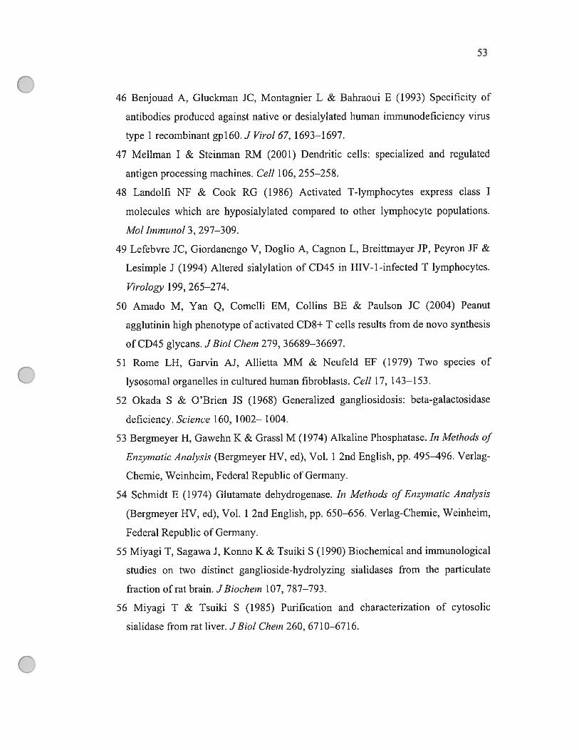
53
46 Benjouad A, Gluckrnan JC, Montagnier L & Bahraoui E (1993) Specificity of
antibodies produced against native or desialylated human immunodeficiency virus
type 1 recombinant gpl6O.J Virol67, 1693—1697.
47 Meïlman I & Steinman RIvI (2001) Dendritic celis: specialized and regulated
antigen processing machines. Celi 106, 255—258.
48 Landolfi Nf & Cook RG (1986) Activated T-lymphocytes express class I
molecules which are hyposialylated compared to other lymphocyte populations.
Mol ImmïtnoÏ 3, 297—309.
49 Lefebvre JC, Giordanengo V, Doglio A, Cagnon L, Breiftmayer JP, Peyron JF &
Lesimple J (1994) Altered sialylation of CD45 in HIV-1-infected T lymphocytes.
ViroÏogy 199, 265—274.
50 Amado M, Yan Q, Comelli EM, Collins 3E & Paulson JC (2004) Peanut
agglutinin high phenotype of activated CD8+ T cells resuits from de novo synthesis
ofCD45 glycans. JBioÏ Chem 279, 36689—36697.
51 Rome LH, Garvin AJ, Allietta MM & Neufeld EF (1979) Two species of
lysosomal organelles in cultured human fibroblasts. CeÏÏ 17, 143—153.
52 Okada S & O’Brien JS (1968) Generalized gangliosidosis: beta-galactosidase
deficiency. Science 160, 1002— 1004.
53 Bergrneyer H, Gawehn K & Grassi M (1974) Aikaline Phosphatase. In Methods of
Enzyrnatic AnaÏysis (Bergmeyer HV, cd), Vol. 1 2nd English, pp. 495—496. Verlag
Chemie, Weinheim, Federal Republic of Germany.
54 Schmidt E (1974) Glutamate dehydrogenase. In Methods ofEnzymatic Analysis
(Bergmeyer HV, cd), Vol. 1 2nd English, pp. 650—656. VerÏag-Chemie, Weinheim,
federal Republic of Germany.
55 Miyagi T, Sagawa J, Konno K & Tsuiki S (1990) Biochemical and immunological
studies on two distinct ganglioside-hydrolyzing sialidases from the particulate
fraction of rat brain. JBiochem 107, 787—793.
56 Miyagi T & Tsuiki S (1985) Purification and characterization of cytosolic
sialidase from rat liver. JBioÏ Chem 260, 6710—6716.
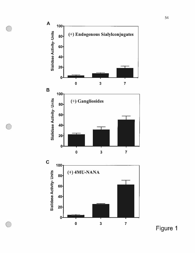
U)
D
(.)
wU)
U)
U)
D
>
o
w0U)
U)
U)
U)
D
>
o
QOU)
U)
U)
A54
B
0 3 7
C
0 3 7
4 fl n
(+) 4MU-NANA
I II
80
60
40
20
n Io 3 7
Figure I
55
Figure 1 Differentiation of monocytes into macrophages is associated with
increased expression of endogenous sialidase.
Monocytes were purified from the peripheral blood of human donors as described in
Experimental procedures and were differentiated into macrophages by growth at
37°C in RPMI medium 1640 with 10% (v/v) human serum and rhM-CSF. Sialidase
activity in celis from three donors was determined immediately after isolation of
monocytes (day 0) and afier ceils had differentiated in culture for 3 and 7 days.
Sialidase activity was measured against endogenous sialylconjugates (A),mixed
bovine gangliosides (B), or 4-MU-NANA (C) as substrates as described in
Experimental procedures. Sialidase activity is reported in units that reftect the amount
of sialidase in 1 mg of cellular protein that releases 1 nmol of sialic acid per hour at
37°C. Data represent the mean + SE of three independent experiments using ceils
from three different donors.
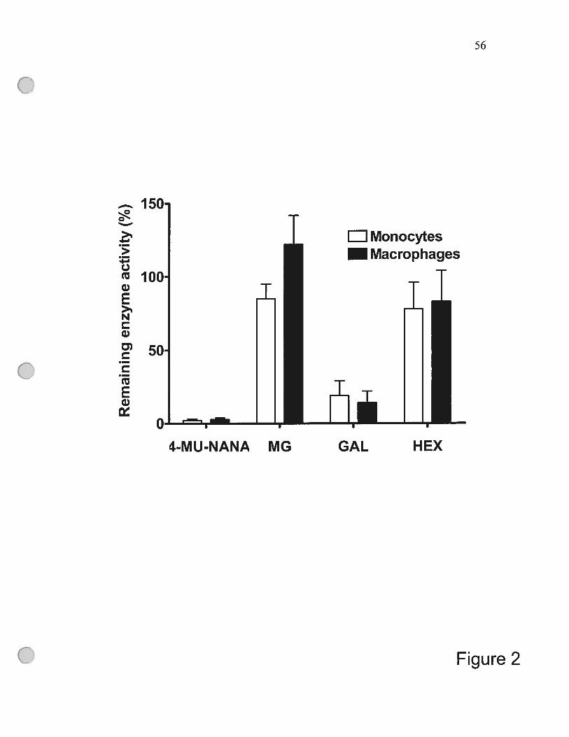
56
>3-.C,tuQ)E>%N
G)
D)CCtuEQ)
Figure 2
150
100•
50
Monocytes— Macrophages
I
4-M U-NANA MG GAL HEX
57
Figure 2. Immunoprecipitation of Neul from ccli extracts removes sialidase
activity using 4-MU-NANA as substrate.
Monocytes and monocyte-derived macrophages were isolated, homogenized and
incubated with rabbit anti-cathepsin A IgG or preimmune IgG as described in
Experimental procedures. Afier immunoprecipitation of the Neul-containing
multienzyme complex that also contains f3-D-galactosidase and cathepsin A, the
depletcd lysate was assayed for 3-ga1actosidase (GAL), f3-hexosaminidase (HEX),
and sialidase activities using either 4-MU-NANA or mixed gangliosides (MG) as
substrates as described in Experimental procedures. The amount of activity of each
enzyme in the presence of preimmune IgG was set to 100% of activity for
comparison with the activity in the samples treated with anti-cathepsin A IgG. Data
represent the mean + SE ofthree independent experiments.
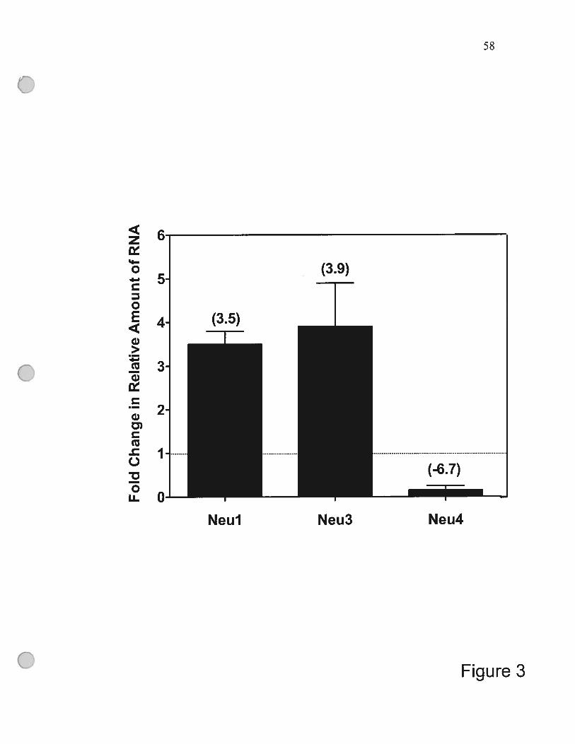
58
z‘I
o
oE
w>1
(uw
w
Q
oLL
Figure 3
(3.9)
(3.5)
.1
5.
4.
3.
2
J.
o.—NeuJ Neu3
(-6.7)
Neu4

59
Figure 3. Differential regulation of genes encoding Neul, Neu3 and Neu4 during
monocyte differentiation.
Total RNA was isolated from monocytes and monocyte-derived macrophages after 7
days in culture and 10 ng of RNA was used with primers that were specific for
Neul—4 in SYBR-green semi-quantitative real-time RT-PCR to detect the relative
amount of RNA encoding each gene as described in Experimental procedures. The
fold change in amount of Neit], Neït3 and Neu4 RNAs in day 7 macrophages
compared to freshly isolated monocytes (listed in parentheses) was calcuÏated afier
normalization to the intemal control 18$ rRNA by the equation as described in
Experimental procedures. The difference in amount of expression of each gene
relative to 18$ rRNA in monocytes was normalized to 1, as noted by the doffed
horizontal line at 1. These data represent the mean + SE of three experirnents using
ceils from different donors.
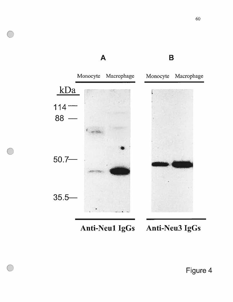
60
A B
Monocyte Macrophage Monocyte Macrophage
kDa
114 488
50.7
____ ____
L_L1
35.5
Anti-Neul IgGs Anti-Neu3 IgGs
Figure 4
61
Figure 4. The amount of Neul and Neu3 proteins increases during
monocyte differentiation.
Monocytes and macrophages were collected at the indicated times and total
cellular protein was separated by electrophoresis on 10% SD$ /polyacrylamide
gels, transferred to polyvinyldifluoride membranes and analyzed for the total
amount of Neul (A) and Neu3 (B) protein using specific antibodies as
described in Experimental procedures. The same amount of total cellular
protein (5 jig) from both monocytes and macrophages was analyzed in each
lane of the gel. The tick marks on the lefi side of the radiograph represent
protein molecular mass markers as noted. These results from one donor are
representative of data from five independent experiments using cells from four
different donors.
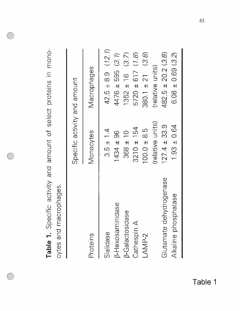
Qo
o
Tab
leJ.
Spe
cifi
cac
tivi
tyan
dam
ount
cfse
lect
prot
eins
inm
ono
cyte
san
dm
acro
phag
es.
Spe
cifi
cac
tivi
tyan
dam
ount
Pro
tein
sM
onoc
ytes
Mac
roph
ages
Sia
lida
se3.
5±
1.4
42.5
±8.
9(1
2.1)
13-H
exos
amin
idas
e14
34±
9644
76±
595
(3.
1)j3
-Gal
acto
sida
se36
$±
1013
52±
16(3
.7)
Cat
hesp
inA
3210
±15
457
20±
617
(1.8
)
LA
MP-
210
0.0
±8.
538
0.1
±21
(3.8
)
(rel
ativ
eun
its)
(rel
ativ
eun
its)
Glu
tam
ate
deh
ydro
gen
ase
127.
4±
33.9
482.
5±
20.2
(3.8
)A
lkal
ine
phosp
hat
ase
1.93
±0.
646.
08±
0.69
(3.2
)
H D)
D (D -
CHAPTER 3: MONOCYTE DIFFERENTIATION
UPREGULATE$ THE EXPRESSION 0F THE
LYSOSOMAL SIALIDASE, NEU1 AND TRIGGERS ITS
TARGETING TO THE PLASMA MEMBRANE VIA MHC
CLASS II-POSITIVE COMPARTMENTS.
ARTICLE 2
Publié dans:
The Journal ofBiological Chernistiy, 2006 Sep 15; 281 (37):27526-38
64
MONOCYTE DIFFERENTIATION UPREGULATE$ THE
EXPRESSION 0F THE LYSOSOMAL SIALIDASE, NEU1
AND TRIGGERS ITS TARGETING TO THE PLASMA
MEMBRANE VIA MHC CLASS II-POSITIVE
COMPARTMENTS.
feng Liangt, Volkan Seyrantepet, Karine Landiyt, Rasheed Ahmadt, Ah Ahmadt,
Nicholas M. Stamatos and Alexey V. Pshezhetsky*
Sainte-Justine Hospitait and Department of Pediatrics, University of Montreal,
Montreal, Quebec H3T 1 C5 Canadat and Institute of Human Virology, University of
Maryland, Baltimore, Maryland 21201, Division oflnfectious Diseases, Department
ofMedicine, University of Maryland Medical Center, Baltimore, Maryland, 21201
Correspondence: Alexey V. Pshezhetsky
Service de génétique médicale
Hôpital Sainte-Justine
3175 Côte Ste-Catherine
Montréal (Qc) H3T 1C5 — Canada
Tel. (514) 345-4931/2736
fax. (514) 345-4766
E-Mail:

65
ABSTRACT
Human sialidase (neuraminidase) Neul catalyzes lysosomal catabolism of
sialylated glycoconjugates. Here we show that during the differentiation of
monocytes and the monocytic ce!! une, THP-1 into macrophages, the majority of
Neul relocalizes from the lysosomes to the ce!! surface. In contrast to other cellular
sialidases Neu2, Neu3 and Neu4, whose expression cither remains unchanged or is
downregulated, Neul mRNA, protein and activity are specifica!!y increased during
the PMA-induced differentiation, consistent with a significant induction of the
transcriptional activity of the Neu] gene promoter. The lysosomal carboxypeptidase,
cathepsin A, which forms a complex with and activates Neul in the lysosome, is
sorted to the plasma membrane of the differentiating ceils similar!y to Ncu 1. Both
proteins are first targeted to the lysosorne and then are sorted to the LAMP-2-
negative, MHC II positive vesicles, which later merge with the plasma membrane.
Similar trafficking was observed for the intemalized fluorescent dextran or
horseradish peroxidase initially stored in the lysosomal/endosomal compartment. The
suppression of Neu 1 expression in the THP- 1 -derived macrophages by siRNA or
with anti-Neul antibodies significant!y reduced the ability of the celis to engu!f
bacteria or to produce cytokines. Altogether our data suggest that the upregulation of
the Neul expression is important for the primary function of macrophages and
establish the link between Neul and the cellular immune response.

66
INTRODUCTION
Human lysosomal sialidase (neuraminidase) Neu 1, encoded by the NEUJ
gene in the MHC III locus (1-3) catalyzes the hydrolytic cleavage of terminal sialic
acid residues from oligosaccharides and glycoproteins. In the lysosome, Neul exists
as a component of the multienzyme complex also containing the lysosomal
carboxypeptidase A (cathepsin A/protective protein), 13-galactosidase and
Nacetylgalactosamine- 6-sulfate sulfatase [reviewed in (4,5)]. The dissociation ofthe
complex in vitro resuits in the reversible inactivation of Neul (6). In vivo, inherited
mutations in cathepsin A cause disruption of the compiex and trigger
galactosialidosis (OMIM 256540), an autosomal recessive disease characterized by
combined deficiency of Neul, B-galactosidase and cathepsin A [reviewed in (4)].
Another autosomal recessive disease, sialidosis (OMIM 256550), is caused by
mutations directly affecting the NEUJ gene [reviewed in (7,8)].
We have previously shown that in addition to the lysosomes, Neul is also
present on the surface of activated T celis (9) where it may influence the immune
function (10-12). Recently (13) we have demonstrated that endogenous Neul
sialidase activity of freshly isolated human monocytes is increased 20 to 30-fold per
ccli as they differentiate into macrophages. Monocyte-macrophages constitute an
important ceilular component of the mononuclear phagocytic system, playing an
essential role in the recognition and activation of both innate and adaptive immune
responses against pathogens. The differentiation of monocytes into macrophages in
extravascular tissues is an essentiai event for the normal functioning of the system.
Therefore, knowledge of the mechanism and the role of the Neiti upregulation during
the monocyte differentiation should provide insights into the cellular immune
response and may eventually lead to developing approaches for therapeutic
intervention.
Here we have studied the regulatoiy mechanisms of the Neul expression and
presented direct evidence that the NEU] gene promoter is activated during the
monocytic differentiation. We have also shown that the newiy synthesized Neul is
first targeted to the lysosomes and then, as a complex with cathepsin A, relocalizes to
67
the ce!! surface, where it could participate in antigen presentation or affect
interceliular interactions.
EXPERIMENTAL PROCEDURES
Isolation of PBMCs and purification of monocytes
Peripherai blood mononuclear cells (PBMCs) were isolated by leukophoresis
of blood from HIV-1 and hepatitis B and C seronegative donors followed by
centrifugation over ficoil- Paque Plus (Amersham Biosciences, Uppsala, Sweden)
gradients using standard procedures. Monocytes were purified from PBMCs by an
additional centrifugation over Percoli (Amersharn Biosciences, Uppsaia, Sweden)
gradients and then by negative selection using monocyte separation kit ($temSep;
Stem Ccli Technologies, Vancouver, BC, Canada) according to the manufacturer’s
protocol. The purity of monocytes exceeded 95% as determined by flow cytometry
after staining the celis with fluorochrome-labeied antibodies to CD3, CDÏ4, CD19,
CD206 (all from BD PharMingen, San Diego, CA) and viability was greater than
97% as determined by tiypan blue dye exclusion.
Culturing of purified monocytes
To obtain monocyte-derived macrophages, purified monocytes were
suspended at 2 x 10⁶ cells/ml in RPMI medium 1640 (Gibco, Grand Island, NY)
containing 10% heat-inactivated human AB serum (Gemini Bioproducts, Caiabasas,
CA) and were maintained at 4 x 10⁶ cells/well in 6 well tissue cuiture plates (Costar,
Corning Inc., Coming, NY) at 37°C in a 5% humidified, C02 incubator. At the
indicated times, nonadherent ceils were removed by two washes with phosphate
buffered saline (PBS) and the adherent, differentiating macrophages (larger and more
granular than monocytes as seen by light microscopy) were harvested in PBS pH 7.4
by gentie scraping with a polyethylene ccii scraper (Nalge Nunc International,
Rochester NY). The harvested cells were confirmed to have characteristic
macrophage ccli surface phenotypic markers (CD i4+, CD206+) by flow cytometry.
68
Culturing and differentiation of THP-1 ceils
THP-1 celis, described in greater detail elsewhere (14), were cultured in the
RPMI 1640 medium (Gibco, Grand Island, NY) containing 10% heat-inactivated fetal
caif serum (FCS), 5000 units of PenicillinlStreptomycin and Fungizone (both Life
Technologies, Inc.). The cells were maintained in suspension at 0.5-1.0 x 10⁶ cells per
ml. The differentiation of THP-l cells into adherent celis with macrophage-like
characteristics widely used as a convenient in vitro model for macrophage
differentiation (15, 16) was induced by the addition of PMA (10-20 ng per ml) to the
culture medium. After 4 days the adherent, differentiating macrophages were
harvested by scraping as above.
Promoter activity assay
The fragments of the 5’-flariking region of the NEU] gene were obtained by
the digestion of pBluescript-SP vector with Bg[ll (position -1), Bcfl (position -176),
SacII (position —316), XmaI (position —542), XbaI (position -936) and ApaI (position
—1598). The fragments were subcloned into pBLCAT6 vector (15), provided by Dr. L.
Simard, (Montreal University) in front of a chloramphenicol acetyl transferase (CAT)
gene resulting in pBL-NEU1CAT1, pBL-NEU1CAT2, pBL-NEUYCAT3, pBL
NEUYCAT4 and pBLNEU1CAT5 constructs. pCMV—f3—gal vector containing
3—galactosidase reporter gene. has been provided by Dr. R. Gravel (University of
Calgary).
Monkey kidney COS-7 ceils were maintained at 37°C in a humidified
atmosphere of 5% C02 in Modified Eagle’s medium (MEM, Life Technologies, Inc.)
supplemented with 10% FCS, 5000 units of Penicillin!Streptomycin and fungizone.
Celis seeded in 100 mm tissue culture plates at 70-80% confluence were
cotransfected with the reporter constructs and pCMV—f3—gal expression vector using
Lipofectamine Plus reagent (Life Technologies mc, Gaithersburg, MD) in accordance
with manufacturers protocol. THP-1 ceils suspended in RPMI 1640 medium at a
density of 5x 1 0 cells/ml medium were cotransfected with the reporter constructs
69
and pCMV—f3—gai expression vector using Effectene transfection kit (Qiagen,
Germay) in accordance with rnanufacturer’s protocol.
Twenty-four hours after the transfection the ceils were washed with PBS and
stimuiated for 2-24 h with PMA (20 ng/rnl), TNF-a (40 ng per ml) or PDGF-AB (10
ng per ml) in the presence or absence of 20 mlvi NAC, or 0.5 tM curcumin (ail
Sigma, Ste-Louise, MO). In a separate experiment curcumin and NAC were added in
the same final concentration 1 h before the addition of PMA or TNf-cL Afier 24 h
incubation the ceils were rinsed with PBS, harvested by scraping and resuspended in
350 ii ofTEN buffer (50 mM Tris-HC1 pH 7.4, 1 mM EDTA, and 150 mM NaCi).
Celis were centrifuged at 12,000 rprn at 4°C for 5 min and lysed by three freezing
thawing cycles in 0.25 M Tris-HC1 pH 7.8. CAT and f3—galactosidase activities were
measured using [‘4C] -chorarnphenicol and 4-methylumbelliferyl— f3—D-
galactopyranoside substrates as described (16). One unit of enzyme activity (U) is
defined as the conversion of 1 imol of substrate/rnin. Proteins were assayed
according to Bradford (17) with bovine serum albumin (Sigma) as standard.
Enzyme assays
Sialidase, J3—galactosidase and 13—hexosaminidase activities in cellular
homogenates and in culture medium were assayed using the corresponding
fluorogenic 4-methylumbelliferyl-glycoside substrates (18,19). Cathepsin A activity
was determined with CBZPhe-Leu (20). Alkaline phosphatase, glutamate
dehydrogenase and GAPDH were measured as described elsewhere (21-23). Sialidase
activity towards mixed bovine gangiiosides was measured as described (24,25) in the
presence of 0.2% Triton X-100. The concentration of reieased siaiic acid was
measured by thiobarbituric method (26). Enzyme activity is expressed as the
conversion of 1 nmoi of substrate per h.
To measure the enzyme activities on the outer ccli surface, the cultured iive
celis (106 celis) were washed several times with Hank’s balanced salt solution and
overiaid with 500 il of 20 mlvi acetate buffer, pH 5.2, 0.25 M sucrose and either 0.4
mlvi siaiidase substrate, cathepsin A substrate, f3-galactosidase substrate or N-acetyl
70
f3-hexosaminidase substrate. After 30 min of incubation, 200 jil aliquots of medium
were collected and the concentrations of the products were measured as described
above. The integrity of plasma membrane was verified by western blot by measuring
the amount of the cytosolic enzyme GAPDH released into the ccli medium during the
incubation. We found that the same amount of GAPDH was released from the ceils
incubated in 20 mM acetate buffer, pH 5.2, 0.25 M sucrose and PBS. To measure the
total amount of enzyme, the assay was performed in the presence of 0.3% Triton X-
100.
Detectïon of CathA and Neul on the macrophage surface by Western blot
Proliferating THP- 1 ceils and THP- 1 -derived macrophages (5x 10⁶ ceils)
were washed 3 times by ice-cold PBS and incubated at room temperature for 1 h in 5
ml ofPBS containing a BAC-Su1foNHS biotinylation reagent (Sigma St. Louis, MO)
at a final concentration of Smg/ml with a gentie rotation. The celis were washed 3
times with TBS and homogenized in 0.5 ml RIPA buffer. The ccli lysate cleared by
10 min centrifugation at 12,000 g was loaded on a 0.2 ml Streptavidin-agarose
coiunm (Sigma St. Louis, MO) and the column was washed by PBS until the
absorbance at 280 nm reached the baseline. Biotinylated proteins were eluted with 0.2
ml of 0.1 M glycine-HC1 (pH 2.5). The pH ofthe eluate was immediately adjusted to
neutral with 1.5 M Tris buffer (pH 7.5). Twenty ig ofprotein from the original ccli
lysate and the Streptavidin-agarose column flow-through fraction and 5 jig ofprotein
from the eluate were separated on a NuPAGE 4-12% Bis-Tris gel (Invitrogen) and
transfered to a PVDF membrane. The membrane was hybridized with the polyclonal
anti-Neul antibodies (dilution 1:5,000) and anti-cathepsin A antibodies (dilution
1:2,000). Detection was performed using a Roche Western kit.
Internalization of Texas Red-labeled Dextran and HRP
THP- 1 ceils suspended at 1 .Ox 106 celis / ml in RPMI 1640 containing 10%
fCS in 6-well tissue culture plate were loaded with Texas red-labeled flurescent
dextran 40,000 (Molecular probe, Oregon US) in a concentration of 50 jig/ml
71
medium or the horseradish peroxidase (Sigma) in a concentration of 2 mg/ml medium.
Treated THP-1 cells were maintained at 37°C in a humidified atmosphere of 5% C02
for 2 hours. Then the celis were harvested by a low-speed centrifugation and washed
with RPMI 1640 medium two times at room temperature and resuspended in the fresh
medium containing 10% fCS. After 16 h of incubation at 37°C cells were treated
with PMA (20 ng/ml of culture medium) as described above.
Confocal immunofinorescence microscopy
Celis were washed twice with ice-coÏd PBS and fixed with 3.8%
paraformaldehyde in PBS for 30 min and if necessaiy were perrneabilized by 0.5%
Triton X-100. Fixed celis were washed twice with PBS and stained with polyclonal
rabbit anti-Neul, or anti-cathepsin A antibodies and counterstained with Texas Red
labeled antibodies against rabbit IgG. Ceils were also double-stained with rabbit
antibodies against Neul or cathepsin A antibodies and monoclonal antibodies against
LAMP-2 (Washington Biotechnology Inc., Baltimore), or against MHC II (BD
Biosciences, Mississauga, ON). The primary monoclonal antibodies were
counterstained with Oregon green 488-conjugated anti-mouse IgG antibodies
(Molecular Probes, Eugene, OR). The cells loaded with the horseradish peroxidase
(HRP) were stained with the Texas Red-conjugated antibodies against HRP (Jackson
ImmunoResearch, West Grove, PA). CelIs were also stained with the rabbit anti
Neul or anti-cathepsin A antibodies counterstained with Oregon green 488-
conjugated anti-rabbit IgG antibodies and PE-conjugated monoclonal anti-maimose
receptor antibodies or anti-CD14 antibodies (BD Pharmingen, USA). Slides were
examined on a Zeiss LSM51O inverted confocal microscope (Carl Zeiss Inc.,
Thomwood).
Western blot analysis
Proteins were resolved by SDS-polyacrylamide gel electrophoresis and
electrotransferred to PVDF membrane. Neul and Neu3 detection was performed with
rabbit antibodies as described (13,27) and LAMP-2 detection with monoclonal anti-

72
human LAMP-2 antibodies (Washington Biotechnology Inc., Baltimore) using the
BM Chemiluminiscence kit (Roche Diagnostics, Lavai, QC) in accordance with the
manufacturer’s protocol.
RNA isolation and real time RT-PCR
THP-l celis and THP-1-derived macrophages were harvested as previously
described and total RNA was isoiated using Trizol reagent (Invitrogen, Carlsbad, CA)
following the protocol suggested by the manufacturer. The RNA preparation was
treated with DNase I (Invitrogen, Carlsbad, CA) for 15 minutes at room temperature
to remove the DNA contamination and then inactivated by adding 25 mlvi EDTA
solution to the reaction mixture. One microgram of RNA was used to synthesize
cDNA in the presence of 0.5 mM dNTPs, 50 ng random hexamers (Invitrogen,
Carlsbad, CA) and 200 Units of SuperScript II reverse transcriptase (Invitrogen,
Carlsbad, CA.). Reverse transcription was allowed to proceed for 50 minutes at 42°C.
PCR amplification was performed on a SmartCycler (Cepheid) using the SYBR
Green PCR kit (Qiagen). The primer pairs, aimealing temperatures, number of PCR
cycles, and RNA amounts are shown in Supplemental Table, which also shows the
predicted sizes of the resulting PCR products. As positive controls for real-time PCR
detection, we used mRNA coding for the ribosornal subunit 185 (Ambion). Ah
reactions were performed in triplicate. The accuracy of the reaction was monitored by
the analysis ofmelting curves and product size on gel electrophoresis.
Northern blot analysis
Purified total RNA from THP- 1 cells and THP- 1 -derived macrophages was
resolved by agarose gel electrophoresis and blotted to Hybond-N+ membrane
(Amersham Pharmacia Biotech). The membrane was hybridized with the following
probes: a 500 bp Neït4 cDNA fragment obtained from clone 4156395 by PvuII
digestion, an entire Neul cDNA, a 700 bp Neu3 fragment obtained from a cDNA
clone by FstI digestion, and an entire cDNA of human f3—actin. Ail probes were

73
Iabeled with [32P]-dCTP by random priming using MegaPrime labeling kit
(Amersham, Bay d’Urfé, QC). Pre-hybridisation of the blots was perfonned at 68°C
for 30 min in ExpressHybTM (Clontech). The denatured probes were added directly
to the pre-hybridisation solution and incubated at 68°C for 1 h. The blots were
washed twice for 30 min with 2x SSC, 0.05% SDS at room temperature, once for 40
min with 0.lx SSC, 0.1% SDS at 50°C and exposed to a BioMax film overnight at —
80°C.
Neul small interfering RNA gene silencing in human skin fibroblasts
SilencerTM pre-designed siRNA ID:8386, ID:8481 and ID:8573 (CAT:16704)
targeting exons 1, 2 and 3 ofthe hurnan Neul gene, respectively, were obtained from
Ambion (Austin, TX). THP-1 celis were transfected with siRNA using the
NucleofectorTM Solution V reagent (Amaxa US) according to the protocol provided
by the manufacturer. GAFDH siRNA (Ambion) was used as a positive control for the
optimization of transfection conditions. The siRNA efficacy was verified in
preliminary experiments where THP-1 cells were harvested 24 and 48 hours after
transfection with 3 Neiti siRNAs, and the sialidase activity was measured with 4
MU-NANA as described above. The most effective Netti siRNA (ID:8481) was
selected to perform the suppression experiments. The expression of the Neul in the
transfected cells was measured on the RNA and protein level by quantitative RT-PCR,
Western blotting and enzyme activity assay as described. On average, sialidase
activity in the differentiated Net,] siRNA-transfected THP- 1 cells was reduced more
then 4-fold as compared to mock-transfected celis (not shown).
Phagocytosis assay
Differentiation of THP- 1 celis was induced by PMA in the presence of either
anti- Neul rabbit antiserum or control preimmune serum (20 and 100 jig per ml of
medium). THP-l celis transfected with Neit] siRNA were differentiated in the
presence of PMA only. After 4 days cells seeded on cover slips were incubated
within 3 hours with ftuorescein-labeled E. cou (K- 12 strain), bioparticles (Molecular

74
Probe, Eugene, OR) in a ratio of 30 particles per one ce!!, washed 3 times with co!d
PBS, fixed with 4% paraforma!dehyde for 1 h, permeabilized with 0.5% Triton X
100, and washed twice with 0.05% Tween-20 in PBS. Ce!!s were counterstained with
3 jiM DAPI and washed again with 0.05% Tween- 20 in PBS. Siides were studied on
the Nikon Eclipse E6000 direct epifluorescence microscope.
Measurement of IL and TNF-a Production in Culture Supernatants by Enzyme
linked Immunosorbent Assay
In order to determine the potential effect of anti-Neul serum on the secretion
of IL-6, IL-10, IL-12, IL-15, IL-lb and TNFa, THP-1 ce!ls cu!tured in a six-we!!
p!ate (3x10⁶ per wel!) were stimulated with PMA (20 ng/ml) in the absence and in the
presence of rabbit pre-immune serum (100 ig/m!) or rabbit antiserum raised against
the human Neul (20 and 100 ig/m1 of medium). After 96-hr incubation, the
differentiated ce!ls were further stimu!ated for 12 hrs by ionomycin (Sigma; 1 ig/m!)
and PMA (20 ng/m!) a!so in the absence and in the presence of rabbit pre-immune
serum or rabbit anti-Neul antiserum. The concentration of IL and TNf-Πin the
culture medium was measured by enzyme-!inked immunosorbent assay at Chemicon
Internationa! Inc. (Temecu!a CA).
RESULTS
Neul expression is increased during PMA-induced differentiation of monocytic
celi une THP-1
Previous studies defined a monocytic ce!! !ine THP-1 from the peripheral
blood of a patient with acute monocytic !eukemia (14). Being induced by 12-O-
tetradecanoy!phorbo!-13-acetate (PMA) these ce!!s differentiate into adherent ce!!s
having morphologica!, biochemica! and physiologica! characteristics of macrophages
and expressing the characteristic macrophage ccl! surface phenotypic markers, such
as CD14+ and CD206+ (28,29). THP-1 ce!ls have several advantages over primaly
human monocytes (higher transfection efficiency, homogeneity, accessibility) that
make them a re!iab!e mode! to smdy the mechanism of monocytic differentiation. We
75
found that during the PMA-induced differentiation, the sialidase activity measured at
acidic pH 4.2 with the synthetic fluorescent substrate 2’ -(4-
methylumbeliiferyi)—a—D-Nacetyineuraminic acid (4 MU-NANA) was increased at
ieast 4-fold whereas the leveis of other lysosomai enzymes and proteins (cathepsin A,
j3-galactosidase, J3-hexosaminidase, LAMP-2), or of cytosolic enzyme GAPDH were
flot changed (Fig. 1 A).
The observed increase of the endogeneous sialidase activity could be caused
by upregulation of any of 4 previousiy described human sialidases: lysosomai (Neul,
gene NEU] and Neu4, gene NEU4), cytoplasmic (Neu2, gene NEU2) and plasma
membrane (Neu3, gene NEU3). Since the cytosolic sialidase Neu2 is mostiy active at
neutrai pH (30), and the sialidase activity measured with 4 MU-NANA at pH 7.5 was
not increased during the differentiation of THP-1 ceils (Fig. 1 A) we could exciude
this enzyme. Three remaining enzymes, Neul, Neu3 and Neu4 ail have acidic pH
optimum but they differ from each other in substrate specificity: Neul shows the
highest activity against oligosacharides and short giycopeptides (24) whereas Neu3
and Neu4 are equally active against a2-3-siaiylated oligosaccharides, glycopeptides
and gangliosides (25,31,32). Sialidase activity of THP-1 ceils measured against
sialylated giycoiipids (rnixed bovine gangiiosides) was siightly reduced during
differentiation (1.1+0.2 versus 1.7±0.3 nmol/h per mg of total protein, Fig. 1 A)
suggesting that the increase of sialidase activity is caused by the upregulation of the
iysosomal sialidase Neul. In addition, both quantitative RT-PCR and Northern blots
showed that during the differentiation the Neul mRNA was increased at least 5-times
(Fig. 1 B and C), whereas both Neu3 and Neit4 mRNA were decreased 3 to 4-fold
according to the RT-PCR data (Fig. 1 B). Neu2 expression couid not be detected in
the THP-1 monocytes as weil as in the THP-1-derived macrophages neither by RT
PCR nor by Northem blots. Western biots using the polyclonai anti-Neul and anti
Neu3 antibodies confirmed up-reguiation ofNeul and downregulation ofNeu3 at the
protein ievel (Fig. 1 D). Aitogether our data showed that the differentiation ofTHP-1
ceils into macrophages resuits in significant and specific induction ofNeul.

76
Promoter activity of the NEU1 gene is increased during the differentiation of
TUP-1 celis
The transcriptional activity of the NEUJ gene promoter was studied by
deletion mapping. f ive restriction fragments of the NEUJ gene 5’-flanking region
were cloned into a pBLCAT6 vector (15) in front of a reporter, chloramphenicol
acetyltransferase (CAT) gene and expressed in COS-7 celis (f ig. 2 A). The
transfection efficiency was measured using pCMV--gal vector containing SV-40
promoter-driven lacZ reporter gene. CAT activity in the homogenates of the ceils
transfected with the reporter constructs was compared to that of the celis transfected
with the promoter-free pBLCAT6 vector (15). We found that the shortest fragment
with a detectable promoter activity was extending 316 bp upstream of the ATG
initiation codon (f ig. 2 3). Truncation ofthis fragment to 176 bp almost completely
abolished the promoter activity. The highest promoter activity was associated with a
542-bp fragment. Extension of this fragment to 936 bp and further to 1592 bp
decreased the promoter activity suggesting that potential elements for down
regulation of the gene expression could be present between the nucleotides -542 and -
1598 (fig. 2 B).
We further tested whether the promoter activity of the NEU1 gene was
induced during the differentiation ofTHP-1 celis into macrophages. THP-1 celis were
transfected with the above reporter constmcts and 24 hours later their differentiation
was induced by adding PMA to the culture medium. We found that 24 hours after the
induction of ceils with PMA the CAT activity was significantly increased for ail
constructs, the highest effect (8-fold increase) being achieved for the reporter
construct, pBL-NEU1CAT3 containing the 542-bp promoter fragment (fig. 2 C). for
this construct the effect was observed as early as 4 hours after the induction and
lasted for at least 4 days (f ig. 2 D). PMA also induced the expression of the reporter
gene in COS-7 ceils, but the effect was significantly lower then that in THP-1 celis
(fig.2 B).
77
Interestingly, the analysis of the sequence of the Neiti promoter with a
TRANSFAC 4.0 software that predicts regulatory elements (33) reveaÏed that the
fragment between the nucleotides-3 16 and -542 contains several conserved sequence
motifs for binding promoter-selective transcription factor (Sp-1), activator proteins 1
and 2 (AP-l and AP-2), hematopoietic transcription factor (E2F-1), adenosin 3’, 5’-
cyclic monophosphate-regulator elernent binding factor (CREB-2), and erythroid
transcription factors (GATA-1 and NF-E2) (Supplemental f ig. 1) suggesting that
some of these factors may be involved in the regulation of the NEUJ gene
transcription.
Indeed, when THP-1 or COS-7 celis transfected with the pBL-NEU1CAT3
reporter constnict were treated with a potent AP-1 activator, tumor necrosis factor-cx
(TNf-a), which increases the synthesis ofboth c-fos and c-Jun proteins in rnany ccli
types (34-36) we found a significant induction of the CAT activity (Fig. 3). A
detectable increase of the CAT activity was also caused by another AP- 1 activator,
PDGF-AB (not shown). In ail celis the effects of TNF-a PDGfAB or PMA could be
blocked by pretreatment of the celis with curcumin and a thiol antioxidant
Nacetylcystein (NAC), which inhibit c-fos or c-Jun expression (34-36) (Fig. 3).
Neul and cathepsin A are targeted to the celi surface during the differentiation
of THP-1 celis and prÏmary human monocytes
Intracellular localization of Ncul in thc prolifcrating THP-1 cclls and the
THP-1-dcrivcd macrophages was studied by confocal immunomicroscopy. In thc
non-diffcrcntiated celis, Neul showed a cytoplasmic punctuate staining paffem co
localizing with thc lysosomal marker LAMP-2 (fig. 4 A, Day 0). 24 h after thc
induction with PMA, thc intcnsity of anti-Ncul staining was significantly incrcased
and its pattcm changed. The celis clearly showcd 2 pools ofNeul, onc intracellular
co-localizing with LAMP-2 and other, pcriphcral (Fig. 4 A, Day 1). Three days later
whcn THP-1 cclls showcd morphological characteristics of macrophages, the
majority of Neul was found on the periphery of thc ccli, whcrcas LAMP-2 stili had
the intracellular Iocalization (fig. 4 A, Day 4).

78
To confirm the presence of Neul on the surface of the differentiating celis,
we performed a double-staining of the non-permeabilized ceils with anti-Neul
antibodies and antibodies against the ceil surface mannose receptor, CD206. The
proliferating ceils did not show any staining for both antigens (Fig. 4 B, Day 0),
whereas on the 1 st and the 4th days of differentiation the celis showed high expression
ofboth Neul and mannose receptor on their surface (f ig. 4 B). Similar resuits were
also obtained for another surface rnarker, CD 14 (flot shown) and in the isolatcd
human primary monocytes and the rnonocyte-derived macrophages (fig. 4 C and D).
In the lysosome, Neul is a component ofa multienzyme complex, which also
contains the lysosomal carboxypeptidase A (also called cathepsin A or protective
protein), B-galactosidase and N-acetylgalactosamine-6-sulfate sulfatase {reviewed in
(4,5)]. If the compiex is disrupted, Neul loses its enzyrnatic activity and is rapidly
degraded by the lysosornal proteases (6,27). Using confocai microscopy, we showed
that during the differentiation of THP-1 celis and isolated monocytes, a significant
part of cathepsin A is aiso targeted to the ceil surface. In the non-differentiated THP
1 ceils as weil as in the primary hurnan monocytes cathepsin A is found only in the
lysosomes, co-localizing with LAMP-2 (fig. 5 A and B, Day 0) and Neul (not
shown). Starting from the first day of the differentiation and in the mature
macrophages, cathepsin A is partially found on the ceil peripheiy where it colocalizes
with the mamiose receptor (Fig. 5 A, Day I and Day 4, fig. 5 3, Day 7) and Neul
(not shown).
The transiocation of Neul and cathepsin A from the lysosomes to the ccli
surface during the differentiation of monocytes and THP-1 ceils was also consistent
with the resuits of enzyme assays. To measure the enzyme activities on the ccli
surface, intact THP-1 ceils or THP-1-derived macrophages were incubated in an
isotonic buffer containing the enzyme substrate. To measure the total enzyme activity,
the assay was performed in the presence of 0.3% Triton X-100. In the non
differentiated THP-l ceils, the rnajority ofNeul was found inside the celis since the
activity was —5-fold higher for the lysed than for the intact celis (fig. 6A, Neul). In
contrast, in the THP-1-derived macrophages, thc rnajority ofNeul was present on the
79
outer ccli surface because the activity was increased only by 25% after lysing the
celis with Triton X-100. During the differentiation the total sialidase activity assayed
in the ccli homogenate increased 3.1 -fold whereas the sialidase activity on the surface
increased 13-fold. In contrast, the activity ofa control lysosomal enzyme, N-acetyi-J3-
hexosaminidase, was similar in the differentiated and non-differentiated cells and in
both cases —77% of the enzyme was not accessible for the substrate in the absence of
detergent (Fig. 6A) showing that the ratio of the disrupted and intact celis was sirnilar
for macrophages and monocytes. We did flot observe an increase of the total
cathepsin A activity during the differentiation, however, 70% of the activity in
macrophages was detected on the ccli surface as opposed to oniy 25% of the activity
in the non-differentiated THP-1 celis (Fig. 6A). Interestingly, neither total activity nor
the distributions of the third component of the multienzyme lysosomal complex, f3-
galactosidase were changed during the differentiation.
Finaily, proteins on the surface of THP-1 ceils and the THP-i-derived
macrophages wcre biotinylated and affinity purified on a Streptavidin-agarose
column. Then, 20 tg of protein from the original ccii lysate and the Streptavidin
agarose coiumn flow-through and 5 jig of the biotinylated proteins from the column
eiuate were analyzed by Western blot using the polyclonal anti-Neul antibodies and
the antibodies against the 32 kDa peptide chain of cathepsin A (figure 6B). In the
non-differentiated THP-1 celis both Neui and cathepsin A were found aimost
exclusively in the flow-through fraction containing uniabeicd intracellular proteins,
whereas in the THP- i -derived macrophages a significant percent of these enzymes
was biotinylated, suggesting that they are present on the cdl surface (f igure 6B).
The differentiation of THP-1 ceils induces fusion of the lysosome (endosome)
derived vesicles with the ceil surface
Aithough enzymatically non-active precursors of Neui and cathepsin A are
activated only in the lysosome (27,37), both enzymes found on the cdl surface in
macrophages have catalytic activity suggesting that they are first targeted to the
iysosorne and later to the ccii surface possibly in the resuit of a fusion of the
80
lysosomal membrane with the plasma membrane. To prove that such fusion happens
during the differentiation we have loaded the lysosomes of THP-1 ceils with
fluorescent dextran and followed its distribution in the cdl. In the non-differentiated
THP-1 celis the labeled intemalized dextran was retained in the endosomal-lysosomal
compartment for at least 7 days showing co-localization with the lysosomal marker
LAMP-2 (Fig.7, Day 0). In contrast, 12 hours after the induction of the celis with
PMA as well as 4 days after, in the fully differentiated THP- 1 -derived macrophages,
the internalized dextran was partially found in the LAMP-2 negative vesicles (which
appeared to be migrating to the plasma membrane), on the cell surface and outside
the ceils while some dextran was still localized in the lysosomes (Fig. 7, Day 1 and
Day 4). On the other hand, the differentiation did not increase the secretion of soluble
lysosomal enzymes (not shown), suggesting that the vesicles that fuse with the celi
surface may lack soluble lysosomal content.
Neul colocalizes witli MHC II and with the internalized antigens during the
differentiation of THP-1
In the differentiated macrophages the intemalized antigens are proteolytically
digested in the late endosomal-lysosomal compartment and the resulting peptides
bind MHC II proteins to be further targeted to the cdl surface as the antigenic
peptide-MHC II complexes, pMHC [reviewed in (38,39)]. Majority of MHC II is
found in the late endosomes, lysosomes and multivesicular bodies (MVBs)
collectively called MHC II-enriched compartments (MIICs), although a distinct
pMHC loading compartments were also isolated from the antigen-presenting celis
and called the class II containing vesicles (CIIV) (40,41). We found that during the
differentiation of THP-1 ceils and in the THP-l-derived macrophages, Neul
colocalizes with MIICs. In the non-differentiated cells, the anti-MHC II antibodies
only partially colocalized with the anti-Neul or the anti-cathepsin A antibodies as
well as with the antibodies against the internalized antigen, horseradish peroxidase
(HRP) (Fig. 8, Day 0).
81
However 12 hours after the PMA induction MHC II positive organelles,
which also contained the majority of Neul and HRP were ciearly distinct from the
LAMP-2-positive lysosomes (Fig. 8, Day 1). In the fully differentiated macrophages
the majority ofNeul, MHC II and anti-HRP imrnunoreactivity was found on the ccli
surface (f ig 8, Day 4).
Suppression of the Neul expression in the THP-1 celis alters their functional
cap acity
In order to understand whether upregulation of the Neul expression and its
targeting to the ccli surface is important for the primary function of macrophages we
have used siRNA to inhibit the expression of Neul in THP-1 celis. The transfected
ceils which had 4-fo1d reduced level of sialidase activity were stiil capable of PMA
induced differentiation, but showed a significantly reduced ability to ingest the
bacteria. When the normal THP-1-derived macrophages were incubated for 3 hours
with the fluorescein-labeled E. cou bioparticles their ingestion index (percentage of
macrophages with phagocytized particles muitiplied on the average number of
particles phagocytized per ingested macrophage) was 15.5. For the siRNA
transfected celis this number was reduced to 3.1 (Figure 9). Similar effect on the
ingestion capacity was observed when differentiated THP-1 ceils were incubated in
the presence of anti-Neul antibodies (ingestion index 2.9), whereas the ingestion
index of 12.2 for the ceils differentiated in the presence of similar amount of pre
immune rabbit serum did not statistically differ from the control (f igure 9).
In a similar fashion, we have observed that the treatment of THP-1-derived
macrophages with the anti-Neul antibodies significantly rcduced their ability to
produce certain cytokines. As before, the cells were differentiated in the presence of
the anti-Neul antiserum or the preimmune rabbit serum and then stimulated for 12
hrs by ionomycin also in the presence of the anti-Neul antiserum or the preimmune
rabbit serum. We found that the anti-Neul antibodies but not the pre-immune serum
have inhibited ionomycin induced production of IL-lb, IL-6 and TNF-a, whereas the

$2
changes in the production of IL- 10 and IL-12 (p40) were flot statistically significant
(figure 10).
DISCUSSION
The endogenous sialidase activity significantly increases during activation in
majority of immune celis including T celis, B celis, and monocytes, whereas
sialylation of some of their surface molecules decreases (10,42-45). In particular, the
reduction of sialylation was reported for MHC class I, required to render T celis
responsive to antigen presenting ceils (43), GM3-ganglioside, which modulates Ca2
immobilization and regulates IL-4 production in T ceils (11) and monocyte receptor
for hyaluronic acid, CD44 (44,45). In the activated B ceils the induction of the
sialidase activity is important to convert so-called, vitamin D3-binding protein into a
macrophage-activating factor (MAf) (46). Indirect evidences suggested the
involvement in these processes of Neul, an ubiquitously expressed lysosomal
sialidase responsible for the catabolism of glycoconjugates. In vivo, SM/J or SM/B10
mice strains with the missense mutation in the NEUJ gene which reduces the enzyme
activity to 30% of normal (47) have an impaired activation of macrophages while
their lymphocytes fail to synthesize IL-4, show low response for antigen presenting
celis and do not produce MAf (10,42,46). In human sialidosis patients Neuf
deficiency may result in reduced capacity of immune cells to produce cytokines and
antibodies leading to partial immunodeficiency and may account for frequent
pulmonary infections (G. Mitchell, personal communication). We have provided a
direct evidence that Neuf message and protein are induced several fold during the
differentiation of circulating human monocytes into professional antigen-presenting
ceils (APC) such as macrophages (13) or dendritic celis, (Stamatos N., manuscript in
preparation). In this study we investigated the mechanisms controlling the expression
of the NEUJ gene and found that the transcriptional activity of the NEUJ gene
promoter was significantly increased during the PMA-induced differentiation of
83
monocytic ceils into macrophages. The highest level of induction was observed for a
550-bp fragment of the NEUJ gene promoter containing several potential sequence
motifs for binding AP-l, a group of homodimeric and heterodimeric complexes
composed of various Fos and June gene products such as c-Jttn, junB, jiinD, c-Fos,
fosB as weil as the fos-related antigens Fi-a-] and Fi-a-2 (48-54). Ail these complexes
(also cailed protooncoproteins) interact with the common binding sites calied PMA
responsive etements and mediate gene induction in response to growth factors,
cytokines, phorbol esters and a variety of other stimuli including oxidative stress (34-
36). In particular, one of AP-l functions is to mediate inflammatory responses in
lymphocytes and macrophages by stimulating cdl activation and the expression of
cytokines (55). Previously it was shown that pro-inflammatory factors, LPS and
TNF-a caused 16-hour MAPK-depcndent induction of the endogenous acidic
sialidase activity in monocytes (44,45) resulting in desialyiation of CD44. In mm,
this increased the affinity of monocytes to hyaÏuronic acid, their adhesion and the
TNF-a production (45). Since in both cases the l.4-1.6-fold increase of the sialidase
activity was detected only with 4 MU-NANA but not with mixed bovine gangliosides
as a substrate it is conceivable that the effect was caused by the upregulation ofNeul.
Our experiments directly showed that the promoter activity of Neul was potently
induced by TNf-a that activates AP-l expression and reduced by AP-1 inhibitors
curcumin and N-acetylcysteine. Although we do not have a direct evidence of the
interaction between the Neul gene prornoter and AP-l it is tempting to speculate that
this protein may be involved in the regulation of the Neul expression during the
monocyte activation and differentiation.
The fact that Neul is specificaiiy induced in professional APCs suggests that
this enzyme may be implicated in the antigen presentation. However both MHC class
II and I moiecuies are expressed at the ccli surface while Neul was found
predorninantly in the endosomal/lysosomal compartment. Here we show that during
the differentiation of monocytes into macrophages the Neul is constitutively
transported to the plasma membrane along with class II molecules rather then being
actively retained in the lysosomes. Using immunocytochemistry we demonstrated
84
that in the differentiating THP-1 celis, THP-1-derived macrophages as welÏ as in the
mature primaly macrophages a vast mai ority of the Neul was found on the ccli
periphery co-localizing with the mannose receptor. In addition, biochemical assays
showed that the biotinylation of the ccli surface proteins resuits in the labeiing of
Neul in macrophages but not in the proliferating THP-1 ceils and that —80% ofNeul
in the THP- 1 -derived macrophages was accessible for the substrate added directiy to
the ceil medium confirming that it is localized on the ccli surface. Similar
observations were made for the lysosomal carboxypeptidase, cathepsin A, whose
interaction with Neul is essential for the activation ofNeul in the lysosomes.
Both Neul and cathepsin A are sorted from the trans-Golgi network to the
endosomai pathway. As the majority of soluble lysosomal lumenal proteins cathepsin
A is targeted through the mannose-6-phosphate receptor pathway (56), whereas the
intracellular trafficking of Neul is mediated by the adaptor proteins through a Tyr
containing motif in its C-terminus (9). In the lysosome the two proteins undergo
activation. The activation of cathepsin A involves its processing by a lysosomal
endopeptidase which removes a 2-kDa “linker” polypeptide between the two
polypeptide chains of the mature enzyme (57). The exact molecular mechanism that
underlies the activation of Neul is unknown but most Iikely it invoives a
conformational change of the Neul polypeptide after the formation of the compiex
with cathepsin A. We speculate that in the differentiating ceils both enzymes are first
targeted to the endosomal/lysosomal compartment, where they gain enzymatic
activity and then are sorted to the vesicies destined to the plasma membrane. The
existence of such lysosome-to-plasma membrane transport pathway has been well
documented for other professional APCs, dendritic ceils where it plays a crucial role
in targeting the peptide-MHC II complexes (pMHCs) to the ccii surface (58). Upon
receipt of a maturation stimulus (such as lipopolysacharide) the intemalized antigens
are processed in the endosomes and pMHCs are formcd. Several hours later they
appear in the LAMP-negative compartments termed CIIV, which are the
intermediates in the transfer of pMHC5 to the plasma membranes (58). The
concentration of pMHCs in CIIV reaches a maximum 12 hours after the
$5
lipopolysacharide treatrnent and after 12 more hours the majority of complexes is
already transfened to the ccli surface. The described targeting ofpMHCs in dendritic
celis resembles the targeting of Neul and cathepsin A in differentiating monocytes
and macrophages. We found that 12 to 24 hours after receiving the differentiation
stimulus the lysosomal-endosomal apparatus of monocytes undergoes a dramatic
rearrangement. The MHC II-positive and Neu 1-positive compartments are segregated
from the LAMP-2 positive lysosomes and late endosomes, relocated doser to the ccli
surface and fused with the plasma membranes. The same vesicles are stained
positively for the intemalized antigen HRP or fluorescent dextran, which in the
proliferating monocytic ceils are normally stored for at least several days in LAMP-2-
positive endosomal/lysosomal compartrnent. In the differentiating celis the dextran
molecules and HRP peptides (probably in the form of pMHCs) were gradually
accumulated on the ccii plasma membrane colocalizing at least in part with Neul and
cathepsin A.
Most importantly, the suppression of Neul expression in the THP-1-derived
macrophages by siRNA or their treatrnent with anti-Neul antibodies significantly
reduced functional capacity of the ceils, such as the ability to enguif bacteria or to
produce cytokines, suggesting that the upregulation of the Neul expression is
important for activation of macrophages and estabiishing the link between Neul and
ceilular immune response. further experiments should clarify if the differentiation
induced translocation of the lysosomai sialidase, Neul and the lysosomal
carboxypeptidase, cathepsin A to the plasma membrane is also important for other
specific functions of the macrophages and, in particular, for the antigen presentation.
Optimal sialylation of the ccli surface glycoproteins may also be important for the
interaction of macrophages with other ceils, e.g., with T, B and NK celis via sialic
acid-binding immunoglobulin-like lectins [reviewed in (59)]. Fuily differentiated
macrophages are present in ail body tissues (microglial celis in the CNS, Kupffer
celis in iiver, etc.). Therefore, it is quite conceivable that if upregulation of the NEUJ
gene is functionally important for macrophage differentiation, then the primaly or
86
secondary deficiency of the Neul activity may affect functions of ail monocyte
derived ceiis and cause defects in multiple tissues and organs.
FOOTNOTES
We thank Mrs Raffaela Ballarano for heip in the preparation of the
manuscript and Drs Grant Mitcheli, Jacques Michaud and Mua Ashmarina for heipfui
advice. This work was supported in part by the operating grants from Canadian
Institutes of Health Research (MOP 15079 and GOP 38107), and by the equipment
grant from Canadian Foundation for Innovation to A.V.P. V.S. acknowledges a
postdoctoral feilowship from the Fonds de la recherche en santé du Québec (FRSQ)
and L.F. a doctoral feliowship from Fondation de l’Hôpital Sainte-Justine. AVP is a
National Investigator of FRSQ. N.M.S. is supported in part by NIH grant K08
HL72 176-02.
REFERENCES
1. Pshezhetsky, A. V., Richard, C., Michaud, L., Igdoura, S., Wang, S., Elsiiger, M.
A., Qu, J., Leclerc, D., Gravel, R., Dallaire, L., and Potier, M. (1997) Nat.Genet.
15, 3 16-320
2. Bonten, E., van der, S. A., Fornerod, M., Grosveld, G., and d’Azzo, A. (1996)
GenesDev 10, 3156- 3169
87
3. Mimer, C. M., Smith, S. V., Carrillo, M. B., Taylor, G. L., Hollinshead, M., and
Campbell, R. D. (1997) JBioÏ Chem. 272, 4549-455 8
4. d’Azzo, A., Andria, G., Strisciuglio, G., and Galjaard, H. (2001) Galactosialidosis.
In Scriver, C. R., Beaudet, A. L., Sly, W. S., and Valle, D., editors. Metabolic and
MoÏecuÏar Bases oflnherited Disease, McGraw-Hill, New York
5. Pshezhetsky, A. V. and Ashmarina, M. (2001) Frog.NucÏeic AcidRes.Mol.Biol. 69,
81-1146. van der Horst, G. T., Galjart, N. J., d’Azzo, A., Galjaard, H., and
Verheijen, F. W. (1989)JBioÏ Chem. 264, 1317-1322
7. Thomas, G. H. and Beaudet, A. L. (2001) Disorders of glycoprotein degradation
and structure: a-maimosidosis, t3-mannosidosis, fucosidosis, sialidosis,
aspartylglucosaminuria, and carbohydratedeficient glycoprotein syndrome. In
Scriver, C. R., Beaudet, A. L., Sly, W. S., and Valle, D., editors. The Metabolic and
Molecïtlar Bases oJlnherited Disease, McGraw-Hill, New York
8. Seyrantepe, V., Poupetova, H., froissart, R., Zabot, M. T., Maire, I., and
Pshezhetsky, A. V. (2003) Hïtm.Mutat. 22, 343-3 52
9. Lukong, K. E., Seyrantepe, V., Landry, K., Trudel, S., Ahmad, A., Gahi, W. A.,
Lefrancois, S., Morales, C. R., and Pshezhetsky, A. V. (2001) iBiol.Chern. 276,
46172-46181
10. Chen, X. P., Enioutina, E. Y., and Daynes, R. A. (1997) ihninïtnol. 158, 3070-
3080
11. Chen, X. P., Ding, X., and Daynes, R. A. (2000) Cytokine 12, 972-985
12. Wang, P., Zhang, J., Bian, H., Wu, P., Kuveikar, R., Kung, T. T., Crawley, Y.,
Egan, R. W., and Billah, M. M. (2004) Biochern.i 380, 425-433
13. Stamatos NM, L. F. N. X. L. K. C. A. W. L. P. A. (2005) fEBSJ272, 2545-2556
14. Tsuchiya, S., Yamabe, M., Yamaguchi, Y., Kobayashi, Y., Konno, T., and Tada,
K. (1980) Int.i Cancer 26, 171-176
15. Boshart, M., Kiuppel, M., Schmidt, A., Schutz, G., and Luckow, B. (1992) Gene
110, 129-130
16. Alam, J. and Cook, J. L. (1990) Anal.Biochern. 18$, 245-254
17. Bradford, M. M. (1976) AnaÏ.Biochein. 72, 248-254
$8
1$. Potier, M., Marneli, L., Belisle, M., Dallaire, L., and Melancon, S. B. (1979)
AnaÏ.Biochein. 94, 287-296
19. Rome, L. H., Garvin, A. J., Allietta, M. M., and Neufeld, E. f. (1979) CelÏ 17,
143-153
20. Tranchemontagne, J., Michaud, L., and Potier, M. (1990) Biochem.Biophvs.Res
Commun. 168, 22-29
21. Bergmeyer, H., Gawhen, K., and Grassi, M. (1974) Phosphatase. In Bergmeyer,
H. V., editor. Methods ofEnzymatic Analysis, Verlag-Chemie, Weinheirn, f ederal
Repub lic of Germany
22. Schmidt, E. (1974) Glutamate dehydrogenase. In Bergrneyer, H. V., editor.
Methods ofEnzymnatic AnaÏysis, Verlag-Chernie, Weinheim, federal Replubic of
Germany
23. Ryazanov, A. G., Ashmarina, L. I., and Muronetz, V. I. (1988) Eur.i3iochem.
171, 301-305
24. Miyagi, T., Hata, K., Hasegawa, A., and Aoyagi, T. (1993) Glvcoconj.i 10, 45-
49
25. Schneider-Jakob, H. R. and Cantz, M. (1991) Biol Chemn.Hoppe SeyÏer 372, 443-
450
26. Manzi, A. E. (1994) Preparation and analysis ofglycoconjugates. In Ausubel, f.
M., Brent, R., Kingston, R. E., Moore, D. D., Seidman, J. G., Smith, J. A., and
Struhi, K., editors. Current Frotocots in MoÏecuïar BioÏogy, J Wiley & Sons,
27. Vinogradova, M. V., Michaud, L., Mezentsev, A. V., Lukong, K. E., El Alfy, M.,
Morales, C. R., Potier, M., and Pshezhetsky, A. V. (199$) Biochem.i 330 (Pt 2),
641-650
2$. Tsuchiya, S., Kobayashi, Y., Goto, Y., Okumura, H., Nakae, S., Koimo, T., and
Tada, K. (1982) Cancer Res. 42, 1530-1536
29. Auwerx, J. (1991) Experientia 47, 22-31
30. Monti, E., Preti, A., Nesti, C., Ballabio, A., and Borsani, G. (1999) GlvcobioÏogy
9, 13 13-1321
31. Li, S. C., Li, Y. T., Moriya, S., and Miyagi, T. (2001) Biochem.i 360, 233-237

89
32. Seyrantepe, V., Landry, K., Tmdel, S., Hassan, J. A., Morales, C. R., and
Pshezhetsky, A. V. (2004) iBioÏ.Chem. 279, 3702 1-37029
33. Wingender, E., Chen, X., Fricke, E., Geffers, R., Hehi, R., Liebich, I., Krull, M.,
Matys, V., Michael, H., Ohnhauser, R., Pmss, M., Schacherer, f., Thiele, S., and
Urbach, S. (2001) NttcÏeic Acids Res. 29, 28 1-283
34. Haliday, E. M., Ramesha, C. S., and Ringold, G. (1991) EMBOI 10, 109-115
35. Hanazawa, S., Takeshita, A., Amano, S., Semba, T., Nirazuka, T., Katoh, H., and
Kitano, S. (1993)iBiol.Chern. 26$, 9526-9532
36. Lo, Y. Y. and Cruz, T. F. (1995) iBiot.Chem. 270, 11727-11730
37. Gaijart, N. J., Moneau, H., Willernsen, R., Gillemans, N., Bonten, E. J., and
dAzzo, A. (1991) JBioÏ Chem. 266, 14754-14762
38. Bryant, P. and Ploegh, H. (2004) Curr.Opin.ImrnunoÏ. 16, 96-102
39. Meilman, I. and Steinman, R. M. (2001) CeIl 106, 255-258
40. Amigorena, S., Webster, P., Drake, J., Newcornb, J., Cresswell, P., and Melirnan,
I. (1995)JExp.Med. 181, 1729-1741
41. Pierre, P., Denzin, L. K., Hammond, C., Drake, J. R., Amigorena, S., Cresswell,
P., and Meliman, I. (1996) hnmunity. 4, 229-239
42. Landolfi, N. F., Leone, J., Wornack, J. E., and Cook, R. G. (1985)
Iminzinogenetics 22, 159—1 67
43. Landolfi, N. f. and Cook, R. G. (1986) MoÏ.Imrnitnol. 23, 297-309
44. Katoh, S., Miyagi, T., Taniguchi, H., Matsubara, Y., Kadota, J., Tominaga, A.,
Kincade, P. W., Matsukura, S., and Kohno, S. (1999) iI,nmunoÏ. 162, 5058-5061
45. Gee, K., Kozlowski, M., and Kumar, A. (2003) iBiol.Chem. 278, 37275-37287
46. Yamamoto, N. and Kumashiro, R. (1993) Jlmmunol. 151, 2794-2802
47. Rottier, R. J., Bonten, E., and d’Azzo, A. (1998) Hum.MoÏ.Genet. 7, 313-321
48. Cohen, D. R., Feneira, P. C., Gentz, R., Franza, B. R., Jr., and Curran, T. (1989)
GenesDev. 3, 173-184
49. Curran, T. and franza, B. R., Jr. (198$) CeÏt 55, 395-397
50. Lee, W., Mitcheli, P., and Tjian, R. (1987) CeÏÏ 49, 74 1-752
51. Ransone, L. J. and Verma, I. M. (1990) Annu.Rev.CellBioÏ. 6, 539-557
90
52. Rauscher, F. J., III, Cohen, D. R., Curran, T., Bos, T. J., Vogt, P. K., Bohmann,
D., Tjian, R., and Franza, B. R., Jr. (198$) Science 240, 1010-1016
53. Rauscher, F. J., III, Sambucetti, L. C., Curran, T., Distel, R. J., and Spiegelman, B.
M. (1988) Celi 52, 471-480
54. Schonthal, A., Herrlich, P., Rahmsdorf, H. J., and Ponta, H. (198$) CetÏ 54, 325-
334
55. Tanaka, C., Kamata, H., Takeshita, H., Yagisawa, H., and Hirata, H. (1997)
Biochern.Biophys.Res. Commun. 232, 568-573
56. Lukong, K. E., Elsiiger, M. A., Mort, J. S., Potier, M., and Pshezhetsky, A. V.
(1999) Biochemistiy 38, 73-80
57. Bonten, E. J., Gaijart, N. J., Willemsen, R., Usmany, M., Vlak, J. M., and d’Azzo,
A. (1995) JBiol Chem. 270, 26441-26445
58. Turley, S. J., Inaba, K., Garrett, W. S., Ebersold, M., Untemaehrer, J., Steinman,
R. M., and Meliman, I. (2000) Science 288, 522-527
59. Crocker, P. R. (2002) Curr.Opin.Struct.BioÏ. 12, 609-6 15
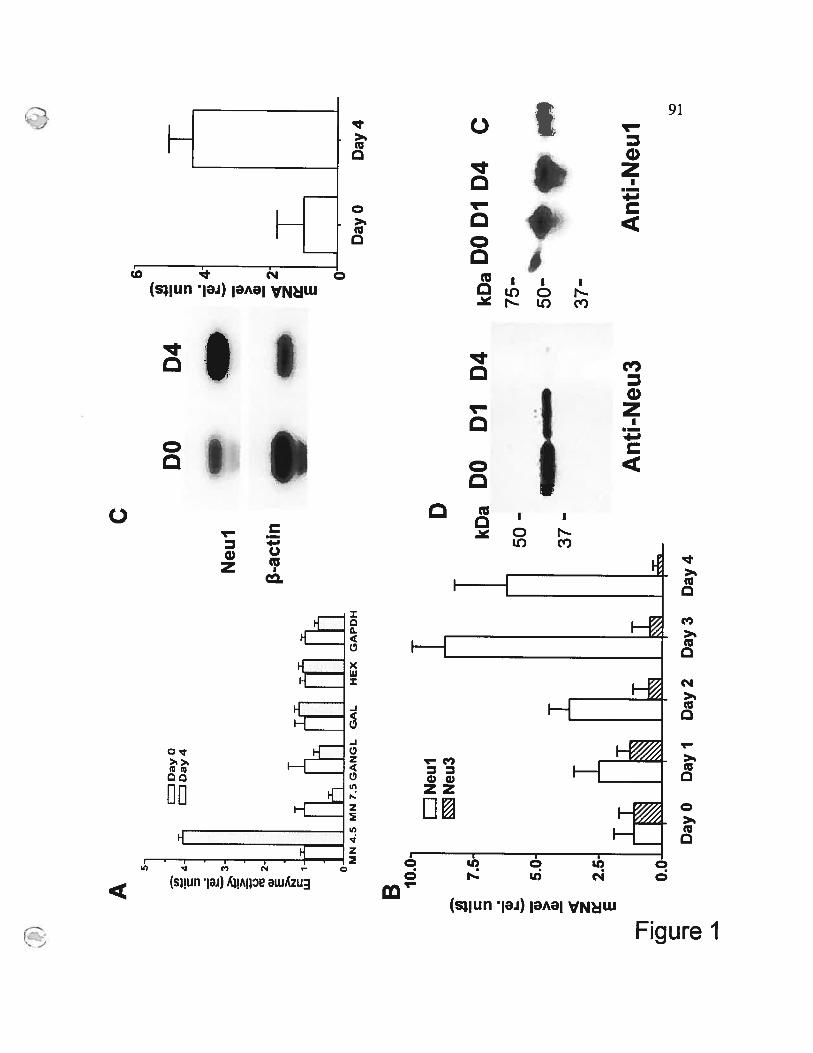
z C) w > G) z E
T
r
As
ci,
z w > > o2
w E u]
E I
DO
ciJ
oyo
JD
ay
4
X
Neu
l
-act
in
“MN
4.5
MN
7.5
GA
NG
LG
AL
HE
XG
AP
DH
C
DiE!
D
kDa
DOD
lD
4
Eli
Neu
lN
eu3
7.5.
Day
OD
ay4
50-
II
37-
-n (D -, (D
kD
aDO
DID
4C
Ant
i-N
eul
Day
OD
ayI
Day
2D
ay3
Day
4
Ant
ï-N
eu3
-

92
Figure 1. Induction of Neul during the differentiation of THP-1 celis into
macrophages
A. Specific activity of sialidase against the synthetic substrate MU-NANA at pH 4.5
(MN 4.5) and pH 7.5 (MN 7.5) and gangliosides (GANGL), 3-gaIactosidase (GAL),
-hexosaminidase (HEX) and glyceraldehyde-3-phosphate dehydrogenase (GAPDH)
rneasured in total homogenates of THP-Ï ceif s before (ctear bars) or 4 days after
(shaded bars) the PMA treatment. Values represent means + S.D. of triplicate
experiments. Data are shown as fractions of the activity measured in undifferentiated
THP-1 celis.
B. mRNA levels of Neul and Neu3 in THP-1 celis during the PMA-induced
differentiation. The rnRNA levels quantified by RT-PCR as described in Materials
and Methods are presented as a fraction of the level detected in the proliferating
THP-1 ceils. Values represent means + S.D. oftriplicate independent experiments.
C. Northern blotting of RNA from undifferentiated THP- 1 celis (Day O) and the ceils
treated for 4 days with 20 nM PMA (Dav 4). The membranes were hybridised with
1321P-labelled Netil and fi-actin cDNA probes and exposed to a BioMax film (left.
Images were scairned and relative intensities of the signais from Netil and /3-actin
probes quantified using a PDQuest software (Bio-Rad) (righi). Values represent
means + S.D. oftriplicate independent experiments.
D. Western blotting ofundifferentiated THP-1 celis (Day O) and the ceils treated for I
(Day 1) and 4 days (Day 4) with 20 nM PMA. Each lane contains 20 g of total ccli
lysates. C, positive control, Neul purified from human placenta. The immunoblotting
was performed with anti-Neul and anti-Neu3 rabbit polyclonal antibodies as
indicated.

Qo
Q
AA
paI
Xb
alX
inal
Sac
IIB
cII
AT
G
II
II
II
-159
8-9
36-5
42
-31
6-1
76
B
LIN
oP
MA
EJ2
On
MP
MA
I
r,fl
pBL
-NE
UJ
CA
T5
‘4.,
(0 4-’ 30.
pBL
CA
T6
pBL
-NE
UIC
AT
I20
pBL
-NE
UIC
AT
210
pBL
-NE
UJC
AT
3
—pB
L-N
EU
JCA
T4
._..3
OECi
)4
-’ 2O
E
C.,
Q
I I
n
LJN
oPM
AL
I2O
nMPM
A
nJnp
[C
AT
IC
AT
2
DU
)4
-’ w >1
4-’ > 4-’ Q I Q
CA
T3
CA
T4
CA
T5
-n (Q C -‘ (D “J
CA
TI
CA
T2
CA
T3
CA
T4
CA
T5
025
5075
100
Tir
ne(h
)
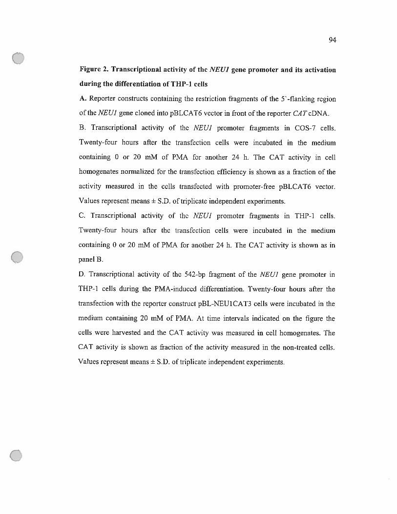
94
Figure 2. Transcriptional activity of the NEU1 gene promoter and its activation
during the differentiation of THP-1 celis
A. Reporter constmcts containing the restriction fragments of the 5’ -flanking region
of the NEUJ gene cloned into pBLCAT6 vector in front of the reporter Cil T cDNA.
B. Transcriptional activity of the NEUJ promoter fragments in COS-7 celis.
Twenty-four hours after the transfection celis were incubated in the medium
containing O or 20 mM of PMA for another 24 h. The CAT activity in celi
homogenates normalized for the transfection efficiency is shown as a fraction of the
activity measured in the celis transfected with promoter-free pBLCAT6 vector.
Values represent means + S.D. oftriplicate independent experiments.
C. Transcriptional activity of the iVEUJ promoter fragments in THP-l celis.
Twenty-four hours afier the transfection celis were incubated in the medium
containing O or 20 mM of PMA for another 24 h. The CAT activity is shown as in
panel B.
D. Transcriptional activity of the 542-hp fiagment of the NEUJ gene prornoter in
THP-l celis during the PMA-induced differentiation. Twenty-four hours afier the
transfection with the reporter constrnct pBL-NEU1CAT3 cefls were incubated in the
medium containing 20 mM of PMA. At time intervals indicated on the figure the
cells were harvested and the CAT activity was measured in ceil homogenates. The
CAT activity is shown as fractioll of the activity measured in the non-treated celis.
Values represent means ± S.D. oftriplicate independent experiments.

95
A
PMA - - + + +
CM - + - + -
NAC - - - - +
B
10.0U)
o
___
.__.
PMA + + +
CM - + -
NAC - - +Figure 3

96
Figure 3. Effect of TNf-a, NAC and cur cumin on NEU1 promoter-driven CAT
expression in COS-7 (A) and THP-1 (B) celis
Twenty-four hours after the transfection with pBL-NEUÏCAT3 vector the ceils were
stimulated for another 24 h with TNf-a (40 ng/ml) or PMA (20 nM) in the presence
or absence of curcumin (CM, 0.5 jiM) and NAC (NA C, 20 mM) as indicated on the
figure. The CAT activity values normalized for the transfection efficiency are shown
as a fraction of the activity measured in the absence of TNF-Πor PMA. Values
represent means + S.D. of triplicate independent experiments.

97
DayO
Dayl
Day4
A
Neul LAMP-2 Merger
C
D
DayO
Day7
DayO
DayZ
DayO
Dayl
Day4
B
Neul CD206 Merger
Figure4

98
Figure 4. Immunohistochemical localization of Neul during the differentiation
of THP-1 cetis (A, B), and primary monocytes (C, D).
After 0, 1, 4 and 7 days of differentiation the ceils were fixed, stained with rabbit
polyclonal anti-Neu 1 antibodies, and countcrstained with mouse monoclonal
antibodies either against human LAMP-2 or against human maimose receptor
(C’D2O6 as indicated on the figure. Panels A, C and D show permeabilized ceils,
panel B nonpermeabilized celis. Panel represent randomly selected typical images
obtained in triplicate experiments; from 140 to 200 cells were studied for each
condition in each experiment. Siides were cxamined on a Zeiss LSM51O inverted
confocal microscope. Magnification 620x.
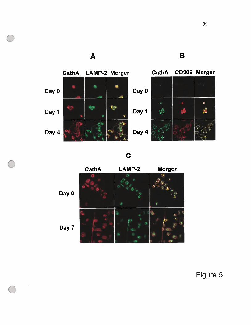
99
DayO
Dayl
Day4
A
CathA LAMP-2 Merqer
DayO
Day7
DayO
Dayl
Day4
B
CathA CD206 Merger
C
Figure 5

100
Figure 5. Immunohïstochemical localization of cathepsin A (CathA) during the
differentiation ofTHP-1 ceLis (A, B), and primary monocytes (C)
Afler 0, 1, 4 and 7 days of differentiation the ceils were fixed, stained with rabbit
polyclonal anti-cathepsin A antibodies, and counterstained with mouse monoclonal
antibodies against human LAMP-2 or against human mannose receptor (C’D206) as
indicated on the figure. Panels A and C show permeabilized ceils, panel B,
nonpermeabilized celis. Panel represent randomly selected typical images obtained in
triplicate experiments; from 140 to 200 celis were studied for each condition in each
experiment. Slides were examined on a Zeiss LSM51O inverted confocal microscope.
Magnification 620x.
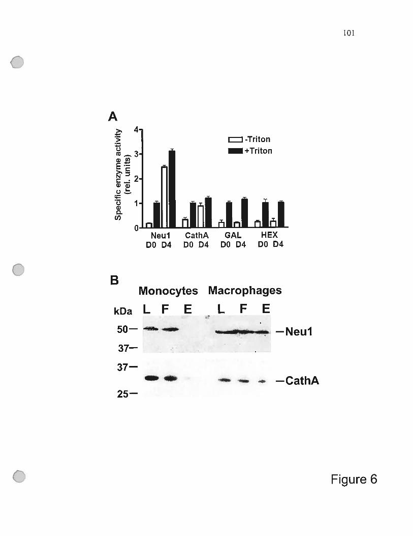
A
>-..
Qcri —
Qri)a
ci,
25—
— 4_ —CathA
Figure 6
101
-Triton
itiÏ ii iiNeul CathA GAL HEX
DO D4 DO D4 DO D4 DO D4
BMonocytes
kDaLF E
50—
37—
37—
Macrophages
LFE
—Neul
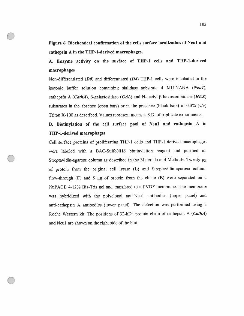
102
Figure 6. Biochemical confirmation of the ceils surface localization of Neul and
cathepsin A in the THP-1-derived macrophages.
A. Enzyme activity on the surface of THP-1 celis and THP-1-derived
macrophages
Non-differentjated (DO) and differentiated (D4) THP-1 ceils were incubated in the
isotonic buffer solution containing sialidase substrate 4 MU-NANA (Netti),
cathepsin A (CathA), J3-galactosidase (GAL) and N-acetyl J3-hexosaminidase (HEX
substrates in the absence (open bars) or in the presence (black bars) of 0.3% (vlv)
Triton X-100 as described. Values represent means + S.D. oftriplicate experiments.
B. Biotinylation of the ccli surface pool of Neul and cathepsin A in
THP-1-derived macrophages
Ccli surface proteins of proliferating THP- 1 celis and THP- 1 -derived macrophages
were Iabeled with a BAC-SuifoNHS biotinylation reagent and purified on
Streptavidin-agarose columu as described in the Materials and Methods. Twenty ig
of protein from the original ccli lysate (L) and Streptavidin-agarose column
flow-through (F) and 5 j.ig of protein from the eluate (E) werc separated on a
NuPAGE 4-12% Bis-Tris gel and transfered to a PVDF membrane. The membrane
was hybridized with the polyclonal anti-Neul antibodies (upper panel) and
anti-cathepsin A antibodies (iower panel). The detection was performed using a
Roche Western kit. The positions of 32-kDa protein chain of cathepsin A (CathA)
and Neul are shown on the right side ofthe blot.
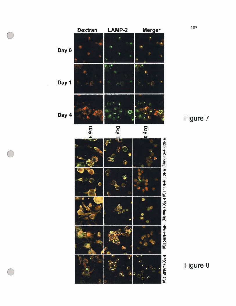
103
Day4 Figure 7
DayO
Dayl
Figure 8
104
Figure 7. Differentiation of THP-1 celis induces the targeting of internalized
Texas Red-iabeled dextran to the ccli surface.
In nondifferentiated ceils (Day O) the labeled dextran is colocalized with the
lysosomal marker LAMP-2. In merged images of the celis harvested 12 h afier the
induction of the celis with PMA (Day 1) and of the differentiated ceils (Day 4) a part
of the internalized dextran appears red (arrojvheads) indicating that the dextran is
Iocated in LAMP-2 negative vesicles, (which appeared to be migrating to the plasma
membrane), on the ceil surface or outside the celis. Panels represent randornly
selected typical images obtained in triplicate experirnents; —$0 ceils were studied for
each condition in each experiment.
Fïgure 8. Immunohistochemical localization of Neul, cathepsin A, MHC II and
the internalized antigen, horseradish peroxidase during the differentiation of
THP-1 celis.
Twelve hours afier the loading with horseradish peroxidase (HRP) THP- 1 celis were
treated with PMA as described. After O h, 24 h and 4 days of differentiation the celis
were fixed, stained with antibodies against HRP, LAMP-2, Netil, cathepsin A
(CathA) and MHC II as indicated on the figure. Panels represent randomly selected
typical images obtained in triplicate experiments; 50 celis were studied for each
condition in each experiment. Siides were examined on a Zeiss LSM5 10 inverted
confocal microscope. Magnification 620x.
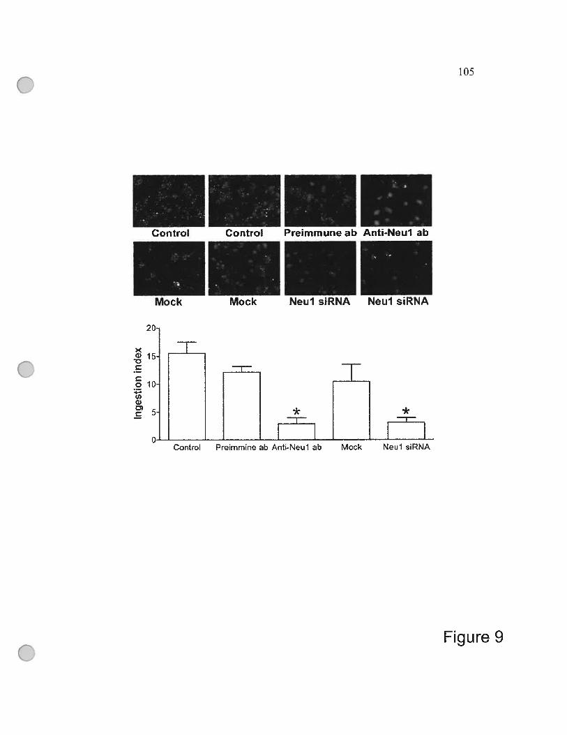
15C
Co 10Q)G)
-
t
20
1
105
Figure 9
ZL
*-n
Control Preimrnne ab Anti-Neul ab
*
ImiMcck Neul siRNA

106
Figure 9. Reduced phagocytosis in THP-1-derived macrophages with suppressed
Neul.
Control THP-1 derived macrophages (ControO, ceils transfected with Neul siRNA
(Netil sil?NA) and scrambled RNA (Mock) as well as the celis differentiated in the
presencc of 20 ig/m1 of anti-Neu 1 antibodies tAnti-Netil ab) or of the preimmune
serum (Preimiitune ab) were incubated for 3 h at 36°C in the presence of
fluorescein-labeled E. cou bioparticles (30 particles per one celi), fixed, and stained
with DAPI. Siides were examined on the Nikon Eclipse E6000 direct epifluorescence
microscope. Magnification 200x. Panels represent typical images obtained in
triplicate experiments; from 70 to 150 ceils (10 panels) were studied for each
condition in each experirnent.
Lowerpanet. Ingestion index was calculated for each type of celis as the percentage
of macrophages with phagocytized particles multiplied on the average number of
particles phagocytized per ingested macrophage. The resuits show an average +SD of
3 independent experiments. * p<O.001 as compared to nontransfected celis,
rnock-transfected ceils or cells treated with preimmune serum.

107
C
Ce
4-CC)oCoowC
o4->1
o
= control
100 pg preimmune serum
20 jig anti-NEUI
100 .ig anti-NEUI
I
*
IL-10 IL-12 (p40) IL-lb IL-6 TNF-Œ
Figure 10

108
Figure 10. Effect of anti-Neul antibodies on the ionomycin-induced production
of IL-10, IL-l2(p4O), IL-lb, IL-6 and TNF-i in THP-1-derived macrophages.
THP- 1 -derived macrophages were stimulated for 12 h with PMA (20 ng/ml) and
ionomycin (1 ig/m1) in the absence (ControO, or in the presence ofpreimmune rabbit
serum (100 igIm1) of anti-Neul serum (20 tg/m1 and 100 tg/m1) followed by the
analysis of IL and TNF-a production by enzyme-linked immunosorbent assay. The
data show an average of 3 independent experiments + standard enor; *- p<O.05 using
unpaired t test.

109
- 1670 CGGCTTGTGGCTCC(ICCTCTGATCTCC’CCACCCAACGCTGGCTTCGTGGOTGTGCTGTGCTCAGTC
- 1602 GGGCCCrANACrTAGTTrAGTGCTCTGCTCTTGCCATCTCGAAACGTGAACAAUTTCATrrrGCACCApaI (-1598)
________________
- 1534 AGTCCCCACAATTACGAjGCCAAITICACTCTCAGCATTGCCAGGCCTCTCACCCTCAACcICCCTlNf-I Myogenin
- 1466 [DTrCTGGCACAGGGTGGTCGTCCTGGACAAAGTCACAGACcrGcTGCTGTrcrrTGGGAAGcrGcT
- I 3
AP-2 Sp-I• 1332 GAAGAAGTGGCCTCAGAGGTGCAGCACTAACCCCCAGAATÏATTCCACAGGGGTCCTGTCCTrUI I t I
- 1264 JTfTCTCCGGTCGCATCCCGGGGCTGCGTAAAGAC1TTAAGAGCCCCCACCTCAACTATrACTGGCfG
- II CCCATCATGGTGAGTGACTCCCCTCTC1GCTGCTCCACCCCCAACTCCCCAGAGGAACCCAATAACCC
- 1128 cAAcGaGTcATGrrccAoAccTccAiccTcGoccctrATGTcATcGccAGcoGcTTcTrcAcccuSp-l
___________
- I TTCGGCATOTGTGTGGACACGCTCTTCCTCTGCTCCCGTGAGTGACACCCCCAjGCCACCTG1TCCCCAA
______________
Nf-KappE2- 992 CCTfAAAGTACTGAGCCGTCAAGCAfl 1111 I I 1111111 GGAGATTGGTGCTfCAGAAACAATGC
XbaI (.936)923
- 856oAoAGToAc4cccccAooccGoTAGocrcTccAoP.GAGcATTcroATooAGAoGTcrcrac
11SF
- 789 [1’A]ÇflDCCTAAGAAATCTCGGGTAGAÀGTTAGCCATTAGAGGCCAOGCATGGTGGCTCACGCCTGTCGÂTA-I
_______
- 722 ATGCCAGCACrGGGGGACCGCGG1ÇGCAATCACCCTGAGGTCAGOAGflCGAGACCAGCCTGCSp- I
- 655 CCAACATGGTGAAACNTCCGTCTCTATTAAAAATACAAAAATTAGCCGAGAGTGGTGGCATGCACCT
- 588 GTCATCCCIAGNTANTcIGGGAGGCTGAGGCAGGAGAATCGCfTGAACCCCGGAGGCAGAGGÎfGCAGGATA- 1,2 XmaI f-542)
- 522 TGAGCCGÀkATCGCGcDACTGCACTCCAACCTGGGTGACAGACTCTGTCTCCAAAACAAAACAAACAE2-FI
- 455 AACAAAAAGATflATTAAAGATAT1TrGTfrflAAATCTGfltTCTCCACGACGA
_________
NF-E2 AP-1 c-jun.c-tos.AP-1
_______
- 387Spi E2F-I Sp-l
_________
CREB- 319 kCGcGDCAATCAAAAAGcCCAATAAGGATGoTGcCAGTcCCTrCrrcA1CcccAGCC1AGTCCG
SadI f-316 AP-2 c-ts2-252 orrcGGTcAcGcGcTcNGccTccAAGAcoGccTccAAoAcGGccAATcGoAAGGGcAAGmcGA
- I GATGCrGCGTGAICACOT000AGGAAGATGAGCTACnGAAGACCAATrAGAGTCC000AAGCGCG&R(-176) . J’
- 119 GCGGGGCI(DTCCAGACCGGGGCGGGCIrrAAGGGTGACATCTGCGCTrrAAAGGGTCCGGGTCAGCTGAM’-2 Spi
- - CTCCCGACTCTGTGGAGTCTAGCTGCCAGGGTCGCGGCAGCTGCGGGGAGAG ATG
Supi. Figure I
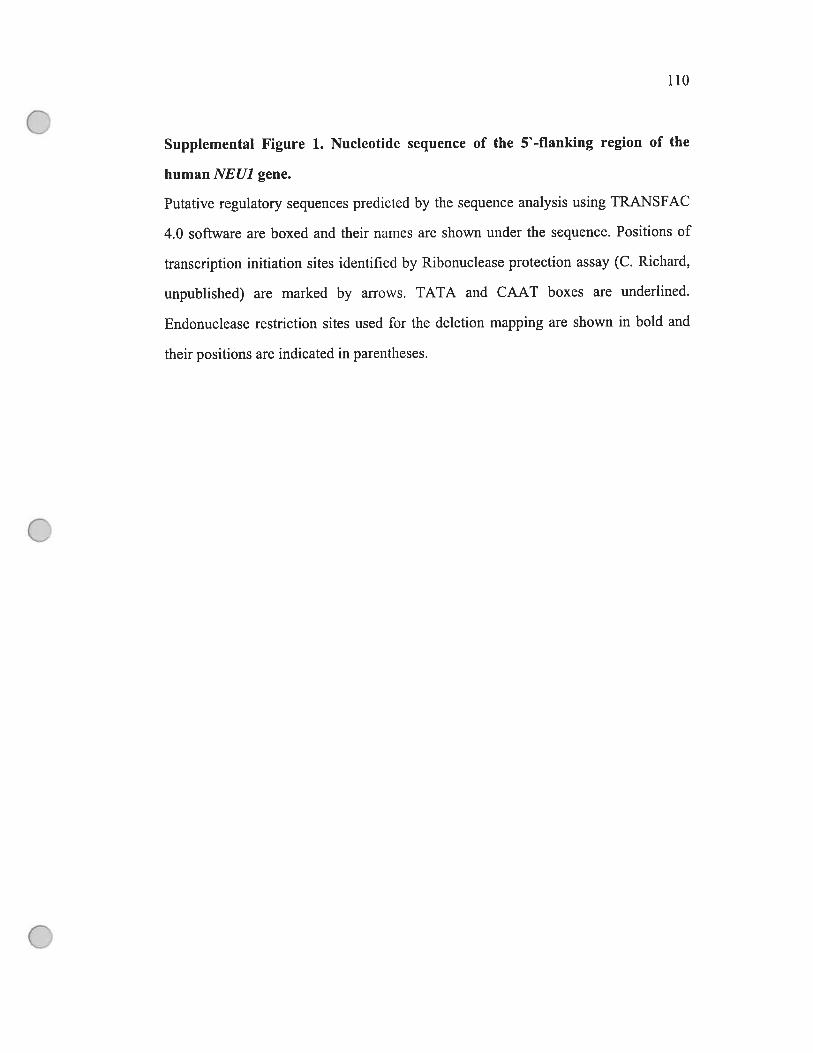
110
$upplemental Figure 1. Nucleotide sequence of the 5-flanking region of the
human NEUJ gene.
Putative regulatory sequences predicted by the sequence analysis using TRÀNSFAC
4.0 software are boxed and their names are shown under the sequence. Positions of
transcription initiation sites identified by Ribonuclease protection assay (C. Richard,
unpublished) are rnarked by arrows. TATA and CAAT boxes are underlined.
Endonuclease restriction sites used for the deletion mapping are shown in bold and
their positions are indicated in parentheses.
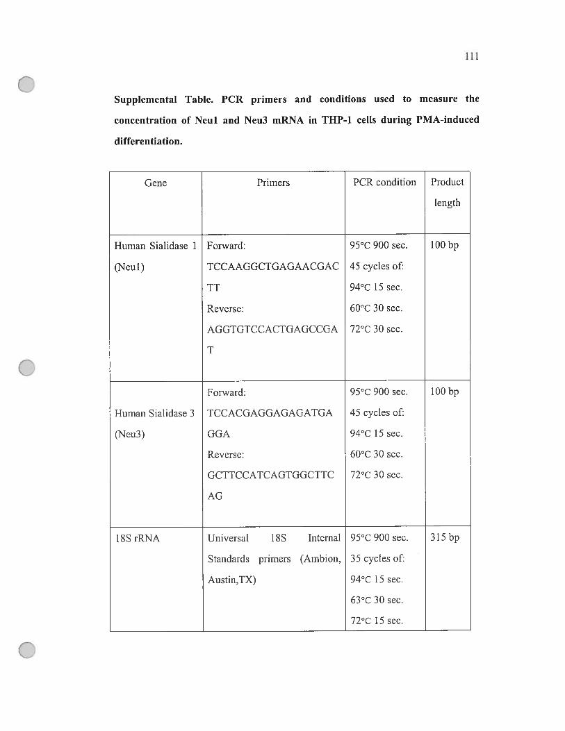
111
Supplemental Table. PCR prirners and conditions used to measure the
concentration of Neul and Neu3 mRNA in THP-1 celis during PMA-induced
differentiatïon.
Gene Prirners PCR condition Product
length
Hurnan Sialidase 1 Forward: 95°C 900 sec. 100 bp
(Neul) TCCAAGGCTGAGAACGAC 45 cycles of:
TT 94°C 15 sec.
Reverse: 60°C 30 sec.
AGGTGTCCACTGAGCCGA 72°C 30 sec.
T
Forward: 95°C 900 sec. 100 bp
Human Sialidase 3 TCCACGAGGAGAGATGA 45 cycles of:
(Neu3) GGA 94°C 15 sec.
Reverse: 60°C 30 sec.
GCTTCCATCAGTGGCTTC 72°C 30 sec.
AG
18$ rRNA Universal 18$ Internal 95°C 900 sec. 315 bp
Standards prirners (Ambion, 35 cycles of:
Austin,TX) 94°C 15 sec.
63°C 30 sec.
72°C 15 sec.
CHAPTER 4: NEU1 SIALIDASE BUT NOT CATHEPSIN
A ACTIVITY ON THE SURFACE 0F TUE IMMUNE
CELLS IS IMPORTANT FOR THE FUNCTIONAL
INTEGRITY
ARTICLE 3
Manuscript, Januaiy, 2007

113
NEUJ SIALIDASE BUT NOT CATHEPSIN A ACTIVITY
ON THE SURFACE 0F THE IMMUNE CELLS 1$
IMPORTANT FOR THE FUNCTIONAL INTEGRITY
feng Liangt, Alexandre lannellot, Voikan Seyrantepet, Ah Ahmadt, and Alexey V.
Pshezhetsky*t
Sainte-Justine Hospitait and Departments ofPaediatrics and Biochemistry,
University ofMontreal, Montreal, Quebec H3T 1C5 Canada
To whom correspondence should be addressed: Alexey V. Pshezhetsky, Service
de génétique médicale, Hôpital Sainte-Justine, 3175 Côte Ste-Catherine, Montréal
(Qc) H3T 1C5 — Canada, Tel. (514) 345-4931/2736, fax. (514) 345-4766, E-Mail:

114
ABSTRACT
The differentiation of monocytes as well the activation of lymphocytes and
macrophages is accornpanied by the appearance on their surface of 2 lysosomal
enzymes, sialidase (neuraminidase 1, Neul) and carboxypeptidase A (cathepsin A,
CathA), which forms a complex with and activates Neul. b clarify the biological
roles ofNeul and CathA in the immune celis we have developed animal models ofa
single CathA deflciency and a double CathA/Neul deficiency by gene targeting in mice.
In the first une of mice, CathA”901, we have replaced the nucleophyl of the CathA
active site, Serl9O with Ala, which abolished the enzymatic activity, while the mutant
protein retained its ability to activate Neul. The second une, CathAS9oe0, was the
hypomorphic CathA mice with the secondary -9O% reduction of the Neul activity.
Macrophages derived from the spienocytes or the peripheral blood monocytes of the
CathA90° mice as well as their immature dendritic ceils showed significantly
reduced capacity to engulf bacteria. The mature dendritic celis also had lower effect
on T cetis proliferation, while properties of the celis derived from the CathAS91
mice were indistinct from those of the wild type controls. Macrophages from the
Neul-deficient mice showed an increased affinity to the sialic acid-binding lectin
from Maackia ainurensis consistent with a compromised processing of sialylated
sugar residues of cdl surface molecules. Altogether our data suggest that the ceil
surface Neul plays an important role in the regulation of affinity of the immune ceils
towards each other and extemal pathogens.
115
INTRODUCTION
Sialic acids are abundantly expressed on the surface of the immune celis and
implicated in mediating recognition between the ceils, between the ceils and
extracellular matrix as well as between the ceils and a range of pathogenic viruses,
bacteria and protozoa during the inflarnmatory and immune reactions (reviewed in
Keim and Schauer, 1997). For example, members of the siglecs (sialic acid binding
IgG lectins) superfamily are known to mediate many of these interactions
contributing, in particular, to scavenging function of macrophages, pathogen uptake
and antigen presentation (reviewed in Munday et ai., 1999). Much iess is known
about the role of sialidases and sialotransferases that can regulate celiular affinity by
modifying the sialylation of ccli surface molecuies.
Previous data showed that the lysosomal sialidase encoded by the NEU] gene
in addition to its roie in intralysosomal catabolisrn may be also involved in celluiar
signalling during the immune response. In particular, during the activation of mouse
T cells Neul is expressed on the plasma membrane and is required for the early
production of IL-4 and for the interaction of T ceils with the antigen presenting celis
(Landolfi et al., 1986; Chen et al., 1997, 2000; Nan et al., 2006; Lukong et ai., 2002).
In addition, Neul of T ceils converts the group specific component (Gc protein) into
a factor necessary for the inflammation-primed activation of macrophages
(Yamamoto et al., 1993, Naraparaju et al., 1994) T-cells derived from SM/J or
BÏ0.SM strains ofmice, with the reduced Neul activity due to a missence mutation
in the Neit] gene (Carrillo et aI., 1997) fail to convert Gc and synthesize IL-4, while
3 cells of these mice cannot produce IgGi and IgE after the immunization with
pertussis toxin (Landolfi et al., 1985, 1986; Naraparaju et al., 1994).
Previously we showed that Neul is increased up to 14-foÏd during the
differentiation of human monocytes into macrophages (Stamatos et al., 2005). We
fiirther showed that the majority of Neul in macrophages is located on the ccli
surface (Liang et al., 2006). Activation and stabilization of the Neul in the lysosome,
requires its association with the lysosomal multienzyme complex (LMC) which also
containing the lysosomal carboxypeptidase A (cathepsin A, Cath A/protective

116
protein), B-galactosidase and N-acetylgalactosamine-6-sulfate sulfatase (reviewed in
Pshezhetsky and Ashmarina, 2001). Interestingly during the differentiation of
macrophages, CathA but flot other components of LMC is sorted to the plasma
membrane similarly to Neul. Both proteins were first targeted to the lysosome and
then sorted to LAMP-2-negative, MHC II-positive vesicles, later merged with the
plasma membrane (Liang et al., 2006).
In order to clarify the physiological rob ofboth enzymes in the immune colis
we have compared essential ftinctional properties of monocyte-derived macrophages,
dendritic ceils (DCs) from the genetically targeted mice having either a single CathA
or a double CathA/Neul deficiency. The obtained data suggest that the ccii surface
Neul regulates the affinity of the immune celis towards each other and extemal
pathogens and is therefore necessary for their fiinctional integrity.
EXPERIMENTAL PROCEDURES
Animais
The CathA’90’1 mice canying the point Serl9OAla mutation in the CathA
gene and the CathA9O10 mice carrying in addition a FGK-neo cassette in the
intron 7 werc developed through the targeted disniption of the CathA gene as
described (Seyrantepe et al. submitted). The CathA91k0 and CathA9A mice
ranging from 4 to 12 weeks of age were compared with appropriate WT littermate
controls. All mice were bred and maintained in the Canadian Councit on Animal Care
(CCAC)-accredited animal facilities of the Ste-Justine Hospital Research Conter
according to the CCAC guidelines. Approval for the animal care and the use in the
experiments was grantcd by the Animal Care and Use Committee of the Ste-Justine
Hospital Research Conter.
Isolation and culturing of blood monocytes
Peripheral blood mononuclear celis (PBMCs) were isolated by leukophoresis
of blood from mice heart followed by consequent centrifugations over Ficoll-Paque
117
Plus and Percoil (both Amersham Biosciences) gradients as described (Liang et al.,
2006). To obtain monocyte-derived macrophages, the ceils were suspended at 2 x 10⁶
ceÏls/ml in RPMI medium 1640 (Gibco, Grand Island, NY) containing 10% heat
inactivated fetal caif serum (fCS). After 2 hrs, non-adherent celis were removed by
two washes with phosphate-buffered saline (PBS) and the adherent monocytes were
maintained at 4 x 10⁶ cells/well in 6 well tissue culture plates (Costar, Coming Inc.,
Corning, NY) at 37°C in a humidified atmosphere containing 5% C02. After 7 days
differentiated macrophages (larger and more granular than monocytes as seen by light
microscopy) were harvested in PBS by gentle scraping with a polyethylene cell
scraper (Nalge Nunc International, Rochester NY). The harvested cells were
confirmed to have characteristic macrophage cdl surface phenotypic markers
+ +(CD 14 , CD206 ) by flow cytometry.
Isolation and culturing of spienocytes
fresh spienocytes were obtained by teasing the mouse spleens under aseptic
conditions. Erythrocytes were removed by centrifugation over Ficoll-Paque Plus
gradient and the remaining unfractionated nucleated spleen celis were washed twice
with PBS and adjusted to a density of 7.5 x i05 of ceils per ml ofRPMI medium 1640
containing 10% of heat-inactivated FCS. To obtain splenocyte-derived macrophages,
purified spienocytes were maintained at 2 x 10⁶ of cells per well in 6 well tissue
culture plates (Costar, Corning Inc., Corning, NY) at 37°C in a humidified
atmosphere containing 5% C02. At the specified times, non-adherent celis were
removed by two washes with PBS and the adherent, differentiated macrophages were
harvested in PBS by gentie scraping with a polyethylene cdl scraper (Nalge Nunc
International, Rochester, NY). The harvested celis were confirmed to have
characteristic macrophage ceil surface phenotypic markers (CD14+, CD206+) by
flow cytometry.
Immature splenocyte-derived DCs were obtained as described earlier
(Berthier et al., 2000). Briefly, purified mouse spienocytes were suspended at a
density of 7.5 x of celis per ml in Iscove’s modified Dulbecco’s medium (IMDM)
118
(Gibco, Life Technologies, Grand Island, NY) supplemented with 12.5% heat
inactivated fCS, non essential amino acids (0.1 iuM/ml), sodium pyruvate (1
rnM/ml), 2-mercaptoethanol (50 iM), penicillin (5000 U/ml), streptomycin (50
mg/ml) and flmgizone (0.5%). Two ml of this suspension were plated per 35 mm
tissue culture dish (Nunclon, Glostmp, Denmark) and culture development proceeded
over two different steps: the first step was devised to favour the proliferation of DC
progenitors by early cytokine IL-6 (R&D Systems, Abingdon, UK) and the second, to
induce the maturation of DC with cytokines fit-3 L and GM-CSF (R&D Systems)
known to exert a differentiation activity on mouse DC. The early phase lasted from
day O to day 6 of incubation when the proliferation of DC progenitors took place
under the influence of IL-6 (25 ng/ml). The second culture phase started on day 6
when IL-6 was withdrawn from the medium. The colis were washed, counted, split
to a concentration of 1.5 x 10 of celis per ml in fresh complete medium and cultured
in the presence of fit-3L (25 ng/ml) and GM-CSf (1000 U/ml) during the next 3 to 4
weeks. The ceils were spiit again on days 10, 14, 20 and 26 by replating at 4 to 6 x
of colis per ml. To obtain mature spienocyte-derived DCs, the celis were cultured
for an additional 48 hrs in the presence of 10 ig/rnl of Iipopolysaccharides (LPS, E.
cou 026:B6, Sigma, St. Louis, MO, USA).
Enzyme assays
Sialidase activity in ceilular hornogenates was assayed using the synthetic
fluorogenic substrate 2’ -(4-methylurnbelliferyÏ)—a—D-N-acetylneuraminic acid
(4MU-NeuAc; Sigma) as described (Potier et ai., 1979) with the following
modifications. Freshly isolated oeils (2-5 x 10⁶) were homogenized in 0.20 ml of a
solution containing 0.5% (vAi) Triton x-100, 0.05 M sodium acetate, pH 4.4 by
sonication and incubated with 0.125 mM 4MU-NeuAc at 37°C for 1 h. The reaction
was terminated by the addition of 1.9 ml of a solution containing 0.15 M glycine, pH
10.5. Liberated 4-methylumbelliferone was measured using a Shimadzu RF-5301
spectrofluorometer with excitation at 355 nm and emission at 460 nm. Enzyme
activity is expressed as the conversion of 1 nmol of substrate per hr. Protein

119
concentration was measured by the Bradford method using a protein assay kit (Bio
Rad, Hercules, CA, USA) and the amount of activity measured in each sample was
corrected based on protein concentration to represent activity per milligram of
protein.
-Hexosarninidase activity in cellular homogenates was assayed with 1.25
mM 4-methylumbelliferyl-2-acetamido-2-deoxy-f3-D-glucopyranoside as previously
described (Rome et al., 1979). After the incubation at 37°C for 15 mm, the reaction
was terminated with 1.9 ml of 0.4 M glycine buffer, pH 10.4, and the fluorescence of
the liberated 4-methylumbelliferone was measured as described above. The
carboxypeptidase activity of cathepsin A was measured as follows. Freshly isolated
ceils (2-5 x 10⁶) were homogenized in 0.20 ml of a water containing 0.5% (y / y)
Triton X-100 and incubated with 100 pi of 1.5 mM CBZ-Phe-Leu (Sigma-Aldrich, St.
Louis, MO, USA) in 100 mM sodium acetate buffer, pH 5.2 at 37°C for 30 min. The
reaction was terminated by addition of 20 pi of trichioroacetic acid. Precipitate was
rernoved by 10 min centrifugation at 12,000g and 100 pi of the supernatant was
mixed with 3 ml of the 0.05 M sodium borate buffer pH 9.5 containing
phthaldialdehyde (Sigma, 0.15 mg’hil) and f3-rnercaptoethanol (0.0075%). After 5
min incubation the concentration of leucine was rneasured using a Shimadzu RF
5301 spectrofluorometer with excitation at 340 nm and emission at 455 nm (Roth,
1973).
Phagocytosis assay
The splenocyte-derived macrophages or immature DCs seeded on cover slips
were incubated for 3 hours with fluorescein-labeled E. cou (K-12 strain) bioparticles
(Molecular Probe, Eugene, OR) in a ratio of 50 partic tes per one cell at 37°C or 4°C,
washed 3 times with cold P35, fixed with 4% paraformaldehyde for 1 h,
permeabilized with 0.5% Triton X-100, and washed twice with 0.05% Tween-20 in
PBS. Cells were counterstained with 3 jiM DAPI and washed again with 0.05%
Tween-20 in PBS. Slides were examined using the Nikon Eclipse E6000
120
epifluorescence microscope and intemalization was quantitated by flow cytometry
using FACS Calibur.
Immunofluorescent labeling and fiow cytometry
Monoclonal antibodies, conjugated with fluorescein isothiocyanate (FITC) or
phycoerythrin (PE), against MHC class II (anti-I-A), HL3—PE (anti-CD 11 c), RMMP
1 (anti-CD$6—PE, B7-2), CD45 and CD14 were purchased from eBioscience (San
Diego, CA). For surface immunostaining adherent ceils were detached by pipeting
after 3-5 minutes incubation at 37°C in PBS containing 3mM EDTA. Aliquots of 1 x
106 cells were incubated with 500 ng/ml of mouse IgGi for 10 minutes on ice in
order to block fc receptors, followed by a double staining with the appropriate
antibodies for 30 minutes on ice and in the dark. Celis were washed three times with
P35 containing 0.05% (w/v) BSA and fixed with 2% paraformaldehyde. The
acquisition was done using a FACS Calibur (Beckton Dickinson, San Diego, USA)
and data were analysed using the CellQuest Pro software.
Lymphocyte proliferation assay
Mice lymphocytes were isolated by collecting the non-adherent celis from
spienocytes of normal mice. Purified mice lymphocytes were maintained at 37°C in a
humidified atmosphere of 5% C02 in RPMI 1640 medium, containing 5,000 units of
PenicillinlStreptomycin and Fungizone (both Life Technologies, Inc.). Mice
lymphocytes (2 x 10⁶ in 3 ml medium) were seeded in six-welI tissue culture plates
and incubated at 37°C in 5% C02 in the presence or absence of mature spienocyte
derived DC (1 x 10g). Celis were exposed to 1 i.iCi/rnl of [methyl-3H]-thymidine
(Amersham Pharmacia Biotech, Piscataway, New Jersey, USA). Ninety-six hours
after the exposure to [rnethyl-3H]-thymidine the celis were first washed twice with
PBS and then four times with 5% trichloroacetic acid (ACP Chemicals) in water. The
cdl pellets were solubilized in 200 i1 of 0.2% Triton X-100, 0.1 M NaOH. The
radioactivity in the celi homogenates was measured using a Top Count instrument
(Packard). At 24, 48 and 96 hrs celis were counted using haemacytorneter (Fisher).
121
Lectin affinity assay
The splenocyte-derived macrophages seeded on cover slips were incubated
for 15 min at 37°C with 100 ig/ml of ftuorescein-labeled Maackia amurensis lectin
(EY Laboratories, INC, San Mateo, CA). Then the celis were washed 3 times with
cold PBS and fixed with 4% paraformaldehyde for 1 h. Ceils were counterstained
with 3 tM DAPI and washed again with 0.05% Tween-20 in PBS. Slides were
examined using the Nikon Eclipse E6000 epifluorescence microscope.
RESULTS
Immune ceils derïved from the CathA9AM0 but not from the CathAS’9/ mïce
show the reduced sialidase activity
Our previous studies performed on the tissues from kidney, livers, brain and
lungs of the CathA9otïe0 and the CathA9A mice (Seyrantepe et al., submitted)
showed that both mice had almost zero level of CathA activity, consistent with the
presence ofthe Serl9OAla point mutation which eliminates the active site nucleophile
(Gaijart et al., 1991). In contrast, a significant difference was observed in the tissue
levels of the sialidase activity measured at acidic pH 4.5 with the synthetic
fluorogenic substrate 2’ -(4-methylumbelliferyl)—a—D-N-acetylneuraminic acid
(4MU-NeuAc). CathA5’9°’ mice showed normal activity levels, whereas in tissues of
the mice, that bas a PGK-neo cassette inserted into the intron 7 of the
CathA gene the sialidase activity was reduced to -10%. Further studies showed that
the CathA579°’ mice express normal levels of inactive CathA protein capable of
forming a complex with and activating the Neul sialidase. In the tissues of the
CathAs]9oe0 mice the CathA mRNA and protein levels were dramatically reduced
consistent with previously reported hypomorphic effect of the neo gene and caused
the secondary deficiency of the Neul (Seyrantepe et al., submitted). Both the
CathAs]9oIle0 and CathA5”9°’ mice are vital, fertile, grow normally and have a normal
lifespan. The FACS calibre immunophenotypic profiling of total splenocytes obtained
from the WT, CathA5”90’1 and CathAS9O?1e0 mice and double-stained with anti-MHC

122
and anti-CD-14 monoclonal antibodies did not reveal any difference in the splenal
population of monocytes and immature DCs.
In order to find whether the immune ceils of the and the
CatÏiA9190” mice show decrease in the CathA and sialidase activities similar to that
observed in the non-lymphatic tissues we have assayed both activities in the blood
monocytes and in the monocyte-derived macrophages. We have also assayed CathA
and sialidase activities in the monocytes purified from the spleen tissues, as well as in
splenocytes-derived macrophages, immature and mature DCs. As we expected, only
background levels (10-15%) of the carboxypeptidase activity measured with the
specific CathA substrate CBZ-PheLeu could be detected in blood monocytes and
macrophages from both the CathA?9ole0 and the CathA9A mice (fig 2A). This
activity does flot change upon the differentiation and reftects the background level of
CBZ-Phe-Leu conversion by other cellular carboxypeptidases. In the ceils from the
WT mice the CathA activity increases but flot significantly (fig. 2A). Sirnilar resuits
were obtained also for the spleen-derived macrophages (fig. 2A) and DCs (flot
shown). In contrast, a significant increase of the sialidase activity was observed
during the differentiation of the monocytes derived from the blood or the spleens of
the wild-type and CathAS9O4 mice (fig. 2B). As compared to both spleen and blood
derived monocytes the sialidase activity was increased at least 2 fold in macrophages,
at least 4 fold in immature DCs, and at ieast 5-fold in the mature DCs. Macrophages
from the CathA59o1!\Je0 mice (both blood- and spleen-derived) showed sialidase
activity almost similar to that in the corresponding monocytes, confirming our
previous finding that the increased sialidase activity in macrophages resuits from the
induced expression ofthe Neul gene. Although the sialidase activity in immature and
mature DCs from the CathAS9O!0 mice is increased it still remains significantly
lower then in the cells from the WT or the CathAS9 mice. The control lysosomal
enzyme, f3-hexosaminidase showed the same activity in the celis from ail mice (flot
shown).

123
Macrophages and immature DCs from the CathASl9O0 mice show impaired
capacity to internalïze pathogens
In order to understand whether the Neul andlor CathA activities are important
for the primary function of macrophages and immature DCs we have studied the
phagocytic capacity of these ceils derived from the wild-type, CathAs79OJe0 and
CathA5’90’1 mice. The macrophages derived from the spienocytes of the WT,
CathASi9OA0 and CathA”9°’ mice were incubated for 3 hours at 37° or 4°C with the
fluorescein-labeled E. cou bioparticles, washed, fixed with paraformaldehyde, stained
with DAPI and studied using an epifluorescent microscope. The ingestion index of
macrophages was calculated as a percentage of macrophages with engulfed particles
muitipiied by the average number ofparticles phagocytized per ingested macrophage.
The control level measured at 4°C was subtracted from ail values. No statistical
difference was observed between the ingestion index of macrophages derived from
the CathASl9 (21.5±4.9) and the WT (19.3±5.3) mice (Figure 3 A). In contrast, theS]90A-Neoingestion index for the macrophages derived from the CathA mice (9.2±2.0)
was statisticaliy reduced as compared to the celis from both the WT and the
CathA’”90’1 (figure 3 A). Similar results were also obtained for the macrophages
(peritoneal adhesive celis) purified from the peritoneal cavities. The cells derived
from the CathA59oIe0 showed ingestion index at least twice lower than that of the
celis from the WT and CathA5’9°’ mice.
Significant difference between the phagocytic activity of the macrophages
from the WT and CathA”9°’° mice was also observed by flow cytometry (figure 3
B). Afier 3hr incubation with fluorescein-Iabeled E. cofl bioparticles, more than 44%
of the splenocyte-derived macrophages from the WT mice were positive for
fluorescein, whereas only 18% of fluorescein positive ceils were observed among the
celis from the mice (Figure 3 B).
As compared to the macrophages, the spenocyte-derived immature DCs have
significantly enhanced phagocytic activity. When immature DCs derived from the
splenocytes of the WT or the CathA”9°” mice were incubated for 3 hours with the
fluorescein-labeied E. cou bioparticles their ingestion index was ranging between 35

124
and 42. The ingestion index of the immature DCs from the CathA”90’° mice was
reduced to 18.8+3.2 (Figure 3 C).
Mature DCs from the CathAS9’0 mice show reduced capacity to activate
lymphocyte proliferation
The ability to activate proliferation of lymphocytes is one of the essential
physiological properties ofthe mature DCs. To understand if this function depends on
the Neul expression we have obtained populations of the mature DCs from the WT,
the CathA’9°’ and the mice by 4$-hr treatment of the spienocytes
derived immature DCs with LPS and studied their ability to induce the proliferation
of lymphocytes. Mouse lymphocytes were isolated by coïlecting the non-adherent
ceils from spienocyte cultures of the WT siblings, the CathA5’90’ and the CathA5’90”Neo mice and placed in aliquots of 2 x 10⁶ cells in 3 ml medium into the culture plates
containing 1 x 1 0 of mature splenocyte-derived DCs in the presence of [methyl-3H]-
thymidine (1 jiCi/ml). After 96 hrs of incubation ceils were harvested and the total
radioactivity was measured in the ceil homogenates (Figure 4 B). At 24, 48 and 96
hrs the ceils were counted using haemocytometer (Figure 4 A). No proliferation of
lymphocytes was observed in the absence of DCs. The DCs from both the WT and
the CathA 901’1eo mice stimulated ce!! proliferation but 20% slower growth rate was
observed in the presence of the ceils from the CathA9Ol\0 mice as compared to the
WT controls. Similarly, —25% lower transcriptional activity of lymphocytes
(measured as the amount of [methyl-3H]-thymidine incorporated by the ceils within
96 hrs) was observed in the presence of the mature DCs from the CathAs’9oe0 mice
then in the presence ofthe mature DCs from the WT mice (Figure 4 B).
Macrophages derived from the Neul-deficient mice show higher affinity towards
the sialic acid binding lectin as compared with the ceils from the WT or CathA
deficient mice.
In order to understand whether Neul is involved in macrophages in
processing the sialylated macromolecules present on the ceil surface we have studied

125
the affinity of the celis derived from the wild-type, and CathAS9A
mice towards the sialic acid-binding lectin from Maackia amurensis. The
macrophages were incubated with the fluorescein-labelled lectin, washed, fixed with
paraformaldehyde, stained with DAPI and studied using an epifluorescent
microscope. We found that the macrophages from the CathA5190”° mice showed
intensive peripheral staining with the lectin (Figure 5) consistent with the high
silyalation of their surface. The ceils from the CathA-deficient CathA’9°’ mice
showed weak staining similar to that ofthe celis from the WT mice.

126
DISCUSSION
Growing body of evidence suggests that in addition to their role in catabolism
of macromolecules lysosomal enzymes have important physiological functions
outside the lysosomes, most ofien on the ceil surface or outside the ccli. In particular,
the lysosomal —hexosaminidase A has been reccntly shown to participate in the
production of ceramide from GM3 ganglioside on the externai leaflet of plasma
membrane of fibroblasts (Mencarelli et al., 2005; Valaperta et al., 2006). In similar
fashion, the components of the multienzyme lysosomat complex, Neul, CathA and f3-
galactosidase (in its alternatively spliced form) have important physiological
functions outside the lysosome participating in the processing of endothelin-1 (ET-1),
a potent vasoconstrictive peptide, which plays multiple roles in nonvascular tissues
(Itoh et al., 1995), assembly ofthc elastic fibres (Hinek et al., 1993; 2006) and cdl
signalling during the immune response (Stamatos et al., 2005; Liang et al., 2006). In
humans inherited mutations in CathA cause disruption of the complex and trigger the
galactosialidosis (OMIM 256540), an autosomal recessive disease characterized by
combined deficiency ofNeul, J3-galactosidase and CathA (reviewed in Thomas et ai.,
2001). Another autosomal recessive disease, sialidosis (OMIM 256550), is caused by
mutations directly affecting the NEUJ gene (reviewed in d’Azzo et al. 2001;
Pshezhetsky and Ashmarina, 2001). Several phenotypic characteristics of patients,
including abnonnal, arterial hypertension and cardiomyopathies related to defects in
elastic fiber formation, are not predicted from the lysosomal hydrolase functions of
these proteins, but consistent with the disruption of their secondary activities on the
ceils surface, related to hemodynamic functions and formation of cartilage (Caciotti
et al., 2005; Kyllerman et aI., 1993). However no data exist clarify whether the Neul
and CathA deficiencies affect immune responses in the galactosialidosis and
sialidosis patients.
Recently we have developed animal modeis of single CathA deficiency and
double CathAAiNeul deficiency by gene targeting in mice (Seyrantepe et al.,
submitted). In the first line of mice (CathAS94) wc have replaced the nucieophyl of
the CathA active site, Serl9O with Aia, which abolished the enzymatic activity, while
127
the mutant protein retained its abiiity to activate Neu 1. We have aiso produced a une
mice) that had a FGK-neo cassette inserted in the CathA gene. These
mice had a significantiy reduced (3-10%) Neul activity, due to dramaticaiiy
decreased CathA mRNA leveis, consistent with previousiy reported hypomorphic
effects of the neo gene (Meyers et ai., 199$; Camieliet et ai., 1996; Moran et ai.,
1999). Therefore, we have obtained a hypomorphic CathA mice with the secondary
deficiency ofNeul activity. Since the residual level ofthe Neul activity in the tissues
of the mice is stili higher than that in previousiy described knockout
CathA mice (Zhou et ai., 1995), the animais do flot develop severe gaiactosialidosis
phenotype.
Our data strongly indicate that the immune celis from the Neul/CathA
deficient mice but not from the CathA-deficient CathA579°” mice have
a significantly reduced functionai capacity. First we found that the spenocyte- and
PBMC-derived macrophages, peritoneal adherent celis as weli as the spienocytes
derived immature DCs were compromised in their abiiity to phagocytose pathogens
(f igure 3). This in tum could mean that these ceiis are iess efficientiy activated,
which is consistent with our previous observation that the THP-1-derived
macrophages showed reduced production of cytokines inciuding IL-lb, IL-6 and
TNf-a after the Neul on the their surface was inhibited with the anti-Neul antibodies
(Liang et ai., 2006). In addition, the macrophages from the Neul-deficient mice
showed higher affinity to the FITC conjugated Maackia ainurensis lectin (figure 5)
suggesting that they have increased siaiylation of the ccii surface glycoproteins and
that Neul piays a major roie in processing of the sialyiated moiecuies on the ceii
surface.
Previously, optimal siaiyiation of both sigiecs and their targets has been
shown to be important for the ecu-ecu and celi-pathogen interactions of
macrophages, DC, T, B and NK ceiis (reviewed in Crocker et ai, 2002), therefore the
latter couid be affected by compromised processing of the sialogiycoconjugates in
the mice. Indeed we have observed a significantiy the reduced potency
of the mature DCs in the activation of T-ceii proliferation (f igure 4). The hosts
12$
defective in these pararneters may be predisposed to repeated infections with bacterial
and viral pathogens, a condition described in the patients suffering from the
lysosomal storage diseases (G. Mitcheli, personal communication).
In conclusion, our data show that the ceil surface Neul plays an important
role in the regulation of affinity of the immune celis towards each other and extemal
pathogens. Although further studies are required to understand how the partial Neul
deficiency in the mice affect their immune response in vivo, our
findings suggest that similarly to CD63, CD152, CD206 and other proteins with
lysosomal/endosomal proteins expressed on the surface of the immune celis, Neul is
important for their functional integrity.
ACKNOWLEDGEMENTS
We thank Mrs Raffaela Ballarano for help in the preparation ofthe manuscript
and Dr. Mua Ashmarina for helpful advice. This work was supported in part by the
operating grants from Canadian Institutes of Health Research (MOP 15079 and GOP
3$ 107), and by the equipment grant from Canadian foundation for limovation to
A.V.P. V.S. acknowledges a post-doctoral fellowship from the Fonds de la Recherche
en Santé du Québec (fRSQ) and Lf. a doctoral fellowship from Fondation de
l’Hôpital Sainte-Justine. AVP is a National Investigator ofFRSQ.
129
REFERENCES
Keim, S., Schauer, R. (1997) Int. Rev. Cytol. 175, 137—240.
Munday J, floyd H, Crocker PR. (1999) Sialic acid binding receptors (siglecs)
expressed by macrophages. JLeukoc Bio!. 66(5): 705-711.
Chen XP, Ding X, Daynes RA. Ganglioside control over IL-4 priming and cytokine
production in activated T celis. Cytokine 12(7):972-985 (2000).
Chen XP, Enioutina EY, Daynes RA. The control of IL-4 gene expression in
activated murine T lymphocytes: a novel role for ncu-1 sialidase. J IrnmunoÏ
158(7):3070-3080 (1997).
Landolfi Nf, Cook RG. Activated T lymphocytes express class I molecules which
are hyposialylated compared to other lymphocyte populations. Mol IminztnoÏ
23(3):297-309 (1986).
Nan X, Carubelli I, Stamatos NM. Sialidase expression in activated human T
lymphocytes influences production of IFN-y J Leukoc Bio!.
Yamamoto N, Kumashiro R. Conversion of vitamin D3-binding protein (group
specific component) to a macrophage activating factor by the stepwise action of f3-
galactosidase of B cells and sialidase of T cells. J hnniztnol 151(5):2794-2802
(1993).
Naraparaju VR, Yamamoto N. Roles of f3-galactosidase of B lymphocytes and
sialidase of T lymphocytes in inflammation-primed activation of macrophages.
hmnunol Lett 43(3): 143-148 (1994).
130
Caniilo MB, Mimer CN, Bali ST, Snock M, Campbeil RD. Cloning and
characterization of siaiidase from the m urine histocompatibiiity-2 complex: iow
levels of mRNA and a single amino acid mutation are responsible for reduced
sialidase activity in mice canying the Neula ailele. Glycobiology 7:975-986 (1997).
Landoifi Nf, Leone J, Womack JE, Cook RG. Activation of T lymphocytes resuits in
an increase in H-2-encoded neurarninidase. Iminitnogenetics 22(2):159-167 (1985).
Lukong KE, Seyrantepe V, Landry K, Trudei S, Ahrnad A, Gahi WA, Lefrancois S,
Morales CR, Pshezhetsky AV. Intraceiluiar distribution of iysosornal sialidase is
controlled by the internalization signal in its cytoplasmic tau. J Biol Chein. (2001)
276(49): 46172-46181.
Pshezhetsky, A. V. and Ashmarina, M. (2001) Fi-og.Nucleic Acid Res.MoÏ.BioÏ. 69,
81-114
Potier M, Marneli L, Belisle M, Dallaire L, Melancon SB. Fiuorometric assay of
neuraminidase with a sodium (4-methylumbeliiferyi-a-D-N-acetylneuraminate)
substrate. Anal Biochem. (1979) 94(2):287-296.
Mencarelli S, Cavalieri C. Magini A, Tancini B, Basso L, Lemansky P, Hasiiik A, Li
YT, Chigomo V, Orlacchio A, Emiliani C, Sonnino S. Identification of plasma
membrane associated mature beta-hexosaminidase A, active towards GM2
ganglioside, in human fibroblasts. fEBSLett. 2005 Oct 24;579(25):550i-6.
Valaperta R, Chigomo V, Basso L, Prinetti A, Bresciani R, Preti A, Miyagi T,
Sonnino S. Plasma membrane production of ceramide from ganglioside GM3 in
human fibroblasts. fASEB J. 2006 Jun;20(8):1227-9.
131
N.J. Gaijart, H. Moffeau, R. Wiliernsen, N. Gillemans, E.J. Bonten, and A. d’Azzo,
Human lysosomal protective protein has cathepsin A-like activity distinct from its
protection function. I Bio!. Chem. 266, 14754-14762 (1991).
Hinek A, Rabinovitcb M, Keeley F, Okamura-Oho Y, Callahan J. The 67-kD
elastinliaminin-binding protein is related to an enzyrnaticaliy inactive, altematively
spliced form of J3-galactosidase. J Clin Invest 9 1(3): 1198-1205 (1993).
Liang f, Seyrantepe V, Landry K, Ahmad R, Ahmad A, Stamatos NM, Pshezhetsky
AV. Monocyte differentiation up-regulates the expression of the lysosomal sialidase,
Neul, and triggers its targeting to the plasma membrane via major histocompatibiiity
complex class II-positive compartments. JBiol Chem. (2006) 281(37):27526-27538
Stamatos NM, Liang f, Nan X, Landry K, Cross AS, Wang LX, Pshezhetsky AV.
Differential expression of endogenous sialidases of human monocytes during celiular
differentiation into macrophages. fFBS 1 (2005) 272(1 0):2545-2556.
Hinek A, Pshezhetsky AV, von Itzstein M, Starcher B. Lysosomal sialidase
(neuraminidase-1) is targeted to the ccli surface in a multiprotein complex that
facilitates elastic fiber assembly. JBioÏ Chem. (2006) 281(6):3698-3710.
Thomas GH. Disorders of glycoprotein degradation: a-maimosidosis, J3-
mannosidosis, fucosidosis, and sialidosis. In: Scriver CR, Beaudet AL, Sly WS, Valie
D, editors. The Metabolic and Molecular Bases of Inherited Disease. New York:
McGraw-Hill. p 3507-3534 (2001).
hoh K, Kase R, Shimmoto M, Satake H, Suzuki Y. Protective protein as an
endogenous endothelin degradation enzyme in human tissues. JBiol Chem 270:5 15-
518 (1995).
132
d’Azzo, A., Andria, G., Strisciuglio, G., and Galjaard, H. (2001) Galactosialidosis. In
Scriver, C. R., Beaudet, A. L., Sly, W. S., and Valle, D., editors. Metabolic and
MoÏecuÏar Bases oflnÏierited Disease, McGraw-Hill, New York
Pshezhetsky, A. V. and Ashmarina, M. (2001) Frog.NucÏeic Acid Res.Mol.BioÏ. 69,
81-114 Caciotti A, Donati MA, Bardelli T, d’Azzo A, Massai G, Luciani L,
Zammarchi E, Morrone A. Prirnary and secondary elastin-binding protein defect
leads to impaired elastogenesis in fibroblasts from GMI-gangliosidosis patients. Am J
PathoÏ. (2005) 167(6):1689-98.
Caciotti A, Donati MA, Boneh A, d’Azzo A, Federico A, Parmi R, Antuzzi D,
Bardelli T, Nosi D, Kimonis V, Zammarchi E, Monone A. Role of -ga1actosidase
and elastin binding protein in lysosomal and nonlysosomal complexes of patients
with GMI -gangliosidosis. Hum Mtttat. (2005) 25(3):285-92.
Kyllerman M, Mansson JE, Westphal O, Conradi N, Nelistrom H. Infantile
galactosialidosis presenting with congenital adrenal hyperpiasia and renal
hypertension. Pediatr NeuroÏ 9(4): 318-322 (1993).
Meyers EN, Lewandoski M, Martin GR. An fgf mutant allelic series generated by
Cre- and f lp-mediated recombination. Nat Genet 18(2):136-141 (1998).
Carmeliet P, ferreira V, Breier G, et al. Abnormal blood vesse! development and
lethality in ernbryos lacking a single VEGF allele. Nature 380(6573):435-439
(1996).
Moran JL, Levorse 1M, Vogt Tf. Limbs move beyond the radical fringe. Nattire
399(6738):742-743 (1999).
133
Zhou XY, Morreau H, Rottier R, Davis D, Bonten E, Gillemans N, Wenger D,
Grosveld FG, Doherty P, Suzuki K, Grosveld G, d’Azzo A. Mouse mode! for the
lysosomal disorder galactosialidosis and correction of the phenotype with
overexpressing erythroid precursor ceils. Genes Dey 9:2623-2634 (1995).
Crocker, P. R. (2002) Curr.Opin.Struct.BioÏ. 12, 609-6 15
Berthier R, Martinon-Ego C, Laharie AM and. Marche PN. A two-step culture
method starting with early growth factors permits enhanced production of functional
dendritic celis from murine spienocytes. J Immunol Methods. May 26;239(1-2):95-
107. (2000)
Roth M, Fluorescence reaction for amino acids. Anal Chem. Jun;43(7):880-2 (1971)
Rome LH, Garvin AJ, Allietta MM & Neufeld Ef. Two species of lysosomal
organelles in cultured human fibroblasts. Ceil 17, 143—153.(1979)
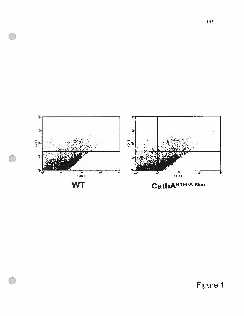
133
Ç)
MIICU MIICII
WT CathAsl9oe0
Fïgure J

134
Figure 1. FACS calibre immunophenotypic profiles of total spienocytes
obtained from the WI, and CathA”90”° mice.
The celis were double-stained with FITC-conjugated anti-MHC monoclonal
antibodies, II-A—FITC and PE-conjugated anti-CD-14 monoclonal antibodies,
CD 1 4-PE. The resuits are representative of three separate experiments.

o
w
Enz
yme
activ
îty(n
moU
hrlm
g)-
.r
Enz
yme
acti
vity
(nm
ollm
inlm
g)
9-o w o & (D - (D CI
,V D o C) (D & (D - o-
010
oo
DD (oc,
,-
-
00
z o
H
1*
‘-I
H
—*
II 1*
o -n o -n o o
-o w o & o (D o 0 V (D D o C) o & (D (D o.
o o
0I0 h z CD o
*
H•*
*
H* *
*
‘J CD C CD r’J
Il
(M
136
Figure 2. A. Specific CathA activity measured with the peptide substrate
CBZ-PheLeu at pH 5.2 in total homogenates of blood-derived and spleen-derived
monocytes (MC) and macrophages (MF). Values represent means + S.D. of 3
independent experiments. * p< 0.05
B. Specific activity of sialidase against the synthetic substrate 4MU-NeuAc at pH 4.5
was rneasured in total homogenates of blood-derived and spleen-derived monocytes
(MC) and macrophages (MF) as well as in the splenocyte-derived immature (IDC)
and mature (MDC) dendritic celis. Splenocyte-derived mature dendritic celis were
induced by 10 jig/ml of lipopolysaccharides for 48 hours. Values represent means
± S.D. oftriplicate experiments. * p< 0.05

coI—DD)
w
oI
________
H_______xapu
UO
!S3
6U
I
oo
ç
oC)
o
I—xepui
uo!ise6ui
Q

138
Figure 3. Reduced phagocytosis in mice splenocytes-derived
macrophages and immature DC.
A. Splenocyte-derived macrophages from the WT, and CatÏ?A59’ mice
were incubated for 3 h at 4°C or at 37°C in the presence of fluorescein-Iabeled E. cou
bioparticles (50 particles per one celi), fixed, and stained with DAPI. Siides were
exarnined on the Nikon Eclipse E6000 direct epifluorescence microscope.
Magnification 200x. Panels represent typical images obtained in triplicate
experiments; from 70 to 150 ceils (10 panels) were studied for each condition in each
experirnent. Ingestion index was calculated for each type of ceils as the percentage of
macrophages with phagocytized particles multiplied by the average number of
particles phagocytized per ingested macrophage. The resuits show an average + SD
of 3 independent experiments. * p< 0.01.
B. The macrophages obtained from spleens of the WT (Ieft panel) or the
CathAS9O0 mice were incubated for 3 hrs with FITC-conjugated E. cou at 37°C or
4° C at 1:50 ratio, washed with PBS, fixed in 4% paraformaldehyde and analyzed by
llow cytometly. The panels show overlap of the histograms of the ceils incubated
with (red une) and without (blue une) FITC-conjugated E. cou. The shifi in the
histograms on the left panel represents the celis that have engulfed the labeled E. cou.
C. Splenocyte-derived immature DC from the WT and CathA9OJ0 mice were
incubated for 3 h at 4°C or at 37°C in the presence of fluorescein-labeled E. cou
bioparticles (50 particles per one ccli), fixed, and stained with DAPI. Siides were
exarnined on the Nikon Eclipse E6000 direct epifluorescence microscope.
Magnification 200x. Panels represent typical images obtained in triplicate
experiments; from 70 to 150 cells (10 panels) were studied for each condition in each
experiment. Ingestion index was calculated as described for marcrophages. The
resuits show an average ± SD of 3 independent experiments. * p < 0.01.

139
A80x1
I-G)
E4.Oxf
20x1
Tîme (h)
w
o
G)
E>L
I
___
Ct)
Control•wr
CathAS 9OANeo *
24 48 96
E
oo
20000
15000
10000
5000
j’
I IControlWT
CathA9oA4e0___
I — I
*
w
Figure 4

140
Figure 4. Proliferation rate of lymphocytes induced by the mature
splenocyte-derived DC measured by ceit counting (A) ami
jmethyl-3H]-thymidine incorporation (B).
A. Mice lymphocytes (2 x 10⁶ in 3 ml medium) were incubated at 37°C in the
presence or absence of I x i05 of mature splenocyte-derived DC. At the time
intervals indicated on the figure the cdils were counted using haemacytometer.
Values represent means + S.D. oftriplicate experiments. * p < 0.05.
B. Mouse lymphocytes (2 x 10⁶ in 3 ml medium) were incubated at 37°C in the
presence or absence of 1 x i05 of mature splenocyte-derived DC in the medium
containing {methyl-3H]-thymidine. After 96 hrs the ceils were harvested and the
[3H]-radioactivity was measured in cdl homogenates by scintillation counting. The
amounts of incorporated [metliyl-31-I]-thymidine are shown as counts per minute.
Values represent means ± S.D. oftriplicate measurements. * p< 0.05.

141
CathASI 90A-Neo
SI9OACathA
WT
Figure 5
142
Figure 5. Induced ceil surface staining of (athASl9O40 mice
splenocytes-derived macrophages by the lectin from Maackia amurensis.
Splenocyte-derived macrophages from the WT, and CathA5’190’1 mice
were incubated for 15 min at 37°C in the presence of fluorescein-Iabeled lectin from
Maackia amurensis fixed, and stained with DAPI. Siides were examined on the
Nikon Eclipse E6000 direct epifluorescence microscope. Magnification l000x.
Panels represent typical images obtained in triplicate experiments; at least 30 cells
were studied for each condition in each experiment.

CHAPTER 5: GENERAL DISCUSSION AND
CONCLUSION

144
5.1 General discussion
The role of the lysosomal sialidase Neul in the degradation of sialylated
glycolipids and glycoproteins is welI-established. However, previous works
including those from our laboratory suggested that Neul is also involved in cellular
signaling during the immune response (Chen et al., 1997; Wang et al., 2004, Lukong
et al., 2001, Katoh et al., 1999). Since the innate immune system ptays an important
role in the defence against infectious agents and initiates inflamrnatory responses,
knowledge of the biological role of the Neul in the immune ceils should provide
insights into the cellular immune response and may eventually lead to developing
new approaches for therapeutic intervention.
Macrophages and DCs together called antigen-presenting ceils (APCs) are
unsurpassed iii their ability to stimulate immunologically naive T celÏs. They
recognize, phagocytize and process foreign objects (e.g. bacteria, vimses) and
present antigens on the ccli surface for stimulation of other celis of the immune
system. Desialylation of ce!! surface glycoconjugates in vivo may make monocytes
and macrophages more receptive to activation (Stamatos et al., 2004) and increase
their chemotactic response to sites of inflammation, as was shown in PMNs (Cross et
al., 2003). In particular, macrophages and DCs may be able to enhance the
immunogenicity of processed antigens if the increased sialidase activity resuits in
removal ofthe sialic acid masks of concealed epitopes (Benjouad et al., 1993).
The main goal of our project was to characterize the biological role ofNeul in
the APCs and in particular to clarif’ its role in ccl! signaling and during the ccl!
maturation and activation.

145
1. Endogenous Neul activity is induced during the differentiation of
monocytes into macrophages.
First, we have studied the expression level ofNeul during the differentiation
of freshly isolated human circulating peripheral blood monocytes. Circulating
peripheral blood monocytes play a key role in potentiating diverse immune activities
and can differentiate into either macrophages or DCs by exposure to specific stimuli
(Sallusto et al., 1994). During the differentiation the ffinction of monocytes changes
from antigen recognition and processing to antigen presentation in macrophages and
DCs. The second celi model that we have used was THP-Ï celis, a monocytic celi une
derived from the peripheral blood of a patient with acute monocytic leukemia
(Tsuchiya et al., 1980). Being induced by 12-O-tetradecanoylphorbol-13-acetate
(PMA) these ceils differentiate into adherent ceils having morphological, biochemical
and physiological characteristics of macrophages and expressing the characteristic
macrophage celi surface phenotypic markers, such as CD14 and CD206 (Auwerx et
al., 1991). THP-1 ceils have several advantages over primaiy human monocytes
(higher transfection efficiency, homogeneity, accessibility) that make them a reliable
model to study the mechanism of monocytic differentiation.
We have shown that Neul was upregulated during monocyte-to-macrophage
differentiation in both models. As we have described in article 1 and article 2, the
endogenous sialidase activity of freshly isolated human monocytes increases 13- to
14-fold as celis differentiate in vitro into macrophages. It also increases 3- to 4-fold
during the differentiation of THP-1 ceils into THP-1-derived macrophages. The
increase in the sialidase activity during the differentiation of
monocyte-to-macrophage was consistent with the data observed during the activation
of T celis by Chen and Landoffi (Chen et al., 2000 and Landolfi et al., 1985) and
Lukong et al. (2001). The observed increase of the endogeneous sialidase activity,
146
however, could be caused by upregulation of any of 3 previously described human
sialidases: lysosomal (Neul, gene NEUI and Neu4, gene NEU4), cytoplasmic (Neu2,
gene NEU2) and plasma membrane (Neu3, gene NEU3). Since the cytosolic sialidase
Neu2 is mostly active at neutral pH (Monti et al., 1999), and the sialidase activity
measured with 4 MU-NANA at pH 7.5 was not increased during the differentiation of
THP-1 cells we were able to exciude this enzyme. The other 3 remaining enzymes,
Neul, Neu3 and Neu4 ail have acidic pH optimum but they differ from each other in
substrate specificity: Neul shows the highest activity against oligosacharides and
short glycopeptides (Miyagi et al., 1993) whereas Neu3 and Neu4 are equally active
against o2-3 -sialylated oiigosaccharides, glycopeptides and gangliosides
(Schneider-Jakob et al., 1991). Sialidase activity of THP-1 celis rneasured against
sialylated glycolipids (mixed bovine gangliosides) was slightly reduced during
differentiation suggesting that the increase of sialidase activity is caused by the
upregulation of the lysosomal sialidase Neu 1.
Neul exists as a part of a multienzyme complex with f3-D-galactosidase and
CathA (Pshezhetsky et ai., 2001, van der Horst et al., 1989, Tranchernontagne et al.,
1990 and Galjart et aI., 1991) and can be immunoprecipitated selectively from cell
lysates using anti-CathA antibodies (Pshezhetsky et al., 1994), but neither Neu2 nor
Neu3 or Neu4 form oligomeric structures when purified from tissues (Miyagi et al.,
1990 and Miyagi et al., 1985). In our studies, we showed that more than 99% ofthe
induced sialidase activity in macrophages was eliminated by anti-CathA antibodies
providing the evidence that Neul was the source of the endogenous sialidase activity
induced during the differentiation of monocytes into macrophages.
To confirm the Neul induction at the RNA level, quantitative RT-PCR and
Northem blots were applied. Both methods showed that during the differentiation the
Neiti mRNA was increased, whereas Neu3 mRNA was decreased. Neu2 expression
147
could flot be detected in the THP-1 monocytes as well as in the THP-1-derived
macrophages neither by RT-PCR nor by Northem blots. Western biots with the
polycional anti-Neul and anti-Neu3 antibodies confirmed the up-regulation of Nett]
and downregulation ofNezt3 in THP-i ceils at the protein level. Interestingiy, in the
primary blood macrophages the amount ofboth Netti and Neu3 RNA detected by reai
tirne RT-PCR was -3-fold increased as monocytes differentiated into macrophages
but Western blot analysis using specific antibodies showed that the Neul protein was
induced at much higher level then the Neu3 protein.
The transcriptionai activity of the NEUJ gene promoter was studied by
deietion mapping. Five restriction fragments of the NEUJ gene 5’-flanking region
were cloned into a pBLCAT6 vector (Boshart et ai., 1992) in front ofa reporter CAT
gene and expressed in COS-7 ceils. We showed that the highest promoter activity was
associated with a 542 bp fragment. Extension of this fragment to 936 bp and fiirther
to 1598 bp decreased the promoter activity, suggesting that potential elements for
down-regulation of the gene expression could be present between the nucleotides
-542 and -1598 (article 2). We further tested whether the promoter activity of the
NEUJ gene was induced during the differentiation ofTHP-1 celis into macrophages.
We found that 24 h after the induction of ceils with PMA, the CAT activity was
significantiy increased for ail constructs, the highest effect (8-fold increase) being
achieved for the reporter construct, pBL-NEUÏ CAT3 containing the 542 bp promoter
fragment. The highest ievei of induction was observed for a 542 bp fragment of the
NEUJ gene prornoter containing several potentiai sequence motifs for binding AP-1,
a group of homodimeric and heterodimeric complexes composed of various fos and
fun gene products, such as c-Jun, JunB, JunD, c-Fos, and FosB as well as the
Fos-reiated antigens fra-1 and Fra-2 (Cohen et ai., 1989, Curran et al., 1988, Lee et
al., 1987, Ransone et ai., 1990, Rauscher et al., 1988 and Schonthal et al., 1988). Ah

148
of these complexes (also called protooncoproteins) interact with the common binding
sites called PMA-responsive elements and mediate gene induction in response to
growth factors, cytokines, phorbol esters, and a variety of other stimuli, including
oxidative stress (Haliday, et al., 1991, Hanazawa et ai., 1993 and Lo et al., 1995). In
particular, one of the frmnctions of AP- 1 is to mediate inflammatory responses in
lymphocytes and macrophages by stimulating ccli activation and the expression of
cytokines (Tanaka et al., 1997).
Altogether our data showed that the differentiation of monocytes to
macrophages resuits in significant and specffic induction of the Neit] gene.
2. Neul and CathA are targeted to the ce!! surface during the
differentiation of monocytes to macrophages.
Our further studies described in article 2 provided a clear evidence for the
Neul transiocation from the lysosomes to the cdl surface during the immune ccli
activation and differentiation. The intracellular localization of Neul was studied by
confocal immunomicroscopy. In the nondifferentiated cells, Neul and CathA showed
a cytoplasmic punctate staining paffem co-localizing with the lysosomal marker
LAMP-2. In contrast, in the differentiated ceils the majority of Neul and CathA was
found on the cdl periphery, whereas LAMP-2 stili had the intracellular localization
(article 2). We speculate that in the differentiating celis both enzymes are first
targeted to the endosomal/lysosomal compartment, where they gain enzymatic
activity and then are sorted to the vesicles destined to the plasma membrane. As
discussed above, in the iysosome, Neul is a component of a multienzyme complex,
with CathA, B-galactosidase and N-acctylgaiactosamine-6-suifate sulfatase
(Pshezhetsky et al., 2001). If the complex is disnmpted, Neul loses its enzymatic
149
activity and is rapidly degraded by the lysosomal proteases (van der Horst et al., 1989
and Manzi et al., 1994). CathA, as the majority of soluble lysosomal lumenal proteins,
is targeted through the mannose-6-phosphate receptor pathway (Lukong et al., 1999),
whereas the intracellular trafficking of Neul is mediated by the adaptor proteins
through a Tyr-containing motif in its C-terminus (Lukong et al., 2001). The two
proteins rneet and undergo activation in the lysosome. The activation of CathA
involves its processing by a lysosomal endopeptidase which removes a 2-kDa
“Iinker” polypeptide between the two polypeptide chains of the mature enzyme
(Bonten et al., 1995). Although the exact molecular mechanism of the Neul
activation is unknown but most likely it involves a conformational change of the
Neul polypeptide after it forms the complex with CathA. It is possible therefore that
lysosomal Neul and CathA may travel to the cell surface as a complex inside the
exocytic vesicles that fuse with the ceil membrane. The existence of such
lysosome-to-plasma membrane transport pathway lias been well documented for
other professional APCs, DCs where it plays a crucial role in targeting the
peptide-MHC II complexes (pMHCs) to the celi surface (Turley et al., 2000). Upon
receipt of a maturation stimulus (such as lipopolysacharide) the internalized antigens
are processed in the endosomes and pMHCs are formed. Several hours later they
appear in the LAMP-negative compartments termed CIIV, which are the
intermediates in the transfer ofpMHCs to tlie plasma membranes (Turley et al., 2000).
The concentration of pMHCs in CIIV reaches a maximum 12 hours after the
lipopolysacharide treatment and after 12 more hours the majority of complexes is
already transferred to the ccli surface. The described targeting of pMHCs in DCs
resembles the targeting of Neul and CathA in differentiating monocytes and
macrophages. We showed (article 2) that during the differentiation of monocytes into
macrophages the Neul is constitutively transported to the plasma membrane along

150
with class II molecules rather then being actively retained in the lysosomes. We found
that 12 to 24 hours after receiving the differentiation stimulus the
lysosomal-endosomal apparatus of monocytes undergoes a dramatic rearrangement.
The MHC II-positive and Neul-positive compartrnents are segregated from the
LAMP-2 positive lysosomes and late endosomes, relocated doser to the ccli surface
and fused with the plasma membranes. The same vesicles are stained positively for
the intemalized antigen horseradish peroxidase (HRP) or fluorescent dextran, which
in the non-differentiated monocytic ceils are normally stored for at least several days
in LAMP-2-positive endosomal/lysosomal compartment. In the differentiating celis
the dextran molecules and HRP peptide (probably in the form of pMHCs) were
gradually accumulated on the ccli plasma membrane colocalizing at least in part with
Neul and CathA.
Although the exact targeting mechanisrn of Neui and CathA stiil has to be
clarified, the data we obtained showed that the most possible pathway is that Neul
and CathA are being sorted from the Golgi to the lysosomes and then to the
LAMP-2-negative, MHC II-positive vesicles, which later merge with the plasma
membrane of immune cells during their differentiation or activation. Most recently,
Nan et al. (2007) showed that the complex between CathA and Neul is also detected
on the surface of activated T ceils (Nan et al., 2007), which is in a good agreement
with our studies. The identified re-localization of Neul during the differentiation of
monocytes flot only provides essential information about the enzym&s biogenesis but
also may provide a due about its role in the immune celis. Our hypothesis is that
Neui on the cdl surface may be involved in the desialylation of membrane proteins
and therefore in the modulation of the cdl activity.

151
3. Suppression of the Neul expression in macrophages alters their
functional capacity
In order to understand whether upregulation of the Neul expression and its
targeting to the ceil surface is important for the primary function of macrophages we
have used siRNA to inhibit the expression ofNeul in THP-1 celis. We found that the
suppression of Neul expression in the THP-1-derived macrophages by siRNA or
their treatment with anti-Neul antibodies significantly reduced functional capacity of
the ceils, such as their ability to enguif bacteria or to produce cytokines (article 2),
suggesting that the upregulation of the Neul expression is important for activation of
macrophages and establishing the link between Neul and ccliular immune response.
Recently, it was also found that Neul is important for the production of interferon
gamma by T ceils (Nan et al., 2007).
4. Neul sialidase but not CathA activity on the surface of the
immune celis is important for the functional integrity.
Activation and stabilization of the Neul in the lysosome, requires its
association with the lysosomal multienzyme complex (LMC) also containing the
lysosomal CathA, B-galactosidase and N-acetylgalactosarnine-6-sulfate sulfatase.
Furthermore we already identified that Neu 1 on the surface of macrophages is tightly
associated with CathA during the cdl differentiation (article 2). Interestingly, during
the differentiation of macrophages, only CathA but not other components of LMC is
sorted to the plasma membrane similarly to Neul. Both proteins were first targeted to
the lysosome and then sorted to LAMP-2-negative, MHC II-positive vesicles, later
merged with the plasma membrane. Therefore, we decided to investigate if CathA
activity on the ccli surface is also required for the immune function.
152
Recently, animal models of single CathA deficiency and double CathA!Neul
deficiency have been developed in our laboratory by gene targeting in mice. In the first
une of mice (CathAS’9O1) we have repiaced the nucleophyl of the CathA active site,
Serl9O with Ala, which aboiished the enzymatic activity, while the mutant protein
retained its ability to activate Neu 1. In addition to mice we have produced
a une (CatÏlAS9oe0 mice) that had a FGK-neo cassette inserted in the CathA gene.
These mice had a signiflcantly reduced (3-10%) Neul activity, due to dramatically
decreased CathA mRNA levels, consistent with previously reported hypomorphic
effects of the neo gene (Seyrantepe et al., in preparation). Therefore, we have
obtained hypomorphic CathA mice with the secondary deficiency ofthe Neul activity.
Since the residual level ofthe Neul activity in the tissues ofthe CathA9O4t0 mice
is stili higher than that in the previously described knockout CathA mice (Zhou et al.,
1995), the animais do flot develop severe galactosialidosis phenotype. Therefore, the
described above mouse models provided us with a possibility to study the impact of
the Neul and CathA deficiency on the ftmnction of different types of the immune cells
ex vivo.
Our data (article 3) strongly indicated that the immune celis from the
Neul/CathA-deficient CathAS]9O10 mice but not from the CathA-deficient
CathAS79A mice have significantly reduced functional capacity. First we found that
the spienocyte- and PBMC-derived macrophages, the peritoneal adherent ceils as
well as the splenocyte-derived immature DCs from the CathAS9O10 mice showed
reduced ability to phagocytose pathogens, whereas the celis from the CathA5’9OA mice
had normal properties. This in tum could mean that these ceils are iess efficiently
activated, which is consistent with our previous observation that the THP-1-derived
macrophages showed reduced production of cytokines including IL-lb, IL-6 and
TNf-a after the Neul activity on the their surface was inhibited with the anti-Neul

153
antibodies (article 2). furthermore, the mature DCs from the Neul-deficient mice
showed reduced abiiity to activate the proliferation of T lymphocytes. In addition, the
macrophages from the Neul-deficient mice showed higher staining with the fITC
conjugated Maackia ainui’ensis lectin (article 3) suggesting that they have increased
sialylation of the ccli surface glycoproteins. Our data suggest, therefore, that the ccli
surface Neul plays an important role in the regulation of affinity of the immune ceils
towards each other and extemal pathogens. They also provide an evidence that Neul
but flot CathA on the surface of immune celis plays a key role in the regulation of
their fiinctional capacity.
In conclusion, our resuits indicate that the lysosomal sialidase Neul plays an
essential role in the removal of terminal sialic acid residues from the ccli surface
glycoproteins and is important for activation of the immune ceils. Previous studies
have identified on the surface of macrophages sialic acid binding receptors (siglecs),
such as siaioadhesin (Sn), CD22, myelin-associated glycoprotein, CD33, and sigiec-5
and showed that they are invoived in discrete functions in the hemopoietic, immune,
and nervous systems (Munday et al., 1999). The expression of siglecs is highly
regulated and noniially only found at high levels on distinct subsets of tissue
macrophages (Crocker et al., 1986, 1994). The molecular features and binding
properties of siglecs suggest that they cvolved as an acccssoiy molecule to promote
interactions of macrophages with host ceils and extracellular matrix displaying
appropriate sialylated Iigands. Characterization of cells that bind to siglecs has
rcvealcd high binding of granulocytcs (Crocker et al., 1995) and lymphoma ceils (van
den Berg et al., 1992). Macrophage-host ccli interactions that couid be influcnced by
sigiecs, also include antigen presentation to primcd T ceils (Muerkostcr et ai., 1999)
and recognition and clearance of apoptotic ceils, effete eiythrocytes, and opsonized
ceils (e.g., in malaria and certain autoimmune diseases). To date, ail sialic

154
acid-dependent binding of macrophages has been found to be siglec-dependent. This
would suggest that on macrophages, siglec is the major sialic acid binding receptor
responsible for mediating ceil-ceil interactions and that the primary function of other
siglecs may be in signaling. Therefore the optimal sialylation of the ccli surface
glycoproteins inciuding siglecs themselves sliould play the key role in the interaction
of macrophages with other ceils including T, B and NK celis. The marked expansion
of CD33-related siglecs in the innate immune system suggests that these proteins piay
also a fiindarnentally important role in host-defense functions since sialic acids are
also present on the surface ofmany pathogens.
Since desialylation of siglecs significantly increases their affinity towards
sialic acid, our data suggest the mechanisrn by which the ccii surface Neul regulates
the affinity of the immune celis towards each other and extemai pathogens. Therefore
it is tempting to speculate that similarly to CD63, CD 152, CD206 and other
lysosomal/endosomal proteins expressed on the surface of the immune ceils, Neul is
important for their functional integrity.
Together with previously obtained data on the importance of the Neul for the
1L4 production by the lymphocytes (Chen et al., 1997, 2000) our resutts show that the
upregulation of Neul sialidase plays an important role in signaling of immune cells
by regulating the cytokine production and suggest that in human sialidosis patients
Neul deficiency may result in reduced capacity of immune ceils to produce cytokines
and antibodies ieading to partial immunodeficiency and may account for frequent
pulmonary infections. further experirnents are necessary to clarify if the
differentiation-induced transiocation of the lysosomal sialidase, Neul and CathA to
the plasma membrane is also important for other specific functions of the immune
ceils and, in particular, for the antigen presentation. However, since fully
differentiated macrophages are present in ail body tissues (microglial cells in the CNS,
155
Kupffer celis in liver, etc.) it is quite conceivable that the primary or secondary
deficiency of the Neul activity may affect functions of ail monocyte-derived celis
and cause defects in multiple tissues and organs.
Altogether our results reveal important role of the Neul sialidase in immune
response and ceil signaling and bring ncw insight into the biogenesis and function of
antigen presenting celis.
5.2 Conclusions
1. Expression of the lysosomal sialidase Neul is upregulated during the
differentiation of monocytes into macrophages and DCs as well as during the
maturation of DCs, due to a significant induction of the transcriptional activity of the
Neul gene promoter.
2. During the differentiation of macrophages Neul together with its activator protein,
CathA is first targeted to the lysosome and then sorted to the LAMP-2-negative,
MHC II-positive vesicles, which later merge with the plasma membrane.
3. Suppression of the Neul expression in the macrophages reduces their ability to
produce cytokines.
4. Macrophages and DCs derived from the hypomorphic CathA mice with the
secondaiy -9O% reduction of the Neul activity show significantly increased
sialylation of the surface, and dramatically reduced capacity to enguif bacteria,
whereas the mature DCs have lower effect on T celis proliferation.
5. Altogether our data suggest that Neu 1 regulates the affinity of the immune celis
towards each other and towards extemal pathogens and is therefore necessary for
their functional integrity.
156
REFERENCES
Akita H, T. Miyagi, K. Hata, and M. Kagayama. Immunohistochemical evidence for
the existence of rat cytosolic sialidase in rat skeletal muscles. Histochem.
Cet!. Bio!. 107(6), 495-503 (1997).
Andria, G., G. Strisciuglio, W.S. Pontarelli, W.S. Sly, and W.E. Dodson. In
“Siatidases and Siatidoses” (P. Durand et al., eds,) p365., Edi. Ermes,
Milano, (1981).
Auwerx J. The human leukemia ceil une, THP-1: a multifacetted model for the study
of monocyte-macrophage differentiation. Experientia. 47(1 ):22-3 1 (1991).
Bame, K. J. and Rome, L. H. Acetyl coenzyme A:alpha-glucosaminide N
acetyltransferase: evidence for a transmembrane acetylation mechanism. J.
Bio!. Chem. 260, 11293-11299 (1985).
Bao M, Booth IL, Elmendorf BJ, Canfield WM Bovine UDP-N
acetylgiucosamine: lysosomaÏ-enzyme N-acetylglucosamine- 1-
phosphotransferase. I. Purification and subunit structure. J Bio! Cheni.
27 1(49), 3 1437-45 (1996).
Barriocanal J.G., Bonifacino J.S., Yuan L., Sandoval I.V. Biosynthesis,
glycosylation, movement through the Golgi system, and transport
lysosomes by an N-linked carbohydrate-independent mechanism of three
lysosomal integral membrane proteins. J Bio! Chem. 261(35), 16755-63
(1986).
Benjouad A, Babas T, Montagnier L, Bahraoui E. N-linked oligosaccharides of
simian immunodeficiency virus envelope glycoproteins are dispensable for

157
the interaction with the CD4 receptor. Biochem Biophvs Res Commun.
190(2):311-9 (1993).
Bonten. E.J., N.J. Gaijart. R. Willemsen, M. Usmany, J.M. Vlak, and A. d’Azzo.
Lysosomal protective protein/cathepsin A. Role of the ‘linker” domain in
catalytic activation. J. Biot. Cheni. 270, 26441-26445 (1995).
Bonten, E.J., A. van der Spoel, M. Fornerod, G. Grosveld, and A. d’ izo
Characterization of human lysosomal neuraminidase defines the molecular
basis of the metabolic storage disorder sialidosis. Genes and Devet. 10,
3156-316$ (1996).
Le Borgne R. and Hoflack B. Protein transport from the secretory to the endocytic
pathwayin marnmalian ceils. Biochim Biophvs Acta. 1404(1-2), 195-209
(1998).
Boshart M, Kiuppel M, Schmidt A, Schutz G, Luckow B. Reporter constructs with
low background activity utilizing the cat gene. Gene. 110(1):129-30 (1992).
Bradshaw 1M, Mitaxov V, Waksman G. Investigation of phosphotyrosine recognition
by the SH2 domain of the Src kinase. J Mot Biot. 293(4), 971-85 (1999).
Braun M., Waheed A., von Figura K. Lysosomal acid phosphatase is transported to
lysosomes via the ceil surface. EMBO J. 8(12), 3633-40 (1989).
Cantz, M., J. Gehier, and J. Spranger. Mucolipidosis I: increased sialic acid content
and deficiency of an alpha-N-acetylneuraminidase in cultured fibroblasts.
Biochem. Biophys. Res. Connnun.74, 732-738 (1977).

158
Carrillo, M.B., C.N. Mimer, S.T. Bail, M. Snock, and R.D. Campbell. Cloning and
characterization of sialidase from the murine histocompatibility-2 complex:
low levels of mRNA and a single amino acid mutation are responsible for
reduced sialidase activity in mice carrying the Neul allele. GÏycobiotogy 7,
975-986 (1997).
Chen XP, E.Y. Enioutina, and R.A. Daynes The control of IL-4 gene expression in
activated murine T lymphocytes: a novel role for neu-1 sialidase. J.
Iminunot. 158(7), 3070-3080 (1997).
Chen XP, X. Ding, and R.A. Daynes. Ganglioside control over IL-4 priming and
cytokine production in activated t celis. Cytokine 12(7), 972-985 (2000).
Chikuma S, Murakami M, Tanaka K, Uede T. Janus kinase 2 is associated with a box
1-like motif and phosphorylates a critical tyrosine residue in the cytoplasmic
region of cytotoxic T lymphocyte associated molecule-4. J Ccli Biochem.
78(2), 241-50 (2000).
Chou MY, Li SC, Li YT. Cloning and expression of sialidase L, a NeuAcalpha2--
>3GaI-specific sialidase from the leech, Macrobdella decora. J Biot Che,n.
27 1(32), 19219-24 (1996).
Cohen DR, Ferreira PC, Gentz R, Franza BR Jr, Curran T. The product of a fos
related gene, fra-1, binds cooperatively to the AP-1 site with Jun:
transcription factor AP-1 is comprised of multiple protein complexes.
Genes Dcv. 3(2):173-84 (1989).
159
Collawn J.F., Kuhn L.A., Liu L..F, Tainer J.A., Trowbridge I.S. Transplanted LDL
and mannose-6-phosphate receptor internalization signais promote high
efficiency endocytosis of the transferrin receptor. EMBO J. 11, 3247-53
(1991).
Colman P.M. Influenza virus neuraminidase: structure, antibodies, and inhibitors.
Protein Sci. 3(10), 1687-96 (1994).
Conzelmann E, Sandhoff K. Activator proteins for lysosomal glycolipid hydrolysis.
Methods Enzymot. 138, 792-$15 (1987).
Corfieid, T. Bacterial sialidases--roles in pathogenicity and nutrition. Glycobiotogy 2,
509-521 (1992).
Crenneil, S.J., E.F. Garman, W.G. Laver, E.R. Vimr, and G. Taylor. Crystai structure
of a bacteriai sialidase (from Salmoneia typhimurium LT2) shows the same
foid as an influenza virus neuraminidase. Proc. Nati. Acad. Sci. USA 90,
9852-9856 (1993).
Cretin H. The proton gradient across the vacuo-Ïysosomal membrane of lutoids from
the latex of Hevea brasiliensis. I. Further evidence for a proton-transiocating
ATPase on the vacuo-iysosomal membrane of intact iutoids. J Membr Biot.
65(3), 175-84 (1982).
Crocker PR, Mucklow S, Bouckson V, McWiliiam A, Wiiiis AC, Gordon S, Miion G,
Keim S, Bradfield P. Sialoadhesin, a macrophage sialic acid binding
receptor for haemopoietic celis with 17 immunoglobulin-iike domains.
EMBO J. 13(19):4490-503 (1994).

160
Crocker PR, Gordon S. Properties and distribution of a lectin-like hemaggiutinin
differentially expressed by murine stromal tissue macrophages.
JExpMed. 164(6):1862-75 (1986).
Crocker PR, freeman S, Gordon S, Keim S. Sialoadhesin binds preferentially to ceils
ofthe granulocytic lineage. J Clin Invest. 95(2):635-43 (1995).
Cross AS, Sakarya S, Rifat S, Held TK, Drysdale 3E, Grange PA, Cassels FJ, Wang
LX, Stamatos NM, Farese A, Casey D, Poweil J, Bhattacharjee AK,
Kleinberg M & Goldblum SE. Recruitment of murine neutrophils in vivo
through endogenous sialidase activity. JBiot Chem 278, 4112—4120 (2003).
Curran T, Franza BR Jr. Fos and Jun: the AP-1 connection. Cetl. 55(3):395-7 (1988).
dAzzo, A., G. Andria, P. Strisciugiio, and H. Galijaard. Galactosialidosis. In
Metabolic and Molectilar Bases of ]nherited Disease, (Scriver C.R., Beaudet
A.L, Sly W.S. and Valte D., eds.) New York: McGraw-Hill, pp. 2835-2837
(1995).
d’Azzo,A., A. Hoogeveen, A.J. Reuser, D. Robinson, and H. Galjaard. Molecular
defect in combined -galactosidase and neuraminidase deficiency in man.
Proc. Nail. Acad. Sci. U.S.A. 79, 4535-4539 (1982).
Dahms NM, Lobel P, Kornfeld S. Mannose 6-phosphate receptors and lysosomal
enzyme targeting. JBiot Cheni. 264(21), 12115-8 (1989).
Dorland L, Haverkamp J, Viliegenthart JE, Strecker G, Michalski JC, Foumet B, Spik
G, Montreuil J. 360-MHz 1H nuclear-magnetic-resonance spectroscopy of
sialyl-otigosaccharides from patients with sialidosis (mucolipidosis I and II).
EurJBiocliem. 87(2), 323-9 (1978).

161
Durand, P., R. Gatti, S. Cavalieri, C. Borrone, M. Tondeur, J.C. Michalski, and C.
Strecker. Sialidosis (mucolipidosis I). Hetvetica Paediatrica Acta, 32, 391-
400 (1977).
Elsiiger M.A., Potier M. Homologous modeling of the lysosomal protective
protein/carboxypeptidase L: structural and functional implications of
mutations identified in gatactosialidosis patients. Proteins. 18(1), 81-93
(1994).
Erickson A.H., Conner G.E., Blobel G. Biosynthesis of a lysosomal enzyme. Partial
structure of two transient and functionally distinct NH2-terminal sequences
in cathepsinD. JBioÏ Chem. 256(21), 11224-31 (1981).
Federico, A., A. Cecio, G.A. Battini, J.C. Michalski, G. Strecker, and G.C. Guazzi.
Macular cherry-red spot and myoclonus syndrome. Juvenile form of
sialidosis J. Neurol. Sci. 48, 157-169 (1980).
Fingerhut R, G.T. van der Horst, F.W. Verheijen, and E. Conzelmann. Degradation of
gangliosides by the lysosomal sialidase requires an activator protein. Eur. J.
Biochem. 208(3), 623-629 (1992).
Frisch A, Neufeld EF. A rapid and sensitive assay for neuraminidase: application to
cultured fibroblasts. Anal Biochem. 95(1), 222-7 (1979).
Fukuhara, Y.; Takano, T.; Shimmoto, M.; Oshima, A.; Takeda, E.; Kuroda, Y.;
Sakuraba, H.; Suzuki, Y.: A new point mutation of protective protein gene in
two Japanese siblings with juvenile galactosialidosis. Brain Dysfimction 5,
3 19-325, (1992).

162
Gaien,J.E. et al. Role of Vibrio cholerae neuraminidase in the function of choiera
toxin. Infect. Immun., 60, 406-415 (1992).
Gatjaard. H., A.T. Hoogeveen, HA. de Wit-Verbeek, A.J.J. Reuser. M.W. Ho, and D.
Robinson. Nature 257, 60-62 (1975).
Gaijart, N.J., N. Gillemans, A. Harris, T. Gijsbertus, J. van der Horst, F.W. Verheijen,
H. Gal] aard, and A. dAsso. Expression of cDNA encoding the human
“protecti ve protein” associated with lysosomai -galactosidase and
neuraminidase: homoiogy to yeast proteases. Ccii 54, 755-764 (1988).
Gaijart, N.J., H. Morreau, R. Willemsen, N. Gillemans. E.J. Bonten, and A. d’Azzo.
Human lysosomai protective protein bas cathepsin A-iike activity distinct
from its protection function. J. Bio!. Chem. 266, 14754-14762 (1991).
Gee. K., Kozlowski, M., and Kumar, A. Tumor necrosis factor-aipha induces
functionally active hyaltironan-adhesive CD44 by activating sialidase through
p38 mitogen-activated protein kinase in hpopolysaccharide-stimulated human
monocytic celis. J Bio! Chem. 278(39), 37275-87 (2003).
Goldberg, M. F.; Cotiier, E.; Fichenscher, L. G.; Kenyon, K. R.; Enat, R.; Borowsky,
S. A.: Macular cherry-red spot, comeal clouding, and beta-galactosidase
deficiency: clinical, biochemical, and eiectron microscopic study of a new
autosomal recessive storage disease. Arch. Inter n. lied. 128, 387-398 (1971).
Gottschalk,A. and P.E. Lind. Product of interaction between influenza virus enzyme
and ovomucin. Nature 164, 232-233 (1989).

163
Guamieri F.G., Arterbum L.M., Penno M.B., Cha Y., August J.T. The motif Tyr-X
X-hydrophobic residue mediates lysosomal membrane targeting of lysosome
associated membrane protein 1. J Bio! Chem 268(3), 194 1-6 (1993).
Hanazawa S, Takeshita A, Amano S, Semba T, Nirazuka T, Katoh H, Kitano S.
Tumor necrosis factor-atpha induces expression of monocyte chemoattractant
JE via fos and jun genes in clonai osteobiastic MC3T3-E1 celis. J Biot Chein.
26$(13):9526-32 (1993).
Haliday EM, Ramesha CS, Ringold G. TNF induces c-fos via a novel pathway
requiring conversion of arachidonic acid to a lipoxygenase metabolite. EMBO
J. 10(1):109-15 (1991).
Havelaar AC, Beerens CE, Mancini GM, Verheijen FW. Transport of organic anions
by the lysosomal sialic acid transporter: a functional approach towards the
gene for sialic acid storage disease. FEBS Lett. 446(1), 65-8 (1999).
Hers, H.G. The role of the pathogeny of storage diseases. Biochimie 54, 753-757
(1972).
Hinek A Biological roles of the non-integrin elastin/laminin receptor Biot. Chem.
377(7-8), 47 1-480 (1996).
Hiraiwa M, M. Nishizawa, Y. Uda, T. Nakajima, and T. Miyatake. Human placental
sialidase: further purification and characterization. J. Biochem. (Tokyo)
103(1), 86-90 (1988).
Hiraiwa M, Y. Uda, M. Nishizawa, and T. Miyatake. Human placental sialidase:
partial purification and characterization. J. Biochem. (Tokyo) 101(5). 1273-
1279 (1987).
164
Hirst J. and M.S. Robinson. C!athrin and adaptors. Biochim. Biophys. Acta 1404(1-2),
173-193 (1998).
Hoflack B. and Komfeld S. Lysosornal enzyme binding to mouse P388D1
macrophage membranes lacking the 215-kDa mannose 6-phosphate receptor:
evidence for the existence of a second mannose 6-phosphate receptor. Proc
Natt Acad Sci U $ A 82(13), 4428-32 (1985).
Hoogeveen A, A. d’Azzo, R. Brossmer, and H. Galjaard. Correction of combined
beta-galactosïdase/neuraminidase deficiency in human fibroblasts. Bioche,n.
Biophys. Res. Commun. 103(1), 292-300 (1981).
Hoogeveen AT, F.W. Verheijen, A. dAzzo, and H. Galjaard. Genetic heterogeneïty
in human neuraminidase deficiency. Natttre 285(5765), 500-502 (1980).
Hoogeveen, A.T., F.W. Verheijen, and H. Galjaard. The relation between human
lysosomal -galactosidase and its protective protein. J. Bio!. Chem. 258,
12143-12146 (1983).
Hrebicek M, Mrazova L, Seyrantepe V, Durand S, Roslin NM, Noskova L,
Hartmannova H, Ivanek R, Cizkova A, Poupetova H, Sikora J, Urinovska J,
Stranecky V, Zeman J, Lepage P, Roquis D, Vemer A, Ausseil J, Beesley
CE, Maire I, Poorthuis BJ, van de Kamp J, van Diggelen OP, Wevers RA,
Hudson TJ, Fujiwara 1M, Majewski J, Morgan K, Kmoch S, Pshezhetsky
AV. Mutations in TMEM76* cause mucopolysaccharidosis hIC (Sanfilippo
C syndrome). Am J Hitin Genet. 79(5), 807-19 (2006).
Hubbes M, DAgrosa RM, Caltahan 1W. Human placenta! beta-galactosidase.
Characterization of the dimer and complex forms of the enzyme.
Biochem J. 285 (Pt 3), 827-3 1 (1992).
165
Huang JQ, 1M. Trasler, S. Igdoura, J. Michaud, N. Hanal, and R.A. Grave!.
Apoptotic ceil death in mouse models of GM2 gangliosidosis and
observations on human Tay-Sachs and Sandhoff diseases. Hum. Mot. Genet.
6(11), 1879-1885 (1997).
Hunziker W., Geuze H.J. Intracellular trafficking of lysosomal membrane proteins.
Bloessays. 18(5), 379-89 (1996).
Igdoura SA, C. Mertineit, J.M. Trasler, and R.A. Gravel. Sialidase-mediated
depletion of GM2 ganglioside in Tay-Sachs neuroglia celis. Hum. Mot.
Genet. 8(6), 1111—1116 (1999).
Igdoura, S.A., C. Gafuik, C. Mertineit, F. Saberi, A.V. Pshezhetsky, M. Potier, J.M.
Trasier, and R.A. Grave!. Cloning of the cDNA and the gene encoding
mouse lysosomal sia!idase and correction of sialidase deficiency in human
sialidosis and mouse SMJJ fibroblasts. Hum. Mot. Genet. 7, 115-121 (1998).
Ikehara Y, Ikehara SK, Paulson JC. Negative regulation of T celi receptor signaling
by Siglec-7 (p70/AIRM) and Siglec-9. J Biol Chem. 2279(41):43117-25
(2004).
Irani DN, Lin KI. Griffin DE. Brain-derived gangliosides regulate the cytokine
production and proliferation of activated T celis. J Innnttnot. 157(10):4333-
40 (1996).
Kanda N, Nakai K, Watanabe S. Gang!iosides GDIb, GT1b, and GQ1b suppress the
growth of human melanoma by inhibiting interleukin-$ production: the
inhibition of adenylate cyc!ase. J Invest Dennatot. 1 17(2):284-93 (2001).

166
Kaplan A., fischer D., Achord D, Sly W. Phosphohexosyl recognition is a general
characteristic of pinocytosis of lysosomal glycosidases by human fibroblasts.
J Clin ]nvest. 60(5), 1088-93 (1977).
Kato K, Shiga K, Yamaguchi K, Hata K, Kobayashi T, Miyazaki K, Saijo S, Miyagi
T. Ptasma-membrane-associated sialidase (NEU3) differentially regulates
integrin-medi ated celi proliferati on through laminin- and fibronectin
derived signalling. Biochent J. 394(Pt 3):647-56 (2006).
Katoh S, Miyagi T, Tanïguchi H, Matsubara Y, Kadota J, Tominaga A, Kincade PW,
Matsukura S, Kohno S. Cutting edge: an inducible sialidase regulates the
hyaÏuronic acid binding abiÏity of CD44-bearing human monocytes. J
Iminunol. 162(9), 5058-61 (1999).
Kelly, T.E. and G. Graetz. Isolated acid neuraminidase deficiency: a distinct
lysosomal storage disease. Ani. J. Mcd. Genet., 1, 3 1-46 (1977).
Kornfeld R., Bao M., Brewer K., Noli C., Canfield W. Molecular cloning and
functional expression of two spiice forms of human N-acetylglucosamine-1-
phosphodiester alpha-N-acetylglucosaminidase. J Biol Chem. 274(46),
3277$-85 (1999).
Kopitz J, C. von Reitzenstein, K. Sinz, and M. Cantz. Selective ganglioside
desialytation in the plasma membrane of human neuroblastoma celis.
Glycobiology 6(3), 367-376 (1996).
167
Kopitz J., Muhi C., Ehemann V., Lehmann C., Cantz M. Effects of celi surface
ganglioside sialidase inhibition on growth control and differentiation of
human neuroblastoma ceils. Eu,- J Ceil Biol. 73(1), 1-9 (1997).
Landolfi NF, J. Leone, J.E. Womack, and R.G. Cook. Activation of T lymphocytes
resuits in an increase in H-2-encoded neuramtnidase. limnuitogenetics 22(2),
159-67 (1985).
Landolfi NF and R.G. Cook. Activated T-lymphocytes express class I molecules
which are hyposialylated compared to other lymphocyte populations. Mol.
JnvnitnoÏ. 23(3), 297-309 (1986).
Lee W, Mitcheli P, Tjian R. Purified transcription factor AP-1 interacts with TPA
inducible enhancer elements. Ce!!. 49(6):741-52 (1987).
Leroy, J. G.; Demars, R. I. Mutant enzymatic and cytological phenotypes in cultured
human fibroblasts. Science 157, 804-806 (1967).
Lin TJ, Garduno R, Boudreau RT, Issekutz AC. Pseudomonas aeruginosa activates
human mast cells to induce neutrophil transendothelial migration via mast
celI-derived IL-1 alpha and beta. J i,n,nunot. 169(8):4522-30 (2002).
Loonen M.C., Lugt L, Franke C.L. Letter: Angiokeratoma corporis diffusum and
Ïysosomal enzyme deficiency. Lancez’. 2(7883); 785 (1974).
Lowden, J. A.; O’Brien, J. S.: Sialidosis: a review of human neuraminidase
deficiency.Anz. J. Huni. Genet. 31; 1-18 (1979).
168
Lo YY, Cruz TF. Involvement of reactive oxygen species in cytokine and growth
factor induction of c-fos expression in chondrocytes. J Biol Chem.
270(20): 11727-30 (1995).
Lukong KE, Elsiiger MA. Mort JS, Potier M, Pshezhetsky AV. Identification of
UDP-N-acetylglucosamine-phosphotransferase-binding sites on the lysosomal
proteases, cathepsins A, 3, and D. Biocheinistiy. 38(1):73-$0 (1999).
Lukong KE, Elsiiger MA, Chang Y, Richard C, Thomas G, Carey W, Tylki
Szymanska A, Czartoryska B, Buchholz T, Criado GR, Palmen S,
Pshezhetsky AV. Characterization of the sialidase molecular defects in
sialidosis patients suggests the structural organization of the lysosomal
multienzyme complex. Hum Mot Genet. 9(7).1075-85 (2000).
Lukong KE, Seyrantepe V, Landry K, Trudel S, Ahmad A, Gahi WA. Lefrancois S.
Morales CR, Pshezhetsky AV. ntracellular distribution of lysosomal
sialidase is controlled by the internalization signal in its cytoplasmic tau.
JBiol Chem. 276(49), 46172-81 (2001).
Mancini GM, A.T. Hoogeveen, H. Galjaard, J.E. Mansson, and L. Svennerholm.
Ganglioside GMY metabolism in living human fibroblasts with beta
galactosidase deficiency Hum. Genet. 73(1), 35-38 (1986).
Manzi AE, Higa HH, Diaz S, Varki A. Intramolecular self-cleavage of polysialic acid.
J Biot Chem. 269(38):23617-24 (1994).

169
Maroteaux P., Humbel R, Strecker G., Michalski J.C., Mande R.. [A new type of
sialïdosis with kidney disease: nephrosialidosis. I. Clinical, radiological and
nosological study].Arch FrPediatr. 35(8), 819-29. (1978) French.
Martin RA, Slaugh R, Natowicz M, Peariman K, Orvisky E, Krasnewich D, Kieta R,
Huizing M, Gahi WA. Sialic acid storage disease of the Salla phenotype in
American monozygous twin female sibs. Am J Mcd Genet A. 120(1):23-7
(2003).
Matoth, Y., Chazan, S.; Cnaan, A., Gelemter, I., Klibansky, C. Frequency of carriers
of chronic (type I) Gaucher disease in Ashkenazi Jews. Am. J.Med. Genet.
27, 561-565 (1987).
Matsuda J, O. Suzuki, A. Oshima, A. Ogura, Y. Noguchi, Y. Yamamoto, T. Asano, K.
Taidmoto, K. Sukegawa, Y. Suzuki, and M. Naiki. Beta-galactosidase
deficient mouse as an animal moUd for GM1-gangliosidosis. Gtycoconj. J.
14(6), 729-736 (1997).
Meikie P.J., Hopwood J.J., Clague A.E., Carey W.F. Prevalence of lysosomal storage
disorders. JAMA. 28 1(3), 249-54 (1999).
Milner, C.M., S.V. Smith, M.B. Carrillo, G.L. Taylor, M. Hollinshead, and R.D.
Campbell. Identification of a sialidase encoded in the human major
histocompatibility complex. J. Biot. Chem., 272, 4549-4558 (1997).
Miyagi T, K. Hata, A. Hasegawa, and T. Aoyagi. Differential effect of various
inhibitors on four types of rat sialidase. Gtycoconj J, 10(1), 45-49 (1993).
170
Miyagi,T., J. Sagawa, K. Konno, and S. Tsuiki. Immunological discrimination of
intralysosomal, cytosolic, and two membrane sialidases present in rat tissues.
J. Biochem. 107, 794-798 (1990).
Miyagi T, K. Hata, K. Konno, and S. Tsuiki. Multiple forms of mammalian sialidase:
attered expression in carcinogenesis. Tohoku]. Exp. lied. 168(2), 223-229
(1992).
Miyagi T, K. Hata, A. Hasegawa, and T. Aoyagi. Differential effect of various
inhibitors on four types of rat sialidase. Glycoconj J, 10(1), 45-49 (1993).
Miyagi T and Tsuiki S. Purification and characterization of cytosolic sialidase from
rat liver.JBiol Chem. 260(11), 6710-6 (1985).
Miyagi T., Wada T., Iwamatsu A., Hata K., Yoshikawa Y., Tokuyama S., Sawada M.
Molecular cloning and characterization of a plasma membrane-associated
sialidase specific for gangliosides. J Biol Cïiem. 274(8), 5004-11(1999).
Monti E., Bassi M.T., Papini N., Riboni M., Manzoni M.. Venerando B., Croci G..
Preti A., Ballabio A., Tettamanti G., Borsani G. Identification and expression
of NEU3, a novel human sialidase associated to the plasma membrane.
Biochem J. 349(Pt 1), 343-35 1 (2000).
Monti E, A. Preti, C. Nesti, A. Ballabio, and G. Borsani. Expression of a novel
human sialidase encoded by the NEU2 gene. Glycobiotogy 9(12),1313-1321
(1999).
Morreau, H., N.J. Galjart, R. Willemsen, N. Gillemans. X.Y. Zhou, and A. d’Azzo.
Human lysosomal protective protein. Glycosylation, intracellular transport.

171
and association with -galactosidase in the endoplasmic reticulum. J. BioÏ.
Chem. 267, 17949-17956 (1992).
Munday J, Floyd H, Crocker PR. Sialic acid binding receptors (siglecs) expressed by
macrophages. JLeukocBiot. 66(5):705-11 (1999).
Muerkoster S, Rocha M. Crocker PR. Schirrmacher V, Umansky V. Sialoadhesin
positive host macrophages play an essential role in graft-versus-Ïeukemia
reactivity in mice. Blood. 93(12):4375-$6 (1999).
Naganawa Y, K. Itoh, M. Shimmoto, K. Takiguchi, H. Doi, Y. Nishizawa, T.
Kobayashi, S. Kamei, K.E. Lukong, A.V. Pshezhetsky. and H. Sakuraba.
Molecular and structural studies of Japanese patients with sialidosis type 1. J.
Htun. Genet. 45(4), 241-249 (2000).
Nan X, Carubelli I, Stamatos NM. Sialidase expression in activated human T
lymphocytes influences production of IFN-{gamma}. J Leukoc Biot.
81(1):284-96 (2007).
Naraparaju VR. and N. Yamamoto. Roles of beta-galactosidase of B lymphocytes and
sialidase of T lymphocytes in inflammation-primed activation of
macrophages Innntuzot. Lett. 43(3), 143-148 (1994).
Nishiyama Y., Sakamoto S., Katayama Y., Mishina M., Terashi A.
[Neuroradiological findings on cerebral blood flow and metabolism of a case
of aduk onset sialidosisi. Rinsho Shinkeigakti. 37(11), 982-6 (1997) Japanese.
Neufeld E.F. Lysosomal storage diseases. Annu Rev Biochem. 60:257-$0 (1991).
172
O’Brien, J.S. The cherry red spot-myoclonus syndrome: a newly recognized
inherited lysosomal storage disease due to acid neuraminidase deficiency.
Clin. Genet., 14, 55-60 (1979).
O’Brien JS, M.B. Stem, B.H. Landing, J.K. OBrien, and G.N. Donneil. Generalized
gangliosidosis. Another inbom error of metabolism? Am. J. Dis. Child. 109,
338 (1965).
O’Brien JS, E. Gugler, A. Giedion, U. Wiessmann, N. Herschkowitz, C. Meier, and J.
Leroy. Spondyloepiphyseal dyspiasia, comeal clouding, normal intelligence
and acid beta-galactosidase deficiency. Clin Genet 9(5), 495-504 (1976).
O’Brïen JS, Wamer 1G. Sialidosis: delineation of subtypes by neuraminidase assay.
Clin Genet. 17(1), 35-8 (1980).
Olcay L., Gumruk F., Boduroglu K., Coskun T., Tuncbilek E. : Anaemia and
thrombocytopenia due to haemophagocytosis in a 7-month-old boy with
galactosialidosis. J. Inherit. Metab. Dis. 21, 679-680 (1998).
Oohira, T., N. Nagata, I. Akaboshi, I. Matsuda, and S. Naito. The infantile form of
sialidosis type II associated with congenital adrenal hypemplasia: possible
linkage between FILA and the neuraminidase deficiency gene. Hum. Genet.
70, 341-343 (1985).
Orsel J.G., Sincock P.M., Krise J.P., Pfeffer S.R. Recognition of the 300-kDa
mannose 6-phosphate receptor cytoplasmic domain by 47-kDa tau
interacting protein. ProcNatlAcadSci USA. 97(16), 9047-51 (2000).

173
Pappu BP, Shrikant PA. Alteration of celi surface sialylation regulates antigen
induced naive CD8+ T ceti responses. J hnmunoÏ. 173(1):275-84 (2004).
Patel MS, Callahan 1W, Zhang S, Chan AK, Unger S, Levin AV, SkomorowsldMA.
Feigenbaum AS, OBrien K. Hellmann J, Ryan G. Veisher L, Chitayat D.
Early-infantile galactosi alidosi s: prenatal presentation and postnatal follow
up. Am JMed Genet. 85(1), 38-47 (1999).
Pearse BM, Smith CJ, Owen Dl. Clathrin coat construction in endocytosis. Curr
Opiit Stncct Biot. 10(2), 220-8 (2000).
Peters C. and K. von Figura. Biogenesis of lysosomal membranes. FEBS Lett 346(1),
10$-114 (1994).
Petersen, G. M., Rotter, J. I., Cantor, R. M., Field, L. L., Greenwald, S., Lim, J. S. T..
Roy, C., Schoenfeld, V., Lowden, J. A.. Kaback, M. M. The Tay-Sachs
disease gene in North American Jewish populations: geographicvariations
and origin. Am. J. Hum. Genet. 35, 1258-1269 (1983).
Pinsky L, Miller J, Shanfield B, Watters G, Wolfe L.S. GMI gangliosidosis in skin
fibroblast culture: enzymatic differences between types 1 and 2 and
observations on a third variant. Ani JHum Genet. 26(5), 563-77 (1974).
Poorthuis B.J., Wevers R.A., Kteijer W.J., Groener J.E., de long J.G., van Weely S,
Niezen-Konin KE, van Diggelen OP. The frequency of lysosomal storage
diseases in The Netherlands. Hum Genet. 105(1-2), 15 1-6 (1999).
174
Potier M, Michaud L. Tranchemontagne J, Thauvette L. Structure of the lysosomal
neuraminidase-f3-galactosidase-carboxypeptidase multienzyme complex.
Biochem. J. 267,197-202 (1990).
Provenzale JM, Barboriak DP, Sims K. Neuroradiologic findings in fucosidosis, a
rare lysosomal storage disease. AJNR Ain J Neuroradiot. 16(4 Suppl), 809-13
(1995).
Pshezhetsky AV, Ashmarina M. Lysosomal multienzyme complex: biochemistry,
genetics, and molecutar pathophysiology. Prog Nucteic Acid Res Mot Biol.
69:81-11 (2001).
Pshezhetsky, A.V. and M. Potier. Association of N-acetyl-galactosamine-6-sulfate
sulfatase with the multienzyme lysosomal complex of -galactosidase,
cathepsin A and neuraminidase: possible implication for intralysosomal
catabolism ofkeratan sulfate. J. Biot. Chem. 271, 28359-28365 (1996).
Pshezhetsky, A.V. and M. Potier. Direct affinity purification and supramolecular
organization of human lysosomal cathepsin A. Arcli. Biochem. and Biophys.
313, 64-70 (1994).
Pshezhetsky, A.V., Richard, C., Michaud, L, Igdoura, S., Wang, S., Elsiiger, M. A.,
Qu, J., Leclerc, D., Gravel, R., Dallaire, L. and Potier, M. Cloning.
expression and chromosomal mapping of human lysosomal sialidase and
characterization of mutations in sialidosis. Nat. Genet., 15, 3 16-20 (1997).
Raas-Rothschild A, Cormier-Daire V, Bao M, Genin E, Salomon R, Brewer K.
Zeigler M. Mandel H, Toth S, Roe B, Munnich A, Canfield WM. Molecular

175
basis of variant pseudo-hurler polydystrophy. J Clin Invest. 105(5), 673-8 1
(2000).
Ransone LI, Verma 1M. Nuclear proto-oncogenes fos and jun. Annu Rev CetÏ Biol.
6:539-57 (1990).
Rapin, I., S. Goldfischer, R. Katzman, J. Engel Ir, and 1.5. OBrien. The cherry-red
spot-myoclonus syndrome. Ann. Neurot., 3, 234-242 (1978).
Rauscher FI 3rd, Cohen DR, Curran T, Bos II, Vogt PK, Bohmann D, Tjian R,
Franza BR Jr. Fos-associated protein p39 is the product of the jun proto
oncogene. Science. 240(4855): 1010-6 (1988).
Remington S.J. Serine carboxypeptidases: a new and versatile family of enzymes.
Curr Opin Biotechnot. 4(4), 462-8 (1993).
Richard C, I. Tranchemontagne, M.A. Elsiiger, G.A. Mitcheli, M. Potier, and A.V.
Pshezhetsky. Molecular pathology of galactosialidosis in a patient affected
with two new frameshift mutations in the cathepsin A/protective protein
gene. Hitm. Mutat. 11(6), 461-469 (1998).
Robbins A.R., Myerowitz R., Youle R.., Murray G.I., Neville D.M. Jr. The mannose
6-phosphate receptor of Chinese Hamster ovary cells. Isolation of mutants
with altered receptors. JBiol Chem. 256(20), 10618-22 (1981).
Roggentin T, Kleineidam RG, Majewski DM, Tirpitz D, Roggentin P, Schauer R. An
immunoassay for the rapid and specific detection of three sialidase
producing clostridia causing gas gangrene. J Invnunol Methods. 157(1-2),
125-33 (1993).

176
Rottier, R.J. E. Bonten, and A. d’ Pzo Aport mttioninthe ne-1 locus causes
the neuraminidase defect in the SM/J mouse. Htun. Mol. Genetics, 7, 313-
321 (1998).
Rudenko G, E. Bonten, W.G. Hol, and A. d’Azzo. The atomic model of the human
protective protein/cathepsin A suggests a structural basis for
galactosialidosis. Proc. NattAcad. Sci. USA 95(2), 621-625 (1998).
Rudenko, G., E. Bonten, A. d’Azzo, and W.G.J. Hol. Three-dimensional structure of
the human protective protein: structure of the precursor form suggests a
complex activation mechanism. Structure 3, 1249-1259 (1995).
Sahagian G.G. The mannose 6-phosphate receptor: function, biosynthesis and
transiocation. Biol Cett. 5 1(2), 207-14 (1984).
Sakuraba H, Y. Suzuki, M. Akagi, M. Sakai, and N. Amano. beta-Galactosidase
neuraminidase deficiency (galactosialidosi s): clinical, pathological, and
enzymatic studies in a postmortem case. Ann. Nettrot. 13(5), 497-503 (1983).
Sallusto F, Lanzavecchia A. Efficient presentation of soluble antigen by cultured
human dendritic celis is maintained by granulocyte/macrophage colony
stimulating factor plus interleukin 4 and downregulated by tumor necrosis
factor alpha. JExp Mcd. 179(4):1109-18 (1994).
Sato K and Miyagi T. Genomic organization and the 5’-upstream sequence of the rat
cytosolic sialidase gene. Gtycobiotogy. 5(5), 5 11-6 (1995).
177
Sato K, Miyagi T. Involvement of an endogenous sialidase in skeletal muscle ceIl
differentiation. Biochem Biophys Res Commun. 221(3), 826-30 (1996)
Schauer T., Trends Biochem. Sci. 10, 357-360 (1985).
Schenkman S, Chaves L.B., Pontes de Carvaiho L.C., Eichinger D. A proteolytic
fragmentof Trypanosoma cruzi trans-si alidase lacking the carboxyl-terminal
domain is active, monomeric, and generates antibodies that inhibit
enzymatic activity.JBiot CÏte,n. 269(11), 7970-5 (1994).
Schneider-Jakob, H.R. and M. Cantz. Lysosomal and plasma membrane ganglioside
GM3 sialidases of cultured human fibroblasts. Differentiation by detergents
and inhibitors. Biol. Chem. Hoppe-Seyter 372, 443-450 (1991).
Schonthal A, Herrlich P, Rahmsdorf HJ, Ponta H. Requirement for fos gene
expression in the transcriptional activation of collagenase by other
oncogenes and phorbol esters. Ceti. 54(3):325-34(1988).
Schweizer A, Stahi P.D. and Rohrer J. A Di-aromatic motif in the cytosolic tail of the
mannose receptor mediates endosomal sorting. J Biot Chem. 275(38),
29694-700 (2000).
Seyrantepe V, Landry K, Trudel S, Hassan JA, Morales CR, Pshezhetsky AV. Neu4,
a novel human lysosomal lumen sialidase, confers normal phenotype to
sialidosis and galactosialidosis celis. JBiot Client. 279(35), 37021-9 (2004).
Shen W, Ladisch S. Ganglioside GDYa impedes lipopolysaccharide-induced
maturation ofhuman dendritic cells. Cell Immunol. 220(2):125-33 (2002).

178
Shimmoto, M., Y. Nakahori, I. Matsuhita, T. Shinka, Y. Kurold, K. Itoh, and H.
Sakuraba. A human protective protein gene partially overlaps the gene
encoding phospholipid transfer protein on the complementary strand of
DNA. Biochem. Biphys. Res. Commwt. 220, 802-806 (1996).
Soggs, R.L., L. Steinman, B. Rathjen, B.R. Tharp, J.S. O’ Rien and KR 1nyon
Cherry-rep spot-myoc]onus syndrome. Ophtatmology, 86, 1861-1874 (1979).
Stamatos NM, Curreli S, Zella D, Cross AS. Desialylation of glycoconjugates on the
surface of monocytes activates the extracellular signa-re1ated kinases ERK
1/2 and resuits in enhanced production of specific cytokines.
JLeukoc Biol. 75(2):307-13 (2004).
Stone DL, and Sidransky E. Hydrops fetalis: lysosomal storage disorders in extremis.
Adv Pediatr. 46, 409-40 (1999).
Strecker G, Herlant-Peers MC, Foumet 3, Montreul J. Structure of seven
oligosaccharides excreted in the urine of a patient with Sandhoffs disease
(GM2 gangtiosidosis-variant O). Eut J Biochem. 81(1). 165-71 (1977).
Suzuki Y., Nanba E., Tsuji A., Yang R.C., Okamura-Oho Y., Yamanaka T. Clinical
and genetic heterogeneity in galactosialidosis. Brain Dysfiutction 1, 285-
293 (1988).
Takano T., Shimmoto M., Fukuara Y., Itoh K., Kase R., Takiyama N., Kobayashi T.,
Oshima A., Sakuraba H., Suzuki Y. Galactosialidosis: Chnical and
molecular analysis of 19 Japanese patients. Brain Dysfiuzction 4, 271-280
(1991).

179
Tang H.B., DiMango E., Bryan R., Gambello M., Iglewski B.H., Goldberg J.B.,
Prince A. Contribution of specific Pseudomonas aeruginosa virulence
factors to pathogenesisof pneumonia in a neonatal mouse model of infection.
Infect Immun. 64(1), 37-43 (1996).
Tanaka S, Kiuchi Y, Numazawa S, Oguchi K, Yoshida T, Kuroiwa Y. Changes in
glutamate receptors, c-fos mRNA expression and activator protein-1 (AP-1)
DNA binding activity in the brain of phenobarbital-dependent and -
withdrawn rats. Brain Res. 756(1-2):35-45 (1997).
Thomas G. H. ,Tipton, R. E., Chien L. T., Reynolds L. W., Miller C. S. Sialidase
(alpha-N-acetyl neuraminidase) deficiency: the enzyme defect in an
adultwith macular cherry red spots and myoclonus without dementia. Clin.
Genet. 13,369-379 (1978).
Tokuyama S, S. Moriya, S. Taniguchi, A. Yasui, I. Miyazaki, S. Orikasa, and T.
Miyagi. Suppression of pulmonary metastasis in murine B16 melanoma
ceils by transfection of a sialidase cDNA. hit. J. Cancer 73(3), 410-415
(1997).
Tranchemontagne J, Michaud L, Potier M. Deficient lysosomal carboxypeptidase
activity in galactosialidosis. Bioche,n Biophys Res Commun. 168(1):22-9
(1990).
Tsuchiya S, Yamabe M, Yamaguchi Y, Kobayashi Y, Konno T, Tada K.
Establishment and characterization of a human acute monocytic leukemia
ceil une (THP-1). IntJ Cancer. 26(2):171-6 (1980).

180
Turley SJ, Inaba K, Garrett WS, Ebersold M, Unternaehrer J, Steinman RM, Meilman
I. Transport of peptide-MHC class II complexes in developing dendritic ceils.
Science. 288(5465):522-7 (2000).
Tylki-Szymanska A., Lugowska A., Czartoryska B. Neuraminidase deficiency
presenting as a nephrosiafidosis: the first case detected in Poland. Acta
Paediatrfpn. 38(5), 529-32m (1996).
Ulrich-Bott B, B. KIem, R. Kaiser, J. Spranger, and M. Cantz. Lysosomal sialidase
defïciency: increased ganglioside content in autopsy tissues of a sialidosis
patient. Enzyme 38(1-4), 262-266 (1987).
van den Berg TK, Breve JJ, Damoiseaux JG, Dopp EA, Keim S, Crocker PR, Dijkstra
CD, Kraal G. Sialoadhesin on macrophages: its identification as a
lymphocyte adhesion molecule. J Exp Med. 176(3):647-55 (1992).
van der Horst GT, Galjart NJ, d’Azzo A, Galjaard H, Verheijen FW. Identification
and in vitro reconstitution of lysosomal neuraminidase from human placenta.
J Biol Chein. 264(2), 1317-22 (1989).
van der Spoel, A., E. Bonten, and A. Azzo. Transport of human lysosomal
neuraminidase to mature lysosomes requires protective proteinlcathepsin A.
EMBO J., 17, 1588-1597 (1998).
van Diggelen OP, A.T. Hoogeveen, P.J. Smith, A.J. Reuser, and H. Galjaard.
Enhanced proteolytic degradation of normal beta-galactosidase in the
lysosomal storage disease with combined beta-galactosidase and
neuraminidase deficiency. Biochim. Biophys. Acta 703(1), 69-76 (1982).

181
Van Hoof, F [Lysosomal Diseases], {French], Bruxelles. Medicat, 56, 193-203 (1976).
Van Peit J., Van Kuik J.A., Kamerling J.P., Viiegenthart J.F., Van Diggelen O.P.,
Galjaard H. Storage of sialic acid-containing carbohydrates in the placenta
of a human galactosialidosis fetus. Isolation and structural characterization
of 16 sialyloligosaccharides. EuriBiochem. 177(2), 327-38 (1988).
Verheijen FW, Janse HC, van Diggelen OP, Bakker HD, Loonen MC, Durand P,
Galjaard H Two genetically different mu-nana neuraminidases in human
leucocytes. Biochem. Biophys. Res. Commun., 117, 470-478 (1983).
Verheijen FW, Palmeri S, Hoogeveen AT, Galjaard H. Human placental
neuraminidase. Activation, stabilization and association with beta
galactosidase and its protective protein. Ettr J Biochem.149(2), 315-21
(1985).
Vinogradova MV, L. Michaud, A.V. Mezentsev, K.E. Lukong, M. El-Alfy, C.R.
Morales, M. Potier, and A.V. Pshezhetsky. Molecular mechanism of
lysosomal sialidase deficiency in galactosialidosis involves its rapid
degradation. Biocheni. J. 330 (Pt 2), 641-650 (1998).
Wang P, Zhang J, Bian H, Wu P, Kuveikar R, Kung TT, Crawley Y, Egan RW,
Billah MM. Induction of lysosomal and plasma membrane-bound sialidases
in human T-cells via T-cell receptor. Biochem J. 380(Pt 2):425-33 (2004).
Wamer T.G., Chang J., Ferrari J., Harris R., McNerney T., Bennett G., Burnier J.,
Sliwkowski M.B. Isolation and properties of a soluble sialidase from the
culture fluid of Chinese hamster ovary cells. GtycobioÏogy. 3(5), 455-63
(1993).

182
Wenger D.A., Tarby T.J., Wharton C. Macular cherry-red spots and myoclonus with
dementia: coexistent neuraminidase and beta-galactosidase deficiencies.
Biochem Biophys Res Commun. 82(2), 589-95 (1978).
Yoshino H, K. Miyashita, N. Miyatani, T. Ariga, Y. Hashimoto, S. Tsuji, K. Oyanagi,
E. Ohama, F. Ikuta, A. Suzuki, et al. Abnormal glycosphingolipid
metabolism in the nervous system of galactosialidosis. J. Neurot. Sci. 97(1),
53-65 (1990).
Zhang S. and Callahan J.W. (1994) The biochemistry and clinical features of
galactosialidosis. BiocÏzim. Biophys Acta 1225,244-254. (1996).
Zhou, X.Y., H. Morreau, R. Rottier, D. Davis, E. Bonten, N. Gillemans, D. Wenger,
F.G. Grosveld, P. Doherty, K. Suzuki, G. Grosveld, and A. dAzzo. Mouse
model for the lysosomal disorder galactosialidosis and correction of the
phenotype with overexpressing erythroid precursor celis. Genes & Dey. 9,
2623-2634 (1995).
Zhou X.Y., van der Spoel A., Rottier R., Hale G., Willemsen R., Berry G.T.,
Strisciuglio P.,Morrone A.Zammarchi E. Andria G., dAzzo A. Molecular
and biochemical analysis of protective proteinlcathepsin A mutations:
correlation with clinical severity in galactosialidosis. Hum Mot Genet. 5(12),
1977-87 (1996).

Qo
à