interaction of microtubule-associated protein-2 and p63: … · département de pathologie et...
TRANSCRIPT

Interaction of microtubule-associated protein-2 and p63: a new link betweenmicrotubules and rough endoplasmic reticulum membranes in neurons
Carole Abi Farah*, Dalinda Liazoghli*, Sébastien Perreault, Mylène Desjardins, AlainGuimont, Angela Anton, Michel Lauzon, Gert Kreibich¶, Jacques Paiement and Nicole
Leclerc†Département de pathologie et biologie cellulaire, Université de Montréal, C.P.6128, Succ.Centre-ville, Montréal, Québec, Canada H3C 3J7 ¶Department of Cell Biology, New YorkUniversity School of Medicine, New York, New York 10016
*These authors contributed equally to the experimental work in this study
Running title: MAP2 links microtubules and ER membranes
Keywords: MAP2, endoplasmic reticulum, microtubules, neuronal polarity
Corresponding author:Dr. Nicole LeclercDépartement de pathologie et biologie cellulaireUniversité de MontréalC.P.6128, Succ. Centre-villeMontréal, QuébecCanada H3C 3J7Phone: (514)-343-5657Fax: (514)-343-5755email: [email protected]
JBC Papers in Press. Published on December 28, 2004 as Manuscript M412304200
Copyright 2004 by The American Society for Biochemistry and Molecular Biology, Inc.
by guest on September 15, 2018
http://ww
w.jbc.org/
Dow
nloaded from

SUMMARY
Neurons are polarized cells presenting two distinct compartments: dendrites and an
axon. Dendrites can be distinguished from the axon by the presence of rough endoplasmic
reticulum (RER). The mechanism by which the structure and distribution of the RER is
maintained in these cells is poorly understood. In the present study, we investigated the role of
the dendritic microtubule-associated protein, MAP2 in the RER membrane positioning by
comparing their distribution in brain subcellular fractions as well as in primary hippocampal
cells and by examining MAP2/microtubule interaction with RER membranes in vitro.
Subcellular fractionation of rat brain revealed a high MAP2 content in a subfraction enriched
with the ER markers, ribophorin and p63. Electron microscope morphometry confirmed the
enrichment of this subfraction with RER membranes. In cultured hippocampal neurons,
MAP2 and p63 were found to concomitantly compartmentalize to the dendritic processes
during neuronal differentiation. Protein-blot overlays using purified MAP2c protein revealed
its interaction with p63 and immunoprecipitation experiments performed in HeLa cells
showed that this interaction involves the projection domain of MAP2. In an in vitro
reconstitution assay, MAP2-containing microtubules were observed to bind to RER
membranes in contrast to microtubules containing tau, the axonal MAP. This binding of
MAP2c-microtubules was reduced when an anti-p63 antibody was added to the assay. The
present results suggest that MAP2 is involved in the association of RER membranes with
microtubules and thereby could participate in the differential distribution of RER membranes
within a neuron.
by guest on September 15, 2018
http://ww
w.jbc.org/
Dow
nloaded from

INTRODUCTION
Neurons are polarized cells that present two distinct compartments, dendrites and an
axon. These compartments can be distinguished by their morphology: dendrites are
multiple and taper whereas the axon is unique and its diameter is uniform 1-4. Dendrites
and axon also differ by their membranous organelle composition: RER is found in the
somato-dendritic compartment but not in the axon and the number of free ribosomes is a
lot higher in dendrites than in the axon 1,2,5. The cytoskeletal elements are also distinctly
distributed in these neuronal compartments: the number of microtubules is higher in
dendrites than in the axon whereas the opposite is noted for neurofilaments 1,2,6.
Microtubules are involved in the establishment of cell polarity. Notably, the
microtubules of dendrites and axon contain different microtubule-associated proteins
(MAPs): the microtubule-associated protein-2 (MAP2) is found in dendrites whereas tau
is present only in the axon 6-8. The contribution of these MAPs to the establishment of
neuronal polarity has been well documented. Tau contributes to the axonal differentiation
in primary neuronal cultures whereas MAP2 is involved in the differentiation of minor
neurites, the neuronal processes that become dendrites, and in the maintenance of
dendrites in adult neurons 9-14. Despite the fact that MAP2 and tau are known to induce
microtubule formation in neurons, their precise role in the elaboration of dendrites and
axon remains elusive. Each of these proteins presents different isoforms that are generated
by alternative splicing. Splicing events occur in the microtubule-binding domain that is
confined to three or four imperfect repeated domains of 18 amino acids located in the
COOH-terminus and in the projection domain that extends at the surface of the
microtubules 14,15. The latter domain, the projection domain, regulates the spacing between
microtubules 16-18. MAP2 and tau share sequence homology in the microtubule-binding
domain and in the adjacent proline-rich region located between the projection domain and
by guest on September 15, 2018
http://ww
w.jbc.org/
Dow
nloaded from
the microtubule-binding domain 19-21. The presence of a distinct class of MAPs in
dendrites and axon suggests that these proteins may have another function than the
stabilization of microtubules. Consistent with this hypothesis, microtubule formation by
MAP2 and tau is not sufficient to induce process outgrowth in Sf9 cells 17,18,22. Indeed,
MAP2b promoted the formation of microtubules in these cells but only 7% of the
MAP2b-expressing cells developed processes. Thus, the stabilization of microtubules does
not seem to be the sole function of MAP2 in dendritic outgrowth. In recent years, our
work and that of others showed that MAP2 could also interact with actin microfilaments
and neurofilaments 23-29. Thus, MAP2 could act as a cytoskeletal integrator in dendrites by
linking together the three cytoskeletal elements. This role remains to be characterized.
Besides interacting with the three constituents of the neuronal cytoskeleton, MAP2 can
also interact with signaling proteins. For example, MAP2 can interact with the regulatory
subunit RII of the c-AMP dependent protein kinase (PKA) 30. In MAP2 knockout mice,
there is a reduction of total PKA in dendrites and the rate of induction of phosphorylated
CREB is reduced after forskolin stimulation 31. These events were accompanied by a
decrease of dendritic elongation. From these data, one can conclude that MAP2 plays an
important role in the polarized distribution of signaling proteins that regulate dendritic
differentiation and plasticity.
In a recent study, we reported that MAP2 was found in a crude membrane preparation
from mouse spinal cord homogenate suggesting that MAP2 could be associated with
membranous organelles 32. In recent years, families of proteins called CLIPs and Hooks
were shown to mediate the interaction between microtubules and membranous organelles
33-36. Interestingly, CLIP-115 was found to be responsible for the polarized distribution of
a membranous organelle exclusively present in dendrites termed the dendritic lamellar
bodies (DLB) 34. Until now, no microtubule-associated protein has been identified that
by guest on September 15, 2018
http://ww
w.jbc.org/
Dow
nloaded from
contributes to the dendritic distribution of RER membranes. Our present data indicates
that MAP2 could play such a role. Here, we report a novel association of MAP2 with RER
membranes using subcellular fractionation, electron microscopy immunocytochemistry
and electron microscopy in an in vitro reconstitution assay. Moreover, we showed that
this association involves the interaction of MAP2 projection domain with a 63 kDa non-
glycosylated type II integral RER membrane protein, termed p63, which was found to
mediate the interaction between RER membranes and microtubules in a previous study 37-
39. Taken together, our results suggest that the interaction between MAP2 and p63 might
contribute to the preferential distribution of RER membranes in the dendritic processes.
by guest on September 15, 2018
http://ww
w.jbc.org/
Dow
nloaded from

MATERIALS AND METHODS
Subcellular fractionation
Preparation of a fraction enriched in rough microsomes (RM) using sucrose gradient
Animals were purchased from Charles River (Charles River Laboratories Inc.,
Montreal, Quebec, Canada). The use of animals and all surgical procedures described in this
article were carried out according to The guide to the Care and Use of Experimental Animals
of the Canadian Council on Animal Care. Brain was dissected from twenty adult rats
Sprague-Dawley, the protocol previously described by Lavoie et al. was used to separate
rough microsomes and Golgi elements from total microsomes 40. A schematic of the protocol
is shown in Fig. 1A. Briefly, total microsomes were isolated by differential centrifugation and
ER and Golgi elements were subsequently purified by ultracentrifugation in a sucrose step-
gradient.
Immunoblot analyses
Protein assay was performed (Bio-Rad kit, Bio-Rad Laboratories Ltd., Mississauga,
Ontario, Canada). Equal amounts of proteins were loaded in each lane and electrophoresed in
a 7.5% polyacrylamide gel. Following separation, proteins were electrophoretically
transferred to a nitrocellulose membrane. The nitrocellulose strips were incubated with the
primary antibodies during 90 min at room temperature. They were then washed with
Phosphate Buffered Saline (PBS) and then incubated with the peroxidase-conjugated
secondary antibodies. Membranes were again washed and then revealed by
chemiluminescence (Amersham Pharmacia Biotech, Quebec, Quebec, Canada). The
following primary antibodies were used: the monoclonal antibody anti-MAP2 (clone HM2,
Sigma, Oakville, ON), the monoclonal antibody anti-tau (clone Tau-5, Oncogene Research
Products, San Diego, California), the monoclonal antibody anti-a-tubulin (clone DM 1A,
by guest on September 15, 2018
http://ww
w.jbc.org/
Dow
nloaded from
Sigma, Oakville, ON), a polyclonal antibody against ribophorin II, a polyclonal and a
monoclonal antibody against p63 (kindly provided by Dr. H.P. Hauri, University of Basel,
Switzerland), a polyclonal antibody against Porin (Oncogene Research Products, San Diego,
California), a polyclonal antibody against mannosidase II (kindly provided by Dr. M.G.
Farquhar, University of California, San Diego) and a polyclonal against NaK-ATPase (kindly
provided by Dr. D. Fambrough, The Johns Hopkins University, Maryland).
Electron microscopy
Microsomes isolated from brain were fixed using 2.5% glutaraldehyde, recovered onto
Millipore membranes by the random filtration technique of Baudhuin et al. 41 and processed
for electron microscopy as previously described 40.
Pre-embedding electron microscope immunocytochemistry
Immunolocalization of HM2 was modified from that used previously by Dominguez et
al. 42. 200mg of the rough microsomal fraction (RM) were resuspended in 10% normal goat
serum/saline (0.9% NaCl / 10 mM Tris-HCl pH 7.4) solution containing the primary antibody.
The monoclonal HM2 antibody was used at a concentration of 1:200. The incubation was
allowed to proceed overnight at 4°C. RM were then fixed for 30 min at 37°C in 0.05%
glutaraldehyde solution and recovered onto Millipore membranes by the filtration technique
of Baudhuin et al. 41. Membranes were then washed with saline solution, blocked in 1%
ovalbumine/PBS solution for 30 min at room temperature and incubated with the anti-mouse
IgG-colloidal gold solution (Sigma, 1:10) diluted in 0.02% Polyethylene glycol/saline for
60min. Following the washing steps, membranes were fixed with 2.5% glutaraldehyde at 4°C
overnight and processed for electron microscopy as described above.
by guest on September 15, 2018
http://ww
w.jbc.org/
Dow
nloaded from
Primary hippocampal cultures
Primary embryonic hippocampal cultures were prepared from18-day-old rat fetuses as
previously described 43. After removal of meninges, hippocampi were treated with trypsin
(0.25% at 37°C for 15 min.) then washed in Hank's balanced solution and dissociated by
several passages through a constricted Pasteur pipette. The cells were then plated on glass
coverslips coated with polylysine. Cells were plated at 200 000 cells per 60 mm Petri dishes.
Then, after 4 h to allow the attachment of the cells to the substrate, the hippocampal cells
were transferred either in a serum-free B-27 supplemented neurobasal medium or in N2
supplemented medium.
Immunofluorescence
Neurons were fixed in 4% paraformaldehyde/PBS for 45 minutes. The cells were then
permeabilized with 0.2% Triton X-100 in PBS for 5 minutes. The MAP2 protein was revealed
using a monoclonal antibody directed against MAP2 (clone HM2, dilution 1:200) purchased
from Sigma (Mississauga, Ontario, Canada) or the rabbit polyclonal anti-MAP2 (1:2000)
(kindly provided by Dr. Richard Vallée, Columbia University, New York, USA). The
endoplasmic reticulum was revealed using the A1/59 monoclonal anti-p63 (1:100) (kindly
provided by Dr. H.P. Hauri, University of Basel, Switzerland). To visualize tau protein, a
rabbit polyclonal anti-tau antibody was used at a concentration of 1:750 (kindly provided by
Dr. Virginia M-Y Lee, University of Pennsylvania, Philadelphia, USA). Microtubules were
revealed using a rat polyclonal antibody directed against a-tubulin (Abcam, Cambridge, UK).
We used the following secondary antibodies: a donkey anti-mouse conjugated to FITC
(dilution 1:100), a donkey anti-rabbit conjugated to Rhodamine!(1:500) an Alexa Fluor 647
anti-rat (1:400) or an AMCA anti-rat (1:300). All these secondary antibodies were purchased
from Jackson Immunoresearch Laboratories, Bio/Cam, Mississauga, Ontario, Canada. These
by guest on September 15, 2018
http://ww
w.jbc.org/
Dow
nloaded from

antibodies were diluted in 5% BSA/PBS. Incubations were carried out at room temperature for
1 h. After three washes in PBS, the coverslips were mounted in polyvinyl alcohol
(Calbiochem, CA, USA). Fluorescently labeled cells were visualized with a Leica TCS-SP1
confocal microscope using 63x or 100x objectives or a Zeiss Axioplant fluorescence
microscope using 20X or 40X objectives.
GFP- MAP2 fusion proteins
Rat MAP2c cDNA were inserted in the expression vector pEGFP-C1 (Clontech, CA,
USA). The polylinker of this expression vector was modified, three cloning sites, BamHI,
ScaI and NotI were inserted by annealing of the oligonucleotides on
5 ’ T C G A G G G A T C C A G T A C T G C G G C C G C T T A A T T A A - 3 ’ a n d 5 ’ -
CCCTAGGTCATGACGCCGGCGAATTAATTCTAG-3’. Then the annealed sequence was
ligated into the vector pEGFP-C1 at the XhoI and BamHI restriction sites. Then the full
length cDNA coding for MAP2c was excised, with BamHI and NotI restriction enzymes,
from the BacPAK-HIS2 vector previously described17. The GFP tag was fused to the MAP2c
sequence at its N-terminus. Deleted mutants corresponding to the projection domain of
MAP2c (Proc: nucleotides 1-444) and to the microtubule-binding domain (Mt: nucleotides
442-1551) were generated. These mutants were inserted in the vector pEGFP-C1. The
pEGFP-C1 Tau4R plasmid was kindly provided by Dr. Ken Kosik44.
Transfection of primary hippocampal neurons
After seven days in culture, hippocampal neurons were transfected using a modified
calcium phosphate transfection protocol45. Briefly, neurons were transferred in a 6-well plate.
The calcium and DNA precipitate was generated 30 min before transfection by mixing drop-
wise 4mg of Qiagen-purified DNA in 60 ml of 250 mM CaCl2 per well with an equal volume
by guest on September 15, 2018
http://ww
w.jbc.org/
Dow
nloaded from

of 2X HBS (274 mM NaCl, 10 mM KCl, 1.4 mM Na2 HPO4, 15 mM glucose, 42 mM
HEPES, pH=7.07). Cells were incubated with transfection precipitate for 30 min at 37˚C and
5% CO2. Following incubation, cells were washed 3 times with Hank’s balanced solution
supplemented with 10 mM Hepes and retransferred in the original Petri dishes containing the
glial momolayer and the N2 serum free medium. Protein expression was allowed to proceed
for 24 h. The cells were then fixed and processed for immunofluorescence.
MAP2c and tau purification from Sf9 cells
Sf9 cells were purchased from the American Type Culture Collection (ATCC # CRL
1711; Rockville, MD). Cells were grown in Grace's medium (Gibco, BRL, Burlington,
Ontario, Canada) supplemented with 10% fetal bovine serum (Hyclone, South Logan, UT) as
a monolayer at 27°C. For protein purification, Sf9 cells were grown as a suspension to obtain
a final concentration of 1.5 x 10⁶ cells/200ml total media volume and were then infected with
MAP2c or tau viral stocks 17,22. Infection was allowed to proceed for 48-72 h before the cells
were centrifuged at 1000x g. The cell pellet was kept at -80°C until protein purification.
Protein purification was performed using the boiling preparation method as previously
described 46.
Overlay Assay
A fraction enriched in proteins of rough microsomes (RM) was prepared from rat liver
as previously described40. RM proteins from rat liver were separated on a 7.5%
polyacrylamide gel. Following separation, proteins were electrophoretically transferred to a
nitrocellulose membrane. The nitrocellulose membranes were then incubated for 1h at room
temperature in the overlay buffer: 25 mM Tris-HCl pH 7.4, 150 mM NaCl, 3% Non fat milk,
0.1% Tween. The membrane was then incubated, overnight at 4ºC, in 500 µl of the overlay
by guest on September 15, 2018
http://ww
w.jbc.org/
Dow
nloaded from

buffer plus 1mM DTT and 1mM PMSF containing 10 µg of the purified MAP2c. The
following day, after two washes in TBS-Tween (25mM Tris-HCl pH 7.4, 150 mM NaCl,
0.1% tween 20), the membranes were incubated for 1h30 min in the overlay buffer
containing 1mM DTT, 1mM PMSF with the monoclonal anti-MAP2 clone HM2 (1:1000) to
detect the interactions of MAP2 with RM proteins. This was followed by an incubation a with
a horseradish peroxidase-conjugated mouse antibody (Jackson Immunoresearch
Laboratories). To reveal proteins, an ECL detection kit was used (Pierce Biotechnology,
Rockford, IL) according to the manufacturer’s instructions.
Co-Immunoprecipitation
Adult or immature embryonic (E19) or neonatal (P0) Sprague-Dawley rat brain were
dissected and homogenized in the immunoprecipitation (IP) buffer: 50 mM Tris-HCl pH 7.4,
50 mM NaCl, 5mM EGTA, 5mM EDTA, 5mM Na3VO4, 50 mM NaF, 1mM DTT, 1%
IGEPAL and a cocktail of protease inhibitors (Roche Diagnostics, Canada). The homogenate
was sonicated for 5 sec at an amplitude of 6% and then centrifuged for 20 min at 13500 x g.
The supernatant was used for the co-immunoprecipitation experiments. The monoclonal anti-
MAP2 (clone HM2) or the polyclonal anti-p63 antibody was added to 1ml of the supernatant
and incubated overnight on a rocking platform at 4ºC. 100 ml of protein A-Sepharose beads
were then added to the immunoprecipitates and incubation was allowed to proceed for 1 h at
4ºC. After a short spin at 14000 x g at 4°C, the supernatant was removed and the beads were
washed 6 times with 1 ml cold IP buffer. Finally, the beads were resuspended in 50ml of
loading buffer and boiled for 5 minutes. The protein A-sepharose was removed by
centrifugation at 12000 x g at room temperature. The samples were analyzed by SDS-PAGE
using 12% polyacrylamide gel.
by guest on September 15, 2018
http://ww
w.jbc.org/
Dow
nloaded from

Cell culture and protein expression
HeLa cells (ATCC # CRL CCL-2, Rockville, MD) were cultured in DMEM media
(Gibco BRL, Burlington, ON, Canada) supplemented with 10% fetal bovine serum (Hyclone,
Logan, Utah) and 1% peniciline/streptomycine at 37ºC in a humidified 5% CO2 incubator. For
transfection, HeLa cells were plated in 35 mm Petri dishes, and grown overnight to
approximately 80% confluency. Cells were then transiently transfected using the polyfect
transfection reagent (Qiagen, Mississauga, ON, Canada) according to the manufacturer’s
instructions. Briefly, a mixture of 1,5 µg of plasmid DNA, 15 µl of polyfect reagent, and 100
µl of DMEM were incubated for 10 min at room temperature and 600 µl of cell culture media
was then added to the complex. Cells were washed with PBS and subsequently incubated with
the DNA-polyfect complex. The expression was allowed to proceed for 24h at 37ºC. The
following day, after two washes with cold PBS, transfected HeLa cells were scrapped off into
1ml IP buffer and incubated on ice for 1h to lyse the cells. The lysate was sonicated for 5 sec
at an amplitude of 6%, passed 10 times through a 25-gauge needle and then centrifuged at
14000 x g for 20 min at 4ºC. The resulting supernatant was used to perform co-
immunoprecipitation as described in the previous section. To immunoprecipitate the full
length GFP-MAP2c fusion protein and the truncated form GFP-Proc, either the monoclonal
anti-MAP2 clone HM2 (1:250) or the monoclonal anti-GFP antibody (Roche Diagnostics,
Canada) was used. To immunoprecipitate GFP-Mt protein or the GFP-tau 4R protein the
monoclonal anti-GFP antibody (Roche Diagnostics, Canada) was used at a concentration of
1:100.
Negative staining for electron microscopy
Tubulin (2mg/ml) was allowed to polymerize in the presence of MAP2c or tau protein
(0.5 mg/ml) in the G-PEM buffer for 35 min at 37°C. Microtubules were then added to a
by guest on September 15, 2018
http://ww
w.jbc.org/
Dow
nloaded from
freshly prepared nuclear fraction from rat liver and the incubation was allowed to proceed for
30 minutes. To monitor the presence of microtubules in this preparation, 5 m l of the
supernatant were placed on a carbon-coated formvar-supported EM grid. After an incubation
of 30 sec, the grid was rinsed with distilled water and stained with 1% (w/v) uranyl acetate for
30 sec. Samples were visualized with a Zeiss CM 902 transmission electron microscope.
Preparation of nuclear fractions
Nuclear fractions were prepared from rat liver homogenates (1:2, w/v) in 0.25 M
sucrose, 0.05 M Tris-HCl, pH 7.5, 0.025 M KCl and 0.005 M MgCl2 (0.25 M sucrose-TKM)
using the procedure of Blobel and Potter47. The isolated nuclei were resuspended in cold
0.25M sucrose-TKM and centrifuged for 10 min at 1000x g. Nuclei from 10 ml of
homogenate were resuspended by gentle stirring with a glass rod in 1.2 ml G-PEM buffer
(80mM PIPES, pH 6.9, 1mM MgCl2 and 1mM EGTA to which 1mM GTP was added prior to
use). To detect p63 protein, the nuclear fraction was treated with Dnase according to a
previously described protocol to get an enrichment of nuclear membranes48.
In vitro microtubule-membrane reconstitution
An in vitro microtubule-membrane reconstitution assay was modified from that
previously described 49. Bovine brain tubulin protein was purchased from Cytoskeleton
(Denver, CO). Tubulin (2mg/ml) was allowed to polymerize in the presence of MAP2c or tau
protein (0.5 mg/ml) in the G-PEM buffer for 35 min at 37°C. Freshly prepared nuclear
fraction was resuspended in the G-PEM buffer to which 5 mM MgCl2, 1mM GTP, 2 mM ATP
and a cocktail of protease inhibitors (Roche, Laval, Quebec, Canada) were added prior to use.
50ml of this fraction were added to the polymerized microtubules and incubation was allowed
to proceed for another 30 min at 37°C. The nuclei were then centrifuged at 1000 x g for 5 min
by guest on September 15, 2018
http://ww
w.jbc.org/
Dow
nloaded from

at 37°C. The supernatant was discarded and the pellet was fixed using 2.5% glutaraldehyde
and 1% sucrose in the G-PEM buffer. Fixation was performed at 37°C for 30 min and the
samples were then processed for electron microscopy as described above. In two sets of
experiments, the polyclonal anti-p63 antibody (1:50) was added to 50ml of the nuclear
preparation and incubated at room temperature for 30 min. Then, this preparation was
incubated with the polymerized microtubules.
by guest on September 15, 2018
http://ww
w.jbc.org/
Dow
nloaded from
RESULTS
The distribution of MAP2 in subcellular fractions of rat brain
In a previous study, we reported that high molecular weight MAP2 proteins (HMW
MAP2) were present in a crude membrane fraction isolated from mouse brain homogenates 32.
In the present study, MAP2 distribution was studied using subcellular fractions from rat brain.
MAP2 distribution was examined in adult rat brain subfractions prepared using a fractionation
protocol designed to purify rat liver ER 40 and summarized in Fig. 1A. MAP2 distribution was
compared to that of various markers for different cellular organelles: Na-K-ATPase, a plasma
membrane marker, ribophorin and p63, markers of the endoplasmic reticulum, mannosidase
II, a Golgi marker and Voltage-Dependent Anion Channel or VDAC, a mitochondrial marker
(Fig. 1B). The cytoskeletal marker tubulin was also used to characterize the subfractions.
HMW MAP2 was found in the total microsomal fraction (P) as shown in Fig. 1B.
Furthermore, HMW MAP2 was found enriched in a subfraction containing p63 and
ribophorin, two proteins mainly found in the rough ER (RER) compartment37,50-53. Based on
the enrichment with these two markers, this subfraction was named RM for rough
microsomes (Fig. 1B). This subfraction also contained mitochondrial membranes as revealed
by the anti-VDAC antibody. MAP2 is known to interact with mitochondria 54-56. However, it
seemed unlikely that MAP2 in the RM subfraction was associated only with mitochondria
since: 1) the subfraction I3 presented an amount of VDAC similar to that of the RM
subfraction but the amount of MAP2 in this subfraction was much lower than that in the RM
subfraction and 2) electron microscopy morphometry analysis confirmed that this subfraction
contained very few mitochondria (see Fig. 2). Taken together, these data suggested that HMW
MAP2 isoforms could be associated with RER membranes.
Since RER is mainly found in neuronal cell bodies and dendrites and MAP2 is a
somato-dendritic MAP 8, it seems reasonable to consider an association between this MAP
by guest on September 15, 2018
http://ww
w.jbc.org/
Dow
nloaded from

and RER. In this case, the RM subfraction was expected to contain little or no tau, the MAP
found in the axon 8. Figure 1C illustrates that RM was highly enriched in MAP2 but in
contrast contained very little tau.
Electron microscopy of the RM subfraction confirmed the presence of rough
microsomes (Fig. 2A and B). Furthermore, morphometric analysis indicated that 51% of the
vesicles in this subfraction presented ribosomes attached to the membrane. The microsomes in
RM were heterogeneous in shape and size (Fig. 2A and B) and were often observed in
association with membrane-free filaments with associated ribosomes (see ovals in Figs. 2A
and B). A small amount of tubulin was detected by immunoblotting (Fig. 1B) but no
microtubule was observed in the RM subfraction (Fig. 2A and B). Presumably, they were
depolymerized at low temperature (4°C) during fractionation. Electron microscope
immunocytochemistry was carried out to reveal the association of MAP2 with membranes in
the RM subfraction (Fig. 2C and D). A quantitative analysis of the gold particles found on
membranes was performed to demonstrate the preferential association of MAP2 with rough
membranes (Table 1). Results from three different sets of experiments revealed that 88,6% of
MAP2 immunogold labeling associated with membranes was present on rough membranes.
Somato-dendritic compartmentalization of p63 and MAP2 in primary hippocampal
cultures
In primary hippocampal cultures, MAP2 is segregated to the somato-dendritic
compartment during establishment of neuronal polarity 6-8. Similarly, in mature neurons, RER
is also found in the cell body and dendrites 1,2. We examined whether MAP2 and RER
concomitantly segregate to the somato-dendritic compartment in hippocampal neurons during
their development. After one day in culture, the hippocampal neurons are polarized cells
presenting 3 to 4 short minor neurites that will differentiate to become dendrites and a long
by guest on September 15, 2018
http://ww
w.jbc.org/
Dow
nloaded from

thin neurite which develops into the axon 1-3. All these neurites terminate in a growth cone, a
motile structure presenting a rich actin network at the periphery and bundles of microtubules
at the center 57. After ten days in culture, the dendrites and axon are differentiated and the
synaptic contacts are established. MAP2 is found in both minor neurites and axon in the first
days in hippocampal cultures and then becomes compartmentalized to the dendrites 58. To
examine whether the compartmentalization of MAP2 and RER occurs concomitantly in
hippocampal neurons, the distribution of p63 was examined before (one-day old cultures) and
after (seven-day old cultures) MAP2 becomes concentrated in the somato-dendritic
compartment. To clearly demonstrate that RER membranes become enriched in the somato-
dendritic compartment during neuronal differentiation, a polyclonal antibody directed against
tau was used to distinghish dendritic and axonal processes. Thus, hippocampal neurons were
double-labeled with a polyclonal antibody directed against tau and either with the anti-MAP2
antibody HM2 which recognizes both the high and low molecular weight isoforms of MAP2
expressed in these neurons or a monoclonal antibody directed against p63 (Fig.3). Tubulin
staining was used to visualize both dendritic and axonal processes (Fig.3). In one-day old
primary hippocampal neurons, MAP2 and tau were present in all neuronal compartments as
indicated by the tubulin staining. Similarly, p63 was also found in all neuronal compartments.
In seven-day old hippocampal neurons, MAP2 and p63 were concentrated in the dendritic
processes but barely evident in the axon as indicated by their poor co-localization with tau
staining (Fig.3). Thus, as previously reported for the RER protein, ribophorin, p63 is enriched
in the somato-dendritic compartment mature neurons51. These results show that MAP2 and
the RER membranes are co-segregated in the somato-dendritic compartment during
hippocampal cell differentiation.
We next determined the effects of overexpression of MAP2c on the distribution of
microtubules and RER in primary hippocampal neurons. MAP2c overexpression causes the
by guest on September 15, 2018
http://ww
w.jbc.org/
Dow
nloaded from
reorganization of microtubules into thin or thick bundles18. Therefore, if RER is tightly
associated with microtubules, it should undergo a reorganization similar to that of
microtubules under MAP2c overexpression. Seven-day old neurons were transfected with a
GFP vector containing cDNA of LMW MAP2, MAP2c. GFP-MAP2c was mostly found in
dendrites in seven-day old control hippocampal neurons (Fig. 4A and A'). A similar
distribution was noted for RER in these neurons. Seven-day old control neurons transfected
with GFP vector revealed no discernable difference in RER distribution compared to control
cells (Fig. 4B and B’). Overexpressing MAP2c produced no important changes in the
structure of the dendrites and axon of seven-day old hippocampal neurons (data not shown)
even though in some of the transfected cells, several thin extensions emerged from the cell
body (Fig.4D). A similar phenotype has been previously described in Sf9 cells expressing
MAP2c 17,18. However, transfection with GFP-MAP2c led to a reorganization of the
microtubule network in the neuronal cytoplasm (Fig.4C’’ and D’’). In most of the MAP2c-
transfected neurons, large microtubule bundles were noted in the cell body (Fig.4C’’). In
these cells, RER staining was found along these bundles (arrows in Fig.4C, C’ and C”). In the
MAP2c-transfected neurons harboring multiple thin extensions, very thin microtubule bundles
were randomly distributed in the cell body and an important reorganization of RER staining
was noted in the perikaryon (arrowhead in the inset of Fig.4D, D’ and D”) along these
bundles (Fig.4D, D’ and D’’). In both types of MAP2c-transfected neurons, GFP-MAP2c and
RER distribution were closely associated with the MAP2c-containing microtubule bundles
(Fig.4C, C’ and C” and D, D’ and D’’). The concomitant reorganization of both microtubules
and RER observed in MAP2c-overexpressing neurons showed that RER and microtubules are
intimately associated in hippocampal cells. However, the above results did not demonstrate
whether MAP2c was directly involved in the association of RER with microtubules.
by guest on September 15, 2018
http://ww
w.jbc.org/
Dow
nloaded from

Interaction of MAP2 with the integral RER membrane protein p63
To identify the proteins mediating the association of MAP2 with ER membranes, we
performed a western blot overlay experiment (Fig.5). The proteins contained in the RM
subfraction isolated from adult rat liver were separated on a 12% polyacrylamide gel and
transferred to a nitrocellulose membrane. The nitrocellulose membrane was then incubated
with MAP2c protein purified from Sf9 cells. The interaction of MAP2c with the proteins
contained in the RM subfraction was revealed by using the anti-MAP2 antibody, HM2. As
expected MAP2 staining was found in a doublet band corresponding to the molecular weight
of tubulin. Moreover, a MAP2 immunoreactive band was also observed at an apparent
molecular weight of 63 kDa. As shown in figure 5, this band reacted with an anti-p63
antibody indicating that p63 could be involved in the association of MAP2 with RER
membranes. To further demonstrate this interaction, we tested whether p63 could co-
immunoprecipitate with MAP2 in a homogenate prepared from adult rat brain (Fig.6A). The
anti-MAP2 antibody, HM2 was used to immunoprecipitate MAP2. As shown in figure 6A, a
band immunoreactive to the anti-p63 antibody was present in MAP2 immunoprecipitate. In
the brain homogenate, two bands were revealed by the polyclonal anti-p63 antibody, one band
at 63 kDa and one band of slightly higher molecular weight. The monoclonal anti-p63
antibody only revealed the lower band at 63 kDa. In the MAP2 immunoprecipitate, only the
band at 63 kDa was revealed either using the monoclonal or the polyclonal antibody. We also
tested whether the LMW MAP2 isoforms could interact with p63 in immature brain. Brain
homogenate was prepared from embryonic (E19) rat or newborn (P0) rat (data not shown) and
immunoprecipitation of either MAP2 or p63 was performed. As noted for HMW MAP2
isoforms, in the LMW MAP2 immunoprecipitate, p63 protein was also present (Fig. 6B).
Most notably, MAP2c could be immunoprecipitated by using the anti-p63 antibody. From
by guest on September 15, 2018
http://ww
w.jbc.org/
Dow
nloaded from

these results, one could conclude that the domain of MAP2 interacting with p63 was located
in the peptide sequence common to HMW and LMW MAP2 isoforms.
MAP2 proteins have two main domains, the projection domain and the microtubule-
binding domain located at the N- and C-terminal respectively. A construct corresponding to
either the projection domain (Proc) or the microtubule-binding domain (Mt) of the LMW
MAP2 isoform, MAP2c was generated and fused to a GFP tag. To determine which of these
domains interacted with p63, they were expressed in HeLa cells, a cell line expressing p63 but
not MAP2 and their interaction with p63 was examined by co-immunoprecipitation. When
MAP2 immunoprecipitation was performed on HeLa cells expressing full-length MAP2c, p63
was found in the immunoprecipitate (Fig.7). In Hela cells, one band of 63 kDa was revealed
with both the monoclonal and polyclonal antibody directed against p63 as previously reported
37,38. However, when immunoprecipitation was performed on HeLa cells expressing full-
length tau or the GFP alone, no p63 was detected in either tau or GFP immunoprecipitate.
Most notably, when Proc was immunoprecipitated from HeLa cells with the anti-MAP2
antibody, HM2, p63 was found in the immunoprecipitate (Fig. 7). However, p63 was absent
from the Mt immunoprecipitate indicating that the MAP2 interacting peptidic sequence with
p63 is located within the first 150 a.a. of MAP2 isoforms.
In vitro reconstitution of the microtubule-MAP2-ER complexes
To show that MAP2 can act as a linker between the ER and microtubules, an in vitro
membrane–microtubule reconstitution assay was developed. Briefly, ER membranes were
incubated with microtubules containing MAP2 and the membrane-bound microtubules were
co-sedimented by centrifugation. The association of microtubules with membranes was then
examined by electron microscopy. Since both microtubules and ER membranes co-sediment
at 100 000xg, we decided to use a nuclear preparation as a source of RER membranes. There
by guest on September 15, 2018
http://ww
w.jbc.org/
Dow
nloaded from

are two main advantages to this preparation: 1) RER membranes are with nuclei 59 and
therefore, the results generated with nuclei are of relevance to the mechanism of interaction
between MAP2-microtubules and RER and 2) nuclei sediment at low speed (1000 x g) in
contrast to microtubules that sediment at high speed (100000x g). Thus, the only way
microtubules can co-sediment with nuclei in this assay is through an association with the
nuclei. A fraction of rat liver nuclei prepared using the Blobel and Potter procedure 47 was
used because it is devoid of MAP2 and tau. Moreover, no tubulin was detected in this fraction
by immunoblotting (Fig.8A). Finally, the presence of p63 in this nuclear preparation was
confirmed by immunoblotting (Fig.8B).
The pre-polymerized microtubules used in the present assay were composed of pure
bovine brain tubulin and MAP2c protein purified from Sf9 cells. As a negative control,
microtubules were pre-polymerized using the axonal MAP, tau also purified from Sf9 cells.
We tested the microtubule polymerizing activity of MAP2c and tau to eliminate the
possibility that any difference in the number of microtubules bound to nuclei could be caused
by a difference of tau and MAP2c capacity to form microtubules in our experimental
conditions. To this purpose, MAP2c (0.5mg/ml) or tau (0.5mg/ml) was incubated with pure
bovine brain tubulin (2mg/ml) in the G-PEM buffer. Following an incubation of 30 min at
37ºC and a high speed centrifugation (100, 000 x g), the pellet and the supernatant were
analyzed by western blotting for their content in tubulin, MAP2c and tau. As shown in
Fig.8C, similar amounts of tubulin were found in the MAP2c and the tau pellets indicating
that these proteins have a similar tubulin polymerizing activity in the present conditions.
Lastly, microtubule formation by MAP2c and tau was examined at the electron microscopic
level by negative staining. Individual and bundles of microtubules were observed in both
MAP2c- and tau- microtubule preparations (Fig.8D and E). Nuclei were incubated with
exogenous microtubules that were pre-polymerized either in the presence of MAP2c or tau.
by guest on September 15, 2018
http://ww
w.jbc.org/
Dow
nloaded from

Following the incubation, nuclei were pelleted at a centrifugation speed of 1000xg. Nuclei
were then resuspended in fresh buffer and processed for electron microscopy. All incubations
of nuclei with microtubules were performed at 37°C since microtubules depolymerize at
lower temperature. No microtubule was observed in association with either the control nuclei
or nuclei incubated in the presence of tau-prepolymerized microtubules (Fig.9A and B). To
eliminate the possibility that the tau-containing microtubules depolymerized during the
incubation with the nuclei, the presence of microtubules was monitored at the electron
microscopic level by negative staining (Fig.8F). However, when nuclei were incubated with
MAP2c-pre-polymerized microtubules, microtubules were observed as small cylinders with a
constant diameter (~25 nm in diameter) sectioned in different orientations on their surface
(see arrows in Fig.9C). Thus the above experiments showed that MAP2c- but not tau-pre-
polymerized microtubules could associate with nuclei and this association was necessary for
microtubules to co-sediment at 1000xg. These results were reproduced in five different series
of experiments.
A quantitative analysis was carried out to evaluate the number of MAP2c-containing
microtubules associated with the rat liver nuclei. Microtubules located within 25 nm of the
outer nuclear membrane were included in the analysis since this is considered a
physiologically relevant distance for microtubule interaction based on measurement of
spacing between microtubules in bundles induced by MAP2c and tau in Sf9 cells 17,60. Most
of the microtubules (>80%) were organized in bundles of two or more microtubules. The
number of microtubule bundles per nucleus was quantified on 19 nuclei from four different
sets of experiments for a total of 76 nuclei. On average, 2.26 + 0.25 microtubule bundles were
found associated with one nucleus. These results indicate that MAP2c might be involved in
the interaction of RER membranes with microtubules in vivo. In the last two sets of
experiments, an additional experimental condition was added to examine the contribution of
by guest on September 15, 2018
http://ww
w.jbc.org/
Dow
nloaded from

p63 in the interaction of MAP2-containing microtubules with nuclei. To do this, the
polyclonal anti-p63 antibody was added to the nuclei preparation 30 min before their
incubation with the MAP2c-microtubules. In these two sets of experiments, anti-p63
antibodies were found to be able to reduce MAP2-microtubule binding to the nuclei.
Quantification revealed 2+0.26 bundles of MAP2c-microtubules associated with one nucleus
in the control experiments whereas in presence of the anti-p63 antibody, 0.94+0.14 bundle
was attached to one nucleus. This indicated that p63 contributes to the association of MAP2c
containing microtubules with nuclei.
by guest on September 15, 2018
http://ww
w.jbc.org/
Dow
nloaded from

DISCUSSION
In the present study, we describe a novel type of ER-microtubule interaction that is
mediated by the dendritic MAP, MAP2. A subfraction enriched in rough ER microsomes
(RM) was isolated from rat brain as indicated by its high content in the two RER proteins,
ribophorin and p63, as well as by an electron microscope morphometric analysis using the
ribosome as a morphological marker for RER. An important amount of MAP2 was
detected in the RM subfraction by immunoblotting. In primary hippocampal neurons,
MAP2 and p63 were co-segregated to the somato-dendritic compartment during neuronal
differentiation. An overlay assay and co-immunoprecipitation experiments indicated that
the association of MAP2 with RER membranes involved p63. The interacting domain of
MAP2 with p63 was found to be located in the first 150 residues of MAP2 projection
domain. Using an in vitro reconstitution assay, MAP2 was shown to mediate the
association between microtubules and RER membranes. Most notably, this association
was significantly reduced in the presence of an anti-p63 antibody. Collectively, our results
point to a role for MAP2 and p63 in the distribution and structural maintenance of RER in
neurons.
Previous studies have shown that the ER membranes are associated with microtubules
61-65. This association could be either dynamic for trafficking of ER membranes or stable
for the positioning of these membranes within a cell 61,65,66. Depolymerization of
microtubules using the drug nocodazole affects both the trafficking and positioning of ER
membranes 62-64,67. The ER movement along microtubules has been well documented
61,64,67,68. The sliding of ER membranes along microtubules seems to be driven by motor
proteins such as kinesin and dynein 69-73
by guest on September 15, 2018
http://ww
w.jbc.org/
Dow
nloaded from
Moreover, it was shown that ER could be moved within a cell by its attachment to the
microtubule-plus end 67. However, when the expression of either dynein or kinesin is
suppressed within a neuron, the association of membranous organelles including the ER
with microtubules is not completely eliminated indicating that other proteins are involved
in this association 74,75. These proteins would be involved in the positioning of
membranous organelles along microtubules and thereby would allow the maintenance of
the structure of these organelles. The maintenance of membrane structure is particularly
important in post-mitotic cells such as mature neurons where ER membranes are found
along the length of dendrites and axon. Recently the relative proportion of dynamic and
static ER was determined within hippocampal neurons. It was found that only a small ER
subcompartment is dynamic in mature hippocampal neurons 70. Proteins mediating a stable
interaction between microtubules and membranous organelles could play a role in the
maintenance of membrane structure and distribution.
Cytoplasmic linker proteins (CLIPs) for example, are known to establish a link
between microtubules and membranous organelles. As such, CLIP-170 has been reported
to mediate the interaction of endocytic carrier vesicles to microtubules 33. Furthermore,
CLIP-115 was shown to be responsible for the polarized distribution of the dendritic
lamellar bodies (DLB) in neurons 34,35. Recently, a new class of proteins termed CLASPs
(CLIP-associated proteins) was identified 76. These proteins bind CLIPs and microtubules
and have a microtubule-stabilizing effect. A family of proteins named Hooks also
mediates the interaction between microtubules and membrane organelles. More
specifically, Hook3 links the Golgi membranes to microtubules 36. Futhermore, p63 was
shown to be involved in the interaction of ER with microtubules in a non-neuronal cell
line 37-39. Since p63 is an integral membrane protein, it was termed a CLIMP
(cytoskeleton-linking membrane protein).
by guest on September 15, 2018
http://ww
w.jbc.org/
Dow
nloaded from
To our knowledge, no dendritic cytosolic linker protein has been identified so far that
mediates the interaction between the ER and microtubules in neurons. Our results show
that MAP2 plays such a role. However, MAP2 is a cytosolic linker protein that has to be
classified in a category of its own for two reasons: 1- the microtubule-binding domain of
MAP2 has no sequence homology with that of CLIPs, CLASPs and Hooks 2- all the
linker proteins identified so far bind to distal end of microtubules whereas MAP2 binds to
microtubules along their length 77.
Our studies suggest that the association of MAP2 with RER membranes involves p63.
The exact function of p63 remains to be clarified. However, convincing data were
generated in COS cells showing its potential role in the positioning of the RER along
microtubules. The overexpression of p63 in COS cells induced an important
rearrangement of the ER from a punctate/reticular pattern to a tubular one38. This was
accompanied by the bundling of microtubules. The cytoplasmic domain of p63 was shown
to be responsible for its microtubule bundling activity38. In neurons, the highly polarized
distribution of the RER in the somato-dendritic compartment might require more complex
molecular interactions between RER membranes and microtubules than in non-polarized
cells. In this context, the direct binding of p63 to microtubules would not be enough to
determine the positioning of RER in neurons. Thus, the selective interaction of MAP2- but
not tau-containing microtubules with p63 would contribute to the concentration of RER
membranes in the somato-dendritic compartment.
Previous studies showed that microtubule motor proteins are involved in the
positioning of ER membranes. The present results indicate that structural microtubule-
associated proteins could also contribute to the distribution of ER membranes within a
neuron. Dendritic remodeling might be regulated by a finely tuned balance between the
activity of the microtubule motor and structural proteins.
by guest on September 15, 2018
http://ww
w.jbc.org/
Dow
nloaded from
ACKNOWLEDGMENTS
The authors would like to thank Dr. Hans-Peter Hauri for providing the anti-p63
antibodies, Dr. M. G. Farquhar for the anti-mannosidase II antibody and Dr. D. Fambrough
for the NaK-ATPase antibody. The polyclonal anti-MAP2 antibody and the polyclonal anti-
tau antibody were generously provided by Dr. Richard Vallée and Dr. Virginia M-Y Lee
respectively. The monoclonal antibody E7 directed against b-tubulin developed by Michael
Klymkowsky was obtained from the Developmental Studies Hybridoma Bank developed
under the auspices of the NICHD and maintained by The University of Iowa, Department of
Biological Sciences, Iowa City, IA 52242. We also thank Jean Léveillé and Annie Vallée for
their excellent technical support and Diane Gingras for helpful discussion. This work was
supported by the National Sciences and Engineering Research council of Canada grant
(NSERC) and by the Canadian Institute of Health Research grants (CIHR), MOP-53218
(N.L.) and MOP-44022 (J.P.). N.L. is a scholar of Fonds de la recherche en santé du Québec
(FRSQ) and C.A.F. and S.P. have a studentship from CRSN and M.D. a studentship from
NSERC.
by guest on September 15, 2018
http://ww
w.jbc.org/
Dow
nloaded from

REFERENCES
1. Bartlett WP, Banker GA: An electron microscopic study of the development of axonsand dendrites by hippocampal neurons in culture. II. Synaptic relationships. J Neurosci1984, 4:1954-1965
2. Bartlett WP, Banker GA: An electron microscopic study of the development of axonsand dendrites by hippocampal neurons in culture. I. Cells which develop withoutintercellular contacts. J Neurosci 1984, 4:1944-1953
3. Dotti CG, Sullivan CA, Banker GA: The establishment of polarity by hippocampalneurons in culture. J Neurosci 1988, 8:1454-1468
4. Hillman DE: Parameters of dendritic shape and substructure: Intrinsic and extrinsicdeterminants? in Intrinsic determinants of neuronal form and function. New York,Alan R., Inc., 1988, pp 83-113
5. Peters A, Palay SL, Webster HD: The fine structure of the nervous system, OxfordUniversity Press, Inc., 1991
6. Hirokawa N: Molecular architecture and dynamics of the neuronal cytoskeleton,Wiley-Liss, Inc., 1991, pp 5-74
7. Caceres A, Banker G, Steward O, Binder L, Payne M: MAP2 is localized to thedendrites of hippocampal neurons which develop in culture. Brain Res 1984, 315:314-318
8. Ludin B, Matus A: The neuronal cytoskeleton and its role in axonal and dendriticplasticity. Hippocampus 1993, 3:61-71
9. Caceres A, Potrebic S, Kosik KS: The effect of tau antisense oligonucleotides onneurite formation of cultured cerebellar macroneurons. J Neurosci 1991, 11:1515-1523
10. Caceres A, Kosik KS: Inhibition of neurite polarity by tau antisense oligonucleotidesin primary cerebellar neurons. Nature 1990, 343:461-463
11. Dinsmore JH, Solomon F: Inhibition of MAP2 expression affects both morphologicaland cell division phenotypes of neuronal differentiation. Cell 1991, 64:817-826
12. Caceres A, Mautino J, Kosik KS: Suppression of MAP2 in cultured cerebellarmacroneurons inhibits minor neurite formation. Neuron 1992, 9:607-618
13. Sharma N, Kress Y, Shafit-Zagardo B: Antisense MAP-2 oligonucleotides inducechanges in microtubule assembly and neuritic elongation in pre-existing neurites of ratcortical neurons. Cell Motil Cytoskeleton 1994, 27:234-247
14. Sanchez C, Diaz-Nido J, Avila J: Phosphorylation of microtubule-associated protein 2(MAP2) and its relevance for the regulation of the neuronal cytoskeleton function.Prog Neurobiol 2000, 61:133-168
15. Buee L, Bussiere T, Buee-Scherrer V, Delacourte A, Hof PR: Tau protein isoforms,phosphorylation and role in neurodegenerative disorders. Brain Res Brain Res Rev2000, 33:95-130
16. Chen J, Kanai Y, Cowan NJ, Hirokawa N: Projection domains of MAP2 and taudetermine spacings between microtubules in dendrites and axons. Nature 1992,360:674-677
17. Belanger D, Farah CA, Nguyen MD, Lauzon M, Cornibert S, Leclerc N: Theprojection domain of MAP2b regulates microtubule protrusion and process formationin Sf9 cells. J Cell Sci 2002, 115:1523-1539
18. Leclerc N, Baas PW, Garner CC, Kosik KS: Juvenile and mature MAP2 isoformsinduce distinct patterns of process outgrowth. Mol Biol Cell 1996, 7:443-455
19. Lewis SA, Ivanov IE, Lee GH, Cowan NJ: Organization of microtubules in dendritesand axons is determined by a short hydrophobic zipper in microtubule-associatedproteins MAP2 and tau. Nature 1989, 342:498-505
by guest on September 15, 2018
http://ww
w.jbc.org/
Dow
nloaded from

20. Felgner H, Frank R, Biernat J, Mandelkow EM, Mandelkow E, Ludin B, Matus A,Schliwa M: Domains of neuronal microtubule-associated proteins and flexural rigidityof microtubules. J Cell Biol 1997, 138:1067-1075
21. Goode BL, Denis PE, Panda D, Radeke MJ, Miller HP, Wilson L, Feinstein SC:Functional interactions between the proline-rich and repeat regions of tau enhancemicrotubule binding and assembly. Mol Biol Cell 1997, 8:353-365
22. Knops J, Kosik KS, Lee G, Pardee JD, Cohen-Gould L, McConlogue L:Overexpression of Tau in a Nonneuronal Cell Induces Long Cellular Processes. J CellBiol 1991, 114:725-733
23. Cunningham C, Leclerc N, Flanagan L, Lu M, Janmey P, Kosik K: Microtubule-associated protein 2c reorganizes both microtubules and microfilaments into distinctcytological structures in an actin-binding protein-280-deficient melanoma cell line. JCell Biol 1997, 136:845-857
24. Sattilaro RF: Interaction of microtubule-associated protein 2 with actin filaments.Biochemistry 1986, 25:2003-2009
25. Correas I, Padilla R, Avila J: The tubulin-binding sequence of brain microtubule-associated proteins, tau and MAP-2, is also involved in actin binding. Biochem J1990, 269:61-64
26. Leterrier JF, Liem RK, Shelanski ML: Interactions between neurofilaments andmicrotubule-associated proteins: a possible mechanism for intraorganellar bridging. JCell Biol 1982, 95:982-986
27. Bloom GS, Vallee RB: Association of microtubule-associated protein 2 (MAP 2) withmicrotubules and intermediate filaments in cultured brain cells. J Cell Biol 1983,96:1523-1531
28. Heimann R, Shelanski ML, Liem RK: Microtubule-associated proteins bindspecifically to the 70-kDa neurofilament protein. J Biol Chem 1985, 260:12160-12166
29. Hirokawa N, Hisanaga S, Shiomura Y: MAP2 is a component of crossbridges betweenmicrotubules and neurofilaments in the neuronal cytoskeleton: quick-freeze, deep-etchimmunoelectron microscopy and reconstitution studies. J Neurosci 1988, 8:2769-2779
30. Rubino HM, Dammerman M, Shafit-Zagardo B, Erlichman J: Localization andcharacterization of the binding site for the regulatory subunit of type II cAMP-dependent protein kinase on MAP2. Neuron 1989, 3:631-638
31. Harada A, Teng J, Takei Y, Oguchi K, Hirokawa N: MAP2 is required for dendriteelongation, PKA anchoring in dendrites, and proper PKA signal transduction. J CellBiol 2002, 158:541-549
32. Farah CA, Nguyen MD, Julien JP, Leclerc N: Altered levels and distribution ofmicrotubule-associated proteins before disease onset in a mouse model of amyotrophiclateral sclerosis. J Neurochem 2003, 84:77-86
33. Pierre P, Scheel J, Rickard JE, Kreis TE: CLIP-170 links endocytic vesicles tomicrotubules. Cell 1992, 70:887-900
34. De Zeeuw CI, Hoogenraad CC, Goedknegt E, Hertzberg E, Neubauer A, Grosveld F,Galjart N: CLIP-115, a novel brain-specific cytoplasmic linker protein, mediates thelocalization of dendritic lamellar bodies. Neuron 1997, 19:1187-1199
35. Hoogenraad CC, Akhmanova A, Grosveld F, De Zeeuw CI, Galjart N: Functionalanalysis of CLIP-115 and its binding to microtubules. J Cell Sci 2000, 113 ( Pt12):2285-2297
36. Walenta JH, Didier AJ, Liu X, Kramer H: The Golgi-associated hook3 protein is amember of a novel family of microtubule-binding proteins. J Cell Biol 2001, 152:923-934
by guest on September 15, 2018
http://ww
w.jbc.org/
Dow
nloaded from
37. Schweizer A, Rohrer J, Slot JW, Geuze HJ, Kornfeld S: Reassessment of thesubcellular localization of p63. J Cell Sci 1995, 108 ( Pt 6):2477-2485
38. Klopfenstein DR, Kappeler F, Hauri HP: A novel direct interaction of endoplasmicreticulum with microtubules. Embo J 1998, 17:6168-6177
39. Klopfenstein DR, Klumperman J, Lustig A, Kammerer RA, Oorschot V, Hauri HP:Subdomain-specific localization of CLIMP-63 (p63) in the endoplasmic reticulum ismediated by its luminal alpha-helical segment. J Cell Biol 2001, 153:1287-1300
40. Lavoie C, Lanoix J, Kan FW, Paiement J: Cell-free assembly of rough and smoothendoplasmic reticulum. J Cell Sci 1996, 109 ( Pt 6):1415-1425
41. Baudhuin P, Evrard P, Berthet J: Electron microscopic examination of subcellularfractions. I. The preparation of representative samples from suspensions of particles. JCell Biol 1967, 32:181-191
42. Dominguez JM, Lanoix J, Paiement J: Localization of ras antigenicity in rathepatocyte plasma membrane and rough endoplasmic reticulum fractions. Exp CellRes 1991, 192:137-147
43. Banker G, Goslin K: Culturing nerve cells, A Bradford Book, the MIT Press, 1998, pp666
44. Lu M, Kosik KS: Competition for microtubule-binding with dual expression of taumissense and splice isoforms. Mol Biol Cell 2001, 12:171-184
45. Micheva KD, Holz RW, Smith SJ: Regulation of presynaptic phosphatidylinositol 4,5-biphosphate by neuronal activity. J Cell Biol 2001, 154:355-368
46. Hernandez MA, Avila J, Andreu JM: Physicochemical characterization of the heat-stable microtubule-associated protein MAP2. Eur J Biochem 1986, 154:41-48
47. Blobel G, Potter VR: Nuclei from rat liver: isolation method that combines purity withhigh yield. Science 1966, 154:1662-1665
48. Dwyer N, Blobel G: A modified procedure for the isolation of a pore complex-laminafraction from rat liver nuclei. J Cell Biol 1976, 70:581-591
49. Paiement J: Interactions of microtubule proteins with outer nuclear membranes invitro. J Cell Biol 1981, 91:329a
50. Schweizer A, Ericsson M, Bachi T, Griffiths G, Hauri HP: Characterization of a novel63 kDa membrane protein. Implications for the organization of the ER-to-Golgipathway. J Cell Sci 1993, 104 ( Pt 3):671-683
51. Torre ER, Steward O: Protein synthesis within dendrites: glycosylation of newlysynthesized proteins in dendrites of hippocampal neurons in culture. J Neurosci 1996,16:5967-5978
52. Rajasekaran AK, Zhou Z, Prakash K, Das G, Kreibich G: Functional characterizationof the cis-regulatory elements of the rat ribophorin I gene. Nucleic Acids Res 1995,23:313-319
53. Kelleher DJ, Kreibich G, Gilmore R: Oligosaccharyltransferase activity is associatedwith a protein complex composed of ribophorins I and II and a 48 kd protein. Cell1992, 69:55-65
54. Jancsik V, Filliol D, Felter S, Rendon A: Binding of microtubule-associated proteins(MAPs) to rat brain mitochondria: a comparative study of the binding of MAP2, itsmicrotubule-binding and projection domains, and tau proteins. Cell MotilCytoskeleton 1989, 14:372-381
55. Jung D, Filliol D, Miehe M, Rendon A: Interaction of brain mitochondria withmicrotubules reconstituted from brain tubulin and MAP2 or TAU. Cell MotilCytoskeleton 1993, 24:245-255
by guest on September 15, 2018
http://ww
w.jbc.org/
Dow
nloaded from

56. Linden M, Nelson BD, Leterrier JF: The specific binding of the microtubule-associated protein 2 (MAP2) to the outer membrane of rat brain mitochondria.Biochem J 1989, 261:167-173
57. Goslin K, Birgbauer E, Banker G, Solomon F: The role of cytoskeleton in organizinggrowth cones: a microfilament-associated growth cone component depends uponmicrotubules for its localization. J Cell Biol 1989, 109:1621-1631
58. Kempf M, Clement A, Faissner A, Lee G, Brandt R: Tau binds to the distal axon earlyin development of polarity in a microtubule- and microfilament-dependent manner. JNeurosci 1996, 16:5583-5592
59. Gerace L, Burke B: Functional organization of the nuclear envelope. Annu Rev CellBiol 1988, 4:335-374
60. Leclerc N, Kosik KS, Cowan N, Pienkowski TP, Baas PW: Process formation in Sf9cells induced by the expression of a microtubule-associated protein 2C-like construct.Proc Natl Acad Sci USA 1993, 90:6223-6227
61. Terasaki M: Recent progress on structural interactions of the endoplasmic reticulum.Cell Motil Cytoskeleton 1990, 15:71-75
62. Terasaki M, Chen LB, Fujiwara K: Microtubules and the endoplasmic reticulum arehighly interdependent structures. J Cell Biol 1986, 103:1557-1568
63. Terasaki M, Reese TS: Interactions among endoplasmic reticulum, microtubules, andretrograde movements of the cell surface. Cell Motil Cytoskeleton 1994, 29:291-300
64. Aihara Y, Inoue T, Tashiro T, Okamoto K, Komiya Y, Mikoshiba K: Movement ofendoplasmic reticulum in the living axon is distinct from other membranous vesiclesin its rate, form, and sensitivity to microtubule inhibitors. J Neurosci Res 2001,65:236-246
65. Hirokawa N: Kinesin and dynein superfamily proteins and the mechanism of organelletransport. Science 1998, 279:519-526.
66. Cole NB, Lippincott-Schwartz J: Organization of organelles and membrane traffic bymicrotubules. Curr Opin Cell Biol 1995, 7:55-64
67. Waterman-Storer CM, Salmon ED: Endoplasmic reticulum membrane tubules aredistributed by microtubules in living cells using three distinct mechanisms. Curr Biol1998, 8:798-806
68. Allan V, Vale R: Movement of membrane tubules along microtubules in vitro:evidence for specialised sites of motor attachment. J Cell Sci 1994, 107 ( Pt 7):1885-1897
69. Lane JD, Allan VJ: Microtubule-based endoplasmic reticulum motility in Xenopuslaevis: activation of membrane-associated kinesin during development. Mol Biol Cell1999, 10:1909-1922
70. Bannai H, Inoue T, Nakayama T, Hattori M, Mikoshiba K: Kinesin dependent, rapid,bi-directional transport of ER sub-compartment in dendrites of hippocampal neurons. JCell Sci 2004, 117:163-175
71. Matanis T, Akhmanova A, Wulf P, Del Nery E, Weide T, Stepanova T, Galjart N,Grosveld F, Goud B, De Zeeuw CI, Barnekow A, Hoogenraad CC: Bicaudal-Dregulates COPI-independent Golgi-ER transport by recruiting the dynein-dynactinmotor complex. Nat Cell Biol 2002, 4:986-992
72. Le Bot N, Antony C, White J, Karsenti E, Vernos I: Role of xklp3, a subunit of theXenopus kinesin II heterotrimeric complex, in membrane transport between theendoplasmic reticulum and the Golgi apparatus. J Cell Biol 1998, 143:1559-1573
73. Watson P, Forster R, Palmer KJ, Pepperkok R, Stephens DJ: Coupling of ER exit tomicrotubules through direct interaction of COPII with dynactin. Nat Cell Biol 2004
by guest on September 15, 2018
http://ww
w.jbc.org/
Dow
nloaded from

74. Feiguin F, Ferreira A, Kosik KS, Caceres A: Kinesin-mediated organelle translocationrevealed by specific cellular manipulations. J Cell Biol 1994, 127:1021-1039
75. Harada A, Takei Y, Kanai Y, Tanaka Y, Nonaka S, Hirokawa N: Golgi vesiculationand lysosome dispersion in cells lacking cytoplasmic dynein. J Cell Biol 1998,141:51-59
76. Akhmanova A, Hoogenraad CC, Drabek K, Stepanova T, Dortland B, Verkerk T,Vermeulen W, Burgering BM, De Zeeuw CI, Grosveld F, Galjart N: Clasps are CLIP-115 and -170 associating proteins involved in the regional regulation of microtubuledynamics in motile fibroblasts. Cell 2001, 104:923-935
77. Al-Bassam J, Ozer RS, Safer D, Halpain S, Milligan RA: MAP2 and tau bindlongitudinally along the outer ridges of microtubule protofilaments. J Cell Biol 2002,157:1187-1196
by guest on September 15, 2018
http://ww
w.jbc.org/
Dow
nloaded from
FIGURE LEGENDS
Figure 1. Distribution of MAP2 in subcellular fractions prepared from adult rat brain
A) Schematic representation of the subcellular fractionation procedure. A step-gradient of
sucrose was used to separate both rough microsomes (RM) and Golgi derivatives (I1 and I2)
from total membrane extract (P). B) Immunoblot analysis of the subcellular fractions obtained
from adult rat brain. Fractions obtained following subcellular fractionation were
electrophoresed on a 7.5% polyacrylamide gel (30 mg/lane) and transferred to a nitrocelllulose
membrane as described in methods. Antibodies directed against MAP2 (HM2), the plasma
membrane marker Na-K-ATPase, the endoplasmic reticulum markers, ribophorin and P63, the
mitochondrial marker porin (VDAC), the Golgi marker mannosidase II and the cytoskeletal
marker tubululin were used. C) Comparison of MAP2 and tau distribution in the RM
subfraction. A monoclonal antibody directed against tau was used (clone tau5). E :
cytoplasmic extract, N: nuclear fraction, P: total membrane extract, S: cytosolic fraction, I:
interface, ML: mitochondria and lysosomes, PS: microsomes and cytosol and RM: rough
microsomes.
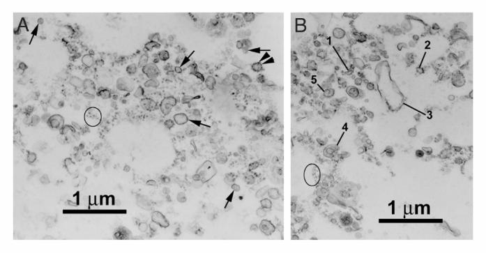
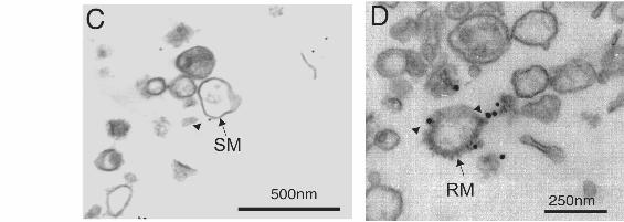
Figure 2. Electron microscopy of rat brain rough microsomes (RM). A and B) Electron
micrographs of the RM subfraction. In A, arrowheads point to ribosomes on the membranes
of rough microsomes. The arrows point to smooth microsomes. The ovals in Figures A and B
surround ribosomes associated with membrane-free filamentous structures. In B, rough
microsomes reveal different morphologies: tubular (1), oval (2), dilated (3), cup-shaped (4)
and with a spiral alignment of ribosomes (5). C and D) Electron microscope
immunocytochemistry of MAP2 in the RM subfraction. The monoclonal anti-MAP2 antibody
HM2 (1:200) was revealed using a secondary anti-mouse antibody conjugated to 10nm
by guest on September 15, 2018
http://ww
w.jbc.org/
Dow
nloaded from
colloidal gold particles. Immunogold labeling of MAP2 was found associated with smooth
membranes (SM) as shown in C and with rough ER membranes (RM) shown in D. Scale bars
Aand B=1mm, C=500nm and D=250nm.
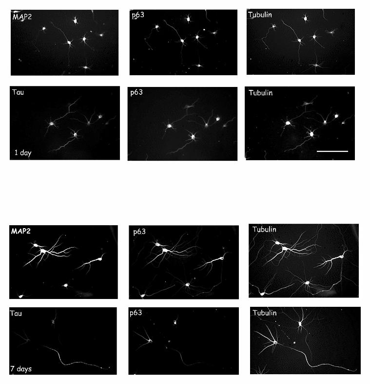
Figure 3. The somato-dendritic compartmentalization of MAP2 and p63 coincides in
cultured hippocampal neurons. The distribution of MAP2, tau, p63 and tubulin is shown in
one- and seven-day old cultured hippocampal neurons. The polyclonal anti-MAP2 antibody
and the polyclonal anti-tau were revealed by using a donkey secondary anti-rabbit conjugated
to FITC. The monoclonal anti-p63 antibody was revealed by using a donkey secondary anti-
mouse antibody conjugated to rhodamine. The rat polyclonal anti-tubulin antibody was
revealed by using a secondary anti-rat conjugated to AMCA. Tubulin staining was used to
visualize both dendritic and axonal processes. In one-day old neuronal cultures, MAP2, p63
and tau were present in all processes whereas in seven-day old neuronal cutures MAP2 and
p63 were enriched in dendrites and tau in the axon. Scale bar: 50mm.
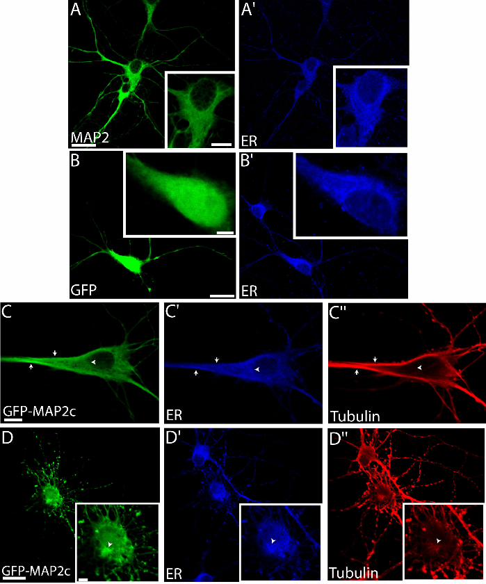
Figure 4. Overexpression of GFP-MAP2c fusion protein in hippocampal neurons. A and
A’ show the distribution of MAP2 (HM2 antibody) and the RER respectively in seven day-
old cultured hippocampal neurons. The polyclonal antibody against ribophorin II was used to
stain the RER. This antibody was revealed using a secondary anti-rabbit conjugated to
rhodamine. Insets show a higher magnification of the cell body. B) and B’) Seven day-old
neurons were transfected with the GFP protein alone as a control. Protein expression was
allowed to proceed for 24hrs before the cells were fixed and processed for
immunofluorescence. Insets show that the GFP protein does not induce a reorganization of the
RER membranes. C, C’ and C” show the distribution of the GFP-MAP2c, ribophorin and
tubulin staining in a transfected neuron presenting thick microtubule bundles. In these cells,
by guest on September 15, 2018
http://ww
w.jbc.org/
Dow
nloaded from
RER staining is found on these bundles (arrows). D, D’ and D’’ show the distribution of the
GFP-MAP2c, ribophorin and tubulin staining in a transfected neuron presenting multiple thin
extensions. In these cells, a reorganization of the RER was noted in the perikaryon
(arrowhead in the inset) and along the thin bundles. Scale bar for all figures except C, C’ and
C”= 20mm. Scale bar for C, C’ and C”= 8mm. Scale bar for the insets= 4mm.
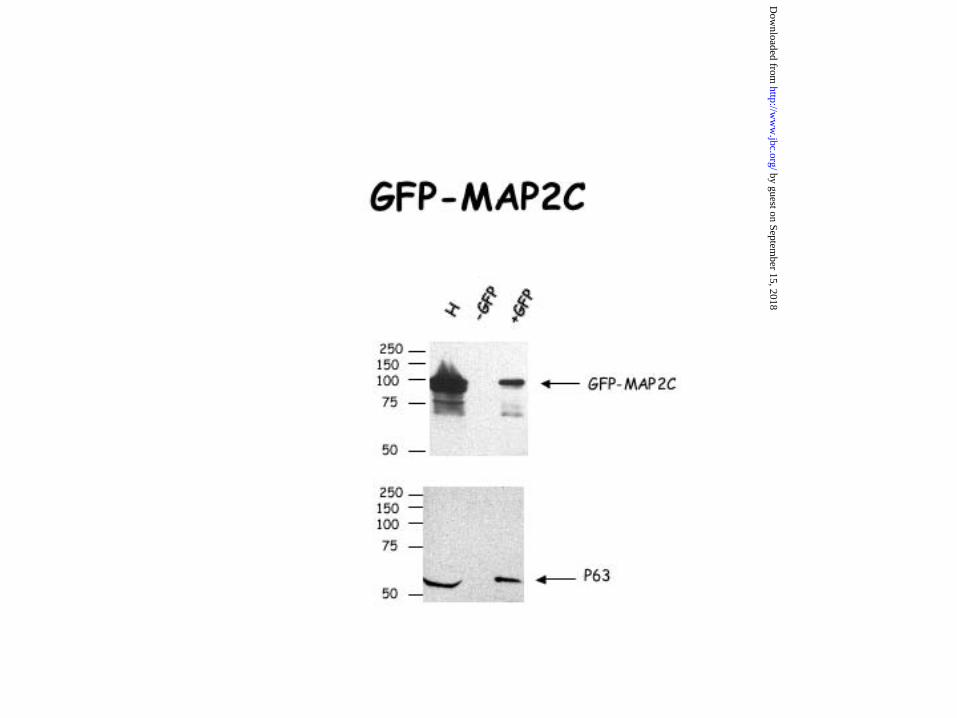
Figure 5. The interaction of MAP2c and p63 in an overlay assay. An overlay assay using
MAP2c purified from Sf9 cells was carried out to show the interaction of MAP2 with ER
proteins from a rough microsomal fraction prepared from rat liver. The overlay experiment
was performed as described in methods. In lane 1, no MAP2c protein was added to the
overlay buffer. In lane 2, the membrane was incubated with purified MAP2c protein for 1h 30
min. In lane 3, the membrane of lane 2 was stained with an anti-p63 polyclonal antibody.
Figure 6. Co-immunoprecipitation of the MAP2/p63 complex from adult and embryonic
(E19) rat brain homogenate. A) The interaction of MAP2 with p63 in adult rat brain was
confirmed by co-immunoprecipitation. MAP2 was immunoprecipitated from an adult rat brain
homogenate as described in Methods. The monoclonal anti-MAP2 HM2 (1:250) was used to
immunoprecipitate the MAP2/p63 complex. The nitrocellulose membrane was then revealed
with the monoclonal anti-MAP2 HM2 (1:1000) and then with the polyclonal anti-p63
(1:5000). The calnexin (CNX) was used as a negative control. Note that the polyclonal
antibody anti-CNX recognizes the calnexin only in the adult rat brain homogenate. B) The
interaction of MAP2 with p63 in embryonic rat brain was confirmed by co-
immunoprecipitation. Either MAP2 or p63 was immunoprecipitated from a neonatal rat brain
homogenate by using the monoclonal anti-MAP2 HM2 (1:250) or the polyclonal antibody
by guest on September 15, 2018
http://ww
w.jbc.org/
Dow
nloaded from

directed against p63 (1:500). The western blot was then revealed with the monoclonal anti-
MAP2 HM2 (1:1000) and then with the monoclonal anti-p63 (1:250). The precision plus
protein standards from Bio-Rad were used for accurate protein sizes.
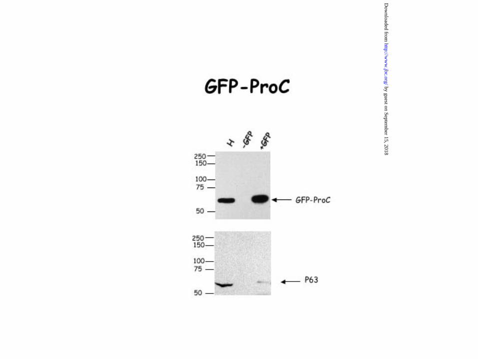
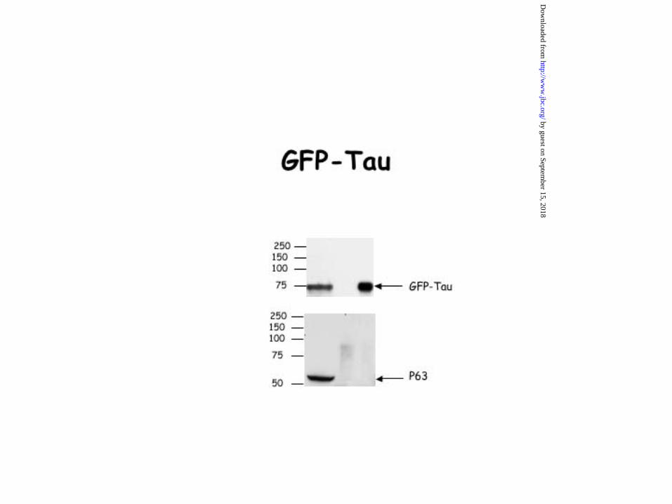
Figure 7. Interaction of the projection domain of MAP2 with p63. HeLa cells were
transfected with an expression vector containing either GFP alone, GFP-tau, GFP-MAP2c,
GFP-Proc, a deleted form of MAP2c corresponding to its projection domain or GFP-Mt, a
deleted form of MAP2c corresponding to its microtubule domain. 24hrs after transfection,
HeLa cells were lysed and co-immunoprecipitation experiments were carried out with an
antibody directed against GFP. Our data showed that the binding peptidic sequence of MAP2
to p63 is located in the projection domain of MAP2c. Indeed, the anti-GFP antibody was able
to co-immunoprecipitate either GFP-MAP2c and p63 or GFP-Proc and p63 but not GFP-Mt
and p63. The GFP antibody was unable to co-immunoprecipitate either p63 and GFP-Tau or
p63 and GFP alone. The precision plus protein standards from Bio-Rad were used for accurate
protein sizes.
Figure 8. In vitro reconstitution of the ER-MAP2-microtubule complexes. A) The nuclear
fraction prepared was examined by western blotting for its content in tubulin. No endogenous
tubulin was detected in this fraction. B) The presence of p63 in the nuclear fraction was
confirmed by western blot by using the polyclonal-anti-p63 antibody. As noted in brain
homogenate, in a subfraction enriched in rough membranes isolated from rat liver, the
polyclonal antibody, anti-p63 revealed two bands. However, in a nuclear fraction isolated
from rat liver and treated with Dnase to concentrate the membranes, only the band at 63 kDa
was present. C) The microtubule polymerizing activity of MAP2c and tau was determined as
by guest on September 15, 2018
http://ww
w.jbc.org/
Dow
nloaded from
described in Materials and Methods. Western blotting shows similar amounts of tubulin in the
MAP2c and in the tau pellets indicating that these proteins have similar capacities to
polymerize microtubules. D) Electron micrograph of MAP2c-pre-polymerized microtubules
visualized by negative staining. E) Electron micrograph of tau-pre-polymerized microtubules
visualized by negative staining. F) Electron micrograph illustrating that tau-pre-polymerized
microtubules were intact after incubation with nuclei. Scale bar D, E and F= .25mm
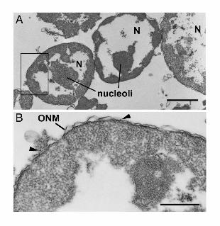
Figure 9. Electron micrographs of the in vitro reconstitution of the ER-MAP2-
microtubule complexes. A) Nuclei (N) are shown after incubation in the presence of
exogenously added tau-pre-polymerized microtubules. Under these conditions no
microtubules were observed in association with the nuclei. A higher magnification of the
region outlined by a rectangle in A is shown in B. Arrowheads point to ribosomes on the outer
nuclear membrane (ONM). C) Micrographs schowing high power electron microscopy of the
surface of nuclei after incubation in the presence of exogenously added MAP2c-pre-
polymerized microtubules. Arrows point to cross and oblique sections of microtubules located
within 25 nm of outer nuclear membrane. Scale bars: A= 2mm, B=1µm and C=500nm.
by guest on September 15, 2018
http://ww
w.jbc.org/
Dow
nloaded from

Table 1: Quantification of HMW MAP2 immunogold in the RM subfraction. The RM
subfraction purified from rat brain was labeled by immunogold using anti-MAP2 antibody
(clone HM2). The distribution of the gold particles was analyzed when HM2 antibody was
omitted (-HM2) and when HM2 antibody was added (+HM2). The numbers represent the
mean and the SEM of 3 sets of distinct experiments. An ANOVA (Tukey-Kramer Multiple
comparisons Test) test was performed. *** means that the P value is less than 0,0001 which is
considered extremely significant.
N=3 Number of gold particles
-HM2 antibody
Number of gold particles
+ HM2 antibody
Smooth membranes 3,33+2,40 16,66+4,09
Rough membranes 9,33+0,66 129,66+9,90***
by guest on September 15, 2018
http://ww
w.jbc.org/
Dow
nloaded from


LeclercGuimont, Angela Anton, Michel Lauzon, Gert Kreibich, Jacques Paiement and Nicole Carole Abi Farah, Dalinda Liazoghli, Sebastien Perreault, Mylène Desjardins, Alain
microtubules and rough endoplasmic reticulum membranes in neuronsInteraction of microtubule-associated protein-2 and p63: a new link between
published online December 28, 2004J. Biol. Chem.
10.1074/jbc.M412304200Access the most updated version of this article at doi:
Alerts:
When a correction for this article is posted•
When this article is cited•
to choose from all of JBC's e-mail alertsClick here
by guest on September 15, 2018
http://ww
w.jbc.org/
Dow
nloaded from