cas cliniques : hémiarthroplastie pour … · incision cutanée de la coracoïde au creux axilaire...
TRANSCRIPT
53
cAS cLINIQUES : HéMIARTHROPLASTIE POUR fRAcTURE
cLINIcAL cASES: HEMI-ARTHROPLASTY fOR fRAcTURE
L. Obert
INSTALLATION (cAS 1)
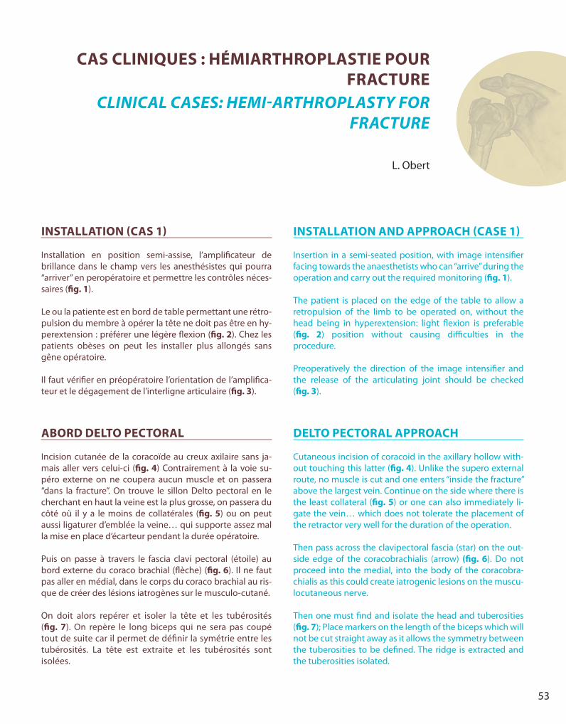
Installation en position semi-assise, l’amplificateur de brillance dans le champ vers les anesthésistes qui pourra “arriver” en peropératoire et permettre les contrôles néces-saires (fig. 1).
Le ou la patiente est en bord de table permettant une rétro-pulsion du membre à opérer la tête ne doit pas être en hy-perextension : préférer une légère flexion (fig. 2). Chez les patients obèses on peut les installer plus allongés sans gêne opératoire.
Il faut vérifier en préopératoire l’orientation de l’amplifica-teur et le dégagement de l’interligne articulaire (fig. 3).
AbORD DELTO PEcTORAL
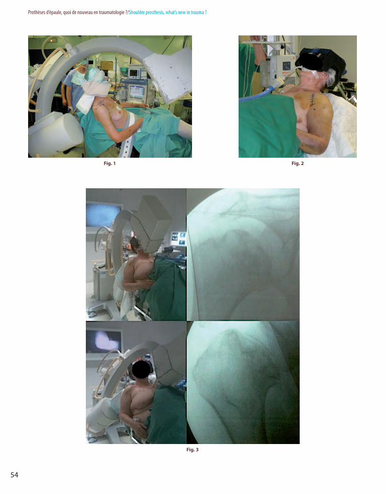
Incision cutanée de la coracoïde au creux axilaire sans ja-mais aller vers celui-ci (fig. 4) Contrairement à la voie su-péro externe on ne coupera aucun muscle et on passera “dans la fracture”. On trouve le sillon Delto pectoral en le cherchant en haut la veine est la plus grosse, on passera du côté où il y a le moins de collatérales (fig. 5) ou on peut aussi ligaturer d’emblée la veine… qui supporte assez mal la mise en place d’écarteur pendant la durée opératoire.
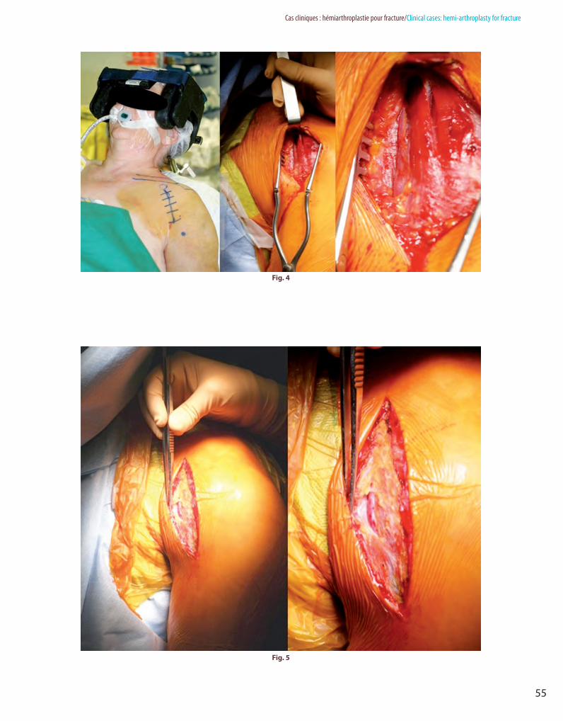
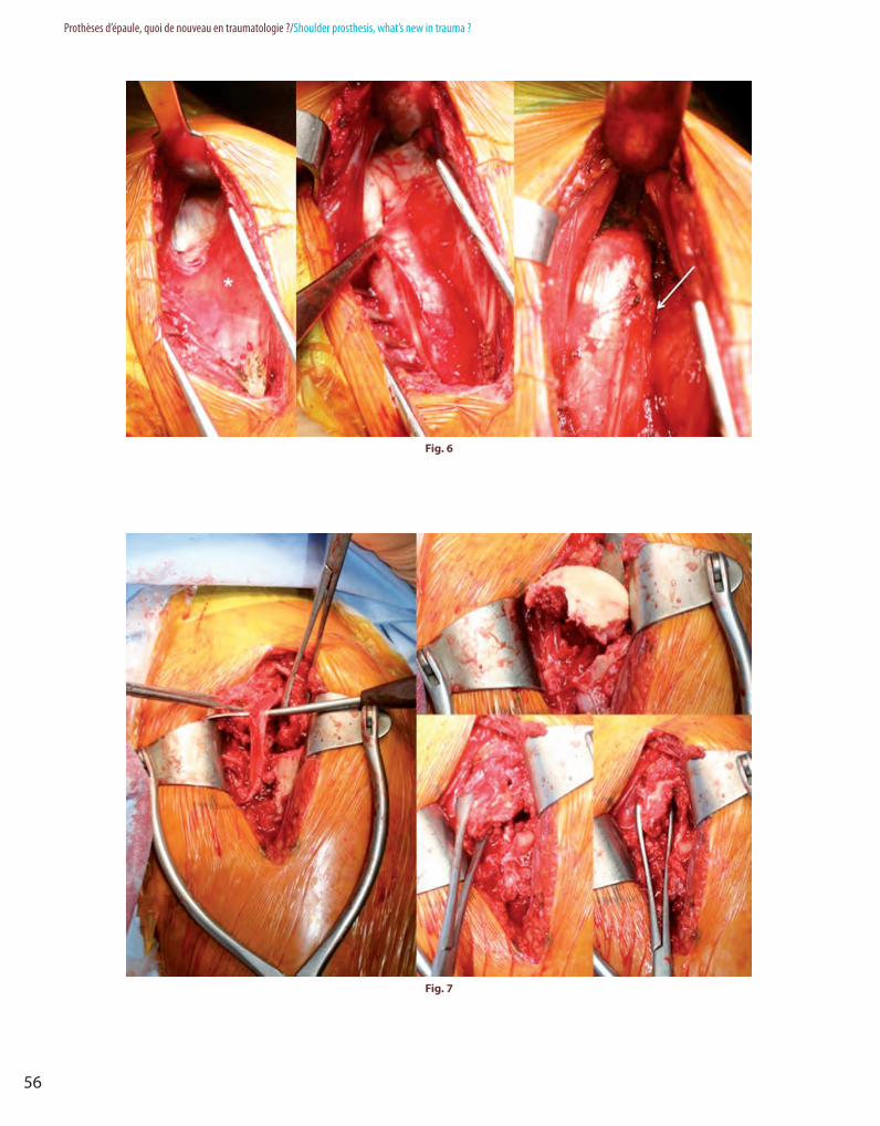
Puis on passe à travers le fascia clavi pectoral (étoile) au bord externe du coraco brachial (flèche) (fig. 6). Il ne faut pas aller en médial, dans le corps du coraco brachial au ris-que de créer des lésions iatrogènes sur le musculo-cutané.
On doit alors repérer et isoler la tête et les tubérosités (fig. 7). On repère le long biceps qui ne sera pas coupé tout de suite car il permet de définir la symétrie entre les tubérosités. La tête est extraite et les tubérosités sont isolées.
INSTALLATION AND APPROAcH (cASE 1)
Insertion in a semi-seated position, with image intensifier facing towards the anaesthetists who can “arrive” during the operation and carry out the required monitoring (fig. 1).
The patient is placed on the edge of the table to allow a retropulsion of the limb to be operated on, without the head being in hyperextension: light flexion is preferable (fig. 2) position without causing difficulties in the procedure.
Preoperatively the direction of the image intensifier and the release of the articulating joint should be checked (fig. 3).
DELTO PEcTORAL APPROAcH
Cutaneous incision of coracoid in the axillary hollow with-out touching this latter (fig. 4). Unlike the supero external route, no muscle is cut and one enters “inside the fracture” above the largest vein. Continue on the side where there is the least collateral (fig. 5) or one can also immediately li-gate the vein… which does not tolerate the placement of the retractor very well for the duration of the operation.
Then pass across the clavipectoral fascia (star) on the out-side edge of the coracobrachialis (arrow) (fig. 6). Do not proceed into the medial, into the body of the coracobra-chialis as this could create iatrogenic lesions on the muscu-locutaneous nerve.
Then one must find and isolate the head and tuberosities (fig. 7); Place markers on the length of the biceps which will not be cut straight away as it allows the symmetry between the tuberosities to be defined. The ridge is extracted and the tuberosities isolated.
Obert2.indb 53 25/10/13 16:08:17
54
Prothèses d’épaule, quoi de nouveau en traumatologie ?/Shoulder prosthesis, what’s new in trauma ?
fig. 1 fig. 2
fig. 3
Obert2.indb 54 25/10/13 16:08:18
55
Cas cliniques : hémiarthroplastie pour fracture/Clinical cases: hemi-arthroplasty for fracture
fig. 4
fig. 5
Obert2.indb 55 25/10/13 16:08:19
56
Prothèses d’épaule, quoi de nouveau en traumatologie ?/Shoulder prosthesis, what’s new in trauma ?
fig. 6
fig. 7
Obert2.indb 56 25/10/13 16:08:20
57
Cas cliniques : hémiarthroplastie pour fracture/Clinical cases: hemi-arthroplasty for fracture
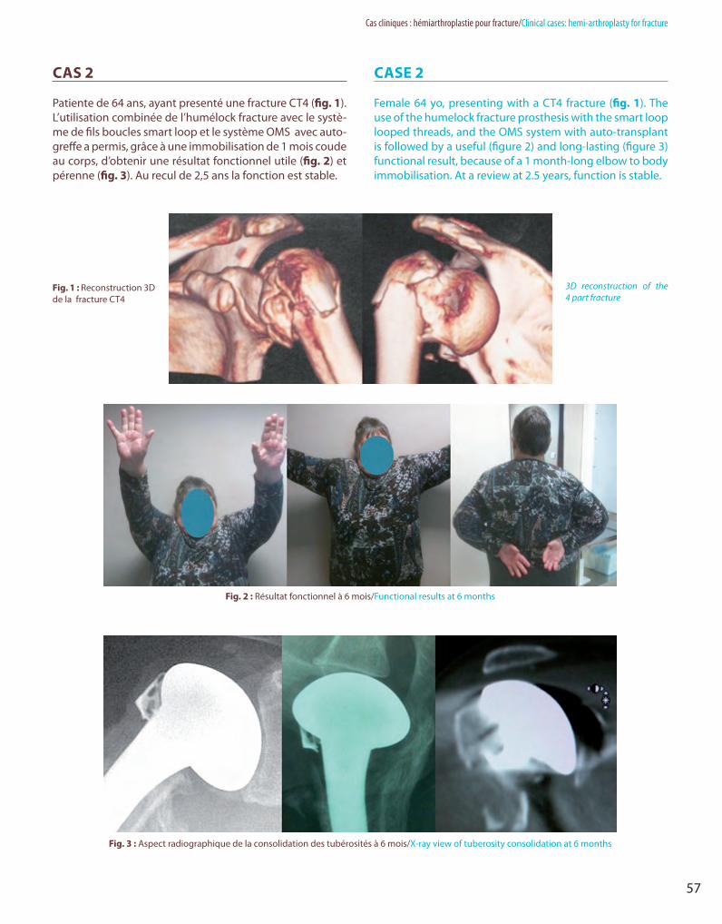
cASE 2
Female 64 yo, presenting with a CT4 fracture (fig. 1). The use of the humelock fracture prosthesis with the smart loop looped threads, and the OMS system with auto-transplant is followed by a useful (figure 2) and long-lasting (figure 3) functional result, because of a 1 month-long elbow to body immobilisation. At a review at 2.5 years, function is stable.
cAS 2
Patiente de 64 ans, ayant presenté une fracture CT4 (fig. 1). L’utilisation combinée de l’humélock fracture avec le systè-me de fils boucles smart loop et le système OMS avec auto-greffe a permis, grâce à une immobilisation de 1 mois coude au corps, d’obtenir une résultat fonctionnel utile (fig. 2) et pérenne (fig. 3). Au recul de 2,5 ans la fonction est stable.
fig. 1 : Reconstruction 3D de la fracture CT4
3D reconstruction of the 4 part fracture
fig. 2 : Résultat fonctionnel à 6 mois/Functional results at 6 months
fig. 3 : Aspect radiographique de la consolidation des tubérosités à 6 mois/x-ray view of tuberosity consolidation at 6 months
Obert2.indb 57 25/10/13 16:08:22
58
Prothèses d’épaule, quoi de nouveau en traumatologie ?/Shoulder prosthesis, what’s new in trauma ?
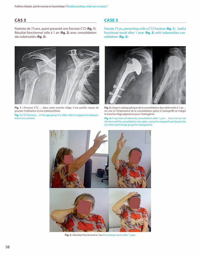
cASE 3
Female 73 yo, presenting with a CT2 fracture (fig. 1). Useful functional result after 1 year (fig. 2) with tuberosities con-solidation (fig. 3).
cAS 3
Patiente de 73 ans, ayant presenté une fracture CT2 (fig. 1). Résultat fonctionnel utile à 1 an (fig. 2) avec consolidation des tubérosités (fig. 3).
fig. 1 : Fracture CT2 … dans cette tranche d’âge, il est parfois risqué de pousser l’indication d’une ostéosynthèse.
fig. 1: CT2 Fracture… in this age group it is often risky to suggest an osteosyn-thesis as a solution.
fig. 3 : Aspect radiographique de la consolidation des tubérosités à 1 an… on voit ici l’importance de la consolidation grâce à l’autogreffe et malgré la tranche d’âge péjorative pour l’ostéogénie.
fig. 3: X-ray view of tuberosity consolidation after 1 year… here one can see the how well the consolidation has taken, using the autograft and despite the less than optimal age group for osteogenesis.
fig. 2 : Résultat fonctionnel à 1an./Functional result after 1 year.
Obert2.indb 58 25/10/13 16:08:24
59
Cas cliniques : hémiarthroplastie pour fracture/Clinical cases: hemi-arthroplasty for fracture
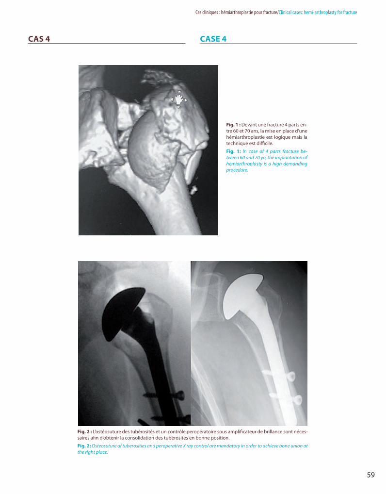
cASE 4cAS 4
fig. 1 : Devant une fracture 4 parts en-tre 60 et 70 ans, la mise en place d’une hémiarthroplastie est logique mais la technique est difficile.
fig. 1: In case of 4 parts fracture be-tween 60 and 70 yo, the implantation of hemiarthroplasty is a high demanding procedure.
fig. 2 : L’ostéosuture des tubérosités et un contrôle peropératoire sous amplificateur de brillance sont néces-saires afin d’obtenir la consolidation des tubérosités en bonne position.
fig. 2: Osteosuture of tuberosities and peroperative X ray control are mandatory in order to achieve bone union at the right place.
Obert2.indb 59 25/10/13 16:08:25