artigo 13.pmd - scielo
TRANSCRIPT

Acta Cirúrgica Brasileira - Vol. 24 (2) 2009 - 150
13 - ORIGINAL ARTICLEWound Healing
Effects of the application of Aloe vera (L.) and microcurrent on the healing of woundssurgically induced in Wistar rats1
Efeitos da aplicação de Aloe vera (L.) e microcorrente no reparo de lesões cirúrgicas induzidasem ratos Wistar
Fernanda Aparecida Sampaio MendonçaI, José Roberto Passarini JuniorII, Marcelo Augusto Marretto EsquisattoIII, JosuéSampaio MendonçaIV, Cristina Cruz FranchiniV, Glaucia Maria Tech dos SantosVI
I PhD, Head, Physiology Division, Herminio Ometto University Center, UNIARARAS, Araras-SP, Brazil.II Graduate Student, School of Biology, Herminio Ometto University Center, UNIARARAS, Araras-SP, Brazil.III PhD, Head, Anatomy Division, Herminio Ometto University Center, UNIARARAS, Araras-SP, Brazil.IV Bucomaxilofacial Surgeon, Sao Lucas Hospital, Sao Paulo-SP, Brazil.V Master, Assistant Professor, Pharmacology Division, Herminio Ometto University Center, UNIARARAS, Araras-SP, Brazil.VI PhD, Associate Professor, Physiology Division, Herminio Ometto University Center, UNIARARAS, Araras-SP, Brazil.
ABSTRACTPurpose: To investigate the effects of topical application of an Aloe vera gel combined or not with microcurrent application on thehealing of skin wounds surgically induced in Wistar rats. Methods: The animals were randomly divided into the following groups:control group, animals topically treated with Aloe vera, animals treated with a microcurrent, and animals receiving topical application ofAloe vera combined with microcurrent application. Results: The results indicated differences in wound healing between the varioustreatments when compared to the control group. Tissue hyperplasia was lower in the control group compared to the other treated groups.Accelerated wound healing was observed in the group treated with Aloe vera compared to control. Animals submitted to microcurrentapplication only and the group treated with microcurrent plus Aloe vera presented an earlier onset of the proliferative phasecompared to the control group and animals treated with Aloe vera gel alone. Morphometric data confirmed the structural findings.Conclusion: Simultaneous application of Aloe vera gel and microcurrent is an excellent choice for the treatment of open wounds thusindicating a synergistic action of these two applicationsKey words: Wound Healing. Aloe. Electric Stimulation. Histology, Comparative. Rats.
RESUMOObjetivo: Investigar os efeitos da aplicação tópica do gel de Aloe vera, combinada ou não com a aplicação de microcorrente no reparode lesões cutâneas induzidas cirurgicamente em ratos Wistar. Métodos: Os animais foram distribuídos aleatoriamente em: grupo controle,tratado topicamente com gel in natura de Aloe vera, tratado com microcorrente e tratado com aplicação tópica de Aloe vera associada àmicrocorrente. Resultados: Os resultados do presente trabalho indicaram que o reparo tecidual ocorreu de forma diferenciada nos váriostratamentos empregados quando comparados ao grupo controle. A hiperplasia tecidual no grupo controle foi menor que a observada nosdemais grupos tratados. No grupo tratado com aplicação de Aloe vera o processo de reparo foi acelerado em relação ao controle. Osanimais do grupo tratado somente com microcorrente e do grupo tratado com microcorrente associada à Aloe vera apresentaram umafase proliferativa mais precoce quando comparados com o grupo controle e tratado somente com Aloe vera. Os dados morfométricosconfirmaram os achados estruturais. Conclusão: A aplicação simultânea do gel de Aloe vera e microcorrente é uma excelente escolhapara o tratamento de feridas abertas indicando uma ação sinérgica dessas duas aplicações.Descritores: Cicatrização de Feridas. Aloe. Estimulação Elétrica. Histologia Comparada. Ratos.1Research performed at Laboratory of Micromorphology, Herminio Ometto University Center – UNIARARAS, Araras-SP, Brazil.
Introduction
The mechanism of tissue healing is a complex biologicalprocess that involves a perfect and coordinated cascade of cellularand molecular events promoting tissue reconstitution. This processarises as a response of the tissue to injuries induced by trauma or
by surgical procedures. The process of wound healing is charac-terized by three phases that overlap and present a characteristicprofile: inflammatory phase, proliferative phase and remodelingphase. Despite some recent advances in the understanding of thesebasic processes, wound healing disorders continue to cause diseasesand even death. A wide variety of therapies has arisen with the

Mendonça FAS et al
151 - Acta Cirúrgica Brasileira - Vol. 24 (2) 2009
advances in technological applications. Electrical stimulation hasbeen shown to modify the healing process in living organisms,especially in terms of factors that delay or impair this process1,2.
Several studies have investigated cellular responses toelectrical currents of different amplitudes and frequencies2,3. In thisrespect, Cheng and Goldman2 and Kloth4 observed that cells ex-posed to electrical fields vary in cell proliferation and metabolism.In in vitro studies, Goldman and Pollack5 found that the applica-tion of electrical currents stimulated fibroblast proliferation andcollagen synthesis. An increase of collagen biosynthesis, numberof fibroblasts and hydroxyproline levels was also observed inanimal experiments6. The technique of microamperage electricalstimulation is also called biostimulation or electrical therapy dueto its capacity to physiologically stimulate cell growth.
The application of phytotherapeutic agents has also shownto be highly effective in the healing of wounds and burns7. Aloevera (L.) (Liliaceae), popularly called aloe, is widely known for itstherapeutic effects and has been used as a medicine since ancienttimes. “Aloe vera” was first called Aloe perfoliata var. vera byLinnaeus and was later recognized as a distinct species by Millerwho called it Aloe barbadensis, and also by Burman who called itAloe vera8. The plant originated in Africa and is well adapted to allregions. Two fractions can be extracted from its leaves: a bitterexudate and a mucilaginous gel. The first fraction is a yellow-redfluid extracted from cells of the pericycle which is rich in anthracenecompounds. The second transparent gel-like portion (mucilage)originates from the leaf parenchyma and has been used for the treat-ment of burns and wounds because of its healing properties9. Thismucilage consists of biologically active molecules that act on fi-broblasts during the formation of cicatricial tissue, stimulating thedeposition of collagen fibers in the extracellular matrix10. Reynoldsand Dweck11 investigated the biological activities of various Aloespecies and observed that the whole gel extract of Aloe verapresents various pharmacological properties such as promotingwound, burn, and frost-bite healing, in addition to having anti-inflammatory, antifungal, hypoglycemic and gastroprotectiveproperties.
In the present study, we investigated the effects of topicalapplication of an Aloe vera gel combined or not with microcurrentapplication on the healing of skin wounds surgically inducedin Wistar rats.
Methods
Experimental groups
Male rats from Wistar strain aged 120 days and with amean weight of 250 to 350 g, obtained from the animal house ofHermínio Ometto University Center, Uniararas, were used. Theanimals were kept in individual cages, with commercial chow andwater available ad libitum. In reason of similar genetic backgroundof animals12 and following orientation of Uniararas EthicsCommission (Protocol number 178/2006) were used four ex-perimental groups with six animals each: control group, animalstopically treated with Aloe vera gel (AV), animals treated with amicrocurrent (10 µA/2 min) (MC), and animals receiving topicalapplication of Aloe vera combined with a microcurrent (10 µA/2min) (AV+MC). A Physiotonus Microcurrent apparatus was usedfor transcutaneous electrical stimulation.
Wound creation
The animals were submitted to trichotomy in the dorsalregion and were anesthetized with 40 mg/kg body weight sodiumpentobarbital. Next, a full-thickness skin surgical incisionmeasuring 20 mm in length and 2 mm in depth was made in theskin of the back of the animal in the caudocranial direction. Theincision was not sutured. The proposed treatments were started 24h after the surgical intervention and were applied daily for 10 days.The animals were sacrificed on days 2, 6 and 10 for removal ofthe damaged area and structural and morphometric analysis. Allsurgical procedures and experimental design were performed bythe same investigator in accordance with institutional ethicalguidelines and following Rao and co-works13 orientation.
Preparation and administration of Aloe vera (L)N.L.Burm. (Liliaceae)
The leaves of Aloe vera plants were collected daily inthe morning in the Experimental garden of Hermínio OmettoUniversity Center - Uniararas, Araras (SP), Brazil, and sent to theLaboratory of Physiology, Hermínio Ometto University Center,for use. For administration, an incision was made in the leaf andthe mucilage was removed with a sterile swab and immediatelyapplied to the skin wounds.
Collection and preparation of wound samples forstructural analysis
Animals of the control and experimental groups weresacrificed with a sodium pentobarbital overdose on days 2, 6 and10 for removal of the healing area for comparative structural andmorphometric analysis. The complete wound region and theunderlying muscle with 2 mm adjacent unwound skin werecollected by excising a rectangular area measuring approximately120-160 mm2. Each sample was fixed in buffered formalin for 24hat room temperature. Next, the specimens were washed in bufferand processed for embedding in ParaplastTM. Longitudinal sections(7 µm thick), obtained from the mid-region toward the margin,were stained with hematoxylin/eosin, picrosirius-hematoxylinand Toluidine blue, pH 4.0. The specimens were observed anddocumented under a Leica DM 2000 photomicroscope at theLaboratory of Micromorphology, Centro Universitario HerminioOmetto, UNIARARAS.
Histomorphometric analysis
The histomorphometry of skin wounds healing in controland experimental groups was analyzed by Sigma Scan Pro 6.0™and Leica Image Measure™ softwares using digital images cap-tured from slides stained by Toluidine blue. Images were captured,using digitalization system (total magnification 200X) from LeicaDM 2000 photomicroscope. Three randomly chosen opticalmicroscopic fields for each histological slide pair were stored andsubmitted to a count of total cells (fibroblastic and inflammatorycells) (x 103 µm2), of newly formed blood vessels and measure-ment of wound healing area (x 103 µm2) and thickness of theregenerating epithelium (µm).
Acta Cirúrgica Brasileira - Vol. 24 (2) 2009 - 152
Effects of the application of Aloe vera (L.) and microcurrent on the healing of wounds surgically induced in Wistar rats
2.6. Statistical analysis
Data obtained for thedifferent experimental groupswere stored in electronic spread-sheets and compared by theANOVA and Tukey test (5% levelof significance) using the Biostatfor Windows XP™ program.
Results
In the present studywe describe the repair of theepidermis and dermis, includedmorphometrical parameters, inwound healing between thevarious treatments when com-pared to control group.
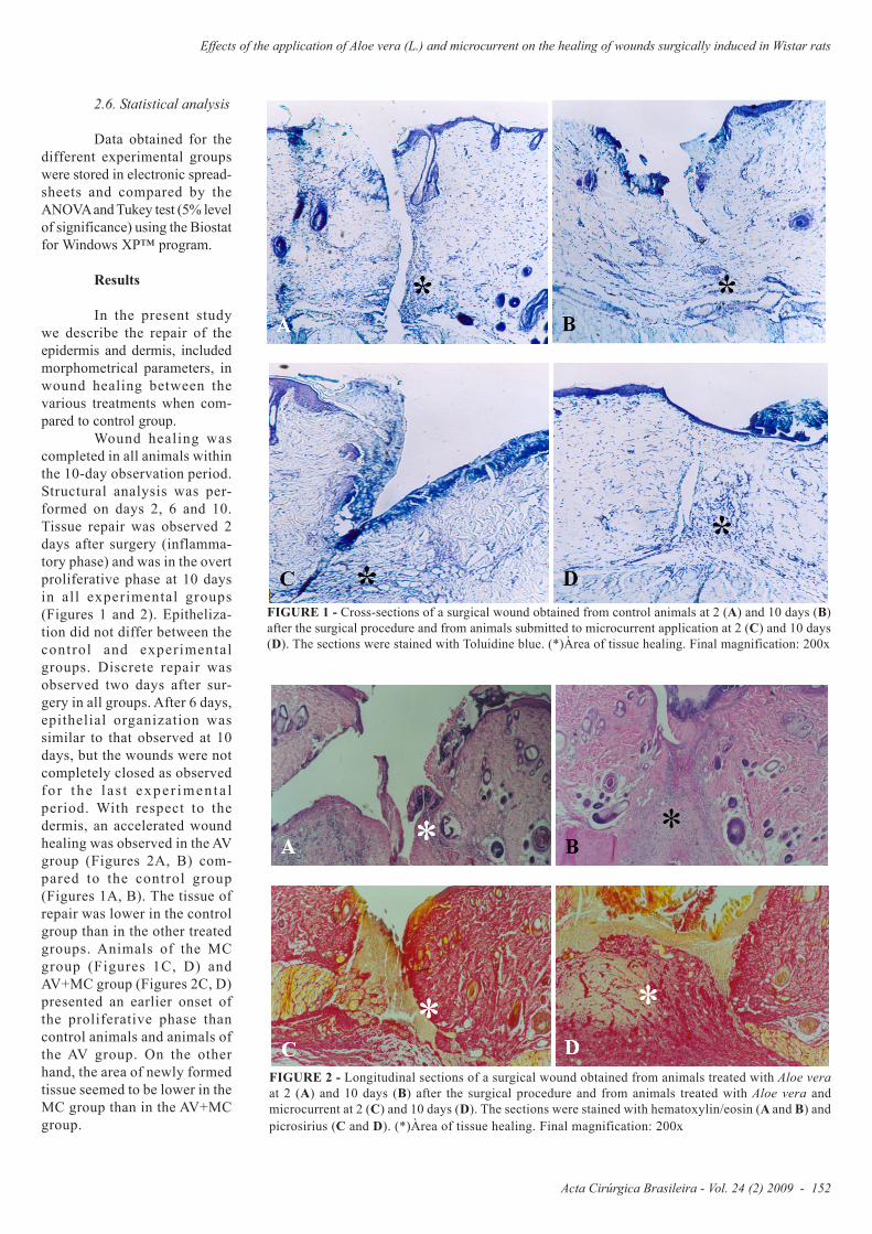
Wound healing wascompleted in all animals withinthe 10-day observation period.Structural analysis was per-formed on days 2, 6 and 10.Tissue repair was observed 2days after surgery (inflamma-tory phase) and was in the overtproliferative phase at 10 daysin all experimental groups(Figures 1 and 2). Epitheliza-tion did not differ between thecontrol and experimentalgroups. Discrete repair wasobserved two days after sur-gery in all groups. After 6 days,epithelial organization wassimilar to that observed at 10days, but the wounds were notcompletely closed as observedfo r the l a s t expe r imen ta lperiod. With respect to thedermis, an accelerated woundhealing was observed in the AVgroup (Figures 2A, B) com-pared to the control group(Figures 1A, B). The tissue ofrepair was lower in the controlgroup than in the other treatedgroups. Animals of the MCgroup (Figures 1C, D) andAV+MC group (Figures 2C, D)presented an earlier onset ofthe proliferative phase thancontrol animals and animals ofthe AV group. On the otherhand, the area of newly formedtissue seemed to be lower in theMC group than in the AV+MCgroup.
FIGURE 1 - Cross-sections of a surgical wound obtained from control animals at 2 (A) and 10 days (B)after the surgical procedure and from animals submitted to microcurrent application at 2 (C) and 10 days(D). The sections were stained with Toluidine blue. (*)Àrea of tissue healing. Final magnification: 200x
FIGURE 2 - Longitudinal sections of a surgical wound obtained from animals treated with Aloe veraat 2 (A) and 10 days (B) after the surgical procedure and from animals treated with Aloe vera andmicrocurrent at 2 (C) and 10 days (D). The sections were stained with hematoxylin/eosin (A and B) andpicrosirius (C and D). (*)Àrea of tissue healing. Final magnification: 200x
Mendonça FAS et al
153 - Acta Cirúrgica Brasileira - Vol. 24 (2) 2009
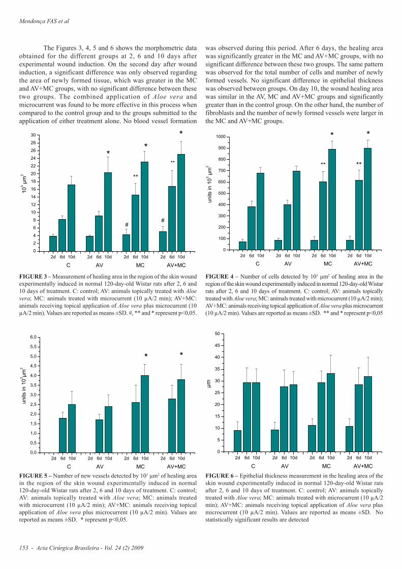
FIGURE 4 – Number of cells detected by 103 µm2 of healing area in theregion of the skin wound experimentally induced in normal 120-day-old Wistarrats after 2, 6 and 10 days of treatment. C: control; AV: animals topicallytreated with Aloe vera; MC: animals treated with microcurrent (10 µA/2 min);AV+MC: animals receiving topical application of Aloe vera plus microcurrent(10 µA/2 min). Values are reported as means ±SD. ** and * represent p<0,05
The Figures 3, 4, 5 and 6 shows the morphometric dataobtained for the different groups at 2, 6 and 10 days afterexperimental wound induction. On the second day after woundinduction, a significant difference was only observed regardingthe area of newly formed tissue, which was greater in the MCand AV+MC groups, with no significant difference between thesetwo groups. The combined application of Aloe vera andmicrocurrent was found to be more effective in this process whencompared to the control group and to the groups submitted to theapplication of either treatment alone. No blood vessel formation
FIGURE 3 – Measurement of healing area in the region of the skin woundexperimentally induced in normal 120-day-old Wistar rats after 2, 6 and10 days of treatment. C: control; AV: animals topically treated with Aloevera; MC: animals treated with microcurrent (10 µA/2 min); AV+MC:animals receiving topical application of Aloe vera plus microcurrent (10µA/2 min). Values are reported as means ±SD. #, ** and * represent p<0,05.
2d 6d 10d 2d 6d 10d 2d 6d 10d 2d 6d 10d
0
2
4
6
8
10
12
14
16
18
20
22
24
26
28
30
**
*
*
*
10
3 µ
m2
C AV MC AV+MC
##
**
2d 6d 10d 2d 6d 10d 2d 6d 10d 2d 6d 10d
0
100
200
300
400
500
600
700
800
900
1000
**
* *
un
its in
10
3 µ
m2
C AV MC AV+MC
**
FIGURE 5 – Number of new vessels detected by 103 µm2 of healing areain the region of the skin wound experimentally induced in normal120-day-old Wistar rats after 2, 6 and 10 days of treatment. C: control;AV: animals topically treated with Aloe vera; MC: animals treatedwith microcurrent (10 µA/2 min); AV+MC: animals receiving topicalapplication of Aloe vera plus microcurrent (10 µA/2 min). Values arereported as means ±SD. * represent p<0,05.
2d 6d 10d 2d 6d 10d 2d 6d 10d 2d 6d 10d
0,0
0,5
1,0
1,5
2,0
2,5
3,0
3,5
4,0
4,5
5,0
5,5
6,0
* *
un
its in
10
3µ
m2
C AV MC AV+MC
FIGURE 6 – Epithelial thickness measurement in the healing area of theskin wound experimentally induced in normal 120-day-old Wistar ratsafter 2, 6 and 10 days of treatment. C: control; AV: animals topicallytreated with Aloe vera; MC: animals treated with microcurrent (10 µA/2min); AV+MC: animals receiving topical application of Aloe vera plusmicrocurrent (10 µA/2 min). Values are reported as means ±SD. Nostatistically significant results are detected
2d 6d 10d 2d 6d 10d 2d 6d 10d 2d 6d 10d
0
5
10
15
20
25
30
35
40
45
50
µm
C AV MC AV+MC
was observed during this period. After 6 days, the healing areawas significantly greater in the MC and AV+MC groups, with nosignificant difference between these two groups. The same patternwas observed for the total number of cells and number of newlyformed vessels. No significant difference in epithelial thicknesswas observed between groups. On day 10, the wound healing areawas similar in the AV, MC and AV+MC groups and significantlygreater than in the control group. On the other hand, the number offibroblasts and the number of newly formed vessels were larger inthe MC and AV+MC groups.

Acta Cirúrgica Brasileira - Vol. 24 (2) 2009 - 154
Effects of the application of Aloe vera (L.) and microcurrent on the healing of wounds surgically induced in Wistar rats
Discussion
In the present study, both microcurrent therapy andtopical application of Aloe vera were effective in accelerating thetissue regeneration process. These findings agree with thosereported in different studies demonstrating that low-intensityelectrical currents stimulate wound healing14,15. Numerousinvestigators have studied the effects of electrical stimulationusing different amplitudes and frequencies and have observedmodifications in the cellular and tissue responses in inducedlesions4,16. Some studies reported variations in cell metabolism,whereas others demonstrated fibroblast proliferation,neovascularization and collagen deposition in the wound area17.Significant acceleration of the healing process after microcurrentelectrical stimulation has been widely documented4,5,18,19. Becker14
reported that electrical currents are present in a biological systemand may promote repair and growth after injury. According to thisauthor, a specific stimulus of injury induces another stimulusof repair and also demonstrated that the membrane electricalpotential is altered in injured tissues. The “injury signal” graduallydecreases in parallel to the repair process and ceases when the latteris complete. The voltage peaks immediately after injury and gradu-ally decreases as the wound heals, a fact leading to the concept thatcurrent flow may be defective in chronic wounds and that applyingelectrical currents to wounds may stimulate healing4,20. Biedeback21
proposed that transmembrane currents open voltage-controlledcalcium channels in fibroblasts, causing ATP resynthesis, activationof protein kinase mechanisms to synthesize new cellular protein,and DNA replication necessary for mitotic cell division.
In the present study, microcurrent application was foundto be highly effective in terms of the parameters analyzed, withpositive effects on the newly formed tissue area, number offibroblasts, number of newly formed vessels and epithelialthickness, in agreement with the literature4,21,22.
Similar, but less pronounced, effects were observed whenAloe vera extract was applied. Mello et al.23 suggested that thehealing capacity of Stryphnodendron adstringens might be attrib-uted to the high tannin content in its stem bark. Tannins precipitateproteins in damaged tissues, forming a protective lining that favorshealing by reducing wound permeability and exudation24. Sarabiaet al.25 demonstrated the presence of antioxidant substances in Aloevera that confer anti-inflammatory and healing properties. Kuzuyaet al.26 also reported an antibacterial action of Aloe vera and attrib-uted these properties to the presence of anthraquinones such asaloenin, barbaloin and iso-barbaloin in its chemical composition.Chitra et al.10 also suggested that treatment with Aloe vera has abeneficial effect on the tissue proliferation phase, influencingfibroplasia and collagen synthesis and thus increasing the healingarea. Mannose-6-phosphate is a major structural constituent ofAloe vera. Davis et al.27 speculated that the binding of mannose-6-phosphate to fibroblast receptors activates fibroblast proliferation.
As observed in the present study, wound healing was moreeffective since the beginning of treatment in animals submitted tothe simultaneous application of microcurrent and Aloe vera, thusindicating a synergistic action of these two applications. Thistreatment presented advantages in all parameters studied whencompared to the control group and to the other treatments. Thesefindings agree with Soares28 who combined a chemical substance,i.e., vitamin C, and physical agents for the healing of experimental
wounds similar to those used in the present study, and demonstratedthat the combination of antioxidant agents and photodynamic orlow-amperage electrical therapy accelerates wound healing.
Conclusion
Despite the lack of scientific studies regarding thecombination of phytochemical and physical agents for the treatmentof surgical wounds, the present investigation shows that thesimultaneous application of Aloe vera and microcurrent waseffective for the treatment of open wounds potentiating woundhealing.
References
1. Houghton PE, Kincaid CB, Lovell M, Campbell KE, Keast DH,Woodbury MG, Harris KA. Effect of electrical stimulation on chronic legulcer size and appearance. Phys Ther. 2003;83(1):17-28.2. Cheng K, Goldman RJ. Electric fields and proliferation in a dermalwound model: cell cycle kinetics. Bioelectromagnetics. 1998;19(2):68-74.3. Mertz PM, Davis SC, Cazzaniga AL, Cheng K, Reich JD, EaglsteinWH. Electrical stimulation: acceleration of soft tissue repair by varyingthe polarity. Wounds. 1993;5(3):153-9.4. Kloth LC. Electrical stimulation for wound healing: a review ofevidence from in vitro studies, animal experiment, and clinical trials. Int JLow Extrem Wounds. 2005;4(1):23-44.5. Goldman R, Pollack S. Electrical fields and proliferation in a chronicwound model. Bioelectromagnetics. 1996;17(6):450-7.6. Alvarez OM, Menz PM, Smerbeck RV, Eaglstein WH. The healing ofsuperficial skin wounds is stimulated by external electrical current. JInvest Dermatol. 1983;81(2):144-8.7. Rao SG, Udupa AL, Udupa SL, Rao PGM, Rao G, Kulkarni DR.Calendula and Hypericum: two homeopathic drugs promoting woundhealing in rats. Fitoterapia. 1991;62(6):508-10.8. Reynolds GW. The aloes of South Africa XXIV The aloes of S.A. Bookfund, 520;1966.9. Dorneles D, Wouk AF, Pontarolo R, Oliveira AB. Efeito de Aloe veraLinné sobre a cicatrização de feridas de pele em coelhos. Visão Acad.2003;4(1):39-46.10. Chithra P, Sajithlal BG. Chandrakasan G. Influence of Aloe vera on thehealing of dermal wounds in diabetic rats. J Ethnopharmacol.1998;59(3):195-201.11. Reynolds T, Dweck AC. Aloe vera leaf gel: a review update. JEthnoparmacol. 1999;68(1):3-37.12. Gill TJ, Smith GJ, Wissler RW. The rat as an experimental animal.Science. 1989;245(4915):269-76.13. Rao KS, Patil PA, Malur PR. Promotion of cutaneous wound healingby famotidine in Wistar rats. Indian J Med Res. 2007;125(2): 149-54.14. Becker R. The body electric. N.Y., William Morrow and Co Inc; 1985.15. Basset CA. Beneficial-effects of electromagnetic-fields. J Cell Biochem.1993;51(4):387-93.16. Bayat M, Asgari-Moghadam Z, Maroufi M, Rezaie FS, Bayat M,Rakhshan M. Experimental wound healing using microamperageelectrical stimulation in rabbits. J Rehabil Res Dev. 2006;43(2): 219-28.17. Aaron RK, Ciombor DM. Therapeutic effects of electromagnetic fieldsin the stimulation of connective tissue repair. J Cell Biochem.1993;52(1):42-6.18. Davis SC, Ovington LG. Electrical stimulation and ultrasound in woundhealing. Dermatol Clin. 1993;11(4):775-81.19. Evans RD, Foltz D, Foltz K. Electrical stimulation with bone and woundhealing. Clin Pediatr Med Surg. 2001;18(1):79-95.20. McGinnis ME, Vanable JW. Voltage gradients in newt limb stumps.Prog Clin Biol Res. 1986;210:231-8.
Mendonça FAS et al
155 - Acta Cirúrgica Brasileira - Vol. 24 (2) 2009
21. Biedeback MC. Accelerated healing of skin ulcers by electrical stimu-lation and intracellular physiological mechanisms involved. AcupunctElectrother Res. 1989;14(1):43-60.22. Markov MS, Colbert AP. Magnetic and electromagnetic field therapy.J Back Musculoskelet Rehabil. 2001;15:17-29.23. Mello JCP, Peterit F, Nahrstedt A. A dimeric proanthocyanidin fromStryphnodendron adstringens. Phytochemistry. 1999;51(8):1105-7.24. Bedi MK, Shenefelt PD. Herbal therapy in dermatology. Arch Dermatol.2002;138(2):232-42.25. Sarabia JEL, Clares VPR, Clares RAR, Hernandez VP. Actividadantiinflamatoria y cicatrizante del ungüento rectal de Aloe vera L (sábila).Rev Cubana Plantas Med. 1999;4(3):106-9.
Conflict of interest: noneFinancial source: PROPESQ/UNIARARAS
Correspondence:Dr. Fernanda Aparecida Sampaio Mendonça.Physiology Division, Hermínio Ometto University Center – UNIARARASAv. Dr. Maximiliano Baruto, 50013607-339 Araras – SP BrazilPhone/Fax: 055 019 [email protected]
Received: October 15, 2008Review: December 18, 2008Accepted: January 14, 2009
26. Kuzuya K, Tamai I, Beppu H, Shimpo K, Chihara T. Determination ofaloenin, barbaloin and isobarbaloin in Aloe species by micellar electroki-netic chromatography. J Chromatogr. 2001;752(1):91-7.27. Davis RH, Di Donato JJ, Hartman GM, Haas RC. Anti-inflammatoryand wound healing activity of a growth substance in Aloe vera. J Am PediatrMed Assoc.1994;84(2):77-81.28. Soares FRL. Reparação de feridas cutâneas tratadas com vitamina C,laser e a associação de vitamina C e laser: estudo histológico em ratos[Tese de Mestrado]. Marília: Universidade de Marília (Unimar), Faculdadede Odontologia; 2005. 80p.
How to cite this articleMendonça FAS, Passarini Junior JR, Esquisatto MAM, Mendonça JS, Franchini CC, Santos GMT. Effects of the application of Aloevera (L.) and microcurrent on the healing of wounds surgically induced in Wistar rats. Acta Cir Bras. [serial on the Internet] 2009 Mar-Apr;24(2). Available from URL: http://www.scielo.br/acb
*Color figures available from www.scielo.br/acb