thermophilic bacteria in moroccan hot springs, salt ... · pdf filethermophilic bacteria in...
TRANSCRIPT
Thermophilic bacteria in Moroccan hot springs, salt marshes and desert soils
Tarik Aanniz1,3, Mouna Ouadghiri1,2, Marouane Melloul5, Jean Swings4,
Elmostafa Elfahime5, Jamal Ibijbijen3, Mohamed Ismaili3, Mohamed Amar1,2
1Laboratoire de Microbiologie et Biologie Moléculaire,
Centre National pour la Recherche Scientifique et Technique, Rabat, Maroc.2Collections Coordonnées Marocaines de Microorganismes, Laboratoire de Microbiologie et Biologie
Moléculaire, Centre National pour la Recherche Scientifique et Technique, Rabat, Maroc.3Faculté des Sciences, Université Moulay Ismail, Meknès, Maroc.
4Laboratory of Microbiology, Gent University, Gent, Belgium.5Unité d’Appui Technique à la Recherche Scientifique,
Centre National pour la Recherche Scientifique et Technique, Rabat, Maroc.
Submitted: March 12, 2014; Approved: September 30, 2014.
Abstract
The diversity of thermophilic bacteria was investigated in four hot springs, three salt marshes and 12
desert sites in Morocco. Two hundred and forty (240) thermophilic bacteria were recovered, identi-
fied and characterized. All isolates were Gram positive, rod-shaped, spore forming and halotolerant.
Based on BOXA1R-PCR and 16S rRNA gene sequencing, the recovered isolates were dominated by
the genus Bacillus (97.5%) represented by B. licheniformis (119), B. aerius (44), B. sonorensis (33),
B. subtilis (subsp. spizizenii (2) and subsp. inaquosurum (6)), B. amyloliquefaciens (subsp.
amyloliquefaciens (4) and subsp. plantarum (4)), B. tequilensis (3), B. pumilus (3) and Bacillus sp.
(19). Only six isolates (2.5%) belonged to the genus Aeribacillus represented by A. pallidus (4) and
Aeribacillus sp. (2). In this study, B. aerius and B. tequilensis are described for the first time as
thermophilic bacteria. Moreover, 71.25%, 50.41% and 5.41% of total strains exhibited high amylo-
lytic, proteolytic or cellulolytic activity respectively.
Key words: thermophilic bacteria, hot springs, diversity, 16S rRNA gene sequencing, hydrolytic en-
zymes.
Introduction
Extremophiles are thriving in extreme ecosystems.
Such environments may have extremely high or low pH,
high or low temperatures, high salinity, high pressure and
various combinations thereof. Extremophilic microorgan-
isms include members of all three domains of life, Archaea,
Bacteria and Eukarya. Many investigations focused on
their potential as sources of highly active enzymes ‘extre-
mozymes’ and other products such as antibiotics, compati-
ble solutes (Robb et al., 2008).
Thermophiles are growing optimally between 55 and
80 °C, while hyperthermophiles grow above 80 °C (Brock
1978; Bertoldo et al., 2002). They may be Gram positive or
negative, spore forming or not, and may exhibit an aerobic
or anaerobic metabolism. Their study has become a major
domain of research and several new thermophilic genera
and species have recently been described (Yoneda et al.,
2013; Cihan et al., 2014). They were intensively studied
due to their potential to produce thermostable enzymes
(proteases, amylases, lipases, xylanases, DNA poly-
merases) and exo-polysaccharides (Meintanis et al., 2008;
Singh et al., 2010). These thermo-enzymes are usually not
only thermostable, but also active at high salinity and ex-
treme pH (Gomez and Steiner, 2004).
The growth, characterization and identification of
thermophiles pose specific problems (Savas et al., 2009).
Development of molecular techniques such as PCR-based
techniques like rep-PCR and 16S rRNA allow their reliable
Brazilian Journal of Microbiology 46, 2, 443-453 (2015) Copyright © 2015, Sociedade Brasileira de Microbiologia
ISSN 1678-4405 www.sbmicrobiologia.org.br
DOI: http://dx.doi.org/10.1590/S1517-838246220140219
Send correspondence to M. Amar. Laboratoire de Microbiologie et Biologie Moléculaire, Centre National pour la Recherche Scientifique et Technique,
Angle av. Allal El Fassi, av. des FAR, Quartier Hay Ryad, BP. 8027 Nations Unies, 10102 Rabat, Maroc. E-mail: [email protected].
Research Paper
identification, whereas conventional methods are time con-
suming and not reliable (Kublanov et al., 2009).
Worldwide, geothermal areas which are favorable
habitats for thermophilic organisms are limited to a restrict
number of sites. In Morocco, there are more than twenty hot
springs distributed in different regions mainly in the Rif,
the Pre-Rif and the Southern Rif (Lakhdar et al., 2006). The
central region includes the hot springs of Ain Allah and
Moulay Yaacoub (Fez region) and Ain Jerri (Meknes re-
gion) (Salame et al., 2013). On the South side of the West-
ern Anti-Atlas, the Abaynou spring is located in the middle
of an oasis of palms and olive trees. It is considered as the
most important hot spring in South Morocco. These springs
are frequently visited by Moroccans with dermal and rheu-
matic diseases (Salame et al., 2013). Merzouga, a small Sa-
haran village, in South Eastern Morocco, is known for its
set of sand dunes near Erg Chebbi, which is part of touristic
itineraries. The climate is very hot and dry and the tempera-
tures are often higher than 40 °C in the day while nights are
cold. In these conditions living organisms have to cope with
extremes temperature, low humidity and low availability of
nutritional compounds. These conditions reduce biodiver-
sity but some bacteria developed survival strategies in or-
der to adapt to such stress (Hecker and Völker, 2001; Bar et
al., 2002).
The wetland of Sidi Moussa-Oualidia is a protected
ecosystem of 10.000 ha recognized by the Ramsar Conven-
tion of 2005. The complex consists of the lagoons Oualidia
and Sidi Moussa and 4 salt marshes (Sidi Abed is one of
them). The main activities in the neighborhood of the site
are agriculture, livestock raising, sandpit exploitation, salt
production, and tourism. Salt mining has reduced biodi-
versity, especially invertebrates (http://www.ramsar.org).
Little is known about the occurrence and distribution
of thermophilic bacteria in Morocco. This work is the first
study focusing on culturable thermophilic bacteria: their
isolation, identification and characterization. Three kind of
sites have been selected for this study i.e. hot springs, hot
desert soils and salty wetland.
Materials and Methods
Sampling sites
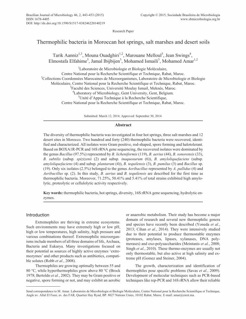
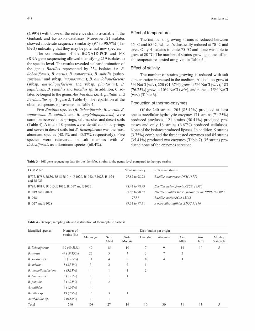
The location of the sampling sites is shown in Figu-
re 1 and Table 1. Their physico-chemical parameters are
given in Table 1. Thirty three samples were collected in
sterile conditions, then were transported to the laboratory,
using thermal boxes, and immediately analyzed (Malkawi
and Al-Omari, 2010).
Temperature and pH of the sites were measured dur-
ing sampling. The total salt concentration was determined
by titration using Mohr’s method (Skoog et al., 1996).
444 Aanniz et al.
Figure 1 - Location of the studied sites.
Enumeration and isolation of thermophilic bacteria
Sand samples
An amount of 15 g of each sand sample was homoge-
nized in 15 mL of sterile saline solution (0.9% NaCl (w/v)).
Tenfold dilutions were prepared using sterile saline solu-
tion. From each dilution, 100 �L were plated on Tryptone
Soy Agar (TSA, Difco, Detroit, USA) and incubated at
55 °C for 96 h. The assay was done in triplicate.
Water samples
For each water sample, 100 mL were filtered through
membrane filters (0.22 �m Millipore Corporation, Bed-
ford); the filters were placed onto TSA and incubated at
55 °C for 96 h in triplicate under aerobic conditions (Mau-
geri et al., 2001; Malkawi and Al-Omari, 2010).
Bacterial colonies with different morphologies were
picked up and plated on TSA until pure cultures were ob-
tained. Numbers were expressed as colony forming units
(cfu) (Khiyami et al., 2012). Purified colonies were culti-
vated on Tryptone Soy Broth (TSB, Difco, Detroit, USA),
stored in 20% of glycerol at -80 °C, freeze-dried for further
studies and deposited in the Moroccan Coordinated Collec-
tions of Microorganisms (www.ccmm.ma).
Phenotypic study
Characterization of each isolate was performed by
observation of its colony morphology, color, size, eleva-
tion, margin and Gram and malachite green spore staining.
Presence of catalase and oxidase were investigated accord-
ing to the methods described by Prescott et al. (2003).
The ability to grow at different temperatures was
evaluated by plating onto TSA and incubating at 55, 60, 65,
70, 75 and 80 °C for 48 h. Halotolerance was assayed by
plating each culture onto TSA supplemented with NaCl to
total concentrations of 0.5 to 15% (w/v) and incubating at
55 °C for 48 h. Growth was determined by visual observa-
tion and all tests were made in triplicate.
Proteolytic and cellulolytic activities were screened
qualitatively as described by Sadfi-Zouaoui et al. (2008).
Amylolytic activity was tested according to Cowan (1991).
Lipolytic and cellulolytic activities were revealed accord-
ing to Sierra (1957). The screening was performed in tripli-
cate.
Genotypic study
BOXA1R-PCR has been used to cluster all obtained
thermophilic isolates. Representative isolates of each clus-
ter were subjected to 16S rRNA gene sequencing.
DNA extraction and BOXA1R-PCR fingerprinting
Total DNA extraction was performed as described by
Pitcher et al. (1989). DNA primers corresponding to BOX
elements sequences were used as described by Versalovic
et al. (1994). The PCR products were separated by electro-
phoresis on 1.5% agarose gels at 35 V for 18 h at 4 °C.
BOXA1R-PCR gels stained were visualized under ultravi-
olet light, followed by digital image capturing using a CCD
camera (CCD Camera 570 LTV-Gel SMART, France).
These fingerprints were analyzed using BioNumerics soft-
ware package v6.6 (Applied Maths, Sint Martens Latem,
Belgium). Similarity among the digitized profiles was cal-
culated using the Pearson correlation coefficient, and an av-
erage linkage (UPGMA) dendrogram was derived (Cherif
et al., 2002; De Clerck and De Vos, 2004).
Thermophilic bacteria in Moroccan biotopes 445
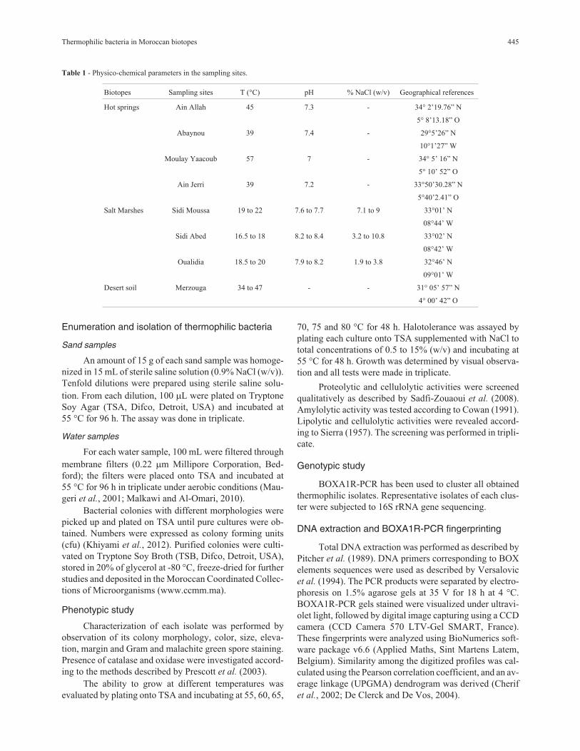
Table 1 - Physico-chemical parameters in the sampling sites.
Biotopes Sampling sites T (°C) pH % NaCl (w/v) Geographical references
Hot springs Ain Allah 45 7.3 - 34° 2’19.76” N
5° 8’13.18” O
Abaynou 39 7.4 - 29°5’26” N
10°1’27” W
Moulay Yaacoub 57 7 - 34° 5’ 16” N
5° 10’ 52” O
Ain Jerri 39 7.2 - 33°50’30.28” N
5°40’2.41” O
Salt Marshes Sidi Moussa 19 to 22 7.6 to 7.7 7.1 to 9 33°01’ N
08°44’ W
Sidi Abed 16.5 to 18 8.2 to 8.4 3.2 to 10.8 33°02’ N
08°42’ W
Oualidia 18.5 to 20 7.9 to 8.2 1.9 to 3.8 32°46’ N
09°01’ W
Desert soil Merzouga 34 to 47 - - 31° 05’ 57” N
4° 00’ 42” O
16S rRNA gene sequencing and phylogeneticanalysis
A set of 144 representative isolates were selected for
16S rRNA gene sequencing using the primers fD1 (5’-
AGAGTTTGATCCTGGCTCAG-3’) and rP2 (5’-TACGG
CTACCTTGTTACGACTT- 3’). Cleaned PCR products
were used as a template for the cycle sequencing reaction
(Berrada et al., 2012).
Forward and reverse sequencing were performed us-
ing Big Dye Terminator version 3.1 cycle sequencing kit
(Applied Biosystems, Foster City, CA) according to the
manufacturer’s instructions. Sequences assembled were
checked and corrected and a preliminary identification was
performed by FASTA search of the NCBI database. For a
more precise identification, the 16S rRNA sequences were
also compared with the prokaryotic small subunit rRNA se-
quence of the Ez-Taxon database (Kim et al., 2012). The
Type and reference strains with highest similarity to the se-
quences of the studied isolates were retrieved and intro-
duced in BioNumerics database. 16S rRNA sequences
were aligned and compared to each other using Bio-
Numerics, and a phylogenetic tree was constructed using
the UPGMA algorithm. Isolates were regarded as belong-
ing to a species when sequence similarity with the species
type strain was at least 99% and to a genus when sequence
similarity with a type strain was at least 97% (Berrada et al.,
2012). The 16S rRNA gene sequences, determined in this
study, have been deposited in the Genbank database under
the accession numbers KF879189 - KF879333.
Results
Sampling sites
Eight sites were prospected in Morocco between
March 2009 and July 2010: 4 hot springs (Ain Allah,
Moulay Yaacoub, Ain Jerri and Abaynou), 3 salt marshes
(Sidi Moussa, Sidi Abed and Oualidia), and the desert soils
of Merzouga (Figure 1). Temperature values were between
34 and 47 °C in the desert soils, while in hot springs they
were between 39 and 57 °C. For salt marshes they were be-
tween 16.5 and 22 °C. In hot springs pH values were be-
tween 7.1 and 7.4 and in salt marshes they were between
7.6 and 8.4. The average salt concentration varied in salt
marshes from 1.9% NaCl (w/v) in Oualidia to 10.8% (w/v)
in Sidi Abed (Table 1).
Thermophilic isolates
The colony forming unit of thermophilic bacteria that
grew on TSA at 55 °C, varied from 0.5 x 10² to 1.2 x 10³ cfu
mL-1 in hot springs, from 1.1 x 10² to1.0 x 103 cfu mL-1 in
salt marshes and was around 5 x 10³ cfu g-1 in desert. A set
of 240 isolates was obtained: 108 isolates from desert
(45%), 79 from hot springs (33%) and 53 from salt marshes
(22%) (Table 2).
Of the 240 isolates, 235 (97.9%) were beige-colored,
3 were white and 2 were yellow on TSA. All of the isolates
were thermophilic (optimal growth up to 55 °C), Gram pos-
itive, rod-shaped, endospore forming and halotolerant
(grew between 0.5% and 10% NaCl (w/v), 225 isolates
(93.75%) were catalase and oxidase positive.
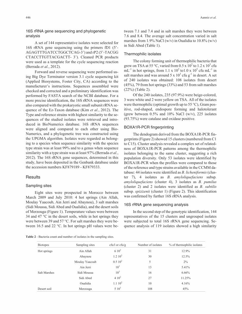
BOXA1R-PCR fingerprinting
The dendogram derived from the BOXA1R-PCR fin-
gerprints (Figure 2) showed 15 clusters (numbered from C1
to C15). Cluster analysis revealed a complex set of related-
ness of BOXA1R-PCR patterns among the thermophilic
isolates belonging to the same cluster, suggesting a rich
population diversity. Only 53 isolates were identified by
BOXA1R-PCR when the profiles were compared to those
of the reference and type strains available in the CCMM da-
tabase: 44 isolates were identified as B. licheniformis (clus-
ter 7), 4 isolates as B. amyloliquefaciens subsp.
amyloliquefaciens (cluster 4), 3 isolates as B. pumilus
(cluster 2) and 2 isolates were identified as B. subtilis
subsp. spizizenii (cluster 1) (Figure 2). This identification
was confirmed by further 16S rRNA analysis.
16S rRNA gene sequencing analysis
In the second step of the genotypic identification, 144
representatives of the 15 clusters and ungrouped isolates
were subjected to total 16S rRNA gene sequencing. Se-
quence analysis of 119 isolates showed a high similarity
446 Aanniz et al.
Table 2 - Bacteria count and number of isolates in the sampling sites.
Biotopes Sampling sites cfu/l or cfu/g Number of isolates % of thermophilic isolates
Hot springs Ain Allah 6 102 31 12.9%
Abaynou 1.2 103 30 12.5%
Moulay Yaacoub 0.5 102 5 2%
Ain Jerri 103 13 5.41%
Salt Marshes Sidi Moussa 103 16 6.66%
Sidi Abed 4 102 27 11.25%
Oualidia 1.1 102 10 4.16%
Desert soil Merzouga 5 103 108 45%

Thermophilic bacteria in Moroccan biotopes 447
Figure 2 - Cluster analysis of BOXA1R-PCR fingerprints and 16S gene sequencing of thermophilic bacteria strains. The dendrogram was generated by
the UPGMA method and Pearson correlation coefficient. LMG strains are reference strains used for identification * = reference strain.
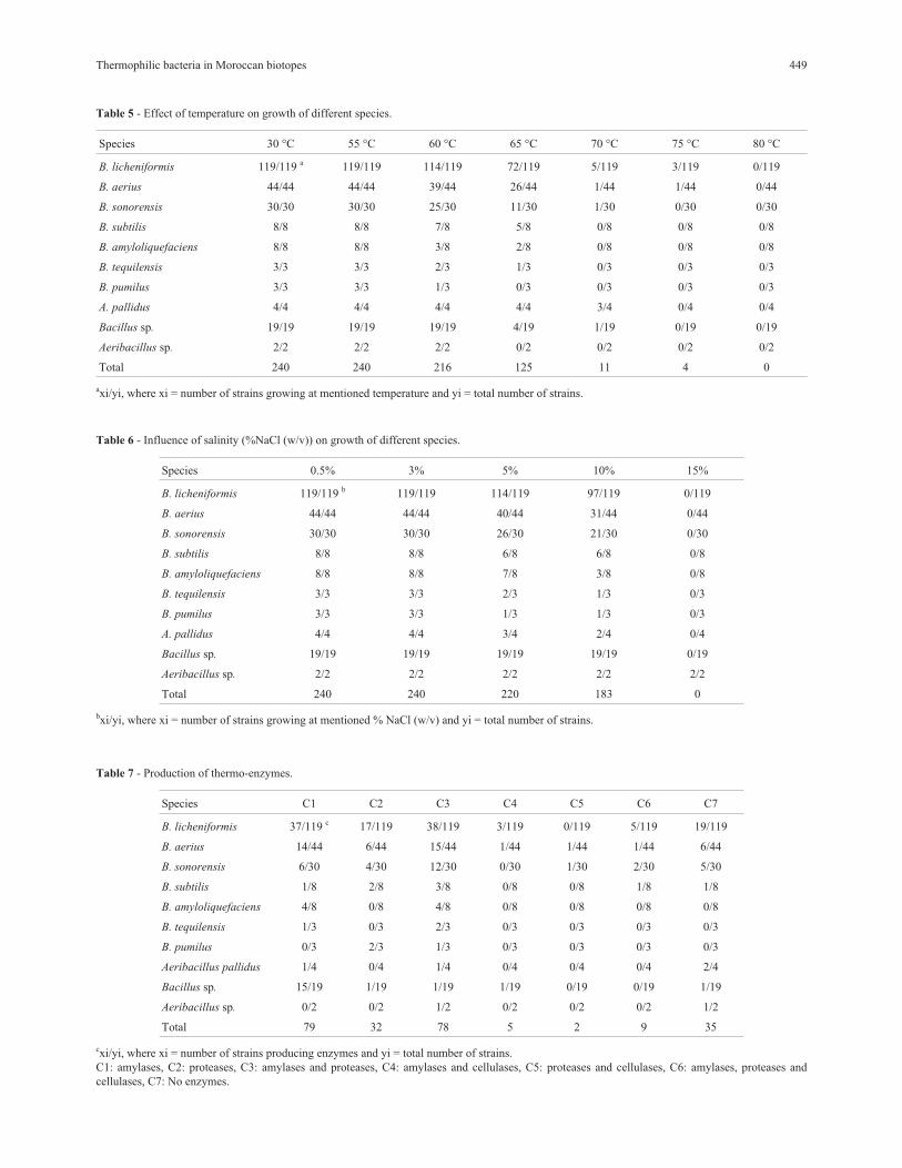
(� 99%) with those of the reference strains available in the
Genbank and Ez-taxon databases. Moreover, 21 isolates
showed moderate sequence similarity (97 to 98.9%) (Ta-
ble 3) indicating that they may be potential new species.
The combination of the BOXA1R-PCR and 16S
rRNA gene sequencing allowed identifying 219 isolates to
the species level. The results revealed a clear domination of
the genus Bacillus represented by 234 isolates i.e. B.
licheniformis, B. aerius, B. sonorensis, B. subtilis (subsp.
spizizenii and subsp. inaquosurum), B. amyloliquefaciens
(subsp. amyloliquefaciens and subsp. plantarum), B.
tequilensis, B. pumilus and Bacillus sp. In addition, 6 iso-
lates belonged to the genus Aeribacillus i.e. A. pallidus and
Aeribacillus sp. (Figure 2, Table 4). The repartition of the
obtained species is presented in Table 4.
Five Bacillus species (B. licheniformis, B. aerius, B.
sonorensis, B. subtilis and B. amyloliquefaciens) were
common between hot springs, salt marshes and desert soils
(Table 4). A total of 8 species were identified in hot springs
and seven in desert soils but B. licheniformis was the most
abundant species (48.1% and 45.37% respectively). Five
species were recovered in salt marshes with B.
licheniformis as a dominant species (60.4%).
Effect of temperature
The number of growing strains is reduced between
55 °C and 65 °C, while it’s drastically reduced at 70 °C and
over. Only 4 isolates tolerate 75 °C and none was able to
grow at 80 °C. The number of strains growing at the differ-
ent temperatures tested are given in Table 5.
Effect of salinity
The number of strains growing is reduced with salt
concentration increased in the medium. All isolates grew at
3% NaCl (w/v), 220 (91.67%) grew at 5% NaCl (w/v), 183
(76.25%) grew at 10% NaCl (w/v), and none at 15% NaCl
(w/v) (Table 6).
Production of thermo-enzymes
Of the 240 strains, 205 (85.42%) produced at least
one extracellular hydrolytic enzyme: 171 strains (71.25%)
produced amylases, 121 strains (50.41%) produced pro-
teases and only 16 strains (6.67%) produced cellulases.
None of the isolates produced lipases. In addition, 9 strains
(3.75%) combined the three tested enzymes and 85 strains
(35.41%) produced two enzymes (Table 7). 35 strains pro-
duced none of the enzymes screened.
448 Aanniz et al.
Table 3 - 16S gene sequencing data for the identified strains to the genus level compared to the type strains.
CCMM N° % of similarity Reference strains
B777, B788, B830, B848 B1014, B1020, B1022, B1023, B1024
and B1025
97.82 to 98.93 Bacillus sonorensis DSM 13779
B797, B819, B1015, B1016, B1017 and B1026 98.42 to 98.99 Bacillus licheniformis ATCC 14580
B1019 and B1021 97.95 to 98.37 Bacillus subtilis subsp. inaquosorum NRRL B-23052
B1018 97.58 Bacillus aerius JCM 13348
B1027 and B1028 97.31 to 97.71 Aeribacillus pallidus ATCC 51176
Table 4 - Biotope, sampling site and distribution of thermophilic bacteria.
Identified species Number of
strains (%)
Distribution per origin
Merzouga Sidi
Abed
Sidi
Moussa
Oualidia Abaynou Ain
Allah
Ain
Jerri
Moulay
Yaacoub
B. licheniformis 119 (49.58%) 49 15 10 7 9 14 10 5
B. aerius 44 (18.33%) 23 5 4 3 7 2
B. sonorensis 30 (12.5%) 11 4 2 8 4 1
B. subtilis 8 (3.33%) 3 2 2 1
B. amyloliquefaciens 8 (3.33%) 4 1 1 2
B. tequilensis 3 (1.25%) 1 1 1
B. pumilus 3 (1.25%) 1 2
A. pallidus 4 (1.66%) 4
Bacillus sp. 19 (7.9%) 15 3 1
Aeribacillus sp. 2 (0.83%) 1 1
Total 240 108 27 16 10 30 31 13 5
Thermophilic bacteria in Moroccan biotopes 449
Table 5 - Effect of temperature on growth of different species.
Species 30 °C 55 °C 60 °C 65 °C 70 °C 75 °C 80 °C
B. licheniformis 119/119 a 119/119 114/119 72/119 5/119 3/119 0/119
B. aerius 44/44 44/44 39/44 26/44 1/44 1/44 0/44
B. sonorensis 30/30 30/30 25/30 11/30 1/30 0/30 0/30
B. subtilis 8/8 8/8 7/8 5/8 0/8 0/8 0/8
B. amyloliquefaciens 8/8 8/8 3/8 2/8 0/8 0/8 0/8
B. tequilensis 3/3 3/3 2/3 1/3 0/3 0/3 0/3
B. pumilus 3/3 3/3 1/3 0/3 0/3 0/3 0/3
A. pallidus 4/4 4/4 4/4 4/4 3/4 0/4 0/4
Bacillus sp. 19/19 19/19 19/19 4/19 1/19 0/19 0/19
Aeribacillus sp. 2/2 2/2 2/2 0/2 0/2 0/2 0/2
Total 240 240 216 125 11 4 0
axi/yi, where xi = number of strains growing at mentioned temperature and yi = total number of strains.
Table 6 - Influence of salinity (%NaCl (w/v)) on growth of different species.
Species 0.5% 3% 5% 10% 15%
B. licheniformis 119/119 b 119/119 114/119 97/119 0/119
B. aerius 44/44 44/44 40/44 31/44 0/44
B. sonorensis 30/30 30/30 26/30 21/30 0/30
B. subtilis 8/8 8/8 6/8 6/8 0/8
B. amyloliquefaciens 8/8 8/8 7/8 3/8 0/8
B. tequilensis 3/3 3/3 2/3 1/3 0/3
B. pumilus 3/3 3/3 1/3 1/3 0/3
A. pallidus 4/4 4/4 3/4 2/4 0/4
Bacillus sp. 19/19 19/19 19/19 19/19 0/19
Aeribacillus sp. 2/2 2/2 2/2 2/2 2/2
Total 240 240 220 183 0
bxi/yi, where xi = number of strains growing at mentioned % NaCl (w/v) and yi = total number of strains.
Table 7 - Production of thermo-enzymes.
Species C1 C2 C3 C4 C5 C6 C7
B. licheniformis 37/119 c 17/119 38/119 3/119 0/119 5/119 19/119
B. aerius 14/44 6/44 15/44 1/44 1/44 1/44 6/44
B. sonorensis 6/30 4/30 12/30 0/30 1/30 2/30 5/30
B. subtilis 1/8 2/8 3/8 0/8 0/8 1/8 1/8
B. amyloliquefaciens 4/8 0/8 4/8 0/8 0/8 0/8 0/8
B. tequilensis 1/3 0/3 2/3 0/3 0/3 0/3 0/3
B. pumilus 0/3 2/3 1/3 0/3 0/3 0/3 0/3
Aeribacillus pallidus 1/4 0/4 1/4 0/4 0/4 0/4 2/4
Bacillus sp. 15/19 1/19 1/19 1/19 0/19 0/19 1/19
Aeribacillus sp. 0/2 0/2 1/2 0/2 0/2 0/2 1/2
Total 79 32 78 5 2 9 35
cxi/yi, where xi = number of strains producing enzymes and yi = total number of strains.
C1: amylases, C2: proteases, C3: amylases and proteases, C4: amylases and cellulases, C5: proteases and cellulases, C6: amylases, proteases and
cellulases, C7: No enzymes.
Discussion
Thermophilic bacteria were present in all samples an-
alyzed. Nevertheless, total counts were very low (50-
5000 cfu mL-1). In a recent study of thermophilic bacteria in
ten Saudi Arabia hot springs, Khiyami et al. (2012) re-
ported the same order of magnitude (170 - 1320 cfu mL-1).
Despite all of the recovered strains can grow at 30 °C, but
their optimal growth temperature is 55 °C (temperature of
isolation). Thus, all strains could be classified as thermo-
philic by Brock (1978), Souza and Martins (2001) and
Cihan et al. (2012). Based on the optimal concentration of
salt and according to Kushner et al. (1985), all the isolated
strains were halotolerants. Morphologic, physiologic and
microscopic characteristics of recovered isolates were con-
sistent with the description of the genus Bacillus, according
to Gordon et al. (1973) and Souza and Martins (2001). High
number of isolates obtained in desert (108 isolates) and hot
springs (79 isolates) could be explained by the hot condi-
tions (temperature ranging from 34 °C to 57 °C) in these
biotopes exerting a pressure for the selection of thermo-
philic flora mostly while in salt marshes characterized by
mesophilic conditions (temperature ranging from 16.5 °C
to 22 °C) only 53 isolates were recovered.
Based on BOXA1R-PCR or/and 16S rRNA sequence
analysis, 97.5% of isolates were assigned to the genus Ba-
cillus and 2.5% to the genus Aeribacillus. Their combina-
tion allowed identifying 219 isolates (91.25%) to the
species level (Figure 2 and Table 4), indicating that these
techniques were not only a powerful tool for identification
of thermophilic bacteria to the species level but also re-
vealed a considerable intra-species diversity (De Clerck
and De Vos, 2004; Adiguzel et al., 2009). Twenty one iso-
lates remained unidentified to the species level; nineteen
were assigned to Bacillus sp. and two to Aeribacillus sp.
This could be an indication for the presence of potential
new thermophilic species. These isolates need to be further
analyzed.
The dendrogram derived from BOXA1R-PCR pro-
files showed a high discriminatory level and revealed 2
sub-groups among B. licheniformis. This is in agreement
with the finding of De Clerck and De Vos (2004). More-
over, Manachini et al. (1998) while studying 182 B.
licheniformis strains reported three distinct groups that
were therefore regarded as genomovars of B. licheniformis.
Our BOXA1R profiles were helpful in distinguishing be-
tween sub-species of B. subtilis (subsp. spizizenii and
subsp. inaquosurum) and also B. amyloliquefaciens (subsp.
amyloliquefaciens and subsp. plantarum). This is in agree-
ment with the grouping reported by Connor et al. (2010).
Physiological behavior of the identified strains
showed several differences between strains of the same
species, such as tolerance to NaCl, temperature and produc-
tion of hydrolytic thermo-enzymes. This finding is in
agreement with previous reports (Burgess et al., 2010;
Adiguzel et al., 2011).
Diversity recovered
The extent of bacterial diversity detected in this study
is not surprising as the majority of bacteria found in the in-
vestigated biotope occur commonly in the environment and
have been described in many different environments stud-
ied elsewhere. Strains of the genus Bacillus are well
adapted to hot and dry environments (Meintanis et al.,
2008; Kawasaki et al., 2012). They have also generally
simple nutritional needs. Therefore, they do not require
specific nutrients for growth and are able to colonize oligo-
trophic niches like salt marshes, hot springs and desert soils
(Cihan et al., 2012; Khiyami et al., 2012). Nevertheless, B.
aerius and B. tequilensis were reported, for the first time, as
thermophilic bacteria in this study.
Malkawi et al. (2010), reported that 97% of the recov-
ered thermophilic isolates belong to the genus Bacillus.
Abou-Shanab (2007) identified 13 thermophilic strains,
from several Jordanian hot springs, belonging to the genus
Bacillus (B. licheniformis and B. pumilus). In another
study, Maugeri et al. (2001), isolated 87 thermophilic, aer-
obic and spore-forming bacteria from Eolian Islands (It-
aly). Most of them were members of Bacillus. Kawasaki et
al. (2012) isolated 12 thermophilic bacteria belonging to
the genus Bacillus, from a saline hot spring in Japan. They
were also relatively halotolerant (grew in a medium with
10% NaCl (w/v)) which is consistent with our finding.
Moreover, Perfumo and Marchant (2010) reported the pres-
ence of thermophilic Aeribacillus strains in samples of dust
collected from Turkey and Greece. The presence of ther-
mophilic strains belonging to A. pallidus from hot springs
was reported by Chamkha et al. (2008) and Savas et al.
(2009).
Bacillus licheniformis was the dominant species (ap-
proximately 50% of total strains), which is in agreement
with those obtained by Derekova et al. (2008). Thermo-
philic strains of B. pumilus, B. amyloliquefaciens and B.
subtilis have been isolated from hot springs, salt lakes, vol-
canic area in Turkey, Bulgaria, Iceland, Jordan, Egypt
(Meintanis et al., 2008; Minana-Galbis et al., 2010 ; Cihan
et al., 2012). Ngoc-Phuc et al. (2007) reported the domi-
nance of B. licheniformis and also the presence of B.
subtilis in a Japanese desert. Lester et al. (2007) reported
the presence of B. subtilis and B. pumilus from extreme arid
Atacama Desert soils. In Morocco, Berrada et al. (2012),
showed that salt marshes in the north of Morocco are colo-
nized by a number of halo-thermophilic bacteria belonging
especially to B. pumilus and B. licheniformis. Bacillus
sonorensis is commonly observed in arid areas, such as the
Sahara, Mojave, Sonoran, and Gobi Deserts (Palmisano et
al., 2001).
Distribution of thermophilic bacteria
The genus Bacillus was isolated from the 8 explored
biotopes and the genus Aeribacillus was obtained only in
the hot spring of Abaynou and desert of Merzouga. The oc-
450 Aanniz et al.
currence of Bacillus strains in all investigated sites could be
explained by the fact that Bacillus has been shown to mi-
grate at extremely high rates, even between continents, and
because Bacillus spores are known to resist to the environ-
mental stress (Connor et al., 2010). B. licheniformis was
isolated from all sites and was also the unique species iden-
tified in the site of Moulay Yaacoub and Oualidia. B.
licheniformis is an ubiquitously occurring spore-forming
bacterium widely distributed as a saprophytic organism in
the environment (Manachini et al., 1998; Rey et al., 2004).
The diversity obtained from the sand of Merzouga
and the hot spring of Abaynou was very close (both located
in a Saharan area) which could be explained by the perpet-
ual movement of sand particles shipped with the wind and
carried microorganisms attached thereto (Perfumo and
Marchant, 2010). The low diversity detected in Moulay
Yaacoub (only 5 strains belonging to B. licheniformis)
could be explained by the high temperature (57 °C) and
mineralization and also the oligotrophic statute of this hot
spring (Lakhdar et al., 2006; Salame et al., 2013). Tekere et
al. (2012), recovered a few bacteria phylotypes at Tshipise
hot spring, where high temperature (58 °C) and high dis-
solved mineral salts occurred, which is consistent with our
finding. It could be also explained by their composition in
gases dominated by nitrogen, methane and carbon dioxide.
Moreover, it has been reported that oxygen is only present
in traces (Lakhdar et al., 2006). This could inhibit the de-
velopment of aerobic flora (Bacillus, Aeribacillus,
Geobacillus) and promotes more probably anaerobic flora
(Clostridium, Anaerobaculum, Thermoanaerobacter,
Thermoanaerobacterium) (Robb et al., 2008).
A total of five identified species belonging to the ge-
nus Bacillus were recovered in all the three salt marshes
while three other isolates remain unidentified. This rela-
tively low diversity could be explained by the negative ef-
fect of high content of salt in these sites resulting in
reducing biodiversity (Ventosa et al., 1998). The three sites
are also situated in a Moroccan region with intense agricul-
tural activity. The use of chemical pesticides could reduce
diversity of all organisms mainly bacteria. The number of
different species obtained from Oualidia was less than
those from Sidi Abed and Sidi Moussa. The site of Oualidia
(located in direct contact with marine water) is influenced
by the quality of marine water with increasing quantities of
heavy metals (Pb, Cd, Zn, Cr, As). Atlantic Moroccan coast
is characterized by lead contamination that occurs around
areas receiving industrial waste outflows, mainly zone situ-
ated near the discharge from phosphate transformation in-
dustries located at Jorf Lasfar (region of El Jadida) which is
very close to the marshes sampling sites (Figure 1) (Ben-
brahim et al., 2006).
Production of thermo-enzymes
It has been reported that thermophilic bacteria,
mainly Bacillus strains, produced high valuable thermo-
enzymes (Meintanis et al., 2008). In accordance with their
saprophytic life style, the secretome of Bacillus strains en-
codes numerous secreted enzymes that hydrolyze polysac-
charides, proteins, lipids and other nutrients (Rey et al.,
2004). Strains obtained in this study produced an assort-
ment of extracellular enzymes that may contribute to nutri-
ent cycling in nature (Rey et al., 2004). In this work,
70.83% and 50.41% of total strains recovered exhibited
high amylolytic and proteolytic activity respectively.
While, only 19 strains (5.41%) produced cellulases. In ad-
dition, 9 strains (3.75%) belonging to B. licheniformis (5),
B. aerius (1), B. sonorensis (2) and B. subtilis (1) produced
all the extracellular hydrolytic enzymes screened except
lipase. This finding indicated also that the tested strains
may have developed genetic and physiological capability
for utilizing available organic matter, via exo-enzymes pro-
duction (Berrada et al., 2012). It could be also explained by
trend of microbial societies toward surviving at low organic
content in such niches and development of adaptable sys-
tem for uptake of any available food (Derekova et al.,
2008).
Conclusion
This is the first investigation of thermophilic bacteria
in Moroccan hot springs, salt marshes and desert. The re-
sults showed a clear domination of thermophilic Bacillus
species mainly B. licheniformis. These strains might be a
source of industrially important enzymes mainly amylases
and proteases.
References
Abou-Shanab RAI (2007) Characterization and 16S rDNA identi-
fication of thermo-tolerant bacteria isolated from hot
springs. J Appl Sci Res 3:994-1000.
Adiguzel A, Ozkan H, Baris O et al. (2009) Identification and
characterization of thermophilic bacteria isolated from hot
springs in Turkey. J Microbiol Meth 79:321-328.
Adiguzel A, Inan K, Sahin F et al. (2011) Molecular diversity of
thermophilic bacteria isolated from Pasinler hot spring
(Erzurum, Turkey). Turk J Biol 35:267-274.
Bar M, von Hardenberg J, Meron E et al. (2002) Modelling the
survival of bacteria in drylands: the advantage of being dor-
mant, Proceedings of the Royal Society B: Biological Sci-
ences 269:937-942.
Benbrahim S, Chafik A, Chfiri R et al. (2006) Survey of the carri-
ers influencing the geographical and temporal distribution of
contamination by heavy metals along the Atlantic Moroccan
coasts: the case of mercury, lead and cadmium. Marine Life
16:37-47.
Berrada I, Willems A, De Vos P et al. (2012) Diversity of cultu-
rable moderately halophilic and halotolerant bacteria in a
marsh and two salterns a protected ecosystem of Lower
Loukkos (Morocco). Afr J Microbiol Res 6:2419-2434.
Bertoldo C, Antranikian G (2002) Starch-hydrolyzing enzymes
from thermophilic Archea and bacteria. Curr Opin Chem
Biol 6:151-160.
Thermophilic bacteria in Moroccan biotopes 451
Brock TD (1978) Thermophilic Microorganisms and Life at High
Temperatures. Springer-verlag, Berlin, Heidelberg and New
York.
Burgess SA, Lindsay D, Flint SH (2010) Thermophilic bacilli and
their importance in dairy processing. Int J Food Microbiol
144:215-225.
Chamkha M, Mnif S, SayadiS (2008) Isolation of a thermophilic
and halophilic tyrosol-degrading Geobacillus from a Tuni-
sian high-temperature oil field. FEMS Microbiol Lett
283:23-29.
Cherif A, Borin S, Rizzi A et al. (2002) Characterization of a re-
petitive element polymorphism-polymerase chain reaction
chromosomal marker that discriminates Bacillus anthracis
from related species. J Appl Microbiol 93:456-462.
Cihan AC, Tekin N, Ozcan B et al. (2012) The genetic diversity of
genus Bacillus and the related genera revealed by 16s rRNA
gene sequences and ARDRA analyses isolated from geo-
thermal regions of Turkey. Braz J Microbiol 43:309-324.
Cihan AC, Cokmus C, Koc M et al. (2014) Anoxybacillus calidus
sp. nov., a thermophilic bacterium isolated from soil near a
thermal power plant. Int J Syst Evol Microbiol 64:211-219.
Connor N, Sikorski J, Rooney AP et al. (2010) Ecology of specia-
tion in the Genus Bacillus. Appl Environ Microbiol
76:1349-1358.
Cowan DA (1991) Industrial enzymes. In: Moses V, Cape RE
(eds) Biotechnology: The Science and the Business. Har-
wood Academic Publishers, United Kingdom, pp 311-340.
De Clerck E, De Vos P (2004) Genotypic diversity among Bacil-
lus licheniformis strains from various sources. FEMS
Microbiol Lett 231:91-98.
Derekova A, Mandeva R, Kambourova M (2008) Phylogenetic
diversity of thermophilic carbohydrate degrading bacilli
from Bulgarian hot springs. World J Microbiol Biot
24:1697-1702.
Gomez J, Steiner W (2004) The biocatalytic potential of extre-
mophiles and extremozymes. Food Technol Biotech
2:223-235.
Gordon RE, Haynes WC, Pang HN (1973) The genus Bacillus
(Agricultural Handbook no. 427). Agricultural research ser-
vice, United State, Department of Agriculture, Government
Printing Office, Washington, D.C.
Hecker M, Völker U (2001) General stress response of Bacillus
subtilis and other bacteria. Adv Microb Physiol 44:35-91.
Kawasaki Y, Aoki M, Makino Y et al. (2012) Characterization of
moderately thermophilic bacteria isolated from saline hot
spring in Japan. Microbiology Indonesia 5:56-60.
Khiyami MA, Serour EA, Shehata MM et al. (2012) Thermo-
aerobic bacteria from geothermal springs in Saudi Arabia.
Afr J Biotechnol 11:4053-4062.
Kim OS, Cho YJ, Lee K et al. (2012) Introducing EzTaxon-e: a
prokaryotic 16S rRNA Gene sequence database with phylo-
types that represent uncultured species. Int J Syst Evol
Microbiol 62:716-721.
Kublanov IV, Perevalova AA, Slobodkina GB et al. (2009) Bio-
diversity of thermophilic prokaryotes with hydrolytic activi-
ties in hot springs of Uzon Caldera, Kamchatka (Russia).
Appl Environ Microbiol 75:286-291.
Kushner DJ (1985) The Halobacteriaceae In: Woese CR, Wolfe
RS (eds) The Bacteria. Academic Press Inc., London,
pp 171-214.
Lakhdar A, Ntarmouchant A, Ribeiro ML et al. (2006) Nouvelle
approche geologique et geodynamique du complexe hydro-
thermal de Moulay Yacoub (Bordure Septentrionale du Sil-
lon Sud Rifain). Comunicacoes Geologicas 93:185-204.
Lester ED, Satomi M, Ponce A (2007) Microflora of extreme arid
Atacama Desert soils. Soil Biol Biochem 39:704-708.
Malkawi HI, Al-Omari MN (2010) Culture-dependent and cul-
ture-independent approaches to study the bacterial and ar-
chaeal diversity from Jordanian hot springs. Afr J Microbiol
Res 4:923-932.
Manachini PL, Fortina MG, Levati L et al. (1998) Contribution to
phenotypic and genotypic characterization of Bacillus
licheniformis and description of new genomovars. Syst Appl
Microbiol 21:520-529.
Maugeri T, Gugliandolo C, Caccamo D et al. (2001) A polyphasic
taxonomic study of thermophilic bacilli from shallow, ma-
rine vents. Syst Appl Microbiol 24:572-587.
Meintanis C, Chalkou KI, Kormas KA et al. (2008) Application of
rpoB sequence similarity analysis, REP-PCR and BOX-
PCR for the differentiation of species within the genus
Geobacillus. Lett Appl Microbiol 46:395-401.
Minana-Galbis D, Pinzon DL, Loren JG et al. (2010) Reclassifi-
cation of Geobacillus pallidus (Scholz et al., 1988) Banat et
al., 2004 as Aeribacillus pallidus gen. nov., comb. nov. Int J
Syst Evol Microbiol 60:1600-1604.
Ngoc-Phuc H, Fumihisa K, Yasunobu I et al. (2007) Detailed
identification of desert-originated bacteria carried by Asian
dust storms to Japan. Aerobiologia 23:291-298.
Palmisano MM, Nakamura LK, Duncan KE et al. (2001) Bacillus
sonorensis sp. nov., a close relative of Bacillus
licheniformis, isolated from soil in the Sonoran Desert, Ari-
zona. Int J Syst Evol Microbiol 51:1671-1679.
Perfumo A, Marchant R (2010) Global transport of thermophilic
bacteria in atmospheric dust. Environ Microbiol Rep 2:333-
339.
Pitcher DG, Saunders NA, Owen RJ (1989) Rapid extraction of
bacterial genomic DNA with guanidiumthiocyanate. Lett
Appl Microbiol 8:151-156.
Prescott LM, Harley GP, Klein DE (1993) Microbiology. 2nd ed.
W. Brown publishers, Dubuque.
Rey MW, Ramaiya P, Nelson BA et al. (2004) Complete genome
sequence of the industrial bacterium Bacillus licheniformis
and comparisons with closely related Bacillus species. Ge-
nome Biol 5:1-12.
Robb F, Antranikian G, Grogan D et al. (2008) Thermophiles, Bi-
ology and Technology at High Temperatures. Boca Raton;
CRC Press, London, New York.
Sadfi-Zouaoui N, Essghaier B, Hajlaoui MR et al. (2008) Ability
of moderately halophilic bacteria to control grey mould dis-
ease on tomato fruits. J Phytopathol 156:42-52.
Salame B, Bahhou J, Bennani B et al. (2013) Qualité bactério-
logique et physico-chimique des eaux thermales d’Ain Allah
région de Fès (Maroc). Science Lib Editions Mersenne
5:130505.
Savas S, Adiguzel A, Inan K et al. (2009) Molecular characteriza-
tion of thermophilic bacteria isolated from Van City Ercis
Town Hasanabdal hot spring. Rom Biotech Lett 14:4445-
4454.
Sierra GA (1957) Simple method for the detection of lipolytic ac-
tivity of microorganisms and some observations on the in-
452 Aanniz et al.
fluence of the contact between cells and fatty substrates.
Anton Leew 23:15-22.
Singh AK, Tripathi BM, Sahay H et al. (2010) Biochemical and
molecular characterization of thermo-alkalitolerant xyla-
nase producing bacteria from thermal springs of Manikaran,
Indian J Microbiol 50:S2-S9.
Skoog DA, West D, Holler FJ (1996) Fundamentals of Analytical
Chemistry. 7th ed, Saunders College Publishing: New York.
Souza AN, Martins MLL (2001) Isolation, properties and kinetics
of growth of a thermophilic Bacillus. Braz J Microbiol
32:1517-8382.
Tekere M, Prinsloo A, Olivier J et al. (2012) An evaluation of the
bacterial diversity at Tshipise, Mphephu and Sagole hot wa-
ter springs, Limpopo Province, South Africa. Afr J Micro-
biol Res 6:4993-5004.
Ventosa A, Nieto JJ, Oren A (1998) Biology of moderately
halophilic aerobic bacteria. Microbiol Mol Biol R 62:504-
544.
Versalovic J, Schneider M, De Bruijn FJ et al. (1994) Genomic
fingerprinting of bacteria using repetitive sequence-based
polymerase chain reaction. Methods Mol Cell B 5:25-40.
YonedaY, Yoshida T, Yasuda H et al. (2013) A novel thermo-
philic, hydrogenogenic, and carboxydotrophic bacterium
Calderihabitans maritimus gen. nov., sp. nov. from a marine
sediment core of an undersea caldera. Int J Syst Evol
Microbiol ijs.0.050468-0.
Associate Editor: Raquel Silva Peixoto
All the content of the journal, except where otherwise noted, is licensed under a
Creative Commons License CC BY-NC.
Thermophilic bacteria in Moroccan biotopes 453