scuola di dottorato in scienze della terra dottorato di ricerca in … · 2014-04-30 · halepensis...
TRANSCRIPT
Università degli Studi di Napoli “Federico II”
Scuola di Dottorato in Scienze della Terra
Dottorato di Ricerca in Analisi dei Sistemi Ambientali
“XXIV Ciclo”
Tesi preparata in cotutela con:
Université Claude Bernard Lyon 1
Ecole doctorale E2M2 Evolution Ecosystèmes Microbiologie Modélisation
Thèse de Doctorat en Paléonvironnements et Évolution
Thèse en Biologie et Science de la Terre
The cuticle micromorphology of extant and fossil plants as indicator of
environmental conditions.
A pioneer study on the influence of volcanic gases on the cuticle structure in
extant plants
Bartiromo Antonello
2011
RELATORI/DIRECTEURS DE RECHERCHE:
Dott.ssa Guerriero Giulia Maître de conférences Guignard Gaëtan
COORDINATORE DEL DOTTORATO: Prof. Barattolo Filippo
CORRELATORE: Dott.ssa Barone Lumaga Maria Rosaria
COMMISSIONE/JURY: Raschi Antonio, Prat Daniel, Guignard Gaëtan, Guida Marco
DATA D’ESAME/DATE DE SOUTENANCE: 14/02/2012
The cuticle micromorphology of extant and fossil plants as indicator of
environmental conditions.
A pioneer study on the influence of volcanic gases on the cuticle
structure in extant plants
Antonello Bartiromo
Alla mia Terra, che non merita tutto questo …
Il nostro destino è inestricabilmente legato a quello di tutte le specie e di tutti gli ecosistemi della Terra. Niles Eldredge, 2000
The cuticle micromorphology of extant and fossil plants as indicator of environmental conditions. A pioneer study on the influence of volcanic gases on the cuticle structure in extant plants
4
INDEX
Riassunto 7
Résumé 9
Abstract 11
Keywords 13
Laboratories 13
General Introduction 14
CHAP. I Influence of volcanic gases on the epidermis of Pinus halepensis
Mill. in Campi Flegrei, Southern Italy: A possible tool detecting
volcanism in present and past floras
17
1.1. Introduction 17
1.2. Material and methods 20
1.3. Results 24
1.3.1. Sulphur measures 24
1.3.2. Scanning electron microscopy observations 24
1.3.3. Transmission electron microscopy observations 27
1.4. Discussion 30
1.4.1. Environmental response of Pinus halepensis to volcanism 31
1.4.1.1. Epicuticular and epistomatal wax 31
Stomatal aperture 31
Volcanic toxic compounds and wax alterations 33
Other sulphur considerations 35
Wettability of leaf surface 36
Microenvironment 37
1.4.1.2. Cuticular membrane (CM) + cell wall (CW) 38
Pinus halepensis cuticle type 38
CM + CW thickness 38
CM + CW development 40
The cuticle micromorphology of extant and fossil plants as indicator of environmental conditions. A pioneer study on the influence of volcanic gases on the cuticle structure in extant plants
5
CM and CW ageing 40
Calcium-oxalate deposits 41
1.4.2. Potential application for extant and fossil material 42
CHAP. II
The cuticle micromorphology of in situ Erica arborea L.
exposed to long-term volcanic gases in Phlegrean Fields,
Campania, Italy
46
2.1. Introduction 46
2.2. Material and methods 48
2.2.1. Plant material and sites description 48
2.2.2. Gas vent 49
2.2.3. SEM, TEM and EDS preparations 50
2.2.4. Gas concentration measurements in air and soil 51
2.2.5. Statistical analysis 52
2.3. Results 52
2.3.1. Energy diffractive X-ray analysis with SEM 52
2.3.2. Scanning electron microscopy observations 53
2.3.3. Trasmission electron microscope observations 55
2.4. Discussion 58
2.4.1. Chemical and SEM considerations 58
2.4.2. TEM considerations 60
CHAP. III An Early Cretaceous flora from Cusano Mutri, Benevento,
southern Italy 63
3.1. Introduction 63
3.2. Geological setting 63
3.2.1. Stratigraphy 65
3.3. Material and methods 68
3.4. Systematic palaeontology 69
3.5. Taphonomic and palaeoecological remarks 88
3.5.1. Taphonomy 88
The cuticle micromorphology of extant and fossil plants as indicator of environmental conditions. A pioneer study on the influence of volcanic gases on the cuticle structure in extant plants
6
3.5.2. Palaeoecology of the Cusano Mutri sedimentary basin 89
3.5.3. Xeromorphic adaptations of the plants 90
3.5.4. Palaeoclimate and floral comparison 91
CHAP. IV
Plant remains from the Early Cretaceous Fossil-Lagerstätte of
Pietraroja, Southern Italy, Benevento 93
4.4.1. Introduction 93
4.2. Geological setting 94
4.3. Material and methods 96
4.4. Systematic Palaeontology 97
4.5. Taphonomic and palaeoecological implications 111
4.5.1. Taphonomy 111
4.5.2. Palaeoecology of the sedimentary basin 114
4.5.3. Palaeoclimate and comparison with other Albian florae 114
General Conclusions 117
Acknowledgments / Ringraziamenti 120
References 121
Photos 155
The cuticle micromorphology of extant and fossil plants as indicator of environmental conditions. A pioneer study on the influence of volcanic gases on the cuticle structure in extant plants
7
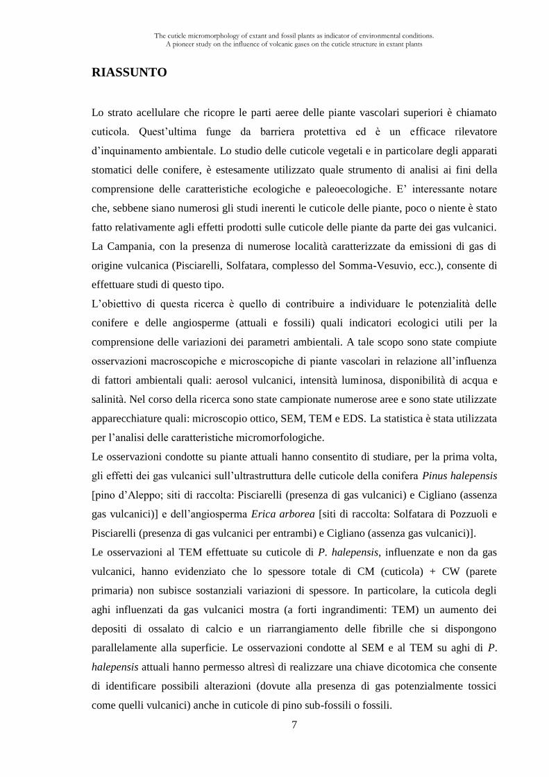
RIASSUNTO
Lo strato acellulare che ricopre le parti aeree delle piante vascolari superiori è chiamato
cuticola. Quest’ultima funge da barriera protettiva ed è un efficace rilevatore
d’inquinamento ambientale. Lo studio delle cuticole vegetali e in particolare degli apparati
stomatici delle conifere, è estesamente utilizzato quale strumento di analisi ai fini della
comprensione delle caratteristiche ecologiche e paleoecologiche. E’ interessante notare
che, sebbene siano numerosi gli studi inerenti le cuticole delle piante, poco o niente è stato
fatto relativamente agli effetti prodotti sulle cuticole delle piante da parte dei gas vulcanici.
La Campania, con la presenza di numerose località caratterizzate da emissioni di gas di
origine vulcanica (Pisciarelli, Solfatara, complesso del Somma-Vesuvio, ecc.), consente di
effettuare studi di questo tipo.
L’obiettivo di questa ricerca è quello di contribuire a individuare le potenzialità delle
conifere e delle angiosperme (attuali e fossili) quali indicatori ecologici utili per la
comprensione delle variazioni dei parametri ambientali. A tale scopo sono state compiute
osservazioni macroscopiche e microscopiche di piante vascolari in relazione all’influenza
di fattori ambientali quali: aerosol vulcanici, intensità luminosa, disponibilità di acqua e
salinità. Nel corso della ricerca sono state campionate numerose aree e sono state utilizzate
apparecchiature quali: microscopio ottico, SEM, TEM e EDS. La statistica è stata utilizzata
per l’analisi delle caratteristiche micromorfologiche.
Le osservazioni condotte su piante attuali hanno consentito di studiare, per la prima volta,
gli effetti dei gas vulcanici sull’ultrastruttura delle cuticole della conifera Pinus halepensis
[pino d’Aleppo; siti di raccolta: Pisciarelli (presenza di gas vulcanici) e Cigliano (assenza
gas vulcanici)] e dell’angiosperma Erica arborea [siti di raccolta: Solfatara di Pozzuoli e
Pisciarelli (presenza di gas vulcanici per entrambi) e Cigliano (assenza gas vulcanici)].
Le osservazioni al TEM effettuate su cuticole di P. halepensis, influenzate e non da gas
vulcanici, hanno evidenziato che lo spessore totale di CM (cuticola) + CW (parete
primaria) non subisce sostanziali variazioni di spessore. In particolare, la cuticola degli
aghi influenzati da gas vulcanici mostra (a forti ingrandimenti: TEM) un aumento dei
depositi di ossalato di calcio e un riarrangiamento delle fibrille che si dispongono
parallelamente alla superficie. Le osservazioni condotte al SEM e al TEM su aghi di P.
halepensis attuali hanno permesso altresì di realizzare una chiave dicotomica che consente
di identificare possibili alterazioni (dovute alla presenza di gas potenzialmente tossici
come quelli vulcanici) anche in cuticole di pino sub-fossili o fossili.
The cuticle micromorphology of extant and fossil plants as indicator of environmental conditions. A pioneer study on the influence of volcanic gases on the cuticle structure in extant plants
8
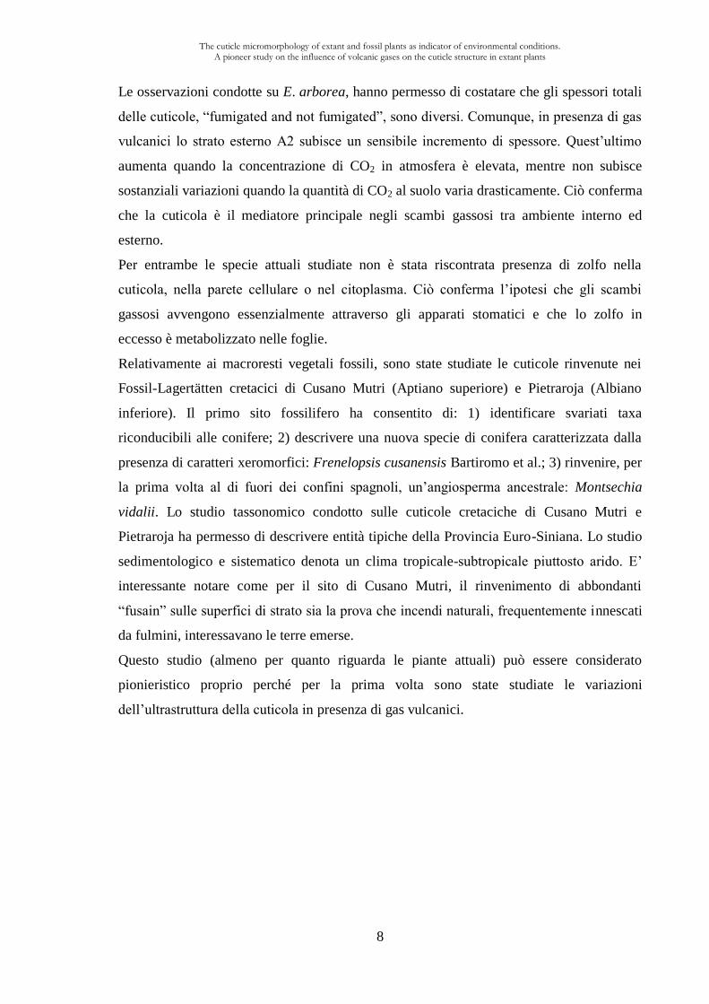
Le osservazioni condotte su E. arborea, hanno permesso di costatare che gli spessori totali
delle cuticole, “fumigated and not fumigated”, sono diversi. Comunque, in presenza di gas
vulcanici lo strato esterno A2 subisce un sensibile incremento di spessore. Quest’ultimo
aumenta quando la concentrazione di CO2 in atmosfera è elevata, mentre non subisce
sostanziali variazioni quando la quantità di CO2 al suolo varia drasticamente. Ciò conferma
che la cuticola è il mediatore principale negli scambi gassosi tra ambiente interno ed
esterno.
Per entrambe le specie attuali studiate non è stata riscontrata presenza di zolfo nella
cuticola, nella parete cellulare o nel citoplasma. Ciò conferma l’ipotesi che gli scambi
gassosi avvengono essenzialmente attraverso gli apparati stomatici e che lo zolfo in
eccesso è metabolizzato nelle foglie.
Relativamente ai macroresti vegetali fossili, sono state studiate le cuticole rinvenute nei
Fossil-Lagertätten cretacici di Cusano Mutri (Aptiano superiore) e Pietraroja (Albiano
inferiore). Il primo sito fossilifero ha consentito di: 1) identificare svariati taxa
riconducibili alle conifere; 2) descrivere una nuova specie di conifera caratterizzata dalla
presenza di caratteri xeromorfici: Frenelopsis cusanensis Bartiromo et al.; 3) rinvenire, per
la prima volta al di fuori dei confini spagnoli, un’angiosperma ancestrale: Montsechia
vidalii. Lo studio tassonomico condotto sulle cuticole cretaciche di Cusano Mutri e
Pietraroja ha permesso di descrivere entità tipiche della Provincia Euro-Siniana. Lo studio
sedimentologico e sistematico denota un clima tropicale-subtropicale piuttosto arido. E’
interessante notare come per il sito di Cusano Mutri, il rinvenimento di abbondanti
“fusain” sulle superfici di strato sia la prova che incendi naturali, frequentemente innescati
da fulmini, interessavano le terre emerse.
Questo studio (almeno per quanto riguarda le piante attuali) può essere considerato
pionieristico proprio perché per la prima volta sono state studiate le variazioni
dell’ultrastruttura della cuticola in presenza di gas vulcanici.
The cuticle micromorphology of extant and fossil plants as indicator of environmental conditions. A pioneer study on the influence of volcanic gases on the cuticle structure in extant plants
9
RÉSUMÉ
La couche qui recouvre les parties aériennes des plantes vasculaires supérieures est appelée
cuticule. Cette dernière agit comme une barrière protectrice et est un détecteur efficace de
la pollution de l'environnement. L’étude de la cuticule des plantes, en particulier des
appareils stomatiques des conifères, est largement utilisée comme un outil d’analyse pour
comprendre les caractéristiques écologiques et paléoécologiques. Il est intéressant de noter
que, bien que les études sur la cuticule des plantes soient nombreuses, peu ou rien n’a été
réalisé sur les effets sur la cuticule des plantes par les gaz volcaniques. La Campanie, avec
ses nombreux endroits caractérisés par des émissions de gaz d'origine volcanique
(Pisciarelli, Solfatara, complexe du Somma-Vésuve), permet d’effectuer ce type d’études.
L’objectif de cette recherche est donc de contribuer à individualiser les potentialités des
conifères et angiospermes (actuelles et fossiles) comme indicateurs écologiques dans la
reconnaissance des variations de paramètres environnementaux. Pour cela, des
observations macroscopiques et microscopiques de plantes vasculaires ont été effectuées
par rapport à l'influence des facteurs environnementaux tels les aérosols volcaniques,
l'intensité de la lumière, disponibilité d’eau et la salinité. Au cours de la recherche un
certain nombre de localités ont été échantillonnées et on a utilisé des équipements comme
le microscope optique, le MEB, le MET et l’EDS. La statistique a été largement mise à
contribution, avec l’intervalle de confiance portant sur 30 mesures.
Les observations effectuées sur les plantes actuelles ont permis d’étudier, pour la première
fois, les effets des gaz volcaniques sur l’ultrastructure des cuticules du conifère Pinus
halepensis [le pin d’Alep, sites de récolte: Pisciarelli (fumigé) et Cigliano (non fumigé)] et
de l’angiosperme Erica arborea [la bruyère arborescente, sites de récolte: Solfatara et
Pisciarelli (fumigé) et Cigliano (non fumigé)].
Les observations conduites au TEM sur les cuticules de P. halepensis, influencé et non
influencé par les gaz volcaniques, ont montré que l'épaisseur totale de la CM (cuticule) +
CW (paroi pectocellulosique) ne subit pas de variations significatives d'épaisseur. En
particulier, la cuticule des aiguilles influencée par les gaz volcaniques montre (à fort
grossissement TEM) une accumulation d’oxalate de calcium ainsi qu’un réarrangement des
fibrilles disposées parallèlement à la surface. Les observations SEM et TEM sur des
aiguilles de P. halepensis actuelles ont permis également de réaliser une clé dichotomique
permettant d’identifier les altérations possibles (dues à la présence de gaz potentiellement
toxiques comme les gaz volcaniques) des cuticules de pins sub-fossiles ou fossiles.
The cuticle micromorphology of extant and fossil plants as indicator of environmental conditions. A pioneer study on the influence of volcanic gases on the cuticle structure in extant plants
10
Les observations conduites sur E. arborea ont permis de constater que les épaisseurs
totales des cuticules, influencées ou non par les gaz, sont significativement différentes. En
présence de gaz volcaniques la couche externe A2 subit un sensible accroissement
d’épaisseur. Cette dernière augmente quand la concentration en CO2 en atmosphère est
élevée, alors qu’elle ne subit pas de variations substantielles quand la quantité de CO2 au
sol varie de manière drastique. Ceci démontre que la cuticule est le médiateur principal
dans les échanges entre l’environnement interne et externe.
Grâce à des analyses EDS, pour les deux espèces actuelles étudiés il n’a pas été trouvé de
présence de soufre dans la cuticule, dans la paroi cellulaire ou dans le cytoplasme. Ceci
confirme que la cuticule est le principale médiateur des échanges gazeux entre
l’environnement interne et externe.
Par rapport aux macro-restes végétaux fossiles, les cuticules du Fossil-Lagertätten du
Crétacé de Cusano Mutri (Aptien sup.) et de Pietraroja (Albien inf.) ont été étudiées. Le
première site fossilifère a permis 1) d’identifier plusieurs taxa appartenant aux conifères; 2)
de décrire une nouvelle espèce de conifère caractérisée par la présence de caractères
xéromorphiques: Frenelopsis cusanensis Bartiromo et al.; 3) de trouver, pour la première
fois à l’extérieur de l’Espagne, une angiosperme ancestrale: Montsechia vidalii. L’étude
taxonomique conduite sur des cuticules du Crétacé de Cusano Mutri et Pietraroja a permis
de décrire entités typiques de la Province Euro-Sinienne. L’étude sédimentologique et
systématique montre une climat tropical-subtropical plutôt sec. Il est intéressant de noter,
comme pour le site de Cusano Mutri, la présence des abondants fusains sur les surfaces des
couches rocheuses, montrant les incendies naturels fréquemment amorcés par des éclairs
intéressant les terres émergées.
Cette étude (au moins en ce qui concerne les plantes actuelles) peut être considérée comme
pionnière car, pour la première fois, a été étudié les variations de l'ultrastructure de la
cuticule en présence de gaz volcaniques.
The cuticle micromorphology of extant and fossil plants as indicator of environmental conditions. A pioneer study on the influence of volcanic gases on the cuticle structure in extant plants
11
ABSTRACT
The leaves of many tracheophytes are covered with a cuticle, an extracellular membrane
covering aerial organs of plants. The gas exchanges between the plant and the surrounding
atmosphere are mediated by the cuticle; its acts as the main barrier to air pollutants. The
study of the plant cuticle, in particular the stomatal apparatuses of conifer, is largely used
as a tool analysis revealing ecological and paleoecological features. It is worth noting that
little is known about the long-term response of micromorphology of natural vegetation to
volcanic toxic gases. Fortunately, Campania Region with its numerous volcanic localities
(Pisciarelli, Solfatara, complexe du Somma-Vésuve) represents a natural laboratory
allowing experiments involving plant-volcano interactions.
The object of this research is to study the conifer and angiosperms potentialities (extant
and fossil) as ecological indicators useful in the identification of the environmental
parameters variations. That is why, macroscopical and microscopical observations in
vascular plants in relation to various environmental factors (volcanic gases, light intensity,
water availability and salinity), have been analysed. A number of localities have been
sampled and SEM, TEM and EDS equipments have been used together with statistic.
Observations made on extant plants allowed for the first time, the study of the effects of
volcanic gases on the cuticle ultrastructure of Pinus halepensis [Aleppo pine; Pisciarelli
(fumigated) and Cigliano (not fumigated) localities] and Erica arborea [tree heather;
Solfatara, Pisciarelli (fumigated) and Cigliano (not fumigated) localities].
TEM observations on P. halepensis cuticles fumigated or not by volcanic gases revealed
insignificant thickness variations of the cell wall plus cuticle among current- and first-year-
old needles of both fumigated and not fumigated trees. In particular, the needle cuticles
experiencing chronic fumigation display (TEM) a calcium oxalate accumulation.
Moreover, in respect to the cell surface, fibrils are parallel disposed. SEM and TEM
observations allowed an identification key enabling distinction between not fumigated and
fumigated material with 9 characters, providing a good tool detecting the influence of
volcanism for extant and fossil plants.
In specimens of E. arborea fumigated or not by volcanic gases, the total thickness of
cuticles varies significantly. In plant experiencing chronic fumigation the A2 layer records
an increase of its thickness. Within three localities, a good correlation between the
atmospheric CO2 concentration and the thickness variation of A2 layer has been found.
The cuticle micromorphology of extant and fossil plants as indicator of environmental conditions. A pioneer study on the influence of volcanic gases on the cuticle structure in extant plants
12
This fact confirms that the cuticle is the main mediator between the plant and the
atmosphere.
As for fossil plants, the cuticles of Cretaceous Fossil-Lagertätten of Cusano Mutri (Late
Aptian) and Pietraroja (Lower Albian) have been studied. In the former: 1) numerous taxa
belonging to conifers have been identified; 2) the new species Frenelopsis cusanensis
Bartiromo et al. bearing xeromorphic features has been described; 3) the occurrence of
Montsechia vidalii is recorded for the first time outside of Spain. Taxonomical studies
carried out on Cretaceous cuticles from Cusano Mutri and Pietraroja allowed the
description of typical Euro-Sinian fossil plants. Sedimentological and taxonomical studies
suggest semi-arid or arid conditions in a subtropical or tropical climate. It is worth noting
as for Cusano Mutri locality, evidence of wildfire (fusain) suggests a periodic combination
of arid periods, high temperatures and lightning strikes.
This study (at least for extant plants) can be considered pioneering, because, for the first
time, the relationships between cuticle ultrastructure variations and volcanic gases have
been studied.
The cuticle micromorphology of extant and fossil plants as indicator of environmental conditions. A pioneer study on the influence of volcanic gases on the cuticle structure in extant plants
13
Parole chiave:
Campi Flegrei, gas vulcanici, Pinus halepensis, Erica arborea, epidermide, ultrastruttura
della cuticola, idrogeno solforato (H2S), piante fossili, Cretacico inferiore, Sud Italia,
Cusano Mutri, Pietraroja, Cheirolepidiaceae, Frenelopsis cusanensis sp. nov.,
paleoecologia, Provincia Euro-Siniana.
Mots clés:
Campi Flegrei, gaz volcaniques, Pinus halepensis, Erica arborea, épiderme, ultrastructure
de la cuticule, hydrogéné sulfuré (H2S), plant fossiles, Crétacée inferieur, Italie du Sud,
Cusano Mutri, Pietraroja, Cheirolepidiaceae, Frenelopsis cusanensis sp. nov.,
paléoécologie, Province Euro-Sinien.
Keywords:
Campi Flegrei, volcanic gases, Pinus halepensis, Erica arborea, epidermis, cuticle
ultrastructure, hydrogen sulphide (H2S), fossil plants, Early Cretaceous, Southern Italy,
Cusano Mutri, Pietraroja, Cheirolepidiaceae, Frenelopsis cusanensis sp. nov.,
Palaeoecology, Euro-Sinian Province.
Laboratori dove la tesi è stata preparata/L'intitulé et l'adresse de l'unité ou du
laboratoire où la thèse a été préparée:
Dipartimento di Scienze della Terra, Università degli Studi di Napoli “Federico II”, Largo
San Marcellino, 10, 80138 Napoli, Italia.
Université de Lyon, F-69622, Lyon, France; Université Lyon 1, Villeurbanne; CNRS,
UMR 5276 Laboratoire de Géologie de Lyon, Herbiers de l’Université Claude-Bernard
Lyon 1.
Dipartimento delle Scienze Biologiche, Università degli Studi di Napoli “Federico II”, Via
Mezzocannone, 8, 80134 Napoli, Italia.
Orto Botanico, Università degli Studi di Napoli “Federico II”, Via Foria, 239, 80139
Napoli, Italia.
The cuticle micromorphology of extant and fossil plants as indicator of environmental conditions. A pioneer study on the influence of volcanic gases on the cuticle structure in extant plants
14
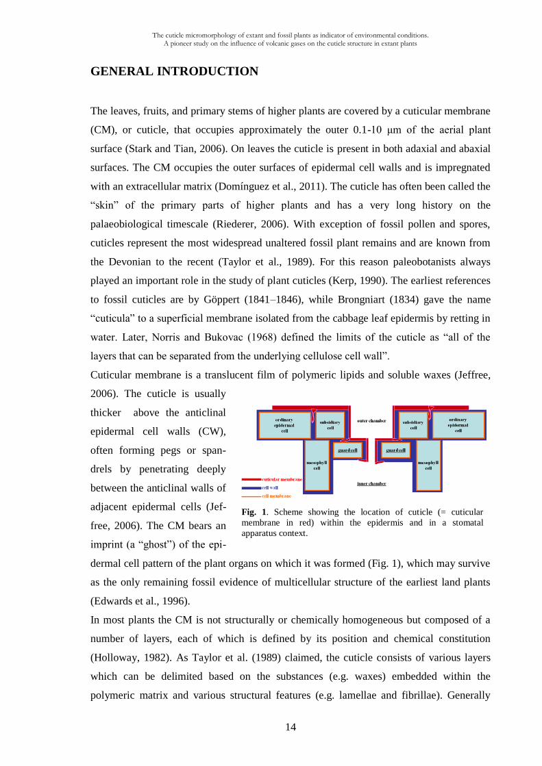
Fig. 1. Scheme showing the location of cuticle (= cuticular
membrane in red) within the epidermis and in a stomatal
apparatus context.
GENERAL INTRODUCTION
The leaves, fruits, and primary stems of higher plants are covered by a cuticular membrane
(CM), or cuticle, that occupies approximately the outer 0.1-10 μm of the aerial plant
surface (Stark and Tian, 2006). On leaves the cuticle is present in both adaxial and abaxial
surfaces. The CM occupies the outer surfaces of epidermal cell walls and is impregnated
with an extracellular matrix (Domínguez et al., 2011). The cuticle has often been called the
“skin” of the primary parts of higher plants and has a very long history on the
palaeobiological timescale (Riederer, 2006). With exception of fossil pollen and spores,
cuticles represent the most widespread unaltered fossil plant remains and are known from
the Devonian to the recent (Taylor et al., 1989). For this reason paleobotanists always
played an important role in the study of plant cuticles (Kerp, 1990). The earliest references
to fossil cuticles are by Göppert (1841–1846), while Brongniart (1834) gave the name
“cuticula” to a superficial membrane isolated from the cabbage leaf epidermis by retting in
water. Later, Norris and Bukovac (1968) defined the limits of the cuticle as “all of the
layers that can be separated from the underlying cellulose cell wall”.
Cuticular membrane is a translucent film of polymeric lipids and soluble waxes (Jeffree,
2006). The cuticle is usually
thicker above the anticlinal
epidermal cell walls (CW),
often forming pegs or span-
drels by penetrating deeply
between the anticlinal walls of
adjacent epidermal cells (Jef-
free, 2006). The CM bears an
imprint (a “ghost”) of the epi-
dermal cell pattern of the plant organs on which it was formed (Fig. 1), which may survive
as the only remaining fossil evidence of multicellular structure of the earliest land plants
(Edwards et al., 1996).
In most plants the CM is not structurally or chemically homogeneous but composed of a
number of layers, each of which is defined by its position and chemical constitution
(Holloway, 1982). As Taylor et al. (1989) claimed, the cuticle consists of various layers
which can be delimited based on the substances (e.g. waxes) embedded within the
polymeric matrix and various structural features (e.g. lamellae and fibrillae). Generally
The cuticle micromorphology of extant and fossil plants as indicator of environmental conditions. A pioneer study on the influence of volcanic gases on the cuticle structure in extant plants
15
speaking, TEM revealed that the cuticle of the cells of the upper epidermis is made of an
outer stratum named cuticle proper (CP) and an inner one called cuticular layer (CL).
According to the international terminology, the former (CP) can be constituted by an A1
(lamellate) or/and an A2 (uniformly electron dense with areas of lacunae) layers; the latter
(CL) is constituted by a reticulate (B = B1) layer (see: Holloway, 1982; Archangelsky,
1991; Jeffree, 2006), in some rare cases an innermost B2 granular layer is present.
The outer surface of cuticle can be coated with epicuticular waxes which confer water
repellency (Adam, 1963). Cuticular wax plays pivotal physiological and ecological roles in
the interactions between plants and their abiotic and biotic environments, respectively
(Jetter et al., 2006).
The cuticle performs numerous functions such as: transpiration control; control of loss and
uptake of polar solutes; control of the exchange of gases and vapours; interface for biotic
interactions and so on. However, the primary function of the cuticle is a permeability
barrier against water vapour loss from tissues (Schreiber et al., 1996). For these and other
reasons (see: Huttunen, 1984; Jeffree, 1986; Archangelsky et al., 1995; Garrec, 1996) the
cuticle can be considered as an “external skeleton” as it represents the interface between
the plant and the atmosphere (McElwain and Chaloner, 1996). As cuticle forms the
interface between plants and atmospheric environment, it is the first point of contact
between plants and air pollutants and it presents an effective barrier to pollutant entry.
During recent years considerable progress has been made for investigating this more
external part of plants and its relations with external environment (Holloway, 1982; Hill
and Dilcher, 1990; McElwain and Chaloner, 1996; Newrath, 2006; Jeffree, 2006; Shepherd
and Griffiths, 2006). However, few studies have been carried out in the study of plant-
volcanic gases interaction.
This doctoral thesis represents a contribution to the study of extant and fossil plant cuticles
by means of optical and electronic (SEM and TEM) microscopy as well as EDS analyses.
In particular, this research is pioneer in the study of ultrasctructure of plant cuticle
submitted to the influence of volcanic gases representing the first part of this thesis.
Similar researches are highly advisable in the Campania Region (Ischia island, Phlegrean
Fields, Roccamonfina and Somma–Vesuvio complex) where volcanic and not volcanic gas
emissions, give great opportunities in the analysis of plant–volcano interaction. However,
Campania Region represents also a “virgin reservoir” for the study of Cretaceous plants
that exactly make the object of the second part of this thesis (Fig. 2).
The cuticle micromorphology of extant and fossil plants as indicator of environmental conditions. A pioneer study on the influence of volcanic gases on the cuticle structure in extant plants
16
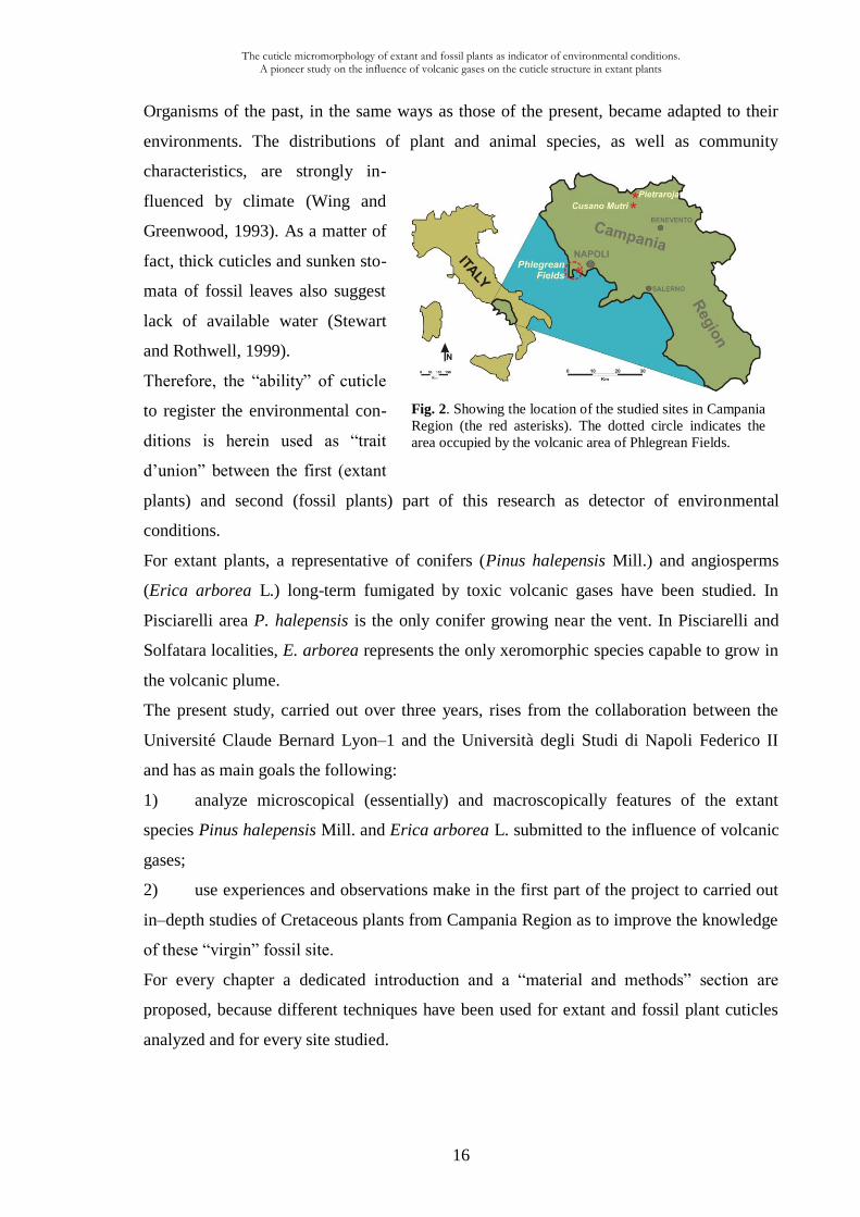
Fig. 2. Showing the location of the studied sites in Campania
Region (the red asterisks). The dotted circle indicates the
area occupied by the volcanic area of Phlegrean Fields.
Organisms of the past, in the same ways as those of the present, became adapted to their
environments. The distributions of plant and animal species, as well as community
characteristics, are strongly in-
fluenced by climate (Wing and
Greenwood, 1993). As a matter of
fact, thick cuticles and sunken sto-
mata of fossil leaves also suggest
lack of available water (Stewart
and Rothwell, 1999).
Therefore, the “ability” of cuticle
to register the environmental con-
ditions is herein used as “trait
d’union” between the first (extant
plants) and second (fossil plants) part of this research as detector of environmental
conditions.
For extant plants, a representative of conifers (Pinus halepensis Mill.) and angiosperms
(Erica arborea L.) long-term fumigated by toxic volcanic gases have been studied. In
Pisciarelli area P. halepensis is the only conifer growing near the vent. In Pisciarelli and
Solfatara localities, E. arborea represents the only xeromorphic species capable to grow in
the volcanic plume.
The present study, carried out over three years, rises from the collaboration between the
Université Claude Bernard Lyon–1 and the Università degli Studi di Napoli Federico II
and has as main goals the following:
1) analyze microscopical (essentially) and macroscopically features of the extant
species Pinus halepensis Mill. and Erica arborea L. submitted to the influence of volcanic
gases;
2) use experiences and observations make in the first part of the project to carried out
in–depth studies of Cretaceous plants from Campania Region as to improve the knowledge
of these “virgin” fossil site.
For every chapter a dedicated introduction and a “material and methods” section are
proposed, because different techniques have been used for extant and fossil plant cuticles
analyzed and for every site studied.
Influence of volcanic gases on the epidermis of Pinus halepensis Mill. in Campi Flegrei, Southern Italy: A possible tool detecting volcanism in present and past floras
17
CHAPTER I
Influence of volcanic gases on the epidermis of Pinus halepensis Mill. in
Campi Flegrei, Southern Italy: A possible tool detecting volcanism in
present and past floras
1.1. Introduction
Over the geological history of the planet, among chronic environmental stress factors
advocated as killing agents (Visscher et al., 2004), changes in atmospheric chemistry had
world-wide dramatic effects on plant life in land (e.g. Visscher et al., 1996; Meyer and
Kump, 2008). For instance, among chemical contaminants that could have disrupted end-
Permian biota, volcanogenic SO2 (Visscher et al., 2004) and biological H2S (euxinia
mechanism: Kump et al., 2005; Berner and Ward, 2006) gases are favoured to explain
extinction. In particular, volcanism subsequently played a role in both maintaining and
perturbing the atmosphere chemistry and physics, with important implications in terms of
the evolution of life (Mather, 2008). The development of large igneous province (LIP) and
continental flood basalt province (CFBP) (Courtillot and Renne, 2003; Jerram et al., 2005;
Keller, 2008; Bryan et al., 2010) commonly coincides with mass extinction events
(Wignall, 2001, 2005; Rampino, 2010; Whiteside et al., 2010) and results in the release of
significant volumes of gases, such as CO2, H2S and SO2 into the atmosphere (Beerling and
Berner, 2002; Berner and Beerling, 2007; Hori et al., 2007). It is widely recognized that
volcanic sulfur dioxide (SO2) and hydrogen sulfide (H2S) emissions are significant sources
of sulfur release to the atmosphere (Bates et al., 1992; Berner and Berner, 1996).
Gases emitted by volcanoes represent both a factor inhibiting vegetation development
(Whittaker et al., 1989) and could have been responsible of the decline of vegetation
during periods of global-scale volcanism (Bond et al., 2010; Visscher et al., 2004;
Whiteside et al., 2010; McElwain and Punyasena, 2007). In particular, H2S is often thought
to be a phytotoxin, being harmful to the growth and development of plants (Lisjak et al.,
2010) especially when the quantities are higher than plant necessity (Thompson and Kats,
1978; Lorenzini and Nali, 2005). Moreover, atmospheric pollutants produced by volcanic
activity and OAEs, such as SO2 and H2S, are said to be absorbed via the cuticle as well as
the stomata (Haworth and McElwain, 2008).
Influence of volcanic gases on the epidermis of Pinus halepensis Mill. in Campi Flegrei, Southern Italy: A possible tool detecting volcanism in present and past floras
18
Plants exposed to poisonous volcanic gases may show signs of diseases to total
defoliation and death (e.g. Dickson, 1965; Clarkson and Clarkson, 1994; Delmelle et al.,
2002). However, plant damages are related to both gas concentration (Delmelle, 2003) and
its persistence (Grattan et al., 1998) in the atmosphere. Under severe pollution conditions,
the direct phytotoxic effects of gaseous pollutants as well as long-term effects of acid
washout (Grattan and Pyatt, 1994) can even be considered as potential environmental
mutagens disturbing plant growth and community structure (Visscher et al., 1996). As a
matter of fact, as Visscher et al. (2004) pointed out, variation in structure and composition
of leaf cuticles is a potential source of botanical evidence on mutational effects of
environmental stress factors.
Therefore, leaves in natural environments are subjected to a range of physical processes
which may damage their surfaces, leading to alterations in the structure and integrity of the
cuticle, and consequently changes in the physical properties of the leaf surfaces (van
Gardingen et al., 1991).
To this end, numerous articles have been published in relation to the effects and
interactions of the volcanic activity products (e.g. tephra or ash fall) on both fossil (e.g.
Kovar-Eder et al., 2001; García Massini and Jacobs, 2011) and extant plants (Winner and
Mooney, 1980b; Cook et al., 1981; Seymour et al., 1983; Dale et al., 2005). Moreover, in
extant plants the concentration of chemical elements in the leaves (Notcutt and Davies,
1989; Martin et al., 2009a and b) and the analysis of the log (Baillie and Munro, 1988;
Battipaglia et al., 2007) together with field studies led to significant advances in
understanding the composition and dispersion of volcanic emissions at source (e.g.
Kempter et al., 1996; Delmelle et al., 2002), including major “gas species” (Costa et al.,
2005; Chiodini, 2008; Chiodini et al., 2010a).
Leaves of plants act as passive and active collectors for natural (e.g. Martin et al.,
2009a) and anthropogenic (e.g. Bačić et al., 1999) airborne pollutants (e.g. gas, aerosols
and dusts) and are more sensitive to air quality than other plant organs (e.g. roots) (Landolt
et al., 1989; Casseles, 1998; Kabata-Pendias, 2001); the gas exchanges between the plant
and the surrounding atmosphere are mediated by the cuticle; this non-living (Riederer,
2006) thin (<0.1-10μm thick in extant plants) and heterogeneous membrane (van
Gardingen et al., 1991) covers the epidermis of the aerial part of many tracheophytes
(Guignard et al., 2004) and consists of a polymer matrix (cutin), polysaccharides and
associated solvent-soluble lipids which are synthesised by the epidermal cells and
deposited on their outer wall (Kirkwood, 1999; Riederer and Schreiber, 2001). The outer
Influence of volcanic gases on the epidermis of Pinus halepensis Mill. in Campi Flegrei, Southern Italy: A possible tool detecting volcanism in present and past floras
19
surface of the cuticle is coated with epicuticular waxes, a general term (Jeffree, 2006)
designating very long chain hydrocarbons found embedded within the cuticle and also in
the crystalline epicuticular wax layer (Bird and Gray, 2003). The main function ascribed to
waxes is to limit the diffusional flow of water and solutes across the cuticle (Heredia and
Dominguez, 2009), providing protection for the leaf cells (Turunen and Huttunen, 1990)
and acting as the main barrier to air pollutants (e.g. Jeffree, 1986). The composition and
amount of waxes in the cuticle have been shown to vary depending to environmental
conditions of the plant (Baker, 1982; Bird and Gray, 2003) and according to many authors
air pollution seems to increase the rate of wax tubules degradation (e.g. Huttunen and
Laine, 1983; Riding and Percy, 1985; Berg, 1987; Turunen and Huttunen, 1990, 1991,
Huttunen, 1994). In particular, wax load and structure can be used as an indicator of
pollution level (Hansell and Oppenheimer, 2006; Holroyd et al., 2002). The epicuticular
wax of pine needles undergoes an ageing procedure during the needle lifetime (Turunen
and Huttunen, 1996; Bačić et al., 1999) and is disturbed by polluted air (Huttunen, 1994).
The literature is replete with references to structural changes in epicuticular waxes
following exposure to air pollutants (see Turunen and Huttunen, 1990), and as a matter of
fact, the erosion of epicuticular waxes is a relevant factor of the multiple forest decline
syndrome (Turunen and Huttunen, 1990).
Few paleobotanical works have been achieved on cuticular characters related to
volcanic stress. Archangelsky et al. (1995) and Villar de Seoane (2001) studied Early
Cretaceous plants from Patagonia (recovered in Baqueró and Springhill Formations,
respectively) demonstrating that the volcanic ash fall played an important role in the
formation of xeromorphic structures. As Haworth and McElwain (2008) claimed, the effect
of toxic atmospheric gases and volcanic dust would explain xeromorphic features of
Pseudofrenelopsis parceramosa (Fontaine) Watson from the Early Cretaceous of England.
Moreover, the relationship between ultrastructural characteristics of cuticle and the
environment is still poorly understood for extinct as well as extant plants (Guignard et al.,
2001) and cuticular ultrastructure data are not numerous for fossil conifers (e.g. Guignard
et al., 1998; Villar de Seoane, 1998; Yang et al., 2009) and seem to be still lacking for
some species belonging to the genus Pinus (Jeffree, 2006).
However, to date, no studies have been carried out relatively to the response of the
ultrastructural features of plant cuticle exposed to the persistent volcanic gases. Conifers
are well suited for studies of pollutant levels because they are evergreen and often have
long-lived foliage. Usually the needles have a life cycle of several years (Hellström, 2003).
Influence of volcanic gases on the epidermis of Pinus halepensis Mill. in Campi Flegrei, Southern Italy: A possible tool detecting volcanism in present and past floras
20
Therefore, the protective role of the epicuticular waxes is particularly important for
conifers that have to ensure their investment in leaf tissue for several years (Chabot and
Chabot, 1977). In the volcanic area of Pisciarelli (Campi Flegrei, Southern Italy) the
gymnosperm Pinus halepensis Mill. (Aleppo pine) is the only conifer growing adjacent to
the fumaroles, and much of the surrounding vegetation (under study) displays indications
of damage caused by toxic gases. P. halepensis is the most abundant pine species in the
western Mediterranean Basin, where it occupies 2.5 million ha (Quézel, 2000) and it is
considered as an opportunistic species (Nathan and Ne’eman, 2000) which is able to
regenerate either in the absence or as a result of fire. In addition, P. halepensis has an
elevated resistance to drought (Boddi et al., 2002), so much so that Emberger (1930)
identifies it as being semiarid, and Oppenheimer (1968) considers it as the most arid-
tolerant of all the Pinus species. As a matter of fact, the present study aims to assess the
cuticular response of this conifer at a prolonged exposition to the volcanic gases using both
SEM and TEM approaches. Moreover, to our knowledge, this is the first study that tests
the cuticle ultrastructure behaviour during two subsequent years (current- and first-year-old
needles) in response to the fumigation of volcanic gases containing H2S.
In particular, this research aimed to investigate: 1) response of plants to volcanic gases
through different aspects: epicuticular and epistomatal waxes and ultrastructural features of
the cuticle; 2) potential implications of the conifer cuticle response across environmental
stress periods during the geological past; 3) a new method detecting the influence of
volcanism for extant and fossil plants.
1.2. Material and methods
The material was collected from two localities in the Phlegrean Fields (Campi Flegrei,
Campania Region), an active caldera which spans the last 50000 years (Scandone et al.,
2010), characterized by significant recent ground deformation (Morhange et al., 2006) and
considered as one of the most dangerous volcanic areas in the world (Chiodini et al.,
2010a). In particular, pine needles were recovered from the famous fumaroles field in
Pisciarelli locality (40°49’48.88’’N, 14°08’46.95’’E) about 1 km SE of the Solfatara
volcano, both characterised by volcanic gas emissions (Fig. 1A,B). Control sample of
needles were collected from a volcanic quiescent area (Cigliano: 40°50’46.46’’N,
14°07’36.31’’E) about 2.5 km from Pisciarelli and characterised by the absence of volcanic
gas emissions and the presence of clean air. Both localities are characterised by the same
Influence of volcanic gases on the epidermis of Pinus halepensis Mill. in Campi Flegrei, Southern Italy: A possible tool detecting volcanism in present and past floras
21
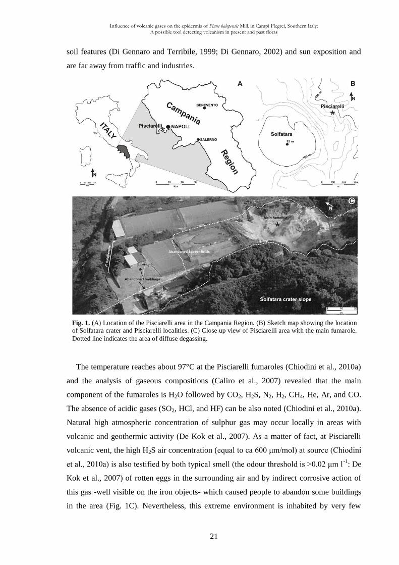
Fig. 1. (A) Location of the Pisciarelli area in the Campania Region. (B) Sketch map showing the location
of Solfatara crater and Pisciarelli localities. (C) Close up view of Pisciarelli area with the main fumarole.
Dotted line indicates the area of diffuse degassing.
soil features (Di Gennaro and Terribile, 1999; Di Gennaro, 2002) and sun exposition and
are far away from traffic and industries.
The temperature reaches about 97°C at the Pisciarelli fumaroles (Chiodini et al., 2010a)
and the analysis of gaseous compositions (Caliro et al., 2007) revealed that the main
component of the fumaroles is H2O followed by CO2, H2S, N2, H2, CH4, He, Ar, and CO.
The absence of acidic gases (SO2, HCl, and HF) can be also noted (Chiodini et al., 2010a).
Natural high atmospheric concentration of sulphur gas may occur locally in areas with
volcanic and geothermic activity (De Kok et al., 2007). As a matter of fact, at Pisciarelli
volcanic vent, the high H2S air concentration (equal to ca 600 μm/mol) at source (Chiodini
et al., 2010a) is also testified by both typical smell (the odour threshold is >0.02 μm l-1
: De
Kok et al., 2007) of rotten eggs in the surrounding air and by indirect corrosive action of
this gas -well visible on the iron objects- which caused people to abandon some buildings
in the area (Fig. 1C). Nevertheless, this extreme environment is inhabited by very few
Influence of volcanic gases on the epidermis of Pinus halepensis Mill. in Campi Flegrei, Southern Italy: A possible tool detecting volcanism in present and past floras
22
angiosperm species providing xeromorphic features (e.g. Erica arborea) and the boiling
water of fumaroles retains the cyanidialean alga Galdieria phlegrea (Pinto et al., 2007).
Distal volcanic impacts have shown that plants are generally less sensitive to eruptions
outside the growing season (Zobel and Antos, 1997; Hotes et al., 2004) and, as Payne and
Blackford (2008) pointed out, in winter, plants are senescent and higher rainfall may serve
to remove rapidly volcanic pollutants. In case of Pinus halepensis, an evergreen plant
permanently fumigated by volcanic gases, retaining leaves for over a year, these “ground
noises” do not exist.
The trees present diffuse damages along the North sides of the crown, while needles
show symptoms consisting in leaf-tip non-specific discoloration which gradually
increasing shootward (terminology from Baskin et al., 2010). Current- and first-year-old
needles were collected from branches at heights over 1.5 m from three trees (15-20 years
old) at each site (Pisciarelli and Cigliano). Needles were carefully handled to avoid
damaging the epicuticular waxes. Following Reed’s remarks (1982) and also Crang and
Klomparens’ ones (1988) about possible changes in epicuticular wax structures occurring
during sample preparation, in order to limit any chemical or physical damages, especially
for preserving and dehydrating samples for wax morphology studies (e.g. Turunen and
Huttunen, 1991; Tuomisto and Neuvonen, 1993), needles were air-dried for 1-week at mild
room temperatures. Among several hundreds of pine needles collected at each site
(fumigated and not fumigated), 60 were selected for scanning electron microscope (SEM),
then 16 were carefully selected for transmission electron microscope (TEM). Stomata were
observed on 15 current- and first-year-old needles for each site. Analysis was performed
within two weeks from sampling. Taxonomical identification of García Álvarez et al.
(2009) approach has been used. Light microscope observations were made using a Leitz
microscope.
In order to quantify the quality of epicuticular and epistomatal waxes, SEM
observations were carried out. Untreated needle sections of approximately 5 mm in length -
obtained from the middle of each needle- were mounted on stubs using double-sided
adhesive tape; both abaxial and adaxial surfaces have been studied. Part of specimens were
sputter-coated with gold using an AGAR Auto Sputter Coater, while the specimens for
energy diffractive x-ray (EDX) analyses were coated with carbon in a Emitech K450,
observed and photographed with JEOL JSM-5310 SEM adapted with an Energy
Diffractive X-ray Oxford Inca X-act at the CISAG (Centro Interdipartimentale di servizi
per analisi Geomineralogiche) in the Dipartimento di Scienze della Terra, Università degli
Influence of volcanic gases on the epidermis of Pinus halepensis Mill. in Campi Flegrei, Southern Italy: A possible tool detecting volcanism in present and past floras
23
Studi di Napoli “Federico II”. The operative conditions were as follows: 25-30 KV
accelerating voltage, 100 μA emission current, 15 μm spot size, 20 mm microscope work
distance and 1 min spectra collection time. To quantify the wax change in stomatal
chamber, the Nicolotti et al. (2005) needle damage classes have been used as a criterion for
the level of crystalline wax degradation.
Samples for TEM were dropped in paraformaldehyde solution mixed in a phosphate-
sodium buffer for 3 weeks using Lugardon's technique (1971), washed and postfixed in a
1% osmium tetroxide solution mixed in a phosphate–sodium buffer for 24 hours.
Dehydrated in graded ethanol series during 48 h, the samples were dropped in propylene
oxide with an increasing percentage of Epon resin for 24 h. Transferred into pure Epon
resin during 24 hours, they were embedded in fresh Epon resin using flat moulds. The
preparations were subsequently treated for polymerization at 56 °C for 3 days. Ultrathin
(60-70 nm) sections were sectioned with a diamond knife, using a Reichert Ultracut
microtome. Ultrathin sections were placed on uncoated 300 Mesh copper grids and stained
manually both with a methanol solution of 7% uranyl acetate for 15 min and an aqueous
lead-citrate solution for 20 min, then observed and photographed with a Philips CM 120
TEM at 80 kV, in the Centre de technologie des microstructures (CTμ) of Lyon-1
University, Villeurbanne, France. Totally 16 pieces of material were embedded in Epon
resin blocks. 90 uncoated mesh copper grids were prepared (80 as transversal sections, i.e.
perpendicular to the leaf length; 10 as longitudinal sections, i.e. parallel to the leaf length).
In order to detect the presence of sulphur in the cuticle, 40 measurements (i.e. 10 measures
for each current- and first-year-old needles, fumigated and not fumigated by volcanic
gases) with EDS microanalysis were carried out on different parts of the cuticle and also
on the cytoplasm remnants of the epidermal cells. The sulphur analysis was performed on
25 coated 300 Mesh grids with transmission electron microscope JEOL 1200EX coupled to
a microanalysis system EDS Si(Li) 30mm² NORAN VOYAGER III with an acceleration
voltage = 80 kV and a spectres acquisition time = 60 s.
All the quantitative TEM measurements were made with tools in the ImageJ program
(Abramoff et al. 2004). The terminology of Holloway (1982) and Archangelsky (1991)
was used for the ultrastructural analysis. All specimens and SEM stubs are housed in the
Dipartimento di Scienze della Terra, Largo San Marcellino, 10, Napoli, Italy. The resin
blocks and TEM negatives are stored in the Lyon-1 University, Villeurbanne, France.
Influence of volcanic gases on the epidermis of Pinus halepensis Mill. in Campi Flegrei, Southern Italy: A possible tool detecting volcanism in present and past floras
24
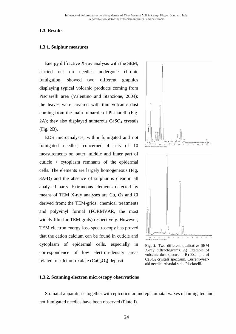
Fig. 2. Two different qualitative SEM
X-ray diffractograms. A) Example of
volcanic dust spectrum. B) Example of
CaSO4 crystals spectrum. Current-year-
old needle. Abaxial side. Pisciarelli.
1.3. Results
1.3.1. Sulphur measures
Energy diffractive X-ray analysis with the SEM,
carried out on needles undergone chronic
fumigation, showed two different graphics
displaying typical volcanic products coming from
Pisciarelli area (Valentino and Stanzione, 2004):
the leaves were covered with thin volcanic dust
coming from the main fumarole of Pisciarelli (Fig.
2A); they also displayed numerous CaSO4 crystals
(Fig. 2B).
EDS microanalyses, within fumigated and not
fumigated needles, concerned 4 sets of 10
measurements on outer, middle and inner part of
cuticle + cytoplasm remnants of the epidermal
cells. The elements are largely homogeneous (Fig.
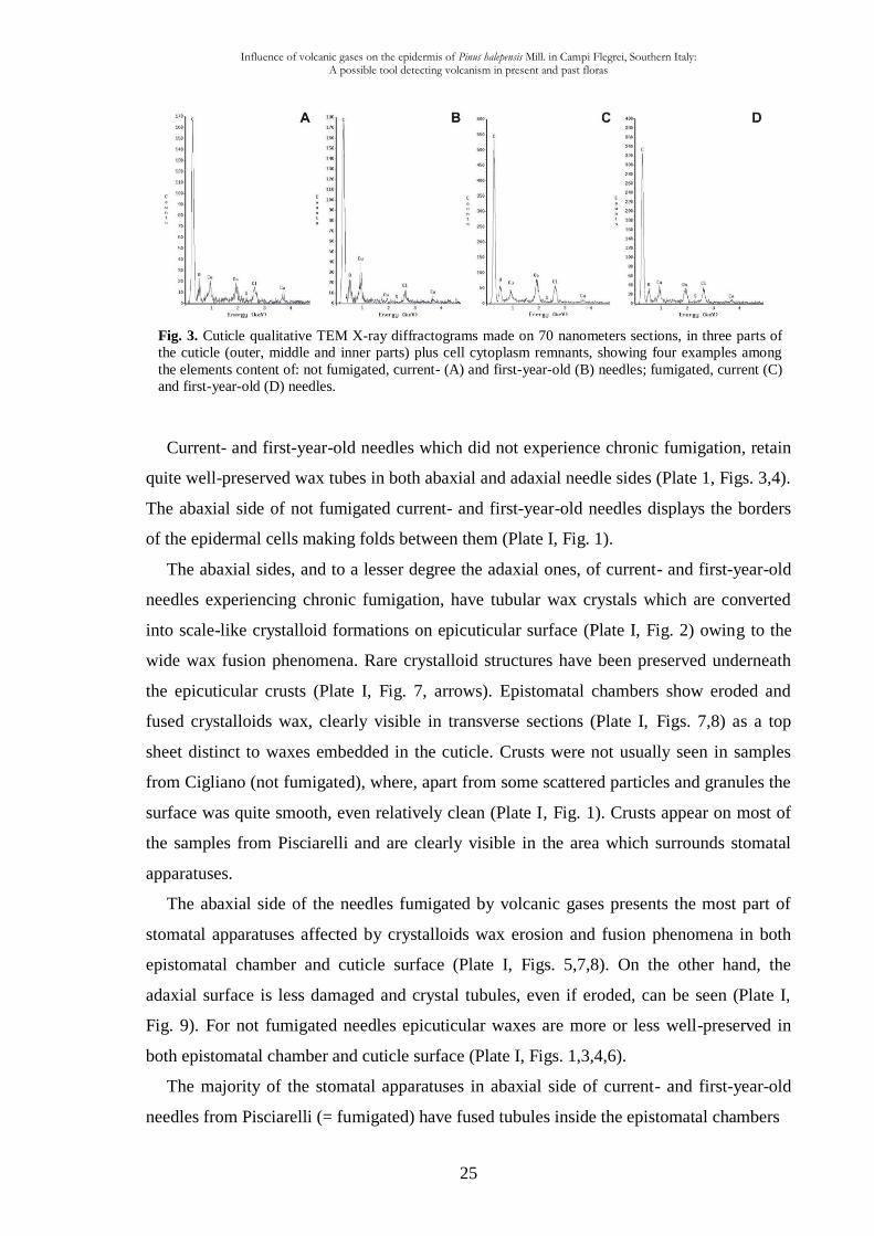
3A-D) and the absence of sulphur is clear in all
analysed parts. Extraneous elements detected by
means of TEM X-ray analyses are Cu, Os and Cl
derived from: the TEM-grids, chemical treatments
and polyvinyl formal (FORMVAR, the most
widely film for TEM grids) respectively. However,
TEM electron energy-loss spectroscopy has proved
that the cation calcium can be found in cuticle and
cytoplasm of epidermal cells, especially in
correspondence of low electron-density areas
related to calcium-oxalate (CaC2O4) deposit.
1.3.2. Scanning electron microscopy observations
Stomatal apparatuses together with epicuticular and epistomatal waxes of fumigated and
not fumigated needles have been observed (Plate I).
Influence of volcanic gases on the epidermis of Pinus halepensis Mill. in Campi Flegrei, Southern Italy: A possible tool detecting volcanism in present and past floras
25
Fig. 3. Cuticle qualitative TEM X-ray diffractograms made on 70 nanometers sections, in three parts of
the cuticle (outer, middle and inner parts) plus cell cytoplasm remnants, showing four examples among
the elements content of: not fumigated, current- (A) and first-year-old (B) needles; fumigated, current (C)
and first-year-old (D) needles.
Current- and first-year-old needles which did not experience chronic fumigation, retain
quite well-preserved wax tubes in both abaxial and adaxial needle sides (Plate 1, Figs. 3,4).
The abaxial side of not fumigated current- and first-year-old needles displays the borders
of the epidermal cells making folds between them (Plate I, Fig. 1).
The abaxial sides, and to a lesser degree the adaxial ones, of current- and first-year-old
needles experiencing chronic fumigation, have tubular wax crystals which are converted
into scale-like crystalloid formations on epicuticular surface (Plate I, Fig. 2) owing to the
wide wax fusion phenomena. Rare crystalloid structures have been preserved underneath
the epicuticular crusts (Plate I, Fig. 7, arrows). Epistomatal chambers show eroded and
fused crystalloids wax, clearly visible in transverse sections (Plate I, Figs. 7,8) as a top
sheet distinct to waxes embedded in the cuticle. Crusts were not usually seen in samples
from Cigliano (not fumigated), where, apart from some scattered particles and granules the
surface was quite smooth, even relatively clean (Plate I, Fig. 1). Crusts appear on most of
the samples from Pisciarelli and are clearly visible in the area which surrounds stomatal
apparatuses.
The abaxial side of the needles fumigated by volcanic gases presents the most part of
stomatal apparatuses affected by crystalloids wax erosion and fusion phenomena in both
epistomatal chamber and cuticle surface (Plate I, Figs. 5,7,8). On the other hand, the
adaxial surface is less damaged and crystal tubules, even if eroded, can be seen (Plate I,
Fig. 9). For not fumigated needles epicuticular waxes are more or less well-preserved in
both epistomatal chamber and cuticle surface (Plate I, Figs. 1,3,4,6).
The majority of the stomatal apparatuses in abaxial side of current- and first-year-old
needles from Pisciarelli (= fumigated) have fused tubules inside the epistomatal chambers
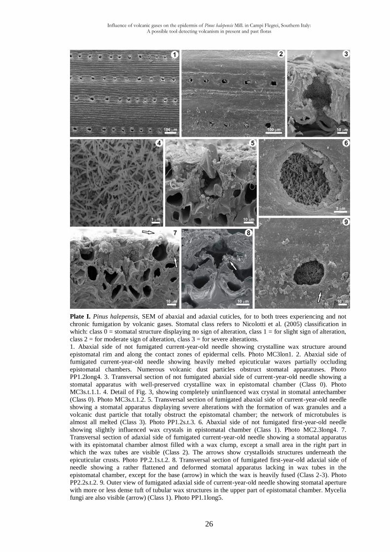
Influence of volcanic gases on the epidermis of Pinus halepensis Mill. in Campi Flegrei, Southern Italy: A possible tool detecting volcanism in present and past floras
26
Plate I. Pinus halepensis, SEM of abaxial and adaxial cuticles, for to both trees experiencing and not
chronic fumigation by volcanic gases. Stomatal class refers to Nicolotti et al. (2005) classification in
which: class 0 = stomatal structure displaying no sign of alteration, class 1 = for slight sign of alteration,
class 2 = for moderate sign of alteration, class 3 = for severe alterations.
1. Abaxial side of not fumigated current-year-old needle showing crystalline wax structure around
epistomatal rim and along the contact zones of epidermal cells. Photo MC3lon1. 2. Abaxial side of
fumigated current-year-old needle showing heavily melted epicuticular waxes partially occluding
epistomatal chambers. Numerous volcanic dust particles obstruct stomatal apparatuses. Photo
PP1.2long4. 3. Transversal section of not fumigated abaxial side of current-year-old needle showing a
stomatal apparatus with well-preserved crystalline wax in epistomatal chamber (Class 0). Photo
MC3s.t.1.1. 4. Detail of Fig. 3, showing completely uninfluenced wax crystal in stomatal antechamber
(Class 0). Photo MC3s.t.1.2. 5. Transversal section of fumigated abaxial side of current-year-old needle
showing a stomatal apparatus displaying severe alterations with the formation of wax granules and a
volcanic dust particle that totally obstruct the epistomatal chamber; the network of microtubules is
almost all melted (Class 3). Photo PP1.2s.t.3. 6. Abaxial side of not fumigated first-year-old needle
showing slightly influenced wax crystals in epistomatal chamber (Class 1). Photo MC2.3long4. 7.
Transversal section of adaxial side of fumigated current-year-old needle showing a stomatal apparatus
with its epistomatal chamber almost filled with a wax clump, except a small area in the right part in
which the wax tubes are visible (Class 2). The arrows show crystalloids structures underneath the
epicuticular crusts. Photo PP.2.1s.t.2. 8. Transversal section of fumigated first-year-old adaxial side of
needle showing a rather flattened and deformed stomatal apparatus lacking in wax tubes in the
epistomatal chamber, except for the base (arrow) in which the wax is heavily fused (Class 2-3). Photo
PP2.2s.t.2. 9. Outer view of fumigated adaxial side of current-year-old needle showing stomatal aperture
with more or less dense tuft of tubular wax structures in the upper part of epistomatal chamber. Mycelia
fungi are also visible (arrow) (Class 1). Photo PP1.1long5.
Influence of volcanic gases on the epidermis of Pinus halepensis Mill. in Campi Flegrei, Southern Italy: A possible tool detecting volcanism in present and past floras
27
forming a flat and solid wax plug or amorphous crusts above the pore which was
completely or partially occluding the stomata in most cases (Plate I, Fig. 7). Instead,
current- and first-year-old needles that do not experience chronic fumigation, regardless of
the age of the needles sampled, present most of their stomatal apertures not occluded,
thereby remaining almost completely open, apart from some minor fusing tubules (Plate I,
Fig. 3). On the abaxial side of current- and first-year-old needles experiencing chronic
fumigation, crystalloid wax tubules rarely persisted and they occupy limited portion only
in epistomatal chambers (Plate I, Fig. 7 top right). It was noticed that in current-year-old
needles from Pisciarelli the wax degradation was beginning very early and it increases.
Frequently, in abaxial side of some Pisciarelli stomatal apparatuses, amorphous wax
and/or particles of other material -30-40 μm width- filled the stomatal aperture, therefore
blocking the direct view into the chamber (Plate I, Figs. 5,7).
In abaxial side of not fumigated needles, the rim of stomatal apparatuses retains
crystalloid wax, whereas in fumigated needles the wax tubes are absent (Plate I, Figs. 1,2).
Frequently, the abaxial side of needles experiencing chronic fumigation presents
deformed stomatal apparatuses so losing the typical funnel-like cavity (Plate 1, Fig. 8).
Stomatal damages are common and may include collapses (Plate I, Fig. 8), depression,
degradation of guard cells (Plate 1, Fig. 2), and occlusion with wax clumps (Plate I, Figs.
2,7). Decay of the epistomatal chambers and empty cavities in stomatal chambers can also
be noted (Plate I, Figs. 5,8).
On both abaxial and adaxial surfaces of fumigated needles, a great amount of dust
occurred (Plate I, Figs. 2,9), while on the needles not fumigated by volcanic gases non-
reactive dusts only mechanically disturb the wax structures (Plate I, Fig. 1). Moreover,
most of Pisciarelli needles are affected by fungal infection (Plate I, Fig. 9).
1.3.3. Transmission electron microscopy observations
The ultrastructure (cuticle and cell wall) of abaxial epidermal cells from fully grown
needles (current- and first-year-old needles) of both localities have been studied in details.
Cuticular membranes (CM) of all ordinary epidermal cell are composed of a fibrillar layer
B (CL) in which an outer, middle and inner zones can be distinguished (Plate II). The
cuticle proper (CP = A) is absent. Differences between the cuticular structures are given
below. All the data given below are the means based on 30 measurements, the percentages
of each component of the cuticle and of cell wall are also given (Table1).
Influence of volcanic gases on the epidermis of Pinus halepensis Mill. in Campi Flegrei, Southern Italy: A possible tool detecting volcanism in present and past floras
28
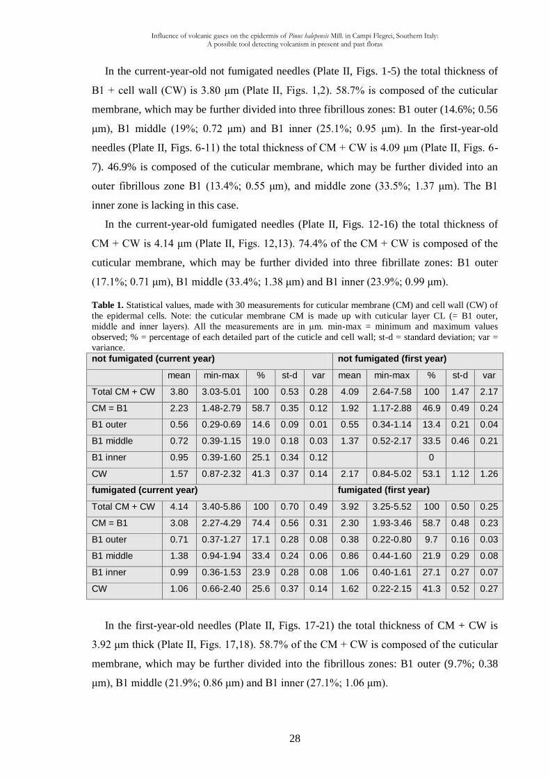
In the current-year-old not fumigated needles (Plate II, Figs. 1-5) the total thickness of
B1 + cell wall (CW) is 3.80 μm (Plate II, Figs. 1,2). 58.7% is composed of the cuticular
membrane, which may be further divided into three fibrillous zones: B1 outer (14.6%; 0.56
μm), B1 middle (19%; 0.72 μm) and B1 inner (25.1%; 0.95 μm). In the first-year-old
needles (Plate II, Figs. 6-11) the total thickness of CM + CW is 4.09 μm (Plate II, Figs. 6-
7). 46.9% is composed of the cuticular membrane, which may be further divided into an
outer fibrillous zone B1 (13.4%; 0.55 μm), and middle zone (33.5%; 1.37 μm). The B1
inner zone is lacking in this case.
In the current-year-old fumigated needles (Plate II, Figs. 12-16) the total thickness of
CM + CW is 4.14 μm (Plate II, Figs. 12,13). 74.4% of the CM + CW is composed of the
cuticular membrane, which may be further divided into three fibrillate zones: B1 outer
(17.1%; 0.71 μm), B1 middle (33.4%; 1.38 μm) and B1 inner (23.9%; 0.99 μm).
Table 1. Statistical values, made with 30 measurements for cuticular membrane (CM) and cell wall (CW) of
the epidermal cells. Note: the cuticular membrane CM is made up with cuticular layer CL (= B1 outer,
middle and inner layers). All the measurements are in μm. min-max = minimum and maximum values
observed; % = percentage of each detailed part of the cuticle and cell wall; st-d = standard deviation; var =
variance. not fumigated (current year) not fumigated (first year)
mean min-max % st-d var mean min-max % st-d var
Total CM + CW 3.80 3.03-5.01 100 0.53 0.28 4.09 2.64-7.58 100 1.47 2.17
CM = B1 2.23 1.48-2.79 58.7 0.35 0.12 1.92 1.17-2.88 46.9 0.49 0.24
B1 outer 0.56 0.29-0.69 14.6 0.09 0.01 0.55 0.34-1.14 13.4 0.21 0.04
B1 middle 0.72 0.39-1.15 19.0 0.18 0.03 1.37 0.52-2.17 33.5 0.46 0.21
B1 inner 0.95 0.39-1.60 25.1 0.34 0.12 0
CW 1.57 0.87-2.32 41.3 0.37 0.14 2.17 0.84-5.02 53.1 1.12 1.26
fumigated (current year) fumigated (first year)
Total CM + CW 4.14 3.40-5.86 100 0.70 0.49 3.92 3.25-5.52 100 0.50 0.25
CM = B1 3.08 2.27-4.29 74.4 0.56 0.31 2.30 1.93-3.46 58.7 0.48 0.23
B1 outer 0.71 0.37-1.27 17.1 0.28 0.08 0.38 0.22-0.80 9.7 0.16 0.03
B1 middle 1.38 0.94-1.94 33.4 0.24 0.06 0.86 0.44-1.60 21.9 0.29 0.08
B1 inner 0.99 0.36-1.53 23.9 0.28 0.08 1.06 0.40-1.61 27.1 0.27 0.07
CW 1.06 0.66-2.40 25.6 0.37 0.14 1.62 0.22-2.15 41.3 0.52 0.27
In the first-year-old needles (Plate II, Figs. 17-21) the total thickness of CM + CW is
3.92 μm thick (Plate II, Figs. 17,18). 58.7% of the CM + CW is composed of the cuticular
membrane, which may be further divided into the fibrillous zones: B1 outer (9.7%; 0.38
μm), B1 middle (21.9%; 0.86 μm) and B1 inner (27.1%; 1.06 μm).
Influence of volcanic gases on the epidermis of Pinus halepensis Mill. in Campi Flegrei, Southern Italy: A possible tool detecting volcanism in present and past floras
29
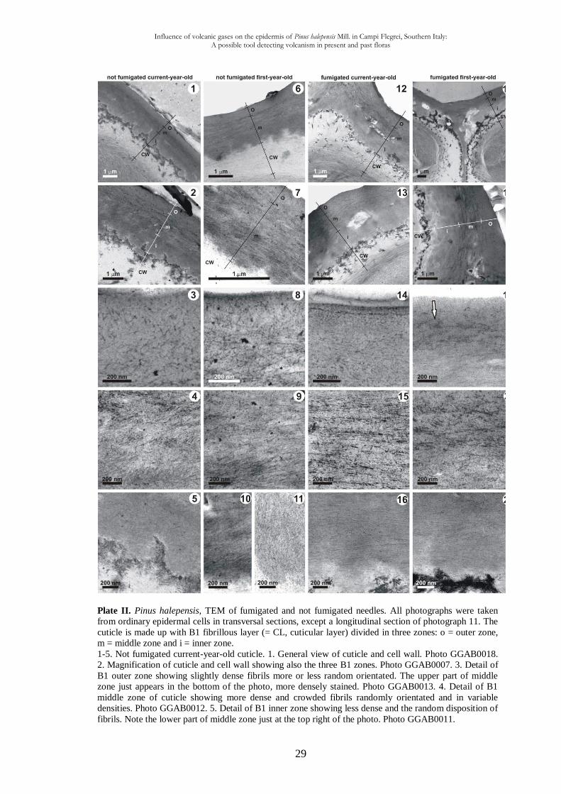
Plate II. Pinus halepensis, TEM of fumigated and not fumigated needles. All photographs were taken
from ordinary epidermal cells in transversal sections, except a longitudinal section of photograph 11. The
cuticle is made up with B1 fibrillous layer (= CL, cuticular layer) divided in three zones: o = outer zone,
m = middle zone and i = inner zone.
1-5. Not fumigated current-year-old cuticle. 1. General view of cuticle and cell wall. Photo GGAB0018.
2. Magnification of cuticle and cell wall showing also the three B1 zones. Photo GGAB0007. 3. Detail of
B1 outer zone showing slightly dense fibrils more or less random orientated. The upper part of middle
zone just appears in the bottom of the photo, more densely stained. Photo GGAB0013. 4. Detail of B1
middle zone of cuticle showing more dense and crowded fibrils randomly orientated and in variable
densities. Photo GGAB0012. 5. Detail of B1 inner zone showing less dense and the random disposition of
fibrils. Note the lower part of middle zone just at the top right of the photo. Photo GGAB0011.
Influence of volcanic gases on the epidermis of Pinus halepensis Mill. in Campi Flegrei, Southern Italy: A possible tool detecting volcanism in present and past floras
30
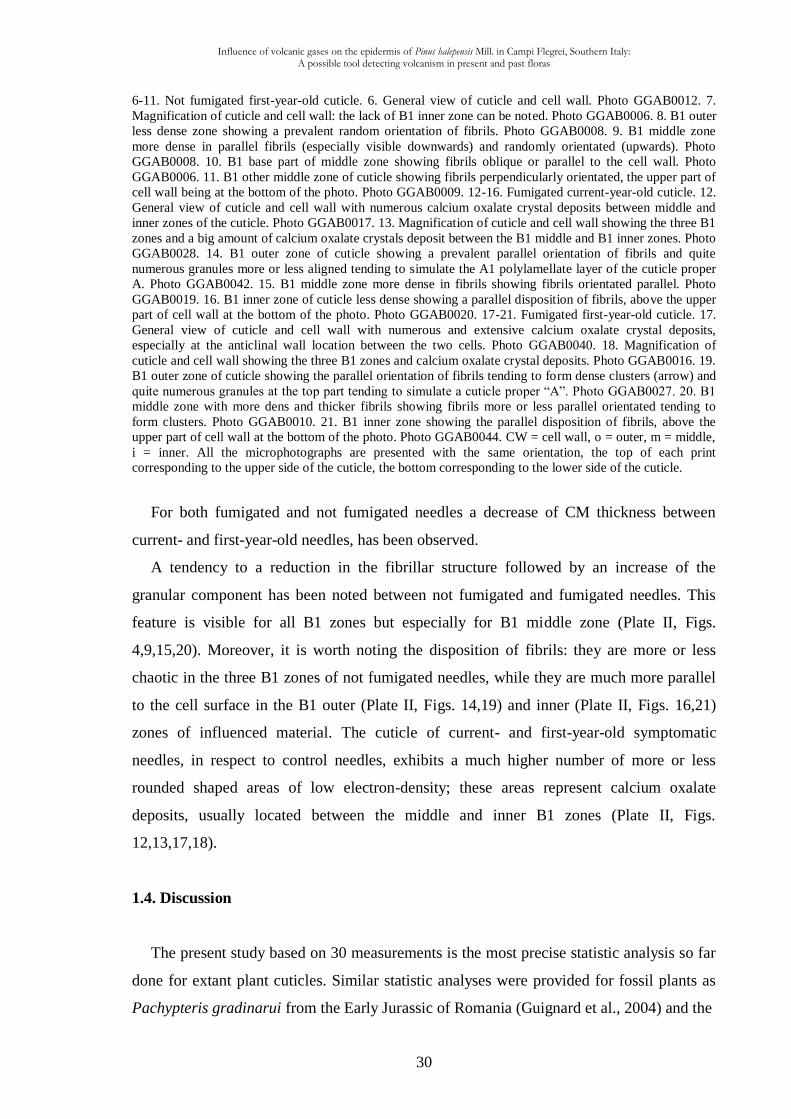
6-11. Not fumigated first-year-old cuticle. 6. General view of cuticle and cell wall. Photo GGAB0012. 7.
Magnification of cuticle and cell wall: the lack of B1 inner zone can be noted. Photo GGAB0006. 8. B1 outer
less dense zone showing a prevalent random orientation of fibrils. Photo GGAB0008. 9. B1 middle zone
more dense in parallel fibrils (especially visible downwards) and randomly orientated (upwards). Photo
GGAB0008. 10. B1 base part of middle zone showing fibrils oblique or parallel to the cell wall. Photo
GGAB0006. 11. B1 other middle zone of cuticle showing fibrils perpendicularly orientated, the upper part of
cell wall being at the bottom of the photo. Photo GGAB0009. 12-16. Fumigated current-year-old cuticle. 12.
General view of cuticle and cell wall with numerous calcium oxalate crystal deposits between middle and
inner zones of the cuticle. Photo GGAB0017. 13. Magnification of cuticle and cell wall showing the three B1
zones and a big amount of calcium oxalate crystals deposit between the B1 middle and B1 inner zones. Photo
GGAB0028. 14. B1 outer zone of cuticle showing a prevalent parallel orientation of fibrils and quite
numerous granules more or less aligned tending to simulate the A1 polylamellate layer of the cuticle proper
A. Photo GGAB0042. 15. B1 middle zone more dense in fibrils showing fibrils orientated parallel. Photo
GGAB0019. 16. B1 inner zone of cuticle less dense showing a parallel disposition of fibrils, above the upper
part of cell wall at the bottom of the photo. Photo GGAB0020. 17-21. Fumigated first-year-old cuticle. 17.
General view of cuticle and cell wall with numerous and extensive calcium oxalate crystal deposits,
especially at the anticlinal wall location between the two cells. Photo GGAB0040. 18. Magnification of
cuticle and cell wall showing the three B1 zones and calcium oxalate crystal deposits. Photo GGAB0016. 19.
B1 outer zone of cuticle showing the parallel orientation of fibrils tending to form dense clusters (arrow) and
quite numerous granules at the top part tending to simulate a cuticle proper “A”. Photo GGAB0027. 20. B1
middle zone with more dens and thicker fibrils showing fibrils more or less parallel orientated tending to
form clusters. Photo GGAB0010. 21. B1 inner zone showing the parallel disposition of fibrils, above the
upper part of cell wall at the bottom of the photo. Photo GGAB0044. CW = cell wall, o = outer, m = middle,
i = inner. All the microphotographs are presented with the same orientation, the top of each print
corresponding to the upper side of the cuticle, the bottom corresponding to the lower side of the cuticle.
For both fumigated and not fumigated needles a decrease of CM thickness between
current- and first-year-old needles, has been observed.
A tendency to a reduction in the fibrillar structure followed by an increase of the
granular component has been noted between not fumigated and fumigated needles. This
feature is visible for all B1 zones but especially for B1 middle zone (Plate II, Figs.
4,9,15,20). Moreover, it is worth noting the disposition of fibrils: they are more or less
chaotic in the three B1 zones of not fumigated needles, while they are much more parallel
to the cell surface in the B1 outer (Plate II, Figs. 14,19) and inner (Plate II, Figs. 16,21)
zones of influenced material. The cuticle of current- and first-year-old symptomatic
needles, in respect to control needles, exhibits a much higher number of more or less
rounded shaped areas of low electron-density; these areas represent calcium oxalate
deposits, usually located between the middle and inner B1 zones (Plate II, Figs.
12,13,17,18).
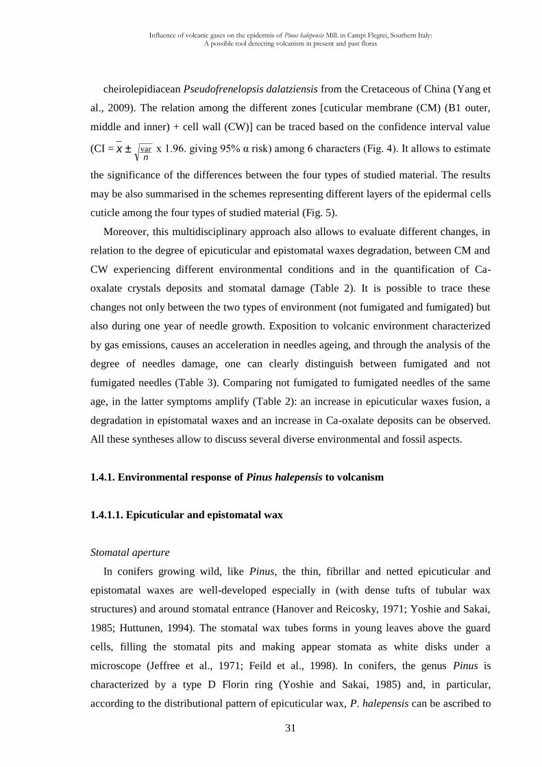
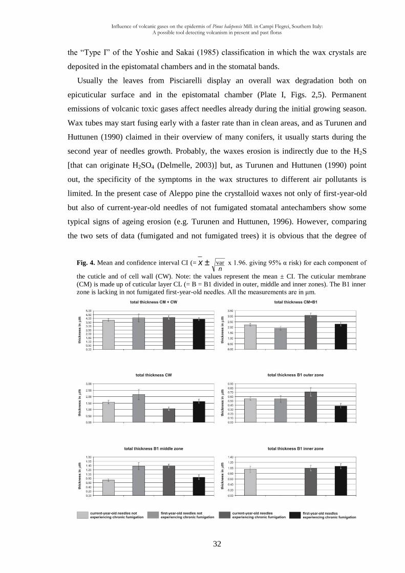
1.4. Discussion
The present study based on 30 measurements is the most precise statistic analysis so far
done for extant plant cuticles. Similar statistic analyses were provided for fossil plants as
Pachypteris gradinarui from the Early Jurassic of Romania (Guignard et al., 2004) and the
Influence of volcanic gases on the epidermis of Pinus halepensis Mill. in Campi Flegrei, Southern Italy: A possible tool detecting volcanism in present and past floras
31
cheirolepidiacean Pseudofrenelopsis dalatziensis from the Cretaceous of China (Yang et
al., 2009). The relation among the different zones [cuticular membrane (CM) (B1 outer,
middle and inner) + cell wall (CW)] can be traced based on the confidence interval value
(CI = x ±n
var x 1.96. giving 95% α risk) among 6 characters (Fig. 4). It allows to estimate
the significance of the differences between the four types of studied material. The results
may be also summarised in the schemes representing different layers of the epidermal cells
cuticle among the four types of studied material (Fig. 5).
Moreover, this multidisciplinary approach also allows to evaluate different changes, in
relation to the degree of epicuticular and epistomatal waxes degradation, between CM and
CW experiencing different environmental conditions and in the quantification of Ca-
oxalate crystals deposits and stomatal damage (Table 2). It is possible to trace these
changes not only between the two types of environment (not fumigated and fumigated) but
also during one year of needle growth. Exposition to volcanic environment characterized
by gas emissions, causes an acceleration in needles ageing, and through the analysis of the
degree of needles damage, one can clearly distinguish between fumigated and not
fumigated needles (Table 3). Comparing not fumigated to fumigated needles of the same
age, in the latter symptoms amplify (Table 2): an increase in epicuticular waxes fusion, a
degradation in epistomatal waxes and an increase in Ca-oxalate deposits can be observed.
All these syntheses allow to discuss several diverse environmental and fossil aspects.
1.4.1. Environmental response of Pinus halepensis to volcanism
1.4.1.1. Epicuticular and epistomatal wax
Stomatal aperture
In conifers growing wild, like Pinus, the thin, fibrillar and netted epicuticular and
epistomatal waxes are well-developed especially in (with dense tufts of tubular wax
structures) and around stomatal entrance (Hanover and Reicosky, 1971; Yoshie and Sakai,
1985; Huttunen, 1994). The stomatal wax tubes forms in young leaves above the guard
cells, filling the stomatal pits and making appear stomata as white disks under a
microscope (Jeffree et al., 1971; Feild et al., 1998). In conifers, the genus Pinus is
characterized by a type D Florin ring (Yoshie and Sakai, 1985) and, in particular,
according to the distributional pattern of epicuticular wax, P. halepensis can be ascribed to
Influence of volcanic gases on the epidermis of Pinus halepensis Mill. in Campi Flegrei, Southern Italy: A possible tool detecting volcanism in present and past floras
32
Fig. 4. Mean and confidence interval CI (= x ±n
var x 1.96. giving 95% α risk) for each component of
the cuticle and of cell wall (CW). Note: the values represent the mean ± CI. The cuticular membrane
(CM) is made up of cuticular layer CL (= B = B1 divided in outer, middle and inner zones). The B1 inner
zone is lacking in not fumigated first-year-old needles. All the measurements are in μm.
the “Type I” of the Yoshie and Sakai (1985) classification in which the wax crystals are
deposited in the epistomatal chambers and in the stomatal bands.
Usually the leaves from Pisciarelli display an overall wax degradation both on
epicuticular surface and in the epistomatal chamber (Plate I, Figs. 2,5). Permanent
emissions of volcanic toxic gases affect needles already during the initial growing season.
Wax tubes may start fusing early with a faster rate than in clean areas, and as Turunen and
Huttunen (1990) claimed in their overview of many conifers, it usually starts during the
second year of needles growth. Probably, the waxes erosion is indirectly due to the H2S
[that can originate H2SO4 (Delmelle, 2003)] but, as Turunen and Huttunen (1990) point
out, the specificity of the symptoms in the wax structures to different air pollutants is
limited. In the present case of Aleppo pine the crystalloid waxes not only of first-year-old
but also of current-year-old needles of not fumigated stomatal antechambers show some
typical signs of ageing erosion (e.g. Turunen and Huttunen, 1996). However, comparing
the two sets of data (fumigated and not fumigated trees) it is obvious that the degree of
Influence of volcanic gases on the epidermis of Pinus halepensis Mill. in Campi Flegrei, Southern Italy: A possible tool detecting volcanism in present and past floras
33
occlusion of suprastomatal chambers (with wax crusts) increased in trees growing in the
polluted site in respect to the control site used in this study (Table 2). In normal conditions,
large amounts of wax occlude the epistomatal chambers and the Florin rings are
characterised by large opening size. Instead, in the needles under the influence of volcanic
gases of Pisciarelli, the reduction of wax tubes in epistomatal chambers is associated to a
reduction of the stomatal aperture. Numerous stomatal apparatuses even display collapse
and decay of the epistomatal chambers (Plate 1, Fig. 8). On the abaxial side of Pisciarelli
needles, amorphous wax filling epistomatal chambers, as well as the rims, could probably
disturb normal gas exchange and, as Holroyd et al. (2002) claimed in their study on
Arabidopsis mutant plants, alterations in wax composition could also affect stomatal
development.
Volcanic toxic compounds and wax alterations
After water and carbon, sulphur is the major constituent of the fumes emitted by
volcanoes (Le Guern et al., 1988) and as a matter of fact, among Pisciarelli fumarolic
effluents a relatively high quantity of hydrogen sulphide (H2S) is released (Chiodini et al.,
2010a). Brown (1982), Lorenzini and Nali (2005) and also Haworth et al. (2010) claimed
that the H2S oxidizes rapidly in the atmosphere to form SO2; this last rapidly converts into
H2SO4 (Visscher et al., 2004) considered as an oxidation product of SO2 or H2S (Mather et
al., 2003). In extant plants SO2 concentrations between 0.1 and 1.0 ppm can cause rapid
changes in stomatal conductance for a wide range of plant species. Changes in stomatal
conductance during period of SO2 exposure are apparently associated with regulating the
extent to which foliar injury develops (Winner and Mooney, 1980b). Winner and Mooney
(1980a), studying the fumigation with SO2 of two Californian shrub chaparral species
(Diplacus aurantiacus and Heteromeles arbutifolia, angiosperms), observed a decline in
both photosynthesis and transpiration during the fumigation periods and a tendency to
close during fumigation for guard cells. They also state that the response of guard cells
during periods of SO2 exposure may be one of the factors contributing to variations in SO2
resistance between plants. According to the same authors, owing to the effects on plant
community structure and plant metabolism, the SO2 may be considered as a chronic
environmental stress for vegetation. Actually, the impact of these sulphurous gases on
plants is much more complex, since they may act as toxin or plant nutrient upon foliar
deposition (De Kok et al., 2007). SO2 and H2S affect leaves causing also dwarfism
phenomena (Wood and Boorman, 1977). More precisely for conifers, in their study on
Influence of volcanic gases on the epidermis of Pinus halepensis Mill. in Campi Flegrei, Southern Italy: A possible tool detecting volcanism in present and past floras
34
Picea abies (Norway spruce) needles with a treatment simulated acid deposition, Raddi et
al. (1994) showed that H2SO4 altered significantly both the epicuticular wax and fibrillar
wax structure in the epistomatal chamber. Rinallo et al. (1986) observed that aqueous
sulphuric acid of pH 3.5 had greater effects on epistomatal waxes of silver fir (Abies alba
Mill.) and Norway spruce needles than aqueous nitric acid of the same concentration.
The present study of Pinus halepensis also contributes to the analysis of sulphur
influence since, on control needles (Cigliano locality) no CaSO4 crystals were found on
needle surfaces. Conversely, in fumigated needles from Pisciarelli, the H2SO4 action is
testified by the occurrence of CaSO4 crystals on needle surface, the result of a reaction
between the calcium leached out of the needles as observed by Turunen et al. (1994) on
Pinus sylvestris and Picea abies needles, subjected to acid rain treatment, and the sulphur
deriving from both H2SO4 and/or from tephra (Smith et al., 1983) deposited on needle
surfaces.
As it was already shown that removal of waxes from cuticles results in an increase of
gas and water permeabilities of up to three orders of magnitude (Lendzian and Kerstiens,
1991), the formation of wax crusts on needles epidermis of the present Aleppo pine could
act as a protecting (closing) device against further H2S, SO2 and H2SO4 attacks increasing
the boundary layer resistance. This was already noticed by Kayode and Otoide (2007) in
matured leaves of an angiosperm Newbouldia laevis. As Haworth and McElwain (2008)
claim, in extant plants resistance to fumigation with toxic gases is often associated with an
increase in the boundary layer resistance. The angiosperm Metrosideros polymorpha
growing in the Kilauea crater region (Island of Hawaii) is resistant to persistent fumigation
with toxic volcanic gases (SO2, H2S, HCl, HF) while other species (e.g. Dodonaea
eriocarpa, an angiosperm) growing in the same region displays a noticeable damage
(Winner and Mooney, 1980b). M. polymorpha from the Kilauea crater region displays
heavily trichomous abaxial surfaces, while those in neighbouring regions, not experiencing
chronic fumigation, possess non-trichomous cuticles (Haworth, 2006). This pattern
suggests (Haworth, 2006) that the development of trichomes in the Kilauea Crater region is
an adaptation to increase boundary layer resistance, and consequently reduces entry of
toxic gases into the leaf. With present observations and measurements for Pinus halepensis
a sulphur reaction seems to exist too.
The almost total fusion of wax crystals in needles from Pisciarelli volcanic area is not
comparable to the mechanical injury under windy condition where leaves scrape against
each other. In her SEM study on Acer pseudoplatanus leaves under the effect of wind,
Influence of volcanic gases on the epidermis of Pinus halepensis Mill. in Campi Flegrei, Southern Italy: A possible tool detecting volcanism in present and past floras
35
Wilson (1984) showed different microscopic damages like dark and light brown lesions,
holes and deformations of the leaf. The wax degradation in specimens from Pisciarelli
presents different features in respect to the impact of wind blown particles. van Gardingen
et al. (1991) studied Picea sitchensis and Pinus sylvestris in a wind tunnel showing
different diseases in respect to the present wax degradation in P. halepensis. In particular,
in P. sylvestris the authors noticed that the most damage was on the cells surrounding the
stomatal antechamber or on needle ridges. Moreover, needles damage cannot be related to
the ozone exposition because the gaseous nature of these molecules should have affected
the overall needles. The structural injuries observed in the stomatal apparatuses are not
correlated to the surfactant action. Usually this kind of damages are observed in plants
growing 100 m far from the sea (because Pisciarelli is located far from sea), near the
mouth of a river or stream, near the outlet of sewage canals and in any other coastal areas
where the surface currents carry a surfactant load (Busotti et al., 1995; Nicolotti et al.,
2005). Moreover, spraying with NaCl solutions can be excluded since it does not induce
the occlusion of stomatal openings (Krause, 1982).
Other sulphur considerations
Although it is not directly the aim of this study, some EDS measurements allow to
discuss about sulphur input. Cuticle and stomata are commonly involved in sulphur and
other molecules absorption (Haworth and McElwain 2008; Haworth et al., 2010;
Kivimäenpää et al., 2010). In their article about various ecological and cuticular
micromorphology aspects, Haworth and McElwain (2008) summarised data of various
authors on different taxa, sulphur (combined with oxygen or hydrogen) is said to be
“absorbed via the cuticle as well as the stomata”. In the present study of Pinus halepensis,
40 analyses of ultrathin sections (70 nm) of both fumigated and not fumigated needles
(Fig. 3A-D) have demonstrated the absence of sulphur within the overall cuticle thickness
of epidermal cells and cytoplasm. Wide epicuticular and epistomatal wax fusion
phenomena can be also observed. The lacking of sulphur in the structure of the cuticle
suggests that the uptake of volcanic toxic gases takes place via stomatal apparatuses. As Le
Guern et al. (1988) point out, the sulphur is metabolized because it is an essential element
for plants, and it participates in physiological reactions and is eliminated accordingly.
Haworth and McElwain (2008) point out that cuticles of evergreens tend to have lower
permeability than those of deciduous species, possibly reflecting adaptation of species with
long-lived foliage to conserve water during periods of reduced water availability (Kirsch et
Influence of volcanic gases on the epidermis of Pinus halepensis Mill. in Campi Flegrei, Southern Italy: A possible tool detecting volcanism in present and past floras
36
al., 1997; Gratani and Bombelli, 2000). With the present study we may think that
epicuticular and epistomatal wax degradation in P. halepensis needles experiencing
chronic fumigation with volcanic gases from Pisciarelli could be related to the H2S and/or
H2SO4 action and inputs.
Wettability of leaf surface
Wettability of plant surfaces is related to the composition, structure and cristallinity of
epicuticular waxes (Heredia and Dominguez, 2009). Airborne pollutants accelerate erosion
of cuticular waxes of conifers (Huttunen, 1994; Riederer, 2006; Shepherd and Griffiths,
2006) and thus may be responsible for forest decline in Europe and elsewhere (Riederer,
2006). Erosion of the waxes can change needle wettability (Cape, 1983) and rain retention
(Turunen and Huttunen, 1990).
As seen above, needles from Pisciarelli experiencing chronic fumigation, is lacking of
well-preserved epicuticular wax crystalloids, therefore, the Lotus-effect (an anti-adhesive
property against particulate contamination; see Barthlott and Neinhuis, 1997; Neinhuis and
Barthlott, 1997) compromises the removal of contaminant particles.
An added point concerns dust particles. As Ots et al. (2010) claimed, tree leaves, such
as conifer needles, can efficiently accumulate dusts, which are an important stress factor.
Moreover, Cape et al. (1989) found that leaf wettability of the conifers Picea abies and
Pinus sylvestris rose with increasing amount of dust on leaf surfaces. As a matter of fact, in
the present study, although it is not directly the aim of this study, the great amount of dust
covering needle surfaces (Plate I, Figs. 2,5,9) emitted by principal fumarole of Pisciarelli
and derived from volcanic airborne (Delmelle, 2003) together with degradation of wax
could determine an increase of wettability. As it is certainly affecting plant biology it could
be one of the multiple reasons of the rarity of plant taxa in this extreme environment.
Additional observation is that a heavy fungal infection affects almost all Pisciarelli
needles (Plate 1, Fig. 9). This is probably related to several factors, making unfavourable
abiotic conditions in these trees, and becoming then more susceptible to pathogens as
noticed by Sieber (2007) for conifers. For instance, the particular topographic leaf surface
of P. halepensis together with the presence of hydrophobic epicuticular waxes -that are
frequently crystals-, may have considerable influence on all fungal spore adhesion and
retention (see Carver and Gurr, 2006). Moreover, the acidified aqueous environment on
needle surface (H2SO4) could favour fungus proliferation as it was shown in Phyllosticta
ampelicida pycnidiospores which adhere firmly to hydrophobic artificial substrata in an
Influence of volcanic gases on the epidermis of Pinus halepensis Mill. in Campi Flegrei, Southern Italy: A possible tool detecting volcanism in present and past floras
37
acidified aqueous environment (Kuo and Hoch, 1996). Another factor is that water
collecting on needle surfaces is essential for all fungal infections (Huttunen, 1984) and thus
related to wettablility. In their study of Picea pungens needles, Patrie and Berg (1994)
noticed that the epicuticular wax progressively deteriorated as fungus proliferated, so for
the present Pinus halepensis the degradation of epicuticular waxes and the consequent
increase in wettability could be also responsible of fungus proliferation.
Microenvironment
According to Nicolotti et al. (2005) stomatal damage classes, the abaxial side of the
current- and first-year-old fumigated needles can be placed in the Classes 2 and 3, namely
a “stomatal structure displaying moderate sign of alteration” and “severe alteration”,
respectively (Plate 1 and Table 2). On the other hand, the adaxial side of needles can be
ascribed to the Classes 0 and 1, namely a “stomatal structure displaying no sign of
alteration” and “slight sign of alteration”, respectively (Table 2). So, for one single needle,
the difference in adaxial and abaxial stomatal Class attribution seems to be related to their
disposition on the two sides of needles. As a matter of fact, P. halepensis retains leaves
spreading by fascicular sheath which are coupled in pairs, therefore the adaxial side of a
needle is more protected than the abaxial one, demonstrating a very differentiated
microenvironmental influence of the epidermis of the leaves.
Another point is that the needles of P. halepensis experiencing chronic fumigation
(Pisciarelli) show another kind of sensibility to environment, as the degree of epicuticular
wax tubule fusion decreases on the adaxial side (more protected) of needles, but increases
towards the needle-tip (less protected). This fact leans towards a H2SO4 action, probably
also in combination with H2S or SO2 gases that would act less on the adaxial side of
needles. The present study adds another very sensible microenvironmental example to
those in plants; in conifers it seems to be a general case as Thompson and Kats (1978)
treated a number of plants (five angiosperms and two conifers) with continuous fumigation
of H2S founding that Douglas fir (with free-leaves) was much more sensitive than
Ponderosa pine (which has leaves in bundle of threes, occasionally in two’s) to foliar
injury.
Influence of volcanic gases on the epidermis of Pinus halepensis Mill. in Campi Flegrei, Southern Italy: A possible tool detecting volcanism in present and past floras
38
1.4.1.2. Cuticular membrane (CM) + cell wall (CW)
Pinus halepensis cuticle type
The effect of volcanic gases on the epidermis in conifers has not been previously
studied by TEM, and no detailed ultrastructural studies have been made so far on the
cuticle of Pinus halepensis. As Guignard et al. (2001) point out, according to the
Achangelsky et al. (1986) terminology used commonly for both fossil and extant plants,
within cuticle thickness it is possible to distinguish a cuticle proper (termed A, often
lamellate in its outermost part and granulous below) and cuticular layer (termed B, mostly
fibrillous). The cuticle proper is absent in the studied species. Since the cuticular material
of fumigated and not fumigated plants by volcanic gases is predominantly made up of
fibrils, P. halepensis cuticle can be attributed to cuticular layer B and divided into three
zones, respectively named in the present study B1 outer, middle and inner. Although
unfortunately to our knowledge previous statistical detailed studies of the cuticle do not
exist, it has to be noticed a certain homogeneity in the cuticle structure among species of
the genus Pinus, as the studied P. halepensis cuticle belongs to the type 4 (“All regions
reticulate”) of Holloway’s (1982) classification in which he included other species of
Pinus (P. sylvestris and P. nigra), as well as other conifers (Picea sitchensis and Abies
balsamea). Besides of the identification to one of Holloway’s general types, the present
material seems to be a good example to conduct further statistical ultrastructural studies of
the cuticle among Pinus species in order to verify the Holloway’s (1982) remark: “Because
the CM (= cuticular membrane) of plants is so heterogeneous in structure it is dangerous
to oversimplify and generalize about its morphology and construction - there is no typical
plant cuticular membrane. Consequently, each species must be considered individually and
it should not be assumed that any structural features which may be observed are of
universal occurrence.”. However, taking into account a TEM photo of Picea sitchensis
type 4 cuticle (Holloway, 1982, fig. 16), it seems that there exist clear differences with
Pinus halepensis, although in this latter genus the cuticle proper A is lacking, but only B1
layer is present.
CM + CW thickness
Plants of the same species experiencing different environments have a response at
histological level. In the studied material, the total thickness of CM + CW is roughly
constant with an average thickness of 3.99 μm (3.80-4.14 μm, see Table 1) among the four
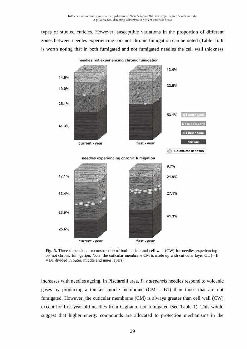
Influence of volcanic gases on the epidermis of Pinus halepensis Mill. in Campi Flegrei, Southern Italy: A possible tool detecting volcanism in present and past floras
39
Fig. 5. Three-dimensional reconstruction of both cuticle and cell wall (CW) for needles experiencing-
or- not chronic fumigation. Note: the cuticular membrane CM is made up with cuticular layer CL (= B
= B1 divided in outer, middle and inner layers).
types of studied cuticles. However, susceptible variations in the proportion of different
zones between needles experiencing- or- not chronic fumigation can be noted (Table 1). It
is worth noting that in both fumigated and not fumigated needles the cell wall thickness
increases with needles ageing. In Pisciarelli area, P. halepensis needles respond to volcanic
gases by producing a thicker cuticle membrane (CM = B1) than those that are not
fumigated. However, the cuticular membrane (CM) is always greater than cell wall (CW)
except for first-year-old needles from Cigliano, not fumigated (see Table 1). This would
suggest that higher energy compounds are allocated to protection mechanisms in the
Influence of volcanic gases on the epidermis of Pinus halepensis Mill. in Campi Flegrei, Southern Italy: A possible tool detecting volcanism in present and past floras
40
cuticular membrane, demonstrating once again that cuticle protects basic leaf functions. In
detail, the lacking of B1 inner zone in not fumigated first-year-old needles (Table 1; Plate
II, Fig. 7; Figs. 4,5) can be explained by an increase in fibril amount in the inner zone
(usually characterized by a low density of fibrils), thus making this layer indistinguishable
from the middle zone (usually with a high density of fibrils).
CM + CW development
This multidisciplinary study clearly shows the sensitivity of P. halepensis to an extreme
environment, during needle ontogeny and therefore the CM and CW formation (Plate II).
For both fumigated and not fumigated current-year-old needles, the total thickness of
cuticle (CM) + cell wall (CW) is statistically comparable (Table 1, Fig. 4), but the
percentage of each layer is different according to the age and exposition to gases (Fig. 5),
thus showing a diverse equilibrium among zones of the same needle as response to growth
and environmental factors. The needles seem to react early in ontogeny to volcanic
environment because the thickness of B1 or CW already differs statistically between not
fumigated and fumigated current-year-old needles (15.7%). After one year of growth, these
differences are still maintained, although they slightly decreased (11.8% of reduction). To
sum up, the variations of the three zones of the cuticle between fumigated and not
fumigated needles are statistically significant (Fig. 5) and this fact mainly concerns B1
outer and middle zones, while B1 inner zone does not change considerably.
CM and CW ageing
If the response of the plant starts early in the growth of cuticle membrane (CM) + cell
wall (CW), after one year Pinus halepensis responds more actively to volcanic
environment than to no-volcanic one (Table 1). Although the CM + CW thickness in
current- and first-year-old needles does not record significant variations statistically, an
equilibrium between CM and CW (i.e. a decrease of CM and an increase of CW) exists.
The rate of reduction is more pronounced for fumigated needles (15.7%) than for those that
are not fumigated (11.8%) inferring that the volcanic inputs have more important effects on
the outermost part of the cuticle. Comparing current- and first-year-old needles, it was
observed that the B1 outer and middle zones of the cuticle decrease (17.1% versus 9.7%
with a difference of 7.4% and a reduction of 57%; 33.4% versus 21.9% with a difference
of 11.5% and a reduction of 66% respectively, see Fig. 5), while the thickness of B1 inner
zone does not vary significantly.
Influence of volcanic gases on the epidermis of Pinus halepensis Mill. in Campi Flegrei, Southern Italy: A possible tool detecting volcanism in present and past floras
41
In current- and first-year-old needles experiencing chronic fumigation, the parallel
orientation of fibrils in respect to the cuticle surface in B1 outer and inner zones could be
interpreted as a further protection device which adds to wax fusion discussed above. The
amount of parallel fibrils of Pinus halepensis from Pisciarelli is mainly located in B1 outer
and inner zones. The sum of the two latter zones (41% in current-year-old needles versus
36.8% in those of first-year) is higher than B1 middle zone that mainly contains random
arranged fibrils (33.4% in current-year-old needles versus 21.9% in those of first-year).
The parallel arrangement of fibrils could assume a role –analogous- like the outermost
polylamellae (layer A1) which are interpreted as the main barrier to the diffusion in
Hedera helix L. (Viougeas et al., 1995), and the main cuticular permeability barrier in
Agave americana (Jeffree, 2006).
Calcium-oxalate deposits
In fumigated and not fumigated needles areas of very low electron-density which occur
between B1 middle and inner zones of the cuticle, represent calcium oxalate (CaC2O4)
crystals deposits. Additionally to morphological observations, Ca is clearly chemically
identified with TEM X-ray analyses (Fig. 3A-D) in these areas. This is already known in
living conifers which precipitate calcium in the apoplast as oxalate (Fink, 1991b). This
latter author observed, in Picea abies needles, tiny Ca oxalate crystals in between
epidermal cell wall and proper cuticle as well as numerous small crystals within epidermal
cells walls, especially in the cuticular layer. Boddi et al. (2002, Fig. 10), thanks to TEM
observations, reported calcium oxalate-like crystals in the cuticle of Pinus halepensis.
Kivimäenpää et al. (2010, Fig. 5), by TEM analysis, claim that calcium oxalate crystals are
typical of P. halepensis needle epidermis. Soda et al. (2000, Fig. 6C,D), studying impacts
of urban levels of ozone on P. halepensis foliage with TEM, remarked symptomatic
needles exhibit a great accumulation of calcium oxalate-like crystals in epidermal tissue.
Although the distribution of calcium oxalate in needles is not completely understood (Fink,
1991a and b), abnormal distribution pattern could be possibly associated with pollution
effects such as acid precipitation (pH 2-5) and ozone fumigations (Fink, 1991a). Our
research demonstrates an increasing number of calcium oxalate deposits occurring in
current- and first-year-old needles recovered from volcanic area of Pisciarelli, (table 2,
Plate 2 and Fig. 5).
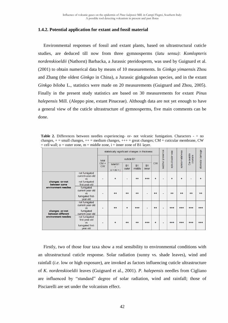
Influence of volcanic gases on the epidermis of Pinus halepensis Mill. in Campi Flegrei, Southern Italy: A possible tool detecting volcanism in present and past floras
42
Table 2. Differences between needles experiencing- or- not volcanic fumigation. Characters - = no
changes, + = small changes, ++ = medium changes, +++ = great changes; CM = cuticular membrane, CW
= cell wall; o = outer zone, m = middle zone, i = inner zone of B1 layer.
1.4.2. Potential application for extant and fossil material
Environmental responses of fossil and extant plants, based on ultrastructural cuticle
studies, are deduced till now from three gymnosperms (latu sensu): Komlopteris
nordenskioeldii (Nathorst) Barbacka, a Jurassic pteridosperm, was used by Guignard et al.
(2001) to obtain numerical data by means of 10 measurements. In Ginkgo yimaensis Zhou
and Zhang (the oldest Ginkgo in China), a Jurassic ginkgoalean species, and in the extant
Ginkgo biloba L., statistics were made on 20 measurements (Guignard and Zhou, 2005).
Finally in the present study statistics are based on 30 measurements for extant Pinus
halepensis Mill. (Aleppo pine, extant Pinaceae). Although data are not yet enough to have
a general view of the cuticle ultrastructure of gymnosperms, five main comments can be
done.
Firstly, two of those four taxa show a real sensibility to environmental conditions with
an ultrastructural cuticle response. Solar radiation (sunny vs. shade leaves), wind and
rainfall (i.e. low or high exposure), are invoked as factors influencing cuticle ultrastructure
of K. nordenskioeldii leaves (Guignard et al., 2001). P. halepensis needles from Cigliano
are influenced by “standard” degree of solar radiation, wind and rainfall; those of
Pisciarelli are set under the volcanism effect.
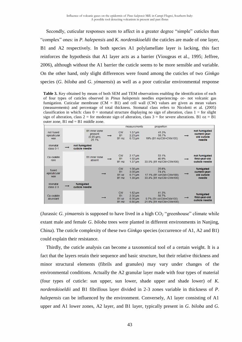
Influence of volcanic gases on the epidermis of Pinus halepensis Mill. in Campi Flegrei, Southern Italy: A possible tool detecting volcanism in present and past floras
43
Table 3. Key obtained by means of both SEM and TEM observations enabling the identification of each
of four types of cuticles observed in Pinus halepensis needles experiencing- or- not volcanic gas
fumigation. Cuticular membrane (CM = B1) and cell wall (CW) values are given as mean values
(measurements) and percentage of total thickness. Stomatal class refers to Nicolotti et al. (2005)
classification in which: class 0 = stomatal structure displaying no sign of alteration, class 1 = for slight
sign of alteration, class 2 = for moderate sign of alteration, class 3 = for severe alterations. B1 oz = B1
outer zone, B1 md = B1 middle zone.
Secondly, cuticular responses seem to affect in a greater degree “simple” cuticles than
“complex” ones: in P. halepensis and K. nordenskioeldii the cuticles are made of one layer,
B1 and A2 respectively. In both species A1 polylamellate layer is lacking, this fact
reinforces the hypothesis that A1 layer acts as a barrier (Viougeas et al., 1995; Jeffree,
2006), although without the A1 barrier the cuticle seems to be more sensible and variable.
On the other hand, only slight differences were found among the cuticles of two Ginkgo
species (G. biloba and G. yimaensis) as well as a poor cuticular environmental response
(Jurassic G. yimaensis is supposed to have lived in a high CO2 “greenhouse” climate while
extant male and female G. biloba trees were planted in different environments in Nanjing,
China). The cuticle complexity of these two Ginkgo species (occurrence of A1, A2 and B1)
could explain their resistance.
Thirdly, the cuticle analysis can become a taxonomical tool of a certain weight. It is a
fact that the layers retain their sequence and basic structure, but their relative thickness and
minor structural elements (fibrils and granules) may vary under changes of the
environmental conditions. Actually the A2 granular layer made with four types of material
(four types of cuticle: sun upper, sun lower, shade upper and shade lower) of K.
nordenskioeldii and B1 fibrillous layer divided in 2-3 zones variable in thickness of P.
halepensis can be influenced by the environment. Conversely, A1 layer consisting of A1
upper and A1 lower zones, A2 layer, and B1 layer, typically present in G. biloba and G.
Influence of volcanic gases on the epidermis of Pinus halepensis Mill. in Campi Flegrei, Southern Italy: A possible tool detecting volcanism in present and past floras
44
yimaensis, can be considered useful for taxa identification because they do not change
under different environmental conditions.
The fourth remark concerns further ultrastructural details, i.e. the progressive reduction
in the fibrillar structure and the corresponding increase in granular component from
current- to first-year-old needles of Pinus halepensis (Plate II, 14-16, 19-21) that can be
interpreted as a response to enhanced environmental stresses. On this regard Villar de
Seoane (2001) illustrates the alteration of the epidermal wall ultrastructure of Otozamites
patagonicus from the Early Cretaceous Springhill Formation of Patagonia, where
multilamellate and reticulate-alveolate layers are transformed to amorphous layers (Villar
de Seoane, 2001 sense) and interpreted as the result of high temperature owing to
persistent volcanic ash fall. Del Fuejo and Archangelsky (2002) point out that amorphous
areas in Araucaria grandifolia Feruglio from the Lower Cretaceous of Patagonia
(Argentina) may be the result of poor preservation of the cuticular membrane.
Moreover deposits of calcium oxalate crystals in P. halepensis could be equivalent
structures to the electron lucent amorphous material of K. nordenskioeldii (Guignard et al.,
2001, Plate II, Fig. 4), although the role of this material is unclear (Guignard et al., 2001).
Similar electron lucent areas are visible in the cuticle of two fossil species (Pseudoctenis
ornata and Restrepophyllum chiguoides) from the Early Cretaceous of Patagonia (Passalia
et al., 2010). It is noteworthy that this vegetation grew under environmental stressful
conditions for persistent volcanic activity (Archangelsky et al., 1995; Archangelsky, 2001;
Del Fuejo and Archangelsky, 2002). The epidermal cells of P. ornata show the sublayer
A2 with irregular isodiametric areas containing material of less electron density
(Archangelsky et al., 1995, Pl. V, Fig. 31). Cuticle degradation is recorded in R. chiguoides
(Passalia et al., 2010, Pl. IV, Fig. 5). To sum up, the increase of Ca-oxalate crystals could
be a key character to identify environmental stress in fossil and extant plants.
The last remark reveals a possible tool to identify current- versus first-year-old Pinus
needles in extant and fossil contexts and make inferences on gas fumigation disease (van
de Schootbrugge et al., 2009 suppose SO2 as a probable cause of plant stress during
periods of global-scale volcanism). According to the present study one can distinguish not
fumigated from fumigated needles (Table 3) by means of eight characters (epicuticular and
epistomatal wax, Ca-oxalate, total B1, CW, B1 inner, outer and middle). Therefore the
degree of wax crystals degradation in stomatal antechamber and epidermal surface,
together with the amount of calcium-oxalate deposits in the cuticle, allow a first distinction
between needles experiencing- or- not volcanic gas fumigation (Table 3). Furthermore
Influence of volcanic gases on the epidermis of Pinus halepensis Mill. in Campi Flegrei, Southern Italy: A possible tool detecting volcanism in present and past floras
45
three characters (i.e. thickness of CW, B1 and B1 middle zone) allow to make a distinction
between current- and first-year-old not fumigated cuticle needles; while four characters
(i.e. thickness of CW, total B1, B1 outer and middle zones) enable to detect fumigated
needles. In particular the presence or absence of B1 inner zone (see Figs. 4,5) can be used
to distinguish not fumigated current- from first-year-old needles. For the thickness of
cuticular membrane (CM) + cell wall (CW) keeps constant in P. halepensis needles
regardless the age and environmental conditions, this character could be considered as a
taxonomical one at species level.
The cuticle micromorphology of in situ Erica arborea L. exposed to long-term volcanic gases in Phlegrean Fields, Campania, Italy
46
CHAPTER II
The cuticle micromorphology of in situ Erica arborea L. exposed to long-
term volcanic gases in Phlegrean Fields, Campania, Italy
2.1. Introduction
Within plant organs, the leaf has been the target of many studies (see Beck, 2010) and is
the most sensitive organ to pollution (Sant’Anna-Santos et al., 2006). The leaves of many
tracheophytes (Guignard et al., 2004) are covered by the cuticle, an extracellular
membrane (Graham, 1993) covering aerial organs of plants (Riederer, 2006). The bulk of
the cuticle in modern plants is composed of a solvent-insoluble matrix makes up the
framework of the cuticle (Tegelaar et al., 1991). This composite matrix provides several
functions at the interface level that enable plants to thrive in different habitats and
withstand adverse environmental conditions (Domínguez et al., 2011). The cuticle could be
considered an “external skeleton” as it represents the interface between the plant and the
atmosphere (McElwain and Chaloner, 1996). For these reasons, in the last three decades
numerous studies have been carried out on fossil and extant plant cuticles in order to
attempt paleoenvironmental reconstructions and evaluate the gaseous atmospheric
composition. This latter composition varied consistently during geological time (Berner,
1999) and major volcanism -e.g. Large Igneous Provinces formation (Courtillot and Renne,
2003)- is frequently invoked as a possible cause of mass extinctions (Rhode and Muller,
2005) by means of massive degassing (Scaillet, 2008; Sobolev et al., 2011; Wignall, 2011)
of CO2, H2S and SO2 into the atmosphere (Beerling and Berner, 2002; Berner and
Beerling, 2007; Hori et al., 2007). The volumes of SO2 outgassed during large basaltic
eruptions are comparable in magnitude to those of CO2 (Thoardarson et al., 1996). For this
reason, volcanism played an important role in past plant communities composition
(Falcon-Lang and Cantrill, 2002) insomuch as, climatic fluctuations and proximal volcanic
eruptions probably contributed to the formation of disturbed habitats favourable for early
angiosperms (Herman, 2002).
Numerous researches spent their efforts onto the study of cuticle topography (number of
stomata per unit area = stomatal density; number of stomata relative to the number of
epidermal cells = stomatal index), at mesoscopic scale (see Collinson 1999), and its
relationships with atmospheric CO2 concentration ([CO2]) in the geological past
The cuticle micromorphology of in situ Erica arborea L. exposed to long-term volcanic gases in Phlegrean Fields, Campania, Italy
47
(Woodward, 1987; Beerling et al., 1998; Royer, 2001). It has been assumed that plants’
stomatal density adjust in response to changing atmospheric pCO2 (Woodward, 1987).
However, little is known (Winner and Mooney, 1980) about the long-term response of
micromorphology of natural vegetation to volcanic toxic gases. Moreover, it is unclear if
small community experiments are capable of both reconstruct and predict gas atmospheric
composition because these experiments often lack the necessary range of natural
disturbances that occur in the long term (Woodward, 1992).
Because of the difficulties to simulate complex environmental conditions within plant-
volcano interactions, we studied the in situ angiosperm Erica arborea L. submitted to
volcanic gases in two solfataras (sulphur-emitting vents): Solfatara di Pozzuoli and
Pisciarelli. Solfataras are exciting habitats for studying wildlife adaptations to a very
peculiar but extreme environment (see Paoletti et al., 2005). The populations of E. arborea
growing around the vents have a multiple-generation and a life-time exposure to volcanic
gases, resulting in long-term adaptations of plants to these naturally environments. The
occurrence of vents emitting volcanic gases provides a rare opportunity to study long-term
response and microevolutionary adaptation (Bettarini et al., 1995) of natural vegetation to
volcanic gases.
Numerous studies have been carried out on in situ plants exposed to geogenic gases
(Bettarini et al., 1995; Bettarini et al., 1998; Tognetti et al., 2000; Paoletti et al., 2005;
Pfanz et al., 2007; Haworth et al., 2010) but as far as we know, there are no studies
relatively to the response of the ultrastructural features of angiosperms plant cuticle
exposed to the persistent volcanic gases.
Therefore, this study aims to try understand: 1) how plants respond to realistic extremes
outdoor conditions; 2) what is the response of the cuticle micromorphology long-term
exposed to volcanic gases, 3) in particular, whether exposure and/or resistance with
volcanic gases is associated with a change in ultrastructure of cuticle; 4) potential
implications of the angiosperm cuticle response across environmental stress periods during
the geological past.
Investigations on volcanic toxic gases-emitting vents offer opportunity to study plant
physiological response to these extreme environments. The volcanic areas in Campania
Region (Roccamonfina, Phlegrean Fields and the complex of Somma-Vesuvio) are suitable
for studies of this type because they are historically monitored and easily accessible
representing natural laboratories allowing experiments to realistic extremes outdoor

The cuticle micromorphology of in situ Erica arborea L. exposed to long-term volcanic gases in Phlegrean Fields, Campania, Italy
48
Fig. 1. (A) Location of the Phlegrean Fields and others gas-emitting volcanic areas (dotted circles) in
Campania Region. (B) Sketch map showing the location of Solfatara crater and Pisciarelli locality. (C,D)
Close up views of Pisciarelli and Solfatara localities with the main fumaroles. Square brackets indicate
sampling areas and white circlets show the location of CO2 measurements.
conditions. In the present study, E. arborea was chosen and X-ray analyses and SEM and
TEM observations were performed focusing on leaf cuticle micromorphology.
2.2. Material and methods
2.2.1. Plant material and sites description
Leaf samples were obtained from Erica arborea L., one of the common and widespread
Mediterranean macchia shrub species belonging to the family Ericaceae including species
that are efficient colonizers (Luteyn, 2002). E. arborea is characterized by hypostomatic
leaves with revolute margins protected by trichomes (Gratani and Varone, 2004). Samples
were collected from three localities in the Phlegrean Fields, an active caldera which spans
the last 50000 years (Scandone et al., 2010), characterized by vertical ground movements
locally called “Bradyseim” (Del Gaudio et al., 2010). This is a very dangerous volcanic
area because it includes part of the city of Napoli, the town of Pozzuoli, and numerous
densely inhabited villages (Chiodini et al., 2011).
Samples long-term fumigated by volcanic gases were recovered from Solfatara crater,
ca. 30 m NNW from the Bocca Grande vent (40°49’38.91’’N, 14°08’32.98’’E), and ca. 10
m NE from the main fumarole in Pisciarelli locality (40°49’48.88’’N, 14°08’46.95’’E)
about 750 m SE of the Solfatara volcano (Fig. 1A,B). These two volcanic areas, active for
centuries, are historically monitored (Gunther, 1897) and make the object of numerous
studies.
The cuticle micromorphology of in situ Erica arborea L. exposed to long-term volcanic gases in Phlegrean Fields, Campania, Italy
49
Control sample of leaves were also collected in a volcanic quiescent area (Cigliano:
40°50’46.46’’N, 14°07’36.31’’E) located about 2.5 km NNW from Pisciarelli, but
characterized by the absence of volcanic gas emissions. The three localities share the same
soil features (Di Gennaro and Terribile, 1999; Di Gennaro, 2002) and sun exposition and
they are also far away from traffic and industries. The climate is typically Mediterranean,
with cool, wet winters and hot, dry summers.
The samples collected around the vents have a multiple-generation resulting in long-
term adaptations of plants to volcanic gases. The outskirt of these extreme environments
(Pisciarelli and Solfatara) is inhabited by a Mediterranean vegetation providing
xeromorphic features. Erica arborea (and some grasses) is the unique typical
Mediterranean angiosperm shrub growing near the vents. Moreover, the boiling water of
Pisciarelli and Solfatara fumaroles retains the cyanidialean algaes Galdieria phlegrea
(Pinto et al., 2007) and Cyanidium caldarium (De Luca et al., 1979) respectively.
Distal volcanic impacts revealed that plants are generally less sensitive to eruptions
outside the growing season (Zobel and Antos, 1997; Hotes et al., 2004) and, as Payne and
Blackford (2008) pointed out, in winter, as plants are senescent, higher rainfall may serve
to remove rapidly volcanic pollutants. For evergreen E. arborea, permanently fumigated
by volcanic gases retaining leaves for over a year, these “ground noises” do not exist. The
plants do not present significant damages along the crown, but some leaves show
symptoms consisting in leaf-tip non-specific discoloration.
2.2.2. Gas vent
At Pisciarelli, the fumarolic discharge temperature from 1999 to 2005 has fluctuated
around 95 °C, reaching 108 °C in October 2010, with the CO2 flux from this pool
estimated at 15 ton/d to 20 ton/d (Chiodini et al., 2011). Measurements of diffuse soil CO2
fluxes in the Solfatara area (1.4 km2) were carried out from 1998 to 2000 and gave a daily
amount of deeply derived CO2 discharged at Solfatara of about 1500 tons (Caliro et al.,
2007). The analysis of gaseous compositions (Caliro et al., 2007) revealed that the main
component of the fumaroles is H2O followed by CO2, H2S, N2, H2, CH4, He, Ar, and CO.
The absence of acidic gases (SO2, HCl, and HF) can be also noted (Chiodini et al., 2010a).
Natural high atmospheric concentration of sulphur gas may occur locally in areas with
volcanic and geothermic activity (De Kok et al., 2007). As a matter of fact, at Pisciarelli
and Solfatara, the high H2S air concentration is equal to ca 600 μm/mol (at source;
The cuticle micromorphology of in situ Erica arborea L. exposed to long-term volcanic gases in Phlegrean Fields, Campania, Italy
50
Chiodini et al., 2010a) and 3.20 ppm (Carapezza et al., 1984) respectively. The great H2S
air concentration is also testified by both typical smell (the odour threshold is >0.02 μm l-1
:
De Kok et al., 2007) of rotten eggs in the surrounding air and by indirect corrosive action
of this gas that led to the destruction, around the vents and in few minutes, of any
electronic device and to a relatively long-term corrosion of metalwork.
2.2.3. SEM, TEM and EDS preparations
Fumigated and not fumigated specimens were collected from branches at heights over
75 cm from the ground at each site (Pisciarelli, Solfatara and Cigliano). Leaves were
carefully handled to avoid damaging the epicuticular waxes. Following Reed’s (1982) and
Crang and Klomparens’ (1988) remarks about possible changes in epicuticular wax
structures occurring during sample preparation, in order to limit any chemical or physical
damages, especially for preserving and dehydrating samples for wax morphology studies,
leaves were air-dried for 1-week at mild room temperature. Among several hundreds of
leaves collected at each site (fumigated and not fumigated), 100 were selected for scanning
electron microscope (SEM), then 60 were carefully selected for transmission electron
microscope (TEM). Light microscope observations were made using a Leitz microscope.
Both untreated and frozen leaves (in liquid nitrogen at -210 °C) were cut transversally
and longitudinally. Subsequently they were mounted onto the stubs using double-sided
adhesive tape then coated with gold using an AGAR Auto Sputter Coater and transferred to
the SEM for mesoscopic (Collinson, 1999) observations. Entire leaves were directly
mounted to observe abaxial and adaxial surfaces. The Barthlott et al. (1998) classification
has been used to typify epicuticular wax.
SEM observations were done in two universities: 1) with a JEOL JSM-5310 SEM
adapted with an Energy Diffractive X-ray Oxford Inca X-act at the CISAG (Centro
Interdipartimentale di servizi per analisi Geomineralogiche; Dipartimento di Scienze della
Terra, Università degli Studi di Napoli “Federico II”). The operative conditions were as
follows: 25-30 KV accelerating voltage, 100 μA emission current, 15 μm spot size, 20 mm
microscope work distance and 60 s spectra collection time; 2) with a Hitachi S800 coupled
to a microanalysis system EDS SiLi Samix (acceleration voltage = 15 kV and a spectres
acquisition time = 60 s; in the Centre de technologie des microstructures (CTμ) of Lyon-1
University, Villeurbanne, France). The specimens for energy diffractive x-ray (EDX)
analyses were then coated with carbon in an Emitech K450 at CISAG. A total of 100 EDS
The cuticle micromorphology of in situ Erica arborea L. exposed to long-term volcanic gases in Phlegrean Fields, Campania, Italy
51
analyses were carried out on both leaf sections (cuticle and cell wall with JEOL SEM) and
surfaces (crystals, hairs and epidermis with SEM Hitachi) experiencing or not chronic
fumigation. It is worth noting that data generated by the X-ray microanalyzer are not as
accurate as other quantitative analyses, therefore they can be considered at best as semi-
quantitative (Van Steveninck and Van Steveninck, 1991).
Samples for TEM were dropped in paraformaldehyde solution mixed in a phosphate-
sodium buffer for 3 weeks using Lugardon's technique (1971), washed and postfixed in a
1% osmium tetroxide solution mixed in a phosphate–sodium buffer for 24 hours.
Dehydrated in graded ethanol series during 48 h, the samples were dropped in propylene
oxide with an increasing percentage of Epon resin for 24 h. Transferred into pure Epon
resin during 24 hours, they were embedded in fresh Epon resin using flat moulds. The
preparations were subsequently treated for polymerization at 56 °C for 3 days. Ultrathin
(60-70 nm) sections were sectioned with a diamond knife, using a Reichert Ultracut
microtome. Ultrathin sections were placed on uncoated 300 Mesh copper grids and stained
manually both with a methanol solution of 7% uranyl acetate for 15 min and an aqueous
lead-citrate solution for 20 min, then observed and photographed with a Philips CM 120
TEM at 80 kV, in the Centre de technologie des microstructures (CTμ) of Lyon-1
University, Villeurbanne, France. Totally 60 pieces of material were embedded in Epon
resin blocks. 210 uncoated mesh copper grids were prepared (180 as transversal sections,
i.e. perpendicular to the leaf length; 30 as longitudinal sections, i.e. parallel to the leaf
length).
All TEM measurements were performed with tools in the ImageJ program (Abramoff et
al. 2004). The terminology of Holloway (1982) and Archangelsky (1991) was used for the
ultrastructural analysis. Ultrastructural measures have been taken in the middle of
epidermal cells. No measures have been carried out in overlying cuticle between two
adjacent epidermal cells where the cuticle is generally thicker. All specimens and SEM
stubs are housed in the Dipartimento di Scienze della Terra, Largo San Marcellino, 10,
Napoli, Italy. The resin blocks and TEM negatives are stored in the Lyon-1 University,
Villeurbanne, France.
2.2.4. Gas concentration measurements in air and soil
CO2 measurements in the soil and in the air surrounding the vegetation were performed
with a portable gas analyser (LICOR 800). Holes were drilled into the soil until roots of
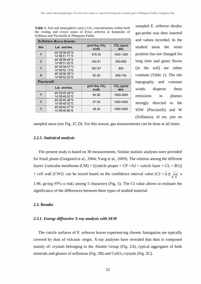
The cuticle micromorphology of in situ Erica arborea L. exposed to long-term volcanic gases in Phlegrean Fields, Campania, Italy
52
Table 1. Soil and atmospheric (atm.) CO2 concentrations within both
the rooting and crown zones of Erica arborea at fumaroles of
Solfatara and Pisciarelli in Phlegrean Fields.
sampled E. arborea shrubs;
gas-probe was then inserted
and values recorded. In the
studied areas the vents
position has not changed for
long time and gases fluxes
(in the soil) are rather
constant (Table 1). The site
topography and constant
winds disperse these
emissions in plumes
strongly directed to the
SSW (Pisciarelli) and W
(Solfatara), id est. just on
sampled areas (see Fig. 1C,D). For this reason, gas measurements can be done at all times.
2.2.5. Statistical analysis
The present study is based on 30 measurements. Similar statistic analyses were provided
for fossil plants (Guignard et al., 2004; Yang et al., 2009). The relation among the different
layers {cuticular membrane (CM) = [(cuticle proper = CP =A2 + cuticle layer = CL = B1)]
+ cell wall (CW)} can be traced based on the confidence interval value (CI = x ±n
var x
1.96. giving 95% α risk) among 5 characters (Fig. 5). The CI value allows to estimate the
significance of the differences between three types of studied material.
2.3. Results
2.3.1. Energy diffractive X-ray analysis with SEM
The cuticle surfaces of E. arborea leaves experiencing chronic fumigation are typically
covered by dust of volcanic origin. X-ray analyses have revealed that dust is composed
mainly of: crystals belonging to the Alunite Group (Fig. 2A), typical aggregates of both
minerals and glasses of solfataras (Fig. 2B) and CaSO4 crystals (Fig. 2C).
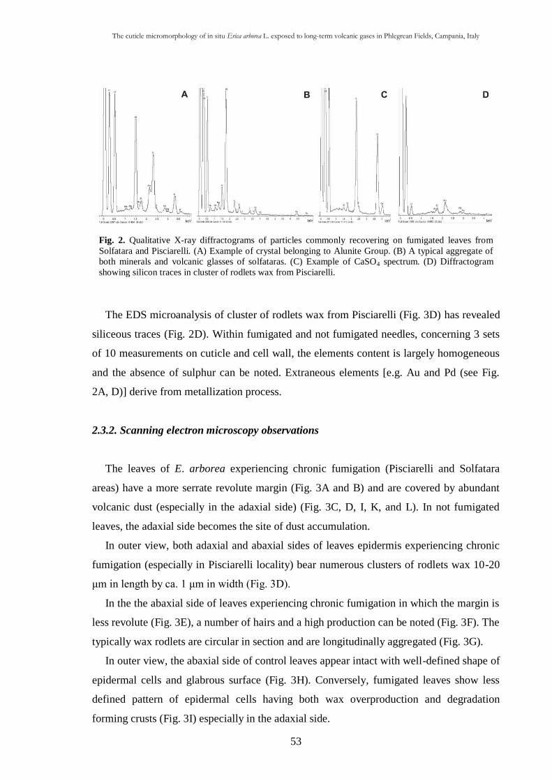
The cuticle micromorphology of in situ Erica arborea L. exposed to long-term volcanic gases in Phlegrean Fields, Campania, Italy
53
Fig. 2. Qualitative X-ray diffractograms of particles commonly recovering on fumigated leaves from
Solfatara and Pisciarelli. (A) Example of crystal belonging to Alunite Group. (B) A typical aggregate of
both minerals and volcanic glasses of solfataras. (C) Example of CaSO4 spectrum. (D) Diffractogram
showing silicon traces in cluster of rodlets wax from Pisciarelli.
The EDS microanalysis of cluster of rodlets wax from Pisciarelli (Fig. 3D) has revealed
siliceous traces (Fig. 2D). Within fumigated and not fumigated needles, concerning 3 sets
of 10 measurements on cuticle and cell wall, the elements content is largely homogeneous
and the absence of sulphur can be noted. Extraneous elements [e.g. Au and Pd (see Fig.
2A, D)] derive from metallization process.
2.3.2. Scanning electron microscopy observations
The leaves of E. arborea experiencing chronic fumigation (Pisciarelli and Solfatara
areas) have a more serrate revolute margin (Fig. 3A and B) and are covered by abundant
volcanic dust (especially in the adaxial side) (Fig. 3C, D, I, K, and L). In not fumigated
leaves, the adaxial side becomes the site of dust accumulation.
In outer view, both adaxial and abaxial sides of leaves epidermis experiencing chronic
fumigation (especially in Pisciarelli locality) bear numerous clusters of rodlets wax 10-20
μm in length by ca. 1 μm in width (Fig. 3D).
In the the abaxial side of leaves experiencing chronic fumigation in which the margin is
less revolute (Fig. 3E), a number of hairs and a high production can be noted (Fig. 3F). The
typically wax rodlets are circular in section and are longitudinally aggregated (Fig. 3G).
In outer view, the abaxial side of control leaves appear intact with well-defined shape of
epidermal cells and glabrous surface (Fig. 3H). Conversely, fumigated leaves show less
defined pattern of epidermal cells having both wax overproduction and degradation
forming crusts (Fig. 3I) especially in the adaxial side.
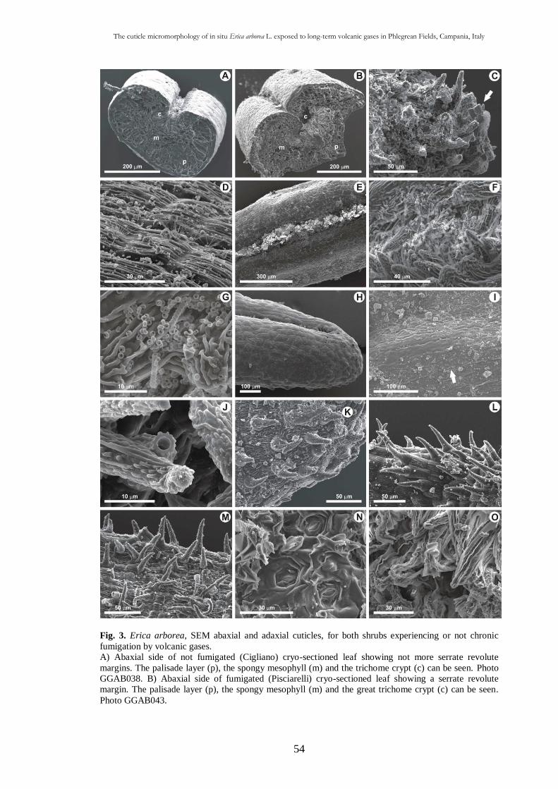
The cuticle micromorphology of in situ Erica arborea L. exposed to long-term volcanic gases in Phlegrean Fields, Campania, Italy
54
Fig. 3. Erica arborea, SEM abaxial and adaxial cuticles, for both shrubs experiencing or not chronic
fumigation by volcanic gases.
A) Abaxial side of not fumigated (Cigliano) cryo-sectioned leaf showing not more serrate revolute
margins. The palisade layer (p), the spongy mesophyll (m) and the trichome crypt (c) can be seen. Photo
GGAB038. B) Abaxial side of fumigated (Pisciarelli) cryo-sectioned leaf showing a serrate revolute
margin. The palisade layer (p), the spongy mesophyll (m) and the great trichome crypt (c) can be seen.
Photo GGAB043.
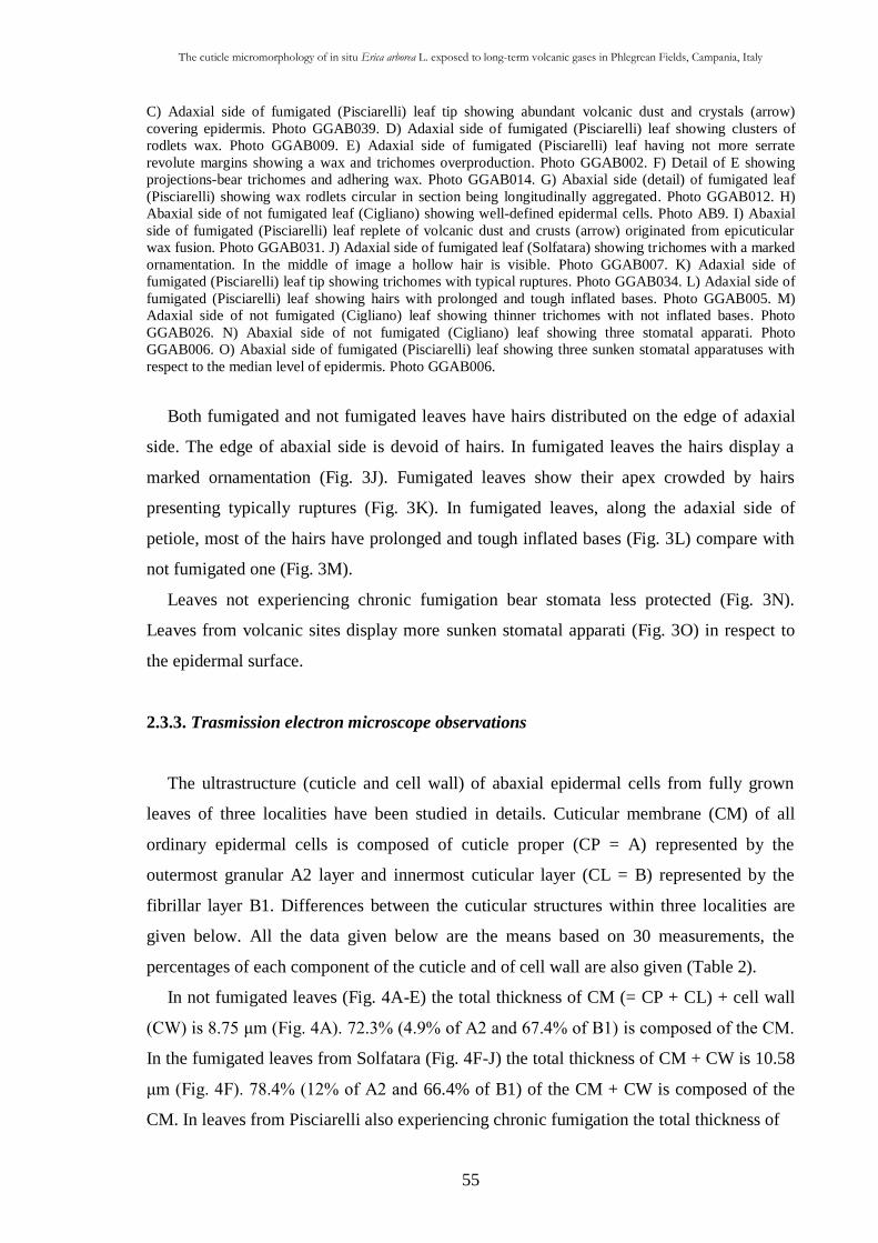
The cuticle micromorphology of in situ Erica arborea L. exposed to long-term volcanic gases in Phlegrean Fields, Campania, Italy
55
C) Adaxial side of fumigated (Pisciarelli) leaf tip showing abundant volcanic dust and crystals (arrow)
covering epidermis. Photo GGAB039. D) Adaxial side of fumigated (Pisciarelli) leaf showing clusters of
rodlets wax. Photo GGAB009. E) Adaxial side of fumigated (Pisciarelli) leaf having not more serrate
revolute margins showing a wax and trichomes overproduction. Photo GGAB002. F) Detail of E showing
projections-bear trichomes and adhering wax. Photo GGAB014. G) Abaxial side (detail) of fumigated leaf
(Pisciarelli) showing wax rodlets circular in section being longitudinally aggregated. Photo GGAB012. H)
Abaxial side of not fumigated leaf (Cigliano) showing well-defined epidermal cells. Photo AB9. I) Abaxial
side of fumigated (Pisciarelli) leaf replete of volcanic dust and crusts (arrow) originated from epicuticular
wax fusion. Photo GGAB031. J) Adaxial side of fumigated leaf (Solfatara) showing trichomes with a marked
ornamentation. In the middle of image a hollow hair is visible. Photo GGAB007. K) Adaxial side of
fumigated (Pisciarelli) leaf tip showing trichomes with typical ruptures. Photo GGAB034. L) Adaxial side of
fumigated (Pisciarelli) leaf showing hairs with prolonged and tough inflated bases. Photo GGAB005. M)
Adaxial side of not fumigated (Cigliano) leaf showing thinner trichomes with not inflated bases. Photo
GGAB026. N) Abaxial side of not fumigated (Cigliano) leaf showing three stomatal apparati. Photo
GGAB006. O) Abaxial side of fumigated (Pisciarelli) leaf showing three sunken stomatal apparatuses with
respect to the median level of epidermis. Photo GGAB006.
Both fumigated and not fumigated leaves have hairs distributed on the edge of adaxial
side. The edge of abaxial side is devoid of hairs. In fumigated leaves the hairs display a
marked ornamentation (Fig. 3J). Fumigated leaves show their apex crowded by hairs
presenting typically ruptures (Fig. 3K). In fumigated leaves, along the adaxial side of
petiole, most of the hairs have prolonged and tough inflated bases (Fig. 3L) compare with
not fumigated one (Fig. 3M).
Leaves not experiencing chronic fumigation bear stomata less protected (Fig. 3N).
Leaves from volcanic sites display more sunken stomatal apparati (Fig. 3O) in respect to
the epidermal surface.
2.3.3. Trasmission electron microscope observations
The ultrastructure (cuticle and cell wall) of abaxial epidermal cells from fully grown
leaves of three localities have been studied in details. Cuticular membrane (CM) of all
ordinary epidermal cells is composed of cuticle proper (CP = A) represented by the
outermost granular A2 layer and innermost cuticular layer (CL = B) represented by the
fibrillar layer B1. Differences between the cuticular structures within three localities are
given below. All the data given below are the means based on 30 measurements, the
percentages of each component of the cuticle and of cell wall are also given (Table 2).
In not fumigated leaves (Fig. 4A-E) the total thickness of CM (= CP + CL) + cell wall
(CW) is 8.75 μm (Fig. 4A). 72.3% (4.9% of A2 and 67.4% of B1) is composed of the CM.
In the fumigated leaves from Solfatara (Fig. 4F-J) the total thickness of CM + CW is 10.58
μm (Fig. 4F). 78.4% (12% of A2 and 66.4% of B1) of the CM + CW is composed of the
CM. In leaves from Pisciarelli also experiencing chronic fumigation the total thickness of
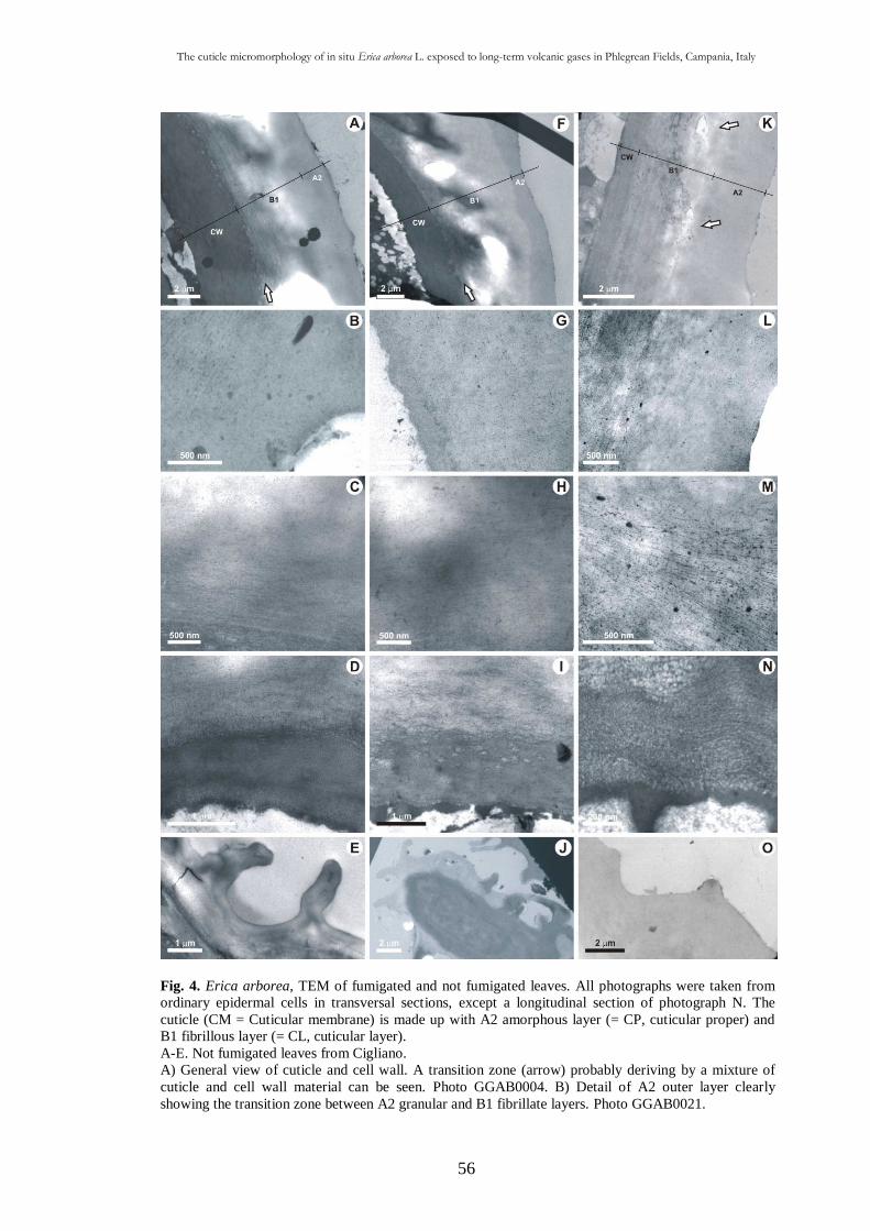
The cuticle micromorphology of in situ Erica arborea L. exposed to long-term volcanic gases in Phlegrean Fields, Campania, Italy
56
Fig. 4. Erica arborea, TEM of fumigated and not fumigated leaves. All photographs were taken from
ordinary epidermal cells in transversal sections, except a longitudinal section of photograph N. The
cuticle (CM = Cuticular membrane) is made up with A2 amorphous layer (= CP, cuticular proper) and
B1 fibrillous layer (= CL, cuticular layer).
A-E. Not fumigated leaves from Cigliano.
A) General view of cuticle and cell wall. A transition zone (arrow) probably deriving by a mixture of
cuticle and cell wall material can be seen. Photo GGAB0004. B) Detail of A2 outer layer clearly
showing the transition zone between A2 granular and B1 fibrillate layers. Photo GGAB0021.
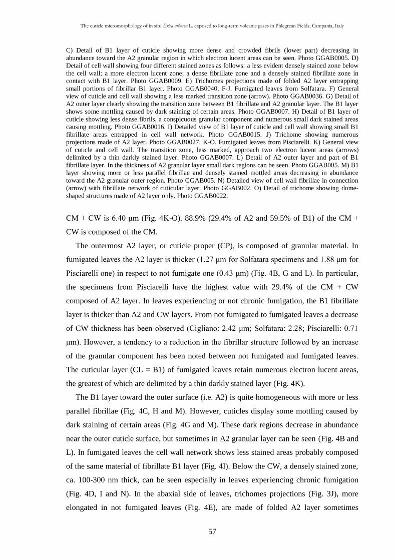
The cuticle micromorphology of in situ Erica arborea L. exposed to long-term volcanic gases in Phlegrean Fields, Campania, Italy
57
C) Detail of B1 layer of cuticle showing more dense and crowded fibrils (lower part) decreasing in
abundance toward the A2 granular region in which electron lucent areas can be seen. Photo GGAB0005. D)
Detail of cell wall showing four different stained zones as follows: a less evident densely stained zone below
the cell wall; a more electron lucent zone; a dense fibrillate zone and a densely stained fibrillate zone in
contact with B1 layer. Photo GGAB0009. E) Trichomes projections made of folded A2 layer entrapping
small portions of fibrillar B1 layer. Photo GGAB0040. F-J. Fumigated leaves from Solfatara. F) General
view of cuticle and cell wall showing a less marked transition zone (arrow). Photo GGAB0036. G) Detail of
A2 outer layer clearly showing the transition zone between B1 fibrillate and A2 granular layer. The B1 layer
shows some mottling caused by dark staining of certain areas. Photo GGAB0007. H) Detail of B1 layer of
cuticle showing less dense fibrils, a conspicuous granular component and numerous small dark stained areas
causing mottling. Photo GGAB0016. I) Detailed view of B1 layer of cuticle and cell wall showing small B1
fibrillate areas entrapped in cell wall network. Photo GGAB0015. J) Trichome showing numerous
projections made of A2 layer. Photo GGAB0027. K-O. Fumigated leaves from Pisciarelli. K) General view
of cuticle and cell wall. The transition zone, less marked, approach two electron lucent areas (arrows)
delimited by a thin darkly stained layer. Photo GGAB0007. L) Detail of A2 outer layer and part of B1
fibrillate layer. In the thickness of A2 granular layer small dark regions can be seen. Photo GGAB005. M) B1
layer showing more or less parallel fibrillae and densely stained mottled areas decreasing in abundance
toward the A2 granular outer region. Photo GGAB005. N) Detailed view of cell wall fibrillae in connection
(arrow) with fibrillate network of cuticular layer. Photo GGAB002. O) Detail of trichome showing dome-
shaped structures made of A2 layer only. Photo GGAB0022.
CM + CW is 6.40 μm (Fig. 4K-O). 88.9% (29.4% of A2 and 59.5% of B1) of the CM +
CW is composed of the CM.
The outermost A2 layer, or cuticle proper (CP), is composed of granular material. In
fumigated leaves the A2 layer is thicker (1.27 μm for Solfatara specimens and 1.88 μm for
Pisciarelli one) in respect to not fumigate one (0.43 μm) (Fig. 4B, G and L). In particular,
the specimens from Pisciarelli have the highest value with 29.4% of the CM + CW
composed of A2 layer. In leaves experiencing or not chronic fumigation, the B1 fibrillate
layer is thicker than A2 and CW layers. From not fumigated to fumigated leaves a decrease
of CW thickness has been observed (Cigliano: 2.42 μm; Solfatara: 2.28; Pisciarelli: 0.71
μm). However, a tendency to a reduction in the fibrillar structure followed by an increase
of the granular component has been noted between not fumigated and fumigated leaves.
The cuticular layer (CL = B1) of fumigated leaves retain numerous electron lucent areas,
the greatest of which are delimited by a thin darkly stained layer (Fig. 4K).
The B1 layer toward the outer surface (i.e. A2) is quite homogeneous with more or less
parallel fibrillae (Fig. 4C, H and M). However, cuticles display some mottling caused by
dark staining of certain areas (Fig. 4G and M). These dark regions decrease in abundance
near the outer cuticle surface, but sometimes in A2 granular layer can be seen (Fig. 4B and
L). In fumigated leaves the cell wall network shows less stained areas probably composed
of the same material of fibrillate B1 layer (Fig. 4I). Below the CW, a densely stained zone,
ca. 100-300 nm thick, can be seen especially in leaves experiencing chronic fumigation
(Fig. 4D, I and N). In the abaxial side of leaves, trichomes projections (Fig. 3J), more
elongated in not fumigated leaves (Fig. 4E), are made of folded A2 layer sometimes
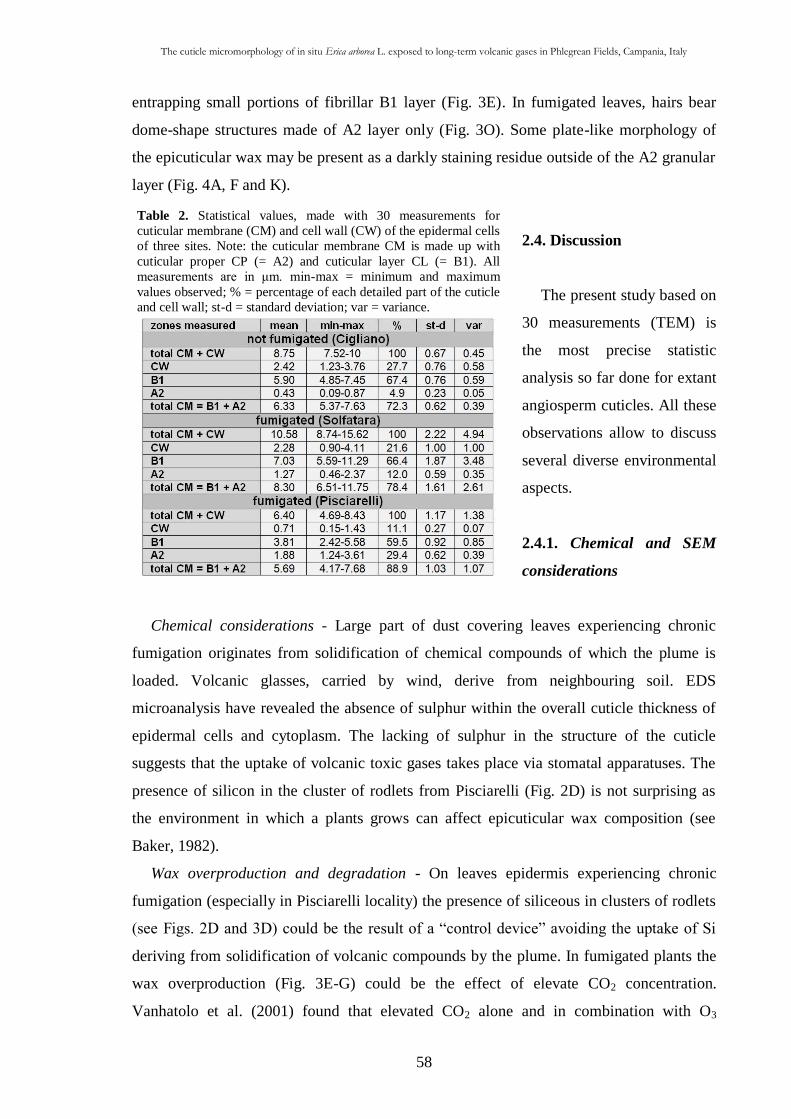
The cuticle micromorphology of in situ Erica arborea L. exposed to long-term volcanic gases in Phlegrean Fields, Campania, Italy
58
Table 2. Statistical values, made with 30 measurements for
cuticular membrane (CM) and cell wall (CW) of the epidermal cells
of three sites. Note: the cuticular membrane CM is made up with
cuticular proper CP (= A2) and cuticular layer CL (= B1). All
measurements are in μm. min-max = minimum and maximum
values observed; % = percentage of each detailed part of the cuticle
and cell wall; st-d = standard deviation; var = variance.
entrapping small portions of fibrillar B1 layer (Fig. 3E). In fumigated leaves, hairs bear
dome-shape structures made of A2 layer only (Fig. 3O). Some plate-like morphology of
the epicuticular wax may be present as a darkly staining residue outside of the A2 granular
layer (Fig. 4A, F and K).
2.4. Discussion
The present study based on
30 measurements (TEM) is
the most precise statistic
analysis so far done for extant
angiosperm cuticles. All these
observations allow to discuss
several diverse environmental
aspects.
2.4.1. Chemical and SEM
considerations
Chemical considerations - Large part of dust covering leaves experiencing chronic
fumigation originates from solidification of chemical compounds of which the plume is
loaded. Volcanic glasses, carried by wind, derive from neighbouring soil. EDS
microanalysis have revealed the absence of sulphur within the overall cuticle thickness of
epidermal cells and cytoplasm. The lacking of sulphur in the structure of the cuticle
suggests that the uptake of volcanic toxic gases takes place via stomatal apparatuses. The
presence of silicon in the cluster of rodlets from Pisciarelli (Fig. 2D) is not surprising as
the environment in which a plants grows can affect epicuticular wax composition (see
Baker, 1982).
Wax overproduction and degradation - On leaves epidermis experiencing chronic
fumigation (especially in Pisciarelli locality) the presence of siliceous in clusters of rodlets
(see Figs. 2D and 3D) could be the result of a “control device” avoiding the uptake of Si
deriving from solidification of volcanic compounds by the plume. In fumigated plants the
wax overproduction (Fig. 3E-G) could be the effect of elevate CO2 concentration.
Vanhatolo et al. (2001) found that elevated CO2 alone and in combination with O3
The cuticle micromorphology of in situ Erica arborea L. exposed to long-term volcanic gases in Phlegrean Fields, Campania, Italy
59
increased wax coverage on the abaxial leaf of an inland clone of pubescent birch Betula
pubescens, but not in that of coastal clone. Among Solfatara and Pisciarelli fumarolic
effluents a relatively high quantity of hydrogen sulphide (H2S) is released (Chiodini et al.,
2010; Caparezza et al., 1984). Brown (1982), Lorenzini and Nali (2005) and Haworth et al.
(2010) claimed that the H2S oxidizes rapidly in the atmosphere to form SO2; this last
rapidly converts into H2SO4 (Visscher et al., 2004) considered as an oxidation product of
SO2 or H2S (Mather et al., 2003). Therefore, the wax degradation noticed in adaxial side of
leaves experiencing volcanic gases fumigation (Fig. 3I) could be due to one of the previous
listed chemical compounds.
Trichomes alteration - In fumigated leaves, the high density of trichomes in the abaxial
side (Fig. 3B) together with hairs having prolonged and tough inflated bases in the adaxial
side of petiole (Fig. 3L) represent a barrier against toxic volcanic gases diffusion
increasing boundary layer resistance (see Haworth and McElwain, 2008). A similar
example can be done with the angiosperm Metrosideros polymorpha growing in the
Kilauea crater region (Island of Hawaii) which is resistant to persistent fumigation with
toxic volcanic gases (SO2, H2S, HCl, HF) (Winner and Mooney, 1980b). M. polymorpha
from the Kilauea crater region displays heavily trichomous abaxial surfaces, while those in
neighbouring regions, not experiencing chronic fumigation, possess non-trichomous
cuticles (Haworth, 2006). This pattern suggests (Haworth, 2006) that the development of
trichomes in M. polymorpha from Kilauea Crater region is an adaptation to increase
boundary layer resistance, and consequently reduces entry of toxic gases into the leaf.
Stomatal apparati - In fumigated leaves, the stomata are located beneath the medium
level of epidermis (Fig. 3O). As stomata are the site of entrance of ca. 90% of atmospheric
pollutants into the plant (Black and Unsworth, 1979), it may be also hypothesized that this
feature could be a further protection device against the uptake of volcanic toxic gases.
These features stress more xeromorphic features already existing in this species. As E.
arborea is the unique typical Mediterranean angiosperm shrub able of growth close to the
vents it could be attributable to a more effective stomatal control than other species with
less effective stomatal control (see Haworth et al. 2011).
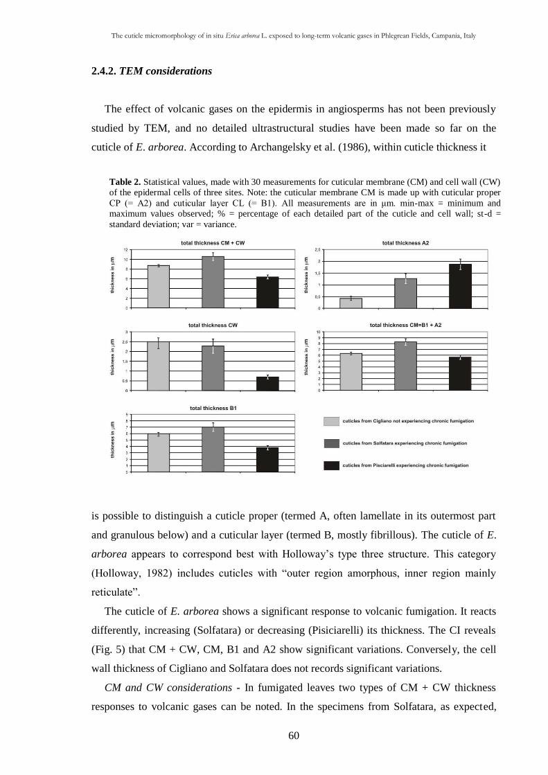
The cuticle micromorphology of in situ Erica arborea L. exposed to long-term volcanic gases in Phlegrean Fields, Campania, Italy
60
Table 2. Statistical values, made with 30 measurements for cuticular membrane (CM) and cell wall (CW)
of the epidermal cells of three sites. Note: the cuticular membrane CM is made up with cuticular proper
CP (= A2) and cuticular layer CL (= B1). All measurements are in μm. min-max = minimum and
maximum values observed; % = percentage of each detailed part of the cuticle and cell wall; st-d =
standard deviation; var = variance.
2.4.2. TEM considerations
The effect of volcanic gases on the epidermis in angiosperms has not been previously
studied by TEM, and no detailed ultrastructural studies have been made so far on the
cuticle of E. arborea. According to Archangelsky et al. (1986), within cuticle thickness it
is possible to distinguish a cuticle proper (termed A, often lamellate in its outermost part
and granulous below) and a cuticular layer (termed B, mostly fibrillous). The cuticle of E.
arborea appears to correspond best with Holloway’s type three structure. This category
(Holloway, 1982) includes cuticles with “outer region amorphous, inner region mainly
reticulate”.
The cuticle of E. arborea shows a significant response to volcanic fumigation. It reacts
differently, increasing (Solfatara) or decreasing (Pisiciarelli) its thickness. The CI reveals
(Fig. 5) that CM + CW, CM, B1 and A2 show significant variations. Conversely, the cell
wall thickness of Cigliano and Solfatara does not records significant variations.
CM and CW considerations - In fumigated leaves two types of CM + CW thickness
responses to volcanic gases can be noted. In the specimens from Solfatara, as expected,
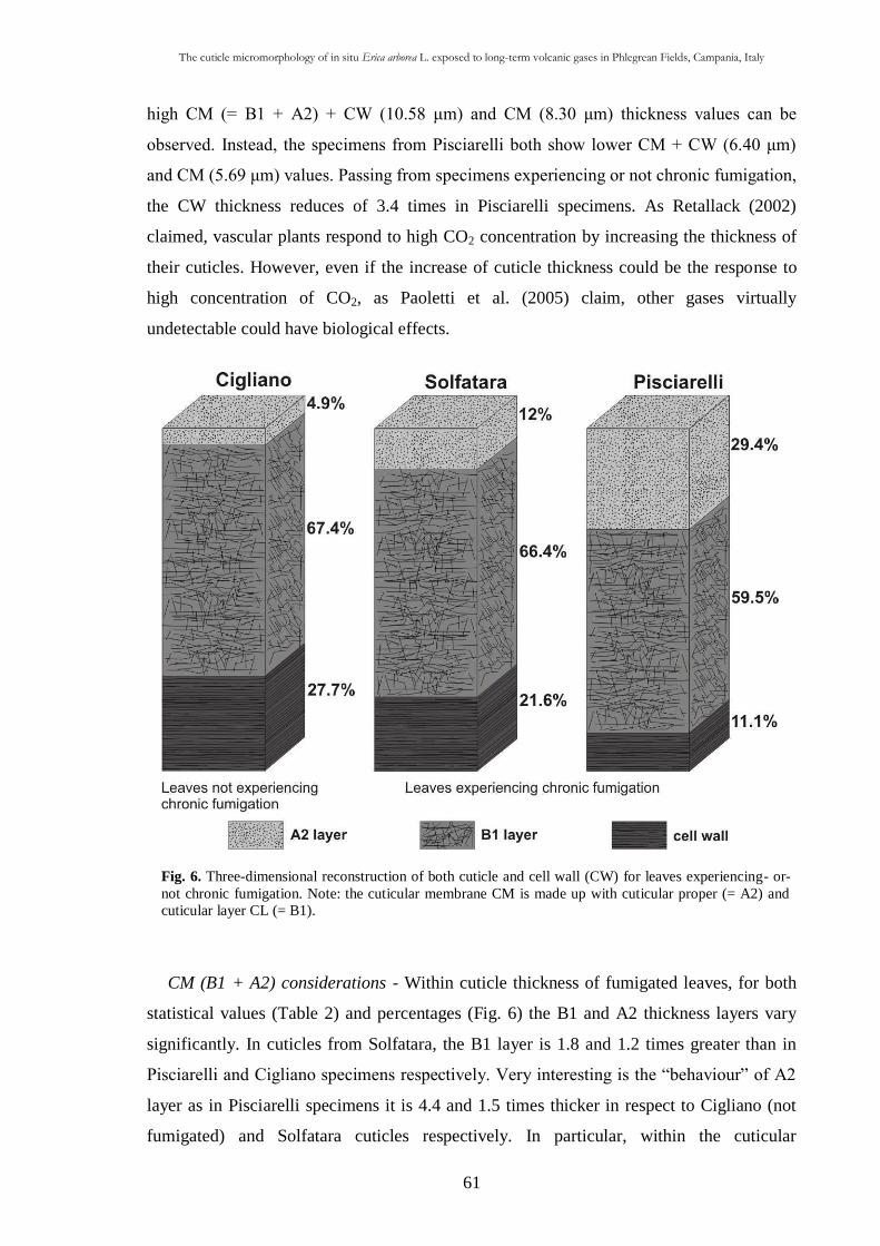
The cuticle micromorphology of in situ Erica arborea L. exposed to long-term volcanic gases in Phlegrean Fields, Campania, Italy
61
Fig. 6. Three-dimensional reconstruction of both cuticle and cell wall (CW) for leaves experiencing- or-
not chronic fumigation. Note: the cuticular membrane CM is made up with cuticular proper (= A2) and
cuticular layer CL (= B1).
high CM (= B1 + A2) + CW (10.58 μm) and CM (8.30 μm) thickness values can be
observed. Instead, the specimens from Pisciarelli both show lower CM + CW (6.40 μm)
and CM (5.69 μm) values. Passing from specimens experiencing or not chronic fumigation,
the CW thickness reduces of 3.4 times in Pisciarelli specimens. As Retallack (2002)
claimed, vascular plants respond to high CO2 concentration by increasing the thickness of
their cuticles. However, even if the increase of cuticle thickness could be the response to
high concentration of CO2, as Paoletti et al. (2005) claim, other gases virtually
undetectable could have biological effects.
CM (B1 + A2) considerations - Within cuticle thickness of fumigated leaves, for both
statistical values (Table 2) and percentages (Fig. 6) the B1 and A2 thickness layers vary
significantly. In cuticles from Solfatara, the B1 layer is 1.8 and 1.2 times greater than in
Pisciarelli and Cigliano specimens respectively. Very interesting is the “behaviour” of A2
layer as in Pisciarelli specimens it is 4.4 and 1.5 times thicker in respect to Cigliano (not
fumigated) and Solfatara cuticles respectively. In particular, within the cuticular
The cuticle micromorphology of in situ Erica arborea L. exposed to long-term volcanic gases in Phlegrean Fields, Campania, Italy
62
membrane, the A2 layer responds more marked in respect to B1 ones. As showed from
data (Table 1 and 2; Figs. 5 and 6), a close relation between atmospheric CO2
concentration and the A2 layer thickness seems to exist. It is worth noting that, in Solfatara
and Pisciarelli localities, cuticles respond to atmospheric CO2 concentration and not to the
CO2 soil concentration (Tables 1, 2 and Fig. 6). In specimens from Pisciarelli, the
statistical reduction of the B1 layer thickness and the corresponding increasing of A2 layer
thickness in respect to those of Solfatara and Cigliano (Table 1) could be the result of a
carbon allocation from the inner layers to the outermost A2 layer. Therefore, the
differences in layers thickness could be due to a different allocation of carbon-based
secondary or structural compounds (CBSSC). According to growth-differentiation balance
hypothesis (Hermes and Mattson, 1992), any environmental condition that affects
photosynthesis (carbon source) and growth (carbon sink) with different intensity will affect
the relative carbon pool available for allocation to carbon based compounds (Peñuelas and
Estiarte, 1998). The carbon allocation could represent a further cost for plants experiencing
chronic fumigation. In fumigated leaves the thickest A2 layer could be the response to
toxic volcanic gases or/and to an increase of CO2 concentration (Table 1). However, in the
specimens from Solfatara and Pisciarelli, the increase in A2 layer thickness is consistent
with Taylor et al. (1989) statement according to whom, amorphous remains within cuticle
envelope are likely to represent the most resistant component of leaf.
An Early Cretaceous flora from Cusano Mutri, Benevento, southern Italy
63
CHAPTER III
An Early Cretaceous flora from Cusano Mutri, Benevento, southern Italy
3.1. Introduction
The distribution of fossil plants is one of the most reliable tools for a better
understanding of terrestrial climatic conditions through geologic time (Francis and Frakes,
1993), and the addition of sedimentological context to species lists greatly refines such
studies (DiMichele and Gastaldo, 2008).
This research represents a continuation of palaeobotanical studies carried out on
Cretaceous plant-bearing localities in the southern Apennines (Bartiromo et al., 2006,
2008, 2009). The aim of this research is to provide supplementary data to investigations of
subaerial Early Cretaceous Tethyan environments. A palaeontological survey on South
Apennine Mesozoic sediments of carbonate platform facies (Campania Region) revealed in
2005 a new Early Cretaceous (Late Aptian) fossil locality near the village of Cusano Mutri
(Benevento Province) (Fig. 1A). This new outcrop rivals in extent, abundance and
preservation of the fossil material only the historical site of Pietraroja (early Albian) (Fig.
1A), the fossil flora of which has already been subjected to a preliminary study (Bartiromo
et al., 2006, 2008). Several excavations were carried out at the site during the years
2005e2008, which provided hundreds of macro-remains among animal and plants
(Bartiromo et al., 2008). This locality (Fig. 1B) displays a more complete fossil record than
is usually found in the sedimentary successions of the Apenninic Carbonate Platform. The
richness, taxonomic diversity and type of preservation make this outcrop worthy of
consideration as a Fossil-Lagerstätte (Seilacher, 1970; Seilacher et al., 1985). Following a
brief account of the invertebrate and vertebrate faunal assemblage we focus on the fossil
flora to understand better the palaeoenvironment in which these plants grew. In addition,
epidermal features were studied and interpreted palaeoecologically.
3.2. Geological setting
The outcrop (Fig. 1B) is located at “Peschera” (IGM Map at scale 1:25000 173 IV NO -
Cerreto Sannita, long.14° 32’ 08.15’’, lat. 41° 17’ 42. 89’’), about 5 kmsouth of the village
of Cusano Mutri (Campania Region, Benevento Province: Fig. 1A), on the eastern slope of
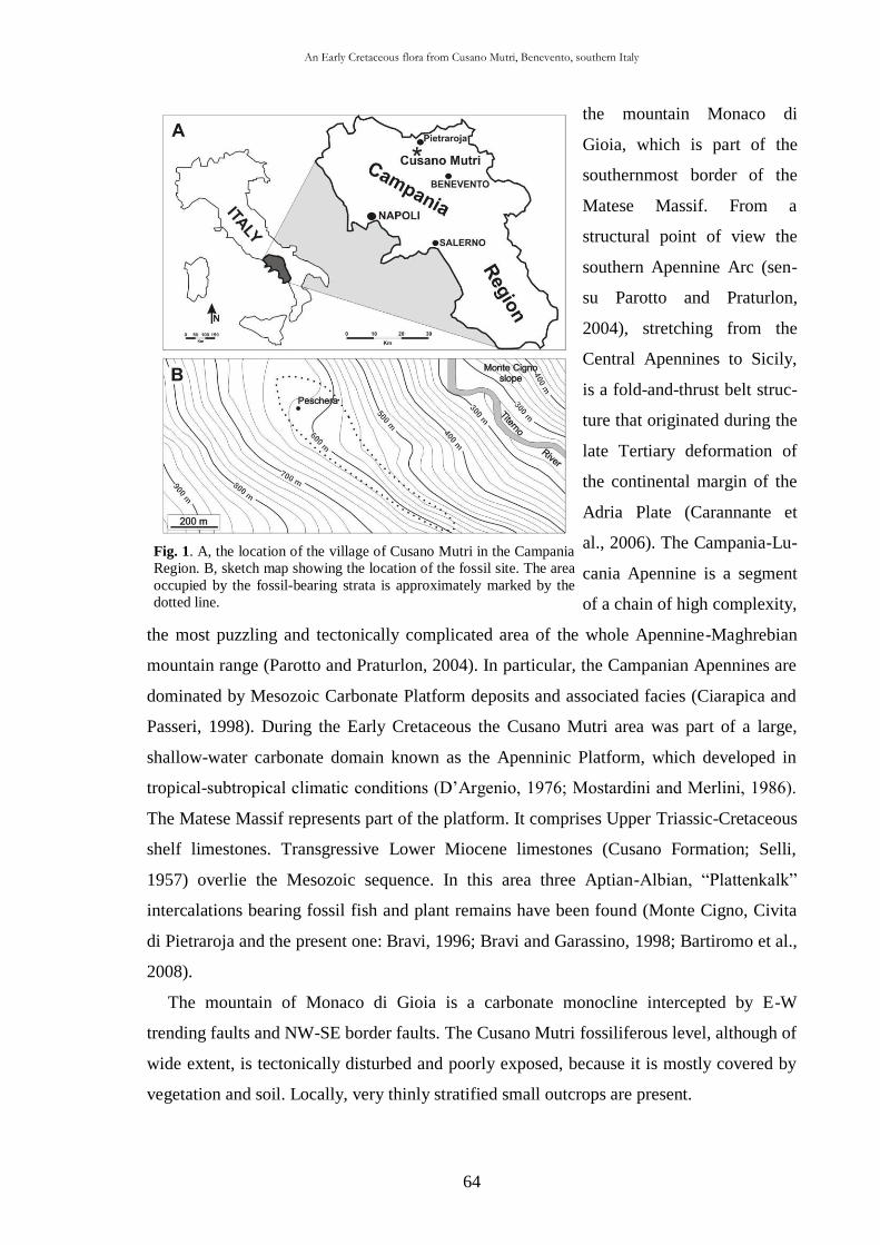
An Early Cretaceous flora from Cusano Mutri, Benevento, southern Italy
64
Fig. 1. A, the location of the village of Cusano Mutri in the Campania
Region. B, sketch map showing the location of the fossil site. The area
occupied by the fossil-bearing strata is approximately marked by the
dotted line.
the mountain Monaco di
Gioia, which is part of the
southernmost border of the
Matese Massif. From a
structural point of view the
southern Apennine Arc (sen-
su Parotto and Praturlon,
2004), stretching from the
Central Apennines to Sicily,
is a fold-and-thrust belt struc-
ture that originated during the
late Tertiary deformation of
the continental margin of the
Adria Plate (Carannante et
al., 2006). The Campania-Lu-
cania Apennine is a segment
of a chain of high complexity,
the most puzzling and tectonically complicated area of the whole Apennine-Maghrebian
mountain range (Parotto and Praturlon, 2004). In particular, the Campanian Apennines are
dominated by Mesozoic Carbonate Platform deposits and associated facies (Ciarapica and
Passeri, 1998). During the Early Cretaceous the Cusano Mutri area was part of a large,
shallow-water carbonate domain known as the Apenninic Platform, which developed in
tropical-subtropical climatic conditions (D’Argenio, 1976; Mostardini and Merlini, 1986).
The Matese Massif represents part of the platform. It comprises Upper Triassic-Cretaceous
shelf limestones. Transgressive Lower Miocene limestones (Cusano Formation; Selli,
1957) overlie the Mesozoic sequence. In this area three Aptian-Albian, “Plattenkalk”
intercalations bearing fossil fish and plant remains have been found (Monte Cigno, Civita
di Pietraroja and the present one: Bravi, 1996; Bravi and Garassino, 1998; Bartiromo et al.,
2008).
The mountain of Monaco di Gioia is a carbonate monocline intercepted by E-W
trending faults and NW-SE border faults. The Cusano Mutri fossiliferous level, although of
wide extent, is tectonically disturbed and poorly exposed, because it is mostly covered by
vegetation and soil. Locally, very thinly stratified small outcrops are present.
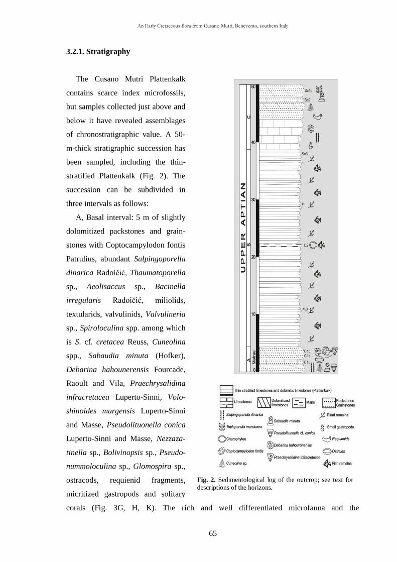
An Early Cretaceous flora from Cusano Mutri, Benevento, southern Italy
65
Fig. 2. Sedimentological log of the outcrop; see text for
descriptions of the horizons.
3.2.1. Stratigraphy
The Cusano Mutri Plattenkalk
contains scarce index microfossils,
but samples collected just above and
below it have revealed assemblages
of chronostratigraphic value. A 50-
m-thick stratigraphic succession has
been sampled, including the thin-
stratified Plattenkalk (Fig. 2). The
succession can be subdivided in
three intervals as follows:
A, Basal interval: 5 m of slightly
dolomitized packstones and grain-
stones with Coptocampylodon fontis
Patrulius, abundant Salpingoporella
dinarica Radoičić, Thaumatoporella
sp., Aeolisaccus sp., Bacinella
irregularis Radoičić, miliolids,
textularids, valvulinids, Valvulineria
sp., Spiroloculina spp. among which
is S. cf. cretacea Reuss, Cuneolina
spp., Sabaudia minuta (Hofker),
Debarina hahounerensis Fourcade,
Raoult and Vila, Praechrysalidina
infracretacea Luperto-Sinni, Volo-
shinoides murgensis Luperto-Sinni
and Masse, Pseudolituonella conica
Luperto-Sinni and Masse, Nezzaza-
tinella sp., Bolivinopsis sp., Pseudo-
nummoloculina sp., Glomospira sp.,
ostracods, requienid fragments,
micritized gastropods and solitary
corals (Fig. 3G, H, K). The rich and well differentiated microfauna and the
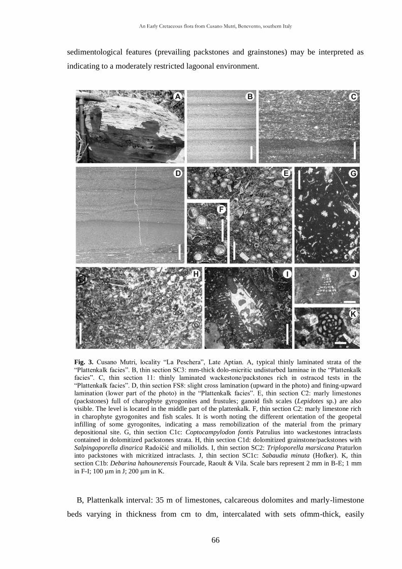
An Early Cretaceous flora from Cusano Mutri, Benevento, southern Italy
66
Fig. 3. Cusano Mutri, locality “La Peschera”, Late Aptian. A, typical thinly laminated strata of the
“Plattenkalk facies”. B, thin section SC3: mm-thick dolo-micritic undisturbed laminae in the “Plattenkalk
facies”. C, thin section 11: thinly laminated wackestone/packstones rich in ostracod tests in the
“Plattenkalk facies”. D, thin section FS8: slight cross lamination (upward in the photo) and fining-upward
lamination (lower part of the photo) in the “Plattenkalk facies”. E, thin section C2: marly limestones
(packstones) full of charophyte gyrogonites and frustules; ganoid fish scales (Lepidotes sp.) are also
visible. The level is located in the middle part of the plattenkalk. F, thin section C2: marly limestone rich
in charophyte gyrogonites and fish scales. It is worth noting the different orientation of the geopetal
infilling of some gyrogonites, indicating a mass remobilization of the material from the primary
depositional site. G, thin section C1c: Coptocampylodon fontis Patrulius into wackestones intraclasts
contained in dolomitized packstones strata. H, thin section C1d: dolomitized grainstone/packstones with
Salpingoporella dinarica Radoičić and miliolids. I, thin section SC2: Triploporella marsicana Praturlon
into packstones with micritized intraclasts. J, thin section SC1c: Sabaudia minuta (Hofker). K, thin
section C1b: Debarina hahounerensis Fourcade, Raoult & Vila. Scale bars represent 2 mm in B-E; 1 mm
in F-I; 100 μm in J; 200 μm in K.
sedimentological features (prevailing packstones and grainstones) may be interpreted as
indicating to a moderately restricted lagoonal environment.
B, Plattenkalk interval: 35 m of limestones, calcareous dolomites and marly-limestone
beds varying in thickness from cm to dm, intercalated with sets ofmm-thick, easily
An Early Cretaceous flora from Cusano Mutri, Benevento, southern Italy
67
exfoliated laminae (Fig. 3A). Some plattenkalk strata contain charophyte gyrogonites and
frustules together with Lepidotes fish scales (Fig. 3E and F), the richest of these occurring
in the middle of this plattenkalk interval, which consists of a 50-cm-thick marly stratum
(Fig. 2). Other beds and laminae sets bear fish, crustacean and plant remains. Land,
freshwater, brackish and marine fossil remains mixed together may be explained by
sediment remobilization from surrounding inner landward areas in a channel context. The
random orientation of the geopetal filling within the gyrogonites may support this
interpretation (Fig. 3F). The typical Plattenkalk facies consists of strata and laminae of
dolomicrite (Fig. 3B). Sometimes, in thin section cm-long fractures can be observed,
seemingly owing to local differential compaction. At a later stage, the sediment probably
underwent zone-mixing diagenesis, so that differential dolomitization developed along the
fractures. Close to the fractures, dolomitization appears sometimes more marked than in
the rest of the rock, and at times less marked. Finingupward structures (Fig. 3D), indicating
decantation of muddy sediments brought in by tides during the normal, calm,
environmental conditions, are also observed. The coarser sediment at the base of laminae
often consists of ostracods (Fig. 3C) and estherid valves; the top is capped by a thin
brownish layer, probably indicating the last phase of decantation of suspended organic
matter. Irregular undulating laminae are also commonly observed. Some of these show
cracks and strain structures, probable desiccation voids and root moulds which at present
cannot be safely determined as subaerial exposure (Fig. 3D).
From an environmental point of view, the plattenkalk facies, which formed in a
sheltered area of the carbonate platform, more or less close to land, strongly influenced the
sedimentation and palaeontological content. Sediments deposited landward in fresh to
brackish ponds were sometimes removed and washed seaward.
C, Upper interval: 10mof grainstones, packstones and packstone wackstones. The
microflora is composed of Thaumathoporella sp., dasycladalean algae (such as
Triploporella marsicana Praturlon and S. dinarica Radoičić) and Cretacicladus minervini
Luperto-Sinni. Foraminifers are represented by milioliids, textulariids, valvulinids and
Ataxophragmidae. Most significant taxa are S. minuta (Hofker), D. hahounerensis
Fourcade, Raoult and Vila, P. infracretacea Luperto-Sinni, Spiroloculina cf. cretacea
Reuss, and Cuneolina spp. Ostreids, requienid fragments, small micritized and bioeroded
gastropods, and thin, smooth valves of ostracods are also present (Fig. 3I and J). This last
interval is broadly characterized by an increase in biodiversity, which may testify the
return to marine conditions.
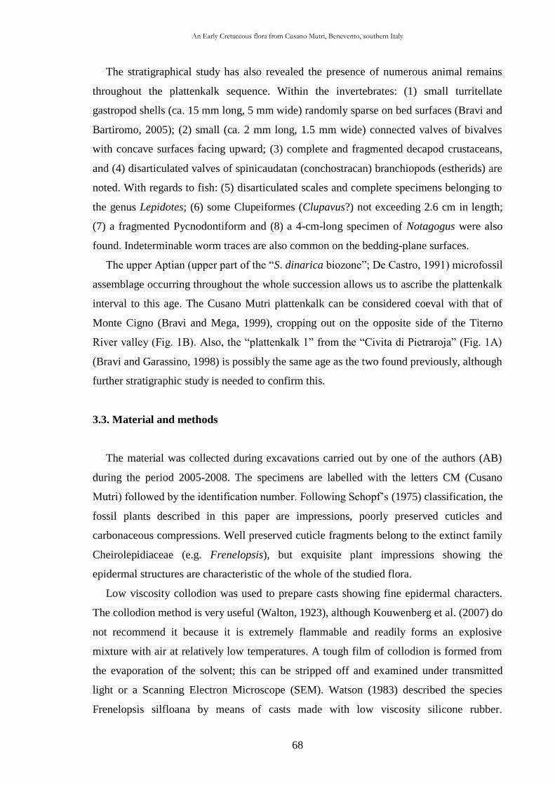
An Early Cretaceous flora from Cusano Mutri, Benevento, southern Italy
68
The stratigraphical study has also revealed the presence of numerous animal remains
throughout the plattenkalk sequence. Within the invertebrates: (1) small turritellate
gastropod shells (ca. 15 mm long, 5 mm wide) randomly sparse on bed surfaces (Bravi and
Bartiromo, 2005); (2) small (ca. 2 mm long, 1.5 mm wide) connected valves of bivalves
with concave surfaces facing upward; (3) complete and fragmented decapod crustaceans,
and (4) disarticulated valves of spinicaudatan (conchostracan) branchiopods (estherids) are
noted. With regards to fish: (5) disarticulated scales and complete specimens belonging to
the genus Lepidotes; (6) some Clupeiformes (Clupavus?) not exceeding 2.6 cm in length;
(7) a fragmented Pycnodontiform and (8) a 4-cm-long specimen of Notagogus were also
found. Indeterminable worm traces are also common on the bedding-plane surfaces.
The upper Aptian (upper part of the “S. dinarica biozone”; De Castro, 1991) microfossil
assemblage occurring throughout the whole succession allows us to ascribe the plattenkalk
interval to this age. The Cusano Mutri plattenkalk can be considered coeval with that of
Monte Cigno (Bravi and Mega, 1999), cropping out on the opposite side of the Titerno
River valley (Fig. 1B). Also, the “plattenkalk 1” from the “Civita di Pietraroja” (Fig. 1A)
(Bravi and Garassino, 1998) is possibly the same age as the two found previously, although
further stratigraphic study is needed to confirm this.
3.3. Material and methods
The material was collected during excavations carried out by one of the authors (AB)
during the period 2005-2008. The specimens are labelled with the letters CM (Cusano
Mutri) followed by the identification number. Following Schopf’s (1975) classification, the
fossil plants described in this paper are impressions, poorly preserved cuticles and
carbonaceous compressions. Well preserved cuticle fragments belong to the extinct family
Cheirolepidiaceae (e.g. Frenelopsis), but exquisite plant impressions showing the
epidermal structures are characteristic of the whole of the studied flora.
Low viscosity collodion was used to prepare casts showing fine epidermal characters.
The collodion method is very useful (Walton, 1923), although Kouwenberg et al. (2007) do
not recommend it because it is extremely flammable and readily forms an explosive
mixture with air at relatively low temperatures. A tough film of collodion is formed from
the evaporation of the solvent; this can be stripped off and examined under transmitted
light or a Scanning Electron Microscope (SEM). Watson (1983) described the species
Frenelopsis silfloana by means of casts made with low viscosity silicone rubber.
An Early Cretaceous flora from Cusano Mutri, Benevento, southern Italy
69
Microscopical details of some cuticles were difficult to observe after chemical treatment.
For this reason, some cuticle fragments were mounted directly onto the stubs for the SEM
observation. Cuticle fragments were separated from the surface of the bed by means of
tweezers. They were cleaned in 37% hydrochloric acid (HCl) for 2 h. Adhering siliceous
debris on the cuticle surfaces was removed by immersion in 40% hydrofluoric acid (HF)
for 1 h. Finally, they were macerated in Schulze’s reagent (HNO3 and KClO3) for 2 h; the
duration of the latter depending on the degree of carbonization and preservation (Kerp,
1990). Parts of cuticle were directly mounted on glass slides for light microscope
examination. Other cuticle fragments were directly mounted onto the stubs for observation
under the SEM. Light microscope (LM) and Stereo-microscope (SM) observations were
made using Leitz microscopes. Specimens examined under an optical microscope were
photographed with a Nikon Coolpix 990 camera. The cuticles were examined using a
JEOL JSM-5310 SEM at the CISAG (Centro Interdipartimentale di servizi per analisi
Geomineralogiche) and a FEI-QUANTAS 200 ESEM (Figs. 6G and 7G) at the CISME
(Centro Interdipartimentale di servizi di Microscopia Elettronica).
The material is housed in the Museo di Paleontologia, “Centro Museale, Centro Musei
delle Scienze Naturali”, Università degli Studi di Napoli Federico II (Italy).
3.4. Systematic palaeontology
Order Coniferales
Family incertae sedis
Genus Cupressinocladus Seward, 1919
Cupressinocladus sp.
Figs. 4A, 5A
Material examined. CM 21, 68
Description. Twig impressions, branched (CM 21: Figs. 4A and 5A) and unbranched.
The leaves are decussately arranged. They are heavily adpressed to the axes and have a
rounded apex. The free part of the leaves arises from whole basal cushion. A poor
impression of a stomatal apparatus 80 μm in diameter has been obtained by means of a
peel.
Discussion. The morphology of the shoots together with the arrangement of the leaves
lead us to ascribe the specimens to the genus Cupressinocladus Seward, 1919. However,
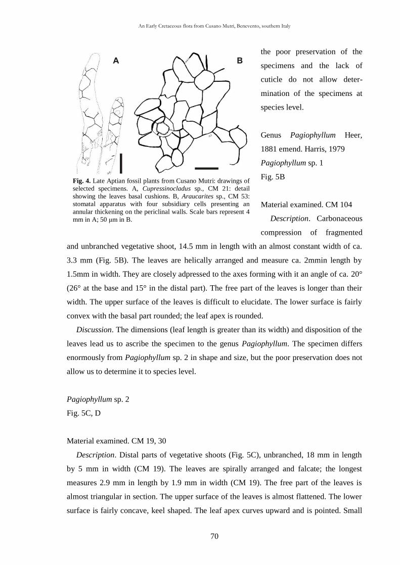
An Early Cretaceous flora from Cusano Mutri, Benevento, southern Italy
70
Fig. 4. Late Aptian fossil plants from Cusano Mutri: drawings of
selected specimens. A, Cupressinocladus sp., CM 21: detail
showing the leaves basal cushions. B, Araucarites sp., CM 53:
stomatal apparatus with four subsidiary cells presenting an
annular thickening on the periclinal walls. Scale bars represent 4
mm in A; 50 μm in B.
the poor preservation of the
specimens and the lack of
cuticle do not allow deter-
mination of the specimens at
species level.
Genus Pagiophyllum Heer,
1881 emend. Harris, 1979
Pagiophyllum sp. 1
Fig. 5B
Material examined. CM 104
Description. Carbonaceous
compression of fragmented
and unbranched vegetative shoot, 14.5 mm in length with an almost constant width of ca.
3.3 mm (Fig. 5B). The leaves are helically arranged and measure ca. 2mmin length by
1.5mm in width. They are closely adpressed to the axes forming with it an angle of ca. 20°
(26° at the base and 15° in the distal part). The free part of the leaves is longer than their
width. The upper surface of the leaves is difficult to elucidate. The lower surface is fairly
convex with the basal part rounded; the leaf apex is rounded.
Discussion. The dimensions (leaf length is greater than its width) and disposition of the
leaves lead us to ascribe the specimen to the genus Pagiophyllum. The specimen differs
enormously from Pagiophyllum sp. 2 in shape and size, but the poor preservation does not
allow us to determine it to species level.
Pagiophyllum sp. 2
Fig. 5C, D
Material examined. CM 19, 30
Description. Distal parts of vegetative shoots (Fig. 5C), unbranched, 18 mm in length
by 5 mm in width (CM 19). The leaves are spirally arranged and falcate; the longest
measures 2.9 mm in length by 1.9 mm in width (CM 19). The free part of the leaves is
almost triangular in section. The upper surface of the leaves is almost flattened. The lower
surface is fairly concave, keel shaped. The leaf apex curves upward and is pointed. Small
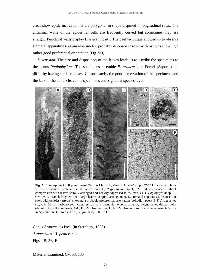
An Early Cretaceous flora from Cusano Mutri, Benevento, southern Italy
71
Fig. 5. Late Aptian fossil plants from Cusano Mutri. A, Cupressinocladus sp., CM 21: branched shoot
with leaf cushions preserved in the apical part. B, Pagiophyllum sp. 1, CM 104: carbonaceous shoot
compression with leaves spirally arranged and heavily adpressed to the axis. CeD, Pagiophyllum sp. 2.,
CM 19. C, branch fragment with large leaves in spiral arrangement. D, stomatal apparatuses disposed in
rows with ostioles (arrows) showing a probable preferential orientation (collodion peel). E-F, Araucarites
sp., CM 53. E, carbonaceous compression of a triangular woody scale. F, polygonal epidermal cells
(detail of E; collodion peel). A-C, E: SM observations; D, F: LM observations. Scale bar represents 5 mm
in A; 2 mm in B; 3 mm in C, E; 50 μm in D; 100 μm F.
areas show epidermal cells that are polygonal in shape disposed in longitudinal rows. The
anticlinal walls of the epidermal cells are frequently curved but sometimes they are
straight. Periclinal walls display fine granulosity. The peel technique allowed us to observe
stomatal apparatuses 50 μm in diameter, probably disposed in rows with ostioles showing a
rather good preferential orientation (Fig. 5D).
Discussion. The size and disposition of the leaves leads us to ascribe the specimens to
the genus Pagiophyllum. The specimens resemble P. araucarinum Pomel (Saporta) but
differ by having smaller leaves. Unfortunately, the poor preservation of the specimens and
the lack of the cuticle leave the specimens unassigned at species level.
Genus Araucarites Presl (in Sternberg, 1838)
Araucarites aff. pedreranus
Figs. 4B, 5E, F
Material examined. CM 53, 135
An Early Cretaceous flora from Cusano Mutri, Benevento, southern Italy
72
Description. Woody scales (CM 53 probably a lower surface) 10.5 mm long by 10.1
mm wide, triangular in shape with entire margin. The base of the scales is 5 mm in width.
The apex is rounded (Fig. 5E). The epidermal features are visible on the distal surface of
the scale (CM 53) and have been obtained by means of collodion peels. The epidermal
cells are polygonal and sometimes pentagonal and or hexagonal in shape (Figs. 4B and
5F). The haplocheilic, monocyclic and incompletely amphycyclic stomatal appara-tuses
have a diameter of ca. 47 μm. They are composed of four subsidiary cells, the largest of
which is 28 μm long and 12 μm wide. The periclinal surface of subsidiary cells seems to
present a coronal thickening around the stomatal rim 4 μm thick (Fig. 4B).
Discussion. The size and shape of the scales together with the epidermal features
indicate an affinity with Araucarites pedreranus described by Barale (1989) for the Early
Cretaceous lithographic limestones of Montsech (Spain, Pedrera and Cabrua quarries).
However, the scale apex is triangular in A. pedreranus but rounded in the specimens from
Cusano Mutri. Although fragmentary, the Cusano Mutri scales share with the Spanish
material the same epidermal features. Unfortunately, the poor state in the preservation of
the specimens and the incompleteness of the epidermal features prevent a specific
assignment.
Family Cheirolepidiaceae Takhtajan, 1963
Genus Frenelopsis (Schenk, 1869) emend. Watson, 1977
Type species. Frenelopsis hoheneggeri (Ettingshausen, 1852) Schenk,
1869
Frenelopsis cusanensis sp. nov.
Figs. 6A-J, 7A-I
Holotype. CM 100, slide number 100v1, 100v2; stub number 100s1, 100s2. Figs. 6BeH, J,
7A-I.
Paratypes. CM 3, 8, 22, 66; stub number 3s, 8s, 22s, 66s.
Type locality. Cusano Mutri village, Benevento Province, southern Italy.
Derivation of name. Refers to the village of Cusano Mutri where the species was found.
Repository. Museo di Paleontologia, “Centro Museale, Centro Musei delle Scienze
Naturali”, Università degli Studi di Napoli Federico II (Italy).
Stratigraphic range. Late Aptian
An Early Cretaceous flora from Cusano Mutri, Benevento, southern Italy
73
Diagnosis. Articulated shoots attaining at least two orders of branching in one plane.
Branches arising at an angle of ca. 11° with internodes 4-10 mm long and 1.3-2.4 mm
wide. Shoots bear whorls of three scale-like leaves 0.5-0.9 mm in length bordered by
unicellular hairs up to 60 μm in length. Internode cuticle is ca. 7 μm thick. The stomatal
apparatuses have an average diameter of 40 μm and are arranged in well-defined rows (7-
10 per linear mm). Average stomatal density is 70-80 per mm2. The stomatal apparatuses,
randomly orientated, bear four subsidiary cells (rarely five), separated into lateral and polar
cells, each bearing one outer and one inner papilla. Papillae quadrangular, small,
sometimes elongated, arise from epidermal cells.
Description. The material consists of impressions and compressions of branched (CM
22, 66), articulated cuticular segments (CM 8, 66, 100) and internode fragments (CM 65)
1.2 up to 11.1 cm long (Fig. 6A and B). The best preserved shoot (CM 100) as well as the
holotype, measures 77 mm in length by 2.4 mm in width and is composed of part and
counterpart (Fig. 6B: part). The branches are produced alternately, in one plane, and the
specimens have one main axis. Branches arise at an angle of ca.11°. The internode length
varies between 4 and 10 mm; the segment width is 1.3-2.4 mm, 1.8 mm on average. At the
level of every node a whorl of three squamiform leaves, laterally fused, form a leaf sheath
developed along the internode length (Fig. 6C and D). At the extremity of the leaf sheath,
three small teeth (Fig. 6F), related to the free leaf extremity are present. There are no
suture lines along the leaf sheath. The free parts of the leaves are identical at each node:
they are triangular in shape and measure 0.9-0.5 mm in length with an average of 0.7 mm.
The leaves are bordered by marginal unicellular hairs up to 60 μm in length (Fig. 6D). The
cuticle of the internode is 7 μm thick (Fig. 7B). In outer view, the epidermal cells bear one
papilla each; close to the stomatal apparatuses the papillae measure ca. 11 μm in length and
10 μm in width extending in the direction of the stomatal apparatuses itself (Figs. 6I and
7D). Elongated epidermal papillae transversally disposed can be frequently observed (Fig.
7D). Usually, the width of the papillae is greater than their length and they are located
above the average surface of the epidermis; the papillae density is 70-80 per mm2
(CM
100). There are no hairs on the epidermal surface (Fig. 6I). Several epidermal cells are
polygonal in shape, but frequently rectangular cells ca. 31 μm long by 15 μm wide can be
observed. The epidermal cells are arranged in more or less well-defined rows running
parallel to the long axis of the internode (Fig. 7C). The anticlinal walls of the epidermal
cells are ca. 8 μm thick. The stomatal apparatuses are arranged in well-defined longitudinal
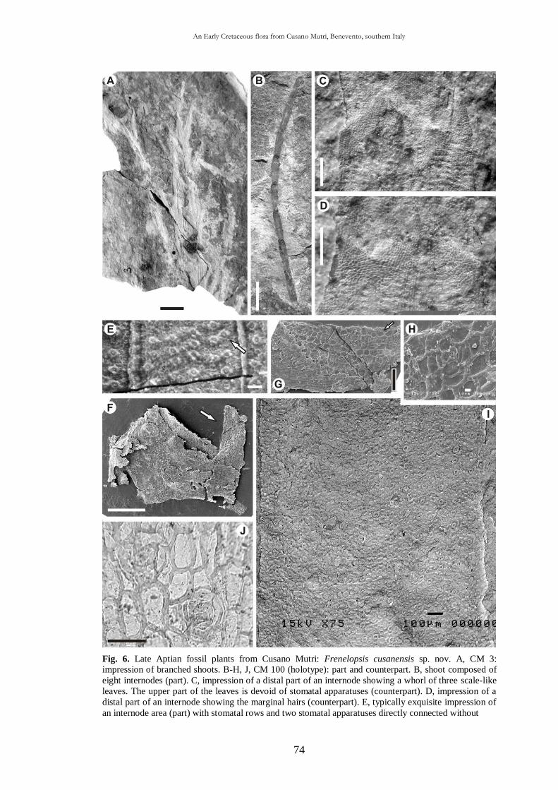
An Early Cretaceous flora from Cusano Mutri, Benevento, southern Italy
74
Fig. 6. Late Aptian fossil plants from Cusano Mutri: Frenelopsis cusanensis sp. nov. A, CM 3:
impression of branched shoots. B-H, J, CM 100 (holotype): part and counterpart. B, shoot composed of
eight internodes (part). C, impression of a distal part of an internode showing a whorl of three scale-like
leaves. The upper part of the leaves is devoid of stomatal apparatuses (counterpart). D, impression of a
distal part of an internode showing the marginal hairs (counterpart). E, typically exquisite impression of
an internode area (part) with stomatal rows and two stomatal apparatuses directly connected without
An Early Cretaceous flora from Cusano Mutri, Benevento, southern Italy
75
interposed epidermal cells (arrow). F, fragment of internode cuticle (part) removed to demonstrate abaxial
and adaxial surfaces. The arrow indicates the position of detail in G. G, inner side of abaxial surface with two
stomatal rows (detail of F). The arrow indicates position of the detail in H. H, inner view of a dicyclic
stomatal apparatus composed by four subsidiary cells (detail of G). I, CM 3: external view of the abaxial
surface showing epidermal papillae and stomatal apparatuses arranged in rows (inverted image: negative). J,
internode cuticle showing two stomatal apparatuses. The upper one focused on external papillae while the
lower one shows the four subsidiary cells distinct in polar and lateral ones. C-E: SM observations; F-I: SEM
observations; J: LM observation. Scale bar represents 10 mm in A, B; 500 μm in C, D, F; 100 μm in E, G, I;
10 μm in H; 50 μm in J.
rows (7-10 per mm) (Figs. 6I and 7C, E, I). The stomatal rows are separated by 1-2 (at
times 3) epidermal cells. On the same stomatal row 1e2 (at times 3) epidermal cells occur
between two stomatal apparatuses (Figs. 6G and 7C, I). Sometimes, the subsidiary cells of
two adjacent stomatal apparatuses on the same row can be in contact without intervening
epidermal cells (Figs. 6E and 7I). The stomatal density is 70-80 per mm2. Stomatal
apparatuses are 40 mm in diameter on average (max. 62 μm in CM66, min. 30 μm in CM
22; 38 μm in CM 100). The stomatal apparatuses are haplocheilic and normally
monocyclic (Fig. 6J); rarely, dicyclic stomatal apparatuses have been observed (Fig. 6H).
The stomatal apparatuses are composed of four (Figs. 6I, J, 7A, C-E, H, I), sometimes five
(Fig. 7G) subsidiary cells, more or less regularly disposed around the stomatal pit. The
stomatal apparatuses are randomly orientated. From a morphological point of view, it is
possible to distinguish two lateral and two polar subsidiary cells, usually with polar cells
greater than laterals ones (Figs. 6J and 7E). Each subsidiary cell bears (1) one outer papilla
protruding on the stomatal rim, partially covering the stomatal pit, and (2) one inner papilla
which conceals the stomatal pit (Fig. 7A, E and F). The fusion of outer papillae results in a
stomatal pit with a stellate outline and an aperture of ca. 14 μm in length and 6 μm in width
(Fig. 7A). Normally, the median level of epidermis is prominent with respect to the
stomatal apparatuses (Figs. 6I and 7A, D). The subsidiary cells are more or less rectangular
in shape, being ca. 18 apparatuses (Figs. 6I and 7A, D). The subsidiary cells are more or
less rectangular in shape, being ca. 18 apparatuses (Figs. 6I and 7A, D). The subsidiary
cells are more or less rectangular in shape, being ca. 18 μm long and 12 μm wide. The
guard cells are sometimes preserved with well-cutinized dorsal plates underneath the
stomatal pits (Fig. 7H). Occasionally, the inner cuticle surface is granulose (Fig. 7E and F).
Discussion. The studied specimens are typical Frenelopsis shoots on the basis of macro-
morphological features such as the cyclic, decussate, leaf-whorl arrangement, and the
absence of longitudinal sutures at the internodes. These characteristics exclude the
attribution of the specimens to the genus Pseudofrenelopsis Nathorst, 1893 emend.
Srinivasan, 1995. As Kunzmann et al. (2006) claimed, Frenelopsis consists of several
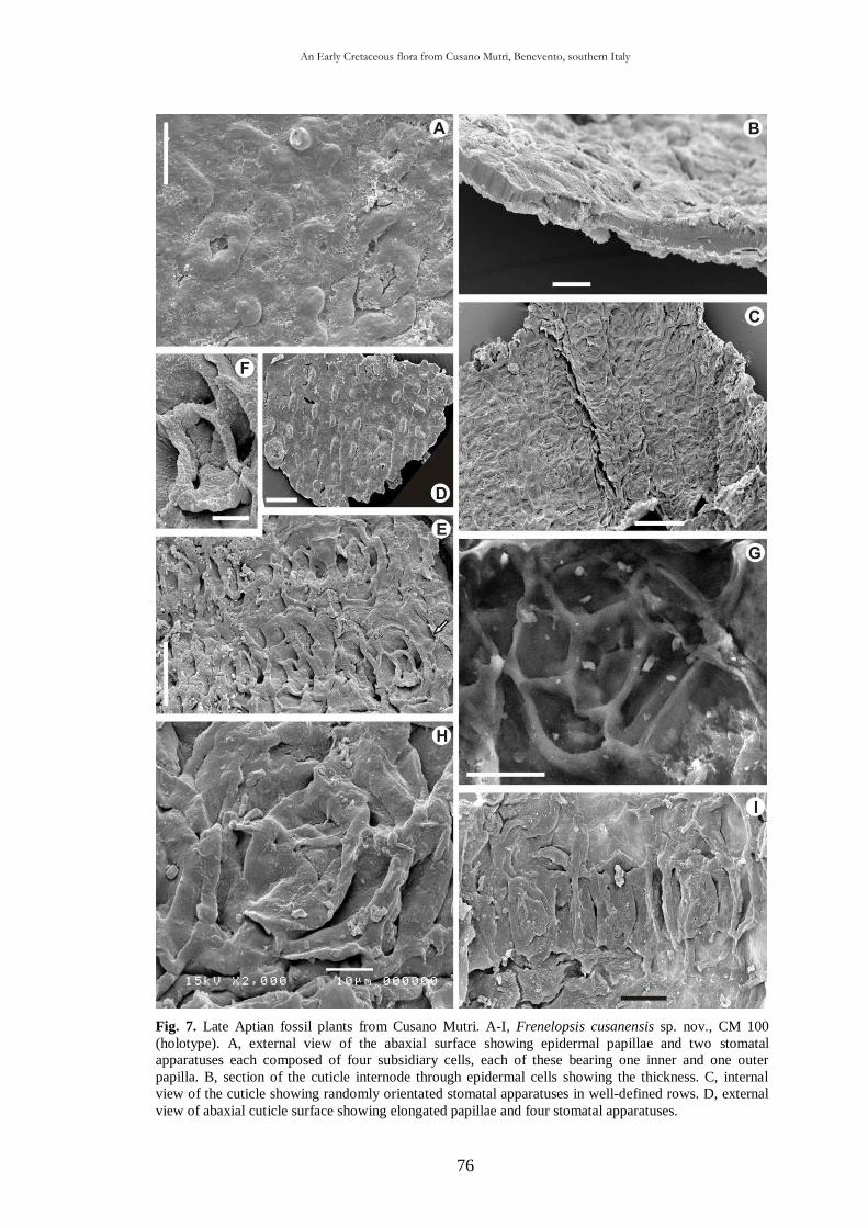
An Early Cretaceous flora from Cusano Mutri, Benevento, southern Italy
76
Fig. 7. Late Aptian fossil plants from Cusano Mutri. A-I, Frenelopsis cusanensis sp. nov., CM 100
(holotype). A, external view of the abaxial surface showing epidermal papillae and two stomatal
apparatuses each composed of four subsidiary cells, each of these bearing one inner and one outer
papilla. B, section of the cuticle internode through epidermal cells showing the thickness. C, internal
view of the cuticle showing randomly orientated stomatal apparatuses in well-defined rows. D, external
view of abaxial cuticle surface showing elongated papillae and four stomatal apparatuses.
An Early Cretaceous flora from Cusano Mutri, Benevento, southern Italy
77
E, internal view of the cuticle showing some stomatal apparatuses presenting fine granulosity (on the right).
The arrow indicates position of the detail figured in F. F, internal surface of a stomatal apparatus bearing four
inner papillae that are very closely linked (detail of E). G, internal view of a rare stomatal apparatus
composed of five subsidiary cells. H, internal view of stomatal apparatus composed of four subsidiary cells
showing the dorsal plates of the guard cells. I, internal view of cuticle showing three stomatal apparatuses
(each composed of four subsidiary cells) of a stomatal row with no epidermal cells on the left and one on the
right, between adjacent stomatal apparatuses. All figures: SEM observations. Scale bar represents 25 μm in
A; 50 μm in D, E; 10 μm in B, F; 100 μm in C; 20 μm in G, I; 10 μm in H.
morphospecies typically bearing three leaves per whorl (Watson, 1988; Kvaček, 2000).
Among the species attributed to Frenelopsis (Bartiromo et al., 2009), the cuticles from
Cusano Mutri are closest to Frenelopsis ugnaensis Gomez et al., 2002 from the upper
Barremian of Spain and Frenelopsis harrisii Doludenko and Reymanówna, 1978 from the
Cenomanian of Tadjikistan (Table 1). However, F. ugnaensis is different from the Cusano
Mutri material in having longer (up to 3 mm) and wider (up to 2 mm) internodes. At a
microscopic level, F. ugnaensis displays: (1) longer hairs in the upper margin of the leaf
(up to 100 vs. up to 60 μm); (2) a greater thickness of the cuticle (15-20 vs. 7 μm); (3) ill-
defined stomatal rows; (4) a lower stomatal density per mm2 (40-55 vs. 70-80); and (5)
greater size of stomatal apparatuses (55-60 vs. 40 μm). Moreover, F. ugnaensis has perfect
conical hairs (up to 100 μm long) on the outer cuticular surface, whereas the Cusano Mutri
specimens possess hemispherical papillae on the outer surface of each epidermal cell
without hairs. F. harrisii is different from the Cusano Mutri material in having: (1) greater
internodes measuring 8-12 mm in length (vs. 4-10 mm) and 1-4mmin width (vs. 1.3-2.4
mm); (2) a longer free part of the leaves (1.5 vs. 0.5-0.9 mm); (3) a greater thickness of
internode cuticle (up to 100 vs. 7 μm); (4) stomatal apparatuses elliptical in shape; and (5)
a smooth cuticle surface. Moreover, it is interesting to note that the specimens from
Cusano Mutri display the smallest stomatal apparatuses of the genus Frenelopsis, with an
average diameter of 40 μm as opposed to 107 μm for Frenelopsis profetiensis Bartiromo et
al., 2009 from the lower-middle Aptian locality of Profeti, which is the Frenelopsis with
the largest stomatal apparatuses (see Table 1). It is for these reasons that the new species,
Frenelopsis cusanensis, is proposed for the material from Cusano Mutri.
Cheirolepidiaceae sp. 1
Fig. 8A-G
Material examined. CM 1
Description. Compression of branched stem 14 mm in length by 2mm in width made by
nodes and internodes. The internode length is ca. 2.2 mm. The specimen (Fig. 8A) bears,
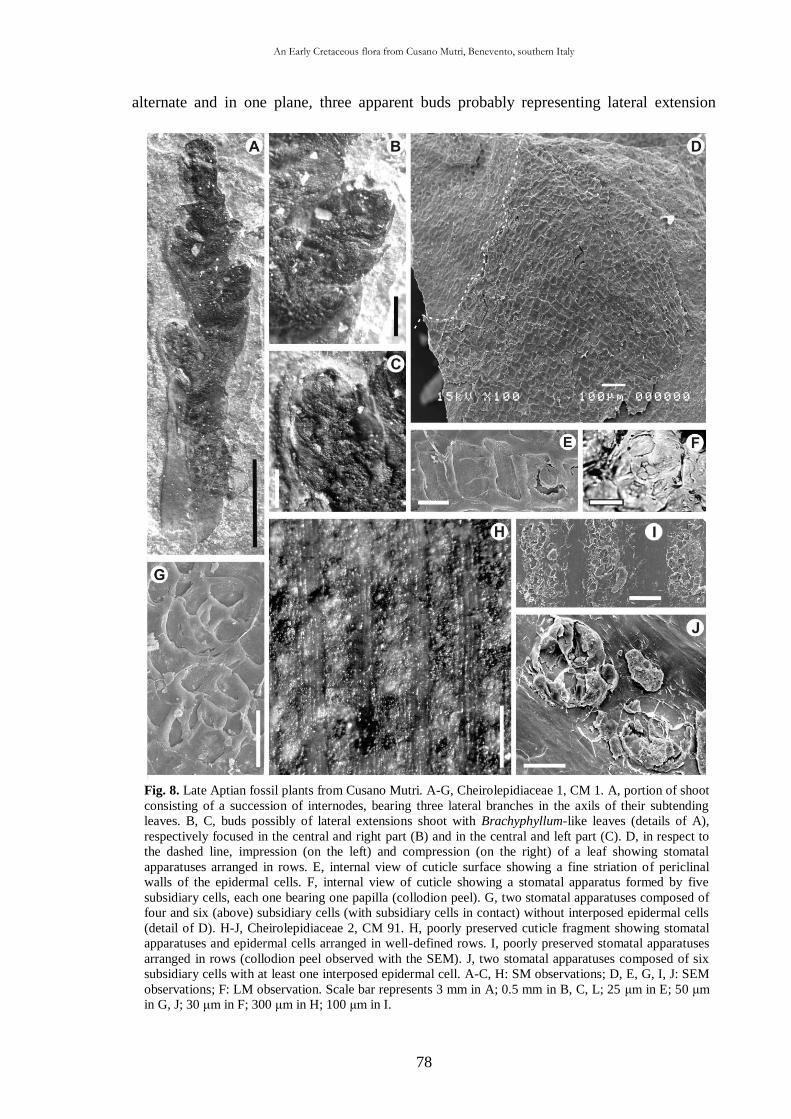
An Early Cretaceous flora from Cusano Mutri, Benevento, southern Italy
78
Fig. 8. Late Aptian fossil plants from Cusano Mutri. A-G, Cheirolepidiaceae 1, CM 1. A, portion of shoot
consisting of a succession of internodes, bearing three lateral branches in the axils of their subtending
leaves. B, C, buds possibly of lateral extensions shoot with Brachyphyllum-like leaves (details of A),
respectively focused in the central and right part (B) and in the central and left part (C). D, in respect to
the dashed line, impression (on the left) and compression (on the right) of a leaf showing stomatal
apparatuses arranged in rows. E, internal view of cuticle surface showing a fine striation of periclinal
walls of the epidermal cells. F, internal view of cuticle showing a stomatal apparatus formed by five
subsidiary cells, each one bearing one papilla (collodion peel). G, two stomatal apparatuses composed of
four and six (above) subsidiary cells (with subsidiary cells in contact) without interposed epidermal cells
(detail of D). H-J, Cheirolepidiaceae 2, CM 91. H, poorly preserved cuticle fragment showing stomatal
apparatuses and epidermal cells arranged in well-defined rows. I, poorly preserved stomatal apparatuses
arranged in rows (collodion peel observed with the SEM). J, two stomatal apparatuses composed of six
subsidiary cells with at least one interposed epidermal cell. A-C, H: SM observations; D, E, G, I, J: SEM
observations; F: LM observation. Scale bar represents 3 mm in A; 0.5 mm in B, C, L; 25 μm in E; 50 μm
in G, J; 30 μm in F; 300 μm in H; 100 μm in I.
alternate and in one plane, three apparent buds probably representing lateral extension
An Early Cretaceous flora from Cusano Mutri, Benevento, southern Italy
79
shoots (two on the left and one on the right). Each bud bears a number (perhaps at least
seven) of more or less Brachyphyllum-like leaves (sensu Alvin, 1983), which do not form a
complete sheath around the axis (Fig. 8B and C). Each node bears one subtending leaf of
“close type” (seeWatson,1977) at the axil of the node (Fig. 8A and D). There are no suture
lines along the sheathing leaves. It was impossible to measure the cuticle thickness. The
few stomatal apparatuses observed seem to be haplocheilic, monocyclic and made by four
(Fig. 8D) or frequently five (Fig. 8F) or six (Fig. 8G) subsidiary cells. They have an ovoid
to a rounded shape and measure on average 85 μm along the major axis and 56 μm along
the minor one. The subsidiary cells measure on average 25 μm in length and 35 μm in
width. Inner papillae are present (Fig. 8F). In external view no stomatal apparatuses have
been observed. The few preserved cuticle fragments show epidermal cells of varying size
having a polygonal (often isodiametric) morphology and disposition in more or less well-
defined rows (Fig. 8D). The anticlinal walls of the epidermal cells are ca. 10 μm thick,
sometimes exceeding 15 μm. The periclinal walls of the epidermal cells display a fine
parallel striation (Fig. 8E). By means of collodion casts the hypodermis can be seen.
Discussion. The specimen presents a macroscopical architecture and micro-
morphological characters that allow it to be placed within the family Cheirolepidiaceae
Takhtajan, 1963. The macroscopical and especially the microscopical features differ from
both F. cusanensis sp. nov. and Cheirolepidiaceae sp. 2. The gross morphology is
comparable with Pseudofrenelopsis parceramosa (Fontaine) Watson, 1977 from the
Wealden beds of the Isle of Wight (England), figured by Alvin and Hluštík (1979, fig.15)
and suggested again by Alvin (1983, Fig. 7H). The few observed stomatal apparatuses are
probably related to the adaxial surface of the cuticle, in agreement with Watson (1977, p.
720): “Adaxial cuticle showing cells of variable shape and arrangement, often strongly
papillate; few, scattered stomata, often abortive”. The micro- and macroscopic features
together with the Brachyphyllum-type foliage are reminiscent of juvenile forms of P.
parceramosa (Alvin, 1977, 1983; Watson, 1977). However, extensive morphological
variation has been historically documented within and between assemblages of P.
parceramosa (Axsmith, 2006). Unfortunately, the lack of well-defined microscopical
features makes it difficult to identify it to genus level within the family Cheirolepidiaceae
which, as Taylor et al. (2009) and others have claimed, certainly represents several
different types of plants based on habit and ecology.
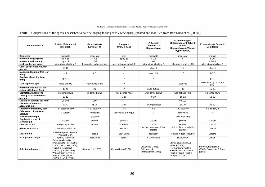
An Early Cretaceous flora from Cusano Mutri, Benevento, southern Italy
80
Table 1. Comparisons of the species described to date belonging to the genus Frenelopsis (updated and modified from Bartiromo et al. (2009)).
Characters/Taxa F. alata (Feistmantel)
Knobloch F choshiensis Kimura et al.
F. elegans Chow & Tsao
F. harrisii Doludenko & Reymanówna
F. hohenneggeri (Ettingshausen) Schenk
emend. Reymanówna & Watson
(type species)
F. kaneviensis Barale & Doludenko
Branching moderate moderate rare moderate moderate profuse Internode length (mm) up to 15 4-4.5 up to 23 8-12 8 8-10
Internode width (mm) up to 5 1.5-2 2-3 1-4 3 1.7-3.5
Leaf number per node alternating whorls of 3 2 opposite and decussate alternating whorls of 3 alternating whorls of 3 alternating whorls of 3 alternating whorls of 3
Outer surface ridge number per leaf
12-13 ? absent 15 absent
Maximum length of free leaf (mm)
~ 1 0.5 ~ 1 up to 1.5 1.5 1-0.7
Depth of sheathing base (mm)
up to 1 ? ? 1 up to 1
Leaf upper margin fringe of hairs hairs up to 4 μm ? ? scarious short hairs up to 20 μm
long
Internode and abaxial leaf cuticles thickness (µm)
30-40 20 ? up to 100μm 40 15-20
Stomatal arrangement Ill-defined rows ill-defined rows well-defined rows well-defined rows well-defined rows ill-defined rows Density of stomatal rows
per mm 10-12 8-10 9-10 10-12 12-16
Density of stomata per mm2
90-100 200 90-100
Diameter of stomatal apparatus (µm)
52-77 45-75 100 45×20 (elliptical) 60-70 35-50
Number of subsidiary cells 4-5, occasionally 6 4-6, usually 4 4-6 4-5 4-6, usually 4 4-6, usually 4
Orientation of stomatal aperture
? random horizontal transverse or oblique ? transverse ?
Surface around pit grooved thickened ring
Papillae in throat of stomatal pit
present present present present present present
Cuticle surface Irregularly ribbed smooth smooth smooth smooth smooth
Rim of stomatal pit stellate with lobed rim elliptical stellate, large pouch-like
papillae stellate, large pouch-like
papillae circular
Distribution Czech Republic; France,
Portugal, USA Japan East China Tajikistan Poland, Czech Republic Ukraine
Stratigraphic range Aptian, Senonian Barremian Aptian Cenomanian Hauterivian Albian
Selected references
Feistmantel (1881); Knobloch (1971); Hluštík (1972, 1974, 1978, 1979); Hluštík & Konzalová (1976a,b); Alvin (1977); Watson(1977); Alvin & Hluštík (1979); Pons (1979); Kvaček (2000).
Kimura et al. (1985). Chow &Tsao (1977). Doludenko (1978); Doludenko & Reymanówna (1978).
Ettingshausen (1852); Schenk (1869); Reymanówna (1965); Reymanówna & Watson (1976); Hluštík (1979); Purkynová (1983).
Barale & Doludenko (1985); Doludenko & Pons (1986).
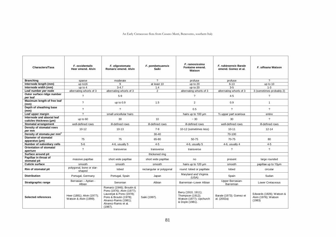
An Early Cretaceous flora from Cusano Mutri, Benevento, southern Italy
81
Characters/Taxa F. occidentalis
Heer emend. Alvin F. oligostomata
Romariz emend. Alvin F. pombetsuensis
Saiki
F. ramosissima Fontaine emend.
Watson
F. rubiesensis Barale emend. Gomez et al.
F. sifloana Watson
Branching sparse moderate ? profuse profuse ?
Internode length (mm) up to16 9 at least 10 up to 20 6-13 up to 10
Internode width (mm) up to 4 3-4.7 1.4 up to 20 3-5 1-3
Leaf number per node alternating whorls of 3 alternating whorls of 3 2 alternating whorls of 3 alternating whorls of 3 3 (sometimes probably 2) Outer surface ridge number
per leaf ? 5-9 ? 4-5 ?
Maximum length of free leaf (mm)
? up to 0.9 1.5 2 0.9 1
Depth of sheathing base (mm)
? ? 0.5 ? ?
Leaf upper margin ? small unicellular hairs hairs up to 100 μm ⅔ upper part scarious entire
Internode and abaxial leaf cuticles thickness (µm)
up to 60 30 10 ~ 30 30 ?
Stomatal arrangement well-defined rows ill-defined rows ill-defined rows ill-defined rows well-defined rows ill-defined rows
Density of stomatal rows per mm
10-12 10-13 7-9 10-12 (sometimes less) 10-11 12-14
Density of stomata per mm2
30-40 70-100
Diameter of stomatal apparatus (µm)
75 75 65-80 50-75 70-75 80
Number of subsidiary cells 5-6 4-6, usually 5 4-5 4-6, usually 5 4-6, usually 4 4-5 Orientation of stomatal aperture
? transverse transverse transverse ? ?
Surface around pit thickened ring
Papillae in throat of stomatal pit
massive papillae short wide papillae short wide papillae no present large rounded
Cuticle surface smooth smooth smooth hairs up to 120 μm smooth papillae up to 10μm
Rim of stomatal pit polygonal, bone or star-
shaped lobed rectangular or polygonal round: lobed or papillate lobed circular
Distribution Portugal, Germany Portugal, Spain Japan Maryland and Virginia
(USA) Spain Sudan
Stratigraphic range Berrasian – Aptian -
Albian Senonian Albian Barremian–Lower Albian
Upper Berrasian-Barremian
Lower Cretaceous
Selected references Heer (1881); Alvin (1977); Watson & Alvin (1999).
Romariz (1946); Broutin & Pons (1976); Alvin (1977); Lauverjat & Pons (1978); Pons & Broutin (1978); Alvarez-Ramis (1981); Alvarez-Ramis et al. (1987).
Saiki (1997).
Berry (1910, 1911);
Thompson (1912); Watson (1977); Upchurch & Doyle (1981).
Barale (1973); Gomez et al. (2002a)
Edwards (1926); Watson & Alvin (1976); Watson (1983)
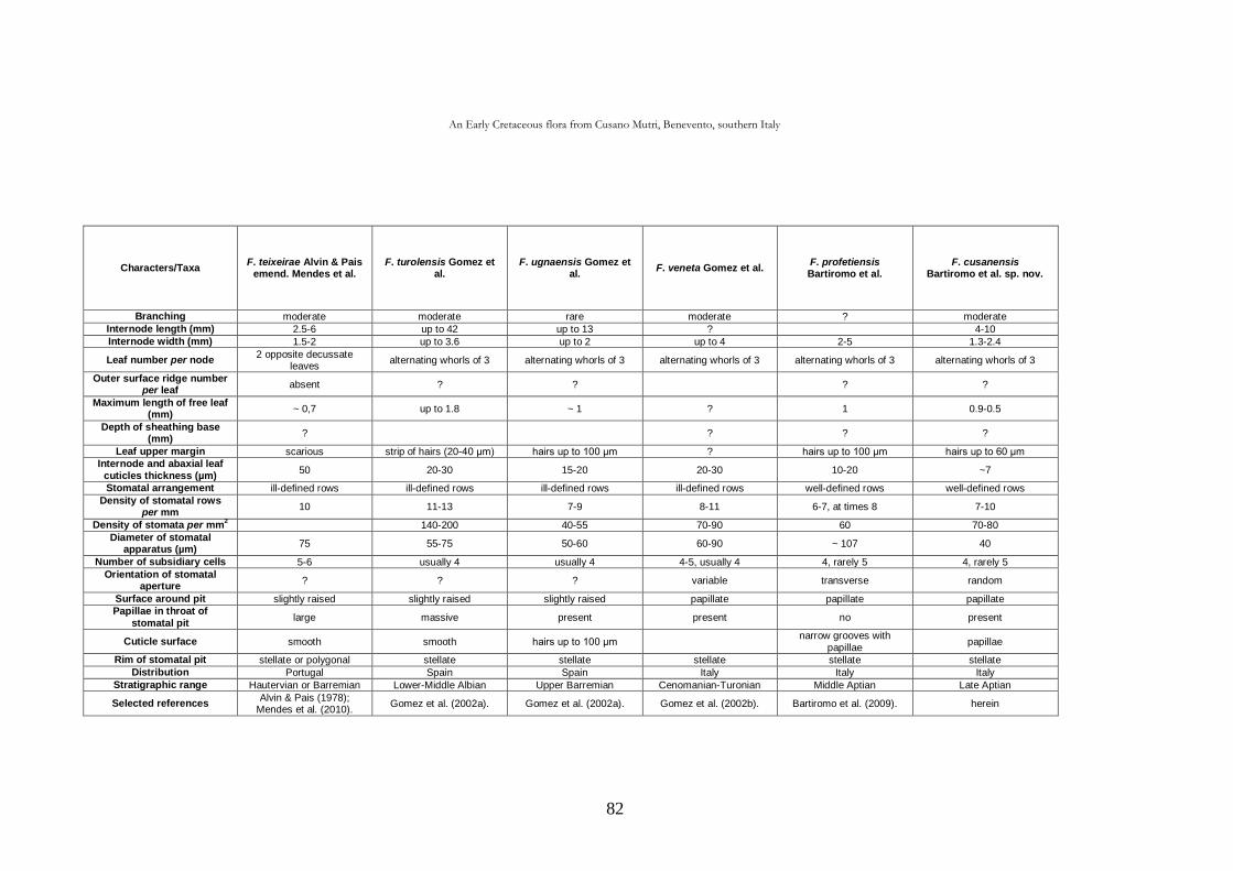
An Early Cretaceous flora from Cusano Mutri, Benevento, southern Italy
82
Characters/Taxa F. teixeirae Alvin & Pais
emend. Mendes et al. F. turolensis Gomez et
al. F. ugnaensis Gomez et
al. F. veneta Gomez et al.
F. profetiensis Bartiromo et al.
F. cusanensis Bartiromo et al. sp. nov.
Branching moderate moderate rare moderate ? moderate
Internode length (mm) 2.5-6 up to 42 up to 13 ? 4-10
Internode width (mm) 1.5-2 up to 3.6 up to 2 up to 4 2-5 1.3-2.4
Leaf number per node 2 opposite decussate
leaves alternating whorls of 3 alternating whorls of 3 alternating whorls of 3 alternating whorls of 3 alternating whorls of 3
Outer surface ridge number per leaf
absent ? ? ? ?
Maximum length of free leaf (mm)
~ 0,7 up to 1.8 ~ 1 ? 1 0.9-0.5
Depth of sheathing base (mm)
? ? ? ?
Leaf upper margin scarious strip of hairs (20-40 μm) hairs up to 100 μm ? hairs up to 100 μm hairs up to 60 μm
Internode and abaxial leaf cuticles thickness (µm)
50 20-30 15-20 20-30 10-20 ~7
Stomatal arrangement ill-defined rows ill-defined rows ill-defined rows ill-defined rows well-defined rows well-defined rows
Density of stomatal rows per mm
10 11-13 7-9 8-11 6-7, at times 8 7-10
Density of stomata per mm2 140-200 40-55 70-90 60 70-80
Diameter of stomatal apparatus (µm)
75 55-75 50-60 60-90 ~ 107 40
Number of subsidiary cells 5-6 usually 4 usually 4 4-5, usually 4 4, rarely 5 4, rarely 5
Orientation of stomatal aperture
? ? ? variable transverse random
Surface around pit slightly raised slightly raised slightly raised papillate papillate papillate Papillae in throat of
stomatal pit large massive present present no present
Cuticle surface smooth smooth hairs up to 100 μm narrow grooves with
papillae papillae
Rim of stomatal pit stellate or polygonal stellate stellate stellate stellate stellate
Distribution Portugal Spain Spain Italy Italy Italy
Stratigraphic range Hautervian or Barremian Lower-Middle Albian Upper Barremian Cenomanian-Turonian Middle Aptian Late Aptian
Selected references Alvin & Pais (1978);
Mendes et al. (2010). Gomez et al. (2002a). Gomez et al. (2002a). Gomez et al. (2002b). Bartiromo et al. (2009). herein
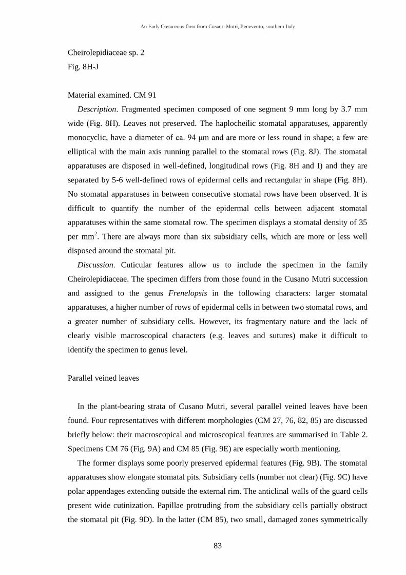
An Early Cretaceous flora from Cusano Mutri, Benevento, southern Italy
83
Cheirolepidiaceae sp. 2
Fig. 8H-J
Material examined. CM 91
Description. Fragmented specimen composed of one segment 9 mm long by 3.7 mm
wide (Fig. 8H). Leaves not preserved. The haplocheilic stomatal apparatuses, apparently
monocyclic, have a diameter of ca. 94 μm and are more or less round in shape; a few are
elliptical with the main axis running parallel to the stomatal rows (Fig. 8J). The stomatal
apparatuses are disposed in well-defined, longitudinal rows (Fig. 8H and I) and they are
separated by 5-6 well-defined rows of epidermal cells and rectangular in shape (Fig. 8H).
No stomatal apparatuses in between consecutive stomatal rows have been observed. It is
difficult to quantify the number of the epidermal cells between adjacent stomatal
apparatuses within the same stomatal row. The specimen displays a stomatal density of 35
per mm2. There are always more than six subsidiary cells, which are more or less well
disposed around the stomatal pit.
Discussion. Cuticular features allow us to include the specimen in the family
Cheirolepidiaceae. The specimen differs from those found in the Cusano Mutri succession
and assigned to the genus Frenelopsis in the following characters: larger stomatal
apparatuses, a higher number of rows of epidermal cells in between two stomatal rows, and
a greater number of subsidiary cells. However, its fragmentary nature and the lack of
clearly visible macroscopical characters (e.g. leaves and sutures) make it difficult to
identify the specimen to genus level.
Parallel veined leaves
In the plant-bearing strata of Cusano Mutri, several parallel veined leaves have been
found. Four representatives with different morphologies (CM 27, 76, 82, 85) are discussed
briefly below: their macroscopical and microscopical features are summarised in Table 2.
Specimens CM 76 (Fig. 9A) and CM 85 (Fig. 9E) are especially worth mentioning.
The former displays some poorly preserved epidermal features (Fig. 9B). The stomatal
apparatuses show elongate stomatal pits. Subsidiary cells (number not clear) (Fig. 9C) have
polar appendages extending outside the external rim. The anticlinal walls of the guard cells
present wide cutinization. Papillae protruding from the subsidiary cells partially obstruct
the stomatal pit (Fig. 9D). In the latter (CM 85), two small, damaged zones symmetrically
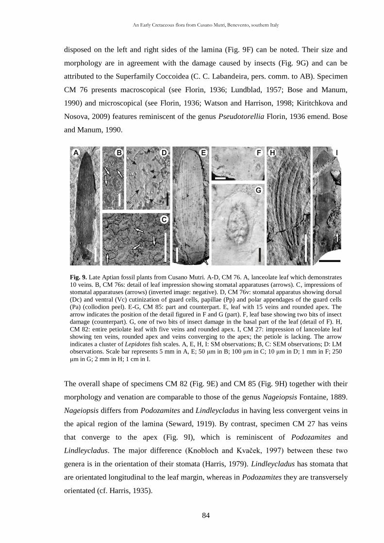
An Early Cretaceous flora from Cusano Mutri, Benevento, southern Italy
84
Fig. 9. Late Aptian fossil plants from Cusano Mutri. A-D, CM 76. A, lanceolate leaf which demonstrates
10 veins. B, CM 76s: detail of leaf impression showing stomatal apparatuses (arrows). C, impressions of
stomatal apparatuses (arrows) (inverted image: negative). D, CM 76v: stomatal apparatus showing dorsal
(Dc) and ventral (Vc) cutinization of guard cells, papillae (Pp) and polar appendages of the guard cells
(Pa) (collodion peel). E-G, CM 85: part and counterpart. E, leaf with 15 veins and rounded apex. The
arrow indicates the position of the detail figured in F and G (part). F, leaf base showing two bits of insect
damage (counterpart). G, one of two bits of insect damage in the basal part of the leaf (detail of F). H,
CM 82: entire petiolate leaf with five veins and rounded apex. I, CM 27: impression of lanceolate leaf
showing ten veins, rounded apex and veins converging to the apex; the petiole is lacking. The arrow
indicates a cluster of Lepidotes fish scales. A, E, H, I: SM observations; B, C: SEM observations; D: LM
observations. Scale bar represents 5 mm in A, E; 50 μm in B; 100 μm in C; 10 μm in D; 1 mm in F; 250
μm in G; 2 mm in H; 1 cm in I.
disposed on the left and right sides of the lamina (Fig. 9F) can be noted. Their size and
morphology are in agreement with the damage caused by insects (Fig. 9G) and can be
attributed to the Superfamily Coccoidea (C. C. Labandeira, pers. comm. to AB). Specimen
CM 76 presents macroscopical (see Florin, 1936; Lundblad, 1957; Bose and Manum,
1990) and microscopical (see Florin, 1936; Watson and Harrison, 1998; Kiritchkova and
Nosova, 2009) features reminiscent of the genus Pseudotorellia Florin, 1936 emend. Bose
and Manum, 1990.
The overall shape of specimens CM 82 (Fig. 9E) and CM 85 (Fig. 9H) together with their
morphology and venation are comparable to those of the genus Nageiopsis Fontaine, 1889.
Nageiopsis differs from Podozamites and Lindleycladus in having less convergent veins in
the apical region of the lamina (Seward, 1919). By contrast, specimen CM 27 has veins
that converge to the apex (Fig. 9I), which is reminiscent of Podozamites and
Lindleycladus. The major difference (Knobloch and Kvaček, 1997) between these two
genera is in the orientation of their stomata (Harris, 1979). Lindleycladus has stomata that
are orientated longitudinal to the leaf margin, whereas in Podozamites they are transversely
orientated (cf. Harris, 1935).
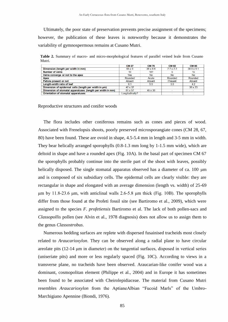
An Early Cretaceous flora from Cusano Mutri, Benevento, southern Italy
85
Table 2. Summary of macro- and micro-morphological features of parallel veined leale from Cusano
Mutri.
Ultimately, the poor state of preservation prevents precise assignment of the specimens;
however, the publication of these leaves is noteworthy because it demonstrates the
variability of gymnospermous remains at Cusano Mutri.
Reproductive structures and conifer woods
The flora includes other coniferous remains such as cones and pieces of wood.
Associated with Frenelopsis shoots, poorly preserved microsporangiate cones (CM 28, 67,
80) have been found. These are ovoid in shape, 4.5-5.4 mm in length and 3-5 mm in width.
They bear helically arranged sporophylls (0.8-1.3 mm long by 1-1.5 mm wide), which are
deltoid in shape and have a rounded apex (Fig. 10A). In the basal part of specimen CM 67
the sporophylls probably continue into the sterile part of the shoot with leaves, possibly
helically disposed. The single stomatal apparatus observed has a diameter of ca. 100 μm
and is composed of six subsidiary cells. The epidermal cells are clearly visible: they are
rectangular in shape and elongated with an average dimension (length vs. width) of 25-69
μm by 11.8-23.6 μm, with anticlinal walls 2.6-5.8 μm thick (Fig. 10B). The sporophylls
differ from those found at the Profeti fossil site (see Bartiromo et al., 2009), which were
assigned to the species F. profetiensis Bartiromo et al. The lack of both pollen-sacs and
Classopollis pollen (see Alvin et al., 1978 diagnosis) does not allow us to assign them to
the genus Classostrobus.
Numerous bedding surfaces are replete with dispersed fusainised tracheids most closely
related to Araucarioxylon. They can be observed along a radial plane to have circular
areolate pits (12-14 μm in diameter) on the tangential surfaces, disposed in vertical series
(uniseriate pits) and more or less regularly spaced (Fig. 10C). According to views in a
transverse plane, no tracheids have been observed. Araucarian-like conifer wood was a
dominant, cosmopolitan element (Philippe et al., 2004) and in Europe it has sometimes
been found to be associated with Cheirolepidiaceae. The material from Cusano Mutri
resembles Araucarioxylon from the AptianeAlbian “Fucoid Marls” of the Umbro-
Marchigiano Apennine (Biondi, 1976).

An Early Cretaceous flora from Cusano Mutri, Benevento, southern Italy
86
Angiospermae
Family incertae sedis
Genus Montsechia Teixeira, 1954
Montsechia vidalii (Zeiller) Teixeira, 1954
Fig. 10D-H
Material examined. CM 9, 98, 106, 107, 145
Description. The material consists of fragmented compressions and small impressions
of vegetative apparatuses. Hitherto only five specimens have been found at Cusano Mutri.
The best preserved specimen (CM 145) is composed of part and counterpart measuring 21
mm in length (measured along the main axis) by 11.6 mm in width (Fig. 10D). The main
axis is 0.7 mm thick at the base. This gradually narrows towards the distal part where it is
0.3 mm wide. The specimens consist of a succession of sub-opposite tufts referred to as
“bouquets” by Blanc-Louvel and Barale (1983) (Fig. 10E). The branches of the last order
depart from the area of bouquet-leaves (Fig. 10D and F). The leaves have an entire margin
and a rounded apex; their length does not exceed 1.5 mm. The leaves curve upward and are
a little narrower at the base. Owing to the poor preservation and brittleness of the cuticle,
some pieces were directly mounted onto the stubs without chemical treatment for SEM
observation, and peels were also taken to determine thoroughly the epidermal features.
Two morphological types of epidermal cells in the leaves can be distinguished: (1) more or
less isodiametric polygonal cells of no particular orientation (Fig. 10H); and (2) elongate
cells disposed in longitudinal rows (Fig. 10G). The former have an average diameter of 15
μm, while the latter are on average 34 μm long and 15 μm wide. The anticlinal walls of the
elongate epidermal cells are ca. 1.2 μm (max. 1.8 μm) thick. The rectangular cells are
located in the central part of the leaves; the polygonal ones are situated in the apex. No
veins or stomatal apparatuses were observed.
Discussion. The macro- and micro-morphology together with the size allow us to
attribute the specimens from Cusano Mutri to M. vidalii (Zeiller) Teixeira, 1954. M. vidalii
is an intriguing plant macrofossil from Barremian lithographic limestones in the Pyrenean
Basin (“El Montsec de Rubiès”) and the Eastern Iberian Basin (“Las Hoyas”) (Daviero-
Gomez et al., 2006) where it occurs abundantly (Diéguez et al., 2010). Crane and
Upchurch (1987) and Crane (1988) suggested that its morphology bears some resemblance
to Drewria potomacensis from the Early Cretaceous (cf. Aptian) Potomac Group of
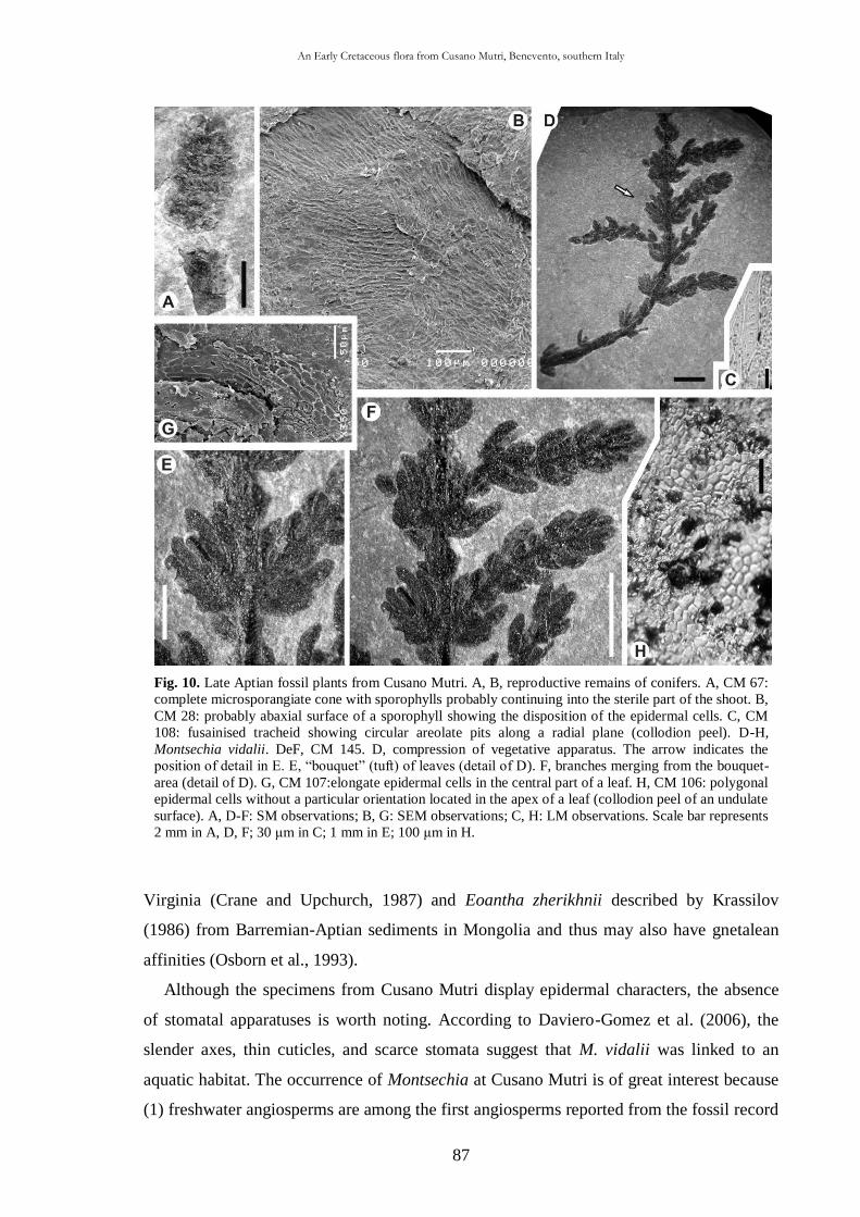
An Early Cretaceous flora from Cusano Mutri, Benevento, southern Italy
87
Fig. 10. Late Aptian fossil plants from Cusano Mutri. A, B, reproductive remains of conifers. A, CM 67:
complete microsporangiate cone with sporophylls probably continuing into the sterile part of the shoot. B,
CM 28: probably abaxial surface of a sporophyll showing the disposition of the epidermal cells. C, CM
108: fusainised tracheid showing circular areolate pits along a radial plane (collodion peel). D-H,
Montsechia vidalii. DeF, CM 145. D, compression of vegetative apparatus. The arrow indicates the
position of detail in E. E, “bouquet” (tuft) of leaves (detail of D). F, branches merging from the bouquet-
area (detail of D). G, CM 107:elongate epidermal cells in the central part of a leaf. H, CM 106: polygonal
epidermal cells without a particular orientation located in the apex of a leaf (collodion peel of an undulate
surface). A, D-F: SM observations; B, G: SEM observations; C, H: LM observations. Scale bar represents
2 mm in A, D, F; 30 μm in C; 1 mm in E; 100 μm in H.
Virginia (Crane and Upchurch, 1987) and Eoantha zherikhnii described by Krassilov
(1986) from Barremian-Aptian sediments in Mongolia and thus may also have gnetalean
affinities (Osborn et al., 1993).
Although the specimens from Cusano Mutri display epidermal characters, the absence
of stomatal apparatuses is worth noting. According to Daviero-Gomez et al. (2006), the
slender axes, thin cuticles, and scarce stomata suggest that M. vidalii was linked to an
aquatic habitat. The occurrence of Montsechia at Cusano Mutri is of great interest because
(1) freshwater angiosperms are among the first angiosperms reported from the fossil record
An Early Cretaceous flora from Cusano Mutri, Benevento, southern Italy
88
(Martín-Closas, 2003) and (2) angiosperm-like fossils become fewer and harder to find
below the Albian and seem to disappear below the Barremian or upper Hauterivian (Cornet
and Habib, 1992). The oldest aquatic angiosperm assemblage is known from the Spanish
Barremian with the species Ranunculus ferreri, M. vidalii and a few specimens related to
the genus Proteaphyllum (Martín-Closas, 2003). This is the first Italian record of M. vidalii
so far and the second oldest angiosperm found in Italy after those from the early-middle
Aptian locality of Profeti (Bartiromo et al., 2009). Moreover, an SEM image of epidermal
cells belonging to M. vidalii is published herein for the first time.
3.5. Taphonomic and palaeoecological remarks
Late Aptian Cusano Mutri plant remains occur in marly and calcareous strata. The marls
contain impressions almost exclusively. The calcareous beds frequently reveal cuticle
fragments, but these are poorly preserved.
3.5.1. Taphonomy
Plant macro-remains are usually of cm size. The absence of bioturbations allows us to
suppose that the fragmentation is probably related to a highly dynamic syn-depositional
environment and not to post-depositional reworking events. Throughout the stratigraphic
column all macro-remains are set parallel to the bedding plane. In addition in marl deposits
they are mainly isooriented, while they are sparse and randomly disposed in those of
limestones. During episodes of freshwater influx (marl levels) the kinetic energy would
have been raised, thus transporting and fragmenting the material prior to deposition. On the
other hand, evidence of low energy phases is testified by the occurrence of random
arrangement, sometimes associated with abundant small bivalves that are still articulated
and mainly preserved as internal moulds with the concave surfaces facing upward (Fig. 3B
and C).
Frenelopsis specimens are the best preserved plant remains. Branched specimens
together with articulated and disarticulated internodes still bear cuticles. The other taxa are
represented by isolated or fragmented leaves with poorly preserved remains of cuticle. The
poor preservation of the cuticles could be explained by (1) prolonged flotation of plant
remains before their final burial or (2) the slow sediment accumulation rate accompanied
by bacterial and saprophagous degradation prior to burial. Differences in cuticle
An Early Cretaceous flora from Cusano Mutri, Benevento, southern Italy
89
preservation may also be explained as an intrinsic response to their different systematic
affinity (see Mösle et al., 1998). It is not clear if shoot detachment in Frenelopsis is the
result of a biotic (e.g. physiological) process or abiotic (e.g. storm damage) origin, or is
owing to a combination of the two as suggested by Martín-Closas and Gomez (2004).
However, Martin (1999) pointed out that in temperate climates conifers tend to shed more
foliage in the autumn. The type and the state of preservation of the Cusano Mutri plants
(see above) allow us to infer the distance between the sedimentary basin and the source
area. Since only Frenelopsis is preserved as branched specimens and has the best preserved
cuticle, it is considered to have suffered a minimum amount of transport (see Gomez et al.,
2001, 2002a) and, therefore, possibly lived close to the sedimentary basin.
The surfaces of the marly strata frequently display many charred coniferous wood
fragments (fusain) that vary from randomly arranged to orientated tracheid elements (Fig.
8C). The more or less constant lengths of the fusain fragments (on average 15 mm)
seemingly are the result of sorting by wind (at least initially in subaerial environment) and
water (e.g. marine currents) or both. The fusain probably derived from a neighbouring
forest that was subjected to wildfires. The occurrence of fusain and fossil charcoal is
accepted as a reliable indicator of the occurrence of wildfire (Watson and Alvin, 1996; Van
Konijnenburg-Van Cittert, 2002), in most cases triggered by lightening (Sellwood and
Price, 1993). The Wealden Cretaceous climate was characterized by frequent rainstorms
leading to flash floods and lightning-generated wildfires (Haywood et al., 2004). During
Jurassic and Cretaceous periods wildfires were increasingly important (Scott, 2000; Pausas
and Keeley, 2009) to the extent that many taphofloras are explained by the occurrence of
frequent wildfires (Martín-Closas and Gomez, 2004). The large amount of fusain
throughout the whole Cusano Mutri succession, especially in marls, indicates that wildfires
were rather frequent events.
3.5.2. Palaeoecology of the Cusano Mutri sedimentary basin
Freshwater influence on the Cusano Mutri Basin is testified by several lines of
evidence. (1) The aquatic angiosperm M. vidalii is recorded in lithographic limestones
deposited in freshwater lakes (Coiffard et al., 2007) and is considered to be a hydrophyte
angiosperm (Teixeira, 1954; Martín-Closas, 2003). (2) Charophytes (Fig. 3E and F) are
essentially freshwater algae (e.g. Martín-Closas, 2003). (3) The ostracod fauna is
composed of strongly oligotypic to nearly monospecific associations, which is consistent
An Early Cretaceous flora from Cusano Mutri, Benevento, southern Italy
90
with a freshwater to brackish palaeoenvironment by comparison with extant assemblages
(Giuseppe Aiello and Diana Barra, pers. comm. to AB: taxonomic study in process). The
highest numbers occur in light-grey and red-brown marlstones, whereas they are generally
scarce in grey and reddish limestones. (4) Several surfaces are densely crowded with
disarticulated valves of estherids (conchostracans/ spinicaudatans). Today these usually
inhabit ephemeral freshwater ponds, but they may also occur in brackish waters of coastal
lagoons and tidally-influenced zones (Cunha Lana and de Souza Carvalho, 2002). The fact
that extant spinacaudatans can withstand brackish water and many assemblages have been
found associated with salt-water faunas led Webb (1979) to assume that some fossil
counterparts inhabited at least brackish and possibly marine environments. Tasch (1961)
attributed the association of spinicaudatan with marine fauna to the existence of temporary
pools or lagoons affected by episodic seawater influx producing faunal mixing. (5)
Lepidotes is the most abundant fish in Cusano Mutri; it is represented by whole and
uncrushed skeletons suggesting parautochthonous conditions of deposition. Most species
of this genus are supposed to have lived in freshwater, and, in Europe, the genus occurs in
alluvial and lacustrine deposits (Buscalioni et al., 2008).
These sedimentological and taphonomic considerations lead us to the conclusion that
the whole plattenkalk sequence is not the result of a “snapshot” of a mass mortality
phenomenon (sensu Martin, 1999), but a continuous, long-lasting sedimentary decantation
affected by tidal influx. The succession may correspond, therefore, to a tidal channel that
was periodically influenced by freshwater with the constant supply of terrigenous material.
3.5.3. Xeromorphic adaptations of the plants
Most of the examined plants display, at both macroscopical and microscopical levels,
xeromorphic adaptations, i.e. any plant structural feature reducing transpiration (Hill,
1998). Haworth and McElwain (2008, 2009) note that the presence of “xeromorphic”
features in fossil plants is often used to infer aridity, although in extant plants these
characters are not specifically restricted to plants growing in arid environments. They
commonly occur also in environments with high precipitation as well as in those with high
water availability. Retallack (2009) stated that xeromorphism is an adaptation related to
dry climate and implies that in the past scleromorphic features (e.g. thick cuticle) were
induced by dry climate. However, scleromorphy is also a result of other environmental
hardships, such as nutrient shortage (e.g. soil poor in phosphorous; see Hill, 1998) and high
An Early Cretaceous flora from Cusano Mutri, Benevento, southern Italy
91
salinity. Therefore, there is no general consensus on the environmental meaning of the high
frequency of “xeromorphic features” and, as Watson and Alvin (1996) suggested, the
unavailability of water is probably related to soil loaded with salt as well as to an arid or
semi-arid climate.
F. cusanensis nov. sp. displays a set of characters that are “classically” considered
xeromorphic (sensu Watson and Alvin, 1996) such as (1) small leaves, (2) stems likely to
be photosynthetic, (3) thick cuticle, (4) sunken stomata, and (5) papillate subsidiary cells.
In addition this species shows marginal hairs and papillate epidermal cells. The marginal
hairs present in Frenelopsis alata Knoblock (Feistmantell) have been compared (Pons,
1979; see also Watson and Alvin, 1996) with the extant North African conifer Tetraclinis
articulata (Vahl) Masters where, in a semi-arid climate, the reduced leaves show similar
marginal hairs that are used to capture condensing atmospheric moisture during the night.
Moreover, the papillate epidermal cells serve to block sunlight and reduce the transpiration
(Watson and Alvin, 1996). The massive presence of epidermal papillae on the external
surface of F. cusanensis would have the effect of reducing the average leaf temperature
and hence decreasing the rate of metabolic reactions associated with various harmful gases
in and around leaves (Sharma, 1977). Jordan et al. (2005) claimed that trichomes or
papillae create a glaucous surface to reduce the light reaching the mesophyll. Finally, the
thick cuticle with the sunken guard cells may have provided sufficient protection under
xeric conditions (Van der Ham et al., 2003).
Aside from stratigraphic and physiognomic indicators (Greb et al., 2006), isotopic
studies on Cretaceous European fossil plant assemblages using 12C/13C analysis indicate
that Frenelopsis in marginal marine facies had elevated 13C relative to other genera in
more distal facies. This would suggest stress and possible saline influence in salt-water
marshes (Nguyen Tu et al., 2002) or lagoons (Nguyen Tu et al., 1999).
3.5.4. Palaeoclimate and floral comparison
The specimens assigned to Cupressinocladus have diamond shaped, scale-like leaves
partially covering one another. The leaves are small, inserted at an acute angle and
adpressed against the axis. These features offer less surface exposure to solar radiation
(Thévenard et al., 2005). The leaves spread wide apart, displayed by the specimens
assigned to the genus Pagiophyllum, are similar to the relatively large, flattened, spreading
leaves of Brachyphyllum patens from the Maastrichtian of Belgium (Van der Ham et al.,
An Early Cretaceous flora from Cusano Mutri, Benevento, southern Italy
92
2003), which might indicate a humid habitat. This would tie in with the presence of
hydrophylous M. vidalii in the Cusano Mutri assemblage. On the other hand, the Profeti
flora (Bartiromo et al., 2009) is characterised by (1) a lower variability of gymnosperm
remains, (2) the absence of hydrophilous plants and (3) a significant lack of anatomical
features that reflect moist conditions. These differences could be related to the progressive
cooling trend that had been initiated during the Aptian (Srinivasan, 1994). According to
Coiffard et al. (2007), the drought phases ended at about the middle Aptian, after which a
wetter, cooler (by 5 °C) climate is evident from the occurrence of detrital quartz deposits in
the Vocontian Trough (Wortmann et al., 2004).
Sedimentological and palaeoecological evidence (e.g. the frequent freshwater influence)
together with the characters of the Cusano Mutri flora, would indicate, that growth of the
vegetation in the region was influenced by a semi-arid climate, seasonally punctuated by
wet phases on a regional to local scale. The wildfires suggested by abundant fusain in the
Cusano Mutri plattenkalk succession could have been engendered by periods of aridity
combined with high temperatures and lightning without accompanying rain (e.g. Watson
and Alvin, 1996).
The Cusano Mutri and Profeti floras have in common the genera Araucarites and
Frenelopsis. Although strictly not coeval, the resemblance between the Cusano Mutri flora
and that of the Montsech (Spain) lithographic limestones (Barale et al.,1984; Lacasa and
Martinez, 1986; Selden, 1989, 1990; Barale, 1991) is remarkable in that Pagiophyllum,
Araucarites, Frenelopsis and Montsechia occur in both. Such an assemblage is typical of
Euro-Sinian Province (Vakhrameev, 1991) and no Gondwanian representatives are
recorded. It is suggested, therefore, that the Cusano Mutri and Profeti successions reflect
relatively long, spatially limited exposed episodes of the Apenninic Carbonate Platform
during the Aptian, providing further evidence that the Cretaceous climate was less stable
than it was previously thought (Francis and Frakes, 1993).
Plant remains from the Early Cretaceous Fossil-Lagerstätte of Pietraroja, Southern Italy, Benevento
93
CHAPTER IV
Plant remains from the Early Cretaceous Fossil-Lagerstätte of
Pietraroja, Southern Italy, Benevento
4.4.1. Introduction
In Italy Cretaceous beds bearing plant macroremains have been known since the 19th
Century. However, with the exception of some recent contributes in which the epidermal
features have been treated (Gomez et al., 2002a; Bartiromo et al., 2008a, 2008b, 2009 and
other works in progress), few palaeobotanical studies have been carried out and these have
been scattered over a wide area. A summary of previous paleobotanical studies of the
Italian Cretaceous has been made by Gomez et al. (2002a, tab. 1, p. 674). Although in the
Campania Region Mesozoic rocks are exposed quite extensively, they only rarely host
well-preserved compression floras (e.g. Bartiromo et al., 2009). Therefore, the purpose of
this work is to contribute to the knowledge of the fossil plants and possibly to infer some
palaeoecological considerations of the famous (Capasso, 2007) Early Cretaceous (Early
Albian) locality of Pietraroja (Fig. 1(1)). This fossil site can be considered a “classic
Konzentrat und Konservat” Fossil-Lagerstätte (Seilacher, 1970; Seilacher et al., 1985) for
its numerous and exceptionally well-preserved animal (see below) and plant (Bartiromo et
al., 2006a and b; Bartiromo et al., 2008a and 2008b) remains yielded. This “taphonomic
window” (sensu Briggs, 2003) yields an extremely rich assemblage of invertebrate fauna
(Costa, 1853-1864; D’Argenio, 1963; Bravi, 1996; Bravi and Garassino, 1998).
Dasycladalean algae are also present (Bravi and Garassino, 1988; Carannante et al., 2006).
Moreover, this fossil site is extraordinary for its findings of several well-preserved fossil
vertebrates such as fish (Costa 1853-1864; Bassani, 1882, 1885; D’Erasmo, 1914-1915),
amphibians (McGowan, 2002; McGowan and Evans, 1995) and reptiles (Costa, 1864;
Estes, 1983; Evans, 2002; Evans et al., 2004; Barbera and Macuglia, 1988; Evans et al.,
2004, 2006), with at least two specimens that can be ascribed to Mesosuchidae. It is worth
mentioning the finding of a theropod dinosaur (Leonardi and Teruzzi, 1993), Scipionyx
samniticus Dal Sasso and Signore, remarkable for its exceptionally well preserved internal
organs (Dal Sasso and Signore, 1998; Dal Sasso and Maganuco, 2011).
As for Pietraroja fossil plants Costa (1865) mentioned “Quattro specie innominate”
(Four nameless species). Some fossil plants collected from this site were assigned to the
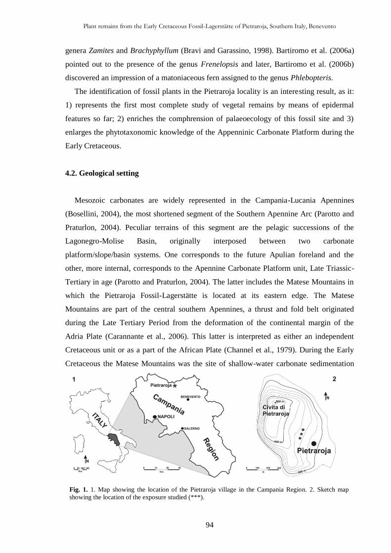
Plant remains from the Early Cretaceous Fossil-Lagerstätte of Pietraroja, Southern Italy, Benevento
94
Fig. 1. 1. Map showing the location of the Pietraroja village in the Campania Region. 2. Sketch map
showing the location of the exposure studied (***).
genera Zamites and Brachyphyllum (Bravi and Garassino, 1998). Bartiromo et al. (2006a)
pointed out to the presence of the genus Frenelopsis and later, Bartiromo et al. (2006b)
discovered an impression of a matoniaceous fern assigned to the genus Phlebopteris.
The identification of fossil plants in the Pietraroja locality is an interesting result, as it:
1) represents the first most complete study of vegetal remains by means of epidermal
features so far; 2) enriches the comphrension of palaeoecology of this fossil site and 3)
enlarges the phytotaxonomic knowledge of the Appenninic Carbonate Platform during the
Early Cretaceous.
4.2. Geological setting
Mesozoic carbonates are widely represented in the Campania-Lucania Apennines
(Bosellini, 2004), the most shortened segment of the Southern Apennine Arc (Parotto and
Praturlon, 2004). Peculiar terrains of this segment are the pelagic successions of the
Lagonegro-Molise Basin, originally interposed between two carbonate
platform/slope/basin systems. One corresponds to the future Apulian foreland and the
other, more internal, corresponds to the Apennine Carbonate Platform unit, Late Triassic-
Tertiary in age (Parotto and Praturlon, 2004). The latter includes the Matese Mountains in
which the Pietraroja Fossil-Lagerstätte is located at its eastern edge. The Matese
Mountains are part of the central southern Apennines, a thrust and fold belt originated
during the Late Tertiary Period from the deformation of the continental margin of the
Adria Plate (Carannante et al., 2006). This latter is interpreted as either an independent
Cretaceous unit or as a part of the African Plate (Channel et al., 1979). During the Early
Cretaceous the Matese Mountains was the site of shallow-water carbonate sedimentation
Plant remains from the Early Cretaceous Fossil-Lagerstätte of Pietraroja, Southern Italy, Benevento
95
Fig. 2. Sedimentological stratigraphic section of
the outcrop (modified from Bravi (1996)). The
arrow indicates the levels studied.
(Bosellini, 2004) characterized by an erosive mid Cretaceous gap, marked by bauxites
cropping out in the north-western area of the southern Apennine (D’Argenio et al., 1987).
The plant-ichtyolithic, organic-rich beds of Pietraroja formed during OAE 1 (sensu
Arthur and Schlanger, 1979) probably along
the NE margin of the Apenninic Carbonate
Platform (Ciarapica and Passeri, 2008) in a
high energy marginal environments (sensu
Ciarapica and Passeri, 2008). The Pietraroja
locality (Fig. 1(2)) crops out at the top of a
faulted monocline named “Civita di
Pietraroja”, at “Le Cavere” quarry [I.G.M.
(Italian Military Geographic Institute) map
162 III SW – Cusano Mutri sheet] and
includes two plattenkalk levels (Catenacci
and Manfredini, 1963; Bravi and Garassino,
1998; Carannante et al., 2006). Freels (1975)
reported three Plattenkalk intervals.
The famous Lower (basal) Albian
“Ichthyolitic level of Pietraroja”, yielding
plant remains (Plattenkalk 2, sensu Bravi e
Garassino, 1998) is about 8 m thick and its
age assignement is controlled by
foraminiferal data (Bravi and Garassino,
1998; Carannante et al., 2006) below and
above the plant-bearing sequence (Fig. 2).
The two Plattenkalks are overcome in
paraconformity by trasgressive Miocene
belonging to the Cusano Formation (Selli,
1957) with ostreids, bryozoans and
lithothamnes (Bravi and Garassino, 1998). In
the “Le Cavere” quarry, the rocks are well
stratified, occurring in flat and even layers
with an average thickness ranging between 2
and 10 cm. They mainly consist of more or
Plant remains from the Early Cretaceous Fossil-Lagerstätte of Pietraroja, Southern Italy, Benevento
96
less markedly micritic, avana to hazel or grey-coloured limestones (wackstones, mudstones
and packstones). These are intercalated by frequent lenses, nodules and beds of grey or
blackish cherts and marly-calcareous and sometimes clay and thin laminated horizons
(Bravi and Garassino, 1998). Strata bearing plant remains are situated in the basal part of
the “Le Cavere” quarry and they generally consist of packstones gradually turning into
wackstones and mudstones.
4.3. Material and methods
The studied plant remains come from “Le Cavere”, a protected locality included in the
“Geopaleontological Park” of Pietraroja, considered the depocentre area (Freels, 1975) of
the outcrop. The core-material of this study is represented by specimens labelled with “M”,
the result of an excavation effected in 1982 by the Museo Civico delle Scienze Naturali of
Turin and the former Istituto di Paleontologia (actually incorporated in the “Dipartimento
di Scienze della Terra”) of the “Federico II” University in Naples.
The marl and bituminous strata containing fossil plants led to the recovery of hundreds
of specimens in a short time. The fossil plants are mostly impressions and most specimens
are carbonaceous compressions without epidermal features. Even after the preliminary
chemical treatments, observation of microscopical details was still difficult on most
specimens. Not all of the adhering carbonaceous material was removed, because long
processing times destroyed the cuticle. That is why some cuticle fragments have been
mounted directly on the stubs for SEM (scanning electron microscope) observations.
Cuticle fragments available were separated from the sediment surface with a pair of
tweezers. They were cleaned in 37% hydrochloric acid (HCl), subsequently immersed in
40% hydrofluoric acid (HF) to remove adhering siliceous debris, and finally macerated in
Schulze’s reagent (HNO3+KClO3): the duration of the latter process depended on the
degree of carbonization and preservation (Kerp, 1990). Some cuticle fragments have been
immersed in ammonia for one hour. Subsequently, part of them was mounted in glycerine
jelly on glass slides for light microscope (LM) examination. Some fragments were
coloured with safranine before mounting. Treated and untreated cuticle samples were
mounted on the stubs for SEM observations. Light microscope and stereo microscope
(SM) observations were made by using Leitz microscopes. Photomicrographs with light
microscope were taken with a Nikon Coolpix 990 camera. The cuticles were examined
using a JEOL JSM-5310 SEM at the CISAG (Centro Interdipartimentale di servizi per
Plant remains from the Early Cretaceous Fossil-Lagerstätte of Pietraroja, Southern Italy, Benevento
97
Analisi Geomineralogiche). All the quantitative measurements were made with tools in the
ImageJ program (Abramoff et al., 2004). The leaf physiognomy of an angiosperm leaf was
described using the terminology of the Leaf Architecture Working Group (Ash et al.,
1999). Studied fossil plants come from: 1) a collection named “Fossili del giacimento di
Pietraroja”, catalogued as “vegetali indeterminati” (unclassified vegetables) and stored in
the Museo di Paleontologia of the Università degli Studi di Napoli “Federico II” (M and
MPUN series); 2) those gathered by the author outside the protected area of the “Le
Cavere” allowing the recovery of two specimens (PI 1-PI 2) actually housed in the
“Dipartimento di Scienze della Terra, Università degli Studi di Napoli “Federico II”; 3) a
private collection (PV 1); 4) a collection stored in the Museo Civico of Montefalcone in
Valfortore (MF).
4.4. Systematic Palaeontology
Order CAYTONIALES
Genus Sagenopteris Presl in Sternberg, 1838
Sagenopteris? sp.
Figs. 3(1), 4(1-3)
Material examined – M 20802.
Description – Part and counterpart of a complete, rounded, petiolated and detached
leaflet, with constricted base and entire margin. The specimen measures 13 mm in length
and 11 mm in width. The lamina is symmetrical. The petiole is 0.8 mm wide. The leaflet
shows a more or less distinct midrib -~ 0.2 mm in width at the base- departing from the
petiole and disappearing shortly in the middle part. Secondary veins are hardly visible and
arise from the rachis with an angle of ~ 30° (Figs. 3(1), 4(1)). Along the leaf margin and on
the petiole area, poorly preserved epidermal features are visible. The cuticle is ~ 0.6 μm
thick (Fig. 4(2)). The epidermal cells are more or less rounded. Numerous circular or ovate
structures of ~ 50 μm in diameter of unknown function can be observed (Fig. 4(3)). No
stomatal apparatuses have been found.
Discussion – Morphology and venation of the specimen resemble Sagenopteris Presl,
1838. In particular, the morphology and the length-width ratio (1.1) are comparable to S.
williamsii described by Berry (1956, Pl. XXXIV, Fig. 1) from the Lower Cretaceous of
Canada. At the microscopical level, observed circular-ovate structures are similar to those
Plant remains from the Early Cretaceous Fossil-Lagerstätte of Pietraroja, Southern Italy, Benevento
98
reported by Barbacka et al. (2006) for the Hettangian S. pualensis and interpreted by the
authors as possibly glands or hair bases. However, the poor state of preservation of the
specimen together with the unclear venation pattern, do not allow to assign the specimen
with confidence to the genus Sagenopteris. In addition, the taxonomy of the genus
Sagenopteris is still confused (Zaton et al., 2006) and a specific determination is
Fig. 3. Early Albian fossil plants from Pietraroja. Line drawings of selected specimens. 1. Sagenopteris?
sp.. Showing primary and secondary veins (M 20802). 2. Brachyphyllum sp. 1. Showing branched
specimen and poorly preserved leaf-scars in its basal part (M 20908). 3. Brachyphyllum sp. 2. Showing
two-order branches and leaves tightly adpressed to the axis (M 20765). 4. Brachyphyllum sp. 2. Showing
three-order branches (M 20909). 5. Frenelopsis sp.. Large branched specimen showing two-order
branches and the central vascular bundle (M 20569). 6. Angiospermae 1. Showing the well-visible
primary and secondary -in the basal part- veins (MF 1265). 7. Incertae sedis. Leaf showing three lobes
and a distinct petiole (M 20796).
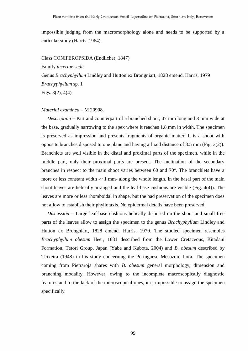
Plant remains from the Early Cretaceous Fossil-Lagerstätte of Pietraroja, Southern Italy, Benevento
99
impossible judging from the macromorphology alone and needs to be supported by a
cuticular study (Harris, 1964).
Class CONIFEROPSIDA (Endlicher, 1847)
Family incertae sedis
Genus Brachyphyllum Lindley and Hutton ex Brongniart, 1828 emend. Harris, 1979
Brachyphyllum sp. 1
Figs. 3(2), 4(4)
Material examined – M 20908.
Description – Part and counterpart of a branched shoot, 47 mm long and 3 mm wide at
the base, gradually narrowing to the apex where it reaches 1.8 mm in width. The specimen
is preserved as impression and presents fragments of organic matter. It is a shoot with
opposite branches disposed to one plane and having a fixed distance of 3.5 mm (Fig. 3(2)).
Branchlets are well visible in the distal and proximal parts of the specimen, while in the
middle part, only their proximal parts are present. The inclination of the secondary
branches in respect to the main shoot varies between 60 and 70°. The branchlets have a
more or less constant width -~ 1 mm- along the whole length. In the basal part of the main
shoot leaves are helically arranged and the leaf-base cushions are visible (Fig. 4(4)). The
leaves are more or less rhomboidal in shape, but the bad preservation of the specimen does
not allow to establish their phyllotaxis. No epidermal details have been preserved.
Discussion – Large leaf-base cushions helically disposed on the shoot and small free
parts of the leaves allow to assign the specimen to the genus Brachyphyllum Lindley and
Hutton ex Brongniart, 1828 emend. Harris, 1979. The studied specimen resembles
Brachyphyllum obesum Heer, 1881 described from the Lower Cretaceous, Kitadani
Formation, Tetori Group, Japan (Yabe and Kubota, 2004) and B. obesum described by
Teixeira (1948) in his study concerning the Portuguese Mesozoic flora. The specimen
coming from Pietraroja shares with B. obesum general morphology, dimension and
branching modality. However, owing to the incomplete macroscopically diagnostic
features and to the lack of the microscopical ones, it is impossible to assign the specimen
specifically.
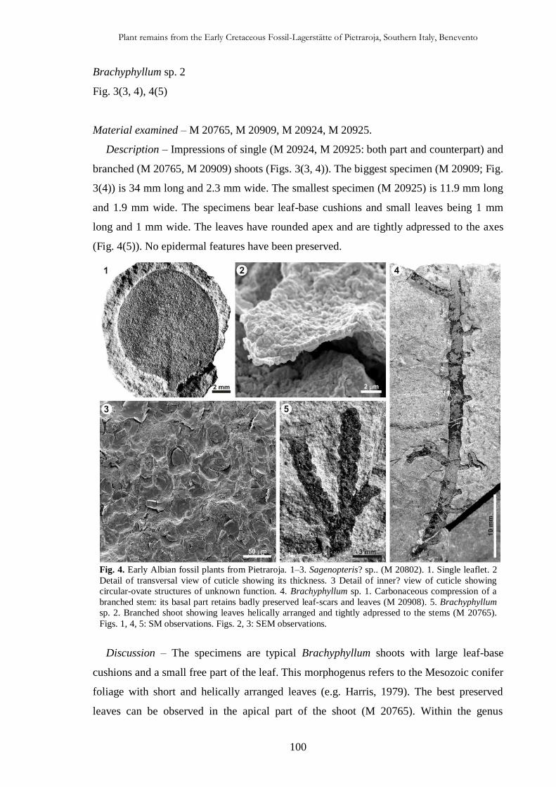
Plant remains from the Early Cretaceous Fossil-Lagerstätte of Pietraroja, Southern Italy, Benevento
100
Brachyphyllum sp. 2
Fig. 3(3, 4), 4(5)
Material examined – M 20765, M 20909, M 20924, M 20925.
Description – Impressions of single (M 20924, M 20925: both part and counterpart) and
branched (M 20765, M 20909) shoots (Figs. 3(3, 4)). The biggest specimen (M 20909; Fig.
3(4)) is 34 mm long and 2.3 mm wide. The smallest specimen (M 20925) is 11.9 mm long
and 1.9 mm wide. The specimens bear leaf-base cushions and small leaves being 1 mm
long and 1 mm wide. The leaves have rounded apex and are tightly adpressed to the axes
(Fig. 4(5)). No epidermal features have been preserved.
Discussion – The specimens are typical Brachyphyllum shoots with large leaf-base
cushions and a small free part of the leaf. This morphogenus refers to the Mesozoic conifer
foliage with short and helically arranged leaves (e.g. Harris, 1979). The best preserved
leaves can be observed in the apical part of the shoot (M 20765). Within the genus
Fig. 4. Early Albian fossil plants from Pietraroja. 1–3. Sagenopteris? sp.. (M 20802). 1. Single leaflet. 2
Detail of transversal view of cuticle showing its thickness. 3 Detail of inner? view of cuticle showing
circular-ovate structures of unknown function. 4. Brachyphyllum sp. 1. Carbonaceous compression of a
branched stem: its basal part retains badly preserved leaf-scars and leaves (M 20908). 5. Brachyphyllum
sp. 2. Branched shoot showing leaves helically arranged and tightly adpressed to the stems (M 20765).
Figs. 1, 4, 5: SM observations. Figs. 2, 3: SEM observations.
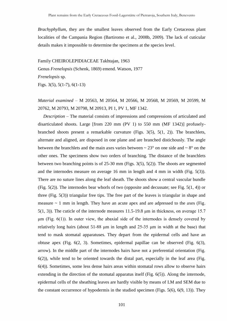
Plant remains from the Early Cretaceous Fossil-Lagerstätte of Pietraroja, Southern Italy, Benevento
101
Brachyphyllum, they are the smallest leaves observed from the Early Cretaceous plant
localities of the Campania Region (Bartiromo et al., 2008b, 2009). The lack of cuticular
details makes it impossible to determine the specimens at the species level.
Family CHEIROLEPIDIACEAE Takhtajan, 1963
Genus Frenelopsis (Schenk, 1869) emend. Watson, 1977
Frenelopsis sp.
Figs. 3(5), 5(1-7), 6(1-13)
Material examined – M 20563, M 20564, M 20566, M 20568, M 20569, M 20599, M
20762, M 20793, M 20798, M 20913, PI 1, PV 1, MF 1342.
Description – The material consists of impressions and compressions of articulated and
disarticulated shoots. Large [from 220 mm (PV 1) to 550 mm (MF 1342)] profusely-
branched shoots present a remarkable curvature (Figs. 3(5), 5(1, 2)). The branchlets,
alternate and aligned, are disposed in one plane and are branched distichously. The angle
between the branchlets and the main axes varies between ~ 23° on one side and ~ 8° on the
other ones. The specimens show two orders of branching. The distance of the branchlets
between two branching points is of 25-30 mm (Figs. 3(5), 5(2)). The shoots are segmented
and the internodes measure on average 16 mm in length and 4 mm in width (Fig. 5(3)).
There are no suture lines along the leaf sheath. The shoots show a central vascular bundle
(Fig. 5(2)). The internodes bear whorls of two (opposite and decussate; see Fig. 5(1, 4)) or
three (Fig. 5(3)) triangular free tips. The free part of the leaves is triangular in shape and
measure ~ 1 mm in length. They have an acute apex and are adpressed to the axes (Fig.
5(1, 3)). The cuticle of the internode measures 11.5-19.8 μm in thickness, on average 15.7
μm (Fig. 6(1)). In outer view, the abaxial side of the internodes is densely covered by
relatively long hairs (about 51-88 μm in length and 25-35 μm in width at the base) that
tend to mask stomatal apparatuses. They depart from the epidermal cells and have an
obtuse apex (Fig. 6(2, 3). Sometimes, epidermal papillae can be observed (Fig. 6(3),
arrow). In the middle part of the internodes hairs have not a preferential orientation (Fig.
6(2)), while tend to be oriented towards the distal part, especially in the leaf area (Fig.
6(4)). Sometimes, some less dense hairs areas within stomatal rows allow to observe hairs
extending in the direction of the stomatal apparatus itself (Fig. 6(5)). Along the internode,
epidermal cells of the sheathing leaves are hardly visible by means of LM and SEM due to
the constant occurrence of hypodermis in the studied specimen (Figs. 5(6), 6(9, 13)). They
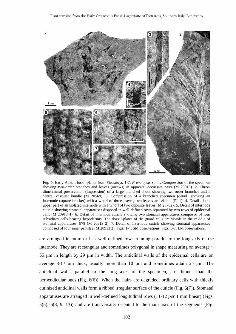
Plant remains from the Early Cretaceous Fossil-Lagerstätte of Pietraroja, Southern Italy, Benevento
102
Fig. 5. Early Albian fossil plants from Pietraroja. 1-7. Frenelopsis sp. 1. Compression of the specimen
showing two-order branches and leaves (arrows) in opposite, decussate pairs (M 20913). 2. Three-
dimensional preservation (impression) of a large branched shoot showing two-order branches and a
central vascular bundle (M 20569). 3. Compression of a branched specimen (detail) showing an
internode (square bracket) with a whorl of three leaves, two leaves are visible (PI 1). 4. Detail of the
upper part of an isolated internode with a whorl of two opposite leaves (M 20762). 5. Detail of internode
cuticle showing stomatal apparatuses disposed in well-defined rows separated by two rows of epidermal
cells (M 20913 4). 6. Detail of internode cuticle showing two stomatal apparatuses composed of four
subsidiary cells bearing hypodermis. The dorsal plates of the guard cells are visible in the middle of
stomatal apparatuses. 978 (M 20913 2). 7. Detail of internode cuticle showing stomatal apparatuses
composed of four inner papillae (M 20913 2). Figs. 1-4: SM observations. Figs. 5-7: LM observations.
are arranged in more or less well-defined rows running parallel to the long axis of the
internode. They are rectangular and sometimes polygonal in shape measuring on average ~
55 μm in length by 29 μm in width. The anticlinal walls of the epidermal cells are on
average 8-17 μm thick, usually more than 10 μm and sometimes attain 25 μm. The
anticlinal walls, parallel to the long axes of the specimen, are thinner than the
perpendicular ones (Fig. 6(6)). When the hairs are degraded, ordinary cells with thickly
cutinized anticlinal walls form a ribbed irregular surface of the cuticle (Fig. 6(7)). Stomatal
apparatuses are arranged in well-defined longitudinal rows (11-12 per 1 mm linear) (Figs.
5(5), 6(8, 9, 13)) and are transversally oriented to the main axes of the segments (Fig.
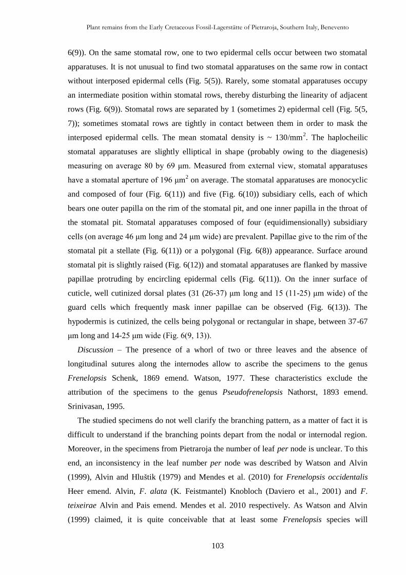
Plant remains from the Early Cretaceous Fossil-Lagerstätte of Pietraroja, Southern Italy, Benevento
103
6(9)). On the same stomatal row, one to two epidermal cells occur between two stomatal
apparatuses. It is not unusual to find two stomatal apparatuses on the same row in contact
without interposed epidermal cells (Fig. 5(5)). Rarely, some stomatal apparatuses occupy
an intermediate position within stomatal rows, thereby disturbing the linearity of adjacent
rows (Fig. 6(9)). Stomatal rows are separated by 1 (sometimes 2) epidermal cell (Fig. 5(5,
7)); sometimes stomatal rows are tightly in contact between them in order to mask the
interposed epidermal cells. The mean stomatal density is ~ 130/mm2. The haplocheilic
stomatal apparatuses are slightly elliptical in shape (probably owing to the diagenesis)
measuring on average 80 by 69 μm. Measured from external view, stomatal apparatuses
have a stomatal aperture of 196 μm2 on average. The stomatal apparatuses are monocyclic
and composed of four (Fig. 6(11)) and five (Fig. 6(10)) subsidiary cells, each of which
bears one outer papilla on the rim of the stomatal pit, and one inner papilla in the throat of
the stomatal pit. Stomatal apparatuses composed of four (equidimensionally) subsidiary
cells (on average 46 μm long and 24 μm wide) are prevalent. Papillae give to the rim of the
stomatal pit a stellate (Fig. 6(11)) or a polygonal (Fig. 6(8)) appearance. Surface around
stomatal pit is slightly raised (Fig. 6(12)) and stomatal apparatuses are flanked by massive
papillae protruding by encircling epidermal cells (Fig. 6(11)). On the inner surface of
cuticle, well cutinized dorsal plates (31 (26-37) μm long and 15 (11-25) μm wide) of the
guard cells which frequently mask inner papillae can be observed (Fig. 6(13)). The
hypodermis is cutinized, the cells being polygonal or rectangular in shape, between 37-67
μm long and 14-25 μm wide (Fig. 6(9, 13)).
Discussion – The presence of a whorl of two or three leaves and the absence of
longitudinal sutures along the internodes allow to ascribe the specimens to the genus
Frenelopsis Schenk, 1869 emend. Watson, 1977. These characteristics exclude the
attribution of the specimens to the genus Pseudofrenelopsis Nathorst, 1893 emend.
Srinivasan, 1995.
The studied specimens do not well clarify the branching pattern, as a matter of fact it is
difficult to understand if the branching points depart from the nodal or internodal region.
Moreover, in the specimens from Pietraroja the number of leaf per node is unclear. To this
end, an inconsistency in the leaf number per node was described by Watson and Alvin
(1999), Alvin and Hluštik (1979) and Mendes et al. (2010) for Frenelopsis occidentalis
Heer emend. Alvin, F. alata (K. Feistmantel) Knobloch (Daviero et al., 2001) and F.
teixeirae Alvin and Pais emend. Mendes et al. 2010 respectively. As Watson and Alvin
(1999) claimed, it is quite conceivable that at least some Frenelopsis species will
Plant remains from the Early Cretaceous Fossil-Lagerstätte of Pietraroja, Southern Italy, Benevento
104
eventually prove to have had varied morphology and leaf number in different parts of the
plant. As for the microscopical features, the specimens from Pietraroja resemble F.
teixeirae in: 1) the presence of hairs on the margin of the leaves; 2) the abaxial internode
cuticle thickness; 3) the diameter of the stomatal apparatuses; 4) the presence of hairs and
papillae on the outer cuticle surface; 5) the morphology of the rim of stomatal pit.
Moreover, the Italian material resembles F. silfloana Watson in: 1) the density of stomatal
rows; 2) the diameter of stomatal apparatuses; 3) the number of subsidiary cells; 4) the
presence of papillae in the throat of stomatal pit. However, at present, the unclear
phyllotaxy at the nodes and the few and more or less poorly preserved cuticle fragments do
not allow to understand well the epidermal architecture, and prevent the assignment of the
specimens in a given species already described or their attribution to a new taxon and the
analysis of further specimens is needed.
Cheirolepidiacean 1
Fig. 7(1-5)
Material examined – M 20914.
Description – Fragmented shoot of 20 mm long and 6 mm wide. It has short leaves of
open-type arranged in a simple spiral that do not completely encircle the stem (Fig. 7(1)).
A small cuticular fragment in inner view (Fig. 7(2)) allowed to observe few monocyclic
stomatal apparatuses and partially to elucidate the thickness of poorly preserved cuticle: ~
25 μm (Fig. 7(3)). The stomatal apparatuses are more or less rounded; they are 66 μm (61-
71 μm) in diameter and are arranged in well-defined rows (Fig. 7(2)). Stomatal apparatuses
are composed of six subsidiary cells (Fig. 7(4)). Some of them -especially those visible in
the right part of the fragment shown in Fig. 7(2)- have preserved the guard cells. The
subsidiary cells have anticlinal walls ~ 3 μm thick. The anticlinal walls of ordinary
epidermal cells are ~ 6 μm thick. From the inner view, outer papillae are visible (Fig. 7(5)).
The epidermal cells are difficult to observe but form one or two longitudinal rows in
between the stomatal rows.
Discussion – The features listed above are comparable to some members of the family
Cheirolepidiaceae, in particular to the genus Pseudofrenelopsis Nathorst, 1893 emend.
Srinivasan, 1995. The two genera Pseudofrenelopsis Nathorst (1893) and Frenelopsis
Schenk (1869) which have similar segmented shoots, differ mainly in the leaf number and
phyllotaxis (Srinivasan, 1995). In Pseudofrenelopsis there are one (Watson, 1977) and two
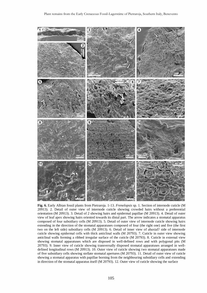
Plant remains from the Early Cretaceous Fossil-Lagerstätte of Pietraroja, Southern Italy, Benevento
105
Fig. 6. Early Albian fossil plants from Pietraroja. 1-13. Frenelopsis sp. 1. Section of internode cuticle (M
20913). 2. Detail of outer view of internode cuticle showing crowded hairs without a preferential
orientation (M 20913). 3. Detail of 2 showing hairs and epidermal papillae (M 20913). 4. Detail of outer
view of leaf apex showing hairs oriented towards its distal part. The arrow indicates a stomatal apparatus
composed of four subsidiary cells (M 20913). 5. Detail of outer view of internode cuticle showing hairs
extending in the direction of the stomatal apparatuses composed of four (the right one) and five (the first
two on the left side) subsidiary cells (M 20913). 6. Detail of inner view of abaxial? side of internode
cuticle showing epidermal cells with thick anticlinal walls (M 20793). 7. Cuticle in outer view showing
anticlinal walls forming a ribbed irregular surface of the cuticle (M 20793). 8. Cuticle in external view
showing stomatal apparatuses which are disposed in well-defined rows and with polygonal pits (M
20793). 9. Inner view of cuticle showing transversally disposed stomatal apparatuses arranged in well-
defined longitudinal rows (M 20913). 10. Outer view of cuticle showing two stomatal apparatuses made
of five subsidiary cells showing stellate stomatal apertures (M 20793). 11. Detail of outer view of cuticle
showing a stomatal apparatus with papillae borning from the neighbouring subsidiary cells and extending
in direction of the stomatal apparatus itself (M 20793). 12. Outer view of cuticle showing the surface

Plant remains from the Early Cretaceous Fossil-Lagerstätte of Pietraroja, Southern Italy, Benevento
106
around stomatal pit which is slightly raised (M 20793). 13. Inner view of cuticle showing two rows of
stomatal apparatuses, the hypodermal cells undercovering ordinary epidermal cells and dorsal plates of guard
cells (M 20913). All figures are made by means of SEM.
(Srinivasan (1995), for the species Pseudofrenelopsis nathorstiana Srinivasan, 1995)
leaves per node spirally arranged and occasionally showing cyclic arrangement
(Srinivasan, 1995). In Frenelopsis, the internodes have two or three leaves per node in an
opposite decussate/cyclic arrangement and the internodes are only of the closed type
(Srinivasan, 1995). However, the specimen from Pietraroja shows similarity to
Pseudofrenelopsis varians (Fontaine) Watson, 1977 at both macroscopically (e.g. the shoot
morphology) and microscopically (e.g. stomatal apparatuses) levels. As a matter of fact,
the material from Pietraroja is comparable to the specimens figured by Watson (1977, Pl.
88, figs. 2, 4; the fig. 2 of the Pl. 2 is also suggested again in Watson 1988, fig. 9.19D).
The specimen from Pietraroja differs from the species P. intermedia (Chow and Tsao) in
having the thinnest cuticle and from the species P. nathorstiana Srinivasan, 1995 in having
the greatest internode width. Moreover, the specimen is different from M 20752 by having
1) spirally arranged leaves and 2) greater stomatal apparatuses. Unfortunately, the lack of
well-defined macro- and micro-features does not allow an identification at the genus level.
However, the co-occurrence of Frenelopsis and Pseudofrenelopsis is not surprising
because they are sometimes mixed in the same bed (Watson, 1977, 1983; Srinivasan, 1992;
Yang et al., 2009).
Long parallel veined leaves
Material examined – M 20761, PI 2.
The plant-bearing strata of Pietraroja are full of leaves showing parallel venation which
are difficult to determine. Herein, two representatives are presented. The specimen M
20761 is a compression of an isolated and petiolate leaf 18 mm long and 5 mm wide (Fig.
7(6)). The apex is rounded and the petiole measures 2 mm in length reaching the maximum
width (2 mm) at the base. The veins converge to the apex (Fig. 7(7)). Structures resembling
stomatal apparatuses (Fig. 7(8)) and transversally? oriented in respect to the long axes of
the leaf can be seen. The general morphology, dimension and venation pattern are
comparable to the genera Lindleycladus and Podozamites. Harris (1979) claims that
Lindleycladus is distinguished from Podozamites for its longitudinally rather than
transversally orientated stomata, which are placed in longitudinal rows. Unfortunately,
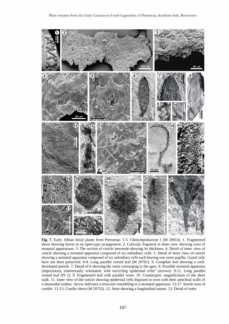
Plant remains from the Early Cretaceous Fossil-Lagerstätte of Pietraroja, Southern Italy, Benevento
107
Fig. 7. Early Albian fossil plants from Pietraroja. 1-5. Cheirolepidiacean 1 (M 20914). 1. Fragmented
shoot showing leaves in an open-type arrangement. 2. Cuticular fragment in inner view showing rows of
stomatal apparatuses. 3. The section of cuticle internode showing its thickness. 4. Detail of inner view of
cuticle showing a stomatal apparatus composed of six subsidiary cells. 5. Detail of inner view of cuticle
showing a stomatal apparatus composed of six subsidiary cells each bearing one outer papilla. Guard cells
have not been preserved. 6-8. Long parallel veined leaf (M 20761). 6. Complete leaf showing a well-
developed petiole. 7. Detail of 6 showing the veins converging to the apex. 8. Possible stomatal apparatus
(impression), transversally orientated, with encircling epidermal cells? (arrows). 9-11. Long parallel
veined leaf (PI 2). 9. Fragmented leaf with parallel veins. 10. Counterpart: magnification of the short
stalk. 11. Inner view of the cuticle showing epidermal cells disposed in rows with their anticlinal walls of
a sinusoidal outline. Arrow indicates a structure resembling to a stomatal apparatus. 12-17. Sterile axes of
conifer. 12-13. Conifer shoot (M 20752). 12. Stem showing a longitudinal suture. 13. Detail of outer
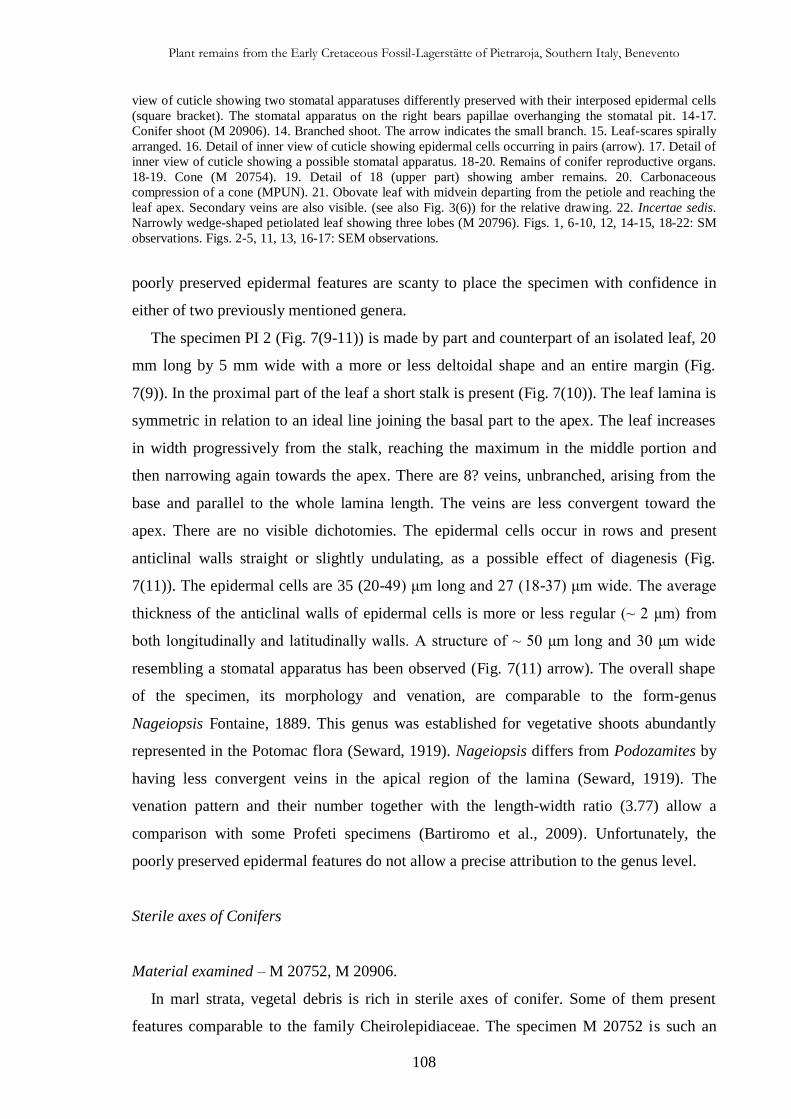
Plant remains from the Early Cretaceous Fossil-Lagerstätte of Pietraroja, Southern Italy, Benevento
108
view of cuticle showing two stomatal apparatuses differently preserved with their interposed epidermal cells
(square bracket). The stomatal apparatus on the right bears papillae overhanging the stomatal pit. 14-17.
Conifer shoot (M 20906). 14. Branched shoot. The arrow indicates the small branch. 15. Leaf-scares spirally
arranged. 16. Detail of inner view of cuticle showing epidermal cells occurring in pairs (arrow). 17. Detail of
inner view of cuticle showing a possible stomatal apparatus. 18-20. Remains of conifer reproductive organs.
18-19. Cone (M 20754). 19. Detail of 18 (upper part) showing amber remains. 20. Carbonaceous
compression of a cone (MPUN). 21. Obovate leaf with midvein departing from the petiole and reaching the
leaf apex. Secondary veins are also visible. (see also Fig. 3(6)) for the relative drawing. 22. Incertae sedis.
Narrowly wedge-shaped petiolated leaf showing three lobes (M 20796). Figs. 1, 6-10, 12, 14-15, 18-22: SM
observations. Figs. 2-5, 11, 13, 16-17: SEM observations.
poorly preserved epidermal features are scanty to place the specimen with confidence in
either of two previously mentioned genera.
The specimen PI 2 (Fig. 7(9-11)) is made by part and counterpart of an isolated leaf, 20
mm long by 5 mm wide with a more or less deltoidal shape and an entire margin (Fig.
7(9)). In the proximal part of the leaf a short stalk is present (Fig. 7(10)). The leaf lamina is
symmetric in relation to an ideal line joining the basal part to the apex. The leaf increases
in width progressively from the stalk, reaching the maximum in the middle portion and
then narrowing again towards the apex. There are 8? veins, unbranched, arising from the
base and parallel to the whole lamina length. The veins are less convergent toward the
apex. There are no visible dichotomies. The epidermal cells occur in rows and present
anticlinal walls straight or slightly undulating, as a possible effect of diagenesis (Fig.
7(11)). The epidermal cells are 35 (20-49) μm long and 27 (18-37) μm wide. The average
thickness of the anticlinal walls of epidermal cells is more or less regular (~ 2 μm) from
both longitudinally and latitudinally walls. A structure of ~ 50 μm long and 30 μm wide
resembling a stomatal apparatus has been observed (Fig. 7(11) arrow). The overall shape
of the specimen, its morphology and venation, are comparable to the form-genus
Nageiopsis Fontaine, 1889. This genus was established for vegetative shoots abundantly
represented in the Potomac flora (Seward, 1919). Nageiopsis differs from Podozamites by
having less convergent veins in the apical region of the lamina (Seward, 1919). The
venation pattern and their number together with the length-width ratio (3.77) allow a
comparison with some Profeti specimens (Bartiromo et al., 2009). Unfortunately, the
poorly preserved epidermal features do not allow a precise attribution to the genus level.
Sterile axes of Conifers
Material examined – M 20752, M 20906.
In marl strata, vegetal debris is rich in sterile axes of conifer. Some of them present
features comparable to the family Cheirolepidiaceae. The specimen M 20752 is such an
Plant remains from the Early Cretaceous Fossil-Lagerstätte of Pietraroja, Southern Italy, Benevento
109
example. It is represented by an unbranched and fragmented shoot, 13.8 mm long by 2 mm
wide with a longitudinal suture (Fig. 7(12)). SEM observations of a single, small and
poorly preserved cuticle fragment, 2 mm long per 1.1 mm wide, allow to observe two
stomatal apparatuses in outer view. They are ~ 50 μm in diameter; the right one in Fig.
7(13) presents only two papillae preserved. These are 7 μm long, each one steming from
one subsidiary cell (Fig. 7(13). The epidermal cells are squared off or rectangular in shape
and are disposed in ill-defined rows. They are on average ~ 30 μm long and 27 μm wide.
The anticlinal walls are 2.4 μm thick. The presence of a longitudinal suture along the shoot
together with the papillate stomatal apparatuses are comparable to the genera
Cupressinocladus Seward, 1919 and Watsoniocladus Srinivasan, 1995. However, the poor
state of preservation does not allow to place the specimen systematically. Moreover, the
absence of the Classopollis pollen makes it difficult to place the specimen in the family
Cheirolepidiaceae.
The specimen M 20906 is a branched shoot measuring 59 mm in length and 1.9 mm in
width at the base. It gradually narrows towards the central part where it reaches a minimum
width of 1.3 mm (Fig. 7(14)). In the distal part it reaches the maximum width of 2.7 mm.
The specimen retains few small fragments of poorly preserved cuticle. Fragments of
organic matter are also present. In the basal part of the shoot, a branch that at its base
measures 3.4 mm in length and 1.5 mm in width, departs from the main axes at an angle of
~ 60°. In the proximal and central parts of the shoot, spirally arranged and more or less
rounded leaf scares have been preserved. They are ~ 0.4 mm in diameter (Fig. 7(15)).
Because of the bad preservation of the specimen it is difficult to establish the phyllotaxis.
The epidermal cells are disposed in longitudinal rows. They are rectangular and measure
on average 22 μm in length by 18 μm in width. The ordinary epidermal cells sometimes
occur in pairs usually longitudinally (Fig. 7(16)). The anticlinal walls of the epidermal
cells are on average 2 μm thick. A structure resembling a stomatal apparatus (58 μm long
and 39 μm wide) has been preserved (Fig. 7(17)). The small dimension of the shoot could
be related to the juvenile nature of the plant. Epidermal cells grouped in pairs are similar to
the so-called "packets of cells" described and figured by Watson (1969, pl. 6, fig. 6) in
English specimens of Pseudotorellia leaves and recognised in several other species
(Watson and Harrison, 1998); each pair would represent a cell which underwent later
division. However, bad preservation of the specimen does not allow to place it in a specific
taxon.
Plant remains from the Early Cretaceous Fossil-Lagerstätte of Pietraroja, Southern Italy, Benevento
110
Remains of conifers reproductive organs
Material examined – M 20754, MPUN.
Two small, isolated and egg-shaped poorly preserved cones. They measure (length per
width) 10 by 9.4 mm (M 20754) (Fig. 7(18, 19) and 16.2 by 15.2 mm (MPUN) (Fig.
7(20)). Their small sizes suggest their microsporangiate nature. The cones bear an
uncertain number of spirally-arranged sporophylls. The cones are positioned on a short (1.3
mm in M 20754) and relative long (9.1 mm in MPUN) leafy axis. Small fragments of
amber remains have been found (Fig. 7(19); no pollen grains were found. Poor
preservation of the cones does not allow to define in detail their morphology. Thereby it is
not possible to place them in any specific taxon.
Angiospermae 1
Fig. 3(6), 7(21)
Material examined – MF 1265.
Description – Impression of a leaf (nanophyll) or leaflet, thick, obovate, and marginally
petiolate. The size (length per width) is 14 per 4.7 mm. The lamina is symmetrical with an
entire margin and an acute apex (Figs. 3(6), 7(21)). The base is decurrent and slightly
asymmetrical, the basal angle is acute. The midvein departs from the base of the
fragmented petiole and reaches the leaf apex. The primary vein is pinnate and runs more or
less straight along the lamina length. There are three secondary veins visible in the basal
part of the lamina. The angle of divergence is more or less acute (ca. 22°). The epidermal
features are not preserved. Some additional specimens coming from the Albian locality of
Pietraroja have a poorly preserved primary vein and are characterised by similar shape and
dimension.
Discussion – The Pietraroja specimen resembles those belonging to the genus
Sagenopteris. However, this latter is characterized in having lanceolate leaflets with a main
vein somewhat distal to the mid-line (Harris, 1969). In the material from Pietraroja, the
lamina is obovate and the primary vein reaches the leaf apex. The specimen presents strong
resemblance to the leaves named Angiosperm 1 found in the early-middle Aptian locality
of Profeti (Bartiromo et al., 2009: Plate V, Figs. 3, 6) and with Dicothylophyllum pusillum
described for the Barremian-Aptian? of Northwestern Transbaikalia (Vakhrameev and
Kotova, 1977). Unfortunately, the poor state of preservation of the material and the
Plant remains from the Early Cretaceous Fossil-Lagerstätte of Pietraroja, Southern Italy, Benevento
111
absence of epidermal features does not allow an assignment of the material to any known
species.
Incertae sedis
Figs. 3(7), 7(22)
Material examined – M 20796.
Description – Carbonaceous compression of a petiolated leaf that enlarges abruptly at
its top to form lamina. The leaf measures 21 mm in length and 12 mm in width. The lamina
is divided into 3 lobes (Figs. 3(7), 7(22)). The width of the lobes measured at the midshaft
varies from 1.9 to 2.5 mm. The petiole is 8.8 mm long and 2 mm wide in its median
portion; it narrows towards the base where it reaches 1.1 mm in width. The lobes form a
basal acute angle that varies from 37.5° (between the first two lobes on the left) to 19.8°
(between the central and the right lobe), with an average inclination of 28.6°. The lobes
measure (length per width in mm) 12 x 1.7 (lobe on the left), 10.8 x 2.6 (central lobe), 10.8
x 2.3 (lobe on the right). The lobe apices are rounded. A single venation is visible in the
central lobe, while two? venations are visible in left ones.
Discussion – The description made above is reminiscent of some Ginkgoales.
Twentysix genera have been erected for Mesozoic and Cenozoic ginkgoalean leaves and
shoots worldwide (Zhou, 2009), but the four most widely used, and in some ways well-
defined, leaf-genera are Ginkgo L., Ginkgoites Seward, Baiera F. Braun and Sphenobaiera
Florin (Watson et al., 1999). The specimen from Pietraroja resembles to the genera Ginkgo
or Baiera (see Harris et al., 1974) but bad preservation of the material and the lacking of
cuticular features do not allow to assign the specimen with confidence. In addition the
attribution is difficult since, how Harris et al. (1974) claimed, leaves of Ginkgoalean
species vary in form.
4.5. Taphonomic and palaeoecological implications
4.5.1. Taphonomy
The Early Cretaceous (Early Albian) plant remains of Pietraroja occur predominantly in
the marly-calcareous strata of the “interval D” (sensu Bravi and Garassino, 1998).
Bituminous mudstones strata bear plant remains randomly orientated, while in the clay-
Plant remains from the Early Cretaceous Fossil-Lagerstätte of Pietraroja, Southern Italy, Benevento
112
marly interlayers evidence of isorientation have been noted. The density of plant-fossils on
marl strata surfaces and especially in the bituminous layers is very high. Plant remains
consist of either large specimens or isolated fragments, sometimes occurring as
compressions, more commonly as impressions, but often as carbonaceous compressions. In
calcareous beds plant remains are generally preserved as impressions in which, sometimes,
epidermal features can be seen. In marl beds impressions to compressions of plants can be
found, but cuticles are poorly preserved. Bituminous beds are full of carbonaceous
compressions. The best preserved cuticles belong to Frenelopsis. Few isolated conifer
reproductive organs are also found. Large Frenelopsis branches are preserved commonly
as impressions both in the calcareous and marl strata, and branches in the form of
compression are common in marly layers. These conditions of preservation could be due to
a long-lasting flotation before burial, to a subaerial exposure in the leaf litter (Gomez et al.,
2002b) or, in general, may be evidence of prolonged exposure to oxic environment before
deposition in an anoxic environment. The different degree of preservation and a number of
articles (e.g. Frenelopsis) of studied fossil plants would seemingly suggest a relative wide
source area. Organic debris was supplied to the sedimentary basin either from the
surrounding emerged lands or possibly from floating vegetation mats.
Most of specimens, especially Frenelopsis ones, have their original volume preserved,
with segments of the thick and fleshy cylindrical leafy part attaining 9 mm in diameter.
Probably, this feature is very close to the life value and could be due to their fast burial
from the enclosing sediments after their detachment (e.g. Fig. 5(2)). Taphonomic evidence
from Frenelopsis of the Early Cretaceous of Spain (Gomez et al., 2001, 2002b) and Late
Cretaceous of Italy (Gomez et al., 2002a), suggests that large branches could not have
endured long time under high energy transport without the shoots being fragmented. This
species might have grown near the place of deposition and thus its habitat was probably
under a brackish influence of a hypothesized lagoon (D’Argenio, 1963; Bravi and
Garassino, 1998). In the Pietraroja Fossil-Lagerstätte, the large detached branches (Figs.
4(5), 5(1, 2)) are likely the result of mechanical (e.g. storms) or physiological processes.
Actually, Gomez et al. (2002a) argue that shoots of the extant conifer Cryptomeria
japonica (L.f.) D. Don are naturally shed during periods of drought. Alvin (1983)
reconstructed one of the dominant conifers of the Wealden, Pseudofrenelopsis
parceramosa, as a medium to large-sized tree with whorled branching and xeromorphic
shoots that might have been shed during times of water stress.
Plant remains from the Early Cretaceous Fossil-Lagerstätte of Pietraroja, Southern Italy, Benevento
113
Limestone and marly-limestone strata-surfaces of Plattenkalk 2 (sensu Bravi and
Garassino, 1988) are full of connected small valves of bivalves, whose concavity is set
upward, co-occurring with plant remains, especially large branches of Frenelopsis. Bravi
and Garassino (1988) and Carannante et al. (2006) relate this mass mortality phenomenon
to an overheating of a very thin water layer, or to water anoxia owing to the preservation of
ligaments, respectively. As hypothesized for the plant fragments found in the Crato
Formation (Martill et al., 2007), storm events could have detached the Frenelopsis
branches found at Pietraroja. Moreover, storm events would have mixed anoxic or toxic
bottom water with the surface as proposed for the Early Cretaceous Romualdo Member
(Martill et al., 2008, Santana Fm., Brazil). These events could have caused the bivalve
mass-mortality registered in the sedimentary basin. Actually, jumbled plant remains
suggest deposition under stormy conditions (Watson and Alvin, 1996). It is worth noting
that Baush and Bravi (1999) reconstructing Pietraroja palaeoenvironment revealed the
sedimentary evidence of occasional storms. At “Le Cavere” no evaporite and no halite
pseudomorph have been found, so it seems unlikely that hypersalinity was a factor of
bivalve mass mortality. On the other hand, hyposalinity could be a possible candidate in
case of oversupply of fresh-water to the sedimentary basin owing to the storms. However,
mass mortality could be the result of multiple factors.
The exceptional degree of preservation of Pietraroja fossils (especially as to vertebrates)
could be due to their fast burial, with high sedimentation rate especially for the plant-
bearing strata, as is testified by preservation in volume of several plant remains (e.g. Fig.
5(2)). This could explain the paucity of the benthic invertebrates registered in the fossil site
of Pietraroja.
In the Early Cretaceous of Pietraroja, Frenelopsis is the dominant, best preserved and
well articulated plant. Sparse disarticulated internodes (Fig. 4(4)) without a preferential
orientation are also present, but it is impossible to use them as paleocurrent indicators (e.g.
Riera et al., 2010). The occurrence of large branches of this genus together with the well
preserved terrestrial fauna allow to hypothesize a parautochthonous condition for plant and
terrestrial animal remains so that a sedimentary basin was very close to the source area.
Frenelopsis were probably not transported for a long distance for they lived seemingly in
the surrounding land areas close to the sedimentary basin.
In contrast with what previously thought (Bravi and Garassino, 1998), during the
Jurassic and Cretaceous the Apenninic Carbonate Platform was separated from continental
areas (Ciarapica and Passeri, 2008). In particular, during the Albian an extensive regressive

Plant remains from the Early Cretaceous Fossil-Lagerstätte of Pietraroja, Southern Italy, Benevento
114
event involved the Apennine Carbonate Platform (Carannante et al., 2009), therefore, the
source areas of the plant debris were probably exposed portions of the Apennine Carbonate
Platform with the formation of small and relatively ephemeral emerged lands surrounding
the Pietraroja sedimentary basin.
4.5.2. Palaeoecology of the sedimentary basin
The paleoenvironmental interpretation of the Pietraroja plattenkalk has led to
controversy hypotheses. The lagoonal (D’Argenio, 1963; Bravi and Garassino, 1998;
Bausch and Bravi, 1999) versus slope/shallow basin paleoenvironmental (Catenacci and
Manfredini, 1963; Freels, 1975) models have long been debated (Carannante et al., 2006).
According to Carannante et al. (2006), both models are inadequate as they fail to take into
account all sedimentological features of the Pietraroja Plattenkalk. Consequently they
interpret it as abandon deposits of a submarine channel of a not specified water depth.
Moreover, according to Carannante et al. (2006, p. 573), “…the faunal assemblage of
Pietraroja represents a mixture of organisms from different types of environments - but
certainly not from a lagoonal environment at all.”. Actually palaeobotanical and
palaeozoological data contradict thereof. The most representative fish genera recorded at
Le Cavere have been interpreted characteristic of reef to lagoon (Coelodus and
Palaeobalistum: Kriwet and Schmitz, 2005; Nursall, 1996), non-marine habitat (Lepidotes
and Clupavus: Martill et al., 2007; Gallo, 2005; Buscalioni et al., 2008; Maisey, 2000) and
of back-reef lagoon and non-marine environments (Belonostomus: Maisey, 2000; Estes and
Berberian, 1970). Moreover, a stratified water column could explain the reduced and the
restricted benthos and the general lack of bioturbation advocated by Carannante et al.
(2006). To sum up, the considerations made above, do not exclude a shallow-water
hypothesis, e.g. a lagoon (see also 5.1, Taphonomy).
4.5.3. Palaeoclimate and comparison with other Albian florae
In the Albian of Pietraroja majority of plant remains are relatively fragmentary and it is
difficult to identify them at the species level. However, it is worth noting that numerous
plants display xeromorphic adaptations (see Thevenard et al., 2005; Watson and Alvin,
1996). The specimens assigned to the Family Cheirolepidiaceae (Frenelopsis and
Cheirolepidiacean 1) show: 1) a probably photosynthetic stem, 2) small leaves and 3)
Plant remains from the Early Cretaceous Fossil-Lagerstätte of Pietraroja, Southern Italy, Benevento
115
epidermal papillae that are indicative of true xerophytism (Mauseth, 1988) together with 4)
a thick cuticle, 5) papillae protecting stomatal apparatuses and 6) sunken stomata
indicating water stress (Wolfe and Upchurch, 1987) or being considered as extreme
xeromorphic characters (e.g. Spicer et al., 1993). Xeromorphy can indicate either dry
conditions or physiological difficulties to uptake or transport water internally (Watson and
Alvin, 1996). Haworth and McElwain (2008) claim that xeromorphic features occur in
plants growing both in arid and high water availability environments, suggesting that their
ecological function may not be purely anti-transpirant. Retallack (2009) points out that
xeromorphism is an adaptation attributed to dry climate and it implies that scleromorphic
features (e.g. thick cuticle) were induced by dry climate. However, the environmental
significance of a high frequency of xeromorphic features in vegetation is far from being
clarified (Watson and Alvin, 1996). As Wing and Sues (1992) point out, the strongly
xeromorphic attributes of many cheirolepidiaceous conifers and their world-wide
abundance at low paleolatitude sites (<40 degree) with sedimentary indicators of aridity
suggest they were dominant in dry-climate vegetation (Vakhrameev, 1970; Upchurch and
Doyle, 1981; Alvin et al., 1978; Francis, 1983). According to Barale (1990) in the
Coniferales two types of series can be distinguished: a wet series, Podozamites-
Elatocladus; and a hot and dry series Pagiophyllum-Brachyphyllum-Cyparissidium and
Cupressinocladus-Pseudofrenelopsis-Frenelopsis. The genus Frenelopsis with its small
leaves is suspected to hold a photosynthetic stem (Watson, 1988; Alvin, 1982). The genera
Frenelopsis and Pseudofrenelopsis exhibit extreme xeromorphism (Spicer et al., 1993) and
morphological and anatomical features of Frenelopsis and Tomaxiella biforme from the
Crato Formation are considered to be an adaptation to a warm and seasonally dry climate
(Kunzmann et al., 2006). The apparent succulence, resembles that of some modern stem-
succulent xeromorphic angiosperms (Watson, 1988). In large branches of Frenelopsis from
Pietraroja, wide zones of parenchymatous tissue between the wood and the epidermis can
be noted (Fig. 5(2)) and possibly indicates succulence (e.g. Axsmith and Jacobs, 2005;
Kunzmann et al., 2006). As claimed by numerous authors (Watson, 1977, 1988; Alvin,
1982; Gomez et al., 2001, 2002b), frenelopsids were probably adapted to aridity. The
middle Aptian flora of Profeti, with the species F. profetiensis, probably grew under a
tropical-subtropical, somewhat arid climate (Bartiromo et al., 2009). However, Kunzmann
et al. (2006) maintain that a high percentage and a large morphological variety of pollen
within pollen spectra (Spicer et al., 1994) indicate that frenelopsids occupied a variety of
ecological niches as also affirmed by Watson (1988) and Axsmith and Jacobs (2005).
Plant remains from the Early Cretaceous Fossil-Lagerstätte of Pietraroja, Southern Italy, Benevento
116
Some Frenelopsis species favour costal habitats (Doludenko and Reymanówna, 1978;
Pons and Broutin, 1978; Pons, 1979).
Therefore, it is possible that Frenelopsis grew in an arid or semi-arid climate. Locally,
the deposition of the fossiliferous Upper Plattenkalk deposits (the Plattenkalk 2 sensu
Bravi and Garassino, 1988) preceded the Albian regressive event associated with the
formation of bauxites in the adjacent areas (e.g. Regia Piana, located only 2 km West of
Pietraroja area) (Carannante et al., 2006). The broad climatic requirements for
bauxitization are in general satisfied by consistently warm temperatures and high amounts
of precipitation (Price et al., 1997). According to the palaeogeographic map (Dercourt et
al., 1993; Schettino e Turco, 2011), between the Lower Aptian and the Late Cenomanian,
the Apennine Carbonate Platform was located at 20-23° of latitude in the tropical belt to
the north of equator.
According to Coiffard et al. (2007), the Lower Albian of Europe was a drier and semi-
aride climate with a marked seasonality. As claimed by Vakhrameev (1991), during the
Early Cretaceous extensive aride zones prevailed in both the Northern and Southern
Hemispheres.
The flora of Pietraroja shows some similarities to that from the Escucha Formation,
Spain (Gomez et al., 1999; Sender et al., 2005, 2008) and with the late Albian flora of the
Ukraine (Doludenko and Teslenko, 1987) by sharing the genera Brachyphyllum and
Frenelopsis.
Moreover, except for the genus Brachyphyllum (that occupies an ill-defined systematic
position with some species included within the family Araucariaceae and others within the
Cheirolepidiaceae: Gomez et al., 2002c) reported from the Tataouine Region in the south
of Tunisie (Barale and Ouaja, 2002), it is worth noting that the Pietraroja flora does not
present similarities with the Early Cretaceous Gondwanian floras (e.g. Barale and Ouaja,
2001). The Euro-Sinian character (Vakrameev, 1991) of the Pietraroja flora excludes a
proximity with Gondwanan continent as was previously thought (e.g. Evans et al., 2004).
The cuticle micromorphology of extant and fossil plants as indicator of environmental conditions. A pioneer study on the influence of volcanic gases on the cuticle structure in extant plants
117
GENERAL CONCLUSIONS
The first part of this research represents a contribution to the knowledge of biotic effect of
volcanism on plant cuticular features permanently fumigated by toxic volcanic gases in a hostile
environment.
The exposure of Aleppo pine to volcanic gases (Cap. I) induces an acceleration of needles ageing
and produces changes in both epicuticular and epistomatal waxes. The change in tubular wax
structure probably affects wettability favouring the ageing process by pollution. The injuries
observed on the abaxial side of needle in cuticular surface and epistomatal chamber represent the
effects of volcanic toxic compounds, directly (H2S) or indirectly (SO2 or/and H2SO4 formation)
affecting P. halepensis. This would explain the deflection of vegetation succession in Pisciarelli
area, where close to the main fumarole only Erica arborea grows in. The research shows that under
the influence of volcanic gases: 1) the epicuticular and epistomatal waxes undergo degradation as
fusion; 2) the thickness of cuticle membrane of P. halepensis increases; 3) the number of oxalate
crystals deposits increases; 4) a different growth and equilibrium among the zones of the cuticle
occur and 5) the fibrils of both B1 inner and B1 outer zones become more parallel to cell surface.
All these features are interpreted as protection devices against volcanic gases and/or acids action.
Moreover, fungal hyphae infection and reduction of fibrillar component -as well as the granular
ones- have been noted in needles experiencing chronic fumigation.
The micro- and ultrastructural responses of P. halepensis to H2S fumigation provide important
insights into plant adaptation. However, further works are needed to confirm our results since this
procedure (leaf cuticular ultrastructure using TEM) on fossil and extant plants should be extended
to a higher number of taxa as it is still only insufficiently developed (Archangelsky and Taylor,
1986; Jeffree, 2006).
The main interest of this research sets in 1) a reliable comparison between not fumigated and
fumigated needles in an extreme environment and 2) evidence that variations in cuticle
ultrastructure in the extant conifer Pinus halepensis are environment-dependant and represent a
sketch of how some fossil plants probably responded to global environmental change during LIP,
CFBP and euxinic interval or however during changes in atmospheric chemistry.
The results could be applied: 1) to use extant and fossil plants as bioindicators through an
intraspecific comparison of ultrastructural features of plant cuticles during periods of normal vs.
volcanic activity; 2) to discriminate those situations of palaeoenvironmental misinterpretation in
which the effects of volcanism mimic those of aridity (Harris and Van Couvering, 2005).
The exposure of E. arborea to volcanic fumigation (Cap. II) revealed alterations detectable with
SEM and TEM approach. In outer view, on both adaxial and abaxial sides of leaves epidermis both
wax degradation and overproduction have been found respectively together with numerous clusters
The cuticle micromorphology of extant and fossil plants as indicator of environmental conditions. A pioneer study on the influence of volcanic gases on the cuticle structure in extant plants
118
of rodlets. Wax overproduction could be linked to the high air and soil CO2 concentration, while
wax degradation could be the direct (H2S) or indirect (SO2 or/and H2SO4 formation) effect of
volcanic toxic compounds. At ultrastructural level, the cuticle showed an outer A2 granular layer
and a beneath B1 reticulate layer. The prevalence of the granular -amorphous- component in
respect to the fibrillar ones can be noted. Under the effect of persistent fumigation the A2 layer
undergoes a remarkable increase of thickening. Perhaps, in presence of volcanic toxic gases, the
cuticle undergoes a carbon-based compound allocation within the cuticle thickness.
This study shows that the cuticle responds to adverse environmental conditions varying both in
total thickness and in A2 and B1 layers. In particular, a good correlation between atmospheric CO2
concentration and the variation in A2 thickness has been found.
However, the alterations noticed in Pisciarelli and Solfatara samples could be the result of different
volcanic chemical compounds action (e.g. CO2 or/and volcanic sulphurous compounds) also
because as Paoletti et al. (2005) claimed, other gases virtually undetectable could have biological
effects. For this reason it is herein better to speak of “volcanic gases action”, avoiding to attribute
meso- and ultrastructural variations to a specific volcanic chemical compound, and further
experiments with different plants will be necessary to discover the exact effect of rising [CO2] on
leaf surface structures (Paoletti et al., 1998).
Therefore, these observations could be useful to understand the behaviour and the adaptability of
plants in ancient ecosystems characterized by volcanic gas emissions. The micromorphological
variations noticed in E. arborea could be an example of what plants experience during long term
volcanic degassing. However, the volcanic degassing vent-derived Pisciarelli and Solfatara
populations of E. arborea shows adaptive resistance to toxic volcanic gases. The present study on
the cuticle ultrastructure of E. arborea finds application in the determination of changes in
atmospheric chemistry in present and past floras since, as Taylor et al. (1989) claimed, diagenetic
factor do not disrupt the structural identity of fossil cuticles.
However, it is important that future assessments of E. arborea responses to volcanic gases consider
better the physiological factors involved.
Fortunately, further similar researches are highly advisable in Campania Region because, with its
numerous volcanic (e.g. Frondini et al., 2004; Chiodini et al., 2010a) and not volcanic (e.g.
Chiodini et al., 2010b) localities emitting gases, represents a natural laboratory allowing
experiments involving plant-volcano interactions.
The second part of this research represents a contribution to the knowledge of Cretaceous fossil
plants found in the Fossil-Lagerstätten of Cusano Mutri and Pietraroja.
The late Aptian flora of Cusano Mutri (Cap. III) is dominated by conifers, with a rare angiosperm,
Montsechia vidalii, in association. The lack of pteridophytes is noteworthy. The conifer shoots
recovered belong to the genera Cupressinocladus, Pagiophyllum, Araucarites, and Frenelopsis.
Coniferous cones are occasionally encountered. Most of the cuticle remains belong to a new
The cuticle micromorphology of extant and fossil plants as indicator of environmental conditions. A pioneer study on the influence of volcanic gases on the cuticle structure in extant plants
119
cheirolepidiacean species, F. cusanensis sp. nov., which has been erected on the basis of both
macroscopic (e.g. the internode dimension) and epidermal (e.g. disposition, morphology and
dimension of stomatal apparatuses) features. Its very small stomatal apparatuses have not been
recorded previously in Frenelopsis. The occurrence of this new species in addition to F.
profetiensis in the early-middle Aptian flora of Profeti testify to the importance of the family
Cheirolepidiaceae in this part of the Tethys region during the late Early Cretaceous. Most of the
conifer taxa are poorly preserved. F. cusanensis is the only well-preserved species. The occurrence
of M. vidalii in the southern Apennines extends its geographical distribution beyond the Iberian
peninsula. Palaeobotanical and sedimentological observations of the plant bearing strata indicate
that the sediments were probably deposited under a tropical to subtropical climate in arid to semi-
arid conditions, possibly with wet phases on a regional to local scale. Evidence of wildfire (fusain)
suggests a periodic combination of arid periods, high temperatures and lightning strikes.
Early Cretaceous (Early Albian) plants from Pietraroja (Cap. IV) are known for a long time, but
never studied systematically. The assemblage revealed a dominance of conifers, in particular the
Cheirolepidiaceae, and the occurrence of rare angiosperms. The following floristic entities were
documented: Sagenopteris? sp., Brachyphyllum sp. 1, Brachyphyllum sp. 2, Frenelopsis sp.,
Cheirolepidiacean 1, and Angiospermae 1. A specimen belonging to the genus Phlebopteris was
previously found (Bartiromo et al., 2006b). However, the finding of numerous specimens
belonging to the genus Frenelopsis is noteworthy because it demonstrates the importance of the
Cheirolepidiaceae remains in this sector of the Tethys Ocean. Frenelopsis might have grown near
Pietraroja lagoon under brackish influence. The hypothesis of small and transient emerged lands
herein proposed for the Appenninic Carbonate Platform would be confirmed by the terrestrial fauna
composed by animals of small size.
Previous sedimentological, palaeontological and palaeogeographical data provided from Pietraroja
Fossil-Lagerstätte together with the palaeobotanical observations presented in this paper are
reminiscent of a tropical-subtropical climate, probably exposed to relative long periods of aridity.
Pietraroja taxa are typical of the Euro-Sinian Province (Vakhrameev, 1991) and no affinity with
Gondwanan floras has been noted. After more than two hundred years (Breislak, 1798) of
researches we are far from a common consensus on the palaeoecological setting for the Pietraroja
Fossil-Lagerstätte which needs a more integrated approach.
120
Acknowledgements / Ringraziamenti / Remerciements
Innanzitutto desidero ringraziare il Prof. Filippo Barattolo quale mio punto di riferimento per questioni di
ogni tipo e per l’elevato livello di sopportazione maturato in questi anni!
Il Doc. Gaëtan Guignard che mi ha sbalordito per la grande efficienza burocratica nonché per l’elevata
competenza in campo ultrastrutturale e per il fatto che, in fondo in fondo, ha “sangue partenopeo”!
La Dott.ssa Maria Rosaria Barone Lumaga che, come sempre, ha saputo coniugare al meglio rigore
scientifico, disponibilità e amicizia.
Ringrazio la Dott.ssa Giulia Guerriero per avermi costantemente seguito e consigliato durante questi tre
anni.
Il Prof. Georges Barale per i tanti consigli e suggerimenti e per avermi condotto nel mondo della
Paleobotanica.
Un ringraziamento speciale va alla Prof.ssa Diana Barra che con il suo sorriso ha sempre smorzato le mie
preoccupazioni.
Ringrazio i due referees, il Dott. Guido Roghi e la Dott.ssa Annalisa Santangelo, per la loro GRANDE
DISPONIBILITÀ e per i suggerimenti ricevuti.
Ringrazio il Dott. Lucio Tufano che ha continuato… la “fornitura bibliografica” nel corso di questi anni e che
ha sempre condiviso con me i momenti meno facili di questo cammino… -lo so che odi i puntini sospensivi!-
Ringrazio il Prof. Giovanni Chiodini e il Dott. Rosario Avino dell’Osservatorio Vesuviano per le misurazioni
effettuate alla Solfatara di Pozzuoli e nell’area di Pisciarelli.
Un ringraziamento particolare va al Dott. Giuseppe Aiello con cui ho condiviso serissimi discorsi sulla
Sostenibilità Ambientale e Sociale. Caro Peppe, non riusciremo a cambiare il mondo ma almeno avremo la
coscienza pulita.
Al Dott. Claudio Scarpati con cui ho spesso condiviso l’”ora d’aria” discorrendo -insieme a Lucio- su
argomenti di ogni tipo. Comunque, caro Prof. Scarpati vorrei ricordarti che io nel cratere del Vesuvio sono
sceso davvero! Infatti, proprio per questo motivo ringrazio Berardino Bocchino del Soccorso Alpino e
Speleologico Campano.
Alla Dott.ssa Maria Carmela Del Re che con “Santa” pazienza mi ha più volte consentito lo studio dei reperti
paleobotanici di Pietraroja.
Ringrazio il Prof. Fréderic Thevenard per l’estrema gentilezza e per avermi ospitato nel suo “laboratoire”
facendomi sempre sentire uno di loro. Grazie al Doc. Marc Philippe per i vari consigli e suggerimenti in
campo paleobotanico e grazie a sua moglie per avermi ospitato più volte a casa loro.
Un ringraziamento particolare va a Maria Tekleva per i tanti messaggi di speranza che ho indirettamente
percepito e per la lettura critica dei manoscritti. Grazie alla Prof.ssa Antonella Guida alla quale ho rubato il
sonno per correggere l’”ultima” frase d’inglese.
Tullio della Croce, la mia valvola di sfogo -ma quella vera- durante i miei passaggi in biblioteca.
Come si possono poi dimenticare Edoardo, Vittorio e Ciro che hanno addolcito tanti momenti amari…
Grazie a Sergio Sorbo (Orto Botanico, CISME) e Sophie Cros (UCBL) per l’aiuto in laboratorio.
Grazie a mia Madre per tutto questo…
Dott.ssa Rita Ranucci sei scomparsa, ma come si dice a Napoli: “tutt pass”!
Grazie alla Dott.ssa Romilda Russo per essersi presa cura di “Vesuvia” e che, specialmente nell’ultimo
periodo, mi ha mi ha sollevato da tante incombenze di natura scolastica…
Ringrazio Annee Pellecchia che mi ha sempre spronato a fare di più, di più, di più: Bast!
Ringrazio Alba per essermi stata vicina nei momenti bui e che spero continuerà a farlo!
Ringrazio anche chi ho dimenticato di ringraziare!
A tutti però va il merito di avermi spronato affinché conoscessi meglio la mia Terra, la Campania, un
meraviglioso coacervo di elementi naturalistici ed antropici di illimitato pregio ma seriamente minacciata
dalla nostra ignoranza. Questa ricerca la dedico proprio a “Lei” che “in fondo in fondo” ha fornito la materia
prima per poter svolgere questo lavoro...
The cuticle micromorphology of extant and fossil plants as indicator of environmental conditions. A pioneer study on the influence of volcanic gases on the cuticle structure in extant plants
121
REFERENCES
Abramoff, M.D., Magelhaes, P.J., Ram, S.J., 2004. Image processing with ImageJ.
Biophotonics International 11, 36–42.
Adam, N.K., 1963. Principles of water–repellency. In: Moilliet, J.L. (Ed.), Waterproofing
and water–repellency. Elsevier, Amsterdam, London, pp. 1–23.
Alvarez, L.W., Alvarez, W., Asaro, F., Michel, H.V., 1980. Extraterrestrial cause for the
Cretaceous–Tertiary extinction. Science 208, 1095–1108.
Alvarez–Ramis, C., 1981. Estudio de Frenelopsis oligostomata (Romariz) Alvin,
procedente del Cretacico supérior de Torrelaguna (Madrid). Estudios Geologicos 37,
83–87.
Alvarez–Ramis, C., Barale, G., Marron, T., 1987. Nouvelles observations au microscope
électronique à balayage sur la cuticule de Frenelopsis oligostomata Romariz emend.
Alvin 1977, du Crétacé supérieur de Torrelaguna (Province de Madrid). In: Comptes
Rendus du 112e Congrès National des Sociétés Savantes, Lyon. Sciences, 2, pp. 67–74.
Alvin, K,L., Spicer, R.A., Watson, J.A., 1978. Classopollis–containing male cone
associated with Pseudofrenelopsis. Palaeontology 21, 847–856.
Alvin, K.L., 1977. The conifer Frenelopsis and Manica in the Cretaceous of Portugal.
Palaeontology 20, 387–404.
Alvin, K.L., 1982. Cheirolepidiaceae: Biology, structure and paleoecology. Review of
Palaeobotany and Palynology 37, 71–98.
Alvin, K.L., 1983. Reconstruction of a lower Cretaceous conifer. Botanical Journal of the
Linnean Society 86, 169–176.
Alvin, K.L., Pais, J.C., 1978. A Frenelopsis with opposite decussate leaves from the lower
Cretaceous of Portugal. Palaeontology 21, 873–879.
Alvin, K.L., Hluštik, A., 1979. Modified axillary branching in species of the genus
Frenelopsis: a new phenomenon among conifer. Botanical Journal of the Linnean
Society 79, 231–241.
Alvin, K.L., Spicer, R.A., Watson, J., 1978. A Classopollis–containing male cone
associated with Pseudofrenelopsis. Palaeontology 21, 847–856.
Archangelsky, S., 1991. Ultrastructural studies in fossil plant cuticles. Current Science 61,
676–677.
Archangelsky, S., 2001. The Ticó flora (Patagonia) and the Aptian extinction event. Acta
Palaeobotanica 42, 115–122.
The cuticle micromorphology of extant and fossil plants as indicator of environmental conditions. A pioneer study on the influence of volcanic gases on the cuticle structure in extant plants
122
Archangelsky, S., Taylor, T.N., 1986. Ultrastructural studies of fossil plant cuticles. II.
Tarphyderma gen. n., a Cretaceous conifer from Argentina. American Journal of Botany
73, 1577–1587.
Archangelsky, S., Taylor, T.N., Kurmann, M.H., 1986. Ultrastructural studies of fossil
plant cuticles: Ticoa harrisii from the early Cretaceous of Argentina. Botanical Journal
of the Linnean Society 92, 101–116.
Archangelsky, A., Andreis, R.R., Archangelsky, S., Artabe, A., 1995. Cuticular characters
adapted to volcanic stress in a new Cretaceous cycad leaf from Patagonia, Argentina.
Considerations on the stratigraphy and depositional history of the Baqueró Formation.
Review of Palaeobotany and Palynology 89, 213–133.
Arthur, M.A., Schlanger, O., 1979. Cretaceous “Oceanic Anoxic Events” as casual factors
in development of reef–reservoired giant oil fields. AAPG Bulletin 63, 870–885.
Ash, A.W., Ellis, B., Hickey, L.J., Johnson, K.R., Wilf, P., Wing, S.L., 1999. Manual of
leaf architecture: morphological description and categorization of dicotyledonous and
net–veined monocotyledonous angiosperms. Smithsonian Institution, Washington, D.C.,
65 pp.
Axsmith, B.J., 2006. The vegetative structure of a Lower Cretaceous conifer from
Arkansas: further implications for morphospecies concepts in the Cheirolepidiaceae.
Cretaceous Research 27, 309–317.
Axsmith, B.J., Jacobs, B.F., 2005. The conifer Frenelopsis ramosissima
(Cheirolepidiaceae) in the lower Cretaceous of Texas: systematic, biogeographical, and
paleoecological implications. International Journal of Plant Science 166, 327–337.
Bačić, T., Lynch, A.H., Cutler, D., 1999. Reactions to cement factory dust contamination
by Pinus halepensis needles. Environmental and Experimental Botany 41, 155–166.
Baillie, M., Munro, M., 1988. Irish tree rings, Santorini and volcanic dust veils. Nature
332, 344–346.
Baker, E.A., 1982. Chemistry and morphology of plant epicuticular waxes. In: Cutler, D.J.,
Alvin, K.L., Price, C.E. (Eds.), The plant cuticle. Academic Press, London, pp. 139–
165.
Baker, E.A., 1982. Chemistry and morphology of plant epicuticular waxes. In: Cutler, D.J.,
Alvin, K.L., Price, C.E. (Eds.), The plant cuticle. Academy Press, London, pp. 139–165.
Barale, G., 1973. Contribution à la connaissance de la flore des calcaires litographiques de
la province de Lerida (Espagne): Frenelopsis rubiesensis n. sp. Review of Palaeobotany
and Palynology 16, 271–287.
The cuticle micromorphology of extant and fossil plants as indicator of environmental conditions. A pioneer study on the influence of volcanic gases on the cuticle structure in extant plants
123
Barale, G., 1989. Sur trois nouvelles espèces de Coniférales du Crétacé inférieur
d‟Espagne: intérêts paléoécologiques et stratigraphiques. Review of Palaeobotany and
Palynology 61, 303–318.
Barale, G., 1990. Les stratégies adaptatives chez les végétaux du Trias supérieur au
Crétacé inférieur dans l‟aire eurosinienne, en relation avec l‟évolution du climat.
Comptes Rendus de l‟Académie des Sciences 311, 265–269.
Barale, G., Blanc–Louvel, C., Buffettaut, E., Courtinat, B., Peybernes, B., Boada, L.V.,
Wenz, S., 1984. Les gisements de calcaires lithographiques du Crétacé inférieur du
Montsech (Province de Lerida, Espagne) considérations paléoecologiques. Geobios
Mémoire Spécial 8, 275–283.
Barale, G., Doludenko, M., 1985. Une nouvelle espèce de Cheirolepidiaceae de l‟Albien
supérieur d‟URRS: Frenelopsis kaneviensis nov. sp. In: Comptes Rendus du 110e
Congrès National des Sociétés Savantes, vol. 5, pp. 99–114. Montpellier.
Barale, G., 1991. La flora fòssil de les calcàires litogràfiques del Cretaci inferior
(Berriasià–Valanginià) del Montsec (Provincia de Lleida, Espanya). In: Martinez–
Declòs, X. (Ed.), Les calcàries litogràphiques del Cretacé inferior del Montsec. Deu
anys de campanyes paleontològique. Institut d‟Estudis Ilerdences, Lleida, pp. 51–69.
Barale, G., Ouaja, M., 2001. Découverte de nouvelles flores avec des restes à affinités
angiospermiennes dans le Crétacé inférieur du Sud Tunisien. Cretaceous Research 22,
131–143.
Barale, G., Ouaja, M., 2002. La biodiversité végétale des gisements d‟âge Jurassique
supérieur–Crétacé inférieur de Merbah El Asfer (Sud–Tunisien). Cretaceous Research
23, 707–737.
Barbacka, M., Pálfy, P., Smith, P.L., 2006. Hettangian (Early Jurassic) plant fossils from
Puale Bay (Peninsular terrane, Alaska). Review of Palaeobotany and Palynology 142,
33–46.
Barbera, C., Macuglia, L., 1988. Cretaceous herpetofauna of Pietraroia. In: Ghiara, G.
(Ed.), Symposium on the Evolution of Terrestrial Vertebrates. Selected Symposia and
Monographs. Unione Zoologica Italiana, Modena, Italy, 4, 421–429.
Barthlott, W., Neinhuis, C., 1997. Purity of the sacred lotus, or escape from contamination
in biological surfaces. Planta 202, 1–8.
Barthlott, W., Neinhuis, C., Cutler, D., Ditsch, F., Meusel, I., Theisen, I., Wilhelmi, H.,
1998. Classification and terminology of plant epicuticular waxes. Botanical Journal of
the Linnean Society 126, 237–260.
The cuticle micromorphology of extant and fossil plants as indicator of environmental conditions. A pioneer study on the influence of volcanic gases on the cuticle structure in extant plants
124
Bartiromo, A., Barone Lumaga, M.R., Bravi, S., 2006. First finding of a fossil fern
(Matoniaceae) in the paleontological site of Pietraroja (Benevento, Southern Italy).
Bollettino Della Società Paleontologica Italiana 45, 29–34.
Bartiromo, A., Barone Lumaga, M.R., Bravi, S., Barale, G., 2006a. First finding
paleobotanical of the genus Frenelopsis (Cheirolepidiaceae) in the Cretaceous of
Campania (Italy). In: Fonda G., Melis, R., Romano, R. (Eds.), Abstract book –Giornate
di Paleontologia 2006–, 8–11 June 2006, Trieste, Italy, p. 5.
Bartiromo, A., Barale, G., Barone Lumaga, M.R., Barattolo, F., Bravi, S., 2008b. New
Early Cretaceous Flora from Campania. Terra Nostra 2008 (2), 21. Abstract Volume of
12th International Palynological Congress (IPC–XII) and 8th International Organization
of Paleobotany Conference (IOPC–XII), Bonn, Germany 2008.
Bartiromo, A., Barale, G., Barone Lumaga, M.R., Bravi, S., Barattolo, F., 2009. The early
Cretaceous flora from Profeti, Caserta, southern Italy. Review of Palaeobotany and
Palynology 158, 101–116.
Bartiromo, A., Barale, G., Barone Lumaga, M.R., Bravi, S., Barattolo, F., 2008a. New
Early Cretaceous fossil plants from a new fossil–lagerstätte within the lithographic
limestones of Campania (South Italy). Geophysical Research Abstracts, Vol. 10,
European Geosciences Union, General Assembly, 13–18 April 2008, Vienna, Austria.
Baskin, T.I., Peret, B., Baluška, F., Benfey, P.N., Bennett, M., Forde, B.G., Gilroy, S.,
Helariutta, Y., Hepler, P.K., Leyser, O., Masson, P.H., Muday, G.K., Murphy, A.S.,
Poethig, S., Rahman, A., Roberts, K., Scheres, B., Sharp, R.E., Somerville,C., 2010.
Shootward and rootward: peak terminology for plant polarity. Trends in Plant Science
15, 593–594.
Bassani, F., 1882. Descrizione dei pesci fossili di Lesina, accompagnata da appunti su
alcune ittiofaune cretacee (Pietraroia, Voirons, Comen, Grodischtz, Crespano, Tolfa,
Hakel, Sahel–Alma e Vestafalia). Denkschr Akademie Wiss 45, 1–218.
Bassani, F., 1885. Risultati ottenuti dallo studio delle principali ittiofaune cretaciche.
Rendiconti dell‟Istituto Lombardo, Accademia di Scienze e Lettere 18, 513–535.
Bates, T.S., Lamb, B.K., Guenther, A., Dignon, J., Stoiber, R.E., 1992. Sulfur emissions to
the atmosphere from natural sources, Journal of Atmospheric Chemistry 14, 315–337.
Battipaglia, G., Cherubini, P., Saurer, M., Siegwolf, R.T.W., Strumia, S., Cotrufo, F.,
2007. Volcanic explosive eruptions of the Vesuvio decrease tree–ring growth but not
photosynthetic rates in the surrounding forests. Global Change Biology 13, 1122–1137.
The cuticle micromorphology of extant and fossil plants as indicator of environmental conditions. A pioneer study on the influence of volcanic gases on the cuticle structure in extant plants
125
Bausch, W.M., Bravi, S., 1999. Geochemistry of the Pietraroja Cretaceous plattenkalk (S–
Italy), and comparison with the Bolca Eocenic plattenkalk (N–Italy). Rivista del Museo
Civico di Scienze Naturali „„E. Caffi‟‟ 20, 25–28.
Beck, C.B., 2010. An introduction to plant structure and development. Plant anatomy for
the Twenty–First century. University Press, Cambridge, 384 pp.
Beerling, D.J., Berner, R.A., 2002. Biogeochemical constraints on the Triassic–Jurassic
boundary carbon cycle event. Global Biogeochemical Cycles 16, 101–113.
Beerling, D.J., McElwain, J.C., Osborne, C.P., 1998. Stomatal responses of the “living
fossil” Ginkgo biloba L. to changes in atmospheric CO2 concentrations, Journal of
Experimental Botany 49, 1603–1607.
Berg, V.S., 1987. Plant cuticle as a barrier to acid rain penetration. In: Hutchinson, T.C.,
Meema, K.M. (Eds.), Effects of atmospheric pollutants on forests, wetlands, and
agricultural ecosystems, Springer–Verlag, Berlin, pp. 145–154.
Berner, E.K., Berner, R.A., 1996. Global Environment. Water, air and geochemical cycles.
Uppersaddle River, NJ, USA, Prentice Hall, 376 pp.
Berner, R.A., 1999. Atmospheric oxygen over Phanerozoic time. Proceedings of the
National Academy of Sciences 96, 10955–10957.
Berner, R.A., Ward, P.D., 2006. Comment and Reply: Positive reinforcement, H2S, and
Permo–Triassic extinction: comment and reply. Geology 34, 100–101.
Berner, R.A., Beerling, D.J., 2007. Volcanic degassing necessary to produce a CaCO3
undersaturated ocean at the Triassic–Jurassic boundary. Palaeogeography,
Palaeoclimatology, Palaeoecology 244, 368–373.
Berry, E.W., 1910. The epidermal characters of Frenelopsis ramosissima. Botanical
Gazette 50, 305–309.
Berry, E.W., 1911. Systematic palaeontology of the lower Cretaceous deposits of
Maryland: pteridophyta–dicotyledonae. In: Maryland Geological Survey, Lower
Cretaceous. John Hopkins Press, Baltimore, p. 597, pp. 173–579.
Berry, W.A., 1956. Lower Cretaceous floras of Western of Canada. Geological Survey of
Canada, Memoir 285, 331 pp.
Bettarini, I., Calderoni, G., Miglietta, F., Raschi, A., Ehleringer, J., 1998. Isotopic carbon
discrimination and leaf nitrogen contento f Erica arborea L. along a CO2 concentration
gradient in a CO2 spring in Italy. Tree Physiology 15, 327–332.
The cuticle micromorphology of extant and fossil plants as indicator of environmental conditions. A pioneer study on the influence of volcanic gases on the cuticle structure in extant plants
126
Bettarini, I., Vaccari, F.P., Miglietta, F., 1998. Elevated CO2 concentrations and stomatal
density: observations from 17 plant species growing in a CO2 spring in central Italy.
Global Change Biology 4, 17–22.
Biondi, E., 1976. Un legno fossile di tipo Araucarioxylon nelle «marne a fucoidi»
dell‟Appeninno Umbro–Marchigiano (Italia centrale). Webbia 30, 479–486.
Bird, S.M., Gray, J.E., 2003. Signals from the cuticle affect epidermal cell differentiation.
New Phytologist 157, 9–23.
Black, V.J., Unsworth, M.H., 1979. Resistance analysis of sulphur dioxide fluxes to Vicia
faba. Nature 282, 17–22.
Blanc–Louvel, C., Barale, G., 1983. Montsechia vidalii (Zeiller) Teixeira 1954. Nouvelles
observations et réflexions sur son attribution systématique. Annales de Paléontologie
69, 151–174.
Boddi, S., Morassi Bonzi, L., Calamassi, R., 2002. Structure and ultrastructure of Pinus
halepensis primary needles. Flora 197, 10–23.
Bond, D.P.G., Hilton, J., Wignall, P.B., Ali, J.R., Stevens, L.G., Sun, Y., Lai, X., 2010.
The Middle Permian (Capitanian) mass extinction on land and in the oceans. Earth–
Science Reviews 102, 100–116.
Bose, M.N., Manum, S.B., 1990. Mesozoic conifer leaves with “Sciadopitys–like” stomatal
distribution. A re–evaluation based on fossils from Spitsbergen, Greenland and Baffin
Island. Norsk Polarinstitutt. Skrifter 192, 1–81.
Bosellini, A., 2004. The western passive margin of Adria and its carbonate platforms. In:
Crescenti, V., D‟Offizi, S., Merlino, S., Sacchi, L. (Eds.), Geology of Italy. Special
Paper of the Italian Geological Society for the I.G.C. 32, Florence, pp. 79–92.
Braun, C.F.W., 1843. Beiträge zur Petrefactenkunde Bayreuth (Graf zu Münster), Heft VI.
Bayreuth.
Bravi, S., 1996. Ricerche sui livelli ad ittioliti in facies di piattaforma carbonatica
dell‟Italia Meridionale. Unpublished PhD Thesis, University of Napoli “Federico II”,
396 pp.
Bravi, S., Garassino, A., 1998. New biostratigraphic and paleoecologic observations on the
“Plattenkalk” of the Lower Cetaceous (Albian) of Pietraroja (Benevento, S Italy), and
its decapod crustaceans assemblage. In: Atti della Società Italiana di Scienze Naturali,
vol. 138. Museo Civico di Storia Naturale di Milano, pp. 119–171.
The cuticle micromorphology of extant and fossil plants as indicator of environmental conditions. A pioneer study on the influence of volcanic gases on the cuticle structure in extant plants
127
Bravi, S., Mega, A., 1999. The Monte Cigno plattenkalk (S–Italy). Biostratigraphy and
paleoenvironment: a first account, vol. 20. Rivista del Museo Civico di Scienze Naturali
“E. Caffi”. 49–52.
Bravi, S., Bartiromo, A., 2005. Alcune considerazioni paleoecologiche sulle tanatocenosi a
molluschi dei “fossil lägerstatten” dell‟Appennino meridionale. Notiziario S.I.M.
Supplemento al Bollettino Malacologico. Anno 23 (5–8), 47. maggio–agosto 2005.
Breislak, S., 1798. Topografia fisica della Campania. Firenze, 367 pp.
Briggs, D.E.G., 2003. Lagerstätten. Exceptionally Preserved Fossils. In: Briggs, D.E.G.,
Crowther, P.R. (Eds.), Palaeobiology II. Blackwell Publishing, Oxford, pp. 328–332.
Brongniart, A., 1828–1837. Histoire des Végétaux fossiles, ou Recherches botaniques et
géologiques sur les Végétaux renfermés dans les diverses couches du globe, 1. Dufour
et d‟Ocagne, Paris, 488 pp.
Brongniart, A.T.,1834. Nouvelles recherches sur la structure de l‟épiderme des végétaux.
Annales des Sciences Naturelles, Botanique, 2nd
ser., 1, 65–71.
Broutin, J., Pons, D., 1976. Nouvelles précisions sur la morphologie et la phytodermologie
de quelques rameaux du genre Frenelopsis Schenk. In: Comptes Rendus du 104e
Congrès National des Sociétés Savantes, Paris. Sciences, 2, pp. 29–46.
Brown, K.A., 1982. Sulphur in the environment: a review. Environmental Pollution Series
B, Chemical and Physical 3, 47–80.
Bryan, S.E., Ukstins Peate, I., Peate, D.W., Self, S., Jerram, D.A., Mawby, M.R., Marsh,
J.S. (Goonie), Miller, J.A., 2010. The largest volcanic eruptions on Earth. Earth–
Science Reviews 102, 207–229.
Buscalioni, A.D., Fregenal, M.A., Bravo, A., Poyato–Ariza, F.J., Sanchiz, B., Baez, A.M.,
Cambra Moo, O., Martín Closas, C., Evans, S.E., Marugan Lobon, J., 2008. The
vertebrate assemblage of Buenache de la Sierra (Upper Barremian of Serrania de
Cuenca, Spain) with insights into its taphonomy and palaeoecology. Cretaceous
Research 29, 687–710.
Busotti, F., Grossoni, P., Pantani, F., 1995. The role of marine salt and surfactants in the
decline of Tyrrhenian coastal vegetation in Italy. Annales Sciences Forestières 52, 251–
261.
Caliro, S., Chiodini, G., Moretti, R., Avino, R., Granieri, D., Russo, R., Fiebig, J., 2007.
The origin of the fumaroles of La Solfatara (Campi Flegrei, South Italy). Geochimica et
Cosmochimica Acta 71, 3040–3055.
The cuticle micromorphology of extant and fossil plants as indicator of environmental conditions. A pioneer study on the influence of volcanic gases on the cuticle structure in extant plants
128
Caparezza, M., Gurrieri, S., Nuccio, P.M., Valenza, M., 1984. CO2 and H2S concentrations
in the atmosphere at the Solfatara of Pozzuoli. Bullettin of Volcanology 47, 287–293.
Capasso, L.L., 2007. Pietraroja: pietre e memorie. Paper‟s World, Teramo, 180 pp.
Cape, J.N., 1983. Contact angles of water droplets on needles of Scots pine (Pinus
sylvestris) growing in polluted atmospheres. New Phytologist 93, 293–299.
Cape, J.N., Paterson, I.S., Wolfenden, J., 1989. Regional variation in surface properties of
Norway spruce and scots pine needles in relation to forest decline. Environmental
Pollution 58, 325–342.
Carannante, G., Pugliese, A., Ruberti, D., Simone, L., Vigliotti, M., Vigorito, M., 2009.
Evoluzione cretacica di un settore della piattaforma apula da dati di sottosuolo e di
affioramento (Appennino campano–molisano). Italian Journal of Geoscience 128, 3–31.
Carannante, G., Signore, M., Vigorito, M., 2006. Vertebrate–rich plattenkalk of Pietraroia
(Lower Cretaceous, southern Apennines, Italy): a new model. Facies 52, 555–577.
Carver, T.L.W., Gurr, S.J., 2006. Filamentous fungi on plant surfaces. In: Riederer, M.,
Müller, C. (Eds.), Biology of the plant cuticle. Blackwell Publishing Ltd., Kundli, pp.
368–397.
Casseles, J., 1998. Levels of lead and other metals in Citrus alongside a motor road. Water,
Air, & Soil Pollution 105, 593–602.
Catenacci, E., Manfredini, M., 1963. Osservazioni stratigrafiche sulla Civita di Pietraroia
(Benevento). Bollettino della Società Geologica Italiana 82, 65–92.
Chabot, J.F., Chabot, B.F., 1977. Ultrastructure of the epidermis and stomatal complex of
Balsam fir (Abies balsamea). Canadian Journal of Botany 55, 1064–1075.
Channel, J., D‟Argenio, B., Horvath, F., 1979. Adria, the African promontory in Mesozoic
Mediterranean paleogeography. Earth Science Review 15, 213–292.
Chiodini, G., 2008. A new web–based catalog of earth degassing sites in Italy. Eos 89,
341–348.
Chiodini, G., Caliro, S., Cardellini, C., Granieri, D., Avino, R., Baldini, A., Donnini, N.,
Minopoli, C., 2010a. Long–term variations of the Campi Flegrei, Italy, volcanic system
as revealed by the monitoring of hydrothermal activity. Journal of Geophysical
Research 115, B03205.
Chiodini, G., Granieri, D., Avino, R., Caliro, S., Costa, A., Minopoli, C., 2010b. Non–
volcanic CO2 Earth degassing: Case of Mefite d'Ansanto (southern Apennines), Italy.
Geophysical Research Letters L11303.
The cuticle micromorphology of extant and fossil plants as indicator of environmental conditions. A pioneer study on the influence of volcanic gases on the cuticle structure in extant plants
129
Chiodini, G., Avino, R., Caliro, Minopoli, C., 2011. Temperature and pressure gas
geoindicators at the Solfatara fumaroles (Campi Flegrei). Annals of Geophysics 54,
151–160.
Chow, Tseyan (Zhou Zhiyan), Tsao, Chengyao (Cao Zhengyao), 1977. On eight new
species of conifers from the Cretaceous of East China with reference to their taxonomic
position and phylogenetic relationship. Acta Palaeontologica Sinica 16, 165–181 [In
Chinese. Abstract in English].
Ciarapica, G., Passeri, L., 1998. Paleogeographic evolution of the Apennines. Atti Ticinesi
di Scienze della Terra 40, 233–290.
Ciarapica, G., Passeri, L., 2008. Bahamian sedimentary models as constraint for the
interpretation of Mesozoic isolated carbonate platforms of Apennines. Italian Journal of
Geoscience 127, 467–475.
Clarkson, B.D., Clarkson, B.R., 1994. Vegetation decline following recent eruptions on
White Island (Whakaari), Bay of Plenty, New Zealand. New Zealand Journal of Botany
32, 21–36.
Coiffard, C., Gomez, B., Thévenard, F., 2007. Early Cretaceous angiosperm invasion of
Western Europe and major environmental changes. Annals of Botany 100, 545–553.
Collinson, M.E., 1999. Scanning electron microscopy of megafossils and mesofossils. In:
Jones, T.P., Rowe, N.P. (Eds.), Fossil Plants and Spores: modern techniques. The
Geological Society of London, 396 pp.
Cook, R.J., Barron, J.C., Papendick, R.I., Williams III, G.J., 1981. Impact on Agriculture
of the Mount St. Helens Eruptions. Science 211, 16–22.
Cornet, B., Habib, D., 1992. Angiosperm–like pollen from the ammonite–dated Oxfordian
(Upper Jurassic) of France. Review of Palaeobotany and Palynology 71, 269–294.
Costa, A., Macedonio, G., Chiodini, G., 2005. Numerical model of gas dispersion emitted
from volcanic sources. Annals of Geophysics 48, 805–815.
Costa, O.G., 1853–1864. Paleontologia del Regno di Napoli. Parte I, II, III. Atti
Accademia Pontaniana, voll. V, VII e VIII, Napoli.
Costa, O.G., 1865. Sopra i terreni ad ittioliti delle provincie napolitane diretti a stabilire
l‟età geologica dei medesimi. Atti della Reale Accademia delle Scienze Fisiche e
Matematiche 2, 1–33.
Courtillot, V., Renne, P.R., 2003. On the ages of flood basalt events. Comptes Rendus de
Géosciences 335, 113–140.
The cuticle micromorphology of extant and fossil plants as indicator of environmental conditions. A pioneer study on the influence of volcanic gases on the cuticle structure in extant plants
130
Crane, P.R., 1988. Major clades and relationships in the “higher” gymnosperms. In: Beck,
C.B. (Ed.), Origin and Evolution of Gymnosperms. Columbia University Press, New
York, pp. 218–272.
Crane, P.R., Upchurch Jr., G.R., 1987. Dreweria potomacensis gen. et sp. nov., an Early
Cretaceous member of Gnetales from the potomac group of Virginia. American Journal
of Botany 74, 1723–1738.
Crang, F.E., Klomparens, K.L., 1988. Artifacts in biological electron microscopy. Plenum
Press, New York and London, 233 pp.
Cunha Lana, C., de Souza Carvalho, I., 2002. Cretaceous conchostracans from Potiguar
Basin (northeast Brazil): relationships with West African conchostracan faunas and
palaeoecological inferences. Cretaceous Research 23, 351–362.
D‟Argenio, B., 1963. I calcari ad ittioliti del Cretacico inferiore del Matese. Atti
dell‟Accademia delle Scienze Fisiche e Matematiche 4, 1–63.
D‟Argenio, B., 1976. Le piattaforme carbonatiche periadriatiche. Una rassegna di problemi
nel quadro geodinamico mesozoico dell‟area mediterranea. Memorie Della Società
Geologica Italiana 13, 137–159.
D‟Argenio, B., Mindszenty, A., Bardossy, G., Juhasz, E., Boni, M., 1987. Bauxites of
Southern Italy revisited. Rendiconti della Società Geologica Italiana 9, 263–268.
D‟Erasmo, G., 1914–1915. La fauna e l‟età dei calcari a ittioliti di Pietraroja (prov. Di
Benevento). Paleontographia Italica 21, 29–111.
Dal Sasso, C., Maganuco, S., 2011. Scipionyx samniticus (Theropoda: Compsognathidae)
from the Lower Cretaceous of Italy. Osteology, ontogenetic assessment, phylogeny, soft
tissue anatomy, taphonomy, and palaeobiology. Memorie della Società Italiana di
Scienze Naturali e del Museo Civico di Storia Naturale di Milano XXXVII, 282 pp.
Dal Sasso, C., Signore, M., 1998. Exceptional soft–tissue preservation in a theropod
dinosaur from Italy. Nature 392, 383–387.
Dale, V.H., Delgado–Acevedo, J., Macahon, J., 2005. Effects of modern volcanic eruptions
on vegetation. In: Martí, J., Ernst, G.G.J. (Eds.), Volcanoes and the Environment.
Cambridge University Press, New York, pp. 227–249.
Daviero, V., Gomez, B., Philippe, M., 2001. Uncommon branching pattern within conifers:
Frenelopsis turolensis, a Spanish Early Cretaceous Cheirolepidiaceae. Canadian Journal
of Botany 79, 1400–1408.
The cuticle micromorphology of extant and fossil plants as indicator of environmental conditions. A pioneer study on the influence of volcanic gases on the cuticle structure in extant plants
131
Daviero–Gomez, V., Gomez, B., Martín–Closas, C., Philippe, M., 2006. Montsechia
vidalii (Zeiller) Teixeira, in Search of a Systematic Affinity. Abstract in “A life of ferns
and gymnosperms”, Montpellier April 6–8.
De Castro, P., 1991. Mesozoic. In: Barattolo, F., De Castro, P., Parente, M. (Eds.), Field
Trip Guide–book, “5th International Symposium on Fossil Algae”. Capri, pp. 21–38.
De Kok, L.J., Durenkamp, M., Yang, L., Stulen, I., 2007. Atmospheric sulfur. In:
Hawkesford, M.J., De Kok, L.J. (Eds.), Sulfur in plants – an Ecological Perspective.
Springer, pp. 91–106.
De Luca, P., Gambardella, R., Merola, A., 1979. Thermoacidophilic algae of North and
Central America. Botanical Gazette 140, 418–427.
Del Fueyo, G.M., Archangelsky, A., 2002. Araucaria grandifolia Feruglio from the Lower
Cretaceous of Patagonia, Argentina. Cretaceous Research 23, 265–277.
Del Gaudio, C., Aquino, I., Ricciardi, G.P., Ricco, C., Scandone, R., 2010. Unrest episodes
at Campi Flegrei: A reconstruction of vertical ground movements during 1905–2009.
Journal of Volcanology and Geothermal Research 195, 48–56.
Delmelle, P., 2003. Environmental impacts of tropospheric volcanic gas plumes. In:
Oppenheimer, C., Pyle, D.M., Barclay, J. (Eds.), Volcanic Degassing. Geological
Society of London, Special Pubblication 213, 381–399.
Delmelle, P., Stix, J., Baxter, P.J., Garcia–Alvarez, J., 2002. Atmospheric dispersion,
environmental effects and potential health hazard associated with the low–altitude gas
plume of Masaya volcano, Nicaragua. Bullettin of Volcanology 64, 423–434.
Dercourt, J., Ricou, L.E., Vrielynck, B., 1993. Atlas Tethys, Palaeoenvironmental Maps,
Gauthier–Villars, Paris.
Di Gennaro, A., 2002. I sistemi di terre della Campania. Selca, Firenze, 64 pp.
Di Gennaro, A., Terribile, F., 1999. I suoli della Provincia di Napoli. S.EL.CA., Firenze;
63 pp.
Dickson, J.H., 1965. The effects of the eruption of 1961 on the vegetation of Tristan da
Cunha. Philosophical Transactions of the Royal Society B249, 403–424.
Diéguez, C., Peyrot, D., Barrón, E., 2010. Floristic and vegetational changes in the Iberian
peninsula during Jurassic and Cretaceous. Review of Palaeobotany and Palynology 162,
325–340.
DiMichele, W.A., Gastaldo, R.A., 2008. Plant paleoecology in deep time. Annals of the
Missouri Botanical Garden 95, 144–198.
The cuticle micromorphology of extant and fossil plants as indicator of environmental conditions. A pioneer study on the influence of volcanic gases on the cuticle structure in extant plants
132
Doludenko, M.P., 1978. The genus Frenelopsis (Coniferales) and its findings in the
Cretaceous of the U.S.S.R. Paleontological Zhurnal 3, 107–121 [In Russian].
Doludenko, M.P., Reymanówna, M., 1978. Frenelopsis harrisii sp. nov. from the
Cretaceous of Tajikistan, USSR. Acta Palaeobotanica 19, 3–12.
Doludenko, M., Pons, D., 1986. Silification remarquable chez une Cheirolepidiaceae
d‟Ukraine (U.R.S.S.) a l‟Albien supérieur. In: Comptes Rendus du 111e Congrès
National des Societés Savantes, Poitiers. Sciences, 2, pp. 23–35.
Doludenko, M.P., Teslenko, Y.V., 1987. New data on the late Albian flora of the Ukraine
(outskirts of Kanev). Paleontologicheskiye Zhournal 3, 14–118.
Domínguez, E., Heredia–Guerrero, J.A., Heredia, A., 2011. The biophysical design of
plant cuticles: an overview. New Phytologist 189, 938–949.
Edwards, D., Abbott, G.D., Raven, J.A., 1996. Cuticles of early land plants: a
palaeoecophysiological evaluation. In: Kerstiens, G. (Ed.), Plant cuticles: an integrated
functional approach. BIOS Scientific Publishers Ltd., Oxford, pp. 1–31.
Edwards, W.N., 1926. Fossil plants from the Nubian sandstone of eastern Darfur.
Quarterly Journal of the Geological Society of London 82, 94–100.
Eggler, W.A., 1948. Plant communities in the vicinity of the volcano El Paricutin, Mexico,
after two and a half years of eruption. Ecology 29, 415–436.
Emberger, L., 1930. La végétation de la règion mediterranèenne. Essai d‟une classification
des groupements végétaux. Revue Génerale de Botanique 42, 503–540.
Endlicher, S., 1847. Synopsis coniferarum. Scheitlin and Zollikofer, St. Gallen, 368 pp.
Estes, R., 1983. Sauria Terrestria, Amphisbaenia. In: Wellnhofer, P. (Ed.), Handbuch der
Paläeoherpetologie 10A. Gustav Fischer Verlag, Stuttgart, 249 pp.
Estes, R., Berberian, P., 1970. Palaeoecology of a Late Cretaceous vertebrate community
from Montana. Museum of Comparative Zoology. Breviora 343, 35 pp.
Ettingshausen, C., 1852. Beiträg zur näheren Kenntniss der Flora der Wealdenperiode.
Abhandlungen der K. K. Geologischen Reichsanstalt 1, 1–32 (Vienna).
Evans, S., 2002. The early Cretaceous fossil record of lizards. 50th SVPCA abstract
volume, Cambridge.
Evans, S., Raia, P., Barbera, C., 2004. New lizards and rhynchocephalians from the Lower
Cretaceous of southern Italy. Acta Palaeontologica Polonica 49, 393–408.
Evans, S., Raia, P., Barbera, C., 2006. The Lower Cretaceous lizard genus
Chometokadmon from Italy. Cretaceous Research 27, 673–683.
The cuticle micromorphology of extant and fossil plants as indicator of environmental conditions. A pioneer study on the influence of volcanic gases on the cuticle structure in extant plants
133
Falcon–Lang, H.J., Cantrill, D.J., 2002. Terrestrial paleoecology of the Cretaceous (Early
Aptian) Cerro Negro Formation, South Shetlands Islands, Antarctica: a record of polar
vegetation in a volcanic arc environment. Palaios 17, 491–506.
Feild, T.S., Zweiniecki, M.A., Donoghue, M.J., Holbrook, N.M., 1998. Stomatal plugs of
Drimys winteri (Winteraceae) protect leaves from mist but not drought. Proceedings of
the National Academy of Sciences 95, 14256–14259.
Feistmantel, K., 1881. Der Hangendflötzung im Schlan–Rakonitzer Steinkohlenbecken
Archiv Naturwissenschaften Landesdürchforsch Böhmen. Prag 4 (6), 1–112.
Fink, S., 1991a. Unusual patterns in the distribution of calcium oxalate in spruce needles
and their possible relationships to the impact of pollutants. New Phytologist 119, 41–51.
Fink, S., 1991b. The micromorphological distribution of bound calcium in needles of
Norway spruce [Picea abies (L.) Karst.]. New Phytologist 119, 33–40.
Florin, R., 1936. Die fossilen Ginkgophyten von Franz–Joseph–Land nebst Erörterungen
über vermeintliche Cordaitales mesozoischen Alters. I. Spezailler Teil.
Palaeontographica B 81, 71–173.
Fontaine, W.M., 1889. The Potomac or younger Mesozoic flora. Monographs of the United
States Geological Survey 15, 1–377.
Francis, J.E., 1983. The seasonal environment of the Purbeck (Upper Jurassic) fossil
forests. Palaeogeography, Palaeoclimatology, Palaeoecology 48, 285–307.
Francis, J.E., Frakes, L.A., 1993. Cretaceous climates. In: Wright, V.P. (Ed.),
Sedimentology Review. Blackwell Scientific Publications, pp. 17–30.
Freels, D., 1975. Plattenkalk–Becken bei Pietraroia (Prov. Benevento, S–Italien) als
Voraussetzung einer Fossillagerstätten–Bildung. Neues Jahrbuch für Geologie und
Paläontologie – Abhandlungen 148, 320–352.
Frondini, F., Chiodini, G., Caliro, S., Cardellini, C., Granieri, D., Ventura, G., 2004.
Diffuse CO2 degassing at Vesuvio, Italy. Bullettin of Volcanology 66, 642–651.
Gallo, V., 2005. Redescription of Lepidotes piauhyensis Roxo and Löfgren, 1936
(Neopterygii, Semionotiformes, Semionotidae) from the ?Late Jurassic–Early
Cretaceous of Brazil. Journal of Vertebrate Paleontology 25, 757–769.
García Álvarez, S., García–Amorena, I., Rubiales, J.M., Morla, C., 2009. The value of leaf
cuticle characteristics in the identification and classification of Iberian Mediterranean
members of the genus Pinus. Botanical Journal of the Linnean Society 161, 436–448.
The cuticle micromorphology of extant and fossil plants as indicator of environmental conditions. A pioneer study on the influence of volcanic gases on the cuticle structure in extant plants
134
García Massini, J.L., Jacobs, B.F., 2011. The effects of volcanism on Oligocene–age plant
communities from the Ethiopian Plateau, and implications for vegetational resilience in
a heterogeneous landscape. Review of Palaeobotany and Palynology 164, 211–222.
Garrec, J.–P., 1994. Cuticular characteristics in the detection of plant stress due to air
pollution – New problems in the use of these cuticular characteristics. In: Percy, K.E.,
Cape, J.N., Jagels, R., Simpson, C.J. (Eds.), Air pollutants and the leaf cuticle.
Springer–Verlag, Heidelberg, pp. 113–121.
Gomez, B., Bamford, M., Martínez–Delclòs, X., 2002c. Lower Cretaceous plant cuticles
and amber (Kirkwood Formation, South Africa). Comptes Rendus de Palevolution 1,
83–87.
Gomez, B., Barale, G., Martín–Closas, C., Thévenard, F., Philippe, M., 1999. Découverte
d‟une flore à Ginkgoales, Bennettitales et Coniférales dans le Crétacé inférieur de la
Formation Escucha (Chaîne Ibérique Orientale, Teruel, Espagne). Jahrbuch für
Geologie und Paläontologie, Monatshefte 11, 661–675.
Gomez, B., Martín–Closas, C., Barale, G., De Porta, N.S., Thévenard, F., Guignard, G.,
2002. Frenelopsis (Coniferales: Cheirolepidiaceae) and related male organ genera from
the lower Cretaceous of Spain. Palaeontology 45, 997–1036.
Gomez, B., Martín–Closas, C., Méon, H., Thévenard, F., Barale, G., 2001. Plant
taphonomy and palaeoecology in the lacustrine Uña Delta (Late Barremian, Iberian
ranges, Spain). Palaeogeography, Palaeoclimatology, Palaeoecology 170, 133–148.
Gomez, B., Thévenard, F., Fantin, M., Giusberti, L., 2002a. Late Cretaceous plants from
Bonarelli Level of the Venetian Alps, northeastern Italy. Cretaceous Research 23, 671–
685.
Göppert, H.R., 1841–1846. Die Gattungen der fossilen pflanzen verglichen mit denen
Jetztwelt und durch Abbildungen erläutert: Henry & Cohen, Bonn, 120 pp.
Graham, L.E., 1993. Origin of land plants. New York, NY, USA: Wiley.
Gratani, L., Bombelli, A., 2000. Correlation between leaf age and other leaf traits in three
Mediterranean maquis shrub species: Quercus ilex, Phillyrea latifolia and Cistus
incanus. Environmental and Experimental Botany 43, 141–153.
Gratani, L., Varone, L., 2004. Leaf key traits of Erica arborea L., Erica multiflora L. and
Rosmarinus officinalis L. co–occurring in the Mediterranean maquis. Flora 199, 58–69.
Grattan, J.P., Brayshay, M., Sadler, J., 1998. Modelling the distal impacts of past volcanic
gas emissions. Evidence of Europe–wide environmental impacts from gases emitted
during the eruption of Italian and Icelandic volcanoes in 1783. Quaternaire 9, 25–35.
The cuticle micromorphology of extant and fossil plants as indicator of environmental conditions. A pioneer study on the influence of volcanic gases on the cuticle structure in extant plants
135
Grattan, J.P., Pyat, F.B., 1994. Acid damage to vegetation following the Laki fissure
eruption in 1783 – an historical review. The Science of the Total Environment 151,
241–247.
Greb, S.F., DiMichele, W.A., Gastaldo, R.A., 2006. Evolution and importance of wetlands
in earth history. Geological Society of America 399, 1–40. Special Paper.
Guignard, G., Bóka, K., Barbacka, M., 2001. Sun and shade leaves? Cuticle ultrastructure
of Jurassic Komlopteris nordenskioldeii (Nathorst) Barbacka. Review of Palaeobotany
and Palynology 114, 191–208.
Guignard, G., Popa, M.E., Barale, G., 2004. Ultrastructure of Early Jurassic fossil plant
cuticles: Pachypteris gradinarui Popa. Tissue & Cell 36, 263–273.
Guignard, G., Thévenard, F., Van Konijnenburg–van Cittert, J.H.A., 1998. Cuticle
ultrastructure of the cheirolepidiaceous conifer Hirmeriella muensteri (Schenk) Jung.
Review of Palaeobotany and Palynology 104, 115–141.
Guignard, G., Zhou, Z., 2005. Comparative studies of leaf cuticle ultrastructure between
living and the oldest known fossil ginkgos in China. International Journal of Plant
Sciences 166, 145–156.
Gunther, R.T., 1897. The Phlegrean Fields. The Geographical Journal 10, 412–435.
Hanover, J.W., Reicosky, D.A., 1971. Surface wax deposits on foliage of Picea pungens
and other conifers. American Journal of Botany 7, 681–687.
Hansell, A., Oppenheimer, C., 2006. Health hazards from volcanic gases: a systematic
literature review. Archives of Environmental Health 59, 628–639.
Harris, J., Van Couvering, J., 2005. Mock aridity and the paleoecology of volcanically
influenced ecosystems. Geology 23, 593–596.
Harris, T.M., 1935. The fossil flora of Scoresby Sound, East Greenland, Part 4:
Ginkgoales, Coniferales, Lycopodiales and isolated fructifications. Meddelelser Om
Grønland 112, 1–176.
Harris, T.M., 1964. The Yorkshire Jurassic flora. II. Caytoniales, Cycadales and
Pteridosperms. British Museum (Natural History), London, UK.
Harris, T.M., 1969. The Yorkshire Jurassic flora. III. Bennettitales. British Museum
(Natural History), London, UK.
Harris, T.M., 1979. The Yorkshire Jurassic flora, vol. V: Coniferales. Trustees of the
British Museum (Natural History), London, 166 pp.
Harris, T.M., Millington, W., Miller, J., 1974. The Yorkshire Jurassic flora. IV. Ginkgoales
and Czekanowskiales. British Museum (Natural History), London, UK.
The cuticle micromorphology of extant and fossil plants as indicator of environmental conditions. A pioneer study on the influence of volcanic gases on the cuticle structure in extant plants
136
Haworth, M., 2006. Mesozoic atmospheric carbon dioxide concentrations from fossil plant
cuticles. University of Oxford, Oxford.
Haworth, M., McElwain, J., 2008. Hot, dry, wet, cold or toxic? Revisiting the ecological
significance of leaf and cuticular micromorphology. Palaeogeography,
Palaeoclimatology, Palaeoecology 262, 79–90.
Haworth, M., McElwain, J., 2009. Reply to the comment on “Hot, dry, wet, cold or toxic?
Revisiting the ecological significance of leaf cuticular micromorphology” by M.
Haworth and J.C. McElwain. Palaeogeography, Palaeoclimatology, Palaeoecology 273,
209–211 [Palaeogeography, Palaeoclimatology, Palaeoecology 262 (2008) 79–90].
Haworth, M., Gallagher, A., Elliott–Kingston, C., Raschi, A., Marandola, D., McElwain,
J.C., 2010. Stomatal index responses of Agrostis canina to carbon dioxide and sulphur
dioxide: implications for palaeo–[CO2] using the stomatal proxy. New Phytologist 188,
845–855.
Haworth, M., Elliott–Kingston, C., McElwain, J.C., 2011. Stomatal control as a driver of
plant evolution. Journal of Experimental Botany 62, 2419–2423.
Haywood, A.M., Valdes, P.J., Markwick, P.J., 2004. Cretaceous (Wealden) climates: a
modelling perspective. Cretaceous Research 25, 303–311.
Heath, O.V.S., 1950. Studies in stomatal behaviour. V. The role of carbon dioxide in the
light response of stomata. Journal of Experimental Botany 1, 29–62.
Heer, O., 1881. Contributions à la flore fossile du Portugal. Communicaçoes da seccao dos
Trabalhos Geologicos de Portugal, Imprimerie de l'Académie Royale des Sciences,
Lisbonne, 192 pp.
Hellström, A., 2003. Uptake of airborne organic pollutants in pine needles. Geographical
and Seasonal Variations. Unpublished Doctoral Thesis.
Heredia, A., Dominguez, E., 2009. The plant cuticle: a complex lipid barrier between the
plant and the environment. An overview. In: Dishovsky, C., Pivovarov, A. (Eds.),
Counteraction to chemical and biological terrorism in east european countries. Springer,
Dordrecht, pp. 109–116.
Herman, A.B., 2002. Late Early–Late Cretaceous floras of the North Pacific Region:
florogenesis and early angiosperm invasion. Review of Palaeobotany and Palynology
122, 1–11.
Hermes, D.A., Mattson, W.J., 1992. The dilemma of plants: to grow or defend. The
Quarterly Review of Biology 67, 283–335.

The cuticle micromorphology of extant and fossil plants as indicator of environmental conditions. A pioneer study on the influence of volcanic gases on the cuticle structure in extant plants
137
Hill, C.R., Dilcher, D.L., 1990. Scanning electron microscopy of the internal ultrastructure
of plant cuticle. In: Claugher, O. (Ed.), Scanning Electron Microscopy in Taxonomy
and Functional Morphology. The Systematics Association, Clarendon Press, Oxford,
Special Volume 41, 95–124.
Hill, R.S., 1998. Fossil evidence for the onset of xeromorphy and scleromorphy in
Australian proteaceae. Australian Systematic Botany 11, 391–400.
Hluštík, A., 1972. Frenelopsis alata (Cupress. Fossil). Taxon 21, 205–212.
Hluštík, A., 1974. New finds of Frenelopsis (Cupressaceae) from the Cretaceous of
Czechoslovakia and their problems. Casopis pro Mineralogii a Geologii 19, 263–268 [In
Czech, English summary].
Hluštík, A., 1978. Frenelopsid plants (Pinopsida) from the Cretaceous of Czechoslovakia,
pp. 129–141. Paleontological Conference, Charles University Praha 1977
(Proceedings), Praha.
Hluštík, A., 1979. Fossil gymnosperms from the Planava Formation (Hauterivian),
Stramberck (Moravia). Acta Musei Moraviae 64, 25–36.
Hluštík, A., Konzalova, M., 1976a. Polliniferous cones of Frenelopsis alata (K. Feistm.)
Knobloch from the Cenomanian of Czechoslovachia. Vestnik Ústredniho Ustavu
Geologického 51, 37–46.
Hluštík, A., Konzalova, M., 1976b. Frenelopsis alata (K. Feistm.) Knobloch
(Cupressaceae) from the Cenomanian of Bohemia, a new plant producing Classopollis
pollen. Evolutionary Biology, 125–131 (Proceedings of Conferences, Liblice, 1975).
Holloway, P.J., 1982. Structure and histochemistry of plant cuticular membranes: an
overview. In: Cutler, D.F., Alvin, K.L., Price, C.E. (Eds.), The Plant Cuticle. Linnean
Society, London, pp. 1–32.
Holroyd, G.H., Hetherington, A.M., Gray, J.E., 2002. A role for the cuticular waxes in the
environmental control of stomatal development. New Phytologist 153, 433–439.
Hori, R.S., Fujiki, T., Inoue, E., Kimura, J.I., 2007. Platinum group element anomalies and
bioevents in the Triassic–Jurassic deep–sea sediments of Panthalassa. Palaeogeography,
Palaeoclimatology, Palaeoecology 244, 391–406.
Hotes, S., Poschlod, P., Takahashi, H., Grootjans, A.P., Adema, E., 2004. Effects of tephra
deposition on mire vegetation: a field experiment in Hokkaido, Japan. Journal of
Ecology 92, 624–634.
The cuticle micromorphology of extant and fossil plants as indicator of environmental conditions. A pioneer study on the influence of volcanic gases on the cuticle structure in extant plants
138
Huttunen, S., 1984. Interactions of disease and other stress factors with atmospheric
pollution. In: Treshow, M. (Ed.), Air pollution and plant life. John Wiley & Sons, New
York, pp. 321–356.
Huttunen, S., 1994. Effects of air pollutants on epicuticular wax structures. In: Percy, K.E.,
Cape, J.N., Jagels, R., Simpson, C.J. (Eds.), Air pollutants and the leaf cuticle.
Springer–Verlag, Heidelberg, pp. 81–96.
Huttunen, S., Laine, K., 1983. Effects of air–borne pollutants on the surface wax structure
of Pinus sylvestris. Annales Botanici Fennici 20, 79–86.
Jeffree, C.E., 1986. The cuticle, epicuticular waxes and trichomes of plants, with reference
to their structure, functions and evolution. In: Juniper, B., Southwood, S.R. (Eds.),
Insects and plant surface. Edward Arnold, London, pp. 23–64.
Jeffree, C.E., 2006. The fine structure of the plant cuticle. In: Riederer, M., Müller, C.
(Eds.), Biology of the plant cuticle. Blackwell Publishing Ltd., Kundli, pp. 11–125.
Jeffree, C.E., Johnson, R.P.C., Jarvis, P.G., 1971. Epicuticular wax in the stomatal
antechamber of Sitka Spruce and its effects on the diffusion of water vapour and carbon
dioxide. Planta 98, 1–10.
Jerram, D.A., Widdowson, M., 2005. The anatomy of Continental Flood Basalt Provinces:
geological constraints on the processes and products of flood volcanism. Lithos 79,
385–405.
Jetter, R., Kunst, L., Samuels, A.L., 2006. Composition of plant cuticular waxes. In:
Riederer, M., Müller, C. (Eds.), Biology of the plant cuticle. Blackwell Publishing Ltd.,
Kundli, pp. 145–175.
Jordan, G.J., Dillon, R.A., Weston, P.H., 2005. Solar radiation as a factor in the evolution
of scleromorphic leaf anatomy in Proteaceae. American Journal of Botany 92, 789–796.
Kabata–Pendias, A., 2001. Trace elements in soils and plants. CRC Press, Boca Raton, 413
pp.
Kayode, J., Otoide, J.E., 2007. Environmental pollution and leaf cuticular variations in
Newbouldia laevis Seem. Ex Bureau. Asian Journal of Plant Sciences 6, 1024–1026.
Keller, G., 2008. Cretaceous climate, volcanism, impacts, and biotic effects. Cretaceous
Research 29, 754–771.
Kempter, K.A., Benner, S.G., Williams, S.N., 1996. Rincón de la Vieja volcano,
Guanacaste province, Costa Rica: geology of the southwestern flank and hazards
implications. Journal of Volcanology and Geothermal Research 71, 109–127.

The cuticle micromorphology of extant and fossil plants as indicator of environmental conditions. A pioneer study on the influence of volcanic gases on the cuticle structure in extant plants
139
Kerp, H., 1990. The study of fossil gymnosperms by means of cuticular analysis. Palaios 5,
548–569.
Kimura, T., Saiki, K., Arai, T., 1985. Frenelopsis choshiensis sp. nov., a
cheirolepidiaceous conifer from the lower Cretaceous Choshi Group in the outer zone of
Japan. Proceedings of the Japan Academy 61B, 426–429.
Kiritchkova, A.I., Nosova, N.V., 2009. The genus Pseudotorellia Florin (Ginkgoales):
taxonomic and stratigraphic Aspects. Stratigraphy and Geological Correlation 17, 615–
631.
Kirkwood, R.C., 1999. Recent developments in our understanding of the plant cuticle as a
barrier to the foliar uptake of pesticides. Pesticide Science 55, 69–77.
Kirsch, T., Kaffarnik, F., Riederer, M., Schreiber, L., 1997. Cuticular permeability of the
three trees species Prunus laurocerasus L, Ginkgo biloba L and Juglans regia L:
comparative investigation of the transport properties of intact leaves, isolated cuticles
and reconstituted cuticular waxes. Journal of Experimental Botany 48, 1035–1045.
Kivimäenpää, M., Sutinen, S., Calatayud, V., Sanz, M.J., 2010. Visible and microscopic
needle alterations of mature Aleppo pine (Pinus halepensis) trees growing on an ozone
gradient in eastern Spain. Tree Physiology 30, 541–554.
Knobloch, E., 1971. Neue Pflanzenfunde aus dem böhmischen und Mährischen Cenoman.
Neues Jahrbuch für Geologie und Paläontologie, Abhandlungen 139, 43–56.
Knobloch, E., Kvaček, J., 1997. Bennettitalean and coniferalean (?) leaves from the
Cretaceous (Cenomanian) of the Bohemian Massif, central Europe. Cretaceous
Research 18, 567–586.
Kouwenberg, L.L.R., Hines, R.R., McElwain, J.C., 2007. A new transfer technique to
extract and process thin and fragmented fossil cuticle using polyester overlays. Review
of Palaeobotany and Palynology 145, 243–248.
Kovar–Eder, J., Haas, M., Hofmann, C.–C., Melleran, B., 2001. Early Miocene plant
assemblage severely influenced by a volcanic eruption, Styria, Austria. Palaeontology
44, 575–600.
Krassilov, V.A., 1986. New floral structure from the lower Cretaceous of Lake Baikal area.
Review of Palaeobotany and Palynology 47, 9–16.
Krause, C.R., 1982. Identification of salt spray injury to Pinus species with scanning
electron microscopy. Phytopathology 72, 382–386.
Kriwet, J., Schmitz, L., 2005. New insight into the distribution and palaeobiology of the
pycnodont fish Gyrodus. Acta Palaeontologica Polonica 50, 49–56.
The cuticle micromorphology of extant and fossil plants as indicator of environmental conditions. A pioneer study on the influence of volcanic gases on the cuticle structure in extant plants
140
Kump, L.R., Pavlov, A., Arthur, M.A., 2005. Massive release of hydrogen sulfide to the
surface ocean and atmosphere during intervals of oceanic anoxia. Geology 33, 397–400.
Kunzmann, L., Mohr, B.A.R., Bernardes de Oliveira, M.E.C., 2006. Gymnosperms from
the lower Cretaceous Crato Formation (Brazil). II. Cheirolepidiaceae. Fossil Record 9,
213–225.
Kuo, K.C., Hoch, H.C., 1996. Germination of Phyllosticta ampelicida pycnidiospores:
prerequisite of adhesion to the substratum and the relationship of substratum wettability.
Fungal Genetics and Biology 20, 18–29.
Kvaček, J., 2000. Frenelopsis alata and its microsporangiate and ovuliferous reproductive
structures from the Cenomanian of Bohemia (Czech Republic, Central Europe). Review
of Palaeobotany and Palynology 112, 51–78.
Lacasa, A., Martinez, X., 1986. Fauna y flora de los yacimentos Neocomienses del
Montsec (Prov. Lérida). Paleontología I Evolución 20, 215–223.
Landolt, W., Guecheva, M., Bucher, J.B., 1989. The spatial distribution of different
elements in and on foliage of Norway spruce growing in Switzerland. Environmental
Pollution 56, 155–167.
Lauverjat, J., Pons, D., 1978. Le gisement sénonien d‟Esgueira (Portugal): stratigraphie et
flore fossile. In: Comptes Rendus du 103e Congrès National des Sociétés Savantes, vol.
2, pp. 119–137. Nancy.
Le Guern., F., Faivre–Pierret, R.X., Garrec, J.P., 1988. Atmospheric contribution of
volcanic sulfur vapor and its influence on the surrounding vegetation. Journal of
Volcanology and Geothermal Research 35, 173–178.
Lendzian, K.J., Kerstiens, G., 1991. Sorption and transport of gases and vapors in plant
cuticles. Reviews of Environmental Contamination and Toxicology 121, 65–128.
Leonardi, G., Teruzzi, G., 1993. Prima segnalazione di uno Scheletro Fossile di Dinosauro
(Theropoda, Coelurosauria) in Italia (Cretacico di Pietraroia, Benevento).
Paleocronache 1, 7–14.
Lisjak, M., Srivastava, N., Teklic, T., Civale, L., Lewandowski, K., Wilson, I., Wood,
M.E., Whiteman, M., Hancock, J.T., 2010. A novel hydrogen sulfide donor causes
stomatal opening and reduces nitric oxide accumulation. Plant Physiology and
Biochemistry 48, 931–935.
Lorenzini, G., Nali, C., 2005. Le piante e l‟inquinamento dell‟aria. Springer–Verlag Italia,
247 pp.
The cuticle micromorphology of extant and fossil plants as indicator of environmental conditions. A pioneer study on the influence of volcanic gases on the cuticle structure in extant plants
141
Lugardon, B., 1971. Contribution à la connaissance de la morphogénèse et de la structure
des parois sporales chez les Filicinées isosporées. Unpublished Thesis. Toulouse
University, France.
Lundblad, A.B., 1957. On the presence of the genus Pseudotorellia (Ginkgophyta) in the
Rhaetic of N.W. Scania. Geologiska Föreningens I Stockholm Föreningens I Stockholm
Förhandlingar 79, 759–765.
Luteyn, J., 2002. Diversity, adaptation and endemism in neotropical Ericaceae:
biogeographical patterns in the Vaccineae. Botanical Review 68, 55–87.
Maisey, J.G., 2000. Continental break up and the distribution of fishes of Western
Gondwana during the Early Cretaceous. Cretaceous Research 21, 281–314.
Martill, D.M., Bechly, G., Loveridge, R.F., 2007. The Crato Fossil Beds of Brazil:
Window into an ancient World. Cambridge University Press, Cambridge, 625 pp.
Martill, D.M., Brito, P.M., Washington–Evans, J., 2008. Mass mortality of fishes in the
Santana Formation (Lower Cretaceous, ?Albian) of northeast Brazil. Cretaceous
Research 29, 649–658.
Martin, R.E., 1999. Taphonomy. A Process Approach. Cambridge. Cambridge University
Press, Cambridge, Paleobiology Series 4, 524 pp.
Martin, R.S., Mather, T.A., Pyle, D.M., Watt, S.F.L., Day, J.A., Collins, S.J., Wright, T.E.,
Aiuppa, A., Calabrese, S., 2009a. Sweet chestnut (Castanea sativa) leaves as a bio–
indicator of volcanic gas, aerosol and ash deposition onto the flanks of Mt Etna in
2005–2007. Journal of Volcanology and Geothermal Research 179, 107–119.
Martin, R.S., Watt, S.F.L., Pyle, D.M., Mather, T.A., Matthews, N.E., Georg, R.B., Day,
J.A., Fairhead, T., Witt, M.L.I., Quayle, B.M., 2009b. Environmental effects of ashfall
in Argentina from the 2008 Chaitén volcanic eruption. Journal of Volcanology and
Geothermal Research 184, 462–472.
Martín–Closas, C., 2003. The fossil record and evolution of freshwater plants: a review.
Geologica Acta 1, 315–338.
Martín–Closas, C., Gomez, B., 2004. Plant taphonomy and palaeoecological
interpretations. A synthesis. Geobios 37, 65–88.
Mather, T.A., 2008. Volcanism and the atmosphere: the potential role of the atmosphere in
unlocking the reactivity of volcanic emissions. Philosophical Transactions of the Royal
Society A 366, 4581–4595.
The cuticle micromorphology of extant and fossil plants as indicator of environmental conditions. A pioneer study on the influence of volcanic gases on the cuticle structure in extant plants
142
Mather, T.A., Pyle, D.M., Oppenheimer, C., 2003. Tropospheric Volcanic Aerosol. In:
Robock, A., Oppenheimer, C. (Eds.), Volcanism and the Earth‟s Atmosphere,
Geophysical Monograph 139, 189–212.
Mauseth, J.D., 1988. Plant anatomy. The Benjamin/Cummings Publishing Company Inc.
560 pp.
McElwain, J.C., Chaloner, W.G., 1996. The fossil cuticle as a skeletal record of
environmental change. Palaios 11, 376–388.
McElwain, J.C., Punyasena, S.W., 2007. Mass extinction events and the plant fossil record.
Trends in Ecology and Evolution 22, 548–557.
McGowan, G.J., 2002. Albanerpetontid amphibians from the Lower Cretaceous of Spain
and Italy: a description and reconsideration of their systematics. Zoological Journal of
the Linnean Society 135, 1–32.
McGowan, G.J., Evans, S.E., 1995. Albanerpetontid Amphibians from the Cretaceous of
Spain. Nature 373, 143–145.
Mendes, M.M., Dinis, J.L., Gomez, B., Pais, J., 2010. Reassessment of the
cheirolepidiaceous conifer Frenelopsis teixeirae Alvin et Pais from the Early
Cretaceous (Hauterivian) of Portugal and palaeoenvironmental considerations. Review
of Palaeobotany and Palynology 161, 30–42.
Meyer, K.M., Kump, L.R., 2008. Oceanic euxinia in earth history: causes and
consequences. Annual Review of Earth and Planetary Sciences 36, 251–88.
Morhange, C., Marriner, N., Laborel, J., Todesco, M., Oberlin, C., 2006. Rapid sea–level
movements and noneruptive crustal deformations in the Phlegrean Fields caldera, Italy.
Geology 34, 93–96.
Mösle, B., Collison, M.E., Finch, P., Stankiewicz, B.A., Scott, A.C., Wilson, R., 1998.
Factors influencing the preservation of plant cuticles: a comparision of morphology and
chemical composition of modern and fossil examples. Organic Geochemistry 29, 1369–
1380.
Mostardini, F., Merlini, S., 1986. Appennino centro–meridionale. Sezioni geologiche e
proposta di modello strutturale. AGIP Spa, San Donato Milanese, 59 pp.
Nathan, R., Ne‟eman G., 2000. Serotinity, seed dispersal and seed predation in Pinus
halepensis. In: Ne'eman, G., Trabaud, L. (Eds.), Ecology, biogeography and
management of Pinus halepensis and Pinus brutia forest ecosystems in the
Mediterranean Basin, Backhuys Publishers, Leiden, The Netherlands, pp. 105–118.
The cuticle micromorphology of extant and fossil plants as indicator of environmental conditions. A pioneer study on the influence of volcanic gases on the cuticle structure in extant plants
143
Nathorst, A.G., 1893. In: Felix, J., Lenk, H., (Eds), Beiträge zur Geologie und
Paläontologie der Republik Mexico. Part 2, Leipzig, pp. 51–54.
Neinhuis, C., Barthlott, W., 1997. Characterization and distribution of water–repellent,
self–cleaning plant surfaces. Annals of Botany 79, 667–677.
Newrath, C., 2006. Unraveling the complex network of cuticular structure and function.
Current Opinion in Plant Biology 9, 281–287.
Nguyen Tu, T., Bocherens, H., Mariotti, A., Baudin, F., Pons, D., Broutin, J., Derenne, S.,
Largeau, C., 1999. Ecological distribution of Cenomanian terrestrial plants based on
13C/12C ratios. Palaeogeography, Palaeoclimatology, Palaeoecology 145, 79–93.
Nguyen Tu, T., Kvaček, J., Uli_cny, D., Bocherens, H., Mariotti, A., Broutin, J., 2002.
Isotope reconstruction of plant paleoecology case study of Cenomanian floras from
Bohemia. Palaeogeography, Palaeoclimatology, Palaeoecology 183, 43–70.
Nicolotti, G., Rettori, A., Paoletti, E., Gullino, M.L., 2005. Morphological and
physiological damage by surfactant–pollutted seaspray on Pinus pinea and Pinus
halepensis. Environmental Monitoring and Assessment 105, 175–191.
Norris, R.F., Bukovac, M.J., 1968. Structure of the pear leaf cuticle with special reference
to cuticular penetration. American Journal of Botany 55, 975–983.
Notcutt, G., Davies, F., 1989. Accumulation of volcanogenic fluoride by vegetation: Mt.
Etna, Sicily. Journal of Volcanology and Geothermal Research 39, 329–333.
Nursall, J.R., 1996. Distribution and ecology of pycnodont fishes. In: Arratia, G., Viohl, G.
(Eds.), Mesozoic Fishes–Systematics and Paleoecology. Verlag, Munich, pp. 115–124.
Oppenheimer, H.R., 1968. Drought resistance of Monterey pine needles. Israel Journal of
Botany 17, 163–168.
Osborn, J.M., Taylor, T.N., de Lima, M.R., 1993. The ultrastructure of fossil ephedroid
pollen with gnetalean affinities from the Lower Cretaceous of Brazil. Review of
Palaeobotany and Palynology 77, 171–184.
Ots, K., Indriksons, A., Varnagiryte–Kabasinskiene, I., Mandre, M., Kuznetsova, T.,
Klõšeiko, J., Tilk, M., Kõresaar, K., Lukjanova, A., Kikamägi, K., 2010. Changes in the
canopies of Pinus sylvestris and Picea abies under alkaline dust impact in the industrial
region of Northeast Estonia. Forest Ecology and Management. In press.
Paoletti, E., Nourrisson, I.G., Garrec, J.P., Raschi, A., 1998. Modifications of the leaf
surface structures of Quercus ilex L. in open, naturally CO2–enriched environments.
Plant, Cell and Environment 21, 1071–1075.
The cuticle micromorphology of extant and fossil plants as indicator of environmental conditions. A pioneer study on the influence of volcanic gases on the cuticle structure in extant plants
144
Paoletti, E., Pfanz, H., Raschi, A., 2005. Pros and cons of CO2 springs as experimental
sites. In: Omasa, K,. Nouchi, I., De Kok, L J.(Eds.), Plant Responses to Air Pollution
and Global Change. Springer–Verlag Tokyo ,pp. 195–202.
Parotto, M., Praturlon, A., 2004. The Southern Apennine Arc. In: Crescenti, V., D‟Offizi,
S., Merlino, S., Sacchi, L. (Eds.), Geology of Italy. Special Paper of the Italian
Geological Society for the I.G.C. 32, Florence 2004, pp. 33–58.
Parotto, M., Praturlon, A., 2004. The southern Apennine Arc. Special Paper of the Italian
Geological Society for the I.G.C. 32. In: Crescenti, V., D‟Offizi, S., Merlino, S., Sacchi,
L. (Eds.), Geology of Italy, pp. 33–58. Florence.
Passalia, M.G., Del Fueyo, G., Archangelsky, S., 2010. An Early Cretaceous zamiaceous
cycad of South West Gondwana: Restrepophyllum nov. gen. from Patagonia, Argentina.
Review of Palaeobotany and Palynology 161, 137–150.
Patrie, J., Berg, V., 1994. Effect of high dose SO2 and ethylene exposure on the structure
of epicuticular wax of Picea pungens. In: Percy, K.E., Cape, J.N., Jagels, R., Simpson,
C.D. (Eds.), Air Pollutants and the Leaf Cuticle. Springer–Verlag, Berlin, pp. 287–294.
Pausas, J.G., Keeley, J.E., 2009. A burning story: the role of fire in the history of life.
BioScience 59, 593–601.
Payne, R., Blackford, J., 2008. Distal volcanic impacts on peatlands: palaeoecological
evidence from Alaska. Quaternary Science Reviews 27, 2012–2030.
Pfanz, H., Vodnik, D., Wittmann, C., Aschan, G., Batic, F., Turk, B., Macek, I., 2007.
Photosynthetic performance (CO2–compensation point, carboxylation efficiency, and
net photosynthesis) of timothy grass (Phleum pratense L.) is affected by elevated
carbon dioxide in post–volcanic mofette areas. Environmental and Experimental Botany
61, 41–48.
Philippe, M., Bamford, M., McLoughlin, S., Alves, L.S.R., Falcon–Lang, H.J.,
Gnaedinger, S., Ottone, E.G., Pole, M., Rajanikanthi, A., Shoemaker, R.E., Torres, T.,
Zamuner, A., 2004. Biogeographic analysis of Jurassic–early Cretaceous wood
assemblages from Gondwana. Review of Palaeobotany and Palynology 129, 141–173.
Pinto, G., Ciniglia, C., Cascone, C., Pollio, A., 2007. Species composition of Cyanidiales
assemblages in Pisciarelli (Campi Flegrei, Italy) and description of Galdieria phlegrea
sp. nov. In: Seckbach, J. (Ed.), Algae and cyanobacteria in extreme environments.
Dordrecht, Netherlands, Springer, pp. 489–502.
The cuticle micromorphology of extant and fossil plants as indicator of environmental conditions. A pioneer study on the influence of volcanic gases on the cuticle structure in extant plants
145
Pons, D., 1979. Les organes reproducteurs de Frenelopsis alata (K. Feistm) Knobloch,
Cheirolepidiaceae du Cénomanien de l‟Anjou, France. Comptes Rendus, 104e Congrès
National des Sociétés Savantes, Bordeaux, Sciences 1, 209–231.
Pons, D., Broutin, J., 1978. Les organes reproducteurs de Frenelopsis oligostomata
(Crétacé, Portugal). In: Comptes Rendus du 103e Congrès National des Sociétés
Savantes, vol. 2, pp. 139–159. Nancy.
Pons, D., Broutin, J., 1978. Les organes reproducteurs de Frenelopsis oligostomata
(Crétacé, Portugal). Comptes Rendus, 103e Congrès National des Sociétés Savantes 2,
139–159.
Presl, G.B., 1838. In: Sternberg, C. von (Ed.), 1820–1838. Versuch einer geognostisch–
botanischen Darstellung der Flora der Vorwelt. Pt. 7 (1838). Leipzig and Prague. 220
pp.
Price, G.D., Valdes, P.J., Sellwood, B.W., 1997. Prediction of modern bauxite occurrence:
implications for climate reconstruction. Palaeogeography, Palaeoclimatology,
Palaeoecology 131, 1–13.
Purkynová, E., 1983. A New Find of Lower Cretaceous Plants in the Tesin–Hradisté
Formation of the West Carpathians in Northern Moravia (Czechoslovakia) Casopis
Slezshého Muzeo Opava, vol. 32 [In Czech, German and Russian summaries], 57–65.
Quézel, P., 2000. Taxonomy and biogeography of Mediterranean pines (Pinus halepensis
and P. brutia). In: Ne'eman, G., Trabaud, L. (Eds.), Ecology, biogeography and
management of Pinus halepensis and Pinus brutia forest ecosystems in the
Mediterranean Basin. Backhuys Publishers, Leiden, The Netherlands, pp. 1–12.
Raddi, P., Morrica, S., Paoletti E., 1994. Effects of acid rain and surfactant pollution on the
foliar structure of some tree species. In: Percy, K.E., Cape, J.N., Jagels, R., Simpson,
C.J. (Eds.), Air pollutants and the leaf cuticle. Springer–Verlag, Heidelberg, pp. 205–
216.
Rampino, M.R., 2010. Mass extinctions of life and catastrophic flood basalt volcanism.
Proceedings of the National Academy of Sciences 107, 6555–6556.
Reed, D.W., 1982. Wax alteration and extraction during electron microscopy preparation
of leaf cuticles. In: Cutler, D.F., Alvin, K.L., Price, C.E. (Eds.), The plant cuticle. The
Linnean Society of London, Academic Press, pp. 181–195.
Retallack, G.J., 2002. Carbon dioxide and climate over the past 300Myr. Philosophical
Transactions of the Royal Society of London 360, 659–673.
The cuticle micromorphology of extant and fossil plants as indicator of environmental conditions. A pioneer study on the influence of volcanic gases on the cuticle structure in extant plants
146
Retallack, G.J., 2009. Comment on “Hot, dry, wet, cold or toxic? Revisiting the ecological
significance of leaf cuticular micromorphology” by M. Haworth & J.C. McElwain.
Palaeogeography, Palaeoclimatology, Palaeoecology 273, 207–208 [Palaeogeography,
Palaeoclimatology, Palaeoecology 262 (2008) 79–90].
Reymanóvna, M., 1965. On Weichselia reticulata and Frenelopsis hoheneggeri from the
western Carpathians. Acta Palaeobotanica 6, 15–26.
Reymanóvna, M., Watson, J., 1976. The genus Frenelopsis Schenk and the type species
Frenelopsis hohenneggeri (Ettingshausen) Schenk. Acta Palaeobotanica 17, 17–26.
Rhode, R.A., Muller, R.A., 2005. Cycles in fossil diversity. Nature 434, 208–210.
Riding, R.T., Percy, K.L., 1985. Effects of SO2 and other air pollutants on the morphology
of epicuticular waxes on needles of Pinus strobus and Pinus banksiana. New
Phytologist 99, 555–563.
Riederer, M., 2006. Biology of the plant cuticle. In: Riederer, M., Müller, C. (Eds.),
Biology of the plant cuticle. Blackwell Publishing Ltd., Kundli, pp. 1–10.
Riederer, M., Schreiber, L., 2001. Protecting against water loss: analysis of the barrier
properties of plant cuticles. Journal of Experimental Botany 52, 2023–2032.
Riera, V., Marmi, J., Oms, O., Gomez, B., 2010. Orientated plant fragments revealing tidal
paleocurrents in the Fumanja mudflat (Maastrichtian, southern Pyrenees): Insights in
palaeogeographic reconstructions. Palaeogeography, Palaeoclimatology, Palaeoecology
288, 82–92.
Rinallo, P., Raddi, P., di Lonardo, V., 1986. Effects of simulated acid deposition on the
surface structure of Norway spruce and silver fir needles. European Journal of Forest
Pathology 16, 440–446.
Romariz, C.,1946. Estudoe revisão das formas portuguesas de Frenelopsis. Boletin Museu
Laboratório de Mineralogia e Geologia da Universidade de Lisboa 4, 135–150.
Royer, D.L., 2001. Stomatal density and stomatal index as indicators of paleoatmospheric
CO2 concentration, Review of Palaeobotany and Palynology 114, 1–28.
Saiki, K., 1997. Frenelopsis pombetsuensis: a new cheirolepidiaceous conifer from the
lower Cretaceous (Albian) of Hokkaido, Japan. Paleontological Research 1, 126–131.
Sant‟Anna–Santos, B.F., Campos da Silva, L., Azevedo, A.A., de Araújo, J.M., Figueiredo
Alves, E., Monteiro da Silva, E.A., Aguiar, R., 2006. Effects of simulated acid rain on
the foliar micromorphology and anatomy of tree tropical species. Environmental and
Experimental Botany 58, 158–168.
Scaillet, B., 2008. Are volcanic gases serial killer? Science 319, 1628–1629.
The cuticle micromorphology of extant and fossil plants as indicator of environmental conditions. A pioneer study on the influence of volcanic gases on the cuticle structure in extant plants
147
Scandone, R., D'Amato, J., Giacomelli, L., 2010. The relevance of the 1198 eruption of
Solfatara in the Phlegraean Fields (Campi Flegrei) as revealed by medieval manuscripts
and historical sources. Journal of Volcanology and Geothermal Research 189, 202–206.
Schenk, A., 1869. Beiträge zur Flora der Vorwelt. 3. Die fossilen Pflfazen der Wernsdorfer
Schichten in den Nordkarpathen. Palaeontographica 19, 1–34.
Schettino, A., Turco, E., 2011. Tectonic history of the western Tethys since the Late
Triassic. Geological Society of America Bulletin 123, 89–105.
Schopf, J.M., 1975. Modes of fossil preservation. Review of Palaeobotany and Palynology
20, 27–53.
Schreiber, L., Kirsch, T., Riederer, M., 1996. Diffusion through cuticles: principles and
models. In: Kerstiensen, G. (Ed.), Plant cuticles: an integrated functional approach.
BIOS Scientific Publishers Ltd., Oxford, pp. 109–119.
Scott, A.C., 2000. The pre–quaternary history of fire. Palaeogeography,
Palaeoclimatology, Palaeoecology 164, 281–329.
Seilacher, A., 1970. Begriff und bedeutung der fossil–lagerstätten. Neues Jahrubuch für
Geologie und Paläontologie. Monatshefte, 34–39.
Seilacher, A., Reif, W.E.,Westphal, F.,1985. Sedimentological, ecological and temporal
patterns of fossil Lagerstätten. Philosophical Transaction of the Royal Society of
London B 311. In: Whittington, H.B., Conway Morris, S. (Eds.), Extraordinary Fossil
Biotas: Their Ecological and Evolutionary Significance, pp. 5–23.
Selden, P.A., 1989. Orb–web weaving spiders in the Early Cretaceous. Nature 340, 711–
713.
Selden, P.A., 1990. Lower Cretaceous spiders from the Sierra de Montsech, North– East
Spain. Palaeontology 33, 257–285.
Selli, R., 1957. Sulla trasgressione del Miocene nell‟Italia meridionale. Giornale di
Geologia 26, 1–54.
Sellwood, B.W., Price, G.D., 1993. Sedimentary facies as indicators of Mesozoic
palaeoclimate. Philosophical Transactions of the Royal Society of London B 341, 225–
233.
Sender, L.M., Diez, J.B., Ferrer, J., Pons, D., Rubio, C., 2005. Preliminary data on a new
Albian flora from the Valle del Río Martín, Teruel, Spain. Cretaceous Research 26,
898–905.
The cuticle micromorphology of extant and fossil plants as indicator of environmental conditions. A pioneer study on the influence of volcanic gases on the cuticle structure in extant plants
148
Sender, L.M., Diez, J.B., Pons, D., Villanueva–Amadoz, U., Ferrer, J., 2008. Middle
Albian gymnosperms from the Río Martín Valley (Teruel, Spain). Comptes Rendus de
Palevolution 7, 37–49.
Seward, A.C., 1919. Fossil plants, a text book for students of botany and geology,
Ginkgoales, Coniferales, Gnetales. IV. Cambridge University Press, Cambridge, UK,
543 pp.
Seymour, V.A., Hinckley, T.M., Morikawa, Y., Franklin, J.F., 1983. Foliage damage in
coniferous trees following volcanic ashfall from Mt. St. Helens. Oecologia 59, 339–343.
Sharma, G.K., 1977. Cuticular features as indicators of environmental pollution. Water,
Air and Soil Pollution 8, 15–19.
Shepherd, T., Griffiths, D.W., 2006. The effects of stress on plant cuticular waxes. New
Phytologist 171, 469–499.
Sieber, T.N., 2007. Endophytic fungi in forest trees: are they mutualists?. Fungal Biology
Reviews 21, 75–89.
Smith, D.B., Zielinski, R.A., Taylor, H.E., Sawyer, M.B., 1983. Leaching characteristics of
ash from the May 18, 1980, eruption of Mount St. Helens volcano, Washington.
Bulletin of Volcanology 46, 103–124.
Sobolev, S.V., Sobolev, A.V., Kuzmin, D.V., Krivolutskaya, N.A., Petrunin, A.G., Arndt,
N.T., Radko, V.A., Vasiliev, Y.R., 2011. Linking mantle plumes, large igneous
provinces and environmental catastrophes. Nature 477, 312–316.
Soda, C., Bussotti, F., Grossoni, P., Barnes, J., Mori, B., Tani, C., 2000. Impacts of urban
levels of ozone on Pinus halepensis foliage. Environmental and Experimental Botany
44, 69–82.
Spicer, R.A., Rees, P.M., Chapman, J.L., 1993. Cretaceous phytogeography and climate
signals. Philosophical Transaction of the Royal Society of London B 341, 277–286.
Spicer, R.A., Rees, P.M., Chapman, J.L., 1994. Cretaceous phytogeography and climate
signals. In: Allen, J.R.L., Hoskins, B.J., Sellwood, B.W., Spicer, R.A., Valdes, P.J.
(Eds.), Palaeoclimates and Their Modelling. The Royal Society, Chapman and Hall,
London, pp. 69–78.
Srinivasan, S.K., 1994. Evolution of Cretaceous phytogeoprovinces, continents and
climates. Review of Palaeobotany and Palynology 82, 197–224.
Srinivasan, V., 1992. Two new species of the conifer Glenrosa from the Lower Cretaceous
of North America. Review of Palaeobotany and Palynology 89, 257–286.
The cuticle micromorphology of extant and fossil plants as indicator of environmental conditions. A pioneer study on the influence of volcanic gases on the cuticle structure in extant plants
149
Srinivasan, V., 1995. Conifers from the Puddledock locality (Potomac Group, Early
Cretaceous) in eastern North America. Review of Palaeobotany and Palynology 89,
257–286.
Stark, R.E., Tian, S., 2006. The cutin biopolymer matrix. In: Riederer, M., Müller, C.
(Eds.), Biology of the plant cuticle. Blackwell Publishing Ltd., Kundli, pp. 126–141.
Sternberg, G.K., 1820–1838. Versuch einer geognostisch–botanischen Darstellung der
Flora der Vorwelt: Leipsig und Prague, v. 2, pts. 7–8, p. 81–220.
Stewart, WN., Rothwell, G.R., 1999. Paleobotany and Evolution of Plants. 2nd
Edition.
Cambridge University Press. 521 pp.
Takhtajan, A.L., 1963. Gymnosperms and angiosperms. In: Orlov, A. (Ed.), Osnovy
Paleontologii, 15. USSR Academy of Sciences, Moscow, p. 1743 [In Russian].
Tasch, P., 1961. Paleolimnology. Part 2. Harvey and Sedgwick counties, Kansas.
Stratigraphy and biota. Journal of Paleontology 35, 836–865.
Taylor, T.T., Taylor, E.L., Krings, M., 2009. Paleobotany. The Biology and Evolution of
Fossil Plants, second ed. Elsevier, 1230 pp.
Taylor, W.A., Taylor, T.N., Archangelsky S., 1989. Comparative ultrastructure of fossil
and living gymnosperm cuticles. Review of Palaeobotany and Palynology 59, 145–151.
Tegelaar, E.W., Kerp, H., Visscher, H., Schenck, P.A., de Leeuw, J.W., 1991. Bias of the
paleobotanical record as a consequence of variations in the chemical composition of
higher vascular plant cuticles. Paleobiology 17, 133–144.
Teixeira, C., 1948. Flora Mesozoica Portuguesa. Direccao General de Minas e Servicos
Geologicos, Lisboa.
Teixeira, C.,1954. La flore fossile des calcaires lithographiques de Santa Maria de Meya
(Lerida, Espagne). Bolletim da Sociedade Geologica de Portugal 11, 139–152.
Thévenard, F., Gomez, B., Daviero–Gomez, V., 2005. Xeromorphic adaptations of some
Mesozoic gymnosperms. A review with palaeoclimatological implications. Comptes
Rendus de Palevolution 4, 67–77.
Thoardarson, T., Miller, D.J., Larsen, G., Self, S., Sigurdsson H., 1996. New estimates of
sulfur degassing and atmospheric mass–loading by the 934 AD Eldgjá eruption, Iceland.
Journal of Volcanology and Geothermal Research 108, 33–54.
Thompson, C.R., Kats, G., 1978. Effects of continuous H2S fumigation on crop and forest
plants. Environmental Science & Technology 12, 550–553.
Thompson, W.P., 1912. The structure of the stomata of certain Cretaceous conifers.
Botanical Gazette 54, 63–67.
The cuticle micromorphology of extant and fossil plants as indicator of environmental conditions. A pioneer study on the influence of volcanic gases on the cuticle structure in extant plants
150
Thordarson, T., Self, S., Óskarsson, N., Hulsebosch, T., 1996. Sulfur, chlorine, and
fluorine degassing and atmospheric loading by the 1783–1784 AD Laki (Skaftár Fires)
eruption in Iceland, Bullettin of Volcanology 58, 205–225.
Tognetti, R., Minnocci, A., Peñuelas, J., Raschi, A., Jones, M.B., 2000. Comparative field
water relations of the three Mediterranean shrub species co–occurring at a natural CO2
vent. Journal of Experimental Botany 51, 1135–1146.
Tuomisto, H., Neuvonen, S., 1993. How to quantify differences in epicuticular wax
morphology of Picea abies L. Karst. needles. New Phytologist 123, 787–799.
Turunen, M., Huttunen, S., 1990. A review of the response of epicuticular wax of conifer
needles to air pollution. Journal of Environment Quality 19, 35–45.
Turunen, M., Huttunen, S., 1991. Effects of simulated acid rain on the epicuticular wax of
Scots pine needles under northerly conditions. Canadian Journal of Botany 69, 412–
419.
Turunen, M., Huttunen, S., 1996. Scots pine needle surfaces on radial transects across the
north boreal area of Finnish Lapland and the Kola peninsula of Russia. Environmental
Pollutants 93, 175–194.
Turunen, M., Huttunen, S., Back, J., 1994. Observations on the effects of acid rain
treatment on needle surfaces of Scots Pine and Norway Spruce seedlings. In: Percy,
K.E., Cape, J.N., Jagels, R., Simpson, C.J. (Eds.), Air pollutants and the leaf cuticle.
Springer–Verlag, Heidelberg, pp. 315–319.
Upchurch, G.R., Doyle, J.A., 1981. In: Romans, R.C. (Ed.), Paleoecology of the Conifers
Frenelopsis and Pseudofrenelopsis (Cheirolepidiaceae) from the Cretaceous Potomac
Group of Maryland and Virginia. Geobotany II. Plenum Press, New York.
Upchurch, G.R., Jr., Doyle, J.A., 1981. Paleoecology of the conifers Frenelopsis and
Pseudofrenelopsis (Cheirolepidiaceae) from the Cretaceous Potomac Group of
Maryland and Virginia. In: Romans, R.C. (Ed.), Geobotany II. Plenum Press, NewYork,
pp. 167–202.
Vakhrameev, V.A., 1970. [Range and paleoecology of Mesozoic conifers: the
Cheirolepidiaceae]. Paleontologicheskiye Zhournal 4, 19–34 (In Russian).
Vakhrameev, V.A., 1991. Jurassic and Cretaceous Floras and Climates of the Earth.
Cambridge University Press, Cambridge, 318 pp.
Vakhrameev, V.A., Kotova I.Z., 1977. Ancient angiosperms and accompanying plants
from the Lower Cretaceous of Transbaikalia. Paleontologicheskii Zhurnal 4, 101–109.
The cuticle micromorphology of extant and fossil plants as indicator of environmental conditions. A pioneer study on the influence of volcanic gases on the cuticle structure in extant plants
151
Valentino, G.M., Stanzione, D., 2004. Geochemical monitoring of the thermal waters of
the Phlegraean Fields. Journal of Volcanology and Geothermal Research 133, 261–289.
van de Schootbrugge, B., Quan, T.M., Lindstrom, S., Puttmann, W., Heunisch, C., Pross,
J., Fiebig, J., Petschick, R., Rohling, H.G., Richoz, S., Rosenthal, Y., Falkowski, P.G.,
2009. Floral changes across the Triassic ⁄ Jurassic boundary linked to flood basalt
volcanism. Nature Geoscience 2, 589–594.
Van der Ham, R.W.J.M., Van Konijnenburg–Van Cittert, J.H.A., Dortangs, G.F.W., Van
Der Burgh, J., 2003. Brachyphyllum patens (Miquel) comb. nov. (Cheirolepidiaceae?):
remarkable conifer foliage from the Maastrichtian type area (Late Cretaceous, NE
Belgium, SE Netherlands). Review of Palaeobotany and Palynology 127, 77–97.
van Gardingen, P.R., Grace, J., Jeffree, C.E., 1991. Abrasive damage by wind to the needle
surfaces of Picea sitchensis (Bong.) Carr. and Pinus sylvestris L. Plant, Cell and
Environment 14, 185–193.
Van Konijnenburg–Van Cittert, J.H.A., 2002. Ecology of some late Triassic to early
Cretaceous ferns in Eurasia. Review of Palaeobotanyand Palynology 119,113–124.
Van Steveninck, R.F.M., Van Steveninck, M.E., 1991. Microanalysis. In: Hall, J.L.,
Hawes, C., (Eds.), Electron Microscopy of Plant Cells. Acedemic Press, pp. 415–455.
Vanhatalo, M., Huttunen, S., Bäck, J., 2001. Effects of elevated [CO2] and O3 on stomatal
and surface wax characteristics in leaves of pubescent birch grown under field
conditions. Trees 15, 304–313.
Villar de Seoane, L., 1998. Comparative study of extant and fossil conifer leaves from the
Baqueró Formation (Lower Cretaceous), Santa Cruz Province, Argentina. Review of
Palaeobotany and Palynology 99, 247–263.
Villar de Seoane, L., 2001. Cuticular study of Bennettitales from the Springhill Formation,
Lower Cretaceous of Patagonia, Argentina. Cretaceous Research 22, 461–479.
Viougeas, M.A., Rohr, R., Chamel, A., 1995. Structural changes and permeability of ivy
(Hedera helix L.) leaf cuticles in relation to leaf development and after selective
chemical treatments. New Phytologist 130, 337–348.
Visscher, H., Brinkhuis, H., Dilcher, D.L., Elsikt, W.C., Eshet, Y., Looy, C.V., Rampino,
M.R., Travers, A., 1996. The terminal Paleozoic fungal event: evidence of terrestrial
ecosystem destabilization and collapse. Proceedings of the National Academy of
Sciences 93, 2155–2158.
Visscher, H., Looy, C.V., Collinson, M.E., Brinkhuis, H., van Konijnenburg–van Cittert,
J.H.A., Kürschner, W.M., Sephton, M.A., 2004. Environmental mutagenesis during the
The cuticle micromorphology of extant and fossil plants as indicator of environmental conditions. A pioneer study on the influence of volcanic gases on the cuticle structure in extant plants
152
end–Permian ecological crisis. Proceedings of the National Academy of Sciences 101,
12952–12956.
Walton, J.M.A., 1923. On a new method of investigating fossil plant impressions or
incrustations. Annals of Botany 37, 379–391.
Wang, Y., Guignard, G., Thévenard, F., Dilcher, D., Barale, G., Mosbrugger, V., Yang, X.,
Mei, S., 2005. Cuticular anatomy of Sphenobaiera huangii (Ginkgoales) from the
Lower Jurassic of Hubei, China. American Journal of Botany 92, 709–721.
Watson, J., 1969. A revision of the English Wealden flora, I. Charales–Ginkgoales.
Bullettin of the British Museum Natural History (Geology) 17, 207–254.
Watson, J., 1977. Some lower Cretaceous conifers of the Cheirolepidiaceae from the
U.S.A. and England. Palaeontology 20, 715–749.
Watson, J., 1983. A new species of the conifer Frenelopsis from the Cretaceous of Sudan.
Botanical Journal of the Linnean Society 86, 161–167.
Watson, J., 1988. The Cheirolepidiaceae. In: Beck, C.B. (Ed.), Origin and Evolution of
Gymnosperms. Columbia University Press, New York, pp. 382–447.
Watson, J., Alvin, K.L., 1976. Silicone rubber casts of silicified plants from the Cretaceous
of Sudan. Palaeontology 19, 641–650.
Watson, J., Alvin, K.L., 1996. An English Wealden floral list, with comments on possible
environmental indicators. Cretaceous Research 17, 5–26.
Watson, J., Alvin, K.L., 1999. The cheirolepidiaceous conifers Frenelopsis occidentalis
Heer and Watsoniocladus valdensis (Seward) in the Wealden of Germany. Cretaceous
Research 20, 315–326.
Watson, J., Harrison, N.A., 1998. Abietites linkii (Römer) and Pseudotorellia heterophylla
Watson: coniferous or ginkgoalean? Cretaceous Research 19, 239–278.
Watson, J., Lydon, S.J., Harrison, N.A., 1999. Consideration of the genus Ginkgoites
Seward and a redescription of two species from the Lower Cretaceous of Germany.
Cretaceous Research 20, 719–734.
Webb, J.A., 1979. A reappraisal of the palaeoecology of conchostracans (Crustacea:
Branchiopoda). Neues Jahrbuch für Geologie und Palaöntologie, Abhandlungen 158,
259–275.
Whiteside, J.H., Olsen, P.E., Eglinton, T., Brookfield, E., Sambrotto, R.N., 2010.
Compound–specific carbon isotopes from Earth‟s largest flood basalt eruptions directly
linked to the end–Triassic mass extinction. Proceedings of the National Academy of
Sciences 107, 6721–6725.
The cuticle micromorphology of extant and fossil plants as indicator of environmental conditions. A pioneer study on the influence of volcanic gases on the cuticle structure in extant plants
153
Whittaker, R.J., Bush, M.B., Richards, K., 1989. Plant recolonization and vegetation
succession on the Krakatau Islands, Indonesia. Ecological Monographs 59, 59–123.
Wignall, P.B., 2001. Large igneous provinces and mass extinctions. Earth–Science
Reviews 53, 1–33.
Wignall, P.B., 2005. The link between Large Igneous Province eruptions and mass
extinctions. Elements 1, 293–297.
Wignall, P.B., 2011. Lethal volcanism. Nature 477, 285–286.
Wilson, J., 1984. Microscopic features of wind damage to leaves of Acer pseudoplatanus
L. Annals of Botany 53, 73–82.
Wing, S.L., Greenwood, D.R., 1993. Fossils and fossil climate: the case for equable
continental interiors in the Eocene. Philosophical Transactions of the Royal Society of
London B 341, 243–252.
Wing, S.L., Sues, H.–D., 1992. Mesozoic and Early Cenozoic terrestrial ecosystems. In:
Behrensmeyer, A.K., Damuth, J.D., DiMichele, W.A., Potts, R., Sues, H.–D., Wing,
S.L. (Eds.), Terrestrial Ecosystems Through Time. Evolutionary Paleoecology of
Terrestrial Plants and Animals. The University of Chicago Press, Chicago, pp 327–416.
Winner, W.E., Mooney, H.A., 1980a. Ecology of SO2 resistance: effects of fumigations on
gas exchange of deciduous and evergreen shrubs. Oecologia 44, 290–295.
Winner, W.E., Mooney, H.A., 1980b. Responses of Hawaiian plants to volcanic sulphur
dioxide: stomatal behavior and foliar injury. Science 210, 789–791.
Wolfe, J.A., Upchurch, G.R. Jr, 1987. Leaf assemblages across the Cretaceous–Tertiary
boundary in the Raton Basin, New Mexico and Colorado. Proceedings of the National
Academy of Sciences USA 84, 5096–5100.
Wood, T., Bormann, F.H., 1977. Short–term effects of a simulated acid rain upon the
growth and nutrient relations of Pinus Strobus, L. Water, Air, and Soil Pollution 7, 479–
488.
Woodward, F.I., 1987. Stomatal numbers are sensitive to increases in CO2 from pre–
industrial levels. Nature 327, 617–618.
Woodward, F.I., 1992. Predicting plant responses to global environmental change. New
Phytologist 122, 239–251.
Wortmann, U.G., Herrle, J.O., Weissert, H., 2004. Altered carbon cycling and coupled
changes in Early Cretaceous weathering patterns: evidence from integrated carbon
isotope and sandstone records of the western Tethys. Earth and Planetary Science
Letters 220, 69–82.
The cuticle micromorphology of extant and fossil plants as indicator of environmental conditions. A pioneer study on the influence of volcanic gases on the cuticle structure in extant plants
154
Yabe, A., Kubota, K., 2004. Brachyphyllum obesum, newly discovered thermophilic
conifer branch from the Lower Cretaceous Kitadani Formation of the Tetori Group,
central Japan. Memoir of the Fukui Prefectural Dinosaur Museum 3, 23–29.
Yang, X., Guignard, G., Thévenard, F., Wang, Y., Barale, G., 2009. Leaf cuticle
ultrastructure of Pseudofrenelopsis dalatzensis (Chow et Tsao) Cao ex Zhou
(Cheirolepidiaceae) from the Lower Cretaceous Dalazi Formation of Jilin, China.
Review of Palaeobotany and Palynology 153, 8–18.
Yoshie, F., Sakai, A., 1985. Types of Florin rings, distributional patterns of epicuticular
wax, and their relationships in the genus Pinus. Canadian Journal of Botany 63, 2150–
2158.
Zaton, M., Barbacka, M., Marynowsky, L., Krzystanek, J., 2006. Sagenopteris
(Caytoniales) with its possible preserved biomarkers from the Bathonian of the Polish
Jura, south–central Poland. Neues Jahrbuch für Geologie und Paläontologie,
Monatshefte 7, 385–402.
Zhou, Z.–Y., 2009. An overview of fossil Ginkgoales. Palaeoworld 18, 1–22.
Zobel, D., Antos, J., 1997. A decade of recovery of understory vegetation buried by
volcanic tephra from Mount St Helens. Ecological Monographs 67, 317–344.
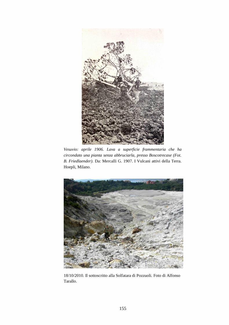
155
Vesuvio: aprile 1906. Lava a superficie frammentaria che ha
circondato una pianta senza abbruciarla, presso Boscotrecase (Fot.
B. Friedlaender). Da: Mercalli G. 1907. I Vulcani attivi della Terra.
Hoepli, Milano.
18/10/2010. Il sottoscritto alla Solfatara di Pozzuoli. Foto di Alfonso
Tarallo.