low-cost, easy-to-build non-invasive pressure …...2020/04/16 · low-cost, easy-to-build...
TRANSCRIPT

Early View
Original article
Low-cost, easy-to-build non-invasive pressure
support ventilator for under-resourced regions:
open source hardware description, performance
and feasibility testing
Onintza Garmendia, Miguel A. Rodríguez-Lazaro, Jorge Otero, Phuong Phan, Alexandrina Stoyanova,
Anh Tuan Dinh-Xuan, David Gozal, Daniel Navajas, Josep M. Montserrat, Ramon Farré
Please cite this article as: Garmendia O, Rodríguez-Lazaro MA, Otero J, et al. Low-cost, easy-
to-build non-invasive pressure support ventilator for under-resourced regions: open source
hardware description, performance and feasibility testing. Eur Respir J 2020; in press
(https://doi.org/10.1183/13993003.00846-2020).
This manuscript has recently been accepted for publication in the European Respiratory Journal. It is
published here in its accepted form prior to copyediting and typesetting by our production team. After
these production processes are complete and the authors have approved the resulting proofs, the article
will move to the latest issue of the ERJ online.
Copyright ©ERS 2020

Low-cost, easy-to-build non-invasive pressure support ventilator for under-
resourced regions: open source hardware description, performance and feasibility
testing
Onintza Garmendia1,2
, Miguel A. Rodríguez-Lazaro1, Jorge Otero
1,3, Phuong Phan
4,
Alexandrina Stoyanova5, Anh Tuan Dinh-Xuan
6, David Gozal
7, Daniel Navajas
1,3,8,
Josep M. Montserrat2,3,9
, Ramon Farré1,3,9,*
1Unitat de Biofísica i Bioenginyeria, Facultat de Medicina i Ciències de la Salut,
Universitat de Barcelona, Barcelona, Spain
2Sleep Lab, Hospital Clinic, Universitat de Barcelona, Barcelona, Spain
3CIBER de Enfermedades Respiratorias, Madrid, Spain
4Hue Central Hospital, Hue, Vietnam
5Department of Economics, Faculty of Economics and Business, Universitat de
Barcelona, Barcelona, Spain,
6Service de Physiologie-Explorations Fonctionnelles, Hôpital Cochin, Assistance
Publique-Hôpitaux de Paris (AP-HP), Paris, France
7Department of Child Health, The University of Missouri School of Medicine,
Columbia, MO, USA
8Institute for Bioengineering of Catalonia (IBEC), The Barcelona Institute of Science
and Technology, Barcelona, Spain
9Institut d’Investigacions Biomediques August Pi Sunyer, Barcelona, Spain.
*Corresponding author: Prof. Ramon Farré
Unitat de Biofísica i Bioenginyeria
Facultat de Medicina i Ciències de la Salut
Casanova 143
08036 Barcelona, SPAIN
Email: [email protected].
ABSTRACT
AIM: Current pricing of commercial mechanical ventilators in low/middle-
income countries (LMICs) markedly restricts their availability, and consequently a
considerable number of patients with acute/chronic respiratory failure cannot be
adequately treated. Our aim was to design and test an affordable and easy-to-build non-
invasive bilevel pressure ventilator to allow reducing the serious shortage of ventilators
in LMICs. METHODS: The ventilator was built using off-the-shelf materials available
via e-commerce and was based on a high-pressure blower, two pressure transducers and
an Arduino Nano controller with a digital display (total retail cost <75 US$), with
construction details open source provided for free replication. The ventilator was
evaluated (and compared with a commercially available device (Lumis-150, Resmed):
a) in the bench using an actively breathing patient simulator mimicking a range of
obstructive/restrictive disease and b) in 12 healthy volunteers wearing a high airway
resistance and thoracic/abdominal bands to mimic obstructive/restrictive patients.
RESULTS: The designed ventilator provided inspiratory/expiratory pressures up to
20/10 cmH2O, respectively, with no faulty triggering or cycling both in the bench test
and in volunteers. Breathing difficulty score rated (1-10 scale) by the loaded breathing
subjects was significantly (p<0.005) decreased from 5.45±1.68 without support to
2.83±1.66 when using the prototype ventilator, which showed no difference with the
commercial device (2.80±1.48; p=1.000). CONCLUSION: The low-cost, easy-to-build
non-invasive ventilator performs similarly as a high-quality commercial device, with its
open-source hardware description, will allow for free replication and use in LMICs,
facilitating application of this life-saving therapy to patients who otherwise could not be
treated.

INTRODUCTION
Non-invasive mechanical ventilation (NIV) is a widely used and accepted
treatment for chronic respiratory diseases and, in some cases, it is also an alternative to
invasive ventilation options for patients with acute respiratory failure caused by a
variety of aetiologies (1). Although positive pressure ventilation in low- and middle-
income countries (LMIC) is most frequently provided invasively, the benefits of NIV
are being increasingly recognised. Indeed, for obvious reasons of cost and ease of use,
NIV appears to be not only an effective, but also a particularly suitable approach to
provide respiratory support in patients living in developing low-income economies (2).
This is especially relevant since in these regions the burden of critical illness is large,
and is expected to increase with growing urbanization, emerging epidemics and
expanding access to hospitals (2). Furthermore, the elevated cost of healthcare staffing,
infrastructure needs, and onerous access to supplies have hampered the development of
fully equipped intensive care units (ICU) in LMICs (3). As a consequence, the demand
for cost-effective medical equipment, such as mechanical ventilators, is likely to greatly
increase in those countries. Moreover, mechanical ventilators are costly, which
markedly restricts their availability, and consequently the ability to adequately treat a
significant number of patients with both acute and chronic respiratory failure in LMICs.
These issues are all the more evident in light of the ongoing corona virus pandemic,
where even industrialized economies are encountering significant shortages in the
number of available ventilators to meet the demands imposed by this disease, such that
availability of non-invasive respiratory support may be valuable for certain patients or
as a temporary bridge (4, 5).
Philanthropic donation of medical devices may help in providing mechanical
ventilators to un-resourced regions in LMICs, but these initiatives are fraught with

considerable limitations. Indeed, donation of commercially available equipment is
expensive and is only partially effective since it has been reported that up to 50% of
donated devices become unusable due to lack of adequate maintenance and inability to
obtain spare parts (6). In addition, donations are hardly sustainable because they require
long-term commitments such as to provide device servicing. In this context, alternative
solutions that are based on in-house manufacturing of pressure support devices (7,8)
could reduce the serious shortage of ventilators in LMICs. Accordingly, the aim of this
study was to design and test a novel low-cost bilevel pressure support ventilator, and
provide open access to the detailed technical information, thereby allowing for free and
unrestricted replication and implementation. To ascertain adequate performance of the
device, bench testing was carried out based on simulated patients with
obstructive/restrictive diseases under well-controlled conditions, a common widely
accepted approach to test therapeutic devices for respiratory support (9-18). Then, and
following the existent literature (19-21), the prototype ventilator was tested in healthy
volunteers subjected to obstructive-restrictive loaded breathing to mimic patients with
respiratory diseases requiring NIV.
METHODS
Ventilator description
The ventilator was designed to be affordable and easy-to-build, providing an
open-source hardware description to allow free replication. The prototype was built
using off-the-shelf materials available via e-commerce: a high-pressure blower and its
driver (WM7040, Ning Bo Feng Hua Wei Cheng Motor Factory, Zhejiang, China), two
pressure transducers (XGZP6847005KPG, CFSensor, Wuhu, Anhui, China) and an
Arduino Nano controller with a digital display. Pressure and flow were continuously
measured at the outlet of the ventilator and fed into the controller which was provided
with a custom-made code to detect inspirations and expirations and to accordingly
trigger the inspiratory and expiratory pressures generated by the blower. The ventilator
can operate in timed or spontaneous timed (ST) mode (spontaneous breaths of patients
are assisted and if the patient’s effort is not detected a timed breath is triggered
according to a rescue frequency). The retail cost of this ventilator prototype was below
75 US$, and includes all required electronic circuits and power source. Noteworthy, this
cost could be considerably reduced by wholesale purchasing. All the technical
information and detailed circuit schematics and controller code required to build this
ventilator (including optional enclosure by conventional 3D printer) is available for
release under free terms following the open-source hardware approach in the online
supplement (Technical_Description.zip) . Figure 1 shows external and internal images
of the prototype.
Bench testing
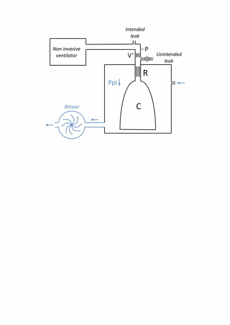
To assess the performance of the novel bilevel pressure ventilator under well-
controlled conditions, the prototype was evaluated in a bench test using an active patient

simulator modelling the respiratory mechanics of patients with different levels of
obstructive/restrictive diseases (Figure 2). The passive component of the respiratory
system model was a variable resistance-compliance (R-C) lung model (Adult
SmartLung, IMT Analytics, Switzerland). To implement an active breathing model, the
passive R-C system simulating the lungs was enclosed in a cylindrical box connected to
a negative pressure source (Figure 2), as explained in detail in the online supplement
(Supplementary_Methods&Results.pdf). Figure 3 shows examples of the simulated
pleural pressures applied to the passive model to implement active patient models,
which combined 2 breathing frequencies (15 and 20 breaths/min) and 3 negative peak
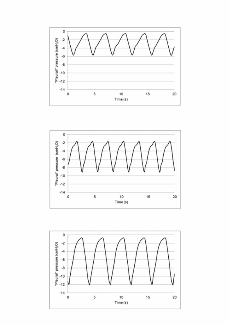
pressure amplitudes (-6, -9, -12 cm H2O). Four respiratory R-C systems were set for
testing the ventilator, mimicking a patient with mild disease, a purely obstructive patient
(increased R), a purely restrictive patient (reduced compliance) and a patient with both
obstruction and restriction (Table 1). Two breathing frequencies were used and different
inspiratory efforts were set according to the level of disease (Table 1). As shown in
Figure 2, the performance of the ventilator prototype was assessed by connecting it to
the patient simulator through flexible conventional tubing (2 m length, 22 mm
diameter), including a 5 mm diameter orifice at the nasal mask to create an intended air
leak orifice to avoid rebreathing, as it is set in conventional clinical applications. The
experimentally measured pressure-flow (P-V’) relationship in this intended leak
(V’≈10·P0.48
; V’ in l/min, P in cmH2O) resulted in a minimum continuous air flow
renewal of 20 l/min for a nasal pressure of 4 cmH2O. In addition to the intentional air
leak, we eventually also included a non-intentional air leak (5 mm diameter orifice) to
simulate the real-life leaks observed in patients subjected to non-invasive ventilation
owing to poor mask fit on the patient’s face skin (Table 1). This unintended leak created

air flow leaks of 30 and 40 l/min at nasal pressure values of 10 and 18 cmH2O,
respectively.
Therefore, the ventilator prototype was tested under 16 different simulated
conditions (Table 1), covering real life settings when applying NIV in clinical practice.
For the sake of comparison, the same 16 bench test conditions were also applied to a
high-performance commercially available mechanical ventilator (Lumis 150, VPAP ST,
Resmed; with default settings). During the tests, nasal pressure, flow and simulated
pleural pressure signals were measured (Figure 2) with a Fleisch pneumotachograph
(Metabo, Switzerland) and pressure transducers (Celesco, Canada; Validyne, USA),
recorded at 100 Hz. Subsequently, tidal volumes were digitally computed by integration
of the flow signal (after adequate zero-flow correction). Inspiratory trigger delay was
measured as the time from starting the decrease in negative inspiratory (simulated
pleural) pressure to the time at which nasal pressure started to be positive (22).
Ventilator testing in healthy volunteers
The ventilator prototype was tested in 12 healthy volunteers (5 of them women)
recruited from the university environment. Their mean age was 32.4±5.8 years and their
body mass index was 23.3±2.0 kg/m2 (mean±SE). To mimic the respiratory load
corresponding to a patient requiring NIV the volunteers were instrumented to increase
their airway resistance and to decrease their respiratory compliance, as explained in
detail in the online supplement (Supplementary_Methods&Results.pdf). The protocol
was carried out by one respiratory physiotherapist expert in NIV. The volunteer subject
was sitting in a comfortable armchair and was equipped with a finger pulse oximeter for
monitoring oxygen saturation (WristOx2, Model 3150, Nonin Medical, Plymouth). First,
he/she was allowed to get familiar with the use of a nasal mask and NIV for 3 minutes.

To this end, he/she was connected to a non-invasive ventilator (Lumis 150, VPAP ST,
Resmed) through conventional nasal mask and tubing, with inspiratory and expiratory
pressures set to 8 and 4 cmH2O, respectively. Subsequently, the subject was equipped
with the resistive and restrictive loads and breathed spontaneously (unsupported) for 2
min. After that period of loaded breathing, the volunteer was asked to score his/her
breathing discomfort sensation on a visual analog scale where 1 would correspond to
spontaneous normal breathing and 10 to the maximum breathing discomfort he/she
could consider unbearable. Then, he/she was connected to a mechanical ventilator with
inspiratory and expiratory pressures set to 16 and 6 cmH2O, respectively, in
spontaneous trigger (ST) mode with a backup frequency of 12 breaths/min. This
ventilator was either the prototype under test or a commercially available high-
performance device (Lumis 150, VPAP ST, Resmed; default settings), determined at
random. At the end of a 2 min period, the subject was again asked to score his/her
breathing discomfort and, without interruption nor notice to the subject, the ventilator
was shifted to the other device for 2 min and then the volunteer was asked again for
scoring discomfort. This process of alternating the ventilator (prototype or commercial)
was repeated twice more. The discomfort scoring finally assigned to each ventilator was
the mean score of the 3 periods corresponding to each device. Any faulty triggering or
cycling, as observed through inspection of the real time nasal pressure signal and
subject’s breathing activity, was registered by the physiotherapist.
Statistics
In the bench test, the different investigated variables were assessed by
comparing the data obtained with the prototype and the commercial ventilator by means
of a paired t-test. In the test with voluntary subjects, discomfort scores when the
individual was not mechanically supported and when supported by the two ventilators
were compared by paired t-test for normally distributed variables and by Wilcoxon
signed rank test for non-normally distributed variables. A value of p<0.05 was
considered statistically significant.
RESULTS
Bench testing
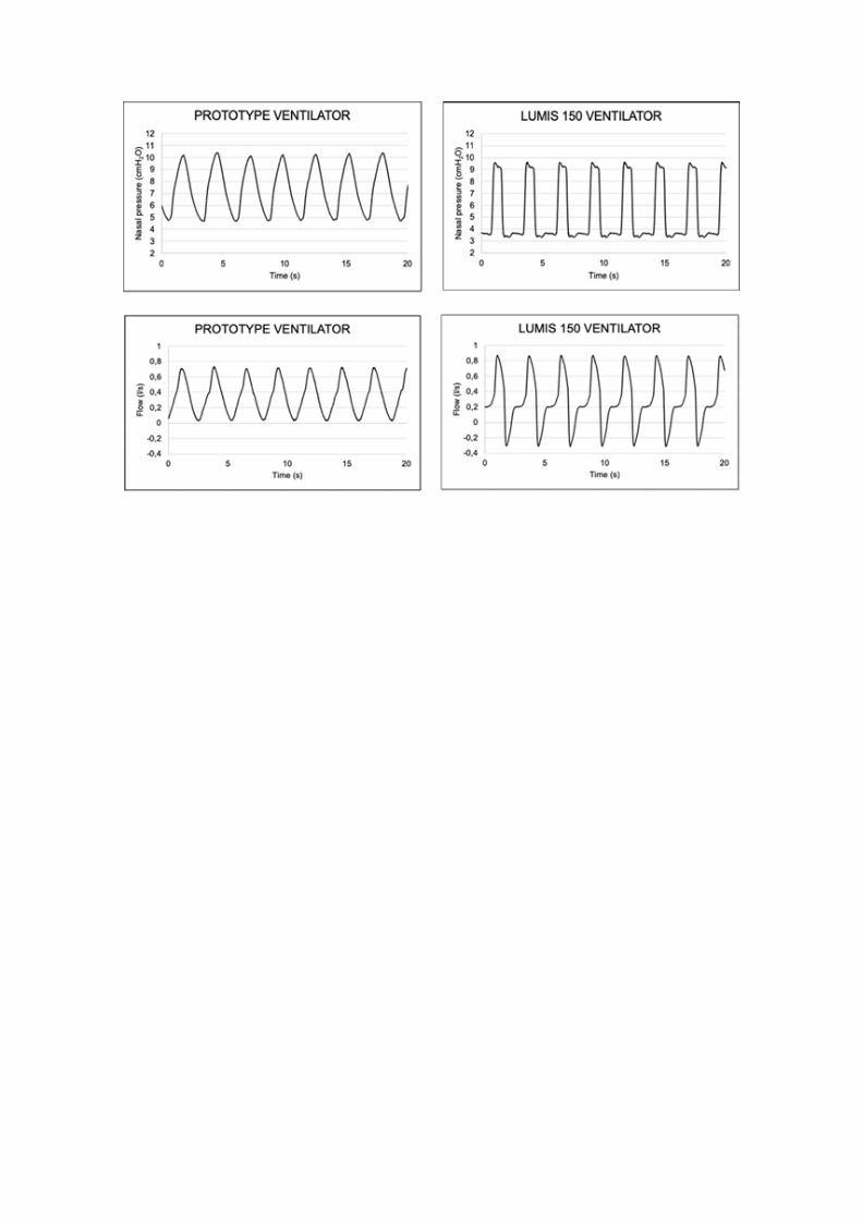
Figure 4 shows an example of the nasal pressure and breathing flow signals
recorded in one of the bench tests simulating a patient with mild disease for both the
prototype and commercial ventilators. The pressure waveform in the commercial device
was close to a square signal whereas the pressure generated by the prototype increased
and decreased more smoothly. Consistently, the inspiratory flow induced by the
commercial ventilator experienced a sudden increase at the beginning of inspiration,
while the flow induced by the prototype increased more progressively.
The ventilator prototype performance was comparable to the commercial
ventilator and provided inspiratory and expiratory pressures (up to 16 and 8 cmH2O,
respectively) with no defective triggering or cycling when tested over the described 16
different simulated conditions. Figure 1.Suppl (on line Supplementary_-
Methods&Results.pdf) shows the pressure waveforms recorded in the 16 test
conditions, showing that the prototype was performant in all cases (as the commercial
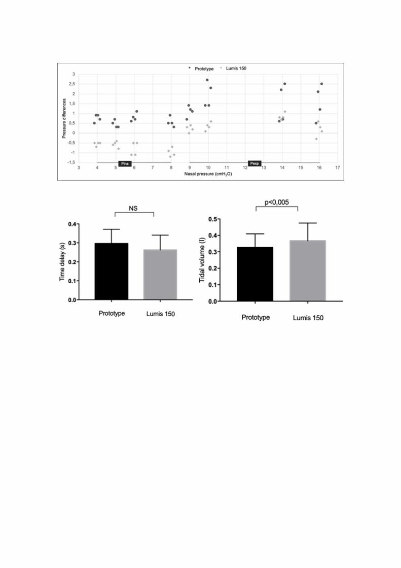
device was; figures not shown). Figure 5.A is a Bland-Altman type plot illustrating the
difference between measured maximum inspiratory and minimum expiratory pressures
and the corresponding target values set at the ventilator control panel, respectively.
Actual minimal expiratory pressures set to range 4-8 cmH2O, differed by less than 1
cmH2O from the target values, with positive and negative differences in the case of the
prototype and commercial ventilator, respectively. Actual peak inspiratory pressures, set
to range 10-16 cmH2O, were systematically higher than the target values for both
ventilators, being greater (by ≈1.5 cmH2O) in case of the prototype ventilator. The
trigger delay in the prototype showed no statistically significant differences when
compared with the delay time in the commercial ventilator (Figure 5.B). Although
statistically significant, the inspiratory tidal volumes achieved with both ventilators in
the 16 different test conditions were similar (difference of 40 ml on average) (Figure
5.B).
Test in healthy volunteers with loaded breathing
Testing in volunteers with resistive and restrictive loads provided positive results
on the feasibility of the ventilator prototype for application in humans. As expected
from healthy subjects, no decrease in oxygen saturation was observed throughout the
whole test period when compared with the unsupported baseline (97.0±1.3%):
96.8±1.0% (p=0.51) and 96.8±0.9% (p=0.23) when supported with the prototype and
commercial ventilator, respectively. As shown in Figure 6, discomfort scoring when the
12 subjects were subjected to respiratory loading was 5.45±1.68. These values
significantly decreased to 2.83±1.66 (p<0.005) when the loaded patient’s breathing was
supported by the prototype ventilator. Interestingly, the relief in breathing difficulty was
virtually the same as the one achieved with the high-performance commercial ventilator
(2.80±1.48; p=1.000). No significant differences were observed in the variability across
the 3 measurements of breathing discomfort (p=0.208; average standard deviation of
0.61). Figure 7 presents an example of the nasal pressure and flow signals when the
prototype ventilator, set to considerable values of NIV inspiratory and expiratory
pressures, was applied to a loaded-breathing volunteer, illustrating that the pressure
waveform was suitable, and that the ventilator smoothly followed the breathing pattern
of the subjects since no faulty triggering of cycling was detected. Figure 2.Suppl (in the

Supplementary_-Methods&Results.pdf) shows that the prototype ventilator was able to
trigger mandatory ventilation cycles in case of absence of subject’s inspiratory effort.
DISCUSSION
Here we describe a very low cost bilevel pressure support ventilator which is
easy-to-build for potential use in under-resourced areas of developing countries or
during pandemic conditions such as those imposed currently by the novel corona virus
disease superimposed on already strained hospital conditions dealing with the influenza
season. The results obtained during both the bench testing, and during the applicability
pilot study in humans confirmed that the newly designed low-cost ventilator performs
comparably to currently available commercial ventilators. Specifically, the device
functioned as expected under stressing conditions in both the bench test (high load
impedance and unintended leaks) and in the test with subjects with obstructive-
restrictive loaded breathing.
The ventilator is based on a modular structure (Fig. 1.B) requiring only basic
electronic training for an overall straightforward and easy assembly. The device
modules are interconnected by compact electrical wiring, and therefore such design
enables simple replacement of each module independently, as needed. The setting
requires no pre-calibration routines, since pressure transducers are thermally corrected,
and are provided with factory calibration. Interestingly, when the ventilator is switched
on, an automatic routine process automatically tests and digitally corrects any drift in
the zero signal of the transducers. Flow is sensed with a pneumotachograph consisting
of a slight constriction in the tube section separating the two pressure transducers. As
such orifice-like resistor is nonlinear, the pressure drop signal across the transducers is
digitally linearized (by computing the square root) in real time. Given that cycling (end
inspiration) is determined when inspiratory flow reaches below a certain percentage

(usually 30-50% (23), selectable by the user) of the inspiratory peak, the flow signal
does not require calibration. In addition to the hardware components, the Arduino code
controlling the ventilator (which is also open access provided) can be re-uploaded at any
time, thereby allowing for simple updates to the ventilator functions in LMICs, with
locally or remotely updated code. For instance, cheap high (or low) pressure acoustic
alarms can be easily added. Moreover, the non-linear resistor to estimate flow can be
replaced by a linear pneumotachograph to measure flow, and the related inspired
volume (although this would increase cost and require periodic calibration).
Furthermore, the ventilator control can be adapted to take into account the pressure drop
induced by a humidifier in case this component is connected between the ventilator
output and the tube connecting to the patient’s mask. Noteworthy, the proposed
approach for ventilator construction empowers the final users in LMICs to fully control
the procedure, to adapt it to the local conditions, and to update the used components in
response to market availability. Moreover, continued support from and collaboration
with experienced teams abroad is easy and readily feasible. In a time when the most
complex devices appear to be needed and can only be provided by a very competitive
and specialized industry, the simplicity and performance of this designed low cost
device reminds us of the need to go back to the basics, inasmuch as the rationale and
implementation of NIV has not changed substantially from the pioneering times when
this therapy was developed (24).
The bench test was carried out using a commonly used type of actively breathing
patient simulator (9-11,16,18), mimicking a wide spectrum of patient’s respiratory
mechanical alterations (obstruction and/or restriction), and including normal and high
breathing frequencies. Moreover, the ventilator was tested under conditions reproducing
an unintended leak, a circumstance frequently encountered during clinical NIV practice.

The 16 different bench test settings (Table 1) used to test the ventilator covered the wide
range of conditions that non-invasive ventilators are exposed to in real-life clinical
applications. The bench test showed that, regardless of the stressing test conditions, the
prototype ventilator provided the target bilevel pressures (Figure 5.A) with no faulty
triggering or cycling, and with a suitable triggering delay (Figure 5.B), thereby enabling
good synchronization with the inspiratory effort of the simulated patient. The relative
simplicity of the feedback control system in the prototype ventilator explains the
slightly different shape of pressure waveform observed over the wide range of test
conditions (Figure 5.A), and also facilitates interpretation of the determinants of the
small increases in peak inspiratory pressures. Whereas the commercial device applied
an almost square pressure signal, the pressure waveform generated by the prototype
exhibited progressive increase and decrease in inspiratory pressures (Figure 4). This is
consistent with the fact that the commercial device used as a comparator was probably
equipped with a more powerful (and hence expensive) feedback system to control its
blower. However, several other commercially available ventilators exhibit patterns of
inspiratory pressures similar to those observed in the prototype (Figure 4.A) (24). In
fact, the slope of the ramp of increasing inspiratory pressure is one of the parameters
that can be set by the user in some commercially available devices, since excessively
rapid increases in early onset of inspiratory pressures may lead to patient discomfort
(Figure 4.B) by not mimicking the physiological inspiratory patterns characterized by
progressive increases in flow. Although tidal volume was not a direct outcome variable
controlled by pressure support ventilators, it is interesting to note that the prototype
ventilator resulted in tidal volumes that were similar to the ones generated by the
commercial device (Figure 5.B), adding further support to the suitability of the
prototype ventilator for generating inspiratory pressure waveforms and adequate tidal
volumes in a wide spectrum of simulated patients (Table 1).
The applicability pilot study was carried out in healthy volunteers subjected to
obstructive and restrictive breathing loads. This model is widely used in the literature to
simulate the mechanical load of the respiratory system for investigating ventilation (19-
21) and for simulating dyspnoea (26,27). In fact, the level of obstruction-restriction we
applied to our volunteers resulted in a breathing discomfort score (Figure 7) similar to
the ones set in recent reports to mimic dyspnoea by loaded breathing (26,27). The
results obtained when testing the applicability of the prototype ventilator in humans
showed that, similar to the bench test, there were no faulty triggering or cycling events,
that the pressure waveform was similar to those typically observed in commercial
ventilators (Figures 7 and 2.Suppl), and that the relief of breathing discomfort was
virtually the same as the one achieved by the commercial ventilator (Figure 6).
In addition to methodological and technical issues discussed above, the work
presented here requires that we specifically address two aspects that are usually lacking
in medical device studies: a) industrial/commercial model and b) safety/ethical issues.
Indeed, in this work we propose an alternative procedure for building ventilators in-
house and locally, i.e., outside the conventional medical device industrial market. There
is little doubt that industry-based conventional production chains, including design,
manufacturing and commercialization, play a key role in the healthcare system.
Accordingly, industry heavily invests in R&D and translates new knowledge from the
laboratory bench to patient bedside. In other words, the medical device industry
searches and delivers life-enhancing innovative solutions. Unfortunately, such
industrially-based model is hardly suitable to low-income settings that are usually
resource scarce, and where the provision of even adequate basic services to the
population is challenging. The main reason why the conventional industrial model does
not work in LMICs is that the standard industrial production scheme, which also applies
to entirely non-for-profit companies, entails significant costs beyond those strictly
required for device manufacturing.
The vast majority of medical device companies are small and medium-sized,
employing less than 50 people, both in Europe (95% of all medical technology firms)
and in the USA (80%) (28). Moreover, those companies, mainly based in USA and
Canada (49% of the world market), Europe (27%), Japan (7%) and China (6%), are
highly and globally regulated to guarantee the safety and performance of their
innovative and high technology products throughout their life-cycle, as well as pre-and
post-marketing (28). Unlike many other industries, R&D expenditures represent a
significant cost component for medical device companies. These companies spend on
average between 6% and 12% of revenues towards R&D investment (29), with some
niche firms or start-ups incurring even higher R&D costs (> 20%). Average selling,
general and administrative expenses (SG&A), which include marketing, advertising and
promotion costs and general and administrative costs, account for about one-third of
total revenues (30). Importantly, the cost of goods sold (COGS), which measures the
total cost that it takes for a medical device company to manufacture its products
including labour, material costs, rental and utility costs, represent between 35% and
40% of revenues (30), with the remaining costs going to taxes, interests and
depreciation. Thus, a disproportionately large share of medical device companies’
revenues is slated for expenses beyond those needed to manufacture their products. In
contrast, in the approach we describe in this work, as ventilator assembly is performed
locally or directly linked local technical partners, the only costs incurred are those
associated with purchasing of the components and the actual labor costs of assembling

the device, both of which are low thanks to e-commerce and labor costs in LMICs,
respectively. Noteworthy, the approach proposed here may not only allow for adequate
availability of ventilators to patients (31-34), but may also contribute to the
development of the local industry network in LMICs (35-37).
Regarding safety and ethical issues, it is important to emphasize that the
development and testing of the NIV devices that are available in market nowadays, was
made possible by development of devices built in-house by physicians and researchers
in developed countries, and that these innovations were designed, tested and improved
in patients before the corresponding labeling was obtained (24). Notwithstanding, the
in-house ventilator proposed here does not have the conventional FDA/CE approvals.
Obviously, such approval procedures are tremendously important for ensuring that
medical devices placed into the market are safe and reliable and, as such, have
contributed to the progress currently achieved in health care. However, obtaining
FDA/CE labels is a process devised mainly for the industry in developed countries and
is extremely expensive in terms of LMICs financial resources. Although these countries
do not have the complex infrastructure required for such costly processes, simplified or
ad-hoc approval procedures could be provided by local authorities or hospital Ethical
Boards. However, particularly in light of the non-existent alternative of using industrial
ventilators, i.e., leaving the patient untreated with the attendant consequences. Under
such difficult circumstances, the ethical trade-off towards compassionate use of medical
devices, a mechanism already in place for non-labelled therapies in developed countries,
may be considered.
In conclusion, we have designed a low-cost easy-assembly ventilator
with excellent performance characteristics in both the bench and in voluntary subjects.
If as anticipated from these preliminary results, clinical field tests are favourable, this
low-cost device may enable provision of respiratory support to patients in LMICs who
otherwise would have no access to this potentially life saving therapy, as well as
escalation of ventilatory support availability in strenuous circumstances such as those
imposed by respiratory virus pandemics.
REFERENCES
1. Rochwerg B, Brochard L, Elliott MW, Hess D, Hill NS, Nava S, et al. Official
ERS/ATS clinical practice guidelines: noninvasive ventilation for acute respiratory
failure. Eur Respir J [Internet]. 2017 Aug 31;50(2):1602426.
http://erj.ersjournals.com/lookup/doi/10.1183/13993003.02426-2016
2. Mandelzweig K, Leligdowicz A, Murthy S, Lalitha R, Fowler RA, Adhikari NKJ.
Non-invasive ventilation in children and adults in low- and low-middle income
countries: A systematic review and meta-analysis. J Crit Care [Internet]. 2018
Oct;47:310–9. https://linkinghub.elsevier.com/retrieve/pii/S0883944117302472
3. Murthy S, Leligdowicz A, Adhikari NKJ. Intensive Care Unit Capacity in Low-
Income Countries: A Systematic Review. Azevedo LCP, editor. PLoS One [Internet].
2015 Jan 24;10(1): e0116949. http://dx.plos.org/10.1371/journal.pone.0116949
4. Truog RD, Mitchell C, Daley CQ. The Toughest Triage — Allocating Ventilators in
a Pandemic. New Eng J Med, in press. DOI: 10.1056/NEJMp2005689.
5. Wang D, Hu B, Hu C, Zhu F, Liu X, Zhang J, Wang B, Xiang H, Cheng Z, Xiong Y,
Zhao Y, Li Y, Wang X, Peng Z. Clinical Characteristics of 138 Hospitalized Patients
With 2019 Novel Coronavirus-Infected Pneumonia in Wuhan, China. JAMA. 2020
Feb 7. doi: 10.1001/jama.2020.1585.

6. Howie SRC, Hill SE, Peel D, Sanneh M, Njie M, Hill PC, et al. Beyond good
intentions: lessons on equipment donation from an African hospital. Bull World
Health Organ 2008;86:52–56. 5.
7. Farré R, Trias G, Solana G, Ginovart G, Gozal D, Navajas D. Novel Approach for
Providing Pediatric Continuous Positive Airway Pressure Devices in Low-Income,
Underresourced Regions. Am J Respir Crit Care Med [Internet]. 2019
Jan;199(1):118–20. https://www.atsjournals.org/doi/10.1164/rccm.201808-1452LE
8. Farré R, Montserrat JM, Solana G, Gozal D, Navajas D. Easy-to-build and affordable
continuous positive airway pressure CPAP device for adult patients in low-income
countries. Eur Respir J. 2019;53(5).
9. Lujan M, Sogo A, Pomares X, Monso E, Sales B, Blanch L. Effect of Leak and
Breathing Pattern on the Accuracy of Tidal Volume Estimation by Commercial
Home Ventilators: A Bench Study. Respir Care [Internet]. 2013 May 1;58(5):770–7.
http://rc.rcjournal.com/cgi/doi/10.4187/respcare.02010
10. Carteaux G, Lyazidi A, Cordoba-Izquierdo A, Vignaux L, Jolliet P, Thille AW, et
al. Patient-Ventilator Asynchrony During Noninvasive Ventilation. Chest [Internet].
2012,Aug;142(2):367–76.
https://linkinghub.elsevier.com/retrieve/pii/S0012369212604498.
11. Olivieri C, Costa R, Conti G, Navalesi P. Bench studies evaluating devices for non-
invasive ventilation: critical analysis and future perspectives. Intensive Care Med
[Internet]. 2012 Jan 29;38(1):160–7. http://link.springer.com/10.1007/s00134-011-
2416-9
12. Farré R, Montserrat JM, Rigau J, Trepat X, Pinto P, Navajas D. Response of
Automatic Continuous Positive Airway Pressure Devices to Different Sleep

Breathing Patterns. Am J Respir Crit Care Med [Internet]. 2002 Aug 15;166(4):469–
73. http://www.atsjournals.org/doi/abs/10.1164/rccm.2111050
13. Rigau J, Montserrat JM, Wöhrle H, Plattner D, Schwaibold M, Navajas D, et al.
Bench Model To Simulate Upper Airway Obstruction for Analyzing Automatic
Continuous Positive Airway Pressure Devices. Chest [Internet]. 2006
Aug;130(2):350–61.
https://linkinghub.elsevier.com/retrieve/pii/S0012369215518485
14. Isetta V, Navajas D, Montserrat JM, Farré R. Comparative assessment of several
automatic CPAP devices’ responses: a bench test study. ERJ Open Res [Internet].
2015 May;1(1):00031–2015.
http://openres.ersjournals.com/lookup/doi/10.1183/23120541.00031-2015
15. Isetta V, Montserrat JM, Santano R, Wimms AJ, Ramanan D, Woehrle H, et al.
Novel Approach to Simulate Sleep Apnea Patients for Evaluating Positive Pressure
Therapy Devices. Cymbalyuk G, editor. PLoS One [Internet]. 2016 Mar
15;11(3):e0151530. https://dx.plos.org/10.1371/journal.pone.0151530
16. Farré R, Navajas D, Montserrat JM. Technology for noninvasive mechanical
ventilation: Looking into the black box. ERJ Open Res. 2016;2(1):1–11.
17. Zhu K, Farré R, Katz I, Hardy S, Escourrou P. Mimicking a flow-limited human
upper airway using a collapsible tube: relationships between flow patterns and
pressures in a respiratory model. J Appl Physiol [Internet]. 2018 Aug 1;125(2):605–
14. https://www.physiology.org/doi/10.1152/japplphysiol.00877.2017
18. Bunburaphong T, Imanaka H, Nishimura M, Hess D, Kacmarek RM. Performance
Characteristics of Bilevel Pressure Ventilators. Chest [Internet]. 1997
Apr;111(4):1050–60.
https://linkinghub.elsevier.com/retrieve/pii/S0012369215468709
19. Eberlein M, Schmidt GA, Brower RG. Chest wall strapping. An old physiology
experiment with new relevance to small airways diseases. Ann Am Thorac Soc.
2014;11(8):1258–1266. doi:10.1513/AnnalsATS.201312-465OI
20. Mols G, von Ungern-Sternberg B, Rohr E, Haberthür C, Geiger K, Guttmann J.
Respiratory comfort and breathing pattern during volume proportional assist
ventilation and pressure support ventilation: A study on volunteers with artificially
reduced compliance. Crit Care Med [Internet]. 2000 Jun;28(6):1940–6.
http://insights.ovid.com/crossref?an=00003246-200006000-00042
21. Mols G, Vetter T, Haberthür C, Geiger K, Guttmann J. Breathing pattern and
perception at different levels of volume assist and pressure support in volunteers. Crit
Care Med [Internet]. 2001 May;29(5):982–8.
https://insights.ovid.com/crossref?an=00003246-200105000-00018
22. Ferreira JC, Chipman DW, Kacmarek RM. Trigger performance of mid-level ICU
mechanical ventilators during assisted ventilation: a bench study. Intensive Care Med
[Internet]. 2008 Sep 30;34(9):1669–75. http://link.springer.com/10.1007/s00134-
008-1125-5
23. Moerer O, Harnisch LO, Herrmann P, Zippel C, Quintel M. Patient-Ventilator
Interaction During Noninvasive Ventilation in Simulated COPD. Respir Care. 2016
Jan;61(1):15-22. doi: 10.4187/respcare.04141. Epub 2015 Nov 10. PubMed PMID:
26556898.
24. Sullivan CE. Nasal Positive Airway Pressure and Sleep Apnea. Reflections on an
Experimental Method That Became a Therapy. Am J Respir Crit Care Med. 2018

Sep 1;198(5):581-587. doi: 10.1164/rccm.201709-1921PP. PubMed PMID:
30011222.
25. Battisti A, Tassaux D, Janssens JP, Michotte JB, Jaber S, Jolliet P. Performance
characteristics of 10 home mechanical ventilators in pressure-support mode: a
comparative bench study. Chest. 2005 May;127(5):1784-92. PubMed PMID:
15888859.
26. Nierat M-C, Demiri S, Dupuis-Lozeron E, Allali G, Morélot-Panzini C, Similowski
T, et al. When Breathing Interferes with Cognition: Experimental Inspiratory
Loading Alters Timed Up-and-Go Test in Normal Humans. Najbauer J, editor. PLoS
One [Internet]. 2016 Mar 15;11(3):e0151625.
http://dx.plos.org/10.1371/journal.pone.0151625
27. Allard E, Canzoneri E, Adler D, Morélot-Panzini C, Bello-Ruiz J, Herbelin B, et al.
Interferences between breathing, experimental dyspnoea and bodily self-
consciousness. Sci Rep [Internet]. 2017 Dec 30;7(1):9990.
http://www.nature.com/articles/s41598-017-11045-x-
28. The European Medical Technology Industry – in figures 2019.
https://www.medtecheurope.org/wp-content/uploads/2019/04/The-European-Medical-
Technology-Industry-in-figures-2019-2.pdf . Last access March 24 2020.
29. Spence P, Babitt J, Welch J, De Busscher L. As change accelerates, how can
medtechs move ahead and stay there? Pulse of the industry 2017. EYGM Limited.
https://www.ey.com/Publication/vwLUAssets/ey-as-change-accelerates-how-can-
medtechs-move-ahead-and-stay-there/$FILE/ey-as-change-accelerates-how-can-
medtechs-move-ahead-and-stay-there.pdf . Last access Mach 24, 2020.

30. Fuhr T, George K, Pai J. The Business Case for Medical Device Quality. McKinsey
Center for Government.
https://www.mckinsey.com/~/media/McKinsey/dotcom/client_service/Public%20Sec
tor/Regulatory%20excellence/The_business_case_for_medical_device_quality.ashx .
Last access Mach 24, 2020.
31. Pearce, J.M. (2015) Quantifying the Value of Open Source Hardware Development.
Modern Economy, 6, 1-11. http://dx.doi.org/10.4236/me.2015.61001.
32. Pearce JM. Maximizing Returns for Public Funding of Medical Research with
Open-source Hardware. Health Policy and Technology. 6(4), 2017, pp. 381-382.
https://doi.org/10.1016/j.hlpt.2017.09.001
33. Eslambolchilar P, Thimbleby H. Open-source hardware for medical devices. BMJ
Innov 2016; 2: 78–83.
34. DePasse JW, Caldwell A, Santorino D, et al. Affordable medical technologies:
bringing Value-Based Design into global health. BMJ Innovations 2016; 2: 4–7.
35. Mackintosh M, Tibandebage P, Karimi Njeru M, et al. Rethinking health sector
procurement as developmental linkages in East Africa. Soc Sci Med 2018; 200: 182–
189.
36. De Maria C, Mazzei D, Ahluwalia A. Open source biomedical engineering for
sustainability in African healthcare: combining academic excellence with innovation.
Proceedings of the ICDS 2014, The Eighth International Conference on Digital
Society; 2014 Mar 23–27, Barcelona, Spain; pp. 48–53. www.thinkmind.org/
index.php?view=article&articleid=ic

37. Clifford KL, Zaman MH. Engineering, global health, and inclusive innovation:
focus on partnership, system strengthening, and local impact for SDGs. Glob Health
Action 2016; 9: 30175. ds_2014_2_40_10173.
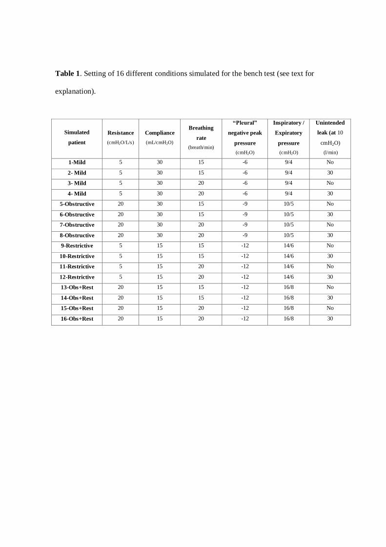
Table 1. Setting of 16 different conditions simulated for the bench test (see text for
explanation).
Simulated
patient
Resistance
(cmH2O/L/s)
Compliance
(mL/cmH2O)
Breathing
rate
(breath/min)
“Pleural”
negative peak
pressure
(cmH2O)
Inspiratory /
Expiratory
pressure
(cmH2O)
Unintended
leak (at 10
cmH2O)
(l/min)
1-Mild 5 30 15 -6 9/4 No
2- Mild 5 30 15 -6 9/4 30
3- Mild 5 30 20 -6 9/4 No
4- Mild 5 30 20 -6 9/4 30
5-Obstructive 20 30 15 -9 10/5 No
6-Obstructive 20 30 15 -9 10/5 30
7-Obstructive 20 30 20 -9 10/5 No
8-Obstructive 20 30 20 -9 10/5 30
9-Restrictive 5 15 15 -12 14/6 No
10-Restrictive 5 15 15 -12 14/6 30
11-Restrictive 5 15 20 -12 14/6 No
12-Restrictive 5 15 20 -12 14/6 30
13-Obs+Rest 20 15 15 -12 16/8 No
14-Obs+Rest 20 15 15 -12 16/8 30
15-Obs+Rest 20 15 20 -12 16/8 No
16-Obs+Rest 20 15 20 -12 16/8 30
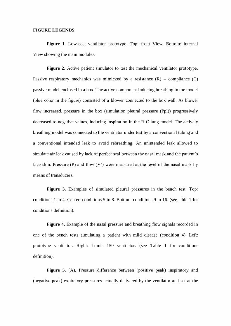
FIGURE LEGENDS
Figure 1. Low-cost ventilator prototype. Top: front View. Bottom: internal
View showing the main modules.
Figure 2. Active patient simulator to test the mechanical ventilator prototype.
Passive respiratory mechanics was mimicked by a resistance (R) – compliance (C)
passive model enclosed in a box. The active component inducing breathing in the model
(blue color in the figure) consisted of a blower connected to the box wall. As blower
flow increased, pressure in the box (simulation pleural pressure (Ppl)) progressively
decreased to negative values, inducing inspiration in the R-C lung model. The actively
breathing model was connected to the ventilator under test by a conventional tubing and
a conventional intended leak to avoid rebreathing. An unintended leak allowed to
simulate air leak caused by lack of perfect seal between the nasal mask and the patient’s
face skin. Pressure (P) and flow (V’) were measured at the level of the nasal mask by
means of transducers.
Figure 3. Examples of simulated pleural pressures in the bench test. Top:
conditions 1 to 4. Center: conditions 5 to 8. Bottom: conditions 9 to 16. (see table 1 for
conditions definition).
Figure 4. Example of the nasal pressure and breathing flow signals recorded in
one of the bench tests simulating a patient with mild disease (condition 4). Left:
prototype ventilator. Right: Lumis 150 ventilator. (see Table 1 for conditions
definition).
Figure 5. (A). Pressure difference between (positive peak) inspiratory and
(negative peak) expiratory pressures actually delivered by the ventilator and set at the

ventilator control panel for both Prototype and Lumis 150. (B) Inspiratory time delay
and tidal volume in the prototype and Lumis 150 ventilators.
Figure 6. Discomfort scoring (Visual Analog Scale) in healthy volunteers
subjected to obstructive-restrictive loaded breathing when unsupported and when
supported by the prototype and Lumis 150 ventilators.
Figure 7. Example of pressure and flow signals recorded when a resistive-
restrictive loaded breathing volunteer’s breathing was supported by the prototype
ventilator. These are unfiltered raw data from the sensors within the ventilator. The flow
signal is uncalibrated in both amplitude and zero.


SUPPLEMENTARY METHODS ANS RESULTS
Low-cost, easy-to-build non-invasive pressure support ventilator for under-
resourced regions: open source hardware description, performance and
feasibility testing
Onintza Garmendia1,2, Miguel A. Rodríguez-Lazaro1, Jorge Otero1,3, Phuong Phan4,
Alexandrina Stoyanova5, Anh Tuan Dinh-Xuan6, David Gozal7, Daniel Navajas1,3,8,
Josep M. Montserrat2,3,9, Ramon Farré1,3,9,*
1Unitat de Biofísica i Bioenginyeria, Facultat de Medicina i Ciències de la Salut,
Universitat de Barcelona, Barcelona, Spain
2Sleep Lab, Hospital Clinic, Universitat de Barcelona, Barcelona, Spain
3CIBER de Enfermedades Respiratorias, Madrid, Spain
4Hue Central Hospital, Hue, Vietnam
5Department of Economics, Faculty of Economics and Business, Universitat de
Barcelona, Barcelona, Spain,
6Service de Physiologie-Explorations Fonctionnelles, Hôpital Cochin, Assistance
Publique-Hôpitaux de Paris (AP-HP), Paris, France
7Department of Child Health, The University of Missouri School of Medicine,
Columbia, MO, USA
8Institute for Bioengineering of Catalonia (IBEC), The Barcelona Institute of Science and Technology, Barcelona, Spain
9Institut d’Investigacions Biomediques August Pi Sunyer, Barcelona, Spain.
*Corresponding author: Prof. Ramon Farré
Unitat de Biofísica i Bioenginyeria Facultat de Medicina i Ciències de la Salut Casanova 143
08036 Barcelona, SPAIN Email: [email protected].
METHODS
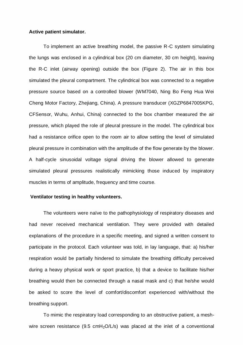
Active patient simulator.
To implement an active breathing model, the passive R-C system simulating
the lungs was enclosed in a cylindrical box (20 cm diameter, 30 cm height), leaving
the R-C inlet (airway opening) outside the box (Figure 2). The air in this box
simulated the pleural compartment. The cylindrical box was connected to a negative
pressure source based on a controlled blower (WM7040, Ning Bo Feng Hua Wei
Cheng Motor Factory, Zhejiang, China). A pressure transducer (XGZP6847005KPG,
CFSensor, Wuhu, Anhui, China) connected to the box chamber measured the air
pressure, which played the role of pleural pressure in the model. The cylindrical box
had a resistance orifice open to the room air to allow setting the level of simulated
pleural pressure in combination with the amplitude of the flow generate by the blower.
A half-cycle sinusoidal voltage signal driving the blower allowed to generate
simulated pleural pressures realistically mimicking those induced by inspiratory
muscles in terms of amplitude, frequency and time course.
Ventilator testing in healthy volunteers.
The volunteers were naïve to the pathophysiology of respiratory diseases and
had never received mechanical ventilation. They were provided with detailed
explanations of the procedure in a specific meeting, and signed a written consent to
participate in the protocol. Each volunteer was told, in lay language, that: a) his/her
respiration would be partially hindered to simulate the breathing difficulty perceived
during a heavy physical work or sport practice, b) that a device to facilitate his/her
breathing would then be connected through a nasal mask and c) that he/she would
be asked to score the level of comfort/discomfort experienced with/without the
breathing support.
To mimic the respiratory load corresponding to an obstructive patient, a mesh-
wire screen resistance (9.5 cmH2O/L/s) was placed at the inlet of a conventional

nasal mask for non-invasive ventilation. The built-in intended leak of the mask was
sealed, and a 5 mm orifice intended leak open to the room air was placed between
the end of the flexible tube connecting the ventilator to the mask and the 9.5
cmH2O/L/s added resistance which hence played the role of an actual increase in
patient’s airway resistance. To also load the volunteer with a restrictive component, a
nonflexible belt (9 cm width) was tightly fit around the abdomen and a spring-based
flexible belt (9 cm width) was adjusted around the thorax at the level of the
manubrium sterni.

RESULTS
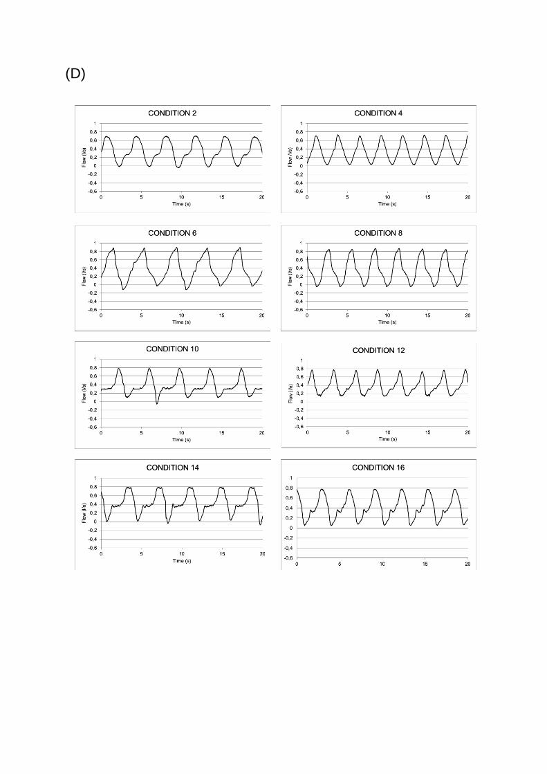
Figure 1. Suppl. Pressure generated by the prototype ventilator when connected to
simulated patients under different conditions without (A) and with (B) unintended air
leaks. (C) and (D) are the flow signals corresponding to the pressures in (A) and (B),
respectively. See Table 1 in the main manuscript for conditions definition.
(A)

(B)
Figure 7B

(C)
(D)
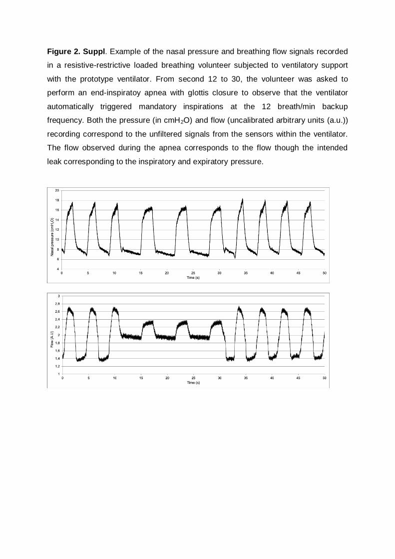
Figure 2. Suppl. Example of the nasal pressure and breathing flow signals recorded
in a resistive-restrictive loaded breathing volunteer subjected to ventilatory support
with the prototype ventilator. From second 12 to 30, the volunteer was asked to
perform an end-inspiratoy apnea with glottis closure to observe that the ventilator
automatically triggered mandatory inspirations at the 12 breath/min backup
frequency. Both the pressure (in cmH2O) and flow (uncalibrated arbitrary units (a.u.))
recording correspond to the unfiltered signals from the sensors within the ventilator.
The flow observed during the apnea corresponds to the flow though the intended
leak corresponding to the inspiratory and expiratory pressure.