infection-related complications during treatment for...
TRANSCRIPT

1
Original article
Infection-related complications during treatment for childhood acute
lymphoblastic leukemia
Short title: Infection in children with ALL
H. Inaba,1,6 D. Pei,2 J. Wolf,3,6 R. T. Hayden,4 M. Go,1 O. Varechtchouk,1 T. Hahn,1 J.
Buaboonnam,1 M. L. Metzger,1,6 J. E. Rubnitz,1,6 R. C. Ribeiro,1,6 J. T. Sandlund,1,6 S.
Jeha,1,6 C. Cheng,2 W. E. Evans,5,7 M. V. Relling,5,7 and C.-H. Pui1,6
1Oncology, St. Jude Children’s Research Hospital, Memphis, TN, USA;
2Biostatistics, St. Jude Children’s Research Hospital, Memphis, TN, USA;
3Infectious Diseases, St. Jude Children’s Research Hospital, Memphis, TN, USA;
4Pathology, St. Jude Children’s Research Hospital, Memphis, TN, USA;
5Pharmaceutical Sciences, St. Jude Children’s Research Hospital, Memphis, TN, USA;
6Pediatrics, the University of Tennessee Health Science Center, Memphis, TN, USA;
7Clinical Pharmacy, the University of Tennessee Health Science Center, Memphis, TN,
USA
Correspondence: Dr. Hiroto Inaba, Department of Oncology, St. Jude Children’s
Research Hospital, 262 Danny Thomas Place, Mail Stop 260, Memphis, TN 38105-
2794, USA. Tel: 1-901-595-3144; Fax: 1-901-521-9005; Email: [email protected]
© The Author 2016. Published by Oxford University Press on behalf of the European Society for Medical
Oncology. All rights reserved. For permissions, please email: [email protected].
Annals of Oncology Advance Access published October 25, 2016 at U
niversity of Western O
ntario on October 27, 2016
http://annonc.oxfordjournals.org/D
ownloaded from

2
Background: Comprehensive studies on neutropenia and infection-related
complications in patients with acute lymphoblastic leukemia (ALL) are lacking.
Patients and methods: We evaluated infection-related complications that were grade
≥3 on National Cancer Institute's Common Terminology Criteria for Adverse Events
(version 3.0) and their risk factors in 409 children with newly diagnosed ALL throughout
the treatment period.
Results: Of the 2420 infection episodes, febrile neutropenia and clinically or
microbiologically documented infection were seen in 1107 and 1313 episodes,
respectively. Among documented infection episodes, upper respiratory was the most
common (n=389), followed by ear (n=151), bloodstream (n=147), and gastrointestinal
(n=145) infections. These episodes were more common during intensified therapy
phases such as remission induction and reinduction, but respiratory and ear infections,
presumably viral in origin, also occurred during continuation phases. The 3-year
cumulative incidence of infection-related death was low (1.0%±0.9%, n=4), including 2
from Bacillus cereus bacteremia. There was no fungal infection-related mortality. Age
1–9.9 years at diagnosis was associated with febrile neutropenia (P=0.002) during
induction and febrile neutropenia and documented infection (both P<0.001) during later
continuation. White race was associated with documented infection (P=0.034) during
induction. Compared with low-risk patients, standard- and high-risk patients received
more intensive therapy during early continuation and had higher incidences of febrile
neutropenia (P<0.001) and documented infections (P=0.043). Furthermore, poor
neutrophil surge after dexamethasone pulses during continuation, which can reflect the
poor bone marrow reserve, was associated with infections (P<0.001).
at University of W
estern Ontario on O
ctober 27, 2016http://annonc.oxfordjournals.org/
Dow
nloaded from
3
Conclusions: The incidence of infection-related death was low. However, young age,
white race, intensive chemotherapy, and lack of neutrophil surge after dexamethasone
treatment were associated with infection-related complications. Close monitoring for
prompt administration of antibiotics and modification of chemotherapy should be
considered in these patients.
Clinical trials number: NCT00137111
Key words: acute lymphoblastic leukemia, children, infection
Key message: This study describes all infection-related complications throughout the
treatment phases in children with ALL. Infection often occurs during intensive therapy
phases such as induction and reinduction, but respiratory and ear infections occur
throughout treatment. Younger age, white race, and lack of neutrophil surge after
dexamethasone during continuation are risk factors for infection.
at University of W
estern Ontario on O
ctober 27, 2016http://annonc.oxfordjournals.org/
Dow
nloaded from
4
Introduction
The long-term survival of patients with acute lymphocytic leukemia (ALL) has increased
to approximately 90% with risk-directed therapy and improved supportive care.[1]
However, intensification and prolonged use of chemotherapeutic drugs are associated
with the increased risk of infections.[2, 3] The frequency of treatment-related mortality in
contemporary ALL trials is reported to be 2%–4%, mostly due to infections.[3, 4]
Chemotherapy intensity, neutropenia, Down syndrome patients, and female gender are
associated with a higher risk of infection-related deaths.[2-4] A minor modification in an
intensive conventional induction regimen can lead to higher infection-related morbidity
and mortality. For example, the substitution of prednisone (40 mg/m2, daily) with
dexamethasone (6 mg/m2, daily) in an induction regimen resulted in a high incidence of
sepsis (16 of 38 children with ALL, 42.1%) and in 4 toxic deaths (10.5%).[5] To prevent
infection-related death, it is essential to evaluate the incidence and pattern of infection-
related complications and associated risk factors in patients with ALL.
Therapy-induced infection-related complications in pediatric acute myeloid
leukemia (AML) patients are well characterized.[6, 7] However, similar data on ALL
patients are limited to those during induction therapy, and data throughout all treatment
phases are lacking. We therefore evaluated the spectrum of and risk factors for
infection-related complications throughout the entire treatment period in ALL patients
treated with a contemporary regimen in a single protocol.
Methods and Patients
patients, treatment, and supportive care
at University of W
estern Ontario on O
ctober 27, 2016http://annonc.oxfordjournals.org/
Dow
nloaded from
5
This retrospective study was approved by the institutional review board and included
ALL patients (n=409) treated in the Total XV study from June 2000 to October 2010 at
St. Jude Children’s Research Hospital (St. Jude).[8] Risk classification and treatment
regimen are described elsewhere.[8] Patients received oral trimethoprim-
sulfamethoxazole 3 days per week starting at day 15 of remission induction until 2
months off therapy for Pneumocystis jiroveci pneumonia prophylaxis. Apart from this,
routine prophylactic antibiotics or antifungals or colony-stimulating factors were not
administered. Patients were advised to wear a particulate filtration mask during the
induction and reinduction phases or whenever ANC was <0.5×10⁹/L. All patients had a
Port-a-Cath or central venous catheter, which was aseptically accessed as necessary
for blood draw or drug infusion. Cefepime or ceftadizime was immediately administered
for neutropenic patients with fever and suspected infections, and meropenem was given
for those with abdominal symptoms. Vancomycin and tobramycin were added for gram-
positive and gram-negative infections, respectively. Empiric antifungal agents (i.e.,
voriconazole, liposomal amphotericin B, caspofungin, or micafungin) were
recommended for neutropenic patients with persistent fever of unclear etiology for 3 to 5
days.
infection and febrile neutropenia episodes
The National Cancer Institute's Common Terminology Criteria for Adverse Events
(version 3.0) was used to define infection episodes, including febrile neutropenia and
clinically or microbiologically documented infections, and episodes grade ≥3 were
recorded prospectively. Data were discussed in biweekly multidisciplinary meetings to
at University of W
estern Ontario on O
ctober 27, 2016http://annonc.oxfordjournals.org/
Dow
nloaded from
6
confirm accuracy. Fever was defined as an oral temperature of 38.0°C for at least 1 h or
a single oral temperature of 38.3°C. Neutropenia and profound neutropenia were
defined as absolute neutrophil counts (ANCs) of <0.5×10⁹/L and <0.1×10⁹/L,
respectively.[9] The duration of neutropenia was calculated as the percentage of actual
neutropenia period (days) in the treatment duration (days). ANC responses were
evaluated 1 week after dexamethasone and vincristine pulses. Responses were defined
as poor when there was less than a 2-fold increase of ANC from a pre-dexamethasone
ANC of 0.5–1.2×10⁹/L, no increase in ANC from a pre-dexamethasone ANC >1.2×10⁹/L,
or ANC <1.0×10⁹/L for a pre-dexamethasone ANC <0.5×10⁹/L. Definitions of the
documented infections are given in the supplementary document.
statistical analysis
The treatment period was divided by chemotherapy intensity as follows: remission
induction (6 weeks); consolidation (8 weeks); continuation weeks 1–6; reinduction I
(weeks 7–9); continuation weeks 10–16; reinduction II (weeks 17–20); and continuation
weeks 21–47, 48–71, 72–103, 104–120, and 121–146 (boys only). Incidences
(events/100 patient-days) of febrile neutropenia and documented infections were
calculated by the number of episodes divided by treatment duration and patient number
during the phase.
Associations among clinical factors (age, sex, race, the presence of Down
syndrome, white blood cell count at diagnosis, immunophenotype, treatment risk group,
CNS status, and body mass index category at diagnosis), ANC, and infection events
were evaluated. The Wilcoxon signed-rank test was used to compare the duration of
at University of W
estern Ontario on O
ctober 27, 2016http://annonc.oxfordjournals.org/
Dow
nloaded from

7
neutropenia. The Poisson regression model was applied to identify risk factors for
infection events. Multivariate regression analysis was performed to identify multiple
independent risk factors. Infection events during the 4-week block after dexamethasone
and vincristine pulses were analyzed for their association with ANC response by the
generalized estimation equation model. Adjustment for multiple hypothesis tests was
not applied. All analyses were performed using SAS (r) Proprietary Software Version
9.3.
Results
infection-related complications
Supplementary Table S1 shows patient demographics and clinical characteristics
(n=409). Supplementary Table S2 lists 2420 episodes of infection-related complications
by treatment phase. Febrile neutropenia was the most common infection-related
complication (n=1107 episodes), followed by documented infections of the upper
respiratory tract (n=389), ear (n=151), bloodstream (n=147), and gastrointestinal tract
(n=145). Incidences of febrile neutropenia and documented infections were higher
during induction and the 2 phases of reinduction (Figure 1A). Lip/perioral,
gastrointestinal, urinary tract, and fungal infections were also seen in these phases
(Figure 1B). Bloodstream infections were mostly seen during induction, followed by
reinduction II in which standard- and high-risk patients received high-dose cytarabine
therapy. Skin infections were often seen in induction and between week 10 and
reinduction II, and pneumonia was frequently seen in reinduction II (Figure 1C). Upper
respiratory tract, ear, and catheter infections were seen throughout the treatment course
at University of W
estern Ontario on O
ctober 27, 2016http://annonc.oxfordjournals.org/
Dow
nloaded from
8
(Figure 1D), and upper respiratory tract and ear infections tended to increase toward the
end of therapy.
Details of blood stream bacterial infections, fungal infections, and respiratory viral
isolates are shown in supplementary Tables S3, S4, and S5, respectively.
Of the 409 patients, 4 (0.98%) died of infection: 2 patients died due to Bacillus cereus
bacteremia during induction therapy on days 12 and 14, respectively, and 2 patients
died of presumed septic shock. Postmortem culture in a patient showed Clostridium
species and Bacteroides caccae in the cerebrospinal fluid after the second dose of high-
dose methotrexate during consolidation. The other patient with Down syndrome had a
presumed infection during continuation week 12, but the causative organism was not
identified. The 3-year cumulative incidence of infection-related death was 1.0%±0.9%.
No patient died from fungal infection.
absolute neutrophil counts during induction and continuation phases and
infection
Because neutropenia is associated with the occurrence of infections, we evaluated the
percentage of days with ANC <0.1×10⁹/L and ANC <0.5×10⁹/L during induction and
continuation therapy (Table 1). The median duration of neutropenia (ANC <0.5×10⁹/L)
was longer during induction (52.0%) than during continuation weeks 1–20 (16.0%) and
21–120 (11.0%). During induction, the longer neutropenia period (for both ANC
<0.1×10⁹/L and <0.5×10⁹/L) was significantly associated with younger age (1–9.9 years)
at diagnosis, low-risk subgroup, white race, and B-lineage ALL (all P≤0.002). Female
at University of W
estern Ontario on O
ctober 27, 2016http://annonc.oxfordjournals.org/
Dow
nloaded from
9
gender was significantly associated with a longer duration of ANC <0.5x10⁹/L
(P=0.001).
For continuation weeks 1–20, age 1–9.9 years at diagnosis (P=0.012) and
standard- and high-risk subgroups (P=0.007) were significantly associated with longer
duration of ANC <0.5×10⁹/L (Table 1). For continuation weeks 21–120, age 1–9.9 years
at diagnosis was significantly associated with a longer duration of ANC <0.5×10⁹/L
(P=0.010), and white race was significantly associated with longer duration of ANC
<0.1×10⁹/L (P=0.044).
risk factors associated with infections
Table 2 lists the clinical factors significantly associated with infection-related
complications. During induction, age 1–9.9 years at diagnosis was significantly
associated with frequent febrile neutropenia (P=0.002) and white race was significantly
associated with documented infection (P=0.034). Female gender was associated with
higher frequencies of bloodstream infections (P=0.041). During continuation weeks 1–
20, standard- and high-risk subgroups were associated with a higher incidences of
febrile neutropenia (P<0.001) and documented infections (P=0.043) than the low-risk
subgroup. For continuation weeks 21–120, age 1–9.9 years at diagnosis was
associated with febrile neutropenia (P<0.001), documented infection (P<0.001), and
upper respiratory infection (P=0.006). B-ALL (P=0.033) and underweight category
(P=0.037) were also associated with febrile neutropenia.
Longer duration of ANC <0.5×10⁹/L was associated with documented infections
during induction (P<0.001), weeks 1–20 (P=0.009), and weeks 21–120 (P=0.050) as
at University of W
estern Ontario on O
ctober 27, 2016http://annonc.oxfordjournals.org/
Dow
nloaded from
10
well as bloodstream (P=0.003) and upper respiratory (P=0.022) infections during
induction.
absolute neutrophil counts after dexamethasone and vincristine pulses and
infection
Between continuation weeks 24 and 103, patients received dexamethasone and
vincristine pulses every 4 weeks. ANC was significantly higher 1 week after the start of
pulses (median 1.9×10⁹/L, range 0–30.2 ×10⁹/L) compared to the pre-treatment value
(median 1.5×10⁹/L, range 0–13.5 ×10⁹/L) (P<0.001). Among the 6990 total treatment
periods, patients had poor ANC responses in 3694 (52.8%) and good responses in
3296 periods (Table 3). Poor ANC response was significantly associated with a high
frequency of infection. Among 984 infection episodes during this time period, 702
(71.3%) occurred in patients with poor ANC responses as compared to 282 (28.7%) in
patients with good ANC responses (P<0.001), with an odds ratio of 2.47 (95%
confidence interval: 2.12–2.88). Febrile neutropenia, upper respiratory tract infections,
and ear infections were common (supplementary Table S6). When ANC responses
were individually evaluated within each 4-week block between pulses, patients with poor
responses had significantly higher risk of infections than those with good responses in
14 of 20 of the 4-week blocks (P<0.05) (supplementary Table S7).
Discussion
This is the first detailed report of all infection-related complications occurring throughout
the entire treatment course of a uniformly treated cohort of patients with ALL. As
at University of W
estern Ontario on O
ctober 27, 2016http://annonc.oxfordjournals.org/
Dow
nloaded from
11
expected, febrile neutropenia and documented infections were common during the
intensified chemotherapy phases (remission induction and reinduction phases).
Intensive chemotherapy increases the frequency and duration of neutropenia, which is a
major risk factor for infections.[2, 3] Bloodstream, lip/perioral, gastrointestinal, urinary
tract, and fungal infections occurred during these phases of treatment, but upper
respiratory tract and ear infections, presumably of viral origin because bacterial
pathogens were rarely isolated, occurred throughout the treatment course and
increased toward the end of therapy. Febrile neutropenia and documented infections,
especially severe bacterial and invasive fungal infections, interfere with the
uninterrupted administration of chemotherapy; therefore, reducing these events could
further improve outcomes of leukemia treatment.
Compared with low-risk patients, standard- and high-risk patients received more
intensive chemotherapy with asparaginase, anthracycline, and high-dose
dexamethasone, in addition to 2 intensified reinduction phases during continuation
weeks 1–20. Thus, they had significantly longer neutropenia periods and a higher
frequency of infection episodes. Interestingly, age 1–9.9 years at diagnosis, compared
to age ≥ 10 years at diagnosis, was associated with a significantly longer duration of
neutropenia in all phases of treatment and with higher incidences of infections during
induction and continuation weeks 21–120 when the chemotherapy intensity was not
remarkably different between low-risk and standard- or high-risk patients. ALL therapy
might induce a more profound immunocompromised status in younger patients, who not
only have lower neutrophil counts but also impaired immunoglobulin production and less
immunologic memory. White and female patients had a longer duration of neutropenia
at University of W
estern Ontario on O
ctober 27, 2016http://annonc.oxfordjournals.org/
Dow
nloaded from

12
during induction and a higher frequency of documented infections and bloodstream
infections, respectively. It is possible that because of pseudo-neutropenia due to a
minor reduction in hematopoietic myeloid progenitors at steady state,[10] African-
American patients received less intensity of chemotherapy. ALL groups in the United
Kingdom and Nordic countries reported increased treatment-related mortality in
females.[3, 4] The Nordic group speculated gender differences in immunologic
response to infections or in toxicity after cytotoxic chemotherapy,[4] which might explain
the longer duration of neutropenia during induction therapy in our study. Underweight
patients had a higher incidence of febrile neutropenia in later continuation. Their
immune responses can be impaired because of decreased production of complement,
cytokines, and immunoglobulins due to malnutrition.[11] Whether a reduction of
chemotherapy intensity with or without novel molecular targeting agents or
immunotherapy leads to a decrease in infection-related complications without
compromising overall treatment outcome merits further study.[1]
The cumulative risk of infection-related mortality in our study (1.0%) is lower than
that in other trials (1.7%–2.4%), although different treatment regimens make direct
comparison difficult. Patients were instructed to call our medical staff first if a febrile
episode occurred, and IV antibiotics were available immediately upon arrival. However,
there were 2 cases of fatal B. cereus bacteremia during induction therapy, which can
cause fatal fulminant infection with a short interval between onset and irreversible
injury.[12] Induction chemotherapy, presence of neutropenia, administration of
glucocorticoids or third-generation cephalosporins, lumbar puncture with intrathecal
chemotherapy, and tea consumption are risk factors associated with this severe
at University of W
estern Ontario on O
ctober 27, 2016http://annonc.oxfordjournals.org/
Dow
nloaded from
13
infection-related complication.[12] It is unlikely that third- and fourth-generation
cephalosporin monotherapy (e.g., ceftazidime and cefepime), which is commonly used
as empiric therapy for patients with febrile neutropenia, can eliminate B. cereus
bacteremia; vancomycin and/or meropenem need to be given.[12] We also instruct
patients not to consume tea made from a ball or bag, but allow bottled pasteurized tea.
One of our patients with Down syndrome also had a fatal infection-related episode.
Increased risk for infection-related complications in patients with Down syndrome is well
recognized, and they require special attention.[3] We did not observe fungal infection–
related mortality, although it has accounted for as many as 20% of cases of infection-
related mortality in some studies.[3] We also captured all possible invasive fungal
infection episodes, not limited to the revised definitions of invasive fungal disease from
the European Organization for Research and Treatment of Cancer/Invasive Fungal
Infections Cooperative Group and the National Institute of Allergy and Infectious
Diseases Mycoses Study Group (EORTC/MSG), and identified these in 31 of 409
(7.6%) patients (supplementary Table S4). This rate is much lower than that from
another study reporting proven or probable invasive fungal infection in 30 of 125
(24.0%) patients by using EORTC/MSG criteria.[13] This difference might be due to
close monitoring of patients especially during induction and reinduction phases, use of
particle filtration masks, early initiation of empiric broad-spectrum antifungal therapy, or
different disease epidemiology in our study. Patients receiving prolonged continuation
chemotherapy are likely to have frequent viral infections (upper respiratory tract or ear
infections), especially in the winter months, as shown in our patient cohort. ALL therapy
also induces profound B-cell lymphopenia with abnormally low IgG and IgM levels
at University of W
estern Ontario on O
ctober 27, 2016http://annonc.oxfordjournals.org/
Dow
nloaded from

14
during continuation therapy and impairs seroconversion after influenza virus
vaccinations.[14] Patients were also allowed to attend schools and other social activities
after week 21 of the continuation phase, which increased their susceptibility to
community-acquired pathogens. As previously suggested,[3] antibiotics and antifungal
prophylaxis could be considered in patients with prolonged neutropenia during intensive
phases of treatment (i.e., induction and reinduction phases) and in those at risk such as
those with Down syndrome. Several studies have shown a potential reduction in
infection with fluoroquinolone prophylaxis without an increase in the incidence of fungal
infections.[15, 16] However, further evaluation is required to assess the efficacy and
potential harms of prophylaxis in this population. For patients with
hypogammaglobulinemia or frequent infection episodes, intravenous immunoglobulin
replacement can be considered.
Our continuation therapy included monthly pulses of dexamethasone and
vincristine. Dexamethasone can induce marrow release, reduce egress into tissue,
delay apoptosis of polymorphonuclear leukocytes, and promote demargination of
granulocytes by downregulating L-selectin expression.[17, 18] Thus, the finding of a
significantly increased risk of infection in the absence of a granulocyte surge after the
dexamethasone block is likely related to decreased bone marrow reserve. Therefore, in
the current Total Therapy study for childhood ALL, we decrease the doses or hold
subsequent myelosuppressive chemotherapy if ANCs do not increase after
dexamethasone pulses. Prolonged use of dexamethasone during the continuation
phase is also associated with an increased risk of severe infection and death.[19] As
randomized studies with or without dexamethasone and vincristine pulses during
at University of W
estern Ontario on O
ctober 27, 2016http://annonc.oxfordjournals.org/
Dow
nloaded from

15
continuation therapy did not show survival benefit for intermediate-risk ALL patients,[20]
it is important to evaluate the effects of pulses on survival and infection in all risk groups
of ALL.
In conclusion, infection-related complications are associated with young age,
white race, intensive chemotherapy, and lack of neutrophil surge after dexamethasone
treatment. These findings can be useful to devise future therapeutic interventions, such
as close monitoring of patients, use of prophylactic antibiotics, administration of
immunoglobulins, modifications of chemotherapy dosing based on bone marrow
reserve, or the rational reduction of intensity of chemotherapy regimen stratified by
leukemia risk factors.
acknowledgments
We thank Vani Shanker (St. Jude Children’s Research Hospital) for editorial assistance.
funding
This work was supported by grants CA21765 and CA02394 from the National Institutes
of Health, and by ALSAC.
disclosure
The authors declare no competing interests.
at University of W
estern Ontario on O
ctober 27, 2016http://annonc.oxfordjournals.org/
Dow
nloaded from
16
references
1. Inaba H, Greaves M, Mullighan CG. Acute lymphoblastic leukaemia. Lancet 2013; 381: 1943-1955. 2. Afzal S, Ethier MC, Dupuis LL et al. Risk factors for infection-related outcomes during induction therapy for childhood acute lymphoblastic leukemia. Pediatr Infect Dis J 2009; 28: 1064-1068. 3. O'Connor D, Bate J, Wade R et al. Infection-related mortality in children with acute lymphoblastic leukemia: an analysis of infectious deaths on UKALL2003. Blood 2014; 124: 1056-1061. 4. Christensen MS, Heyman M, Mottonen M et al. Treatment-related death in childhood acute lymphoblastic leukaemia in the Nordic countries: 1992-2001. Br J Haematol 2005; 131: 50-58. 5. Hurwitz CA, Silverman LB, Schorin MA et al. Substituting dexamethasone for prednisone complicates remission induction in children with acute lymphoblastic leukemia. Cancer 2000; 88: 1964-1969. 6. Sung L, Lange BJ, Gerbing RB et al. Microbiologically documented infections and infection-related mortality in children with acute myeloid leukemia. Blood 2007; 110: 3532-3539. 7. Inaba H, Gaur AH, Cao X et al. Feasibility, efficacy, and adverse effects of outpatient antibacterial prophylaxis in children with acute myeloid leukemia. Cancer 2014; 120: 1985-1992. 8. Pui CH, Campana D, Pei D et al. Treating childhood acute lymphoblastic leukemia without cranial irradiation. N Engl J Med 2009; 360: 2730-2741. 9. Freifeld AG, Bow EJ, Sepkowitz KA et al. Clinical practice guideline for the use of antimicrobial agents in neutropenic patients with cancer: 2010 update by the infectious diseases society of america. Clin Infect Dis 2011; 52: e56-93. 10. Hsieh MM, Tisdale JF, Rodgers GP et al. Neutrophil count in African Americans: lowering the target cutoff to initiate or resume chemotherapy? J Clin Oncol 2010; 28: 1633-1637. 11. Chandra RK. Nutrition and the immune system from birth to old age. Eur J Clin Nutr 2002; 56 Suppl 3: S73-76. 12. Gaur AH, Patrick CC, McCullers JA et al. Bacillus cereus bacteremia and meningitis in immunocompromised children. Clin Infect Dis 2001; 32: 1456-1462. 13. Sahbudak Bal Z, Yilmaz Karapinar D, Karadas N et al. Proven and probable invasive fungal infections in children with acute lymphoblastic leukaemia: results from an university hospital, 2005-2013. Mycoses 2015; 58: 225-232. 14. Caver TE, Slobod KS, Flynn PM et al. Profound abnormality of the B/T lymphocyte ratio during chemotherapy for pediatric acute lymphoblastic leukemia. Leukemia 1998; 12: 619-622. 15. Sulis ML, Blonquist TM, Athale UH et al. Effectiveness of Antibacterial Prophylaxis during Induction Chemotherapy in Children with Acute Lymphoblastic Leukemia. Blood 2015; 126: 249-249. 16. Yeh TC, Liu HC, Hou JY et al. Severe infections in children with acute leukemia undergoing intensive chemotherapy can successfully be prevented by ciprofloxacin, voriconazole, or micafungin prophylaxis. Cancer 2014; 120: 1255-1262.
at University of W
estern Ontario on O
ctober 27, 2016http://annonc.oxfordjournals.org/
Dow
nloaded from
17
17. Nakagawa M, Bondy GP, Waisman D et al. The effect of glucocorticoids on the expression of L-selectin on polymorphonuclear leukocyte. Blood 1999; 93: 2730-2737. 18. Weber PS, Toelboell T, Chang LC et al. Mechanisms of glucocorticoid-induced down-regulation of neutrophil L-selectin in cattle: evidence for effects at the gene-expression level and primarily on blood neutrophils. J Leukoc Biol 2004; 75: 815-827. 19. Te Poele EM, de Bont ES, Marike Boezen H et al. Dexamethasone in the maintenance phase of acute lymphoblastic leukaemia treatment: is the risk of lethal infections too high? Eur J Cancer 2007; 43: 2532-2536. 20. Conter V, Valsecchi MG, Silvestri D et al. Pulses of vincristine and dexamethasone in addition to intensive chemotherapy for children with intermediate-risk acute lymphoblastic leukaemia: a multicentre randomised trial. Lancet 2007; 369: 123-131.
at University of W
estern Ontario on O
ctober 27, 2016http://annonc.oxfordjournals.org/
Dow
nloaded from
18
figure legend Figure 1. Incidences of infection-related complications during therapy in children with
acute lymphoblastic leukemia. (A) All infections (combined), febrile neutropenia (FN),
and documented infections (documented). (B) Lip/perioral, gastrointestinal (GI), urinary
tract (UTI), and fungal infections. (C) Bloodstream infections, skin infections, and
pneumonia. (D) Upper respiratory (URI), ear, and catheter infections. Incidence
(events/100 patient-days) was calculated by the number of episodes divided by
treatment duration and patient number during the phase.
at University of W
estern Ontario on O
ctober 27, 2016http://annonc.oxfordjournals.org/
Dow
nloaded from
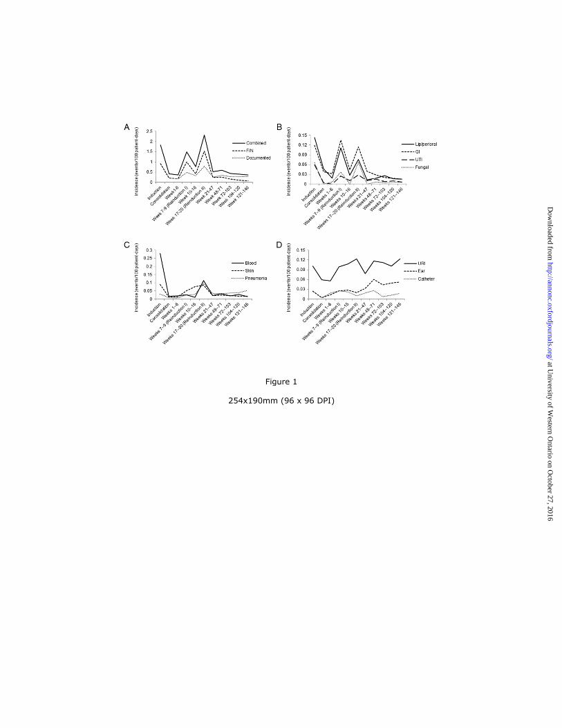
Figure 1
254x190mm (96 x 96 DPI)
at University of W
estern Ontario on O
ctober 27, 2016http://annonc.oxfordjournals.org/
Dow
nloaded from

Table 1. Association of clinical factors with the duration of neutropenia by treatment phase
Median (%) Range (%) P Median (%) Range (%) P
Induction
All patients 385 8.0 0.0-68.0 52.0 0.0-98.0
Age group
1–9.9 years 283 10.0 0.0-68.0 <0.001 56.0 0.0-100.0 <0.001
≥10 years 102 0.0 0.0-60.0 39.5 0.0-96.0
Risk group
Low risk 187 13.0 0.0-68.0 <0.001 58.0 0.0-100.0 <0.001
Standard or high risk 198 3.0 0.0-60.0 45.0 0.0-98.0
Gender
Female 173 8.0 0.0-68.0 0.231 57.0 0.0-116.0 0.001
Male 212 8.0 0.0-64.0 47.5 0.0-96.0
Race
White 304 10.0 0.0-68.0 <0.001 55.5 0.0-100.0 <0.001
Other 81 0.0 0.0-45.0 40.0 0.0-93.0
Immunophenotype
B-lineage 328 9.5 0.0-68.0 0.002 56.0 0.0-100.0 <0.001
T-lineage 57 0.0 0.0-50.0 32.0 0.0-93.0
Continuation weeks 1-20
All patients 365 0.0 0.0-32.0 16.0 0.0-72.0
Age group
1–9.9 years 282 0.0 0.0-25.0 0.068 19.0 0.0-72.0 0.012
≥10 years 83 0.0 0.0-32.0 11.0 0.0-60.0
Risk group
Low risk 196 0.0 0.0-24.0 0.299 15.0 0.0-72.0 0.007
Standard or high risk 169 0.0 0.0-32.0 20.0 0.0-67.0
Gender
Female 165 0.0 0.0-25.0 0.941 19.0 0.0-72.0 0.091
Male 200 0.0 0.0-32.0 15.0 0.0-60.0
Race
White 289 0.0 0.0-32.0 0.318 17.0 0.0-72.0 0.197
Other 76 0.0 0.0-24.0 15.5 0.0-59.0
Immunophenotype
B-lineage 314 0.0 0.0-32.0 0.204 16.0 0.0-72.0 0.785
T-lineage 51 0.0 0.0-26.0 15.0 0.0-56.0
Continuation weeks 21-120
All patients 350 2.0 0.0-10.0 11.0 0.0-41.0
Age group
1–9.9 years 271 2.0 0.0-10.0 0.127 12.0 0.0-41.0 0.010
≥10 years 79 0.0 0.0-10.0 9.0 0.0-35.0
Risk group
Low risk 186 2.0 0.0-10.0 0.595 11.0 0.0-33.0 0.363
Standard or high risk 164 2.0 0.0-10.0 11.0 0.0-41.0
Gender
Female 161 2.0 0.0-10.0 0.063 11.0 0.0-41.0 0.757
Male 189 1.0 0.0-10.0 11.0 0.0-35.0
Race
White 278 2.0 0.0-10.0 0.044 12.0 0.0-35.0 0.164
Other 72 0.0 0.0-10.0 10.0 0.0-41.0
Immunophenotype
B-lineage 303 2.0 0.0-10.0 0.620 11.0 0.0-35.0 0.796
T-lineage 47 2.0 0.0-8.0 10.0 0.0-41.0
Abbreviations: ANC, absolute neutrophil count.
*Data for ANCs not available for all patients.†Duration of neutropenia was calculated as the percentages of actual neutropenia period (days) among treatment duration (days).
ANC<0.1x10⁹/L ANC<0.5x10
9/L
N *
#No significant differences were seen among groups based on the presence of Down syndrome, white blood cell count at
diagnosis, central nervous system status, or body mass index category at diagnosis.
at University of W
estern Ontario on O
ctober 27, 2016http://annonc.oxfordjournals.org/
Dow
nloaded from
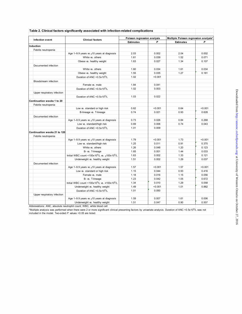
Table 2. Clinical factors significantly associated with infection-related complications
Estimates P Estimates P
Induction
Febrile neutropenia
Age 1–9.9 years vs ≥10 years at diagnosis 2.03 0.002 2.04 0.002
White vs. others 1.61 0.039 1.52 0.071
Obese vs. healthy weight 1.63 0.027 1.34 0.107
Documented infection
White vs. others 1.60 0.034 1.61 0.034
Obese vs. healthy weight 1.55 0.035 1.27 0.181
Duration of ANC <0.5x10⁹/L 1.02 <0.001
Bloodstream infection
Female vs. male 1.84 0.041
Duration of ANC <0.5x10⁹/L 1.02 0.003
Upper respiratory infection
Duration of ANC <0.5x10⁹/L 1.03 0.022
Continuation weeks 1 to 20
Febrile neutropenia
Low vs. standard or high risk 0.62 <0.001 0.64 <0.001
B-lineage vs. T-lineage 0.74 0.021 0.93 0.628
Documented infection
Age 1–9.9 years vs ≥10 years at diagnosis 0.73 0.026 0.84 0.268
Low vs. standard/high risk 0.69 0.006 0.74 0.043
Duration of ANC <0.5x10⁹/L 1.01 0.009
Continuation weeks 21 to 120
Febrile neutropenia
Age 1–9.9 years vs ≥10 years at diagnosis 1.79 <0.001 1.73 <0.001
Low vs. standard/high risk 1.25 0.011 0.91 0.370
White vs. others 1.26 0.048 1.20 0.123
B- vs. T-lineage 1.65 0.001 1.44 0.033
Initial WBC count <100x109/L vs. ≥100x109/L 1.63 0.002 1.33 0.121
Underweight vs. healthy weight 1.51 0.002 1.29 0.037
Documented infection
Age 1–9.9 years vs ≥10 years at diagnosis 1.57 <0.001 1.57 <0.001
Low vs. standard or high risk 1.15 0.044 0.93 0.416
Female vs. male 1.18 0.016 1.15 0.056
B- vs. T-lineage 1.23 0.042 1.05 0.672
Initial WBC count <100x109/L vs. ≥100x109/L 1.34 0.010 1.29 0.058
Underweight vs. healthy weight 1.49 <0.001 1.01 0.882
Duration of ANC <0.5x10⁹/L 1.01 0.050
Upper respiratory infection
Age 1–9.9 years vs ≥10 years at diagnosis 1.59 0.007 1.61 0.006
Underweight vs. healthy weight 1.51 0.047 0.90 0.507
Abbreviations: ANC, absolute neutrophil count; WBC, white blood cell
Infection event Clinical factorsPoisson regression analysis Multiple Poisson regression analysis*
*Multiple analysis was performed when there were 2 or more significant clinical presenting factors by univariate analysis. Duration of ANC <0.5x10⁹/L was not
included in the model. Two-sided P values <0.05 are listed.
at University of W
estern Ontario on O
ctober 27, 2016http://annonc.oxfordjournals.org/
Dow
nloaded from
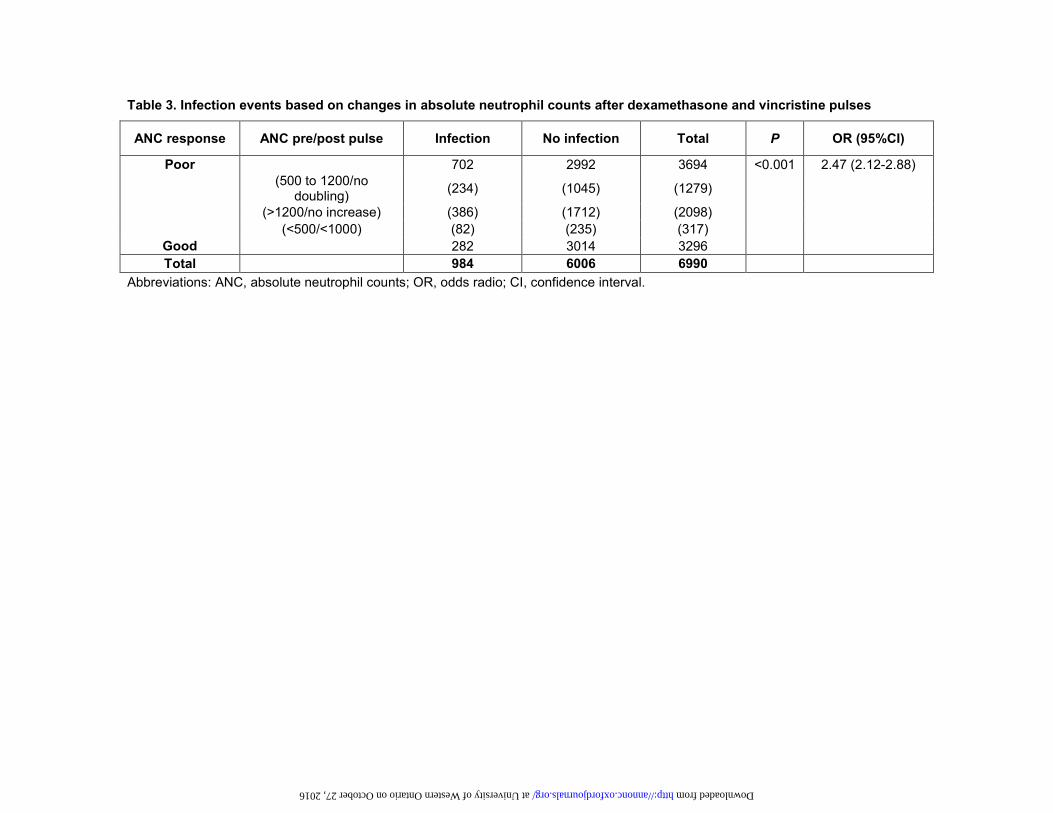
Table 3. Infection events based on changes in absolute neutrophil counts after dexamethasone and vincristine pulses
ANC response ANC pre/post pulse Infection No infection Total P OR (95%CI)
Poor 702 2992 3694 <0.001 2.47 (2.12-2.88)
(500 to 1200/no
doubling) (234) (1045) (1279)
(>1200/no increase) (386) (1712) (2098)
(<500/<1000) (82) (235) (317)
Good 282 3014 3296
Total 984 6006 6990
Abbreviations: ANC, absolute neutrophil counts; OR, odds radio; CI, confidence interval.
5051525354555657585960
at University of Western Ontario on October 27, 2016 http://annonc.oxfordjournals.org/ Downloaded from