extratympanic electrocochleography in ménière's disease
TRANSCRIPT
Acta Otorrinolaringol Esp. 2012;63(6):421---428
www.elsevier.es/otorrino
ORIGINAL ARTICLE
Extratympanic Electrocochleography in Ménière’s Disease�
Eduardo Martín-Sanz,∗ Jonathan Esteban Sánchez, Manuel González Juliao,Christiane Zschaeck Luzardo, Teresa Mato Patino, Laura Rodriganez Riesco,Ricardo Sanz Fernández
Servicio de Otorrinolaringología, Hospital Universitario de Getafe, Getafe, Madrid, Spain
Received 28 November 2011; accepted 28 March 2012
KEYWORDSExtratympanic elec-trocochleography;Ménière’s disease;Vestibular testing
AbstractIntroduction: Electrocochleography is the registration of an electrophysiological event whichtakes place in the cochlea after an acoustical stimulus. Most of the authors consider anincreased summating potential (SP)/action potential (AP) ratio as characteristic of endolym-phatic hydrops.Methods: A longitudinal, prospective study of a unilateral Ménière’s population diagnosedaccording to the American Academy of Otolaryngology---Head and Neck Surgery classificationwas carried out. A complete clinical history and bedside examination were performed, in addi-tion to complete auditory and vestibular testing and an extratympanic electrocochleography.We selected 20 normal hearing subjects with no history of vestibular and otological pathologyas a control group.Results: Of the 100 patients included in the study, 62 were diagnosed as definite Ménière’sdisease, and 13 and 25 as probable and possible Ménière’s disease, respectively. In the elec-trocochleography, 85% of all the patients had an SP/AP ratio above 0.5. A sensibility of 92%,78% and 75% was obtained in the definite, probable and possible Ménière’s disease patients,respectively.Discussion and conclusions: Electrocochleography is a useful method for diagnosing and evalu-ating patients with Ménière’s disease syndrome. It provides information about the progressionof the process and shows a significant correlation with the clinical stage.© 2011 Elsevier España, S.L. All rights reserved.
PALABRAS CLAVE Electrococleografía extratimpánica en la enfermedad de Ménière
Electrococleografíaextratimpánica;Enfermedadde Ménière;Pruebas vestibularesResumenIntroducción: La electrococleografía (ECoG) es un registro de un episodio electrofisiológico,que tiene lugar en la cóclea tras un estímulo acústico. La mayoría de los autores consideranque un aumento del cociente SP/AP utilizando clicks, es característico del hidrops.
� Please cite this article as: Martín-Sanz E, et al. Electrococleografía extratimpánica en la enfermedad de Ménière. ActaOtorrinolaringol Esp. 2012;63:421---8.
∗ Corresponding author.E-mail address: [email protected] (E. Martín-Sanz).
2173-5735/$ – see front matter © 2011 Elsevier España, S.L. All rights reserved.
422 E. Martín-Sanz et al.
Métodos: Presentamos un estudio longitudinal y prospectivo de una población de pacientescon enfermedad de Ménière unilateral definida según los criterios de la Academia Americanade Otorrinolaringología y patología cervicofacial. Tras una correcta anamnesis y exploraciónotoneurológica completa, todos los pacientes fueron sometidos a un estudio de la funciónauditiva y vestibular mediante audiometría tonal liminar, pruebas calóricas, rotatorias y unaECoG extratimpánica. Como controles se escogieron 20 sujetos normoacúsicos carentes deantecedentes de afección vestibular u otológica, ni antecedentes quirúrgicos en el oído medio.Resultados: De los 100 pacientes incluidos, 62 fueron diagnosticados de enfermedad de Ménièredefinida, 13 de probable y 25 de enfermedad de Ménière posible. Un 85% de la población conenfermedad de Ménière, cumplían criterios electrofisiológicos de enfermedad de Ménière segúnlos parámetros de la ECoG (SP/AP > 0,5). El porcentaje de ECoG alteradas según el diagnósticoera del 92, 78 y 75% para el Ménière definido, probable y posible respectivamente.Discusión y conclusiones: La ECoG es un método útil para el diagnóstico y evaluación de laenfermedad de Ménière. Aporta información fidedigna del progreso de la enfermedad y existe
diagn. Tod
I
Aiw
itt
dhE
aitiO
tita
a
egn
uiti
goubti
i
fott
fidc
M
WlaO
a
Oi
eva
I
1
2
3
E
correlación con su estadio
© 2011 Elsevier España, S.L
ntroduction
s the term itself suggests, electrocochleography (ECoG)s a recording of an electrophysiological episode occurringithin the cochlea after acoustic stimulation.
Far from being a new technique, ECoG emerged as a clin-cal tool in the 1970s, although the first attempts to recordhe cochlear microphonic potential date from 1930, whenhis potential was discovered in cats.
Subsequently, the summation potential (SP) wasescribed in animals in 1950, but the first record inumans was not obtained until the decade of the 1970s byggermont and Odenthal1 and Gibson et al.2
The discovery of the clinical applications of brainstemuditory evoked potentials (BAEP) increased the interestn all auditory evoked potentials. This fact, coupled withhe development of noninvasive techniques, facilitated themplementation of ECoG as a method of study in manytorhinolaryngology services and units.3
The indications for study by ECoG are not limited tohe study of endolymphatic hydrops/Ménière’s disease, ast can also have clinical applications as an auditory study inhe paediatric population or for intraoperative monitoring,mong others.
The 2 approaches currently available for ECoG recordingre the transtympanic and the extratympanic.
Its ease of use, low morbidity and, in most cases, unnec-ssary presence of a physician, have contributed to therowing presence of extratympanic ECoG in various diag-ostic protocols.4
It is a proven fact that patients with Ménière’s diseasesually develop increased SP amplitudes. This is because thencreased endolymphatic volume creates an impairment forhe vibration capacity of the organ of Corti, to which the SPs sensitive.
Although this finding is frequent, its applicability isreatly reduced by its low specificity, since the valuesbtained largely overlap with those of the normal pop-lation. However, thanks to the relationship establishedetween the SP parameter and the action potential (AP),
he diagnostic performance of the test has increased signif-cantly, thus improving its development as a clinical tool.2In fact, at present most authors believe that an increasen the SP/AP ratio using clicks is characteristic of hydrops.
1
2
óstico.os los derechos reservados.
In an attempt to improve the sensitivity of the testor the diagnosis of Ménière’s disease, Devaiah et al.5 notnly measured the SP/AP ratio, but also the SP/AP area,hereby increasing the sensitivity of the test, according tohe author.
In our study we aimed to assess the sensitivity and speci-city of ECoG in our population of patients with Ménière’sisease and to correlate it with parameters obtained throughlinical and vestibular studies.
aterial and Methods
e present a prospective and longitudinal study of a popu-ation of patients with unilateral Ménière’s disease, definedccording to the criteria of the American Academy oftolaryngology---Head and Neck Surgery (AAO---HNS).
The study was conducted between March 2008 and Febru-ry 2011.
We consecutively studied all patients attending thetoneurology Service during the study period who met the
nclusion criteria.After a correct anamnesis and complete otoneurological
xamination, all patients underwent a study of auditory andestibular function through liminal tone audiometry, caloricnd rotational testing and extratympanic ECoG.
nclusion Criteria
Patients with unilateral Ménière’s disease, classified asdefinite, probable or possible according to the criteria ofthe AAO---HNS.
Correct completion and interpretation of all auditory,vestibular and electrophysiological tests necessary forthis study.
Normoacusis of the contralateral ear.
xclusion Criteria
Bilateral involvement or presence of symptoms suggestinginvolvement of the contralateral ear.
Symptoms suggestive of the presence of neurologicalinvolvement.

(eaaT
V
Wrt
IPpflweomwspp
SPia0mcartmitsp
E
Alteeid
bt
m
Extratympanic Electrocochleography in Ménière’s Disease
3 History of middle ear surgery, such as myringotomy,myringoplasty, tympanoplasty or stapedectomy, whichcould alter the correct interpretation of vestibular testsand ECoG.
4 Anatomical abnormalities of the external and/or middleear likely to affect the implementation and/or interpre-tation of vestibular and electrophysiological testing.
5 Presence of any other vestibular condition other thanMénière’s disease in both the affected ear and in thecontralateral ear (benign paroxysmal positional ver-tigo [BPPV], vestibular neuritis, perilymphatic fistula ormigraine-associated vertigo).
Normal Group
We selected 20 subjects with normal hearing and with nohistory of vestibular or otological involvement, nor surgicalhistory in the middle ear.
Anamnesis
The medical records collected the relevant data about thedisease of each patient. We included a detailed history ofthe main symptoms and a review of the medical (previousdiagnoses, medical and surgical history) and family history.
The number and duration of episodes, associated symp-toms, their duration and functional level recommended bythe AAO---HNS were expressly reflected in the personal his-tory.
Physical Exploration
All patients underwent a standard oculomotor assessmentthrough videonystagmography equipment which systemati-cally evaluated the presence of spontaneous, head shaking,positional and positioning nystagmus. All patients under-went the oculocephalic manoeuvre or Halmagyi test, inaddition to an assessment using the Romberg and Fukudatests.
Tone Audiometry
The audiometric threshold by air and bone routes was deter-mined for frequencies of 0.25, 0.5, 1, 2, 3, 4 and 6 kHzwithin a soundproof booth (IAC mini 250). From the resultwe obtained the value of the pure-tone average (PTA) atfrequencies of 0.5, 1, 2 and 3 kHz, as recommended by theAAO---HNS, as well as the PTA for all frequencies studied.
As recommended by the AAO---HNS, we entered into thedatabase the worst audiometry obtained within the periodof 6 months before the study.
Vestibular Caloric Tests
All patients underwent caloric testing at the time of inclu-sion in the study. We used a VN415 module, 2-channel VNG
device, from Interacoustics®.The type of caloric stimulation used was based on themethodology described by Fitzgerald and Hallpike in 1942.Its aim was to stimulate the horizontal semicircular canal
p2w4
423
HSC) on each side at different temperatures. This gen-rated a nystagmic response in opposite directions, whichllowed us to determine the reflex activity from each ear,s well as its integration in the central nervous system (CNS).his procedure has been described in previous publications.6
estibular Rotatory Tests
e distinguished the sinusoidal harmonic accelerationotary test and the impulse rotatory test according to theype of stimulus and temporal presentation.
mpulse Rotatory Testrocedure. In order to obtain an adequate stimulation,atients were placed in a dark environment with the headexed approximately 30◦, so that the semicircular canalsere in a correct plane. Patients were then sharply accel-rated or decelerated from 0 to 100◦ s−1 in 1 s. In the casef acceleration, once the desired speed was reached it wasaintained constant during 60 s. After this time, the speedas slowed in 1 s and maintained for a further 60 s. This
equence was repeated 3 times towards each side. Theostrotatory time constant of each ear was recorded as aarameter.
inusoidal Harmonic Accelerationrocedure. Patients were subjected to a complex and var-ed stimulus. This was a sinusoidal oscillation in the verticalxis using the following harmonic frequencies: 0.01, 0.02,.04, 0.08, 0.16, 0.32 and 0.64 Hz. In each of them, theaximum speed obtained was 60◦ s−1 and the stimulation
ycle was repeated several times. In order to achieve andequate stimulation, patients were placed in a dark envi-onment with the head flexed approximately 30◦, so thathe semicircular canals were in a correct plane. In order toinimise discomfort and the time of completion of the test
n the present study, the studied frequencies were reducedo 0.16, 0.32 and 0.64 Hz. The values of phase, gain andymmetry of each studied frequency were determined asarameters.
lectrocochleography
ll patients underwent the test within an acoustically iso-ated room, in darkness and with the head flexed towardshe contralateral side of the ear being stimulated. Thextratympanic electrode was introduced following otoscopicxamination and cleaning if necessary. It was also soakedn saline solution and conductive gel in order to minimiseiscomfort.
All recordings were made with a Smart EP® auditoryrainstem response (ABR) device (Intelligent Hearing Sys-ems, Miami, FL, USA).
We used a single-channel recording obtained with aonaural stimulus consisting of 0.1 ms clicks, of alternating
olarity at 90 dB, with apportionment of 7.1 s. We obtainedrecordings for each ear and the evoked potential activityas filtered with a range of 5---1500 Hz. A sweep from 250 to00 clicks was conducted for each recording in all patients.
424 E. Martín-Sanz et al.
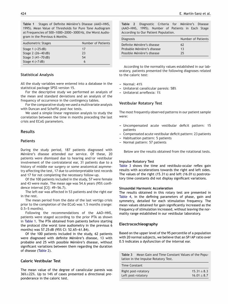
Table 1 Stages of Definite Ménière’s Disease (AAO---HNS,1995). Mean Value of Thresholds for Pure Tone Audiogramat Frequencies of 500---1000---2000---3000 Hz, the Worst Audio-gram in the Previous 6 Months.
Audiometric Stages Number of Patients
Stage 1 (<25 dB) 17Stage 2 (26---40 dB) 23
S
As
tf
w
cc
R
P
DMpihta
ad
i
p0
pitm
wpso
C
T3p
Table 2 Diagnostic Criteria for Ménière’s Disease(AAO---HNS, 1995). Number of Patients in Each StageAccording to Our Patient Population.
Diagnosis Number of Patients
Definite Ménière’s disease 62
ot
---
V
Tw
-
---
ITrTt
STTsmfm
E
Based on the upper level of the 95 percentile of a populationwith 20 normal subjects, we believe that an SP/AP ratio over0.5 indicates a dysfunction of the internal ear.
Table 3 Mean Gain and Time Constant Values of the Popu-lation in the Impulse Rotatory Test.
Stage 3 (41---70 dB) 54Stage 4 (>7 dB) 6
tatistical Analysis
ll the study variables were entered into a database in thetatistical package SPSS version 15.
For the descriptive study we performed an analysis ofhe mean and standard deviations and an analysis of therequency of occurrence in the contingency tables.
For the comparative study we used a multivariate analysisith Duncan and Scheffé post hoc tests.
We used a simple linear regression analysis to study theorrelation between the time in months preceding the lastrisis and ECoG parameters.
esults
atients
uring the study period, 187 patients diagnosed withénière’s disease attended our service. Of these, 20atients were dismissed due to hearing and/or vestibularnvolvement of the contralateral ear, 31 patients due to aistory of middle ear surgery or some anatomical asymme-ry affecting the test, 17 due to uninterpretable test recordsnd 17 for not completing the necessary follow-up.
Of the 100 patients included in the study, 57 were femalend 43 were male. The mean age was 54.6 years (95% confi-ence interval [CI]: 49---56.7).
The left ear was affected in 53 patients and the right earn the rest.
The mean period from the date of the last vertigo crisisrior to the completion of the ECoG was 1.5 months (range:.5---5 months).
Following the recommendations of the AAO---HNS,atients were staged according to the prior PTA as shownn Table 1. The PTA obtained from patients before startinghe protocol (the worst tone audiometry in the previous 6onths) was 57.25 dB (95% CI: 52.65---61.84).Of the 100 patients included in the study, 62 patients
ere diagnosed with definite Ménière’s disease, 13 withrobable and 25 with possible Ménière’s disease, withoutignificant variations between them regarding the durationf disease (Table 2).
aloric Vestibular Test
he mean value of the degree of canalicular paresis was6%±22%. Up to 14% of cases presented a directional pre-onderance in the caloric test.
Probable Ménière’s disease 13Possible Ménière’s disease 25
According to the normality values established in our lab-ratory, patients presented the following diagnoses relatedo the caloric test:
-- Normal: 41%-- Unilateral canalicular paresis: 58%-- Unilateral arreflexia: 1%
estibular Rotatory Test
he most frequently observed patterns in our patient sampleere:
-- Uncompensated acute vestibular deficit pattern: 15patients
-- Compensated acute vestibular deficit pattern: 23 patients-- Habituation pattern: 5 patients-- Normal pattern: 57 patients
Below are the results obtained from the rotational tests.
mpulse Rotatory Testable 3 shows the time and vestibulo-ocular reflex gainesults with accelerations towards the right and left sides.he values of the right (15.31 s) and left (16.01 s) postrota-ory time constants did not display significant variations.
inusoidal Harmonic Accelerationhe results obtained in this rotary test are presented inable 4, in the defining parameters of phase, gain andymmetry, detailed for each stimulation frequency. Theean values obtained for gain significantly increased as the
requency of stimulation increased, without leaving the nor-ality range established in our vestibular laboratory.
lectrocochleography
Time Constant
Right post-rotatory 15.31 ± 8.3Left post-rotatory 16.01 ± 8.7
Extratympanic Electrocochleography in Ménière’s Disease 425
Right SP/AP ratio
Left SP/AP ratio
Right SP/AP ratio
Left SP/AP ratio
Right SP/AP ratio
Left SP/AP ratio
-0.5
0.0
0.5
1.0
1.5
2.0
-0.5
0.0
0.5
1.0
1.5
2.0
Definite Probable
AAO-HNS diagnosis
Possible
LeftR
ight
Side w
ith Ménière's disease
95%
CI
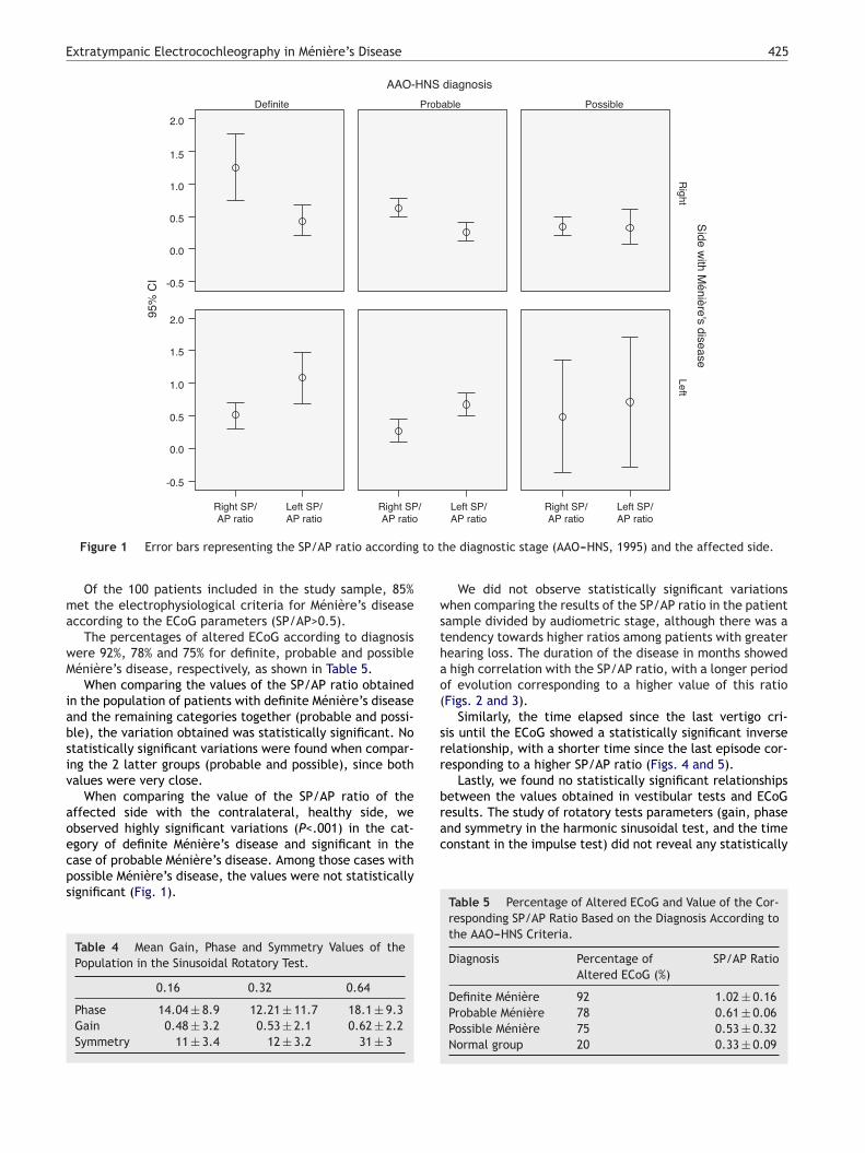
Figure 1 Error bars representing the SP/AP ratio according to the diagnostic stage (AAO---HNS, 1995) and the affected side.
Of the 100 patients included in the study sample, 85%met the electrophysiological criteria for Ménière’s diseaseaccording to the ECoG parameters (SP/AP>0.5).
The percentages of altered ECoG according to diagnosiswere 92%, 78% and 75% for definite, probable and possibleMénière’s disease, respectively, as shown in Table 5.
When comparing the values of the SP/AP ratio obtainedin the population of patients with definite Ménière’s diseaseand the remaining categories together (probable and possi-ble), the variation obtained was statistically significant. Nostatistically significant variations were found when compar-ing the 2 latter groups (probable and possible), since bothvalues were very close.
When comparing the value of the SP/AP ratio of theaffected side with the contralateral, healthy side, weobserved highly significant variations (P<.001) in the cat-egory of definite Ménière’s disease and significant in thecase of probable Ménière’s disease. Among those cases withpossible Ménière’s disease, the values were not statisticallysignificant (Fig. 1).
Table 4 Mean Gain, Phase and Symmetry Values of thePopulation in the Sinusoidal Rotatory Test.
0.16 0.32 0.64
Phase 14.04 ± 8.9 12.21 ± 11.7 18.1 ± 9.3Gain 0.48 ± 3.2 0.53 ± 2.1 0.62 ± 2.2Symmetry 11 ± 3.4 12 ± 3.2 31 ± 3
We did not observe statistically significant variationswhen comparing the results of the SP/AP ratio in the patientsample divided by audiometric stage, although there was atendency towards higher ratios among patients with greaterhearing loss. The duration of the disease in months showeda high correlation with the SP/AP ratio, with a longer periodof evolution corresponding to a higher value of this ratio(Figs. 2 and 3).
Similarly, the time elapsed since the last vertigo cri-sis until the ECoG showed a statistically significant inverserelationship, with a shorter time since the last episode cor-responding to a higher SP/AP ratio (Figs. 4 and 5).
Lastly, we found no statistically significant relationshipsbetween the values obtained in vestibular tests and ECoGresults. The study of rotatory tests parameters (gain, phaseand symmetry in the harmonic sinusoidal test, and the timeconstant in the impulse test) did not reveal any statistically
Table 5 Percentage of Altered ECoG and Value of the Cor-responding SP/AP Ratio Based on the Diagnosis According tothe AAO---HNS Criteria.
Diagnosis Percentage ofAltered ECoG (%)
SP/AP Ratio
Definite Ménière 92 1.02 ± 0.16Probable Ménière 78 0.61 ± 0.06Possible Ménière 75 0.53 ± 0.32Normal group 20 0.33 ± 0.09

426 E. Martín-Sanz et al.
Right SP/AP ratio
Evo
lutio
n in
mon
ths
0.00 0.50 1.00 1.50 2.00 3.00 2.50
R Sq Linear = 0.622
50
30
40
20
10
0
Figure 2 Linear regression graph establishing the relationshipbetween the SP/AP ratio and the evolution time in months ofMénière’s disease in the right ear.
Left SP/AP ratio
Evo
lutio
n in
mon
ths
0.00 0.50 1.00 1.50 2.00 2.50
R Sq Linear = 0.526
50
40
30
20
10
0
Figure 3 Linear regression graph establishing the relationshipbetween the SP/AP ratio and the evolution time in months ofMénière’s disease in the left ear.
Right SP/AP ratio
Tim
e be
fore
cris
is
0.00 0.50 1.00 1.50 2.00 2.50 3.00
R Sq Linear = 0.512
6
4
2
0
Figure 4 Regression graph between the time in months priorto the last crisis and the SP/AP ratio in patients with rightMénière’s disease.
Left SP/AP ratio
Tim
e be
fore
cris
is
0.00 0.50 1.00 1.50 2.00 2.50
R Sq Linear = 0,632
6
7
4
5
3
2
1
Figure 5 Regression graph between the time in months priortM
so
D
Tec
co
iap
sie
toe0oiGmtdout(a
aid
o the last crisis and the SP/AP ratio in patients with lefténière’s disease.
ignificant relationship with the value of the SP/AP ratio inur patient sample.
iscussion
his study demonstrates the presence of alterations in thextratympanic ECoG of patients with Ménière’s disease, thusonfirming its usefulness in the diagnosis of this condition.
Pappas et al.7 established the need for each centre toonduct the study on its own patient population, in order tobtain significant parameters and confidence levels.
This fact is particularly important with regard to obtain-ng figures for optimal sensitivity and specificity for the test,s this largely depends on the values of normality and thearametric range.
There are numerous publications reporting sensitivity andpecificity values for ECoG.8 These works often describe thenherent difficulties of the test, given the wide dispersion ofxisting data.
Reviewing the various publications, it is possible to iden-ify some variability in the SP/AP thresholds for the diagnosisf Ménière’s disease between different authors. Ridenourt al.9 calculated an SP/PA ratio of 0.39 for males and.25 for females, whereas Sass8 established a unified ratiof 0.41. Pappas et al.7 believed that any result over 0.5s suggestive of endolymphatic hydrops, whereas Iseli andibson10 established a significantly lower value of 0.33, thusodifying the specificity figures. Therefore, each labora-
ory must establish a threshold for the SP/AP ratio for theiagnosis of Ménière’s disease and thus obtain figures forptimal sensitivity and specificity. Among our patient pop-lation and with our estimate of a required SP/AP ratiohreshold greater than 0.5, we obtained a sensitivity of 85%85 out of 100 patients obtained a value greater than 0.5)nd a specificity of 80%.
These figures are similar to those obtained by otheruthors in various studies.11 Nevertheless, despite obtain-ng objective data suggestive of the diagnosis of Ménière’sisease, clinical presentation and evolution remain the gold
essawopEsd
C
-
-
-
C
T
R
1
Extratympanic Electrocochleography in Ménière’s Disease
standard. Therefore, the sensitivity and specificity values ofany diagnostic test for this syndrome will always be subjectto variation depending on the study population, evolutiontime, medical treatment, etc.
In the present study there was a significant positivecorrelation between the duration of symptoms and ECoGfindings. Although not all authors have found this statis-tical significance,11---13 our experience was similar to thatof other authors14,15 in that patients with a longer evolu-tion time presented higher SP/AP ratios. This was consistentwith the observation that those patients in our study whopresented clearer clinical criteria for the disease (definiteMénière’s disease) also presented significantly higher SP/APratios than patients in the remaining stages (probable andpossible Ménière’s disease). Given the natural history ofthe disease and its initial clinical presentation, it is com-mon for patients with a longer evolution to present a morecomplete clinical form, characteristic of the category of def-inite Ménière’s disease. By contrast, the initial presentationform is usually not very evident,16---18 in which case physi-cians merely report a clinical suspicion (probable or possibleMénière’s disease).
Similarly, our study population showed a trend towardsstatistical significance when comparing their auditory sta-tus with the SP/AP ratio. Although the statistical analysisshowed no significance (coinciding with the experience ofauthors such as Chung et al.11), it is likely that a larger sam-ple would show a link between greater hearing losses andhigher SP/AP ratios. It is also likely that the time variablein months from the last vertigo crisis before performing theECoG could represent an interaction or confounding factorwith other variables, such as the PTA, for example.
Given the pathophysiology of Ménière’s disease, inwhich the damage on the membrane of Reissner becomesirreversible while hydrops persists,19 it is quite likely thatthis change is at least partly responsible for elevating theSP/AP ratio. This would partly explain the notable inverserelationship between the time elapsed since the last vertigocrisis prior to the ECoG and the SP/AP ratio. Since ECoG isan objective measurement of the degree of endolymphatichydrops, a significant link between the proximity of the crisisand the parameters recorded is to be expected.
In fact, this finding was used by some authors againstthe test, arguing that ECoG would only reflect the hydropicnature of the disease during crises.20 Successive publica-tions have refuted these observations, noting a permanenceof ECoG changes during the inter-crisis periods of thedisease.11,12,14
Although videonystagmography with caloric and rota-tional tests is currently considered to be the gold standardfor the diagnosis of vestibular disorders, such as Ménière’sdisease,21 the parameters obtained from these tests haveshown no significant correlation with the SP/AP ratio.
While it is true that caloric and rotational vestibular testshave known sensitivity and specificity values for the detec-tion of peripheral vestibular involvement,22 this sensitivitybecomes markedly decreased in the early stages of Ménière’sdisease, in which hydrops has not yet generated relevant
lesions on the lateral semicircular canal. This fact has beenwidely reported in daily clinical practice, in which classicalvestibular tests are often negative in the early stages of thedisease.1
427
The pathophysiological evolution of Ménière’s diseaseventually leads to involvement of the macules of theemicircular canals by endolymphatic hydrops in advancedtages. This is reflected in the classification by evolution-ry stages by Shea,23 in which conventional vestibular testsould be affected in stage IV. This fact explains the inabilityf these tests to observe vestibular damage in a significantroportion of patients with Ménière’s disease. By contrast,CoG is capable of initially detecting cochlear hydrops,ince this is usually present from the initial phases of theisease.24
onclusions
-- ECoG is a useful method in the diagnosis and assessmentof Ménière’s disease.
-- It provides accurate information to assess the progress ofthe disease and there is a correlation with the diagnosticstage.
-- ECoG helps to understand the pathophysiological phe-nomenon of endolymphatic hydrops in Ménière’s disease.
onflict of Interests
he authors have no conflict of interests to declare.
eferences
1. Eggermont JJ, Odenthal DW. Methods in electrocochleography.Acta Otolaryngol Suppl. 1974;316:17---24.
2. Gibson WP, Moffat DA, Ramsden RT. Clinical electrocochleog-raphy in the diagnosis and management of Ménière’s disorder.Audiology. 1977;16:389---401.
3. Ferraro JA, Durrant JD. Electrocochleography in the evaluationof patients with Ménière’s disease/endolymphatic hydrops. JAm Acad Audiol. 2006;17:45---68.
4. Burkard RF, Don M, Eggermont JJ. Auditory evoked potentials:basic principles and clinical application. Philadelphia: Lippin-cott Williams & Wilkins; 2007.
5. Devaiah AK, Dawson KL, Ferraro JA, Ator GA. Utility of areacurve ratio electrocochleography in early Ménière disease. ArchOtolaryngol Head Neck Surg. 2003;129:547---51.
6. Martín E, Pérez N. Hearing loss after intratympanic gentami-cin therapy for unilateral Ménière’s disease. Otol Neurotol.2003;24:800---6.
7. Pappas Jr DG, Pappas Sr DG, Carmichael L, Hyatt DP, Toohey LM.Extratympanic electrocochleography: diagnostic and predictivevalue. Am J Otol. 2000;21:81---7.
8. Sass K. Sensitivity and specificity of transtympanic elec-trocochleography in Ménière’s disease. Acta Otolaryngol.1998;118:150---6.
9. Ridenour BD, Meyerhoff WL, Wang X, Gerken GM. Summating-potential/action-potential ratio in normal ears: effects ofdehydration. Am J Otolaryngol. 1993;14:24---30.
0. Iseli C, Gibson WA. Comparison of three methods of usingtranstympanic electrocochleography for the diagnosis ofMénière’s disease: click summating potential measurements,tone burst summating potential amplitude measurements, andbiasing of the summating potential using a low frequency tone.
Acta Otolaryngol. 2010;130:95---101.1. Chung WH, Cho DY, Choi JY, Hong SH. Clinical usefulnessof extratympanic electrocochleography in the diagnosis ofMénière’s disease. Otol Neurotol. 2004;25:144---9.
4
1
1
1
1
1
1
1
1
2
2
2
21993;14:224---9.
24. Kingma CM, Wit HP. Cochlear hydrops analysis masking proce-
28
2. Orchik DJ, Shea Jr JJ, Ge NN. Summating potential and actionpotential ratio in Ménière’s disease before and after treatment.Am J Otol. 1998;19:478---82.
3. Levine S, Margolis RH, Daly KA. Use of electrocochleog-raphy in the diagnosis of Ménière’s disease. Laryngoscope.1998;108:993---1000.
4. Ge X, Shea Jr JJ. Transtympanic electrocochleography: a 10-year experience. Otol Neurotol. 2002;23:799---805.
5. Kim JS, Nam EC, Park SI. Electrocochleography is more sensitivethan distortion-product otoacoustic emission test for detec-ting noise-induced temporary threshold shift. Otolaryngol HeadNeck Surg. 2005;133:619---24.
6. Mancini F, Catalani M, Carru M, Monti B. History of Ménière’sdisease and its clinical presentation. Otolaryngol Clin North Am.2002;35:565---80.
7. Kinney SE, Sandridge SA, Newman CW. Long-term effects ofMénière’s disease on hearing and quality of life. Am J Otol.
1997;18:67---73.8. Green Jr JD, Blum DJ, Harner SG. Longitudinal followup ofpatients with Ménière’s disease. Otolaryngol Head Neck Surg.1991;104:783---8.
E. Martín-Sanz et al.
9. Yoon TH, Paparella MM, Schachern PA, Le CT. Cellular changes inReissner’s membrane in endolymphatic hydrops. Ann Otol RhinolLaryngol. 1991;100:288---93.
0. Kim HH, Kumar A, Battista RA, Wiet R. JElectrocochleogra-phy in patients with Ménière’s disease. Am J Otolaryngol.2005;26:128---31.
1. Fushiki H, Junicho M, Aso S, Watanabe Y. Recurrence rate ofidiopathic sudden low-tone sensorineural hearing loss with-out vertigo: a long-term follow-up study. Otol Neurotol.2009;30:295---8.
2. Pérez N, Martín E, Romero M, García-Tapia R. Influence of canalparesis and compensation on gain and time constant of nistag-mus slow-phase velocity to yaw axis rotation. Acta Otolaryngol(Stockh). 2001;121:715---23.
3. Shea Jr J. Classification of Ménière’s disease. Am J Otol.
dure results in patients with unilateral Ménière’s disease. OtolNeurotol. 2010;31:1004---8.