universitÉ de la rochelle - archimerarchimer.ifremer.fr/doc/00230/34093/32540.pdf · niveau:...
TRANSCRIPT
UNIVERSITÉ DE LA ROCHELLE
ÉCOLE DOCTORALE
Science pour l'Environnement Gay-Lussac
Laboratoire Littoral Environnement et Sociétés (LIENSs) UMR 7266-ULR
Laboratoire Génétique et Pathologie des Mollusques Marins (SG2M-LGPMM) Ifremer
La Tremblade
THESE
Présentée par
Rachida Mersni-Achour
Soutenance prévue le 20 mai 2014
Pour l'obtention du grade de Docteur de l'Université de la Rochelle
Discipline: Aspects moléculaires et cellulaires de la biologie
Jury:
Paco Bustamante Professeur, Université de la Rochelle, Président du jury
Carolyn Friedman Professeur, JISAO Senior Fellow, Rapporteur
Philippe Soudant Directeur de recherche CNRS, IUEM-UBO, Rapporteur
Vianney Pichereau Professeur, IUEM-UBO, IUEM-UBO, Examinateur
Delphine Destoumieux-Garzon Chargée de recherche CNRS, Université Montpellier 2, Examinateur
Ingrid Fruitier-Arnaudin Maître de conférences, Université de la Rochelle, Directeur de thèse
Denis Saulnier Chercheur, Centre Ifremer du Pacifique, Co-directeur
Marie-Agnès Travers Chercheur, Ifremer SG2M-LGPMM, Responsable scientifique
Vibrio tubiashii en France: description d’isolats pathogènes affectant des
mollusques et étude de leurs mécanismes de virulence
Contributions scientifiques
Publications
Publications en revue
1/. R. Mersni Achour, M A. Travers, P. Haffner, D. Tourbiez, N. Faury, A L. Cassone, B.
Morga, I. Doghri, C. Garcia, T. Renault, I. Fruitier-Arnaudin, D. Saulnier. (2014). First
description of French V. tubiashii strains pathogenic to mollusk: I. Characterization of
isolates and detection during mortality events. Journal of Invertebrate Pathology (IF: 2.6).
2/ R. Mersni Achour, N. Imbert, V. Huet, Y. Ben Cheikh, N. Faury, I. Doghri, S. Rouatbi, S.
Bordenave, M A Travers, D. Saulnier, I. Fruitier-Arnaudin. First description of French V.
tubiashii strains pathogenic to mollusk: II. Characterization of secreted proteases
affecting oyster immunity. Sous révision in Journal of Invertebrate Pathology (IF: 2.6).
Publications en attente de soumission
3/. R. Mersni Achour , Y. Ben Cheikh,
V. Pichereau, I. Doghri, C. Etien, L. Dégremont, D.
Saulnier, I. Fruitier-Arnaudin, M A. Travers Molecular characterizations of the French V.
tubiashii virulence factors on oyster’s larvae give evidences that metalloprotease is not
the only main toxic factor .(Journal visé Microbiology IF: 2.8).
4/. R. Mersni Achour, I. Doghri, V. Pichereau, Y. Ben cheikh , P. Haffner, S. Sable, D.
Saulnier, M A. Travers, I. Fruitier Arnaudin. Toxicity and proteases composition of
extracellular products from the French Vibrio tubiashii are modulated by physical and
chemical characteristics of growth media. (Journal visé Microbiology IF: 2.8).
Communications orales
R. Mersni Achour, P. Haffner, I. Doghri, Y. Ben Cheikh, N. Faury, D. Tourbiez, N. Imbert,
V. Huet, S. Rouatbi, A L. Cassone, B. Morga, C. Garcia, B. Chollet, S. Bordenave, V.
Pichereau, T. Renault, D. Saulnier, M A. Travers, I. Fruitier-Arnaudin. Identification of a
French Vibrio tubiashii, a pathogen of bivalves and characterization of its extracellular
products. Vibrio 2014. Avril 1-4, 2014 Edinbough, UK.
R. Mersni Achour. M.A Tarvers. Vibrio tubiashii in France: description of pathogenic
isolates affecting shellfish and characterization of their virulence mechanisms. The
Annual Meeting of the National Reference Laboratories for Mollusc Diseases, Mars 25-26,
2014, Nantes France.
R. Mersni Achour. Etude du mode d’action de toxines bactériennes chez un invertébré
marin: l'huître creuse Crassostrea gigas. Colloque des Doctorants de 2ème année. Mai 31,
2012 Ecole doctorale Gay LUSSAC La Rochelle, France.
Communications affichées
R. Mersni Achour, M A. Travers, P. Haffner D. Tourbiez, A L. Cassone, B. Morga, I.
Doghri, C. Garcia, T. Renault, I. Fruitier-Arnaudin, D. Saulnier. Genotypic and phenotypic
characterization of Vibrio tubiashii subsp. francensis nov., a phylogenetically distinct
bacterial pathogen of bivalves. 10th International Meeting on Microbial Epidemiological
Markers (IMMEM-10) Institut Pasteur. Octobre 2-5, 2013 Paris, France.
R. Mersni Achour. Etude du mode d’action de toxines bactériennes chez un invertébré
marin: l'huître creuse Crassostrea gigas. Colloque de la Fédération de Recherche en
Environnement et Développement Durable. Juin 27-29, 2012 CAES- Ile d'Oléron, France.
I. Fruitier-Arnaudin, R. Mersni, D. Saulnier. Purification and characterization of
secreted proteases from three Vibrio harveyi like strains isolated from the Pacific oyster
Crassostrea gigas. ESF-COST High-Level Research Conference Marine Biotechnology:
Future Challenges. Juin 20-25, 2010 Acquafredda di Maratea, Italy.
Activités d’enseignement
2013-2014: Attaché temporaire d’Enseignement et de Recherche (ATER) au sein du
département de Biotechnologie à l’université de la Rochelle, France. Volume horaires: 48h
équivalent TD. Niveau: Licence 3 (10h CM et 6h TD Anabolisme) et Licence 1 (TD de
biochimie structurale 1).
2012-2013: Activités complémentaires d’enseignement (DCACE) au sein du département de
Biotechnologies à l’université de la Rochelle, France. Volume horaires: 64h équivalent TD.
Niveau: Master 1 (TP de microbiologie fondamentale et appliquée).
2011-2012: Activités complémentaires d’enseignement (DCACE) au sein du Département de
Biotechnologies à l’université de la Rochelle, France. Volume horaire: 64h équivalent TD.
Niveau: Master 1 (TP de microbiologie fondamentale et appliquée) et Licence 3 (TP de
génétique bactérienne, métabolisme et croissance).

Remerciements
Cet ouvrage est le fruit de plus de 3 ans de labeur et de dévouement de ma part mais surtout de la part des personnes qui m’entourent et qui m’ont soutenu et qui ont grandement contribué au succès de ce travail. Mes vifs remerciements s'adressent aux membres du Jury qui, par leur expertise, ont examiné ce travail. Ces travaux ont été réalisés au sein du laboratoire rochelais LIENSs UMR 7266 CNRS-ULR dirigé par Mr Olivier de Viron et le laboratoire Trembladais de l'IFREMER: le LGPMM dirigé par Mr Tristan Renault et Mme Sylvie Lapègue. Je les remercie de m'avoir accueillie dans les structures qu'ils dirigent. Je remercie particulièrement ma directrice de thèse Dr Ingrid Fruitier-Arnaudin qui n'a ménagé aucun effort pour le bon déroulement de la thèse dans de bonnes conditions et qui a veillé à la disponibilité des ressources nécessaires à ces travaux. Je remercie également mon co-directeur de thèse Dr Denis Saulnier, qui, malgré la distance et le décalage horaire, a toujours le souci de suivre mes travaux. Un grand merci à Marie-Agnès Travers, mon encadrant scientifique, pour son soutien, ses encouragements, ses conseils, sa bienveillance envers moi et son implication pleine dans ce travail. Agnès, je te remercie pour toutes les discussions fructueuses, pour les ondes positives que tu m’as constamment transmises et qui m'ont poussé au delà de mes limites. J’ai beaucoup appris de toi. Merci à Vianney Pichereau et à toute l’équipe LEMAR pour m’avoir appris la technique d’électrophorèse bidimensionnelle. J’ai passé un séjour très agréable à Brest. Je remercie Mr Gérard Blanchard pour m’avoir accueillie au sein de l’Université de La Rochelle dans laquelle j'ai réalisé, dans de bonnes conditions, mes charges d'enseignements et de recherche.
Une grande partie de mon travail a été effectué au laboratoire LGPMM à la Tremblade, une belle petite ville dans laquelle je me rendais souvent en covoiturage avec Patrick Azema et Cyrille François. Je vous remercie pour les bons moments musicaux sur la route.
D'un autre coté, je remercie toute l'équipe de ce laboratoire, Nicole Faury, Philippe Haffner, Delphine tourbiez, Bruno Chollet, Céline Garcia, Lionel Dégremont, Raphaël Brizard et Véronique Betto. Je les remercie pour leur accueil chaleureux, leur disponibilité et leur bonne humeur. L'autre partie de ce travail à été réalisée à la Rochelle au sein du laboratoire LIENSs entre le bâtiment Marie Curie et le bâtiment ILE.
Je remercie chaleureusement tous mes collègues de bureau Amandine Adrien Dit Richard, Oussama Achour (qui est aussi mon mari), Nicolas Joguet, Nicolas Poupard et Florian Le Joubioux. Ils ont supporté tous mes états d'humeurs pendant plus de 3 ans.
J’adresse mes remerciements aux étudiants Ibtissem doghri, Yosra Ben cheikh, Cédric Etien, Aurélie Trefier et Julie Lepinay que j’ai eu la chance d’encadrer et qui ont grandement contribué à la réussite de ce travail. Un grand merci à tous mes collèges et amis Hervé Rouillard, Camille Juin, Valérie Sopena pour leur sympathie et pour les bons repas au RU (poisson et encore du poisson). Je remercie également tout le personnel de l’équipe AMES pour leur sympathie. Un grand merci à tous les étudiants tunisiens qui sont passés par ce Laboratoire pour les bons moments partagés. Au Bâtiment ILE, je remercie Nathalie Imbert, Valérie Huet et Laureen Beaugeard pour leur aide, leur patience et leur implication dans les manips de cytométries en flux. Je remercie également David pour sa gentillesse et pour les discussions sympathiques lors des pauses thé-café.
Je remercie vivement tout le personnel administratif du laboratoire LIENSs Viviane Biou, Laetitia Darre, Johan Guiard et Marie Chivaille qui ont facilité toutes les tâches administratives et ont toujours répondu à mes demandes quasi instantanément avec une bonne humeur. Enfin, il existe des personnes sans lesquelles ces travaux n'auraient probablement jamais eu lieu. Ils ont, sans le savoir, réalisé ce travail avec moi car ils étaient toujours présents dans mon cœur. C'est à ma famille et mes amis que je m'adresse pour manifester ma reconnaissance.
D'abord à mes parents: ma très chère maman Yamina (Ya), mon très cher papa Salem (Baba) et mon très cher frère Amine (Minov). Malgré les 1400 Km qui nous séparent, votre soutien émotionnel, moral et physique, m'ont permis de surmonter toutes les difficultés. J’ai pu à chaque fois me ressourcer auprès de vous. Que Dieu vous bénisse et vous protège. Un grand merci à ma future belle sœur Narjess, tu es un amour.
Je remercie chaleureusement mes beaux-parents Sondes et khaled, ainsi que mes beaux-frères Ahlem (7arim), Ahmed (7amid) et Belhassan (Bach). Merci Dieu pour cette belle famille. Un grand merci à toute ma famille : cousins et cousines, oncles et tantes pour votre soutien. Merci tata Henda et Mostafa pour votre soutien et tous les cadeaux. Merci tonton Khaled Ben Cheikh d'avoir toujours cru en moi. Un très grand merci à ma sœur Balkis Eddif pour tous les bons moments passés ensemble (à Paris, à Toulouse, à Bordeaux, à Poitiers, à la Rochelle…) et pour les heures et les heures d’appels. Notre complicité et notre grande fraternité ne cessent de grandir avec beaucoup d’amour. ET un grand merci à mon ami et mon beau frère Amine Chebbi (et enfin on est une même famille). Un gros merci à mes amies Souhir Jazzar et Asma Soussi Ben Ali. Vous étiez loin des yeux mais tout le temps proches du cœur.
Je remercie Ibtissem Doghri qui a grandement contribué à ce travail et qui m'a quotidiennement accompagné à la Tremblade pour m'aider pour les manips. Je remercie l'amie qu'elle est devenue et je lui souhaite le succès pour sa thèse. Un merci à mes amies Yesmine Ben Henda et Mariem Laamari pour les beaux moments que l’on a vécu à La Rochelle, pour le soutien moral et les fous rires que nous avons partagés "L'amitié double les joies et réduit de moitié les peines." Un énorme merci à ma petite famille: à mon mari chéri Oussama (Merci mon amour) et à mon bout de chou Yoyo (tu es l’étoile qui a illuminé notre vie). Je vous aime de tout mon cœur.
Finalement, je remercie tous ceux qui ont participé de prés ou de loin à ce travail.
On manque trop souvent les occasions pour exprimer nos sentiments Merci à la thèse
Table des matières
Index des figures ...................................................................................................................... 14
Index des tableaux .................................................................................................................... 15
Liste des unités et des principales abréviations ........................................................................ 16
CONTEXTE GENERAL ET OBJECTIFS DE L’ETUDE ...................................... 18
INTRODUCTION BIBLIOGRAPHIQUE ................................................................... 22
Partie A: Les bactéries du genre Vibrio ............................................................. 23
1. Systématique, caractéristiques biologiques et écologiques .............................................. 24
2. Historique et taxonomie ................................................................................................... 25
3. Structure du génome ......................................................................................................... 27
4. Espèces pathogène du genre Vibrio.................................................................................. 28
4.1. Les vibrions pathogènes pour l’Homme .................................................................. 28
4.2. Les vibrions pathogènes des organismes marins ...................................................... 30
Vibrio shiloi .............................................................................................................................. 32
4.3. Vibrio tubiashii ......................................................................................................... 35
5. Les facteurs de virulence du genre Vibrio ........................................................................ 41
5.1. Les notions de pathogénicité et de virulence ........................................................... 41
5.2. Les mécanismes de virulence chez les Vibrio .......................................................... 42
5.3. Régulation de l’expression des facteurs de virulence .............................................. 48
6. Méthodes de détection du genre Vibrio ............................................................................ 50
Partie B: Les facteurs de virulence chez les vibrions marins, types et
mode d’action ................................................................................................................... 53
1. Les protéases extracellulaires chez le genre Vibrio ......................................................... 54
1.1 Généralités sur les protéases .................................................................................... 54
1.2 Rôle des protéases dans la virulence du genre Vibrio .............................................. 55
1.3 Les métalloprotéases ................................................................................................ 57
1.3.1 Généralités et classification ................................................................................ 57
1.3.2 Mécanisme d’action de la métalloprotéase ........................................................ 58
1.3.3 Synthèse et régulation ........................................................................................ 61
1.3.4 Implication des métalloprotéases dans la virulence du genre Vibrio ................. 62
1.4 Les protéases à sérine ............................................................................................... 64
1.4.1 Généralités et mode d’action .............................................................................. 64
1.4.2 Synthèse et régulation ........................................................................................ 66
1.4.3 Les protéases à sérine dans le genre Vibrio ........................................................ 66
1.5 Les protéases à cystéine ........................................................................................... 67
1.5.1 Généralités et mode d’action .............................................................................. 67
1.5.2 Synthèse et régulation ........................................................................................ 68
1.5.3 Les protéases à cystéines dans le genre Vibrio................................................... 68
2. Les sidérophores ............................................................................................................... 69
2.1 Généralités ................................................................................................................ 69
2.2 Mode d’action .......................................................................................................... 70
2.3 Biosynthèse et régulation ......................................................................................... 71
2.4 Sidérophores chez Vibrio ......................................................................................... 72
3. Les hémolysines ............................................................................................................... 73
3.1 Généralités ................................................................................................................ 73
3.2 Mode d’action .......................................................................................................... 74
3.3 Sécrétion et régulation .............................................................................................. 75
3.4 Implication des hémolysines dans la virulence des Vibrio ...................................... 76
Partie C: Les différents modèles d'études de la virulence du genre
Vibrio ..................................................................................................................................... 79
1. Approche in vivo .............................................................................................................. 80
1.1 Modèle d’étude: généralités sur l’huître creuse C. gigas ......................................... 80
1.1.1 Systématique et répartition ................................................................................. 80
1.1.2 Ecologie et biologie ............................................................................................ 81
1.1.3 Rappels anatomiques et physiologiques ............................................................. 81
1.1.4 Reproduction ...................................................................................................... 82
1.2 Les différentes techniques utilisées en infections expérimentales ........................... 83
2. Approche in vitro ............................................................................................................. 85
2.1 Les modèles cellulaires ............................................................................................ 85
2.1.1 Généralités sur les hémocytes ............................................................................ 85
2.1.2 Les hémocytes un bon modèle d’étude .............................................................. 87
2.1.3 Autres modèles cellulaires ................................................................................. 89
2.2 Les modèles moléculaires ........................................................................................ 89
RESULTATS ....................................................................................................................... 92
A/ Première description d’une nouvelle souche française, pathogène de
mollusques et phylogénétiquement proche de la souche type américaine V.
tubiashii ................................................................................................................................. 93
A1: Caractérisation et détection des isolats français lors d’épisodes de mortalité ................. 93
ArticleI: FIRST DESCRIPTION OF FRENCH V. TUBIASHII STRAINS
PATHOGENIC TO MOLLUSK: I.CHARACTERIZATION OF ISOLATES AND
DETECTION DURING MORTALITY EVENTS .............................................................. 95
A2: Caractérisation des protéases secrétées par la souche française V. tubiashii affectant les
fonctions immunitaires de l’huître creuse C. gigas ................................................................ 130
Article II: FIRST DESCRIPTION OF FRENCH V. TUBIASHII STRAINS
PATHOGENIC TO MOLLUSK: II. CHARACTERIZATION OF PROPERTIES OF
THE PROTEOLYTIC FRACTION OF EXTRACELLULAR PRODUCTS ................ 132
B/ Influences physique et chimique des conditions de culture de la souche
française V. tubiashii sur la toxicité de ses produits extracellulaires............ 165
Article III: TOXICITY AND PROTEASES COMPOSITION OF EXTRACELLULAR
PRODUCTS FROM THE FRENCH VIBRIO TUBIASHII ARE MODULATED BY
PHYSICAL AND CHEMICAL CHARACTERISTICS OF GROWTH MEDIA. ........ 167
C/ La caractérisation moléculaire des facteurs de virulence produits par la
souche française V. tubiashii ........................................................................................ 191
Article IV: MOLECULAR CHARACTERIZATIONS OF THE FRENCH V.
TUBIASHII VIRULENCE FACTORS ON OYSTER’S LARVAE GIVE EVIDENCES
THAT METALLOPROTEASE IS NOT THE ONLY MAIN TOXIC FACTOR ......... 195
DISCUSSION GENERALE & PERSPECTIVES .................................................. 226
REFERENCES BIBLIOGRAPHIQUES .................................................................... 238
Résumé ................................................................................................................................... 276
Abstract .................................................................................................................................. 276
Index des figures
Figure 1: Représentation de l’arbre phylogénétique regroupant les différentes familles de
vibrions basé sur la méthode de neighbor-joining,. ................................................................. 27
Figure 2: Photo de larves d’huître C. gigas issus d’une infection par V. tubiashii. ................ 36
Figure 3: Le phénomène de la formation de CEVC et leur ressuscitation chez V. cholerae. . 48
Figure 4: Impact du quorum sensing sur l’expression des facteurs de virulence chez V.
harveyi. ..................................................................................................................................... 49
Figure 5: Les différentes fonctions attribuées aux protéases bactériennes au cours du
processus infectieux. ................................................................................................................ 55
Figure 6: La Classification des différentes familles de métalloprotéase à zinc basée sur la
séquence autour du site de fixation du zinc.. ............................................................................ 58
Figure 7: Structure du site actif de la thermolysine chez Bacillus thermoproteolyticus ......... 59
Figure 8: Illustration schématique du mécanisme catalytique de la thermolysine .................. 60
Figure 9: Les différentes étapes menant à la synthèse d’une métalloprotéase mature chez les
bactéries.. .................................................................................................................................. 61
Figure 10: La triade catalytique de la chymotrypsine ............................................................. 65
Figure 11: La triade catalytique de la protéase à cystéine (papaine) ...................................... 67
Figure 12: Exemple de sidérophores chez le genre Vibrio...................................................... 70
Figure 13: Représentation schématique de l’acquisition du fer par les sidérophores chez les
bactéries à Gram négatifs. ........................................................................................................ 71
Figure 14: Les différents mécanismes pour endommager la membrane cytoplasmique de
l’hôte. ........................................................................................................................................ 74
Figure 15: Le processus de formation de pores par les PFTs ................................................. 75
Figure 16: Anatomie de l’huître creuse C. gigas ..................................................................... 81
Figure 17: Développement larvaire chez l’huître creuse, C gigas .......................................... 83
Figure 18: Mécanisme de défense chez les bivalves contre l'invasion bactérienne. ............... 86
Figure 19: Observation microscopique des hémocytes de la palourde japonaise (V.
philippinarum). ......................................................................................................................... 88
Figure 20: Etiologie multifactorielle du phénomène de mortalité d’huîtres creuse C. gigas en
France. ...................................................................................................................................... 20
Figure 21: Relation entre le pourcentage d’hémolyse et l’activité protéasique (AP) au cours
de la croissance de la souche 07/118 T2 ................................................................................ 193
Figure 22: Schéma récapitulatif de la démarche expérimentale utilisée lors de ce projet .... 228
Figure 23: Représentation générale des mécanismes potentiels de pathogénicité mis en œuvre
par la souche française V. tubiashii 07/118 T2 ...................................................................... 237
Index des tableaux
Tableau 1: Association des espèces de Vibrio pathogènes avec des syndromes cliniques ..... 30
Tableau 2: Espèce de Vibrio pathogène de poissons, de crustacées et de mollusques ........... 31
Tableau 3: Les Vibrios chez les mollusques bivalves (larves et juvéniles) issus des épisodes
de mortalités ............................................................................................................................. 33
Tableau 4: Historique des études réalisées sur l’espèce V. tubiashii ...................................... 37
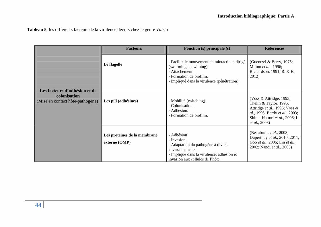
Tableau 5: Les differents facteurs de la virulence décrits chez le genre Vibrio ..................... 44
Tableau 6: Familles des protéases classées selon la nature de leur site actif .......................... 54
Tableau 7: Protéases produites par le genre Vibrio................................................................. 56
Tableau 8: Rôle biologiques de métalloprotéases décrites chez le genre Vibrio .................... 63
Tableau 9: Différents sidérophores décrits au sein du genre Vibrio ....................................... 73
Tableau 10:Récapitulatif des différentes techniques utilisées en infections expérimentales
chez C. gigas par V. aestuarianus, V. splendidus, ................................................................... 84
Liste des unités et des principales abréviations
aa
acide aminé
ABC ATP-Binding Cassette
ARN 16S L'ARN ribosomique 16S
ATP Adénosine Tri-Phosphate
Caco-2 Colon carcinoma cell line
CHO Chinese hamster ovary
CGH Hybridation Génomique Comparative
Da Dalton
DL50 Dose létale 50
ECF ExtraCytoplasmic Function
ECPs Extracellular products/ produits extracellulaires
ELISA Enzyme-linked immunosorbent assay
EMSF Eau de mer sterile filtree
FAO Food and Agriculture Organization
Fur Ferric Uptake Repressor
g gramme
GP-HPLC Gel Permeation-High Performance Liquid Chromatography
gyrB Gyrase B
H Heure/ Hour
HeLa Henrietta Lacks (Human cervical carcinoma)
kDa Kilo Dalton
ml millilitre
LBS Luria Bertani broth salé
LBSA Luria Bertani agar salé
LPS Lipopolysaccharides
LS MS-MS Liquid chromatography coupled to tandem mass spectrometry
MA Marine Agar
MB Marine Broth
MLSA MultiLocus Sequence Analysis
MMP Métalloprotéase matricielle
NIP NRPS Independent Pathway
NRPS Nonribosomal Peptide Synthetase
OMP Outer membrane protein
PCR Polymerase Chain Reaction
PFTs Les toxines formant des pores
QPCR Quantitative polymerase chain reaction
QS Quorum sensing

REPAMO REseau de PAthologie des MOllusques
ROS Espèces réactives de l'oxygène
RTX Repeats in toxin
SOD Superoxyde dismutase
SSIII Système de sécrétion de type trois
TIMPs Tissue Inhibitors of Matrix Metalloproteases
Cfu (cfu) Colony-forming unit
V Vibrio
VBNC Viable But Non-Culturable
Vero Verda Reno

18
CONTEXTE GENERAL ET
OBJECTIFS DE L’ETUDE
Contexte géneral et objectifs de l'étude
19
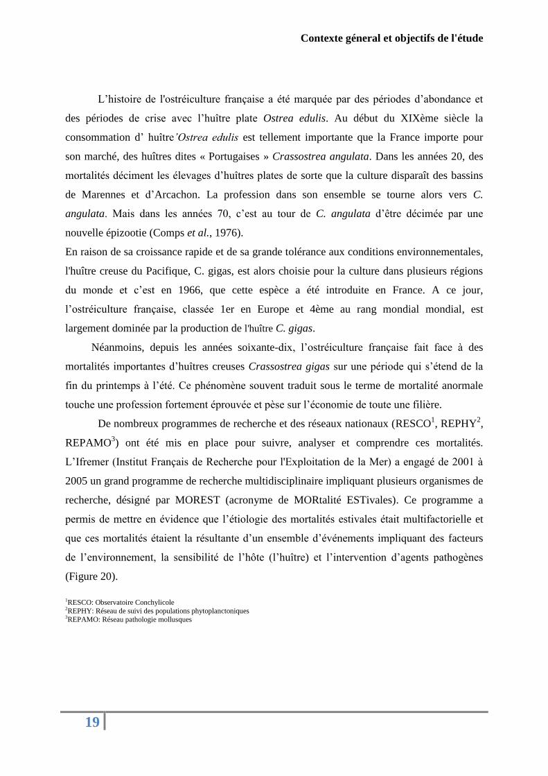
L’histoire de l'ostréiculture française a été marquée par des périodes d’abondance et
des périodes de crise avec l’huître plate Ostrea edulis. Au début du XIXème siècle la
consommation d’ huître’Ostrea edulis est tellement importante que la France importe pour
son marché, des huîtres dites « Portugaises » Crassostrea angulata. Dans les années 20, des
mortalités déciment les élevages d’huîtres plates de sorte que la culture disparaît des bassins
de Marennes et d’Arcachon. La profession dans son ensemble se tourne alors vers C.
angulata. Mais dans les années 70, c’est au tour de C. angulata d’être décimée par une
nouvelle épizootie (Comps et al., 1976).
En raison de sa croissance rapide et de sa grande tolérance aux conditions environnementales,
l'huître creuse du Pacifique, C. gigas, est alors choisie pour la culture dans plusieurs régions
du monde et c’est en 1966, que cette espèce a été introduite en France. A ce jour,
l’ostréiculture française, classée 1er en Europe et 4ème au rang mondial mondial, est
largement dominée par la production de l'huître C. gigas.
Néanmoins, depuis les années soixante-dix, l’ostréiculture française fait face à des
mortalités importantes d’huîtres creuses Crassostrea gigas sur une période qui s’étend de la
fin du printemps à l’été. Ce phénomène souvent traduit sous le terme de mortalité anormale
touche une profession fortement éprouvée et pèse sur l’économie de toute une filière.
De nombreux programmes de recherche et des réseaux nationaux (RESCO1, REPHY
2,
REPAMO3) ont été mis en place pour suivre, analyser et comprendre ces mortalités.
L’Ifremer (Institut Français de Recherche pour l'Exploitation de la Mer) a engagé de 2001 à
2005 un grand programme de recherche multidisciplinaire impliquant plusieurs organismes de
recherche, désigné par MOREST (acronyme de MORtalité ESTivales). Ce programme a
permis de mettre en évidence que l’étiologie des mortalités estivales était multifactorielle et
que ces mortalités étaient la résultante d’un ensemble d’événements impliquant des facteurs
de l’environnement, la sensibilité de l’hôte (l’huître) et l’intervention d’agents pathogènes
(Figure 20).
1RESCO: Observatoire Conchylicole
2REPHY: Réseau de suivi des populations phytoplanctoniques 3REPAMO: Réseau pathologie mollusques
Contexte géneral et objectifs de l'étude
20
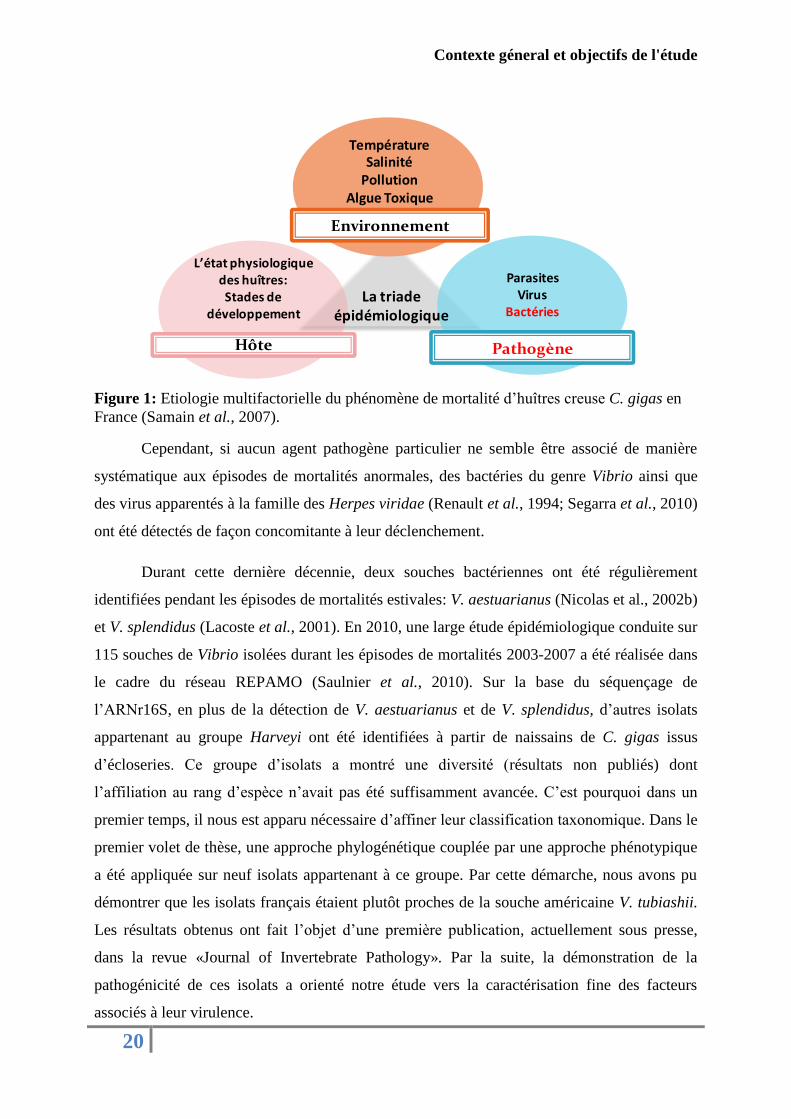
Figure 1: Etiologie multifactorielle du phénomène de mortalité d’huîtres creuse C. gigas en
France (Samain et al., 2007).
Cependant, si aucun agent pathogène particulier ne semble être associé de manière
systématique aux épisodes de mortalités anormales, des bactéries du genre Vibrio ainsi que
des virus apparentés à la famille des Herpes viridae (Renault et al., 1994; Segarra et al., 2010)
ont été détectés de façon concomitante à leur déclenchement.
Durant cette dernière décennie, deux souches bactériennes ont été régulièrement
identifiées pendant les épisodes de mortalités estivales: V. aestuarianus (Nicolas et al., 2002b)
et V. splendidus (Lacoste et al., 2001). En 2010, une large étude épidémiologique conduite sur
115 souches de Vibrio isolées durant les épisodes de mortalités 2003-2007 a été réalisée dans
le cadre du réseau REPAMO (Saulnier et al., 2010). Sur la base du séquençage de
l’ARNr16S, en plus de la détection de V. aestuarianus et de V. splendidus, d’autres isolats
appartenant au groupe Harveyi ont été identifiées à partir de naissains de C. gigas issus
d’écloseries. Ce groupe d’isolats a montré une diversité (résultats non publiés) dont
l’affiliation au rang d’espèce n’avait pas été suffisamment avancée. C’est pourquoi dans un
premier temps, il nous est apparu nécessaire d’affiner leur classification taxonomique. Dans le
premier volet de thèse, une approche phylogénétique couplée par une approche phénotypique
a été appliquée sur neuf isolats appartenant à ce groupe. Par cette démarche, nous avons pu
démontrer que les isolats français étaient plutôt proches de la souche américaine V. tubiashii.
Les résultats obtenus ont fait l’objet d’une première publication, actuellement sous presse,
dans la revue «Journal of Invertebrate Pathology». Par la suite, la démonstration de la
pathogénicité de ces isolats a orienté notre étude vers la caractérisation fine des facteurs
associés à leur virulence.
Hôte Pathogène
Environnement
La triade épidémiologique
ParasitesVirus
Bactéries
TempératureSalinité
PollutionAlgue Toxique
L’état physiologique des huîtres:Stades de
développement
Contexte géneral et objectifs de l'étude
21
Pour réaliser cela, la souche française V. tubiashii (07/118 T2) induisant plus de 60%
de mortalité après 24 h d’injection et présentant un phénotype métalloprotéase marqué a été
sélectionnée comme souche modèle pour la suite du travail. La caractérisation biochimique et
l’évaluation in vitro de la toxicité des produits extracellulaires (ECPs) issus de la souche
modèle 07/118 T2 a été conduite sur les hémocytes de C. gigas. L’ensemble des résultats
obtenus sont regroupés dans un second article, en cours de révision, dans la revue «Journal of
Invertebrate Pathology»
Un deuxième volet a consisté en l’étude de l’effet in vitro des conditions de culture
bactérienne sur la toxicité et la composition des ECPs de la souche modèle (07/118 T2). Les
principaux résultats ont été regroupés dans une troisième publication qui sera soumise dans le
journal «Microbiology».
Dans un dernier volet, il est apparu essentiel de caractériser les facteurs de virulence
potentiellement impliqués dans la pathogénicité de la souche française V. tubiashii. Dans cet
objectif, le stade larvaire de C. gigas a été utilisé comme modèle d’interaction par le biais
d’infections expérimentales, d’observations microscopiques, d’analyses protéomiques et
transcriptomiques. Les résultats obtenus ont fait l’objet d’un quatrième article qui sera soumis
prochainement dans le journal «Microbiology».

22
INTRODUCTION
BIBLIOGRAPHIQUE
23
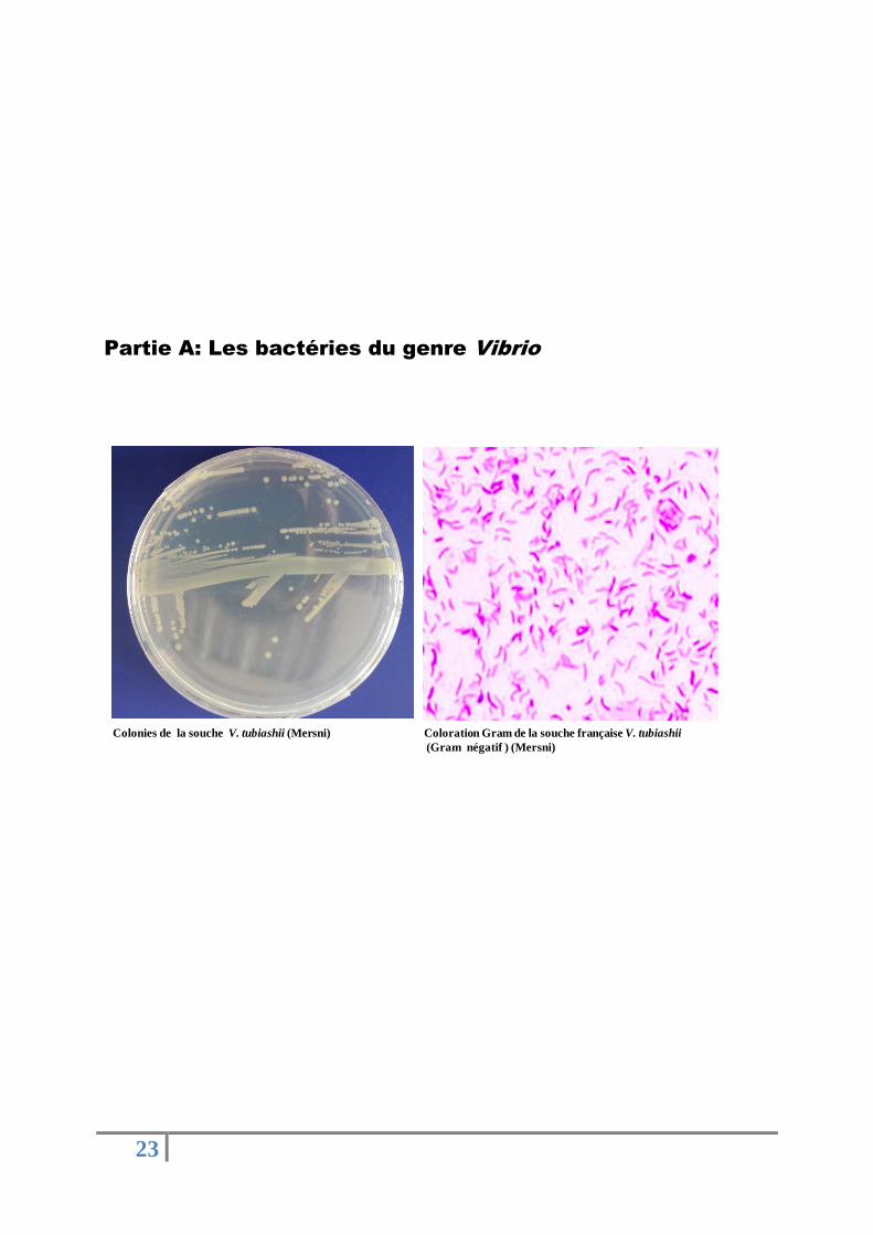
Partie A: Les bactéries du genre Vibrio
Coloration Gram de la souche française V. tubiashii
(Gram négatif ) (Mersni)
Colonies de la souche V. tubiashii (Mersni)
Introduction bibliographique: Partie A
24
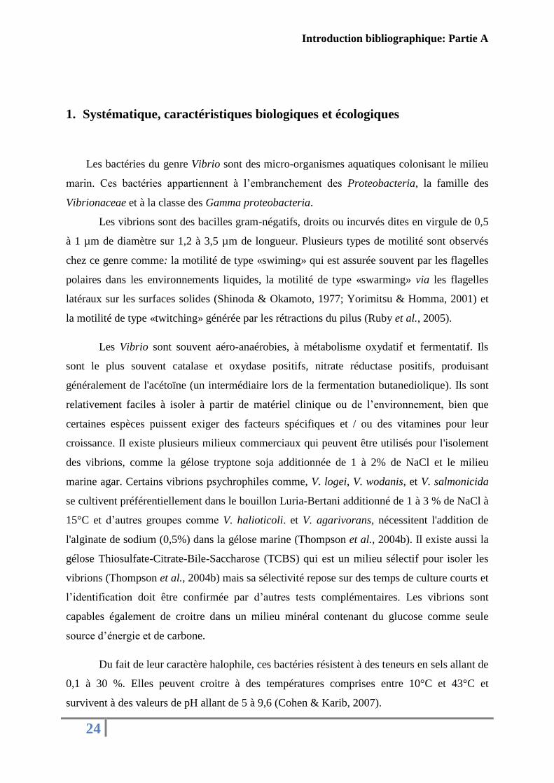
1. Systématique, caractéristiques biologiques et écologiques
Les bactéries du genre Vibrio sont des micro-organismes aquatiques colonisant le milieu
marin. Ces bactéries appartiennent à l’embranchement des Proteobacteria, la famille des
Vibrionaceae et à la classe des Gamma proteobacteria.
Les vibrions sont des bacilles gram-négatifs, droits ou incurvés dites en virgule de 0,5
à 1 µm de diamètre sur 1,2 à 3,5 µm de longueur. Plusieurs types de motilité sont observés
chez ce genre comme: la motilité de type «swiming» qui est assurée souvent par les flagelles
polaires dans les environnements liquides, la motilité de type «swarming» via les flagelles
latéraux sur les surfaces solides (Shinoda & Okamoto, 1977; Yorimitsu & Homma, 2001) et
la motilité de type «twitching» générée par les rétractions du pilus (Ruby et al., 2005).
Les Vibrio sont souvent aéro-anaérobies, à métabolisme oxydatif et fermentatif. Ils
sont le plus souvent catalase et oxydase positifs, nitrate réductase positifs, produisant
généralement de l'acétoïne (un intermédiaire lors de la fermentation butanediolique). Ils sont
relativement faciles à isoler à partir de matériel clinique ou de l’environnement, bien que
certaines espèces puissent exiger des facteurs spécifiques et / ou des vitamines pour leur
croissance. Il existe plusieurs milieux commerciaux qui peuvent être utilisés pour l'isolement
des vibrions, comme la gélose tryptone soja additionnée de 1 à 2% de NaCl et le milieu
marine agar. Certains vibrions psychrophiles comme, V. logei, V. wodanis, et V. salmonicida
se cultivent préférentiellement dans le bouillon Luria-Bertani additionné de 1 à 3 % de NaCl à
15°C et d’autres groupes comme V. halioticoli. et V. agarivorans, nécessitent l'addition de
l'alginate de sodium (0,5%) dans la gélose marine (Thompson et al., 2004b). Il existe aussi la
gélose Thiosulfate-Citrate-Bile-Saccharose (TCBS) qui est un milieu sélectif pour isoler les
vibrions (Thompson et al., 2004b) mais sa sélectivité repose sur des temps de culture courts et
l’identification doit être confirmée par d’autres tests complémentaires. Les vibrions sont
capables également de croitre dans un milieu minéral contenant du glucose comme seule
source d’énergie et de carbone.
Du fait de leur caractère halophile, ces bactéries résistent à des teneurs en sels allant de
0,1 à 30 %. Elles peuvent croitre à des températures comprises entre 10°C et 43°C et
survivent à des valeurs de pH allant de 5 à 9,6 (Cohen & Karib, 2007).
Introduction bibliographique: Partie A
25
Généralement, ces bactéries peuvent coloniser de nombreux habitats, sous forme libre
planctonique et biofilms ou être associées à des hôtes avec lesquels elles peuvent entretenir
des relations du type symbiotique, commensal ou pathogène. Parmi les habitats, on cite les
sédiments, l’eau de mer, les estuaires, les infrastructures aquacoles et notamment les
organismes marins comme les poissons, les mollusques, les crustacés, les éponges, les coraux,
les algues et les zooplanctons (Austin, 2010; Cohen & Karib, 2007).
2. Historique et taxonomie
La première description d’une bactérie du genre Vibrio, a été réalisée par le médecin
italien Filipo Pacini en 1854 et puis en 1883 par Robert Koch, qui ont contribué à la
découverte du rôle de V. cholerae dans le choléra. Jusqu'à la moitié du vingtième siècle,
l'identification des vibrions a été basée sur quelques descriptions phénotypiques comme la
morphologie (flagelle, incurvation de la cellule…) et la croissance dans certains milieux de
culture.
Ces simples outils d’identification, ont abouti à une hétérogénéité de classification au
sein du genre Vibrio. Davis et Park ont montré que 25 souches classées auparavant dans le
genre Vibrio pouvaient être classées dans trois genres différents (Davis & Park, 1962). À la
fin des années soixante, Colwell et Sparks (1967) ont mis en avant le concept de la taxonomie
polyphasique qui se base sur des analyses morphologiques, physiologiques, biochimiques et la
taxonomie numérique (taxonomie assistée par ordinateur). Cette approche utilisée jusqu'à
aujourd’hui a permis de marquer et révolutionner l’histoire de la taxonomie bactérienne.
Durant les années 90, quelques tests phénotypiques reposent sur l'utilisation de différentes
sources de carbone, la mise en évidence de certaines activités enzymatiques, la tolérance au
sel, la croissance à différentes températures, la résistance à des antibiotiques et la composition
en GC du génome ont permis une meilleure identification des bactéries appartenant au genre
Vibrio (Alsina & Blanch, 1994a, 1994b).
L’approche phénotypique seule est insuffisante pour identifier une nouvelle espèce
d’où la nécessité d’introduire des approches de génotypage moléculaire. Le séquençage du
gène codant l’ARNr 16S est devenu un des principaux outils de la taxonomie bactérienne.
Pourtant pour la classification des Vibrio, cette technique trouve ses limites pour différencier
les espèces au sein du même groupe comme le cas pour du groupe V.splendidus (Le Roux et
Introduction bibliographique: Partie A
26
al., 2004) et pour distinguer entre V. brasiliensis et V. tubiashii, V. coralliilyticus et V.
neptunius, V. anguillarum et V. ordalii (Thompson et al., 2004a). En plus, cette technique
reste incomplète pour décrire une nouvelle espèce. Lorsque la similarité des séquences est
proche ou au-dessus de 97%, d’autres méthodes d’identification telles que l’hybridation
ADN-ADN (DDH) doivent être utilisées (Stackebrandt & Goebel, 1994).
L’hybridation ADN-ADN reste l’outil le plus puissant pour identifier sans ambiguïté
les procaryotes, offrant pour la première fois, un moyen fiable pour bien classer le monde
bactérien. En utilisant cette technique couplée à des analyses phylogéniques et biochimiques,
Ben–Haim et collaborateurs (2003) ont reclassé la souche V. tubiashii LMG 10953 (Hada et
al., 1984) comme V. coralliilyticus. Cependant, cette technique souffre de diverses
limitations, y compris la nécessité d'inclure des souches de référence à chaque nouvelle
expérience et son utilisation reste limitée à certains laboratoires de référence (Thompson et
al., 2009). D’après les règles de classification, une espèce bactérienne est considérée
appartenant au sein d’un groupe (y compris la souche de type) lorsque elle présente une DDH
similitude > 70%, un Tm (température de dénaturation <5 °C, une % de G+ C <5% et ARNr
16S similarité > 97% (Stackebrandt & Goebel, 1994; Thompson et al., 2009).
En plus, le séquençage du génome complet des différentes bactéries a facilité la
comparaison inter et intra-espèces, le regroupement des gènes orthologues et paralogues et la
comparaison de l'ordre des gènes (Winsor et al., 2009). Il est intéressant aussi de savoir que
l'hybridation génomique comparative (CGH), qui est utilisé en clinique, peut être appliquée
dans le typage de souches bactériennes (Lin et al., 2010a).
Aujourd’hui, l’analyse de séquences de plusieurs gènes de ménage (MLSA:
Multilocus Sequence Anlaysis) a permis d’affiner la classification du genre Vibrio. En
utilisant cet outil, Thompson et collaborateurs (2005) ont montré que V. harveyi et V.
campbellii forment deux groupes distincts et Hoffman et collaborateurs (2012) ont prouvé que
V. communis et V. owensii appartiennent à la même espèce.
Il semble difficile de présenter une classification précise des bactéries appartenant au
genre Vibrio, groupe qui subit régulièrement des modifications importantes et qui ne cesse
d’évoluer. Jusqu'à maintenant 115 espèces et 2 sous-espèces ont été identifiées (J.P Euzeby:
List of Prokaryotic Names with Standing in Nomenclature
[http://www.bacterio.cict.fr/uw/vibrio.html]).
Introduction bibliographique: Partie A
27
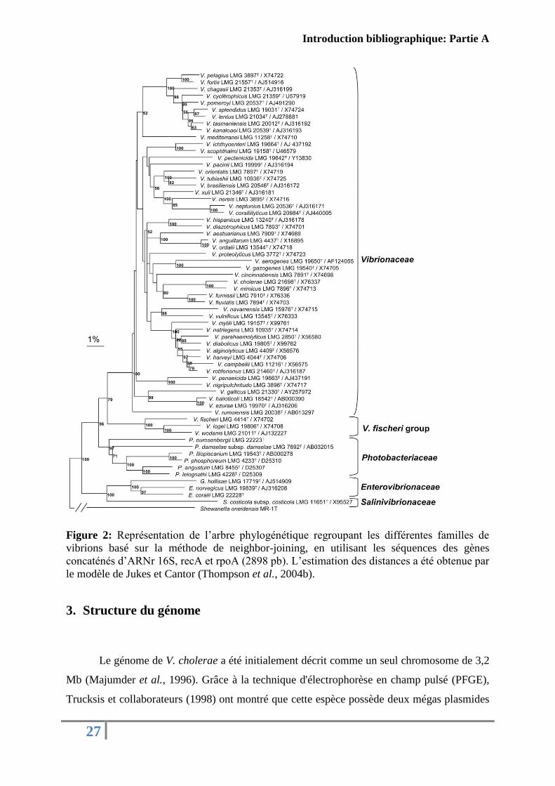
Figure 2: Représentation de l’arbre phylogénétique regroupant les différentes familles de
vibrions basé sur la méthode de neighbor-joining, en utilisant les séquences des gènes
concaténés d’ARNr 16S, recA et rpoA (2898 pb). L’estimation des distances a été obtenue par
le modèle de Jukes et Cantor (Thompson et al., 2004b).
3. Structure du génome
Le génome de V. cholerae a été initialement décrit comme un seul chromosome de 3,2
Mb (Majumder et al., 1996). Grâce à la technique d'électrophorèse en champ pulsé (PFGE),
Trucksis et collaborateurs (1998) ont montré que cette espèce possède deux mégas plasmides
Introduction bibliographique: Partie A
28
séparés. Depuis, toutes les études de séquençage de génomes complets (Schoolnik & Yildiz,
2000; Chen et al., 2003; Makino et al., 2003; Ruby et al., 2005) ainsi que les analyses en
PFGE (Okada et al., 2005) de différentes espèces de Vibrio ont confirmé que cette
organisation structurale était une caractéristique commune des Vibrionaceae. Ainsi, le
génome des Vibrio est composé de deux chromosomes circulaires, un chromosome I, de taille
plus importante que le chromosome II (V. cholerae, 2,4 Mb pour le chromosome I et 1,5 Mb
pour le chromosome II (Trucksis et al., 1998))
Généralement, le chromosome I, contient les gènes codant pour les principales
fonctions vitales de la bactérie: la réplication et la réparation de l’ADN, la transcription, la
traduction, la biosynthèse de la membrane cellulaire, des gènes impliqués dans des voies
centrales cataboliques ou de biosynthèse et les gènes connus pour être essentiels dans la
pathogénicité bactérienne (les gènes codant les toxines, lipopolysacsaccharides et toute la
machinerie de sécrétion des protéines extracellulaires). Contrairement au chromosome I, le
chromosome II contient une proportion importante de gènes hypothétiques et des gènes à
fonctions inconnues (Heidelberg et al., 2000).
4. Espèces pathogène du genre Vibrio
4.1. Les vibrions pathogènes pour l’Homme
V. cholerae, V. parahaemolyticus et V. vulnificus sont les espèces les plus pathogènes
pour l’Homme et à l’origine des plus grands risques en termes de santé publique.
V. cholerae, l’espèce type du genre Vibrio, est l’agent causal du choléra. Cette maladie
infectieuse à caractère le plus souvent épidémique se manifeste par une violente diarrhée
accompagnée de vomissements après une incubation variant de quelques heures à quelques
jours. Cette espèce est subdivisée en 200 sérogroupes établis à partir de l’antigène O (un
constituant du complexe lipo-saccharidique situé dans la membrane externe de bactéries).
Seuls les sérogroupes O1 et O139 sont à l’origine des pandémies cholériques (Reidl & Klose,
2002). Cette souche qui infecte l’hôte à travers l’eau et/ou une alimentation contaminée
s’adhère au niveau des intestins et produit une entérotoxine désignée par «cholera toxin» (CT)
provoquant une diarrhée intense qui peut conduire à la mort. Cependant, dans
Introduction bibliographique: Partie A
29
l’environnement V. cholerae ne présente aucun effet sur les organismes marins (Reidl &
Klose, 2002; Thompson et al., 2004b).
Depuis 2000, l’incidence du choléra a augmenté régulièrement. Entre 2003 et 2004,
l'organisation mondiale de la santé (OMS) a signalée 15 000 nouveaux cas de choléra
seulement en Afrique, principalement au Mali, au Mozambique et en Zambie. En octobre
2010, des cas de choléra ont été rapportés en Haïti pour la première fois depuis plus de cent
ans. La recherche a révélé qu’il s’agit du V. cholerae sérogroup O1, biotype Ogawa. Jusqu’en
juillet 2011, plus de 220 000 cas avaient été enregistrés, dont 5 968 décès (Ceccarelli et al.,
2011; Pfrimmer, 2010; Tappero & Tauxe, 2011).
V. parahaemolyticus cause de sévères gastroentérites à caractère parfois pandémique
chez l'Homme. Il existe 13 sérogroupes de type O et 71 sérogroupes de type K. Il a été décrit
que le pouvoir pathogène de cette bactérie est lié à la présence de l’hémolysine TDH
(Thermostable Direct Hemolysin) et l’hémolysine TRH (Tdh-Related Hemolysin) produites
dans le tube digestif. Ces deux toxines présentent des activités lytiques, cytotoxiques et
entérotoxiques (Nishibuchi & Kaper, 1995; Okuda et al., 1997; Thompson et al., 2004b). Le
séquençage du génome complet de V. parahaemolyticus (Makino et al., 2003) a révélé la
présence du système de sécrétion de type III (SSTT), qui est essentiel pour la pathogénicité de
plusieurs bactéries pathogènes telles que Salmonella, Shigella et Yersinia mais absent chez V.
cholerae (Thompson et al., 2004b). Cette souche est largement distribuée en milieu marin
côtier; elle a été décelée dans l'eau de mer et les sédiments et fait partie de la flore normale
des produits de la mer.
La première épidémie confirmée est survenue au Japon en 1950. 272 malades et 20
morts ont été recensés après la consommation de jeune sardines mi-séchées (Nair et al.,
2007). Depuis, cette bactérie, particulièrement le sérotype O3: K6, a été détectée dans
plusieurs endroits dans le monde où la consommation de poissons et fruits de mer crus est
importante comme au Chili, en France, au Japon, en Corée, en Espagne, à Taïwan, aux États-
Unis, et particulièrement dans les pays d'Extrême-Orient comme l'Inde, le Bangladesh et la
Thaïlande (Nair et al., 2007).
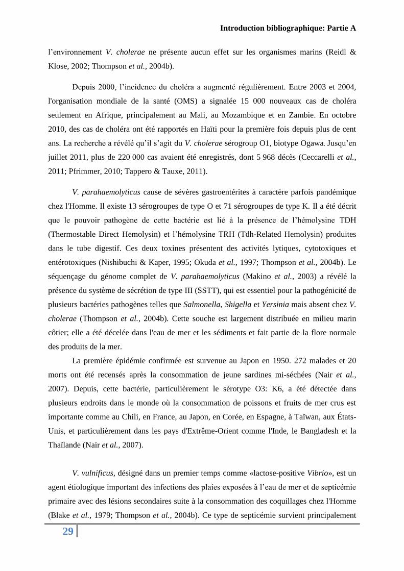
V. vulnificus, désigné dans un premier temps comme «lactose-positive Vibrio», est un
agent étiologique important des infections des plaies exposées à l’eau de mer et de septicémie
primaire avec des lésions secondaires suite à la consommation des coquillages chez l'Homme
(Blake et al., 1979; Thompson et al., 2004b). Ce type de septicémie survient principalement
Introduction bibliographique: Partie A
30
chez les personnes immunodéprimées ou souffrant de désordres hépatiques (alcoolisme ou
surcharge en fer). Le taux de mortalité chez ces malades est de l'ordre de 50 % mais il peut
atteindre 90 % chez des patients souffrant d'hypotension. Chez les immunocompétents,
uniquement des gastro-entérites, sans septicémie, sont observées (Miossec, 2002). Deux
biotypes ont été définis pour V. vulnificus, Biotype 1 et 2, classés selon des critères
phénotypiques et selon l’espèce de l’hôte (Tison et al., 1982). Quelques facteurs de virulence
ont été caractérisés chez cette espèce comme: la présence de polysaccharide capsulaire (CPS),
qui confère à la bactérie une résistance à la phagocytose et aux protéines de compléments
(Miyoshi et al., 2002), des chélateurs du fer comme les sidérophores et la production des
exotoxines (Amaro & Biosca, 1996).
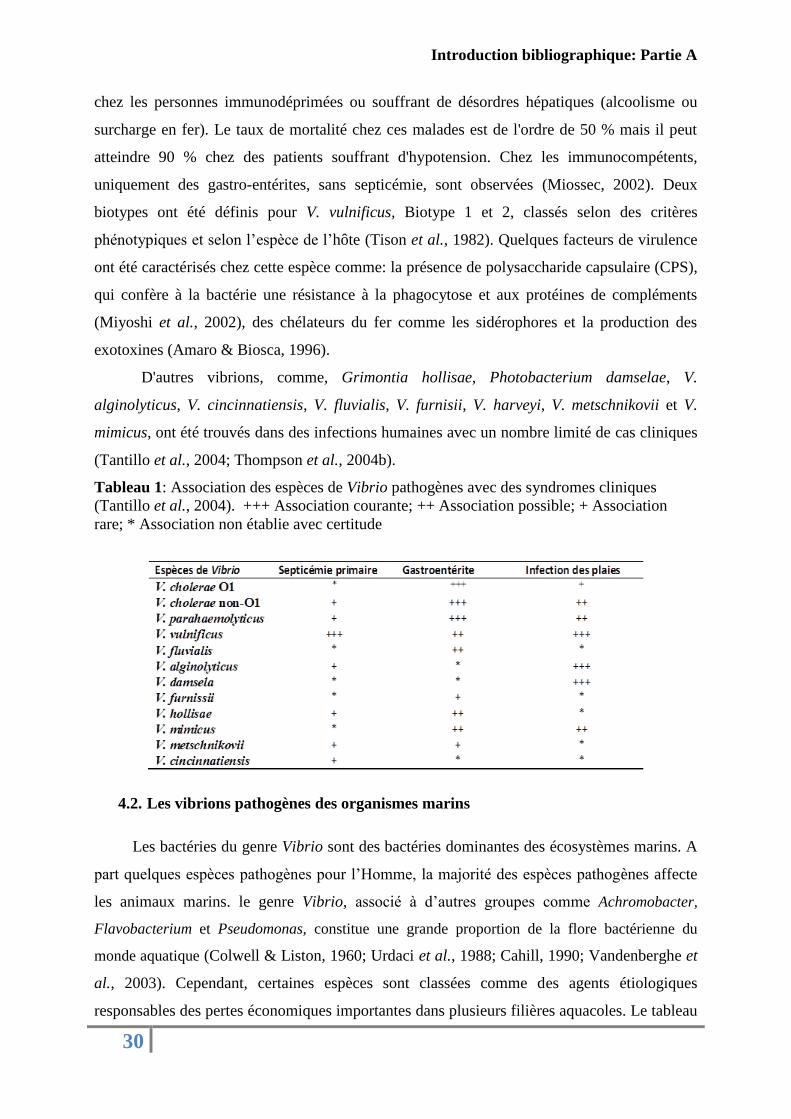
D'autres vibrions, comme, Grimontia hollisae, Photobacterium damselae, V.
alginolyticus, V. cincinnatiensis, V. fluvialis, V. furnisii, V. harveyi, V. metschnikovii et V.
mimicus, ont été trouvés dans des infections humaines avec un nombre limité de cas cliniques
(Tantillo et al., 2004; Thompson et al., 2004b).
Tableau 1: Association des espèces de Vibrio pathogènes avec des syndromes cliniques
(Tantillo et al., 2004). +++ Association courante; ++ Association possible; + Association
rare; * Association non établie avec certitude
4.2. Les vibrions pathogènes des organismes marins
Les bactéries du genre Vibrio sont des bactéries dominantes des écosystèmes marins. A
part quelques espèces pathogènes pour l’Homme, la majorité des espèces pathogènes affecte
les animaux marins. le genre Vibrio, associé à d’autres groupes comme Achromobacter,
Flavobacterium et Pseudomonas, constitue une grande proportion de la flore bactérienne du
monde aquatique (Colwell & Liston, 1960; Urdaci et al., 1988; Cahill, 1990; Vandenberghe et
al., 2003). Cependant, certaines espèces sont classées comme des agents étiologiques
responsables des pertes économiques importantes dans plusieurs filières aquacoles. Le tableau
Introduction bibliographique: Partie A
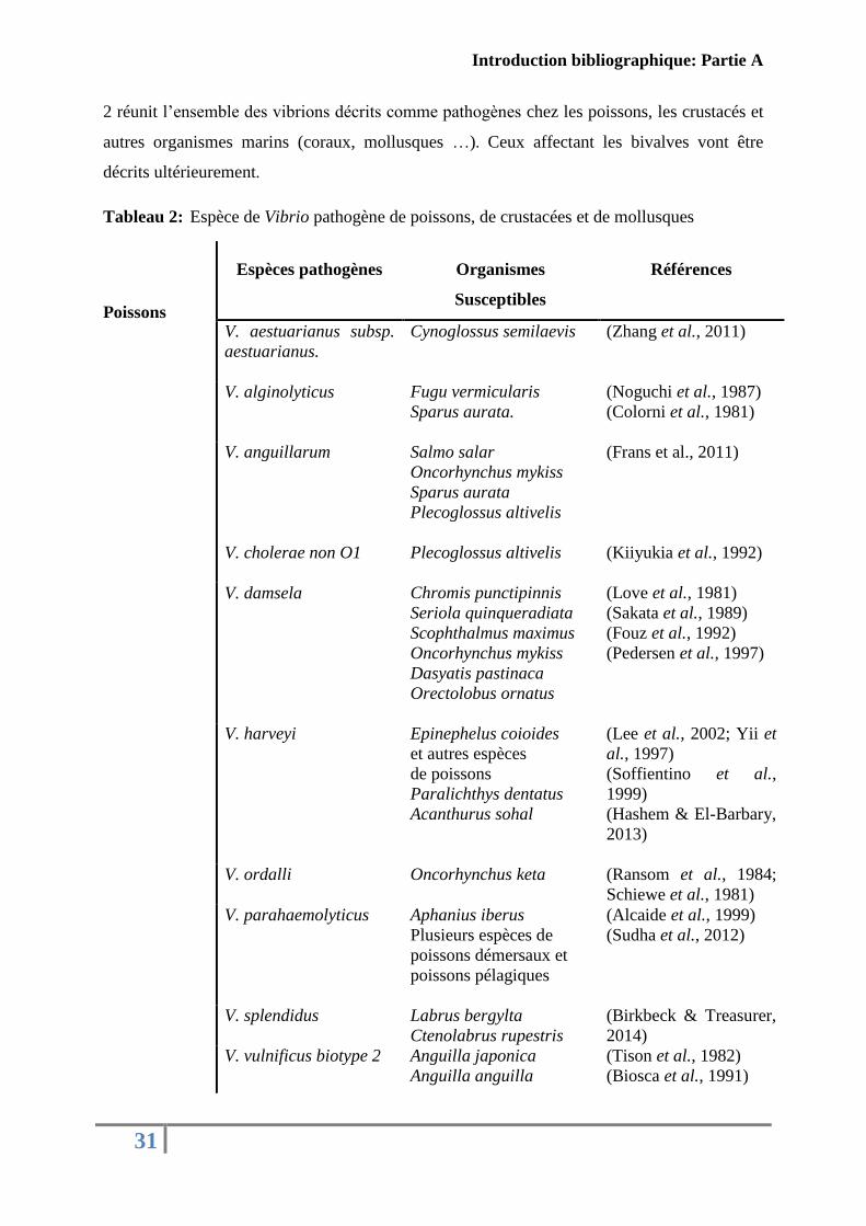
31
2 réunit l’ensemble des vibrions décrits comme pathogènes chez les poissons, les crustacés et
autres organismes marins (coraux, mollusques …). Ceux affectant les bivalves vont être
décrits ultérieurement.
Tableau 2: Espèce de Vibrio pathogène de poissons, de crustacées et de mollusques
Poissons
Espèces pathogènes Organismes
Susceptibles
Références
V. aestuarianus subsp.
aestuarianus.
Cynoglossus semilaevis (Zhang et al., 2011)
V. alginolyticus Fugu vermicularis
Sparus aurata.
(Noguchi et al., 1987)
(Colorni et al., 1981)
V. anguillarum Salmo salar
Oncorhynchus mykiss
Sparus aurata
Plecoglossus altivelis
(Frans et al., 2011)
V. cholerae non O1 Plecoglossus altivelis
(Kiiyukia et al., 1992)
V. damsela Chromis punctipinnis
Seriola quinqueradiata
Scophthalmus maximus
Oncorhynchus mykiss
Dasyatis pastinaca
Orectolobus ornatus
(Love et al., 1981)
(Sakata et al., 1989)
(Fouz et al., 1992)
(Pedersen et al., 1997)
V. harveyi Epinephelus coioides
et autres espèces
de poissons
Paralichthys dentatus
Acanthurus sohal
(Lee et al., 2002; Yii et
al., 1997)
(Soffientino et al.,
1999)
(Hashem & El-Barbary,
2013)
V. ordalli Oncorhynchus keta (Ransom et al., 1984;
Schiewe et al., 1981)
V. parahaemolyticus Aphanius iberus
Plusieurs espèces de
poissons démersaux et
poissons pélagiques
(Alcaide et al., 1999)
(Sudha et al., 2012)
V. splendidus Labrus bergylta
Ctenolabrus rupestris
(Birkbeck & Treasurer,
2014)
V. vulnificus biotype 2 Anguilla japonica
Anguilla anguilla
(Tison et al., 1982)
(Biosca et al., 1991)
Introduction bibliographique: Partie A
32
Crustacés V. alginolyticus Litopenaeus vannamei
Charybdis japonica
(Liu et al., 2004)
(Xu et al., 2013)
V. harveyi Peneaus monodon
Litopenaeus vannamei
Penaeus monodon
Litopenaeus stylirostris
(Lavilla-Pitogo et al.,
1990)
(Soto-Rodriguez et al.,
2010)
(Alavandi et al., 2008)
(Goarant et al., 2006)
(Cano-Gómez et al.,
2010)
V. metschnikovii et
V. fluvialis
V. nigripulchritudo
V. owensii
Panulirus ornatus
Penaeus monodon
V. penaeicida Penaeus stylirostris (Costa et al., 1998)
V. parahaemolyticus Penaeus monodon
(Alapide-Tendencia &
Dureza, 1997)
Mollusques
V. coralliilyticus
V. harveyi
Pocillopora damicornis
Scleractinian
(Ben-Haim &
Rosenberg, 2002; Ben-
Haim et al., 2003)
(Weil et al., 2006)
V. lentus
V. alginolyticus
Octopus vulgaris
Haliotis diversicolor
supertexta
(Farto et al., 2003)
(Liu et al., 2001)
V. harveyi Haliotis tuberculata
Haliotis diversiclor
Pinctada maxima
(Nicolas et al., 2002a;
Travers et al., 2008)
(Jiang et al., 2013)
(Pass et al., 1987)
Vibrio shiloi
Oculina patagonica
(Kushmaro et al., 2001)
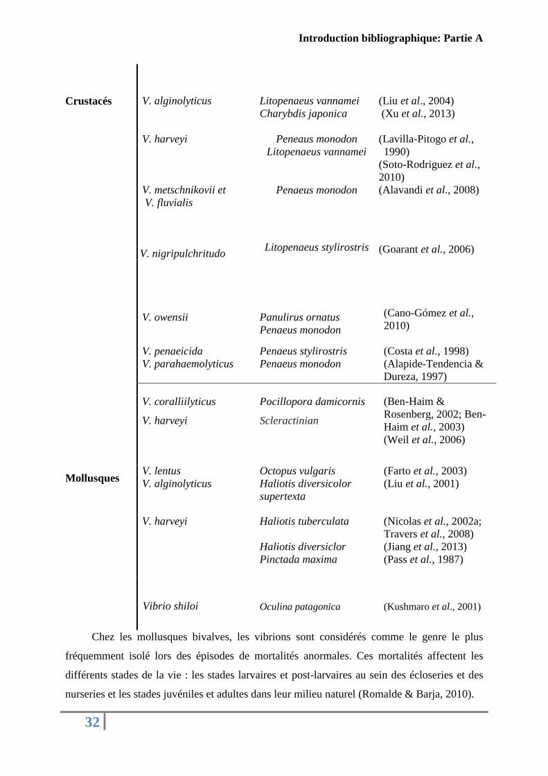
Chez les mollusques bivalves, les vibrions sont considérés comme le genre le plus
fréquemment isolé lors des épisodes de mortalités anormales. Ces mortalités affectent les
différents stades de la vie : les stades larvaires et post-larvaires au sein des écloseries et des
nurseries et les stades juvéniles et adultes dans leur milieu naturel (Romalde & Barja, 2010).
Introduction bibliographique: Partie A
33
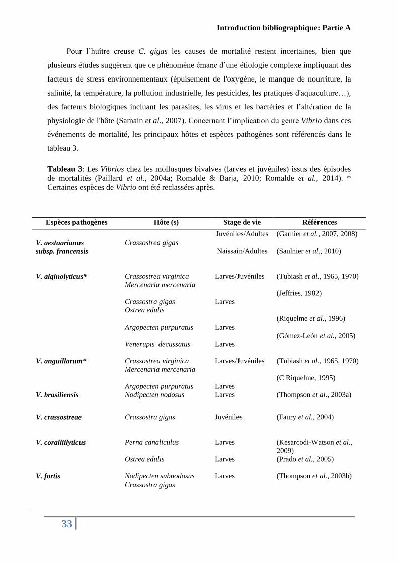
Pour l’huître creuse C. gigas les causes de mortalité restent incertaines, bien que
plusieurs études suggèrent que ce phénomène émane d’une étiologie complexe impliquant des
facteurs de stress environnementaux (épuisement de l'oxygène, le manque de nourriture, la
salinité, la température, la pollution industrielle, les pesticides, les pratiques d'aquaculture…),
des facteurs biologiques incluant les parasites, les virus et les bactéries et l’altération de la
physiologie de l'hôte (Samain et al., 2007). Concernant l’implication du genre Vibrio dans ces
événements de mortalité, les principaux hôtes et espèces pathogènes sont référencés dans le
tableau 3.
Tableau 3: Les Vibrios chez les mollusques bivalves (larves et juvéniles) issus des épisodes
de mortalités (Paillard et al., 2004a; Romalde & Barja, 2010; Romalde et al., 2014). *
Certaines espèces de Vibrio ont été reclassées après.
Espèces pathogènes Hôte (s) Stage de vie Références
V. aestuarianus
subsp. francensis
Crassostrea gigas
Juvéniles/Adultes
Naissain/Adultes
(Garnier et al., 2007, 2008)
(Saulnier et al., 2010)
V. alginolyticus* Crassostrea virginica
Mercenaria mercenaria
Crassostra gigas
Ostrea edulis
Argopecten purpuratus
Venerupis decussatus
Larves/Juvéniles
Larves
Larves
Larves
(Tubiash et al., 1965, 1970)
(Jeffries, 1982)
(Riquelme et al., 1996)
(Gómez-León et al., 2005)
V. anguillarum* Crassostrea virginica
Mercenaria mercenaria
Argopecten purpuratus
Larves/Juvéniles
Larves
(Tubiash et al., 1965, 1970)
(C Riquelme, 1995)
V. brasiliensis Nodipecten nodosus Larves (Thompson et al., 2003a)
V. crassostreae
Crassostra gigas
Juvéniles
(Faury et al., 2004)
V. coralliilyticus
Perna canaliculus
Ostrea edulis
Larves
Larves
(Kesarcodi-Watson et al.,
2009)
(Prado et al., 2005)
V. fortis Nodipecten subnodosus
Crassostra gigas
Larves (Thompson et al., 2003b)
Introduction bibliographique: Partie A
34
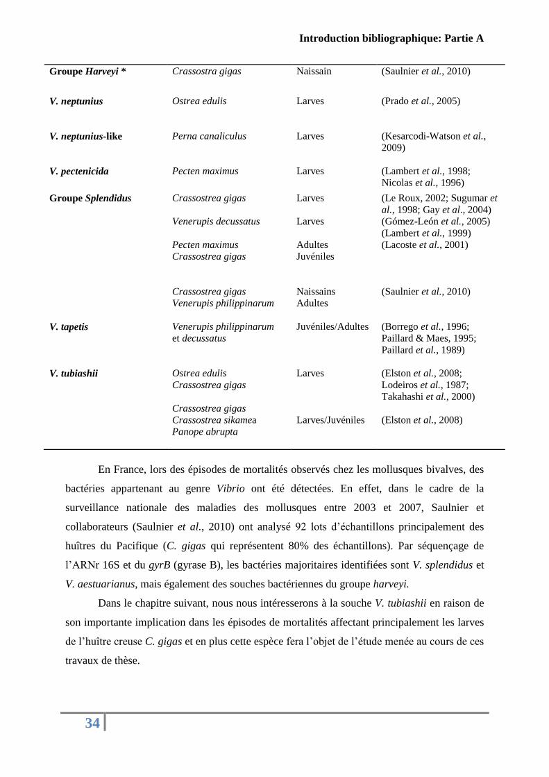
En France, lors des épisodes de mortalités observés chez les mollusques bivalves, des
bactéries appartenant au genre Vibrio ont été détectées. En effet, dans le cadre de la
surveillance nationale des maladies des mollusques entre 2003 et 2007, Saulnier et
collaborateurs (Saulnier et al., 2010) ont analysé 92 lots d’échantillons principalement des
huîtres du Pacifique (C. gigas qui représentent 80% des échantillons). Par séquençage de
l’ARNr 16S et du gyrB (gyrase B), les bactéries majoritaires identifiées sont V. splendidus et
V. aestuarianus, mais également des souches bactériennes du groupe harveyi.
Dans le chapitre suivant, nous nous intéresserons à la souche V. tubiashii en raison de
son importante implication dans les épisodes de mortalités affectant principalement les larves
de l’huître creuse C. gigas et en plus cette espèce fera l’objet de l’étude menée au cours de ces
travaux de thèse.
Groupe Harveyi * Crassostra gigas
Naissain (Saulnier et al., 2010)
V. neptunius
V. neptunius-like
Ostrea edulis
Perna canaliculus
Larves
Larves
(Prado et al., 2005)
(Kesarcodi-Watson et al.,
2009)
V. pectenicida
Pecten maximus
Larves
(Lambert et al., 1998;
Nicolas et al., 1996)
Groupe Splendidus
Crassostrea gigas
Venerupis decussatus
Pecten maximus
Crassostrea gigas
Crassostrea gigas
Venerupis philippinarum
Larves
Larves
Adultes
Juvéniles
Naissains
Adultes
(Le Roux, 2002; Sugumar et
al., 1998; Gay et al., 2004)
(Gómez-León et al., 2005)
(Lambert et al., 1999)
(Lacoste et al., 2001)
(Saulnier et al., 2010)
V. tapetis Venerupis philippinarum
et decussatus
Juvéniles/Adultes (Borrego et al., 1996;
Paillard & Maes, 1995;
Paillard et al., 1989)
V. tubiashii
Ostrea edulis
Crassostrea gigas
Crassostrea gigas
Crassostrea sikamea
Panope abrupta
Larves
Larves/Juvéniles
(Elston et al., 2008;
Lodeiros et al., 1987;
Takahashi et al., 2000)
(Elston et al., 2008)
Introduction bibliographique: Partie A
35
4.3. Vibrio tubiashii
En 1965, trois souches codées ATCC 19105, 19106 et 19109 isolées chez des
mollusques bivalves (larves et juvéniles d’huîtres et de praire) pendant un épisode de
mortalité à Milford aux Etats-Unis ont montré une similarité avec la souche V. anguillarum
(Tubiash et al., 1965, 1970). En 1984, suite à des progrès dans la taxonomie bactérienne
(West et al., 1983), Hada et collaborateurs (1984) ont reclassé ces trois souches en tant qu’une
nouvelle espèce du genre Vibrio désignée V. tubiashii hommage à Tubiash, et ATCC 1909 a
été désignée comme la souche type pour cette nouvelle espèce.
V. tubiashii est une bactérie gram négatif, non lumineuse, sous forme de bâtonnet
court droit ou incurvé (0,5 à 1,5 µm). Elle est dotée d’une grande motilité assurée par un seul
flagelle polaire. Les colonies sur le milieu Marine agar sont blanchâtres, lisses, circulaires
avec une texture crémeuse. Sur TCBS, les colonies sont aussi lisses et circulaires mais avec
une coloration jaunâtre (fermentation du saccharose). Le chlorure de sodium est nécessaire
pour sa croissance avec une concentration optimale de 1 à 3 % (m/v) et pas de croissance à 8
% de NaCl. Elle est anaérobie facultative, catalase et oxydase positive, Voges-Proskauer
négative et elle réduit le nitrate en nitrite. Elle hydrolyse d’une manière extracellulaire la
chitine, l'ADN, la gélatine, la lécithine, l'amidon, le Tween 80, la tyrosine, et la xanthine mais
pas l'alginate de sodium et l'élastine. Elle peut croitre sur un milieu minéral simple sur une
variété de sources de carbone dont la glycine, le mannose et le galactose mais pas en présence
de D-sorbitol ou γ-aminobutyrate et elle exprime la β-galactosidase. Elle fermente le
saccharose et le D-mannose en acide mais pas le L-arabinose, l’arbutine (β-glucoside
d'hydroquinone), le m-inositol, le lactose, le raffinose, le L-rhamnose, la salicine, le D-
sorbitol et le D-xylose. Elle est sensible à l’agent vibriostatique O/129 et à la polymyxine
(Hada et al., 1984). Ce qui caractérise la souche V. tubiashii par rapport aux autres espèces du
genre Vibrio, est sa capacité d’hydrolyser la xanthine et la tyrosine et de croitre sur l’adénine
et le mélibiose (Hada et al., 1984).
La nécrose bacillaire «bacillary necrosis» est la première maladie décrite causée par la
souche V. tubiashii. Cette infection touche particulièrement les larves et les juvéniles de
mollusques bivalves et constitue aux USA l'un des problèmes majeurs rencontrés dans les
écloseries entrainant des pertes graves dans la filière conchylicole. Tubiash et collaborateurs
étaient les premiers à décrire les symptômes de cette maladie: «une maladie rapide et
dramatique». Dès les premières heures d’infection (4 à 5 h), les premiers signes apparaissent:
une réduction de la motilité avec un velum étendu et des amas de bactéries commençant à
Introduction bibliographique: Partie A
36
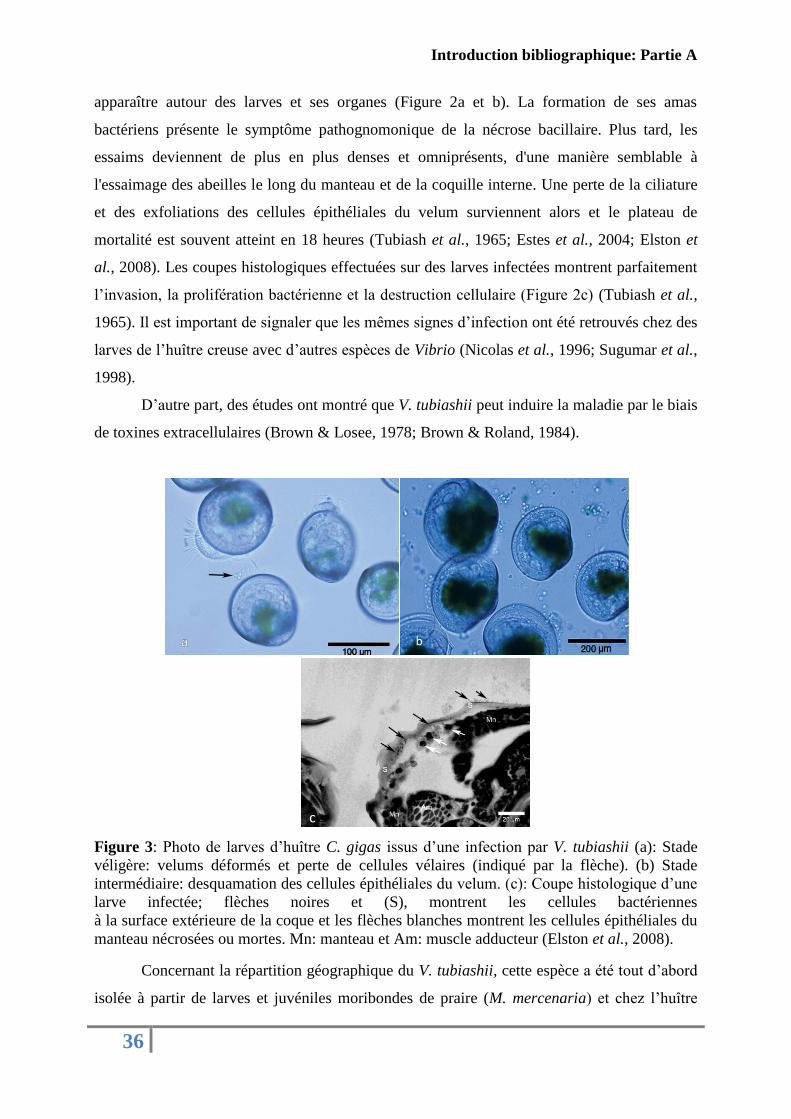
apparaître autour des larves et ses organes (Figure 2a et b). La formation de ses amas
bactériens présente le symptôme pathognomonique de la nécrose bacillaire. Plus tard, les
essaims deviennent de plus en plus denses et omniprésents, d'une manière semblable à
l'essaimage des abeilles le long du manteau et de la coquille interne. Une perte de la ciliature
et des exfoliations des cellules épithéliales du velum surviennent alors et le plateau de
mortalité est souvent atteint en 18 heures (Tubiash et al., 1965; Estes et al., 2004; Elston et
al., 2008). Les coupes histologiques effectuées sur des larves infectées montrent parfaitement
l’invasion, la prolifération bactérienne et la destruction cellulaire (Figure 2c) (Tubiash et al.,
1965). Il est important de signaler que les mêmes signes d’infection ont été retrouvés chez des
larves de l’huître creuse avec d’autres espèces de Vibrio (Nicolas et al., 1996; Sugumar et al.,
1998).
D’autre part, des études ont montré que V. tubiashii peut induire la maladie par le biais
de toxines extracellulaires (Brown & Losee, 1978; Brown & Roland, 1984).
Figure 3: Photo de larves d’huître C. gigas issus d’une infection par V. tubiashii (a): Stade
véligère: velums déformés et perte de cellules vélaires (indiqué par la flèche). (b) Stade
intermédiaire: desquamation des cellules épithéliales du velum. (c): Coupe histologique d’une
larve infectée; flèches noires et (S), montrent les cellules bactériennes
à la surface extérieure de la coque et les flèches blanches montrent les cellules épithéliales du
manteau nécrosées ou mortes. Mn: manteau et Am: muscle adducteur (Elston et al., 2008).
Concernant la répartition géographique du V. tubiashii, cette espèce a été tout d’abord
isolée à partir de larves et juvéniles moribondes de praire (M. mercenaria) et chez l’huître
b
c
Introduction bibliographique: Partie A
37
juvénile (C. virginica) sur la Côte Est des Etats-Unis (Tubiash et al., 1965; Hada et al., 1984).
Après, cette espèce a été retrouvée dans des écloseries en Angleterre et en Espagne infectant
des larves de l’huître creuse du Pacifique (C. gigas) et l’huître plate (O. edulis) (Jeffries,
1982; Lodeiros et al., 1987). Durant cette dernière décennie, une réapparition de V. tubiashii a
été détectée sur la Côte Ouest de l'Amérique du Nord et Hawaï infectant des stades larvaires
et juvéniles. En plus de l’huître creuse, cette espèce a été isolée de l’huître Kumamoto (C.
sikamea) et de la palourde royale (P. abrupta) (Estes et al., 2004; Elston et al., 2008).
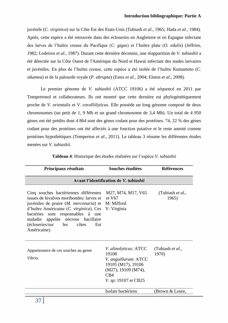
Le premier génome de V. tubiashii (ATCC 19106) a été séquencé en 2011 par
Tempertonol et collaborateurs. Ils ont montré que cette dernière est phylogénétiquement
proche de V. orientalis et V. coralliilyticus. Elle possède un long génome composé de deux
chromosomes (un petit de 1, 9 Mb et un grand chromosome de 3,4 Mb). Un total de 4 950
gènes ont été prédits dont 4 864 sont des gènes codant pour des protéines. 74, 22 % des gènes
codant pour des protéines ont été affectés à une fonction putative et le reste annoté comme
protéines hypothétiques (Temperton et al., 2011). Le tableau 3 résume les différentes études
menées sur V. tubiashii.
Tableau 4: Historique des études réalisées sur l’espèce V. tubiashii
Principaux résultats Souches étudiées Références
Avant l’identification de V. tubiashii
Cinq souches bactériennes différentes
issues de bivalves moribondes: larves et
juvéniles de praire (M. mercenaria) et
d’huître Américaine (C. virginica). Ces
bactéries sont responsables à une
maladie appelée nécrose bacillaire
(écloseries/sur les côtes Est
Américaine).
M27, M74, M17, V65
et V67
M: Milford
V: Virginia
(Tubiash et al.,
1965)
Appartenance de ces souches au genre
Vibrio.
V. alinolyticus: ATCC
19108
V. anguillarum: ATCC
19105 (M17), 19106
(M27), 19109 (M74),
CB4
V. sp: 19107 et CB25
(Tubiash et al.,
1970)
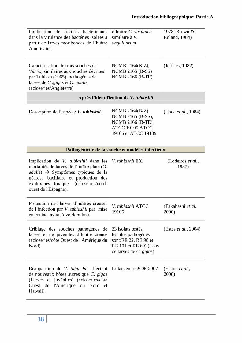
Isolats bactériens (Brown & Losee,
Introduction bibliographique: Partie A
38
Implication de toxines bactériennes
dans la virulence des bactéries isolées à
partir de larves moribondes de l’huître
Américaine.
d’huître C. virginica
similaire à V.
anguillarum
1978; Brown &
Roland, 1984)
Caractérisation de trois souches de
Vibrio, similaires aux souches décrites
par Tubiash (1965), pathogènes de
larves de C .gigas et O. edulis
(écloseries/Angleterre)
NCMB 2164(B-Z),
NCMB 2165 (B-SS)
NCMB 2166 (B-TE)
(Jeffries, 1982)
Après l’identification de V. tubiashii
Description de l’espèce: V. tubiashii. NCMB 2164(B-Z),
NCMB 2165 (B-SS),
NCMB 2166 (B-TE),
ATCC 19105 ATCC
19106 et ATCC 19109
(Hada et al., 1984)
Pathogénicité de la souche et modèles infectieux
Implication de V. tubiashii dans les
mortalités de larves de l’huître plate (O.
edulis) Symptômes typiques de la
nécrose bacillaire et production des
exotoxines toxiques (écloseries/nord-
ouest de l'Espagne).
V. tubiashii EXl,
(Lodeiros et al.,
1987)
Protection des larves d’huîtres creuses
de l’infection par V. tubiashii par mise
en contact avec l’ovoglobuline.
V. tubiashii ATCC
19106
(Takahashi et al.,
2000)
Criblage des souches pathogènes de
larves et de juvéniles d’huître creuse
(écloseries/côte Ouest de l'Amérique du
Nord).
33 isolats testés,
les plus pathogènes
sont:RE 22, RE 98 et
RE 101 et RE 60) (issus
de larves de C. gigas)
(Estes et al., 2004)
Réapparition de V. tubiashii affectant
de nouveaux hôtes autres que C. gigas
(Larves et juvéniles) (écloseries/côte
Ouest de l'Amérique du Nord et
Hawaii).
Isolats entre 2006-2007
(Elston et al.,
2008)
Introduction bibliographique: Partie A
39
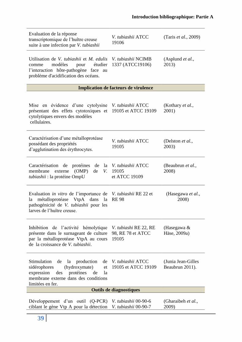
Evaluation de la réponse
transcriptomique de l’huître creuse
suite à une infection par V. tubiashii
V. tubiashii ATCC
19106
(Taris et al., 2009)
Utilisation de V. tubiashii et M. edulis
comme modèles pour étudier
l’interaction hôte-pathogène face au
problème d'acidification des océans.
V. tubiashii NCIMB
1337 (ATCC19106)
(Asplund et al.,
2013)
Implication de facteurs de virulence
Mise en évidence d’une cytolysine
présentant des effets cytotoxiques et
cytolytiques envers des modèles
cellulaires.
V. tubiashii ATCC
19105 et ATCC 19109
(Kothary et al.,
2001)
Caractérisation d’une métalloprotéase
possédant des propriétés
d’agglutination des érythrocytes.
V. tubiashii ATCC
19105
(Delston et al.,
2003)
Caractérisation de protéines de la
membrane externe (OMP) de V.
tubiashii : la protéine OmpU
V. tubiashii ATCC
19105
et ATCC 19109
(Beaubrun et al.,
2008)
Evaluation in vitro de l’importance de
la métalloprotéase VtpA dans la
pathogénicité de V. tubiashii pour les
larves de l’huître creuse.
V. tubiashii RE 22 et
RE 98
(Hasegawa et al.,
2008)
Inhibition de l’activité hémolytique
présente dans le surnageant de culture
par la métalloprotéase VtpA au cours
de la croissance de V. tubiashii.
V. tubiashi RE 22, RE
98, RE 78 et ATCC
19105
(Hasegawa &
Häse, 2009a)
Stimulation de la production de
sidérophores (hydroxymate) et
expression des protéines de la
membrane externe dans des conditions
limitées en fer.
V. tubiashii ATCC
19105 et ATCC 19109
(Junia Jean-Gilles
Beaubrun 2011).
Outils de diagnostiques
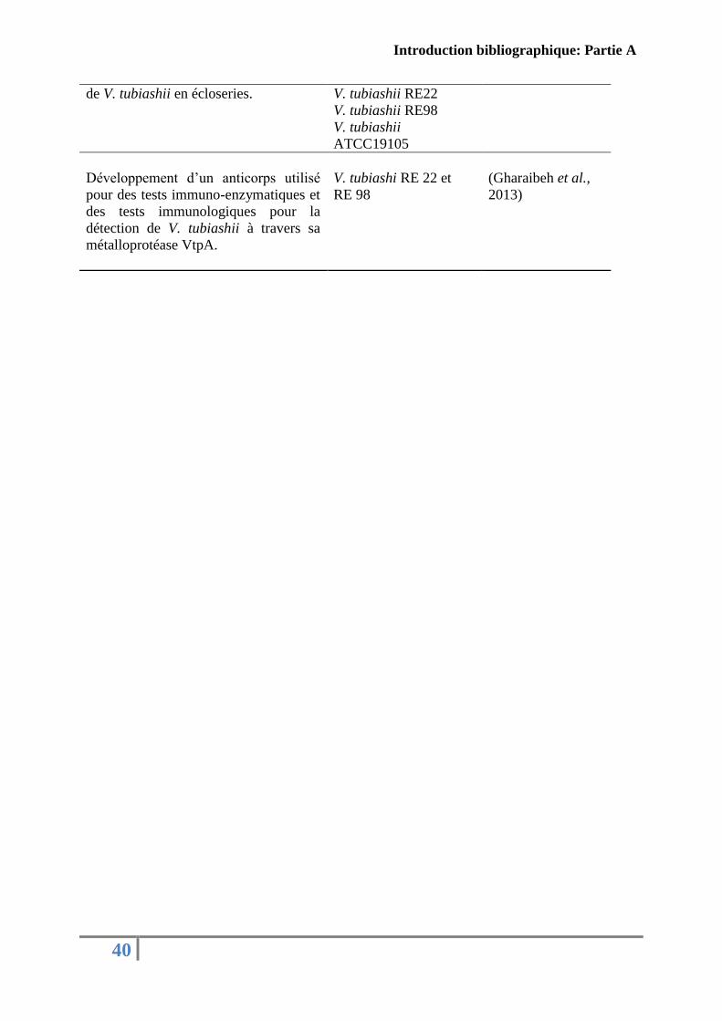
Développement d’un outil (Q-PCR)
ciblant le gène Vtp A pour la détection
V. tubiashii 00-90-6
V. tubiashii 00-90-7
(Gharaibeh et al.,
2009)
Introduction bibliographique: Partie A
40
de V. tubiashii en écloseries.
V. tubiashii RE22
V. tubiashii RE98
V. tubiashii
ATCC19105
Développement d’un anticorps utilisé
pour des tests immuno-enzymatiques et
des tests immunologiques pour la
détection de V. tubiashii à travers sa
métalloprotéase VtpA.
V. tubiashi RE 22 et
RE 98
(Gharaibeh et al.,
2013)
Introduction bibliographique: Partie A
41
5. Les facteurs de virulence du genre Vibrio
5.1. Les notions de pathogénicité et de virulence
Avant de décrire les facteurs de virulence associés au genre Vibrio, il est indispensable
de différentier les notions de pathogénicité et virulence et de comprendre la relation entre ces
deux concepts.
Classiquement décrit, un agent pathogène est un microorganisme qui provoque une
maladie ou qui peut se multiplier dans le tissu vivant et induire des maladies (Casadevall &
Pirofski, 1999) et le pouvoir pathogène est la capacité d'un microorganisme à induire une
maladie (Smith, 1977). Cette interprétation des termes suggère que la pathogénicité est une
caractéristique liée seulement au microorganisme et indépendante de l’hôte. Or cette
définition semble insuffisante car un microorganisme peut coloniser sans nuire à l’hôte. En
1999, Casadevall et Pirofski ont apporté des améliorations dans les définitions de base
(Casadevall & Pirofski, 1999). Ainsi, un agent pathogène est un microorganisme capable de
causer des dommages à l’hôte et le pouvoir pathogène est la capacité d’un microorganisme à
causer des dommages à l’hôte. Avec cette nouvelle reformulation des définitions, la
pathogénicité n’est plus une caractéristique invariante et indépendante de l’hôte mais le
résultat d’une interaction entre l’hôte et le microorganisme (Casadevall & Pirofski, 1999,
2003)
Concernant la virulence, plusieurs définitions ont été aussi proposées à cette la notion
(Casadevall & Pirofski, 1999):
- Le degré de la pathogénicité,
- La force de l’activité d’un pathogène,
- Le synonyme de la pathogénicité,
- Le pourcentage de mortalité par infection,
- La capacité relative de surmonter la défense disponible d’un hôte,
- La mesure de la capacité d'un microorganisme à infecter ou endommager un
hôte,
- La capacité relative d'entrer et de se multiplier dans un hôte donné,
- La gravité de la maladie.
Introduction bibliographique: Partie A
42
Plus tard, une définition plus drastique a été accordée à la notion de la virulence et qui
associe de la même manière l’hôte et l’agent infectieu: c’est la capacité relative d’un
microorganisme à causer des dommages à l’hôte (Casadevall & Pirofski, 1999).
Certaines définitions varient entre et au sein des disciplines (Shapiro-Ilan et al., 2005)
et peuvent entrainer une confusion. Ainsi, la majorité des pathologistes choisissent d’utiliser
une définition au sens large qui associe la notion de la virulence à la notion de la
pathogénicité (Thomas & Elkinton, 2004).
De même, la terminologie utilisée pour décrire les facteurs de virulence a été modifiée
pour passer de « produits microbiens qui permettent à un agent pathogène de provoquer une
maladie» (Smith, 1977) à une définition au sens large «tout composant d’un microorganisme
qui est nécessaire ou qui potentialise sa capacité à provoquer une maladie» (Schaechter et al.,
1999). Cette définition absolue inclut implicitement les substances non toxiques pour l’hôte
qui peuvent quand même être considérées comme des facteurs de virulence si leur absence
rendent le microorganisme moins virulent (Schaechter et al., 1999).
Enfin, il est important de considérer les thèmes suivants: (1) très peu de facteurs de
virulence fonctionnent comme des facteurs déterminants de la virulence; (2) les dommages
sont causés soit directement par les microorganismes ou par un composant microbien soit par
la réponse immunitaire de l’hôte et (3) les réponses immunitaires peuvent neutraliser plusieurs
ou une partie des facteurs de virulence (Casadevall & Pirofski, 2009).
5.2. Les mécanismes de virulence chez les Vibrio
Pour provoquer la maladie chez l'hôte, l'agent pathogène doit posséder de nombreux
attributs: la possibilité de pénétrer un hôte, la capacité à échapper aux défenses de l'hôte,
l'aptitude à croître dans l’hôte, la capacité d’affronter les réponses immunitaires de l'hôte, la
capacité à acquérir le fer et les nutriments nécessaires de l'environnement (Casadevall &
Pirofski, 2009).
Avec toutes ces propriétés, la classification des facteurs de virulences semble être un
exercice difficile (Casadevall & Pirofski, 2009) car plusieurs déterminants de virulence
partagent les mêmes mécanismes et les mêmes caractéristiques (Finlay & Falkow, 1997) et
même si leur classification en catégorie fonctionnelle parait plus rationnelle, nous ne pouvons

Introduction bibliographique: Partie A
43
pas ignorer que certains composants peuvent être attribués à plus d’une fonction (Casadevall
& Pirofski, 1999, 2009).
Concernant le genre Vibrio, plusieurs facteurs de virulence ont été décrits dans la
littérature. Le Tableau présenté ci-dessous (Tableau 5) résume les principaux facteurs de
virulence identifiés chez des Vibrio pathogènes pour l’Homme et pathogènes pour les
organismes marins classés arbitrairement par rapport à leurs fonctions pendant les différentes
étapes de l’infection.
Introduction bibliographique: Partie A
44
Tableau 5: les differents facteurs de la virulence décrits chez le genre Vibrio
Les facteurs d’adhésion et de
colonisation
(Mise en contact hôte-pathogène)
Facteurs Fonction (s) principale (s) Références
Le flagelle
- Facilite le mouvement chimiotactique dirigé
(swarming et swiming).
- Attachement.
- Formation de biofilm.
- Impliqué dans la virulence (pénétration).
(Guentzel & Berry, 1975;
Milton et al., 1996;
Richardson, 1991; R. & E.,
2012)
Les pili (adhésines)
- Mobilité (twitching).
- Colonisation.
- Adhésion.
- Formation de biofilm.
(Voss & Attridge, 1993;
Thelin & Taylor, 1996;
Attridge et al., 1996; Voss et
al., 1996; Bardy et al., 2003;
Shime-Hattori et al., 2006; Li
et al., 2008)
Les protéines de la membrane
externe (OMP)
- Adhésion.
- Invasion.
- Adaptation du pathogène à divers
environnements.
- Impliqué dans la virulence: adhésion et
invasion aux cellules de l’hôte.
(Beaubrun et al., 2008;
Duperthuy et al., 2010, 2011;
Goo et al., 2006; Lin et al.,
2002; Nandi et al., 2005)
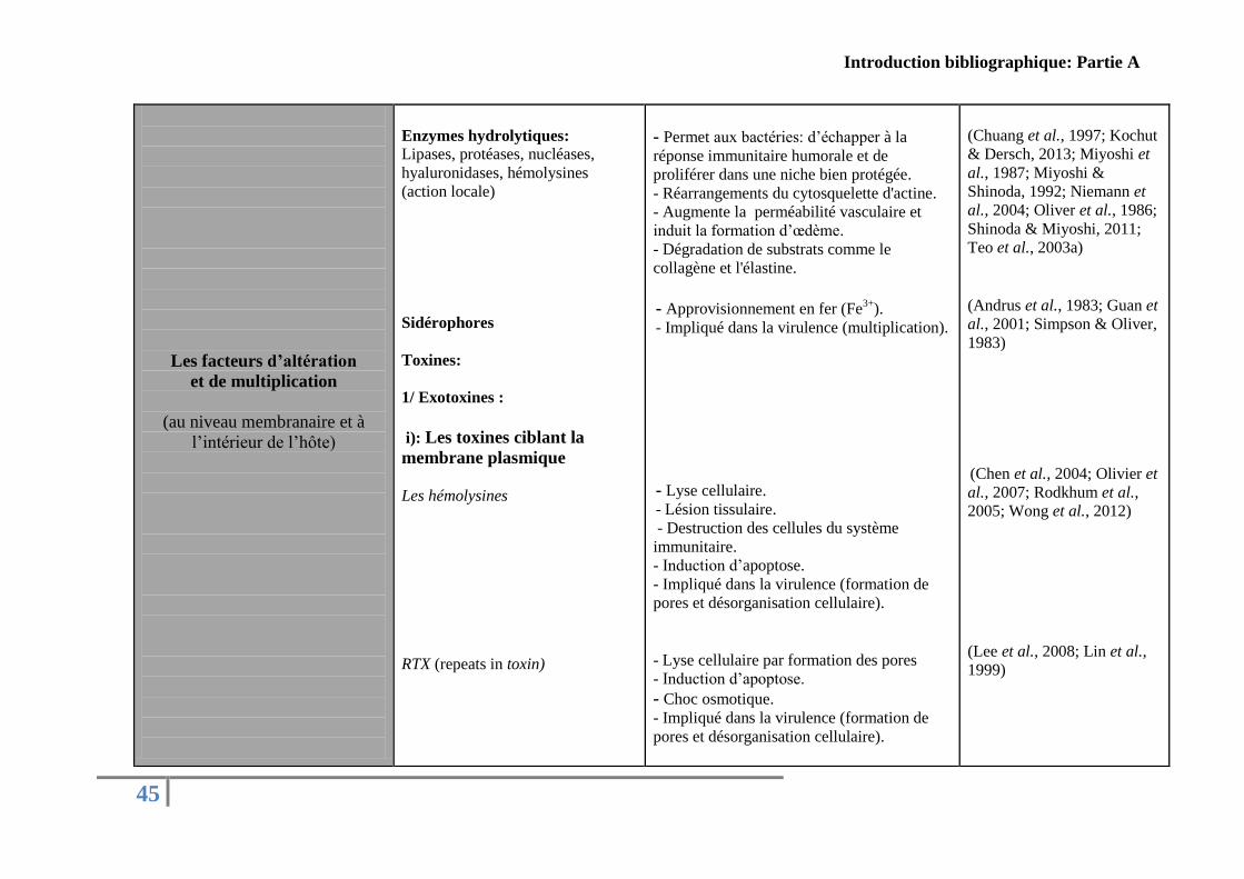
Introduction bibliographique: Partie A
45
Les facteurs d’altération
et de multiplication
(au niveau membranaire et à
l’intérieur de l’hôte)
Enzymes hydrolytiques:
Lipases, protéases, nucléases,
hyaluronidases, hémolysines
(action locale)
Sidérophores
Toxines:
1/ Exotoxines :
i): Les toxines ciblant la
membrane plasmique
Les hémolysines
RTX (repeats in toxin)
- Permet aux bactéries: d’échapper à la
réponse immunitaire humorale et de
proliférer dans une niche bien protégée.
- Réarrangements du cytosquelette d'actine.
- Augmente la perméabilité vasculaire et
induit la formation d’œdème.
- Dégradation de substrats comme le
collagène et l'élastine.
- Approvisionnement en fer (Fe3+
).
- Impliqué dans la virulence (multiplication).
- Lyse cellulaire.
- Lésion tissulaire.
- Destruction des cellules du système
immunitaire.
- Induction d’apoptose.
- Impliqué dans la virulence (formation de
pores et désorganisation cellulaire).
- Lyse cellulaire par formation des pores
- Induction d’apoptose.
- Choc osmotique.
- Impliqué dans la virulence (formation de
pores et désorganisation cellulaire).
(Chuang et al., 1997; Kochut
& Dersch, 2013; Miyoshi et
al., 1987; Miyoshi &
Shinoda, 1992; Niemann et
al., 2004; Oliver et al., 1986;
Shinoda & Miyoshi, 2011;
Teo et al., 2003a)
(Andrus et al., 1983; Guan et
al., 2001; Simpson & Oliver,
1983)
(Chen et al., 2004; Olivier et
al., 2007; Rodkhum et al.,
2005; Wong et al., 2012)
(Lee et al., 2008; Lin et al.,
1999)
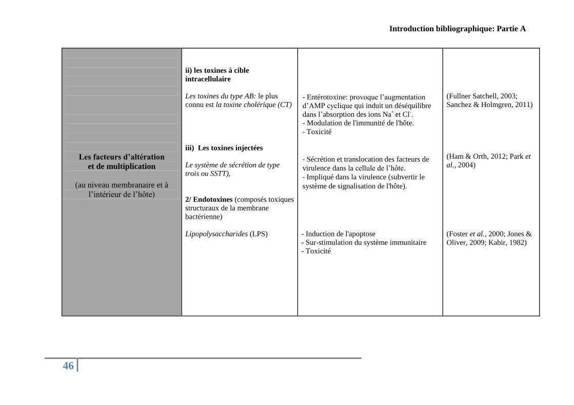
Introduction bibliographique: Partie A
46
Les facteurs d’altération
et de multiplication
(au niveau membranaire et à
l’intérieur de l’hôte)
ii) les toxines à cible
intracellulaire
Les toxines du type AB: le plus
connu est la toxine cholérique (CT)
iii) Les toxines injectées
Le système de sécrétion de type
trois ou SSTT),
2/ Endotoxines (composés toxiques
structuraux de la membrane
bactérienne)
Lipopolysaccharides (LPS)
- Entérotoxine: provoque l’augmentation
d’AMP cyclique qui induit un déséquilibre
dans l’absorption des ions Na+
et Cl-.
- Modulation de l'immunité de l'hôte.
- Toxicité - Sécrétion et translocation des facteurs de
virulence dans la cellule de l’hôte. - Impliqué dans la virulence (subvertir le
système de signalisation de l'hôte).
- Induction de l'apoptose
- Sur-stimulation du système immunitaire
- Toxicité
(Fullner Satchell, 2003;
Sanchez & Holmgren, 2011)
(Ham & Orth, 2012; Park et
al., 2004)
(Foster et al., 2000; Jones &
Oliver, 2009; Kabir, 1982)
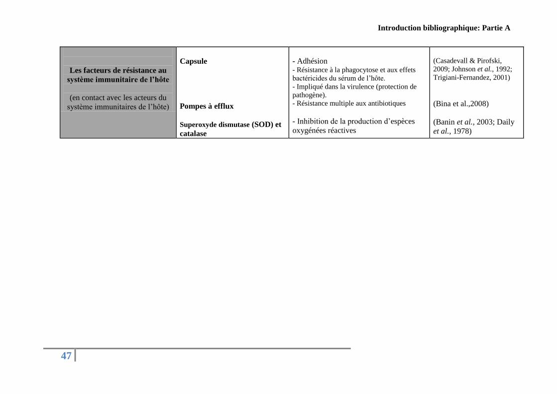
Introduction bibliographique: Partie A
47
Les facteurs de résistance au
système immunitaire de l’hôte
(en contact avec les acteurs du
système immunitaires de l’hôte)
Capsule
Pompes à efflux
Superoxyde dismutase (SOD) et
catalase
- Adhésion - Résistance à la phagocytose et aux effets
bactéricides du sérum de l’hôte.
- Impliqué dans la virulence (protection de
pathogène).
- Résistance multiple aux antibiotiques
- Inhibition de la production d’espèces
oxygénées réactives
(Casadevall & Pirofski,
2009; Johnson et al., 1992;
Trigiani-Fernandez, 2001)
(Bina et al.,2008)
(Banin et al., 2003; Daily
et al., 1978)
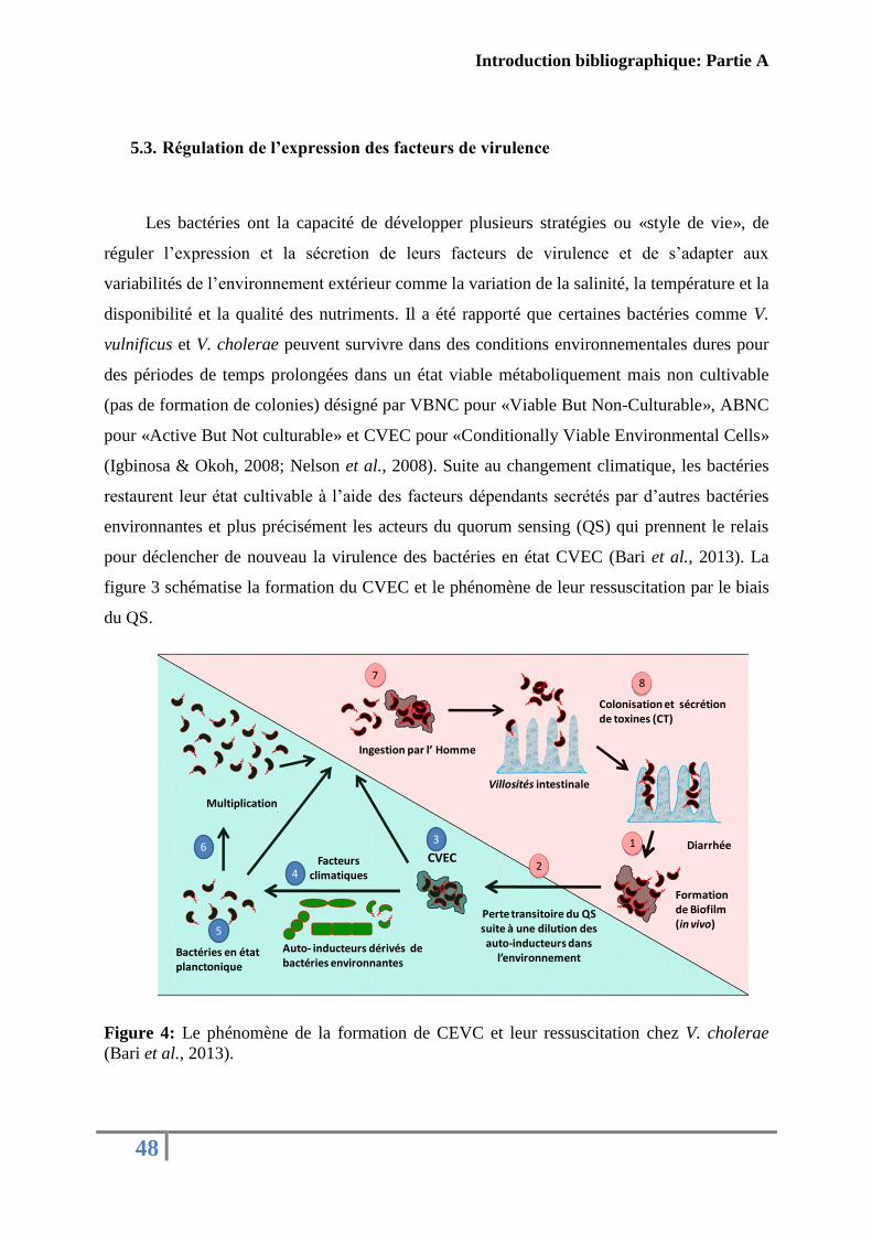
Introduction bibliographique: Partie A
48
5.3. Régulation de l’expression des facteurs de virulence
Les bactéries ont la capacité de développer plusieurs stratégies ou «style de vie», de
réguler l’expression et la sécretion de leurs facteurs de virulence et de s’adapter aux
variabilités de l’environnement extérieur comme la variation de la salinité, la température et la
disponibilité et la qualité des nutriments. Il a été rapporté que certaines bactéries comme V.
vulnificus et V. cholerae peuvent survivre dans des conditions environnementales dures pour
des périodes de temps prolongées dans un état viable métaboliquement mais non cultivable
(pas de formation de colonies) désigné par VBNC pour «Viable But Non-Culturable», ABNC
pour «Active But Not culturable» et CVEC pour «Conditionally Viable Environmental Cells»
(Igbinosa & Okoh, 2008; Nelson et al., 2008). Suite au changement climatique, les bactéries
restaurent leur état cultivable à l’aide des facteurs dépendants secrétés par d’autres bactéries
environnantes et plus précisément les acteurs du quorum sensing (QS) qui prennent le relais
pour déclencher de nouveau la virulence des bactéries en état CVEC (Bari et al., 2013). La
figure 3 schématise la formation du CVEC et le phénomène de leur ressuscitation par le biais
du QS.
Figure 4: Le phénomène de la formation de CEVC et leur ressuscitation chez V. cholerae
(Bari et al., 2013).
Ingestion par l’ Homme
Colonisation et sécrétion de toxines (CT)
78
1
2
Diarrhée
4
Formation de Biofilm(in vivo)
Perte transitoire du QS suite à une dilution des auto-inducteurs dans
l’environnement
CVEC6
Multiplication
Bactéries en état planctonique
Auto- inducteurs dérivés de bactéries environnantes
5
Facteurs climatiques
3
Villosités intestinale
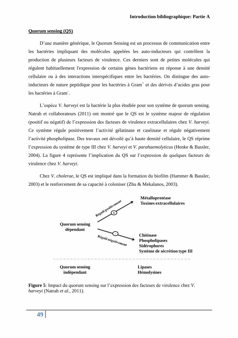
Introduction bibliographique: Partie A
49
Quorum sensing (QS)
D’une manière générique, le Quorum Sensing est un processus de communication entre
les bactéries impliquant des molécules appelées les auto-inducteurs qui contrôlent la
production de plusieurs facteurs de virulence. Ces derniers sont de petites molécules qui
régulent habituellement l'expression de certains gènes bactériens en réponse à une densité
cellulaire ou à des interactions interspécifiques entre les bactéries. On distingue des auto-
inducteurs de nature peptidique pour les bactéries à Gram+ et des dérivés d’acides gras pour
les bactéries à Gram-.
L’espèce V. harveyi est la bactérie la plus étudiée pour son système de quorum sensing.
Natrah et collaborateurs (2011) ont montré que le QS est le système majeur de régulation
(positif ou négatif) de l’expression des facteurs de virulence extracellulaires chez V. harveyi.
Ce système régule positivement l’activité gélatinase et caséinase et régule négativement
l’activité phospholipase. Des travaux ont dévoilé qu’à haute densité cellulaire, le QS réprime
l’expression du système de type III chez V. harveyi et V. parahaemolyticus (Henke & Bassler,
2004). La figure 4 représente l’implication du QS sur l’expression de quelques facteurs de
virulence chez V. harveyi.
Chez V. cholerae, le QS est impliqué dans la formation du biofilm (Hammer & Bassler,
2003) et le renforcement de sa capacité à coloniser (Zhu & Mekalanos, 2003).
Figure 5: Impact du quorum sensing sur l’expression des facteurs de virulence chez V.
harveyi (Natrah et al., 2011).
Quorum sensing
dépendant
+
Métalloprotéase
Toxines extracellulaires
Chitinase
Phospholipases
Sidérophores
Système de sécrétion type III
Lipases
Hémolysines
Quorum sensing
indépendant
-
Introduction bibliographique: Partie A
50
Il faut noter qu’il existe d’autres facteurs de virulence qui ne sont pas régulés par le
QS comme les hémolysines et les lipases (Natrah et al., 2011). De plus, même si la
perturbation de la communication intracellulaire pourrait constituer une stratégie pour lutter
contre les infections dans le domaine de l’aquaculture (Defoirdt et al., 2004) il ne faut pas nier
que dans le domaine médical malgré la perte du QS, certaines bactéries sont capables de
provoquer des infections (Schaber et al., 2004).
6. Méthodes de détection du genre Vibrio
La détection des espèces au sein du genre Vibrio a été toujours basée sur des
techniques phénotypiques traditionnelles (Vandenberghe et al., 2003) et plus récemment sur
des outils innovants de biologie moléculaire (Kim & Jeong, 2001; Saulnier et al., 2009;
Gharaibeh et al., 2009; Hossain et al., 2013).
En général, les méthodes classiques d’identification bactérienne nécessitent plusieurs
étapes: commençant par l’isolement, l’enrichissement, l’étalement sur milieux sélectif, suivis
de caractérisation, d’observation morphologique et finissant par des analyses biochimiques.
Cependant, toute cette procédure traditionnelle de détection bactérienne reste lente, laborieuse
et nécessite souvent plusieurs jours pour sa réalisation. En plus, certains tests phénotypiques
sont insensibles pour la détection des bactéries à faible concentration ou qui présentent des
profils phénotypiques inhabituels (Iii & Hickman-Brenner, 2006).
La variabilité des caractères biochimiques atypiques des souches de Vibrio isolées de
l’environnement rend leur identification et leur distinction difficile. Vandenberghe et
collaborateurs (2003) ont montré les limites des tests biochimiques dans l’identification des
bactéries appartenant au genre Vibrio isolées à partir de différents systèmes d'aquaculture
dans de nombreux pays. En outre, certaines bactéries peuvent subir des lésions ou être dans
des états de dormance, à cause des conditions de stress ou des changements défavorables
comme la variation de la salinité, le changement de la température ou la carence en quelques
éléments nutritifs, et deviennent non détectables par les méthodes bactériologiques classiques
(Brauns et al., 1991; Lee et al., 1998b; Cohen & Karib, 2007).
Introduction bibliographique: Partie A
51
Les progrès spectaculaires de diverses technologies moléculaires dans la discipline de
microbiologie ont permis le développement des outils d’identification plus fiables, rapides,
sensibles et spécifiques pour la détection des bactéries même à très faible concentration
cellulaire dans leur environnement naturel. Les outils les plus utilisés sont la PCR (Réaction
en Chaîne par Polymérase) et outils dérivés: la qPCR (PCR en temps réel), l’AFLP (le
polymorphisme de longueur des fragments amplifiés), la FISH (l'hybridation fluorescente in
situ), la MLEE (électrophorèse enzymatique multilocus), la RFLP (Polymorphisme de taille
des fragments de restriction) et la Ribotypie (Igbinosa & Okoh, 2008).
La PCR a été souvent utilisée pour identifier des espèces du genre Vibrio, déterminer
leurs relations phylogénétiques et taxonomiques et rechercher des gènes codant pour des
facteurs de virulence par le biais de plusieurs cibles moléculaires. En visant une région
variable de l’ARNr 16S, la PCR a été utilisée pour la détection et la différenciation de V.
vulnificus dans les milieux marins (Kim & Jeong, 2001). Egalement, la PCR a été utilisé pour
détecter V. parahaemolyticus, en ciblant plusieurs gènes de virulence comme toxR ou les
deux hémolysines TDH et TRH (Kim et al., 1999; Robert-Pillot et al., 2004). De même, la V.
cholerae, est systématiquement recherchés par PCR à travers les gènes ctxA et ctxB, codant
pour la toxine cholérique (Fields et al., 1992). La technique dz multiplexe PCR a été aussi
employée pour détecter le genre Vibrio ( Panicker et al., 2004; Hossain et al., 2013; Paydar et
al., 2013).
La méthode qPCR a été développée récemment pour détecter et quantifier des espèces
de Vibrio en ciblant différents gènes éventuellement impliqués dans la virulence de l’espèce.
La QPCR a été utilisée pour la détection de V. aestuarianus (Saulnier et al., 2009) et V.
tubiashii (Gharaibeh et al., 2009) durant les épisodes de mortalités de l’huître creuse C. gigas.
Aussi, cet outil a été utilisé pour détecter et quantifier V. cholerae dans l’eau et les produits de
la pêche (Gubala & Proll, 2006). Cependant, cette technique exige un niveau élevé de
compétences en laboratoire et un matériel onéreux non compatible à une utilisation directe par
exemple sur site.
Des méthodes sérologiques ont été aussi utilisées avec succès pour l'identification des
espèces de Vibrio dans les aliments et les environnements aquatiques. La méthode ELISA est
le test immunologique le plus utilisé (Igbinosa & Okoh, 2008; Tantillo et al., 2004).
Récemment, Gharaibeh et collaborateurs (2013) ont développé un test ELISA et un test

Introduction bibliographique: Partie A
52
immunologique à flux latéral (test de jauge) pour la détection de la métalloprotéase à zinc
sécrétée par la souche V. tubiashii. Ces deux outils s'appuient sur la spécificité des anticorps
monoclonaux qui reconnaissent des épitopes spécifiques de la métalloprotéase VtpA mature.
53
Partie B: Les facteurs de virulence chez les vibrions
marins, types et mode d’action
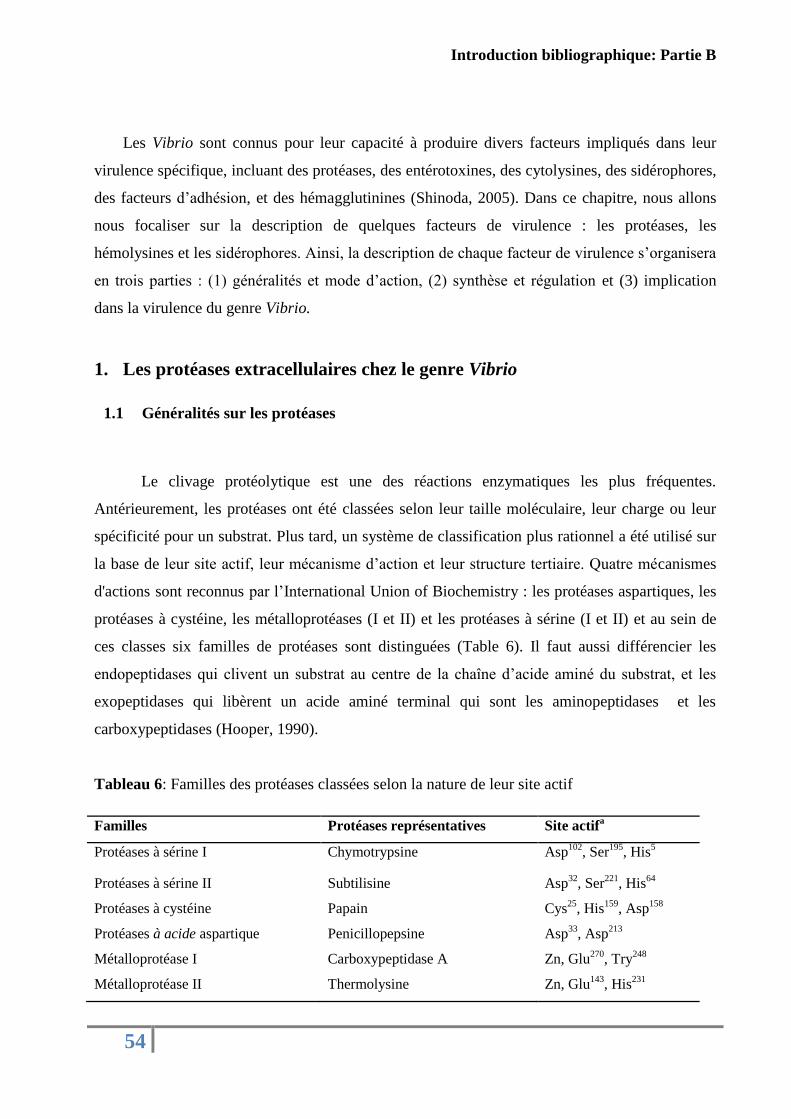
Introduction bibliographique: Partie B
54
Les Vibrio sont connus pour leur capacité à produire divers facteurs impliqués dans leur
virulence spécifique, incluant des protéases, des entérotoxines, des cytolysines, des sidérophores,
des facteurs d’adhésion, et des hémagglutinines (Shinoda, 2005). Dans ce chapitre, nous allons
nous focaliser sur la description de quelques facteurs de virulence : les protéases, les
hémolysines et les sidérophores. Ainsi, la description de chaque facteur de virulence s’organisera
en trois parties : (1) généralités et mode d’action, (2) synthèse et régulation et (3) implication
dans la virulence du genre Vibrio.
1. Les protéases extracellulaires chez le genre Vibrio
1.1 Généralités sur les protéases
Le clivage protéolytique est une des réactions enzymatiques les plus fréquentes.
Antérieurement, les protéases ont été classées selon leur taille moléculaire, leur charge ou leur
spécificité pour un substrat. Plus tard, un système de classification plus rationnel a été utilisé sur
la base de leur site actif, leur mécanisme d’action et leur structure tertiaire. Quatre mécanismes
d'actions sont reconnus par l’International Union of Biochemistry : les protéases aspartiques, les
protéases à cystéine, les métalloprotéases (I et II) et les protéases à sérine (I et II) et au sein de
ces classes six familles de protéases sont distinguées (Table 6). Il faut aussi différencier les
endopeptidases qui clivent un substrat au centre de la chaîne d’acide aminé du substrat, et les
exopeptidases qui libèrent un acide aminé terminal qui sont les aminopeptidases et les
carboxypeptidases (Hooper, 1990).
Tableau 6: Familles des protéases classées selon la nature de leur site actif
Familles Protéases représentatives Site actifa
Protéases à sérine I Chymotrypsine Asp102
, Ser195
, His5
Protéases à sérine II Subtilisine Asp32
, Ser221
, His64
Protéases à cystéine Papain Cys25
, His159
, Asp158
Protéases à acide aspartique Penicillopepsine Asp33
, Asp213
Métalloprotéase I Carboxypeptidase A Zn, Glu270
, Try248
Métalloprotéase II Thermolysine Zn, Glu143
, His231
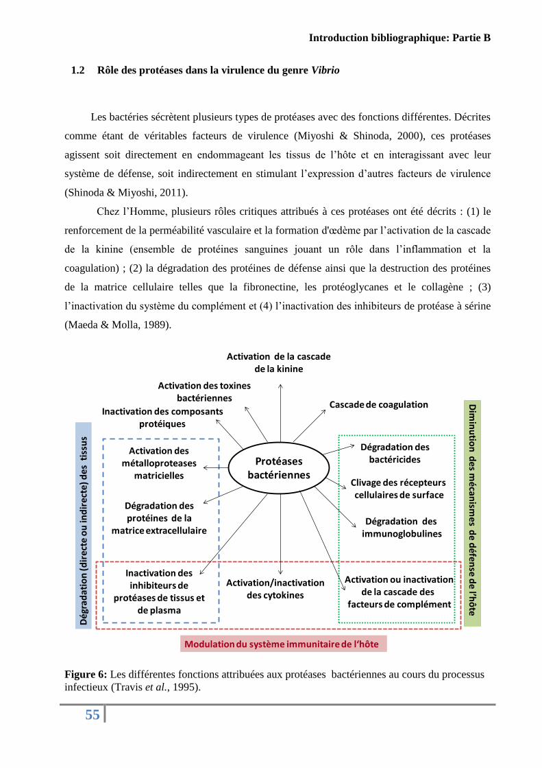
Introduction bibliographique: Partie B
55
1.2 Rôle des protéases dans la virulence du genre Vibrio
Les bactéries sécrètent plusieurs types de protéases avec des fonctions différentes. Décrites
comme étant de véritables facteurs de virulence (Miyoshi & Shinoda, 2000), ces protéases
agissent soit directement en endommageant les tissus de l’hôte et en interagissant avec leur
système de défense, soit indirectement en stimulant l’expression d’autres facteurs de virulence
(Shinoda & Miyoshi, 2011).
Chez l’Homme, plusieurs rôles critiques attribués à ces protéases ont été décrits : (1) le
renforcement de la perméabilité vasculaire et la formation d'œdème par l’activation de la cascade
de la kinine (ensemble de protéines sanguines jouant un rôle dans l’inflammation et la
coagulation) ; (2) la dégradation des protéines de défense ainsi que la destruction des protéines
de la matrice cellulaire telles que la fibronectine, les protéoglycanes et le collagène ; (3)
l’inactivation du système du complément et (4) l’inactivation des inhibiteurs de protéase à sérine
(Maeda & Molla, 1989).
Figure 6: Les différentes fonctions attribuées aux protéases bactériennes au cours du processus
infectieux (Travis et al., 1995).
Protéases bactériennes
Activation/inactivation des cytokines
Activation des métalloproteases
matricielles
Dégradation des protéines de la
matrice extracellulaire
Inactivation des inhibiteurs de
protéases de tissus et de plasma
Dég
rad
atio
n (d
irec
te o
u in
dir
ecte
) des
tis
sus
Modulation du système immunitaire de l‘hôte
Dim
inu
tion
des m
écanism
es de d
éfense d
e l’hô
te
Inactivation des composants protéiques
Activation des toxines bactériennes
Activation de la cascadede la kinine
Cascade de coagulation
Dégradation des bactéricides
Clivage des récepteurs cellulaires de surface
Dégradation desimmunoglobulines
Activation ou inactivation de la cascade des
facteurs de complément
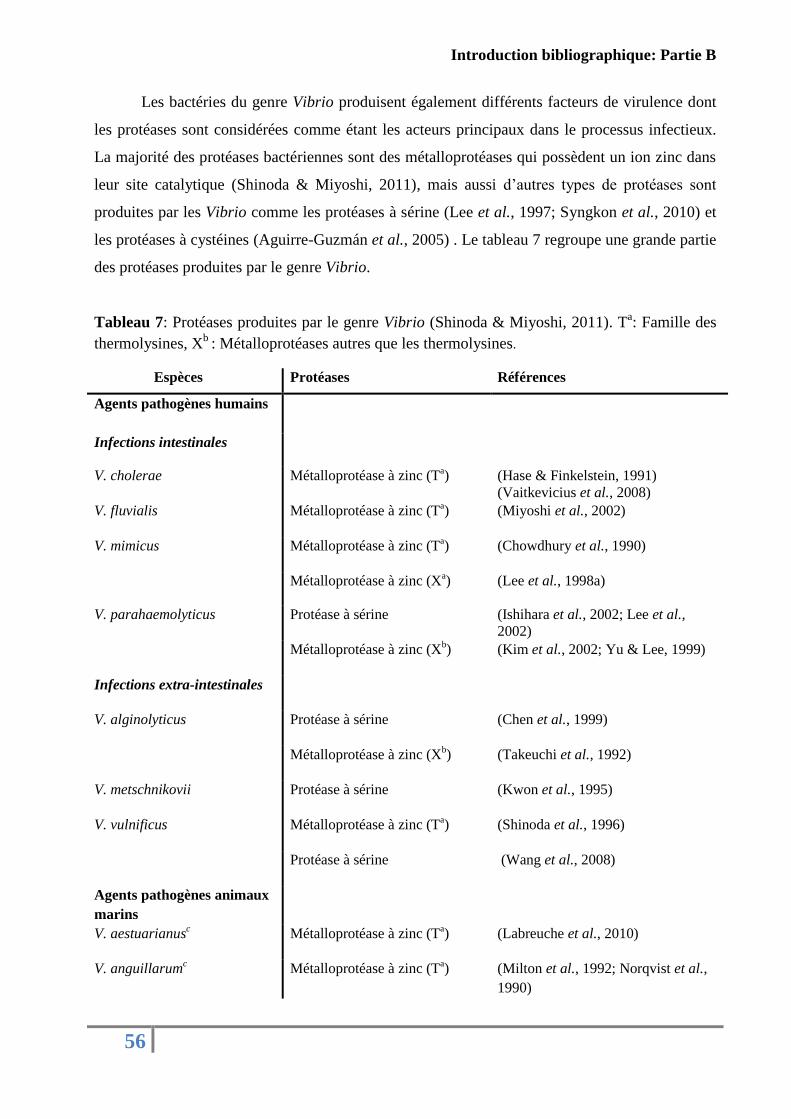
Introduction bibliographique: Partie B
56
Les bactéries du genre Vibrio produisent également différents facteurs de virulence dont
les protéases sont considérées comme étant les acteurs principaux dans le processus infectieux.
La majorité des protéases bactériennes sont des métalloprotéases qui possèdent un ion zinc dans
leur site catalytique (Shinoda & Miyoshi, 2011), mais aussi d’autres types de protéases sont
produites par les Vibrio comme les protéases à sérine (Lee et al., 1997; Syngkon et al., 2010) et
les protéases à cystéines (Aguirre-Guzmán et al., 2005) . Le tableau 7 regroupe une grande partie
des protéases produites par le genre Vibrio.
Tableau 7: Protéases produites par le genre Vibrio (Shinoda & Miyoshi, 2011). Ta: Famille des
thermolysines, Xb
: Métalloprotéases autres que les thermolysines.
Espèces Protéases Références
Agents pathogènes humains
Infections intestinales
V. cholerae Métalloprotéase à zinc (Ta) (Hase & Finkelstein, 1991)
(Vaitkevicius et al., 2008)
V. fluvialis Métalloprotéase à zinc (Ta) (Miyoshi et al., 2002)
V. mimicus Métalloprotéase à zinc (Ta) (Chowdhury et al., 1990)
Métalloprotéase à zinc (Xa) (Lee et al., 1998a)
V. parahaemolyticus Protéase à sérine (Ishihara et al., 2002; Lee et al.,
2002)
Métalloprotéase à zinc (Xb) (Kim et al., 2002; Yu & Lee, 1999)
Infections extra-intestinales
V. alginolyticus Protéase à sérine (Chen et al., 1999)
Métalloprotéase à zinc (Xb) (Takeuchi et al., 1992)
V. metschnikovii Protéase à sérine (Kwon et al., 1995)
V. vulnificus Métalloprotéase à zinc (Ta) (Shinoda et al., 1996)
Protéase à sérine (Wang et al., 2008)
Agents pathogènes animaux
marins
V. aestuarianusc Métalloprotéase à zinc (T
a) (Labreuche et al., 2010)
V. anguillarumc Métalloprotéase à zinc (T
a) (Milton et al., 1992; Norqvist et al.,
1990)
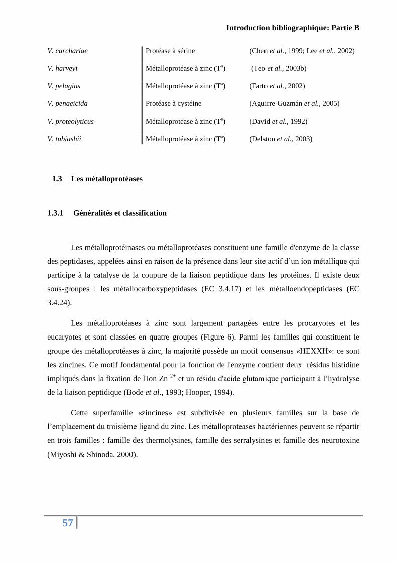
Introduction bibliographique: Partie B
57
V. carchariae Protéase à sérine (Chen et al., 1999; Lee et al., 2002)
V. harveyi Métalloprotéase à zinc (Ta) (Teo et al., 2003b)
V. pelagius Métalloprotéase à zinc (Ta) (Farto et al., 2002)
V. penaeicida Protéase à cystéine (Aguirre-Guzmán et al., 2005)
V. proteolyticus Métalloprotéase à zinc (Ta) (David et al., 1992)
V. tubiashii Métalloprotéase à zinc (Ta) (Delston et al., 2003)
1.3 Les métalloprotéases
1.3.1 Généralités et classification
Les métalloprotéinases ou métalloprotéases constituent une famille d'enzyme de la classe
des peptidases, appelées ainsi en raison de la présence dans leur site actif d’un ion métallique qui
participe à la catalyse de la coupure de la liaison peptidique dans les protéines. Il existe deux
sous-groupes : les métallocarboxypeptidases (EC 3.4.17) et les métalloendopeptidases (EC
3.4.24).
Les métalloprotéases à zinc sont largement partagées entre les procaryotes et les
eucaryotes et sont classées en quatre groupes (Figure 6). Parmi les familles qui constituent le
groupe des métalloprotéases à zinc, la majorité possède un motif consensus «HEXXH»: ce sont
les zincines. Ce motif fondamental pour la fonction de l'enzyme contient deux résidus histidine
impliqués dans la fixation de l'ion Zn 2+
et un résidu d'acide glutamique participant à l’hydrolyse
de la liaison peptidique (Bode et al., 1993; Hooper, 1994).
Cette superfamille «zincines» est subdivisée en plusieurs familles sur la base de
l’emplacement du troisième ligand du zinc. Les métalloproteases bactériennes peuvent se répartir
en trois familles : famille des thermolysines, famille des serralysines et famille des neurotoxine
(Miyoshi & Shinoda, 2000).
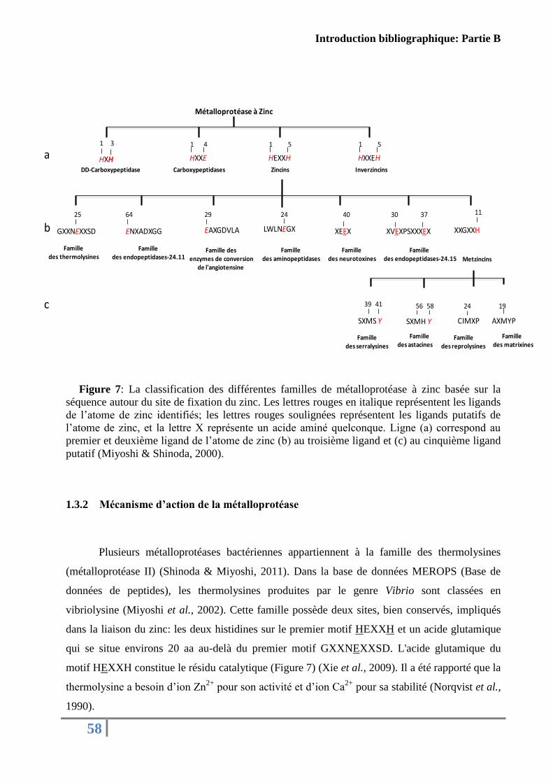
Introduction bibliographique: Partie B
58
Figure 7: La classification des différentes familles de métalloprotéase à zinc basée sur la
séquence autour du site de fixation du zinc. Les lettres rouges en italique représentent les ligands
de l’atome de zinc identifiés; les lettres rouges soulignées représentent les ligands putatifs de
l’atome de zinc, et la lettre X représente un acide aminé quelconque. Ligne (a) correspond au
premier et deuxième ligand de l’atome de zinc (b) au troisième ligand et (c) au cinquième ligand
putatif (Miyoshi & Shinoda, 2000).
1.3.2 Mécanisme d’action de la métalloprotéase
Plusieurs métalloprotéases bactériennes appartiennent à la famille des thermolysines
(métalloprotéase II) (Shinoda & Miyoshi, 2011). Dans la base de données MEROPS (Base de
données de peptides), les thermolysines produites par le genre Vibrio sont classées en
vibriolysine (Miyoshi et al., 2002). Cette famille possède deux sites, bien conservés, impliqués
dans la liaison du zinc: les deux histidines sur le premier motif HEXXH et un acide glutamique
qui se situe environs 20 aa au-delà du premier motif GXXNEXXSD. L'acide glutamique du
motif HEXXH constitue le résidu catalytique (Figure 7) (Xie et al., 2009). Il a été rapporté que la
thermolysine a besoin d’ion Zn2+
pour son activité et d’ion Ca2+
pour sa stabilité (Norqvist et al.,
1990).
Métalloprotéase à Zinc
HXXE HEXXH HXXEHHXH
1 3 1 11 4 5 5
DD-Carboxypeptidase Carboxypeptidases Zincins Inverzincins
GXXNEXXSD
25
Familledes thermolysines
ENXADXGG
64
Familledes endopeptidases-24.11
EAXGDVLA
29
Famille desenzymes de conversion
de l'angiotensine
LWLNEGX
24
Familledes aminopeptidases
XEEX
40
Familledes neurotoxines
XVEXPSXXXEX
30
Familledes endopeptidases-24.15
37
XXGXXH
11
Metzincins
SXMS Y
Famille des serralysines
39 41
SXMH Y
Famille des astacines
56 58
CIMXP
Famille des reprolysines
24
AXMYP
Famille des matrixines
19
a
b
c
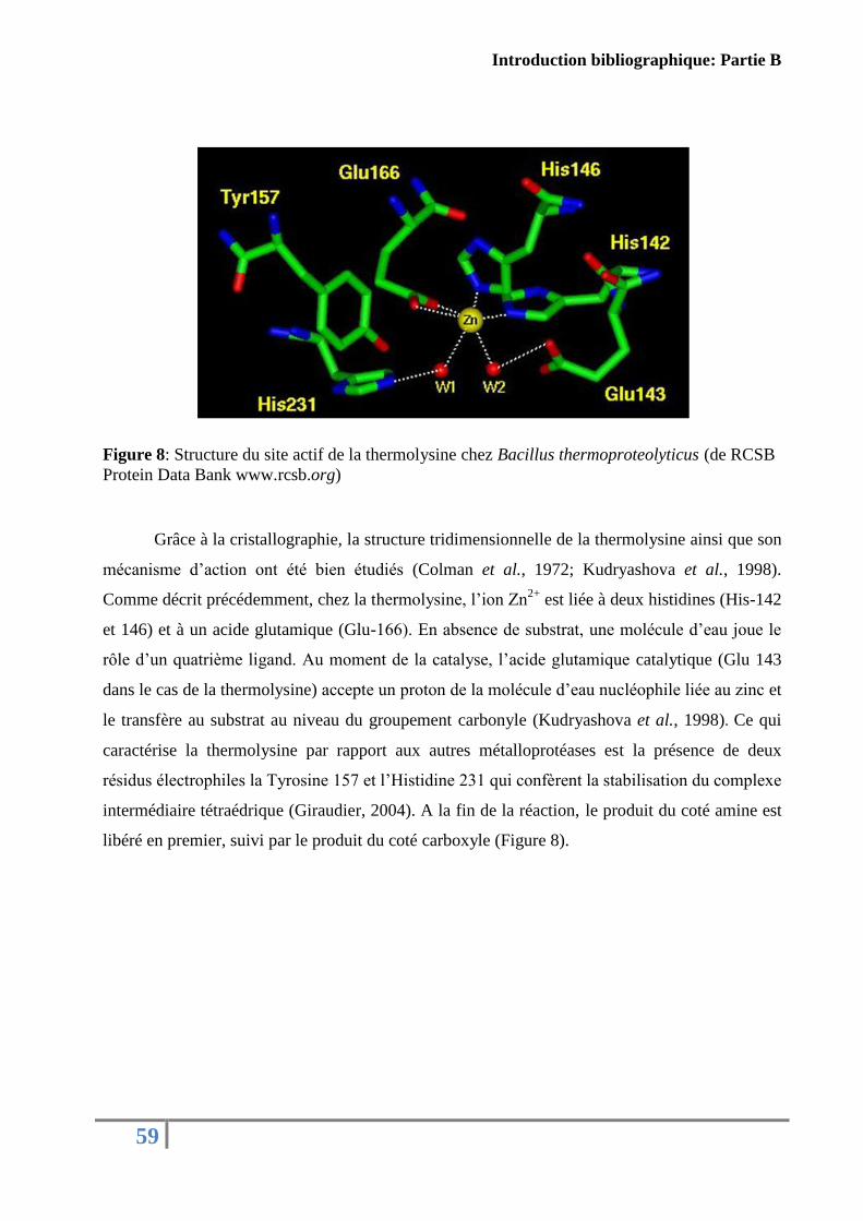
Introduction bibliographique: Partie B
59
Figure 8: Structure du site actif de la thermolysine chez Bacillus thermoproteolyticus (de RCSB
Protein Data Bank www.rcsb.org)
Grâce à la cristallographie, la structure tridimensionnelle de la thermolysine ainsi que son
mécanisme d’action ont été bien étudiés (Colman et al., 1972; Kudryashova et al., 1998).
Comme décrit précédemment, chez la thermolysine, l’ion Zn2+
est liée à deux histidines (His-142
et 146) et à un acide glutamique (Glu-166). En absence de substrat, une molécule d’eau joue le
rôle d’un quatrième ligand. Au moment de la catalyse, l’acide glutamique catalytique (Glu 143
dans le cas de la thermolysine) accepte un proton de la molécule d’eau nucléophile liée au zinc et
le transfère au substrat au niveau du groupement carbonyle (Kudryashova et al., 1998). Ce qui
caractérise la thermolysine par rapport aux autres métalloprotéases est la présence de deux
résidus électrophiles la Tyrosine 157 et l’Histidine 231 qui confèrent la stabilisation du complexe
intermédiaire tétraédrique (Giraudier, 2004). A la fin de la réaction, le produit du coté amine est
libéré en premier, suivi par le produit du coté carboxyle (Figure 8).
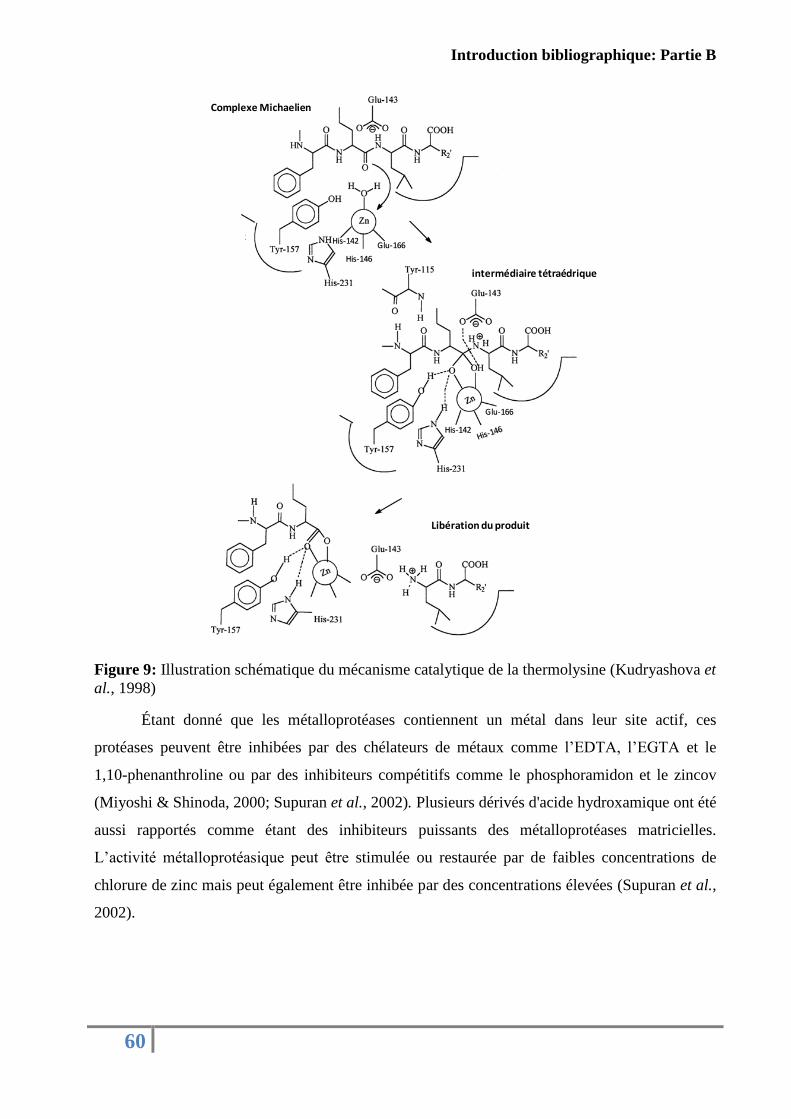
Introduction bibliographique: Partie B
60
Figure 9: Illustration schématique du mécanisme catalytique de la thermolysine (Kudryashova et
al., 1998)
Étant donné que les métalloprotéases contiennent un métal dans leur site actif, ces
protéases peuvent être inhibées par des chélateurs de métaux comme l’EDTA, l’EGTA et le
1,10-phenanthroline ou par des inhibiteurs compétitifs comme le phosphoramidon et le zincov
(Miyoshi & Shinoda, 2000; Supuran et al., 2002). Plusieurs dérivés d'acide hydroxamique ont été
aussi rapportés comme étant des inhibiteurs puissants des métalloprotéases matricielles.
L’activité métalloprotéasique peut être stimulée ou restaurée par de faibles concentrations de
chlorure de zinc mais peut également être inhibée par des concentrations élevées (Supuran et al.,
2002).
Complexe Michaelien
Libération du produit
intermédiaire tétraédrique
His-142
His-146
Glu-166
His-142
Glu-166
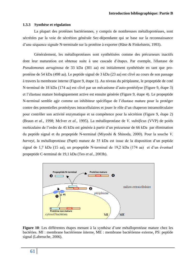
Introduction bibliographique: Partie B
61
1.3.3 Synthèse et régulation
La plupart des protéines bactériennes, y compris de nombreuses métalloprotéases, sont
sécrétées par la voie de sécrétion générale Sec-dépendante qui se base sur la reconnaissance
d’une séquence signale N-terminale sur la protéine à exporter (Häse & Finkelstein, 1993).
Généralement, les métalloprotéases sont synthétisées comme des précurseurs inactifs
dont leur maturation est obtenue suite à une cascade d’étapes. Par exemple, l'élastase de
Pseudomonas aeruginosa de 33 kDa (301 aa) est initialement synthétisée en tant que pro-
protéine de 54 kDa (498 aa). Le peptide signal de 3 kDa (23 aa) est clivé au cours de son passage
à travers la membrane interne (Figure 9, étape 1). Au niveau du périplasme, le propeptide de coté
N-terminal de 18 kDa (174 aa) est clivé par un mécanisme d’auto-protéolyse (Figure 9, étape 3)
et l’élastase mature biologiquement active est ensuite générée (Figure 9, étape 4). Le propeptide
N-terminal semble agir comme un inhibiteur spécifique de l’élastase mature pour la protéger
contre des potentielles protéolyses intracellulaires et jouer le rôle d’un chaperon intramoléculaire
pour contrôler son activité enzymatique et sa compétence pour la sécrétion (Figure 9, étape 2)
(Braun et al., 1998; McIver et al., 1995). La métalloprotéase de V. vulnificus (VVP) de poids
moléculaire de l’ordre de 45 kDa est générée à partir d’un précurseur de 66 kDa par élimination
du peptide signal et du propeptide N-terminal (Miyoshi & Shinoda, 2000). Pour la souche V.
harveyi, la métalloprotéase (Pap6) mature de 35 kDa est issue de la disposition d’un peptide
signal de 1,7 kDa (15 aa), un propeptide N-terminal de 19,2 kDa (174 aa) et d’un éventuel
propeptide C-terminal de 19,1 kDa (Teo et al., 2003b).
Figure 10: Les différentes étapes menant à la synthèse d’une métalloprotéase mature chez les
bactéries. MI : membrane bactérienne interne, ME : membrane bactérienne externe, PS: peptide
signal (Labreuche, 2006).
PS
Propeptide N-terminal
Protéine non mature
Protéine mature
Introduction bibliographique: Partie B
62
Concernant la régulation des métalloprotéases, il a été démontré que l’expression de Vvp
(V. vulnificus), hemagglutinine/protease (V. cholerae) et l’EmpA (V. anguillarum) est régulée
par Smcr, HapR et VanS respectivement, qui sont des systèmes homologues à LuxR. Cette
protéine est un activateur de transcription qui contrôle l'expression de l'opéron lux chez V.
harveyi à haute densité bactérienne et qui fait partie du quorum-sensing (Shao & Hor, 2001).
Chez V. tubiashii, un système de régulation de type VtpR (pour V. tubiashii protease regulator)
homologue au HapR de V. cholerae a été décrit comme responsable de l’activation de la
sécrétion de deux métalloprotéases VtpA et VtpB (Hasegawa & Häse, 2009b).
Il existe également des inhibiteurs endogènes spécifiques de types TIMPs (Tissue Inhibitors of
Matrix Metalloproteases) pour réguler l’activité protéasique des métalloprotéases matricielles
(MMP, famille des matrixines) (Gomis-Rüth et al., 1997). Chez les vertébrés, ces TIMPs
occupent plusieurs fonctions biochimiques, physiologiques et biologiques, y compris l'inhibition
de la MMP active, la promotion de la croissance cellulaire, l'inhibition de l'angiogenèse et de
l'induction de l'apoptose (Brew et al., 2000). Chez les invertébrés, un gène codant une protéine
comportant une organisation et une signature analogue à celle des TIMPs de vertébrés a été
caractérisé chez l’huître creuse C. gigas. L’expression de la TIMPs est régulée positivement
quand l’huître est soumise à des dommages au niveau de sa coque ou à des infections
bactériennes (Montagnani et al., 2001).
1.3.4 Implication des métalloprotéases dans la virulence du genre Vibrio
L’implication des métalloprotéases dans la virulence des bactéries du genre Vibrio fait
l’objet d’un intérêt marqué, comme l’atteste le nombre croissant des publications sur le sujet.
Différents travaux ont ainsi établi que les métalloprotéases exercent des effets pathologiques
variés. En plus de leur pouvoir protéolytique via la dégradation d’une grande variété de substrats
protéiques de l’hôte, comme la lactoferrine, le collagène, l’élastine, les protéines plasmatiques,
ou encore l’albumine, les métalloprotéases entraînent l’augmentation de la perméabilité
vasculaire, le déclenchement de réactions hémorragiques liées à la stimulation du système
kallicreine-kinine, l’hémagglutination des érythrocytes et l’induction de nécroses tissulaires
(Shinoda et al., 1996; Miyoshi et al., 2002). Les différentes métalloprotéases sécrétées par des
espèces de Vibrio ainsi que leurs rôles biologiques dans la virulence sont référencées dans le
tableau présenté ci après.
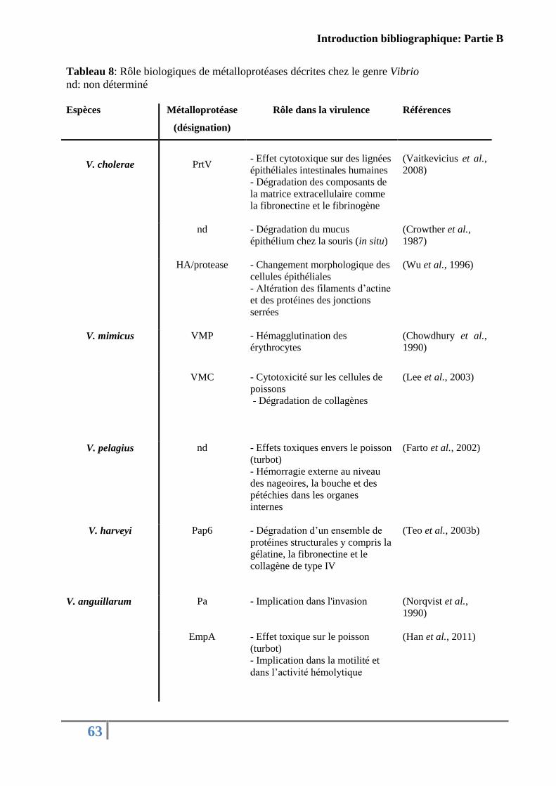
Introduction bibliographique: Partie B
63
Tableau 8: Rôle biologiques de métalloprotéases décrites chez le genre Vibrio
nd: non déterminé
Espèces Métalloprotéase
(désignation)
Rôle dans la virulence Références
V. cholerae
PrtV
- Effet cytotoxique sur des lignées
épithéliales intestinales humaines
- Dégradation des composants de
la matrice extracellulaire comme
la fibronectine et le fibrinogène
(Vaitkevicius et al.,
2008)
nd - Dégradation du mucus
épithélium chez la souris (in situ)
(Crowther et al.,
1987)
HA/protease - Changement morphologique des
cellules épithéliales
- Altération des filaments d’actine
et des protéines des jonctions
serrées
(Wu et al., 1996)
V. mimicus VMP - Hémagglutination des
érythrocytes
(Chowdhury et al.,
1990)
VMC - Cytotoxicité sur les cellules de
poissons
- Dégradation de collagènes
(Lee et al., 2003)
V. pelagius nd - Effets toxiques envers le poisson
(turbot)
- Hémorragie externe au niveau
des nageoires, la bouche et des
pétéchies dans les organes
internes
(Farto et al., 2002)
V. harveyi Pap6 - Dégradation d’un ensemble de
protéines structurales y compris la
gélatine, la fibronectine et le
collagène de type IV
(Teo et al., 2003b)
V. anguillarum
Pa - Implication dans l'invasion (Norqvist et al.,
1990)
EmpA - Effet toxique sur le poisson
(turbot)
- Implication dans la motilité et
dans l’activité hémolytique
(Han et al., 2011)
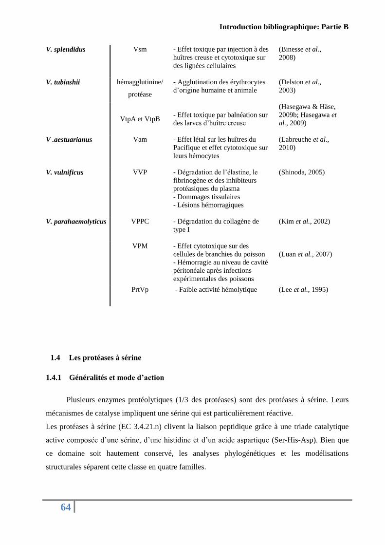
Introduction bibliographique: Partie B
64
V. splendidus Vsm - Effet toxique par injection à des
huîtres creuse et cytotoxique sur
des lignées cellulaires
(Binesse et al.,
2008)
V. tubiashii hémagglutinine/
protéase
- Agglutination des érythrocytes
d’origine humaine et animale (Delston et al.,
2003)
VtpA et VtpB
- Effet toxique par balnéation sur
des larves d’huître creuse
(Hasegawa & Häse,
2009b; Hasegawa et
al., 2009)
V .aestuarianus Vam - Effet létal sur les huîtres du
Pacifique et effet cytotoxique sur
leurs hémocytes
(Labreuche et al.,
2010)
V. vulnificus VVP - Dégradation de l’élastine, le
fibrinogène et des inhibiteurs
protéasiques du plasma
- Dommages tissulaires
- Lésions hémorragiques
(Shinoda, 2005)
V. parahaemolyticus
VPPC - Dégradation du collagène de
type I
(Kim et al., 2002)
VPM - Effet cytotoxique sur des
cellules de branchies du poisson
- Hémorragie au niveau de cavité
péritonéale après infections
expérimentales des poissons
(Luan et al., 2007)
PrtVp - Faible activité hémolytique (Lee et al., 1995)
1.4 Les protéases à sérine
1.4.1 Généralités et mode d’action
Plusieurs enzymes protéolytiques (1/3 des protéases) sont des protéases à sérine. Leurs
mécanismes de catalyse impliquent une sérine qui est particulièrement réactive.
Les protéases à sérine (EC 3.4.21.n) clivent la liaison peptidique grâce à une triade catalytique
active composée d’une sérine, d’une histidine et d’un acide aspartique (Ser-His-Asp). Bien que
ce domaine soit hautement conservé, les analyses phylogénétiques et les modélisations
structurales séparent cette classe en quatre familles.
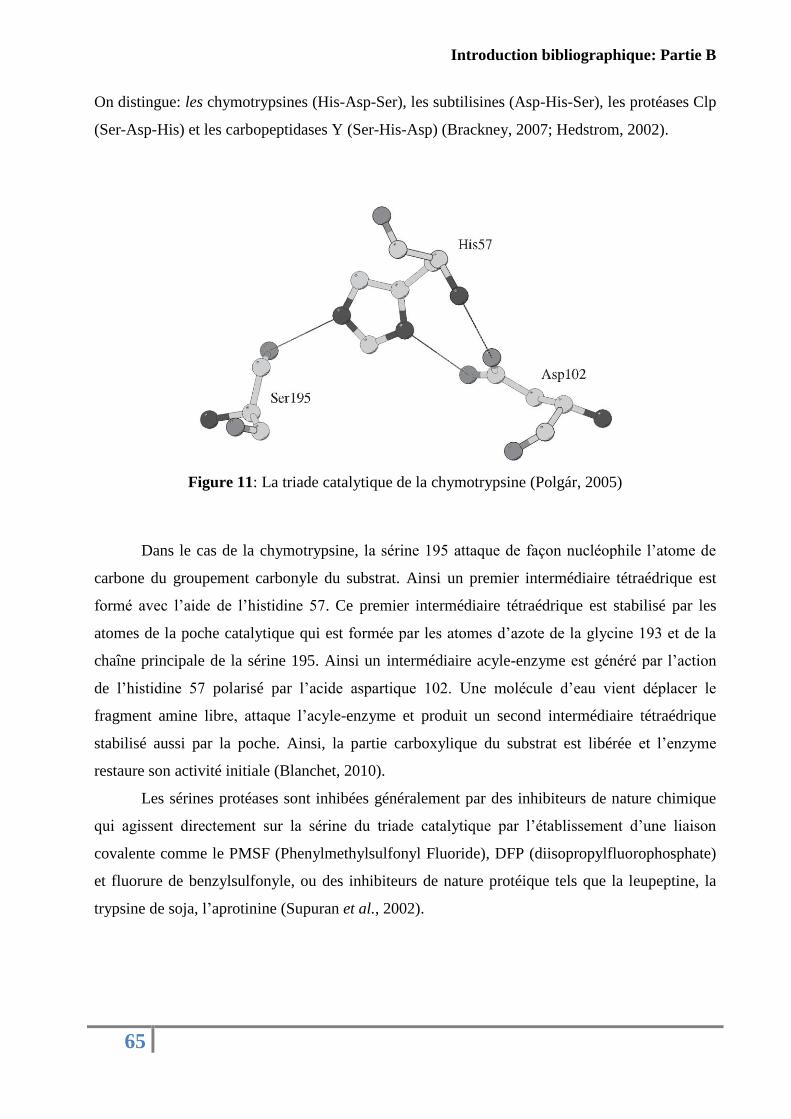
Introduction bibliographique: Partie B
65
On distingue: les chymotrypsines (His-Asp-Ser), les subtilisines (Asp-His-Ser), les protéases Clp
(Ser-Asp-His) et les carbopeptidases Y (Ser-His-Asp) (Brackney, 2007; Hedstrom, 2002).
Figure 11: La triade catalytique de la chymotrypsine (Polgár, 2005)
Dans le cas de la chymotrypsine, la sérine 195 attaque de façon nucléophile l’atome de
carbone du groupement carbonyle du substrat. Ainsi un premier intermédiaire tétraédrique est
formé avec l’aide de l’histidine 57. Ce premier intermédiaire tétraédrique est stabilisé par les
atomes de la poche catalytique qui est formée par les atomes d’azote de la glycine 193 et de la
chaîne principale de la sérine 195. Ainsi un intermédiaire acyle-enzyme est généré par l’action
de l’histidine 57 polarisé par l’acide aspartique 102. Une molécule d’eau vient déplacer le
fragment amine libre, attaque l’acyle-enzyme et produit un second intermédiaire tétraédrique
stabilisé aussi par la poche. Ainsi, la partie carboxylique du substrat est libérée et l’enzyme
restaure son activité initiale (Blanchet, 2010).
Les sérines protéases sont inhibées généralement par des inhibiteurs de nature chimique
qui agissent directement sur la sérine du triade catalytique par l’établissement d’une liaison
covalente comme le PMSF (Phenylmethylsulfonyl Fluoride), DFP (diisopropylfluorophosphate)
et fluorure de benzylsulfonyle, ou des inhibiteurs de nature protéique tels que la leupeptine, la
trypsine de soja, l’aprotinine (Supuran et al., 2002).
Introduction bibliographique: Partie B
66
1.4.2 Synthèse et régulation
Les protéases à sérine sont synthétisées sous forme de zymogènes inactives avec un
peptide signal de 18 à 26 aa, un pro-dipeptide et une protéase mature. Directement après leur
synthèse, le peptide signal est éliminé par un signal peptidase en laissant une pro-séquence N-
terminale composée de deux résidus. Pour leur activation, les sérines protéases doivent éliminer
la séquence pro-dipeptide qui nécessite la contribution d’une dipeptidyl peptidase I (DPPI)
(Pham, 2006).
Concernant leurs mécanismes de régulation, il a été rapporté que chez V. vulnificus
l’expression et la production d’une sérine protéase (VvsA) dépendent du QS pendant les
premières heures de la phase stationnaire de la croissance bactérienne. Il a été démontré que la
perturbation du gène luxS (responsable de la production des autoinducteurs du type 2 (AI-2))
cause la diminution de l’expression de VvsA et la production de VvsA (Elgaml et al., 2013)
1.4.3 Les protéases à sérine dans le genre Vibrio
Plusieurs protéases à sérine ont été décrites comme des facteurs de virulence chez le
genre Vibrio. Pour V. alginolyticus, pathogène de crevette impériale (Penaeus japonicus), une
sérine protéase purifiée a été identifiée comme une toxine létale avec une DL50 de 0,29 µg de
protéines/ g. Des propriétés anticoagulantes de l’hémolymphe ont été aussi observées suite à
l’injection de cette toxine aux crevettes (Chen et al., 1999; Lee et al., 1997). En 2002, Lee et
collaborateurs ont caractérisé une protéase à sérine chez V. parahaemolyticus. Suite à une
injection par voie intrapéritonéale et par voie intraveineuse, la protéine purifiée provoque une
hémorragie tissulaire et la mort des souris. Également, chez V. cholerae, une sérine protéase a été
identifiée comme responsable des hémorragies au niveau de l'intestin grêle distal du lapin
(Syngkon et al., 2010) et nouvellement une autre sérine protéase extracellulaire (VesB)
appartenant à la superfamille des chymotrypsines a été caractérisée (Gadwal et al., 2014). Enfin,
une nouvelle protéase à sérine a été identifiée dans le sécrétome de V. metschnikovii. Cette sérine
protéase a montré sa capacité de cliver plusieurs protéines de la coagulation sanguine, dont le
fibrinogène, la prothrombine et la thrombine (Park et al., 2012).
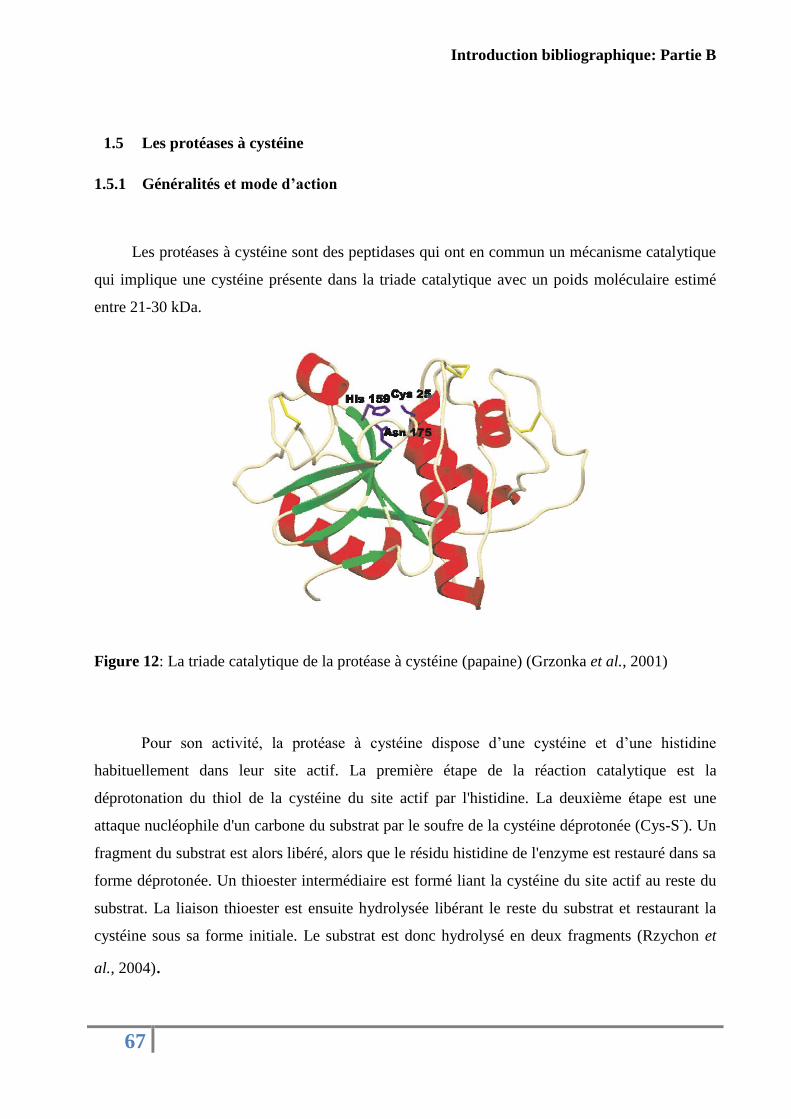
Introduction bibliographique: Partie B
67
1.5 Les protéases à cystéine
1.5.1 Généralités et mode d’action
Les protéases à cystéine sont des peptidases qui ont en commun un mécanisme catalytique
qui implique une cystéine présente dans la triade catalytique avec un poids moléculaire estimé
entre 21-30 kDa.
Figure 12: La triade catalytique de la protéase à cystéine (papaine) (Grzonka et al., 2001)
Pour son activité, la protéase à cystéine dispose d’une cystéine et d’une histidine
habituellement dans leur site actif. La première étape de la réaction catalytique est la
déprotonation du thiol de la cystéine du site actif par l'histidine. La deuxième étape est une
attaque nucléophile d'un carbone du substrat par le soufre de la cystéine déprotonée (Cys-S-). Un
fragment du substrat est alors libéré, alors que le résidu histidine de l'enzyme est restauré dans sa
forme déprotonée. Un thioester intermédiaire est formé liant la cystéine du site actif au reste du
substrat. La liaison thioester est ensuite hydrolysée libérant le reste du substrat et restaurant la
cystéine sous sa forme initiale. Le substrat est donc hydrolysé en deux fragments (Rzychon et
al., 2004).
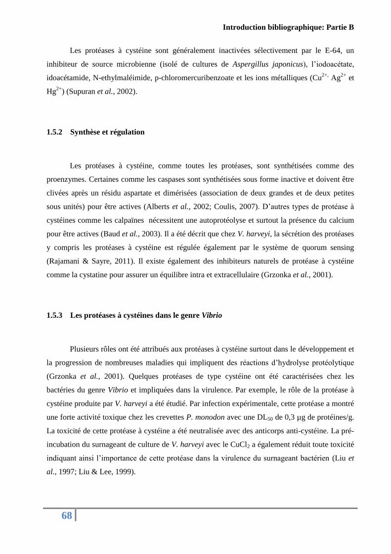
Introduction bibliographique: Partie B
68
Les protéases à cystéine sont généralement inactivées sélectivement par le E-64, un
inhibiteur de source microbienne (isolé de cultures de Aspergillus japonicus), l’iodoacétate,
idoacétamide, N-ethylmaléimide, p-chloromercuribenzoate et les ions métalliques (Cu2+,
Ag2+
et
Hg2+
) (Supuran et al., 2002).
1.5.2 Synthèse et régulation
Les protéases à cystéine, comme toutes les protéases, sont synthétisées comme des
proenzymes. Certaines comme les caspases sont synthétisées sous forme inactive et doivent être
clivées après un résidu aspartate et dimérisées (association de deux grandes et de deux petites
sous unités) pour être actives (Alberts et al., 2002; Coulis, 2007). D’autres types de protéase à
cystéines comme les calpaïnes nécessitent une autoprotéolyse et surtout la présence du calcium
pour être actives (Baud et al., 2003). Il a été décrit que chez V. harveyi, la sécrétion des protéases
y compris les protéases à cystéine est régulée également par le système de quorum sensing
(Rajamani & Sayre, 2011). Il existe également des inhibiteurs naturels de protéase à cystéine
comme la cystatine pour assurer un équilibre intra et extracellulaire (Grzonka et al., 2001).
1.5.3 Les protéases à cystéines dans le genre Vibrio
Plusieurs rôles ont été attribués aux protéases à cystéine surtout dans le développement et
la progression de nombreuses maladies qui impliquent des réactions d’hydrolyse protéolytique
(Grzonka et al., 2001). Quelques protéases de type cystéine ont été caractérisées chez les
bactéries du genre Vibrio et impliquées dans la virulence. Par exemple, le rôle de la protéase à
cystéine produite par V. harveyi a été étudié. Par infection expérimentale, cette protéase a montré
une forte activité toxique chez les crevettes P. monodon avec une DL50 de 0,3 µg de protéines/g.
La toxicité de cette protéase à cystéine a été neutralisée avec des anticorps anti-cystéine. La pré-
incubation du surnageant de culture de V. harveyi avec le CuCl2 a également réduit toute toxicité
indiquant ainsi l’importance de cette protéase dans la virulence du surnageant bactérien (Liu et
al., 1997; Liu & Lee, 1999).
Introduction bibliographique: Partie B
69
En plus, Lee et collaborateurs (1999) ont montré que cette protéase interfère avec les
propriétés de coagulation de l’hémolymphe de crevette et réduit la quantité du coagulogène dans
le plasma. Aguirre-Guzman et collaborateurs (2005) ont démontré aussi qu’une protéase purifiée,
à partir du surnageant de culture de V. penaeicida, présente des caractéristiques biochimiques des
protéases de type cystéine (comme l’inhibition par le CuCl2 ou l’E-64) et est associée à des
activités toxiques chez la crevette blanche L. vannamei.
2. Les sidérophores
2.1 Généralités
Le mot sidérophore, du grec pherein et sidero, signfie porter le fer. Les sidérophores sont
des peptides de bas poids moléculaire compris entre 200 et 2000 Da. Ces métabolites secondaires
sont synthétisés et sécrétés particulièrement par les bactéries et les champignons en réponse à des
conditions de carence en fer (Butler & Theisen, 2010). Leur rôle est de chélater et d’extraire le
fer ferrique (Fe3+
) de nombreux complexes minéraux ou organiques et de le rendre ainsi
accessible aux cellules microbiennes (Neilands, 1995).
Les sidérophores forment généralement des hexadentates (molécules à six ligands pour
fixer l’ion fer) octaèdre complexés avec le fer ferrique et emploient spécifiquement les
catécholates, les hydroxamates, les carboxylates et les phénolates comme des ligands efficaces
du Fe3+
(Andrews et al., 2003). Il existe plus de 500 sidérophores différents chez les bactéries
Gram négatives comme les Gram positives.
Le fer est un élément essentiel dans la vie des microorganismes aérobies qui participe à
de nombreuses voies métaboliques y compris la réduction de l’oxygène pour la synthèse de
l'ATP, la réduction des ribonucleotides et la formation de l'hème (Neilands, 1995). Les bactéries
ont besoin d’une concentration minimale de fer de l’ordre de 10-5
M pour leur croissance. Une
carence en cet élément engendre un effet bactériostatique et l’extrême carence génère un effet
bactéricide.
En conséquence, plusieurs genres de bactéries produisent des sidérophores afin de
pomper le fer essentiel à leur croissance et leur développement dans les environnements pauvres
comme le sol et les milieux aquatiques.
Introduction bibliographique: Partie B
70
Figure 13: Exemple de sidérophores chez le genre Vibrio
2.2 Mode d’action
Après la reconnaissance de l’ion Fe3+
par les sidérophores, le complexe est transporté à
travers la membrane externe par un récepteur spécifique. Ce passage nécessite un transporteur
hautement spécifique à chaque sidérophore et un transducteur d’énergie. Dans le cas des
bactéries à Gram négatifs, l’énergie requise est procurée et transmise par le complexe TonB-
ExbBExbD localisé dans la membrane interne (Figure 13). Des protéines de liaison permettent
ensuite le transport du complexe ferri-sidérophore du récepteur à la membrane interne et par
suite à travers les perméases via les transporteurs de type ABC (ATP Binding Cassette) qui
utilise l’ATP comme énergie, où il se dissocie soit par digestion partielle, soit par un processus
réductif et libère l’ion Fe2+.
Anguibactine
V. anguillarumFluvibactineV. fluvialis
VanchrobactinV. anguillarum Vulnibactine
V. vulnificus
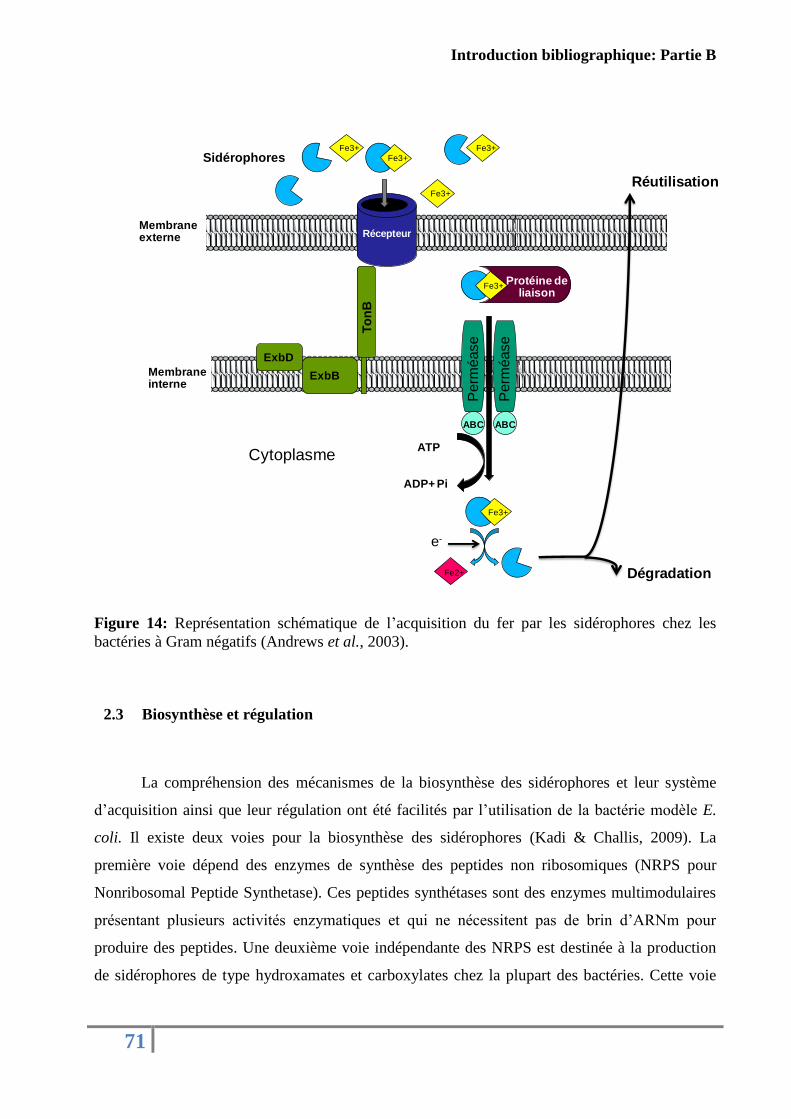
Introduction bibliographique: Partie B
71
Figure 14: Représentation schématique de l’acquisition du fer par les sidérophores chez les
bactéries à Gram négatifs (Andrews et al., 2003).
2.3 Biosynthèse et régulation
La compréhension des mécanismes de la biosynthèse des sidérophores et leur système
d’acquisition ainsi que leur régulation ont été facilités par l’utilisation de la bactérie modèle E.
coli. Il existe deux voies pour la biosynthèse des sidérophores (Kadi & Challis, 2009). La
première voie dépend des enzymes de synthèse des peptides non ribosomiques (NRPS pour
Nonribosomal Peptide Synthetase). Ces peptides synthétases sont des enzymes multimodulaires
présentant plusieurs activités enzymatiques et qui ne nécessitent pas de brin d’ARNm pour
produire des peptides. Une deuxième voie indépendante des NRPS est destinée à la production
de sidérophores de type hydroxamates et carboxylates chez la plupart des bactéries. Cette voie
Fe3+
Fe3+
Fe3+
Fe3+
Récepteur
Fe3+Protéine de
liaison
ATP
ADP+ Pi
Fe3+
Fe2+
Cytoplasme
Membrane interne
Membrane externe
ABC
Perm
éase
Perm
éase
ABC
ExbD
ExbBT
on
B
Dégradation
Réutilisation
e-
Sidérophores
Introduction bibliographique: Partie B
72
repose sur plusieurs activités enzymatiques telles que la mono-oxygénase, la décarboxylase,
l’aminotransférase, la ligase, et l’aldolase (Miethke & Marahiel, 2007).
Pour éviter la privation ou la surabondance de fer, les bactéries possèdent des systèmes de
régulation pour maintenir un taux suffisant en fer. Chez les bactéries Gram négatif, il existe un
facteur de régulation Fur (pour Ferric Uptake Regulator) conservé dans les espèces E. coli,
Vibrio et Yersinia. Ce facteur joue un rôle fondamental dans la régulation de l’expression des
sidérophores (Massé & Arguin, 2005; Neilands, 1995). D’autres facteurs sont aussi impliqués
dans la régulation de la biosynthèse de certains sidérophores et ses protéines d’acquisition
comme les facteurs -ECF (σ-ExtraCytoplasmic Function). Ces derniers sont des facteurs
alternatifs qui se fixent sur l’ARN polymérase pour réguler la transcription d’un nombre de
gènes en réponse à des changements extracellulaires (Potvin et al., 2008). En cas de carence en
fer, une cascade de signalisation via les facteurs σ-ECF va activer la transcription des gènes de
production et d’acquisition du sidérophore et dans le cas d’un excès, un rétrocontrôle génique par
les facteurs de répression Fur bloque la production des sidérophores ainsi que ses systèmes
d’acquisition (Yeterian, 2010).
2.4 Sidérophores chez Vibrio
Beaucoup de vibrions produisent des sidérophores comme une stratégie pour séquestrer le
fer dans le milieu marin. Le rôle des sidérophores dans la virulence des bactéries a été souvent
démontré. Chez V. vulnificus, en utilisant des mutants incapables de produire des sidérophores et
d’acquérir de fer à partir de la transferrine, Litwin et collaborateurs (1996) ont prouvé la
diminution de la virulence de V. vulnificus envers des souris. Également, une autre étude a
montré l’importance des sidérophores dans la virulence de Staphylococcus aureus (Dale et al.,
2004). Chez V. anguillarum, il a été confirmé le rôle des transporteurs d’anguibactine dans la
virulence de la souche (Stork et al., 2004). Cependant, Sigel et collaborateurs (1985) ont montré
que les sidérophores et leurs transporteurs ne sont pas nécessaires pour la virulence V. cholerae
et qu’elle peut utiliser le citrate ou l'asparagine pour sa croissance dans les milieux pauvres en
fer.
Une grande diversité de structures a été observée chez les sidérophores produits par les
Vibrio. Le tableau 9 illustre les différents sidérophores secrétés par le genre Vibrio.
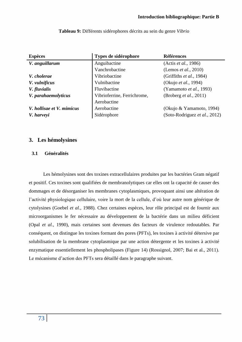
Introduction bibliographique: Partie B
73
Tableau 9: Différents sidérophores décrits au sein du genre Vibrio
Espèces Types de sidérophore Références
V. anguillarum Anguibactine
Vanchrobactine
(Actis et al., 1986)
(Lemos et al., 2010)
V. cholerae Vibriobactine (Griffiths et al., 1984)
V. vulnificus Vulnibactine (Okujo et al., 1994)
V. fluvialis Fluvibactine (Yamamoto et al., 1993)
V. parahaemolyticus Vibrioferrine, Ferrichrome,
Aerobactine
(Broberg et al., 2011)
V. hollisae et V. mimicus Aerobactine (Okujo & Yamamoto, 1994)
V. harveyi Sidérophore (Soto-Rodriguez et al., 2012)
3. Les hémolysines
3.1 Généralités
Les hémolysines sont des toxines extracellulaires produites par les bactéries Gram négatif
et positif. Ces toxines sont qualifiées de membranolytiques car elles ont la capacité de causer des
dommages et de désorganiser les membranes cytoplasmiques, provoquant ainsi une altération de
l’activité physiologique cellulaire, voire la mort de la cellule, d’où leur autre nom générique de
cytolysines (Goebel et al., 1988). Chez certaines espèces, leur rôle principal est de fournir aux
microorganismes le fer nécessaire au développement de la bactérie dans un milieu déficient
(Opal et al., 1990), mais certaines sont devenues des facteurs de virulence redoutables. Par
conséquent, on distingue les toxines formant des pores (PFTs), les toxines à activité détersive par
solubilisation de la membrane cytoplasmique par une action détergente et les toxines à activité
enzymatique essentiellement les phospholipases (Figure 14) (Rossignol, 2007; Bai et al., 2011).
Le mécanisme d’action des PFTs sera détaillé dans le paragraphe suivant.
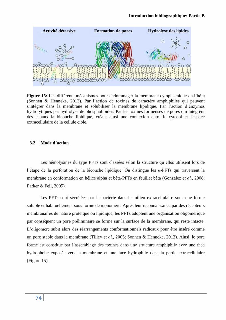
Introduction bibliographique: Partie B
74
Figure 15: Les différents mécanismes pour endommager la membrane cytoplasmique de l’hôte
(Sonnen & Henneke, 2013). Par l’action de toxines de caractère amphiphiles qui peuvent
s'intégrer dans la membrane et solubiliser la membrane lipidique. Par l’action d’enzymes
hydrolytiques par hydrolyse de phospholipides. Par les toxines formeuses de pores qui intègrent
des canaux la bicouche lipidique, créant ainsi une connexion entre le cytosol et l'espace
extracellulaire de la cellule cible.
3.2 Mode d’action
Les hémolysines du type PFTs sont classées selon la structure qu’elles utilisent lors de
l’étape de la perforation de la bicouche lipidique. On distingue les α-PFTs qui traversent la
membrane en conformation en hélice alpha et bêta-PFTs en feuillet bêta (Gonzalez et al., 2008;
Parker & Feil, 2005).
Les PFTs sont sécrétées par la bactérie dans le milieu extracellulaire sous une forme
soluble et habituellement sous forme de monomère. Après leur reconnaissance par des récepteurs
membranaires de nature protéique ou lipidique, les PFTs adoptent une organisation oligomérique
par conséquent un pore préliminaire se forme sur la surface de la membrane, qui reste intacte.
L’oligomère subit alors des réarrangements conformationnels radicaux pour être inséré comme
un pore stable dans la membrane (Tilley et al., 2005; Sonnen & Henneke, 2013). Ainsi, le pore
formé est constitué par l’assemblage des toxines dans une structure amphiphile avec une face
hydrophobe exposée vers la membrane et une face hydrophile dans la partie extracellulaire
(Figure 15).
Activité détersive Formation de pores Hydrolyse des lipides
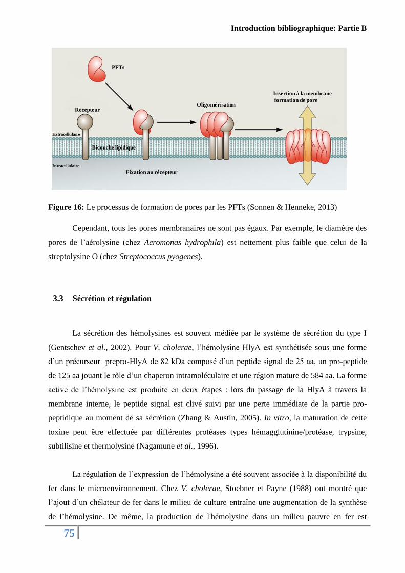
Introduction bibliographique: Partie B
75
Figure 16: Le processus de formation de pores par les PFTs (Sonnen & Henneke, 2013)
Cependant, tous les pores membranaires ne sont pas égaux. Par exemple, le diamètre des
pores de l’aérolysine (chez Aeromonas hydrophila) est nettement plus faible que celui de la
streptolysine O (chez Streptococcus pyogenes).
3.3 Sécrétion et régulation
La sécrétion des hémolysines est souvent médiée par le système de sécrétion du type I
(Gentschev et al., 2002). Pour V. cholerae, l’hémolysine HlyA est synthétisée sous une forme
d’un précurseur prepro-HlyA de 82 kDa composé d’un peptide signal de 25 aa, un pro-peptide
de 125 aa jouant le rôle d’un chaperon intramoléculaire et une région mature de 584 aa. La forme
active de l’hémolysine est produite en deux étapes : lors du passage de la HlyA à travers la
membrane interne, le peptide signal est clivé suivi par une perte immédiate de la partie pro-
peptidique au moment de sa sécrétion (Zhang & Austin, 2005). In vitro, la maturation de cette
toxine peut être effectuée par différentes protéases types hémagglutinine/protéase, trypsine,
subtilisine et thermolysine (Nagamune et al., 1996).
La régulation de l’expression de l’hémolysine a été souvent associée à la disponibilité du
fer dans le microenvironnement. Chez V. cholerae, Stoebner et Payne (1988) ont montré que
l’ajout d’un chélateur de fer dans le milieu de culture entraîne une augmentation de la synthèse
de l’hémolysine. De même, la production de l'hémolysine dans un milieu pauvre en fer est
Récepteur
PFTs
Fixation au récepteur
Oligomérisation
Insertion à la membrane
formation de pore
Extracellulaire
Intracellulaire
Bicouche lipidique
Introduction bibliographique: Partie B
76
réprimée par l'addition de fer. Récemment, chez V. vulnificus, Lee et collaborateurs (2013) ont
démontré qu’au niveau transcriptionel, la production d’hémolysine VvhA est contrôlée par le
facteur Fur (un facteur de transcription majeur contrôlant l'homéostasie du fer). Au niveau post-
traductionnel, ils ont prouvé que la régulation de VvhA dépend de l’expression des protéases qui
la dégradent comme la VvpE (V. vulnificus elastase) et la VvpM (V. vulnificus metalloprotease).
3.4 Implication des hémolysines dans la virulence des Vibrio
Chez les Vibrionaceae, plusieurs hémolysines ainsi que leurs rôles dans la virulence ont
été décrits. Chez V. cholerae, la plupart des souches non-O1, produisent une toxine cytolytique
sécrétée dans le milieu extérieur : la toxine El Tor cytolysine/hémolysine ou HlyA (Ichinose et
al., 1987). Cette toxine présente une activité entérotoxique et apparaît comme la cause principale
de la gastroentérite causée par les souches V. cholerae non-O1. Chez V. vulnificus, une exotoxine
de type hémolysine (VvhA) a été identifiée (Gray & Kreger, 1985). En plus de son rôle dans la
libération du fer de l'hémoglobine et la formation de pores dans la membrane cellulaire. Cette
toxine provoque des effets cytotoxiques. L'injection intradermique de la VvhA purifiée à des
souris a entraîné des dommages au niveau des cellules graisseuses, des cellules endothéliales et
des cellules musculaires et des infiltrations inflammatoires modérées. D'autres études ont
démontré que la Vvha stimule l’augmentation de la perméabilité vasculaire, l'apoptose des
cellules endothéliales et l’hyperproduction de l'oxyde nitrique (Horseman & Surani, 2011). De
même, chez V. parahaemolyticus, une hémolysine thermostable directe (TDH) et une
hémolysine apparentée à la TDH (TRH) ont été décrites (T. Honda, 1993). Ces deux toxines
possèdent une activité de type entérotoxique, cardiotoxique et cytotoxique (Nair et al., 2007).
Chez V. harveyi, l’hémolysine (VHH) de type phospholipase a montré une cytotoxicité contre
des cellules de poisson et une forte pathogénicité envers le poisson flet par injection qui s’est
manifestée par des hémorragies au niveau de la cavité péritonéale et la formation des zones
nécrotiques à l’endroit de l’injection (Zhong et al., 2006). En plus, la VHH a provoqué des
changements structuraux et une induction de l’apoptose chez les cellules de flet (Bai et al.,
2010). Chez V. anguillarum, quatre hémolysines ont été identifiées VAH 2, VAH 3, VAH 4 et
VAH 5 (Rodkhum et al., 2005b). Les hémolysines purifiées ont montré des activités
hémolytiques contre les érythrocytes de poissons, de moutons et de lapins. Les souches mutées à
un seul gène des quatre ont été trouvées moins virulentes que la souche sauvage envers les truites
Introduction bibliographique: Partie B
77
(Oncorhynchus mykiss), indiquant ainsi que chaque gène d'hémolysine contribue à la virulence
de V. anguillarum. Une hémolysine Vh-rTDH produite par V. hollisae, similaire à l’hémolysine
TDH de V. parahaemolyticus, a été identifiée. Cette toxine a montré des activités cytolytiques
envers plusieurs types d’érythrocytes animales (Yoh et al., 1986). Enfin, chez V. tubiashii, une
cytolysine similaire à celle de Vibrio vulnificus capable de lyser plusieurs types de cellules et des
érythrocytes a été décrite (Kothary et al., 2001).

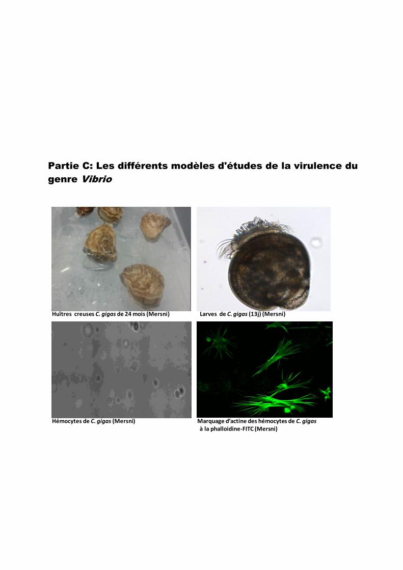
Partie C: Les différents modèles d'études de la virulence du
genre Vibrio
Mersni : marquage d’actine des hémocytes de C. gigas e à la phalloidine-FITc
Hémocytes de C. gigas (Mersni)
Larves de C. gigas (13j) (Mersni)Huîtres creuses C. gigas de 24 mois (Mersni)
Marquage d’actine des hémocytes de C. gigasà la phalloidine-FITC (Mersni)
Introduction bibliographique: Partie C
80
Afin d’évaluer le potentiel de virulence d’un agent pathogène et dans le but
d’identifier de nouvelles cibles thérapeutiques plusieurs modèles d’interactions ont été
adoptés. L’approche la plus classique constitue à étudier in vivo l’impact du microorganisme
pathogène sur des organismes vivants en vue de détecter des manifestations cliniques et une
réponse immunitaire. Des approches in vitro assez simples optant pour des modèles cellulaires
et moléculaires ont été également développées et leurs études ont été facilitées par le progrès
au niveau des techniques d’analyse comme la cytométrie en flux et la microscopie.
En ce qui concerne la compréhension des mortalités observés chez l’huître, il est
évident que l’hôte lui même constitue le meilleur modèle d’interaction afin de mieux
interpréter les stratégies et les mécanismes d’action adoptés par le pathogène. Dans ce
chapitre, nous aborderons les différents modèles d’études utilisés dans la littérature pour
expliquer les types d’interactions entre les vibrions aussi bien que leurs produits de sécrétion
et l’huître par des approches in vivo et in vitro.
1. Approche in vivo
Dans cette partie, nous allons nous focaliser sur l’huître creuse C. gigas car elle
représentera le modèle d’étude utilisé dans ce projet de thèse.
1.1 Modèle d’étude: généralités sur l’huître creuse C. gigas
1.1.1 Systématique et répartition
L'huître creuse C. gigas, aussi appelée huître japonaise, est un mollusque bivalve
appartenant à l'embranchement des Mollusques, classe des Lamellibranches (Bivalvia), ordre
des Filibranchia, sous-ordre des Anisomyaria, super-famille des Ostreoidea, famille des
Ostreidae, genre Crassostrea et espèce gigas.
Historiquement, son foyer évolutif est centré sur l'Asie et plus précisément sur le
Japon. Elle a été introduite dans de plusieurs régions du monde pour des raisons
commerciales. Il s'agit aujourd'hui de l'espèce d'huître la plus cultivée au monde, en Europe
(Grizel & Héral, 1991), sur la côte ouest des États-Unis, au Canada, en Afrique du Sud, en
Tasmanie, en Nouvelle-Zélande ainsi qu’en Russie et en Australie (Orlenko, 1994; Thomson,
1952, 1959) en raison de sa croissance rapide et sa grande tolérance aux conditions
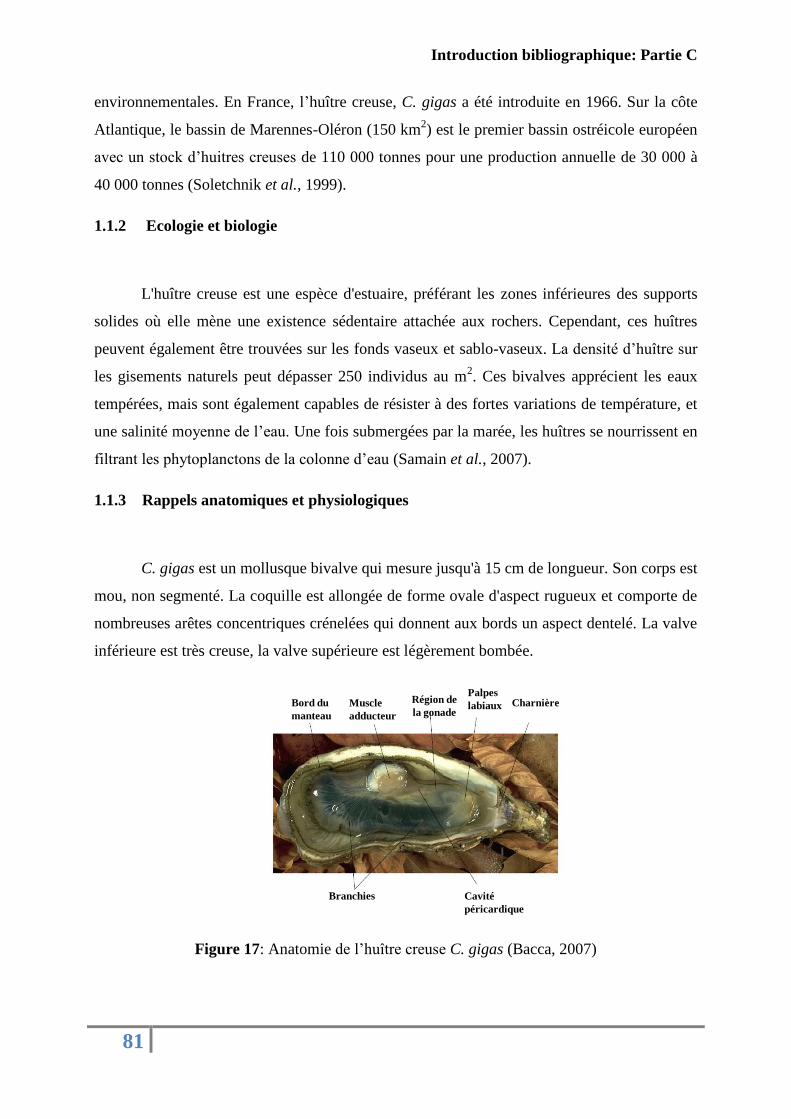
Introduction bibliographique: Partie C
81
environnementales. En France, l’huître creuse, C. gigas a été introduite en 1966. Sur la côte
Atlantique, le bassin de Marennes-Oléron (150 km2) est le premier bassin ostréicole européen
avec un stock d’huitres creuses de 110 000 tonnes pour une production annuelle de 30 000 à
40 000 tonnes (Soletchnik et al., 1999).
1.1.2 Ecologie et biologie
L'huître creuse est une espèce d'estuaire, préférant les zones inférieures des supports
solides où elle mène une existence sédentaire attachée aux rochers. Cependant, ces huîtres
peuvent également être trouvées sur les fonds vaseux et sablo-vaseux. La densité d’huître sur
les gisements naturels peut dépasser 250 individus au m2. Ces bivalves apprécient les eaux
tempérées, mais sont également capables de résister à des fortes variations de température, et
une salinité moyenne de l’eau. Une fois submergées par la marée, les huîtres se nourrissent en
filtrant les phytoplanctons de la colonne d’eau (Samain et al., 2007).
1.1.3 Rappels anatomiques et physiologiques
C. gigas est un mollusque bivalve qui mesure jusqu'à 15 cm de longueur. Son corps est
mou, non segmenté. La coquille est allongée de forme ovale d'aspect rugueux et comporte de
nombreuses arêtes concentriques crénelées qui donnent aux bords un aspect dentelé. La valve
inférieure est très creuse, la valve supérieure est légèrement bombée.
Figure 17: Anatomie de l’huître creuse C. gigas (Bacca, 2007)
Bord du
manteau
Muscle
adducteur
Région de
la gonade
Palpes
labiaux Charnière
Branchies Cavité
péricardique
Introduction bibliographique: Partie C
82
La coquille calcaire, sécrétée par le manteau, est formée de deux valves asymétriques..
Les deux lobes sont reliés entre eux par une charnière et par le ligament interne qui prolonge
la charnière. Le manteau est un fin voile de chair constitué de deux lobes. Son rôle principal
est d’assurer la croissance et le développement de la coquille et d’intervenir dans la
nutrition. Le muscle adducteur est un organe massif, situé dans la partie postério-dorsale du
corps, attaché à chaque valve par une couche de cellules spécialisées. Son rôle, avec la
charnière, est de garantir le mécanisme de fermeture et d’ouverture de la coquille afin de
protéger l’huître des prédateurs et d’assurer l'expulsion des gamètes et des excréments.
L'huître possède d’autres fibres musculaires; au sein du manteau pour sa contraction, au
niveau des branchies et du cœur pour assurer la circulation de l'hémolymphe (le fluide
circulatoire) et encore autour de l’estomac pour assurer la digestion. Le cœur est situé contre
le muscle adducteur dans la cavité péricardique (Figure 16). L’huître, comme tous les
bivalves, est un coquillage filtreur. Elle se nourrit exclusivement des petits éléments
contenus dans l’eau de mer (phytoplanctons, bactéries…).
1.1.4 Reproduction
La sexualité des huîtres repose sur un hermaphrodisme du type alternatif irrégulier
(Gerard et al., 1995). Elle se reproduit une ou plusieurs fois durant l’été entre juin et
septembre et c’est à cette période que l’huître est laiteuse. Lors de la ponte, les femelles
expulsent les ovules et les mâles laissent échapper les spermatozoïdes comme un mince filet
continu. 24 heures après la fécondation, une petite larve d'huître sous forme de D est formée.
Elle présente une coquille avec deux valves et une charnière ainsi qu'un velum et une sorte
de voile cilié. La vie pélagique de la larve dure une vingtaine de jours pendant lesquels elle
grandit et subit des transformations. Une terminologie particulière, basée sur la taille des
larves, est employée à l'usage des ostréiculteurs (Figure 17).
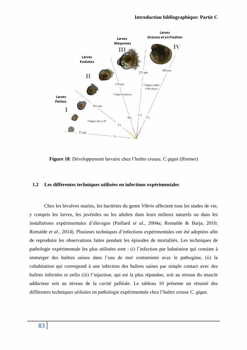
Introduction bibliographique: Partie C
83
Figure 18: Développement larvaire chez l’huître creuse, C gigas (Ifremer)
1.2 Les différentes techniques utilisées en infections expérimentales
Chez les bivalves marins, les bactéries du genre Vibrio affectent tous les stades de vie,
y compris les larves, les juvéniles ou les adultes dans leurs milieux naturels ou dans les
installations expérimentales d’élevages (Paillard et al., 2004a; Romalde & Barja, 2010;
Romalde et al., 2014). Plusieurs techniques d’infections expérimentales ont été adoptées afin
de reproduire les observations faites pendant les épisodes de mortalités. Les techniques de
pathologie expérimentale les plus utilisées sont : (i) l’infection par balnéation qui consiste à
immerger des huîtres saines dans l’eau de mer contaminée avec le pathogène, (ii) la
cohabitation qui correspond à une infection des huîtres saines par simple contact avec des
huîtres infectées et enfin (iii) l’injection, qui est la plus répandue, soit au niveau du muscle
adducteur soit au niveau de la cavité palléale. Le tableau 10 présente un résumé des
différentes techniques utilisées en pathologie expérimentale chez l’huître creuse C. gigas.
LarvesGrosses et en FixationLarves
Moyennes
LarvesEvoluées
LarvesPetites
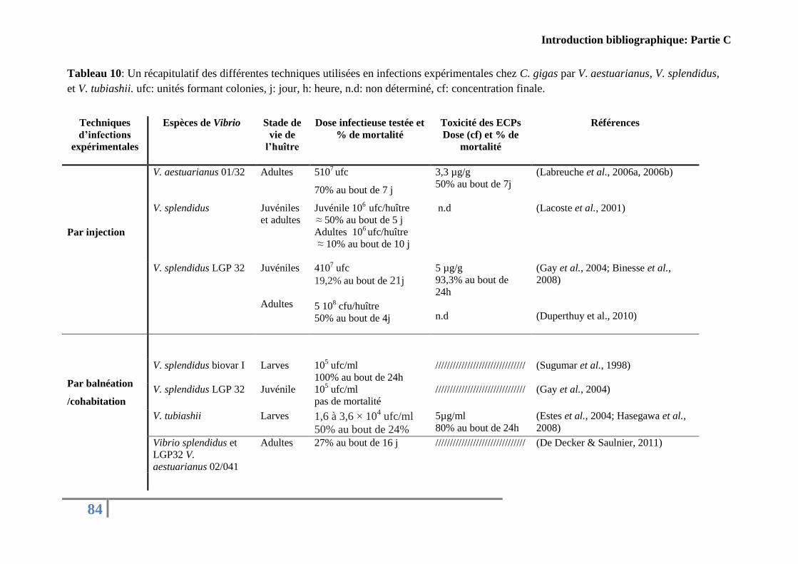
Introduction bibliographique: Partie C
84
Tableau 10: Un récapitulatif des différentes techniques utilisées en infections expérimentales chez C. gigas par V. aestuarianus, V. splendidus,
et V. tubiashii. ufc: unités formant colonies, j: jour, h: heure, n.d: non déterminé, cf: concentration finale.
Techniques
d’infections
expérimentales
Espèces de Vibrio
Stade de
vie de
l’huître
Dose infectieuse testée et
% de mortalité
Toxicité des ECPs
Dose (cf) et % de
mortalité
Références
Par injection
V. aestuarianus 01/32
Adultes 5107 ufc
70% au bout de 7 j
3,3 µg/g
50% au bout de 7j
(Labreuche et al., 2006a, 2006b)
V. splendidus
Juvéniles
et adultes
Juvénile 106
ufc/huître ≈ 50% au bout de 5 j
Adultes 106 ufc/huître
≈ 10% au bout de 10 j
n.d (Lacoste et al., 2001)
V. splendidus LGP 32
Juvéniles
Adultes
4107 ufc
19,2% au bout de 21j
5 10
8 cfu/huître
50% au bout de 4j
5 µg/g
93,3% au bout de
24h
n.d
(Gay et al., 2004; Binesse et al.,
2008)
(Duperthuy et al., 2010)
Par balnéation
/cohabitation
V. splendidus biovar I Larves 105 ufc/ml
100% au bout de 24h
/////////////////////////////// (Sugumar et al., 1998)
V. splendidus LGP 32 Juvénile 105 ufc/ml
pas de mortalité
/////////////////////////////// (Gay et al., 2004)
V. tubiashii Larves 1,6 à 3,6 × 10⁴ ufc/ml
50% au bout de 24%
5µg/ml
80% au bout de 24h
(Estes et al., 2004; Hasegawa et al.,
2008)
Vibrio splendidus et
LGP32 V.
aestuarianus 02/041
Adultes 27% au bout de 16 j /////////////////////////////// (De Decker & Saulnier, 2011)
Introduction bibliographique: Partie C
85
En effet, il n’est pas évident de retrouver le bon modèle d’infection expérimentale et
d’imiter le phénomène observé dans la nature. Même si les infections comme la balnéation ou
la cohabitation sont les plus proches des phénomènes naturels, le manque de la
reproductibilité potentiellement lié à la simplicité des microcosmes utilisés a mené de
nombreux auteurs à privilégier l’injection.
2. Approche in vitro
2.1 Les modèles cellulaires
2.1.1 Généralités sur les hémocytes
Durant ces dernières décennies, l’implication des hémocytes dans la défense des
bivalves est de plus en plus étudiée afin de comprendre le comportement des bivalves lors
d’une infection et de pouvoir mettre la main sur les facteurs qui peuvent nuire au bon
déroulement de la réaction immunitaire.
Le système immunitaire chez les invertébrés repose exclusivement sur les défenses
innées (ou non-spécifiques). En plus des barrières physico-chimiques telles que la coquille, la
cuticule, les couches de mucus et le manteau qui représentent une première ligne de défense
contre les agresseurs, l’immunité non spécifique des mollusques s'articule autour d'une
réponse humorale et d'une réponse cellulaire qui interagissent étroitement (Leclerc, 1996).
Néanmoins, l'immunité cellulaire semble jouer le rôle principal dans les processus
immunitaires des mollusques bivalves (Roch, 1999).
Contrairement à ce qu’il a été rapporté que les mollusques bivalves sont incapables de
développer des acteurs de la mémoire immunitaire adaptative et ne se défendent vis à vis des
pathogènes que par des réponses rapides, mettant en jeu la phagocytose et les réactions
inflammatoires (Tiscar & Mosca, 2004). Zhang et al. (2014), ont montré que le phénomène de
« priming » (la stimulation des cellules T naives pour devenir des cellules T effectrices) existe
chez les huîres creuses (C.gigas).
Le système humoral et plus spécifiquement l’hémolymphe (le plasma), est constitué
majoritairement par des molécules solubles à fonctions cytotoxiques comme les enzymes
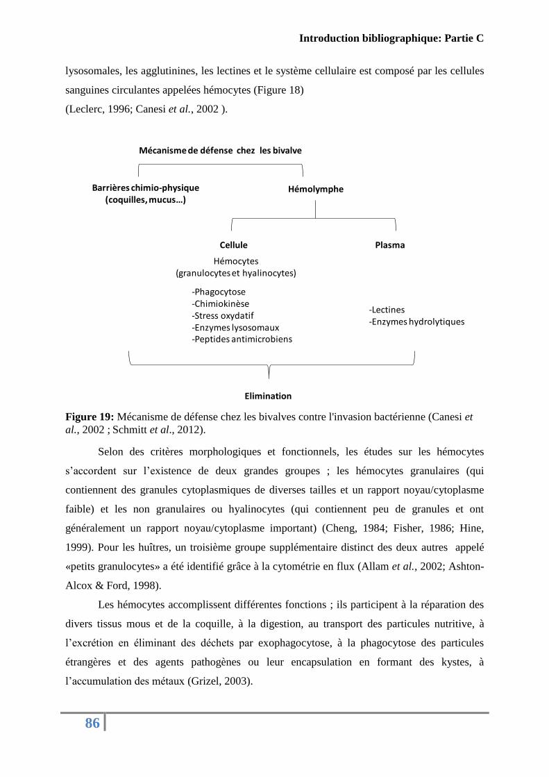
Introduction bibliographique: Partie C
86
lysosomales, les agglutinines, les lectines et le système cellulaire est composé par les cellules
sanguines circulantes appelées hémocytes (Figure 18)
(Leclerc, 1996; Canesi et al., 2002 ).
Figure 19: Mécanisme de défense chez les bivalves contre l'invasion bactérienne (Canesi et
al., 2002 ; Schmitt et al., 2012).
Selon des critères morphologiques et fonctionnels, les études sur les hémocytes
s’accordent sur l’existence de deux grandes groupes ; les hémocytes granulaires (qui
contiennent des granules cytoplasmiques de diverses tailles et un rapport noyau/cytoplasme
faible) et les non granulaires ou hyalinocytes (qui contiennent peu de granules et ont
généralement un rapport noyau/cytoplasme important) (Cheng, 1984; Fisher, 1986; Hine,
1999). Pour les huîtres, un troisième groupe supplémentaire distinct des deux autres appelé
«petits granulocytes» a été identifié grâce à la cytométrie en flux (Allam et al., 2002; Ashton-
Alcox & Ford, 1998).
Les hémocytes accomplissent différentes fonctions ; ils participent à la réparation des
divers tissus mous et de la coquille, à la digestion, au transport des particules nutritive, à
l’excrétion en éliminant des déchets par exophagocytose, à la phagocytose des particules
étrangères et des agents pathogènes ou leur encapsulation en formant des kystes, à
l’accumulation des métaux (Grizel, 2003).
Mécanisme de défense chez les bivalve
-Phagocytose-Chimiokinèse-Stress oxydatif-Enzymes lysosomaux-Peptides antimicrobiens
-Lectines-Enzymes hydrolytiques
Hémolymphe
Cellule Plasma
Hémocytes (granulocytes et hyalinocytes)
Elimination
Barrières chimio-physique (coquilles, mucus…)
Introduction bibliographique: Partie C
87
Au sein d'une même espèce, il peut y avoir des variations considérables dans la
composition des hémocytes et dans leurs fonctions.
L'activité des hémocytes nécessite un contact avec la cellule cible : ce premier niveau
de reconnaissance du non-soi ainsi que l'élimination du corps étranger est un phénomène
commun dans tous les invertébrés à travers le processus de la phagocytose.
L'adhésion des bactéries à la membrane des cellules phagocytaires ne dépend pas
seulement des forces physiques (hydrophobicité et charges d’interactions) mais peut
également être médiée par des «molécules scouts». Chez les bivalves, la reconnaissance entre
cellule phagocytaire et pathogène semble être facilitée par des lectines. Ces protéines
dépourvues d’activités enzymatiques, sont trouvées sous forme libre dans l’hémolymphe de
plusieurs invertébrés (Leclerc, 1996). Plusieurs rôles sont accordés aux lectines notamment
l’agglutination pour immobiliser les corps étrangers favorisant ainsi leur lyse par des enzymes
et/ou la phagocytose, l’opsonisation par liaison entre des récepteurs présents à la surface des
particules et des hémocytes, et la fixation sur les hémocytes pour constituer des facteurs de
reconnaissance pour les microorganismes (Chu, 2000; Gagnaire, 2005). Plusieurs types de
lectines ont été trouvés dans l’hémolymphe de C. gigas (gigalins) (Olafsen et al., 1992), et en
plus de l’agglutination, les lectines agissent comme des opsonines (Olafsen, 1995).
Suite à la reconnaissance et à l’adhésion des particules étrangères, les hémocytes
peuvent ainsi les internaliser par phagocytose grâce au phénomène de remaniement du
cytosquelette (Cheng 1981; Cheng, 1996; Cheng, 2000). Soit par l’intermédiaire de
pseudopodes, de filopodes ou de vacuoles d’endocytoses, les particules étrangères se
retrouvent piégées dans un phagosome. Après internalisation des particules étrangères au sein
du phagosome, celui-ci fusionne avec les lysosomes pour donner naissance à un
phagolysosome. La dégradation des particules étrangères ingérées s’y effectue par la
formation d’intermédiaires oxygénés connu sous le terme de «bouffée respiratoire» et/ou par
l’activité des enzymes lysosomales (Bachère et al., 1995; Roch, 1999).
2.1.2 Les hémocytes un bon modèle d’étude
Le système d’interaction entre hémocytes-bactéries reste le modèle le plus étudié. À
l'aide de cette interaction et les techniques de microscopies et de la cytométrie en flux,
plusieurs travaux de recherche ont mis en évidence quelques stratégies employées par les
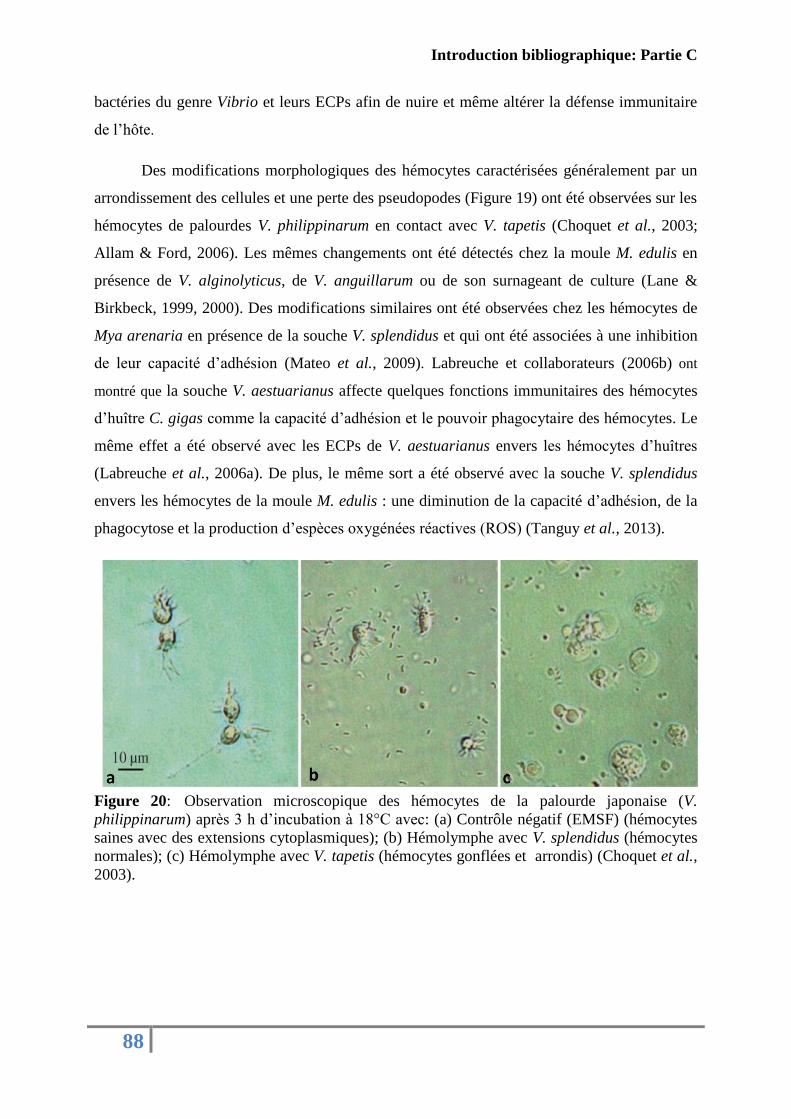
Introduction bibliographique: Partie C
88
bactéries du genre Vibrio et leurs ECPs afin de nuire et même altérer la défense immunitaire
de l’hôte.
Des modifications morphologiques des hémocytes caractérisées généralement par un
arrondissement des cellules et une perte des pseudopodes (Figure 19) ont été observées sur les
hémocytes de palourdes V. philippinarum en contact avec V. tapetis (Choquet et al., 2003;
Allam & Ford, 2006). Les mêmes changements ont été détectés chez la moule M. edulis en
présence de V. alginolyticus, de V. anguillarum ou de son surnageant de culture (Lane &
Birkbeck, 1999, 2000). Des modifications similaires ont été observées chez les hémocytes de
Mya arenaria en présence de la souche V. splendidus et qui ont été associées à une inhibition
de leur capacité d’adhésion (Mateo et al., 2009). Labreuche et collaborateurs (2006b) ont
montré que la souche V. aestuarianus affecte quelques fonctions immunitaires des hémocytes
d’huître C. gigas comme la capacité d’adhésion et le pouvoir phagocytaire des hémocytes. Le
même effet a été observé avec les ECPs de V. aestuarianus envers les hémocytes d’huîtres
(Labreuche et al., 2006a). De plus, le même sort a été observé avec la souche V. splendidus
envers les hémocytes de la moule M. edulis : une diminution de la capacité d’adhésion, de la
phagocytose et la production d’espèces oxygénées réactives (ROS) (Tanguy et al., 2013).
Figure 20: Observation microscopique des hémocytes de la palourde japonaise (V.
philippinarum) après 3 h d’incubation à 18°C avec: (a) Contrôle négatif (EMSF) (hémocytes
saines avec des extensions cytoplasmiques); (b) Hémolymphe avec V. splendidus (hémocytes
normales); (c) Hémolymphe avec V. tapetis (hémocytes gonflées et arrondis) (Choquet et al.,
2003).
Introduction bibliographique: Partie C
89
2.1.3 Autres modèles cellulaires
Vue l’absence des lignées cellulaires de mollusques marins, d’autres lignées d’origine
humaine ou animale ont été utilisées pour mettre en évidence la cytotoxicité des produits de
sécrétion des vibrions.
Une protéase à sérine purifiée de V. parahaemolyticus a inhibé la croissance de lignées
de cellules de mammifères (HeLa, Vero, CHO et Caco-2) en causant des arrondissements
cellulaires. En contact des globules rouges, cette toxine a montré une activité hémolytique et
la capacité de dégrader l'hémoglobine (Lee et al., 2002). Binesse et collaborateurs (2008) ont
montré que les ECPs de la souche V. splendidus causent des rétractions des prolongements
cytoplasmiques et l'arrondissement des cellules d’escargots (cellules Bge) et de
fibroplastiques (cellules NIH). Les ECPs de la souche V. harveyi exercent un effet
cytotoxique intense sur les lignées cellulaires du type HeLa en moins d’une heure de contact
(Soto-Rodriguez et al., 2012).
2.2 Les modèles moléculaires
Comme évoqué précédemment, au cours d’une infection, les Vibrio pathogènes ont la
capacité de produire plusieurs facteurs de virulence. Les protéases sont classées parmi les
facteurs les plus toxiques, ils agissent directement sur des protéines de l’hôte en provoquant
des dommages tissulaires et cellulaires ou indirectement via la stimulation de l’expression des
autres facteurs de virulence. Afin de mette en évidence ce type d’activité, des substrats
protéiques qui sont présents dans tous les organismes vivants et qui sont nécessaires pour le
bon fonctionnement cellulaire et tissulaire ont été utilisés in vitro en tant que modèles
d’interactions. Teo et collaborateurs (2003b) ont démontré que la métalloprotéase de V.
harveyi (Pap6) est capable d’hydrolyser plusieurs protéines comme la gélatine, la fibronectine
et le collagène du type IV. Aussi, la métalloprotéase de V. mimicus a montré une spécificité
envers plusieurs types de collagènes et la gélatine. Cependant elle n'a pas montré une activité
hydrolytique envers d'autres protéines biologiques comme le lysozyme, la lactoferrine et
l'albumine de sérum bovin (Lee et al., 2003). Chez la souche V. fluvialis, une activité
protéolytique envers la caséine et l'élastine a été provoquée par la métalloprotéase VFP (V.
fluvialis protease) (Miyoshi et al., 2002). Enfin, une protéase à sérine sécrétée par la souche

Introduction bibliographique: Partie C
90
V. metschnikovii a été capable de cliver plusieurs protéines intervenant dans la coagulation
sanguine dont le fibrinogène, la prothrombine et de la thrombine (Park et al., 2012).

Contexte général et objectifs de l’étude
91

92
RESULTATS
Résultats: Article I
93
A/ Première description d’une nouvelle souche française,
pathogène de mollusques et phylogénétiquement proche de la
souche type américaine V. tubiashii
A1: Caractérisation et détection des isolats français lors
d’épisodes de mortalité
Les épisodes récurrents de mortalité affectent depuis plusieurs années les mollusques
bivalves et plus spécifiquement l’huître creuse du Pacifique C. gigas. Ce phénomène souvent
désigné sous le terme de mortalité anormale inquiète les professionnels, qui craignent la
disparition de C. gigas à l’instar de l’huître portugaise, C. angulata (Comps et al., 1976). Ces
mortalités anormales ont une étiologie complexe et sont fonction à la fois de facteurs
environnementaux, de l’état physiologique de l’hôte ainsi que de facteurs biotiques comme les
parasites, les virus et les bactéries (Samain et al., 2007). En France, en milieu naturel aussi
bien que dans les écloseries et les nurseries, à coté des virus de type herpès (Renault et al.,
1994; Segarra et al., 2010), des bactéries appartenant au genre Vibrio (Lacoste et al., 2001; Le
Roux, 2002; Garnier et al., 2008) ont souvent été détectées.
Dans le cadre d'une large étude épidémiologique menée par Saulnier et collaborateurs
(Saulnier et al., 2010) entre 2003 et 2007 et sous les auspices du réseau REPAMO, des
bactéries dominantes ont été isolées principalement à partir d’huîtres creuses et identifiées
comme appartenant au genre Vibrio. En plus de V. splendidus et V. aestuarianus, des isolats
appartenant au groupe Harveyi (33 isolats) ont été identifiés, infectant plusieurs lots de
naissains de C. gigas présents dans les installations d'une écloserie/nurserie.
La diversité des souches appartenant à ce groupe et leur seule classification par le biais
du séquençage de l’ARNr 16S, nous a amené à vouloir affiner leur classification au rang de
l’espèce. Bien que le séquençage de l'ARNr 16S soit largement utilisé pour la classification
taxonomique de plusieurs bactéries, il apparaît pas suffisant pour la classification des espèces
au sein du groupe Harveyi (Thompson et al., 2004b; Thompson et al., 2007; Lin et al., 2010).
Résultats: Article I
94
Dans l’optique d’affiner l’affiliation de ces isolats, nous avons donc effectué des
analyses complémentaires, basées sur des critères génétiques et biochimiques. Les résultats
sont regroupés dans le premier article de la thèse.
Les résultats de l’analyse phylogénétique ont montré que les 9 isolats choisis du groupe
Harveyi formaient un groupe phylogénétique homogène, éloigné de la souche type de V.
harveyi et distinct mais proche de la souche Américaine V. tubiashii. La parenté avec l’espèce
V. tubiashii a également été confirmée par les différents tests phénotypiques. Une souche
représentative du groupe, initialement codifiée par 07/118 T2 a fait l’objet d’une étude
génétique plus poussée. Les résultats d’hybridation ADN/ADN obtenus ont montré que la
souche 07/118 T2 était génétiquement proche de la souche type américaine V. tubiashii
(hybridation ADN/ADN < 70% ± 13).
En dehors des installations d’élevage, cette nouvelle espèce a été détectée, par PCR
quantitative ciblant le gène gyrB (séquencé sur une des souches isolées en 2007) dans un lot
de mortalité d’ormeaux issus d’écloseries françaises en 2004 et dans deux lots de mortalités
d’huîtres issus d’écloseries en 2008. Cependant, cette souche n’a été détectée dans aucun des
lots d'animaux moribonds issus de cultures en milieu naturel.
Dans un premier temps, l’analyse de la virulence des différents isolats a été effectuée
sur des huîtres de 10 à 12 mois par des injections expérimentales induisant 70 % de mortalité
au bout de 24 h mais à des densités cellulaires élevées uniquement (DO600 de 1 et de 0,5).
Dans un deuxième temps, l’injection des produits extracellulaires (ECPs) de la souche 07/118
T2 à des huîtres juvéniles et à des concentrations de 5 µg/g de chair humide, a provoqué une
mortalité rapide estimée à 41 % dans les 24 h post-injection. La présence de métalloprotéase
(confirmée par dosage de l’activité protéasique en présence d’un inhibiteur de
métalloprotéase, la phénanthroline) ainsi que la stabilité de cette activité protéolytique a pu
être observée sur la durée de l’infection. Ces premiers résultats suggèreraient que la virulence
de la bactérie observée in vivo pourrait être en partie liée à sa capacité à produire des
exoenzymes hydrolytiques de type métalloprotéase.
Les résultats obtenus font l’objet d’un premier article publié dans «Journal of
Invertebrate pathology», 2014.
Résultats: Article I
95
ArticleI: FIRST DESCRIPTION OF FRENCH V. TUBIASHII STRAINS
PATHOGENIC TO MOLLUSK
I.CHARACTERIZATION OF ISOLATES AND DETECTION DURING
MORTALITY EVENTS
Published in “Journal of invertebrate pathology”
Travers Marie-Agèns1
, Mersni-Achour Rachida1,2,3
, Haffner Philippe1, Tourbiez,
Delphine1, Cassone, Anne-Laure
1, Morga, Benjamin
1, Doghri Ibtissem
2, Garcia Céline
1,
Renault, Tristan1, Fruitier-Arnaudin Ingrid
2,3, Saulnier Denis
1,4
1 Ifremer, SG2M-LGPMM, Laboratoire de Génétique et Pathologie des Mollusques Marins
Avenue de Mus de Loup, 17390 La Tremblade, France
2 UMR 7266 CNRS-ULR, LIENSs, Equipe Approches Moléculaires, Environnement-Santé,
Université de La Rochelle, Avenue Michel Crépeau, 17042 La Rochelle, France
3 Fédération de Recherche en Environnement et Développement Durable, FR CNRS 3097,
Université de La Rochelle, France
4 Present address : Ifremer, Centre Ifremer du Pacifique, UMR EIO 241 Ecosystèmes
Insulaires Océaniens, Tahiti, 98719 Taravao, French Polynesia
Marie-Agnès Travers and Rachida Mersni Achour participated equally to this work and
should be considered as primary co-authors
Ingrid Fruitier-Arnaudin and Denis Saulnier participated equally to this work and should be
considered as last co-authors
Résultats: Article I
96
Abstract
Nine dominant bacterial isolates were obtained from different batches of Crassostrea
gigas spat experiencing high mortality rates in a French experimental hatchery/nursery in
2007. Using phenotypic analysis combined with multilocus sequence analysis, the isolates
were shown to be genetically close to the V. tubiashii type strain. Based on 1) analyses of the
recA gene sequences; 2) the results of DNA-DNA hybridization assays between 07/118 T2
(LMG 27884 = CECT 8426), which is a representative strain, and the V. tubiashii type strain
(69%); and 3) phenotypic traits, the bacteria were classified in a group close to American V.
tubiashii strain. Its virulence (70% of mortalities) and the toxicity of the extracellular products
of 07/118 T2 was demonstrated (41% of mortalities).
Moreover, a QPCR diagnostic tool targeting the gyrB gene was developed to
investigate the epidemiological significance of V. tubiashii in French oyster mortality
outbreaks recorded by the national surveillance network. Of the 21 batches originating from
hatcheries, only two were positive, whereas V. tubiashii DNA could not be detected in any of
the batches of moribund animals collected in field/outdoor facilities.
These results demonstrate the existence of a group of virulent V. tubiashii in France that
episodically infect Crassostrea gigas.
Keywords: Crassostrea gigas; Pathogenicity; Extracellular Products; Polyphasic approach,
Real time PCR
Résultats: Article I
97
1. Introduction
The causes of abnormal mortalities during summer in Crassostrea gigas (C. gigas)
remains unclear although several studies suggest that this phenomenon has a complex
etiology involving environmental stressors (oxygen depletion, lack of food, salinity,
temperature, industrial pollution, pesticides, aquaculture practices) and biological factors that
include pathogens and an altered physiology of the host (Samain et al., 2007). These
mortalities can affect both diploid and triploid oysters, their different life stages (Romalde and
Barja, 2010; De Decker et al., 2011) both in the field (for juveniles and adults) and in
hatcheries (for larval and post-larval stages). Among the biological factors, microorganisms
are often associated with oyster diseases. Three infectious microorganisms are frequently
described as major pathogens of oysters : herpes-like viruses (Renault et al., 1994; Segarra et
al., 2010), parasites (Meyers et al., 1991; Bower et al., 1997; Tun et al., 2008) and bacteria of
the genus Vibrio which have been described as important causal agents affecting all life stages
of bivalve mollusks (Le Roux et al., 2002; Garnier et al., 2008; Saulnier et al., 2010; Beaz-
Hidalgo et al., 2010). V. splendidus and V. aestuarianus are presented as the two main Vibrio
species associated with summer mortalities of C. gigas in France (Lacoste et al., 2001; Le
Roux et al., 2002; Garnier et al., 2008). Besides these two major bacterial, another Vibrio
species was reported by Saulnier et al. (2010) in a large-scale epidemiological study including
nine virulent strains that belong to the V. harveyi group. The virulence of these strains was
investigated by experimental infection on C. gigas spat. The results showed a strong
correlation between the virulence of Vibrio and their capacity to produce metalloprotease-like
activity in the culture supernatant, particularly for the strains 07/108 T1 and 07/118 T2. These
two strains can secrete a high level of proteases and can induce extremely high levels of
mortality just 20 hours post injection. Partial sequence analysis based on 16S rDNA revealed
that 07/108 T1 and 07/118 T2 were clustered within the V. harveyi group.
Nevertheless, phenotypic analysis and the use of standard molecular techniques are
insufficient to classify Vibrio, and indeed cannot even be used to identify related species. The
identification of vibrios at the family and genus level are habitually based on 16S rDNA gene
sequencing, but this method is unable to separate certain closely related species (Gomez-Gil
et al., 2004). The association of other analyses, including DNA-DNA hybridization and
molecular fingerprinting methods, is useful for Vibrio identification. However, these tools are
restricted to some reference laboratories and inter-laboratory comparisons of fingerprint
patterns are difficult (Thompson et al., 2005).
Résultats: Article I
98
Multilocus sequence analysis (MLSA) has been adopted to fine-tune the approach and
clarify certain doubts concerning Vibrio classification. Using this methodology, Thompson et
al. have shown that V. harveyi. and V. campbellii form two separate clusters (Thompson et al.,
2007) and V. communis and V. owensii, which were assigned to the Harveyi clade, were
recently discovered to be the same species after identification using the MLSA approach
(Hoffmann et al., 2012).
The aim of the present study was to refine the taxonomic affiliation of these virulent
strains, previously clustered with V. harveyi, using a polyphasic approach (phenotypic and
genotypic characterization). Secondly, quantitative challenges with live bacteria and in vitro
culture supernatants were performed with C. gigas in order to evaluate their virulence and the
toxicity of the extracellular products, respectively. Finally, a quantitative real-time PCR
diagnostic tool was developed to screen isolates/DNA collected during the episodes of
mortality that have occurred in France since 2004 and evaluate their epidemiological
significance.
Résultats: Article I
99
2. Materials and methods
2.1 Taxonomic affiliation
2.1.1 Bacteria genotyping and phylogenetic analyses (10 isolates)
The strains used in this study are described in Table 1. The predominant bacteria,
which were not been yet described, were isolated, as previously described (Saulnier et al.,
2010), during the mortalities recorded in oyster hatchery stocks in 2007. Briefly, after 10-2
and
10-3
dilutions in Sterile Artificial Sea Water (SASW : 2.3 % (w/v) NaCl, 20 mM KCl, 5 mM
MgSO4, 2 mM CaCl2), moribund oyster samples (hemolymph for adults and crushed tissues
for juveniles) were plated on thiosulfate-citrate-bile salts-sucrose (TCBS, Difco™), Zobell
agar (0.4 % peptone, 0.1 % yeast extract, 0.01% ferric citrate and 1.5 % agar in SASW, pH
7.6) or marine agar (Conda) and the predominant bacteria were isolated after 48h at 20°C.
Pure cultures of bacterial strains were conserved frozen at -80°C in Zobell broth with glycerol
15%.
For genotyping, DNA was extracted following the FTA®
procedure (Whatman).
Bacterial isolates were cultured during 24 hours at 20 °C in Zobell broth and 65 μl of bacterial
suspension at the stationary phase of in vitro culture was deposited on FTA® paper matrix
cards according to the manufacturer’s instructions. Briefly, a punch from the FTA® Card was
washed three times with FTA® Purification Reagent and rinsed with 200 µl of TE buffer (10
mM Tris-HCI, 0.1 mM EDTA, pH 8). This punch was either directly transferred into a PCR
tube, or conserved at -20°C before use.
The 16S rRNA (ribosomal RNA), ftsZ (cell division protein, cell division), pyrH
(uridylate kinase, nucleotide biosynthesis), recA (recA protein, DNA repair) and topA
(topoisomerase I, DNA replication and repair) genes were amplified using the universal
bacterial primer pairs listed in supplementary Table 1 and the methods previously described
(Thompson et al., 2005). Amplicons with the expected size were purified using a Microcon
PCR filter kit (Millipore). Purified PCR products were mixed (final volume 10 μl) with 0.4 μl
ABI Prism Big Dye Terminator ready reaction mix (Applied Biosystems®) and 0.75 μM of
primer. Cycle sequencing reactions were performed using a Gene Amp PCR System 2700
(Applied Biosystems®) and following the manufacturer’s instructions. Separation of the DNA
fragments was carried out in an ABI PRISM 3100 Genetic Analyzer (Applied Biosystems).

Résultats: Article I
100
Sequences were aligned using ClustalW http://www.ebi.ac.uk/Tools/msa/clustalw2/ and
BioEdit software http://www.mbio.ncsu.edu/bioedit/bioedit.html. Pairwise similarity was
calculated with BioEdit.
Phylogenetic trees were built using Mega4
http://www.megasoftware.net/mega4/mega.html. The trees were drawn using the Neighbor-
Joining method with the Kimura two-parameter model (Saitou and Nei, 1987) and the
maximum parsimony method (Eck and Dayhoff, 1966). Reliability of topologies was assessed
by the bootstrap method (Felsenstein, 1985) with 1000 replicates. The Genbank accession
numbers for the 16S rDNA, gyrB, recA, topA, pyrH, ftsZ gene sequences obtained in this
study or already published in GenBank are presented in Supplementary Table 3.
2.1.2. DNA-DNA hybridization assays (2 isolates)
DNA-DNA hybridization (DDH) was performed by the BCCMTM
/LMG laboratory at
Gent University. After a genomic DNA extraction according to a modified Wilson protocol
(Wilson, 1987), hybridizations were performed in microplates at 40°C (Ezaki et al., 1989) and
DNA relatedness values (%) reported were the means of a minimum of 6 hybridizations.
2.1.3. Bacteria phenotypic characterization (10 isolates)
The phenotypes of the selected isolates were characterized in different ways. Bacteria
growth on TCBS medium (AES Chemunex), chromID™ Vibrio medium (Biomérieux) and
CHROMagar™ Vibrio medium (CHROMagar) was observed. The capacity of bacteria to
grow in Zobell broth with 8 and 10% NaCl at 22°C was determined and the ability of
bacteria to grow on Zobell agar at 40°C was established.
Antibiograms were carried out using disc diffusion assays on Mueller-Hinton agar
(Oxoid) with commercial impregnated discs (Oxoid): Flumequine 30 µg (UB30), Kanamycin
30 µg (K30), Tetracycline 30 µg (TE30), Sulphamethoxazole/trimethoprim 19:1 25 µg
(SXT25), Streptomycin 25 µg (S25), Sulphonamides 300 µg (S3), Erythromycin 30 µg (E30),
Penicillin 10 UI (6 µg) (P10), Trimethoprim 5 µg (W5), Chloramphenicol 30 µg (C30),
Ampicillin 10 µg (AMP10), Gentamicin 30 µg (CN30) or the Vibriostatic agent, Pteridine
150 µg and 10 µg (O129/150 and O129/10). Inhibition was observed after 24 to 48h at 22°C
on Zobell agar. API 20E, API 20NE and API 50CH (Biomérieux) were used in accordance
with the manufacturer’s recommendations. Fresh colonies, obtained from culture bacteria on
TSA (Difco™
) adjusted to 2% NaCl, were suspended in SASW to obtain about OD600 = 0.1.
Résultats: Article I
101
After inoculation, the strips were incubated for 48 h at 22 °C and the results were determined
according to the manufacturer's instructions.
2.2. Pathogenicity assays
2.2.1. Bacterial virulence tests (7 isolates)
Bacteria (07/038 2T2, 07/110 T1, 07/112 T1, 07/120 T1, 07/119 T1, 07/118 T2 and
04/002 1T2) were grown at 22 °C for 20 h in marine broth with constant shaking at 40 rpm
(Rotator SB3; Stuart). The bacterial concentrations in the cultures were evaluated
spectrophotometrically at an optical density (OD) of 600 nm and checked by plating. The
cells were centrifuged at 3,200 × g for 10 min, the supernatant discarded and the resulting
pellet resuspended in SASW to obtain an OD 600 nm of 1. Ten to twelve-month-old oyster spat
were anesthetized for 1 to 2 hours at 22 °C in a magnesium chloride solution (MgCl2, Fluka)
at a final concentration of 50 g l−1
(1/4: v/v seawater/freshwater) and under aeration.
Subsequently, 100 μl of bacterial suspension (107
cfu per oyster) was injected into the
adductor muscle. A group of oysters were injected with SASW to serve as negative controls.
After injection, the oysters were transferred to tanks (triplicates of 10 oysters in 2 liters) filled
with UV-treated and 1 μm-filtered seawater at 3.1% salinity and maintained under static
conditions at 22 °C with aeration. Mortality was monitored twice a day and any newly dead
oysters were removed from the tanks over a four day period. Animals were considered to be
dead when the valves did not close following stimulation.
2.2.2. Toxicity and characterization of ECPs from the V. tubiashii 07/118 T2
Bacterial extracellular products (ECPs) from 07/118 T2 were produced in two types of
culture medium, plate agar and liquid medium.For culture in liquid medium: bacteria were
grown on Luria-Bertani (Difco™) medium supplemented with 1% sodium chloride (LBS-1%
NaCl). Briefly 100 µl of a pre-culture of bacteria (OD 600 of 1) was cultured in 100 ml of LBS
medium in a 500 ml Erlenmeyer flask at 22°C with constant shaking at 100 rpm (Aquatron,
INFORS HT). After 24 h of incubation, ECPs were obtained by centrifugation at 3,000 × g for
40 min at 4°C. The supernatant containing the ECPs was filtered at 0.22 µm and then
concentrated by lyophilisation. The concentrated sample was resuspended in Tris-HCl buffer
(50 mM Tris-HCl, pH 8.0) and dialyzed against distillated water in the first step for 12h and
against Tris-HCl buffer for 12h in the second step. Finally, the samples were stored at -80°C
until use. For culture on solid medium: ECPs were produced using the cellophane plate
Résultats: Article I
102
technique described by Liu (1957). A volume of 1 ml of exponential phase culture in LBS
(OD600 of 1, 22°C for 18 h) was spread on a sterile cellophane film overlying LBS agar
(LBSA) plates. After 48 h of culture at 22°C, the cells were washed off the cellophane using
10 ml of cold SASW and removed by centrifugation at 3,000 × g (45 min at 4°C). The
supernatant containing the ECPs was then treated in the same way as the ECPs from liquid
media.
The toxicity of both ECPs was tested as follows: a volume of 100 µl of ECP solution,
adjusted in Tris-HCl buffer to a concentration of 5 µg g-1
of soft body weight, was injected
into the adductor muscle of ten to twelve month-old oyster spat after anesthetizing them (see
above). A group of oysters used as controls were injected with SASW and Tris-HCl buffer.
Mortality was monitored twice a day and any newly dead oysters were removed from the
tanks. ECPs proteins concentration was determined using the method described by Bradford
(using Bradford Reagent, from Sigma -Aldrich) with bovine albumin serum as a standard
(Sigma-Aldrich).
The proteolytic activity was determined using azocasein (Sigma-Aldrich) as a
substrate according to the method of Teo et al (2003) with some modifications. Briefly, 100 μl
of a suitable dilution of ECP solution (1/10 v/v) was added to 100 μl of azocasein (5 mg ml-1
in 50 mM Tris–HCl, pH 8.0). The mixture was incubated at 22°C for 2 h in darkness and
stirred constantly. The reaction was stopped by adding 200 μl of 10% trichloroacetic acid. The
mixture was centrifuged at 12,000 × g for 10 min and 50 μl of the supernatant was added to
50 μl of 1M NaOH in 96 well plates (Nunc/Thermo Scientific). The absorbance was measured
at 440 nm in a microplate reader (Molecular Devices). One unit of azocaseinase activity was
defined as the amount of crude ECPs that caused an increase of 0.01 absorbance unit after a 2
h incubation at 22°C.
The stability of the proteolytic activity was determined by incubating ECPs from LBSA for 24
h and 48 h at 22°C without substrate. After the preincubation, the azocaseinase activity was
measured according to the protocol described previously.
The effect of 1,10-phenantroline on proteolytic activity was examined. Stock solutions of
1,10-phenantroline (Sigma-Aldrich) were dissolved in a small volume of absolute ethanol
(until total solubilization) and adjusted to the appropriate concentration with Tris-HCl buffer.
Briefly, 25 µl of inhibitor, diluted to the appropriate concentrations, was pre-incubated with
25 µl of ECPs for 30 min at 22°C and the proteolytic activity was then measured (see above).
Residual azocaseinase activity was calculated on the basis of assays run in the absence of
inhibitor.
Résultats: Article I
103
2.3. Quantitative real-time PCR diagnostic tool (07/118 T2 and 07/112 T2)
2.3.1. Diagnostic tool development (2 isolates)
Oligonucleotide sequences were derived from the sequence alignment of the gyrB
(DNA gyrase B subunit, basic replication machinery) gene in different Vibrio species,
including the V. tubiashii type strain and the sequence obtained from the 07/118 T2 and
07/112 T2 strains with the primers listed in supplementary Table 1. Primer 3 software
http://frodo.wi.mit.edu/ was used to design a real-time PCR probe (Table S1) and forward and
reverse PCR primers. The probes consists of an oligonucleotide dually labeled with a 5‘-
reporter dye and the downstream 3‘-quencher dye indicated Table S1 (Eurogentec). The probe
and primers were assessed for species specificity using a BLAST search to determine
homology to known sequences.
A Real-time PCR assay was conducted on MX3000 and MX3005 Thermocyclers
(Agilent) using Brilliant III Ultrafast kit (Stratagene). Each reaction was run in duplicate in a
final volume of 20 μl containing various concentrations of a DNA sample (5 μl), 200 nM of
each primer and 200 nM of oligonucleotidic probe. The thermal cycle was performed with a
two-step PCR protocol: 1 cycle at 95 °C for 3 min followed by 40 cycles at 95 °C for 10 s and
60°C for 20 s. Fluorescence intensity was expressed in delta reporters (dR) after background
subtraction. The threshold was set using an amplification based algorithm from the MX3000-
3005 software (Stratagene) for the initial plate. Amplicons were sequenced to check primers
specificity. Serial dilutions were performed on genomic DNA extracts quantified by
spectrophotometry (Nanodrop/Thermo Scientific) and adjusted to 108 bacteria/ml = 1.05 ng/µl
(for the 5,353 kbp V. tubiashii genome).
2.3.2. Screening on DNA isolated from mollusks
Zoosanitary monitoring was performed by the REPAMO network (REseau de
PAthologie des MOllusques) between 2003 and 2012. During this period 254 batches of
animal samples were collected from abnormal mortality episodes affecting Crassostrea gigas
oysters, Haliotis tuberculata abalone, Venerupis philippinarum clams and Pecten maximus
scallops. Samples from 49 of these batches were analyzed in this work. They were selected to
provide a range of origin (hatchery/field), year of sampling, age and ploidy of the animals.
The isolation of dominant bacteria was carried out between 2003 and 2012 on animals that
were moribund on their arrival in the laboratory. Total DNA from infected animals was
Résultats: Article I
104
systematically extracted between 2003 and 2012. Depending on mollusk species and the age
of the animals, different tissues were sampled in sterile conditions for bacteriological analysis:
haemolymph was taken for each oyster or abalone; a piece of adductor muscle, gill and
mantle mixed together for each scallop; and a mixture of five mantles from individual clams.
All these tissues were prepared using five individuals or five pools of animals from each
batch, and in each case this corresponded either to an all-adult or all-juvenile age class.
Haemolymph was drawn into a syringe from the adductor muscle or pericardial cavity of
oysters and from the pericardial cavity of abalone using a 23-gauge needle. Tissues from
scallop and clams were homogenized in 100 μl of SASW with a sterile pellet-pestle (Sigma)
for 1 minute on average. For larvae and early spat stages (shell size smaller than 3 cm in
length), several whole animals were taken and disrupted in SASW as a single pool of
individuals for each batch sample.
Bacterial DNA from a log-phase culture was extracted from cultured Vibrio either by
boiling in 200 μl of ultra pure water (Saulnier et al., 2009) or using the standard QIAmp DNA
mini kit (Qiagen) procedure. After extracting DNA according to the manufacturer’s
instructions, the pelleted DNAs were resuspended in 100 μl of AE buffer and stored at -20°C.
DNA yield and purity were determined by spectrophotometry (Nanodrop/Thermo Scientific).
DNA from the crushed tissues of mollusk was extracted using a similar QIAmp DNA mini kit
(Qiagen) method.
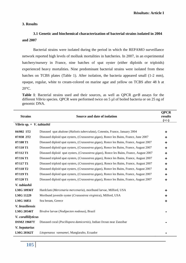
Résultats: Article I
105
3. Results
3.1 Genetic and biochemical characterization of bacterial strains isolated in 2004
and 2007
Bacterial strains were isolated during the period in which the REPAMO surveillance
network reported high levels of mollusk mortalities in hatcheries. In 2007, in an experimental
hatchery/nursery in France, nine batches of spat oyster (either diploids or triploids)
experienced heavy mortalities. Nine predominant bacterial strains were isolated from these
batches on TCBS plates (Table 1). After isolation, the bacteria appeared small (1-2 mm),
opaque, regular, white to cream-colored on marine agar and yellow on TCBS after 48 h at
20°C.
Table 1: Bacterial strains used and their sources, as well as QPCR gyrB assays for the
different Vibrio species. QPCR were performed twice on 5 µl of boiled bacteria or on 25 ng of
genomic DNA.
Strains Source and date of isolation QPCR
results (+/-)
Vibrio sp. = V. tubiashii
04/002 1T2 Diseased spat abalone (Haliotis tuberculata), Cotentin, France, January 2004 +
07/038 2T2 Diseased diploid spat oysters, (Crassostrea gigas), Ronce les Bains, France, June 2007 +
07/108 T1 Diseased diploid spat oysters, (Crassostrea gigas), Ronce les Bains, France, August 2007 +
07/110 T1 Diseased diploid spat oysters, (Crassostrea gigas), Ronce les Bains, France, August 2007 +
07/112 T1 Diseased diploid spat oysters, (Crassostrea gigas), Ronce les Bains, France, August 2007 +
07/116 T1 Diseased triploid spat oysters, (Crassostrea gigas), Ronce les Bains, France, August 2007 +
07/117 T1 Diseased triploid spat oysters, (Crassostrea gigas), Ronce les Bains, France, August 2007 +
07/118 T2 Diseased diploid spat oysters, (Crassostrea gigas), Ronce les Bains, France, August 2007 +
07/119 T1 Diseased diploid spat oysters, (Crassostrea gigas), Ronce les Bains, France, August 2007 +
07/120 T1 Diseased diploid spat oysters, (Crassostrea gigas), Ronce les Bains, France, August 2007 +
V. tubiashii
LMG 10936T Hardclam (Mercenaria mercenaria), moribund larvae, Milford, USA +
LMG 11229 Moribund juvenile oyster (Crassostrea virginica), Milford, USA +
LMG 16851 Sea bream, Greece +
V. brasiliensis
LMG 20546T Bivalve larvae (Nodipecten nodosus), Brazil -
V. coralliilyticus
DSMZ 19607T Diseased coral (Pocillopora damicornis), Indian Ocean near Zanzibar -
V. hepatarius
LMG 20362T Litopenaeus vannamei, Manglaralto, Ecuador -
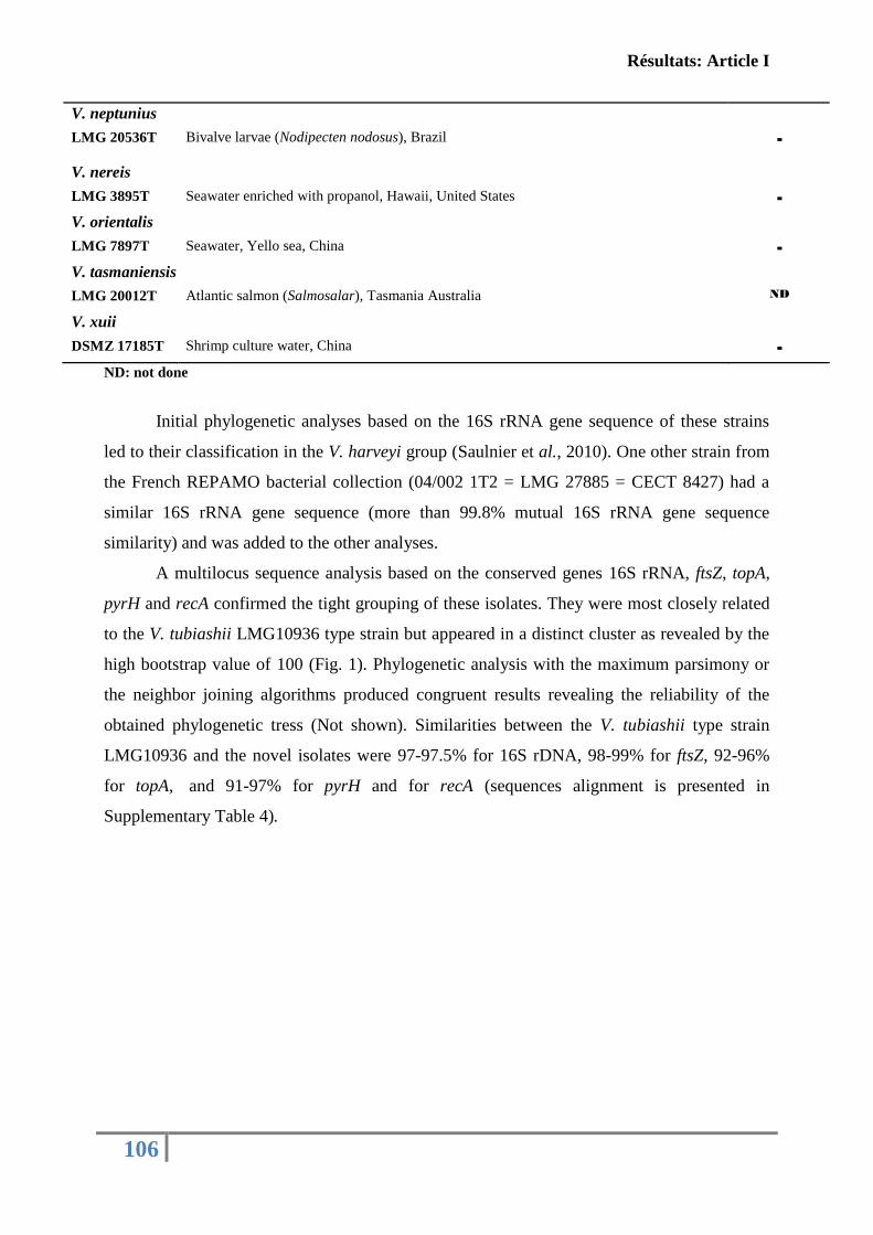
Résultats: Article I
106
V. neptunius
LMG 20536T Bivalve larvae (Nodipecten nodosus), Brazil
-
V. nereis
LMG 3895T Seawater enriched with propanol, Hawaii, United States -
V. orientalis
LMG 7897T Seawater, Yello sea, China -
V. tasmaniensis
LMG 20012T Atlantic salmon (Salmosalar), Tasmania Australia ND
V. xuii
DSMZ 17185T Shrimp culture water, China -
ND: not done
Initial phylogenetic analyses based on the 16S rRNA gene sequence of these strains
led to their classification in the V. harveyi group (Saulnier et al., 2010). One other strain from
the French REPAMO bacterial collection (04/002 1T2 = LMG 27885 = CECT 8427) had a
similar 16S rRNA gene sequence (more than 99.8% mutual 16S rRNA gene sequence
similarity) and was added to the other analyses.
A multilocus sequence analysis based on the conserved genes 16S rRNA, ftsZ, topA,
pyrH and recA confirmed the tight grouping of these isolates. They were most closely related
to the V. tubiashii LMG10936 type strain but appeared in a distinct cluster as revealed by the
high bootstrap value of 100 (Fig. 1). Phylogenetic analysis with the maximum parsimony or
the neighbor joining algorithms produced congruent results revealing the reliability of the
obtained phylogenetic tress (Not shown). Similarities between the V. tubiashii type strain
LMG10936 and the novel isolates were 97-97.5% for 16S rDNA, 98-99% for ftsZ, 92-96%
for topA, and 91-97% for pyrH and for recA (sequences alignment is presented in
Supplementary Table 4).
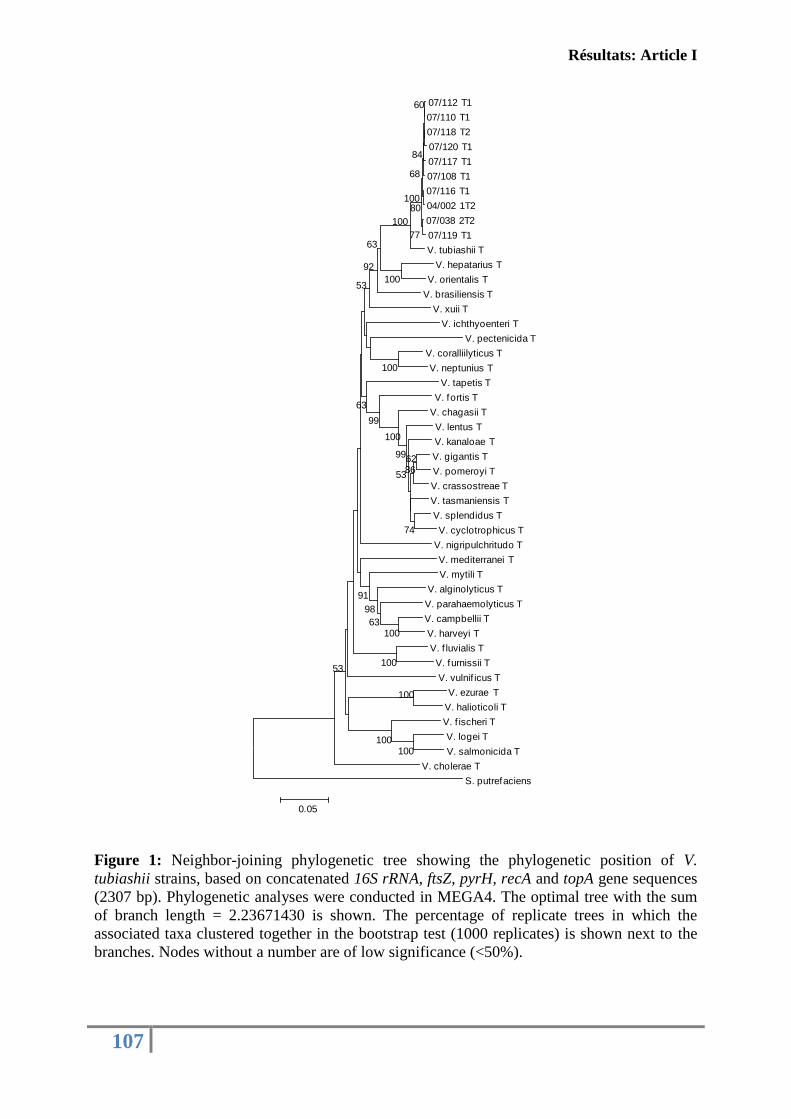
Résultats: Article I
107
Figure 1: Neighbor-joining phylogenetic tree showing the phylogenetic position of V.
tubiashii strains, based on concatenated 16S rRNA, ftsZ, pyrH, recA and topA gene sequences
(2307 bp). Phylogenetic analyses were conducted in MEGA4. The optimal tree with the sum
of branch length = 2.23671430 is shown. The percentage of replicate trees in which the
associated taxa clustered together in the bootstrap test (1000 replicates) is shown next to the
branches. Nodes without a number are of low significance (<50%).
07/112 T1
07/110 T1
07/118 T2
07/120 T1
07/117 T1
07/108 T1
07/116 T1
04/002 1T2
07/038 2T2
07/119 T1
V. tubiashii T
V. hepatarius T
V. orientalis T
V. brasiliensis T
V. xuii T
V. ichthyoenteri T
V. pectenicida T
V. coralliilyticus T
V. neptunius T
V. tapetis T
V. fortis T
V. chagasii T
V. lentus T
V. kanaloae T
V. gigantis T
V. pomeroyi T
V. crassostreae T
V. tasmaniensis T
V. splendidus T
V. cyclotrophicus T
V. nigripulchritudo T
V. mediterranei T
V. mytili T
V. alginolyticus T
V. parahaemolyticus T
V. campbellii T
V. harveyi T
V. f luvialis T
V. furnissii T
V. vulnif icus T
V. ezurae T
V. halioticoli T
V. f ischeri T
V. logei T
V. salmonicida T
V. cholerae T
S. putrefaciens
100
100100
100
100
100
62
74
8653
99
100
100
63
98
99
91
53
63
53
92
63
100
77
80100
68
84
60
0.05
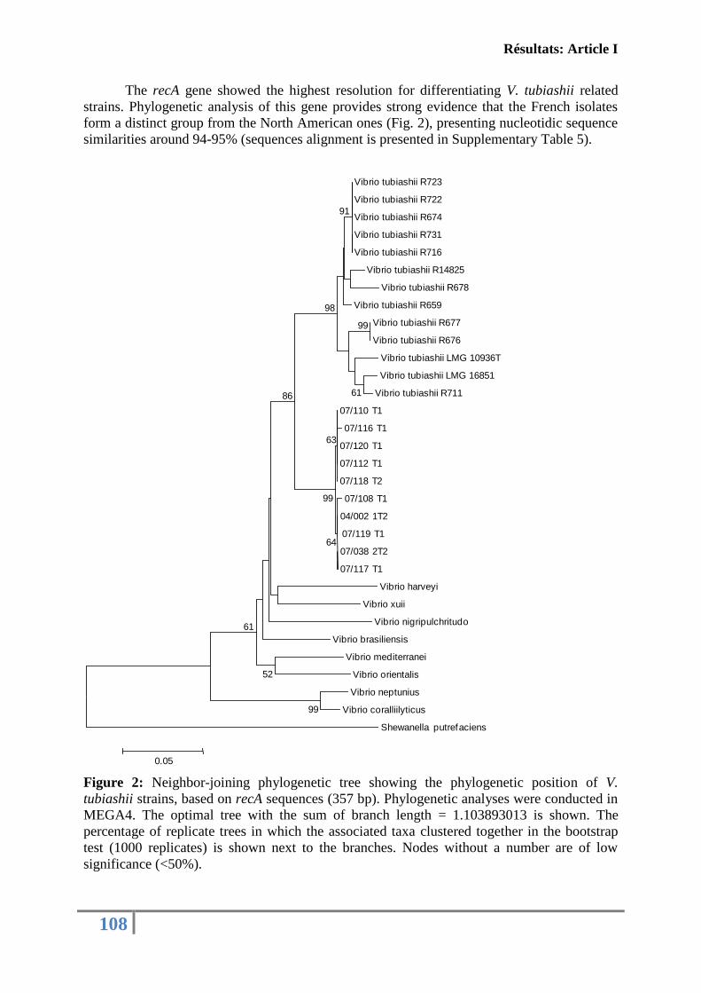
Résultats: Article I
108
The recA gene showed the highest resolution for differentiating V. tubiashii related
strains. Phylogenetic analysis of this gene provides strong evidence that the French isolates
form a distinct group from the North American ones (Fig. 2), presenting nucleotidic sequence
similarities around 94-95% (sequences alignment is presented in Supplementary Table 5).
Figure 2: Neighbor-joining phylogenetic tree showing the phylogenetic position of V.
tubiashii strains, based on recA sequences (357 bp). Phylogenetic analyses were conducted in
MEGA4. The optimal tree with the sum of branch length = 1.103893013 is shown. The
percentage of replicate trees in which the associated taxa clustered together in the bootstrap
test (1000 replicates) is shown next to the branches. Nodes without a number are of low
significance (<50%).
Vibrio tubiashii R723
Vibrio tubiashii R722
Vibrio tubiashii R674
Vibrio tubiashii R731
Vibrio tubiashii R716
Vibrio tubiashii R14825
Vibrio tubiashii R678
Vibrio tubiashii R659
Vibrio tubiashii R677
Vibrio tubiashii R676
Vibrio tubiashii LMG 10936T
Vibrio tubiashii LMG 16851
Vibrio tubiashii R711
07/110 T1
07/116 T1
07/120 T1
07/112 T1
07/118 T2
07/108 T1
04/002 1T2
07/119 T1
07/038 2T2
07/117 T1
Vibrio harveyi
Vibrio xuii
Vibrio nigripulchritudo
Vibrio brasiliensis
Vibrio mediterranei
Vibrio orientalis
Vibrio neptunius
Vibrio coralliilyticus
Shewanella putrefaciens
99
52
61
99
61
91
98
86
64
99
63
0.05
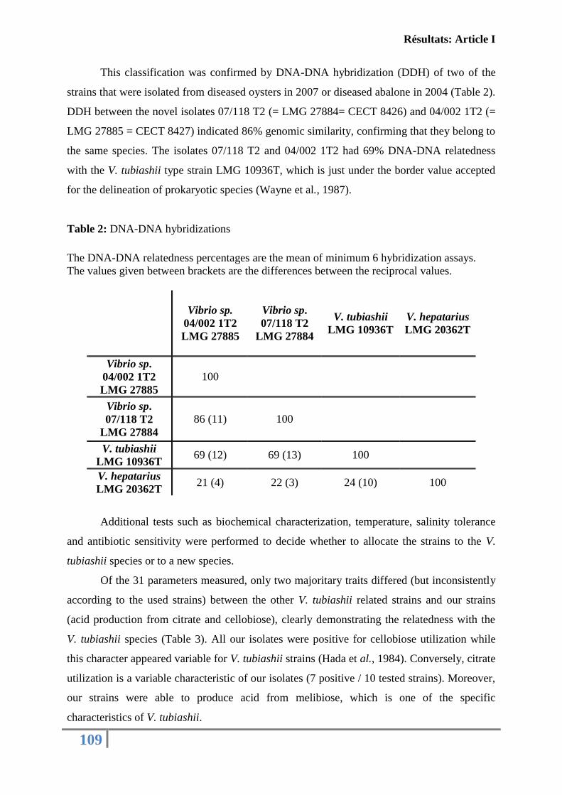
Résultats: Article I
109
This classification was confirmed by DNA-DNA hybridization (DDH) of two of the
strains that were isolated from diseased oysters in 2007 or diseased abalone in 2004 (Table 2).
DDH between the novel isolates 07/118 T2 (= LMG 27884= CECT 8426) and 04/002 1T2 (=
LMG 27885 = CECT 8427) indicated 86% genomic similarity, confirming that they belong to
the same species. The isolates 07/118 T2 and 04/002 1T2 had 69% DNA-DNA relatedness
with the V. tubiashii type strain LMG 10936T, which is just under the border value accepted
for the delineation of prokaryotic species (Wayne et al., 1987).
Table 2: DNA-DNA hybridizations
The DNA-DNA relatedness percentages are the mean of minimum 6 hybridization assays.
The values given between brackets are the differences between the reciprocal values.
Vibrio sp.
04/002 1T2
LMG 27885
Vibrio sp.
07/118 T2
LMG 27884
V. tubiashii
LMG 10936T
V. hepatarius
LMG 20362T
Vibrio sp.
04/002 1T2
LMG 27885
100
Vibrio sp.
07/118 T2
LMG 27884
86 (11) 100
V. tubiashii
LMG 10936T 69 (12) 69 (13) 100
V. hepatarius
LMG 20362T 21 (4) 22 (3) 24 (10) 100
Additional tests such as biochemical characterization, temperature, salinity tolerance
and antibiotic sensitivity were performed to decide whether to allocate the strains to the V.
tubiashii species or to a new species.
Of the 31 parameters measured, only two majoritary traits differed (but inconsistently
according to the used strains) between the other V. tubiashii related strains and our strains
(acid production from citrate and cellobiose), clearly demonstrating the relatedness with the
V. tubiashii species (Table 3). All our isolates were positive for cellobiose utilization while
this character appeared variable for V. tubiashii strains (Hada et al., 1984). Conversely, citrate
utilization is a variable characteristic of our isolates (7 positive / 10 tested strains). Moreover,
our strains were able to produce acid from melibiose, which is one of the specific
characteristics of V. tubiashii.
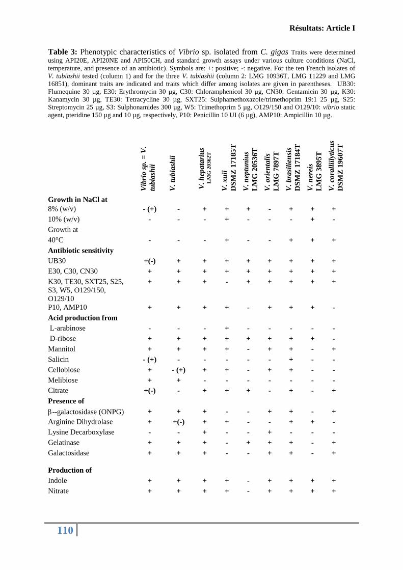
Résultats: Article I
110
Table 3: Phenotypic characteristics of Vibrio sp. isolated from C. gigas Traits were determined
using API20E, API20NE and API50CH, and standard growth assays under various culture conditions (NaCl,
temperature, and presence of an antibiotic). Symbols are: +: positive; -: negative. For the ten French isolates of
V. tubiashii tested (column 1) and for the three V. tubiashii (column 2: LMG 10936T, LMG 11229 and LMG
16851), dominant traits are indicated and traits which differ among isolates are given in parentheses. UB30:
Flumequine 30 µg, E30: Erythromycin 30 µg, C30: Chloramphenicol 30 µg, CN30: Gentamicin 30 µg, K30:
Kanamycin 30 µg, TE30: Tetracycline 30 µg, SXT25: Sulphamethoxazole/trimethoprim 19:1 25 µg, S25:
Streptomycin 25 µg, S3: Sulphonamides 300 µg, W5: Trimethoprim 5 µg, O129/150 and O129/10: vibrio static
agent, pteridine 150 µg and 10 µg, respectively, P10: Penicillin 10 UI (6 µg), AMP10: Ampicillin 10 µg.
Vib
rio
sp
. =
V.
tub
iash
ii
V.
tub
iash
ii
V.
hep
ata
riu
s
L
MG
20
362
T
V.
xu
ii
DS
MZ
17
18
5T
V.
nep
tun
ius
LM
G 2
05
36
T
V.
ori
enta
lis
LM
G 7
89
7T
V.
bra
sili
ensi
s
DS
MZ
17
18
4T
V.
ner
eis
LM
G 3
89
5T
V.
cora
llii
lyti
cus
DS
MZ
19
60
7T
Growth in NaCl at
8% (w/v) - (+) - + + + - + + +
10% (w/v) - - - + - - - + -
Growth at
40°C - - - + - - + + +
Antibiotic sensitivity
UB30 +(-) + + + + + + + +
E30, C30, CN30 + + + + + + + + +
K30, TE30, SXT25, S25,
S3, W5, O129/150,
O129/10
+ + + - + + + + +
P10, AMP10 + + + + - + + + -
Acid production from
L-arabinose - - - + - - - - -
D-ribose + + + + + + + + -
Mannitol + + + + - + + - +
Salicin - (+) - - - - - + - -
Cellobiose + - (+) + + - + + - -
Melibiose + + - - - - - - -
Citrate +(-) - + + + - + - +
Presence of
--galactosidase (ONPG) + + + - - + + - +
Arginine Dihydrolase + +(-) + + - - + + -
Lysine Decarboxylase - - + - - + - - -
Gelatinase + + + - + + + - +
Galactosidase
+ + + - - + + - +
Production of
Indole + + + + - + + + +
Nitrate + + + + - + + + +
Résultats: Article I
111
In conclusion, with 69% DNA-DNA relatedness with V. tubiashii type strains, two
phenotypic variables differences revealed by majoritary trait analysis and distinct grouping by
MLST analysis, our isolates should be considered as V. tubiashii and not as a new species.
3.2 Pathogenicity assays
The pathogenicity of seven French isolates (07/038 2T2, 07/110 T1, 07/112 T1, 07/120
T1, 07/119 T1, 07/118 T2 and 04/002 1T2) was investigated by experimental infections that
also included a non virulent strain, LMG20012T (Fig. 3). One year-old oysters were intra
muscularly injected with these bacterial suspensions used at the same concentration. After 24
h, cumulative mortalities were comprised between 36.7% and 83.3% indicating that these
seven isolates had different degrees of virulence. 07/110 T1, 07/038 2T2, 07/120 T1 and
07/118 T2 were the most virulent and caused more than 60% mortality after 24h. 07/112 T1,
07/119 T1 and 04/002 1T2 caused less than 60% mortality at 24 h but all the isolates induced
more than 60% mortality after 3 days. In some moribund oysters, several predominant
bacterial isolates were obtained in muscle and haemolymph tissues. All bacterial isolates were
found to belong to V. tubiashii species using our gyrB-QPCR diagnosis assay.
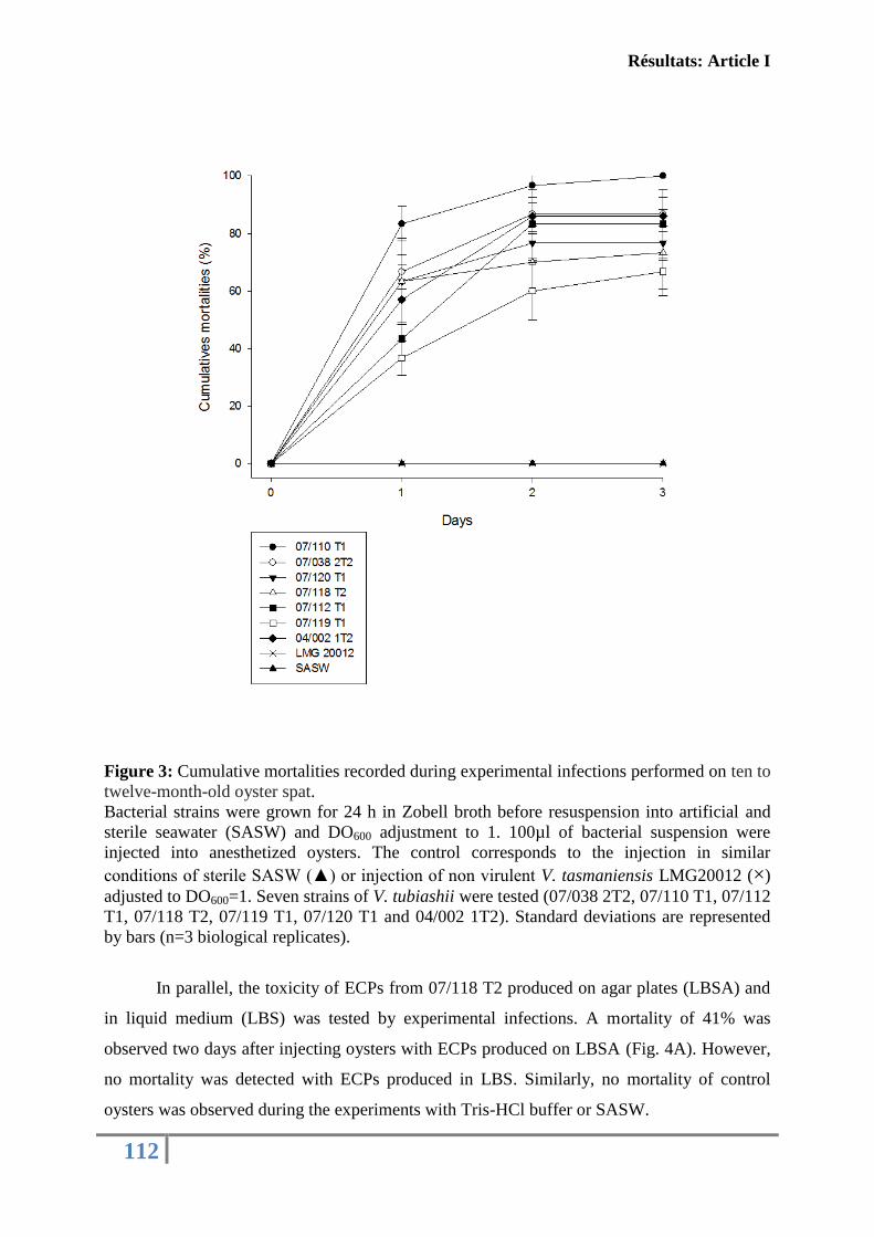
Résultats: Article I
112
Figure 3: Cumulative mortalities recorded during experimental infections performed on ten to
twelve-month-old oyster spat.
Bacterial strains were grown for 24 h in Zobell broth before resuspension into artificial and
sterile seawater (SASW) and DO600 adjustment to 1. 100µl of bacterial suspension were
injected into anesthetized oysters. The control corresponds to the injection in similar
conditions of sterile SASW (▲) or injection of non virulent V. tasmaniensis LMG20012 (×)
adjusted to DO600=1. Seven strains of V. tubiashii were tested (07/038 2T2, 07/110 T1, 07/112
T1, 07/118 T2, 07/119 T1, 07/120 T1 and 04/002 1T2). Standard deviations are represented
by bars (n=3 biological replicates).
In parallel, the toxicity of ECPs from 07/118 T2 produced on agar plates (LBSA) and
in liquid medium (LBS) was tested by experimental infections. A mortality of 41% was
observed two days after injecting oysters with ECPs produced on LBSA (Fig. 4A). However,
no mortality was detected with ECPs produced in LBS. Similarly, no mortality of control
oysters was observed during the experiments with Tris-HCl buffer or SASW.

Résultats: Article I
113
07/118 T2 ECPs produced in LBS
07/118 T2 ECPs produced on LBSA
% of residual activity after preincubation with 1,10
phenanthroline (10mM) a
5.06 ± 0.5
25.8 ± 5.8
Figure 4: (A) Cumulative mortalities of juvenile oysters following intramuscular injection of
V. tubiashii (07/118 T2= LMG 27884= CECT 8426) ECPs produced on LBSA and LBS
medium and in vitro stability of protease activity (azocaseinase activity). The primary axis
corresponds to cumulative mortalities recorded during experimental infections performed on juvenile
oysters after injection of 07/118 T2 ECPs produced in LBS and on LBSA medium (solid curves). 100
µl of ECPs (5µg g-1
of proteins) were injected into anesthetized oysters. The control corresponds to the
injection in similar condition of artificial and sterile seawater (SASW) and Tris- HCl buffer (50 mM
pH 8).The secondary axis corresponds to the stability of protease activity measured in vitro (dashed
line curve). 100 µl of 07/118T2 ECPs produced on LBSA were incubated at 22°C for two days. The
azocaseinase activity (after 24 h and 48 h) was measured using an azocasein assay. Standard
deviations are represented by bars (n=2 biological replicates).
(B) Effect of one metalloprotease inhibitor on azocaseinase activity of 07/118 T2 ECPs
produced on LBSA and LBS medium.a
Final concentration of the inhibitor in the assay
mixture.
3.3 Characterization of proteolytic activity of 07/118 T2 ECPs
The proteolytic activity of ECPs from 07/118 T2 (in liquid and solid medium) was
severely reduced by metalloprotease inhibitor (1, 10-phenanthroline), with residual activities
of 5% and 25% for ECPs produced on LBS and LBSA, respectively (Fig. 4B). For the
(B)
(A)
Résultats: Article I
114
stability of ECPs produced on LBSA, 80% of the protease activities was maintained 48 hours
post- incubation at 22°C (Fig. 4A).
3.4 Development of a QPCR assay and hatchery- and field-samples screening
The gene encoding gyrase B (gyrB) in the 07/112 T2 and 07/118 T2 strains was
sequenced (KF270490 and KF270491 respectively). After sequence alignments with the V.
tubiashii type strain and closely related strains (V. hepatarius, V. orientalis, V. brasiliensis, V.
coralliilyticus, V. neptunius, V harveyi and V. xuii), species-specific (gyrB) primers and probes
were designed. The gyrB primers and probes were designed to amplify all V. tubiashii
isolates.
The concentration of the primers and probes was optimized at 200 nM. Using these
conditions, excellent real time PCR efficiencies were obtained by 10-fold dilutions of 07/118
T2 and V. tubiashii type strain DNAs, 99.2 % and 96.3 % respectively (Fig. S1). Finally, the
specificity was investigated on different bacterial DNA extracts (Table 1). Among 20 tested
strains including 7 type strains belonging to different Vibrio species, only all tested V.
tubiashii strains (n=13, including the type strain) were found positive. By considering Ct
above cycle 38, our QPCR assay appeared to be specific and able to detect more than 103
bacteria/ml (Fig. S1).
Using this new diagnostic tool, the French LNR hatchery and field DNA collection
were screened. These DNA were extracted from oysters tissues sampled in a context of
declared mortalities analyzed by the REPAMO network. Of the 49 batches of animals (379
samples), only DNA from two batches were positive with the V. tubiashii diagnostic tool.
These batches came from two different French hatcheries (2 positive batches from the 21
analysed) (Table S2). On the 28 batches of moribund animals from the field collection, all
were negative for the presence of V. tubiashii DNA.
Résultats: Article I
115
4. Discussion
The aims of the present study were (1) to fine-tune the taxonomic affiliation of
virulent strains collected during an episode of high level Crassostrea gigas mortality in
France using a polyphasic approach (phenotypic and genotypic characterization); (2) to
evaluate both their virulence and the toxicity of their extracellular products; and (3) to
investigate the geographical distribution of these bacteria.
The strains used in this study were isolated from an experimental hatchery/nursery in
France in 2007, during a period of heavy oyster mortalities, under the auspices of the French
institutional surveillance network, REPAMO.
Hatchery systems provide ideal conditions for the growth of larvae and bacteria
multiplication (density, temperature, organic matter load, etc.) (Prado et al., 2005)
Furthermore, farmed animals are monitored daily, allowing precise observations of mortality
and investigations into pathogens. Hatcheries are therefore ideal places for the isolation of
bacteria and the description of new bacterial species (Sugumar et al., 1998; Estes et al., 2004;
Gay et al., 2004; Labreuche et al., 2006b; Elston et al., 2008).
For mollusks pathogens, the investigation of the pathogenic potential of a particular
strain is still in its infancy due to the absence of animal models, cloned or knock-out animals
for in vivo studies, or the availability of culture cell lines for in vitro assays. Furthermore, the
virulence of Vibrio species is known to involve many virulence factors and different pathways
(Shinoda & Miyoshi, 2011) and there is a lack of standardized in vitro protocols for virulence
phenotype characterization.
In an in vivo assay, bacteria can be injected into animal muscle (injection (Gay et al.,
2004), put into seawater surrounding the animals after a culture phase (immersion or bath
challenge (Travers et al., 2008; De Decker et al., 2011)), or transmitted by cohabitation
challenges (De Decker & Saulnier, 2011). Even if they rely on the injection of large quantities
of cultured bacteria, bypass the penetration step and consequently the first lines of defense,
injection protocols are still the most reproducible and widely-used methodology for initial
Vibrio challenges (Labreuche et al., 2006a; Gagnaire et al., 2007; Garnier et al., 2007;
Williams et al., 2009). In this study, this protocol was applied to our isolated strains and
allows the demonstration of the high degree of virulence of the strains into oyster tissues
(between 36.7 and 83.3% of mortality as soon as 24 hours post-injection) of mortality as soon
as 24 hours post-injection. This mortality rate is comparable to the one induced by other
Vibrio species when used at same dose that the present study: V. aestuarianus (02/041)
Résultats: Article I
116
induced 62.5% of mortality eight days post-infection (Labreuche et al., 2006b) and the co-
infection of a mix of two Vibrio (V. splendidus LGP 32 and V. aestuarianus 02/041) gave a
mean mortality rate of 88.9% five days post-infection (De Decker and Saulnier, 2011).
After isolating the bacteria and demonstrating their high level of virulence, a precise
classification was needed. Initial identification of strains isolated in 2007, based on partial
16S rRNA analysis, led to the affiliation of these strains to the V. harveyi species (Saulnier et
al., 2010). It is difficult to classify them as part of the Vibrio clades, and particularly the V.
harveyi clade, based on 16S rRNA analysis. A reliable classification needs to be supported by
sequence analyses of multiple housekeeping genes (Thompson et al., 2005). Here, this
analysis with 16S rRNA, ftsZ, topA, pyrH and recA showed a tight grouping of 07/118 T2
with other French isolates and close to the V. tubiashii type strain, but clearly distinct from V.
tubiashii group. This was confirmed by the DNA-DNA hybridization of two selected strains
(04/002 1T2 and 07/118 T2) and phenotypic characterization based on 31 different parameters
(salt, temperature and antibiotic sensitivity, sugar utilization, enzymatic activities). Although
DNA-DNA hybridization with the V. tubiashii type strain was below 70% (69%), as
biochemical assays do not allow a clear distinction between our isolates and the V. tubiashii
strains, we propose to affiliate the 07/118 T2 (LMG 27884 = CECT 8426T) strain and other
proximate isolates to a group belonging to the V. tubiashii species.
V. tubiashii, misclassified until 1984, was first implicated in bivalve mortalities on the
east coast of the United States (Tubiash et al., 1965; Tubiash et al., 1970; Hada et al., 1984).
In the eighties, in England, three virulent isolates of V. tubiashii were described that were
pathogenic for C. gigas and Ostrea edulis larvae (Jeffries, 1982). More recently, in 2006 and
2007, some V. tubiashii strains were detected on the north American Pacific coast that caused
a serious loss of hatchery production (Estes et al., 2004; Elston et al., 2008). The associated
disease was characterized by a rapid appearance of symptoms resulting in reduced larval
motility and necrosis in extensive soft tissue.
It is important to note that our isolates are close but clearly separated to those from the
USA. Interestingly, a strain isolated during a mortality episode of Haliotis tuberculata
abalone (Normandy, 2004; 04/002 1T2 = LMG 27885 = CECT 8427) clusters with other
french V. tubiashii. V. tubiashii has therefore been present in France since at least 2004. This
strain can also induce high mortality in C. gigas oysters after intramuscular injection. In
contrast to work on other Vibrio demonstrating a specific virulence for their natural hosts
(Saulnier et al., 2000; Travers et al., 2009), one V. tubiashii virulent strain seems to affect

Résultats: Article I
117
both oysters and abalone, even if virulence has not yet experimentally demonstrated on
abalone.
Vibrio tubiashii is a pathogen that is known to affect hatchery and nursery production
and cause serious financial losses. Specific and sensitive detection tools are therefore
necessary. A previously described, real time PCR assay targeting the V. tubiashii vtpA gene
(Gharaibeh et al., 2009) was tested but we encountered specificity issues (data not shown).
Taqman real time PCR based on the dnaJ sequence has also been used to detect and quantify
V. aestuarianus (Saulnier et al., 2009). This technique is sensitive, rapid and reproducible,
irrespective of the nature of the sample (seawater or oyster homogenate samples). In this
paper, a taqman real time PCR diagnostic tool was developed to investigate the distribution of
V. tubiashii in France and its potential implication in mollusk mortalities. Based on the
hypothesis that a pathogen implicated in a mortality event is present in high amounts, a cut-
off was fixed in our QPCR assay to ensure specificity rather than sensitivity. In spite of this,
using these protocols we could detect as little as 103 bacteria/ ml, allowing a reliable detection
of this pathogen.
The production of extracellular enzymes such as proteases by pathogenic Vibrio has
been widely observed (Shinoda and Miyoshi, 2011). These proteases, which are often from
the family of zinc metalloproteases, are known to be one of the most toxic factors to the host
(Delston et al., 2003; Teo et al., 2003; Binesse et al., 2008; Hasegawa et al., 2008; Labreuche
et al., 2010), particularly for V. tubiashii. Hasegawa et al. (2008) showed that V. tubiashii
ECPs were toxic to C. gigas larvae, and identified virulence factors in these ECPs: the
metalloprotease VtpA and the hemolysin VthA.
Besides the infection of oysters by french V. tubiashii, ECPs from the 07/118 T2 strain
produced either from plate agar or from liquid medium were tested for toxicity on oysters by
experimental injection.
The stability test showed that 80% of protease activity is maintained after 48 h
pointing out its potential role in the infection process and the colonization step in particular
since metalloproteases are known to degrade the extracellular matrix of a variety of host and
tissues even if we cannot exclude the existence of other virulence factors also acting.
Interestingly, only ECPs from culture on agar plates (LBSA) showed toxicity. However,
differences in protease composition and quantity produced could be suggested, supported by
the differential effect of a specific inhibitor such as 1,10-phenanthroline on residual protease
activity. This type of difference in bacterial protein composition after culture in liquid or solid
medium has been reported previously (Cheung & Fischetti, 1988; McCarter & Silverman,
Résultats: Article I
118
1990). Culture on solid medium favors biofilm formation and microenvironments that can
influence bacterial metabolite production (Cheung and Fischetti, 1988). Bacterial adherence is
an important step in local multiplication and infection. Lorian proposed that in vitro systems
providing a solid support surface for the growth of bacteria reflect more accurately in vivo
conditions (Lorian, 1989). The toxicity of V. tubiashii ECPs produced only on agar plates may
indicate the importance of adherence in V. tubiashii virulence factor production and in
consequence in the infection cycle.
It should be noticed that protease activity was only characterized by testing azocasein
as a non-specific substrate, as already reported in other studies, and this was completed using
inhibitor assays. A more specific cocktail of inhibitors and substrates should be developed to
obtain a more precise composition of the ECPs protease.
The French survey network allows the isolation of dominant strains from haemolymph or
tissue samples, but also the conservation of bacteria in cryoprotectant as well as bacterial
DNA. With the support of the LNR (National Reference Laboratory for mollusk disease), we
were able to screen the different bacterial isolates and also the DNA library, thus avoiding the
isolation biases (only the predominant strains isolated on TCBS at 20°C are conserved).
A gyrB gene was the target of the screening and primers were designed to amplify
DNA from all strains belonging to V. tubiashii species. Using this approach, we detected V.
tubiashii DNA in a strain isolated during 2 episodes of hatchery mortality in 2008. This strain
was also isolated in 10 hatchery batches affected by mortalities, 9 from 2007 and 1 from
2004. In conclusion, we have evidence of the presence of high amounts of V. tubiashii during
12 episodes of mortality in French hatcheries since 2004. None of the field samples tested
revealed for instance the presence of such high amounts of V. tubiashii.
5. Conclusions
Based on their biochemical and genetic characterization, nine predominant bacterial
strains, isolated from a French experimental hatchery/nursery during mortalities events, were
classified as a novel group belonging to V. tubiashii species. Two reference strains, isolated
from diseased abalone and diseased oysters in 2004 and 2007, respectively, were described
for this group. The significant pathogenicity of different isolates and the toxicity of the
extracellular products of 07/118 T2 were confirmed by experimental challenge. Further
studies will be required to complete our knowledge on the pathogenic factors secreted by
these strains of V. tubiashii and their mechanisms of action.
Résultats: Article I
119
Acknowledgements
This study was supported by a Ph.D grant from the CNRS and IFREMER for R.
Mersni-Achour (2010-2013). The PRES (Pôle de Recherche et d’Enseignement Supérieur) of
Limousin PoitouCharentes and the FR CNRS 3097 «Fédération de Recherche en
Environnement et Développement Durable» are acknowledged for financial support through
the research project «Caractérisation in vitro des effets de métalloprotéases sur différents
types cellulaires de l’huître creuse C. gigas».
Résultats: Article I
120
References
Aguirre-Guzman, G., Ascencio, F., Saulnier, D., 2005. Pathogenicity of Vibrio penaeicida or
white shrimp Litopenaeus vannamei: a cysteine protease-like exotoxin as a virulence
factor. Diseases of Aquatic Organisms. 67, 201-207.
Beaz-Hidalgo, R., Balboa, S., Romalde, J. L., Figueras, M. J., 2010. Diversity and
pathogenecity of Vibrio species in cultured bivalve molluscs. Environmental
Microbiology Reports. 2, 34-43.
Binesse, J., Delsert, C., Saulnier, D., Champomier-Verges, M. C., Zagorec, M., Munier-
Lehmann, I., et al., 2008. Metalloprotease Vsm Is the Major Determinant of Toxicity
for Extracellular Products of Vibrio splendidus. Applied and Environmental
Microbiology. 74, 7108-7117.
Bower, S. M., Hervio, D., Meyer, G. R., 1997. Infectivity of Mikrocytos mackini, the
causative agent of Denman Island disease in Pacific oysters, Crassostrea gigas, to
various species of oysters. . Diseases of Aquatic Organism. 29, 111-116.
Cheung, A. L., Fischetti, V. A., 1988. Variation in the expression of cell wall proteins of
Staphylococcus aureus grown on solid and liquid media. Infect Immun. 56, 1061-5.
De Decker, S., Normand, J., Saulnier, D., Pernet, F., Castagnet, S., Boudry, P., 2011.
Responses of diploid and triploid Pacific oysters Crassostrea gigas to Vibrio infection
in relation to their reproductive status. Journal of Invertebrate Pathology. 106, 179-
191.
De Decker, S., Saulnier, D., 2011. Vibriosis induced by experimental cohabitation in
Crassostrea gigas: Evidence of early infection and down-expression of immune-
related genes. Fish & Shellfish Immunology. 30, 691-699.
Delston, R. B., Kothary, M. H., Shangraw, K. A., Tall, B. D., 2003. Isolation and
characterization of a zinc-containing metalloprotease expressed by Vibrio tubiashii.
Canadian Journal of Microbiology. 49, 525-529.
Eck, R. V., Dayhoff, M. O., 1966. Atlas of protein sequence and structure. National
Biomedical Research Foundation.
Elston, R. A., Hasegawa, H., Humphrey, K. L., Polyak, I. K., Hase, C. C., 2008. Re-
emergence of Vibrio tubiashii in bivalve shellfish aquaculture: severity, environmental
drivers, geographic extent and management. Diseases of Aquatic Organisms. 82, 119-
134.
Estes, R. M., Friedman, C. S., Elston, R. A., Herwig, R. P., 2004. Pathogenicity testing of
shellfish hatchery bacterial isolates on Pacific oyster Crassostrea gigas larvae.
Diseases of Aquatic Organisms. 58, 223-30.
Ezaki, T., Hashimoto, Y., Yabuuchi, E., 1989. Fluorometric deoxyribonucleic acid-
deoxyribonucleic acid hybridization in microdilution wells as an alternative to
membrane filter hybridization in which radioisotopes are used to determine genetic
relatedness among bacterial strains. International Journal of Systematic Bacteriology.
39, 224-229.
Felsenstein, J., 1985. Confidence limits on phylogenies: an approach using the boostrap.
Evolution. 39, 783-791.
Gagnaire, B., Gay, M., Huvet, A., Daniel, J. Y., Saulnier, D., Renault, T., 2007. Combination
of a pesticide exposure and a bacterial challenge: in vivo effects on immune response
of Pacific oyster, Crassostrea gigas (Thunberg). Aquatic toxicology. 84, 92-102.
Garnier, M., Labreuche, Y., Garcia, C., Robert, M., Nicolas, J. L., 2007. Evidence for the
involvment of pathogenic bacteria in summer mortalities of the Pacific oyster
Crassostrea gigas. Microbial Ecology. 53, 173-186.
Résultats: Article I
121
Garnier, M., Labreuche, Y., Nicolas, J. L., 2008. Molecular and phenotypic characterization
of Vibrio aestuarianus subsp. francensis subsp. nov., a pathogen of the oyster
Crassostrea gigas. Systematic and Applied Microbiology. 31, 358-65.
Gay, M., Berthe, F. C., Le Roux, F., 2004. Screening of Vibrio isolates to develop an
experimental infection model in the Pacific oyster Crassostrea gigas. Diseases of
Aquatic Organisms. 59, 49-56.
Gharaibeh, D. N., Hasegawa, H., Hase, C. C., 2009. Development of a quantitative real-time
PCR assay for detection of Vibrio tubiashii targeting the metalloprotease gene. Journal
of Microbiological Methods. 76, 262-268.
Gomez-Gil, B., Soto-Rodriguez, S., Garcia-Gasca, A., Roque, A., Vazquez-Juarez, R.,
Thompson, F. L., et al., 2004. Molecular identification of Vibrio harveyi-related
isolates associated with diseased aquatic organisms. Microbiology. 150, 1769-77.
Hada, H. S., West, P. A., Lee, J. V., Stemmler, J., Colwell, R. R., 1984. Vibrio tubiashii sp.
nov. a pathogen of bivalve mollusks. International Journal of Systematic Bacteriology.
34, 1-4.
Hasegawa, H., Lind, E. J., Boin, M. A., Hase, C. C., 2008. The extracellular metalloprotease
of Vibrio tubiashii is a major virulence factor for Pacific oyster (Crassostrea gigas)
larvae. Applied and Environmental Microbiology. 74, 4101-4110.
Hoffmann, M., Monday, S. r., Fischer, M., Brown, E. w., 2012. Genetic and phylogenetic
evidence for misidentification of Vibrio species within the Harveyi clade. Letters in
applied Microbiology. 54, 160-165.
Jeffries, V. E., 1982. Three Vibrio strains pathogenic to larvae of Crassostrea gigas and
Ostrea edulis. Aquaculture. 29, 201-226.
Kothary, M. H., Delston, R. B., Curtis, S. K., McCardell, B. A., Tall, B. D., 2001. Purification
and Characterization of a Vulnificolysin-Like Cytolysin Produced by Vibrio tubiashii.
Applied and Environmental Microbiology. 67, 3707-3711.
Labreuche, Y., Lambert, C., Soudant, P., Boulo, V., Huvet, A., Nicolas, J.-L., 2006a. Cellular
and molecular hemocyte responses of the Pacific oyster, Crassostrea gigas, following
bacterial infection with Vibrio aestuarianus strain 01/32. Microbes and Infection. 8,
2715-2724.
Labreuche, Y., Le Roux, F., Henry, J., Zatylny, C., Huvet, A., Lambert, C., et al., 2010.
Vibrio aestuarianus zinc metalloprotease causes lethality in the Pacific oyster
Crassostrea gigas and impairs the host cellular immune defenses. Fish & Shellfish
Immunology. 29, 753-758.
Labreuche, Y., Soudant, P., Goncalves, M., Lambert, C., Nicolas, J., 2006b. Effects of
extracellular products from the pathogenic Vibrio aestuarianus strain 01/32 on
lethality and cellular immune responses of the oyster Crassostrea gigas.
Developmental & Comparative Immunology. 30, 367-379.
Lacoste, A., Jalabert, F., Malham, S., Cueff, A., Gélébart, F., Cordevant, C., et al., 2001. A
Vibrio splendidus strain is associated with summer mortality of juvenile oysters
Crassostrea gigas in the Bay of Morlaix (North Brittany, France). Diseases of Aquatic
Organisms. 46, 139-145.
Le Roux, F., Gay, M., Lambert, C., Waechter, M., Poubalanne, S., Chollet, B., et al., 2002.
Comparative analysis of Vibrio splendidus-related strains isolated during Crassostrea
gigas mortality events. Aquatic Living Resources. 15, 251-258.
Liu, P. V., 1957. Survey of hemolysin production among species of Aeromonas hydrophila
with reduced virulence for fish. Infection and Immunity. 74, 718-727.
Lorian, V., 1989. In vitro simulation of in vivo conditions: physical state of the culture
medium. Journal of Clinical Microbiology. 27, 2403-6.
Résultats: Article I
122
McCarter, L., Silverman, M., 1990. Surface-induced swarmer cell differentiation of Vibrio
parahaemolyticus. Molecular Microbiology. 4, 1057-62.
Meyers, J. A., Burreson, E. M., Barber, B. J., Mann, R., 1991. Susceptibility of diploid and
triploid Pacific oysters, Crassostrea gigas (Thunberg, 1793), and eastern oysters,
Crassostrea virginica (Gmelin, 1791), to Perkinsus marinus. Journal of Shellfish
Research. 10, 433-437.
Prado, S., Romalde, J., Montes, J., Barja, J. L., 2005. Pathogenic bacteria isolated from
disease outbreaks in shellfish hatcheries. First description of Vibrio neptunius as an
oyster pathogen Diseases of Aquatic Organisms. 67, 209-215.
Renault, T., Le Deuff, R.-M., Cochennec, N., Maffart, P., 1994. Herpesviruses associated
with mortalities among Pacific oyster, Crassostrea gigas, in France-Comparative
study. Revue Médicale Vétérinaire. 145, 735-742.
Romalde, J. L., Barja, J. L., 2010. Bacteria in molluscs: good and bad guys. Current Research,
Technology and Education Topics in Applied Microbiology and Microbial
Biotechnology. 1, 136-147.
Saitou, N., Nei, M., 1987. The neighbor-joining method: a new method for reconstructing
phylogenetic trees. Molecular Biology and Evolution. 4, 406-25.
Samain, J. F., Dégremont, L., Soletchnik, P., Haure, J., Bédier, E., Ropert, M., et al., 2007.
Genetically based resistance to summer mortality in the Pacific oyster (Crassostrea
gigas) and its relationship with physiological, immunological characteristics and
infection processes. Aquaculture. 268, 227-243.
Saulnier, D., De Decker, S., Haffner, P., 2009. Real-time PCR assay for rapid detection and
quantification of Vibrio aestuarianus in oyster and seawater: A useful tool for
epidemiologic studies. Journal of Microbiological Methods. 77, 191-197.
Saulnier, D., De Decker, S., Haffner, P., Cobret, L., Robert, M., Garcia, C., 2010. A large-
scale epidemiological study to identify bacteria pathogenic to pacific oyster
Crassostrea gigas and correlation between virulence and metalloprotease-like activity.
Microbial Ecology. 59, 787-798.
Saulnier, D., Haffner, P., Goarant, C., Levy, P., Ansquer, D., 2000. Experimental infection
models for shrimp vibriosis studies: a review. Aquaculture. 191, 133-144.
Segarra, A., Pepin, J. F., Arzul, I., Morga, B., Faury, N., Renault, T., 2010. Detection and
description of a particular Ostreid herpesvirus 1 genotype associated with massive
mortality outbreaks of Pacific oysters, Crassostrea gigas, in France in 2008. Virus
Research. 153, 92-9.
Shinoda, S., Miyoshi, S., 2011. Proteases produced by vibrios. Biocontrol Sci. 16, 1-11.
Sugumar, G., Nakai, T., Hirata, Y., Matsubara, D., Muroga, K., 1998. Vibrio splendidus
biovar II as the causative agent of bacillary necrosis of Japanese oyster Crassostrea
gigas larvae Diseases of Aquatic Organisms. 33, 111-118.
Teo, J. W. P., Zhang, L.-H., Poh, C. L., 2003. Cloning and characterization of a
metalloprotease from Vibrio harveyi strain AP6. Gene. 303, 147-156.
Thompson, F. L., Gevers, D., Thompson, C. C., Dawyndt, P., Naser, N., Hoste, B., et al.,
2005. Phylogeny and molecular identification of vibrios on the basis of multilocus
sequence analysis. Applied and Environmental Microbiology. 71, 5107-5115.
Thompson, F. L., Gomez-Gil, B., Vasconcelos, A. T., Sawabe, T., 2007. Multilocus sequence
analysis reveals that Vibrio harveyi and V. campbellii are distinct species. Appl
Environmental Microbiology. 73, 4279-85.
Travers, M. A., Barbou, A., Le Goïc, N., Huchette, S., Paillard, C., Koken, M., 2008.
Construction of a stable GFP-tagged Vibrio harveyi strain for bacterial dynamics
analysis of abalone infection. FEMS Microbiol Lett. 289, 34–40.
Résultats: Article I
123
Travers, M. A., Le Bouffant, R., Friedman, C. S., Buzin, F., Cougard, B., Huchette, S., et al.,
2009. Pathogenic Vibrio harveyi, in contrast to non-pathogenic strains, intervenes with
the p38 MAPK pathway to avoid an abalone haemocyte immune response. Journal of
Cellular Biochemistry. 106, 152-160.
Tubiash, H. S., Chanley, P. E., Leifson, E., 1965. Bacillary necrosis, a disease of larval and
juvenile bivalve mollusks I. Etiology and epizootiology. Journal of Bacteriology. 90,
1036-1044.
Tubiash, H. S., Colwell, R. R., Sakazaki, R., 1970. Marine vibrios associated with bacillary
necrosis, a disease of larval and juvenile bivalve mollusks. Journal of Bacteriology.
103, 272-273.
Tun, K. L., Itoh, N., Shimizu, Y., Yamanoi, H., Yoshinaga, T., Ogawa, K., 2008.
Pathogenicity of the protozoan parasite Marteilioides chungmuensis in the Pacific
oyster Crassostrea gigas. International Journal for Parasitology. 38, 211-217.
Wayne, L. G., Brenner, D. J., Colwell, R. R., Grimont, P. A. D., Kandler, O., Krichevsky, M.
I., et al., 1987. Report of the Ad Hoc Committee on Reconciliation of Approaches to
Bacterial Systematics. International Journal of Systematic Bacteriology. 37, 463-464.
Williams, H. R., Macey, B. M., Burnett, L. E., Burnett, K. G., 2009. Differential localization
and bacteriostasis of Vibrio campbellii among tissues of the Eastern oyster,
Crassostrea virginica. Developmental and Comparative Immunology. 33, 592-600.
Wilson, K., Preparation of genomic DNA from bacteria. In: F. M. Ausubel, et al., Eds.),
Current protocols in molecular biology. Greene Publishing and Wiley Interscience,
New York, 1987, pp. 2.4.1-2.4.5.
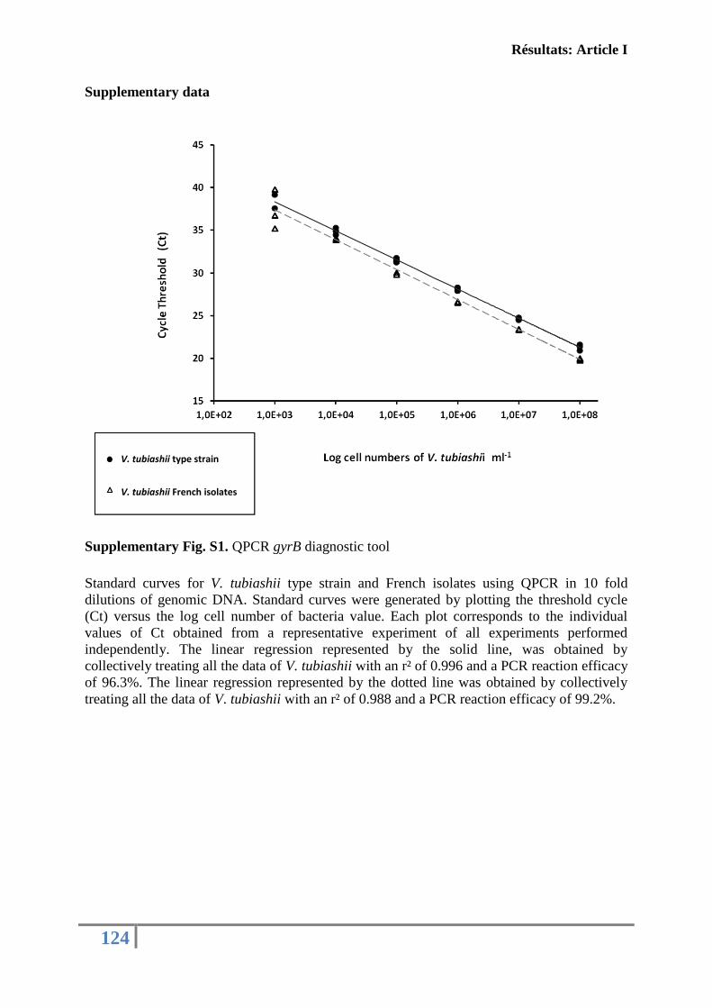
Résultats: Article I
124
Supplementary data
Supplementary Fig. S1. QPCR gyrB diagnostic tool
Standard curves for V. tubiashii type strain and French isolates using QPCR in 10 fold
dilutions of genomic DNA. Standard curves were generated by plotting the threshold cycle
(Ct) versus the log cell number of bacteria value. Each plot corresponds to the individual
values of Ct obtained from a representative experiment of all experiments performed
independently. The linear regression represented by the solid line, was obtained by
collectively treating all the data of V. tubiashii with an r² of 0.996 and a PCR reaction efficacy
of 96.3%. The linear regression represented by the dotted line was obtained by collectively
treating all the data of V. tubiashii with an r² of 0.988 and a PCR reaction efficacy of 99.2%.
V. tubiashii type strain
V. tubiashii French isolates
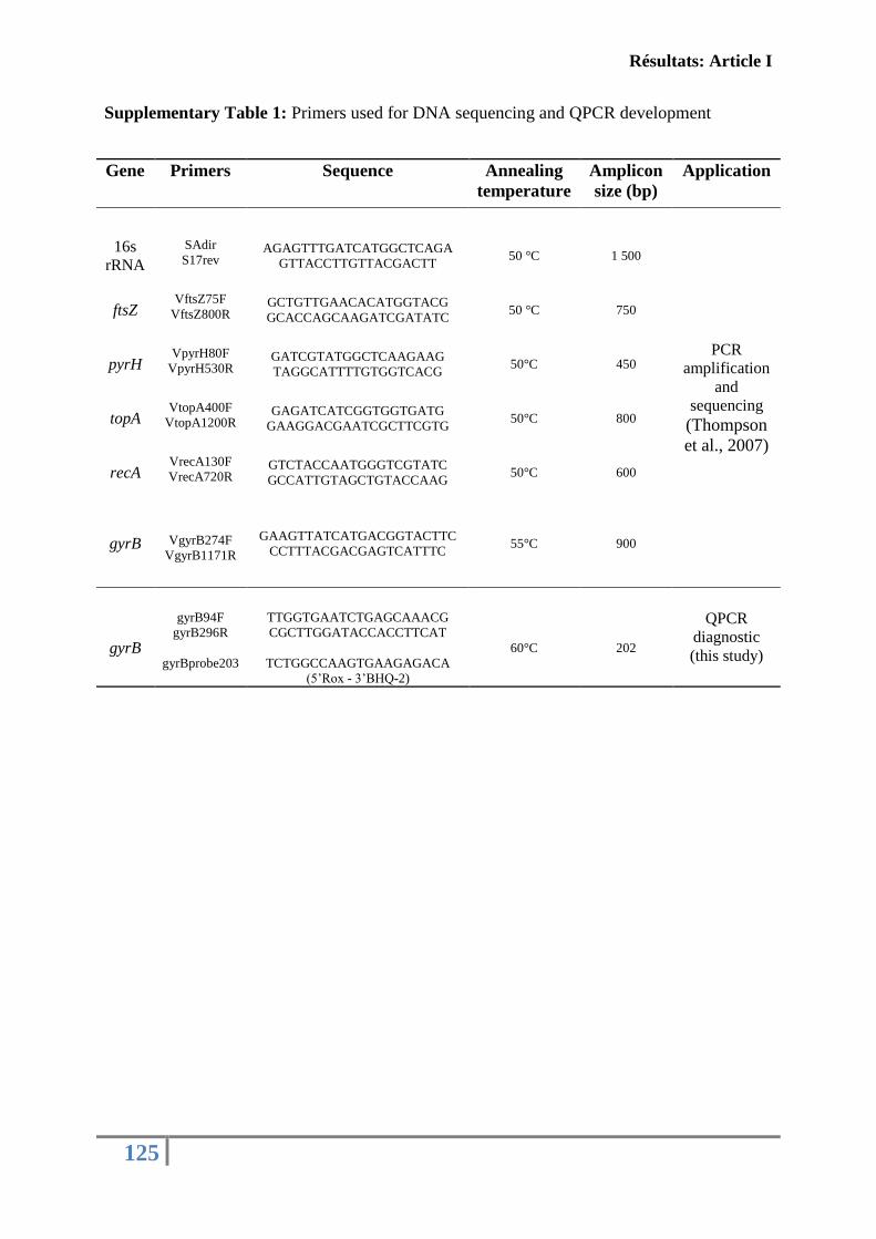
Résultats: Article I
125
Supplementary Table 1: Primers used for DNA sequencing and QPCR development
Gene Primers Sequence Annealing
temperature
Amplicon
size (bp)
Application
16s
rRNA
SAdir
S17rev
AGAGTTTGATCATGGCTCAGA
GTTACCTTGTTACGACTT 50 °C 1 500
PCR
amplification and
sequencing (Thompson
et al., 2007)
ftsZ
VftsZ75F
VftsZ800R
GCTGTTGAACACATGGTACG
GCACCAGCAAGATCGATATC 50 °C 750
pyrH
VpyrH80F
VpyrH530R
GATCGTATGGCTCAAGAAG
TAGGCATTTTGTGGTCACG 50°C 450
topA
VtopA400F
VtopA1200R
GAGATCATCGGTGGTGATG
GAAGGACGAATCGCTTCGTG 50°C 800
recA
VrecA130F
VrecA720R
GTCTACCAATGGGTCGTATC
GCCATTGTAGCTGTACCAAG 50°C 600
gyrB
VgyrB274F
VgyrB1171R
GAAGTTATCATGACGGTACTTC
CCTTTACGACGAGTCATTTC 55°C 900
QPCR
diagnostic
(this study) gyrB
gyrB94F
gyrB296R
gyrBprobe203
TTGGTGAATCTGAGCAAACG
CGCTTGGATACCACCTTCAT
TCTGGCCAAGTGAAGAGACA
(5’Rox - 3’BHQ-2)
60°C 202
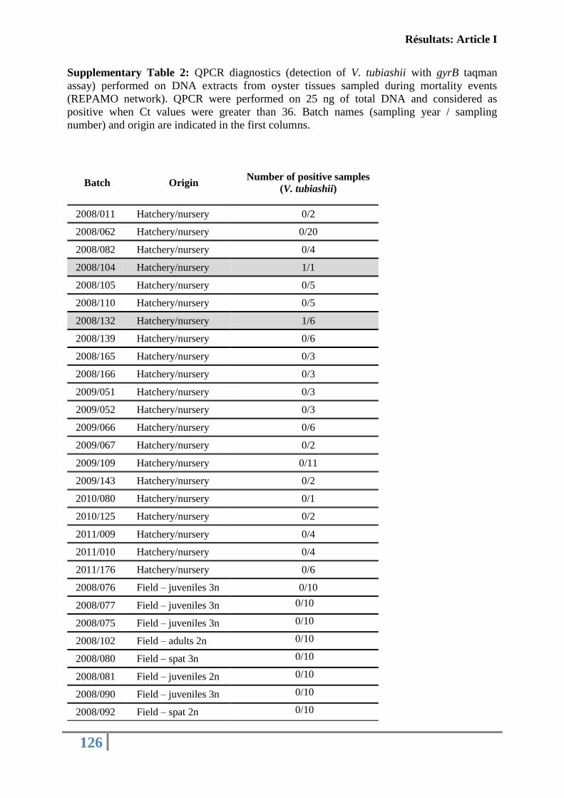
Résultats: Article I
126
Supplementary Table 2: QPCR diagnostics (detection of V. tubiashii with gyrB taqman
assay) performed on DNA extracts from oyster tissues sampled during mortality events
(REPAMO network). QPCR were performed on 25 ng of total DNA and considered as
positive when Ct values were greater than 36. Batch names (sampling year / sampling
number) and origin are indicated in the first columns.
Batch Origin Number of positive samples
(V. tubiashii)
2008/011 Hatchery/nursery 0/2
2008/062 Hatchery/nursery 0/20
2008/082 Hatchery/nursery 0/4
2008/104 Hatchery/nursery 1/1
2008/105 Hatchery/nursery 0/5
2008/110 Hatchery/nursery 0/5
2008/132 Hatchery/nursery 1/6
2008/139 Hatchery/nursery 0/6
2008/165 Hatchery/nursery 0/3
2008/166 Hatchery/nursery 0/3
2009/051 Hatchery/nursery 0/3
2009/052 Hatchery/nursery 0/3
2009/066 Hatchery/nursery 0/6
2009/067 Hatchery/nursery 0/2
2009/109 Hatchery/nursery 0/11
2009/143 Hatchery/nursery 0/2
2010/080 Hatchery/nursery 0/1
2010/125 Hatchery/nursery 0/2
2011/009 Hatchery/nursery 0/4
2011/010 Hatchery/nursery 0/4
2011/176 Hatchery/nursery 0/6
2008/076 Field – juveniles 3n 0/10
2008/077 Field – juveniles 3n 0/10
2008/075 Field – juveniles 3n 0/10
2008/102 Field – adults 2n 0/10
2008/080 Field – spat 3n 0/10
2008/081 Field – juveniles 2n 0/10
2008/090 Field – juveniles 3n 0/10
2008/092 Field – spat 2n 0/10
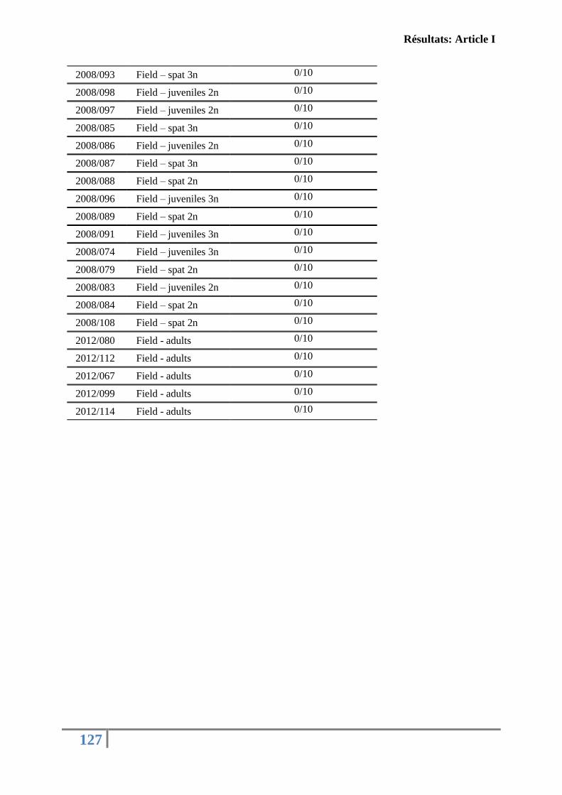
Résultats: Article I
127
2008/093 Field – spat 3n 0/10
2008/098 Field – juveniles 2n 0/10
2008/097 Field – juveniles 2n 0/10
2008/085 Field – spat 3n 0/10
2008/086 Field – juveniles 2n 0/10
2008/087 Field – spat 3n 0/10
2008/088 Field – spat 2n 0/10
2008/096 Field – juveniles 3n 0/10
2008/089 Field – spat 2n 0/10
2008/091 Field – juveniles 3n 0/10
2008/074 Field – juveniles 3n 0/10
2008/079 Field – spat 2n 0/10
2008/083 Field – juveniles 2n 0/10
2008/084 Field – spat 2n 0/10
2008/108 Field – spat 2n 0/10
2012/080 Field - adults 0/10
2012/112 Field - adults 0/10
2012/067 Field - adults 0/10
2012/099 Field - adults 0/10
2012/114 Field - adults 0/10
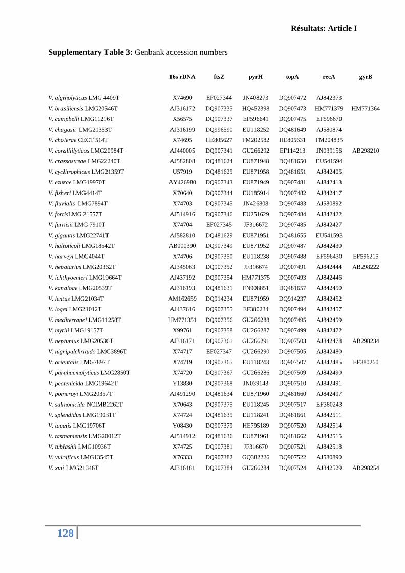
Résultats: Article I
128
Supplementary Table 3: Genbank accession numbers
16s rDNA ftsZ pyrH topA recA gyrB
V. alginolyticus LMG 4409T X74690 EF027344 JN408273 DQ907472 AJ842373
V. brasiliensis LMG20546T AJ316172 DQ907335 HQ452398 DQ907473 HM771379 HM771364
V. campbelli LMG11216T X56575 DQ907337 EF596641 DQ907475 EF596670
V. chagasii LMG21353T AJ316199 DQ996590 EU118252 DQ481649 AJ580874
V. cholerae CECT 514T X74695 HE805627 FM202582 HE805631 FM204835
V. coralliilyticus LMG20984T AJ440005 DQ907341 GU266292 EF114213 JN039156 AB298210
V. crassostreae LMG22240T AJ582808 DQ481624 EU871948 DQ481650 EU541594
V. cyclitrophicus LMG21359T U57919 DQ481625 EU871958 DQ481651 AJ842405
V. ezurae LMG19970T AY426980 DQ907343 EU871949 DQ907481 AJ842413
V. fisheri LMG4414T X70640 DQ907344 EU185914 DQ907482 AJ842417
V. fluvialis LMG7894T X74703 DQ907345 JN426808 DQ907483 AJ580892
V. fortisLMG 21557T AJ514916 DQ907346 EU251629 DQ907484 AJ842422
V. furnisii LMG 7910T X74704 EF027345 JF316672 DQ907485 AJ842427
V. gigantis LMG22741T AJ582810 DQ481629 EU871951 DQ481655 EU541593
V. halioticoli LMG18542T AB000390 DQ907349 EU871952 DQ907487 AJ842430
V. harveyi LMG4044T X74706 DQ907350 EU118238 DQ907488 EF596430 EF596215
V. hepatarius LMG20362T AJ345063 DQ907352 JF316674 DQ907491 AJ842444 AB298222
V. ichthyoenteri LMG19664T AJ437192 DQ907354 HM771375 DQ907493 AJ842446
V. kanaloae LMG20539T AJ316193 DQ481631 FN908851 DQ481657 AJ842450
V. lentus LMG21034T AM162659 DQ914234 EU871959 DQ914237 AJ842452
V. logei LMG21012T AJ437616 DQ907355 EF380234 DQ907494 AJ842457
V. mediterranei LMG11258T HM771351 DQ907356 GU266288 DQ907495 AJ842459
V. mytili LMG19157T X99761 DQ907358 GU266287 DQ907499 AJ842472
V. neptunius LMG20536T AJ316171 DQ907361 GU266291 DQ907503 AJ842478 AB298234
V. nigripulchritudo LMG3896T X74717 EF027347 GU266290 DQ907505 AJ842480
V. orientalis LMG7897T X74719 DQ907365 EU118243 DQ907507 AJ842485 EF380260
V. parahaemolyticus LMG2850T X74720 DQ907367 GU266286 DQ907509 AJ842490
V. pectenicida LMG19642T Y13830 DQ907368 JN039143 DQ907510 AJ842491
V. pomeroyi LMG20357T AJ491290 DQ481634 EU871960 DQ481660 AJ842497
V. salmonicida NCIMB2262T X70643 DQ907375 EU118245 DQ907517 EF380243
V. splendidus LMG19031T X74724 DQ481635 EU118241 DQ481661 AJ842511
V. tapetis LMG19706T Y08430 DQ907379 HE795189 DQ907520 AJ842514
V. tasmaniensis LMG20012T AJ514912 DQ481636 EU871961 DQ481662 AJ842515
V. tubiashii LMG10936T X74725 DQ907381 JF316670 DQ907521 AJ842518
V. vulnificus LMG13545T X76333 DQ907382 GQ382226 DQ907522 AJ580890
V. xuii LMG21346T AJ316181 DQ907384 GU266284 DQ907524 AJ842529 AB298254
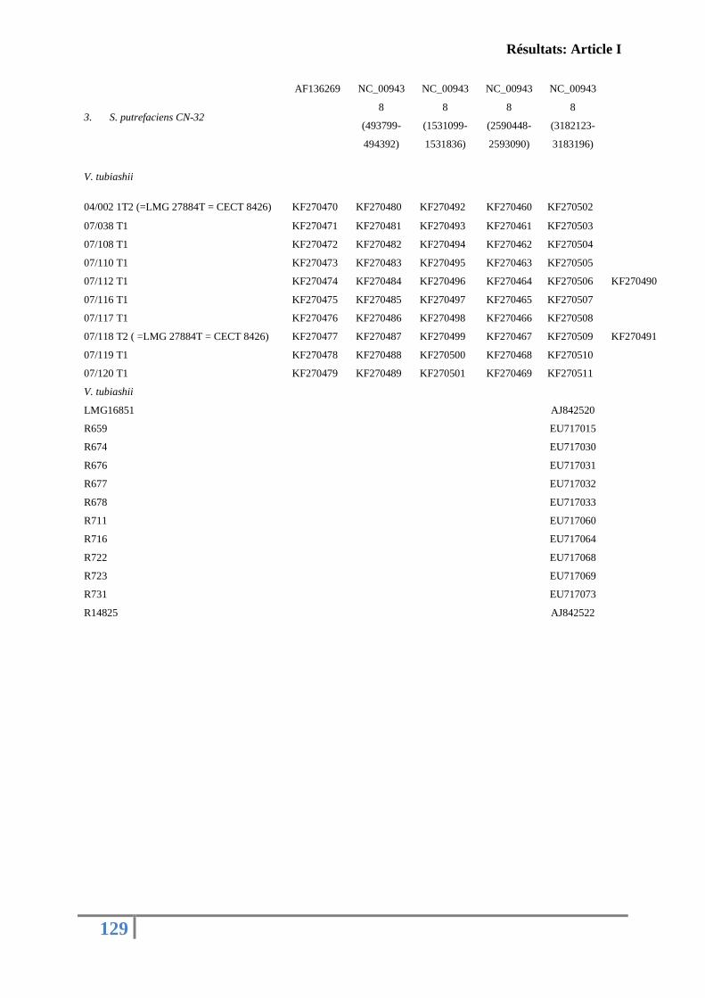
Résultats: Article I
129
3. S. putrefaciens CN-32
AF136269 NC_00943
8
(493799-
494392)
NC_00943
8
(1531099-
1531836)
NC_00943
8
(2590448-
2593090)
NC_00943
8
(3182123-
3183196)
V. tubiashii
04/002 1T2 (=LMG 27884T = CECT 8426) KF270470 KF270480 KF270492 KF270460 KF270502
07/038 T1 KF270471 KF270481 KF270493 KF270461 KF270503
07/108 T1 KF270472 KF270482 KF270494 KF270462 KF270504
07/110 T1 KF270473 KF270483 KF270495 KF270463 KF270505
07/112 T1 KF270474 KF270484 KF270496 KF270464 KF270506 KF270490
07/116 T1 KF270475 KF270485 KF270497 KF270465 KF270507
07/117 T1 KF270476 KF270486 KF270498 KF270466 KF270508
07/118 T2 ( =LMG 27884T = CECT 8426) KF270477 KF270487 KF270499 KF270467 KF270509 KF270491
07/119 T1 KF270478 KF270488 KF270500 KF270468 KF270510
07/120 T1 KF270479 KF270489 KF270501 KF270469 KF270511
V. tubiashii
LMG16851
AJ842520
R659 EU717015
R674 EU717030
R676 EU717031
R677 EU717032
R678 EU717033
R711 EU717060
R716 EU717064
R722 EU717068
R723 EU717069
R731 EU717073
R14825 AJ842522
Résultats: Article II
130
A2: Caractérisation des protéases secrétées par la souche
française V. tubiashii affectant les fonctions immunitaires de
l’huître creuse C. gigas
Les bactéries du genre Vibrio ont la capacité de produire divers facteurs de virulence
impliqués dans leur pathogénicité spécifique. Ces facteurs incluent les protéases, les
entérotoxines, les cytolysines, les sidérophores, les facteurs d’adhésion, et les
hémagglutinines (Shinoda, 2005). Les protéases sont considérées parmi les acteurs principaux
de la virulence avec une majorité appartenant à la famille des métalloprotéases (Shinoda et
al., 1996; Shinoda & Miyoshi, 2011). Concernant la souche américaine V. tubiashii, diverses
études ont montré que cette bactérie sécrète des toxines extracellulaires avec des activités de
type protéolytiques et hémolytiques (Kothary et al., 2001; Delston et al., 2003; Hasegawa et
al., 2008).
Dans l’objectif de caractériser et d’évaluer la toxicité du surnageant bactérien de la
souche française V. tubiashii (07/118 T2), des analyses in vitro ont été réalisées sur quelques
fonctions immunitaires de l’huître. En parallèle, une identification moléculaire des ECPs de la
souche 07/118 T2 a été effectuée par spectrométrie de masse et complétée par une
caractérisation fonctionnelle des activités hydrolytiques. L’ensemble de ces résultats est
regroupé dans l’article 2.
Les analyses des fonctions immunitaires de l’huître nous ont permis de démontrer que
la capacité d'adhérence (28 % d'inhibition) et le pouvoir phagocytaire (56 % d'inhibition)
étaient les deux fonctions hémocytaires les plus affectées par les ECPs de la souche 07/118
T2. Également, les ECPs ont montré une capacité de dégrader différents substrats impliqués
dans la formation de la matrice extracellulaire tels que l’actine, la fibronectine et le collagène.
Ainsi, les anomalies hémocytaires observées pourraient s’expliquer par le pouvoir
hydrolytique des ECPs induisant de probables altérations morphologiques de leur
cytosquelette. L’analyse globale du surnageant bactérien par SDS-PAGE et zymographie a
montré un profil électrophorétique assez complexe et une large gamme d’activités
protéolytiques.
Résultats: Article II
131
L’analyse plus fine des ECPs par nanoLC-Orbitrap a révélé la présence d’une
«extracellular zinc metalloprotease» de GI: 343500312. Afin de caractériser cette
métalloprotéase secrétée par la souche française V. tubiashii, une approche de biologie
moléculaire a été développée. L’amplification et le séquençage du gène codant pour la
métalloprotéase, en utilisant des amorces élaborées à partir du génome complet du V. tubiashii
ATCC 19109_25 (GI=343500308) ont confirmé la présence du gène codant pour cette
protéine avec un cadre de lecture ouvert de 1821 pb codant un polypeptide de 606 aa. Des
motifs spécifiques caractérisant la famille des thermolysines ainsi que des sites de
glycosylations et de phosphorylations ont été prédites. La prédominance d’une activité
métalloprotéasique dans les ECPs de la souche 07/118 T2, a été confirmée par des essais
enzymatiques avec un ensemble d’inhibiteurs et d’ions métalliques tout en utilisant un
substrat spécifique (ADAM substrat) et un substrat général de toute activité protéolytique
(azocaséine).
Les résultats obtenus font l’objet d’un deuxiéme article actuellement sous révision dans
«Journal of Invertebrate pathology», 2014.
Résultats: Article II
132
Article II: FIRST DESCRIPTION OF FRENCH V. TUBIASHII STRAINS
PATHOGENIC TO MOLLUSK
II. CHARACTERIZATION OF PROPERTIES OF THE PROTEOLYTIC
FRACTION OF EXTRACELLULAR PRODUCTS
Under revision in “Journal of invertebrate pathology”
Mersni-Achour Rachida
1,2,6, Imbert Nathalie
3, Huet Valérie
3, Ben Cheikh Yosra
1, Faury
Nicole2, Doghri
Ibtissem
1, Rouatbi Sonia
3, Bordenave Stéphanie
1, Travers Marie-Agnès
2,
Saulnier Denis2,5
, Fruitier-Arnaudin Ingrid1,6
1UMR 7266 CNRS-ULR, LIENSs, Equipe Approches Moléculaires, Environnement-Santé,
Université de La Rochelle, Avenue Michel Crépeau, 17042 La Rochelle, France.
2 Ifremer, SG2M-LGPMM, Laboratoire de Génétique et Pathologie des Mollusques Marins
Avenue de Mus de Loup, 17390 La Tremblade, France.
3UMR 7266 CNRS-ULR, LIENSs, Equipe Animaux MARins à la variabilité
Environnementale, Université de La Rochelle, Avenue Michel Crépeau, 17042 La Rochelle,
France.
4 LIENSs, UMR 6250, CNRS-Université de La Rochelle, 2 rue Olympe de Gouges, F-17042
La Rochelle Cedex 01, France
5Ifremer, Centre Ifremer du Pacifique, UMR 241 Ecosystèmes Insulaires Océaniens, Tahiti,
98719 Taravao, French Polynesia.
6Fédération de Recherche en Environnement et Développement Durable, FR CNRS 3097,
Université de La Rochelle, France.
Résultats: Article II
133
Abstract
Extracellular products (ECPs) of the French V. tubiashii strain 07/118 T2 were
previously reported to be toxic for the Pacific oyster Crassostrea gigas. In this study we now
assessed host cellular immune responses and bacterial potential effectors by which these
ECPs can be associated with host damages.
The adhesion capacity (28% inhibition) and phagocytosis ability (56% inhibition) of oyster
hemocytes were the main functions affected following in vitro contact between hemocytes
and V. tubiashii ECPs. This may be linked to the demonstration of the capability of ECPs to
cleave various cellular substrates as oyster collagen.
Moreover, a strong metalloproteolytic activity was recorded with general (azocasein)
and specific (ADAM) substrates and characterized by the use of standard inhibitors and metal
ions. The addition of 1, 10-phenanthroline and Zn2+
decreased proteolytic activity by about
80% and 50% respectively, confirming the presence of zinc metalloproteolytic activity in the
ECPs. Mass spectrometry analyses of crude ECPs identified an extracellular zinc
metalloprotease encoded by a gene with an open reading frame of 1821 bp (606 aa).
Consensus zinc-binding motifs specific to thermolysin family and some glycosylation and
phosphorylation sites were located on the deduced protein sequence.
Even if taken together, our results let us wonder if this (these) zinc metalloprotease(s) could
be involved in the impairment of hemocyte immunological functions, the direct implication of
this (these) protein(s) in ECPs toxicity still has to be demonstrated.
Keywords: ECPs; Oyster immune responses; Thermolysin- metalloprotease; Mass
spectroscopy; ADAM substrate; Vibrio
Résultats: Article II
134
1. Introduction
Vibriosis is economically the most important bacterial disease affecting several
species of marine organisms, at all its life stages, in their natural habitat and especially in
aquaculture systems. Among these species, V. tubiashii is one of the main causative agents of
larval and juvenile bivalve mollusc mortalities in hatcheries. Vibrio tubiashii, originally found
in American seas (Elston et al., 2008; Hada et al., 1984; Tubiash et al., 1970, 1965), after in
England (Jeffries, 1982), later in Spain (Lodeiros et al., 1987) and recently in France (Travers
et al., 2014) is considered as the causative agent of bacillary necrosis affecting early life
stages of molluscan bivalves of the hard clam and the oyster. This disease is characterized by
a reduction in larval motility and necrosis in extensive soft tissue (Elston et al., 2008; Estes et
al., 2004; Tubiash et al., 1965).
Usually, during infection steps, various bacterial adhesion factors are expressed to
allow the contact with the host cell that is necessary for the secretion of effectors and toxin
proteins (Broberg et al., 2011). Some secreted factors are also involved in bacterial
penetration, i.e. host tissue lysis. These secreted toxins factors are often designated as
extracellular products (ECPs) and are typically composed of various proteins that possess
cytotoxic, cytolytic and immunosupressive activities such as enterotoxins, hemolysins,
cytotoxins, lipases, adhesive agents, exopolysaccharide, siderophores and proteases (Shinoda
and Miyoshi, 2011; Travis et al., 1995). Various studies on virulent V. tubiashii strains have
shown that these bacteria secrete extracellular toxins with proteolytic and hemolytic activities
(Delston et al., 2003; Kothary et al., 2001).
Bacterial proteases are generally described as one of the most toxic factors to the host,
constantly implicated in virulence and pathogenicity (Shinoda et al., 1996). Many toxic
proteases are zinc-dependent metalloproteases (Miyoshi and Shinoda, 2000a)
In a recent study, different bacterial isolates from diseased abalone (Haliotis
tuberculata) and oysters (C. gigas) spat in a French experimental hatchery/nursery during
mortalities events between 2004 and 2008, was identified as belonging to V tubiashii species.
As we had previously demonstrated the virulence of V. tubiashii 07/118 T2 and the toxicity of
their ECPs on juvenile oysters (Travers et al., 2014), in this study we now assessed host
cellular immune responses and bacterial potential effectors by which these ECPs can be
associated with host damages. The main objective of this work was to achieve a biochemical
and molecular identification of these toxic ECPs coupled with the evaluation of their
Résultats: Article II
135
biological activities by (1) the identification of exoproteolytic fraction using electrophoretic,
enzymatic, molecular and mass spectrometry analyses, (2) the determination of the effect of
ECPs on the cellular immune response of the oyster.
Résultats: Article II
136
2. Materials and methods
2.1 Materials
All reagents, unless otherwise specified, were purchased from Sigma Aldrich® (St
Quentin Falavier, France). The ADAM substrate Dabcyl-Leu-Ala-Gln-Ala-Homophe-Arg-
Ser-Lys (5-FAM)-NH2 was purchased from Enzo life Sciences®
(USA) and was more than
95% pure. Sypro Ruby protein gel stain and molecular weight markers (Precision Plus
Protein™ Unstained Standards) were provided by BIO-RAD. Page Blue Protein Staining
Solution and molecular weight markers (Page Ruler Plus Prestained Protein Ladder) were
purchased from Thermo Scientific. The cellular probes (Propidium iodide, FluoSpheres red,
Fluorescein Diacetate, Fluo-4, AM and LysoTracker Green DND-26) were provided by Life
Technologies™, and Molecular Probes®. For PCR, Taq polymerase mixes (Diamond Taq®
DNA polymerase) were purchased from Eurogentec and dNTPs (dNTP Master Mix) from
Euromedex.
2.2 Bacterial isolates and culture conditions
Vi
brio tubiashii 07/118 T2, isolated from moribund oysters during mollusk mortalities reported
in hatcheries in France in 2007, was used in this study (Travers et al., 2014). Culture of
07/118 T2 was routinely performed in Luria-Bertani medium (Difco) supplemented with 1%
sodium chloride (LBS-1% NaCl). Stock culture was stored at -80°C in LBS with glycerol
15% (v/v).
2.3 Preparation of extracellular products and estimation of protein concentration and
profile
Extracellular products were produced using the cellophane overlay method as
described previously by Travers et al. (2014). Briefly, bacteria were grown in 5 ml of LBS-
1% NaCl at 22°C for 18h. A volume of 1 ml of exponential phase culture (OD 600 = 1) was
spread on a sterile cellophane film overlying LBSA (LBS Agar) plates. After 48h of culture at
22°C, the cells were washed off the cellophane using 10 ml of cold sterile artificial sea water
(SASW) and removed by centrifugation at 3,000 × g (45 min at 4°C). The supernatant was
filtered at 0.22 µm, concentrated by lyophilisation, and then resuspended in Tris-HCl buffer
Résultats: Article II
137
(50 mM Tris-HCl, pH 8) and dialyzed against distilled water for 12 h and Tris-HCl buffer for
12 h. Finally, the samples were stored at -80°C until use.
Proteins were quantified using the method described by Bradford (Bradford, 1976)
with bovine serum as a standard albumin. Twenty micrograms of crude ECPs was analyzed
on a 10% SDS-PAGE gel according to the method of Laemmli (Laemmli, 1970) and
visualized by SYPRO Ruby staining (GelDocTM
XR+ and ImageLab
TM software, Biorad).
2.4 Biochemical characterization of proteolytic activity of ECPs
2.4.1 Electrophoretic analyses
Azocasein-gel zymography analysis was used to characterize proteolytic activity in the
ECPs. One milliliter of a 5 mg/ml solution of azocasein, which had first been boiled for 20
min, was co-polymerized in 10% polyacrylamide gels for SDS-PAGE. Twenty micrograms of
ECPs proteins were run under denaturing (SDS) and non-reducing conditions. After
electrophoresis, the gel was washed with 2.5% Triton X-100 for 1 h. This step allows the
exchange of the SDS with Triton, after which the proteases recover their activity.
Subsequently, the gel was incubated for 2 h at 22°C in 50 mM Tris-HCl (pH 8). Protein bands
with azocaseinase activity were visualized after Coomassie blue staining of the gel.
SDS-PAGE was also used to demonstrate the digestive effect of crude ECPs on
various types of protein substrates. Actin from bovine muscle, fibronectin from bovine plasma
and collagen from oyster muscle were obtained from twenty C. gigas oysters using the
method described by Mizuta et al. (Mizuta et al., 2005). Based on the method of Lee et al.
(Lee et al., 2003), about 20 µg of each substrate dissolved in 20 µl of 50 mM Tris-HCl pH 8
was added to 5 µg of ECPs (10 µl). After a 48 h of incubation at 22°C, the reactions were
stopped by adding cold 10% trichloroacetic acid (v/v) and the samples centrifuged at
12,000×g for 10 min. The supernatants were analyzed by SDS-PAGE using the Laemmli
method (Laemmli, 1970) with 4% stacking gel and 10% resolving gel. All migrations were
carried out at 170 V on a Mini-PROTEAN® Tetra Cell unit (Bio-Rad) and the gel visualized
by Coomassie blue staining (GelDocTM
XR+ and ImageLab
TM software, Biorad).
2.4.2 Effect of inhibitors and metal ions on azocaseinase activity
The proteolytic activity of ECPs from V. tubiashii 07/118 T2 was determined using
azocasein as a substrate with a slightly modified version of the method developed by Teo et
al. (Teo et al., 2003). Twenty five microliters of ECPs (initial protein concentration Ci= 2.4
Résultats: Article II
138
µg/ml) and 25 µl of Tris-HCl buffer was added to 50 μl of azocasein (5 mg/ml in 50 mM
Tris–HCl, pH 8). The mixture was incubated at 22°C for 13 min in darkness and stirred
constantly. The reaction was stopped by adding 100 μl of 10% trichloroacetic acid (v/v). The
mixture was centrifuged at 12,000 × g for 10 min and 50 μl of the supernatant was added to
50 μl of 1 M NaOH in 96 well plates (Nunc/Thermo Scientific). The optical density was
measured at 440 nm in a microplate reader (Versamax, Molecular Devices).
The effect on protease activity of the following inhibitors was examined: 1,10-
phenantroline (Zn+2
specific chelator); ethylenediamine tetraacetic acid (EDTA) (metal ion
chelator); phenylmethanesulfonyl fluoride (PMSF) (serine protease inhibitor); and metal ions
(MgSO4, CaCl2 and ZnCl2). 1,10-phenantroline and PMSF were dissolved in a small volume
of absolute ethanol (until total solubilization) and adjusted to the appropriate concentration
with 50 mM Tris-HCl pH 8, whereas EDTA and the different metal ions were directly
dissolved in Tris-HCl Buffer. Briefly, after the preincubation of 25 µl of each inhibitor at the
appropriate concentration with 25 µl of ECPs at 22°C, a 50 µl of azocasein were added. All
the mixtures were incubated at 22°C for 13 min and the reactions were terminated as
described before. For two independent experiments, the residual protease activity was
calculated as follows:
2.4.3 Effect of temperature and pH on azocaseinase activity
The effect of temperature on the azocaseinase activity of 07/118 T2 ECPs was
examined using azocasein as a substrate and the same protocol as described previously.
Briefly, in this case, azocasein-ECPs mixtures were incubated at 4°C, 15°C, 22°C, 30°C,
37°C, 57°C and 100°C. The samples were assayed in duplicate for each temperature.
The effect of pH on the protease activity of the ECPs was tested by an azocasein assay
with the following modifications. Azocasein substrate (5 mg/ml) was dissolved in the
appropriate pH buffer: Sodium acetate buffer (50 mM) for pH 3.7 and 4.6; Sodium MES
buffer (50 mM) for pH 5.6 and 6; MOPS sodium buffer (50 mM) for pH 6.8 and 7.5; Tris-
HCl buffer (50 mM) for pH 8, 8.5 and 9; and Tris Base (50 mM) for pH 11. Azocaseinase
Résultats: Article II
139
activity was measured in duplicate for each pH as previously described. The activities were
calculated relative to the reaction at which the maximal activity was taken as 100%.
2.4.4 Metalloprotease activity assays with ADAM substrate
Metalloprotease activity was tested using the ADAM substrate (Dabcyl-Leu-Ala-Gln-
Ala-Homophe-Arg-Ser-Lys(5-FAM)-NH2), supplied by Enzo® Life Sciences (Dabcyl=4-(4-
dimethylaminophenylazo) benzoyl; FAM=carboxyfluorescein). The fluorescence intensity of
the liberated FAM was measured in white 96 half well plates (Corning® #3693) using a BMG
Labtech Fluostar Omega spectrofluorometer. The fluorescence was measured at λem=485 nm
and λex=520 nm. A stock solution of ADAM substrate at 6.5 mM was first prepared in pure
DMSO and then adjusted to the appropriate concentration with Tris-HCl buffer.
The same inhibitors and metal ions were tested for their effect on metalloprotease
activity as described previously. In a final volume of 100 µl, 25 µl of crude ECPs (2.4 µg/ml)
and 25 µl of each inhibitor or metal ions at the appropriate concentration were incubated with
50 µl of ADAM substrate at 10 µM, close to the apparent Km (Kmapp) value determined in
preliminary experiments (data not shown). The reaction was conducted at 22°C for 13 min
(fluorescence was measured every 8 seconds). For each inhibitor or metal ion, two
independent experiments were performed and the residual ECPs metalloprotease activity was
calculated according to the initial slope (Vi) of the assays run without inhibitors or ions.
2.5 Effect of extracellular products on hemocytes responses
2.5.1 Hemolymph sampling
All experiments were performed using Pacific oysters (8-12 cm) from the same batch
of oysters grown in Marennes Oléron (Establishments ROGER, Etaules). After reception, the
oysters were stored at 4°C for a maximum of two days. Before the experiment, the oysters
were well washed and immersed 30 s in ethanol (70°) to sanitize oyster and prevent
hemolymph contamination during its extraction.
Résultats: Article II
140
For each experiment, hemolymph from ten Pacific oysters was pooled for the flow
cytometry analyses. For the extraction of hemolymph, a small notch was made in the dorsal
side of the shell, adjacent to the adductor muscle. Using a 1 ml plastic syringe fitted with a 23
gauge needle, approximately 1.5 ml of whole hemolymph was withdrawn from the adductor
muscle of individual oysters, filtered through 40 μm mesh and immediately placed in ice to
minimize cell clumping. After collecting the hemolymph, the integrity and number of
hemocytes was checked by light-microscopic observations on a Malassez hemocytometer.
2.5.2. Assays measuring hemocyte cellular parameters
The effect of ECPs on cell viability, phagocytosis, intra-cytoplasmic calcium, esterase
activity, the presence of lysosomes and adhesion capacity was assessed by referring to the
study described by Gagnaire et al. (Gagnaire et al., 2003).
Hemocytes were analysed with a BD Biosciences FACSCantoTM
II flow cytometer
and FACSDiva Software Version 6.1.3. For each test, 10 000 events were counted and the
results were expressed as cell cytograms indicating cell size (FSC value: Forward Scatter
Height), cell complexity (SSC value: Size Scatter Height) and the fluorescence channel(s)
corresponding to the marker used.
For all tests, the osmolarity of the samples was set at 1,000 mosm/l to approximate
seawater osmolality by the adding of an equivalent quantity of NaCl and after the osmolarity
was checked using a cryoscopic osmometer (Roebling).
For adhesion capacity, 150 µl of ECPs at a final protein concentration of 6µg/ml were
added to 150 µl of hemolymph (approximately 80, 000 cell/well). After a 3h of incubation at
22°C, the cells were fixed by addition of 150 µl of formalin (6% v/v) (Labreuche et al.,
2006a). The percentage of non adherent hemocytes was calculated relative to the initial total
hemocytes count at t=0 min. SASW and Tris-HCl Buffer were used as a control.
For cell mortality, phagocytosis activity, esterase activity, the presence of lysosomes
and intra-cytoplasmic calcium activity were tested at different times of incubation (1 h, 2 h, 4
h and 6 h). Briefly, for each activity, 100 μl of a hemocyte suspension were added to 100 μl of
ECPs at a final concentration of 6 µg proteins per ml in flow cytometer tubes then incubated
at 22°C under agitation in the dark, when specific probes were added. Cell mortality was
quantified using 10 μl of propidium iodide (IP) at a concentration of 0.5 mg/ml. Phagocytosis
was measured by ingestion of 10 µl of fluorescent beads (FluoSpheres® Carboxylate-
Résultats: Article II
141
Modified Microspheres, 1.0 µm) diluted 1/10 with pure water. Esterase activity was evaluated
by Fluorescein Diacetate substrate (FDA) and the presence of lysosomes was assayed with
Lysotracker (LysoTracker® Green DND-26). For both analyses, the reaction was stopped after
5min by placing in ice. The detection of intracytoplasmic calcium was carried out with Fluo-4,
AM (Fluo-4, AM, cell permeant, Molecular Probes®). For all tests, SASW and Tris-HCl
Buffer were used as a control.
For esterase, lysosome and intracytoplasmic calcium, the percentage of cells with high
enzymatic activities was defined on the basis of the most strongly fluorescent cells
(percentage of cells showing the strongest fluorescence) among all cells.
2.6 Mass spectrometry analyses
Mass spectrometry analyses of ECPs from V. tubiashii 07/118 T2 were performed by
the "Structural and Functional Mass Spectrometry Facility” in the JACQUES MONOD
Institute (CNRS and Paris-Diderot University). ECPs were analyzed by a LTQ Velos Orbitrap
(Thermo Fisher Scientific, San Jose, CA) coupled to an Easy nano-LC Proxeon system
(Thermo Fisher Scientific, San Jose, CA). Chromatographic separation of peptides was
performed with the following parameters : column Easy Column Proxeon C18 (10 cm, 75 μm
i.d., 120 A), 300 nl/min flow, gradient rising from 95% solvent A (water - 0,1% formic acid)
to 25% solvent B (100 % acetonitrile, 0,1% formic acid) in 20 minutes, then to 45% with
solvent B in 40 min and finally to 80% with solvent B in 10 min. Peptides were analyzed in
the orbitrap in full ion scan mode at a resolution of 30, 000 and a mass range of 400-1800
m/z. Fragments were obtained with a collision-induced dissociation (CID) activation with a
collisional energy of 40% and analyzed in the LTQ. MS/MS data were acquired in a data
dependent mode in which 20 most intense precursor ions were isolated, with a dynamic
exclusion of 15 s, an isolation width of 1 Dalton, an activation of 0.250 for 10 ms. Maximum
injection time were set to 100 ms for MS and 50 ms for MS/MS analysis. Data were
processed with Proteome Discoverer 1.3 software (Thermo Fisher scientific, San Jose, CA)
coupled to an in house Mascot search server (Matrix Science, Boston, MA; version 2.3.2).
The mass tolerance of fragment ions was set to 10 ppm for precursor ions and 0.6 Dalton for
fragments. The following modifications were used in variable modifications: oxidation (M),
phosphorylations (STY). The maximum number of missed cleavages was limited to 2 for
trypsin digestion. MS-MS data were searched against NCBInr database and V. tubiashii
ATCC 19109genome NCBI database. Peptide validator node was used for False Discovery
Résultats: Article II
142
Rate (FDR) determination and a 1 % threshold was used for the validation of peptide
identification.
2.7 Sequencing of the metalloprotease gene, functional annotation and phylogenetic
analysis
Primer 3 software (http://frodo.wi.mit.edu) was used to design PCR primers of the
metalloprotease gene based on the V. tubiashii ATCC 19109_25 whole genome sequence
(GI=343500308). After extraction of V. tubiashii 07/118 T2 DNA using the standard
phenol/chloroform/isoamyl-alcohol method, genomic DNA was amplified using two couples
of primers: metalloD F1 (CAACAACGTCATTTAAGCTGGA)/metalloD R2
(ACCGAAGTAGTGAGCGTCGT) and metalloD F2 (AGTGCAAGCTGGAAAATGCT)/
metalloNc R1 (TACTCGTCGTTGGTTTGCTG). A PCR reaction was performed using
Diamond Taq® DNA polymerase according to the manufacturer‘s instructions in a Gene Amp
PCR System 2700 (Applied Biosystems). The PCR procedure was as follows: initial
denaturation at 95°C for 3 min and 35 cycles of 1 min at 95°C, 1 min at 55°C and 2 min at
72°C followed by an additional 10 min at 72°C.
The amplicons with the expected size were purified using an Amicon Ultra centrifugal
filter from Millipore (30 kDa). The purified PCR products were mixed (final volume 10 μl)
with 0.4 μl ABI Prism Big Dye Terminator ready reaction mix (Applied Biosystems) and 4
μM of primer. Cycle sequencing reactions were performed using a Gene Amp PCR System
2700 (Applied Biosystems) following the manufacturer‘s instructions. Separation of the DNA
fragments was carried out in an ABI PRISM 3100 Genetic Analyzer (Applied Biosystems).
The sequence data for the metalloprotease gene was submitted to the GenBank database under
accession number AHG50771.
The metalloprotease sequence annotation was determined by ExPASy - PROSITE
(http://prosite.expasy.org/mydomains/), and NetPhosbac
(http://www.cbs.dtu.dk/services/NetPhosBac/). The signal peptide cleavage sites were
predicted by the SignalP 4.1 Server (http://www.cbs.dtu.dk/services/SignalP/) and the
physical and chemical parameters by ExPASy ProtParam
(http://expasy.org/tools/protparam.html).
For Vibrio metalloprotease phylogenetic comparison, metalloprotease sequences were
aligned using ClustalW (http://www.ebi.ac.uk/Tools/msa/clustalw2/) and BioEdit software
(http://www.mbio.ncsu.edu/bioedit/bioedit.html).
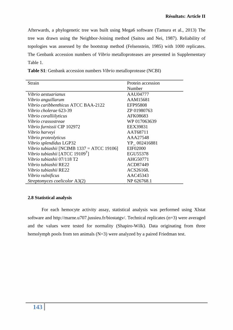
Résultats: Article II
143
Afterwards, a phylogenetic tree was built using Mega6 software (Tamura et al., 2013) The
tree was drawn using the Neighbor-Joining method (Saitou and Nei, 1987). Reliability of
topologies was assessed by the bootstrap method (Felsenstein, 1985) with 1000 replicates.
The Genbank accession numbers of Vibrio metalloproteases are presented in Supplementary
Table 1.
Table S1: Genbank accession numbers Vibrio metalloprotease (NCBI)
Strain Protein accession
Number
Vibrio aestuarianus AAU04777
Vibrio anguillarum AAM15681
Vibrio caribbenthicus ATCC BAA-2122 EFP95808
Vibrio cholerae 623-39 ZP 01980763
Vibrio coralliilyticus AFK08683
Vibrio crassostreae WP 017063639
Vibrio furnissii CIP 102972 EEX39831
Vibrio harveyi AAT68711
Vibrio proteolyticus AAA27548
Vibrio splendidus LGP32 YP_ 002416881
Vibrio tubiashii [NCIMB 1337 = ATCC 19106] EIF02000
Vibrio tubiashii [ATCC 19109T] EGU55378
Vibrio tubiashii 07/118 T2 AHG50771
Vibrio tubiashii RE22 ACD87449
Vibrio tubiashii RE22 ACS26168.
Vibrio vulnificus AAC45343
Streptomyces coelicolor A3(2) NP 626768.1
2.8 Statistical analysis
For each hemocyte activity assay, statistical analysis was performed using Xlstat
software and http://marne.u707.jussieu.fr/biostatgv/. Technical replicates (n=3) were averaged
and the values were tested for normality (Shapiro-Wilk). Data originating from three
hemolymph pools from ten animals (N=3) were analyzed by a paired Friedman test.
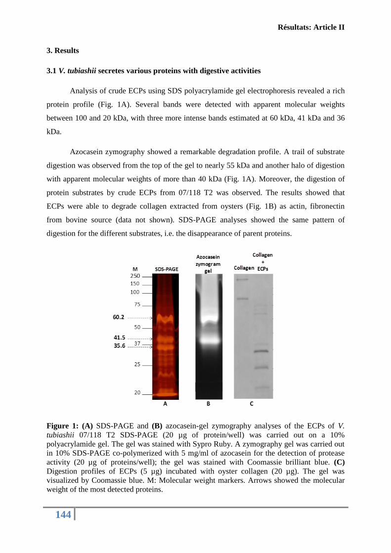
Résultats: Article II
144
3. Results
3.1 V. tubiashii secretes various proteins with digestive activities
Analysis of crude ECPs using SDS polyacrylamide gel electrophoresis revealed a rich
protein profile (Fig. 1A). Several bands were detected with apparent molecular weights
between 100 and 20 kDa, with three more intense bands estimated at 60 kDa, 41 kDa and 36
kDa.
Azocasein zymography showed a remarkable degradation profile. A trail of substrate
digestion was observed from the top of the gel to nearly 55 kDa and another halo of digestion
with apparent molecular weights of more than 40 kDa (Fig. 1A). Moreover, the digestion of
protein substrates by crude ECPs from 07/118 T2 was observed. The results showed that
ECPs were able to degrade collagen extracted from oysters (Fig. 1B) as actin, fibronectin
from bovine source (data not shown). SDS-PAGE analyses showed the same pattern of
digestion for the different substrates, i.e. the disappearance of parent proteins.
Figure 1: (A) SDS-PAGE and (B) azocasein-gel zymography analyses of the ECPs of V.
tubiashii 07/118 T2 SDS-PAGE (20 µg of protein/well) was carried out on a 10%
polyacrylamide gel. The gel was stained with Sypro Ruby. A zymography gel was carried out
in 10% SDS-PAGE co-polymerized with 5 mg/ml of azocasein for the detection of protease
activity (20 µg of proteins/well); the gel was stained with Coomassie brilliant blue. (C)
Digestion profiles of ECPs (5 µg) incubated with oyster collagen (20 µg). The gel was
visualized by Coomassie blue. M: Molecular weight markers. Arrows showed the molecular
weight of the most detected proteins.
Résultats: Article II
145
3.2 Evidences of hemocytes impairment by crude ECPs
The impact of ECPs secreted by V. tubiashii 07/118 T2 was evaluated following in
vitro contact with C. gigas hemocytes. Different cellular immune functions were tested with
flow cytometry: cellular mortality, phagocytosis activity, adhesion capacity, esterase and
intra-cytoplasmic calcium activity, and finally the presence of lysosomes.
A significant inhibition of adhesion capacity (p<0.05) was observed, as shown in
Figure 2 A Around 28 % of adhesion capacity was lost after 3 h incubation with ECPs at 6 µg
ml-1
of proteins compared to the SASW and Tris- HCl buffer controls.
Similarly, a significant inhibition in phagocytic activity was detected as early as the
first hour of incubation (p<0.05). Around 56 % of hemocyte phagocytic activity was lost
following contact with ECPs at 6 µg/ ml compared to controls (hemocytes incubated with
SASW or Tris-HCl buffer) (Fig. 2B). This decrease was constant over time, even after 6 h of
incubation.
For the other cellular functions like cell mortality, intra-cytoplasmic free calcium,
esterase activity and the presence of lysosomes, no significant effect was observed (p> 0.05)
(data not shown).
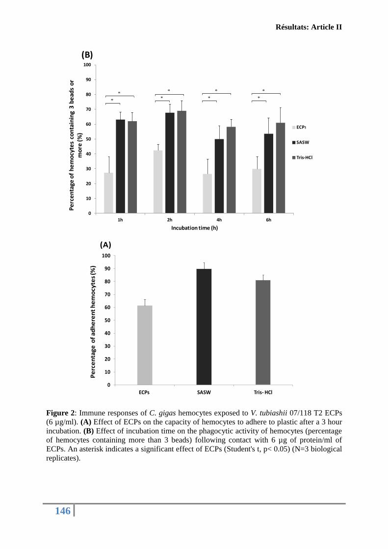
Résultats: Article II
146
Figure 2: Immune responses of C. gigas hemocytes exposed to V. tubiashii 07/118 T2 ECPs
(6 µg/ml). (A) Effect of ECPs on the capacity of hemocytes to adhere to plastic after a 3 hour
incubation. (B) Effect of incubation time on the phagocytic activity of hemocytes (percentage
of hemocytes containing more than 3 beads) following contact with 6 µg of protein/ml of
ECPs. An asterisk indicates a significant effect of ECPs (Student's t, p< 0.05) (N=3 biological
replicates).
0
10
20
30
40
50
60
70
80
90
100
1h 2h 4h 6h
Pe
rce
nta
ge o
f h
em
ocy
tes
con
tain
ing
3 b
ead
s o
r m
ore
(%
)
Incubation time (h)
ECPs 6µg/ml
SASW
Tris-HCl
*
**
**
*
*
*
(B)
(A)
0
10
20
30
40
50
60
70
80
90
100
ECPs SASW Tris- HCl
Pe
rce
nta
ge
of
ad
he
ren
th
em
ocy
tes
(%)
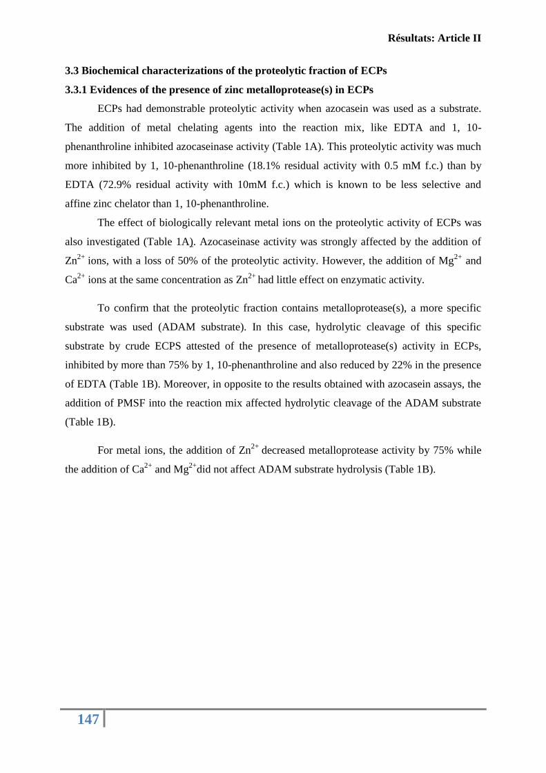
Résultats: Article II
147
3.3 Biochemical characterizations of the proteolytic fraction of ECPs
3.3.1 Evidences of the presence of zinc metalloprotease(s) in ECPs
ECPs had demonstrable proteolytic activity when azocasein was used as a substrate.
The addition of metal chelating agents into the reaction mix, like EDTA and 1, 10-
phenanthroline inhibited azocaseinase activity (Table 1A). This proteolytic activity was much
more inhibited by 1, 10-phenanthroline (18.1% residual activity with 0.5 mM f.c.) than by
EDTA (72.9% residual activity with 10mM f.c.) which is known to be less selective and
affine zinc chelator than 1, 10-phenanthroline.
The effect of biologically relevant metal ions on the proteolytic activity of ECPs was
also investigated (Table 1A). Azocaseinase activity was strongly affected by the addition of
Zn2+
ions, with a loss of 50% of the proteolytic activity. However, the addition of Mg2+
and
Ca2+
ions at the same concentration as Zn2+
had little effect on enzymatic activity.
To confirm that the proteolytic fraction contains metalloprotease(s), a more specific
substrate was used (ADAM substrate). In this case, hydrolytic cleavage of this specific
substrate by crude ECPS attested of the presence of metalloprotease(s) activity in ECPs,
inhibited by more than 75% by 1, 10-phenanthroline and also reduced by 22% in the presence
of EDTA (Table 1B). Moreover, in opposite to the results obtained with azocasein assays, the
addition of PMSF into the reaction mix affected hydrolytic cleavage of the ADAM substrate
(Table 1B).
For metal ions, the addition of Zn2+
decreased metalloprotease activity by 75% while
the addition of Ca2+
and Mg2+
did not affect ADAM substrate hydrolysis (Table 1B).
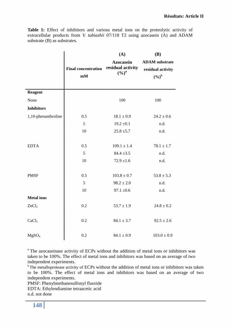
Résultats: Article II
148
Table 1: Effect of inhibitors and various metal ions on the proteolytic activity of
extracellular products from V. tubiashii 07/118 T2 using azocasein (A) and ADAM
substrate (B) as substrates.
Final concentration
mM
(A)
Azocasein
residual activity
(%)a
(B)
ADAM substrate
residual activity
(%)b
Reagent
None 100 100
Inhibitors
1,10-phenanthroline 0.5
5
10
18.1 ± 0.9
19.2 ±0.1
25.8 ±5.7
24.2 ± 0.6
n.d.
n.d.
EDTA 0.5 109.1 ± 1.4 78.1 ± 1.7
5 84.4 ±3.5 n.d.
10 72.9 ±1.6 n.d.
PMSF 0.5 103.8 ± 0.7 53.8 ± 5.3
5 98.2 ± 2.0 n.d.
10 97.1 ±0.6 n.d.
Metal ions
ZnCl2
0.2 53.7 ± 1.9 24.8 ± 0.2
CaCl2
0.2 84.1 ± 3.7 92.5 ± 2.6
MgSO4 0.2 84.1 ± 0.9 103.0 ± 0.9
a The azocaseinase activity of ECPs without the addition of metal ions or inhibitors was
taken to be 100%. The effect of metal ions and inhibitors was based on an average of two
independent experiments. b
The metalloprotease activity of ECPs without the addition of metal ions or inhibitors was taken
to be 100%. The effect of metal ions and inhibitors was based on an average of two
independent experiments.
PMSF: Phenylmethanesulfonyl fluoride
EDTA: Ethylendiamine tetraacetic acid
n.d. not done
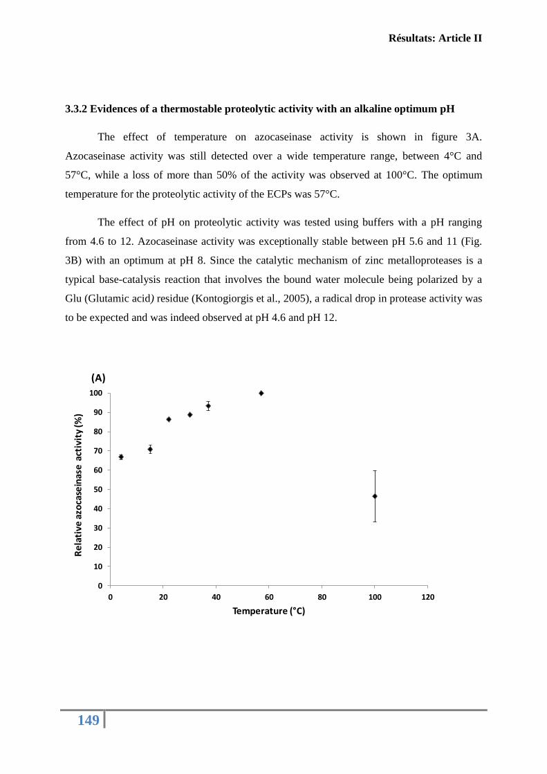
Résultats: Article II
149
3.3.2 Evidences of a thermostable proteolytic activity with an alkaline optimum pH
The effect of temperature on azocaseinase activity is shown in figure 3A.
Azocaseinase activity was still detected over a wide temperature range, between 4°C and
57°C, while a loss of more than 50% of the activity was observed at 100°C. The optimum
temperature for the proteolytic activity of the ECPs was 57°C.
The effect of pH on proteolytic activity was tested using buffers with a pH ranging
from 4.6 to 12. Azocaseinase activity was exceptionally stable between pH 5.6 and 11 (Fig.
3B) with an optimum at pH 8. Since the catalytic mechanism of zinc metalloproteases is a
typical base-catalysis reaction that involves the bound water molecule being polarized by a
Glu (Glutamic acid) residue (Kontogiorgis et al., 2005), a radical drop in protease activity was
to be expected and was indeed observed at pH 4.6 and pH 12.
0
10
20
30
40
50
60
70
80
90
100
0 20 40 60 80 100 120
Re
lati
ve a
zoca
sein
ase
act
ivit
y (%
)
Temperature (°C)
(A)
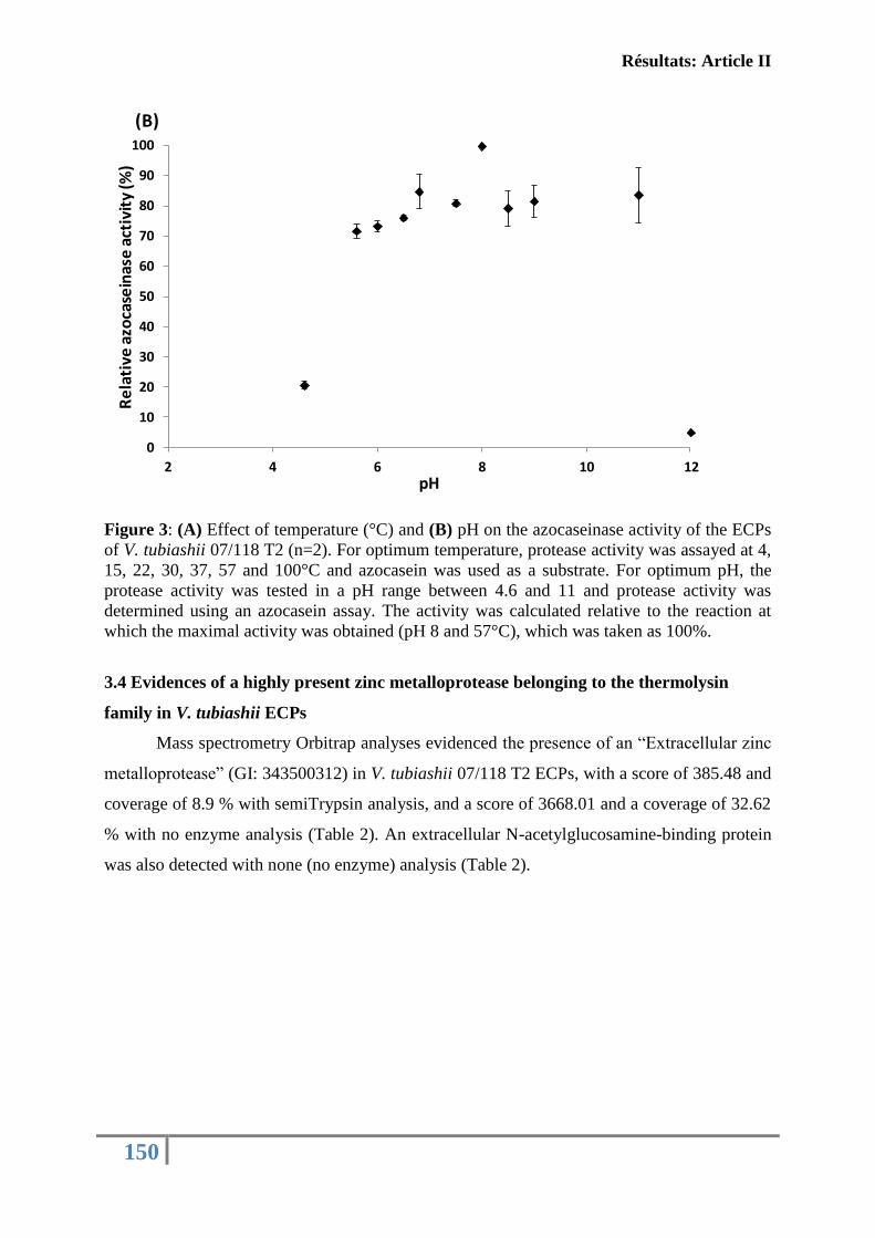
Résultats: Article II
150
Figure 3: (A) Effect of temperature (°C) and (B) pH on the azocaseinase activity of the ECPs
of V. tubiashii 07/118 T2 (n=2). For optimum temperature, protease activity was assayed at 4,
15, 22, 30, 37, 57 and 100°C and azocasein was used as a substrate. For optimum pH, the
protease activity was tested in a pH range between 4.6 and 11 and protease activity was
determined using an azocasein assay. The activity was calculated relative to the reaction at
which the maximal activity was obtained (pH 8 and 57°C), which was taken as 100%.
3.4 Evidences of a highly present zinc metalloprotease belonging to the thermolysin
family in V. tubiashii ECPs
Mass spectrometry Orbitrap analyses evidenced the presence of an “Extracellular zinc
metalloprotease” (GI: 343500312) in V. tubiashii 07/118 T2 ECPs, with a score of 385.48 and
coverage of 8.9 % with semiTrypsin analysis, and a score of 3668.01 and a coverage of 32.62
% with no enzyme analysis (Table 2). An extracellular N-acetylglucosamine-binding protein
was also detected with none (no enzyme) analysis (Table 2).
0
10
20
30
40
50
60
70
80
90
100
2 4 6 8 10 12
Re
lati
ve a
zoca
sein
ase
acti
vity
(%
)
pH
(B)
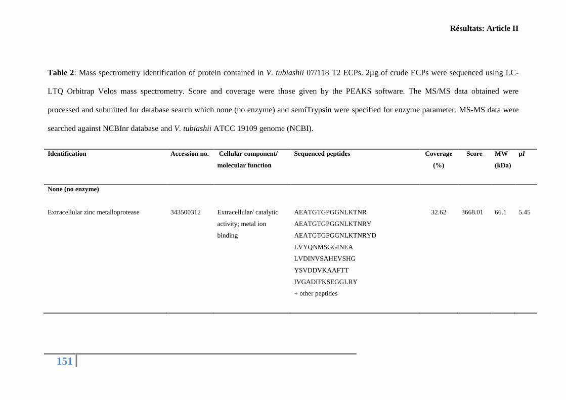
Résultats: Article II
151
Table 2: Mass spectrometry identification of protein contained in V. tubiashii 07/118 T2 ECPs. 2µg of crude ECPs were sequenced using LC-
LTQ Orbitrap Velos mass spectrometry. Score and coverage were those given by the PEAKS software. The MS/MS data obtained were
processed and submitted for database search which none (no enzyme) and semiTrypsin were specified for enzyme parameter. MS-MS data were
searched against NCBInr database and V. tubiashii ATCC 19109 genome (NCBI).
Identification
Accession no. Cellular component/
molecular function
Sequenced peptides Coverage
(%)
Score MW
(kDa)
pI
None (no enzyme)
Extracellular zinc metalloprotease
343500312
Extracellular/ catalytic
activity; metal ion
binding
AEATGTGPGGNLKTNR
AEATGTGPGGNLKTNRY
AEATGTGPGGNLKTNRYD
LVYQNMSGGINEA
LVDINVSAHEVSHG
YSVDDVKAAFTT
IVGADIFKSEGGLRY
+ other peptides
32.62
3668.01
66.1
5.45
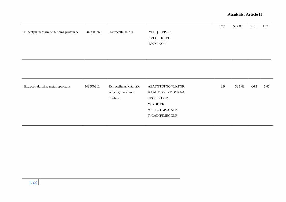
Résultats: Article II
152
N-acetylglucosamine-binding protein A
343503266
Extracellular/ND
VEDQTPPPGD
SVEGPDGFPE
DWNPNQPL
5.77 527.87 53.1 4.69
Extracellular zinc metalloprotease
343500312
Extracellular/ catalytic
activity; metal ion
binding
AEATGTGPGGNLKTNR
AAADMGYSVDDVKAA
FDQPSKDGR
YSVDDVK
AEATGTGPGGNLK
IVGADIFKSEGGLR
8.9
385.48
66.1
5.45
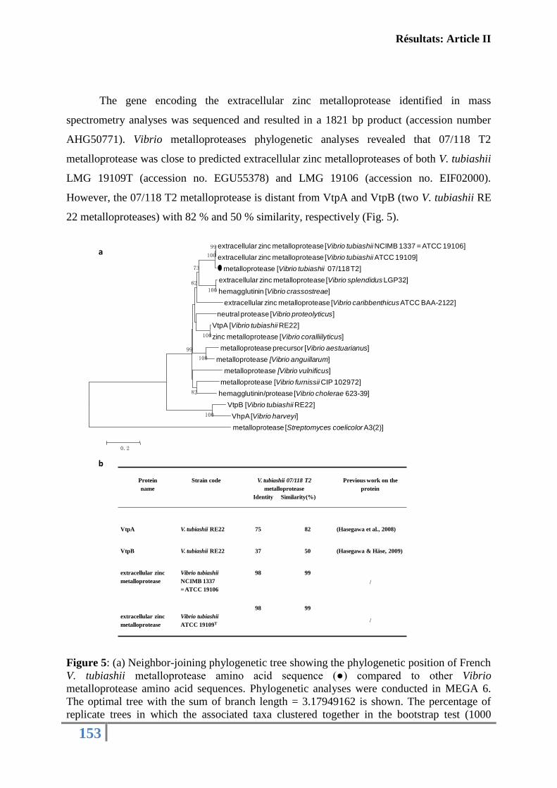
Résultats: Article II
153
The gene encoding the extracellular zinc metalloprotease identified in mass
spectrometry analyses was sequenced and resulted in a 1821 bp product (accession number
AHG50771). Vibrio metalloproteases phylogenetic analyses revealed that 07/118 T2
metalloprotease was close to predicted extracellular zinc metalloproteases of both V. tubiashii
LMG 19109T (accession no. EGU55378) and LMG 19106 (accession no. EIF02000).
However, the 07/118 T2 metalloprotease is distant from VtpA and VtpB (two V. tubiashii RE
22 metalloproteases) with 82 % and 50 % similarity, respectively (Fig. 5).
Figure 5: (a) Neighbor-joining phylogenetic tree showing the phylogenetic position of French
V. tubiashii metalloprotease amino acid sequence (●) compared to other Vibrio
metalloprotease amino acid sequences. Phylogenetic analyses were conducted in MEGA 6.
The optimal tree with the sum of branch length = 3.17949162 is shown. The percentage of
replicate trees in which the associated taxa clustered together in the bootstrap test (1000
extracellular zinc metalloprotease [Vibrio tubiashii NCIMB 1337 = ATCC 19106]
extracellular zinc metalloprotease [Vibrio tubiashii ATCC 19109]
metalloprotease [Vibrio tubiashii 07/118 T2]
extracellular zinc metalloprotease [Vibrio splendidus LGP32]
hemagglutinin [Vibrio crassostreae]
extracellular zinc metalloprotease [Vibrio caribbenthicusATCC BAA-2122]
neutral protease [Vibrio proteolyticus]
VtpA [Vibrio tubiashii RE22]
zinc metalloprotease [Vibrio coralliilyticus]
metalloprotease precursor [Vibrio aestuarianus]
metalloprotease [Vibrio anguillarum]
metalloprotease [Vibrio vulnificus]
metalloprotease [Vibrio furnissii CIP 102972]
hemagglutinin/protease [Vibrio cholerae 623-39]
VtpB [Vibrio tubiashii RE22]
VhpA[Vibrio harveyi]
metalloprotease [Streptomyces coelicolor A3(2)]
100
100
99
100
100
100
82
73
62
99
0.2
Protein
name
Strain code V. tubiashii 07/118 T2
metalloprotease
Identity Similarity(%)
Previous work on the
protein
VtpA V. tubiashii RE22 75 82 (Hasegawa et al., 2008)
VtpB V. tubiashii RE22 37 50 (Hasegawa & Häse, 2009)
extracellular zinc
metalloprotease
Vibrio tubiashii
NCIMB 1337
= ATCC 19106
98 99
/
extracellular zinc
metalloprotease
Vibrio tubiashii
ATCC 19109T
98 99
/
a
b
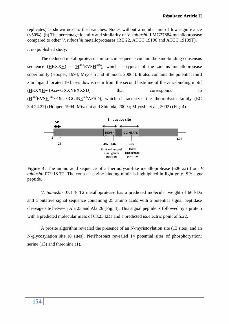
Résultats: Article II
154
replicates) is shown next to the branches. Nodes without a number are of low significance
(<50%). (b) The percentage identity and similarity of V. tubiashii LMG27884 metalloprotease
compared to other V. tubiashii metalloproteases (RE 22, ATCC 19106 and ATCC 19109T).
/: no published study.
The deduced metalloprotease amino-acid sequence contain the zinc-binding consensus
sequence (HEXXH) = (H342
EVSH346
), which is typical of the zincins metalloprotease
superfamily (Hooper, 1994; Miyoshi and Shinoda, 2000a). It also contains the potential third
zinc ligand located 19 bases downstream from the second histidine of the zinc-binding motif
(HEXXH∼19aa∼GXXNEXXSD) that corresponds to
(H342
EVSH346∼19aa∼GGINE
366AFSD), which characterizes the thermolysin family (EC
3.4.24.27) (Hooper, 1994; Miyoshi and Shinoda, 2000a; Miyoshi et al., 2002) (Fig. 4).
Figure 4: The amino acid sequence of a thermolysin-like metalloprotease (606 aa) from V.
tubiashii 07/118 T2. The consensus zinc-binding motif is highlighted in light gray. SP: signal
peptide.
V. tubiashii 07/118 T2 metalloprotease has a predicted molecular weight of 66 kDa
and a putative signal sequence containing 25 amino acids with a potential signal peptidase
cleavage site between Ala 25 and Ala 26 (Fig. 4). This signal peptide is followed by a protein
with a predicted molecular mass of 63.25 kDa and a predicted isoelectric point of 5.22.
A prosite algorithm revealed the presence of an N-myristoylation site (13 sites) and an
N-glycosylation site (8 sites). NetPhosbact revealed 14 potential sites of phosphoryation:
serine (13) and threonine (1).
SP
25 342 346
First and second zinc ligands
position
366
Third zinc ligands
position
Zinc active site
1 606
GGINEAFSHEVSH
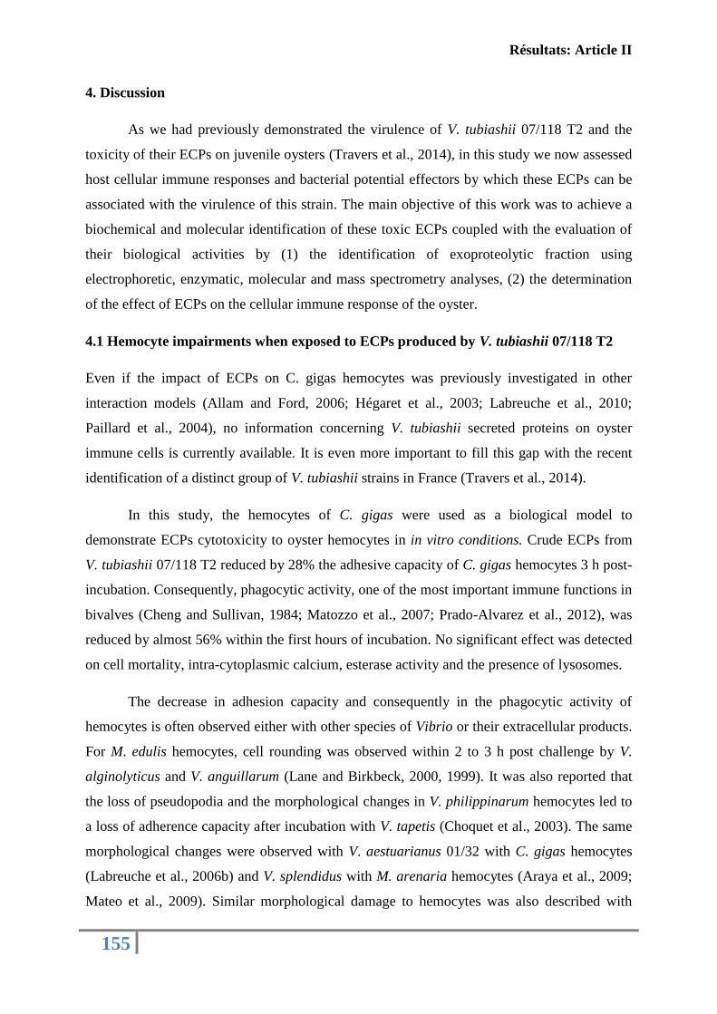
Résultats: Article II
155
4. Discussion
As we had previously demonstrated the virulence of V. tubiashii 07/118 T2 and the
toxicity of their ECPs on juvenile oysters (Travers et al., 2014), in this study we now assessed
host cellular immune responses and bacterial potential effectors by which these ECPs can be
associated with the virulence of this strain. The main objective of this work was to achieve a
biochemical and molecular identification of these toxic ECPs coupled with the evaluation of
their biological activities by (1) the identification of exoproteolytic fraction using
electrophoretic, enzymatic, molecular and mass spectrometry analyses, (2) the determination
of the effect of ECPs on the cellular immune response of the oyster.
4.1 Hemocyte impairments when exposed to ECPs produced by V. tubiashii 07/118 T2
Even if the impact of ECPs on C. gigas hemocytes was previously investigated in other
interaction models (Allam and Ford, 2006; Hégaret et al., 2003; Labreuche et al., 2010;
Paillard et al., 2004), no information concerning V. tubiashii secreted proteins on oyster
immune cells is currently available. It is even more important to fill this gap with the recent
identification of a distinct group of V. tubiashii strains in France (Travers et al., 2014).
In this study, the hemocytes of C. gigas were used as a biological model to
demonstrate ECPs cytotoxicity to oyster hemocytes in in vitro conditions. Crude ECPs from
V. tubiashii 07/118 T2 reduced by 28% the adhesive capacity of C. gigas hemocytes 3 h post-
incubation. Consequently, phagocytic activity, one of the most important immune functions in
bivalves (Cheng and Sullivan, 1984; Matozzo et al., 2007; Prado-Alvarez et al., 2012), was
reduced by almost 56% within the first hours of incubation. No significant effect was detected
on cell mortality, intra-cytoplasmic calcium, esterase activity and the presence of lysosomes.
The decrease in adhesion capacity and consequently in the phagocytic activity of
hemocytes is often observed either with other species of Vibrio or their extracellular products.
For M. edulis hemocytes, cell rounding was observed within 2 to 3 h post challenge by V.
alginolyticus and V. anguillarum (Lane and Birkbeck, 2000, 1999). It was also reported that
the loss of pseudopodia and the morphological changes in V. philippinarum hemocytes led to
a loss of adherence capacity after incubation with V. tapetis (Choquet et al., 2003). The same
morphological changes were observed with V. aestuarianus 01/32 with C. gigas hemocytes
(Labreuche et al., 2006b) and V. splendidus with M. arenaria hemocytes (Araya et al., 2009;
Mateo et al., 2009). Similar morphological damage to hemocytes was also described with
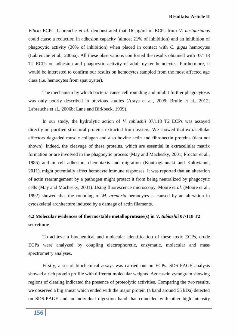
Résultats: Article II
156
Vibrio ECPs. Labreuche et al. demonstrated that 16 µg/ml of ECPs from V. aestuarianus
could cause a reduction in adhesion capacity (almost 21% of inhibition) and an inhibition of
phagocytic activity (30% of inhibition) when placed in contact with C. gigas hemocytes
(Labreuche et al., 2006a). All these observations comforted the results obtained with 07/118
T2 ECPs on adhesion and phagocytic activity of adult oyster hemocytes. Furthermore, it
would be interested to confirm our results on hemocytes sampled from the most affected age
class (i.e. hemocytes from spat oyster).
The mechanism by which bacteria cause cell rounding and inhibit further phagocytosis
was only poorly described in previous studies (Araya et al., 2009; Brulle et al., 2012;
Labreuche et al., 2006b; Lane and Birkbeck, 1999).
In our study, the hydrolytic action of V. tubiashii 07/118 T2 ECPs was assayed
directly on purified structural proteins extracted from oysters. We showed that extracellular
effectors degraded muscle collagen and also bovine actin and fibronectin proteins (data not
shown). Indeed, the cleavage of these proteins, which are essential in extracellular matrix
formation or are involved in the phagocytic process (May and Machesky, 2001; Proctor et al.,
1985) and in cell adhesion, chemotaxis and migration (Koutsogiannaki and Kaloyianni,
2011), might potentially affect hemocyte immune responses. It was reported that an alteration
of actin rearrangement by a pathogen might protect it from being neutralized by phagocytic
cells (May and Machesky, 2001). Using fluorescence microscopy, Moore et al. (Moore et al.,
1992) showed that the rounding of M. arenaria hemocytes is caused by an alteration in
cytoskeletal architecture induced by a damage of actin filaments.
4.2 Molecular evidences of thermostable metalloprotease(s) in V. tubiashii 07/118 T2
secretome
To achieve a biochemical and molecular identification of these toxic ECPs, crude
ECPs were analyzed by coupling electrophoretic, enzymatic, molecular and mass
spectrometry analyses.
Firstly, a set of biochemical assays was carried out on ECPs. SDS-PAGE analysis
showed a rich protein profile with different molecular weights. Azocasein zymogram showing
regions of clearing indicated the presence of proteolytic activities. Comparing the two results,
we observed a big smear which ended with the major protein (a band around 55 kDa) detected
on SDS-PAGE and an individual digestion band that coincided with other high intensity
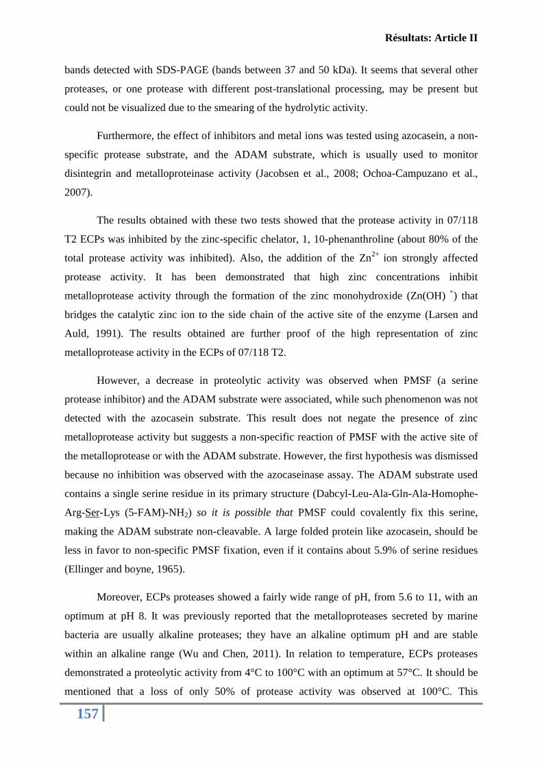
Résultats: Article II
157
bands detected with SDS-PAGE (bands between 37 and 50 kDa). It seems that several other
proteases, or one protease with different post-translational processing, may be present but
could not be visualized due to the smearing of the hydrolytic activity.
Furthermore, the effect of inhibitors and metal ions was tested using azocasein, a non-
specific protease substrate, and the ADAM substrate, which is usually used to monitor
disintegrin and metalloproteinase activity (Jacobsen et al., 2008; Ochoa-Campuzano et al.,
2007).
The results obtained with these two tests showed that the protease activity in 07/118
T2 ECPs was inhibited by the zinc-specific chelator, 1, 10-phenanthroline (about 80% of the
total protease activity was inhibited). Also, the addition of the Zn2+
ion strongly affected
protease activity. It has been demonstrated that high zinc concentrations inhibit
metalloprotease activity through the formation of the zinc monohydroxide (Zn(OH) +
) that
bridges the catalytic zinc ion to the side chain of the active site of the enzyme (Larsen and
Auld, 1991). The results obtained are further proof of the high representation of zinc
metalloprotease activity in the ECPs of 07/118 T2.
However, a decrease in proteolytic activity was observed when PMSF (a serine
protease inhibitor) and the ADAM substrate were associated, while such phenomenon was not
detected with the azocasein substrate. This result does not negate the presence of zinc
metalloprotease activity but suggests a non-specific reaction of PMSF with the active site of
the metalloprotease or with the ADAM substrate. However, the first hypothesis was dismissed
because no inhibition was observed with the azocaseinase assay. The ADAM substrate used
contains a single serine residue in its primary structure (Dabcyl-Leu-Ala-Gln-Ala-Homophe-
Arg-Ser-Lys (5-FAM)-NH2) so it is possible that PMSF could covalently fix this serine,
making the ADAM substrate non-cleavable. A large folded protein like azocasein, should be
less in favor to non-specific PMSF fixation, even if it contains about 5.9% of serine residues
(Ellinger and boyne, 1965).
Moreover, ECPs proteases showed a fairly wide range of pH, from 5.6 to 11, with an
optimum at pH 8. It was previously reported that the metalloproteases secreted by marine
bacteria are usually alkaline proteases; they have an alkaline optimum pH and are stable
within an alkaline range (Wu and Chen, 2011). In relation to temperature, ECPs proteases
demonstrated a proteolytic activity from 4°C to 100°C with an optimum at 57°C. It should be
mentioned that a loss of only 50% of protease activity was observed at 100°C. This
Résultats: Article II
158
performance over a wide range of temperatures and pH tends to confirm the idea that
proteolytic fraction of ECPs contains thermostable active zinc metalloprotease(s)
(Kudryashova et al., 1998).
By liquid chromatography coupled with mass spectrometry, an extracellular zinc
metalloprotease (GI: 343500312) was identified and the gene encoding this protein was
sequenced in V. tubiashii 07/118 T2. Annotation of the gene encoding this exometalloprotease
showed that this enzyme is a member of the thermolysin family (EC 3.4.24.27) (Miyoshi and
Shinoda, 2000a; Miyoshi et al., 2002). In addition, glycosylation and phosphorylation motifs
were identified on the metalloprotease sequence. The presence of these motifs suggested that
the 07/118 T2 metalloprotease could be subject to post-translational modifications which can
enable it to perform functions other than those determined by its primary sequence (Ahmad et
al., 2007). Furthermore, it was shown that the HEXXH metalloprotease family is the only
family of this protease group that is extensively modified post-translationally by both N-
glycosylation and glycosyl phosphatidylinositol (McGwire and Chang, 1996).
Moreover, based on the phylogenetic tree, a large distance between the 07/118 T2
metalloprotease and the American V. tubiashii RE22 metalloproteases VtpA and VtpB
(Hasegawa & Häse, 2009b; Hasegawa et al., 2008) were observed. This data could suggest
that the 07/ 118 T2 metalloprotease is different from the metalloproteases of the American
strains previously described as major virulence factors.
In fact, proteases are generally described as one of the factors that are most toxic to the
host, and are involved in virulence and pathogenicity (Shinoda et al., 1996). Most of these
proteases have been identified as zinc metalloproteases (Miyoshi and Shinoda, 2000b).
Studies on V. aestuarianus metalloprotease (Vam) showed its toxicity and its capacity to
impair oyster hemocyte functions (Labreuche et al., 2010). A metalloprotease from V.
splendidus for pacific oyster C. gigas (Vsm) has been shown to be the major factor in the
toxicity of the ECPs (Binesse et al., 2008). Extracellular products from V. tubiashii were also
shown to be toxic to oyster larvae and its pathogenicity was linked to a metalloprotease
(VtpA) and a hemolysin (VthA) (Delston et al., 2003; Hasegawa and Häse, 2009a; Hasegawa
et al., 2008; Kothary et al., 2001).
5. Conclusions
It had previously been shown that the ECPs produced by V. tubiashii 07/118 T2 were
toxic for juvenile oysters in experimental challenges. In this study, it was shown that these
Résultats: Article II
159
toxic ECPs could inhibit hemocytes adhesion capacity and phagocytosis activity, and digest
matrix host proteins. The proteolytic fraction of ECPs contained active and thermostable
extracellular zinc metalloprotease(s) and a thermolysin.
Even if taken together, our results let us wonder if this (these) zinc metalloprotease(s)
could be involved in the impairment of hemocyte immunological functions, the direct
implication of this protein in ECPs toxicity still has to be demonstrated.
Acknowledgements
This study was supported by a Ph.D grant from the CNRS and IFREMER for R.
Mersni-Achour (2010-2013). The PRES (Pôle de Recherche et d’Enseignement Supérieur) of
the Limousin and PoitouCharentes regions and the FR CNRS 3097 « Fédération de
Recherche en Environnement et Développement Durable» are acknowledged for their
financial support through the research project «Caractérisation in vitro des effets de
métalloprotéases sur différents types cellulaires de l’huître creuse C. gigas». Special thanks
go to L. Beaugeard, for technical assistance during the flow cytometer analyses. Thanks go to
the platform “Structural and Functional Mass Spectrometry Facility, Jacques Monod Institute, UMR7592
CNRS – Univ Paris Diderot, Sorbonne Paris Cité” and especially to T. Léger and C. Garcia for mass
spectrometry analysis. Acknowledgment also goes to M. Francis Healy for his revision of the
English language.
Résultats: Article II
160
References
Ahmad, I., Hoessli, D.C., Gupta, R., Walker-Nasir, E., Rafik, S.M., Choudhary, M.I.,
Shakoori, A.R., Nasir-ud-Din, 2007. In silico determination of intracellular
glycosylation and phosphorylation sites in human selectins: Implications for biological
function. J. Cell. Biochem. 100, 1558–1572.
Allam, B., Ford, S.E., 2006. Effects of the pathogenic Vibrio tapetis on defence factors of
susceptible and non-susceptible bivalve species: I. Haemocyte changes following in
vitro challenge. Fish Shellfish Immunol. 20, 374–383.
Araya, M.T., Siah, A., Mateo, D.R., Markham, F., McKenna, P., Johnson, G.R., Berthe,
F.C.J., 2009. Morphological and Molecular Effects of Vibrio splendidus on
Hemocytes of Softshell Clams, Mya arenaria. J. Shellfish Res. 28, 751–758.
Binesse, J., Delsert, C., Saulnier, D., Champomier-Vergès, M.-C., Zagorec, M., Munier-
Lehmann, H., Mazel, D., Roux, F.L., 2008. Metalloprotease Vsm Is the Major
Determinant of Toxicity for Extracellular Products of Vibrio splendidus. Appl.
Environ. Microbiol. 74, 7108–7117.
Bradford, M.M., 1976. A rapid and sensitive method for the quantitation of microgram
quantities of protein utilizing the principle of protein-dye binding. Anal. Biochem. 72,
248–254.
Broberg, C.A., Calder, T.J., Orth, K., 2011. Vibrio parahaemolyticus cell biology and
pathogenicity determinants. Microbes Infect. 13, 992–1001.
Brulle, F., Jeffroy, F., Madec, S., Nicolas, J.-L., Paillard, C., 2012. Transcriptomic analysis of
Ruditapes philippinarum hemocytes reveals cytoskeleton disruption after in vitro
Vibrio tapetis challenge. Dev. Comp. Immunol. 38, 368–376
Cheng, T.C., Sullivan, J.T., 1984. Effects of heavy metals on phagocytosis by Molluscan
hemocytes. Mar. Environ. Res. 14, 305–315.
Choquet, G., Soudant, P., Lambert, C., Nicolas, J., Paillard, C., 2003. Reduction of adhesion
properties of Ruditapes philippinarum hemocytes exposed to Vibrio tapetis. Dis.
Aquat. Organ. 57, 109–116.
Delston, R.B., Kothary, M.H., Shangraw, K.A., Tall, B.D., 2003. Isolation and
characterization of a zinc-containing metalloprotease expressed by Vibrio tubiashii.
Can. J. Microbiol. 49, 525–529.
Duperthuy, M., Binesse, J., Le Roux, F., Romestand, B., Caro, A., Got, P., Givaudan, A.,
Mazel, D., Bachère, E., Destoumieux-Garzón, D., 2010. The major outer membrane
protein OmpU of Vibrio splendidus contributes to host antimicrobial peptide
resistance and is required for virulence in the oyster Crassostrea gigas. Environ.
Microbiol. 12, 951–963.
Duperthuy, M., Schmitt, P., Garzón, E., Caro, A., Rosa, R.D., Roux, F.L., Lautrédou-Audouy,
N., Got, P., Romestand, B., Lorgeril, J. de, Kieffer-Jaquinod, S., Bachère, E.,
Destoumieux-Garzón, D., 2011. Use of OmpU porins for attachment and invasion of
Crassostrea gigas immune cells by the oyster pathogen Vibrio splendidus. Proc. Natl.
Acad. Sci. 201015326.
Ellinger, G.M., Boyne, E.B., 1965. Amino acid composition of some fish products and casein.
Br. J. Nutr. 19, 587–592.
Elston, R.A., Hasegawa, H., Humphrey, K.L., Polyak, I.K., Häse, C.C., 2008. Re-emergence
of Vibrio tubiashii in bivalve shellfish aquaculture: severity, environmental drivers,
geographic extent and management. Dis. Aquat. Organ. 82, 119–134.
Felsenstein, J., 1985. Confidence Limits on Phylogenies: An Approach Using the Bootstrap.
Evolution 39, 783.
Résultats: Article II
161
Gagnaire, B., Renault, T., Bouilly, K., Lapegue, S., Thomas-Guyon, H., 2003. Study of
atrazine effects on Pacific oyster, Crassostrea gigas, haemocytes. Curr. Pharm. Des. 9,
193–199.
Garnier, M., Labreuche, Y., Nicolas, J.-L., 2008. Molecular and phenotypic characterization
of Vibrio aestuarianus subsp. francensis subsp. nov., a pathogen of the oyster
Crassostrea gigas. Syst. Appl. Microbiol. 31, 358–365.
Hada, H.S., West, P.A., Lee, J.V., Stemmler, J., Colwell, R.R., 1984. Vibrio tubiashii sp.
nov., a Pathogen of Bivalve Mollusks. Int. J. Syst. Bacteriol. 34, 1–4.
Hasegawa, H., Häse, C.C., 2009. The extracellular metalloprotease of Vibrio tubiashii directly
inhibits its extracellular haemolysin. Microbiology 155, 2296–2305.
Hasegawa, H., Lind, E.J., Boin, M.A., Häse, C.C., 2008. The Extracellular Metalloprotease of
Vibrio tubiashii Is a Major Virulence Factor for Pacific Oyster (Crassostrea gigas)
Larvae. Appl. Environ. Microbiol. 74, 4101–4110.
Hégaret, H., Wikfors, G.H., Soudant, P., 2003. Flow cytometric analysis of haemocytes from
eastern oysters, Crassostrea virginica, subjected to a sudden temperature elevation: II.
Haemocyte functions: aggregation, viability, phagocytosis, and respiratory burst. J.
Exp. Mar. Biol. Ecol. 293, 249–265.
Hooper, N.M., 1994. Families of zinc metalloproteases. FEBS Lett. 354, 1–6.
Jacobsen, J., Visse, R., Sørensen, H.P., Enghild, J.J., Brew, K., Wewer, U.M., Nagase, H.,
2008. Catalytic properties of ADAM12 and its domain deletion mutants. Biochemistry
(Mosc.) 47, 537–547.
Jeffries, V.E., 1982. Three Vibrio strains pathogenic to larvae of Crassostrea gigas and
Ostrea edulis. Aquaculture 29, 201–226.
Kontogiorgis, C.A., Papaioannou, P., Hadjipavlou-Litina, D.J., 2005. Matrix
metalloproteinase inhibitors: a review on pharmacophore mapping and (Q)SARs
results. Curr. Med. Chem. 12, 339–355.
Kothary, M.H., Delston, R.B., Curtis, S.K., McCardell, B.A., Tall, B.D., 2001. Purification
and Characterization of a Vulnificolysin-Like Cytolysin Produced by Vibrio tubiashii.
Appl. Environ. Microbiol. 67, 3707–3711.
Koutsogiannaki, S., Kaloyianni, M., 2011. Effect of 17β-estradiol on adhesion of Mytilus
galloprovincialis hemocytes to selected substrates. Role of alpha2 integrin subunit.
Fish Shellfish Immunol. 31, 73–80.
Kudryashova, E.V., Mozhaev, V.V., Balny, C., 1998. Catalytic activity of thermolysin under
extremes of pressure and temperature: modulation by metal ions. Biochim. Biophys.
Acta BBA - Protein Struct. Mol. Enzymol. 1386, 199–210.
Labreuche, Yannick, Lambert, C., Soudant, P., Boulo, V., Huvet, A., Nicolas, J.-L., 2006 b.
Cellular and molecular hemocyte responses of the Pacific oyster, Crassostrea gigas,
following bacterial infection with Vibrio aestuarianus strain 01/32. Microbes Infect. 8,
2715–2724.
Labreuche, Y., Le Roux, F., Henry, J., Zatylny, C., Huvet, A., Lambert, C., Soudant, P.,
Mazel, D., Nicolas, J.-L., 2010. Vibrio aestuarianus zinc metalloprotease causes
lethality in the Pacific oyster Crassostrea gigas and impairs the host cellular immune
defenses. Fish Shellfish Immunol. 29, 753–758.
Labreuche, Y., Soudant, P., Goncalves, M., Lambert, C., Nicolas, J., 2006 a. Effects of
extracellular products from the pathogenic Vibrio aestuarianus strain 01/32 on
lethality and cellular immune responses of the oyster Crassostrea gigas. Dev. Comp.
Immunol. 30, 367–379.
Laemmli, U.K., 1970. Cleavage of Structural Proteins during the Assembly of the Head of
Bacteriophage T4. Publ. Online 15 August 1970, 680–685.
Résultats: Article II
162
Lane, E., Birkbeck, T.H., 1999. Toxicity of bacteria towards haemocytes of Mytilus edulis.
Aquat. Living Resour. 12, 343–350.
Lane, E., Birkbeck, T.H., 2000. Species specificity of some bacterial pathogens of bivalve
molluscs is correlated with their interaction with bivalve haemocytes. J. Fish Dis. 23,
275–279.
Larsen, K.S., Auld, D.S., 1991. Characterization of an inhibitory metal binding site in
carboxypeptidase A. Biochemistry (Mosc.) 30, 2613–2618.
Lee, J.-H., Ahn, S.-H., Lee, E.-M., Kim, Y.-O., Lee, S.-J., Kong, I.-S., 2003. Characterization
of the enzyme activity of an extracellular metalloprotease (VMC) from Vibrio mimicus
and its C-terminal deletions. FEMS Microbiol. Lett. 223, 293–300.
Mateo, D.R., Siah, A., Araya, M.T., Berthe, F.C.J., Johnson, G.R., Greenwood, S.J., 2009.
Differential in vivo response of soft-shell clam hemocytes against two strains of Vibrio
splendidus: Changes in cell structure, numbers and adherence. J. Invertebr. Pathol.
102, 50–56.
Matozzo, V., Rova, G., Marin, M.G., 2007. Haemocytes of the cockle Cerastoderma
glaucum: Morphological characterisation and involvement in immune responses. Fish
Shellfish Immunol. 23, 732–746.
May, R.C., Machesky, L.M., 2001. Phagocytosis and the actin cytoskeleton. J. Cell Sci. 114,
1061–1077.
McGwire, B.S., Chang, K.-P., 1996. Posttranslational Regulation of a Leishmania HEXXH
Metalloprotease (gp63).J. Biol. Chem. 271, 7903–7909.J. Biol. Chem. 271, 7903–
7909
Miyoshi, S., Shinoda, S., 2000. Microbial metalloproteases and pathogenesis. Microbes
Infect. 2, 91–98.
Miyoshi, S., Sonoda, Y., Wakiyama, H., Rahman, M.M., Tomochika, K., Shinoda, S.,
Yamamoto, S., Tobe, K., 2002. An exocellular thermolysin-like metalloprotease
produced by Vibrio fluvialis: purification, characterization, and gene cloning. Microb.
Pathog. 33, 127–134.
Mizuta, S., Miyagi, T., Yoshinaka, R., 2005. Characterization of the quantitatively major
collagen in the mantle of oyster Crassostrea gigas. Fish. Sci. 71, 229–235.
Moore, C.A., Beckmann, N., Patricia Morse, M., 1992. Cytoskeletal structure of diseased and
normal hemocytes of Mya arenaria. J. Invertebr. Pathol. 60, 141–147.
Ochoa-Campuzano, C., Real, M.D., Martínez-Ramírez, A.C., Bravo, A., Rausell, C., 2007.
An ADAM metalloprotease is a Cry3Aa Bacillus thuringiensis toxin receptor.
Biochem. Biophys. Res. Commun. 362, 437–442.
Paillard, C., Allam, B., Oubella, R., 2004. Effect of temperature on defense parameters in
manila clam Ruditapes philippinarum challenged with Vibrio tapetis. Dis. Aquat.
Organ. 59, 249–262.
Prado-Alvarez, M., Romero, A., Balseiro, P., Dios, S., Novoa, B., Figueras, A., 2012.
Morphological characterization and functional immune response of the carpet shell
clam (Ruditapes decussatus) haemocytes after bacterial stimulation. Fish Shellfish
Immunol. 32, 69–78.
Proctor, R.A., Textor, J.A., Vanin, J.M., Mosher, D.F., 1985. Role of Fibronectin in Human
Monocyte and Macrophage Bactericidal Activity. Infect. Immun. 47, 629–637.
Saitou, N., Nei, M., 1987. The neighbor-joining method: a new method for reconstructing
phylogenetic trees. Mol. Biol. Evol. 4, 406–425
Shinoda, S., Miyoshi, S.-I., 2011. Proteases Produced by Vibrios. Biocontrol Sci. 16, 1–11.
Shinoda, S., Miyoshi, S.-I., Wakae, H., Rahman, M., Tomochika, K.-I., 1996. Bacterial
Proteases as Pathogenic Factors, with Special Emphasis on Vibrio Proteases. Toxin
Rev. 15, 327–339.
Résultats: Article II
163
Tamura, K., Stecher, G., Peterson, D., Filipski, A., Kumar, S., 2013. MEGA6: Molecular
Evolutionary Genetics Analysis version 6.0. Mol. Biol. Evol. mst197.
Teo, J.W.., Zhang, L.-H., Poh, C.L., 2003. Cloning and characterization of a metalloprotease
from Vibrio harveyi strain AP6. Gene 303, 147–156.
Travers, M.-A., Mersni Achour, R., Haffner, P., Tourbiez, D., Cassone, A.-L., Morga, B.,
Doghri, I., Garcia, C., Renault, T., Fruitier-Arnaudin, I., Saulnier, D., In press. First
description of French V. tubiashii strains pathogenic to mollusk: I. Characterization of
isolates and detection during mortality events. J. Invertebr. Pathol.
Travis, J., Potempa, J., Maeda, H., 1995. Are bacterial proteinases pathogenic factors? Trends
Microbiol. 3, 405–407.
Tubiash, H.S., Chanley, P.E., Leifson, E., 1965. Bacillary Necrosis, a Disease of Larval and
Juvenile Bivalve Mollusks I. Etiology and Epizootiology. J. Bacteriol. 90, 1036–1044.
Tubiash, H.S., Colwell, R.R., Sakazaki, R., 1970. Marine Vibrios Associated with Bacillary
Necrosis, a Disease of Larval and Juvenile Bivalve Mollusks. J. Bacteriol. 103, 271–
272.

164
Résultats: Article III
165
B/ Influences physique et chimique des conditions de culture de
la souche française V. tubiashii sur la toxicité de ses produits
extracellulaires.
L’effet de l’état physique d’un milieu de culture (solide ou liquide) ainsi que sa
composition et la nature de ses éléments constitutifs dans le comportement métabolique des
bactéries ont été souvent étudiés (Lorian, 1988, 1989; Wondrak et al., 1991; Erz et al., 1993;
Mohedano et al., 1997; Lee et al., 2002; Chopp et al., 2003; Cobaxin et al., 2014).
Dans une étude précédente, (Article 1), nous avons montré que les ECPs d’une souche
française de V. tubiashii, issus d’une culture sur milieu solide LBSA (milieu Luria-Bertani
gélosé supplémenté de 1% de chlorure de sodium) étaient toxiques pour l’huître creuse C.
gigas. Cependant, aucun effet létal n’avait été observé avec les ECPs issus d’une culture sur le
même milieu mais liquide (LBS). Afin de clarifier l’influence du milieu de culture sur la
toxicité et la composition des ECPs bactériens, plusieurs types de milieux de culture ont été
testés, i.e LBSA, Marine Agar (MA), Marine Broth (MB) et LBS.
Par des séries d’infections expérimentales sur des huîtres juvéniles, nous avons montré
que seul le surnageant bactérien issu d’une culture sur milieu gélosé LBSA était toxique.
Aucune mortalité n’a été observée quand les surnageants bactériens, respectivement préparés
sur MA, MB et LBS, ont été testés.
L’analyse de la complexité moléculaire de ces différents ECPs a été résolue par
combinaison de dosages enzymatiques (mesurant les activités protéasiques) et de séparations
électrophorétiques (SDS-PAGE et zymographie sur azocaséine). Ces expériences ont permis
de démontrer que les ECPs produits sur LBSA, MA et MB possédaient une activité
azocaséinase plus importante que ceux issus de LBS et une diversité moléculaire plus grande.
L’analyse protéomique a confirmé ces résultats. Elle a permis de montrer qu’une
métalloprotéase à zinc semblait être la protéine majoritaire des surnageants bactériens, quel
que soit le milieu utilisé et sécrétée sous diverses isoformes.
La sécrétion singulière de ces isoformes protéiques de haut poids moléculaire,
restreinte aux conditions de culture utilisant le milieu LBSA, et la toxicité exclusive de ces
ECPs, témoigne d’une variation phénotypique forte. Ces modes de culture influencent
nettement la production d’exotoxines mais semblent également affecter l’activité
(A)

Résultats: Article III
166
transcriptionnelle de cette souche, au regard de la différence enregistrée d’expression de
certains gènes codant pour l’hémolysine, la métalloprotéase et l’exochitinase.
Les résultats de ce chapitre font l’objet d’un troisième article qui sera soumis
prochainement à la revue «Microbiology».
Résultats: Article III
167
Article III: TOXICITY AND PROTEASES COMPOSITION OF
EXTRACELLULAR PRODUCTS FROM THE FRENCH VIBRIO
TUBIASHII ARE MODULATED BY PHYSICAL AND CHEMICAL
CHARACTERISTICS OF GROWTH MEDIA.
(Prepared for submission in the journal “Microbiology”)
Mersni-Achour Rachida1,2,5
, Doghri Ibtissem 1, Pichereau Vianney
3, Ben cheikhYosra
1,
Phillipe Haffner2, Sophie Sablé
1, Denis Saulnier
2,4, Marie-Agnès Travers
2, Ingrid Fruitier-
Arnaudin1, 5
1UMR 7266 CNRS-ULR, LIENSs, Equipe Approches Moléculaires, Environnement-Santé,
Université de La Rochelle, Avenue Michel Crépeau, 17042 La Rochelle, France.
2 Ifremer, SG2M-LGPMM, Laboratoire de Génétique et Pathologie des Mollusques Marins
Avenue de Mus de Loup, 17390 La Tremblade, France.
3Laboratoire des Sciences de l'Environnement Marin LEMAR, Institut Universitaire Europeen
de la Mer, Université Européenne de Bretagne, Université de Brest, UMR 6539
CNRS/UBO/IRD/Ifremer, 29280 Plouzané, France.
4 Ifremer, Centre Ifremer du Pacifique, UMR 241 Ecosystèmes Insulaires Océaniens, Tahiti,
98719 Taravao, French Polynesia
5Fédération de Recherche en Environnement et Développement Durable, FR CNRS 3097,
Université de La Rochelle, France

Résultats: Article III
168
Abstract
In a previous study, we showed that the extracellular products (ECPs) of the French V.
tubiashii obtained from bacteria grown on solid medium were toxic towards juvenile C. gigas
oyster. In opposite, bacteria grown in liquid medium was able to produce toxic ECPs. In order
to decipher the basis of such results, the toxicity and proteases characterization of the ECPs of
French V. tubiashii, cultured on various physical state media and in different compositions,
were studied. Only the ECPs produced on the agar Luria-Bertani medium supplemented with
sodium chloride (LBSA) contained toxicity towards C. gigas oysters. In addition, protease
assays and zymography analysis revealed that ECPs produced on Marine Broth (MB), Marine
Agar (MA) and LBSA showed an important protease activity compared to ECPs produced in
LBS media. Besides, results obtained with inhibitors assays and SDS-PAGE analysis showed
a difference in both protease composition and quantity of each V. tubiashii ECPs. Proteomic
analysis conducted on a various ECPs proved that a zinc metalloprotease seemed to be the
most dominate protein regardless of the medium used and probably in various isoforms.
Interestingly a group of metalloprotease with high molecular weights (apparently due to
glycosylation modifications) seemed to characterize ECPs produced on LBSA and correlated
with their toxicity. Finally transcriptomic analyses proved that there are also differences in V.
tubiashii hemolysin, metalloprotease and exochitinase gene expressions when bacteria it grew
on different media. These observations suggested the importance of the microenvironment for
the expression and secretion of exotoxins by the French V. tubiashii.
Keywords: Vibrio; ECPs; Cuture media; Proteomic approach; Transcriptomic approach
Résultats: Article III
169
Introduction
Marine fish and shellfish production are seriously affected in worldwide by bacterial
pathogens that cause high losses in the aquaculture sector. One of the principal causes of
mortality events in marine bivalves is the presence of a number of Vibrio species (Paillard et
al., 2004a; Romalde & Barja, 2010; Romalde et al., 2014). Among these species, V. tubiashii
is a well-known pathogen that causes bacillary necrosis, a disease of larval and juvenile
mollusks (Tubiash et al., 1965, 1970; Hasegawa et al., 2008; Elston et al., 2008; Travers et
al., 2014). Very recently in France (Travers et al., 2014), several isolates affiliated to V.
tubiashii have been associated to mortality events of molluscs and particularly C. gigas. In a
previous study, we showed that extracellular products of the French strain V. tubiashii
obtained from culture on LBSA agar were toxic towards juvenile C. gigas oyster. However,
no mortality was observed with the LBS broth (Travers et al., 2014).
The majority of Vibrio could grow in synthetic media with glucose as a source of
carbon and energy and capable of both fermentative and respiratory metabolism. Most
halophilic species require an enrichment with NaCl for optimal growth (Thompson et al.,
2004b; Cohen & Karib, 2007). For V. tubiashii, it was reported that the production of
extracellular proteases was reduced by elevated salt concentrations and temperatures. Also,
they showed that glucose and sucrose used as the sole carbon source in a minimal salts
medium caused severe reduction in protease production (Hasegawa & Häse, 2009b). As
extracellular proteases produced by Vibrio species are regularly described to play a significant
role in pathogenesis (Shinoda & Miyoshi, 2011), we proposed to investigate the influence of
culture conditions on the variability of the phenotypes expressed by the French V. tubiashii. In
this objective, productions of V. tubiashii ECPs were carried out on various types of media
(agar vs broth), coupled with experimental challenges and completed by proteomic and
transcriptomic analyses.
Résultats: Article III
170
Materials and methods
1. Bacterium, media and extracellular product (ECPs) production
The French V. tubiashii (07/118 T2) isolated from moribund spat oysters in
experimental hatchery/nursery during 2007 molluscan mortalities was used in this study
(Travers et al., 2014). ECPs from this representative strain were produced on plate agar and in
liquid medium.
For liquid media: LB Broth, Miller (Difco™) medium supplemented with 1% sodium
chloride (LBS) and MB (CONDA) were used. Briefly, 100 µl of a pre-culture of bacteria (OD
600 of 1) was inoculated in 100 ml of LBS or MB medium. After 20 h of incubation, ECPs
were obtained by centrifugation at 3, 000 × g for 40 min at 4°C, filtrated through 0.22 µm,
concentrated by lyophilisation and dialysed against distillated water and Tris-HCl 50mM pH 8
buffer.
For plate agar media, LBS supplemented with 1.5% agar (LBSA) and MB
supplemented with 1.5% agar (MA) were used. ECPs were produced using the cellophane
plate technique as described by Travers et al (Travers et al., 2014). A volume of 1 ml of an
exponential phase culture in LBS or MB (OD600 of 1, 22°C at 18h) was spread on a sterile
cellophane film overlying medium plates. After 48h of culture at 22°C, the bacteria cells were
washed off the cellophane using 10 ml of cold SASW and removed by centrifugation at 3, 000
× g (45min at 4°C). The supernatant containing the ECPs was then treated in the same way as
the ECPs from liquid media (see above). All ECPs obtained were stored at -80 °C until used.
Proteins were quantified using the method described by Bradford (Bradford, 1976) using
Bradford reagent (Sigma-Aldrich) and bovine serum albumin as a standard protein. Inhibitors
effect on protease activity was determined as previously described (Travers et al., 2014 ,
Mersni Achour et al., 2014).
2. In vivo challenges
The toxicity of different ECPs (produced on MA and LBSA, and in LBS and MB )
was tested as follows: a volume of 100 µl of each ECPs solution at a concentration of 5 µg of
proteins. g-1
of soft body weight was injected into the adductor muscle of juvenile oysters
(twelve month-old) after anesthetizing them (for 3 hours at 22 °C in a magnesium chloride
solution (MgCl2, Fluka) at a final concentration of 50 g l−1
(1/4: v/v seawater/freshwater) and
Résultats: Article III
171
under aeration). After injection, oysters were transferred to tanks (triplicates of 10 oysters in 2
liters) filled with UV-treated and 1 μm-filtered seawater at 3.1% of salinity and maintained
under static conditions at 22 °C with aeration. A group of oysters used as controls were
injected with SASW and Tris-HCl buffer. Mortality was monitored twice a day and any newly
dead oysters were removed from the tanks. Animals were considered to be dead when the
valves did not close following stimulation. Two independent experimental infections were
carried.
3. Proteomic analysis:
Besides 2-DE analysis, SDS-PAGE and zymography were carried for each ECP as
described previously by Mersni Achour et al. (2014).
For 2-DE analysis and before isoelectric focusing, a volume corresponding to 50 µg of
proteins of ECPs from three different culture media (MA, LBSA and LBS) were prepared and
concentrated for 2-DE analyses using Ready Prep 2-D Cleanup kit (Biorad). The final pellet
was suspended for protein solubilization in 250 μl of Destreak Reydratation (GE Healthcare)
containing 1% IPG buffer pH 3–10 (GE Healthcare). Strips were hydrated with each ECPs
fraction for 15 h prior IEF. 2-DE was carried using protocol described by Galland et al.
(2013). Briefly, the first dimension was performed on linear pH 3–10 gradient IPG strips (13
cm, GE Healthcare) in an Ettan IPGphor III isoelectric focusing system (GE Healthcare),
using the following protocol: step 1: 15 min at 250 V, step 2: 2 h at 500 V, step 3: 1 h gradient
to 1, 000 V, step 4: 2.5 h gradient to 8, 000 V, and a final step 4: 1.5h at 8, 000 V. Before the
second dimension, strips were equilibrated 15 min in an equilibration solution (2% SDS, 6 M
Urea, 30% glycerol (v/v), 50 mM Tris–HCl pH 8.8 and Bromophenol Blue, 1% of
Dithiotreitol and 260 mM Iodoacetamide). The strips were then transferred onto vertical 10–
15% gradient SDS-polyacrylamide gels and the separation was performed at 50 mA per gel.
Gels were stained with coomassie blue solution (overnight) and destained in a methanol/acetic
acid/water (30/7/63: v/v/v) solution. Gels were scanned with SFLauncher software and
comparison by using the Prodigy SameSpot software (Nonlinear Dynamics).
Selected spots were manually excised from gels and mass spectrometric analysis was
performed by the "Structural and Functional Mass Spectrometry Facility” in the JACQUES
MONOD Institute (CNRS and Paris-Diderot University). After trypsin digestion, spots were
analyzed by Matrix-Assisted Laser Desorption Ionization Time-Of-Flight tandem mass
Résultats: Article III
172
spectrometry (MALDI TOF–TOF). System and peptide mass fingerprint obtained was
examined using MASCOT (Matrix Science Ltd, London, UK). MS-MS data were searched
against NCBInr database and V. tubiashii genome database.
4. Relative expression of virulence factors
4.1 RNA extraction and RT cDNA synthesis
Bacterial pellets of each culture media was fixed by RNA protect bacteria reagent
(Qiagen, UK) to stabilize/fixe bacterial RNA for quantitative real-time PCR (qPCR) assays.
Briefly, A volume of each bacterial culture was added to 2 volume of RNA protect reagent.
The mix was centrifugated for 10 min at 5, 000 × g. Pellets were stored at -80 °C until use.
For each medium three biological replicates were performed.
Total RNA was extracted from each bacterial pellet using TRIzol® Reagent
(Invitrogen), at a ratio of 1 ml Trizol to each pellet according to the manufacturer protocol
Briefly, Bacterial RNA was separated by chloroform (0.2 chloroform: 1 Trizol v/v) and
precipitated by cold isopropanol (0.5 isopropanol: 1 Trizol). RNA pellets were washed with 1
ml of 75% ethanol and resuspended in ultrapure water. 17µl of RNA suspensions were treated
by 2 µl of DNase (2U for 10 µg of RNA) (Ambicon). Total RNA concentration was analyzed
by spectrophotometry (Nanodrop/Thermo Scientific) and a negative Reverse Transcription
(qPCR reaction with diluted RNA) was performed to check RNA purity.
The RT-PCR reaction was carried following the protocol of SuperScript™ III Reverse
Transcriptase (Invitrogen). For the first-Strand cDNA Synthesis, in a total volume of 13 µl,
800 ng of total RNA was well mixed with 200 ng of random primers, 1 µl of dNTP (10 mM)
and nuclease free water. The mix was incubated at 60 °C for 5 min and after chilled on ice. 1
μl of SuperScript III RT (200 U µl -1
) was added to 1 µl of RNase out (40 U µl-1
), 1 µl of DTT
and 4 µL of 5X buffer. The reaction mix was incubated 5 min at 25 C followed by an
activation step of 60 min at 50 °C and ends with a deactivation step of 15 min at 70°C. cDNA
was conserved at -20 °C until use.
Résultats: Article III
173
4.2 Real-time PCR analyses
QPCR was used to analyze the expression of different virulence factors secreted by
V. tubiashii grown on LBSA, LBS, MA and MB: Thermolabile hemolysin (gi: 342822783),
metalloprotease (gi: 574609375), and exochitinase (gi: 342818655).
QPCR analysis was conducted on MX3000 and MX3005 Thermocycler (Agilent)
using Brilliant III Ultra-Fast Syber Green QPCR Master Mix (Stratagene). Each reaction was
run in triplicate in a final volume of 20 µl containing 5 μl cDNA (1/30 dilution) and 200 nM
of each primer. Reactions were initiated with an initial denaturation for 3 min at 95 °C
followed by 40 cycles at 95 °C for 10 s and 60°C for 20s. Each run included blank controls
(water) for each primers and fluorescence intensity was expressed in delta reporters (dR) after
background subtraction threshold. The threshold was set using amplification based algorithm
from MX3000-3005 software (Stratagene) for the initial plate.
For QPCR efficiencies of each primer pair used, a standard curves were generated
using eight serial dilutions (2.10-1
to 2.10-8
) of linearized plasmid (pCR2.1-TOPO, Invitrogen)
containing the insert of gyrase, metalloprotease, and thermolabile hemolysin gene obtained by
PCR (Table 1) and directly with genomic DNA for exochitinase. The level of expression of
the target genes in different media culture, normalized to the gyrB housekeeping gene, was
then calculated using the (1+efficiency) −ΔΔCt
formula (Pfaffl, 2001) with reference celles
grown in MB. Primer 3 software was used to design QPCR and PCR primers (Table 1)
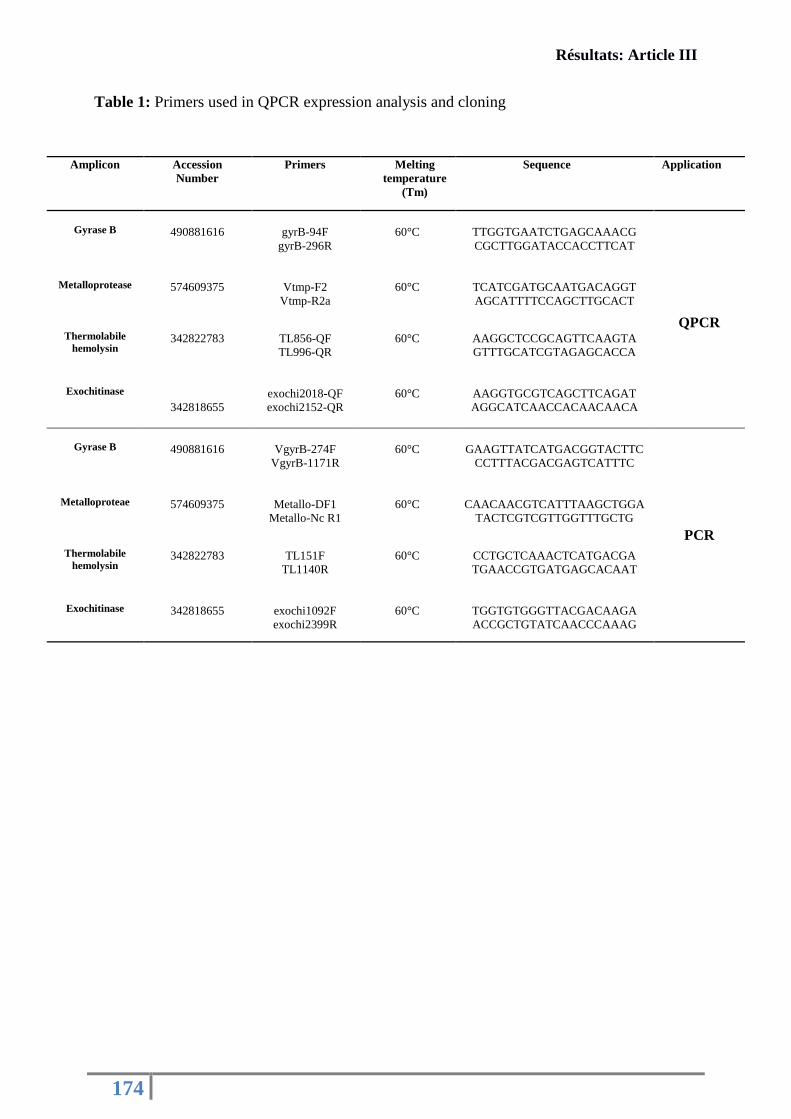
Résultats: Article III
174
Table 1: Primers used in QPCR expression analysis and cloning
Amplicon
Accession
Number
Primers
Melting
temperature
(Tm)
Sequence Application
Gyrase B
490881616
gyrB-94F
gyrB-296R
60°C
TTGGTGAATCTGAGCAAACG
CGCTTGGATACCACCTTCAT
QPCR
Metalloprotease
574609375
Vtmp-F2
Vtmp-R2a
60°C
TCATCGATGCAATGACAGGT
AGCATTTTCCAGCTTGCACT
Thermolabile
hemolysin
342822783
TL856-QF
TL996-QR
60°C
AAGGCTCCGCAGTTCAAGTA
GTTTGCATCGTAGAGCACCA
Exochitinase
342818655
exochi2018-QF
exochi2152-QR
60°C
AAGGTGCGTCAGCTTCAGAT
AGGCATCAACCACAACAACA
Gyrase B
490881616
VgyrB-274F
VgyrB-1171R
60°C
GAAGTTATCATGACGGTACTTC
CCTTTACGACGAGTCATTTC
PCR
Metalloproteae
574609375
Metallo-DF1
Metallo-Nc R1
60°C
CAACAACGTCATTTAAGCTGGA
TACTCGTCGTTGGTTTGCTG
Thermolabile
hemolysin
342822783
TL151F
TL1140R
60°C
CCTGCTCAAACTCATGACGA
TGAACCGTGATGAGCACAAT
Exochitinase
342818655
exochi1092F
exochi2399R
60°C
TGGTGTGGGTTACGACAAGA
ACCGCTGTATCAACCCAAAG
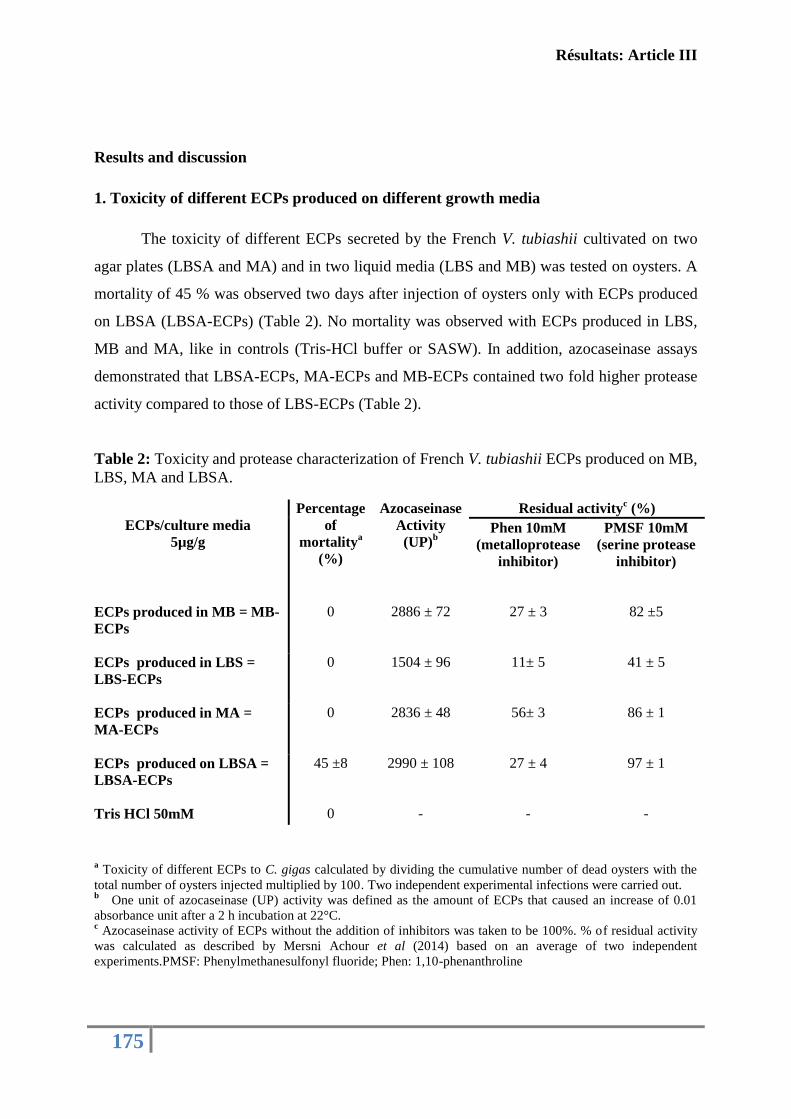
Résultats: Article III
175
Results and discussion
1. Toxicity of different ECPs produced on different growth media
The toxicity of different ECPs secreted by the French V. tubiashii cultivated on two
agar plates (LBSA and MA) and in two liquid media (LBS and MB) was tested on oysters. A
mortality of 45 % was observed two days after injection of oysters only with ECPs produced
on LBSA (LBSA-ECPs) (Table 2). No mortality was observed with ECPs produced in LBS,
MB and MA, like in controls (Tris-HCl buffer or SASW). In addition, azocaseinase assays
demonstrated that LBSA-ECPs, MA-ECPs and MB-ECPs contained two fold higher protease
activity compared to those of LBS-ECPs (Table 2).
Table 2: Toxicity and protease characterization of French V. tubiashii ECPs produced on MB,
LBS, MA and LBSA.
ECPs/culture media
5µg/g
Percentage
of
mortalitya
(%)
Azocaseinase
Activity
(UP)b
Residual activityc (%)
Phen 10mM
(metalloprotease
inhibitor)
PMSF 10mM
(serine protease
inhibitor)
ECPs produced in MB = MB-
ECPs
0 2886 ± 72 27 ± 3 82 ±5
ECPs produced in LBS =
LBS-ECPs
0 1504 ± 96 11± 5 41 ± 5
ECPs produced in MA =
MA-ECPs
0 2836 ± 48 56± 3 86 ± 1
ECPs produced on LBSA =
LBSA-ECPs
45 ±8 2990 ± 108 27 ± 4 97 ± 1
Tris HCl 50mM 0 - - -
a Toxicity of different ECPs to C. gigas calculated by dividing the cumulative number of dead oysters with the
total number of oysters injected multiplied by 100. Two independent experimental infections were carried out. b
One unit of azocaseinase (UP) activity was defined as the amount of ECPs that caused an increase of 0.01
absorbance unit after a 2 h incubation at 22°C. c Azocaseinase activity of ECPs without the addition of inhibitors was taken to be 100%. % of residual activity
was calculated as described by Mersni Achour et al (2014) based on an average of two independent
experiments.PMSF: Phenylmethanesulfonyl fluoride; Phen: 1,10-phenanthroline
Résultats: Article III
176
The addition of 1, 10-phenanthroline ( a metalloprotease inhibitor) into the reaction
mix strongly inhibited azocaseinase activity of all ECPs, with a loss of 90 % of activity for
LBS-ECPs, about 73 % for MB-ECPs and LBSA-ECPs and 45 % for MA-ECPs.
Azocaseinase activity was slightly affected by the presence of PMSF (a serine protease
inhibitor) for LBSA-ECPs, MA-ECPs and MB-ECPs however a loss of 60% was observed for
LBS-ECPs (Table 1), suggesting differences in term of both protease composition and
concentration.
Typically, to evaluate virulence of Vibrio extracellular products, exotoxins are
produced by the cellophane overlay method (Labreuche et al., 2006a; Binesse et al., 2008;
Soto-Rodriguez et al., 2012). In this study, to test the toxicity of the French V. tubiashii ECPs,
two physical states of a culture medium were used: solid medium to mimic in vivo conditions
(colonization, adhesion…) and broth medium to reflect body fluids, where bacteria can be
found after a detachment from the infection site or a rupture of a tissue that forms a natural
physical barrier (Lorian, 1989).
The difference observed between ECPs in terms of toxicity or proteases composition
could be explained by Quorum sensing (QS) process. It was reported that QS signal
production levels, which differs between planktonic (free-living) and biofilm cells (surface-
attached) (Chopp et al., 2003) is known to modulate the expression of virulence gene for a
large range of bacteria (Antunes et al., 2010; Natrah et al., 2011). For V. harveyi, extracellular
toxins, such as a metalloproteases, a type III secretion system and a siderophore were quorum
sensing dependent (Defoirdt et al., 2008; Natrah et al., 2011). Also, it was shown that cell-to-
cell communication phenomenon has effects on virulence factor expression of V. splendidus
and V. aestuarianus (De Decker et al., 2013).
Moreover, bacteria behavior depends not only on the physical state of culture medium,
but also on the composition and concentration of medium compounds that affect bacteria
metabolisms. Mortality observed with LBSA-ECPs but not with MA-ECPs, could be
explained by the effect of the medium composition on toxin secretions. MA contains different
types of minerals (14 salts) that nearly duplicate the major mineral composition of sea water.
However, LBSA contains only sodium chloride as a salt. Presumably, the difference in salt
composition between the two media might be disadvantageous for protease production, a
cellular activity implicated in metabolism, survival and pathogenicity (Shinoda et al., 1996).
Indeed, it has been described that protease activity of polymorphonuclear neutrophils was
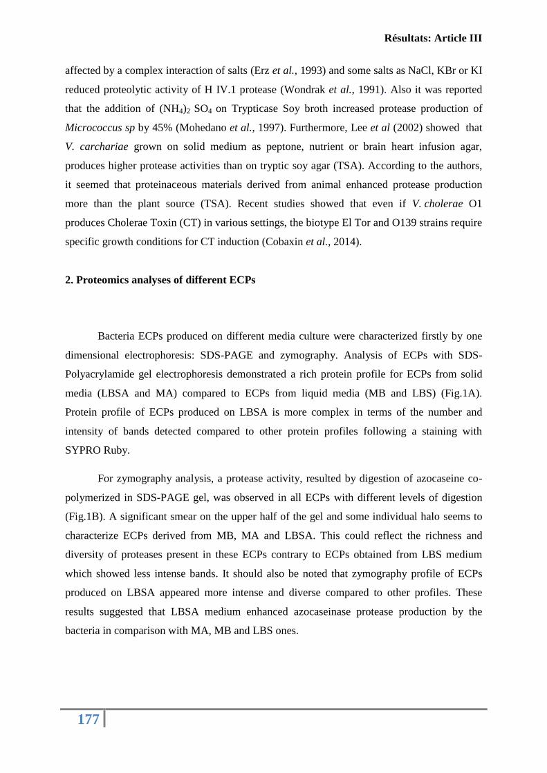
Résultats: Article III
177
affected by a complex interaction of salts (Erz et al., 1993) and some salts as NaCl, KBr or KI
reduced proteolytic activity of H IV.1 protease (Wondrak et al., 1991). Also it was reported
that the addition of (NH4)2 SO4 on Trypticase Soy broth increased protease production of
Micrococcus sp by 45% (Mohedano et al., 1997). Furthermore, Lee et al (2002) showed that
V. carchariae grown on solid medium as peptone, nutrient or brain heart infusion agar,
produces higher protease activities than on tryptic soy agar (TSA). According to the authors,
it seemed that proteinaceous materials derived from animal enhanced protease production
more than the plant source (TSA). Recent studies showed that even if V. cholerae O1
produces Cholerae Toxin (CT) in various settings, the biotype El Tor and O139 strains require
specific growth conditions for CT induction (Cobaxin et al., 2014).
2. Proteomics analyses of different ECPs
Bacteria ECPs produced on different media culture were characterized firstly by one
dimensional electrophoresis: SDS-PAGE and zymography. Analysis of ECPs with SDS-
Polyacrylamide gel electrophoresis demonstrated a rich protein profile for ECPs from solid
media (LBSA and MA) compared to ECPs from liquid media (MB and LBS) (Fig.1A).
Protein profile of ECPs produced on LBSA is more complex in terms of the number and
intensity of bands detected compared to other protein profiles following a staining with
SYPRO Ruby.
For zymography analysis, a protease activity, resulted by digestion of azocaseine co-
polymerized in SDS-PAGE gel, was observed in all ECPs with different levels of digestion
(Fig.1B). A significant smear on the upper half of the gel and some individual halo seems to
characterize ECPs derived from MB, MA and LBSA. This could reflect the richness and
diversity of proteases present in these ECPs contrary to ECPs obtained from LBS medium
which showed less intense bands. It should also be noted that zymography profile of ECPs
produced on LBSA appeared more intense and diverse compared to other profiles. These
results suggested that LBSA medium enhanced azocaseinase protease production by the
bacteria in comparison with MA, MB and LBS ones.
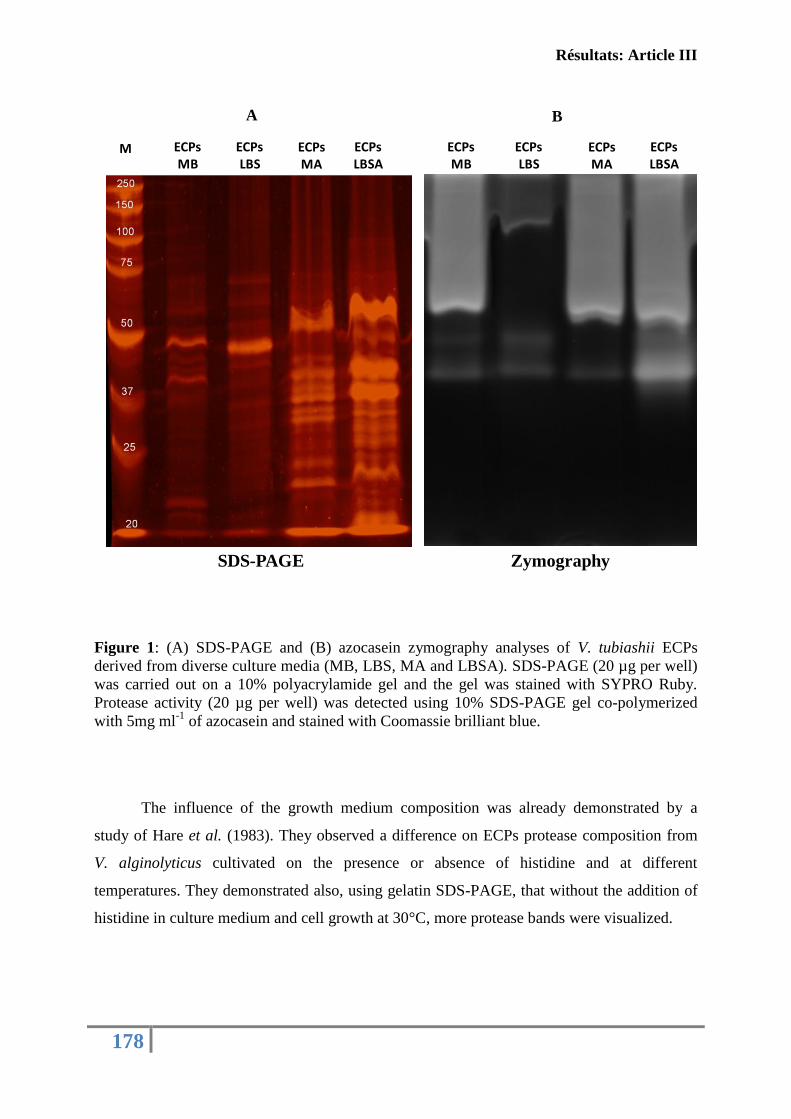
Résultats: Article III
178
Figure 1: (A) SDS-PAGE and (B) azocasein zymography analyses of V. tubiashii ECPs
derived from diverse culture media (MB, LBS, MA and LBSA). SDS-PAGE (20 µg per well)
was carried out on a 10% polyacrylamide gel and the gel was stained with SYPRO Ruby.
Protease activity (20 µg per well) was detected using 10% SDS-PAGE gel co-polymerized
with 5mg ml-1
of azocasein and stained with Coomassie brilliant blue.
The influence of the growth medium composition was already demonstrated by a
study of Hare et al. (1983). They observed a difference on ECPs protease composition from
V. alginolyticus cultivated on the presence or absence of histidine and at different
temperatures. They demonstrated also, using gelatin SDS-PAGE, that without the addition of
histidine in culture medium and cell growth at 30°C, more protease bands were visualized.
ECPsMB
ECPsLBS
ECPsMA
ECPsLBSA
M ECPsMB
ECPsLBS
ECPsMA
ECPsLBSA
SDS-PAGE Zymography
B A
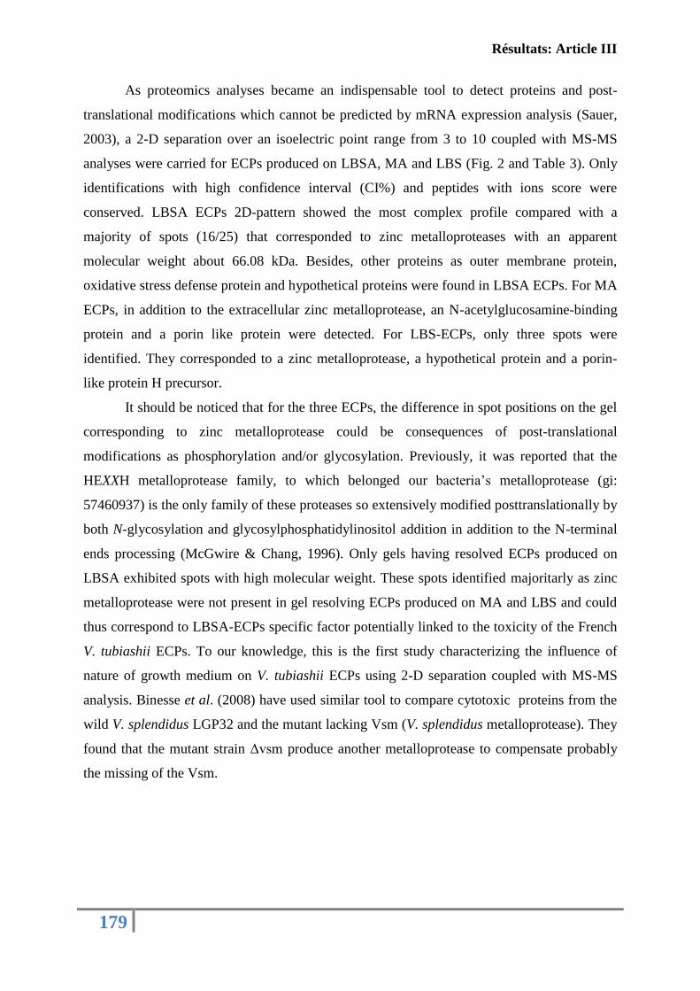
Résultats: Article III
179
As proteomics analyses became an indispensable tool to detect proteins and post-
translational modifications which cannot be predicted by mRNA expression analysis (Sauer,
2003), a 2-D separation over an isoelectric point range from 3 to 10 coupled with MS-MS
analyses were carried for ECPs produced on LBSA, MA and LBS (Fig. 2 and Table 3). Only
identifications with high confidence interval (CI%) and peptides with ions score were
conserved. LBSA ECPs 2D-pattern showed the most complex profile compared with a
majority of spots (16/25) that corresponded to zinc metalloproteases with an apparent
molecular weight about 66.08 kDa. Besides, other proteins as outer membrane protein,
oxidative stress defense protein and hypothetical proteins were found in LBSA ECPs. For MA
ECPs, in addition to the extracellular zinc metalloprotease, an N-acetylglucosamine-binding
protein and a porin like protein were detected. For LBS-ECPs, only three spots were
identified. They corresponded to a zinc metalloprotease, a hypothetical protein and a porin-
like protein H precursor.
It should be noticed that for the three ECPs, the difference in spot positions on the gel
corresponding to zinc metalloprotease could be consequences of post-translational
modifications as phosphorylation and/or glycosylation. Previously, it was reported that the
HEXXH metalloprotease family, to which belonged our bacteria’s metalloprotease (gi:
57460937) is the only family of these proteases so extensively modified posttranslationally by
both N-glycosylation and glycosylphosphatidylinositol addition in addition to the N-terminal
ends processing (McGwire & Chang, 1996). Only gels having resolved ECPs produced on
LBSA exhibited spots with high molecular weight. These spots identified majoritarly as zinc
metalloprotease were not present in gel resolving ECPs produced on MA and LBS and could
thus correspond to LBSA-ECPs specific factor potentially linked to the toxicity of the French
V. tubiashii ECPs. To our knowledge, this is the first study characterizing the influence of
nature of growth medium on V. tubiashii ECPs using 2-D separation coupled with MS-MS
analysis. Binesse et al. (2008) have used similar tool to compare cytotoxic proteins from the
wild V. splendidus LGP32 and the mutant lacking Vsm (V. splendidus metalloprotease). They
found that the mutant strain Δvsm produce another metalloprotease to compensate probably
the missing of the Vsm.
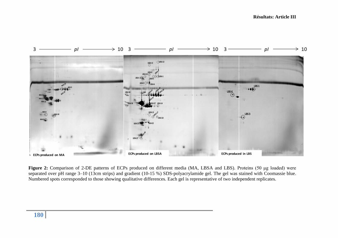
Résultats: Article III
180
Figure 2: Comparison of 2-DE patterns of ECPs produced on different media (MA, LBSA and LBS). Proteins (50 μg loaded) were
separated over pH range 3–10 (13cm strips) and gradient (10-15 %) SDS-polyacrylamide gel. The gel was stained with Coomassie blue.
Numbered spots corresponded to those showing qualitative differences. Each gel is representative of two independent replicates.
LBS 1
LBS 3
LBS 4
MA 5
MA 4
MA 11
MA 1
MA 12
MA 3
MA 18
ECPs produced on MA
LBSA
LBSA 13
LBSA 1LBSA 5
LBSA 12
LBSA 8LBSA 10
LBSA 11
LBSA 9 LBSA 6
LBSA 34
LBSA 36 LBSA 37 LBSA 14
LBSA 27
LBSA 18
LBSA 15
LBSA 22
LBSA SANS 14 ET 11
LBSA 21
ECPs produced on MA ECPs produced on LBSA
3 10 3 10 3 10pIpI pI
ECPs produced in LBS
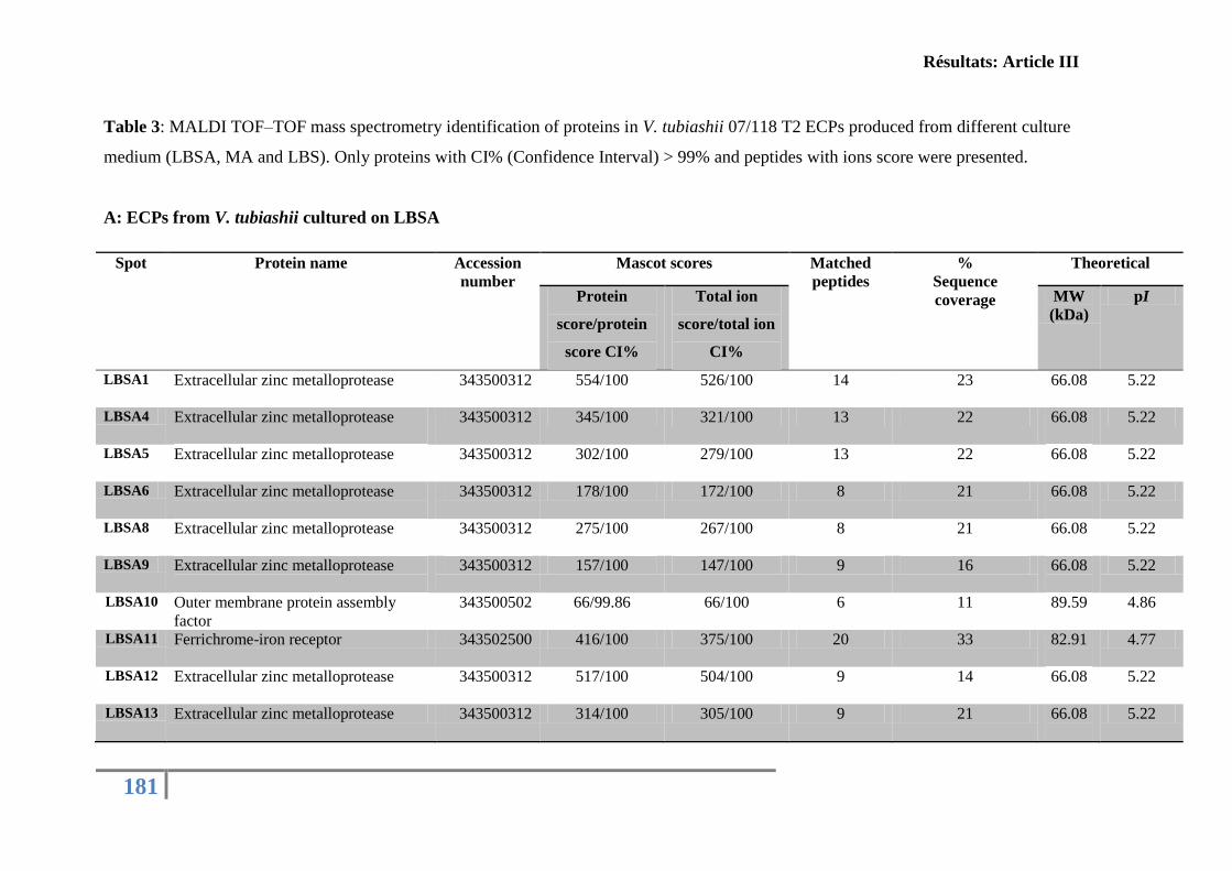
Résultats: Article III
181
Table 3: MALDI TOF–TOF mass spectrometry identification of proteins in V. tubiashii 07/118 T2 ECPs produced from different culture
medium (LBSA, MA and LBS). Only proteins with CI% (Confidence Interval) > 99% and peptides with ions score were presented.
A: ECPs from V. tubiashii cultured on LBSA
Spot Protein name Accession
number
Mascot scores Matched
peptides
%
Sequence
coverage
Theoretical
Protein
score/protein
score CI%
Total ion
score/total ion
CI%
MW
(kDa)
pI
LBSA1 Extracellular zinc metalloprotease
343500312 554/100 526/100 14 23 66.08
5.22
LBSA4 Extracellular zinc metalloprotease
343500312 345/100 321/100 13 22 66.08
5.22
LBSA5 Extracellular zinc metalloprotease
343500312 302/100 279/100 13 22 66.08
5.22
LBSA6 Extracellular zinc metalloprotease
343500312 178/100 172/100 8 21 66.08
5.22
LBSA8 Extracellular zinc metalloprotease
343500312 275/100 267/100 8 21 66.08
5.22
LBSA9 Extracellular zinc metalloprotease
343500312 157/100 147/100 9 16 66.08
5.22
LBSA10 Outer membrane protein assembly
factor
343500502 66/99.86 66/100 6 11 89.59 4.86
LBSA11 Ferrichrome-iron receptor 343502500 416/100 375/100 20 33 82.91
4.77
LBSA12 Extracellular zinc metalloprotease 343500312 517/100 504/100 9 14 66.08
5.22
LBSA13 Extracellular zinc metalloprotease 343500312 314/100 305/100 9 21 66.08
5.22
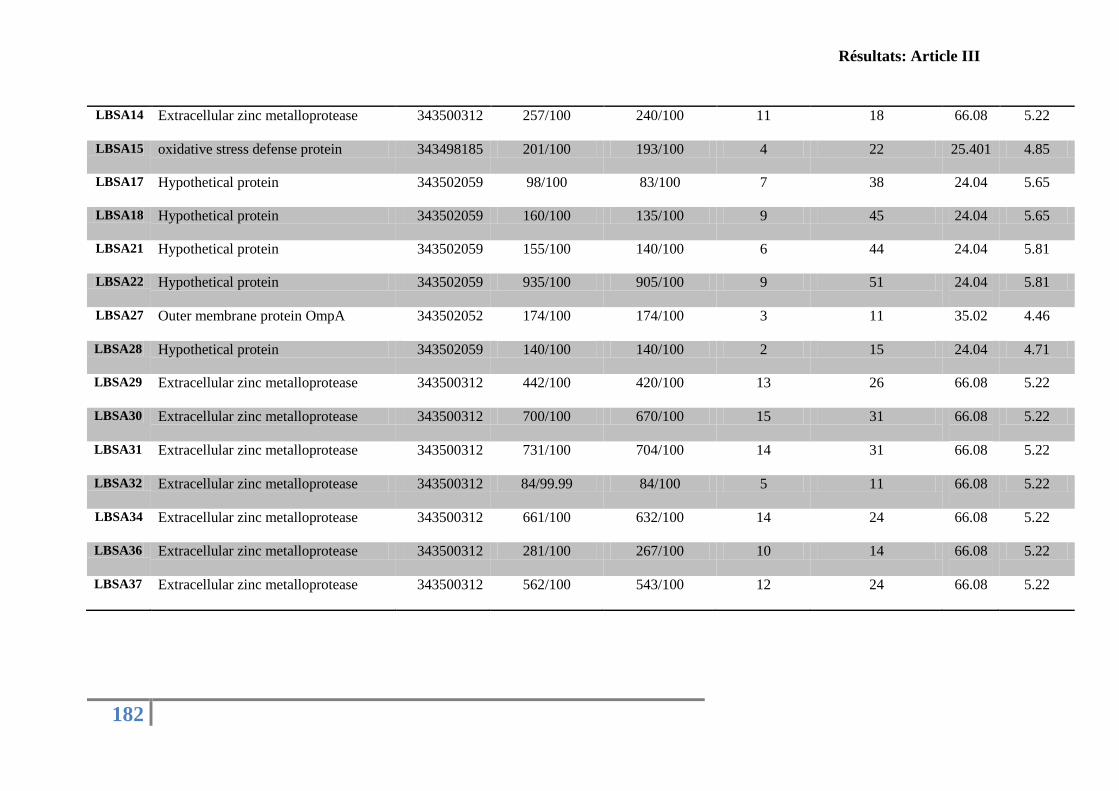
Résultats: Article III
182
LBSA14 Extracellular zinc metalloprotease 343500312 257/100 240/100 11 18 66.08
5.22
LBSA15 oxidative stress defense protein 343498185 201/100 193/100 4 22 25.401 4.85
LBSA17 Hypothetical protein 343502059 98/100 83/100 7 38 24.04 5.65
LBSA18 Hypothetical protein 343502059 160/100 135/100 9 45 24.04 5.65
LBSA21 Hypothetical protein 343502059 155/100 140/100 6 44 24.04 5.81
LBSA22 Hypothetical protein 343502059 935/100 905/100 9 51 24.04
5.81
LBSA27 Outer membrane protein OmpA 343502052 174/100 174/100 3 11 35.02
4.46
LBSA28 Hypothetical protein 343502059 140/100 140/100 2 15 24.04 4.71
LBSA29 Extracellular zinc metalloprotease 343500312 442/100 420/100 13 26 66.08
5.22
LBSA30 Extracellular zinc metalloprotease 343500312 700/100 670/100 15 31 66.08
5.22
LBSA31 Extracellular zinc metalloprotease 343500312 731/100 704/100 14 31 66.08
5.22
LBSA32 Extracellular zinc metalloprotease 343500312 84/99.99 84/100 5 11 66.08
5.22
LBSA34 Extracellular zinc metalloprotease 343500312 661/100 632/100 14 24 66.08
5.22
LBSA36 Extracellular zinc metalloprotease 343500312 281/100 267/100 10 14 66.08
5.22
LBSA37 Extracellular zinc metalloprotease 343500312 562/100 543/100 12 24 66.08
5.22
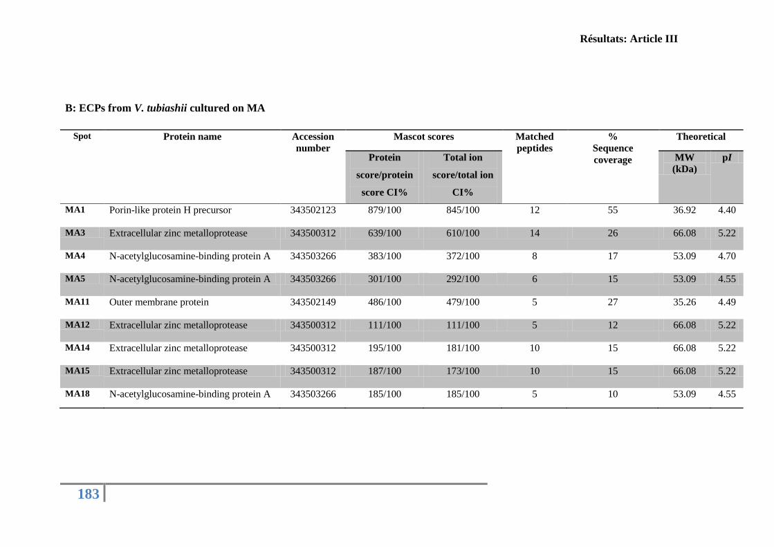
Résultats: Article III
183
B: ECPs from V. tubiashii cultured on MA
Spot Protein name Accession
number
Mascot scores Matched
peptides
%
Sequence
coverage
Theoretical
Protein
score/protein
score CI%
Total ion
score/total ion
CI%
MW
(kDa)
pI
MA1 Porin-like protein H precursor 343502123 879/100 845/100 12 55 36.92 4.40
MA3 Extracellular zinc metalloprotease 343500312 639/100 610/100 14 26 66.08 5.22
MA4 N-acetylglucosamine-binding protein A 343503266 383/100 372/100 8 17 53.09 4.70
MA5 N-acetylglucosamine-binding protein A 343503266 301/100 292/100 6 15 53.09 4.55
MA11 Outer membrane protein 343502149 486/100 479/100 5 27 35.26 4.49
MA12 Extracellular zinc metalloprotease 343500312 111/100 111/100 5 12 66.08 5.22
MA14 Extracellular zinc metalloprotease 343500312 195/100 181/100 10 15 66.08 5.22
MA15 Extracellular zinc metalloprotease 343500312 187/100 173/100 10 15 66.08 5.22
MA18 N-acetylglucosamine-binding protein A 343503266 185/100 185/100 5 10 53.09 4.55

Résultats: Article III
184
C: ECPs from V. tubiashii cultured on LBS
Spot Protein name Accession
number
Mascot scores Matched
peptides
%
Sequence
coverage
Theoretical
Protein
score/protein
score CI%
Total ion
score/total ion
CI%
MW
(kDa)
pI
LBS1 Extracellular zinc metalloprotease 343500312 620/100 598/100 12 21 66.08 5.22
LBS3 Hypothetical protein 343502059 387/100 367/100 7 34 24.04 5.81
LBS4 Porin-like protein H precursor 343502123 534/100 508/100 11 53 36.92 4.40
Résultats: Article III
185
3. Differential relative gene expression of the French V. tubiashii cultivated on different
media
The relative gene expression of metalloprotease, thermostable hemolysin and
exochitinase of the French V. tubiashii 07/118 T2 grew on LBSA, MA and LBS was
analysed. Because these proteins were usually described as potential toxins secreted by the
genus Vibrio (Actis et al., 1986; Fabbri et al., 1999; Binesse et al., 2008; Park et al., 2000;
Hasegawa et al., 2009; Wong et al., 2012). Results demonstrated a difference in V. tubiashii
hemolysin, metalloprotease and exochitinase gene expression between media (Fig. 3). For
metalloprotease gene expression, transcriptomic results obtained sustain results obtained with
proteomics analyses: solid medium (LBSA and MA) enhances the production of
metalloproteases compared to the liquid medium (LBS). The thermostable hemolysin gene
expression of the V. tubiashii cultivated on MA was up regulated about 14 times compared to
bacteria grown in LBS and on LBSA medium. Exochitinase is more expressed when bacteria
are grown on LBSA than LBS and MA. The effect of culture medium nature on bacterial
genes expression has been already described. For example, for V. salmonicida, a cause of
cold-water vibriosis in seawater-farmed Atlantic salmon, studies showed that the levels of
flagellin expression are significantly higher under elevated salt conditions than at lower
salinity (Karlsen et al., 2008).
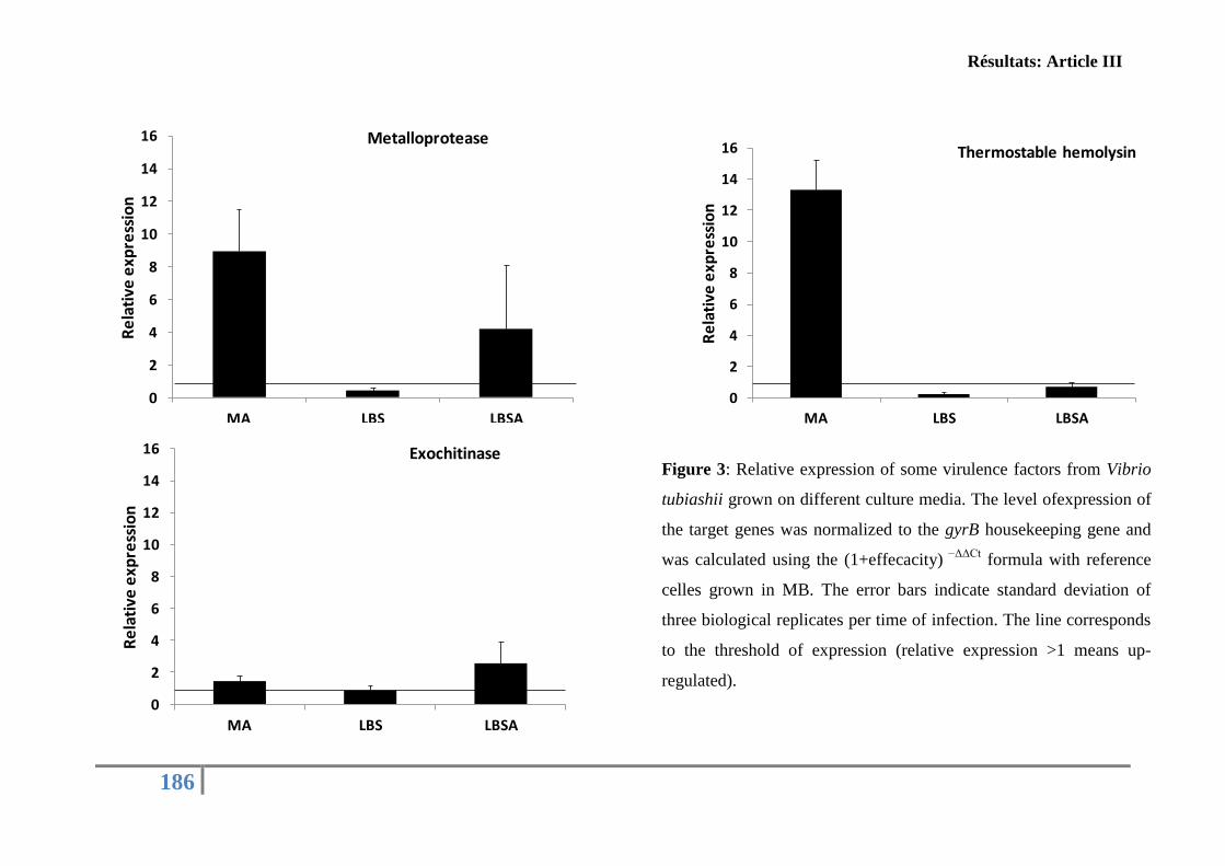
Résultats: Article III
186
0
2
4
6
8
10
12
14
16
MA LBS LBSA
Re
lati
ve e
xpre
ssio
nMetalloprotease
0
2
4
6
8
10
12
14
16
MA LBS LBSA
Re
lati
ve e
xpre
ssio
n
Thermostable hemolysin
0
2
4
6
8
10
12
14
16
MA LBS LBSA
Re
lati
ve e
xpre
ssio
n
ExochitinaseFigure 3: Relative expression of some virulence factors from Vibrio
tubiashii grown on different culture media. The level ofexpression of
the target genes was normalized to the gyrB housekeeping gene and
was calculated using the (1+effecacity) −ΔΔCt
formula with reference
celles grown in MB. The error bars indicate standard deviation of
three biological replicates per time of infection. The line corresponds
to the threshold of expression (relative expression >1 means up-
regulated).
Résultats: Article III
187
Conclusion
In this present study, using experimental infections, proteomics and transcriptomics
approach, we showed that the physical state and the composition of growth media culture
affect the French V. tubiashii behavior regarding toxin production in terms of composition
and concentration. Interestingly, the toxicity of the French V. tubiashii ECPs seems to not be
linked to the over expression of metalloprotease even if it is dominant in the secretome but
probably to other virulence factors or a specific metalloprotease post-translational
modification forms. Therefore, further studies are underway to explain the importance this
assumption.
Acknowledgements
This work was carried out with financial support by a Ph.D grant from the CNRS and
IFREMER for R. Mersni-Achour (2010-2013). The PRES (Pôle de Recherche et
d’Enseignement Supérieur) of Limousin PoitouCharentes and the FR CNRS 3097
«Fédération de Recherche en Environnement et Développement Durable» are acknowledged
for financial support through the research project «Caractérisation in vitro des effets de
métalloprotéases sur différents types cellulaires de l’huître creuse C. gigas». Thanks go to the
platform “Structural and Functional Mass Spectrometry Facility, Jacques Monod Institute,
UMR7592 CNRS – Univ Paris Diderot, Sorbonne Paris Cité” and especially to T. Léger and C.
Garcia for mass spectrometry analysis
Résultats: Article III
188
References
Actis, L. A., Fish, W., Crosa, J. H., Kellerman, K., Ellenberger, S. R., Hauser, F. M. &
Sanders-Loehr, J. (1986). Characterization of anguibactin, a novel siderophore from
Vibrio anguillarum 775(pJM1). J Bacteriol 167, 57–65.
Antunes, L. C. M., Ferreira, R. B. R., Buckner, M. M. C. & Finlay, B. B. (2010). Quorum
sensing in bacterial virulence. Microbiology 156, 2271–2282.
Binesse, J., Delsert, C., Saulnier, D., Champomier-Vergès, M.-C., Zagorec, M., Munier-
Lehmann, H., Mazel, D. & Roux, F. L. (2008). Metalloprotease Vsm Is the Major
Determinant of Toxicity for Extracellular Products of Vibrio splendidus. Appl Environ
Microbiol 74, 7108–7117.
Bradford, M. M. (1976). A rapid and sensitive method for the quantitation of microgram
quantities of protein utilizing the principle of protein-dye binding. Anal Biochem 72,
248–254.
Chopp, D. L., Kirisits, M. J., Moran, B. & Parsek, M. R. (2003). The dependence of
quorum sensing on the depth of a growing biofilm. Bull Math Biol 65, 1053–1079.
Cobaxin, M., Martínez, H., Ayala, G., Holmgren, J., Sjöling, Å. & Sánchez, J. (2014). Cholera toxin expression by El Tor Vibrio cholerae in shallow culture growth
conditions. Microb Pathog 66, 5–13.
Cohen, N. & Karib, H. (2007). Vibrio spp. dans les produits de la pêche: Risques et
prévention.
De Decker, S., Reynaud, Y. & Saulnier, D. (2013). First molecular evidence of cross-
species induction of metalloprotease gene expression in Vibrio strains pathogenic for
Pacific oyster Crassostrea gigas involving a quorum sensing system. Aquaculture
392–395, 1–7.
Defoirdt, T., Boon, N., Sorgeloos, P., Verstraete, W. & Bossier, P. (2008). Quorum sensing
and quorum quenching in Vibrio harveyi: lessons learned from in vivo work. ISME J
2, 19–26.
Elston, R. A., Hasegawa, H., Humphrey, K. L., Polyak, I. K. & Häse, C. C. (2008). Re-
emergence of Vibrio tubiashii in bivalve shellfish aquaculture: severity, environmental
drivers, geographic extent and management. Dis Aquat Organ 82, 119–134.
Erz, W., Carrle, D. & Klessen, C. (1993). Proteases of polymorphonuclear neutrophils:
Influence of different salts and buffers on pericellular lysis. Acta Histochem 94, 46–
53.
Fabbri, A., Falzano, L., Frank, C., Donelli, G., Matarrese, P., Raimondi, F., Fasano, A.
& Fiorentini, C. (1999). Vibrio parahaemolyticus Thermostable Direct Hemolysin
Modulates Cytoskeletal Organization and Calcium Homeostasis in Intestinal Cultured
Cells. Infect Immun 67, 1139–1148.
Galland, C., Dupuy, C., Capitaine, C., Auffret, M., Quiniou, L., Laroche, J. &
Pichereau, V. (2013). Comparisons of liver proteomes in the European flounder
Platichthys flesus from three contrasted estuaries. J Sea Res 75, 135–141.
Hare, P., Scott-Burden, T. & Woods, D. R. (1983). Characterization of Extracellular
Alkaline Proteases and Collagenase Induction in Vibrio alginolyticus. J Gen Microbiol
129, 1141–1147.
Hasegawa, H. & Häse, C. C. (2009b). TetR-Type Transcriptional Regulator VtpR Functions
as a Global Regulator in Vibrio tubiashii. Appl Environ Microbiol 75, 7602–7609.
Résultats: Article III
189
Hasegawa, H., Lind, E. J., Boin, M. A. & Häse, C. C. (2008). The Extracellular
Metalloprotease of Vibrio tubiashii Is a Major Virulence Factor for Pacific Oyster
(Crassostrea gigas) Larvae. Appl Environ Microbiol 74, 4101–4110.
Hasegawa, H., Gharaibeh, D. N., Lind, E. J. & Häse, C. C. (2009). Virulence of
metalloproteases produced by Vibrio species on Pacific oyster Crassostrea gigas
larvae. Dis Aquat Organ 85, 123–131.
Karlsen, C., Paulsen, S. M., Tunsjø, H. S., Krinner, S., Sørum, H., Haugen, P. &
Willassen, N.-P. (2008). Motility and flagellin gene expression in the fish pathogen
Vibrio salmonicida: Effects of salinity and temperature. Microb Pathog 45, 258–264.
Labreuche, Y., Soudant, P., Goncalves, M., Lambert, C. & Nicolas, J. (2006a). Effects of
extracellular products from the pathogenic Vibrio aestuarianus strain 01/32 on
lethality and cellular immune responses of the oyster Crassostrea gigas. Dev Comp
Immunol 30, 367–379.
Lee, K. K., Liu, P. C. & Chuang, W. H. (2002). Pathogenesis of gastroenteritis caused by
Vibrio carchariae in cultured marine fish. Mar Biotechnol N Y N 4, 267–277.
Lorian, V. (1989). In vitro simulation of in vivo conditions: physical state of the culture
medium. J Clin Microbiol 27, 2403–2406.
Lorian, V. (1988). Differences between in vitro and in vivo studies. Antimicrob Agents
Chemother 32, 1600–1601.
McGwire, B. S. & Chang, K.-P. (1996). Posttranslational Regulation of a Leishmania
HEXXH Metalloprotease (gp63). J Biol Chem 271, 7903–7909.
Mohedano, A. f., Fernández, J., Gaya, P., Medina, M. & Nuñez, M. (1997). Effect of pH,
temperature and culture medium composition on the production of an extracellular
cysteine proteinase by Micrococcus sp. INIA 528. J Appl Microbiol 82, 81–86.
Natrah, F. M. I., Ruwandeepika, H. A. D., Pawar, S., Karunasagar, I., Sorgeloos, P.,
Bossier, P. & Defoirdt, T. (2011). Regulation of virulence factors by quorum sensing
in Vibrio harveyi. Vet Microbiol 154, 124–129.
Paillard, C., Le Roux, F. & Borrego, J. J. (2004). Bacterial disease in marine bivalves, a
review of recent studies: Trends and evolution. Aquat Living Resour 17, 477–498.
Park, S., Lee, J. & Lee, W. (2000). Purification and Characterization of Chitinase from a
Marine Bacterium, Vibrio sp. 98CJ11027. J Microbiol 38, 224–229.
Pfaffl, M. W. (2001). A new mathematical model for relative quantification in real-time RT-
PCR. Nucleic Acids Res 29, e45.
Romalde, J. L. & Barja, J. L. (2010). Bacteria in molluscs: good and bad guys. Curr Res
Technol Educ Top Appl Microbiol Microb Biotechnol 1, 136–147.
Romalde, J. L., Dieguez, A. L., Lasa, A. & Balboa, S. (2014). New Vibrio species
associated to molluscan microbiota: a review. Front Microbiol 4, 413.
Sauer, K. (2003). The genomics and proteomics of biofilm formation. Genome Biol 4, 219.
Shinoda, S. & Miyoshi, S.-I. (2011). Proteases Produced by Vibrios. Biocontrol Sci 16, 1–
11.
Shinoda, S., Miyoshi, S.-I., Wakae, H., Rahman, M. & Tomochika, K.-I. (1996). Bacterial
Proteases as Pathogenic Factors, with Special Emphasis on Vibrio Proteases. Toxin
Rev 15, 327–339.
Soto-Rodriguez, S. A., Gomez-Gil, B., Lozano, R., del Rio-Rodríguez, R., Diéguez, A. L.
& Romalde, J. L. (2012). Virulence of Vibrio harveyi responsible for the ‘Bright-red’
Syndrome in the Pacific white shrimp Litopenaeus vannamei. J Invertebr Pathol 109,
307–317.
Thompson, F. L., Iida, T. & Swings, J. (2004). Biodiversity of Vibrios. Microbiol Mol Biol
Rev 68, 403–431.
Résultats: Article III
190
Travers, M.-A., Mersni Achour, R., Haffner, P., Tourbiez, D., Cassone, A.-L., Morga, B.,
Doghri, I., Garcia, C. G., Renault, T. & other authors. (In press). First description
of French V. tubiashii strains pathogenic to mollusk: I. Characterization of isolates
and detection during mortality events. J Invertebr Pathol.
Tubiash, H. S., Chanley, P. E. & Leifson, E. (1965). Bacillary Necrosis, a Disease of Larval
and Juvenile Bivalve Mollusks I. Etiology and Epizootiology. J Bacteriol 90, 1036–
1044.
Tubiash, H. S., Colwell, R. R. & Sakazaki, R. (1970). Marine Vibrios Associated with
Bacillary Necrosis, a Disease of Larval and Juvenile Bivalve Mollusks. J Bacteriol
103, 271–272.
Wondrak, E. M., Louis, J. M. & Oroszlan, S. (1991). The effect of salt on the Michaelis
Menten constant of the HIV-1 protease correlates with the Hofmeister series. FEBS
Lett 280, 344–346.
Wong, S. K., Zhang, X.-H. & Woo, N. Y. S. (2012). Vibrio alginolyticus thermolabile
hemolysin (TLH) induces apoptosis, membrane vesiculation and necrosis in sea bream
erythrocytes. Aquaculture 330–333, 29–36.
Résultats: Article IV
191
C/ La caractérisation moléculaire des facteurs de virulence
produits par la souche française V. tubiashii
La souche V. tubiashii est un agent pathogène affectant les larves et les juvéniles de
mollusques bivalves (Tubiash et al., 1965 ; 1970). Elle a été détectée dans plusieurs endroits
du monde (Amérique, Angleterre, Espagne et France) (Tubiash et al., 1965; Jeffries, 1982;
Lodeiros et al., 1987; Estes et al., 2004; Travers et al., 2014). Cette souche est responsable
d’une maladie nommée nécrose bacillaire caractérisée par une réduction de la motilité des
larves et la formation d'amas bactériens autour de la coque et les organes internes (Tubiash et
al., 1965; Estes et al., 2004). Plusieurs études ont montré l’implication de toxines
extracellulaires dans la virulence de V. tubiashii (Brown & Roland, 1984; Kothary et al.,
2001; Delston et al., 2003; Hasegawa et al., 2008).
Dans l’objectif de caractériser les mécanismes et les facteurs de virulence responsable
de la pathogenicité de la souche française V. tubiashii, nous avons utilisé le modèle larvaire
d’huîtres creuses C. gigas de 13 jours. La souche 07/118 T2 a montré une virulence
remarquable envers les larves d’huîtres avec une DL50 d'environ 2, 3 × 10³ CFU/ ml après 24
h d’infection). Les observations morphologiques indiquent qu’elle induit des symptômes
semblables à ceux de la nécrose bacillaire et qui se manifestent par une réduction de la
motilité larvaire et la formation d’amas bactériens autour des organes et des débris cellulaires.
En outre, le surnageant bactérien total et les deux fractions obtenues par GP-HPLC ont montré
une toxicité pour les larves avec un phénotype similaire mais pas complet par rapport à celui
observé avec la souche vivante. Ce dernier résultat suggère que l’étape du fractionnement de
l’ECPs brut a conduit à perdre un certain nombre de facteurs et/ou leur synergie d’action qui
semble fondamentale à l’expression de ces anomalies larvaires. En conséquence, il nous est
apparu nécessaire à ce stade du travail d’analyser plus finement le contenu moléculaire de ces
deux fractions. Une électrophorèse 2-DE suivie d’une identification par spectrométrie de
masse MALDI-TOF de spots protéiques résolus ont été menés.
L’anayse des spots protéiques des deux fractions obtenues a révélé l'existence (i) d'une
métalloprotéase extracellulaire (caractéristique de la fraction 2) différente de métalloprotéases
Résultats: Article IV
192
décrites comme facteurs de virulence chez les souches de V. tubiashii américaines (Hasegawa
& Häse, 2009b; Hasegawa et al., 2008, 2009) et (ii) d'autres facteurs de virulence comme les
porines et les protéines de la membrane externe (fraction 1).
La diversité et l'action séquentielle des facteurs de virulence sécrétés par cette souche
ont été également suggérées par l'utilisation d'inhibiteurs de protéases et par l'étude de
l'expression de gènes codant pour trois potentiels facteurs de virulence (une métalloprotéase,
une hémolysine thermostable et une exochitinase). En effet, ni l’ajout de l’EDTA (un
inhibiteur de métalloprotéase) ni l’inactivation de la bactérie par la chaleur ont réussi à
restaurer la viabilité larvaire alors que les traitements combinés EDTA/chaleur l’ont permis.
Enfin, les gènes codant pour la métalloprotéase et l’exochitinase sont sous-exprimés pendant
les premières heures de l’infection puis surexprimés au-delà de 16 h d’infection. Le gène
codant pour une hémolysine thermostable est quant à lui est sur-exprimé à tous les temps
analysés.
Ces principaux résultas suggèrent que la toxicité des ECPs de la souche française
07/118 T2 n’est pas due seulement à un facteur unique, la métalloprotéase, mais probablement
à la conjonction d’un ensemble de facteurs.
La différence de sécrétion de la métalloprotéase et du gène de l’hémolysine au cours
de l’infection a été aussi corrélée aux dosages de l’activité métalloprotéasique et hémolytique
in vitro dans le surnageant de culture de la souche 07/118 T2 dans le milieu LBS (Figure 21)
(un résultat complémentaire non inclus dans l’article 4).
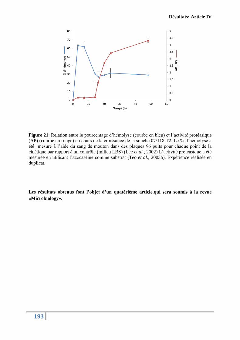
Résultats: Article IV
193
Figure 21: Relation entre le pourcentage d’hémolyse (courbe en bleu) et l’activité protéasique
(AP) (courbe en rouge) au cours de la croissance de la souche 07/118 T2. Le % d’hémolyse a
été mesuré à l’aide du sang de mouton dans des plaques 96 puits pour chaque point de la
cinétique par rapport à un contrôle (milieu LBS) (Lee et al., 2002) L’activité protéasique a été
mesurée en utilisant l’azocaséine comme substrat (Teo et al., 2003b). Expérience réalisée en
duplicat.
Les résultats obtenus font l’objet d’un quatérième article.qui sera soumis à la revue
«Microbiology».

194
Résultats: Article IV
195
Article IV: MOLECULAR CHARACTERIZATIONS OF THE
FRENCH V. TUBIASHII VIRULENCE FACTORS ON OYSTER’S
LARVAE GIVE EVIDENCES THAT METALLOPROTEASE IS NOT
THE ONLY MAIN TOXIC FACTOR
(Prepared for submission in the journal “Microbiology”)
Mersni-Achour Rachida1,2,5
, Ben Cheikh Yosra1, Pichereau Vianney
3, Doghri
Ibtissem
1,
Etien Cédric1, Dégremont Lionel
2, Saulnier Denis
2,4, Fruitier-Arnaudin Ingrid
1,5, Travers
Marie-Agnès2
1UMR 7266 CNRS-ULR, LIENSs, Equipe Approches Moléculaires, Environnement-Santé,
Université de La Rochelle, Avenue Michel Crépeau, 17042 La Rochelle, France.
2 Ifremer, SG2M-LGPMM, Laboratoire de Génétique et Pathologie des Mollusques Marins
Avenue de Mus de Loup, 17390 La Tremblade, France.
3Laboratoire des Sciences de l'Environnement Marin LEMAR, Institut Universitaire Europeen
de la Mer, Université Européenne de Bretagne, Université de Brest, UMR 6539
CNRS/UBO/IRD/Ifremer, 29280 Plouzané, France.
4 Ifremer, Centre Ifremer du Pacifique, UMR 241 Ecosystèmes Insulaires Océaniens, Tahiti,
98719 Taravao, French Polynesia.
5Fédération de Recherche en Environnement et Développement Durable, FR CNRS 3097,
Université de La Rochelle, France
Résultats: Article IV
196
Abstract
Vibrio tubiashii is a reemerging pathogen isolated from larval and juvenile bivalve
molluscs causing bacillary necrosis. Recent studies demonstrated the isolations of this species
in a French experimental hatchery/nursery affecting Crassostrea gigas spat in 2007. Here,
using larvae of C. gigas as an interaction model, we showed that the French V. tubiashii is
virulent to larvae and can cause bacillary necrosis symptoms with a LD50 about 2.3 × 10³
cfu
ml-1
after 24 h. Moreover, complete or GP- HPLC fractionated extracellular products (ECPs)
of this strain appeared toxic to larvae. MS-MS analysis of the different ECPs fractions
revealed the existence of an extracellular metalloprotease and other suspected virulence
factors. This observation is also supported by the expression level of some potential virulence
factors.
The overall results suggest that the pathology caused by the French V. tubiashii in C.
gigas oysters is caused by a group of toxic factors
Keywords
Crassostrea gigas; larvae; pathogenicity; toxins; mass spectrometry
Résultats: Article IV
197
Introduction
Marine Vibrio species are considered as the most serious problem affecting hatchery-
reared oyster larvae (Tubiash et al., 1970; Sugumar et al., 1998; Hada et al., 1984; Estes et
al., 2004; Elston et al., 2008). Among these species, V. tubiashii is one of the main causative
agents of larval and juvenile bivalve mollusc mortalities in hatcheries. Originally found on
the east coast of America isolated from diseased hard-shell clams (Teo et al., 2003; Miyoshi
et al., 2002; Sousa et al., 2007; Hasegawa et al., 2009; Labreuche et al., 2010), in England
and Spain isolated from Pacific and flat oysters (Jeffries, 1982; Lodeiros et al., 1987), V.
tubiashii has been detected in recent decades in North of America infecting new hosts, the
Pacific and Kumamoto oysters and Geoduck clams (Elston et al., 2008) and very recently in
France in diseased Pacific oysters and abalone (Travers et al., 2014). Strains of this species
are known to cause bacillary necrosis in mollusc larvae. This disease is clinically
characterized by a reduction in larval motility and an increase in soft-tissue necrosis
(Tubiash et al., 1970). Considering the potential impact of this bacterium, complete
genomes were recently sequenced (V. tubiashii ATCC 19106, ATCC 19109T) (Temperton et
al., 2011) and will obviously allow the exploration of potentially encoded virulence factors.
It has been well described that marine Vibrio species produce a variety of
extracellular products (ECPs) as potential pathogenic factors during infection steps (Shinoda
& Miyoshi, 2011). With regards to the American V. tubiashii, Nottage et al. (1989) were the
first to describe toxins produced by this strain; a proteinase and a ciliostatic toxin.
Subsequently, Kothary et al. (2001) showed that V. tubiashii could produce a cytolysin with
cytotoxic and cytolytic properties and Delston et al. (2003) demonstrated that this species
secretes a metalloprotease that shows a highly similar to others produced by pathogenic
marine vibrios. Finally, Hasegawa et al. (2008, 2009) confirmed that the V. tubiashii
metalloprotease acts as one of the critical factors explaining the toxicity of its extracellular
products to Pacific oyster larvae.
The concept of sequential expression of virulence factors depending on bacterial
environment was shown for V. cholerae toxin-coregulated pilus (a colonization factor) and
choleric toxin (CT) (Krukonis & DiRita, 2003). In V. tubiashii, Hasegawa et al. (2009b)
observed a sequential secretion of haemolytic and metalloproteasic activities in bacterial
supernatant during growth kinetics, probably explained by the degradation of hemolysin by
the metalloprotease(s) expressed in the late exponential phase.
Résultats: Article IV
198
The occurrence of the sequential expression of virulence factors clearly shows that
virulence is most often multifactorial and that bacterial pathogenicity is the result of the
association of several virulence determinants which differ according to the pathogen, the
host, and the type of infection (Jacques, 2004; Valiente et al., 2008; Paranjpye et al., 2013).
In a previous study, Travers et al. (2014) reported the isolation of a group of strains,
close to the American V. tubiashii, found in an experimental hatchery/nursery of C. gigas
spat during mortality events in France in 2007. These isolates and their ECPs showed a
significant virulence to juvenile Pacific oysters in experimental challenges. Moreover,
Mersni et al. (under review) demonstrated that the extracellular products of 07/118 T2, a
representative strain of the group, inhibited the adhesion capacity and phagocytosis activity
of C. gigas hemocytes. Biochemical analyses showed that the proteolytic fraction of ECPs
majority contain active and thermostable extracellular zinc metalloprotease(s).
This present study aims at (i) reproducing the pathogenicity of the French V. tubiashii and
the toxicity of its ECPs on Pacific oyster larvae, and (ii) characterizing some potential
virulence factors supporting the pathogenicity of the French V. tubiashii 07/118 T2.
Therefore, experimental infections coupled with microscopic observations were performed,
and the expression of some potential virulence factors during infection was monitored.
Finally, the fractionation of ECPs and proteomic analysis of the purified material were
conducted to access the molecular diversity of this bacterial secretome.
Résultats: Article IV
199
Material and methods
1. Bacteria growth conditions
V. tubiashii 07/118 T2 (LMG 27884 = CECT 8426) (Travers et al., 2014), V.
coralliilyticus 06/210 (Genard et al., 2013) and V. nigripulchritudo LMG3896T (Le Roux et
al., 2011) were used in this study. Bacteria were grown in Luria-Bertani medium (Difco)
supplemented with 1% sodium chloride (LBS) and stock culture was stored at -80°C in LBS
containing glycerol 15% (v/v).
2. Larval production
Larval production was achieved according to the protocol described by Dégremont et
al. (2005) at the Ifremer hatchery in La Tremblade. Briefly, fifteen C. gigas oysters were
randomly sampled from a wild population of the Marennes-Oléron Bay. Oysters were
opened, and a sample of the gonad was analyzed under microscope in order to separate the
oysters by sex. Eggs were collected by stripping of the gonad, as well as the sperm, and after
cleaning the gametes by appropriate sieving, mating was carried out. Larvae were reared in
30-liter tanks at 26 °C in filtered sea water, which was changed 3 times per week. The larvae
were fed daily with a mixed diet of cultured phytoplankton (Isochrysis galbana,
Chaetoceros calcitrans and Skeletonema costatum). When larvae were retained on a 150 µm
screen, they were transferred into the laboratory and then tested with different Vibrio strains.
At this step, larvae were 13 days old and their shell length were between 200 to 300 µm.
3. Preparation of V. tubiashii ECPs, fractionation and biochemical assays
Extracellular products were produced using the cellophane overlay method as
described previously by Travers et al. (2014). Briefly, bacteria were grown in 5 ml of LBS
at 22 °C for 18 h. A volume of 1 ml of exponential phase culture (OD 600 =1) was spread on a
sterile cellophane film overlying LBSA (LBS Agar) plates. After 48 h of incubation at 22
°C, the cells were washed off the cellophane using 10 ml of cold sterile artificial sea water
(SASW) and removed by centrifugation at 3,000 × g (45 min at 4°C). The supernatant was
filtered at 0.22 µm and then concentrated by lyophilisation, resuspended in Tris-HCl buffer
(50 mM Tris-HCl, pH 8.0) and dialyzed against distilled water for 12 h and Tris-HCl buffer
for 12 h. Finally, crude ECPs samples were stored at -80°C until use.

Résultats: Article IV
200
Crude ECPs were purified using a gel permeation-high performance liquid
chromatography (GP-HPLC). This purification step was performed using a Biosuite TM 250
column (5 μm, 300 × 7.8 mm, Waters) and conducted with a Waters system (600 Controller,
2996 Photodiode Array detector and 2707 Autosampler). 10 µg per run of crude ECPs
fractions were eluted with 50 mM Tris-HCl buffer, pH 8, at a rate of 1 ml min-1
and
collected according to the chromatographic profile obtained at 215 nm. Each eluted fraction
obtained was concentrated by lyophilisation, resuspended in Tris-HCl buffer, assayed from
protein concentration and stored at -20°C until further utilization. The column was calibrated
using “Gel Filtration Markers Kit for Protein Molecular Weights 29, 000-700, 000 Da”
(Sigma-Aldrich) following the same analysis conditions applied to the samples.
Proteins were quantified using the method described by Bradford (Bradford, 1976)
using Bradford reagent (Sigma-Aldrich) and bovine serum albumin as the standard protein.
Azocaseinase activity was determined as previously described (Travers et al., 2014).
4. In vivo larval experiments
Cultures of early stationary phase (18 h) Vibrio strains grown in LBS at 22°C were
centrifuged at 3,000 × g for 20 min, washed twice and resuspended in sterile sea water
(SSW). In a 6-well flat-bottomed plate (Falcon), 1.5 ml of ten-fold dilutions of the cell
suspensions between 10 and 106
cfu ml-1
were added to 1.5 ml of the larval suspension at a
density of about 50 larvae per well and incubated at 22 °C with a gentle stirring. V.
nigripulchritudo was used as a non virulent control strain and V. coralliilyticus as a virulent
one for C. gigas larvae. Boiled bacteria (15 min at 100 °C), some incubated with EDTA
(Ethylenediaminetetraacetic acid) (at a final concentration, f.c. = 5 mM), and others not
incubated with EDTA, were also tested. Further, the toxicity of different crude ECP
concentrations (f.c. 10 µg, 5 µg and 2.5 µg of protein per ml) and GP-HPLC fractions of
ECPs (f.c. 5 µg of proteins per ml) were tested on model larvae in the presence and absence
of EDTA (preincubated with EDTA 5 mM, f.c., for 30 min).
The counting of the live larvae was carried out by visualization using a binocular
microscope and a Sedgewick Rafter cell. Oyster larvae were considered dead based on
microscopic examination and phenotypic observations described by Hasegawa et al. (2008).
The percentage of live larvae was calculated with reference to live larvae incubated in SSW
for the live-bacteria experiments, and with reference to the live larvae incubated with Tris-
Résultats: Article IV
201
HCl buffer for ECPs and GP-HPLC fractions. Larval survival was recorded after 24 h, three
biological replicates were performed for each assay and assays were replicated
independently.
Following the same protocol described above, a kinetic was performed by bathing 25
ml of larvae (about 50 larvae per ml) with 25 ml of bacteria (105 cfu ml
-1) at different times
(30 min, 3 h, 6 h, 16 h, and 18 h) in aerated bottles with gentle stirring. Azocaseine activity
in the bath was performed as previously described (Travers et al., 2014) for each kinetic
point. At the end of each kinetic point, a centrifugation at 3,000 × g for 15 min was carried
out and the pellet (larvae + bacteria) was conserved with RNAprotect Bacteria Reagent
(Qiagen, UK) to stabilize/fix the bacterial RNA for QPCR assays. Pellets were stored at -
80°C until use. Three biological replicates were performed for each assay and assays were
replicated.
5. Microscopic observations
For microscopic observations, larvae from each well were filtered and fixed in a 1 ml
glutaraldehyde (0.25%) - paraformaldehyde (4%) fixation solution (Sigma-Aldrich)
overnight. Pictures were taken and treated using a BX51 System Microscope (Olympus®).
6. RNA extraction and RT cDNA synthesis
Total RNA was extracted from each bacterial pellet using TRIzol® Reagent
(Invitrogen), at a ratio of 1 ml Trizol per pellet according to the manufacturer's protocol.
Bacterial RNA was separated using chloroform (0.2:1 chloroform:Trizol, v/v) and
precipitated using cold isopropanol (0.5:1 isopropanol:Trizol). RNA pellets were washed
with 1 ml of 75% ethanol and resuspended in ultrapure water. 17 µl RNA suspensions were
treated with 2 µl of DNase (2U for 10 µg of RNA) (Ambion) for 45 min at 37°C. Total RNA
concentration was analyzed by spectrophotometry (Nanodrop/Thermo Scientific) and a
negative reverse transcription (NO-RT) QPCR with diluted RNA was performed to check
RNA purity.
The RT-PCR was carried out following the protocol of SuperScript™ III Reverse
Transcriptase (Invitrogen). For the first-strand cDNA Synthesis, in a total volume of 13µl,
800 ng of total RNA was well mixed with 200 ng of random primers, 1 µl of dNTP (10 mM)
and nuclease free water. The mix was incubated at 60 °C for 5 min and then chilled on ice. 1
Résultats: Article IV
202
μl of SuperScript III RT (200 U µl -1
) was added to 1 µl of RNase OUT (40 U µl-1
), 1 µl of
DTT and 4 µL of 5X buffer. The reaction mix was incubated for 5 min at 25 C, followed by
an activation step of 60 min at 50 °C and ending with a deactivation step of 15 min at 70 °C.
cDNA was conserved at -20°C until use.
7. Quantitative polymerase chain reaction (QPCR)
QPCR analysis was conducted on an MX3000 and MX3005 Thermocycler
(Agilent) using Brilliant III Ultra-Fast SYBR Green QPCR Master Mix (Stratagene). Each
reaction was run in triplicate with a final volume of 20 µl containing 5 μl cDNA (1/30
dilution) and 200 nM of each primer.
Reactions were initiated with an initial denaturation for 3 min at 95 °C followed by
40 cycles at 95 °C for 10 s and at 60°C for 20 s. Each run included blank controls (water) for
each primer and fluorescence intensity was expressed in delta reporters (dR) after the
background threshold was subtracted. The threshold was set using an amplification based
algorithm from MX 3000-3005 software (Stratagene) for the initial plate.
For the QPCR efficiencies of each primer pair used, standard curves were generated using
eight serial dilutions (2.10-1
to 2.10-8
) of linearized plasmid (pCR2.1-TOPO, Invitrogen)
containing an insert of gyrase, metalloprotease, aerobactin and thermolabile hemolysin gene
obtained by PCR (Table 1) and the efficiency of exochitinase primer was generated directly
with genomic 07/118 T2 DNA . The level of expression of the target genes at different
kinetic times, normalized to the gyrB housekeeping gene, was then calculated using the
(1+efficiency) −ΔΔCt
formula (Pfaffl, 2001) with reference to the point of infection at 30 min.
Used primers were designed as described previously (Article III; Achour et al. unpublished).
8. 2-Dimensional electrophoresis (2-DE) analyses and mass spectrometric analysis
Before isoelectric focusing, a volume corresponding to 50 µg of protein for each
ECP fraction was prepared and concentrated for 2-DE analysis using the Ready Prep 2-D
Cleanup kit (Biorad). The final pellet was suspended in 250 μl of Destreak Rehydration (GE
Healthcare) containing 1% IPG buffer pH 3–10 (GE Healthcare) for protein solubilization.
Strips were hydrated with each ECPs fraction for 15 h prior to IEF.
2-DE was carried out using the protocol described by Galland et al. (2013). Briefly,
the first dimension was performed on linear pH 3–10 gradient IPG strips (13 cm, GE

Résultats: Article IV
203
Healthcare) in an Ettan IPGphor III isoelectric focusing system (GE Healthcare), using the
following protocol: step 1: 15 min at 250 V, step 2: 2 h at 500 V, step 3: 1 h gradient to 1000
V, step 4: 2.5 h gradient to 8, 000 V, and finally step 5: 1.5h at 8, 000 V.
Before the second dimension, strips were equilibrated for 15 min in an equilibration
solution (2% SDS, 6 M Urea, 30% glycerol (v/v), 50 mM Tris–HCl pH 8.8 and BBP)
containing 1% of DTT, followed by a 15-min incubation in the same solution containing 260
mM of iodoacetamide. The strips were then transferred onto vertical 10–15% gradient SDS-
polyacrylamide gels (12.5 cm × 12. 5 cm) and the separation was performed at 50 mA per
gel. Gels were stained with coomassie blue solution (overnight) and destained in a
methanol/acetic acid/water (30/7/63: v/v/v) solution. Gels were scanned with SFLauncher
software and compared using the Prodigy SameSpot software (Nonlinear Dynamics).
Selected spots were manually excised from the gels and mass spectrometric analysis
was performed by the "Structural and Functional Mass Spectrometry Facility” in the
JACQUES MONOD Institute (CNRS and Paris-Diderot University). After trypsin digestion,
the spots were analyzed by Matrix-Assisted Laser Desorption Ionization Time-Of-Flight
tandem mass spectrometry (MALDI TOF–TOF). The system and peptide mass fingerprint
obtained was examined using MASCOT (Matrix Science Ltd, London, UK). MS-MS data
were compared against both the NCBInr and V. tubiashii genome databases.
9. Statistical analysis
Significant differences between analyses were tested using the "Student t" test in
http://marne.u707.jussieu.fr/biostatgv/. Results were considered significant at P < 0.05.
Résultats: Article IV
204
Results
1. Virulence of the French V. tubiashii 07/118 T2 on oyster larvae
The virulence of the 07/118 T2 strain on 13-day old oyster larvae was estimated 24 h
post infection (Fig. 1). V. tubiashii 07/118 T2 caused important dose-dependent mortality: no
live larvae were observed at 106
cfu ml-1
and the LD50 (lethal dose 50%) values were
approximately 2.3 × 10³
cfu ml-1
(Fig. 1a). Moreover, 100% mortality was noticed with heat-
killed V. tubiashii 07/118 T2 at 106 cfu ml
-1 with live bacteria preincubated with EDTA (ion
metal chelator). However, no mortality was observed with the boiled 07/118 T2 strain,
preincubated with EDTA, at 106 cfu ml
-1. In the positive controls (Fig. 1b), V. coralliilyticus
caused significant mortality even at 103 cfu ml
-1 (only 6% live larvae) compared with 07/118
T2. With the negative control, V. nigripulchritudo, no considerable mortality was detected
even at 106 cfu ml
-1.
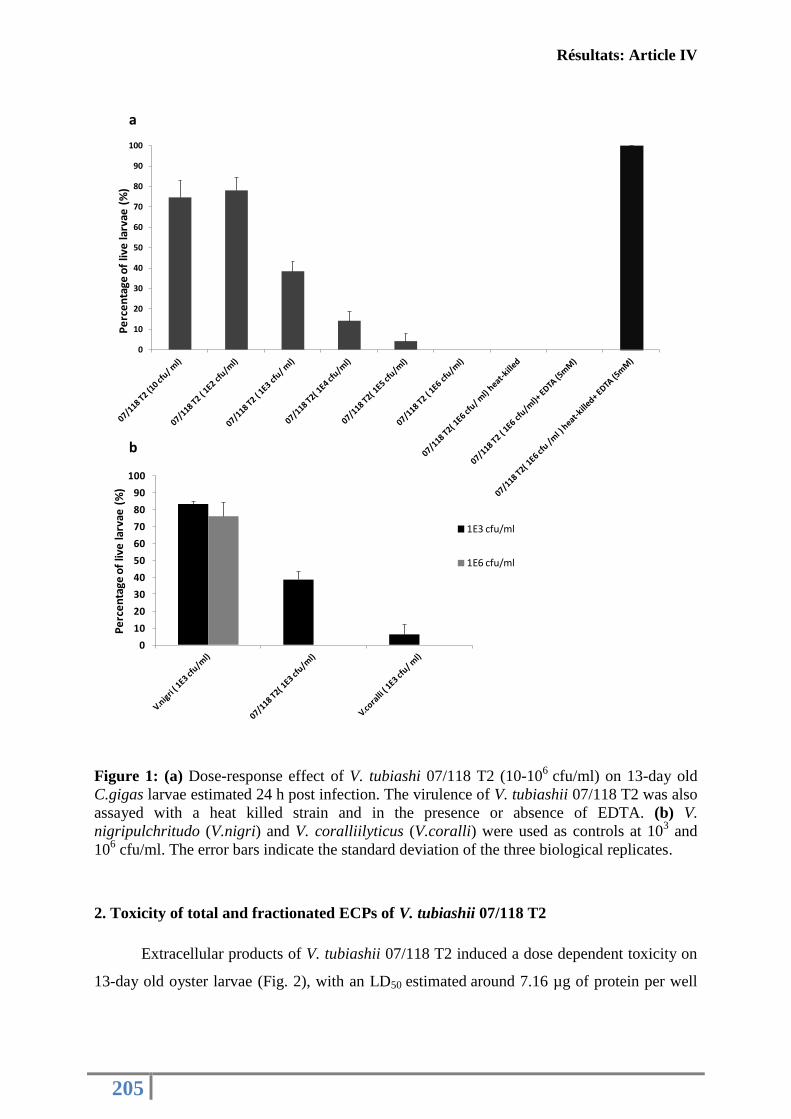
Résultats: Article IV
205
Figure 1: (a) Dose-response effect of V. tubiashi 07/118 T2 (10-106
cfu/ml) on 13-day old
C.gigas larvae estimated 24 h post infection. The virulence of V. tubiashii 07/118 T2 was also
assayed with a heat killed strain and in the presence or absence of EDTA. (b) V.
nigripulchritudo (V.nigri) and V. coralliilyticus (V.coralli) were used as controls at 103 and
106 cfu/ml. The error bars indicate the standard deviation of the three biological replicates.
2. Toxicity of total and fractionated ECPs of V. tubiashii 07/118 T2
Extracellular products of V. tubiashii 07/118 T2 induced a dose dependent toxicity on
13-day old oyster larvae (Fig. 2), with an LD50 estimated around 7.16 µg of protein per well
0
10
20
30
40
50
60
70
80
90
100
Pe
rce
nta
ge o
f liv
e la
rvae
(%
)
b
0
10
20
30
40
50
60
70
80
90
100
Pe
rce
nta
geo
f liv
e la
rvae
(%)
1E3 cfu/ml
1E6 cfu/ml
a
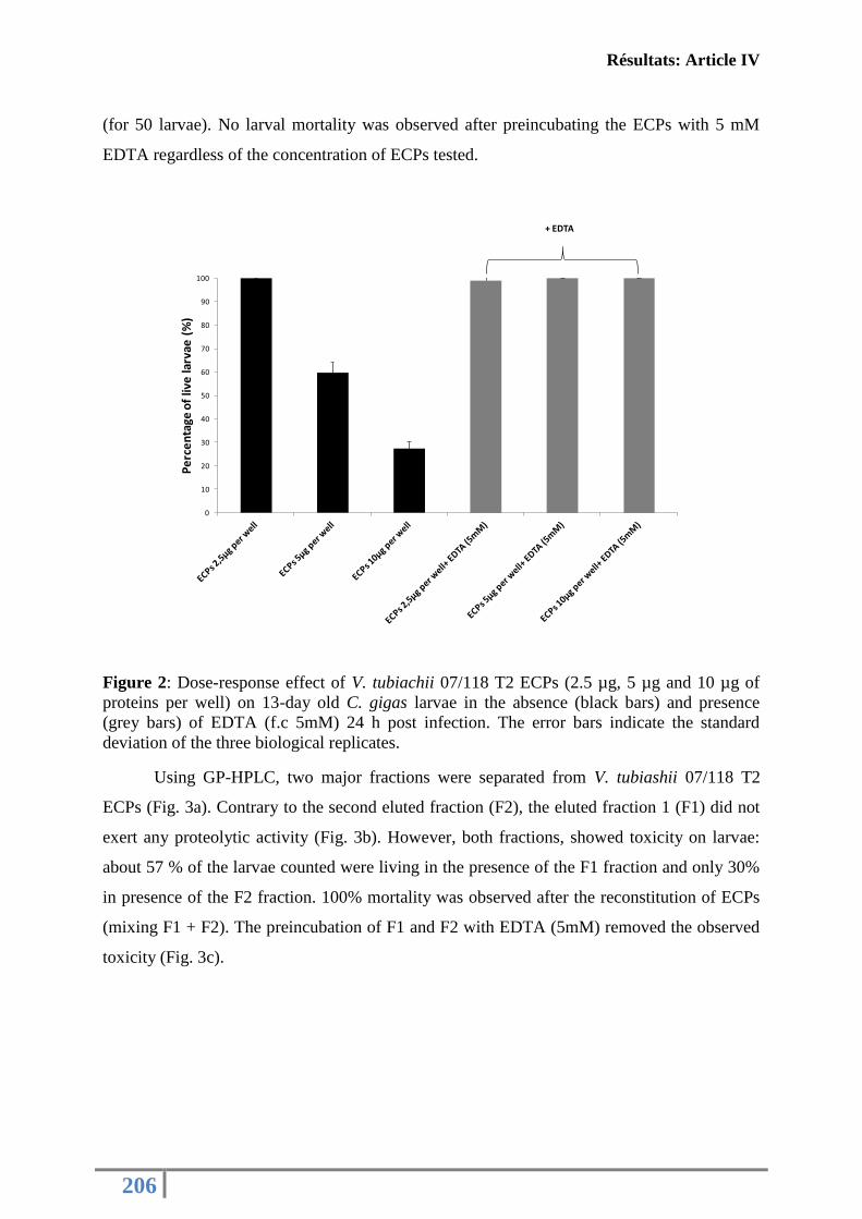
Résultats: Article IV
206
(for 50 larvae). No larval mortality was observed after preincubating the ECPs with 5 mM
EDTA regardless of the concentration of ECPs tested.
Figure 2: Dose-response effect of V. tubiachii 07/118 T2 ECPs (2.5 µg, 5 µg and 10 µg of
proteins per well) on 13-day old C. gigas larvae in the absence (black bars) and presence
(grey bars) of EDTA (f.c 5mM) 24 h post infection. The error bars indicate the standard
deviation of the three biological replicates.
Using GP-HPLC, two major fractions were separated from V. tubiashii 07/118 T2
ECPs (Fig. 3a). Contrary to the second eluted fraction (F2), the eluted fraction 1 (F1) did not
exert any proteolytic activity (Fig. 3b). However, both fractions, showed toxicity on larvae:
about 57 % of the larvae counted were living in the presence of the F1 fraction and only 30%
in presence of the F2 fraction. 100% mortality was observed after the reconstitution of ECPs
(mixing F1 + F2). The preincubation of F1 and F2 with EDTA (5mM) removed the observed
toxicity (Fig. 3c).
0
10
20
30
40
50
60
70
80
90
100
Pe
rce
nta
ge o
f liv
e la
rvae
(%
)
+ EDTA
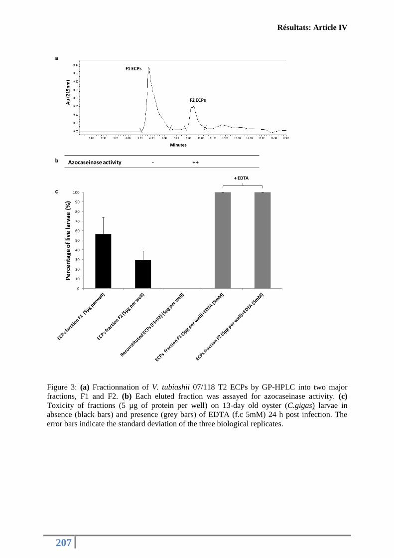
Résultats: Article IV
207
Figure 3: (a) Fractionnation of V. tubiashii 07/118 T2 ECPs by GP-HPLC into two major
fractions, F1 and F2. (b) Each eluted fraction was assayed for azocaseinase activity. (c)
Toxicity of fractions (5 µg of protein per well) on 13-day old oyster (C.gigas) larvae in
absence (black bars) and presence (grey bars) of EDTA (f.c 5mM) 24 h post infection. The
error bars indicate the standard deviation of the three biological replicates.
0
10
20
30
40
50
60
70
80
90
100
Pe
rce
nta
ge o
f liv
e la
rvae
(%
)
Azocaseinase activity - ++
Au
(21
5n
m)
Minutes
F1 ECPs
F2 ECPs
a
b
+ EDTA
c
Résultats: Article IV
208
3. 2D and MS-MS analyses of GP-HPLC fractions
The two fractions obtained from crude V. tubiashii 07/ 118 T2 ECPs were separated
on large 2-DE gels in a pI range of 3-10 (Fig. 4). More proteins were detected in the first
fraction of ECPs (F1) than in the second one (F2). For both, most of the proteins detected
were localised in the acidic part of the pH range. For F1, visual inspection revealed different
proteins which differed in their molecular weights. However, for F2 the detected proteins
were in single horizontal line which differed only by their isoelectric point (Fig. 4).
All the visualised proteins, ie 19 spots for F1 and 12 spots for F2, were analysed by using
mass spectrometry. Only 12 proteins for F1 and 5 proteins for F2 were well identified using
MS/MS analyses (Fig. 4, Table 2). F1 Fraction ECPs showed a diversity of outer membrane
proteins (porin-like protein H precursor, outer membrane channel protein, long chain fatty
acid transport protein, Outer membrane protein N and hypothetical protein). By contrast, the
second fraction of ECPs (F2) appeared as an enrichment of a unique protein “extracellular
zinc metalloprotease” (accession no. EGU55378) with potentially different isoforms.
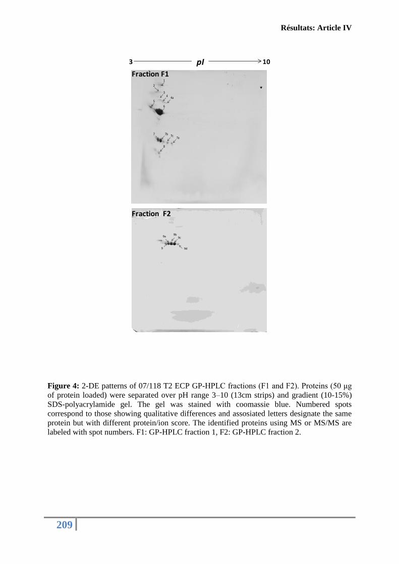
Résultats: Article IV
209
Figure 4: 2-DE patterns of 07/118 T2 ECP GP-HPLC fractions (F1 and F2). Proteins (50 μg
of protein loaded) were separated over pH range 3–10 (13cm strips) and gradient (10-15%)
SDS-polyacrylamide gel. The gel was stained with coomassie blue. Numbered spots
correspond to those showing qualitative differences and assosiated letters designate the same
protein but with different protein/ion score. The identified proteins using MS or MS/MS are
labeled with spot numbers. F1: GP-HPLC fraction 1, F2: GP-HPLC fraction 2.
9
9a9b
9c
9d
1
2
34 4a
5
6
7 7b 7c7a
8
3 10pI
Fraction F1
Fraction F2
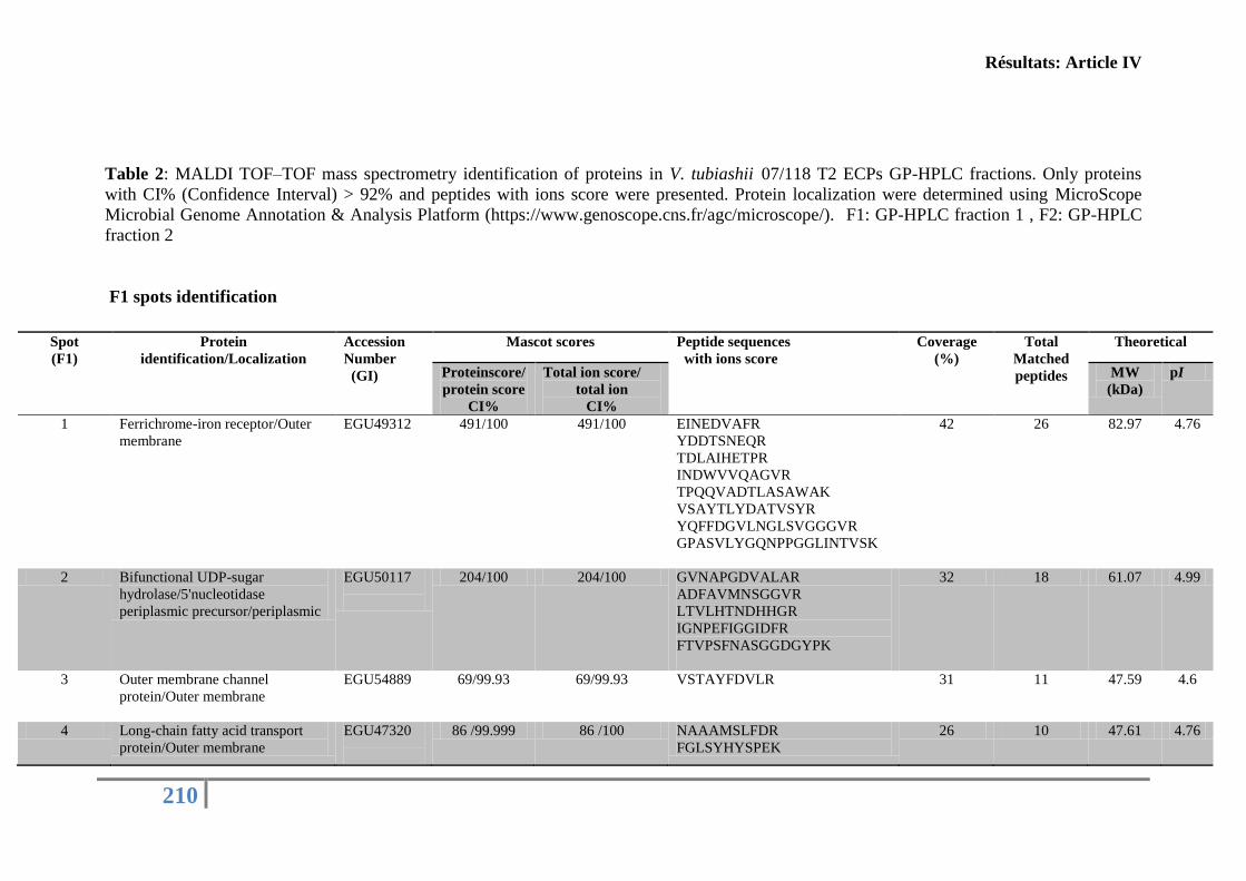
Résultats: Article IV
210
Table 2: MALDI TOF–TOF mass spectrometry identification of proteins in V. tubiashii 07/118 T2 ECPs GP-HPLC fractions. Only proteins
with CI% (Confidence Interval) > 92% and peptides with ions score were presented. Protein localization were determined using MicroScope
Microbial Genome Annotation & Analysis Platform (https://www.genoscope.cns.fr/agc/microscope/). F1: GP-HPLC fraction 1 , F2: GP-HPLC
fraction 2
F1 spots identification
Spot
(F1)
Protein
identification/Localization
Accession
Number
(GI)
Mascot scores Peptide sequences
with ions score
Coverage
(%)
Total
Matched
peptides
Theoretical
Proteinscore/
protein score
CI%
Total ion score/
total ion
CI%
MW
(kDa)
pI
1 Ferrichrome-iron receptor/Outer
membrane
EGU49312
491/100 491/100 EINEDVAFR
YDDTSNEQR
TDLAIHETPR
INDWVVQAGVR
TPQQVADTLASAWAK
VSAYTLYDATVSYR
YQFFDGVLNGLSVGGGVR
GPASVLYGQNPPGGLINTVSK
42 26 82.97 4.76
2 Bifunctional UDP-sugar
hydrolase/5'nucleotidase
periplasmic precursor/periplasmic
EGU50117
204/100 204/100 GVNAPGDVALAR
ADFAVMNSGGVR
LTVLHTNDHHGR
IGNPEFIGGIDFR
FTVPSFNASGGDGYPK
32 18 61.07 4.99
3 Outer membrane channel
protein/Outer membrane
EGU54889
69/99.93 69/99.93 VSTAYFDVLR 31 11 47.59 4.6
4 Long-chain fatty acid transport
protein/Outer membrane
EGU47320
86 /99.999 86 /100 NAAAMSLFDR
FGLSYHYSPEK
26 10 47.61 4.76
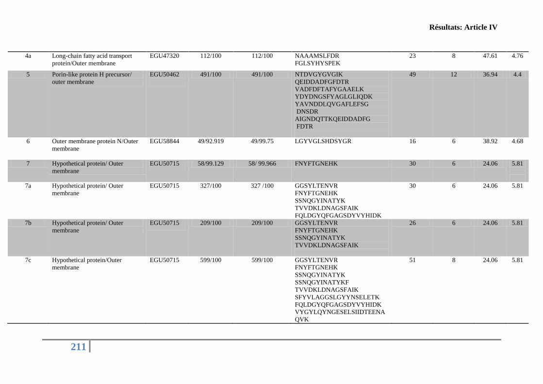
Résultats: Article IV
211
4a Long-chain fatty acid transport
protein/Outer membrane
EGU47320
112/100 112/100 NAAAMSLFDR
FGLSYHYSPEK
23 8 47.61 4.76
5 Porin-like protein H precursor/
outer membrane
EGU50462
491/100 491/100 NTDVGYGVGIK
QEIDDADFGFDTR
VADFDFTAFYGAAELK
YDYDNGSFYAGLGLIQDK
YAVNDDLQVGAFLEFSG
DNSDR
AIGNDQTTKQEIDDADFG
FDTR
49 12 36.94 4.4
6 Outer membrane protein N/Outer
membrane
EGU58844
49/92.919 49/99.75 LGYVGLSHDSYGR 16 6 38.92 4.68
7 Hypothetical protein/ Outer
membrane
EGU50715 58/99.129 58/ 99.966 FNYFTGNEHK 30 6 24.06 5.81
7a Hypothetical protein/ Outer
membrane
EGU50715 327/100 327 /100 GGSYLTENVR
FNYFTGNEHK
SSNQGYINATYK
TVVDKLDNAGSFAIK
FQLDGYQFGAGSDYVYHIDK
30 6 24.06 5.81
7b Hypothetical protein/ Outer
membrane
EGU50715 209/100 209/100 GGSYLTENVR
FNYFTGNEHK
SSNQGYINATYK
TVVDKLDNAGSFAIK
26 6 24.06 5.81
7c Hypothetical protein/Outer
membrane
EGU50715 599/100 599/100 GGSYLTENVR
FNYFTGNEHK
SSNQGYINATYK
SSNQGYINATYKF
TVVDKLDNAGSFAIK
SFYVLAGGSLGYYNSELETK
FQLDGYQFGAGSDYVYHIDK
VYGYLQYNGESELSIIDTEENA
QVK
51 8 24.06 5.81
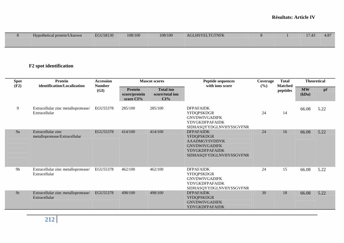
Résultats: Article IV
212
8 Hypothetical protein/Uknown EGU58130
108/100 108/100 AGLHSYELTGTNFK 8 1 17.43 4.87
F2 spot identification
Spot
(F2)
Protein
identification/Localization
Accession
Number
(GI)
Mascot scores Peptide sequences
with ions score
Coverage
(%)
Total
Matched
peptides
Theoretical
Protein
score/protein
score CI%
Total ion
score/total ion
CI%
MW
(kDa)
pI
9
Extracellular zinc metalloprotease/
Extracellular
EGU55378
285/100
285/100
DFPAFAIDK
YFDQPSKDGR
GNVDWIVGADIFK
YDYGKDFPAFAIDK
SIDHASQYYDGLNVHYSSGVFNR
24
14
66.08
5.22
9a
Extracellular zinc
metalloprotease/Extracellular
EGU55378
414/100 414/100 DFPAFAIDK
YFDQPSKDGR
AAADMGYSVDDVK
GNVDWIVGADIFK
YDYGKDFPAFAIDK
SIDHASQYYDGLNVHYSSGVFNR
24 16 66.08
5.22
9b Extracellular zinc metalloprotease/
Extracellular
EGU55378
462/100 462/100 DFPAFAIDK
YFDQPSKDGR
GNVDWIVGADIFK
YDYGKDFPAFAIDK
SIDHASQYYDGLNVHYSSGVFNR
24 15 66.08
5.22
9c Extracellular zinc metalloprotease/
Extracellular
EGU55378
498/100 498/100 DFPAFAIDK
YFDQPSKDGR
GNVDWIVGADIFK
YDYGKDFPAFAIDK
30 18 66.08
5.22
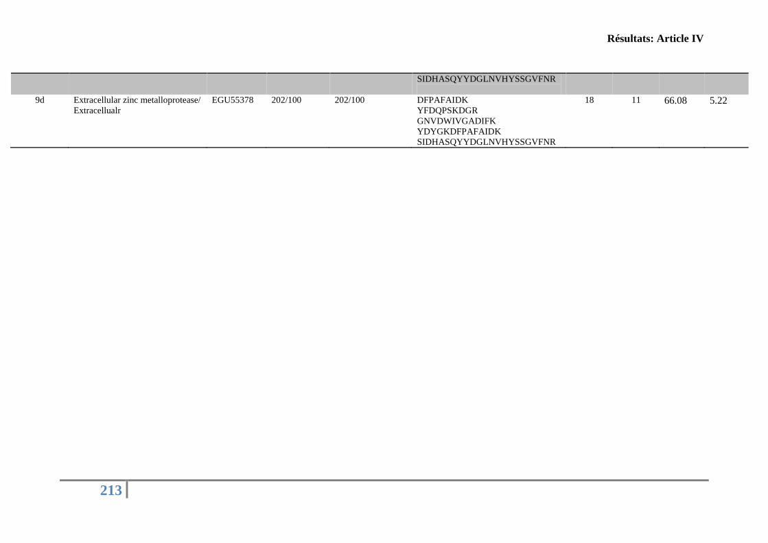
Résultats: Article IV
213
SIDHASQYYDGLNVHYSSGVFNR
9d Extracellular zinc metalloprotease/
Extracellualr
EGU55378
202/100 202/100 DFPAFAIDK
YFDQPSKDGR
GNVDWIVGADIFK
YDYGKDFPAFAIDK
SIDHASQYYDGLNVHYSSGVFNR
18 11 66.08
5.22
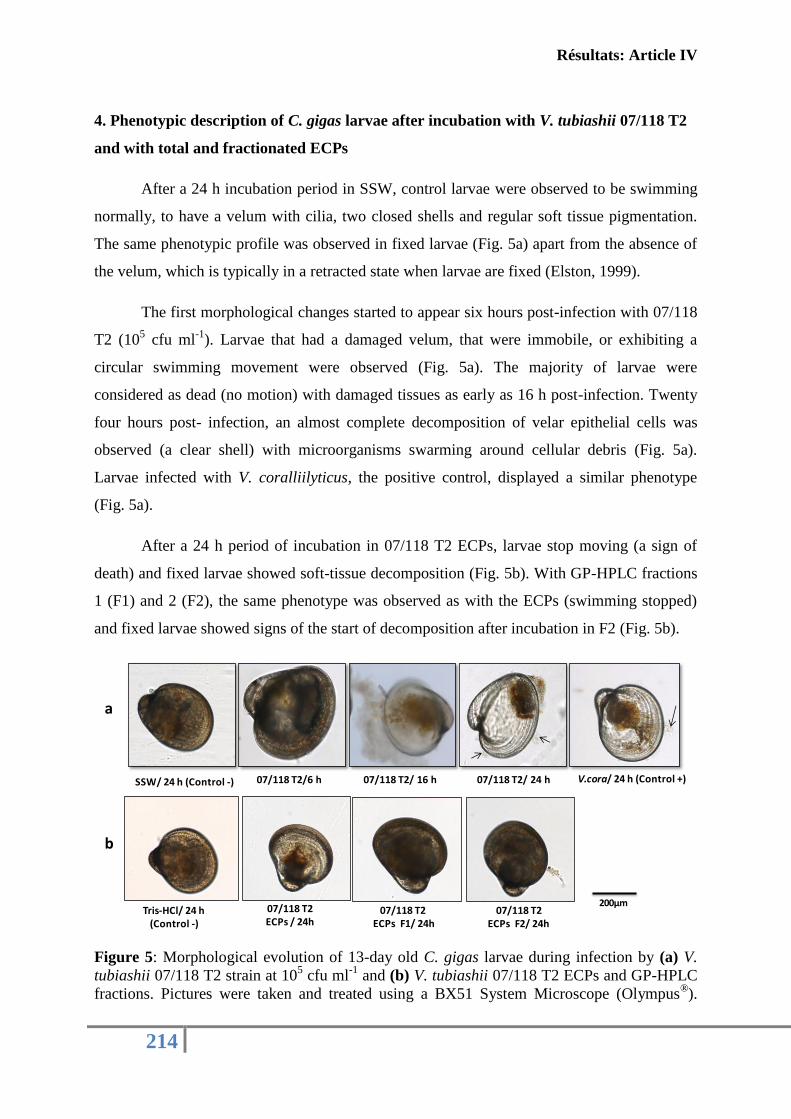
Résultats: Article IV
214
4. Phenotypic description of C. gigas larvae after incubation with V. tubiashii 07/118 T2
and with total and fractionated ECPs
After a 24 h incubation period in SSW, control larvae were observed to be swimming
normally, to have a velum with cilia, two closed shells and regular soft tissue pigmentation.
The same phenotypic profile was observed in fixed larvae (Fig. 5a) apart from the absence of
the velum, which is typically in a retracted state when larvae are fixed (Elston, 1999).
The first morphological changes started to appear six hours post-infection with 07/118
T2 (105 cfu ml
-1). Larvae that had a damaged velum, that were immobile, or exhibiting a
circular swimming movement were observed (Fig. 5a). The majority of larvae were
considered as dead (no motion) with damaged tissues as early as 16 h post-infection. Twenty
four hours post- infection, an almost complete decomposition of velar epithelial cells was
observed (a clear shell) with microorganisms swarming around cellular debris (Fig. 5a).
Larvae infected with V. coralliilyticus, the positive control, displayed a similar phenotype
(Fig. 5a).
After a 24 h period of incubation in 07/118 T2 ECPs, larvae stop moving (a sign of
death) and fixed larvae showed soft-tissue decomposition (Fig. 5b). With GP-HPLC fractions
1 (F1) and 2 (F2), the same phenotype was observed as with the ECPs (swimming stopped)
and fixed larvae showed signs of the start of decomposition after incubation in F2 (Fig. 5b).
Figure 5: Morphological evolution of 13-day old C. gigas larvae during infection by (a) V.
tubiashii 07/118 T2 strain at 105 cfu ml
-1 and (b) V. tubiashii 07/118 T2 ECPs and GP-HPLC
fractions. Pictures were taken and treated using a BX51 System Microscope (Olympus®).
07/118 T2ECPs F1/ 24h
07/118 T2 ECPs F2/ 24h
07/118 T2 ECPs / 24h
07/118 T2/6 h 07/118 T2/ 24 h07/118 T2/ 16 h
200µm
SSW/ 24 h (Control -) V.cora/ 24 h (Control +)
Tris-HCl/ 24 h(Control -)
a
b
Résultats: Article IV
215
Arrows indicate swarming bacteria. SSW: sterile sea water, V. cora: V. coralliilyticus 06/210,
F1: GP-HPLC fraction 1 and F2: GP-HPLC fraction 2.
5. Expression of genes encoding potential virulence factor during infection with V.
tubiashii 07/118 T2
The relative expression of some potential virulence genes from V. tubiashii 07/118 T2
was carried out using RT-QPCR at different larval infection times (3 h, 6 h, 16 h and 18 h)
(Fig.6). The chosen genes corresponded to previously described factors implicated in bacterial
virulence: thermostable hemolysin (TLH) (Wong et al., 2012), metalloprotease (Mersni et al.
under review) and exochitinase (Wang et al., 2001).
Metalloprotease gene expression underwent a small down regulation during the first
few hours of infection (3 h) a significant (20-fold) increase in expression was observed 16 h
post infection (p < 0.05). This over expression remained stable even 18 h post infection (Fig.
6a). At the same post-infection time-points (16 h and 18 h), azocaseinase activity was
detected in the supernatant of larval + bacterial interaction (Fig. 6a’).
By contrast, TLH gene expression was significantly up-regulated during the first few
hours of larval infection (3 h and 6 h, p < 0.05) (Fig. 6b). Exochitinase gene expression
showed few variations: a little down-regulation 6 hours post-infection, followed by an up-
regulation (p< 0.05) (Fig. 6c).
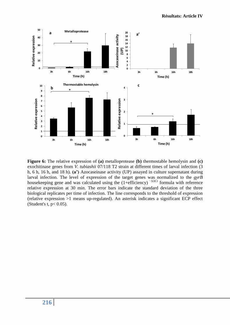
Résultats: Article IV
216
Figure 6: The relative expression of (a) metalloprotease (b) thermostable hemolysin and (c)
exochitinase genes from V. tubiashii 07/118 T2 strain at different times of larval infection (3
h, 6 h, 16 h, and 18 h). (a’) Azocaseinase activity (UP) assayed in culture supernatant during
larval infection. The level of expression of the target genes was normalized to the gyrB
housekeeping gene and was calculated using the (1+efficiency) −ΔΔCt
formula with reference
relative expression at 30 min. The error bars indicate the standard deviation of the three
biological replicates per time of infection. The line corresponds to the threshold of expression
(relative expression >1 means up-regulated). An asterisk indicates a significant ECP effect
(Student's t, p< 0.05).
0
1
2
3
4
3h 6h 16h 18h
Re
lati
ve e
xpre
ssio
n
Time (h)
0
1
2
3
4
5
6
7
8
9
10
3h 6h 16h 18h
Re
lati
ve e
xpre
ssio
n
Time (h)
Thermostable hemolysin
0
10
20
30
40
50
3h 6h 16h 18h
Re
lati
ve e
xpre
ssio
n
Time (h)
Metalloprotease
0
2
4
6
8
10
12
14
16
18
20
3h 6h 16h 18h
Azo
case
inas
eac
tivi
ty
(UP
)
Time (h)
a’a
bc
*
*
*
Résultats: Article IV
217
Discussion
It has previously been demonstrated that the French V. tubiashii 07/118 T2 and its
extracellular products cause pathogenesis to juvenile Pacific oyster (Travers et al., 2014). A
thermolysin-like metalloprotease was suggested to be one of the virulence factors responsible
for the toxicity of this strain (Mersni et al. under review).
In this paper, using experimental infection of larvae, we showed for the first time that
the French V. tubiashii 07/118 T2 is virulent to 13-day old larvae with an LD50 value of
approximately 2.3 × 10³
cfu ml-1
24 h post-infection. These results were comparable to those
found with the American V. tubiashii by Estes et al. (2004) who observed an LD50 between
1.6 and 3.6 × 10⁴ cfu ml
-1 24 h post infection in 10- to 12-day old larvae. Secondly, consistent
with previous studies (Takahashi et al., 2000; Hasegawa et al., 2008), the 07/118 T2 culture
supernatants showed a high toxicity to larvae. Furthermore, with the aim to explore the
diversity of toxic ECPs, an HPLC-gel permeation separation was conducted. Both collected
fractions showed a partial toxicity to oyster larvae with the greatest toxicity for F2, the only
fraction that demonstrated protease activity. It is interesting also to note that the reconstitution
of ECPs with the two GP-HPLC fractions caused total toxicity to the larvae although the loss
of some of the toxins may have occurred during the purification step. At this stage, it could be
supposed that several virulence factors, that would act synergistically, are necessary for the
full virulence phenotype induced by the French V. tubiashii.
During experimental infection, larval pathological manifestations were monitored. It
was reported that V. tubiashii caused bacillary necrosis, a larval and juvenile bivalve disease
characterized by a reduction in larval motility and the formation of bacterial swarms (Tubiash
et al., 1965). Histological sections showed an extensive destruction of larval epithelial cells
with a massive bacterial invasion and proliferation (Tubiash et al., 1965; Estes et al., 2004;
Elston et al., 2008). Herein, with the French V. tubiashii, approximately the same signs of
bacillary necrosis were observed. However, infection with 07/118 T2 ECPs showed a less
pronounced effect (without complete decomposition) and GP-HPLC fractions caused larval
mortality without reproducing the same phenotype observed with live bacteria or with crude
ECPs.
Results obtained, both by experimental infections and by microscope observations,
raise the question of whether proteases, specifically metalloproteases as previously reported
(Binesse et al., 2008; Hasegawa et al., 2008; Labreuche et al., 2010), are the sole primary
Résultats: Article IV
218
virulence factor or whether the association of several factors is required to induce a complete
infection phenotype. The toxicity of the GP-HPLC fraction 1, which doesn’t present protease
activity, is an important finding to support this second assumption.
It has been described that pathogenic Vibrio is capable of producing various pathogenic
factors. Of these, enterotoxins, hemolysin and cytotoxins are the direct toxic factors causing
the symptoms, whereas siderophores and adhesive factors are indirect factors involved in the
establishment of the infection and proteases are recognized as playing pathogenic roles in the
subsequent infection (Shinoda & Miyoshi, 2011).
In order to explore the involvement of several factors in the pathogenicity of the
French V. tubiashii, other characterization tests were applied using EDTA as a cation
chelating agent, to further characterize metalloprotease activity (Teo et al., 2003; Miyoshi et
al., 2002; Sousa et al., 2007; Hasegawa et al., 2009; Labreuche et al., 2010), and heat
treatment to kill the bacteria via the restriction of adhesion capacity, proliferation ability and
the denaturation of a group of thermolabile factors. Interestingly, neither the use of EDTA nor
the heat killed bacteria were sufficient to limit the toxicity of the French V. tubiashii.
Indeed, heat treatment could destroy the bacterial cell and make it non culturable but
couldn’t abolish all pathogenic effectors (Fontana, 1988; Kudryashova et al., 1998; Lin et al.,
2010; Shinoda & Miyoshi, 2011). Moreover, the addition of EDTA into the infection bath
could partially affect protease activity (Mersni et al.,under review; Labreuche et al., 2010;
Teo et al., 2003) and some bacterial properties (Kavitha et al., 2013) but not all bacterial
virulence factors. However, when we combined the chemical and physical treatments, toxicity
to larvae was removed. In addition, using EDTA, the importance of the secreted
metalloprotease in the toxicity of the French V. tubiashii supernatants was confirmed; more
clearly and completely with the GP-HPLC fraction 2. Nevertheless, a total inhibition of the F1
toxicity was observed despite the absence of protease activity. This could be explained by a
nonspecific interaction of EDTA with other kind of secreted toxins (Marvin et al., 1989).
These results expand our previous observations on the implication of multiple factors on the
toxicity of the French V. tubiashii (Hasegawa & Häse, 2009a; Shinoda & Miyoshi, 2011)
In order to examine the composition of culture supernatants in more detail, a 2-D separation
of GP-HPLC fraction coupled with MS- MS analyses was performed. Analyses revealed the
presence of some potential toxic factors in fraction 1, such as the UDP-sugar hydrolase
(cleaves uridine nucleotides (Glaser et al., 1967)), the outer membrane protein N, the porin-
like protein H precursor, the long-chain fatty acid transport protein and some hypothetical
proteins. It should be noted that some receptor, transport and channel proteins were also
Résultats: Article IV
219
detected in F1. Their presence may have come from bacterial cell lysis during the
experimental steps.
Contrary to F1, a single protein, an extracellular zinc metalloprotease (accession no.
EGU55378) was observed in fraction 2.
Extracellular metalloproteases have been well studied as the main pathogenic factors
in several species of Vibrio pathogen for the Pacific oyster (Binesse et al., 2008; Hasegawa &
Häse, 2009b; Hasegawa et al., 2008; Labreuche et al., 2010). Nevertheless, other virulence
factors have also been found to play an important role in the pathogenicity of the genus
Vibrio. Indeed, Valiente et al. (2008) suggested that the pathogenicity of V. vulnificus in the
eel is not only caused by bacterial growth in the blood and internal organs, but also by the
effect of potent toxic factors other than metalloprotease. They explain that the abundance of
proteases in ECPs could mask the effect produced by other cytotoxins like the RtxA toxin.
Duperthuy et al. (2010, 2011) showed that the outer membrane protein (OMP) OmpU is
required for V. splendidus LGP32 virulence by acting on the adhesion and on the destruction
of the host cell actin cytoskeleton during the invasion step. Lee et al. (2008) proved that V.
vulnificus RtxA toxin (a pore-forming protein toxin) induced apoptotic death in human
intestinal epithelia. Moreover, long-chain fatty acid transport proteins have the ability to
uptake lipids used for bacterial growth (Black, 1988) which were also shown to be essential
for larval development (Chu & Webb, 1984).
The monitoring of gene expression of some potential virulence factors during infection
was also carried out in this study. Results showed that one metalloprotease and one
exochitinase gene are up regulated 16 h post infection. However, the thermostable hemolysin
gene was up regulated at all the infection times examined. This difference in the gene
expression of some virulence factors was in accordance with the morphological evolution of
oyster larvae observed during infection and could partially explain what really happened
during bacterial infection steps (Hasegawa & Häse, 2009a; Krukonis & DiRita, 2003).
In summary, in order to understand the processes leading to larval disease, C. gigas
larvae were used as an interaction model. Results showed that the virulence of the French V.
tubiashii seems to be related to multiple toxic factors, including metalloprotease. Further
studies will be required, using gene mutation or recombinant proteins of the potent virulence
factors to further understand the involvement of these toxins in the virulence of the French
strain.
Résultats: Article IV
220
Acknowledgements
This study was supported by a Ph.D grant from the CNRS and IFREMER for R.
Mersni-Achour (2010-2013). The PRES (Pôle de Recherche et d’Enseignement Supérieur) of
Limousin PoitouCharentes and the FR CNRS 3097 «Fédération de Recherche en
Environnement et Développement Durable» are acknowledged for financial support through
the research project « Caractérisation in vitro des effets de métalloprotéases sur différents
types cellulaires de l’huître creuse C. gigas». Special thanks go to Bruno Chollet, for technical
assistance during microscopic analyses.
Résultats: Article IV
221
References
Binesse, J., Delsert, C., Saulnier, D., Champomier-Vergès, M.-C., Zagorec, M., Munier-
Lehmann, H., Mazel, D. & Roux, F. L. (2008). Metalloprotease Vsm Is the Major
Determinant of Toxicity for Extracellular Products of Vibrio splendidus. Appl Environ
Microbiol 74, 7108–7117.
Black, P. N. (1988). The fadL gene product of Escherichia coli is an outer membrane protein
required for uptake of long-chain fatty acids and involved in sensitivity to
bacteriophage T2. J Bacteriol 170, 2850–2854.
Bradford, M. M. (1976). A rapid and sensitive method for the quantitation of microgram
quantities of protein utilizing the principle of protein-dye binding. Anal Biochem 72,
248–254.
Chu, F.-L. E. & Webb, K. L. (1984). Polyunsaturated fatty acids and neutral lipids in
developing larvae of the oyster,Crassostrea virginica. Lipids 19, 815–820.
Dégremont, L., Bédier, E., Soletchnik, P., Ropert, M., Huvet, A., Moal, J., Samain, J.-F.
& Boudry, P. (2005). Relative importance of family, site, and field placement timing
on survival, growth, and yield of hatchery-produced Pacific oyster spat (Crassostrea
gigas). Aquaculture 249, 213–229.
Delston, R. B., Kothary, M. H., Shangraw, K. A. & Tall, B. D. (2003). Isolation and
characterization of a zinc-containing metalloprotease expressed by Vibrio tubiashii.
Can J Microbiol 49, 525–529.
Elston, R. A., Hasegawa, H., Humphrey, K. L., Polyak, I. K. & Häse, C. C. (2008). Re-
emergence of Vibrio tubiashii in bivalve shellfish aquaculture: severity, environmental
drivers, geographic extent and management. Dis Aquat Organ 82, 119–134.
Elston, R. A. (1999). Health Management, Development and Histology of Seed Oysters.
World Aquaculture Society.
Estes, R. M., Friedman, C. S., Elston, R. A. & Herwig, R. P. (2004). Pathogenicity testing
of shellfish hatchery bacterial isolates on Pacific oyster Crassostrea gigas larvae. Dis
Aquat Organ 58, 223–230.
Fontana, A. (1988). Structure and stability of thermophilic enzymes Studies on thermolysin.
Biophys Chem 29, 181–193.
Galland, C., Dupuy, C., Capitaine, C., Auffret, M., Quiniou, L., Laroche, J. &
Pichereau, V. (2013). Comparisons of liver proteomes in the European flounder
Platichthys flesus from three contrasted estuaries. J Sea Res 75, 135–141.
Résultats: Article IV
222
Glaser, L., Melo, A. & Paul, R. (1967). Uridine Diphosphate Sugar Hydrolase:Purification
of Enzyme and protein inhibitor. J Biol Chem 242, 1944–1954.
Hada, H. S., West, P. A., Lee, J. V., Stemmler, J. & Colwell, R. R. (1984). Vibrio tubiashii
sp. nov., a Pathogen of Bivalve Mollusks. Int J Syst Bacteriol 34, 1–4.
Hasegawa, H., Häse, C.C., (2009a). The extracellular metalloprotease of Vibrio tubiashii
directly inhibits its extracellular haemolysin. Microbiology 155, 2296–2305.
Hasegawa, H. & Häse, C. C. (2009b). TetR-Type Transcriptional Regulator VtpR Functions
as a Global Regulator in Vibrio tubiashii. Appl Environ Microbiol 75, 7602–7609.
Hasegawa, H., Lind, E. J., Boin, M. A. & Häse, C. C. (2008). The Extracellular
Metalloprotease of Vibrio tubiashii Is a Major Virulence Factor for Pacific Oyster
(Crassostrea gigas) Larvae. Appl Environ Microbiol 74, 4101–4110.
Hasegawa, H., Gharaibeh, D.N., Lind, E.J., Häse, C.C., 2009b. Virulence of
metalloproteases produced by Vibrio species on Pacific oyster Crassostrea gigas
larvae. Dis. Aquat. Organ. 85, 123–131.
Jacques, M. (2004). Surface polysaccharides and iron-uptake systems of Actinobacillus
pleuropneumoniae. Can J Vet Res 68, 81–85.
Jeffries, V. E. (1982). Three Vibrio strains pathogenic to larvae of Crassostrea gigas and
Ostrea edulis. Aquaculture 29, 201–226.
Kavitha, S., Adish Kumar, S., Yogalakshmi, K. N., Kaliappan, S. & Rajesh Banu, J.
(2013). Effect of enzyme secreting bacterial pretreatment on enhancement of aerobic
digestion potential of waste activated sludge interceded through EDTA. Bioresour
Technol 150, 210–219.
Kothary, M. H., Delston, R. B., Curtis, S. K., McCardell, B. A. & Tall, B. D. (2001).
Purification and Characterization of a Vulnificolysin-Like Cytolysin Produced by
Vibrio tubiashii. Appl Environ Microbiol 67, 3707–3711.
Krukonis, E. S. & DiRita, V. J. (2003). From motility to virulence: sensing and responding
to environmental signals in Vibrio cholerae. Curr Opin Microbiol 6, 186–190.
Kudryashova, E. V., Mozhaev, V. V. & Balny, C. (1998). Catalytic activity of thermolysin
under extremes of pressure and temperature: modulation by metal ions. Biochim
Biophys Acta BBA - Protein Struct Mol Enzymol 1386, 199–210.
Labreuche, Y., Le Roux, F., Henry, J., Zatylny, C., Huvet, A., Lambert, C., Soudant, P.,
Mazel, D. & Nicolas, J.-L. (2010). Vibrio aestuarianus zinc metalloprotease causes
lethality in the Pacific oyster Crassostrea gigas and impairs the host cellular immune
defenses. Fish Shellfish Immunol 29, 753–758.
Résultats: Article IV
223
Lee, B. C., Choi, S. H. & Kim, T. S. (2008). Vibrio vulnificus RTX toxin plays an important
role in the apoptotic death of human intestinal epithelial cells exposed to Vibrio
vulnificus. Microbes Infect 10, 1504–1513.
Lin, J. E., Valentino, M., Marszalowicz, G., Magee, M. S., Li, P., Snook, A. E., Stoecker,
B. A., Chang, C. & Waldman, S. A. (2010). Bacterial Heat-Stable Enterotoxins:
Translation of Pathogenic Peptides into Novel Targeted Diagnostics and Therapeutics.
Toxins 2, 2028–2054.
Lodeiros, C., Bolinches, J., Dopazo, C. P. & Toranzo, A. E. (1987). Bacillary necrosis in
hatcheries of Ostrea edulis in Spain. Aquaculture 65, 15–29.
Marvin, H. J., ter Beest, M. B. & Witholt, B. (1989). Release of outer membrane fragments
from wild-type Escherichia coli and from several E. coli lipopolysaccharide mutants
by EDTA and heat shock treatments. J Bacteriol 171, 5262–5267.
Mersni Achour, R., Imbert, N., Huet, V., Ben Cheikh, Y., Faury, N., Doghri, I., Rouatbi,
S., Bordenave, S., Travers, M.-A. & other authors. (under review). First
description of French V. tubiashii strains pathogenic to mollusk: II. . Characterization
of properties of the proteolytic fraction of extracellular products. JIP.
Miyoshi, S., Sonoda, Y., Wakiyama, H., Rahman, M. M., Tomochika, K., Shinoda, S.,
Yamamoto, S. & Tobe, K. (2002). An exocellular thermolysin-like metalloprotease produced
by Vibrio fluvialis: purification, characterization, and gene cloning. Microb Pathog 33,
127–134.
Nottage, A. S., Sinclair, P. D. & Birkbeck, T. H. (1989). Role of Low-Molecular-Weight
Ciliostatic Toxins in Vibriosis of Bivalve Mollusks. J Aquat Anim Health 1, 180–186.
Paranjpye, R. N., Myers, M. S., Yount, E. C. & Thompson, J. L. (2013). Zebrafish as a
model for Vibrio parahaemolyticus virulence. Microbiol Read Engl 159, 2605–2615.
Pfaffl, M. W. (2001). A new mathematical model for relative quantification in real-time RT-
PCR. Nucleic Acids Res 29, e45.
Le Roux, F., Labreuche, Y., Davis, B. M., Iqbal, N., Mangenot, S., Goarant, C., Mazel,
D. & Waldor, M. K. (2011). Virulence of an emerging pathogenic lineage of Vibrio
nigripulchritudo is dependent on two plasmids. Environ Microbiol 13, 296–306.
Shinoda, S. & Miyoshi, S.-I. (2011). Proteases Produced by Vibrios. Biocontrol Sci 16, 1–
11.
Sousa, F., Jus, S., Erbel, A., Kokol, V., Cavaco-Paulo, A. & Gubitz, G. M. (2007). A
novel metalloprotease from Bacillus cereus for protein fibre processing. Enzyme
Microb Technol 40, 1772–1781.
Résultats: Article IV
224
Sugumar, G., Nakai, T., Hirata, Y., Matsubara, D. & Muroga, K. (1998). Vibrio
splendidus biovar II as the causative agent of bacillary necrosis of Japanese oyster
Crassostrea gigas larvae. Dis Aquat Organ 33, 111–118.
Takahashi, K. G., Nakamura, A. & Mori, K. (2000). Inhibitory Effects of Ovoglobulins on
Bacillary Necrosis in Larvae of the Pacific Oyster, Crassostrea gigas. J Invertebr
Pathol 75, 212–217.
Temperton, B., Thomas, S., Tait, K., Parry, H., Emery, M., Allen, M., Quinn, J.,
MacGrath, J. & Gilbert, J. (2011). Permanent draft genome sequence of Vibrio
tubiashii strain NCIMB 1337 (ATCC19106). Stand Genomic Sci 4, 183–190.
Teo, J. W. ., Zhang, L.-H. & Poh, C. L. (2003). Cloning and characterization of a
metalloprotease from Vibrio harveyi strain AP6. Gene 303, 147–156.
Travers, M.-A., Mersni Achour, R., Haffner, P., Tourbiez, D., Cassone, A.-L., Morga, B.,
Doghri, I., Garcia, C., Renault, T. & other authors. (In press). First description of
French V. tubiashii strains pathogenic to mollusk: I. Characterization of isolates and
detection during mortality events. J Invertebr Pathol.
Tubiash, H. S., Chanley, P. E. & Leifson, E. (1965). Bacillary Necrosis, a Disease of Larval
and Juvenile Bivalve Mollusks I. Etiology and Epizootiology. J Bacteriol 90, 1036–
1044.
Tubiash, H. S., Colwell, R. R. & Sakazaki, R. (1970). Marine Vibrios Associated with
Bacillary Necrosis, a Disease of Larval and Juvenile Bivalve Mollusks. J Bacteriol
103, 271–272.
Valiente, E., Padrós, F., Lamas, J., Llorens, A. & Amaro, C. (2008). Microbial and
histopathological study of the vibriosis caused by Vibrio vulnificus serovar E in eels:
The metalloprotease Vvp is not an essential lesional factor. Microb Pathog 45, 386–
393.
Wang, S.-Y., Moyne, A.-L., Thottappilly, G., Wu, S.-J., Locy, R. D. & Singh, N. K.
(2001). Purification and characterization of a Bacillus cereus exochitinase. Enzyme
Microb Technol 28, 492–498.
Wong, S. K., Zhang, X.-H. & Woo, N. Y. S. (2012). Vibrio alginolyticus thermolabile
hemolysin (TLH) induces apoptosis, membrane vesiculation and necrosis in sea bream
erythrocytes. Aquaculture 330–333, 29–36.

Résultats: Article IV
225

226
DISCUSSION GENERALE &
PERSPECTIVES
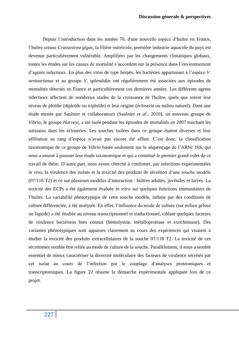
Discussion génerale & perspectives
227
Depuis l’introduction dans les années 70, d'une nouvelle espèce d’huître en France,
l’huître creuse Crassostrea gigas, la filière ostréicole, première industrie aquacole du pays est
devenue particulièrement vulnérable. Amplifiées par les changements climatiques globaux,
toutes les études sur les causes de mortalité s’accordent sur la présence dans l’environnement
d’agents infectieux. En plus des virus de type herpès, les bactéries appartenant à l’espèce V.
aestuarianus et au groupe V. splendidus ont régulièrement été associées aux épisodes de
mortalités détectés en France et particulièrement ces dernières années. Les différents agents
infectieux affectent de nombreux stades de la croissance de l'huître, quels que soient leur
niveau de ploïdie (diploïde ou triploïde) et leur origine (écloserie ou milieu naturel). Dans une
étude menée par Saulnier et collaborateurs (Saulnier et al., 2010), un nouveau groupe de
Vibrio, le groupe Harveyi, a été isolé pendant les épisodes de mortalités en 2007 touchant les
naissains dans les écloseries. Les souches isolées dans ce groupe étaient diverses et leur
affiliation au rang d’espèce n’avait pas encore été affiné. C’est donc, la classification
taxonomique de ce groupe de Vibrio basée seulement sur le séquençage de l’ARNr 16S, qui
nous a amené à pousser leur étude taxonomique et qui a constitué le premier grand volet de ce
travail de thèse. D’autre part, nous avons cherché à confirmer, par infections expérimentales
in vivo, la virulence des isolats et la toxicité des produits de sécrétion d’une souche modèle
(07/118 T2) et ce sur plusieurs modèles d’interaction : huîtres adultes, juvéniles et larves. La
toxicité des ECPs a été également évaluée in vitro sur quelques fonctions immunitaires de
l’huître. La variabilité phénotypique de cette souche modèle, induite par des conditions de
culture différenciée, a été analysée. En effet, l’influence du mode de culture (sur milieu gélosé
ou liquide) a été étudiée au niveau transcriptionnel et traductionnel, ciblant quelques facteurs
de virulence bactériens bien connus (hémolysine, métalloprotéase et exochitinase). Des
variantes phénotypiques sont apparues clairement au cours des expériences qui visaient à
étudier la toxicité des produits extracellulaires de la souche 07/118 T2. La toxicité de ces
sécrétomes semble être reliée au mode de culture de la souche. Parallèlement, il nous a semblé
essentiel de mieux caractériser la diversité moléculaire des facteurs de virulence sécrétés par
cet isolat au cours de l’infection par le couplage d’analyses protéomiques et
transcriptomiques. La figure 22 résume la démarche expérimentale appliquée lors de ce
projet.
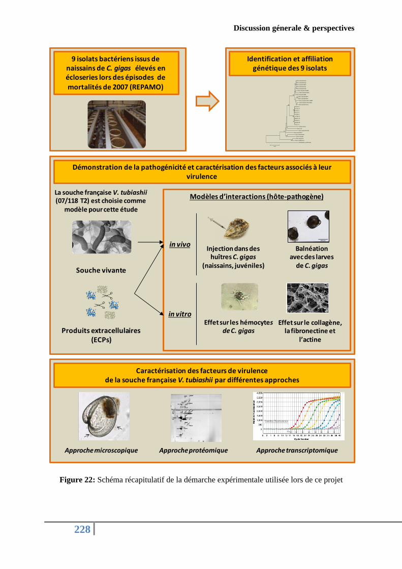
Discussion génerale & perspectives
228
Figure 22: Schéma récapitulatif de la démarche expérimentale utilisée lors de ce projet
9 isolats bactériens issus de naissains de C. gigas élevés en écloseries lors des épisodes de mortalités de 2007 (REPAMO)
Identification et affiliation génétique des 9 isolats
Démonstration de la pathogénicité et caractérisation des facteurs associés à leur virulence
Souche vivante
Produits extracellulaires (ECPs)
Vibrio tubiashii R723
Vibrio tubiashii R722
Vibrio tubiashii R674
Vibrio tubiashii R731
Vibrio tubiashii R716
Vibrio tubiashii R14825
Vibrio tubiashii R678
Vibrio tubiashii R659
Vibrio tubiashii R677
Vibrio tubiashii R676
Vibrio tubiashii LMG 10936T
Vibrio tubiashii LMG 16851
Vibrio tubiashii R711
07/110 T1
07/116 T1
07/120 T1
07/112 T1
07/118 T2
07/108 T1
04/002 1T2
07/119 T1
07/038 2T2
07/117 T1
Vibrio harveyi
Vibrio xuii
Vibrio nigripulchritudo
Vibrio brasiliensis
Vibrio mediterranei
Vibrio orientalis
Vibrio neptunius
Vibrio coralliilyticus
Shewanella putrefaciens
99
52
61
99
61
91
98
86
64
99
63
0.05
Modèles d’interactions (hôte-pathogène)
Injection dans des huîtres C. gigas
(naissains, juvéniles)
Balnéation avec des larves
de C. gigas
in vivo
Effet sur les hémocytes de C. gigas
Effet sur le collagène, la fibronectine et
l’actine
La souche française V. tubiashii (07/118 T2) est choisie comme
modèle pour cette étude
Caractérisation des facteurs de virulencede la souche française V. tubiashii par différentes approches
Approche protéomique Approche transcriptomique
in vitro
Approche microscopique
Discussion génerale & perspectives
229
1. V. TUBIASHII EN France !!!
Même si l’espèce V. tubiashii n’a été officiellement reconnue qu’en 1984 (Hada et al.,
1984), l’histoire de cette espèce bactérienne a commencé avec les travaux de Tubiash en
1965, qui a décrit une nouvelle maladie ‘la nécrose bacillaire’ caractérisée par une réduction
de la motilité larvaire et la formation d’amas bactériens autour des organes et des débris
cellulaires. Cette espèce a été isolée pour la première fois dans des écloseries sur la côte Est
des États-Unis chez l’huître Crassostrea virginica (larves et juvéniles) et la praire Mercenaria
mercenaria (Tubiash et al., 1965; Tubiash, 1970). Quelques années plus tard, des souches de
V. tubiashii ont été identifiées dans des écloseries en Angleterre et en Espagne affectant les
larves d’huîtres Ostrea edulis et C. gigas (Jeffries, 1982; Lodeiros et al., 1987). Entre 1999 et
2000, des bactéries appartenant à l’espèce V. tubiashii ont pu être ensuite isolées de larves
d’huître creuse sur la côte ouest de l’Amérique du Nord (Estes et al., 2004), puis une
réémergence de V. tubiashii a été observée en 2006-2007 sur la côte ouest américaine, ainsi
qu’à Hawaï, infectant de nouveaux mollusques bivalves aux stades larvaire et juvénile de
l’huître Kumamoto (C. sikamea) et de la palourde royale (Panopea abrupta). Elle a également
été identifiée dans l’eau de mer, dans les réserves de microalgues et d’algues, dans les
réservoirs d’eau et même dans les conduites d’air d’écloseries (Elston et al., 2008). Le
premier isolat français date de 2004 où il avait été isolé dans une écloserie de mollusques
gastéropodes. Cette bactérie n’a pu être détectée de nouveau en France qu’en 2007, au
moment de sa réémergence en Amérique entre 2006 et 2007 (Article 1). Ainsi, depuis sa
description initiale, des bactéries appartenant à l’espèce V. tubiashii, ont pu être décrites à
travers le globe, toujours dans les écloseries, isolées d’hôtes jeunes (larves-juvéniles) et
surtout sur un spectre d’hôtes variés.
Toutes ces observations nous amènent à envisager deux questions majeures:
(1) S’agit-il d’un agent infectieux spécifique aux stades précoces de la vie des mollusques
bivalves? (2) S’agit-il d’un agent infectieux spécifique des écloseries ?
Rétrospectivement, et par le biais de la PCR quantitative (ciblant le gène gyrB), notre
étude a montré qu’en France, ces nouveaux isolats de V. tubiashii avaient été associés à un
épisode de mortalité en 2004 d’ormeaux (Haliotis tuberculata) issus d’écloseries françaises, à
des épisodes de mortalités en 2007 de naissains d’huîtres creuses d’écloserie et à des
Discussion génerale & perspectives
230
mortalités en 2008 d’huîtres juvéniles également issus d’écloseries. Cependant, aucune
identification de cette souche n’a été faite à partir d'huîtres moribondes (naissains, juvéniles
ou adultes) prélevées en milieu naturel (Article 1).
Dans le même temps, la virulence de ces isolats a été démontrée par des séries
d’infections expérimentales d’huîtres à différents stades de développement (Article 1 et
Article 4). L’utilisation de plusieurs techniques d’infection expérimentales (par balnéation et
par injection) sur plusieurs stades de vie de l’hôte et sur plusieurs familles d’huîtres de
résistances différentes (données non présentées), nous ont permis de modéliser au maximum
les phénomènes observés naturellement et de cibler le stade de vie le plus sensible. Pour C.
virginica, une étude comparative des stades larvaires et juvéniles a rapporté que la sensibilité
de l’hôte envers la souche V. tubiashii diminuait avec l’âge de l’hôte, un temps moyen de
survie de 24 h pour les larves et plus de 6 semaines pour les juvéniles (Gomez-Leon et al.,
2008). Dans notre étude, et utilisant le modèle larvaire, nous avons pu visualiser les
symptômes de la nécrose bacillaire et repérer à quel moment de l’infection on pouvait
commencer à apercevoir les premiers changements phénotypiques (absence de mouvement)
(Article 4). Par contre, cela est apparu plus délicat d’observer ces anomalies avec le modèle
juvénile car en moins de 24 h post-infection le corps humide de l’huître était dégradé.
Par conséquent, pour la souche française V. tubiashii, le modèle larvaire semble le plus
sensible et le mieux adapté pour imiter les phénomènes naturels (infection par balnéation avec
des concenrtations bactériennes plus contrôlées) par rapport au modèle juvénile qui nécessite
déjà des injections intramusculaires et des concentrations importantes en bactéries. Il apparaît
donc désormais nécessaire de procéder à des examens histologiques et microscopiques sur des
larves moribondes de C. gigas afin de cibler les organes les plus atteints par la souche
française V. tubiashii. Le laboratoire LGPMM Ifremer de la station Ronce-Les-Bains dispose
déjà de ces techniques et devrait donc pouvoir prochainement aider à l’évaluation de ces
atteintes larvaires.
Ce qui est remarquable dans les phénomènes de mortalités observées en France ou
ailleurs, c’est que la souche de V. tubiashii ait souvent été isolée à partir d’animaux
d’écloseries. Cet environnement confiné qui est devenu indispensable pour répondre aux
demandes en naissains des conchyliculteurs trouve ici ses limites. Ces installations, dont la
température, la salinité, la qualité de l’eau de mer, l’alimentation sont contrôlées, ont été à
plusieurs reprises associées aux phénomènes de mortalités touchant plusieurs types d’animaux
Discussion génerale & perspectives
231
d’élevage. En effet, il a été rapporté que la prolifération des bactéries du genre Vibrio chez
l’ormeau semblait être relative aux conditions d'élevage qualifiées de stressantes et
d’intensives qui rendent les jeunes ormeaux plus sensibles à l'infection (Berthe, 2005).
Néanmoins, il suffit qu’une seule condition d’élevage soit déréglée ou non optimisée
(température, propreté des installations, air, microalgues contaminées…) pour que l’agent
pathogène trouve un champ favorable et passe d’un état de veille à un état actif exposant ainsi
la production à une véritable menace (Le Deuff Rose-marie, 1996; Berthe, 2005). Ce cas est
très courant fréquent chez les larves qui montrent rarement des symptômes pathologiques
prolongés et subissent des mortalités très rapides.
En conclusion, il est important de retenir que les bactéries de l’espèce V. tubiashii
semblent trouver des conditions favorables en écloserie et affecter même le stade larvaire
quand les bactéries sont présentes dans l’eau de mer environnante et ce, pour différentes
espèces hôtes. Il semble évident que les systèmes d’élevages au lieu d’être un moyen assurant
une production de qualité et continue de naissains pourraient devenir des niches de
contamination et de développement de plusieurs types de microorganismes pathogènes. Par
conséquent, il semble essentiel que la surveillance des écloseries devrait souvent être
maintenue pour assurer une production de qualité.
L’autre question pertinente que l’on peut se poser concernant l’apparition de la souche
V. tubiashii en France est:
S’agit-il de la même souche qui affecte tous ces différents hôtes dans le monde ou divers
«pathotypes» que l’on ne sait pas encore décrire?
Les changements globaux sont susceptibles d'influer tant sur les animaux marins
(fragilité, déséquilibre de reproduction et de développement que sur les agents pathogènes
(évolution, mutation…) et favorisent ainsi leur émergence et leur prévalence (Burge et al.,
2014). L’élévation de la température de l’eau de mer est parmi les facteurs majeurs qui
contribuent à l’aggravation des infections. Elston et collaborateurs (Elston et al., 2008) ont
expliqué que la réapparition de la vibriose dont celle causée par la souche V. tubiashii sur les
côtes ouest des États-Unis d'Amérique est probablement due à des changements climatiques
divers y compris les variations thermiques, l’acidification de l’eau mer, la mauvaise nutrition
et l’accumulation des produits toxiques issus de la dégradation des algues ou de la flore
marine. De plus, certains isolats bactériens repérés pendant la réémergence de la souche V.
Discussion génerale & perspectives
232
tubiashii en Amérique en 2006 ont montré des nouveaux phénotypes, par comparaison avec la
souche V. tubiashii RE22 isolée durant les épisodes de mortalités de 2000.
Concernant les souches françaises de V. tubiashii, les différentes études menées dans
ce travail de thèse ont permis de révéler quelques différences sur le plan phénotypique et
métabolique par rapport à la souche américaine. La souche V. tubiashii française a été isolée à
partir de naissains de C. gigas alors que la souche américaine a souvent été détectée chez les
larves et à une reprise chez les juvéniles de C. gigas (Jeffries, 1982; Estes et al., 2004; Elston
et al., 2008). Les essais phylogénétiques basées sur l’analyse de plusieurs gènes de ménages
ont montré que les isolats français formaient un groupe phylogénétique homogène, distinct
mais proche de la souche américaine V. tubiashii (Article 1). En outre, sur les 31 analyses
phénotypiques réalisées (portant sur différents critères biochimiques et métaboliques, et en
comparaison avec la souche type V.tubiashii) ont montré que tous les isolats étaient capables
d’utiliser le cellobiose en tant que source de carbone mais qui reste un caractère variable chez
les souches de V .tubiashii américaines (Article 1). L’étude moléculaire de la structure
primaire de la métalloprotéase de la souche française a montré des différences avec des
métalloprotéases déjà décrites chez la souche américaine RE22 (VtpA et VtpB) (Article 2 et
Article 4). Toutes ces différences laissent suggérer qu’il pourrait s’agir d'un autre «pathotype»
ou groupe de virulence) appartenant à l’espèce V. tubiashii qui pourrait avoir évolué afin de
s’adapter à un nouvel environnement. Nos études in vitro illustrent parfaitement les capacités
d’adaptation des souches de V. tubiashii aux changements environnants. En effet, leurs
produits de sécrétion diffèrent en termes de toxicité, de qualité et de quantités protéiques
selon les modes de culture utilisés pour notre souche modèle (liquide vs solide et
compositions variées) utilisés (Article 3).
A l’issue de ces résultats, il serait intéressant de réaliser une étude phylogénétique plus
poussée sur des souches de V. tubiashii isolées sur différentes années, dans différents endroits
du monde et à partir de différents hôtes et de chercher les points communs et les spécificités.
2. ROLE DES FACTEURS DE VIRULENCE DANS LA PATHOGENICITE DES
VIBRIO.
S’agit- il de la conjonction de plusieurs facteurs ou de la prédominance d’un seul
facteur?
Les bactéries ont la capacité de sécréter plusieurs types de facteur de virulence avec des
fonctions différentes. Au moyen de ces déterminants, les bactéries pathogènes sont capables
Discussion génerale & perspectives
233
d’être transmises à un hôte, de se fixer et de coloniser cet hôte, de se multiplier et d’échapper
à ses mécanismes de défenses immunitaires. Chez les bactéries du genre Vibrio, plusieurs
facteurs de virulence ont été décrits incluant les protéases, les entérotoxines, les cytolysines,
les sidérophores, les facteurs d’adhésion et les hémagglutinines (Shinoda, 2005).
L’importance des produits extracellulaires dans le cycle de la virulence d’une bactérie
pathogène font toujours l’objet d’une attention accrue, comme l’atteste le nombre important
de publications sur le sujet. Dans l’objectif de comprendre les interactions entre hôte et
pathogène, les approches ont souvent consisté à simplifier le système infectieux en: i)
commençant par la validation de la virulence de la souche et l’examen de potentielles cibles
sur l’hôte (infection expérimentale, microscopie, histologie…), ii) passant par la mesure de la
toxicité des produits de sécrétion et iii) finissant par caractériser le facteur majeur responsable
de la toxicité de l’ECPs sous sa forme pure après avoir vérifié par mutagenèse son importance
dans la virulence de l’agent pathogène (Binesse et al., 2008; Hasegawa et al., 2008, 2009;
Labreuche et al., 2006a,b, 2010). Dans notre étude, une démarche expérimentale proche a été
mise en place. Après avoir démontré la virulence de notre souche française modèle sur des
juvéniles de C. gigas, la toxicité de ses produits de sécrétion, produits in vitro, a été prouvée
à travers plusieurs modèles d’infection comprenant des larves, des naissains et des juvéniles
d’huître creuse (Article 1, Article 3 et Article 4). Les ECPs ont également montré un effet
inhibiteur sur la capacité d’adhésion et le pouvoir phagocytaire des hémocytes issus d’huître
creuse (Article 2). Cette dérégulation de quelques fonctions immunitaires, sous-entend des
modifications morphologiques des hémocytes (arrondissement des cellules), a été renforcée
par la capacité des ECPs de la souche française V. tubiashii à dégrader des protéines
structurales de l’hôte notamment le collagène. Le même processus de digestion a également
pu être démontré pour d’autres protéines structurales d’origine bovine telles que l’actine et la
fibronectine (Article 2).
Ces résultats expérimentaux nous amènent à discuter du rôle des protéases et
notamment de celui des métalloprotéases dans la toxicité des surnageants bactériens. En effet,
plusieurs travaux ont décrit l’impact pathologique des métalloprotéases au cours du processus
de l’infection (Shinoda et al., 1996; Miyoshi et al., 2002). Pour la souche de V. tubiashii
française, l’activité métalloprotéasique a été caractérisée dans les ECPs obtenus par culture in
vitro de notre souche modèle (Article 2) mais également dans les surnageants d’infection
(larves + V. tubiashii) (Article 4). Le séquençage du gène de cette métalloprotéase bactérienne
Discussion génerale & perspectives
234
ainsi que l’analyse comparative avec d’autres séquences décrites de métalloprotéases ont
validé son appartenance à la famille des thermolysines (Article 2).
Cependant, l’ajout de l’EDTA, dans le bain d’infection des larves de C. gigas, comme
indicateur de la présence de métalloprotéases, n’a pas permis de réduire les mortalités
larvaires. Ce résultat important pose désormais la question du rôle de métalloprotéases actives
dans l’expression d’une toxicité par ces ECPs, jusqu’à présent documentées comme facteurs
de virulence majeurs (Binesse et al., 2008; Hasegawa et al., 2008). Le fractionnement par
chromatographie des ECPs bruts produits in vitro par la souche française V. tubiashii a permis
d’obtenir deux fractions. Une première fraction se compose majoritairement de porine, de
protéines de la membrane externe (OMPs) et de protéines hypothétiques alors que la
deuxième fraction se trouve enrichie en métalloprotéases actives (Article 4). Ce qui est
étonnant dans ce résultat, c’est que les deux fractions se sont révélées toxiques pour les larves
d’huître creuse (Article 4) alors que nous nous attendions à une corrélation étroite entre la
présence exclusive de métalloprotéases actives dans une fraction et leur toxicité. Cela renforce
l’hypothèse que la toxicité des ECPs n’est pas seulement due à un facteur unique, mais plutôt
à la conjonction d’un ensemble de facteurs.
L’analyse protéomique des ECPs montre clairement que les métalloprotéases sont les
protéines les plus abondantes. Cette abondance relative pourrait masquer d’autres protéines
présentes en plus faibles concentrations mais tout aussi importantes dans l’expression de
phénotypes de virulence (Valiente et al., 2008). Pour tester cette hypothèse, il serait bon de
développer un certain nombre d’outils complémentaires, comme la production de plusieurs
métalloprotéases recombinantes afin de tester la robustesse de ce facteur dans la toxicité des
sécrétomes bactériens.
La deuxième question qu’on peut poser est:
Pour induire une infection chez l’hôte, les facteurs de virulence agissent d’une manière
séquentielle régulée ou d’une façon conjointe?
L’étude menée par Hasegawa et Häse (2009a) a montré que l’activité hémolytique et
l’activité métalloprotéasique étaient produites de façon ordonnée au cours de la croissance de
la souche américaine V. tubiashii. Nous avons observé le même phénomène lorsque nous
avons suivi les activités hémolytiques et métalloprotéasiques des surnageants d’infection de
Discussion génerale & perspectives
235
larves par notre souche modèle. En effet, nous avons pu montrer que les gènes codants pour
une métalloprotéase et une exochitinase étaient sous-exprimés pendant les premières heures
de l’infection puis surexprimés au-delà de 16 h d’infection alors que le gène codant pour une
hémolysine thermostable était sur-exprimé tout au long de l’infection (Article 4). Ces résultats
suggèrent que la sécrétion de certains facteurs de virulence par la souche française V. tubiashii
pourrait être régulée pour assurer leur action séquentielle pendant l’infection, à la lumière de
ce que les auteurs décrivent déjà pour les vibrions pathogènes pour l'Homme. En effet,
Shinoda et Miyoshi (2011) suggèrent que les entérotoxines et les cytotoxines agissent
directement sur l’hôte et sont responsables des premiers symptômes, les sidérophores et les
facteurs d’adhésions agissent indirectement et sont impliqués dans l'établissement de
l'infection et enfin les protéases sont reconnues pour jouer un rôle important dans le
développement de l'infection. Il ne pas faut nier aussi le rôle important du Quorum Sensing
dans la régulation de l’expression de plusieurs facteurs de virulence (De Decker et al., 2013).
Le suivi de l’expression d’autres déterminants de la virulence au cours de l’infection
par cette souche française est donc nécessaire afin de mettre en évidence le rôle de chaque
facteur dans le déclenchement et l’installation de la pathologie chez les organismes marins.
En plus, il paraîtrait intéressant d’analyser le surnageant d’infection (larve + V. tubiashii)
obtenu dans les conditions in vivo à différents temps par spectrométrie de masse afin de
chercher la corrélation entre expression, sécrétion de facteurs de virulence et phénotype
induit.
L’utilisation du terme «majeur» employé par plusieurs auteurs pour décrire le rôle
d’un facteur de virulence est à prendre avec beaucoup de précautions. Comme nous avons
démontré précédemment, un agent pathogène est capable de sécréter plusieurs facteurs de
virulence de façon séquentielle et à chaque étape de l’infection.
Ainsi, le rôle d’un facteur peut être majeur dans une étape bien déterminée de
l’infection comme au moment de la colonisation ou pendant l’invasion par exemple mais pas
dans toutes les étapes de l’infection. L’illustration de ce point de vue est la démonstration
dans notre étude de l’inhibition, par les ECPs de la souche française V. tubiashii, de la
capacité d’adhésion et du pouvoir phagocytaire des hémocytes d’huîtres sans affecter les
activités lysosomales et estérasiques (Article 2).
Ainsi, il semble que la virulence bactérienne résulte d’une conjonction de plusieurs
facteurs agissant d’une manière régulée et séquentielle pour installer une infection chez l’hôte.
Cependant, des expérimentations complémentaires devront donc être entreprises afin de
Discussion génerale & perspectives
236
mieux comprendre et définir le rôle accordé à chaque potentielle toxine (hémolysines, toxines
RTX, OMPs, les métalloprotéases…) dans la virulence de la souche française de V. tubiashii
ou n’importe quelle autre espèce de Vibrio impliquées dans les épisodes de mortalités de
mollusques bivalves. Le schéma 23 illustre globalement, en se basant sur les résultats de ce
projet de thèse, le mécanisme de pathogénicité mis en œuvre par la souche française de V.
tubiashii 07/118 T2.
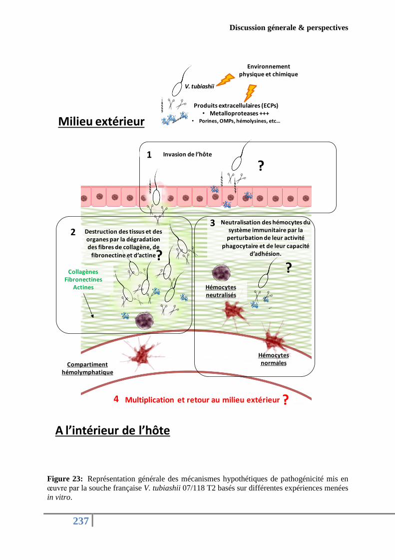
Discussion génerale & perspectives
237
Figure 23: Représentation générale des mécanismes hypothétiques de pathogénicité mis en
œuvre par la souche française V. tubiashii 07/118 T2 basés sur différentes expériences menées
in vitro.
Invasion de l’hôte 1
V. tubiashii
Hémocytesnormales
Destruction des tissus et des organes par la dégradation des fibres de collagène, de
fibronectine et d’actine
2Neutralisation des hémocytes du
système immunitaire par la perturbation de leur activité
phagocytaire et de leur capacité d’adhésion.
3
Compartiment hémolymphatique
Hémocytesneutralisés
Collagènes Fibronectines
Actines
A l’intérieur de l’hôte
Milieu extérieur
Produits extracellulaires (ECPs)• Metalloproteases +++
• Porines, OMPs, hémolysines, etc…
Environnement physique et chimique
Multiplication et retour au milieu extérieur
?
??
?4

238
REFERENCES
BIBLIOGRAPHIQUES
Références bibliographiques
239
-A-
Actis, L. A., Fish, W., Crosa, J. H., Kellerman, K., Ellenberger, S. R., Hauser, F. M. &
Sanders-Loehr, J. (1986). Characterization of anguibactin, a novel siderophore from
Vibrio anguillarum 775(pJM1). J Bacteriol 167, 57–65.
Aguirre-Guzmán, G., Ascencio, F. & Saulnier, D. (2005). Pathogenicity of Vibrio
penaeicida for white shrimp Litopenaeus vannamei: a cysteine protease-like exotoxin
as a virulence factor. Dis Aquat Organ 67, 201–207.
Ahmad, I., Hoessli, D.C., Gupta, R., Walker-Nasir, E., Rafik, S.M., Choudhary, M.I.,
Shakoori, A.R., Nasir-ud-Din. (2007). In silico determination of intracellular
glycosylation and phosphorylation sites in human selectins: Implications for biological
function. J. Cell. Biochem. 100, 1558–1572.
Alapide-Tendencia, E. V. & Dureza, L. A. (1997). Isolation of Vibrio spp. from Penaeus
monodon (Fabricius) with red disease syndrome. Aquaculture 154, 107–114.
Alavandi, S. V., Babu, T. D., Abhilash, K. S., Vijayan, K. K., Kalaimani, N. & Santiago,
T. C. (2008). Loose shell syndrome of farmed Penaeus monodon in India is caused by
a filterable agent. Dis Aquat Organ 81, 163–171.
Alberts, B., Johnson, A., Lewis, J., Raff, M., Roberts, K. & Walter, P. (2002).
Programmed Cell Death (Apoptosis). Text, .
Alcaide, E., Amaro, C., Todol, R. & Oltra, R. (1999). Isolation and characterization of
Vibrio parahaemolyticus causing infection in Iberian toothcarp Aphanius iberus. Dis
Aquat Organ 35, 77–80.
Allam, B., Ashton-Alcox, K. A. & Ford, S. E. (2002). Flow cytometric comparison of
haemocytes from three species of bivalve molluscs. Fish Shellfish Immunol 13, 141–
158.
Allam, B., Ford, S.E. (2006). Effects of the pathogenic Vibrio tapetis on defence factors of
susceptible and non-susceptible bivalve species: I. Haemocyte changes following in
vitro challenge. Fish Shellfish Immunol. 20, 374–383.
Alsina, M. & Blanch, A. R. (1994a). A set of keys for biochemical identification of
environmental Vibrio species. J Appl Bacteriol 76, 79–85.
Alsina, M. & Blanch, A. R. (1994b). Improvement and update of a set of keys for
biochemical identification of Vibrio species. J Appl Bacteriol 77, 719–721.
Amaro, C. & Biosca, E. G. (1996). Vibrio vulnificus biotype 2, pathogenic for eels, is also an
opportunistic pathogen for humans. Appl Environ Microbiol 62, 1454–1457.
Références bibliographiques
240
Andre s, S. C., Robinson, A. . Rodr guez-Quiñones, F. (2003). Bacterial iron
homeostasis. FEMS Microbiol Rev 27, 215–237.
Andrus, C. R., Walter, M., Crosa, J. H. & Payne, S. M. (1983). Synthesis of siderophores
by pathogenicVibrio species. Curr Microbiol 9, 209–214.
Antunes, L. C. M., Ferreira, R. B. R., Buckner, M. M. C. & Finlay, B. B. (2010). Quorum
sensing in bacterial virulence. Microbiology 156, 2271–2282.
Araya, M.T., Siah, A., Mateo, D.R., Markham, F., McKenna, P., Johnson, G.R., Berthe,
F.C.J., (2009). Morphological and Molecular Effects of Vibrio splendidus on
Hemocytes of Softshell Clams, Mya arenaria. J. Shellfish Res. 28, 751–758.
Ashton-Alcox, K. A. & Ford, S. E. (1998). Variability in molluscan hemocytes: a flow
cytometric study. Tissue Cell 30, 195–204.
Asplund, M. E., Baden, S. P., Russ, S., Ellis, R. P., Gong, N. & Hernroth, B. E. (2013).
Ocean acidification and host-pathogen interactions: blue mussels, Mytilus edulis,
encountering Vibrio tubiashii. Environ Microbiol.
Attridge, S. R., Manning, P. A., Holmgren, J. & Jonson, G. (1996). Relative significance
of mannose-sensitive hemagglutinin and toxin-coregulated pili in colonization of
infant mice by Vibrio cholerae El Tor. Infect Immun 64, 3369–3373.
Austin, B. (2010). Vibrios as causal agents of zoonoses. Vet Microbiol 140, 310–317.
-B-
Bacca, H. (2007). Etude des voies metaboliques des sucres chez l’huitre creuse crassostrea
gigas. Implication dans les mortalites estivales. Thèse de doctorat ,Université de
RENNES I.
Bachère, E., Mialhe, E., Noël, D., Boulo, V., Morvan, A. & Rodriguez, J. (1995).
Knowledge and research prospects in marine mollusc and crustacean immunology.
Aquaculture 132, 17–32.
Bai, F., Sun, B., Woo, N. Y. S. & Zhang, X.-H. (2010). Vibrio harveyi hemolysin induces
ultrastructural changes and apoptosis in flounder (Paralichthys olivaceus) cells.
Biochem Biophys Res Commun 395, 70–75.
Banin, E., Vassilakos, D., Orr, E., Martinez, R. J. & Rosenberg, E. (2003). Superoxide
Dismutase Is a Virulence Factor Produced by the Coral Bleaching Pathogen Vibrio
shiloi. Curr Microbiol 46, 0418–0422.
Références bibliographiques
241
Bardy, S. L., Ng, S. Y. M. & Jarrell, K. F. (2003). Prokaryotic motility structures.
Microbiology 149, 295–304.
Bari, S. M. N., Roky, M. K., Mohiuddin, M., Kamruzzaman, M., Mekalanos, J. J. &
Faruque, S. M. (2013). Quorum-sensing autoinducers resuscitate dormant Vibrio
cholerae in environmental water samples. Proc Natl Acad Sci U S A 110, 9926–9931.
Baud, L., Fouqueray, B., Bellocq, A. & Peltier, J. (2003). Les calpaïnes participent au
développement de la réaction inflammatoire. MS Médecine Sci 19, 71–76.
Beaubrun, J. J.-G., Kothary, M. H., Curtis, S. K., Flores, N. C., Eribo, B. E. & Tall, B.
D. (2008). Isolation and Characterization of Vibrio tubiashii Outer Membrane Proteins
and Determination of a toxR Homolog. Appl Environ Microbiol 74, 907–911.
Beaz-Hidalgo, R., Balboa, S., Romalde, J. L. & Figueras, M. J. (2010). Diversity and
pathogenecity of Vibrio species in cultured bivalve molluscs. Environ Microbiol Rep
2, 34–43.
Ben-Haim, Y. & Rosenberg, E. (2002). A novel Vibrio sp. pathogen of the coral Pocillopora
damicornis. Mar Biol 141, 47–55.
Ben-Haim, Y., Thompson, F. L., Thompson, C. C., Cnockaert, M. C., Hoste, B., Swings,
J. & Rosenberg, E. (2003). Vibrio coralliilyticus sp. nov., a temperature-dependent pathogen
of the coral Pocillopora damicornis. Int J Syst Evol Microbiol 53, 309–315
Berthe, F. (2005). Diseases in mollusc hatcheries and their paraedox in health management.
Presented at the Diseases in Asian Aquaculture.
Bi, S., Wang, W., Zhao, Y., Ru, S. & Liu, Y. (2011). Studies on hemolysis of hemolysin
produced by Synechocystis sp. PCC 6803. J Ocean Univ China 10, 362–368.
Bina, X. R., Provenzano, D., Nguyen, N. & Bina, J. E. (2008). Vibrio cholerae RND
Family Efflux Systems Are Required for Antimicrobial Resistance, Optimal Virulence
Factor Production, and Colonization of the Infant Mouse Small Intestine. Infect
Immun 76, 3595–3605.
Binesse, J., Delsert, C., Saulnier, D., Champomier-Vergès, M.-C., Zagorec, M., Munier-
Lehmann, H., Mazel, D., Roux, F.L., 2008). Metalloprotease Vsm Is the Major
Determinant of Toxicity for Extracellular Products of Vibrio splendidus. Appl.
Environ. Microbiol. 74, 7108–7117.
Biosca, E. G., Amaro, C., Esteve, C., Alcaide, E. & Garay, E. (1991). First record of Vibrio
vulnificus biotype 2 from diseased European eel, Anguilla anguilla L. J Fish Dis 14,
103–109.
Références bibliographiques
242
Birkbeck, T. H. & Treasurer, J. W. (2014). Vibrio splendidus, Vibrio ichthyoenteri and
Vibrio pacinii isolated from the digestive tract microflora of larval ballan wrasse,
Labrus bergylta Ascanius, and goldsinny wrasse, Ctenolabrus rupestris (L.). J Fish Dis
37, 69–74.
Black, P. N. (1988). The fadL gene product of Escherichia coli is an outer membrane protein
required for uptake of long-chain fatty acids and involved in sensitivity to
bacteriophage T2. J Bacteriol 170, 2850–2854.
Blake, P. A., Merson, M. H., Weaver, R. E., Hollis, D. G. & Heublein, P. C. (1979).
Disease Caused by a Marine Vibrio. N Engl J Med 300, 1–5.
Blanchet, X. (2010). Des gènes impliqués dans la variabilité de la tendreté des viandes
bovines : étude structure-fonction de l’anti-protéase codée par le gène bovin
SERPINA3-3. Thèse de doctorat, Université de Limoges.
Bode, W., Gomis-Rüth, F.-X. & Stöckler, W. (1993). Astacins, serralysins, snake venom
and matrix metalloproteinases exhibit identical zinc-binding environments
(HEXXHXXGXXH and Met-turn) and topologies and should be grouped into a
common family, the ‘metzincins’. FEBS Lett 331, 134–140.
Borrego, J. J., Castro, D., Luque, A., Paillard, C., Maes, P., Garcia, M. T. & Ventosa, A.
(1996). Vibrio tapetis sp. nov., the Causative Agent of the Brown Ring Disease
Affecting Cultured Clams. Int J Syst Bacteriol 46, 480–484.
Bower, S., Hervio, D. & Meyer, G. (1997). Infectivity of Mikrocytos mackini, the causative
agent of Denman Island disease in Pacific oysters Crassostrea gigas, to various
species of oysters. Dis Aquat Organ 29, 111–116.
Brackney, D. E. (2007). The Role of Midgut Serine Proteases in Aedes Aegypti Vector
Competence. ProQuest.
Bradford, M. M. (1976). A rapid and sensitive method for the quantitation of microgram
quantities of protein utilizing the principle of protein-dye binding. Anal Biochem 72,
248–254.
Braun, P., de Groot, A., Bitter, W. & Tommassen, J. (1998). Secretion of Elastinolytic
Enzymes and Their Propeptides by Pseudomonas aeruginosa. J Bacteriol 180, 3467–
3469.
Brauns, L. A., Hudson, M. C. & Oliver, J. D. (1991). Use of the polymerase chain reaction
in detection of culturable and nonculturable Vibrio vulnificus cells. Appl Environ
Microbiol 57, 2651–2655.
Références bibliographiques
243
Brew, K., Dinakarpandian, D. & Nagase, H. (2000). Tissue inhibitors of
metalloproteinases: evolution, structure and function. Biochim Biophys Acta 1477,
267–283.
Broberg, C.A., Calder, T.J., Orth, K. (2011). Vibrio parahaemolyticus cell biology and
pathogenicity determinants. Microbes Infect. 13, 992–1001.
Brown, C. & Losee, E. (1978). Observations on natural and induced epizootics of vibriosis in
Crassostrea virginica larvae. J Invertebr Pathol 31, 41–47.
Brown, C. & Roland, G. (1984). Characterization of exotoxin produced by a shellfish-
pathogenic Vibrio sp. J Fish Dis 7, 117–126.
Bruno Gomez-Gil, S. S.-R. (2004). Molecular identification of Vibrio harveyi-related
isolates associated with diseased aquatic organisms. Microbiol Read Engl 150, 1769–
77.
Burge, C. A., Mark Eakin, C., Friedman, C. S., Froelich, B., Hershberger, P. K.,
Hofmann, E. E., Petes, L. E., Prager, K. C., Weil, E. & other authors. (2014).
Climate Change Influences on Marine Infectious Diseases: Implications for
Management and Society. Annu Rev Mar Sci 6, 249–277.
Butler, A. & Theisen, R. M. (2010). Iron(III)–siderophore coordination chemistry:
Reactivity of marine siderophores. Coord Chem Rev 254, 288–296.
-C-
Cahill, M. M. (1990). Bacterial flora of fishes: A review. Microb Ecol 19, 21–41.
Canesi, L., Gallo, G., Gavioli, M. & Pruzzo, C. (2002). Bacteria-hemocyte interactions and
phagocytosis in marine bivalves. Microsc Res Tech 57, 469–476.
Cano-Gómez, A., Goulden, E. F., Owens, L. & Høj, L. (2010). Vibrio owensii sp. nov.,
isolated from cultured crustaceans in Australia. FEMS Microbiol Lett 302, 175–181.
Casadevall, A. & Pirofski, L. (1999). Host-Pathogen Interactions: Redefining the Basic
Concepts of Virulence and Pathogenicity. Infect Immun 67, 3703–3713.
Casadevall, A. & Pirofski, L. (2003). Microbial virulence results from the interaction
between host and microorganism. Trends Microbiol 11, 157–158.
Casadevall, A. & Pirofski, L.-A. (2009). Virulence factors and their mechanisms of action:
the view from a damage-response framework. J Water Health 7 Suppl 1, S2–S18.
Ceccarelli, D., Spagnoletti, M., Cappuccinelli, P., Burrus, V. & Colombo, M. M. (2011).
Origin of Vibrio cholerae in Haiti. Lancet Infect Dis 11, 262.
Références bibliographiques
244
Chen, C.-Y., Wu, K.-M., Chang, Y.-C., Chang, C.-H., Tsai, H.-C., Liao, T.-L., Liu, Y.-
M., Chen, H.-J., Shen, A. B.-T. & other authors. (2003). Comparative genome
analysis of Vibrio vulnificus, a marine pathogen. Genome Res 13, 2577–2587.
Chen, F. R., Liu, P. C. & Lee, K. K. (1999). Purification and partial characterization of a
toxic serine protease produced by pathogenic Vibrio alginolyticus. Microbios 98, 95–
111.
Chen, Y.-C., Chang, M.-C., Chuang, Y.-C. & Jeang, C.-L. (2004). Characterization and
Virulence of Hemolysin III from Vibrio vulnificus. Curr Microbiol 49, 175–179.
Cheng, T. C. (1984). A Classification of Molluscan Hemocytes Based on Functional
Evidences. In Invertebr Blood, Comparative Pathobiology, pp. 111–146. Edited by T.
C. Cheng. Springer US.
Cheng, T. C. & Sullivan, J. T. (1984). Effects of heavy metals on phagocytosis by
Molluscan hemocytes. Mar Environ Res 14, 305–315.
Cheung, A. L. & Fischetti, V. A. (1988). Variation in the expression of cell wall proteins of
Staphylococcus aureus grown on solid and liquid media. Infect Immun 56, 1061–1065.
Chopp, D. L., Kirisits, M. J., Moran, B. & Parsek, M. R. (2003). The dependence of
quorum sensing on the depth of a growing biofilm. Bull Math Biol 65, 1053–1079.
Choquet, G., Soudant, P., Lambert, C., Nicolas, J., Paillard, C. (2003). Reduction of
adhesion properties of Ruditapes philippinarum hemocytes exposed to Vibrio tapetis.
Dis. Aquat. Organ. 57, 109–116.
Chowdhury, M. A., Miyoshi, S. & Shinoda, S. (1990). Purification and characterization of a
protease produced by Vibrio mimicus. Infect Immun 58, 4159–4162.
Chu, F.-L. E. (2000). Defense mechanisms of marine bivalves In Recent Advances in Marine
Biotechnology. Immunobiology and Pathology. Publishers., M. Fingerman and R.
Nagabhushanam. Enfield, NH, Plymouth, UK: Science.
Chu, F.-L. E. & Webb, K. L. (1984). Polyunsaturated fatty acids and neutral lipids in
developing larvae of the oyster,Crassostrea virginica. Lipids 19, 815–820.
Chuang, Y. C., Chang, T. M. & Chang, M. C. (1997). Cloning and characterization of the
gene (empV) encoding extracellular metalloprotease from Vibrio vulnificus. Gene 189,
163–168.
Cobaxin, M., Martínez, H., Ayala, G., Holmgren, J., Sjöling, Å. & Sánchez, J. (2014).
Cholera toxin expression by El Tor Vibrio cholerae in shallow culture growth
conditions. Microb Pathog 66, 5–13.

Références bibliographiques
245
Cohen, N. & Karib, H. (2007). Vibrio spp. dans les produits de la pêche: Risques et
prévention.
Colman, P. M., Jansonius, J. N. & Matthews, B. W. (1972). The structure of thermolysin:
an electron density map at 2-3 A resolution. J Mol Biol 70, 701–724.
Colorni, A., Paperna, I. & Gordin, H. (1981). Bacterial infections in gilt-head sea bream
Sparus aurata cultured at Elat. Aquaculture 23, 257–267.
Colwell, R. R. & Liston, J. (1960). Microbiology of Shellfish. Bacteriological Study of the
Natural Flora of Pacific Oysters (Crassostrea gigas). Appl Microbiol 8, 104–109.
Colwell, R. R. & Sparks, A. K. (1967). Properties of Pseudomonas enalia, a Marine
Bacterium Pathogenic for the Invertebrate Crassostrea gigas (Thunberg)1. Appl
Microbiol 15, 980–986.
Comps, M., Bonami, J.-R., Vago, C. & Razet, D. (1976). La mise en évidence d’une
infection virale chez l’huître portugaise à l’occasion de l’épizootie de 1970-1974. Sci
Pêche 256, 13–15.
Costa, R., Mermoud, I., Koblavi, S., Morlet, B., Haffner, P., Berthe, F., Legroumellec,
M. & Grimont, P. (1998). Isolation and characterization of bacteria associated with a
Penaeus stylirostris disease (Syndrome 93) in New Caledonia. Aquaculture 164, 297–
309.
Coulis, G. (2007). Interactions moléculaires des calpaïnes 1 et 3 avec la région N1 de la titine
humaine. Thèse de doctorat, Université Blaise Pascal - Clermont-Ferrand II.
Crowther, R. S., Roomi, N. W., Fahim, R. E. & Forstner, J. F. (1987). Vibrio cholerae
metalloproteinase degrades intestinal mucin and facilitates enterotoxin-induced
secretion from rat intestine. Biochim Biophys Acta 924, 393–402.
-D-
Daily, O. P., Debell, R. M. & Joseph, S. W. (1978). Superoxide dismutase and catalase
levels in halophilic vibrios. J Bacteriol 134, 375–380.
Dale, S. E., Doherty-Kirby, A., Lajoie, G. & Heinrichs, D. E. (2004). Role of Siderophore
Biosynthesis in Virulence of Staphylococcus aureus: Identification and
Characterization of Genes Involved in Production of a Siderophore. Infect Immun 72,
29–37.
Références bibliographiques
246
David, V. A., Deutch, A. H., Sloma, A., Pawlyk, D. & Durham, D. R. (1992). Cloning,
sequencing and expression of the gene encoding the extracellular neutral protease,
vibriolysin, of Vibrio proteolyticus. Gene 112, 107–112.
Davis, G. H. G. & Park, R. W. A. (1962). A Taxonomic Study of Certain Bacteria Currently
Classified as Vibrio Species. J Gen Microbiol 27, 101–119.
De Decker, S., Normand, J., Saulnier, D., Pernet, F., Castagnet, S. & Boudry, P. (2011).
Responses of diploid and triploid Pacific oysters Crassostrea gigas to Vibrio infection
in relation to their reproductive status. J Invertebr Pathol 106, 179–191.
De Decker, S. & Saulnier, D. (2011). Vibriosis induced by experimental cohabitation in
Crassostrea gigas: evidence of early infection and down-expression of immune-
related genes. Fish Shellfish Immunol 30, 691–699.
De Decker, S., Reynaud, Y. & Saulnier, D. (2013). First molecular evidence of cross-
species induction of metalloprotease gene expression in Vibrio strains pathogenic for
Pacific oyster Crassostrea gigas involving a quorum sensing system. Aquaculture
392–395, 1–7.
Defoirdt, T., Boon, N., Bossier, P. & Verstraete, W. (2004). Disruption of bacterial quorum
sensing: an unexplored strategy to fight infections in aquaculture. Aquaculture 240,
69–88.
Defoirdt, T., Boon, N., Sorgeloos, P., Verstraete, W. & Bossier, P. (2008). Quorum sensing
and quorum quenching in Vibrio harveyi: lessons learned from in vivo work. ISME J
2, 19–26.
Dégremont, L., Bédier, E., Soletchnik, P., Ropert, M., Huvet, A., Moal, J., Samain, J.-F.
& Boudry, P. (2005). Relative importance of family, site, and field placement timing
on survival, growth, and yield of hatchery-produced Pacific oyster spat (Crassostrea
gigas). Aquaculture 249, 213–229.
Delston, R.B., Kothary, M.H., Shangraw, K.A., Tall, B.D. (2003). Isolation and
characterization of a zinc-containing metalloprotease expressed by Vibrio tubiashii.
Can. J. Microbiol. 49, 525–529.
Duperthuy, M., Binesse, J., Le Roux, F., Romestand, B., Caro, A., Got, P., Givaudan, A.,
Mazel, D., Bachère, E. & Destoumieux-Garzón, D. (2010). The major outer
membrane protein OmpU of Vibrio splendidus contributes to host antimicrobial
peptide resistance and is required for virulence in the oyster Crassostrea gigas.
Environ Microbiol 12, 951–963.
Références bibliographiques
247
Duperthuy, M., Schmitt, P., Garzón, E., Caro, A., Rosa, R. D., Roux, F. L., Lautrédou-
Audouy, N., Got, P., Romestand, B. & other authors. (2011). Use of OmpU porins
for attachment and invasion of Crassostrea gigas immune cells by the oyster pathogen
Vibrio splendidus. Proc Natl Acad Sci 201015326.
-E-
Elgaml, A., Higaki, K. & Miyoshi, S. (2013). Regulation system of serine protease
production in Vibrio vulnificus strain NCIMB 2137, a metalloprotease-gene negative
strain isolated from a diseased eel. Aquaculture 416–417, 315–321.
Ellinger, G.M., Boyne, E.B.(1965). Amino acid composition of some fish products and
casein. Br. J. Nutr. 19, 587–592.
Elston, R. A., Hasegawa, H., Humphrey, K. L., Polyak, I. K. & Häse, C. C. (2008). Re-
emergence of Vibrio tubiashii in bivalve shellfish aquaculture: severity, environmental
drivers, geographic extent and management. Dis Aquat Organ 82, 119–134.
Elston, R. A. (1999). Health Management, Development and Histology of Seed Oysters.
World Aquaculture Society.
Erz, W., Carrle, D. & Klessen, C. (1993). Proteases of polymorphonuclear neutrophils:
Influence of different salts and buffers on pericellular lysis. Acta Histochem 94, 46–
53.
Estes, R. M., Friedman, C. S., Elston, R. A. & Herwig, R. P. (2004). Pathogenicity testing
of shellfish hatchery bacterial isolates on Pacific oyster Crassostrea gigas larvae. Dis
Aquat Organ 58, 223–230.
Ezaki, T., Hashimoto, Y. & Yabuuchi, E. (1989). Fluorometric Deoxyribonucleic Acid-
Deoxyribonucleic Acid Hybridization in Microdilution Wells as an Alternative to
Membrane Filter Hybridization in which Radioisotopes Are Used To Determine
Genetic Relatedness among Bacterial Strains. Int J Syst Bacteriol 39, 224–229.
-F-
Fabbri, A., Falzano, L., Frank, C., Donelli, G., Matarrese, P., Raimondi, F., Fasano, A.
& Fiorentini, C. (1999). Vibrio parahaemolyticus Thermostable Direct Hemolysin
Références bibliographiques
248
Modulates Cytoskeletal Organization and Calcium Homeostasis in Intestinal Cultured
Cells. Infect Immun 67, 1139–1148.
Farto, R., Pérez, M. ., Fernández-Briera, A. & Nieto, T. . (2002). Purification and partial
characterisation of a fish lethal extracellular protease from Vibrio pelagius. Vet Microbiol 89,
181–194.
Farto, R., Armada, S. P., Montes, M., Guisande, J. A., Pérez, M. J. & Nieto, T. P. (2003).
Vibrio lentus associated with diseased wild octopus (Octopus vulgaris). J Invertebr
Pathol 83, 149–156.
Faury, N., Saulnier, D., Thompson, F. L., Gay, M., Swings, J. & Le Roux, F. (2004).
Vibrio crassostreae sp. nov., isolated from the haemolymph of oysters (Crassostrea
gigas). Int J Syst Evol Microbiol 54, 2137–2140.
Felsenstein, J. (1985). Confidence Limits on Phylogenies: An Approach Using the Bootstrap.
Evolution 39, 783.
Fields, P. I., Popovic, T., Wachsmuth, K. & Olsvik, O. (1992). Use of polymerase chain
reaction for detection of toxigenic Vibrio cholerae O1 strains from the Latin American
cholera epidemic. J Clin Microbiol 30, 2118–2121.
Finlay, B. B. & Falkow, S. (1997). Common themes in microbial pathogenicity revisited.
Microbiol Mol Biol Rev MMBR 61, 136–169.
Fisher, W. S. (1986). Structure and Functions of Oyster Hemocytes. In Immun Invertebr,
Proceedings in Life Sciences, pp. 25–35. Edited by D. M. Brehélin. Springer Berlin
Heidelberg.
Fontana, A. (1988). Structure and stability of thermophilic enzymes Studies on thermolysin.
Biophys Chem 29, 181–193.
Foster, J. S., Apicella, M. A. & McFall-Ngai, M. J. (2000). Vibrio fischeri
Lipopolysaccharide Induces Developmental Apoptosis, but Not Complete
Morphogenesis, of the Euprymna scolopes Symbiotic Light Organ. Dev Biol 226,
242–254.
Fouz, B., Larsen, J. L., Nielsen, B., Barja, J. L. & Toranzo, A. E. (1992). Characterization
of Vibrio damsela strains isolated from turbot Scophthalmus maximus in Spain. Dis
Aquat Organ 12, 155–166.
Frans, I., Michiels, C. W., Bossier, P., Willems, K. A., Lievens, B. & Rediers, H. (2011).
Vibrio anguillarum as a fish pathogen: virulence factors, diagnosis and prevention. J
Fish Dis 34, 643–661.
Références bibliographiques
249
Fullner Satchell, K. J. (2003). Activation and suppression of the proinflammatory immune
response by Vibrio cholerae toxins. Microbes Infect 5, 1241–1247.
-G-
Gadwal, S., Korotkov, K. V., Delarosa, J. R., Hol, W. G. J. & Sandkvist, M. (2014).
Functional and structural characterization of Vibrio cholerae extracellular serine
protease B, VesB. J Biol Chem jbc.M113.525261.
Gagnaire, B., Renault, T., Bouilly, K., Lapegue, S., Thomas-Guyon, H. (2003). Study of
atrazine effects on Pacific oyster, Crassostrea gigas, haemocytes. Curr. Pharm. Des. 9,
193–199.
Gagnaire, B. (2005). Etude des effets de polluants sur les paramètres hémocytaires de l’huître
creuse, Crassostrea gigas - Interactions entre environnement, mécanismes de défense
et maladies infectieuses. Thèse de doctorat, Université de la Rochelle.
Galland, C., Dupuy, C., Capitaine, C., Auffret, M., Quiniou, L., Laroche, J. &
Pichereau, V. (2013). Comparisons of liver proteomes in the European flounder
Platichthys flesus from three contrasted estuaries. J Sea Res 75, 135–141.
Garnier, M., Labreuche, Y., Garcia, C., Robert, M. & Nicolas, J.-L. (2007). Evidence for
the Involvement of Pathogenic Bacteria in Summer Mortalities of the Pacific Oyster
Crassostrea gigas. Microb Ecol 53, 187–196.
Garnier, M., Labreuche Y., Nicolas, J.-L. (2008). Molecular and phenotypic
characterization of Vibrio aestuarianus subsp. francensis subsp. nov., a pathogen of
the oyster Crassostrea gigas. Syst. Appl. Microbiol. 31, 358–365.
Gay, M., Renault, T., Pons, A.-M. & Le Roux, F. (2004). Two vibrio splendidus related
strains collaborate to kill Crassostrea gigas: taxonomy and host alterations. Dis Aquat
Organ 62, 65–74.
Gentschev, I., Dietrich, G. & Goebel, W. (2002). The E. coli alpha-hemolysin secretion
system and its use in vaccine development. Trends Microbiol 10, 39–45.
Gerard, A., Naciri-Graven, Y., Boudry, P., Launey, S., Heurtebise, S., Ledu, C. &
Phelipot, P. (1995). Contrôle de la gamétogénèse des huîtres creuses et plates.
Relations ‘reproduction’ et ‘génétique’. Presented at the Groupe de travail sur la
Reproduction des Mollusques. Bivalves Aquaculture Marine.
Références bibliographiques
250
Gharaibeh, D. N., Hasegawa, H. & Häse, C. C. (2009). Development of a quantitative real-
time PCR assay for detection of Vibrio tubiashii targeting the metalloprotease gene. J
Microbiol Methods 76, 262–268.
Gharaibeh, D. N., Biel, F. M. & Häse, C. C. (2013). Development of monoclonal antibody-
based assays for the detection of Vibrio tubiashii zinc-metalloprotease (VtpA). J
Microbiol Methods 94, 125–132.
Giraudier, S. (2004). Remodelage dynamique de gels de protéines : études de transitions de
gélification catalysées par des enzymes à activités antagonistes. Thèse de doctorat,
Cergy-Pontoise.
Glaser, L., Melo, A. & Paul, R. (1967). Uridine Diphosphate Sugar Hydrolase purification
of enzyme and protein inhibitor. J Biol Chem 242, 1944–1954.
Goebel, W., Chakraborty, T. & Kreft, J. (1988). Bacterial hemolysins as virulence factors.
Antonie Van Leeuwenhoek 54, 453–463.
Gómez-León, J., Villamil, L., Lemos, M. L., Novoa, B. & Figueras, A. (2005). Isolation of
Vibrio alginolyticus and Vibrio splendidus from Aquacultured Carpet Shell Clam
(Ruditapes decussatus) Larvae Associated with Mass Mortalities. Appl Environ
Microbiol 71, 98–104.
Gomez-Leon, J., Villamil, L., Salger, S. A., Sallum, R. H., RemachaTrivio, A., Leavitt, D.
F. & Gomez-Chiarri, M. (2008). Survival of eastern oysters Crassostrea virginica
from three lines following experimental challenge with bacterial pathogens. Dis Aquat
Organ 79, 95–105.
Gomis-Rüth, F. X., Maskos, K., Betz, M., Bergner, A., Huber, R., Suzuki, K., Yoshida,
N., Nagase, H., Brew, K. & other authors. (1997). Mechanism of inhibition of the
human matrix metalloproteinase stromelysin-1 by TIMP-1. Nature 389, 77–81.
Gonzalez, M. R., Bischofberger, M., Pernot, L., Goot, F. G. van der & Frêche, B. (2008).
Bacterial pore-forming toxins: The whole story? Cell Mol Life Sci 65, 493–507.
Goo, S. Y., Lee, H.-J., Kim, W. H., Han, K.-L., Park, D.-K., Lee, H.-J., Kim, S. M., Kim,
K.-S., Lee, K.-H. & Park, S.-J. (2006). Identification of OmpU of Vibrio vulnificus
as a Fibronectin-Binding Protein and Its Role in Bacterial Pathogenesis. Infect Immun
74, 5586–5594.
Gray, L. D. & Kreger, A. S. (1985). Purification and characterization of an extracellular
cytolysin produced by Vibrio vulnificus. Infect Immun 48, 62–72.
Griffiths, G. L., Sigel, S. P., Payne, S. M. & Neilands, J. B. (1984). Vibriobactin, a
siderophore from Vibrio cholerae. J Biol Chem 259, 383–385.

Références bibliographiques
251
Grizel, H. (2003). Atlas d’histologie et de cytologie des mollusques bivalves marins. Editions
Quae.
Grizel, H. & Héral, M. (1991). Introduction into France of the Japanese oyster (Crassostrea
gigas). J Cons ICES J Mar Sci 47, 399–403.
Grzonka, Z., Janko ska, E., asprzyko ski, F., asprzyko ska, R., Łankie icz, L.,
Wiczk, W., Wieczerzak, E., Ciarkowski, J., Drabik, P. & other authors. (2001).
Structural studies of cysteine proteases and their inhibitors. Acta Biochim Pol 48, 1–
20.
Guan, L. L., Kanoh, K. & Kamino, K. (2001). Effect of Exogenous Siderophores on Iron
Uptake Activity of Marine Bacteria under Iron-Limited Conditions. Appl Environ
Microbiol 67, 1710–1717.
Gubala, A. J. & Proll, D. F. (2006). Molecular-Beacon Multiplex Real-Time PCR Assay for
Detection of Vibrio cholerae. Appl Environ Microbiol 72, 6424–6428.
Guentzel, M. N. & Berry, L. J. (1975). Motility as a virulence factor for Vibrio cholerae.
Infect Immun 11, 890–897.
Cyrille Goarant, D. A. (2006). ‘Summer Syndrome’ in Litopenaeus stylirostris in New
Caledonia: Pathology and epidemiology of the etiological agent, Vibrio
nigripulchritudo. Aquaculture 253, 105–113.
-H-
Hada, H.S., West, P.A., Lee, J.V., Stemmler, J., Colwell, R.R. (1984). Vibrio tubiashii sp.
nov., a Pathogen of Bivalve Mollusks. Int. J. Syst. Bacteriol. 34, 1–4.
Ham, H. & Orth, K. (2012). The role of type III secretion system 2 in Vibrio
parahaemolyticus pathogenicity. J Microbiol Seoul Korea 50, 719–725.
Hammer, B. K. & Bassler, B. L. (2003). Quorum sensing controls biofilm formation in
Vibrio cholerae. Mol Microbiol 50, 101–104.
Han, Y., Mo, Z., Xiao, P., Hao, B., Li, J. & Yang, G. (2011). Characterization of EmpA
protease in Vibrio anguillarum M3. J Ocean Univ China 10, 379–384.
Hare, P., Scott-Burden, T. & Woods, D. R. (1983). Characterization of Extracellular
Alkaline Proteases and Collagenase Induction in Vibrio alginolyticus. J Gen Microbiol
129, 1141–1147.
Références bibliographiques
252
Hase, C. C. & Finkelstein, R. A. (1991). Cloning and nucleotide sequence of the Vibrio
cholerae hemagglutinin/protease (HA/protease) gene and construction of an
HA/protease-negative strain. J Bacteriol 173, 3311–3317.
Häse, C. C. & Finkelstein, R. A. (1993). Bacterial extracellular zinc-containing
metalloproteases. Microbiol Rev 57, 823–837.
Hasegawa, H., Häse, C.C., (2009a). The extracellular metalloprotease of Vibrio tubiashii
directly inhibits its extracellular haemolysin. Microbiology 155, 2296–2305.
Hasegawa, H. & Häse, C. C. (2009b). TetR-Type Transcriptional Regulator VtpR Functions
as a Global Regulator in Vibrio tubiashii. Appl Environ Microbiol 75, 7602–7609.
Hasegawa, H., Lind, E.J., Boin, M.A., Häse, C.C.(2008). The Extracellular Metalloprotease
of Vibrio tubiashii Is a Major Virulence Factor for Pacific Oyster (Crassostrea gigas)
Larvae. Appl. Environ. Microbiol. 74, 4101–4110.
Hasegawa, H., Gharaibeh, D. N., Lind, E. J. & Häse, C. C. (2009). Virulence of
metalloproteases produced by Vibrio species on Pacific oyster Crassostrea gigas
larvae. Dis Aquat Organ 85, 123–131.
Hashem, M. & El-Barbary, M. (2013). Vibrio harveyi infection in Arabian Surgeon fish
(Acanthurus sohal) of Red Sea at Hurghada, Egypt. Egypt J Aquat Res 39, 199–203.
Hedstrom, L. (2002). Serine Protease Mechanism and Specificity. Chem Rev 102, 4501–
4524.
Hégaret, H., Wikfors, G.H., Soudant, P. (2003). Flow cytometric analysis of haemocytes
from eastern oysters, Crassostrea virginica, subjected to a sudden temperature
elevation: II. Haemocyte functions: aggregation, viability, phagocytosis, and
respiratory burst. J. Exp. Mar. Biol. Ecol. 293, 249–265.
Heidelberg, J. F., Eisen, J. A., Nelson, W. C., Clayton, R. A., Gwinn, M. L., Dodson, R.
J., Haft, D. H., Hickey, E. K., Peterson, J. D. & other authors. (2000). DNA
sequence of both chromosomes of the cholera pathogen Vibrio cholerae. Nature 406,
477–483.
Henke, J. M. & Bassler, B. L. (2004). Quorum Sensing Regulates Type III Secretion in
Vibrio harveyi and Vibrio parahaemolyticus. J Bacteriol 186, 3794–3805.
Hine, P. . (1999). The inter-relationships of bivalve haemocytes. Fish Shellfish Immunol 9,
367–385.
Hoffmann, M., Monday, S. r., Fischer, M. & Brown, E. w. (2012). Genetic and
phylogenetic evidence for misidentification of Vibrio species within the Harveyi
clade. Lett Appl Microbiol 54, 160–165.
Références bibliographiques
253
Hooper, N. M. (1990). Proteolytic enzymes: A practical approach Edited by R J Beynon and
J S Bond. pp 259. IRL Press at Oxford University Press, Oxford. 1989. £29 (spiral
bound)/£19 (paper) ISBN 0-19-963058-5/963059-3. Biochem Educ 18, 55–55.
Hooper, N. M. (1994). Families of zinc metalloproteases. FEBS Lett 354, 1–6.
Horseman, M. A. & Surani, S. (2011). A comprehensive review of Vibrio vulnificus: an
important cause of severe sepsis and skin and soft-tissue infection. Int J Infect Dis 15,
157–166.
Hossain, M. T., Kim, Y.-O. & Kong, I.-S. (2013). Multiplex PCR for the detection and
differentiation of Vibrio parahaemolyticus strains using the groEL, tdh and trh genes.
Mol Cell Probes 27, 171–175.
-I-
Igbinosa, E. O. & Okoh, A. I. (2008). Emerging Vibrio species: an unending threat to public
health in developing countries. Res Microbiol 159, 495–506.
Iii, J. J. F. & Hickman-Brenner, F. W. (2006). The Genera Vibrio and Photobacterium. In
The Prokaryotes, pp. 508–563. Edited by M. D. P. Dr, S. Falkow, E. Rosenberg, K.-H.
Schleifer & E. Stackebrandt. Springer New York.
Ishihara, M., Kawanishi, A., Watanabe, H., Tomochika, K., Miyoshi, S. & Shinoda, S.
(2002). Purification of a serine protease of Vibrio parahaemolyticus and its
characterization. Microbiol Immunol 46, 298–303.
-J-
Jacobsen, J., Visse, R., Sørensen, H. P., Enghild, J. J., Brew, K., Wewer, U. M. &
Nagase, H. (2008). Catalytic properties of ADAM12 and its domain deletion mutants.
Biochemistry (Mosc) 47, 537–547.
Jacques, M. (2004). Surface polysaccharides and iron-uptake systems of Actinobacillus
pleuropneumoniae. Can J Vet Res 68, 81–85.
Jeffries, V.E.(1982). Three Vibrio strains pathogenic to larvae of Crassostrea gigas and
Ostrea edulis. Aquaculture 29, 201–226.
Jiang, Q., Shi, L., Ke, C., You, W. & Zhao, J. (2013). Identification and characterization of
Vibrio harveyi associated with diseased abalone Haliotis diversicolor. Dis Aquat
Organ 103, 133–139.
Références bibliographiques
254
Johnson, J. A., Panigrahi, P. & Morris, J. G. (1992). Non-O1 Vibrio cholerae NRT36S
produces a polysaccharide capsule that determines colony morphology, serum
resistance, and virulence in mice. Infect Immun 60, 864–869.
Jones, M. K. & Oliver, J. D. (2009). Vibrio vulnificus: Disease and Pathogenesis. Infect
Immun 77, 1723–1733.
Kabir, S. (1982). Characterization of the lipopolysaccharide from Vibrio cholerae 395
(Ogawa). Infect Immun 38, 1263–1272.
-K-
Kadi, N. & Challis, G. L. (2009). Chapter 17. Siderophore biosynthesis a substrate
specificity assay for nonribosomal peptide synthetase-independent siderophore
synthetases involving trapping of acyl-adenylate intermediates with hydroxylamine.
Methods Enzymol 458, 431–457.
Karlsen, C., Paulsen, S. M., Tunsjø, H. S., Krinner, S., Sørum, H., Haugen, P. &
Willassen, N.-P. (2008). Motility and flagellin gene expression in the fish pathogen
Vibrio salmonicida: Effects of salinity and temperature. Microb Pathog 45, 258–264.
Kavitha, S., Adish Kumar, S., Yogalakshmi, K. N., Kaliappan, S. & Rajesh Banu, J.
(2013). Effect of enzyme secreting bacterial pretreatment on enhancement of aerobic
digestion potential of waste activated sludge interceded through EDTA. Bioresour
Technol 150, 210–219.
Kesarcodi-Watson, A., Kaspar, H., Lategan, M. J. & Gibson, L. (2009). Two pathogens of
GreenshellTM
mussel larvae, Perna canaliculus: Vibrio splendidus and a V.
coralliilyticus/neptunius-like isolate. J Fish Dis 32, 499–507.
Kiiyukia, C., Nakajima, A., Nakai, T., Muroga, K., Kawakami, H. & Hashimoto, H.
(1992). Vibrio cholerae non-O1 isolated from ayu fish (Plecoglossus altivelis) in
Japan. Appl Environ Microbiol 58, 3078–3082.
Kim, M. S. & Jeong, H. D. (2001). Development of 16S rRNA targeted PCR methods for the
detection and differentiation of Vibrio vulnificus in marine environments. Aquaculture
193, 199–211.
Kim, S. K., Yang, J. Y. & Cha, J. (2002). Cloning and sequence analysis of a novel
metalloprotease gene from Vibrio parahaemolyticus 04. Gene 283, 277–286.
Kim, Y. B., Okuda, J., Matsumoto, C., Takahashi, N., Hashimoto, S. & Nishibuchi, M.
(1999). Identification of Vibrio parahaemolyticus Strains at the Species Level by PCR
Targeted to the toxR Gene. J Clin Microbiol 37, 1173–1177.
Références bibliographiques
255
Kochut, A. & Dersch, P. (2013). Bacterial invasion factors: Tools for crossing biological
barriers and drug delivery? Eur J Pharm Biopharm 84, 242–250.
Kothary, M.H., Delston, R.B., Curtis, S.K., McCardell, B.A., Tall, B.D. (2001).
Purification and Characterization of a Vulnificolysin-Like Cytolysin Produced by
Vibrio tubiashii. Appl. Environ. Microbiol. 67, 3707–3711.
Koutsogiannaki, S., Kaloyianni, M. (2011). Effect of 17β-estradiol on adhesion of Mytilus
galloprovincialis hemocytes to selected substrates. Role of alpha2 integrin subunit.
Fish Shellfish Immunol. 31, 73–80.
Krukonis, E. S. & DiRita, V. J. (2003). From motility to virulence: sensing and responding
to environmental signals in Vibrio cholerae. Curr Opin Microbiol 6, 186–190.
Kudryashova, E. V., Mozhaev, V. V. & Balny, C. (1998). Catalytic activity of thermolysin
under extremes of pressure and temperature: modulation by metal ions. Biochim
Biophys Acta BBA - Protein Struct Mol Enzymol 1386, 199–210.
Kushmaro, A., Banin, E., Loya, Y., Stackebrandt, E. & Rosenberg, E. (2001). Vibrio
shiloi sp. nov., the causative agent of bleaching of the coral Oculina patagonica. Int J
Syst Evol Microbiol 51, 1383–1388.
Kwon, Y. T., Kim, J. O., Moon, S. Y., Yoo, Y. D. & Rho, H. M. (1995). Cloning and
characterization of the gene encoding an extracellular alkaline serine protease from
Vibrio metschnikovii strain RH530. Gene 152, 59–63.
-L-
Labreuche, Y. (2006). Caractérisation de la virulence d’une souche de Vibrio aestuarianus,
pathogène de l’huître Crassostrea gigas.Thesis, Université de Bretagne occidentale -
BREST.
Labreuche, Y., Soudant, P., Goncalves, M., Lambert, C., Nicolas, J. (2006 a). Effects of
extracellular products from the pathogenic Vibrio aestuarianus strain 01/32 on
lethality and cellular immune responses of the oyster Crassostrea gigas. Dev. Comp.
Immunol. 30, 367–379.
Labreuche, Yannick., Lambert, C., Soudant, P., Boulo, V., Huvet, A., Nicolas, J.-L.
(2006b). Cellular and molecular hemocyte responses of the Pacific oyster, Crassostrea
gigas, following bacterial infection with Vibrio aestuarianus strain 01/32. Microbes
Infect. 8, 2715–2724.
Références bibliographiques
256
Labreuche, Y., Le Roux, F., Henry, J., Zatylny, C., Huvet, A., Lambert, C., Soudant, P.,
Mazel, D., Nicolas, J.-L. (2010). Vibrio aestuarianus zinc metalloprotease causes
lethality in the Pacific oyster Crassostrea gigas and impairs the host cellular immune
defenses. Fish Shellfish Immunol. 29, 753–758.
Lacoste, A., Jalabert, F., Malham, S., Cueff, A., Gélébart, F., Cordevant, C., Lange, M.
& Poulet, S. A. (2001). A Vibrio splendidus strain is associated with summer
mortality of juvenile oysters Crassostrea gigas in the Bay of Morlaix (North Brittany,
France). Dis Aquat Organ 46, 139–145.
Laemmli, U. K. (1970). Cleavage of Structural Proteins during the Assembly of the Head of
Bacteriophage T4. Publ Online 227, 680–685.
Lambert, C., Nicolas, J. L., Cilia, V. & Corre, S. (1998). Vibrio pectenicida sp. nov., a
pathogen of scallop (Pecten maximus) larvae. Int J Syst Bacteriol 48 Pt 2, 481–487.
Lambert, C., Nicolas, J. L. & Cilia, V. (1999). Vibrio splendidus-related strain isolated from
brown deposit in scallop (Pecten maximus) cultured in Brittany (France). Bull Eur
Assoc Fish Pathol 19, 102–106.
Lane, E. & Birkbeck, T. H. (2000). Species specificity of some bacterial pathogens of
bivalve molluscs is correlated with their interaction with bivalve haemocytes. J Fish
Dis 23, 275–279.
Lane, E., Birkbeck, T.H. (1999). Toxicity of bacteria towards haemocytes of Mytilus edulis.
Aquat. Living Resour. 12, 343–350.
Larsen, K. S. & Auld, D. S. (1991). Characterization of an inhibitory metal binding site in
carboxypeptidase A. Biochemistry (Mosc) 30, 2613–2618.
Lavilla-Pitogo, C. R., Baticados, M. C. L., Cruz-Lacierda, E. R. & de la Pena, L. D.
(1990). Occurrence of luminous bacterial disease of Penaeus monodon larvae in the
Philippines. Aquaculture 91, 1–13.
Leclerc, M. (1996). Humoral Factors in Marine Invertebrates. In Invertebr Immunol, Progress
in Molecular and Subcellular Biology, pp. 1–9. Edited by P. D. B. Rinkevich & P. D.
W. E. G. Müller. Springer Berlin Heidelberg.
Le Deuff Rose-marie., Renault. T., Gerard, A (1996). Effects of temperature on herpes-like
virus detection among hatchery-reared larval Pacific oyster Crassostrea gigas. Dis
Aquat Org Inter-Res. 24 149-157.
Lee, B. C., Choi, S. H. & Kim, T. S. (2008). Vibrio vulnificus RTX toxin plays an important
role in the apoptotic death of human intestinal epithelial cells exposed to Vibrio
vulnificus. Microbes Infect 10, 1504–1513.
Références bibliographiques
257
Lee, C. Y., Su, S. C. & Liaw, R. B. (1995). Molecular analysis of an extracellular protease
gene from Vibrio parahaemolyticus. Microbiol Read Engl 141, 2569–2576.
Lee, C.-Y., Cheng, M.-F., Yu, M.-S. & Pan, M.-J. (2002). Purification and characterization
of a putative virulence factor, serine protease, from Vibrio parahaemolyticus. FEMS
Microbiol Lett 209, 31–37.
Lee, H.-J., Kim, J.-A., Lee, M.-A., Park, S.-J. & Lee, K.-H. (2013). Regulation of
haemolysin (VvhA) production by ferric uptake regulator (Fur) in Vibrio vulnificus:
repression of vvhA transcription by Fur and proteolysis of VvhA by Fur-repressive
exoproteases. Mol Microbiol 88, 813–826.
Lee, J.-H., Kim, G.-T., Lee, J.-Y., Jun, H.-K., Yu, J.-H. & Kong, I.-S. (1998a). Isolation
and sequence analysis of metalloprotease gene from Vibrio mimicus. Biochim Biophys
Acta BBA - Protein Struct Mol Enzymol 1384, 1–6.
Lee, J.-H., Ahn, S.-H., Lee, E.-M., Kim, Y.-O., Lee, S.-J., Kong, I.-S., (2003).
Characterization of the enzyme activity of an extracellular metalloprotease (VMC)
from Vibrio mimicus and its C-terminal deletions. FEMS Microbiol. Lett. 223, 293–
300.
Lee, K. K., Yu, S. R. & Liu, P. C. (1997). Alkaline serine protease is an exotoxin of Vibrio
alginolyticus in kuruma prawn, Penaeus japonicus. Curr Microbiol 34, 110–117.
Lee, K. K., Liu, P. C. & Chuang, W. H. (2002). Pathogenesis of gastroenteritis caused by
Vibrio carchariae in cultured marine fish. Mar Biotechnol N Y N 4, 267–277.
Lee, K.-K., Chen, Y.-L. & Liu, P.-C. (1999). Hemostasis of Tiger Prawn Penaeus monodon
Affected by Vibrio harveyi, Extracellular Products, and a Toxic Cysteine Protease.
Blood Cells Mol Dis 25, 180–192.
Lee, S. E., Kim, S. Y., Kim, S. J., Kim, H. S., Shin, J. H., Choi, S. H., Chung, S. S. &
Rhee, J. H. (1998b). Direct Identification of Vibrio vulnificus in Clinical Specimens
by Nested PCR. J Clin Microbiol 36, 2887–2892.
Lemos, M. L., Balado, M. & Osorio, C. R. (2010). Anguibactin- versus vanchrobactin-
mediated iron uptake in Vibrio anguillarum: evolution and ecology of a fish pathogen.
Environ Microbiol Rep 2, 19–26.
Le Roux, F. (2002). Comparative analysis of Vibrio splendidus-related strains isolated during
Crassostrea gigas mortality events. Aquat Living Resour 15, 251–258.
Le Roux, F., Gay, M., Lambert, C., Nicolas, J. L., Gouy, M. & Berthe, F. (2004).
Phylogenetic study and identification of Vibrio splendidus-related strains based on
gyrB gene sequences. Dis Aquat Organ 58, 143–150.
Références bibliographiques
258
Le Roux, F., Labreuche, Y., Davis, B. M., Iqbal, N., Mangenot, S., Goarant, C., Mazel,
D. & Waldor, M. K. (2011). Virulence of an emerging pathogenic lineage of Vibrio
nigripulchritudo is dependent on two plasmids. Environ Microbiol 13, 296–306.
Li, J., Lim, M. S., Li, S., Brock, M., Pique, M. E., Woods Jr., V. L. & Craig, L. (2008).
Vibrio cholerae Toxin-Coregulated Pilus Structure Analyzed by Hydrogen/Deuterium
Exchange Mass Spectrometry. Structure 16, 137–148.
Lin, B., Wang, Z., Malanoski, A. P., O’Grady, E. A., Wimpee, C. F., Vuddhakul, V.,
Alves Jr, N., Thompson, F. L., Gomez-Gil, B. & Vora, G. J. (2010a). Comparative
genomic analyses identify the Vibrio harveyi genome sequenced strains BAA-1116
and HY01 as Vibrio campbellii. Environ Microbiol Rep 2, 81–89.
Lin, J. E., Valentino, M., Marszalowicz, G., Magee, M. S., Li, P., Snook, A. E., Stoecker,
B. A., Chang, C. & Waldman, S. A. (2010b). Bacterial Heat-Stable Enterotoxins:
Translation of Pathogenic Peptides into Novel Targeted Diagnostics and Therapeutics.
Toxins 2, 2028–2054.
Lin, J., Huang, S. & Zhang, Q. (2002). Outer membrane proteins: key players for bacterial
adaptation in host niches. Microbes Infect Inst Pasteur 4, 325–331.
Lin, W., Fullner, K. J., Clayton, R., Sexton, J. A., Rogers, M. B., Calia, K. E.,
Calderwood, S. B., Fraser, C. & Mekalanos, J. J. (1999). Identification of a Vibrio
cholerae RTX toxin gene cluster that is tightly linked to the cholera toxin prophage.
Proc Natl Acad Sci U S A 96, 1071–1076.
Litwin, C. M., Rayback, T. W. & Skinner, J. (1996). Role of catechol siderophore synthesis
in Vibrio vulnificus virulence. Infect Immun 64, 2834–2838.
Liu, C.-H., Cheng, W., Hsu, J.-P. & Chen, J.-C. (2004). Vibrio alginolyticus infection in
the white shrimp Litopenaeus vannamei confirmed by polymerase chain reaction and
16S rDNA sequencing. Dis Aquat Organ 61, 169–174.
Liu, P. C., Chen, Y. C. & Lee, K. K. (2001). Pathogenicity of Vibrio alginolyticus isolated
from diseased small abalone Haliotis diversicolor supertexta. Microbios 104, 71–77.
Liu, P.-C. & Lee, K.-K. (1999). Cysteine protease is a major exotoxin of pathogenic
luminous Vibrio harveyi in the tiger prawn, Penaeus monodon. Lett Appl Microbiol
28, 428–430.
Liu, P.-C., Lee, K.-K., Tu, C.-C. & Chen, S.-N. (1997). Purification and Characterization of
a Cysteine Protease Produced by Pathogenic Luminous Vibrio harveyi. Curr
Microbiol 35, 32–39.
Références bibliographiques
259
Lodeiros, C., Bolinches, J., Dopazo, C. P. & Toranzo, A. E. (1987). Bacillary necrosis in
hatcheries of Ostrea edulis in Spain. Aquaculture 65, 15–29.
Lorian, V. (1989). In vitro simulation of in vivo conditions: physical state of the culture
medium. J Clin Microbiol 27, 2403–2406.
Lorian, V. (1988). Differences between in vitro and in vivo studies. Antimicrob Agents
Chemother 32, 1600–1601.
Love, M., Teebken-Fisher, D., Hose, J. E., Farmer, J. J., 3rd, Hickman, F. W. &
Fanning, G. R. (1981). Vibrio damsela, a Marine Bacterium, Causes Skin Ulcers on
the Damselfish Chromis punctipinnis. Science 214, 1139–1140.
Luan, X., Chen, J., Zhang, X., Li, Y. & Hu, G. (2007). Expression and characterization of a
metalloprotease from a Vibrio parahaemolyticus isolate. Can J Microbiol 53, 1168–
1173.
-M- Maeda, H. & Molla, A. (1989). Pathogenic potentials of bacterial proteases. Clin Chim Acta
Int J Clin Chem 185, 357–367.
Majumder, R., Sengupta, S., Khetawat, G., Bhadra, R. K., Roychoudhury, S. & Das, J.
(1996). Physical map of the genome of Vibrio cholerae 569B and localization of
genetic markers. J Bacteriol 178, 1105–1112.
Makino, K., Oshima, K., Kurokawa, K., Yokoyama, K., Uda, T., Tagomori, K., Iijima,
Y., Najima, M., Nakano, M. & other authors. (2003). Genome sequence of Vibrio
parahaemolyticus: a pathogenic mechanism distinct from that of V cholerae. The
Lancet 361, 743–749.
Marvin, H. J., ter Beest, M. B. & Witholt, B. (1989). Release of outer membrane fragments
from wild-type Escherichia coli and from several E. coli lipopolysaccharide mutants
by EDTA and heat shock treatments. J Bacteriol 171, 5262–5267.
Massé, E. & Arguin, M. (2005). Ironing out the problem: new mechanisms of iron
homeostasis. Trends Biochem Sci 30, 462–468.
Mateo, D.R., Siah, A., Araya, M.T., Berthe, F.C.J., Johnson, G.R., Greenwood, S.J.
(2009). Differential in vivo response of soft-shell clam hemocytes against two strains
of Vibrio splendidus: Changes in cell structure, numbers and adherence. J. Invertebr.
Pathol. 102, 50–56.

Références bibliographiques
260
Matozzo, V., Rova, G. & Marin, M. G. (2007). Haemocytes of the cockle Cerastoderma
glaucum: Morphological characterisation and involvement in immune responses. Fish
Shellfish Immunol 23, 732–746.
May, R. C. & Machesky, L. M. (2001). Phagocytosis and the actin cytoskeleton. J Cell Sci
114, 1061–1077.
McCarter, L. & Silverman, M. (1990). Surface-induced swarmer cell differentiation of
Vibrio parahaemoiyticus. Mol Microbiol 4, 1057–1062.
McGwire, B.S., Chang, K.-P. (1996). Posttranslational Regulation of a Leishmania HEXXH
Metalloprotease (gp63).J. Biol. Chem. 271, 7903–7909.J. Biol. Chem. 271, 7903–
7909.
McIver, K. S., Kessler, E., Olson, J. C. & Ohman, D. E. (1995). The elastase propeptide
functions as an intramolecular chaperone required for elastase activity and secretion in
Pseudomonas aeruginosa. Mol Microbiol 18, 877–889.
Mersni Achour, R., Doghri, I., Pichereau, V., Ben Cheikh, Y., Haffner, P., Sablé, S.,
Saulnier, D., Travers, M.-A. & Fruitier-Arnaudin, I. (unpublished). Toxicity and
proteases composition of extracellular products from the french Vibrio tubiashii are
modulated by physical and chemical characteristics of growth media. Microbilogy.
Mersni Achour, R., Imbert, N., Huet, V., Ben Cheikh, Y., Faury, N., Doghri, I., Rouatbi,
S., Bordenave, S., Travers, M.-A. & other authors. (under review). First
description of french V. tubiashii strains pathogenic to mollusk: II. Characterization of
properties of the proteolytic fraction of extracellular products. J Invertebr Pathol.
Meyers, J. ., Burreson, E. ., Barber, B. . & Mann, R. (1981). Susceptibility of diploid and
triploid pacific oysters, Crassostrea gigas (Thunberg, 1793) and eastern oysters,
Crassostrea virginica (Gmelin, 1791), to Perkinsus marinus.
Miethke, M. & Marahiel, M. A. (2007). Siderophore-based iron acquisition and pathogen
control. Microbiol Mol Biol Rev MMBR 71, 413–451.
Milton, D. L., O’Toole, R., Horstedt, P. Wolf-Watz, H. (1996). Flagellin A is essential
for the virulence of Vibrio anguillarum. J Bacteriol 178, 1310–1319.
Milton, D. L., Norqvist, A. & Wolf-Watz, H. (1992). Cloning of a metalloprotease gene
involved in the virulence mechanism of Vibrio anguillarum. J Bacteriol 174, 7235–
7244.
Miossec, L. (2002). Les vibrions pathogènes pour l’homme : le risque associé au milieu marin
en France - Vibrions et risque sanitaire.
Références bibliographiques
261
Miyoshi, N., Shimizu, C., Miyoshi, S.-I. & Shinoda, S. (1987). Purification and
Characterization of Vibrio vulnificus Protease. Microbiol Immunol 31, 13–25.
Miyoshi, S. & Shinoda, S. (1992). Activation mechanism of human Hageman factor-plasma
kallikreinkinin system by Vibrio vulnificus metalloprotease. FEBS Lett 308, 315–319.
Miyoshi, S., Shinoda, S. (2000). Microbial metalloproteases and pathogenesis. Microbes
Infect. 2, 91–98.
Miyoshi, S., Sonoda, Y., Wakiyama, H., Rahman, M.M., Tomochika, K., Shinoda, S.,
Yamamoto, S., Tobe, K. (2002). An exocellular thermolysin-like metalloprotease
produced by Vibrio fluvialis: purification, characterization, and gene cloning. Microb.
Pathog. 33, 127–134.
Mizuta, S., Miyagi, T., Yoshinaka, R. (2005). Characterization of the quantitatively major
collagen in the mantle of oyster Crassostrea gigas. Fish. Sci. 71, 229–235.
Mohedano, A. f., Fernández, J., Gaya, P., Medina, M. & Nuñez, M. (1997). Effect of pH,
temperature and culture medium composition on the production of an extracellular
cysteine proteinase by Micrococcus sp. INIA 528. J Appl Microbiol 82, 81–86.
Montagnani, C., Le Roux, F., Berthe, F. & Escoubas, J. M. (2001). Cg-TIMP, an inducible
tissue inhibitor of metalloproteinase from the Pacific oyster Crassostrea gigas with a
potential role in wound healing and defense mechanisms(1). FEBS Lett 500, 64–70.
Moore, C.A., Beckmann, N., Patricia Morse, M. (1992). Cytoskeletal structure of diseased
and normal hemocytes of Mya arenaria. J. Invertebr. Pathol. 60, 141–147.
Morga, B., Renault, T., Faury, N., Chollet, B., Arzul, I., 2011. Cellular and molecular
responses of haemocytes from Ostrea edulis during in vitro infection by the parasite
Bonamia ostreae. Int. J. Parasitol. 41, 755–764.
-N-
Nagamune, K., Yamamoto, K., Naka, A., Matsuyama, J., Miwatani, T. & Honda, T.
(1996). In vitro proteolytic processing and activation of the recombinant precursor of
El Tor cytolysin/hemolysin (pro-HlyA) of Vibrio cholerae by soluble
hemagglutinin/protease of V. cholerae, trypsin, and other proteases. Infect Immun 64,
4655–4658.
Nair, G. B., Ramamurthy, T., Bhattacharya, S. K., Dutta, B., Takeda, Y. & Sack, D. A.
(2007). Global dissemination of Vibrio parahaemolyticus serotype O3:K6 and its
serovariants. Clin Microbiol Rev 20, 39–48.
Références bibliographiques
262
Nandi, B., Nandy, R. K., Sarkar, A. & Ghose, A. C. (2005). Structural features, properties
and regulation of the outer-membrane protein W (OmpW) of Vibrio cholerae.
Microbiology 151, 2975–2986.
Natrah, F. M. I., Ruwandeepika, H. A. D., Pawar, S., Karunasagar, I., Sorgeloos, P.,
Bossier, P. & Defoirdt, T. (2011). Regulation of virulence factors by quorum sensing
in Vibrio harveyi. Vet Microbiol 154, 124–129.
Neilands, J. B. (1995). Siderophores: Structure and Function of Microbial Iron Transport
Compounds. J Biol Chem 270, 26723–26726.
Nelson, E. J., Chowdhury, A., Flynn, J., Schild, S., Bourassa, L., Shao, Y., LaRocque, R.
C., Calderwood, S. B., Qadri, F. & Camilli, A. (2008). Transmission of Vibrio
cholerae is antagonized by lytic phage and entry into the aquatic environment. PLoS
Pathog 4, e1000187.
Nicolas, J. L., Basuyaux, O., Mazurié, J. & Thébault, A. (2002a). Vibrio carchariae, a
pathogen of the abalone Haliotis tuberculata. Dis Aquat Organ 50, 35–43.
Nicolas, J.-L., Garnier, M., Gay, M. & Le Roux, F. (2002b). Vibrio associated with
juvenile oyster during summer mortality in France. J Shellfish Res 21.
Nicolas, J., Corre, S., Gauthier, G., Robert, R. & Ansquer, D. (1996). Bacterial problems
associated with scallop Pecten maximus larval culture. Dis Aquat Organ 27, 67–76.
Niemann, H. H., Schubert, W.-D. & Heinz, D. W. (2004). Adhesins and invasins of
pathogenic bacteria: a structural view. Microbes Infect 6, 101–112.
Nishibuchi, M. & Kaper, J. B. (1995). Thermostable direct hemolysin gene of Vibrio
parahaemolyticus: a virulence gene acquired by a marine bacterium. Infect Immun 63,
2093–2099.
Noguchi, T., Hwang, D. F., Arakawa, O., Sugita, H., Deguchi, Y., Shida, Y. &
Hashimoto, K. (1987). Vibrio alginolyticus, a tetrodotoxin-producing bacterium, in
the intestines of the fish Fugu vermicularis vermicularis. Mar Biol 94, 625–630.
Norqvist, A., Norrman, B. & Wolf-Watz, H. (1990). Identification and characterization of a
zinc metalloprotease associated with invasion by the fish pathogen Vibrio
anguillarum. Infect Immun 58, 3731–3736.
Nottage, A. S., Sinclair, P. D. & Birkbeck, T. H. (1989). Role of Low-Molecular-Weight
Ciliostatic Toxins in Vibriosis of Bivalve Mollusks. J Aquat Anim Health 1, 180–186.
Références bibliographiques
263
-O-
Ochoa-Campuzano, C., Real, M. D., Martínez-Ramírez, A. C., Bravo, A. & Rausell, C.
(2007). An ADAM metalloprotease is a Cry3Aa Bacillus thuringiensis toxin receptor.
Biochem Biophys Res Commun 362, 437–442.
Okada, K., Iida, T., Kita-Tsukamoto, K. & Honda, T. (2005). Vibrios Commonly Possess
Two Chromosomes. J Bacteriol 187, 752–757.
Okuda, J., Ishibashi, M., Abbott, S. L., Janda, J. M. & Nishibuchi, M. (1997). Analysis of
the thermostable direct hemolysin (tdh) gene and the tdh-related hemolysin (trh) genes
in urease-positive strains of Vibrio parahaemolyticus isolated on the West Coast of the
United States. J Clin Microbiol 35, 1965–1971.
Okujo, N. & Yamamoto, S. (1994). Identification of the siderophores from Vibrio hollisae
and Vibrio mimicus as aerobactin. FEMS Microbiol Lett 118, 187–192.
Okujo, N., Saito, M., Yamamoto, S., Yoshida, T., Miyoshi, S. & Shinoda, S. (1994).
Structure of vulnibactin, a new polyamine-containing siderophore from Vibrio
vulnificus. Biometals Int J Role Met Ions Biol Biochem Med 7, 109–116.
Olafsen, J. A., Fletcher, T. C. & Grant, P. T. (1992). Agglutinin activity in Pacific oyster
(Crassostrea gigas) hemolymph following in vivo Vibrio anguillarum challenge. Dev
Comp Immunol 16, 123–138.
Olafsen, J. A. (1995). Role of Lectins (C-Reactive Protein) in Defense of Marine Bivalves
Against Bacteria. In Adv Mucosal Immunol, Advances in Experimental Medicine and
Biology, pp. 343–348. Edited by J. Mestecky, M. W. Russell, S. Jackson, S. M.
Michalek, H. Tlaskalová-Hogenová & J. Šterzl. Springer US.
Oliver, J. D., Wear, J. E., Thomas, M. B., Warner, M. & Linder, K. (1986). Production of
extracellular enzymes and cytotoxicity by Vibrio vulnificus. Diagn Microbiol Infect
Dis 5, 99–111.
Olivier, V., Haines, G. K., Tan, Y. & Satchell, K. J. F. (2007). Hemolysin and the
Multifunctional Autoprocessing RTX Toxin Are Virulence Factors during Intestinal
Infection of Mice with Vibrio cholerae El Tor O1 Strains. Infect Immun 75, 5035–
5042.
Opal, S. M., Cross, A. S., Gemski, P. & Lyhte, L. W. (1990). Aerobactin and alpha-
hemolysin as virulence determinants in Escherichia coli isolated from human blood,
urine, and stool. J Infect Dis 161, 794–796.
Références bibliographiques
264
Orlenko, A. N. (1994). The acclimatization of giant oyster Crassostrea gigas (Bivalvia,
Mytiliformes, Crassostreidae) and the principal stages of its introduction in the Black
Sea. Zool Ž 73, 51–54.
Paillard, C. & Maes, P. (1995). The Brown Ring Disease in the Manila Clam, Ruditapes
philippinarum: I. Ultrastructural Alterations of the Periostracal Lamina. J Invertebr
Pathol 91–100.
Paillard, C., Percelay, L., Le Pennec, M. & Le Picard, D. (1989). Origine pathogène de
«l’anneau brun» chez Tapes philippinarum (Mollusque, bivalve). Comptes Rendus
Académie Sci Sér 3 Sci Vie 309, 235–241.
Paillard, C., Le Roux, F. & Borrego, J. J. (2004a). Bacterial disease in marine bivalves, a
review of recent studies: Trends and evolution. Aquat Living Resour 17, 477–498.
Paillard, C., Allam, B., Oubella, R. (2004b). Effect of temperature on defense parameters in
manila clam Ruditapes philippinarum challenged with Vibrio tapetis. Dis. Aquat.
Organ. 59, 249–262.
Panicker, G., Call, D. R., Krug, M. J. & Bej, A. K. (2004). Detection of Pathogenic Vibrio
spp. in Shellfish by Using Multiplex PCR and DNA Microarrays. Appl Environ
Microbiol 70, 7436–7444.
Paranjpye, R. N., Myers, M. S., Yount, E. C. & Thompson, J. L. (2013). Zebrafish as a
model for Vibrio parahaemolyticus virulence. Microbiol Read Engl 159, 2605–2615.
Park, J. Y., Park, J. E., Park, J. W., Yoon, S. M. & Lee, J. S. (2012). Purification and
characterization of a novel alkaline serine protease secreted by Vibrio metschnikovii.
Int J Mol Med 29, 263–268.
Park, K.-S., Ono, T., Rokuda, M., Jang, M.-H., Okada, K., Iida, T. & Honda, T. (2004).
Functional Characterization of Two Type III Secretion Systems of Vibrio
parahaemolyticus. Infect Immun 72, 6659–6665.
Park, S., Lee, J. & Lee, W. (2000). Purification and Characterization of Chitinase from a
Marine Bacterium, Vibrio sp. 98CJ11027. J Microbiol 38, 224–229.
Parker, M. W. & Feil, S. C. (2005). Pore-forming protein toxins: from structure to function.
Prog Biophys Mol Biol 88, 91–142.
Pass, D. A., Dybdahl, R. & Mannion, M. M. (1987). Investigations into the causes of
mortality of the pearl oyster, Pinctada maxima (Jamson), in Western Australia.
Aquaculture 65, 149–169.
Références bibliographiques
265
Paydar, M., Teh, C. S. J. & Thong, K. L. (2013). Prevalence and characterisation of
potentially virulent Vibrio parahaemolyticus in seafood in Malaysia using
conventional methods, PCR and REP-PCR. Food Control 32, 13–18.
Pedersen, K., Dalsgaard, I. & Larsen, J. L. (1997). Vibrio damsela associated with diseased
fish in Denmark. Appl Environ Microbiol 63, 3711–3715.
Pfaffl, M. W. (2001). A new mathematical model for relative quantification in real-time RT-
PCR. Nucleic Acids Res 29, e45.
Pfrimmer, D. M. (2010). Cholera in Haiti. J Contin Educ Nurs 41, 536–537.
Pham, C. T. N. (2006). Neutrophil serine proteases: specific regulators of inflammation. Nat
Rev Immunol 6, 541–550.
Polgár, L. (2005). The catalytic triad of serine peptidases. Cell Mol Life Sci CMLS 62, 2161–
2172.
Potvin, E., Sanschagrin, F. & Levesque, R. C. (2008). Sigma factors in Pseudomonas
aeruginosa. FEMS Microbiol Rev 32, 38–55.
Prado, S., Romalde, J. L., Montes, J. & Barja, J. L. (2005). Pathogenic bacteria isolated
from disease outbreaks in shellfish hatcheries. First description of Vibrio neptunius as
an oyster pathogen. Dis Aquat Organ 67, 209–215.
Prado-Alvarez, M., Romero, A., Balseiro, P., Dios, S., Novoa, B., Figueras, A. (2012).
Morphological characterization and functional immune response of the carpet shell
clam (Ruditapes decussatus) haemocytes after bacterial stimulation. Fish Shellfish
Immunol. 32, 69–78.
Proctor, R.A., Textor, J.A., Vanin, J.M., Mosher, D.F. (1985). Role of Fibronectin in
Human Monocyte and Macrophage Bactericidal Activity. Infect. Immun. 47, 629–637.
-R-
Rajamani, S. & Sayre, R. T. (2011). A sensitive fluorescence reporter for monitoring
quorum sensing regulated protease production in Vibrio harveyi. J Microbiol Methods
84, 189–193.
Ransom, D. P., Lannan, C. N., Rohovec, J. S. & Fryer, J. L. (1984). Comparison of
histopathology caused by Vibrio anguillarum and Vibrio ordalii in three species of
Pacific salmon*. J Fish Dis 7, 107–115.
Références bibliographiques
266
Reidl, J. & Klose, K. E. (2002). Vibrio cholerae and cholera: out of the water and into the
host. FEMS Microbiol Rev 26, 125–139.
Renault, T., Le Deuff, R.-M., Cochennec, N. & Maffart, P. (1994). Herpesviruses
associated with mortalities among Pacific oyster, Crassostrea gigas, in France-
Comparative study. Rev Médicale Vét 145, 735–742.
Richardson, K. (1991). Roles of motility and flagellar structure in pathogenicity of Vibrio
cholerae: analysis of motility mutants in three animal models. Infect Immun 59, 2727–
2736.
Riquelme, C., Hayashida, G., H., Toranzo, A. E., Vilches, J., Chavez P. (1995).
Pathogenicity studies on a Vibrio anguillarum-related (VAR) strain causing an
epizootic in Argopecten purpuratus larvae cultured in Chile. Dis Aquat Org 22,135
141
Riquelme, C., Toranzo, A. E., Barja, J. L., Vergara, N. & Araya, R. (1996). Association
ofAeromonas hydrophilaand Vibrio alginolyticus with Larval Mortalities of Scallop
(Argopecten purpuratus). J Invertebr Pathol 67, 213–218.
Robert-Pillot, A., Guénolé, A., Lesne, J., Delesmont, R., Fournier, J.-M. & Quilici, M.-L.
(2004). Occurrence of the tdh and trh genes in Vibrio parahaemolyticus isolates from
waters and raw shellfish collected in two French coastal areas and from seafood
imported into France. Int J Food Microbiol 91, 319–325.
Roch, P. (1999). Defense mechanisms and disease prevention in farmed marine invertebrates.
Aquaculture 172, 125–145.
Rodkhum, C., Hirono, I., Crosa, J. H. & Aoki, T. (2005). Four novel hemolysin genes of
Vibrio anguillarum and their virulence to rainbow trout. Microb Pathog 39, 109–119.
Romalde, J. L. & Barja, J. L. (2010). Bacteria in molluscs: good and bad guys. Curr Res
Technol Educ Top Appl Microbiol Microb Biotechnol 1, 136–147.
Romalde, J. L., Dieguez, A. L., Lasa, A. & Balboa, S. (2014). New Vibrio species
associated to molluscan microbiota: a review. Front Microbiol 4, 413.
Rossignol, G. (2007). Contribution à l’étude de facteurs de virulence d’une souche
hospitalière de Pseudomonas fluorescens : activité hémolytique et variation
phénotypique. Thesis Université de Rouen.
Ruby, E. G., Urbanowski, M., Campbell, J., Dunn, A., Faini, M., Gunsalus, R., Lostroh,
P., Lupp, C., McCann, J. & other authors. (2005). Complete genome sequence of
Références bibliographiques
267
Vibrio fischeri: A symbiotic bacterium with pathogenic congeners. Proc Natl Acad Sci
U S A 102, 3004–3009.
Rugel. A.R. & Klose, K.E. (2012). Vibrio cholerae Flagellar Synthesis and Virulence. In
Cholera. Edited by S. Gowder. InTech.
Rzychon, M., Chmiel, D. & Stec-Niemczyk, J. (2004). Modes of inhibition of cysteine
proteases. Acta Biochim Pol 51, 861–873.
-S-
Saitou, N. & Nei, M. (1987). The neighbor-joining method: a new method for reconstructing
phylogenetic trees. Mol Biol Evol 4, 406–425.
Sakata, T., Matsuura, M. & Shimokawa, Y. (1989). Characteristics of Vibrio damsela
isolated from diseased yellowtail Seriola quinqueradiata. Bull Jpn Soc Sci Fish Jpn.
Samain, J. F., Dégremont, L., Soletchnik, P., Haure, J., Bédier, E., Ropert, M., Moal, J.,
Huvet, A., Bacca, H. & other authors. (2007). Genetically based resistance to
summer mortality in the Pacific oyster (Crassostrea gigas) and its relationship with
physiological, immunological characteristics and infection processes. Aquaculture
268, 227–243.
Sanchez, J. & Holmgren, J. (2011). Cholera toxin - A foe & a friend. Indian J Med Res 133,
153–163.
Sauer, K. (2003). The genomics and proteomics of biofilm formation. Genome Biol 4, 219.
Saulnier, D., De Decker, S. & Haffner, P. (2009). Real-time PCR assay for rapid detection
and quantification of Vibrio aestuarianus in oyster and seawater: A useful tool for
epidemiologic studies. J Microbiol Methods 77, 191–197.
Saulnier, D., De Decker, S., Haffner, P., Cobret, L., Robert, M. & Garcia, C. (2010). A
large-scale epidemiological study to identify bacteria pathogenic to Pacific oyster
Crassostrea gigas and correlation between virulence and metalloprotease-like activity.
Microb Ecol 59, 787–798.
Schaber, J. A., Carty, N. L., McDonald, N. A., Graham, E. D., Cheluvappa, R.,
Griswold, J. A. & Hamood, A. N. (2004). Analysis of quorum sensing-deficient
clinical isolates of Pseudomonas aeruginosa. J Med Microbiol 53, 841–853.
Schaechter, M., Medoff, G. & Eisenstein, B. I. (1999). Microbiologie et pathologie
infectieuse. De Boeck Supérieur.
Références bibliographiques
268
Schiewe, M. H., Trust, T. J. & Crosa, J. H. (1981). Vibrio ordalii sp. nov.: A causative
agent of vibriosis in fish. Curr Microbiol 6, 343–348.
Schmitt, P., Lorgeril, J. de, Gueguen, Y., Destoumieux-Garzón, D. & Bachère, E. (2012).
Expression, tissue localization and synergy of antimicrobial peptides and proteins in
the immune response of the oyster Crassostrea gigas. Dev Comp Immunol 37, 363–
370.
Schoolnik, G. K. & Yildiz, F. H. (2000). The complete genome sequence of Vibrio cholerae:
a tale of two chromosomes and of two lifestyles. Genome Biol 1, REVIEWS1016.
Segarra, A., Pépin, J. F., Arzul, I., Morga, B., Faury, N. & Renault, T. (2010). Detection
and description of a particular Ostreid herpesvirus 1 genotype associated with massive
mortality outbreaks of Pacific oysters, Crassostrea gigas, in France in 2008. Virus Res
153, 92–99.
Shao, C.-P. & Hor, L.-I. (2001). Regulation of Metalloprotease Gene Expression in Vibrio
vulnificus by a Vibrio harveyi LuxR Homologue. J Bacteriol 183, 1369–1375.
Shapiro-Ilan, D. I., Fuxa, J. R., Lacey, L. A., Onstad, D. W. & Kaya, H. K. (2005).
Definitions of pathogenicity and virulence in invertebrate pathology. J Invertebr
Pathol 88, 1–7.
Shime-Hattori, A., Iida, T., Arita, M., Park, K.-S., Kodama, T. & Honda, T. (2006). Two
type IV pili of Vibrio parahaemolyticus play different roles in biofilm formation.
FEMS Microbiol Lett 264, 89–97.
Shinoda, S. & Okamoto, K. (1977). Formation and function of Vibrio parahaemolyticus
lateral flagella. J Bacteriol 129, 1266–1271.
Shinoda, S. (2005). Pathogenic Factors of Vibrios with Special Emphasis on Vibrio
vulnificus. Yakugaku Zasshi 125, 531–547.
Shinoda, S., Miyoshi, S.-I. (2011). Proteases Produced by Vibrios. Biocontrol Sci. 16, 1–11.
Shinoda, S., Miyoshi, S.-I., Wakae, H., Rahman, M., Tomochika, K.-I. (1996). Bacterial
Proteases as Pathogenic Factors, with Special Emphasis on Vibrio Proteases. Toxin
Rev. 15, 327–339.
Sigel, S. P., Stoebner, J. A. & Payne, S. M. (1985). Iron-vibriobactin transport system is not
required for virulence of Vibrio cholerae. Infect Immun 47, 360–362.
Simpson, L. M. & Oliver, J. D. (1983). Siderophore production by Vibrio vulnificus. Infect
Immun 41, 644–649.
Smith, H. (1977). Microbial surfaces in relation to pathogenicity. Bacteriol Rev 41, 475–500.
Références bibliographiques
269
Soffientino, B., Gwaltney, T., Nelson, D. R., Specker, J. L., Mauel, M. & Gómez-Chiarri,
M. (1999). Infectious necrotizing enteritis and mortality caused by Vibrio carchariae
in summer flounder Paralichthys dentatus during intensive culture. Dis Aquat Organ
38, 201–210.
Soletchnik, P., Lemoine, O., Faury, N., Razet, D., Geairon, P. & Goulletquer, P. (1999).
Mortalité de l’huître dans le bassin de Marennes-Oléron: étude de la variabilité
spatiale de son environnement et de sa biologie par un système d’informations
géographiques (SIG). Aquat Living Resour 12, 131–143.
Sonnen, A. F.-P. & Henneke, P. (2013). Role of Pore-Forming Toxins in Neonatal Sepsis. J
Immunol Res 2013. 608456.
Soto-Rodriguez, S. A., Gomez-Gil, B. & Lozano, R. (2010). ‘Bright-red’ syndrome in
Pacific white shrimp Litopenaeus vannamei is caused by Vibrio harveyi. Dis Aquat
Organ 92, 11–19.
Soto-Rodriguez, S. A., Gomez-Gil, B., Lozano, R., del Rio-Rodríguez, R., Diéguez, A. L.
& Romalde, J. L. (2012). Virulence of Vibrio harveyi responsible for the ‘Bright-red’
Syndrome in the Pacific white shrimp Litopenaeus vannamei. J Invertebr Pathol 109,
307–317.
Sousa, F., Jus, S., Erbel, A., Kokol, V., Cavaco-Paulo, A. & Gubitz, G. M. (2007). A
novel metalloprotease from Bacillus cereus for protein fibre processing. Enzyme
Microb Technol 40, 1772–1781.
Stackebrandt, E. & Goebel, B. M. (1994). Taxonomic Note: A Place for DNA-DNA
Reassociation and 16S rRNA Sequence Analysis in the Present Species Definition in
Bacteriology. Int J Syst Bacteriol 44, 846–849.
Stoebner, J. A. & Payne, S. M. (1988). Iron-regulated hemolysin production and utilization
of heme and hemoglobin by Vibrio cholerae. Infect Immun 56, 2891–2895.
Stork, M., Lorenzo, M. D., Mouriño, S., Osorio, C. R., Lemos, M. L. & Crosa, J. H.
(2004). Two tonB Systems Function in Iron Transport in Vibrio anguillarum, but Only
One Is Essential for Virulence. Infect Immun 72, 7326–7329.
Sudha, S., Divya, P. S., Francis, B. & Hatha, A. A. M. (2012). Prevalence and distribution
of Vibrio parahaemolyticus in finfish from Cochin (south India). Vet Ital 48, 269–281.
Sugumar, G., Nakai, T., Hirata, Y., Matsubara, D. & Muroga, K. (1998). Vibrio
splendidus biovar II as the causative agent of bacillary necrosis of Japanese oyster
Crassostrea gigas larvae. Dis Aquat Organ 33, 111–118.
Références bibliographiques
270
Supuran, C. T., Scozzafava, A. & Clare, B. W. (2002). Bacterial protease inhibitors. Med
Res Rev 22, 329–372.
Syngkon, A., Elluri, S., Koley, H., Rompikuntal, P. K., Saha, D. R., Chakrabarti, M. K.,
Bhadra, R. K., Wai, S. N. & Pal, A. (2010). Studies on a novel serine protease of a
ΔhapAΔprtV Vibrio cholerae O1 strain and its role in hemorrhagic response in the
rabbit ileal loop model. PloS One 5.
-T-
T. Honda, T. I. (1993). The pathogenicity of Vibrio parahaemolyticus and the role of the
thermostable direct haemolysin and related haemolysins. Rev Med Microbiol 4.
Takahashi, K. G., Nakamura, A. & Mori, K. (2000). Inhibitory Effects of Ovoglobulins on
Bacillary Necrosis in Larvae of the Pacific Oyster, Crassostrea gigas. J Invertebr
Pathol 75, 212–217.
Takeuchi, H., Shibano, Y., Morihara, K., Fukushima, J., Inami, S., Keil, B., Gilles, A.
M., Kawamoto, S. & Okuda, K. (1992). Structural gene and complete amino acid
sequence of Vibrio alginolyticus collagenase. Biochem J 281 ( Pt 3), 703–708.
Tamura, K., Stecher, G., Peterson, D., Filipski, A. & Kumar, S. (2013). MEGA6:
Molecular Evolutionary Genetics Analysis version 6.0. Mol Biol Evol mst197.
Tanguy, M., McKenna, P., Gauthier-Clerc, S., Pellerin, J., Danger, J.-M. & Siah, A.
(2013). Functional and molecular responses in Mytilus edulis hemocytes exposed to
bacteria, Vibrio splendidus. Dev Comp Immunol 39, 419–429.
Tantillo, G. m., Fontanarosa, M., Di Pinto, A. & Musti, M. (2004). Updated perspectives
on emerging vibrios associated with human infections. Lett Appl Microbiol 39, 117–
126.
Tappero, J. & Tauxe, R. (2011). Lessons Learned during Public Health Response to Cholera
Epidemic in Haiti and the Dominican Republic. Emerg Infect Dis 17.
Taris, N., Lang, R. P., Reno, P. W. & Camara, M. D. (2009). Transcriptome response of
the Pacific oyster (Crassostrea gigas) to infection with Vibrio tubiashii using cDNA
AFLP differential display. Anim Genet 40, 663–677.
Temperton, B., Thomas, S., Tait, K., Parry, H., Emery, M., Allen, M., Quinn, J.,
MacGrath, J. & Gilbert, J. (2011). Permanent draft genome sequence of Vibrio
tubiashii strain NCIMB 1337 (ATCC19106). Stand Genomic Sci 4, 183–190.
Teo, J. W. P., Zhang, L.-H. & Poh, C. L. (2003a). Cloning and characterization of a novel
lipase from Vibrio harveyi strain AP6. Gene 312, 181–188.
Références bibliographiques
271
Teo, J.W.., Zhang, L.-H., Poh, C.L. (2003b). Cloning and characterization of a
metalloprotease from Vibrio harveyi strain AP6. Gene 303, 147–156.
Thelin, K. H. & Taylor, R. K. (1996). Toxin-coregulated pilus, but not mannose-sensitive
hemagglutinin, is required for colonization by Vibrio cholerae O1 El Tor biotype and
O139 strains. Infect Immun 64, 2853–2856.
Thomas, S. R. & Elkinton, J. S. (2004). Pathogenicity and virulence. J Invertebr Pathol 85,
146–151.
Thompson, C. C., Thompson, F. L., Vandemeulebroecke, K., Hoste, B., Dawyndt, P. &
Swings, J. (2004a). Use of recA as an alternative phylogenetic marker in the family
Vibrionaceae. Int J Syst Evol Microbiol 54, 919–924.
Thompson, C. C., Vicente, A. C. P., Souza, R. C., Vasconcelos, A. T. R., Vesth, T., Alves,
N., Ussery, D. W., Iida, T. & Thompson, F. L. (2009). Genomic taxonomy of
vibrios. BMC Evol Biol 9, 258.
Thompson, F. L., Li, Y., Gomez-Gil, B., Thompson, C. C., Hoste, B.,
Vandemeulebroecke, K., Rupp, G. S., Pereira, A., De Bem, M. M. & other
authors. (2003a). Vibrio neptunius sp. nov., Vibrio brasiliensis sp. nov. and Vibrio
xuii sp. nov., isolated from the marine aquaculture environment (bivalves, fish, rotifers
and shrimps). Int J Syst Evol Microbiol 53, 245–252.
Thompson, F. L., Thompson, C. C., Hoste, B., Vandemeulebroecke, K., Gullian, M. &
Swings, J. (2003b). Vibrio fortis sp. nov. and Vibrio hepatarius sp. nov., isolated from
aquatic animals and the marine environment. Int J Syst Evol Microbiol 53, 1495–1501.
Thompson, F. L., Gevers, D., Thompson, C. C., Dawyndt, P., Naser, S., Hoste, B., Munn,
C. B. & Swings, J. (2005). Phylogeny and Molecular Identification of Vibrios on the
Basis of Multilocus Sequence Analysis. Appl Environ Microbiol 71, 5107–5115.
Thompson, F. L., Iida, T. & Swings, J. (2004b). Biodiversity of Vibrios. Microbiol Mol Biol
Rev 68, 403–431.
Thompson, F. L., Gomez-Gil, B., Vasconcelos, A. T. R. & Sawabe, T. (2007). Multilocus
Sequence Analysis Reveals that Vibrio harveyi and Vibrio. campbellii Are Distinct
Species. Appl Environ Microbiol 73, 4279–4285.
Thomson, J. (1952). The Acclimatization and Growth of the Pacific Oyster (Gryphaea gigas)
in Australia. Mar Freshw Res 3, 64–73.
Thomson, J. (1959). The Naturalization of the Pacific Oyster in Australia. Mar Freshw Res
10, 144–149.
Références bibliographiques
272
Tilley, S. J., Orlova, E. V., Gilbert, R. J. C., Andrew, P. W. & Saibil, H. R. (2005).
Structural Basis of Pore Formation by the Bacterial Toxin Pneumolysin. Cell 121,
247–256.
Tiscar, P. G. & Mosca, F. (2004). Defense Mechanisms in Farmed Marine Molluscs. Vet Res
Commun 28, 57–62.
Tison, D. L., Nishibuchi, M., Greenwood, J. D. & Seidler, R. J. (1982). Vibrio vulnificus
biogroup 2: new biogroup pathogenic for eels. Appl Environ Microbiol 44, 640–646.
Travers, M.-A., Le Goïc, N., Huchette, S., Koken, M. & Paillard, C. (2008). Summer
immune depression associated with increased susceptibility of the European abalone,
Haliotis tuberculata to Vibrio harveyi infection. Fish Shellfish Immunol 25, 800–808.
Travers, M.-A., Mersni Achour, R., Haffner, P., Tourbiez, D., Cassone, A.-L., Morga, B.,
Doghri, I., Garcia, C., Renault, T. & other authors. (In press). First description of
French V. tubiashii strains pathogenic to mollusk: I. Characterization of isolates and
detection during mortality events. J Invertebr Pathol.
Travis, J., Potempa, J. & Maeda, H. (1995). Are bacterial proteinases pathogenic factors?
Trends Microbiol 3, 405–407.
Trigiani-Fernandez, E. R. (2001). The capsular polysaccharide of Vibrio cholerae O139
Bengal: Induction and modulation of host-cell mediated immune responses.
dissertation, .
Trucksis, M., Michalski, J., Deng, Y. K. & Kaper, J. B. (1998). The Vibrio cholerae
genome contains two unique circular chromosomes. Proc Natl Acad Sci U S A 95,
14464–14469.
Tubiash, H. S., Chanley, P. E. & Leifson, E. (1965). Bacillary Necrosis, a Disease of Larval
and Juvenile Bivalve Mollusks I. Etiology and Epizootiology. J Bacteriol 90, 1036–
1044.
Tubiash, H. S., Colwell, R. R. & Sakazaki, R. (1970). Marine Vibrios Associated with
Bacillary Necrosis, a Disease of Larval and Juvenile Bivalve Mollusks. J Bacteriol
103, 271–272.
Tun, K. L., Itoh, N., Shimizu, Y., Yamanoi, H., Yoshinaga, T. & Ogawa, K. (2008).
Pathogenicity of the protozoan parasite Marteilioides chungmuensis in the Pacific
oyster Crassostrea gigas. Int J Parasitol 38, 211–217.
Références bibliographiques
273
-U-
Urdaci, M. ., Marchand, M. & Grimont, P. A. . (1988). Espèces du genre Vibrio associées
aux produits marins du bassin d’arcachon. Ann Inst Pasteur Microbiol 139, 351–362.
-V-
Vaitkevicius, K., Rompikuntal, P. K., Lindmark, B., Vaitkevicius, R., Song, T. & Wai, S.
N. (2008). The metalloprotease PrtV from Vibrio cholerae Purification and properties.
Febs J 275, 3167–3177.
Valiente, E., Padrós, F., Lamas, J., Llorens, A. & Amaro, C. (2008). Microbial and
histopathological study of the vibriosis caused by Vibrio vulnificus serovar E in eels:
The metalloprotease Vvp is not an essential lesional factor. Microb Pathog 45, 386–
393.
Vandenberghe, J., Thompson, F. L., Gomez-Gil, B. & Swings, J. (2003). Phenotypic
diversity amongst Vibrio isolates from marine aquaculture systems. Aquaculture 219,
9–20.
Voss, E., Manning, P. A. & Attridge, S. R. (1996). The toxin-coregulated pilus is a
colonization factor and protective antigen of Vibrio cholerae El Tor. Microb Pathog
20, 141–153.
Voss, E. & Attridge, S. R. (1993). In vitro production of toxin-coregulated pili by Vibrio
cholerae El Tor. Microb Pathog 15, 255–268.
-W-
Wang, J., Sasaki, T., Maehara, Y., Nakao, H., Tsuchiya, T. & Miyoshi, S. (2008).
Variation of extracellular proteases produced by Vibrio vulnificus clinical isolates:
Genetic diversity of the metalloprotease gene (vvp), and serine protease secretion by
vvp-negative strains. Microb Pathog 44, 494–500.
Références bibliographiques
274
Wang, S.-Y., Moyne, A.-L., Thottappilly, G., Wu, S.-J., Locy, R. D. & Singh, N. K.
(2001). Purification and characterization of a Bacillus cereus exochitinase. Enzyme
Microb Technol 28, 492–498.
Wayne, L. G., Brenner, D. J., Colwell, R. R., Grimont, P. a. D., Kandler, O., Krichevsky,
M. I., Moore, L. H., Moore, W. E. C., Murray, R. G. E. & other authors. (1987).
Report of the Ad Hoc Committee on Reconciliation of Approaches to Bacterial
Systematics. Int J Syst Bacteriol 37, 463–464.
Weil, E., Smith, G. & Gil-Agudelo, D. L. (2006). Status and progress in coral reef disease
research. Dis Aquat Organ 69, 1–7.
West, P. A., Lee, J. V. & Bryant, T. N. (1983). A numerical taxonomic study of species of
Vibrio isolated from the aquatic environment and birds in Kent, England. J Appl
Bacteriol 55, 263–282.
Wilson, K. (2001). Preparation of Genomic DNA from Bacteria. In Curr Protoc Mol Biol.
John Wiley & Sons, Inc.
Winsor, G. L., Van Rossum, T., Lo, R., Khaira, B., Whiteside, M. D., Hancock, R. E. W.
& Brinkman, F. S. L. (2009). Pseudomonas Genome Database: facilitating user-
friendly, comprehensive comparisons of microbial genomes. Nucleic Acids Res 37,
D483–488.
Wondrak, E. M., Louis, J. M. & Oroszlan, S. (1991). The effect of salt on the Michaelis
Menten constant of the HIV-1 protease correlates with the Hofmeister series. FEBS
Lett 280, 344–346.
Wong, S. K., Zhang, X.-H. & Woo, N. Y. S. (2012). Vibrio alginolyticus thermolabile
hemolysin (TLH) induces apoptosis, membrane vesiculation and necrosis in sea bream
erythrocytes. Aquaculture 330–333, 29–36.
Wu, J.-W. & Chen, X.-L. (2011). Extracellular metalloproteases from bacteria. Appl
Microbiol Biotechnol 92, 253–262.
Wu, Z., Milton, D., Nybom, P., Sjö, A. & Magnusson, K.-E. (1996). Vibrio cholerae
hemagglutinin/protease (HA/protease) causes morphological changes in cultured
epithelial cells and perturbs their paracellular barrier function. Microb Pathog 21,
111–123.
-X-
Xie, B.-B., Bian, F., Chen, X.-L., He, H.-L., Guo, J., Gao, X., Zeng, Y.-X., Chen, B.,
Zhou, B.-C. & Zhang, Y.-Z. (2009). Cold adaptation of zinc metalloproteases in the
Références bibliographiques
275
thermolysin family from deep sea and arctic sea ice bacteria revealed by catalytic and
structural properties and molecular dynamics: new insights into relationship between
conformational flexibility and hydrogen bonding. J Biol Chem 284, 9257–9269.
Xu, S.-L., Qiu, C.-G., Zhou, W., Wang, D.-L., Jia, C.-Y. & Wang, C.-L. (2013).
Pathological analysis of hemolymphs of Charybdis japonica infected with Vibrio
alginolyticus. Fish Shellfish Immunol 35, 1577–1584.
-Y-
Yamamoto, S., Okujo, N., Fujita, Y., Saito, M., Yoshida, T. & Shinoda, S. (1993).
Structures of two polyamine-containing catecholate siderophores from Vibrio fluvialis.
J Biochem (Tokyo) 113, 538–544.
Yeterian, E. (2010). Bases moléculaires de la maturation et de la sécrétion de la pyoverdine
chez Pseudomonas aeruginosa. Thèse de doctorat, Université de Strasbourg.
Yii, K. C., Yang, T. I. & Lee, K. K. (1997). Isolation and characterization of Vibrio
carchariae, a causative agent of gastroenteritis in the groupers, Epinephelus coioides.
Curr Microbiol 35, 109–115.
Yoh, M., Honda, T. & Miwatani, T. (1986). Purification and partial characterization of a
Vibrio hollisae hemolysin that relates to the thermostable direct hemolysin of Vibrio
parahaemolyticus. Can J Microbiol 32, 632–636.
Yorimitsu, T. & Homma, M. (2001). Na+-driven flagellar motor of Vibrio. Biochim Biophys
Acta BBA - Bioenerg 1505, 82–93.
Yu, M. S. & Lee, C. Y. (1999). Expression and characterization of the prtV gene encoding a
collagenase from Vibrio parahaemolyticus in Escherichia coli. Microbiol Read Engl
145 ( Pt 1), 143–150.
-Z-
Zhang, T., Qiu, L., Sun, Z., Wang, L., Zhou, Z., Liu, R., Yue, F., Sun, R. & Song, L.
(2014). The specifically enhanced cellular immune responses in Pacific oyster
(Crassostrea gigas) against secondary challenge with Vibrio splendidus. Dev Comp
Immunol 45, 141–150.
Références bibliographiques
276
Zhang, X.-H. & Austin, B. (2005). Haemolysins in Vibrio species. J Appl Microbiol 98,
1011–1019.
Zhang, X.-J., Qin, G.-M., Bing, X.-W., Yan, B.-L. & Liang, L.-G. (2011). Molecular and
phenotypic characterization of Vibrio aestuarianus, a pathogen of the cultured tongue
sole, Cynoglossus semilaevis Günther. J Fish Dis 34, 57–64.
Zhong, Y., Zhang, X.-H., Chen, J., Chi, Z., Sun, B., Li, Y. & Austin, B. (2006).
Overexpression, Purification, Characterization, and Pathogenicity of Vibrio harveyi
Hemolysin VHH. Infect Immun 74, 6001–6005.
Zhu, J. & Mekalanos, J. J. (2003). Quorum sensing-dependent biofilms enhance
colonization in Vibrio cholerae. Dev Cell 5, 647–656.
Abstract The oyster farming constitutes one of the major components of the global aquaculture. However,
this sector is facing abnormal mortalities outbreaks that affect diploid and triploid oyster at their
different life stages, in the hatcheries and in the field. During bivalve molluscs mortality events in
France, bacteria initially classified into Harveyi group, were regularly isolated along with herpes
virus, V. splendidus or V. aestuarianus.
In order to fine tune the taxonomic affiliation of those isolates, a genotypic and phenotypic
approach was used. The bacterial isolates, initially misclassified into the Harveyi clade, were
shown to be genetically closed to V. tubiashii strains already recognized as the main causative
agents of larvae and juvenile mollusc mortalities in America and in England. A diagnostic tool was
developed to evaluate its spread in mortality events since 2007, supporting this first description of
V. tubiashii in France. Moreover, the virulence of isolates and the toxicity of their extracellular
products (ECPs) were confirmed on C. gigas larvae and juveniles by experimental infections.
Using in vitro assays, French V. tubiashii ECPs revealed their ability to alter some hemocytes
immune defense probably through the degradation of matrix structural proteins. Finally, proteomic
and transcriptomic analyses revealed the conjunction of multiples virulence factors including
metalloproteases in the virulence of the French V. tubiashii strains.
Keywords: Crassostrea gigas, French isolates, Vibrio tubiashii, Hemocytes, Pathogenicity,
Experimental infection, Extracellular products
Vibrio tubiashii en France: description d’isolats pathogènes affectant des mollusques et étude de
leurs mécanismes de virulence
Vibrio tubiashii in France: description of pathogenic isolates affecting molluscs and study of
their virulence mechanisms
Résumé L’ostréiculture constitue l'une des principales composantes de l’aquaculture. Cependant, ce secteur
est confronté à des épisodes de mortalités anormales survenant aussi bien en écloseries que dans le
milieu naturel, affectant les huîtres diploïdes et triploïdes et à différents stades de leur vie. Pendant
les épisodes de mortalité des mollusques bivalves en France, des bactéries, initialement classées
dans le groupe de V. harveyi, ont été régulièrement isolées à coté des virus de type herpès, V.
splendidus ou de V. aestuarianus.
Afin d'affiner l’affiliation taxonomique de ces isolats, une caractérisation génotypique et
phénotypique a été réalisée. Les isolats bactériens, initialement classés dans le groupe de V.
harveyi, se sont révélés génétiquement plus proche de souches du groupe V. tubiashii, reconnues
comme agents pathogènes affectant larves et juvéniles de mollusques aux Etats-Unis et en
Angleterre. Des outils de diagnostic ont été élaborés pour évaluer la propagation de cette espèce
lors des périodes de mortalité depuis 2007, supportant cette première description de V. tubiashii en
France. La virulence des isolats et la toxicité de leurs produits extracellulaires (ECPs) ont été
confirmés par infections expérimentales sur des larves et des juvéniles de C. gigas. Les essais in
vitro ont révélé la capacité des ECPs de V. tubiashii à perturber des fonctions immunitaires
hémocytaires probablement via la dégradation de certaines protéines structurales. Finalement, des
analyses protéomiques et transcriptomiques ont révélé la conjonction de multiples facteurs de
virulence, y compris les métalloprotéases dans la virulence des souches françaises de V. tubiashii.
Mots clés: Crassostrea gigas, Isolats français, Vibrio tubiashii, Hémocytes, Pathogénicité,
Infection expérimentale, Produits extracellulaires, Facteurs de virulence, Métalloprotéases