thèse de doctorat · ... superieur et de la recherche scientifique. université des frères...
TRANSCRIPT
REPUBLIQUE ALGERIENNE DEMOCRATIQUE ET POPULAIRE
MINISTERE DE L'ENSEIGNEMENT SUPERIEUR ET DE LA RECHERCHE SCIENTIFIQUE
Université des frères Mentouri Constantine1
Faculté des Sciences de la Nature et de la Vie
Département de Biologie animale
N° d´ordre :23/Ds/2018
N° de série :01/Boi.A/2018
Thèse de Doctorat :
EN VUE DE L’OBTENTION DU DIPLOME DE DOCTORAT EN SCIENCE EN BIOLOGIE ANIMALE
Spécialité : Biologie Moléculaire et Cellulaire
Option : Toxicologie Cellulaire
Intitulé:
Neurodegeneration, inflammation, oxidative stress and behavioral
deficit following bilateral short term adrenalectomy in the nervous
system of albino Wistar rats
Présentée par:
Naserddine Hamadi
Devant le jury :
Présidente : Pr. AMEDAH SOUAD Université des frères Mentouri Constantine1
Directrice de thèse : Pr. KHELIFI-TOUHAMI FATIMA Université des frères Mentouri Constantine1
Examinateurs: Pr. BAGHIANI ABDERRAHMANE Universite Abbas Ferhat setif-1
Pr. ARRAR LEKHMICI Université Abbas Ferhat Sétif-1
Pr. KHENNOUF SEDDIK Université Abbas Ferhat Sétif-1
Dr. CHETTOUM AZIEZ Universite des freres mentouri Constantine1
Année Universitaire: 2017-2018

Dedication
First, my praises and thanks to “Allah" for his guidance and
support in every step of my journey of success that I have achieved
in my life.
I dedicate this work
To the greatest woman in my life, My Mother.
To the greatest caring and supporting man in my life, My Father.
I would like to dedicate this work to my dear wife Cherine Charifa.
To all my brothers Hichem, Norou with a special thanks and
acknowledgement to my hero Rida for their endless support and
encouragement.
To the most beautiful girls of my life my sisters Dalel, Hala, Hajer,
Nora and with a special dedication to my little sister Chahinaze.
THANK YOU ALL FOR YOUR UNCODITIONAL LOVE

Acknowledgments
I offer my gratitude and appreciation to my supervisors without them this
thesis might not have been completed. I would like to thank Prof. Fatima Khelifi-
Touhami my main supervisor and director of Ethnobotany‑Palynology and
Ethnopharmacology‑Toxicology laboratory for believing in me and honoring me
by her supervision and accepting me to be one of her student, for the constant
support and encouragement all along the way in achieving this work and for her
guidance and patience.
I wish to express my special thanks and deep appreciation to Prof. Abdu Adem,
Department of Pharmacology, College of Medicine and Health Sciences, United
Arab Emirates University for allowing me to work in his lab, suggesting the topic
of this work and standing behind it financially and scientifically.
I am so glad and thankfull to the president of the jury Pr. Amedah Souad and all
the examiners Pr. Baghiani Abderrahmane, Pr. Arrar Lekhmici, Pr. Khennouf Seddik and Dr.
Chettoum Aziez for accepting to judge my work and honor me by their blessed
presence on the day of my thesis defense.
I am thankful so deeply to Dr. Loay lubbad, Department of surgery, College of
Medicine and Health Sciences, UAE University for his constant technical support.
I am also so grateful to Prof. Abderrahim Nemmar, Department of Physiology.
CMHS.UAEU for his priceless support and guidance.
I am so grateful to Prof. Safa Sheheb, Department of Anatomy, CMHS. UAEU
for introducing me to the world of histology.

Very special thanks and gratitude to Prof. Adnan Kermandji, Constantine-1
University for the support, guidance and inspirational motivation along the way in
producing the current work.
Special thanks to Mr. Rasheed Hameed, Department of Anatomy, CMHS. UAEU
for the emotional, humoral, and technical support.
Without a doubt I am thankful to Azimullah Sheikh and Naheed Amir, Department
of Pharmacology, CMHS. UAEU for teaching me different techniques in the first
place and their endless technical support.
I am so grateful to have very wonderful great friends along the process of
achieving this work and having wonderful time together: Khalid Cheleghema (El
IBCHINI), Karim Doha, Aws Rashed Diab, Mohamed Mahjoub, Dr.Mustafa Ardah, Dr. Salah Alzajali,
Muhamad Shafiullah, Ali Saad, Jamel Ziane, Houari Sherit and Hani Bouziane.

LIST Of ABBREVIATIONS
AD: Alzheimer’s disease.
ALS: amyotrophic lateral sclerosis.
BDNF: brain derived growth factor.
CRF: corticotrophin-releasing factor.
EAE: experimental auto-immune encephalitis.
EIA: enzyme immunoassay.
DTNB: 5, 5′-dithio-bis-(2-nitrobenzoic acid).
DG: dentate gyrus.
GCL: granule cell layer.
GFAP: glial fibrillary acidic protein.
GSH: reduced glutathione.
HRP: horseradish peroxidase.
FTD: Fronto-temporal dementias.
Iba1: ionizedcalcium-binding adaptor molecule 1.
IL-1β: intrleukin-1.
IL-6: intrleukin-6.
iNOS: inducible nitric oxide synthase.
IGF-1: insulin-like growth factor.
MDA: malondialdehyde.
ML: Molecular layer.
MPTP: 1-methyl-4-phenyl-1,2,3,6-tetrahydropyridine.
MS: multiple sclerosis.
NF-kB: nuclear factor- kB.

NeuN: neuronal nuclear antigen.
NGF: nerve growth factor.
NO: nitric oxide.
NT: neurotrophin.
PBS: phosphate-buffered saline.
PD: Parkinson’s disease.
PL: polymorphous layers.
PUFA: polyunsaturated fatty acids.
ROS: reactive oxygen species.
RT: Room Tempurature.
SEM: Standard error of the mean.
SPSS: statistical package for the socialsciences.
SL: stratum lucidum
SOD: superoxide dismutase.
TMT: trimethyl tin.
TBA: thiobarbituric acid.
TNF-α: tumor necrosis factor

List of Figures:
Figure-1: Structure of different cells of the hippocampus.
Figure-2: The trisynaptic paradigm in the hippocampus.
Figure-3:Role of astrocytes in the brain.
Figure-4: Role of microglia in the brain.
Figure-5: Role of Reactive microgliosis in neuronal cell death.
Figure-6: Scheme of Enzyme Immunoassay.
Figure-7: Two-fold serial dilution.
Figure-8: Scheme of Enzyme-Linked Immunosorbent Assay (ELISA).
Figure-9: Scheme of Superoxide dismutase assay.
Figure-10: Scheme of the TBARS assay.
Figure-11: Scheme of Reduced glutathione assay.
Figure-12: Scheme demonstrating the perfusion-fixation procedure.
Figure-13: The set for microtome with water bath.
Figure-14: Passive avoidance task.
Figure-15: Standard curve used in the determination of corticosterone levels in the sera
of adrenalectomized and sham operated rats.
Figure-16: Bar graphs showing levels of serum corticosterone.
Figure-17: Interleukin-1β Standard Curve.
Figure-18: Bar graphs showing IL-1β level in the hippocampus of adrenalectomized and
sham operated rats over course of time (4h, 24h, 3days, 1week and 2weeks).
Figure-19: Interleukin-6 Standard Curve.
Figure-20: Bar graphs showing IL-6 levels in the hippocampus of adrenalectomized and
sham operated rats over course of time (4h, 24h, 3days, 1week and 2weeks).
Figure -21: Tumor Necrosis Factor-α Standard Curve.

Figure-22: Bar graphs showing TNF-α level in the hippocampus of adrenalectomized
and sham operated rats over course of time (4h, 24h, 3days, 1week and 2weeks).
Figure-23: Images of coronal sections of the hippocampus stained with Fluoro-Jade B.
Figure-24: Representative confocal images of with Fluoro-Jade B staining.
Figure-25: Electron micrographs of dentate gyrus upper blade of ADX rats compared to
sham operated three days postoperatively.
Figure-26: Electron micrographs of CA4 pyramidal cells of ADX compared to sham
operated rats three days following surgery.
Figure-27: Electron micrographs showing degeneration in different areas of the
hippocampus of ADX rats compared to sham operated were taken seven days
postoperatively.
Figure-28: Electron micrographs taken from the upper blade of the dentate gyrus two
weeks following adrenalectomy.
Figure-29: High power microscopy of degenerated granule cell in the hippocampus of
adrenalectomized rats two weeks following adrenalectomy.
Figure-30: High power microscopy taken from CA4 area of the hippocampus following
two weeks of adrenalectomy.
Figure-31: Electron micrograph showing CA3 pyramidal cells at different stage of
degeneration two weeks following adrenalectomy.
Figure-32: Images of microgliosis in the hippocampus.
Figure-33: Bar graphs showing the comparison between the number of Iba-1 positive
cells in the hippocampus of adrenalectomized and sham operated rats.
Figure-34: Representative images of coronal sections of the whole hippocampus.
Figure-35: Images of astrogliosis in the hippocampus.
Figure-36: Bar graphs showing the comparison between the number of GFAP positive
cells in the hippocampus of adrenalectomized and sham operated rats.
Figure-37: Representative images of coronal sections of the whole hippocampus stained
with NeuN (red) and GFAP (green) antibodies showing astrogliosis after two weeks of
adrenalectomized rats compared to bilateral sham operated rats.

Figure-38: Representative images of coronal sections of the whole hippocampus
approximately from -4.68mm relative to bregma stained with NeuN (red) and Iba-1
(green) showing microgliosis after two weeks of adrenalectomized rats compared to
bilateral sham operated rats.
Figure-39: Representative images of coronal sections of the whole hippocampus
approximately from -5.5mm relative to bregma stained with NeuN (red) and Iba-1
(green) showing microgliosis after two weeks of adrenalectomized rats compared to
bilateral sham operated rats.
Figure-40: Representative confocal images of triple staining of hippocampal sections.
Figure-41: Reduced glutathione Standard Curve.
Figure-42: Bar graphs showing GSH levels in the hippocampus of adrenalectomized and
Sham operated rats over course of time (4h, 24h, 3days, 1week and 2weeks).
Figure-43: Superoxide dismutase Standard Curve.
Figure-44: Bar graphs showing SOD activity in hippocampus of adrenalectomized and
sham operated rats over the course of time (4h, 24h, 3days, 1week and 2weeks).
Figure-45: Malondialdehyde Standard Curve.
Figure-46: Bar graphs showing MDA levels in the hippocampus of adrenalectomized
and Sham operated rats.
Figure-47A: Bar graphs showing latency time of adrenalectomized and Sham operated
rats following three days of bilateral adrenalectomy.
Figure-47B: Bar graphs showing latency time of adrenalectomized and Sham operated
rats following one week of bilateral adrenalectomy.
Figure-47C: Bar graphs showing latency time of adrenalectomized and Sham operated
rats following two weeks of bilateral adrenalectomy.
Figure-48: summary of our findings.

LIST OF TABLES:
Table -1: Serial dilution of IL-1β, IL-6 and TNF-α Standards preparation
Table -2: Serial dilution of SOD Standards preparation.
Table -3: Serial dilution of MDA Standards preparation.
Table -4: Serial dilution of MDA Standards preparation.
Table-5: Dehydration of brain tissue for electron microscopy.
Table-6: Concentration of corticosterone in the serum of adrenalectomized rats compared
to sham operated rats over the course of time (4h, 24h, 3days, 1week and 2weeks).
Table-7: Concentration of Interleukin-1β levels in the hippocampal homogenates of
adrenalectomized rats compared to the sham operated rats over the course of time (4h,
24h, 3days, 1week and 2weeks).
Table-8: Concentration of Interleukin-6 levels in the hippocampal homogenates of
adrenalectomized rats compared to the sham operated rats over the course of time (4h,
24h, 3days, 1week and 2weeks).
Table-9: Concentration of TNF-α levels in the hippocampal homogenates of
adrenalectomized rats compared to the sham operated rats over the course of time (4h,
24h, 3days, 1week and 2weeks).
Table-10: Concentration of TNF-α levels in the hippocampal homogenates of
adrenalectomized rats compared to the sham operated rats over the course of time (4h,
24h, 3days, 1week and 2weeks).
Table-11: Concentration of SOD levels in the hippocampal homogenates of
adrenalectomized rats compared to the sham operated rats over the course of time (4h,
24h, 3days, 1week and 2weeks).
Table-12: Concentration of MDA levels in the hippocampal homogenates of
adrenalectomized rats compared to the sham operated rats over the course of time (4h,
24h, 3days, 1week and 2weeks).

Table-13A: latency time of adrenalectomized rats compared to the sham operated rats
after three days of adrenalectomy.
Table-13B: latency time of adrenalectomized rats compared to the sham operated rats
after one week of adrenalectomy.
Table-13C: latency time of adrenalectomized rats compared to the sham operated rats
after two weeks of adrenalectomy.

TABLE OF CONTENTS
LIST Of ABBREVIATIONS
LIST OF FIGURES
LIST OF TABLES
OVERVIEW
INTRODUCTION
Chapter1: Cytoarchitecture and functions of the hippocampus ................................. 1
1-Neuronal populations in the hippocampus .......................................................... 1
1-1-Pyramidal cells .............................................................................................. 1
1-2-Granule cells ................................................................................................. 1
2-Cytoarchitectur: ................................................................................................... 3
3-Functions of the hippocampu: ............................................................................. 5
3-1-Learning and memory ................................................................................... 5
3-2-Stress responses ............................................................................................. 5
3-3-Motor activity ................................................................................................ 6
Chapter 2: Neuroinflammation .................................................................................. 7
1-Astrocytes ............................................................................................................ 7
1-1-Origin ............................................................................................................ 7
1-2-Morphology: .................................................................................................. 7
1-3-Role in the CNS ............................................................................................ 8
2-Microglia ...........................................................................................................10
2-1-Origin: .........................................................................................................10
2-2-Morphology .................................................................................................11
2-3-Role in the CNS ..........................................................................................12
3-Inflammatory Cytokines ....................................................................................14
3-1-Interleukin-1 ................................................................................................14

3-2-Interleukin 6 ................................................................................................15
3-3-Tumor necrosis factor (TNF-α) ...................................................................17
Chapter 3: Oxidative Stress .....................................................................................19
1-Definition of Reactive oxygen species (ROS) ..................................................19
2-Lipid Peroxidation .............................................................................................19
3-Reduced glutathione (GSH) ..............................................................................20
4-Catalase ..............................................................................................................21
5-Superoxide Dismutase (SOD) ...........................................................................22
Chapter 4: Relation between neurodegeneration, neuroinflammation and oxidative
stress .........................................................................................................................23
1-Gliosis and cell death .........................................................................................23
2-Cytokines and cell death:...................................................................................25
3-Oxidative stress and neurodegeneration ............................................................27
MATERIALS AND METHODS .......................................................................29
Aim of the study ...................................................................................................30
2- Animals and adrenalectomy surgery ................................................................30
3-Determination of serum corticosterone levels by Enzyme Immunoassay
(EIA) .....................................................................................................................31
4-Biochemistry ......................................................................................................33
4-1-Samples preparation ....................................................................................33
4-2-Determination of Total protein ...................................................................33
4-3-Determination of hippocampal cytokines (IL-1β, IL-6 and TNF-α) levels
by Enzyme-Linked Immunosorbent Assay (ELISA) ........................................34
4-4-Determination of the levels of oxidative stress markers (GSH, SOD, and
MDA) by colorimetric assay .............................................................................40
4-4-1-Superoxide dismutase (SOD) ..................................................................40
4-4-2-Malondialdehyde (MDA) ........................................................................42
4-4-3- Reduced glutathione (GSH) ....................................................................45

5-Histology ...........................................................................................................48
5-1-Hippocampal tissue preparation ..................................................................48
5-1-1-Perfusion-fixation ....................................................................................48
5-1-2-Paraffin Embedding .................................................................................49
5-1-3-Brain sections preparation .......................................................................50
5-2-Neuronal cell death examination using Fluoro-Jade B (FJB) staining ..........52
5-3-Electron microscopy .......................................................................................54
5-4-Immunoflurescent labeling.............................................................................57
5-5-Quantitative analysis of the Iba-1 and GFAP positive cells ..........................60
6-Animal behavior test ..........................................................................................61
7-Statistical analysis .............................................................................................62
RESULTS ............................................................................................................63
1-Concentration of corticosterone ........................................................................64
2-Neuroinflammation ............................................................................................67
2-1-Interleukin-1 beta (IL-1β) ...........................................................................68
2-2-Interleukin-6 ................................................................................................70
2-3-Tumor Necrosis Factor-α (TNF-α) .............................................................72
3-Neurodegeneration ............................................................................................75
3-1-Neuronal cell death using FJB ....................................................................76
3-2-Neuronal cell death using electron microscopy ..........................................78
4-Microgliosis and astrogliosis .............................................................................87
4-1-Activation of microglia ...............................................................................88
4-2-Activation of astrocyte ................................................................................92
5-Oxidative stress ...............................................................................................100
5-1-Reduced glutathione (GSH) ......................................................................101
5-2-Superoxide dismutase (SOD) ....................................................................103
5-3-Malondialdehyde (MDA): ........................................................................105

6- Animal Behavior ..........................................................................................107
DISCUSSION ....................................................................................................111
CONCLUSION AND PERSPECTIVES ........................................................124
128.................................................................................................. ملخص باللغة العربية
REFERENCES ....................................................................................................154

Abstract
Bilateral adrenalectomy has been shown to damage the hippocampal
neurons. Although the effects of long-term adrenalectomy have been studied
extensively there are few publications on the effects of short-term adrenalectomy.
In the present study we med to investigate the effects of short-term bilateral
adrenalectomy on the levels of pro-inflammatory cytokines IL-1β, IL-6 and TNF-
α; neurodegeneration, the response of microglia and astrocytes to neuronal cell
death, animal behavior as well as oxidative stress markers GSH, SOD and MDA
over the course of time (4h, 24h, 3days, 1week and 2weeks) in the hippocampus of
Wistar rats.
Our results showed a transient significant elevation of pro-inflammatory
cytokines IL-1β and IL-6 from four hours to three days in the adrenalectomized
compared to sham operated rats. After one week, the elevation of both cytokines
returns to the sham levels. Surprisingly, TNF-α levels were significantly elevated
at four hours only in adrenalectomized compared to sham operated rats. The
occurrence of neuronal cell death in the hippocampus following adrenalectomy
was confirmed by Fluoro-Jade B staining and electron microscopy. Our results
showed a time dependent increase in degenerated neurons in the dorsal blade of the
dentate gyrus from three days to two weeks after adrenalectomy. Our results
revealed an early activation of microglia on day three whereas activation of
astroglia in the hippocampus was observed at one week postoperatively. A
progression of microglia and astroglia activation all over the dentate gyrus and
their appearance for the first time in CA3 of adrenalectomized rats hippocampi
compared to sham operated was seen after two weeks of surgery. Quantitative
analysis revealed a significant increase in the number of microglia (3, 7 and 14
days) and astrocytes (7 and 14 days) of ADX compared to sham operated rats. Our

study revealed no major signs of oxidative stress until two weeks after
adrenalectomy when a significant decrease of GSH levels and SOD activity as well
as an increase in MDA levels were found in adrenalectomized compared to sham
rats. In the current study we used passive avoidance test to evaluate the cognitive
functions of the ADX rats, we have found that the removal of the adrenal gland
cause a behavioral deficit in the adrenalectomized rats compared to the shams over
the time (3, 7 and 14 days).
Our study showed an early increase in the levels of pro-inflammatory
cytokines followed by neurodegeneration and activation of glial cells as well as
oxidative stress and all these changes manifested in a behavioral deficit in the
adrenalectomized rats. Taking these findings together it could be speculated that
the early inflammatory components might contribute to the initiation of the
biological cascade responsible for subsequent neuronal death in the current
neurodegenerative animal model. These findings suggest that inflammatory
mechanisms precede neurodegeneration and glial activation.
Keywords: Adrenalectomy, hippocampus, neuroinflammation, neurodegeneration,
oxidative stress.

OVERVIEW

Overview
Glucocorticoids are steroid hormones produced in the adrenal gland and
secreted in the blood stream in response to the circadian rhythm as well as in stress
situations (Dickmeis, 2009). The human and rodent versions of these hormones are
cortisol and corticosterone respectively. It is well known since the second half of
the last century that the hippocampus is the main target of these hormones due to
the high level of their receptors in this area of the brain (McEwen et al., 1968). In
addition, several other brain areas such as the amygdala, neocortex and
hypothalamus are targets of the adrenal hormones because of the abundance of
mineralo and/or glucocorticoid receptors (Reul et al., 2015).
Immunohistochemical studies showed glucocorticoid receptors in the brain
are of two types, mineralocorticoid or type 1 and glucocorticoid or type 2 receptors
(Reul and de Kloet, 1985). The abundance of these receptors in the hippocampus
differs from one neuronal population to another where the granule cells are rich in
type 1 and pyramidal neurons in type 2 receptors. In addition to their suppressive
effect a inflammatory mediators (MacLennan et al., 1998). In the brain such
hormones play a major role in the production of neurotrophin-3 (NT-3), Brain
Derived Growth factor (BDNF) and Nerve Growth Factor (NGF) (Barbany and
Persson, 1992; Nichols et al., 2005; Smith et al., 1995; Vollmayr et al., 2001).
At the hippocampal level, several studies showed dual effect of these
hormones. It has been reported chronic administration of high dose of
glucocorticoids results in degeneration of pyramidal neurons (Sapolsky and
Pulsinelli, 1985). However, multiple studies have demonstrated that adrenalectomy
(ADX) induced massive and selective degeneration of the granule cells of the
dentate gyrus (Sloviter et al., 1989, 1993a, 1993b.,Gould et al., 1990; Nichols et
al., 2005; Spanswick et al., 2011; Stienstra et al., 1998). Moreover, in addition to
granule cells degeneration, pyramidal cells degeneration were observed in the

Overview
hippocampus after long-term adrenalectomy (Sapolsky et al., 1991;Adem et al.,
1994).
The discovery made by Sloviter et al.1989 (Sloviter et al., 1989) that
adrenalectomy induces hippocampal granule cell loss and the duplication of this
finding by several groups (Krugers et al., 1994; Nichols et al., 2005; Sousa et al.,
1997; Sugama et al., 2013) as well as the findings by Sapolsky et al. 1991
(Sapolsky et al., 1991) and Adem et al. 1994 (Adem et al., 1994) that showed
hippocampal pyramidal cell loss after long-term adrenalectomy have established
this model as a neurodegenerative model to study of hippocampal cell death.
Moreover, the neurodegeneration and glial response to the cell death that takes
place in this model is comparable to what is seen in other established
neurodegenerative models such as ischemia (Rothwell, 2003; Zhu et al., 2006),
surgical injury (Tchelingerian et al., 1993), TMT (Trimethyltin) (Liu et al., 2005)
KA (Kainic acid) (Minami et al., 1991) and cis-2,4-methanoglutamate (MGlu)
(Pearson et al., 1999). Hence the ADX model can be used as an experimentally
controlled neuronal cell death in the hippocampus which might be a relevant model
to what is happening in neurodegenerative disorders.
Pro-inflammatory cytokines interleukin-1β (IL-1β) (Rothwell and Luheshi,
2000; Touzani et al., 1999; Vitkovic et al., 2000), Intrleukin-6 (IL-6) (Gadient and
Otten, 1994; Godbout and Johnson, 2004) and tumor necrosis factor alpha (TNF-α)
(Perry et al., 2002; Sternberg, 1997; Vitkovic et al., 2000) have been shown to be
produced by different cells in the brain (Rothwell, 2003). Elevated levels of such
cytokines have been found in numerous degenerative diseases such as Alzheimer’s
disease (AD) (Rubio-Perez and Morillas-Ruiz, 2012; Veerhuis et al., 1999),
Amyotrophic lateral sclerosis (ALS) (Rothwell and Luheshi, 2000) and
Parkinson’s disease (PD) (Blum-Degen et al., 1995; Nagatsu et al., 2000).

Overview
Different studies showed the early expression of cytokines in various
neurodegenerative animal models in which kainic acid (Yabuuchi et al., 1993),
trimethyltin (TMT) (Fiedorowicz et al., 2001), ischemia-reperfusion (Sairanen et
al., 1997; Yabuuchi et al., 1994), 1-methyl-4-phenyl-1,2,3,6-tetrahydropyridine
(MPTP) (Kumar et al., 2012; Selley, 2005) treatments were used. It has been
reported that these inflammatory mediators are involved in the initiation of the
morphological changes which occur during neuronal death and exacerbation of the
damage induced by different insults (Allan et al., 2000; Apelt and Schliebs, 2001;
Berti et al., 2002; Botchkina et al., 1997; Knoblach et al., 1999; Touzani et al.,
2002).
Oxidative stress is well-known as a cytotoxic phenomenon when imbalance
between the antioxidants and oxidants occurs leading to different aspects of tissue
endangerment and subsequently the contribution to the neurodegenerative process
(Halliwell, 2006; Lee et al., 2010; Li et al., 2004; Murakami et al., 2011). Reactive
oxygen species (ROS) have important physiological functions (Hsieh and Yang,
2013; Schieber and Chandel, 2014). Production of high levels of ROS can cause
oxidative stress. Since oxidative stress can induce cell damage and promote
inflammation (Haddad, 2002), cells have a battery of anti-oxidizing molecules and
enzymes to prevent the accumulation of ROS (van Horssen et al., 2011).


Introduction
1
Chapter1: Cytoarchitecture and functions of the
hippocampus
1-Neuronal populations in the hippocampus:
1-1-Pyramidal cells:
Pyramidal neurons of the cornu Amonis are characterized by two sets of
dendritic arborsation, the apical dendrites emerge from the apex of the neuron
heading towards stratum lacunosum through the stratum radiatum where they give
off a few fine branches at the right angles toward CA1. At the Contrary, the basal
dendrites emerge not only from one spot of the cell body but from several places
(Isaacson et al., 1974, Spruston,2014 ; Ito et al.2008 ). The axons of these giant
cells emerge either from the soma (Rasband. 2010 ; Höfflin et al.2017) or the
proximal segment of one of the dendrites (Lorincz and Nusser, 2010).
Pyramidal cells are considered as essential cells in the hippocampal circuitry
(Babateenet al.2017) and they are one of the most studied neurons in the brain. It
has been shown that these cells are excitatory neurons generating the glutamate as
neurotransmitter and play a crucial role in the integration of spatial, contextual and
emotional information. In addition to their role in transferring all hippocampal
output to various targets all over the brain (Graves et al., 2012).
1-2-Granule cells:
Before we discuss the morphology and physiology of these small cells. It is
interesting to indicate that the granule cells are the only cells capable of
proliferating and generating new neurons during the adulthood, such discovery was

Introduction
2
one of the enormous breakthroughs in the nineties of the last century by Elizabeth
Gould at Rockefeller University (Gould et al., 1997).
The granule cells considered the smallest cells that can be found in different
regions of the brain such as olfactory bulb (Egger et al., 2005), cerebellum (Duguid
et al., 2015) and the dentate gyrus (Claiborne et al.,1990). The histological
examination of the hippocampus and more precisely the dentate gyrus revealed that
the granule cells are the principal cells of the dentate gyrus. They are densely
packed to form the primary layer of the dentate gyrus. In most cases, there is no
glial sheath interposed between the cells and the ultrastructure examination of the
dentate gyrus demonstrated the rise of their apical dendrite in the molecular layer;
this later was bordered by the hippocampus fissure. (Figure-1a) (Amaral et al.,
2007).
Studies have shown that granule cells play a major role in spatial memory
and integration of the information because they are considered as the major gate of
communication between the hippocampus and the parahippocampal structures (kee
et al., 2007). The physiological studies of these cells revealed that they are
glutamergic neurons and play a major role in the trisynaptic circuit of the
hippocampus (Gutiérrez. 2003). In epileptic patient, granule cells become
hyperactive due to the development of the basal dendrite which they lack naturally.
Such an abnormality allowed them to increase the area of contact with input fibers
and eventually increase their activation which in return will push the granule cells
to activate the pyramidal cells of the CA3 and create a state of electric seizure
results in the behavior alteration that we see in these patients (Figure-1b) (Dudek
et al., 2004).

Introduction
3
Figure-1:Structure of different cells of the hippocampus (O'Keefe and Nadel.,
1974)
2-Cytoarchitecture:
Ramon y Cajal in 1914 is considered one of the pioneering figures in the
determination of the hippocamppal cyto-architecture, followed by the exquisite
work of his student Lorente de No 1934 by using silver and Nissl staining (Turner
et al., 1998; El Falougy et al., 2008).The hippocampal formation comprises dentate
gyrus, hippocampus proper (CA1, CA2 and CA3) and the adjacent
parahippocampal cortices (Cao et al.2017). the hippocampus is organized in a
laminar fashion and its connectivity is mostly unidirectional (Lopez-Rojas and
Kreutz. 2016).
The double horseshoe shape of the hippocampus imposes a tri-synaptic
paradigm of connections between different parts and facilitates the flow of the
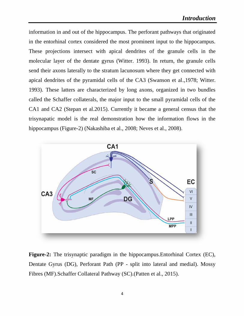
Introduction
4
information in and out of the hippocampus. The perforant pathways that originated
in the entorhinal cortex considered the most prominent input to the hippocampus.
These projections intersect with apical dendrites of the granule cells in the
molecular layer of the dentate gyrus (Witter. 1993). In return, the granule cells
send their axons laterally to the stratum lacunosum where they get connected with
apical dendrites of the pyramidal cells of the CA3 (Swanson et al.,1978; Witter.
1993). These latters are characterized by long axons, organized in two bundles
called the Schaffer collaterals, the major input to the small pyramidal cells of the
CA1 and CA2 (Stepan et al.2015). Currently it became a general census that the
trisynapatic model is the real demonstration how the information flows in the
hippocampus (Figure-2) (Nakashiba et al., 2008; Neves et al., 2008).
Figure-2: The trisynaptic paradigm in the hippocampus.Entorhinal Cortex (EC),
Dentate Gyrus (DG), Perforant Path (PP - split into lateral and medial). Mossy
Fibres (MF).Schaffer Collateral Pathway (SC).(Patten et al., 2015).

Introduction
5
3-Functions of the hippocampus:
It has been a big fallacy in most people’s believe of linking the role of
hippocampus merely with learning and memory. Studies have shown this part of
the brain plays a major role in stress response feedback, motor and emotions
control. It was very hard for us to resume the function of the hippocampus in one
or two pages since hundreds of books have been written about this subject.
3-1-Learning and memory:
The hippocampus is considered the center of acquiring and learning new
experiences which called short term memory (Burgess et al, 2002). It is worthy to
note that the hippocampus plays a major role in the embedding, storing and
consolidation of of the information for a long period in the isocortex to form
another different kind of memory that called long-term memory (Dudai, 2012,
Adam et al.2014). Also the hippocampus plays a major role in processing, storing
and retrieving different kind of memory such as episodic memory, a memory that
permits conscious recollection of different events (Squire, 2007; Eichenbaum,
2000; Kopelman, 1993). In addition, the hippocampus plays a chief role in spatial
memory that involves spatial location recognition (Squire, 2007; Herman et al.,
1989).
3-2-Stress responses:
Today it is proofed and well known that the hippocampus plays an important
role in the regulation of stress response (Kjelstrup et al.2008), via the regulation of
the hypothalamic–pituitary–adrenal axis (HPA) axis and suppression of the ACTH
secretion from the hypothalamus, a direct inducer of production and secretion of

Introduction
6
glucocorticoids in the adrenal gland (Lathe.2001, Gutiérrez. 2003). Furthermore,
since the early 1960s it has been known that hippocampal removal (or section of
the fornix) precipitates adrenal hypersecretion of glucocorticoids (Fendler et al.,
1961; Knigge, 196 1; Moberg et al., 1971).
3-3-Motor activity:
Since James Papez in 1947 defined the hippocampus as part of the limbic
system (Arszovszki et al.2014), a common believe started to emerge about the
involvement of the hippocampus in the control of ventral striatal loop, a group of
brain areas responsible for motility control (Humphries and Prescott., 2010).
The ventral striatum is not only important for spatial processing and
contextual cues (Hauber and Sommer.2011; Ferretti et al., 2010), processes the
information relevant to effort (Hauber and Sommer.2011). The nucleus
accumbens, the main structure of the ventral striatum, can be divided into core and
shell subregions. The shell receives hippocampal inputs predominantly from
ventral CA1 and subiculum, whereas the core receives them from dorsal CA1,
subiculum and parahippocampal regions (Voorn et al., 2004).
Introduction
7
Chapter 2: Neuroinflammation
1-Astrocytes:
1-1-Origin:
The german neuropathologist Rudolf Virchow was the first scientist that
described the astrocytes as nerve glue in the second half of the nineteenth century
(Scheller et al., 2011). Astrocytes are the most abundant neuroglial cells in the
central nervous system, generally they outnumber neurons by five folds and they
can be found all over the brain (Romero et al., 2014). It is now clear that astrocytes
are of two types, white matter abundant astrocytes called fibrous and grey matter
located astrocytes called protoplasmic (Parpura et al.,2012). The two types differ
widely in their anatomy and physiology (Shigetomi et al., 2008).
The astrocytes processes ensure thousands of contacts with neural synapses
per cell. Despite the large number of the processes that astrocytes possess, these
cells tile the entire central nervous system in extraordinary network-like fashion
where no overlapping between the cells can be seen, using the gap junctions as an
essential way of communication between each other to meet the transportation of
different mediators between the cells (Giaume et al., 2010).
1-2-Morphology:
The morphology of astrocytes differs from one region of the brain to another
where we can find fibrous astrocytes with the processes oriented along the fiber
tracts in the white matter (Wang et al., 2008). Protoplasmic astrocytes abundant in
the grey matter such as the dentate gyrus of the hippocampus and corpus callosum,
their processes are oppositely directed against the nerve fibers (Oberheim et
Introduction
8
al.,2012;Bushong et al., 2003). The possession of multiple processes enable
astrocytes to envelope the pre and postsynaptic terminals and play a remarkable
role in fluid, ions, PH and neurotransmitters homeostasis in the synaptic space
which is very crucial for the synaptic functioning and transmission (Brown and
Ransom, 2007).
Interestingly, the heterogeneous nature of astrocytes population based on
their morphology, expression of different sets of receptors, transporters, ions
channels and other proteins raises the intriguing possibility that different subtypes
of astrocyte are implicated in distinct metabolic/homeostatic functions (Matyash
and Kettenmann, 2010).
1-3-Role in the CNS:
Astrocytes play a pivotal role in maintaining the homeostasis of the central
nervous system (Placone et al., 2015), through ensuring a constant equilibrium of
ions and water levels in the neuronal surrounding where a quick internalization of
potassium ions by astrocytes during action potential takes place to maintain
extracellular low levels of potassium according to the physiological needs
(Sofroniew et al., 2010).
In addition, in order to sustain the energy demands and due to their
possession of lactate shuttle, astrocytes supply neurons by lactate during critical
moment where the oxygen is not sufficient for energy production (Magistretti,
2006; Tsacopoulos and Magistretti, 1996). In line with this, it has been shown that
astrocytes play an important role in neurovascular and neurometabolic coupling.
Indeed, neuronal activity triggers the release of a variety of vasoactive substances
by astrocytes such as prostaglandins (PGE), nitric oxide (NO) and arachidonic acid
(AA) to maintain the harmony in the nervous system (Gordon et al., 2007; Iadecola

Introduction
9
and Nedergaard, 2007). In addition, there is a growing evidence that astrocytes
influence directly neuronal communication through monitoring the release of
multiple substances including glutamate, purines (ATP and adenosine), (γ-
Aminobutyric acid) (GABA) (Halassa et al., 2009; Nedergaard et al., 2003; Perea
et al., 2009; Shigetomi et al., 2008). Considering the metabolic relation between
the two cells, we can assume the dependence of neurons on astrocytes to maintain
the homeostasis and the integrity of the brain. It also permits us to predict any
defection in the astrocyte machinery may lead to serious consequences in the brain
(Figure-3).
Figure-3: Role of astrocytes in the brain (Sofroniew and Vinters.2010)

Introduction
10
It has been demonstrated that these metabolic relations between neurons and
astrocytes qualified the later to play a very important role in brain physiology and
ultimately in one of its sophisticated activities such as breath controlling (Gourine
et al., 2010) and sleep homeostasis (Halassa et al., 2009).
Furthermore, several studies have evidenced the contribution of astrocytes in
cognitive functions of the brain such as memory consolidations (Gibbs et al.,
2008). Numerous homeostatic functions of astrocytes have been observed
including defense against oxidative stress, energy storage in the form of glycogen
and tissue repair. In addition they play a great role in the formation and remodeling
of synapses (Perea et al., 2009; Shigetomi et al., 2008).
2-Microglia:
2-1-Origin:
Parallel to the periphery, the central nervous system has its own immune
cells called microglia, The discovery of microglia returned to the twenties of the
last century where the Spanish pioneer Pio del Rio-Hortega was able to describe
the microglia differently from the astrocytes (Kettenmann et al., 2011). Microglia
are known as CNS resident macrophage due to the huge similarity between the two
cells in terms of function and phenotype (Habib and Beyer, 2015; Benedek et al.,
2017).
Both macrophage and microglia cells express major histocompatibility
complex (MHC) antigens, as well as T and B cell markers such as various cluster
of differentiation (CD) proteins (McGeer and McGeer, 1995; Perry et al., 1985;
Williams et al., 1994). However, still some differences that distinguish the two
cells, contrary to the macrophages that are in constant contact with serum proteins,
a slightest disruption in blood-brain barrier which ultimately results in leakage of

Introduction
11
serum protein can induce the activation of microglia (Ransohoff and Perry, 2009).
Another distinction, unlike the macrophages that are continuously replaced by new
myeloid progenitors, microglia cells are generated upon activation which could be
represented by an ultimate activation (Kettenmann et al., 2011; Ransohoff and
Perry, 2009).
2-2-Morphology:
Microglia cells represent 15-20% of the total number of cells in the brain
(Carson et al., 2006) and it is worthy to note that the density of these immune cells
are slightly different from one region to another across the brain where up to 12%
of substantia nigra cells are microglia, this applies to only 5% of cells in corpus
callosum. Similarly, microglia morphology varies considerably where in the white
matter they have elongated somata and the processes are preferentially oriented
along the fiber tracts. On the contrary, microglia in the circumventricular organs, a
region characterized by a leaky blood–brain barrier they exhibit a compact
morphology with a few short processes. In the gray matter the microglia exhibit
many elaborate radially oriented arbors (Harry and Kraft, 2012).
There has been a long debate concerning the origin of these cells when the
common believe that these cells are originated in the neuroectoderm (Fedoroff et
al., 1997). However, today due to the cutting edge technology it has become a
general consensus that microglia are the descents of progenitors that have been
originated from embryonic mesoderm in the periphery and migrated to the brain
during the development (Kettenmann et al., 2011).

Introduction
12
2-3-Role in the CNS:
Microglial cells are considered the first barrier of defense in the brain
parenchyma (Piirainen et al., 2017). They play a very important roles in the
regulation of neuronal processes development, maintenance of the neural
environment, response to injury and subsequent repair. Microglia cells are similar
to monocyte, actively surveying and shaping the structure and function of the
neuronal circuit (Wake et al., 2013).
Phagocytosis is the process of terminal removal of cellular debris by
microglia (Tremblay et al., 2011). The postnatal regulation of the number of
neurons in the brain requires the induction of apoptosis in wide number of neurons
which leads to the accumulation of cellular debris that requires its clearance by
phagocytosis (Lee et al., 2010), this phenomena followed by synaptic remodeling
and arrangement. Activated microglia were seen at different regions of the brain
where synaptic remodeling is active like thalamus, cerebellum, olfactory bulb and
hippocampus revealing the contribution of microglia in the development of the
neuronal circuitry at early stage (Figure-4) (Dalmau et al., 1998; Fiske and
Brunjes, 2000; Perry et al., 1985).
Furthermore, electron microscopy observation proofed the involvement of
these cells in synaptic network remodeling (Wake et al., 2013) through
phagocytosis in the developed brain. In addition, microglia cells were observed
near to dendritic spines in mouse cortex in an experience-dependent manner
indicating the contribution of such cells in synaptic plasticity (Schafer and Stevens,
2015).The discovery of neurogenesis during adulthood in the subgranular layer of
the dentate gyrus of the hippocampus (Gouldet al., 1997), revealed another activity
of microglia during this process where studies showed the majority of newborn
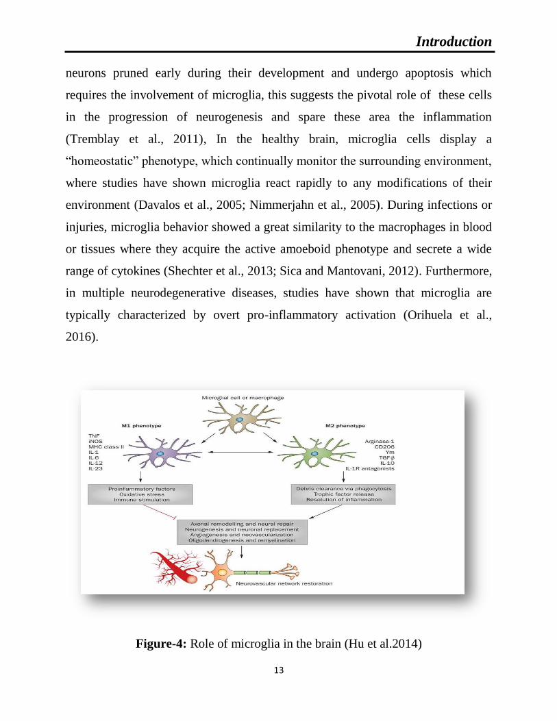
Introduction
13
neurons pruned early during their development and undergo apoptosis which
requires the involvement of microglia, this suggests the pivotal role of these cells
in the progression of neurogenesis and spare these area the inflammation
(Tremblay et al., 2011), In the healthy brain, microglia cells display a
“homeostatic” phenotype, which continually monitor the surrounding environment,
where studies have shown microglia react rapidly to any modifications of their
environment (Davalos et al., 2005; Nimmerjahn et al., 2005). During infections or
injuries, microglia behavior showed a great similarity to the macrophages in blood
or tissues where they acquire the active amoeboid phenotype and secrete a wide
range of cytokines (Shechter et al., 2013; Sica and Mantovani, 2012). Furthermore,
in multiple neurodegenerative diseases, studies have shown that microglia are
typically characterized by overt pro-inflammatory activation (Orihuela et al.,
2016).
Figure-4: Role of microglia in the brain (Hu et al.2014)

Introduction
14
3-Inflamatory Cytokines:
3-1-Interleukin-1:
First described as lymphocytes activating cytokine in 1972 (Gery and
Waksman, 1972). Interleukin-1, is a term used to designate a class of three proteins
interleukin-1α, interleukin-1β and interleukin-1ra (receptor antagonist), encoded by
three separate genes located on the chromosome 2.
The morphological examination of the primary structure of IL-1α and IL-1β
revealed a slight similarity between the two molecules that does not exceed 26% of
homology. However, they share a quite similarity for the tertiary structure (Murzin
et al., 1992) which enables the two molecule to have a common affinity for the two
IL-1 receptors. In addition to morphological similarity, both cytokines can be
regulated by the same regulatory machine and exert quite the same biological
activities (Auron et al., 2007). The main producers of these cytokines are
macrophages. However, many other cellular sources have been identified like
neutrophils, lymphocytes, dendritic cells, keratinocytes, endothelial cells,
hepatocytes, fibroblasts (Vicenová et al., 2009) and tumor cells (Miller et al., 2000;
Wolf et al., 2001).
It is worthy to note that IL-1α is continuously produced by cells. However,
in the case of IL-1β unless the cells receive a stimulatory factors during
inflammation they are not able to produce it (Pelegrin and Surprenant, 2006). The
principal targets of IL-1 are primarily cells of the immune system such as
monocytes, lymphocytes, granulocytes and dendritic cells. The effects of these
cytokines are not merely immune but they may have an influence on non- immune
cells such as epithelial cells, fibroblasts and endothelial cells (Vicenová et al.,
2009). In the brain, IL-1 is constantly expressed at low levels in healthy adult brain
by a variety of cell types both neuronal and glial cells (Vitkovic et al., 2000).

Introduction
15
Due to the close similarity of the general effect of IL-1α and IL-1β, it is
important to emphasize here that when we mention IL-1β only, we are referring to
the effect shared by the two cytokines. IL-1β is a pluripotent, proinflammatory
factor that exert and orchestrate inflammatory responses in the periphery
(Dinarello, 1996). Interleukin 1 is considered one of the most potent activators of
T cells and indirectly influences the activation of B cells. Such cytokine exerts a
regulatory effect on different components of the inflammatory cascade such as
nuclear factor kappa B (NF-κB) and activator protein-1 (AP-1). Furthermore, it has
the capacity to promote the activation of certain genes involved in cell division,
survival and development of the vascular system (Schneider et al., 1998).
It has been shown that IL-1 molecule has a pluripotent effects on multiple
brain functions including normal regulation of non-rapid eye movement (slow-
wave) during sleep (Krueger et al., 2001), normal synaptic remodeling (Schneider
et al., 1998), modulation of the hypothalamic pituitary-adrenal hormonal axis
(Rothwell and Luheshi, 2000) and the appetite-suppressing hormone, leptin
(Friedman and Halaas, 1998). It has been found in response to neuronal insult the
microglia increase the production of IL-1 in the brain.
3-2-Interleukin 6:
At the beginning IL-6 was known as differentiation factor for B-cells
through promoting the differentiation of these cells from it active form to an
antibody producing cells (Hirano et al., 1986; Mihara et al., 2012).The majority of
the immune cells (T and B cells, monocytes) produce IL-6. In addition, multiple
non-immune cells like fibroblasts, keratinocytes, endothelial cells, meningeal cells,
adipocytes and some tumor cells have the capacity to produce such
proinflammatory cytokine (Scheller et al., 2011). In the nervous system both glial

Introduction
16
and neuronal cells are expressing IL-6 and its receptors (Gadient and Otten, 1993;
Schöbitz et al., 1993).
IL-6 exerts its effects through attachment to its transmembrane receptor, this
latter is associated to a signaling receptor protein gp130 and it is worthy to note
that IL-6 influences multiple cells by triggering different effects via its unique
receptor and gp130 (Mihara et al., 2012). Several studies have shown that IL-6
plays a major role in the brain in different perspectives such as neurogenesis, a
phenomenon which neurons are able to produce new cells (Bauer et al., 2007;
Deverman and Patterson, 2009). In addition to its promoting effect in the
production of new neurons, IL-6 has the capacity to influence the determination of
cholinergic neurons phenotype (Fann and Patterson, 1994; März et al., 1998).
The influence of this cytokine is not restricted only to neurons but it shows a
major effect on non-neuronal cells like astrocytes and microglia where an
overexpression of such cytokine in transgenic mice displays a massive prominent
astrogliosis and microgliosis (Campbell et al., 1993; Chiang et al., 1994; Fattori et
al., 1995; Giralt et al., 2002). On the flip side, IL-6 knockout mice showed
alteration of both cell populations activity in different models of neuronal injury
(Cardenas and Bolin, 2003; Galiano et al., 2001; Penkowa et al., 1999a; Sugiura et
al., 2000). Also it promotes sprouting and functional recovery of organ typical
cultures of the hippocampus (Hakkoum et al., 2007).
IL-6 is famously known as pro-inflammatory cytokine which refers to its
early production during infection and injuries. Such effect returns to the expression
of IL-6 by the damaged or infected area. IL-6 along with IL-1β or TNF-α induce
attraction of neutrophils to the site of injury, Down regulation of such cytokine
halts such activity (Hurst et al., 2001; Jones, 2005; Scheller et al., 2011). In
addition, to the neuronal and inflammatory activities of IL-6, other effects have
been attributed to this cytokine such as metabolic activity where they showed

Introduction
17
during exercise the level of IL-6 increases significantly (Pedersen and Febbraio,
2008; Pedersen et al., 2003). Furthermore, IL-6 knockout mice develop glucose
intolerance and insulin resistance (Matthews et al., 2010).
3-3-Tumor necrosis factor (TNF-α):
Tumor necrosis factor (TNF, also known as TNF-α) was first described as a
physiological agent that has the capacity to induce hemorrhagic necrosis into
tumors transplant in mice (Carswell et al., 1975). Since then, it has been
recognized as a potent player in inflammation (Bradley, 2008). Similar to IL-1β
and IL-6, TNF-α is produced by different types of cells; the main sources of this
cytokine are immune cells such as T cells, macrophages and natural killer cells
(Sedger and McDermott, 2014).
The expression of TNF-α was observed in the brain by microglia cells in
1986 (Frei et al., 1987) and since then it becomes well known that the majority of
the CNS cells have the capacity to produce it (Sternberg, 1997).
Two transmembrane receptors have been identified for such cytokine TNF-
R1 (p55) and TNF-R2 (p75) and almost all the cells express at least one of the two
receptors (Wiens and Glenney., 2011). Interestingly, this cytokine induces different
effects on different cells and this referred to non-homology of the two receptors
where the structural examination revealed a minor similarity between the two
receptors. The genetic manipulation to the two receptors in mice revealed different
impact on the physiology (Pfeffer et al., 1993). It has been reported that TNF-α
acts as multifunctional cytokine exerting a variety of immunological functions by
up-regulating the production of immune cytokines, synthesis of prostaglandin E2
and reactive oxygen species (Pfeffer., 2003).

Introduction
18
It is worthy to note that TNF-α induces a contradictory actions on different
cells, studies have shown the diversity of the actions of such cytokine depends on
the kind of intracellular signaling induced where TNF-α has the ability to induce
cell death through the activation of both caspase-3 and caspase-8 on one hand
(Nicholson and Thornberry, 1997). On the other hand, the same cytokine plays a
pivotal role in cell survival through the activation of NF-kB and mitogen-activated
protein kinase (MAPK)(Liu and Han, 2001; Van Antwerp et al., 1996).
TNF-α is a pro-inflammatory cytokine that has a central role in the
protection against microbial agents in different animals. In addition to its
physiological effect on promoting T cells proliferation and the anticoagulant
properties of endothelial cells (Beutler et al., 1988; Tracey et al., 1993). It has been
found an excessive expression of such cytokine leads a pathogenesis of both acute
and chronic inflammatory diseases like AIDS, arthritis and cancer (Beutler et al.,
1988; Tracey et al., 1993). In the central nervous system, TNF-α is produced
primarily by microglia and astrocytes in response to a wide range of pathological
processes including infection, inflammatory disease, ischemia and traumatic injury
(Liuet al.1994).

Introduction
19
Chapter 3: Oxidative Stress
It has been said that, “A disturbance in the pro-oxidant/antioxidant systems
in favor of the former may be denoted as an oxidative stress” (Davies, 2000). Since
pro-oxidant or reactive oxygen species are natural byproduct of our metabolism,
the body develops an antioxidant system to antagonize the damage that may result
from their excessive formation (Halliwell, 1999). In this section different kinds of
reactive oxygen species and mechanisms of defense that are able to neutralize their
deleterious effects will be discussed.
1-Definition of Reactive oxygen species (ROS):
Reactive oxygen species (ROS) is a collective term used for a group of
oxidants, which are either free radicals or molecular species capable of generating
free radicals (Kunwar and Priyadarsini, 2011). Free radicals are chemical species
that lack an electron that made them unstable and became highly reactive in order
to pair up with other molecules and gain a state of stability.
2-Lipid Peroxidation:
Lipids are essential components of cell membrane that maintain structure
and control the function of the cell. They are primary targeted by ROS such as
oxygen free radicals. The oxidation of lipids is associated with various
pathological states (Esterbauer, 1993). Lipid peroxidation, or interaction of lipids
with reactive oxygen species has been an intensive area of research for decades
(Pratt et al., 2011).
Lipid peroxidation is initiated by hydrogen abstraction or addition of an
oxygen radical resulting in oxidative damage of polyunsaturated fatty acids

Introduction
20
(PUFA). The free radical chain reaction propagates until two free radicals
conjugate with each other to terminate the chain. The reaction can also be
terminated in the presence of a chain-breaking antioxidant such as vitamin E (α-
tocopherol) (Halliwell and Gutteridge, 1984). In the presence of transition metal
ions, ROOH can give rise to the generation of radicals capable of re-initiating lipid
peroxidation by redox-cycling of these metal ions (Halliwell and Gutteridge,
1984). Lipid peroxidation causes a decrease in membrane fluidity and in barrier
functions of the membranes. Many products of lipid peroxidation such as
hydroperoxides or their aldehyde derivatives inhibit protein synthesis, blood
macrophage actions, alter chemotactic signals and enzyme activity (Fridovich and
Porter, 1981).
The complex nature of the lipid peroxidation process and its potential
biological significance has attracted the attention of scientists from different fields
ranging from chemistry and biochemistry to biology and clinical sciences. Such
attention results in to the elucidation that lipid peroxidation is implicated in several
human diseases like atherosclerosis (Berliner and Heinecke, 1996), cancer
(Hammad et al., 2009; Wu et al., 2010), diabetes (Silverstein and Febbraio, 2009),
chronic alcohol exposure (Yang et al., 2010), acute lung injury (Imai et al., 2008;
Nonas et al., 2006) as well as neurodegenerative disorders like Alzheimer’s
(Montine et al., 2005) and Parkinson’s diseases (Porter et al., 2010).
3-Reduced glutathione (GSH):
Considered the most abundant thiol compound in all cells (Meister and
Anderson, 1983), GSH is a tri-peptide antioxidant found in all compartment of the
cell (Meister and Anderson, 1983).The importance of such antioxidant manifests in
its capacity to interact and neutralize the deleterious effect of reactive oxygen

Introduction
21
species, serving as a supplier of protons to antioxidant enzymes. The ratio between
reduced-GSH / oxidized-GSSG plays a major role as mirro to the oxidative status
of the cell (Jones, 2002). As mentioned above reduced glutathione plays at
different levels in the protection of the cell against reactive oxygen species. GSH
acts as a donor of proton to the oxidized lipids in the membrane during lipid
peroxidation and thereby stops the propagation of such deleterious phenomena
throughout the cell membrane which might lead to serious damage to the cell
(Curello et al., 1985).
In addition, it plays a role as co-substrate for glutathione peroxidase and
transferases during peroxidation (Gregus et al., 1996). Another antioxidant role of
GSH appears through the reduction of major non enzymatic oxidants such as
vitamin C. Furthermore, it is involved in the detoxification process of certain
xenobiotics (Gregus et al., 1996; Halliwell and Gutteridge, 1984).
4-Catalase:
Catalase is one of the important enzymes in the defenses against free
radicals. It was first noticed unknowingly in 1811 when Louis Jacques Thénard
discovered hydrogen peroxide and proposed that an unknown substance is causing
its breakdown (Loew, 1900). More than one century later, catalase from beef liver
was crystalized by B. Sumner and A. Dounce (Sumner and Dounce, 1937).
Catalase is an ubiquitous enzyme found almost in all living cells and considered
one of the most active enzyme, studies have shown the turnover of such enzyme is
more than multiple millions of hydrogen peroxide molecules to water and oxygen
in each second.
The hydrogen peroxide detoxification feature of catalase plays an important
role in the protection against one of the dangerous reactive species and spares the
Introduction
22
cell from the damage that might result from the attack of cellular components of
hydrogen peroxide. In addition, it has been found that the survival rates was
extended after CAT containing liposomes were injected after exposure to 100% of
oxygen (Speranza et al., 1993). Catalase plays an important role in the
management of oxidative stress during certain pathophysiological conditions,
inflammatory diseases, aging and cancer (Kim et al., 2002).
5-Superoxide Dismutase (SOD):
Superoxide dismutase (EC 1.15.1.1) is a class of oxide-reductase enzymes
and one of the most important antioxidant enzymes in the neutralization of reactive
oxygen species due to its capacity to dismute the superoxide anion and made less
reactive species such as H2O2, thereby reducing the likelihood of superoxide anion
interacting with nitric oxide to form reactive peroxynitrite. There are three
isoforms for SOD named after their localization in the cell where we can
distinguish cytosolic Cu/Zn-SOD, mitochondrial Mn-SOD and extracellular SOD
(EC-SOD). Interestingly it has been observed that through all over the body, the
amount of Cu/Zn-SOD are double as large as the Mn-SOD (Marklund, 1980).
Isoforms of SOD are variously located within the cell. CuZn-SOD is found in both
cytoplasm and nucleus. Mn-SOD is confined to the mitochondria, but can be
released into extracellular space (Reiter et al., 2000).
Extracellular superoxide dismutase (EC-SOD) is a secretory, tetrameric,
copper and zinc containing glycoprotein with a high affinity for certain
glycosaminoglycans such as heparin and heparin sulfate. EC-SOD was found in
the interstitial spaces of tissues and also in extracellular fluids, accounting for the
majority of the SOD activity in plasma, lymph and synovial fluid (Marklund, 1980;
Sandström et al., 1994).

Introduction
23
Chapter 4: Relation between neurodegeneration,
neuroinflammation and oxidative stress
1-Gliosis and cell death:
The traditionally perceived of the passive role of microglia in the brain as
maintenance cells has been questioned and microglia are now accepted as active
mediators of neurodegeneration (Block and Hong, 2005). It has been demonstrated
in Alzheimer’s disease, Aβ is responsible for the activation and recruitment of
microglia in-vitro (Davis et al., 1992; Sasaki et al., 1997). In line with this
observations Craft et al., 2004 demonstrated in AD animal models, the
intraventricular infusion of Aβ1–42 peptide along with inhibition of glial activation
by aminopyridazines led to the reduction of neuropathlogical symptoms seen in
this model. The degeneration of the nigro-striatal dopaminergic (DA) pathways is
the major feature in the brain of Parkinson’s disease (PD) patients (Olanow and
Tatton, 1999). Substantia niagra is recognized as the richest region with microglia
in the brain which contains 4.5 fold compared to neocortex and other regions (Kim
et al., 2000). It has been shown beside the direct toxicity of the neurotoxin, rotenon
to dopaminergic neurons, this substance has the ability to recruit and involve the
microglia in the neurodegeneration occurring in such PD model (Gao et al., 2003).
Frontal and anterior temporal loss of neurons is typical feature of Fronto-temporal
dementia (FTD) (Munoz et al., 2003). In an attempt to investigate the involvement
of the inflammatory process in neurodegeneration in such diseases, the work of
Schofield et al., 2003 showed the activation of microglia at early stages of the
disease, providing a rare evidence of an early and perhaps initiating role of
microglial inflammation in fronto-temporal dementia (Figure-5).

Introduction
24
Figure-5: Role of Reactive microgliosis in neuronal cell death
(Lull and Block. 2010).
Astrocytes are known as supportive cells to neurons, studies have shown
these cells play an important role in the clearance and degradation of Aβ
(Koistinaho et al., 2004; Li et al., 2011; Wyss-Coray et al., 2003) and have the
ability to phagocyte the fibers of Aβ (Jiang et al., 1998; Niino et al., 2001). In
support of this hypothesis many reports have shown the accumulation of the Aβ1–
42 and the amount of this material correlates positively with the extent of local AD
pathology (Nagele et al., 2003). In vitro, the Aβ peptide decrease the uptake of the
neurotransmitter glutamate by astrocytes and neurotoxin substance likewise. In
addition, they found such peptide promotes the mitogen-activated protein kinase
cascades in astrocytes that triggers a state of astrogliosis (Agostinho et al., 2010;
Matos et al., 2008). Several studies showed while the astrocytes performing their

Introduction
25
assisting and repairing activity in the brain, their direct involvement in neural
damage (Pekny et al., 2014; Placone et al., 2015). From pathological examinations,
an increase in the number of astrocytes as well as in GFAP expression is observed
in PD (Ciesielska et al., 2009; Muramatsu et al., 2003). There are abundant
evidences that astroglial abnormalities and physiological dysfunction precede
clinical disease. These changes include reactive astrocytosis that can be seen many
months before motor neuron degeneration (Bruijn et al., 1997).
Multiple Sclerosis is a chronic inflammatory demyelinating disease of the
central nervous system in which glial cells play a prominent role. In murine
experimental autoimmune encephalomyelitis (EAE), an established animal model
of multiple sclerosis, astrocyte hypertrophy coincided with manifestation of axonal
damage (Wang et al., 2005).
2-Cytokines and cell death:
In this work the focus was was on a subset of cytokines called interleukins,
substances that play a major role in the inflammatory process and the host
defenses. In the last three decades there are growing evidences concerning the
implication of the interleukins in the endangerment of the cell survival and
ultimately in cell death or neurodegeneration.
One of the most studied cytokines and its involvement in neurodegeneration
is IL-1β, such cytokine presents at high rate in postmortem tissue of AD and PD
patients. In addition to that, different studies have proven the induction of an array
of responses such as damage to the cerebral vasculature, activation of glia, amyloid
precursor protein, adhesion molecules and corticotrophin-releasing factor (CRF)
(Betz et al., 1996; Rothwell, 1997; Rothwell et al., 1997, 1996). Related findings
showed the involvement of such cytokine in the endangerment of the neuronal

Introduction
26
survival where its levels raise in few hours following the administration of multiple
neurotoxins. If the only conclusion that can be drawn from those studies is the
possibility of such cytokine in the initiation of the endangerment of the cell
survival and even indirectly the activation of the glial cell that may contribute to
the cell death later on (Allan and Rothwell, 2001; Boutin et al., 2001; Griffin and
Mrak, 2002; Lawrence et al., 1998; Pearson et al., 1999; Perry et al., 2003).
Accumulating evidences from both animal and human studies have indicated
that chronic inflammatory processes contribute to the pathogenesis of
neurodegenerative diseases and IL-6 is one of the cytokines that is strongly
correlated to the onset and progression of the disease. Accordingly in IL-6
deficient mice the inflammatory response is significantly reduced during
pathological conditions of the brain (Eugster et al., 1998; Klein et al., 1997;
Mendel et al., 1998; Penkowa et al., 1999b; Raivich et al., 1999).
Studies have shown the knockout of IL-6 gene in mice causes a significant
decrease in the activity and recruitment of microglia and astrocytes after brain
injury (Penkowa and Hidalgo, 2000; Penkowa et al., 1999a). Similar observation
has been made during facial nerve transaction (Klein et al., 1997). It is estimated
that IL-6 plays a major role in the induction of acute-phase proteins (APP) and the
presence of these proteins (i.e. α1-anti-chymotrypsin, α2-macroglobulin, and C-
reactive protein (CRP) proteins) involved in the alteration of the normal process of
the key proteins that caused cell death in the neurodegenerative disease such as
levy body, amyloid precursor protein (APP) and mitochondrial dysfunction in
Amyotrophic lateral sclerosis (Graeber and Streit, 2010; Hofmann et al., 2009).
TNF-α is considered one of the major cytokine in the immune defense and
plays an important role in the activation of immune cells in the brain but studies
have shown a double effect of such cytokine where TNF-α positive astrocytes have
been found in all over the areas of the lesion reflecting the possibility of its

Introduction
27
contribution in neuronal death in the lesioned area (Okuda et al., 1995; Probert et
al., 1997; Renno et al., 1995; Selmaj et al., 1991).
A study showed a very elevated concentration of TNF-α in activated
microglia that are in proximity of the amyloid plaque in the brain of Alzheimer
disease patients (Blasko et al., 1999; Meda et al., 1995). In addition, the increase in
the concentration of such cytokine has been found only after 30 min of ischemia
and such an increase has been shown to be damaging to the neuronal cells (Buttini
et al., 1996; Forloni et al., 1997; Lavine et al., 1998; Wallach et al., 1997).
3-Oxidative stress and neurodegeneration:
The deleterious effects of the oxidative stress in neurodegeneration have
received a lot of attention from the scientific community and a new kind of
thinking started to emerge like considering the anti-oxidative therapy as an
alternative in the treatment of neurodegenerative disease. In this section different
diseases and different neurodegenerative models where oxidative stress played a
decisive role in the neuronal survival will be discussed.
Several researchers considered the brain to be abnormally sensitive to
oxidative damage and many studies demonstrate the vulnerability of the brain to
lipid peroxidation (Chance et al., 1979; Floyd and Carney, 1992; Zaleska and
Floyd, 1985) and this could be due to the abundance of fatty acids in the brain
(Butterfield et al., 2002).
In addition to that, the brain is not well equipped with very strong
antioxidant machinery which makes the fight against the reactive oxygen species
not an easy mission. Studies have shown that brain contains 10 % of antioxidant
compared to the liver (Uttara et al., 2009). Furthermore, 20% of the oxygen of the
body is consumed by the brain (Zaleska and Floyd, 1985). It have been found an

Introduction
28
increase in the amount of H2O2 and O2•–
production in the mitochondria in aged
mouse brain. The kainic acid model has been used extensively to study different
ways of neurodegeneration and multiple studies have showed a significant link
between the cell death and the raise in the levels of reactive oxygen species in such
model (Bondy and Lee, 1993; Ueda et al., 1997). Peroxidation of the plasma
membrane is one of the major signs of oxidative stress due to its impact on the cell
survival and the induction of apoptotic markers, studies have shown the
implication of the lipid peroxidation in the toxic effects of many chemicals in
many tissue injuries and disease processes. Free radicals have been reported for
their great contribution to neuronal loss in cerebral ischemia, seizure disorders,
schizophrenia, Parkinson's disease and Alzheimer's disease (Uttara et al., 2009).
Concerning the implication of oxidative stress in different neurodegenerative
diseases different studies have shown through the examination of postmortem
tissue of AD patients a significant increase in lipid peroxidation in specific areas of
the brain (Lovell et al., 1995; Pappolla et al., 1998).


Materials and Methods
30
Aim of the study:
The regional ethical committee of the College of Medicine and Health
Sciences (UAE University) approved this study (RECA/01/05).
The aim of the current study was to examine the impact of short-term
bilateral adrenalectomy on neuronal cell death in different regions of the
hippocampus, production of pro-inflammatory cytokines IL-1β, IL-6 and TNF-α,
the activation of different glial cells astrocytes and microglia, oxidative stress
MDA, GSH and SOD, the impact of the absence of such glucorticoids on the
animal’s behavior and the possibility of their association with neurodegeneration.
2- Animals and adrenalectomy surgery:
Eight-week old male Wistar rats 170–220g obtained from the animal facility
of College of Medicine and Health Sciences, United Arab Emirates University (Al
ain, United Arab Emirates) were used in the current study. All animals were placed
in Plexiglas cages (Techniplast, Milan, Italy); four rats per cage, maintained in a
temperature (22°C) and 12:12 light–dark cycle conditions, animals received ad
libitum access to food (National feed and flour production and marketing
company, Abu Dhabi, UAE) and water throughout the experiment.
Adrenalectomized rats were provided with 0.9% saline instead of drinking
water in order to maintain electrolyte balance and prevent the deleterious effects of
sodium chloride (Sigma-Aldrich, Missouri, USA) insufficiency.
Under Pentobarbital (Ilium-Troy Laboratories, New South Wales, Australia)
anesthesia (35 mg/kg body weight), rats were subjected either to adrenalectomy or
to sham operations (laparotomy) as described by Sloviter et al. 1989. Shaving the
back of the animals was done by using an electric shaving machine (Wahl, Illinois,

Materials and Methods
31
USA). The rat was placed on the surgical table ventrally and in order to perform an
aseptic surgery the back of the animals was cleaned with 70% ethanol (Sigma-
Aldrich, Missouri, USA).
Bilateral incisions were made into the skin and the dorsal muscle to access
the peritoneal cavity. The size of the muscle incision was just big enough to expose
the adrenal gland on the top of the kidney and some free space to remove it without
leaving any intact residuals of the gland. The peri-adrenal fat was grasped using a
blunt forceps and the adrenal gland was exteriorized and with a tipped-blunt ceaser
a cut was made at the connective tissue between the kidney and the adrenal gland.
After that the incision was sutured and the animal was returned to the cage. The
same procedure was applied for the sham operated animal except the removal of
the adrenal gland.
3-Determination of serum corticosterone levels by Enzyme
Immunoassay (EIA):
The levels of corticosterone in the serum were used to assess the
effectiveness of adrenalectomy, approximately 1 ml of blood samples were taken
directly from the heart at the time of sacrifice. For both biochemical and
histological assessments (n=120) rats were sacrificed by decapitation after surgery
at different times: 4h (n=24), 24h (n=24), 3days (n=24), 1week (n=24) and 2weeks
(n=24). The sera samples were stored at -80°C until corticosterone levels were
measured by Enzyme Immunoassay (EIA) Kit (Life sciences, Lausen,
Switzerland). The assay is based on the competitive binding technique. Briefly, a
polyclonal antibody specific for corticosterone becomes bound to the donkey anti-
sheep antibody coated onto the micro- plate. Following a wash to remove excess
polyclonal antibody, corticosterone present in the serum competes with a fixed

Materials and Methods
32
amount of horseradish peroxidase (HRP)-labeled corticosterone for sites on the
polyclonal antibody. This is followed by another wash to remove excess of the
conjugate and unbound sample. A substrate solution is added to the wells to
determine the bound enzyme activity. The color development is stopped and the
absorbance is read at 450 nm (Figure-6). The intensity of the color is inversely
proportional to the concentration of corticosterone in the sample. A standard curve
in the range 32 to 20,000 pg/ml was constructed to calculate corticosterone
concentrations in the samples.
Figure-6: Scheme of Enzyme Immunoassay (EIA) (Life sciences, Lausen,
Switzerland).

Materials and Methods
33
4-Biochemistry
4-1-Samples preparation:
Animals (n=80) were sacrificed by decapitation after surgery at different
times: 4h (Sham=8, ADX=8), 24h (Sham=8, ADX=8), 3days (Sham=8, ADX=8),
1week (Sham=8, ADX=8) and 2weeks (Sham =8, ADX=8). The brain was
removed, the hippocampus was immediately dissected out on ice, frozen in liquid
nitrogen (Sharjah Oxygen Company, Sharjah, United Arab Emirates) and stored at
−80°C until assayed. Thawed hippocampal tissues were homogenized in ice-cold
T-PER Tissue Protein Extraction buffer (Thermo Fisher Scientific,
Massachusetts, USA ), supplied with 1:100 protease inhibitor cocktail (Sigma-
Aldrich, Missouri, USA). After sonication, the samples were centrifuged at 16000
rpm for 30 min at 4°C and the supernatant was collected and stored at −80°C until
use. Protein concentrations were determined using Bradford’s protein assay kit
(Bio-Rad, California, USA)and levels of cytokines (IL-1β, IL-6 and TNF-α) were
assessed by ELISA using the commercially provided antibodies and standard
proteins (R&D Systems; Minneapolis, USA).
4-2-Determination of Total protein:
A set of standards is created in duplicate from a stock of Bovine serum
albumin (BSA) (Sigma-Aldrich, Missouri, USA) solution ranging from 0.2 to
1.4mg/ml. The 5X Bradford Reagent was diluted 1:4 with distilled water followed
by filtration using wattman1 filter paper (Whatman, Maidstone, United Kingdom).
A volume of 1.25ml of Bradford Reagent was added to each disposable
polystyrene tube. Our samples were diluted in the homogenation buffer 1:10 and
the same buffer was used as a blank for the assay. We added 25µl of the standards

Materials and Methods
34
and the samples into the labeled tubes containing the Bradford Reagent after the
time of incubation 5min. the absorbance was measured at 595nm.
4-3-Determination of hippocampal cytokines (IL-1β, IL-6 and TNF-α)
levels by Enzyme-Linked Immunosorbent Assay (ELISA):
Materials:
Equipment:
Micro-plates 96-well flat-bottom (NUNC, Roskilde, Denmark).
Plate Sealers
Plate reader (Tecan, Zürich, Switzerland)
Micro-plate autowasher (Tecan, Zürich, Switzerland).
Micro-plates shaker (Stuart, Staffordshire, United Kingdome).
ELISA Software (Magellan, version7,Tecan, Zürich, Switzerland)
Micropipette of different type (P10, P20, P200, p1000) and
multichannel Pipettes (BioHit healthcare, Helsinki, Finland).
Nylon Membrane Filters (0.2 μm) (Corning, New York. USA).
Reagents:
Deionized water
PBS (Phosphate-buffered saline) (Sigma-Aldrich, Missouri, USA),
PH 7.2-7.4, 0.2 μm filtered.
137 mM NaCl
2.7 mM KCl, 8.1 mM

Materials and Methods
35
Na2HPO4
1.5 mM KH2PO4
Wash Buffer: 0.05% Tween®20 (Promega, Madison USA) in PBS,
pH 7.2-7.4
Reagent diluent: 1% BSA in PBS, pH 7.2-7.4, 0.2 μm filtered.
Substrate Solution (Sigma-Aldrich, Missouri, USA): One to one
mixture of Color Reagent A (hydrogen peroxide H2O2) and Color
Reagent B (41.6 mM Tetramethylbenzidine).
Stop Solution: 2 N H2SO4 (sulfuric acid) (Sigma-Aldrich, Missouri,
USA).
Antibodies: All the antibody from (R&D Systems, Minnesota, USA)
Capture Antibody: Mouse Anti-Rat IL-6 of concentration 4μg/ml in
PBS.
Detection Antibody: Biotinylated goat Anti-Rat IL-6 of concentration
400ng/ml dissolved in Reagent diluent.
Streptavidin conjugated to horseradish peroxidase (HRP) dissolved in
Reagent diluent.
Standard antibody: Recombinant rat IL-6 290ng/ml dissolved in
Reagent diluent.
Methods: the Elisa test was performed through two main steps
Plate Preparation:
1. The micro-plate was coated (100μL) per well with the capture
antibody that was freshly diluted.

Materials and Methods
36
2. The micro-plate was sealed and left for incubation (time for capture
antibody to attach on the walls of each well) overnight at room
temperature (RT).
3. The following day the micro-plate was washed three times in the plate
autowasher machine by filling each well with Wash Buffer (400μL)
and aspiring it immediately afterward.
4. The complete removal of the wash buffer is very critical for the better
performance of ELISA and in order to ensure that the plate was
inverted and blotted gently against clean paper towels.
5. The micro-plate was blocked by adding to each well a (300μL)
freshly and filtered blocking buffer (BSA) and was left 1 hour for
incubation at room temperature (Optimal time found in our laboratory
for the BSA to occupy all the inter spaces that was not attached to
capture antibody)
6. The third and fourth steps were repeated for the second washing to
remove all the additional molecule of BSA and by then the plate
became ready for samples addition.
Assay Procedure:
Seven Standards were prepared through 2-fold serial dilution of the desired
recombinant Interleukin IL-1β, IL-6 and TNF-α see figure-7 for IL-6.

Materials and Methods
37
Figure-7: Two-fold serial dilution of IL-6 (R&D Systems, Minnesota, USA)
However, each interleukin has its own range of standards see table-1 for the serial
dilution of IL-1β and TNF-α.
Table -1: Serial dilution of IL-1β, IL-6 and TNF-α Standards preparation
1. Once the standards prepared we started to add 100μL from the Blank,
standards and hippocampal samples to each well of the coated plate. The
microplate was sealed and left for 2 hours incubation at room temperature
(Optimal time for the samples and standards to attach to the capture
antibody).
2. The third and fourth steps were repeated for the third washing to remove all
the non-adhered samples and standards.
1 2 3 4 5 6 7
IL-1β pg /ml 2000 1000 500 250 125 67.5 33.75
TNF-α pg /ml 500 250 125 62.5 31.25 15.62 7.81
IL-6 pg /ml 8000 4000 2000 1000 500 250 125

Materials and Methods
38
4. At this stage the micro-plate is ready for detection, (100μL) of detection
antibody per well was added. The plate was sealed and left for 2 hours
incubation (Optimal time for the detection antibody to attach to the capture
antibody and form a SANDWICH) at room temperature.
5. The third and fourth steps were repeated for the fourth washing to remove
the non-adhered molecules of detection antibody.
6. Away from the light (100μL) of Streptavidin-HRP per well was added and
left for 20min incubation at room temperature (Optimal time for
Streptavidin-HRP to attach to the detection antibody).
7. The third and fourth steps were repeated for the fifth washing to remove the
non-adhered molecules of Streptavidin-HRP.
8. At this step, the plate is ready to develop the color that reflect the amount of
the desired cytokine by adding (100μL) per well and away from light the
plate left for 20min at room temperature (optimal time for the HRP to react
with the substrate to generate the yellow color).
9. In order to stop the reaction (50μL) added of stop solution per well followed
by gentle shaking.
10. Immediately the optical density for each well was determined at 450 nm
using plate reader see Figure-8.

Materials and Methods
39
Figure-8: Scheme of Enzyme-Linked Immunosorbent Assay (ELISA) (R&D
Systems, Minnesota, USA).
The levels of IL-1β, IL-6 and TNF-α were evaluated by comparison with the
regression standard curve for each cytokine. The cytokine protein concentration
was divided by the total protein concentration obtained in the Bradford’s protein
assay to correct the differences in tissue volumes. Cytokine concentration is
reported as pg of cytokine/mg of protein.

Materials and Methods
40
4-4-Determination of the levels of oxidative stress markers (GSH, SOD,
and MDA) by colorimetric assay:
It is worthy to note here that the same homogenate that was used for the
evaluation of cytokines by ELISA was used for the colorimetric assay to determine
the levels of oxidative stress markers GSH, SOD and MDA.
4-4-1-Superoxide dismutase (SOD):
Materials:
Equipement:
Micro-plates 96-well flat-bottom (NUNC, Roskilde, Denmark).
Plate reader
Software.
Micropipette of different type (P10, P20, P200, p1000)
Multichannel Pipettes
Glass test tube.
Reagents: The kit was purchased from (Cayman chemical, Michigan, USA)
Deionized water
Assay buffer (50mM Tris-HCL, pH 8, containing 0.1mM
Diethylenetriaminepentaacetic acid (DTPA) and 0.1 mM
hypoxanthine)
Sample buffer 50mM Tris-HCL, pH 8.
Radical detector: Tetrazolium salt solution
Superoxide dismutase standards: Bovine erythrocyte SOD (Cu/Zn).
Xanthine oxidase

Materials and Methods
41
Methods:
SODs are metalloenzymes that catalyze the dismutation of the superoxide
radical (O2•-) into hydrogen peroxide (H2O2) and molecular oxygen (O2). The
measurement of the SOD amount in the hippocampal homogenate is based on the
use of the tetrazolium salt for detection of superoxide radicals generated by
xanthine oxidase and hypoxanthine.
Assay procedure:
Standards preparation: Seven Standards were prepared from the stock
solution (20 μL of Bovine erythrocyte SOD with 1.98 of sample buffer ) as follow
(Table -2),(figure-9).
Table -2: Serial dilution of SOD Standards preparation
1. A volume 10µl of each sampleand standard were added to the designated
wells of the microplate
2. Followed by adding 200 µl of diluted radical detector (diluted solution of 50
µl of tetrazolium salt with 19.95ml of distilled water).
3. Once the previous steps was completed, a volume of 20 µl of xanthine
oxidase was added to all the wells in order to initiate the reaction.
4. The plate was covered and incubated on the shaker for 20min at RT.
5. The plate was read at 440nm using plate reader.

Materials and Methods
42
6. SOD activity was calculated by using the equation obtained from the linear
regression of the standard curve substituting the linearized rate (LR) for each
sample. One unit of SOD is defined as the amount of enzyme needed to
exhibit 50% dismutation of the superoxide radical.
SOD (U/ml) = [(Sample LR-y-intercept/slope) × (0, 23/0.01)] ×sample dilution
Figure-9: Scheme of Superoxide dismutase assay, (Cayman chemical, Michigan,
USA)
4-4-2-Malondialdehyde (MDA):
The measurement of the TBARS amount in the hippocampal homogenate is
based on the interaction of thiobarbituric acid (TBA) with malondialdehyde
(MDA) under acidic conditions (acetic acid) and high temperature (95°C) using a
kit purchased from (Cayman chemical, Michigan, USA).

Materials and Methods
43
Materials:
Equipement:
Micro-plates 96-well flat-bottom (NUNC, Roskilde, Denmark).
Plate reader
Software.
Micropipette of different type (P10, P20, P200, p1000)
Multichannel Pipettes
boiler
5ml polypropylene crew-cap centrifuge tubes
Reagents:
Thiobarbituric Acid (TBA)
TBA Acetic acid used to prepare the color reagent.
TBA sodium hydroxide used to prepare the color reagent.
TBA malondialdehyde standard 500µM
TBA-SDS (sodium dodecyl sulfate) solution.
Assay procedure:
Standards preparation: A volume of 250μl of the MDA standard was mixed
with 750μl to obtain a stock solution of 125μM. Eight standards made out of the
stock solution see table-3.

Materials and Methods
44
Table -3: Serial dilution of MDA Standards preparation
1. A volume of 100µl of the hippocampal homogenate and standards were
mixed very well with the same amount of Sodium dodecyl sulfate (SDS)
solution.
2. A volume of 4ml of color reagent (2120mg of thiobarbituric acid (TBA)
dissolved in a mixture of 200 ml TBA acetic acid and 200 ml TBA sodium
hydroxide) was added.
3. The mixture of the capped 5ml tube was boiled for one hour.
4. Once the incubation completed, the tubes quickly transferred to an ice bath
for 10min in order to stop the reaction immediately.
5. The tubes were centrifuged at 16000g at 4°C for 10min.
6. Volume of 150µl of the resultant supernatant was taken and added to micro-
plate see figure-10.
7. The formed TBA-MDA adduct was measured calorimetrically at 530-
540nm. The MDA level was evaluated by comparison with the regression
curve for standard.

Materials and Methods
45
Figure-10: Scheme of the TBARS assay, (Cayman chemical, Michigan, USA)
4-4-3- Reduced glutathione (GSH):
The concentration of reduced glutathione (GSH) was determined in the
hippocampal homogenate using the commercial kit (Sigma-Aldrich, Missouri,
USA).
Materials:
Equipement:
Micro-plates 96-well flat-bottom (NUNC, Roskilde, Denmark).
Plate reader
Software.
Micropipette of different type (P10, P20, P200, p1000)
Multichannel Pipettes
Glass test tube.

Materials and Methods
46
Reagents:
Deionized water
Assay buffer: 500mM potassium phosphate, pH 7 containing 500mM
Ethylenediaminetetraacetic acid (EDTA).
Glutathione reductase: 400 units of glutathione reductase from baker’s
yeast in 3.6M ammonium sulfate, pH 7 containing 0.1 mM
dithiothreitol diluted in assay buffer.
Glutathione Reduced, Standard (10mM)
5,5’-Dithiobis(2-nitrobenzoic acid) [DTNB] solution: 1.5mg/ml of
Dimethyl Sulfoxide (DMSO) diluted in assay buffer.
5% 5-Sulfosalicylic Acid
Nicotinamide adenine dinucleotide phosphate NADPH solution:
40mg/ml diluted in assay buffer.
Dimethyl Sulfoxide (DMSO)
Assay procedure:
Standards preparation: A volume of 50μl of the GSH standard as stock
solution of 50μM. Five standards made out of the stock solution see Table -4.
Table -4: Serial dilution of GSH Standards preparation

Materials and Methods
47
1. The hippocampal samples were first deproteinized with the 5% of 5-
sulfosalicylic acid solution followed by centrifugation to remove the
precipitated protein and then assayed for glutathione evaluation.
2. The measurement of GSH uses a kinetic assay in which catalytic amounts
(nmoles) of GSH cause a continuous reduction of 5,5’-dithiobis (2-
nitrobenzoic acid) (DTNB) to TNB.
3. The GSSG formed is recycled by glutathione reductase and NADPH.
4. The yellow product, 5-thio-2-nitrobenzoic acid (TNB) is measured
spectrophotometrically at 412 nm see figure-11.
5. The assay uses a standard curve of reduced glutathione to determine the
amount of glutathione in the sample (Ellman, 1959).
Figure-11: Scheme of Reduced glutathione assay (Sigma-Aldrich, Missouri,
USA).
Materials and Methods
48
5-Histology:
5-1-Hippocampal tissue preparation:
5-1-1-Perfusion-fixation:
The purpose of such procedure is to obtain rapid and uniform fixation on one
hand and maintain the integrity of the epitopes of the targeted protein in the
immunohistochemistry staining.
In order to avoid any kind of blood clotting and ensure the smooth flow of
our fixative, the animal was kept alive through a deep anesthesia with single
intraperitoneal dose 35mg/kg of pentobarbital, after that animal was placed on its
back on the dissection board.
Using a blunt scissor, an incision through the abdomen was made to get
access to the diaphragm membrane and carefully a cut in this later was made to
expose the rib cage. In order to expose the heart, another incision through the
sternum was made. A cut was made in the right atrium with a small scissor and
quickly 1ml blood was collected for later determination of the corticosterone levels
in the serum. Once the blood is collected, a blunt needle that already connected to
two bottles; one contains freshly prepared 0.1 PBS (PH 7.4) and the other 10%
formalin (PH 7.4) (Sigma-Aldrich, Missouri, USA), was inserted properly through
the apex of the heart until it can be seen in the aorta to begin the perfusion. The
animal was perfused with 50ml of 0.1 PBS until the liver turn pale and almost the
blood has been cleared from the body. Then the perfusion was stopped and we
switch to the fixation with (10% formalin) for an amount 300ml per animal. It is
worthy to mention that the flow of both solutions was fixed 20ml/min which is
equal the blood flow in the rat (Figure-12).
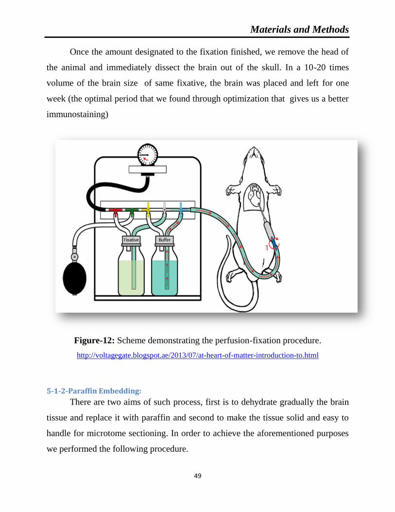
Materials and Methods
49
Once the amount designated to the fixation finished, we remove the head of
the animal and immediately dissect the brain out of the skull. In a 10-20 times
volume of the brain size of same fixative, the brain was placed and left for one
week (the optimal period that we found through optimization that gives us a better
immunostaining)
Figure-12: Scheme demonstrating the perfusion-fixation procedure.
http://voltagegate.blogspot.ae/2013/07/at-heart-of-matter-introduction-to.html
5-1-2-Paraffin Embedding:
There are two aims of such process, first is to dehydrate gradually the brain
tissue and replace it with paraffin and second to make the tissue solid and easy to
handle for microtome sectioning. In order to achieve the aforementioned purposes
we performed the following procedure.

Materials and Methods
50
A-Dehydration:Through this step we made sure the tissue is gradually was water
free through three changes in gradual concentration of ethanol (50, 70 and 90%)
each one hour followed by three changes in absolute ethanol each 2 hours.
B-Clearing: This step is to replace the alcohol that is in the tissue with the xylene
(Sigma-Aldrich, Missouri, USA), through three changes each two hours, because
the alcohol and paraffin (Thermo Fisher Scientific, Massachusetts, USA ) are
immiscible and it is considered a preparatory step to the next one by that the
paraffin will be able to infiltrate the tissue.
C-Waxing:This is last step before embedding where the brain placed in fresh
paraffin at 60°C to get rid of all the xylene in the tissue through three changes in
fresh xylene each two hours.
D-Embedding: The actual embedding takes place when the paraffin infiltrated the
brain. The brain is adjusted in the mold in a manner where coronal section can be
taken.
5-1-3-Brain sections preparation:
The excess of paraffin was removed and extra brain tissue was trimmed
using the microtome (Thermo Fisher Scientific, Massachusetts, USA ) (Figure-
13), until we reach the area of our interest which is commonly called the mustache
area that is approximately -3.7 mm relative to bregma according to Paxinos and
Watson. 2005. The reason of choosing such area of the hippocampus is at this part
of the brain the different parts of the hippocampus (Dentate gyrus, CA1, CA2,
CA3 and CA4) are well defined and easy to be distinguished. To make sure that we
are at the designated area we used the toluidine blue (Agar scientific, Essex, United
Kingdom) as a quick staining. Coronal sections 1μm thick were taken. In order to

Materials and Methods
51
flatten the brain section, immediately the sections transferred to water bath
(Thermo Fisher Scientific, Massachusetts, USA) at 50°C . Once we made sure that
the section wrinkles free, using slide coated by gelatin (Sigma-Aldrich, Missouri,
USA). The sections were picked and left to dry by keeping the slide vertically on
a tissue paper. Once the sections were dried. The slide was transferred to a heating
plate (Kunz Instruments AB, Nynashamn, Sweden) to make sure that the section is
free of water and properly dried. All the slides were kept in a slide box at RT till
use for different histology staining.
Figure-13: The set for microtome with a water bath.
Materials and Methods
52
5-2-Neuronal cell death examination using Fluoro-Jade B (FJB) staining:
Fluoro-Jade B staining was performed as described by Schmued and Hopkins
(Schmued and Hopkins, 2000). 32 animals, 4h (Sham =4, ADX=4), 24h (Sham =4,
ADX=4), 3days (Sham =4, ADX=4), 1week (Sham =4, ADX=4) and 2weeks
(Sham =4, ADX=4) were used for this staining.
5-2-1-Materials:
Equipement:
A set of jars of graduated ethanol (70, 80 90 and 100%) and absolute
xylene.
Humide chamber.
Reagents:
Distilled water.
PBS (Phosphate-buffered saline): PH 7.2-7.4, 0.2 μm filtered.
137 mM NaCl
2.7 mM KCl, 8.1 mM
Na2HPO4
1.5 mM KH2PO4
1% sodium hydroxide
5% NaOH
Ethanol
Xylene
0. 6% potassium permanganate
0.0004% Fluoro-Jade B solution (Millipore, Massachusetts, USA ).

Materials and Methods
53
Mounting media DPX (Sigma-Aldrich, Missouri, USA).
5-2-2-Method:
1. The coronal sections of the brain were first deparaffinized through two
changes of xylene each 10min.
2. Then rehydrated through a graduated alcohol series (100, 90, 80 and 70 %)
for a period of 5min for each.
3. Once the slides rehydrated, were ready to be immersed in 1% sodium
hydroxide in 80% alcohol (20ml of 5% NaOH added to 80ml absolute
alcohol) for 5min.
4. The slides were kept in 70% alcohol for dehydration and in distilled water
for 2min respectively.
5. Fllowing dehydration the slides were transferred to a solution of 0. 6%
potassium permanganate for 10min.
6. In order to get rid of any remaining of potassium permanganate the slides
were rinsed in distilled water for two min and immersed in 0.0004% Fluoro-
Jade B (Darmstadt, Germany) for 20min.
7. The slides were then rinsed for one min in each of three distilled water
washes and placed on a slide warmer for 10min.
8. The dried slides were cleared by immersion in xylene for at least 1min and
were then cover slipped with DPX and at this stage they were ready for
examination.
Materials and Methods
54
5-3-Electron microscopy:
5-3-1-Materials:
Equipement:
Rota mixer (Agar scientific, Essex, United Kingdom).
Oven (Thermo Fisher Jouan, , Massachusetts, USA )
Transmission electron microscope (Tecan, Zürich, Switzerland)
Ultra microtome (Leica EMUC7, Wetzlar, Germany).
200 mesh copper grids (Agar scientific, Essex, United Kingdom).
Molds (Agar scientific, Essex, United Kingdom).
Reagents:
KARNOVSKY’S fixative at PH 7.2
0.2 M phosphate buffer
0.1 M phosphate buffer
Paraformaldehyde (Sigma-Aldrich, Missouri, USA).
Glutaraldehyde (EM grade; 50% solution) (Sigma-Aldrich,
Missouri, USA)
1 N sodium hydroxide (NaOH)
0.1M PB
1% osmium tetroxide (Agar scientific, Essex, United Kingdom).
Propylene oxide (Agar scientific, Essex, United Kingdom).
Ethanol
Agar 100 (Agar scientific, Essex, United Kingdom).
Resin (Agar scientific, Essex, United Kingdom).
Uranyl acetate (Agar scientific, Essex, United Kingdom).
Lead citrate (Agar scientific, Essex, United Kingdom).

Materials and Methods
55
5-3-2- Method:
The hippocampus was removed and transferred into KARNOVSKY’S
fixative at PH 7.2 in glass vials for overnight at 4°C.
The next day the tissue was washed three times in 0.1M PB for 30min each. A
volume of 1ml of buffered 1% osmium tetroxide was added to each vial and mixed
for 1h at RT on Rota mixer. The osmium tetroxide was removed and followed by
three distilled water washes for 2 min each. The samples were dehydrated as
showed in table-5.
Table-5: Dehydration of brain tissue for electron microscopy.
Ethanol concentration Timing
30% Ethanol 15min
50% Ethanol 15min
70% Ethanol 15min
80% Ethanol 15min
90% Ethanol 15min
95% Ethanol 15min
100% Ethanol I 15min
100% Ethanol II 15min
Propylene oxide I 15min
Propylene oxide II 15min
During the last Propylene oxide, the Agar100 epoxy resin was prepared to be
ready for the following processing:
Agar 100Resin: Propylene oxide 1:1 for 1h.

Materials and Methods
56
Agar 100Resin: Propylene oxide 2:1 for 1h.
Agar 100Resin: Propylene oxide 3:1 for 1h.
Agar 100Resin for overnight at 4°C.
The next day the blocks embedded in resin were retrieved from the fridge
and warmed up in the oven at 60°C for 20min and the procedure was carried out as
follows; the molds were filled with freshly prepared resin and kept for 3-4 hours to
remove all air bubbles and proper infiltration. The samples were rested and
embedded in the molds and left in the oven for 24h at 64°C.
Blocks were trimmed and ultrathin sections were selected approximately -
3.7 mm relative to bregma according to Paxinos and Watson (2005). For cutting
coronal section of the hippocampus, ultra microtome was used. The semithin
sections (1μm) were stained with 1% aqueous toluidine blue on glass slides. The
ultrathin sections (95nm) were transferred on 200 mesh copper grids were
contrasted with uranyl acetate followed by lead citrate double stain. The grids were
examined and photographed under a Philips CM10 transmission electron
microscope and the photo were taken from different area of the hippocampus in
order to examine the occurrence of neuronal cell death in the current model.

Materials and Methods
57
5-4-Immunoflurescent labeling:
Animals (n=32) were sacrificed after surgery at different times: 4h (Sham
=4, ADX=4), 24h (Sham =4, ADX=4), 3days (Sham =4, ADX=4), 1week (Sham
=4, ADX=4) and 2weeks (Sham =4, ADX=4).
5-4-1-Materials:
Equipement:
Humid chamber.
Microwave (Daewoo electronics crop, Seoul, Korea).
Microtome (Thermo Fisher Scientific, Massachusetts, USA).
Liquid blocker (Daido Sangyo,Tokyo.Japan).
Fluorescent microscope (Zeiss, Oberkochen, Germany).
Confocal microscope (Nikon, Tokyo, Japan).
Adobe PhotoShop (CS6 version, USA).
Reagents:
Xylene.
Ethanol.
Distilled water.
10mM sodium citrate (pH 6) (Sigma-Aldrich, Missouri, USA).
0.1 M phosphate buffered saline (PBS, pH 7.4).
1% BSA.
Primary antibodies:

Materials and Methods
58
o Rabbit polyclonal anti-ionized calcium-binding adaptor
molecule 1 (Iba1, Wako, Massachusetts, USA 1:2000).
o Mouse monoclonal anti-neuronal nuclear antigen
antibody (Neun, Millipore, Massachusetts,USA, 1:1000).
o Rabbit polyclonal anti-glial fibrillary acidic protein
(GFAP, Dako, Copenhagen, Denmark, 1:1000).
o Goat Iba1 (Abcam, Massachusetts,USA 1:1000).
Secondary antibodies:
o Donkey anti-rabbit conjugated to Alexa488
(Invitrogen,Paisley, UK, 1:200).
o Donkey anti-mouse conjugated to Rhodamine (Jakson,
Pennsylvania, USA, 1:100).
o Donkey anti-goat conjugated to cy5 (Jakson,
Pennsylvania, USA, 1:100).
o PBS-Triton X-100 0.3% (Sigma-Aldrich, Missouri,
USA).
5-4-2-Method:
For all fluorescence labeling, the paraffin-embedded tissue sections were
deparaffinized with xylene, rehydrated with ethanol (100, 90, 80, and 70%) and
finally washed in distilled water. To unmask the epitopes and obtain efficient
immunostaining, the brain sections were subjected to an antigen retrieval
procedure with 10mM sodium citrate (PH 6) followed by heating for 1 min at high
power and 10 min at low power in microwave. After this procedure the samples
Materials and Methods
59
were washed with 0.1 M phosphate buffered saline (PBS, pH 7.4) and cooled down
at RT.
For double-immunofluorescence labeling, the sections were blocked by 1%
BSA at room temperature for 30 min and then were incubated with each primary
antibody at 4°C overnight. The primary antibodies used were rabbit polyclonal
anti-ionized calcium-binding adaptor molecule 1 (Iba1, 1:2000) mixed with mouse
monoclonal anti-neuronal nuclear antigen antibody (Neun, 1:1000) and rabbit
polyclonal anti-glial fibrillary acidic protein (GFAP, 1:1000) mixed with mouse
monoclonal anti-neuronal nuclear antigen (Neun, 1:1000). After washing three
times in PBS, tissue sections were incubated with donkey anti-rabbit conjugated to
Alexa488 (1:200) mixed with donkey anti-mouse conjugated to Rhodamine
(1:100) all diluted in PBS-Triton X-100 0.3% for 1h at room temperature.
Afterwards, the sections were washed several times in PBS; slides were cover
slipped with fluoromount medium (Thermo Fisher Scientific, Massachusetts,
USA).
Similar procedure was used for triple-immunofluorescence labeling in order
to define the localization of microglia, astroglia and neurons in the same brain
section, the mixture of the following primary antibodies was used, rabbit GFAP
(1:1000) mixed with mouse Neun (1:1000) and goat Iba1 (1:1000) at 4°C
overnight. Regarding the secondary antibody, tissue sections were incubated with
donkey anti-rabbit conjugated to Alexa 488 (1:200) mixed with donkey anti-mouse
conjugated to Rhodamine (1:100) and donkey anti-goat conjugated to cy5 (1:100).
Representative digital images were captured using a Zeiss AxioCamHRc Digital
camera with AxioVision 3.0 software (Carl Zeiss, Germany). Some sections were
also examined with a C1 laser scanning confocal microscope (Nikon,Tokyo,
Japan). 4X and 20X objectives were used to capture confocal images for the
preparation of figures 32, 35 and 36 respectively where only one optical section

Materials and Methods
60
was utilized. The resulting files were used to generate the figures in Adobe
PhotoShopCS6 where the photomicrographs were adjusted for contrast and
brightness in order to determine the relationship between labeled neurons,
astrocytes and microglia.
5-5-Quantitative analysis of the Iba-1 and GFAP positive cells:
Both microglia and astrocytes undergo morphological changes and became
reactive cells in response to CNS injury. In this study we investigated the response
of both these glial cells in the hippocampus after ADX. Typically, reactive
microglia show shortening and widening of their processes and some of them they
show amoeboid shape by losing their processes. Reactive astrocytes show
increased size of their processes. These features can be easily visualized in sections
stained with Iba1 and GFAP antibodies as markers for microglia and astrocytes
respectively. In this study, only positively stained Iba1-positive microglia and
GFAP-positive astrocytes with visible cell body at the focal plane were counted.
Three ADX and three sham rats were used for each time point. Three sections of
the hippocampus separated by 30µm were used from each rat. From each section
three optical fields were selected for cell counting. Images of stained sections were
taken at 20X magnification using an AxioCam HRc Digital camera with
AxioVision 3.0 software (Carl Zeiss, Germany). In order to avoid being biased, the
parameters of acquiring images like the time of exposure and the gain were first
fixed for labeled microglia and astroglia and kept constant for all the examined
time points. Cells matching the above mentioned criteria were counted manually
using cell counter software (Heracle BioSoft SRL, Romania). A comparison was
then carried out between the number of microglia and astrocytes in ADX and sham
operated rats.

Materials and Methods
61
6-Animal behavior test:
All animals were handled daily for at least 3 days before the experiments.
The Passive Avoidance task was conducted as described by (Misane and Ogren
2000; Ahlander-Luttgen et al., 2003) using a two-compartment standard shuttle
box (51x25x24 cm) (Harvard Apparatus, Massachusetts, USA).
The two communicating (7x7 cm sliding door built in the separating wall)
compartments were of equal size and had a stainless-steel bar floor. The right-hand
compartment (shock compartment) was painted black to obtain a dark chamber,
while the left-hand compartment was illuminated by a bulb (24 V; 5 W) installed
on the top Plexiglas cover. PA training was conducted in a single session (day 1).
Rats were placed in the lighted compartment (with no access to the dark
compartment) and were allowed to explore for 60s. During the exploration period
in the PA apparatus. When 60s expired, the sliding door was automatically opened
and the rat was allowed to cross over into the dark compartment. Once the rat had
entered the dark compartment with all four paws, the sliding door was
automatically closed and a weak electrical current (constant current, scrambled,
duration 3s, 0.3 mA) was delivered through the grid floor. Latency to cross into the
dark compartment (training latency) was recorded. If a rat failed to move into the
dark compartment within 300 s (cut off latency), the door was reopened and the rat
was gently moved into the dark compartment, where it received the foot shock.
After exposure to the foot shock, the rat was allowed to stay for 30 s in the
dark compartment before it was removed from the PA apparatus to its home cage.
Retention was tested 24 h after training (day 2). The animal was again placed in
the lighted (safe) compartment with access to the dark compartment for a period of
300s. The latency to enter the dark compartment with all four paws was
automatically measured (retention latency). If the rat failed to enter the dark

Materials and Methods
62
compartment within 300 s, it was removed and assigned a maximum test latency
score of 300s (Figure-14).
Figure-14: Passive avoidance task
(https://www.radiantthinking.us/memory-theory/ii-behavioral-assessments-in-rodents.html)
7-Statistical analysis:
All data are reported as the mean ± standard error of the mean and the
analysis was considered significantly different if P ≤ 0.05. Results for (IL-1β, IL-6
and TNF-α) و (GSH, SOD and MDA) and microglia and astrocytes counting were
analyzed by two-tail Student using t test SPSS version 20 (IBM, USA).


1-Concentration of corticosterone

Results
65
The efficacy of adrenalectomy was confirmed post-mortem by analysis of
plasma corticosterone levels. The concentration of corticosterone was measured
using Logit-Log paper plot Percent bound versus concentration of corticosterone
for standards that ranges from 32-20000 pg/ml and by interpolation the
concentration of corticosterone in the sera of adrenalectomized and sham operated
rats was determined see the (Figure-15).
Figure-15: Standard curve used in the determination of corticosterone levels in the
sera of adrenalectomized and sham operated rats.
We found the levels of corticosterone were significantly decreased after 4h
(P=0.001) in the serum of adrenalectomized compared to bilateral sham operated
rats and continued in the same trend after 24h (P<0.001), 72h (P<0.001), 1week
(P<0.001) and 2weeks (P<0.001) following the surgery see (Figure-16) and (Table-
6).
0
0.1
0.2
0.3
0.4
0.5
0.6
0
10
20
30
40
50
60
70
80
90
100
10 100 1000 10000 100000A
bso
rban
ce
B/B
0 (
%)
Corticostirone Conc. (pg/ml)
B/B0 (%)
ABSORBANCE

Results
66
Table-6: Concentration of corticosterone in the serum of adrenalectomized rats compared to
sham operated rats over the course of time (4h, 24h, 3days, 1week and 2weeks). ***P<0.001.
Data are expressed as mean ±SEM.
Time Groups SHAM ADX
Statistical
significance
4H 24493.6±1732.3 7503.3±860.0 ***P=0.001
24H 18629.9±3668.8 199.4±59.0 ***P<0.001
72H 12527.5±1914.6 126.5±33.8 ***P<0.001
1 Week 16291.5±2770.2 224.6±94.3 ***P<0.001
2 Weeks 16123.7±2835.8 163.6±49.4 ***P<0.001
Unit of measurement: pg/ml
Figure-16: Bar graphs showing levels of serum corticosterone. Levels of corticosterone were
measured by EIA in the serum of adrenalectomized rats compared to the sham operated rats over
the course of time (4h, 24h, 3days, 1week and 2weeks). ***P<0.001. Data are expressed as mean
± SEM.
0
5000
10000
15000
20000
25000
30000
4H 24H 72H 1W 2W
Glu
coco
rtic
oid
s (p
g/m
l)
Post surgical time
SHAM
ADX***
*** *** *** ***

2-Neuroinflammation

Results
68
2-1-Interleukin-1 beta (IL-1β):
The pro-inflammatory cytokines IL-1β, IL-6 and TNF-α are the major
players in multiple responses to different insults and injuries. In addition they play
a significant role in different neurodegenerative diseases. In the current
neurodegeneration model we intended to evaluate the levels of these cytokines in
the hippocampus of Wistar rats subjected either to bilateral adrenalectomy or to
sham operation using ELISA.
Figure-17: Interleukin-1β Standard Curve. The curve obtained from the absorbance at 450nm of
varying concentrations of IL-1β standards ranging from 12.5-600 pg/ml (R²: Pearson Coefficient
of Determination).
The levels of pro-inflammatory cytokine Interleukin-1β in the hippocampal
homogenates of adrenalectomized and sham operated rats were examined over the
course of time (4h, 24h, 3days, 1week and 2weeks). In order to determine the
y = 0.0012x + 0.034
R² = 0.998
0
0.1
0.2
0.3
0.4
0.5
0.6
0.7
0 100 200 300 400 500 600
Ab
sorb
an
ce
IL-1β (pg/ml)

Results
69
concentration of IL-1β by ELISA, a standard curve in the range 12.5-600 pg/ml
was constructed and the levels of IL-1β in the samples was determined by
interpolation see (Figure-17).
Our results showed that the levels of IL-1β were significantly increased in
the hippocampus of adrenalectomized rats compared to bilateral sham operated rats
after 4h (P<0.05), 24h (P<0.05) and 3days (P<0.01) of surgery. However, one and
two weeks after adrenalectomy no statistical difference in the hippocampal levels
of IL-1β were found between the two groups, see (Table-7) and (Figure-18).
Table-7: Concentration of Interleukin-1β levels in the hippocampal homogenates of
adrenalectomized rats compared to the sham operated rats over the course of time (4h, 24h,
3days, 1week and 2weeks). Data are expressed as mean ± SEM.
Time Groups SHAM ADX Statistical significance
4H 114.25±11.17 155.87±12.51 *P<0.05
24H 108.98±6.55 129.67±4.40 *P<0.05
72H 112.60±4.12 133.72±5.53 **P<0.01
1 Week 121.84±7.38 102.03±7.41 P>0.05
2 Weeks 76.62±5.85 72.86±4.65 P>0.05
Unit of measurement: pg/mg of total protein

Results
70
Figure-18: Bar graphs showing IL-1β level in the hippocampus of adrenalectomized and sham
operated rats over course of time (4h, 24h, 3days, 1week and 2weeks) .*P<0.05. **P<0.01.Data
are expressed as mean ± SEM.
2-2-Interleukin-6:
The levels of pro-inflammatory cytokine Interleukin-6 in the hippocampal
homogenates of adrenalectomized and sham operated rats were examined over the
course of time (4h, 24h, 3days, 1week and 2weeks). In order to determine the
concentration of IL-6 by ELISA, a standard curve in the range 125-8000 pg/ml was
constructed and the levels of IL-6 in the samples were determined by interpolation
(Figure-19).
0
20
40
60
80
100
120
140
160
180
4H 24H 72H 1W 2W
IL-1
β (p
g/m
g T
P)
Post surgical time
SHAM
ADX
*
* **

Results
71
Figure-19: Interleukin-6 Standard Curve. The curve obtained from the absorbance at 450nm of
varying concentrations of IL-6 standards ranging from 125-8000 pg/ml (R²: Pearson Coefficient
of Determination).
A significant increase in IL-6 levels was observed in the hippocampus 4h
(P<0.01), 24h (P<0.01) and 3days (P<0.05) after ADX, however, no change was
seen in IL-6 levels after one and two weeks postoperatively (Table-8) and (Figure-
20).
Table-8: Concentration of Interleukin-6 levels in the hippocampal homogenates of
adrenalectomized rats compared to the sham operated rats over the course of time (4h, 24h,
3days, 1week and 2weeks). Data are expressed as mean ± SEM.
Time Group SHAM ADX Statistical significance
4H 621.77±35.80 802.74±30.62 **P<0.01
24H 543.59±21.45 656.24±17.45 **P<0.01
72H 580.95±20.90 644.39±17.00 *P<0.05
1 Week 643.80±59.05 520.00±24.61 P>0.05
2 Weeks 560.83±29.41 587.01±41.52 P>0.05
Unit of measurement: pg/mg of total protein
y = 0.0001964x - 0.0081
R² = 0.9989
0
0.5
1
1.5
2
0 2000 4000 6000 8000 10000
Ab
sorb
an
ce
IL-6 (pg/ml)

Results
72
Figure-20: Bar graphs showing IL-6 levels in the hippocampus of adrenalectomized and sham
operated rats over course of time (4h, 24h, 3days, 1week and 2weeks).*P<0.05. **P<0.01.Data
are expressed as mean ± SEM.
2-3-Tumor Necrosis Factor-α (TNF-α):
The levels of pro-inflammatory cytokine TNF-α in the hippocampal
homogenates of adrenalectomized and sham operated rats were examined over the
course of time (4h, 24h, 3days, 1week and 2weeks). In order to determine the
concentration of TNF-α by ELISA, a standard curve in the range 3.9-500 pg/ml
was constructed and the levels of TNF-α in the samples was determined by
interpolation (Figure -21).
0
100
200
300
400
500
600
700
800
900
4H 24H 72H 1W 2W
IL-6
(p
g/m
g T
P)
Post surgical time
SHAM
ADX
**
** *

Results
73
Figure -21: Tumor Necrosis Factor-α Standard Curve. The curve obtained from the absorbance
at 450nm of varying concentrations of TNF-α standards ranging from 3.9-500 pg/ml (R²: Pearson
Coefficient of Determination).
The levels of TNF-α were significantly increased in the hippocampus of
adrenalectomized rats compared to bilateral sham operated rats after 4h only
(P<0.01) and remain unchanged after 24h, 72h, 1week, 2weeks of surgery see
(Table-9) and (Figure-22).
Table-9: Concentration of TNF-α levels in the hippocampal homogenates of adrenalectomized
rats compared to the sham operated rats over the course of time (4h, 24h, 3days, 1week and
2weeks). Data are expressed as mean ± SEM.
Time Group SHAM ADX Statistical significance
4H 21.39±0.99 29.31±1.33 **P<0.01
24H 16.90±0.63 17.33±0.89 P>0.05
72H 16.58±0.71 16.52±1.07 P>0.05
1 Week 18.30±0.93 15.52±1.11 P>0.05
2 Weeks 18.19±2.04 18.47±1.62 P>0.05
Unit of measurement: pg/mg of total protein
y = 0.0025x + 0.0372
R² = 0.9973
0
0.2
0.4
0.6
0.8
1
1.2
1.4
0 100 200 300 400 500
Ab
sorb
an
ce
TNF-α (pg/ml)

Results
74
Figure-22: Bar graphs showing TNF-αlevels in the hippocampus of adrenalectomized and sham
operated rats over course of time (4h, 24h, 3days, 1week and 2weeks). **P<0.01.Data are
expressed as mean ± SEM.
0
5
10
15
20
25
30
35
4H 24H 72H 1W 2W
TN
F-α
(p
g/m
g T
P)
Post surgical time
SHAM
ADX
**

3-Neurodegeneration

Results
76
3-1-Neuronal cell death using FJB:
In order to examine the occurrence of neuronal cell death in the
hippocampus following adrenalectomy over a course of time (4h, 24h, 3days,
1week and 2weeks), a Fluoro-Jade B staining was performed. FJB staining is a
specific staining for neurons undergoing degeneration, regardless of the specific
insult or mechanism of cell death.
After four hours (results not shown) and 24h following adrenalectomy our results
showed no positive staining for degenerated neurons in both groups ADX and
sham rats (Figure-23A', A, E', E). Three days after adrenalectomy we observed a
few degenerated neurons in the dorsal blade of the dentate gyrus in the
hippocampii of adrenalectomized rats only (Figure-23B', B, F', F). Moreover, one
week after adrenalectomy, more degenerated neurons were seen on the dorsal
blade of the dentate gyrus (Figure-23C', G') whilst no positive FJB staining was
observed in sham operated rats (Figure-23C, G). After two weeks of
adrenalectomy, we observed a progression of cell death throughout the dorsal
blade of the dentate gyrus (Figure-23D', H') where an intense FJB staining was
seen. In addition, less intense staining was observed in the ventral blade indicating
that differential effect of the adrenal gland removal on both blades of the dentate
gyrus (Figure-24). No positive FJB staining was observed in sham operated rats
(Figure-24) and (Figure-23D, H).

Results
77
Figure-23: Images of coronal sections of the hippocampus stained with Fluoro-Jade B showing
the progression of neuronal cell death over the course of time (1day, 3days, 1week and 2weeks)
in the dentate gyrus of the hippocampus of adrenalectomized rats compared to sham operated
rats .Absence of cell death was noticed in the dentate gyrus (A, B, C, D) and (E, F, G, H) of
sham operated rats during the different time points (1day, 3days, 1week and 2weeks). One day
after adrenalectomy the rats showed no signs of neuronal cell death in the dentate gyrus (A') and
(E'). A progression of neuronal cell death was seen in the dentate gyrus three days (B'), one
week (C'), and two weeks (D') after adrenalectomy. Molecular layer (ML), granule cell layer
(GCL), polymorphous layers (PL). Scale bar=50µm.

Results
78
Figure-24: Representative confocal images of with Fluoro-Jade B staining (green) showing the
occurrence of neurodegeneration after two weeks in both blades of the dentate gyrus of the
hippocampus of adrenalectomized rats compared to sham operated rats. Molecular layer (ML),
granule cell layer (GCL), polymorphous layers (PL). Scale bar=50µm.
3-2-Neuronal cell death using electron microscopy:
The motive behind the current ultrastructural study was to determine the
impact of the absence such hormones on different cells of the hippocampus after
short-term adrenalectomy at different time points (3, 7 and 14 days). In addition,
Prof. Adem and his team in previous study reported the absence of glucocorticoids

Results
79
after long period of time (5 months) induces degeneration in the different parts of
the hippocampus where different neuronal populations exhibited signs of
abnormalities and different types of cell death.
The examination of the thin section under the electron microscope three days
following adrenalectomy showed granule cells degeneration of dorsal blade of the
dentate gyrus of adrenalectomized rats hippocampus where the cells showed
condensed chromatin and irregular cell membrane and the beginning of
vacuolation in the cytoplasm. Interestingly we have not noticed cell abnormalities
in the rest of the dentate gyrus (Figure-25B, D). Concerning different areas of the
Cornu Ammonis (CA), solely CA4 pyramidal cells displayed signs of cell damages
following the removal of the adrenal gland(Figure-26) while the big CA3
pyramidal cells, small pyramidal CA1 and CA2 cells did not show any signs of
neurodegeneration after three days of adrenalectomy (Figure-25B,D). The
examination of the hippocampus of the sham operated rats revealed no signs of
neurodegeneration in different parts of the hippocampus (Figure-25A, 1C, 2A, 2C).
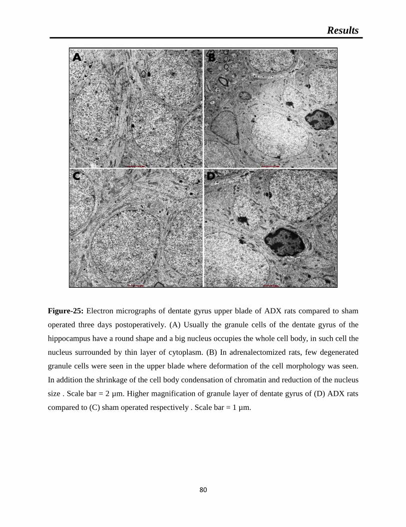
Results
80
Figure-25: Electron micrographs of dentate gyrus upper blade of ADX rats compared to sham
operated three days postoperatively. (A) Usually the granule cells of the dentate gyrus of the
hippocampus have a round shape and a big nucleus occupies the whole cell body, in such cell the
nucleus surrounded by thin layer of cytoplasm. (B) In adrenalectomized rats, few degenerated
granule cells were seen in the upper blade where deformation of the cell morphology was seen.
In addition the shrinkage of the cell body condensation of chromatin and reduction of the nucleus
size . Scale bar = 2 µm. Higher magnification of granule layer of dentate gyrus of (D) ADX rats
compared to (C) sham operated respectively . Scale bar = 1 µm.
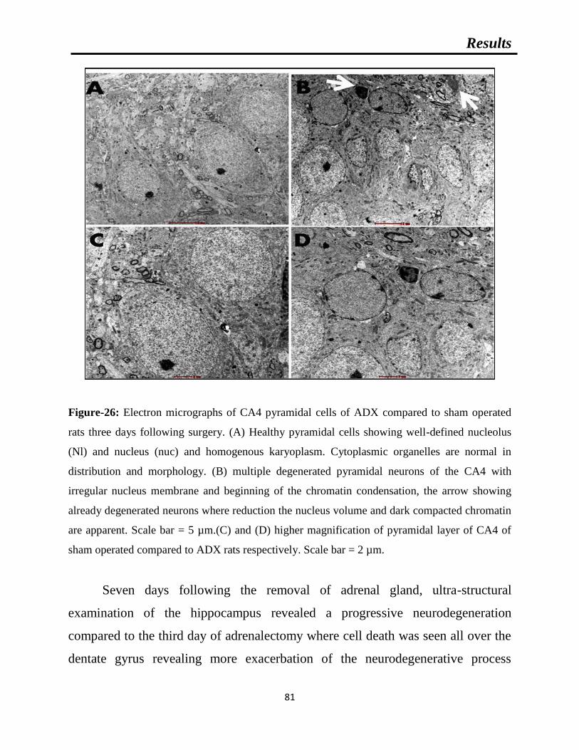
Results
81
Figure-26: Electron micrographs of CA4 pyramidal cells of ADX compared to sham operated
rats three days following surgery. (A) Healthy pyramidal cells showing well-defined nucleolus
(Nl) and nucleus (nuc) and homogenous karyoplasm. Cytoplasmic organelles are normal in
distribution and morphology. (B) multiple degenerated pyramidal neurons of the CA4 with
irregular nucleus membrane and beginning of the chromatin condensation, the arrow showing
already degenerated neurons where reduction the nucleus volume and dark compacted chromatin
are apparent. Scale bar = 5 µm.(C) and (D) higher magnification of pyramidal layer of CA4 of
sham operated compared to ADX rats respectively. Scale bar = 2 µm.
Seven days following the removal of adrenal gland, ultra-structural
examination of the hippocampus revealed a progressive neurodegeneration
compared to the third day of adrenalectomy where cell death was seen all over the
dentate gyrus revealing more exacerbation of the neurodegenerative process

Results
82
(Figure-27A’). In addition to what we have seen on the third day after
adrenalectomy more degenerative cells were observed in the CA4 (Figure-27B’).
In addition, EM observation of the neuropil in the CA3 area of hippocampus of
adrenalectomized rats revealed cell death of the CA3 Pyramidal cells for the first
time (Figure-27C’). No sign of degeneration was seen in the CA1 of the
adrenalectomized rats. The hippocampus of the sham rats did not show any
abnormalities in different neuronal populations (Figure-27A, B, and C)
Figure-27: Electron micrographs showing degeneration in different areas of the hippocampus of
ADX rats compared to sham operated were taken seven days postoperatively. (A) Intact granule
cell with well defined nucleus and cell membrane of sham rats. (A’) degenerated granule cell of
the dentate gyrus with condensed chromatin and irregular cell membrane. (B) and (C) healthy
pyramidal cells of CA4 and CA3 respectively. (B’) showing ultrastructural of CA4 dead neurons
with disintegration of the cell body, condensation of the chromatin and nucleus volume
reduction. (C’) multiple degenerated CA3 pyramidal neurons with irregular nucleus membrane
and beginning of the chromatin condensation. Scale bar = 1 µm.

Results
83
Our results exhibited an extensive cell death on the dorsal blade of the
dentate gyrus compared to less degree of cell death in ventral blade after two
weeks of adrenalectomy. A thorough examination of granule layer of the dentate
gyrus showed differences in the gross appearance of the neuropil between the
adrenalectomized and sham operated rats. In the adrenalectomized animals, the
granule layer showed empty areas with multiple ‘‘blank’’ spaces that indicated
already degenerated cells. In addition to the changes in the neuropil, we observed
ultrastructural changes in the upper and lower blade of the dentate gyrus (Figure-
28).
Figure-28: Electron micrographs taken from the upper blade of the dentate gyrus two weeks
following adrenalectomy showing granule cells engulfed by microglia (white arrows), in
addition, it shows undergoing degeneration of granule cells where fragmentation of the nucleus
and appearance of vacuolation in the cytoplasm (black arrow). Scale bar = 5 µm.

Results
84
A condensation of chromatin was seen in addition of the cell body shrinkage
revealing both apoptotic and necrotic process occurring in this part of the
hippocampus and major vulnerability of such cell type to the absences of
glucocorticoids (Figure-29). In contrast, in the sham rats the neuropil was
continuous with abundant, tightly packed granule cells. As the granule cells are
small neurons, the nucleus occupied most of the cell body with only a thin rim of
cytoplasm surrounding it.
Figure-29: High power microscopy of degenerated granule cell in the hippocampus of
adrenalectomized rats two weeks following adrenalectomy. Appearance of vacuolation in the
cytoplasm with condensed chromatin (thin arrow) and adjacent granule cell in the process to be
phagocytosed by microglia (big arrow). Scale bar = 1 µm.

Results
85
The examination of the Cornu Ammonis of the adrenalectomized rats
revealed an extensive cell death in the CA4 area where chromatin condensation
and vacuolation were seen (Figure-30).
Figure-30: High power microscopy taken from CA4 area of the hippocampus following two
weeks of adrenalectomy showing empty space in pyramidal layer. In addition, many neurons
with compacted chromatin surrounded by irregular nucleus membrane (black arrow).A glial cell
in the process to phagocyte Pyramidal cell of the CA4 (white arrow).Scale bar = 5 µm.
Electron microscopy examination of the CA3 area of ADX rats revealed the
occurrence of cell death of pyramidal cells along the principal layer of such area
where extensive chromatin condensation, and reduction of the soma volume and
the wavy shrinkage of the nuclear membrane and the vacuolization of
mitochondria where seen (Figure-31). Sham operated rats displayed healthy

Results
86
clustered granule cells with regular plasma membrane and well defined nucleus
with regular dispersed chromatin. More importantly, our results showed no
ultrastructural abnormalities of the CA1pyramidal cells of both groups.
Figure-31: Electron micrograph showing CA3 pyramidal cells at different stage of degeneration
two weeks following adrenalectomy confirming that withdrawal of glucocorticoids by
adrenalectomy is not selective where different neuronal populations in the hippocampus are
subject of substantial degeneration. Scale bar = 2µm.

4-Microgliosis and astrogliosis

Results
88
4-1-Activation of microglia:
The activation of microglia is one of the major signs of neurodegeneration.
In order to investigate the activation of these cells in response to
neurodegeneration occurring after short-term adrenalectomy, a double
immunofluorescent labeling of neurons (Neun) and microglia (Iba-1) over the
course of time (4h, 24h, 3days, 1week and 2weeks) was performed.
Immunofluorescence staining with Iba-1 antibody after four hours and one day of
adrenalectomy did not show activated microglia in adrenalectomized and sham
operated groups (Figure-32A' and A). However, after three days we observed few
activated microglia in the dorsal blade of the dentate gyrus of adrenalectomized
rats while no sign of activation was seen in the sham operated rats (Figure-32B'
and B). One week following the adrenal gland removal, activated microglia were
seen in the whole dorsal blade of the dentate gyrus indicating a progression of
microgliosis (Figure-32C'). In contrast, no sign of activated microglia were
observed in the hippocampus of sham operated rats (Figure-32C).
Fluorescent double immunostaining of neurons and microglia of brain sections
after two weeks postoperatively revealed a strong Iba-1 immunoreactivity in the
hippocampus of adrenalectomized rats most particularly in the dentate gyrus
(Figure-32D') and stratum lucidum of CA3 (Figure-32H') areas compared to the
corresponding regions in the sham operated rats (Figure-32D and H).

Results
89
Figure-32: Images of microgliosis in the hippocampus. Coronal sections of the hippocampus stained with NeuN
antibody (red) as a neuronal marker and Iba-1 antibody (green) as a microglia marker showing the progression of
microgliosis over the course of time (1day, 3days, 1week and 2weeks) in the dentate gyrus and CA3 of the
hippocampus of adrenalectomized rats compared to sham operated rats. Absence of microgliosis in the dentate
gyrus (A, B, C, D) and CA3 (E,F,G,H) of sham operated rats during 1day, 3days, 1week and 2weeks. The
adrenalectomized rats showed no signs of microgliosis in the dentate gyrus (A') and CA3 (E') after one day.
However, a progression of microgliosis was seen in the dentate gyrus after three days (B'), one week (C') and two
weeks (D'). In comparison, the microgliosis in the CA3 of adrenalectomized rats did not appear until after 2 weeks
as the Iba1 immunoreactivity (H’) after 1day, 3days and 1 week was not significantly different from that in sham
operated rats(E’,F’,G’). Molecular layer (ML), granule cell layer (GCL), polymorphous layers (PL) and (SL)
stratum lucidum. Scale bar =50µm.

Results
90
The quantitative analysis showed that the number of Iba-1-positive microglia in the
hippocampus of ADX rats was significantly higher in days 3 (P<0.01), 7 (P<0.001) and 14 days
(P<0.001) compared to the sham operated rats. The results showed progressive increase in the
number of microglia in the hippocampus from day three with a further increase one and two
weeks after adrenalectomy (Figure-33).
Figure-33: Bar graphs showing the comparison between the number of Iba-1 positive cells in the
hippocampus of adrenalectomized and sham operated rats. The number of microglia was
significantly and progressively increased in adrenalectomized rats 3 days, 1 week and 2 weeks
postoperatively. **P<0.01. ***P<0.001. Data are expressed as mean (±SEM).
Iba-1 labeling exhibited a differential distribution throughout the dentate
gyrus where it shows an intensive microgliosis in the molecular and granular layer
of the dorsal blade and predominately at the tip where the Neun staining shows
apparent absence of neurons compared to the ventral blade two weeks
postoperatively (Figure-34). Interestingly, at the same time point, two weeks after
adrenalectomy, we observed for the first time a strip of Iba-1 positive cells in the
stratum lucidum layer of the CA3 area (Figure-34) and (Figure-32D'). No sign of
microgliosis was seen in CA3 area of sham operated rats over the course of time
(Figure-32E, F, G, and H).
0
10
20
30
40
50
60
70
80
72H 1W 2W
Nu
mb
er o
f m
icro
gli
a/F
ield
Post surgical time
SHAM
ADX
***
***
**

Results
91
Figure-34: Representative images of coronal sections of the whole hippocampus stained with
NeuN (red) and Iba-1 (green) antibodies showing microgliosis after two weeks of
adrenalectomized rats compared to bilateral sham operated rats. Molecular layer (ML), granule
cell layer (GCL), polymorphous layers (PL) and (SL) stratum lucidum. Scale bar=100µm.

Results
92
4-2-Activation of astrocyte:
In order to examine the effect of adrenal gland removal on astrocyte
immunoreactivity in the hippocampus over a course of time (4h, 24h, 3days, 1week
and 2weeks), a double immunofluorescent labeling of neurons (Neun) and
astrocytes (GFAP) was performed. After four hours (results not shown), one and
three days of adrenalectomy, we did not observe an apparent difference in the
GFAP immunoreactivity in the hippocampii of adrenalectomized and sham groups
(Figure-35A', A, B’, B). However, one week after adrenalectomy, activated
astrocytes were seen in the dentate gyrus (Figure-35C') whilst no difference in the
GFAP immunoreactivity was observed in sham operated rats (Figure-35C).
An intense GFAP immunoreactivity was observed, two weeks after adrenalectomy,
throughout the hippocampus most particularly in the dentate gyrus (Fig.37) where
the astrocytes undergo morphological changes and appeared hypertrophic and
showed intense GFAP immunoreactivity (Figure-35D'). The GFAP immuno-
labeling exhibited extensive astrogliosis in the molecular and polymorphous layers
of the dentate gyrus; however less intense astogliosis was seen in the granular layer
(Figure-37). The stratum lucidum of CA3 area of adrenalectomized rats showed an
increase in GFAP immunoreactivity (Figure-35H').
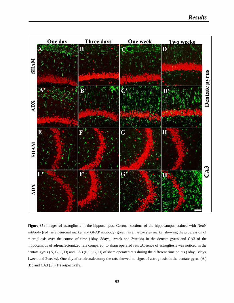
Results
93
Figure-35: Images of astrogliosis in the hippocampus. Coronal sections of the hippocampus stained with NeuN
antibody (red) as a neuronal marker and GFAP antibody (green) as an astrocytes marker showing the progression of
microgliosis over the course of time (1day, 3days, 1week and 2weeks) in the dentate gyrus and CA3 of the
hippocampus of adrenalectomized rats compared to sham operated rats .Absence of astrogliosis was noticed in the
dentate gyrus (A, B, C, D) and CA3 (E, F, G, H) of sham operated rats during the different time points (1day, 3days,
1week and 2weeks). One day after adrenalectomy the rats showed no signs of astrogliosis in the dentate gyrus (A')
(B') and CA3 (E') (F') respectively.

Results
94
A progression of astrogliosis was seen in the dentate gyrus after one week (C') and two weeks
(D') whilst no immunoreactivity was seen in CA3 after one week (G').However appearance. On
the other hand, there was no significant difference between the number of GFAP-positive
astrocytes in the hippocampus of ADX and sham operated rats three days following
adrenalectomy. However, a significant increase (P< 0.001) in the number of astrocytes was
observed one and two weeks after adrenalectomy in the ADX compared to sham operated rats
(Figure-36).
Figure-36: Bar graphs showing the comparison between the number of GFAP positive cells in
the hippocampus of adrenalectomized and sham operated rats. No difference in the number of
astrocytes revealed after 3 days but significantly increased 1 and 2 weeks following
adrenalectomy. ***P<0.001. Data are expressed as mean (±SEM).
Moreover, GFAP immunostaining also showed extensive astrogliosis in the
CA4 but not in the CA1 area of the adrenalectomized hippocampus (Figure-
37).The dentate gyrus (Figure-35A, B, C, and D), CA3 (Figure-35E, F, G, and H).
CA4, CA2 and CA1 (Figure-37) areas in the hippocampi of bilateral sham operated
rats did not show astrogliosis.
0
10
20
30
40
50
60
70
80
72H 1W 2W
Nu
mb
er o
f ast
rog
lia
/Fie
ld
Post surgical time
SHAM
ADX
***
***
*

Results
95
Figure-37: Representative images of coronal sections of the whole hippocampus stained with
NeuN (red) and GFAP (green) antibodies showing astrogliosis after two weeks of
adrenalectomized rats compared to bilateral sham operated rats. Molecular layer (ML), granule
cell layer (GCL), polymorphous layers (PL) and (SL) stratum lucidum. Scale bar=100µm.
It was very interesting idea that came from one of the reviewers of our
manuscript that to check at different levels through the rostral-caudal axis of the
hippocampus whether the effect of the removal of the adrenal gland is consistent or
not. Deciding to do that we choose to perform double immunofluorescence with

Results
96
NeuN (red) and Iba-1 antibodies for neurons and microglia respectively on brain
coronal sections at two levels -4.68mm and -5.5mm relative to bregma according
to Paxinos and Watson.2005. Indeed we have found similar to the effect that we
have observed at the level -3.7 where activated microglia on both blades of the
dentate gyrus and predominantly on the dorsal blade of the ADX rats (Figure-34).
Similar to the level -3.7 no sign of microgliosis were seen in the sham operated rats
at two levels -4.68mm (Figure-38) and -5.5mm (Figure-39). In addition our results
showed the activation of microglia at CA4 which is similar what was found at the -
3.7 levels and no signs in the sham rats. Regarding the CA3 area we observed the
microglia activation bordering the pyramidal cells of the CA3 at stratum lucidum
which is a total similarity to what have been seen at the other levels and no signs in
sham operated rats.
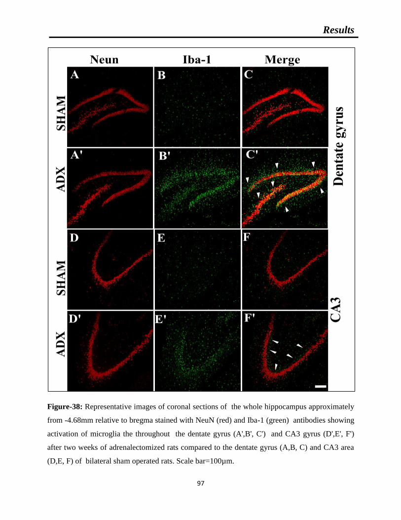
Results
97
Figure-38: Representative images of coronal sections of the whole hippocampus approximately
from -4.68mm relative to bregma stained with NeuN (red) and Iba-1 (green) antibodies showing
activation of microglia the throughout the dentate gyrus (A',B', C') and CA3 gyrus (D',E', F')
after two weeks of adrenalectomized rats compared to the dentate gyrus (A,B, C) and CA3 area
(D,E, F) of bilateral sham operated rats. Scale bar=100µm.
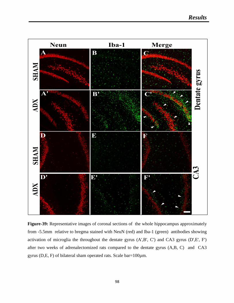
Results
98
Figure-39: Representative images of coronal sections of the whole hippocampus approximately
from -5.5mm relative to bregma stained with NeuN (red) and Iba-1 (green) antibodies showing
activation of microglia the throughout the dentate gyrus (A',B', C') and CA3 gyrus (D',E', F')
after two weeks of adrenalectomized rats compared to the dentate gyrus (A,B, C) and CA3
gyrus (D,E, F) of bilateral sham operated rats. Scale bar=100µm.
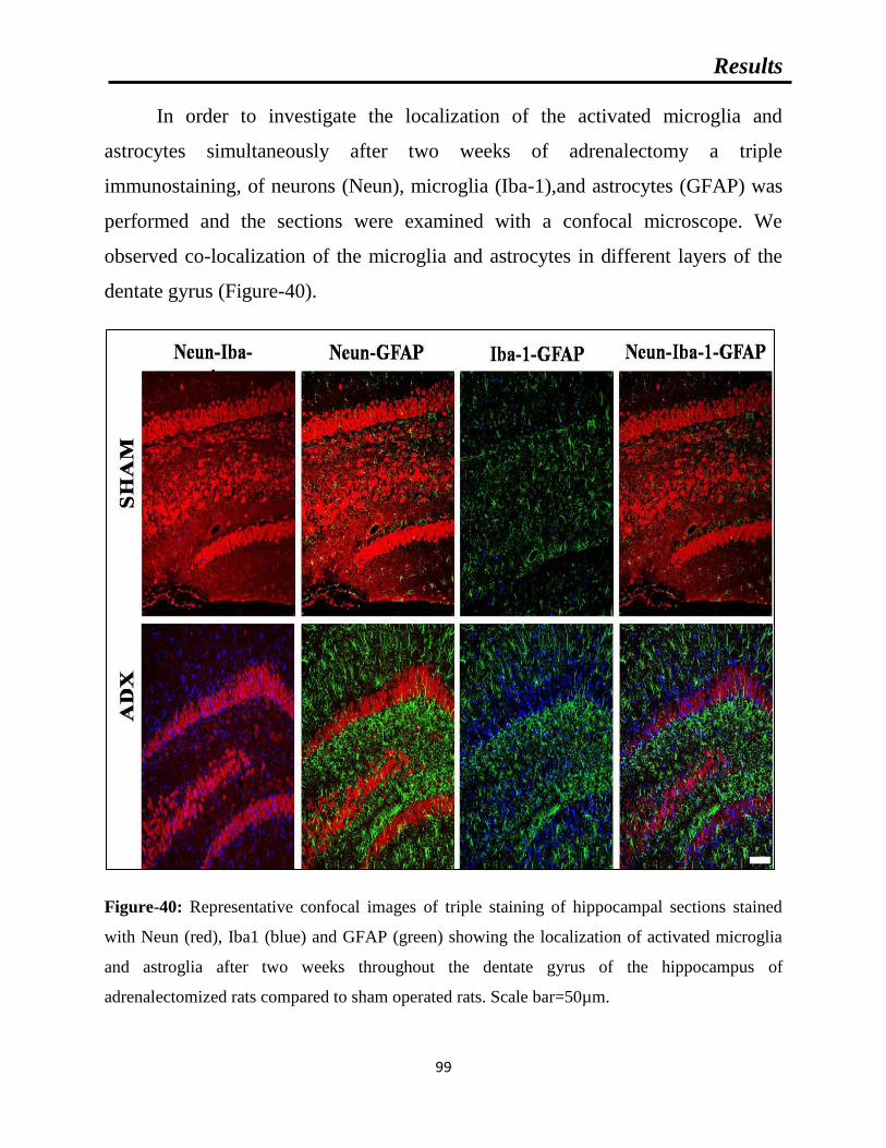
Results
99
In order to investigate the localization of the activated microglia and
astrocytes simultaneously after two weeks of adrenalectomy a triple
immunostaining, of neurons (Neun), microglia (Iba-1),and astrocytes (GFAP) was
performed and the sections were examined with a confocal microscope. We
observed co-localization of the microglia and astrocytes in different layers of the
dentate gyrus (Figure-40).
Figure-40: Representative confocal images of triple staining of hippocampal sections stained
with Neun (red), Iba1 (blue) and GFAP (green) showing the localization of activated microglia
and astroglia after two weeks throughout the dentate gyrus of the hippocampus of
adrenalectomized rats compared to sham operated rats. Scale bar=50µm.

5-Oxidative stress

Results
101
5-1-Reduced glutathione (GSH):
Reduced glutathione (GSH) is the major source of proton for biochemical
reaction and detoxification of free radicals in the cell. In order to determine the
concentration of GSH by colorimetric assay, a standard curve in the range 1.56-50
nmol/ml was constructed and the levels of GSH in the samples was determined by
interpolation see (Figure-41).
Figure-41: Reduced glutathione Standard Curve. The curve obtained from the absorbance at
450nm of varying concentrations of GSH standards ranging from 3.9-500 pg/ml (R²: Pearson
Coefficient of Determination).
As shown in see (Table-10) and (Figure-42). The evaluation of the levels of
this antioxidant component in the hippocampal homogenates over the course of
time (4h, 24h, 3days, 1week and 2weeks) in bilateral adrenalectomized rats
compared to sham operated indicated no significant increase in the first day
following surgery. However, at day three a significant increase (P<0.05) in the
level of GSH was observed in adrenalectomized rats compared to the sham. At one
week postoperatively, no significant difference in the level of GSH was seen
between the two groups, however, two weeks later we noticed a significant
decrease (P<0.05) in GSH levels in adrenalectomized rats.
y = 0.0018x - 0.0013
R² = 0.9986
0
0.02
0.04
0.06
0.08
0.1
0 10 20 30 40 50 60
Ab
sorb
an
ce
GSH (nmol/ml)

Results
102
Table-10: Concentration of GSH levels in the hippocampal homogenates of adrenalectomized
rats compared to the sham operated rats over the course of time (4h, 24h, 3days, 1week and
2weeks). Data are expressed as mean ± SEM.
Time Group SHAM ADX Statistical significance
4H 22.69±2.14 24.58±3.56 P>0.05
24H 14.39±1.47 17.42±1.13 P>0.05
72H 9.66±1.39 15.62±2.18 *P<0.05
1 Week 12.39±1.49 12.75±1.90 P>0.05
2 Weeks 10.66±1.75 6.18±0.85 *P<0.05
Unit of measurement: mol/ml
Figure-42: Bar graphs showing GSH levels in the hippocampus of adrenalectomized and Sham
operated rats over course of time (4h, 24h, 3days, 1week and 2weeks) in adrenalectomized
compared to sham operated rats .*P<0.05. Data are expressed as mean ± SEM.
0
5
10
15
20
25
30
4H 24H 72H 1W 2W
GS
H (
nm
ol/
ml)
Post surgical time
SHAM
ADX
*
*

Results
103
5-2-Superoxide dismutase (SOD):
The measurement of the antioxidant enzyme, superoxide dismutase (SOD)
activity was performed by a colorimetric assay in the hippocampal homogenates of
adrenalectomized compared to sham operated rats and in order to determine the
SOD concentration, a standard curve of the linearized rate (LR) of the standards
that range 0.025-0.25 U/ml was constructed and the levels of SOD in the samples
was determined by interpolation see (figure-43).
Figure-43: Superoxide dismutase Standard Curve. The curve obtained from the absorbance at
450nm of varying concentrations of SOD standards ranging from 0.025-0.25 U/ml (R²: Pearson
Coefficient of Determination).
Our results indicated no significant changes in SOD activity in the
hippocampal homogenates of adrenalectomized compared to sham operated rats
after 4h, 24h, 72h and 1week of adrenalectomy. However, two weeks after surgery
a significant decrease in the enzyme activity (P<0.01) saw revealed in the
hippocampus of adrenalectomized compared to sham operated rats see (Table-11)
and (Figure-44).
y = 12.383x + 1.0246
R² = 0.994
0
1
2
3
4
5
0.00 0.05 0.10 0.15 0.20 0.25 0.30
LR
SOD (U/ml)

Results
104
Table-11: Concentration of SOD levels in the hippocampal homogenates of adrenalectomized
rats compared to the sham operated rats over the course of time (4h, 24h, 3days, 1week and
2weeks). Data are expressed as mean ± SEM.
Time Group SHAM ADX Statistical significance
4H 324.51±16.97 274.55±7.37 P>0.05
24H 276.61±21.07 299.38±11.82 P>0.05
72H 302.45±20.94 322.09±15.23 P>0.05
1 Week 288.12±20.23 304.46±24.06 P>0.05
2 Weeks 370.99±20.08 260.23±24.40 *P<0.05
Unit of measurement: U/ml
Figure-44: Bar graphs showing SOD activity in hippocampus of adrenalectomized and sham
operated rats over the course of time (4h, 24h, 3days, 1week and 2weeks) in adrenalectomized
compared to sham operated rats .*P<0.05. Data are expressed as mean ± SEM.
0
50
100
150
200
250
300
350
400
450
4H 24H 72H 1W 2W
SO
D U
/ml)
Post surgical time
SHAM
ADX
*

Results
105
5-3-Malondialdehyde (MDA):
Lipid peroxidation is one of the most important manifestations of the
imbalance between the antioxidants and the oxidants which leads to oxidative
stress. We examined the occurrence of lipid peroxidation in the hippocampus by
the measurement of malondialdehyde (MDA) levels and in order to determine the
MDA concentration, a standard curve in the range 0.625-50 U/ml was constructed
and the levels of MDA in the samples was determined by interpolation see (Figure-
45).
Figure-45: Malondialdehyde Standard Curve. The curve obtained from the absorbance at 450nm
of varying concentrations of MDA standards ranging from 0.025-0.25 U/ml (R²: Pearson
Coefficient of Determination).
As shown see (Table-12) and (Figure-46) no statistical differences in the
level of MDA until two weeks after surgery when a significant increase (P<0.05)
in lipid peroxidation by-product MDA was seen in adrenalectomized rats
compared to sham operated rats.
y = 0.0004x + 0.039
R² = 0.9977
0
0.02
0.04
0 10 20 30 40 50
Ab
sorb
an
ce
MDA µM

Results
106
Table-12: Concentration of MDA levels in the hippocampal homogenates of adrenalectomized
rats compared to the sham operated rats over the course of time (4h, 24h, 3days, 1week and
2weeks). Data are expressed as mean ± SEM.
Time Group SHAM ADX Statistical significance
4H 18.82±1.24 16.96±0.93 P>0.05
24H 26.02±2.07 25.13±1.74 P>0.05
72H 25.07±2.63 27.23±1.44 P>0.05
1 Week 23.77±1.73 25.93±2.05 P>0.05
2 Weeks 22.71±1.79 28.59±1.79 *P<0.05
Unit of measurement: µM
Figure-46: Bar graphs showing MDA levels in the hippocampus of adrenalectomized and Sham
operated rats. Levels of MDA were measured by colorimetric assay over course of time (4h, 24h,
3days, 1week and 2weeks) in adrenalectomized rats compared to sham operated rats .*P<0.05.
Data are expressed as mean ± SEM.
0
5
10
15
20
25
30
35
4H 24H 72H 1W 2W
MD
A µ
M
Post surgical time
SHAM
ADX
*

6- Animal Behavior
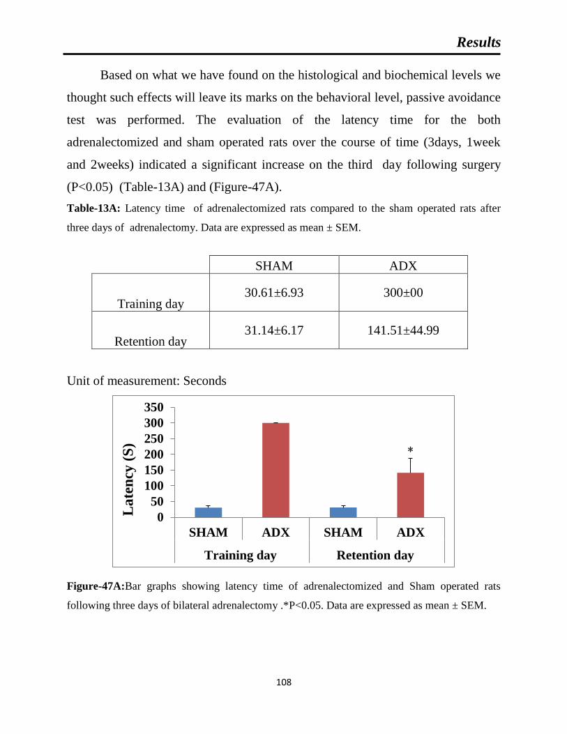
Results
108
Based on what we have found on the histological and biochemical levels we
thought such effects will leave its marks on the behavioral level, passive avoidance
test was performed. The evaluation of the latency time for the both
adrenalectomized and sham operated rats over the course of time (3days, 1week
and 2weeks) indicated a significant increase on the third day following surgery
(P<0.05) (Table-13A) and (Figure-47A).
Table-13A: Latency time of adrenalectomized rats compared to the sham operated rats after
three days of adrenalectomy. Data are expressed as mean ± SEM.
SHAM ADX
Training day 30.61±6.93 300±00
Retention day 31.14±6.17 141.51±44.99
Unit of measurement: Seconds
Figure-47A:Bar graphs showing latency time of adrenalectomized and Sham operated rats
following three days of bilateral adrenalectomy .*P<0.05. Data are expressed as mean ± SEM.
0
50
100
150
200
250
300
350
SHAM ADX SHAM ADX
Training day Retention day
La
ten
cy (
S)

Results
109
Furthermore, at day seven a significant increase (P<0.05) in the latenecy
time was observed in adrenalectomized rats compared to the sham (Table-13B) and
(Figure-47B).
Table-13B: Latency time of adrenalectomized rats compared to the sham operated rats after one
week of adrenalectomy. Data are expressed as mean ± SEM.
SHAM ADX
Training day
23.86±6.79
259.66±40.34
Retention day
25.76±6.25
146.11±52.85
Unit of measurement: Seconds
Figure-47B: Bar graphs showing latency time of adrenalectomized and Sham operated rats
following one week of bilateral adrenalectomy .*P<0.05. Data are expressed as mean ± SEM.
0
50
100
150
200
250
300
350
SHAM ADX SHAM ADX
Training Retention
La
ten
cy (
S)

Results
110
At two weeks postoperatively, the significant increase persisted between the
two groups (P<0.05) (Table-13C) and (Figure-47C).
Table-13C: Latency time of adrenalectomized rats compared to the sham operated rats after two
weeks of adrenalectomy. Data are expressed as mean ± SEM.
SHAM ADX
Training day
14.38±3.66
281±19
Retention day
15.8±2.34
174.36±30.85
Unit of measurement: Seconds
Figure-47C: Bar graphs showing latency time of adrenalectomized and Sham operated rats
following two weeks of bilateral adrenalectomy.*P<0.05. Data are expressed as mean ± SEM.
0
50
100
150
200
250
300
350
SHAM ADX SHAM ADX
Training Retention
Lat
ency
(S
)


Discussion
112
The aim of this study was to investigate the effects of short-term bilateral
adrenalectomy on the levels of pro-inflammatory cytokines IL-1β, IL-6 and TNF-
α, the response of microglia and astrocytes to neuronal cell death as well as on
oxidative stress markers GSH, SOD and MDA over the course of time (4h, 24h,
3days, 1week and 2weeks) in the hippocampus of Wistar rats.
Our results showed a significant decrease in the levels of corticosterone 4h
following the adrenal gland removal and this decrease persisted over the course of
the designated time of the experiment 24h, 3days, 1week and 2weeks in the
adrenalectomized rats compared to the sham operated. A significant decrease in
corticosterone following long-term adrenalectomy was also reported(Gould et al.,
1990; Jaarsma et al., 1992; Sloviter et al., 1989, 1993a, 1993b; Spanswick et al.,
2011).Regarding the pro-inflammatory cytokines IL-1β, IL-6 levels, our results
showed transient elevation that lasts for three days in the hippocampus of
adrenalectomized compared to sham operated rats. One week after adrenalectomy,
the elevation of these cytokines dropped to sham levels. Both the sham operated
and adrenalectomized rats showed significant elevated IL-1β and IL-6 levels
compared to the naïve rats. The significant difference between the naïve and sham
operated animals might be attributed to the surgical stress. However, the significant
difference between the sham and adrenalectomized rats can only be attributed to
the effect of adrenalectomy.
The occurrence of neuronal cell death in the hippocampus following
adrenalectomy was confirmed by Fluoro-Jade B staining and electron microscopy.
Our results showed that the induction of cell death in the granule cell layer of the
dentate gyrus by adrenalectomy begun three days postoperatively where we have
seen few FJB positive cells in the dorsal blade. the ultrastrucstural examination of
the dorsal blade showed cell abnormalities appeared in condensed chromatin and

Discussion
113
irregular cell membrane, such observation goes hand in hand with the finding of
(Sloviter et al., 1989, 1993a, 1993b) where they showed granule cells degeneration
after four days following adrenalectomy. In addition, we have made the same
observation as Conrad et al. (Conrad and Roy, 1993; Sloviter et al., 1993a, 1993b)
on the third day of adrenalectomy where no signs of degeneration were seen other
than dentate gyrus that gives the impression that the current model of
neurodegeneration seems to be a selective model for the exclusive loss of granule
cells . The ultrastructural results showed degeneration of the pyramidal cells of the
CA4 and CA3 after one week. After two weeks of adrenalectomy we observed a
progression of cell death throughout the dorsal blade of the dentate gyrus. These
findings are in line with those of Spanswick et al. (Spanswick et al., 2011) who
reported, degenerated neurons throughout the granular layer of the dentate gyrus
two weeks after adrenalectomy using Fluoro-Jade B staining.
In addition we observed a progression in pyramidal cells degeneration of
CA3 and CA4 where more damages and affected cells were seen which goes in
agreement with the observation of sapolsky et al. (Sapolsky et al., 1991) where
they have observed degeneration up to 19% of pyramidal cells of the CA4 area.
Subsequently, Prof. Adem and his team Islam et al.1999 demonstrated clearly
different populations of neurons have been affected following long term
adrenalectomy (5 months). Our findings are not in line with the observations that
have been made by different groups where they found both short and long term
adrenalectomy caused a selective cell death of the granule cells of the dentate
gyrus (Conrad and Roy, 1993; Sloviter et al., 1993a, 1993b). We think the reason
of that might returned to the sensitivity of different strains of animals or may be the
sensitivity of the techniques used in different Labs. In addition, the close
examination of the dentate gyrus indicates that adrenalectomy affects differentially

Discussion
114
the dorsal and ventral blades of the dentate gyrus and suggest that the granule cells
of the dorsal blade of the dentate gyrus are more vulnerable to adrenalectomy. The
question that raises itself why this granule neurons are the most vulnerable to the
absence of glucocorticoid hormones? There is strong evidence that those hormones
are playing trophic role for this population of neurons, many studies from different
groups confirmed the trophic crisis occurred in the absence of glucocorticoids.
In situ hybridization revealed DEX injection increase the NGF mRNA levels in the
granule layer of the dentate gyrus however two weeks of adrenalectomy showed a
significant reduction in the NGF expression (Mocchetti et al,1996). Chao et
al,1994 showed BDNF and NT-3 mRNA decrease in pyramidal and granule cells
of the dentate gyrus following seven days of adrenalectomy. Isalm et al. showed a
significant reduction of IGF-1 receptors mRNA in the dentate, CA1, CA3 and CA4
areas.
To conclude regarding the cell death in the current model our results showed
the vulnerability of different neuronal population to glucocorticoids absence on
one hand and the non-selectivity of the current model of neurodegeneration. In
addition, these findings might open the possibility to investigate the occurrence of
neurodegeneration in other parts of the brain.
It is an issue of debate regarding the accurate time of cell death onset in the
hippocampus and that returned to the idea of linking the raise in the inflammatory
cytokines levels and neurodegeneration and to reach the conclusion if such
increase precedes the cell death or it comes as a consequence to it. For that reason
our findings are in line with the observations that have been made by several
investigators that the earliest cell death occurs at least after three days of
adrenalectomy (Gould et al., 1990; Nichols et al., 2005; Sloviter et al., 1989;

Discussion
115
Spanswick et al., 2011; Stienstra et al., 1998). However, few investigators reported
cell death on day two after adrenalectomy (Jaarsma et al., 1992; Nichols et al.,
2005; Sloviter et al., 1993a).
It is interesting to note that the early elevation of the pro-inflammatory
cytokines IL-1β, IL-6 and TNF-α was seen prior to neuronal death. Taken together
our findings suggest that cell death was preceded by an increase of these pro-
inflammatory cytokines. Similar findings have been reported in animal models of
ischemia (Rothwell, 2003; Zhu et al., 2006), surgical injury (Tchelingerian et al.,
1993), TMT (Trimethyltin) (Liu et al., 2005) KA (Kainic acid) (Minami et al.,
1991) and cis-2,4-methanoglutamate (MGlu) (Pearson et al., 1999)where an early
increase of different pro-inflammatory cytokines preceded neuronal damage.
Two studies reported the direct involvement of IL-1β in neurodegeneration.
(Rothwell, 2003) reported the injection of low doses of IL-1β into the cerebral
ventricles or brain parenchyma of mice or rats exposed to neurotoxins markedly
exacerbates the damage. The second study showed the antagonism of IL-1β by co-
infusion of IL-1β receptor antagonist with the neurotoxin N-Methyl-D-
aspartate (NMDA) inhibits brain damage induced by such excitotoxin (Lawrence et
al., 1998; Panegyres and Hughes, 1998). Moreover, IL-1β plays a crucial role in
the exacerbation of acute neurodegeneration caused by ischemia, head trauma and
stroke. In addition, it is implicated in the pathology of multiple sclerosis,
Alzheimer’s disease and other CNS chronic diseases (Allan and Rothwell, 2001;
Boutin et al., 2001; Griffin and Mrak, 2002; Pearson et al., 1999).
IL-6 levels were found to be elevated in the serum and CSF of MS patients,
locally around MS lesions (Malmeström et al., 2006; Stelmasiak et al., 2001) and
in experimental auto-immune encephalitis (EAE) animal models (Gijbels et al.,
Discussion
116
1990). IL-6 expression was significantly increased in the acute phase of cerebral
ischemia (Berti et al., 2002). In AD patients, IL-6 is further increased locally
around amyloid plaques and in the CSF (Blum-Degen et al., 1995; Lucin and
Wyss-Coray, 2009). In agreement with the early increase of TNF-α that was
observed in the current neurodegenerative model, several studies showed the same
pattern of expression when elevation in the levels TNF-α mRNA were seen at early
stage following different neuronal insults (Berti et al., 2002; Chiu et al., 2015; Liu
et al., 2015; Zhu et al., 2006).
In summary, putting together the previous three finding regarding
corticosterone, cytokines levels and neuronal cell death after short term
adrenalectomy. Our results and others might suggest that the depletion in the levels
of corticosterone after removal of the adrenal gland did not induce an immediate
neuronal cells death However it was accompanied by an instant increase in the pro-
inflammatory cytokines IL-1β, IL-6 and TNF-α. The transient elevation in these
inflammatory components was followed directly by degeneration of the granule
cells of the dentate gyrus of the adrenalectomized rats compared to the sham
operated.
We think that the induction of the cell death was the result of both the
increase of such cytokines which has been largely proved for their contribution in
the exacerbation of the neuronal damage from one side and the depletion of the
corticosterone which has been shown to play major role in the survival of the
hippocampal neurons and the depletion in its levels like what is happening in the
current model affects the production of essential factors for the survival of the
hippocampal neurons on other side. several groups showed that the removal of the
adrenal gland caused a depletion in the production of several neurotrophins such
production of neurotrophin-3 (NT-3), Brain Derived Growth factor (BDNF), Nerve

Discussion
117
Growth Factor (NGF) (Barbany and Persson, 1992; Nichols et al., 2005; Smith et
al., 1995; Vollmayr et al., 2001).
In addition Prof. Adem and his colleagues demonstrated that the levels of
IGF-1 were decreased after long term adrenalectomy. Taken together the
aforementioned findings might lead us to suggest that after adrenalectomy IL-1β,
IL-6 and TNF-α might contribute to the initiation of the biological cascade
responsible for subsequent hippocampal neuronal cell death.
Synergistic effects of microglia and astrocytes are needed for tissue
reconstitution after lesions, involving control of the BBB and invasion of
haematogenous cells, removal of pro-inflammatory cytokines and their down
regulation by TGF (Kreutzberg, 1996). Microglias are the main resident immune
cells in the brain which presents 12% of the brain cells, playing a crucial role both
in physiological and pathophysiological conditions (Polazzi and Monti,2010).
Virtually ,every neurological disorder ,AD,PD,MS results in activation of these
cells accompanied by biochemical and physiological changes leading to alteration
in cell phenotype ,a phenomena generally termed “reactive gliosis” (Wenk, 2003).
Moreover, acute neurodegenerative insults such as MPTP, TMT, Kainic acid,
ischemia-reperfusion, colchicines induce the activation of these brain resident
immune cells to compromise the neuronal survival and indirectly trigger
neuroinflammation (Lalancette-He´et al.,2007; Lambertsen et al.,2009).
The activation of microglia is one of the major signs of neurodegeneration
(Lucin and Wyss-Coray, 2009). Similar to Gould et al.(Gould et al., 1990) our
results showed cell death starts in the dorsal blade of the dentate gyrus three days
after adrenalectomy. Activated microglia were observed in the molecular and
granular layers of the dorsal blade of the dentate gyrus three days after

Discussion
118
adrenalectomy. One week after adrenalectomy as the cell death progresses in the
dorsal blade the number of activated microglia are increased. Two weeks after
adrenalectomy we showed that the whole dentate gyrus was invaded by activated
microglia. In line with these findings quantitative analysis showed a progressive
increase in the number of microglia in the hippocampus from day three with a
further increase one and two weeks after adrenalectomy. Moreover, in addition to
the invasion of activated microglia to the granule layer. we noted, the area
bordered by granule layer and the hippocampal fissure, so-called molecular layer
was occupied by these cells suggesting degeneration of the apical dendrites of the
granule cells and this goes hand-in-hand with observation that (Jaarsma et
al.,1992) made, using silver impregnation, a specific staining of neuron
degeneration where they noticed argypholia at this layer.
Moreover, in a previous study Prof adem and his team in (Mulugeta et al., 2006)
reported after two weeks of adrenalectomy the decrease in the concentration of the
acetylcholine receptor M4, a receptor found on the presynaptic membrane of the
perforant pathways in molecular layer of the dentate gyrus, where they synapse the
apical dendrites of the granule cells. It is very important to mention the
disintegration of synapses at this layer might lead to retrograde degeneration of the
entorhinal cortex neurons and eventually microgliosis starting from the dentate
gyrus back to the entorhinal cortex. To investigate this hypothesis and it is
correlation with microgliosis, extension of the experiment survival timing one of
our future plans.
Regarding the cornu amonis, we found invasion of activated microglia to the
pyramidal layer of the CA4 which suggest vulnerability of this population of
neurons at early stage of adrenalectomy. Nevertheless, no sign of microgliosis was
seen in the pyramidal cells of CA3/CA2/CA1. However we noticed activated

Discussion
119
microglia along the stratum lacunosum of the CA3, this layer where the mossy
fibers get connected to the apical dendrites of big pyramidal cells of CA3
suggesting two weeks of glucocorticoids deprivation results in synapses
disintegration which needs by standard the appearance of activated microglia for
repairing and cleaning the debris of the degenerated axons.
Our current study showed a strong correlation between cell death and
microglia activation over the course of time. Taken together our observations
suggest a correlation between microgliosis and cell death.
Astrocytes are known to communicate with microglia, and such interaction is
very important in pathological conditions (Verderio and Matteoli .,2001; Von
Bernhardi and Ramirez.,2001). It is well known that brain injury is associated with
increased astrocyte reactivity (Fiedorowicz et al., 2001). Reactive astrocytes
synthesize a variety of cytokines, neurotrophins and components of the
extracellular matrix, many of which are known to rescue damaged neurons after
brain insults (Schwartz et al.,1993) and their scavenging effect for reactive oxygen
species (Chen and Swanson,2003).
It is well known that brain injury is associated with increased astrocytes
reactivity (Fiedorowicz et al., 2001). Astrocytes react profoundly to brain damage
or disease by increasing both their number and size, a process referred to as
reactive astrogliosis/astrocytosis which represents a remarkably homotypic
response to CNS injuries (Penkowa et al., 2008). In this study, activation of
astrocytes was observed throughout the dentate gyrus one week after
adrenalectomy.
However, Krugers et al.(Krugers et al., 1994) found activation of astrocytes
earlier, on the third day after adrenalectomy. Although we could not see activated
Discussion
120
astrocytes at day three, it is possible that the activation of astrocytes takes place
between day 3 and 7 after adrenalectomy. The observed astrogliosis in our study
was predominantly in the molecular and polymorphous layers of the dentate gyrus
whilst no apparent astrogliosis was seen in other areas of the hippocampus. Our
findings are in line with those findings which showed astrocytes invasion of the
dentate gyrus after TMT injection. TMT is a selective neurotoxin of granule cells
(Fiedorowicz et al., 2001).
In addition to the activation of astrocytes after one week in the DG our
results showed progressive activation of astrocytes in the CA4 and CA3 of the
hippocampus two weeks after adrenalectomy. In line with these findings
quantitative analysis showed a significant increase in the number of astrocytes in
the hippocampus from one week until two weeks after adrenalectomy.
Astrocytes are known to communicate with microglia, and such interaction is very
important in pathological conditions (von Bernhardi and Ramirez, 2001; Verderio
and Matteoli, 2001). The progressive degeneration of granule cells correlated well
with the activation of microglia and astroglia in the dentate gyrus over the course
of two weeks. It is very important to note almost in all neurodegenerative models,
microglia are the first cells to respond to neuronal damage and followed by
activation of astrocytes , which leads us to suggest the important of cross talk
between the two cell populations during neuronal damage and consider microglia
as driving force in astroglia recruitment.These observations it goes hand in hand
what we found in the current study where we noted microglia activation on the
third day in first place and followed later by astrocytes activation on the seventh
days.
Our results showed microglia activation on the third day followed by
astrocytes activation on the seventh day after adrenalectomy. These findings are in

Discussion
121
line with what has been found in different neurodegenerative models where
microglia were found to be the first cells to respond to neuronal damage followed
by activation of astrocytes (Frautschy et al., 1998; Gahtan and Overmier, 1999;
Matsumoto et al., 1992; Reali et al., 2005). Taken together these findings suggest a
cross talk between the two cell populations during neuronal damage and consider
microglia as driving force in astroglia recruitment.
Oxidative stress occurs when there is an overproduction, or accumulation of
reactive oxygen species (ROS) in conjunction with reduced antioxidant capacity
within the cell (Sies, 1997). As a response to neuronal death, the quiescent
microglia became functionally active and upregulate enzymes such as inducible
nitric oxide synthase (iNOS) leading to an imbalance between free radicals
production and the antioxidant defenses (Tran et al., 1997).
Our findings showed that a state of oxidative stress manifested in the
decrease of reduced glutathione (GSH) two weeks after adrenalectomy. In
addition, our study showed a significant decrease in the activity of the superoxide
dismutase. In contrast to the decrease of the antioxidant defenses, malondialdehyde
(MDA) the indicator of the plasma membrane disruption was significantly
increased.
This imbalance, observed two weeks after adrenalectomy, reflects a state of
oxidative stress which possibly resulted from the activated glial cells. It has been
found after one week of adrenalectomy an overexpression of iNOS in activated
microglia revealing these cells as source of the free radical in this model (Sugama
et al., 2013). Taken together our findings are consistent with the literature showing
that several neurodegenerative paradigms revealed the association between
neuroinflammation accompanied by reactive gliosis and a state of oxidative stress
(Zhu et al., 2004).

Discussion
122
The hippocampus is well known for its role in cognition (Strange et al.2004;
Zeidman and Maguire.2016), most importantly in learning and memory (Morgado-
Bernal, 2011). As it was aforementioned due to the high concentration of
glucocorticoids receptors in the hippocampus, it is considered the most targeted
area of the brain by such hormones. Our study has shown the withdrawal of these
hormones causes degeneration of different neuronal cell populations in the
hippocampus. Several studies and most of them have investigated the impact of the
neuronal damage caused by long term absence of such hormones on the behavior
of the animals where Islam et al.1995 showed after 19 weeks of adrenalectomy a
significant increase in the latency in the Morris maze task and significant lower
rearing scores in the adrenalectomized rats compared to sham operated. In
coherence with that Conrad and Roy.1995 showed that after long term
adrenalectomy, the adrenalectomized rats exhibited difficulties in acquiring a new
spatial memory. Furthermore Spanswick et al. 2007 showed the inability to reverse
such deficit in the spatial memory by chronic treatment (6 weeks) with
corticosterone or anther neurogenic compound called Fluoxetine of long term
adrenalectomized rats, indicating the crucial role of hippocampal formation in
building memories and acquiring new information. So far, we did not find in the
literature the effect of adrenalectomy on cognitive functions following short-term
adrenalectomy and in order to examine that we performed the passive avoidance
test which is a legitimate test to verify the ability of the animals to learn and save
new task that they have been taught recently. Our results showed the
adrenalectomized rats failed to retain their cognitive capacity compared to the
sham operated where we have seen from day 3 to day 7 and 14 a significant
increase in the latency for the adrenalectomized rats revealing a cognitive decline
similar to what had been seen after chronic adrenalectomy and suggested that
such behavior impairement might start in day 3 following glucocorticoids

Discussion
123
withdrawal and by that our results go hand in hand with the work of different
research groups (Islam et al.1995; Conrad and Roy.1995; Spanswick et al. 2007).
The mechanisms underlying cell degeneration after bilateral adrenalectomy
are not clear. We have previously suggested the possibility that adrenocortical
hormones might have direct effects on the survival of hippocampal neurons.
Hippocampal neurons might die when the mineralocorticoids (Type I) and /or
glucocorticoid (Type II) receptors are not occupied by adrenocortical hormones
(Adem et al., 1994; Islam et al., 1999). Subsequently, Hu et al. (Hu et al. 1997)
reported that granule cell death after adrenalectomy was necessarily accompanied
by the disappearance of glucocorticoid receptor immunoreactivity in the granule
cell layer.
We have previously also suggested that the loss of adrenocortical hormones
after adrenalectomy may cease to stimulate a factor(s), in the hippocampus or other
tissues, that is (are) necessary for the survival of hippocampal neurons (Adem et
al., 1994; Islam et al., 1999). It could be speculated that in several
neurodegenerative diseases like Alzheimer’s disease, chronic adrenal hormones
and/or their receptors’ decrease may result in inflammatory mechanisms which
lead to hippocampal neurodegeneration (Heuser and Lammers, 2003; Landfield,
1978; McEwen, 1997; Woulfe et al., 2002).


Conclusion and Perspectives
125
The removal of the adrenal gland leads to almost the depletion of
glucocorticoids from the animals body, such change was assumed by many
researchers around the world to cause an exclusive death of the granule cells in the
hippocampus. However, in the current study we reached along with few
aforementioned studies that there is another population of neurons different than
the granule cells was affected by the absence of glucocorticoids called the
pyramidal cells. This finding suggested that the cell death in the hippocampus is
not selective to the granule cells as it was suggested before and opens the door for
further investigation on the impact of such hormones on different cells in the brain.
The common believe that the inflammation is a phenomenon occurs as a
result or response to cell death to get rid of the debris resulted from the cell
degeneration which lead us to say that the cell death always precedes the
inflammation. However, in the current study our results made us conclude that the
inflammation does not play the usual beneficial effect in the current
neurodegenerative model, on the contrary it might contribute or initiate the cell
damage and this conclusion manifests in the early increase of different
inflammatory cytokines before the neuronal death followed by the activation of
another components of inflammation such as astrocytes and microglia cells.
In addition, our study revealed a strong correlation between the appearance
of the inflammatory cells and the deterioration of the antioxidant defense from one
side and the increase of the MDA, this later is a strong indicator of the oxidation
and disruption of the cell membrane of neurons in the hippocampus which supports
the idea that the inflammation has contributed to neuronal cell death. Furthermore,
the neuronal cell death that was observed in the hippocampus has a direct impact
on the cognitive capacity of the animal that manifested in memory shortage
ollowing the removal of the adrenal gland (Figure-48).

Conclusion and Perspectives
126
Figure-48: summary of our findings
In summary, our study showed that short-term bilateral adrenalectomy leads
to an early increase in pro-inflammatory cytokines followed by neurodegeneration
and activation of glial cells as well as oxidative stress and behavioral change.
Taking all these findings together it could be suggested that the early inflammatory
components might contribute to the initiation of the biological cascade responsible
for subsequent hippocampal neuronal cell death and cognitive decline in the
current neurodegenerative animal model.
Based on the results of the current work and the other eleven papers
published by Prof. Adem et al. we are aiming in the future to go deep in our
investigation conering the sources of the cytokines that appeared at the early satge
following adrenalectomy. In addition to that we aime to investigate the impact of
the removal of the adrenal gland on the production of a large group of growth

Conclusion and Perspectives
127
factors, since they are considered an essential components of cell survival. In
order, to put our work in a medical sphere we planned to investigate the production
of the APP (Amyloid precursor) in the hippocampus in attempt to relate it to the
cell death occurring in the current model on one side and to Alzheimer,s diseases
from another side .


ملخص باللغة العربية
129
مقدمة:
الغدة الكظرية على مستوىهي هرمونات ستيرويدية تصنع (Glucocorticoids) الهرمونات القشرية السكرية
(adrenal gland) حاالت التوتر ثناء أما تفرز تتبعا للساعة البيولوجية وك وتفرز في الدم(Dickmeis, 2009) النسخة.
النصف بداية منذ اثبت علميالقد البشرية والقوارضية لهذه الهرمونات هي الكورتيزول والكورتيكوستيرون على الترتيب.
إلى العديد باالضافة(.hippocampus) الحصين فيالهرمونات هذه لمستقبالت ىمستو أعلىتواجد الثاني من القرن الماضي
، تحت المهاد(neocortex) ، القشرة المخية الجديدة (amygdala)من المناطق الدماغية األخرى مثل اللوزة
(hypothalamus) (Reul et al., 2015)، الدماغ على مستوى المستهدف الرئيسي لهذه الهرمونات الحصين يعتبر
(McEwen et al., 1968) .
للدماغ وجود نوعين من المستقبالت (Immunohistochemistry) أظهرت الدراسات المناعية النسيجية
Reul) 2أو نوع و غلوكوكورتيكويد 1أو نوع (mineralocorticoid) مينيرالوكورتيكويدالويد، مستقبالت للجلوكوكورتيك
and de Kloet, 1985) الخاليا الحبيبية تكون . تختلف وفرة هذه المستقبالت في الحصين من نوع خلوي إلى آخر حيث
(granule cells ) الخاليا العصبية الهرميةما أ 1غنية بالنوع (pyramidal neurons فهي غنية ) 2بالمستقبالت من النوع .
، تلعب هذه الهرمونات دورا رئيسيا في المخ (MacLennan et al., 1998)تأثيرها الكبحي كوسطاء لاللتهابات جانب إلى
( وعامل نمو Brain Derived Growth factor(، مشتق عامل النمو المخي )neurotrophin-3في إنتاج نيوروتروفين )
,.Barbany and Persson, 1992; Nichols et al., 2005; Smith et al)( Nerve Growth Factorاألعصاب )
1995; Vollmayr et al., 2001).
أن الحقن أظهرتحيث لهذه الهرمونات على مستوى الحصين، مزدوج التأثير الأظهرت العديد من الدراسات
. (Sapolsky and Pulsinelli, 1985)تالشي الخاليا العصبية الهرمية إلىالمزمن لجرعة عالية من الجاليكورتيكود تؤدي
للخاليا يسبب تالشي هائل وانتقائي (adrenalectomy)أن استئصال الغدة الكظرية دراساتالعديد من الأظهرت غير انه
;Gould et al., 1990) (Sloviter et al., 1989, 1993a, 1993b) (dentate gyrus) الحبيبية للتلفيف المسنن
Nichols et al., 2005; Sloviter et al., 1989; Spanswick et al., 2011; Stienstra et al., 1998).
خاليا الهرمية في الحصين بعد استئصال الغدة الكظرية على اللوحظ تالشي فقد تالشي الخاليا الحبيبية، ى إلاضافة
Sloviter et) 1191اكتشف سلوفيتر ومساعديه سنة .(Adem et al., 1994)و(Sapolsky et al., 1991) المدى الطويل
al., 1989). نفس النتيجة من قبل إلىالتوصل الخاليا الحبيبية الحصينية و قد تم موتأن استئصال الغدة الكظرية يؤدي إلى
، (Krugers et al., 1994; Nichols et al., 2005; Sousa et al., 1997; Sugama et al., 2013)عدة فرق بحث
1114و أدم ومساعديه سنة (Sapolsky et al., 1991) 1111فضال عن النتائج التي توصل إليها سابولسكي ومساعديه سنة
(Adem et al., 1994) التي أظهرت موت الخاليا الهرمية للحصين بعد استئصال الغدة الكظرية على المدى الطويل وقد
الموت الخلوي العصبي إنعتبر هذا النموذج كنموذج لدراسة موت الخاليا العصبية على مستوى الحصين. ا
(neurodegeneration) له الدبقية ستجابةواإل(glial response) التي تحدث في هذا النموذج هو مماثل لما هو مبين في
، و (Rothwell, 2003; Zhu et al., 2006a)(ischemia-reperfusion)عادة التروية إ-ةقفارياإلنماذج أخرى مثل

ملخص باللغة العربية
130
,.TMT (Trimethyltin) (Liu et al، (Tchelingerian et al., 1993) (surgical injury) الجراحية صابةاإل
2005) KA (Kainic acid) (Minami et al., 1991) وcis-2,4-methanoglutamate (MGlu) (Pearson et
al., 1999a)ج لدراسة موت الخاليا العصبية في الحصين والتي ذ. وبالتالي يمكن استخدام نموذج استئصال الغدة الكظرية كنمو
.المزمنة العصبية األمراضكون نموذجا مماثال لما يحدث في يقد
1β (IL-1β) (Rothwell-كاإلنترلوكين ( Pro-inflammatory cytokinesية )اللتهابايتم إنتاج السيتوكينات
and Luheshi, 2000a; Touzani et al., 1999; Vitkovic et al., 2000a)6-وكين، إنترلي (IL-6) (Gadient and
Otten, 1994; Godbout and Johnson, 2004) و عامل نخر الورم ألفا(TNF-α) (Perry et al., 2002;
Sternberg, 1997a; Vitkovic et al., 2000a) من قبل خاليا عديدة في المخ(Rothwell, 2003) ولقد تم العثور على.
مثل مرض الزهايمر (degenerative diseases) مستويات مرتفعة من هذه السيتوكينات في العديد من األمراض التنكسية
(Alzheimer’s)(Rubio-Perez and Morillas-Ruiz, 2012; Veerhuis et al., 1999)، التصلب الجانبي
باركنسونالومرض (Rothwell and Luheshi, 2000a)( Amyotrophic lateral sclerosis) الضموري
(Parkinson’s) (Blum-Degen et al., 1995; Nagatsu et al., 2000) . الى الكشف دراسات مختلفة وقد توصلت
، KA (Yabuuchi et al., 1993)فراز المبكر لهذه السيتوكينات في مختلف النماذج الحيوانية العصبية كنمودج اإلعن
TMT (Fiedorowicz et al., 2001) نقص التروية ،(Sairanen et al., 1997; Yabuuchi et al., 1994) ،1-
methyl-4-phenyl-1,2,3,6-tetrahydropyridine (MPTP) (Kumar et al., 2012; Selley, 2005).
شروع في التغيرات المورفولوجية التي تحدث أثناء موت االلتهابية تساهم في ال ائطأن هذه الوس العديد من الدراسات وقد ذكرت
;Allan et al., 2000; Apelt and Schliebs, 2001)المختلفة تصاباإلاالخاليا العصبية وتفاقم األضرار الناجمة عن
Berti et al., 2002a; Botchkina et al., 1997; Knoblach et al., 1999; Touzani et al., 2002) يعرف .
سامة ناتجة عن اختالل في التوازن بين مضادات األكسدة خلوية ظاهرة نه بأ (Oxidative stress) اإلجهاد التأكسدي
(antioxidants ) والمواد المؤكسدة (oxidants ) مخاطر مختلفة وبالتالي اإلسهام في ى لامما يؤدي إلى تعريض األنسجة
أنهغير .(Halliwell, 2006; Lee et al., 2010a; Li et al., 2004; Murakami et al., 2011)الموت العصبي
Hsieh and Yang, 2013; Schieber and)الوظائف الفسيولوجية المهمة )من تملك هذه المواد المؤكسدة العديد
Chandel, 2014)بدوره يمكن أن االخير اذاإلجهاد التأكسدي. وه إلى فراط في إنتاج مستويات عالية منهااإليؤدي دق. و
مضادة اللك تملك الخاليا مخزون من المركبات واألنزيمات ذولمواجهة (Haddad, 2002) يسبب تلف خلوي ويعزز االلتهاب
.(van Horssen et al., 2011)للتأكسد للحيلولة دون تراكم هذه المواد المؤكسدة

ملخص باللغة العربية
131
العمل: وطرق المواد
:الهدف من الدراسة-1
لقد وافقت اللجنة األخالقية اإلقليمية لكلية الطب والعلوم الصحية )جامعة اإلمارات العربية المتحدة( على هذه
وكان الهدف من الدراسة الحالية هو بحث تأثير استئصال الغدة الكظرية على المدى القصير على (.RECA/01/05الدراسة )
مختلف ، تنشيطTNF-αو IL-1β،IL-6إنتاج السيتوكينات االلتهابية مناطق مختلفة من الحصين،موت الخاليا العصبية في
و GSH ،SODاإلجهاد التأكسدي وعلى مؤشرات (microglia) والدبقية الصغيرة (astrocytes) الخاليا الدبقية النجمية
MDA الكظرية على سلوك وأثر غياب الهرمونات (animal’s behavior) ر البيضاءاذان الويستجر.
:الحيوانات و استئصال الغدة الكظرية -2
أسابيعالبالغة العمر ثمانية ( Wistar albino)البيضاء يستار الوان ذانتقينا لهذه الدراسة جر
نية بكلية الطب والعلوم الصحية )العين، والتي تم الحصول عليها من المصلحة الحيوا غ221-171ها بين أوزانو تتراوح
، كل مجموعة تتكون من أربعة (Plexiglas) العربية المتحدة(. وضعت جميع الحيوانات في أقفاص بالستيكيةاالمارات
، تركت الحيوانات حرة م°22تقدر حرارة الدرجة حيث سا12/ سا12 ظالم فيهاوال ضوءتواتر الي وفي ظروف ، جرذان
٪ 1.1 (saline)مالح ماء شرب الكظريةالمستأصلة الغدة ذانالجر عطاءإ للوصول إلى الغذاء والماء طوال التجربة. وقد تم
يونات ومنع اآلثار الضارة الناتجة عن غياب كلوريد الصوديومألمن أجل الحفاظ على توازن االعادية بدال من مياه الشرب
(sodium chloride).
ظهورمغ/كغ من وزن الجسم( ، تم حلق 53) (Pentobarbital) خدرت الحيوانات باستعمال بينتوباربيتال
كما (Sham surgery) استئصال الغدة الكظرية أو عمليات مزيفةبسواء قمنالة حالقة كهربائية ثم آلحيوانات باستخدام ا
وبعد ٪71الحيوان بااليثانول ظهر. من أجل إجراء عملية جراحية معقمة تم تنظيف 1191 مساعديه سنةوضحه سلوفيتر و
بالقدر حيث يكون(peritoneal cavity) الجلد والعضلة الظهرية للوصول إلى الجوف الصفاقي ذلك قمنا بشق على مستوى
مسك. باستخدام ملقط حاد تم منهادون ترك أي بقايا رؤية الغدة الكظرية وبعض المساحة الحرة إلزالتها الذي يسمح لنا بفي االك
بمساعدة ملقط حاد و مائل. بعد يةالكلوبين هاقطع النسيج الضام الموجود بينلت عن طريق ئصتم اسثالدهون المحيطة بالكظرية
السابقة اتراءنفس اإلجل (SHAM) الشاهدة المزيفة اتالحيوان خضغت خياطة الشق ووضع الحيوان في القفص. قمنا ب ذلك
.(6-)الشكل باستثناء إزالة الغدة الكظرية

ملخص باللغة العربية
132
Enzyme Immunoassayبواسطة المصلفي تحديد مستويات الكورتيكوستيرون -3
(EIA):
ستئصال الغدة عملية امستويات الكورتيكوستيرون في المصل لتقييم فعالية الهدف من وراء هذه الخطوة هو معرفة
التضحية من ت. وقد تم (Animal sacrifice) الجرذان تضحية قبلمل من الدم مباشرة 1من القلب حوالي حصلناالكظرية،
5(، 24سا )ن = 24(، 24سا )ن = 4 من اجراء الجراحة مختلفة فتراتبعد (decapitation) الحيوان خالل قطع رأس
- عند درجة حرارة(sera samples) ينات المصل ع خزنت (.24ين )ن = اسبوع( و 24)ن = اسبوع(، 24أيام )ن =
Life) (Enzyme Immunoassay) قياس مستويات الكورتيكوستيرون عن طريق تقنية الى غاية موعد م91°
sciences, Lausen, Switzerland) ة االنسالتعددالمم اجساال ترتبط. باختصار، في هذه التقنية (polyclonal
antibody) لألغنام ةالمضاد و ة االنسال المصنعة في الحمارتعددالماالضداد بللكورتيكوستيرون ةالمضاد (donkey anti-
sheep antibody) حفر التي تكون مثبتة في(wells )صفيحة بالستيكية تدعى micro- plate.
وستيرون الموجود في المصل مع الزائدة، يتنافس الكورتيك ة االنسالتعددالمإلزالة األجسام المضادة الصفيحة ناغسل
( على المواقع الموجودة على horseradish peroxidaseكمية ثابتة من الكورتيكوستيرون الموسوم ببيروكسيديز الفجل )
محلول الركيزة اضفنا إلزالة العينات الزائدة والغير مرتبطة. غسلنا الصفيحة مرة ثانية. ة االنسالتعددالماألجسام المضادة
(substrate ) ف التفاعل باضافة محلولقمنا بوقنزيم المرتبط. االلتحديد نشاط الحفرإلىTMB stop solution لكل حفرة.
بالمطيافنانومتر 431 ةموجالعلى طول (absorbance) متصاصيةقسنا اال وبعد ذلكالصفيحة بهدوء ترج
(Spectrometer) . تيكوستيرون في العينة. لحساب تركيز الكورتيكوستيرون في كثافة اللون تتناسب عكسيا مع تركيز الكور
بيكوغرام/مل. ومن خالل 21،111إلى 52 ضمن مجال يتراوح من(Standard Curve) العينات تم إنشاء منحنى العيارية
الكثافة الضوئية هذا المنحني يتم استخراج معادلة المنحني العيارية. وبذلك يمكن قياس تركيز الكورتيكوستيرون اعتمادا على
.المقاسة بالمطياف
:الكيمياء الحيوية-4
:تحضير العينات-4-1
سا )ن = 4الجراحة: من اجراء بعد أوقات مختلفة هاقطع رأسعن طريق ( 91)ن = جرذانتضحية الب قمنا
ه وعزل تشريحقمنا ب المخإزالة بعد (. 16ين )ن = اسبوع( و 16)ن = اسبوع(، 16أيام )ن = 5(، 16سا )ن = 24(، 16
في درجة( و وضع liquid nitrogen)في سائل النيتروجين األخير اذهوبعد ذلك جمد الجليد، فوق على الفور الحصين
موعد الفحص. إلى غاية م°91-حرارة
البارد )بيرس التكنولوجيا T-PERأنسجة الحصين المذابة في محلول على الجليد تسحقالعينات جناسة لتحضير
protease inhibitor) كوكتيل مثبط البروتياز 111: 1الحيوية، روكفورد، إيل، الواليات المتحدة األمريكية(، المظاف اليه
cocktail) (Sigma-Aldrich, Missouri, USA)بعد صوتنة . (sonication) للطرد المركزي هاالعينات، تم تعريض
ملخص باللغة العربية
133
(centrifugation) ي طافالمحلول ال استرجعنا .م°4حرارة دقيقة في درجة 51الدقيقة لمدة /دورة 16111بسرعة
(supernatant) واسطة اختبارب اإلجمالي تركيز البروتينكل من تحديد في استعمالهم حتى موعد °91-تخزينه في ثم قمنا ب
-ILمستوي السيتوكينات )قياس ، ((Bio-Rad, California, USA) (Bradford’s protein assay) برادفورد
1β،IL-6 وTNF-α عن طريق تقنية )ELISA (R&D Systems; Minneapolis, USA) اإلجهاد مؤشرات و
. (Colorimetry) قياس اللونبواسطة تقنية MDAو GSH ،SODالتأكسدي
العينات:( في Total protein) البروتين تركيزتحديد -4-2
سلسلة من العينات تكوينبقمنا BSA )1% ( (Bovine serum Albumine) محلولانطالقا من
Bradford)كاشف برادفورد 3Xمغ/مل. خفف 1.4 و 1.2 بينتراكيزها تتراوح بشكل مضاعف (standards)يةاريالمع
Reagent) كل أنبوب ل اأضفن. رشح المحلول المخفف الناتج 1-ورقة ترشيح واتمان واسطةب ثمبالماء المقطر 4: 1بنسبة
(homogenation buffer)في محلول التجانسالحصين عينات نا. خففالمخفف مل من كاشف برادفورد 1.23البوليسترين
عينات و يةاريالمع العيناتمن ميكرولتر 23 اضفناخالل قياس االمتصاص. ( blank)كمرجع خيراأل هذا نااستخدمكما 11:1
تم قياس االمتصاصية في ( incubation)دقائق من التحضين 3بعد .تحتوي على كاشف برادفورالتي نابيب األ إلى الحصين
منهاالستيفاء وعن طريق للبروتين . عياري المنحنى القمنا برسم يةاريالمعالعينات متصاصية اعتمادا على ا. نانومتر 313
.لكل عينة الحصين (Homogenate) تركيز البروتين في جناسة حددنا
بواسطة( TNF-αو IL-1β، IL-6الحصين )االلتهابية في السيتوكينات تحديد مستوى --4-3
ELISA:
(. wellحفر ) 9من يتشكل واحدصف وكل 12من ةمكون صفيحةعبارة عن يوه ELISA صفيحة ناستخدما
. المختبرفي درجة حرارة ه وتترك لليلة كاملة لسيتوكين المراد الكشف عنبا االجسام المضادة الخاصة هذه الصفيحة الى اضفنا
لبومين مصل البقر ابواسطة ( blockت)م حظرالمضادة الغير ملتصقة ث جسامغسلنا الصفيحة للتخلص من األفي اليوم التالي
لمدة مختبردرجة حرارة ال وتركت للتحضين في (Phosphate-buffered saline)اب في محلول الفوسفات المالحة ذ٪ الم1
عروفة ( لتراكيز مserial dilutionعن طريق التخفيف التسلسلي ) عياريةال عيناتلا حضرنا . لتشكيل المنحنى العياريساعتين
هذه تم حضنو عينات الحصين في حفر الصفيحة ث عياريةعينات اللوضعت كل من ا . (7-المراد قياسه )الشكل لسيتوكينامن
في درجة حرارة الغرفة لليلة واحدة.خيرة األ
األجسام المضادة الكاشفة ثم عينات الغير ملتصقةلغسلنا الصفيحة مرة ثانية للتخلص من ا في اليوم التالي،
كمتفاعل ثالث تم اضافة المختبر.درجة حرارة للتحضين في عةسا لمدة ( وتركتBiotinylated goat)بيوتين مرتبطة بالال
Streptavidin conjugated to horseradish) الفجلبيروكسيديز وفيدين لسترابدابا مرتبطةاألجسام المضادة ال
peroxidaseميكرولتر من 111ضافة إ . تمالمختبردقيقة في في درجة حرارة 21لمدة توحضن ( للصفيحة
Tetramethylbenzidine (41.6 لكل يلي موالريةم )التفاعل بواسطة حامض الكبريتيك ناوقفااللون. لحصول علىل حفرة
بواسطةنانومتر 431 ةموجالوقف تم قياس االمتصاصية على طول ال من ائقدق 11و في غضون ( N H2SO42)مالرية 2

ملخص باللغة العربية
134
وعن طريق . لكل سيتوكين عياري المنحنى القمنا برسم يةالمعيار العيناتمتصاصية اعتمادا على ا (.9)الشكل المطياف
تركيز البروتين لكل عينة على. تم تقسيم تركيز السيتوكين لكل عينة TNF-αو IL-1β،IL-6 تركيز حددنا منهاالستيفاء
من سيتوكين/ملغ من رامغبيكوتركيز كالتالي الختالفات في حجم األنسجة. يتم التعبير عن اإل عديللت لنفس العينة االجمالي
.البروتين
قياس شدة تقنية ( بواسطة MDAو GSH ،SODاإلجهاد التأكسدي ) مؤشرات تحديد مستويات -4-4
:اللون
السيتوكينات تم استخدامها لتحديد اسالتي استخدمت لقيالحصين هنا أن نفس جناسةان ننوه لجدير بالذكر انه و
.قياس اللون تقنية بواسطة GSH ،SOD MDAاإلجهاد التأكسدي مؤشراتمستويات
4-4-1- Superoxide dismutase:
O2)هي انزيمات معدنية تحفز عملية تفكيك الجذر الفائق •-) (•-dismutation of the superoxide radical
(O2 الى ( بيروكسيد الهيدروجينH2O2( واألكسجين الجزيئي )O2 ويستند قياس كمية .)SOD الحصين على جناسةفي
التي تم إنشاؤها (superoxide radicals) للكشف عن الجذور الفوقية( tetrazolium salt)استخدام ملح تيترازوليوم
(.1-)الشكل hypoxanthine (Cayman chemical, Michigan, USA)و xanthine oxidaseبواسطة
ميكرولتر من الكاشف 211ها إضافة تال حفر الصفيحة. الىميكرولتر من العينات والمعايير 11أضيف حجم
من الماء المقطر(. وبمجرد االنتهاء مل 11.13ميكرولتر من الملح تيترازوليوم مع 31المخفف )محلول مخفف من ريذالج
من أجل الشروع في التفاعل. غطيت الحفرجميع الى xanthine oxidaseميكرولتر من 21من الخطوات السابقة تم إضافة
على طول الموجة الصفيحةد في درجة حرارة الغرفة. تم قراءة 21لمدة (shakerرجاجة )وحضنت على الصفيحة
الجذر الفائق من ٪ 31 (dismutation) لتطافركمقدار كمية االنزيم الالزمة SODنانومتر. يتم تعريف وحدة واحدة من 441
(O2•-).

ملخص باللغة العربية
135
:(MDAمالونديالدهيد ) -4-4-2
thiobarbituric acidفي جناسة الحصين على تفاعل حمض الثيوباربيتوريك ) TBARSويستند قياس كمية
TBA( مع مالونديالدهيد )MDA( في ظروف حامضية )acetic acid( و درجة حرارة مرتفعة )درجة مئوية( 13
(Cayman chemical, Michigan, USA). ل من جناسة الحصين مع نفس الكمية من محلو ميكرولتر111يتم خلط حجم
المتشكل من مل من كاشف اللون 4 مع( وخلطها بشكل جيد sodium dodecyl sulfate)الكبريتي صوديوم دوديسيل
TBA- sodiumمل 211و من TBA- acetic acid مل 211الذائب في خليط من TBAمن حمض مغ2121)
hydroxide د انتهاء المدة الزمنية للغلي توضع مباشرة وبسرعة بعلمدة ساعة واحدة. مل3أنبوب في(. تم غلي الخليط
لطرد مركزي سرعته األنابيب بعد ذلك عرضت على الفور. التفاعلمن أجل وقف دقائق11الجليد لمدة نابيب على األ
16111g يطافالمن ميكرولتر 131. تم أخذ حجم دقائق 11لمدة م°4ة حرارة في درج (supernatant )الى تهالناتج وإضاف
طول في TBA-MDAامتصاصية (. تم قياس11-تطبيق إجراء مماثل للمعايير و جناسة الحصين )الشكل صفيحة. تمال
في MDA. تم تحديد تركيز MDAالمنحنى القياسي إنشاءفي لمعايير اامتصاصية نااستخدام .نانومتر 341-351 الموجة
منحنى القياسية. منالحصين عن طريق االستيفاء جناسة
:(GSHالمرجع ) الجلوتاثيون-4-4-3
-Commercial kit (Sigmaجناسة الحصين باستخدام ( فيGSHالمرجع ) تم تحديد تركيز الجلوتاثيون
Aldrich, Missouri, USA) 3)سلفوساليسيليك -3٪ من حمض 3نزع البروتونات من عينة الحصين بواسطة ب قمنا. بدءا-
Sulfosalicylic Acid )قياس يعتمد للطرد المركزي إلزالة البروتين المترسب وبعد ذلك تم قياس الجلوتاثيون. تثم تعرض
تسببالتي ( من الجلوتاثيون انومولعتمد على الكميات المحفزة )نتالتي ( a kinetic assay)مقايسة الحركية علىالجلوتاثيون
المتشكل يتم GSSG و .GSSGو dithiobis (2-nitrobenzoic acid) (DTNB) TNB-’5 ,5 مستمر لمركبرجاع إ
. يتم قياس الناتج NADPHو (glutathione reductase) قبل انزيم الجلوتاثيون ريدوكتاس( من recycled)تدويره
نانومتر 412طيفيا على طول الموجة thio-2-nitrobenzoic acid (5-TNB)-5حمض المتمثل في األصفراللون
.(Ellman, 1959)لتحديد كمية الجلوتاثيون في العينة المرجع لجلوتاثيونل(. يستخدم منحنى القياسية 11-)الشكل
نسيجية:الدراسة ال-5
:نسجة الحصينأإعداد -5-1
:Perfusion-fixation تثبيتال-اإلرواء-5-1-1
من جهة والحفاظ على سالمة محددات متجانسوالغرض من هذا اإلجراء هو الحصول على تثبيت سريع و
(epitopes ) الهيستولوجي الَمناِعي فها في تقنية التلوين هدااست الى سعىنالبروتين التي
(immunohistochemistrystaining). سالسة تدفق المحلول المثبتمن أجل تجنب تخثر الدم، ولضمان(fixative.) تم
مغ/كغ وبعد ذلك وضع على ظهره على لوحة 53 بنتوباربيتالبواسطة يهبقاء الحيوان على قيد الحياة عن طريق تخديرإ

ملخص باللغة العربية
136
diaphragm)غشاء الحجاب الحاجز الىباستخدام مقص حاد تم إجراء شق على مستوى البطن للوصول التشريح.
membrane) خير لكشف القفص الصدري وبالتالي القلب. باستعمال مقص صغير تم احداث قطع في األا ذهقطع تم وبعناية
غير مل من الدم لقياس مستويات الكورتيزون في المصل. مباشرة بعد جمع الدم، يتم إدخال إبرة 1 نااألذين األيمن وبسرعة جمع
برة تكون مرتبطة بزجاجتين. واحدة تحتوي اإلهذه (.aorta)بهر األمن خالل الشريان غاية رؤيتهاالى حادة من اسفل القلب
(.PH 7.4٪ )11لى محلول الفورمالين ع خرىواأل (PH 7.4) مولPBS 1.1 على محلول
تالدم من الجسم. وبعد ذلك تمكل تقريبا راجخحتى يظهر الكبد بلون شاحب داللة على ا PBSمل من 31 م ضخ حواليت والأ
بالذكر أن سرعة تدفق المحلول جديرإنه لولكل حيوان. (formalin% 11)مل من الفورمالين 511عملية التثبيت بواسطة
التثبيت، تم قطع رأس الحيوان منمباشرة بعد االنتهاء (.12-)الشكل الجرذانملل/دقيقة وهو يماثل تدفق الدم عند 21تكون
مرة ويترك 21-11من المخنفس المحلول المثبت بمقدار يفوق حجم في وضع الستخراج المخ ثم وعلى الفور شرح الدماغ
(الهيستولوجي التلوين لل نتائج أفضل يمكننا الحصول على هالمدة واحد )الفترة المثلى التي وجدنا من خالل .َمناِعي
(:Paraffin Embedding) البارافينبالتضمين -5-1-2
صلب النسيجالبارافين وثانيا لجعل وضعه في قالب منمن هذه العملية، أوال التجفيف التدريجي لنسيج المخ وهناك هدفان
. من أجل تحقيق األغراض المذكورة أعاله قمنا باإلجراء التالي.(microtome) وسهل للتقطيع بالمشراح
كيز اتروضعه في تدريجيا من الماء من خالل بتجفيف نسيج المخ قمنامن خالل هذه الخطوة : (Dehydration) التجفيف-ا
ليها ثالثة تغييرات في ي تقريبا ثم ساعة واحدةحيث تستغرق كل خطوة ٪( 11و 71، 31لإليثانول )مختلفة وتصاعدية
ساعة. 2كل لاإليثانول المطلق
الخطوة السابقة اثناءلنسيج في اتغلغل استبدال الكحول الذي الىنهدف هذه الخطوة من خالل : (Clearing) التصفية-ب
قابال لالمتزاج غير الكحول والبارافين ن أوبما لكل ساعتين، في الزيلين المطلق من خالل ثالثة تغييرات( xylene)بالزيلين
.لنسيجعلى التسلل ليصبح قادرخطوة تحضيرية للمرحلة التالية اين البارافين هذه الخطوة كتعتبر
عند ئب ذافي البارافين ال مخوضع اليحيث (Embedding) القولبةهذه هي الخطوة األخيرة قبل (: Waxing) التشميع-ج
ائب لكل ذللتخلص من جميع الزيلين الموجود في األنسجة من خالل ثالثة تغييرات في البارافين ال م°61 درجة حرارة تقارب
ساعتين.
في القالب بطريقة مخ. يتم ضبط المخنسيج ال الىتسلل البارافين ييحدث التضمين الفعلي عندما (:Embeddingالقولبة )-د
.الحصول على مقاطع عرضيةمكانية إتتناسب و
مخ:التحضير مقاطع -5-1-3
منطقة الى( حتى نصل 1و تقليم أنسجة المخ اإلضافية باستخدام مشراح )الشكل لزائدإزالة البارافين اب نقوم
بريجما وفقا الطلس باكسينوس ال منملم 5.7-تقريبا التي تبعد (mustache area) منطقة الشارببسمى عادة ت تيال ااهتمامن
اذهعلى أنه سبب اختيار هذه المنطقة من الحصين يرجع. (15-)الشكل (Paxinos and Watson.2005واتسون )
محددة وسهلة جد تكون و( CA4و DG ،CA1 ،CA2 ،CA3) بوضوح لحصينالمستوى تظهر جميع المناطق المشكلة ل
. وبعد ذلك (toluidine blue) للتمييز. للتأكد من أننا في المنطقة المخصصة استخدمنا التلوين السريع بالتولويدين األزرق

ملخص باللغة العربية
137
خيرة نقل هذه األ، يتم على الفور هاميكرون. من أجل تسطيح1ات سمك يقدر ذ( Coronal sections)أخذت مقاطع عرضية
ودة على النسيج. وبمجرد أن نتأكد من أن النسيج خال من الطي جدرجة مئوية الزالة كل انكماشات المو 31حمام مائي الى
الشريحة بوضعيتم التقاط المقطع وتركه ليجف ( slide coated by gelatin)والتجاعيد، باستخدام شريحة مغلفة بالجيالتين
خلو ( لضمانheating plate)لوحة التدفئة الى . بمجرد تجفيف المقطع يتم نقل الشريحة تجفيف على ورقة يعمودبشكل
مختلف التلوينات النسيجية.في استعمالها من الماء وتجفيفه بشكل صحيح. يتم االحتفاظ بجميع الشرائح حتى موعد مقطعال
:FJB (Fluoro-Jade B)التلوين -5-2
. استخدم (Schmued and Hopkins, 2000)2111 سنةو هوبكنز شميود كما وصفه FJB تم إجراء تلوين
أوال تم نزع (.9ين )ن = اسبوع( و 9)ن = اسبوع(، 9أيام )ن = 5(، 9سا )ن = 24(، 9)ن= سا4حيوان لهذا التلوين، 52
عبر (rehydrationرواء )اإلليها بعد ذلك منهما. ي لكل د11 من خالل تغييرين في الزيلين مخالمقاطع العرضية لل الشمع من
(.% 71 ، 91 ، 11، 111) متدرجة من الكحول وضع الشرائح في تراكيز
٪ هيدروكسيد 3مل من 21٪ كحول )91اب في ذ٪ الم1 د في هيدروكسيد الصوديوم3تغمس الشرائح لمدة
د على 2الماء المقطر لمدة ثم ٪ 71مل من الكحول المطلق(. بعد ذلك توضع الشرائح في الكحول 91 مضافة الىالصوديوم
الشرائح في الماء المقطر لمدة تغسل. وبعد ذلك د11٪ لمدة 1.6محلول برمنجنات البوتاسيومالى التوالي. يتم نقل الشرائح
كل لتغسل بالماء المقطر ثالثة مرات لمدة دقيقة واحدة ثم ٪. تشطف الشرائح FJB 1.1114 محلولد في 21دقيقتين وتغمس
د. يتم تطهير الشرائح الجافة عن طريق الغمر في الزيلين لدقيقة على األقل وبعد ذلك 11على شريحة تدفئة لمدة تجفف. مرة
.DPX مادةتغطى ب
:المجهر اإللكترونيدراسة نسيجية باستعمال -5-3
مثبت كارنوفسكيتحتوي على قارورة زجاجيةوضعها في و مخالتمت إزالة منطقة الحصين من
(KARNOVSKY’S fixative) 7.2 PH في اليوم التالي تم غسل األنسجة ثالث مرات في .م°4في لمدة ليلة كاملة
٪ 1مل من محلول رباعي أكسيد األوسميومlف حجم يضادقيقة لكل منهما. 51لمدة ولم PB 1.1محلول
(1 %osmium tetroxide) ثم بواسطة رباعي أكسيد األوسميوم نتخلص من لمدة ساعة. األخيرةكل قارورة وترج هذه ل
متدرجة من التراكيز العن طريق وضعها في جفف العينات نلكل منهما. مرات لمدة دقيقتين ثالثةالنسيج غسلنالماء المقطر
I -أكسيد البروبيلينمحلول الىوبعد ذلك تنقل تركيزد لكل 13( %111 ،13،111 ،11 ،71،91 ،31 ،51) الكحول
(Propylene oxide 1 )أكسيد البروبيلين و- II دقيقة لكل منهما. 13لمدة
لتكون جاهزة للمعالجة التالية: Agar100 epoxy resinخالل آخر أكسيد البروبيلين، تم إعداد
Agar 100Resin: Propylene oxide 1:1 ساعة لمدة.
Agar 100Resin: Propylene oxide 2:1 ساعة لمدة.
Agar 100Resin: Propylene oxide 3:1 ساعة لمدة.
Agar 100Resin م°4درجة حرارة لليلة كاملة في.

ملخص باللغة العربية
138
دقيقة 21لمدة م°61 عند درجة حرارة في الفرن ووضعتمن الثالجة resin قوالبفي اليوم التالي استخرجت
ساعات 4-5 تتراوح بين لمدة تركت على الطاولةو resinالعملية على النحو التالي. تم ملء القوالب بمادة واصلناوبعد ذلك
وضعت العينات في القوالب وتركت في الفرن لمدة بعد ذلك داخل النسيج resinوتسهيل تغلغل ئيةفقاعات الهواال للتخلص من
باكسينوسالطلس وفقا ( bregma)بريجما الملم نسبة 5.7- مستوىعلى مقاطع عرضية من العينات تذ. اخم°64سا عند 24
سمكها مقاطع عرضية من الحصين ألخد Reichert Ultracutsيسمى متناهي الحدة مشراح م. استخد2113سنة واتسون و
المقاطع ت. وضعدراستهاالمرغوب نطقة ممن ال للتأكد التوليدينزرق أ ٪1المقاطع العرضية بواسطة ت، لوننانومتر 13يقدر
تلوين تالهاثم uranyl acetateواسطة بنت ولو (mesh copper grids 211) 211-العرضية الرفيعة على شبكة نحاسية
للمقاطع من مناطق مختلفة ر صو تذاخو يةالشبك ت الشرائح. فحص(lead citrate) سترات الرصاصبمحلول مضاعف
CM10 (Philips CM10 transmission electronفيليبس نوعية لحصين تحت المجهر اإللكتروني النافذ العرضية ل
microscopeمكانية حدوث موت الخاليا العصبية في النموذج الحالي.إدراسة بحث و ( ل
(:Immunoflurescent labeling) النسيجية المناعة كيمياءتقنية -5-4
بوضعها في تراكيز رواء لمقاطع المخقمنا بعملية اإلنزع الشمع من شرائح العينات عن طريق غمسها في الزيلين،
في الماء المقطر. لكشف المحددات المستهدفة والحصول على تغسل د ذلك عب٪( و71، و 91، 11، 111يثانول )مختلفة لإل
محلول باستعمال (antigen retrieval) مستضدالسترجاع الالنسيجية، تعرضت شرائح العينات المناعة كيمياءل ةمثالينتائج
على ( microwave)ليه تسخين في الميكروويف ي (PH 6)( 10mM sodium citrateميلي موالر )11سيترات الصوديوم
(PH 7.4)مول PBS 0.1بعد هذا اإلجراء تم غسل العينات بواسطة .دقائق 11دنى قوة لمدة أقصى قوة للجهاز لمدة دقيقة ثم أ
وتبريدها في درجة حرارة الغرفة.
المقاطع (Block)، تم حظر ( double-immunofluorescence labeling) للوسم المزدوج المناعي
primary) األولية مع كل األجسام المضادة هادقيقة ثم حضن 51في درجة حرارة الغرفة لمدة BSA 1٪باستعمال
antibodies) األجسام المضادة األولية المستخدمة كانت مدة ليلة كاملةمئوية ل ةدرج 4في .anti-ionized calcium-
binding adaptor molecule 1 األرنبالمستخلص من (Iba1, Wako, Massachusetts, USA 1:2000) مزوجةم
,Neun, Millipore, Massachusetts,USA) الفأرالمستخلص من anti-neuronal nuclear antigen antibodyمع
,rabbit polyclonal anti-glial fibrillary acidic protein (GFAP, Dako, Copenhagenو (1:1000
Denmark, 1:1000) .غسل شرائح العينات ثالث مرات في ب في اليوم التالي قمناPBS ثانوية جسام مضادةأحضنت مع
,Invitrogen,Paisley, UK) األرنبالمضادة المستخرجة من لألجساممن الحمار مضادة لصةمستخ Alexa488وسومة م
,Jakson, Pennsylvania) مستخرجة من الحمار( Rhodamine) الرودامينب وسومةمضادة م بأجسامممزوجة (1:200
USA, 1:100) فيالمذابة رأالفالمضادة المستخرجة من لألجساممضادةPBS-Triton X-100 0.3% لمدة ساعة في
.fluoromount. تغطى الشرائح بمادة PBSدرجة حرارة الغرفة. بعد ذلك، تم غسل شرائح العينات عدة مرات في

ملخص باللغة العربية
139
من ( triple-immunofluorescence labeling) ةالنسيجية الثالثي المناعة يمياءلكواستخدمت إجراءات مماثلة
، استخدم خليط من األجسام بالنسبة لبعضها البعضأجل تحديد تموضع الخاليا الدبقية الصغيرة، النجمية والخاليا العصبية
. وفيما لليلة واحدة م°4 درجة فيوتركت الماعز Iba1 و رأفال Neunأرنب مختلطة مع GFAPالمضادة األولية التالية،
موسومةمستخرجة من الحمار لألرنباألنسجة مع اجسام مضادة شرائح تحضن ،ةالثانوي ةالمضاد باألجساميتعلق
Alexa488 جسام مضادة للماعز مستخرجة من أو الرودامينب وسومةمضادة للفار مستخرجة من الحمار م بأجسامممزوجة
.cy5 (Jakson, Pennsylvania, USA, 1:100) وسومةالحمار م
مزودة Zeiss AxioCamHRc كاميرا الرقميةالالتمثيلية باستخدام (digital images) تم التقاط الصور الرقمية
C1 laser scanning) بؤري الماسحال C1مجهر ليزر. كما تم فحص بعض العينات باستعمال AxioVision 3.0برنامجب
confocal microscope4العدسات نا (. استعملX 21وX بصري واحد فقط. واستخدم مقطع اخترنا اللتقاط الصور حيث
الخاليا العصبية، الخاليا النجمية تموضع التمييز بوضوحمن أجل ملتقطة و الصور ال ضاءةإلتعديل ShopCS6فوتوشوب
بالنسبة لبعضها البعض. والدبقية الصغيرة
:GFAP و Iba-1التحليل الكمي للخاليا اإليجابية -5-5
تخضع حيث ي إصابة تحدث على مستوى الجهاز العصبي المركزيأل الدبقية الصغيرة استجابةتتنشط الخاليا
مكانية استجابة كل من هذه الخاليا الدبقية على مستوى الحصين إقمنا ببحث الحالية . في الدراسة عديدةلتغيرات مورفولوجية
التشعباتشكل اميبي مع قصر وتوسع في تأخذ فإنهاتنشط الخاليا الدبقية الصغيرة تبعد استئصال الغدة الكظرية. عادة، عندما
و Iba1األجسام المضادة استعمال النسيجية ب المناعة كيمياءتقنية باستعمال التغيرات المورفولوجيةهذه فحص. يمكن الطرفية
GFAP الدبقية الصغيرة و الخاليا الياالخعد اقتصرنا على ،في هذه الدراسة رتيب.للخاليا الدبقية الصغيرة والنجمية على الت
خاضعة جرذانثالثة والكظرية مستأصلة الغدة جرذانثالثة مناكليا اثناء التقاط الصورة. استخد ها الخلويالتي يظهر جسم
من كل وميكرومتر. 51متباعدة فيما بينها بحوالي ثالثة مناطق من الحصين خترناا ذلكل تجربة. من كل جر لجراحة مزيفة
باستخدام الكاميرا 21Xالتكبير بحصين للمنطقة تم اختيار ثالثة حقول بصرية لعد الخاليا. اتخذت صور من أجزاء ملونة
وال أ، ثبتت ثناء عملية العدأ التحيز تفادي)كارل زايس، ألمانيا(. من أجل AxioCam HRc AxioVision 3.05.1 الرقمية
بالنسبة للخاليا الدبقية الصغيرة و النجمية وأبقيت ثابتة بالنسبة لجميع النقاط الزمنية في البرنامجلصور معايير الحصول على ا
الخاليا المطابقة للمعايير المذكورة أعاله يدويا باستخدام برنامج عد الخاليا )هيراكل بيوسوفت سرل، بعد قمناالتي تم فحصها.
جرذانال والكظرية مستأصلة الغدة جرذانعند العدد الخاليا الدبقية الصغيرة والنجمية رومانيا(. وبعد ذلك أجريت مقارنة بين
.خاضعة لجراحة مزيفةال

ملخص باللغة العربية
140
:سلوك الحيواندراسة -6
يناجراعلى األقل قبل التجارب. و أيام 5يوميا لمدة مالطفتها والمسح على فروها تتمبغية تعود الحيوانات علينا
باستخدام Ahlander-Luttgen et al., 2003) (Misane and طرفمن كما تم وصفهPassive Avoidance اختبار
يتكون من سم(. 31x25x24)( two-compartment standard shuttle box)صندوق معياري مزدوج المقصورة
يفصلهما باب انزالق ورضيتهما متكونة من شبكة مصنوعة من الفوالذ المقاوم للصدأ أمقصورتين متساويتين في الحجم و
باللون األسود للحصول على غرفة مظلمة، في حين أن مصبوغة الحجرة اليمنى )مقصورة الصدمة( تكون سم . 7x7 ابعاده
العلوي. وأجري التدريب على واط( مثبتة على الغطاء الزجاجي 3فولت، 24بمصباح ) اءةالمقصورة اليسرى كانت مض
Passive Avoidance )في حجرة مضاءة )مع جرذانوضعت ال حيث في جلسة واحدة )اليوم األول
ثانية، تم فتح الباب 61ثانية. عندما انتهت 61لمدة الغرفة ستكشافبا امظلمة( وسمح لهالحجرة ال الىالوصول استحالة
الباب المنزلق لقغااألربعة، رجل كافة األب خيرةهذه األ هالعبور المقصورة المظلمة. بعد دخولبا لها المنزلق تلقائيا و سمح
زمنسجل ي. الفوالذية( من خالل الشبكة ثا5مدة ل مللي أمبير 1.5 تهشدتيار كهربائي ضعيف )تيار مستمر، اعطي تلقائيا و
االنتقال في حال امتنعت الحيوانات عن. (training latency) التدريب كمونب ي يعرف ذوال المقصورة المظلمة الىلعبور ا
جرذاندفع التفتح الباب ويعاد (cut off latency) قطعالكمون و ما يعرف بأثانية 511مظلمة في غضون الالمقصورة لى إ
ثانية في 51بالبقاء لمدة جرذانسمح للي. بعد التعرض لصدمة القدم، الكهربائيةصدمة التلقى لمظلمة الالمقصورة خارجبلطف
قفصها. الى من الجهاز وارجاعها تنحيتهاالمقصورة المظلمة قبل
البابابقاء مضاءة مع ال(. وضع الحيوان في المقصورة 2ساعة بعد التدريب )اليوم 24( Retention) تم اختبار االحتفاظ
لدخول المقصورة الذي يستغرقه الحيوانالوقت قسنا ثانية. 511 خالل المقصورة المظلمة الىللسماح له بالمرور مفتوح
511مظلمة في غضون الالمقصورة الىعبور لا يوان عنامتنع الحاألربعة تلقائيا )كمون االحتفاظ(. إذا رجلاألجميع بالمظلمة
.(14-الشكل ) ثانية 511 الذي يقدرزمن الكمون ي أ الحد األقصى لالختباريقيم ب و يرجع الحيوان الى القفصثانية،
التحليل االحصائي:-7
-TNFو IL-1β، IL-6)السيتوكينات االلتهابية قمنا بالتعبير عن النتائج التي توصلنا عليها فيما يخص مستويات
α ،) مؤشرات اإلجهاد التأكسدي(GSH ،SOD وMDA و عدد الخاليا الدبقية الصغيرة والخاليا النجمية بالمتوسط ) ± الخطأ
، SPSS (IBMالنسحة العشرون لبرنامج حصائيا استخدمناإو فيما يخص مقارنتها وتحليلها (SEM ±). المعياري المتوسطي
ا كان إذ إحصائيا . اعتبرنا النتائج معنويةt test two-tail Studentواعتمدنا في ذلك على اختبار الواليات المتحدة األمريكية(
P ≤ 0.05حصائية كالتالي:. حددت مستوى المعنوية اإل
(P* ≤0.05, P** ≤0.01, P*** ≤0.001)

ملخص باللغة العربية
141
النتائج:
:الخاضعة لجراحة مزيفة نذاالجرمصل الجرذان المستأصلة و في الكورتيزون تركيز1-
. عملية استئصال الغدة الكظرية تضحية الجرذان للتأكد من فعالية بعد مصلال كورتيزون في مستوياتلقد قمنا بقياس
معروفة للكورتيزون الورق اللوغاريتمي التي تعتمد على نسبة االرتباط بداللة تراكيز باستخدام الكورتيزون تركيز قياس تم
مصل في الكورتيزون تراكيز المنحنى يتم الحصول على وبواسطة االستيفاء من مل/ بيكوغرام 21111-52تتراوح بين التي
بشكل تراجعت الكورتيزون ن مستوياتأ وجدنا لقد .(13 -الشكل) الجرذان الخاضعة لجراحة مزيفة الجرذان المستأصلة و
مقارنة بالجرذان الخاضعة لجراحة مزيفة الجرذان المستأصلة مصل في (P = 0.001)سا 4 بعد احصائيا ومعنوي ملحوظ
(P <0.001 ) ،اسبوع(P <0.001) سا 72 ،(P <0.001) سا 24 في التركيز على مدى التجربة ضاالنخفاواستمر هدا
.( 6-الجدول) و( 16 -الشكل) الجراحة بعد (P <0.001) واسبوعين
:العصبيااللتهاب 2-
:1β -انترلوكين-2-1
مختلف االستجابات المناعية في الرئيسية من العوامل TNF-α و IL-1β ،IL-6االلتهابية الثالثة تعتبر السيتوكينات
لموت الخاليا الحالي النموذج في. العصبية التنكسية مختلف األمراض في هاما دورا تلعب أنها ذلك و. للعديد من االصابات
الخاضعة نذاالجرو المستأصلة الغدة الكظرية الويستار جرذان حصين في السيتوكينات هذه مستويات تقييم الى هدفنا العصبية
حصين علىالفي جناسة 1β -قمنا بقياس مستويات السيتوكين االلتهابي االنترلوكين .ELISAباستخدام تقنية لجراحة مزيفة
واسبوعين(. أيام،اسبوع 5 سا، 24 سا، 4) مدى
المعياري المشكل من نتائج على المنحنى اعتمدنا العينات. في ELISAبواسطة IL-1β تركيز تحديد أجل من
. مل بداللة هذه التراكيز نفسها/بيكوغ 611- 12.3 بين التي تتراوح 1β -النترلوكينلمعيارية معروفة االمتصاص لتراكيز
.(17 -الشكل)في كل العينات 1β-تركيز االنترلوكيناالستيفاء من المنحى تم تحديد وعن طريق
عينات جناسة في ملحوظ ومعنوي احصائيا بشكل ازدادت IL-1β أن مستويات إليها توصلنا التي النتائج وأظهرت
P)أيام 5و (P <0.05) سا24، (P <0.05) سا 4 بعدمقارنة بالجرذان الخاضعة لجراحة مزيفة حصين الجرذان المستأصلة
واسبوعين عن عملية استئصال الغدة الكظرية اسبوع مرور بعد غير أنه، .من خضوع الحيوانات للعملية الجراحية (0.01>
-الشكل) و( 7 -الجدول) للمجموعتين حصين على مستوى IL-1βفي تراكيز الحظنا أنه ال توجد أي فروق معنوية احصائيا
19.)

ملخص باللغة العربية
142
(:-6IL) 6-انترلوكين--22
الخاضعة نذامقارنة بالجر الجرذان المستأصلة جناسة حصينفي 6-انترلوكين السيتوكين االلتهابي مستوياتتم قياس
اعتمدنا ،ELISAبواسطة IL-6 تركيز تحديد أجل من .واسبوعين( اسبوع أيام،5 سا، 24 سا، 4) مدار على لجراحة مزيفة
9111-123 بين تتراوح 1β -معيارية معروفة النترلوكين معياري المشكل من قياس امتصاص تراكيزال على المنحنى
-الشكل) في كل عينة 6 -النترلوكينااالستيفاء من المنحى تم تحديد تركيز مل بداللة هذه التراكيز نفسها. وعن طريق /بيكوغ
(.21 -الشكل) و( 9 -الجدول) IL-1βوكانت النتائج مماثلة لما توصلنا ليه فيا يخص تراكيز .( 11
: (TNF-α) الورم نخر عامل-2-3
مقارنة بالجرذان الخاضعة الجرذان المستأصلة جناسة حصين في TNF-α السيتوكين االلتهابي تم قياس مستويات
اعتمدنا ،ELISAبواسطة TNF-αتركيز تحديد أجل من .واسبوعين( أيام،اسبوع5 سا،24 سا،4) مدار علىلجراحة مزيفة
مل بداللة /بيكوغ 311-1.5 بين تتراوح TNF-αمعيارية معروفة من قياس امتصاص تراكيزمعياري المشكل على المنحنى
لقد الحظنا .(21-الشكل) انظر في كل عينة TNF-αاالستيفاء من المنحى تم تحديد تركيز هذه التراكيز نفسها. وعن طريق
مقارنة بالجرذان حصين الجرذان المستأصلةعينات جناسة في احصائيا ومعنوي ملحوظ بشكل ازدادت TNF-α أن مستويات
مقارنة بالجرذان الخاضعة الجرذان المستأصلة فقط. غير أنها بقيت متقاربة عند (P <0.01) سا4 بعدالخاضعة لجراحة مزيفة
(.22-الشكل) و( 1-الجدول) ةيين من خضوعها للعملية الجراحاسبوعواسبوع أيام،5 سا، 24بعد لجراحة مزيفة

ملخص باللغة العربية
143
:العصبي لويموت الخال -3
: FJB موت الخاليا العصبية باستخدامدراسة -3-1
سا، 24 سا، 4)على مدار يةالكظراستئصال الغدة بعد الحصينموت الخاليا العصبية في حدوثمن أجل دراسة
عبارة عن تلوين خاص هوو . FJBتقنية خاصة لتلوين الخاليا الميتة تدعى التلوين بصبغة الىلجأنا واسبوعين(. أيام،اسبوع5
أظهرت يةالكظر سا من استئصال الغدة 24أربع ساعات و د بع .ويموت الخلال، بغض النظر عن آلية الميتة لخاليا العصبيةل
الجرذان المستأصلة الغدة ي اثار للموت الخلوي على مستوى الحصين عند كل منأنه ال توجد أ FJBالتلوين بصبغة نتائج
’25A ،A’،E،.( E-الخاضعة لجراحة مزيفة )الشكل الجرذانالكظرية و
لتلفيف العلوية لشفرة ال على مستوى ميتة عصبيةبضع خاليا الحظنا يةالكظراستئصال الغدة منبعد ثالثة أيام
الخاضعة لجراحة نذالجري تغير عند اأفي المقابل لم نالحظ الغدة الكظرية. غير أنه الجرذان المستأصلةلحصين المسنن
من خضوع الحيوانات لعملية االستئصال اظهرت واحد اسبوعوعالوة على ذلك، بعد 25B’ ،B،F’،.( F-)الشكل مزيفة
لجراحة وبقيت الحيوانات الخاضعة ’25C’، .(G-)الشكلنتائجنا زيادة في عدد الخاليا الميتة عند الحيوانات المستأصلة الغدة
اظهر ، يةالكظراستئصال الغدة منين اسبوعبعد 25C ،.(G-)الشكلكما هي عليه بغياب تام ألي اثار للموت الخلوي مزيفة
الجرذان على مستوى الشفرتين العلوية والسفلى للتلفيف المسنن لحصين واضح للموت الخلوي تطور FJBالتلوين بصبغة
التلوين كان اكثر كثافة على مستوى الشفرة العلوية أننشير هنا ان كرذنه لجدير بالأ و ’25D’ ، .(H-)الشكلالمستاصلة
وفي الجهة المقابلة (. 11)الشكل تفاضلي لعملية االستئصال على كلتا الشفرتين تأثيروجود على مقارنة بالشفرة السفلية موحيا
)الشكل و 25D ، (H-)الشكل الخاضعة لجراحة مزيفة نذالجرسلبية على مستوى حصين ا FJBكانت نتائج التلوين بصبغة
24).
:اإللكتروني المجهر باستخدام العصبية الخاليا دراسة موت-3-2
على مختلف الخاليا الهرمونات هذه غيابل المترتبة اآلثار المجهرية تحديد التركيبية الدراسة هذه وراء كان الدافع
مختلفة زمنية نقاط في على المدى القصير الحيوانات الستئصال الغدة الكظريةبعد خضوع الحصين الموجودة على مستوى
3)أن غياب هذه الهرمونات على المدى الطويل سابقة دراسة في وفريقه آدم نشر البروفيسور قد و(. واسبوعين أيام،اسبوع5)
.الحصين مختلف أجزاء في خلوية أنواع موت عدة الى يؤدي ( أشهر
أيام لمقطع عرضي شديد الدقة لحصين جرذان مستأصلة الغدة الكظرية لمدة ثالثة اإللكتروني بالمجهر أدى الفحص
بدت بغشاء خلوي حيث المسنن للتلفيف العلوية الشفرة الموجودة على مستوى الحبيبية مالحظة اضمحالل و تالشي للخالياالى
لم نالحظ أننا لالهتمام المثير و. النووي تكدس وتكثف الكروماتين غير منتظم وبداية ظهور فجوات على مستوى السيتوبالزم
بامكانية حدوث تشوهات على مستوى يتعلق وفيما23B ، .( D-)الشكلالمسنن اجزاء التلفيف باقي في خلوية تشوهات اي
لكل الهرمية خاليا أن حين في( 26-الشكل) CA4التأثير كان منحصرا على الخاليا الهرمية للمنطقة فان (CA) آمون قرن

ملخص باللغة العربية
144
الفحص المجهري من ازالة الغدة الكظرية. أيام ثالثة تنكس عصبي بعد عالمات أي تظهر لم CA2 وخاليا CA3، CA1من
الحصين أجزاء جميع للموت الخلوي في مؤشرات عن أي يكشفلم الخاضعة لجراحة مزيفة نذالجرلحصين ا االلكتروني
(.2Cو 23A ، 1C، 2A-الشكل)
من ازالة الغدة الكظرية زيادة معتبرة في عدد الخاليا الميتة اسبوعاظهر الفحص المجهري اإللكتروني للحصين بعد
. '(A 27-الشكل)الشفرة السفلية الىلم تقتصر الظاهرة على الشفرة العلوية للتلفيف المسنن بل انتقلت .مقارنة باليوم الثالث
. و ألول مرة الحظنا '(B 27-الشكل) CA4في المنطقة سع وانتشار للموت الخلويوعلى مستوى قرن امون الحظنا تو
اظهرت نتائجنا انه بعد سبعة ايام .(C' 27-الشكل) CA3ظهور خاليا ميتة ومضمحلة على مستوى الخاليا الهرمية للمنطقة
الخاضعة لجراحة نذاالجر. في الجهة المقابلة الفحص المجهري لحصين CA1الكظرية عدم تأثر منطقة استئصال الغدةمن
(. Cو 27A،B-الشكل)لم يظهر اي تشهوات خلوية مزيفة
أظهرت نتائجنا انتشار واسع وكثيف للموت الخلوي على مستوى الشفرة العلوية للتلفيف المسنن مقارنة بالشفرة
تغيرات في المظهر و شاهدنا. وقد ين من استئصال الغدة الكظريةاسبوعبعد السفلية أين الحظنا ان هذه االخيرة اقل تضررا
تغيرات تركيبية على مستوى ظهور والخلوي موت ال الىالعديد من الفراغات على مستوى هذه الطبقة مما يوحي ظهور
الى انكماش وتالشي الجسم الخلوي موحيا جانب الىنا تكدس وتكثف للكرماوتين النووي ظالح ن( أي28-الشفرتين )الشكل
بغياب الهرمونات الكظرية تأثرهوسرعة الياالنوع من الخ هداحدوث الموت الخلوي المبرمج والنيكروز من جهة و حساسية
صبية (. في الجهة المقابلة الحظنا عند الجرذان الخاضعة لجراحة مزيفة استمرارية ووفرة وتماسك في الخاليا الع29-)الشكل
تحتل النواة كامل الجسم الخلوي في حين يشغل السيتوبالزم حيز صغير نرة أيعصبية صغي ياالحبيبية التي هي عبارة عن خال
خيرة.األمحيط بهذه
حدوث موت شاهدناحيث المستأصلةالجرذان لحصين CA3 نفس المالحظات تقريبا فيما يخص منطقة بتسجيل قمنا
وظهور فجوات الكروماتين النووي تكدس اين الحظناالخاليا الهرمية على طول الطبقة الرئيسية من هذه المنطقة واضمحالل
في المقابل ظهرت الخاليا (.31 الشكل) الميتوكوندريا وتحجم في الغشاء النووي اتتموجحدوث على مستوى السيتوبالزم
الحبيبة للجرذان الغير المستأصلة الغدة الكظرية بشكل طبيعي أين كان الغشاء السيتوبالزمي و الغشاء النووي منتظمين جانب
أي تشوهات بالنسبة للمجموعتين من الجرذان. CA1انتشار طبيعي للكروماتين على مستوى النواة. لم تظهر الخاليا الهرمية

ملخص باللغة العربية
145
الصغيرة: الخاليا النجمية و الدبقية تنشيط -4
الصغيرة: الخاليا الدبقية طيتنش-4-1
امكانية بحث أجل من. العصبي لحدوث الموت الخلوي الكبرى المعالم من واحد هو الصغيرة الخاليا الدبقية طيتنش
تلوين اجرينا تقنية ال بعد استئصال الغدة الكظرية على المدى القصير ، لحدوث الموت الخلوي استجابةكالخاليا هذه طيتنش
الصغيرة الدبقية و بالنسبة للخاليا (Neun) باستعمال الجسم المضادالخاليا العصبية المزدوج لكل من َمناِعي ال هيستولوجيال
من واحد ساعات ويوم أربعة بعد .واسبوعين( أيام، اسبوع5 سا، 24 سا، 4) مدار على(Iba-1) الجسم المضاداستخدمنا
الدبقية للخاليا نشاط أي (Iba-1)باستعمال الجسم المضاد َمناِعي الهيستولوجي التلوين ال استئصال الغدة الكظرية لم تظهر نتائج
A).و 'A 52 -الشكل) الخاضعة لجراحة مزيفة نذاالجرالجرذان المستأصلة الغدة الكظرية او سواء عند الصغيرة
الجرذان المسنن عند الشفرة العلوية للتلفيف في الصغيرة بعض الخاليا الدبقية تنشيط الحظنا أيام ثالثة بعد غيرانه ،
B).و 'B 52 -الشكل) الخاضعة لجراحة مزيفة نذاالجرلم تظهر أي عالمات لنشاط هذه الخاليا عند حين فيالمستأصلة الغدة
على طول الشفرة العلوية للتلفيف المسنن الصغيرة الخاليا الدبقيةالحظنا نشاط الكظرية، الغدة من إزالة بعد مرور يوم واحد
لم نشاهد أي المقابل، غير أنه في'C) 52 -الشكل) الصغيرة للحصين داللة على تطور وزيادة في نشاط هذه الخاليا الدبقية
C). 52 -)الشكل مزيفةالخاضعة لجراحة نذاالجر على مستوى حصين الصغيرة الخاليا الدبقية اثار مشابهة لتنشيط
الخاليا المزدوج لكل من َمناِعي الهيستولوجي التلوين ين من استئصال الغدة الكظرية اظهرت نتائج الاسبوعبعد
الصغيرة على مستوى الحصين وعلى الدبقية نشاط عالي جدا للخاليا (Iba-1) الصغيرة والخاليا الدبقية (Neun) العصبية
عند مقارنتها'H) 52 -الشكل) CA3 من lucidumالتي تدعى والطبقة 'D) 52 -الشكل) المسنن التلفيف وجه الخصوص في
H). و D 52-الشكل) الخاضعة لجراحة مزيفة نذاالجرمماثلة لحصين مناطق مع
على مستوى (Iba-1)باألجسام المضادة وسومةالم الصغيرة الخاليا الدبقية لنشاط التحليل الكميأظهرت نتائج
(P <0.01) أيام الجرذان المستأصلة الغدة الكظرية على عدد كبير ومتزايد لهذه الخاليا النشيطة بعد مرور ثالثة حصين
نتائج التلوين بينت ).55-الشكل) الخاضعة لجراحة مزيفةالجرذان ب مقارنة (P <0.001) اسبوعين و (P <0.001)،اسبوع
التلفيف أنحاء كافة فيالصغيرة الدبقية لخاليا انتشار تفاضلي بعد اسبوعين من استئصال الغدة الكظرية عن Iba-1باستعمال
النهاية الطرفية لهذه االخيرةالعلوية خاصة عند الشفرة من والحبيبية الجزيئية الطبقة في مكثف إنه يظهر بشكل حيث المسنن
انه في نفس (. وجدير بالذكر54-الشكلالسفلية ) بالشفرة مقارنة الخاليا العصبية في الواضح النقص Neun تلوين يظهر أين
مستوى الطبقةالصغيرة على الدبقيةأي بعد اسبوعين من استئصال الغدة الكظرية الحظنا و ألول مرة شريط من الخاليا الوقت
lucidum من CA3(54-الشكل.) لجراحة الخاضعة نذاالجربالمقابل لم نالحظ اي نشاط لهذه الخاليا في مناطق مماثلة عند
H). و G 32،F ،E-الشكل)بعد اسبوعين من العملية مزيفة

ملخص باللغة العربية
146
نجمية:التنشيط الخاليا 2-4-
الحصين على مدار في النجمية للخاليا َمناِعي الهيستولوجي التلوين ال على الكظرية الغدة إزالة تأثير دراسة أجل من
الخاليا من المزدوج لكل َمناِعي الهيستولوجي اللوين بالتقمنا أيام،اسبوع واسبوعين(5 سا، 24 سا، 4)
أيام نالحظ اي وثالثة ساعات، يوم واحد أربع لم بعد الكظرية الغدة إزالة من .(GFAP)النجمية والخاليا (Neun) العصبية
على مستوى حصيني (GFAP)للخاليا النجمية باستعمال الجسم المضاد َمناِعي الهيستولوجي اللوين الت في واضح اختالف
'B). و53A '، A، B-الشكل) الخاضعة لجراحة مزيفة نذاالجرالمستأصلة الغدة و نذاالجر
التلفيف على مستوى الكظرية الحظنا نشاط كبير للخاليا النجمية من استئصال الغدة واحد اسبوع بعد مرور غير انه،
بعدC). 53 -الشكل) الخاضعة لجراحة مزيفة نذاالجرلم نشاهد اي نشاط لهذه الخاليا عند حين في ، 'C) 53-الشكل) المسنن
المسنن التلفيف أنحاء كافة وواسع للخاليا النجمية في اسبوعين من خضوع الجرذان لعملية االستئصال الحظنا نشاط كبير
تضخم على مستوى الجسم الخلوي وزيادة تجلت في تغيرات مورفولوجيةل شاهدنا خضوع الخاليا النجمية حيث 37)-الشكل)
ان نشاط الخاليا النجمية كان اكثر على مستوى الطبقة الجزيئية والمتعددة هنابالذكر جديرل. 'D)53-الشكل) GFAPفي انتاج
الحظنا نشاط للخاليا CA3 وفيما يخص منطقة(.57-الشكل) أقل مقارنة مع الطبقة الخلوية الحبيبية اين كان نشاط هذه الخاليا
.'H)53-الشكل) lucidumالمسماة على مستوى الطبقة النجمية
نذاالجر حصين الخاليا النجمية النشيطة في عدد في كبير فرق هناك يكن لم على المستوى الكميأخرى، ناحية ومن
واحد من اسبوعغير انه مع مرور . الغدة الكظرية استئصال أيام من بعد ثالثة الخاضعة لجراحة مزيفة نذاالجرالمستأصلة و
نفس قمنا بتسجيل و (P <0.001)الخاليا النجمية عدد في خضوع الحيوانات للعملية الجراحية اظهرت نتائجنا زيادة كبيرة
).56-الشكل) الخاضعة لجراحة مزيفة نذاالجرالمستأصلة مقارنة ب نذاالجر عند من عملية االستئصال المالحظة بعد اسبوعين
نشاط (GFAP)للخاليا النجمية باستعمال الجسم المضاد َمناِعي الهيستولوجي اللوين الت أظهر ذلك، على وعالوة
المستأصلة و نذاالجر حصين في CA1 على مستوى منطقة هاوغياب تام لنشاط CA4واسع لهذه الخاليا على مستوى منطقة
لم الخاضعة لجراحة مزيفة نذاالجران فمن خالل النتائج التي توصلنا اليها (. 57-الشكل( الخاضعة لجراحة مزيفة نذاالجر
53A، B، C، (D-الشكل) الحصين كالتلفيف المسنن تبدي أي نشاط لهذه الخاليا النجمية على مستوى مختلف أجزاء
.(57-)الشكل CA1و CA4،CA2وباقي المناطق H)و 53E، F ،G-الشكل( CA3منطقةو
من تأثير لمنشورنا حيث طلب منا التحقق المراجعين أحد من طرف والتي طرحت لالهتمام مثيرة فكرة كانت لقد
تالف في هدا خهناك ا أم ثابت ما كان إذا نشاط خاليا الدبق على طول المستوى الطولي للحصين الكظرية على الغدة إزالة
والخاليا (Neun) الخاليا العصبية المزدوج لكل من َمناِعي الهيستولوجي التلوين وللقيام بذلك قررنا ان نجري ال. النشاط
مم 3.3-مم والثاني 4.69-مستويين مختلفين االول يكون على بعد على مخلل لمقاطع عرضية (Iba-1) الصغيرة الدبقية
2005.وطسون سنة باكسينوس و حسب اطلس bregmaمن
ملخص باللغة العربية
147
الصغيرة الدبقية الحظنا نشاط للخاليا أين 5.7- المستوى في تحصلنا على نفس النتائج التي تحصلنا عليها الواقع في
على(. 54 )الشكلالمستأصلة نذاالجر لحصينوبشكل أكبر على مستوى الشفرة العلوية المسنن التلفيف مستوى شفرتي على
الخاضعة لجراحة نذاالجر لم تظهر نتائجنا اي نشاط للخاليا الدبقية الصغيرة في حصين 5.7- المستوى غرار ما الحظناه على
الصغيرة الدبقية الخاليا نشاط مماثل لهذه نتائجنا بينت (. 51-مم )الشكل3.3-و (59-مم )الشكل 4.69-على المستويين مزيفة
الدبقية نشاط الخاليا الحظناCA3 المنطقة . وفيما يخص5.7- المستوى مم لما توصلنا على3.3-مم و 4.69-المستويين على
اي نشاط على هذين المستووين لم نالحظ . CA3 من الهرمية بطبقة الخاليا المحيطة lucidum طبقة على مستوى الصغيرة
.الخاضعة لجراحة مزيفة نذاالجر لهذه الخاليا النجمية في مختلف اجزاء الحصين بالنسبة
لوين التبالنسبة للخاليا العصبية. اجرينا تقنية والخاليا النجمية الصغيرة بحث نمط تموضع الخاليا الدبقية أجل من
والخاليا النجمية )( Iba-1) الصغيرة ، الخاليا الدبقية (Neun) العصبية الخاليا منالثالثي لكل َمناِعي الهيستولوجي ال
(GFAP المجهرمن خضوع الحيوانات عملية استئصال الغدة الكظرية وقد اعطت نتائج الفحص تحت مرور اسبوعين بعد
(.41-الشكل) المسنن التلفيف على مستوى مختلف طبقات والخاليا النجمية الصغيرة البؤر تموضع متباين للخاليا الدبقية متحد
اإلجهاد التأكسدي: -5
:(GSH)الجلوتاثيون المرجع -5-1
سمية إلزالة الحيوية الكيميائية لمعظم التفاعالت للبروتونات الرئيسي المصدر هو (GSH) المرجع الجلوتاثيون
المعياري المشكل من اعتمدنا على المنحنى اللونية، فحص طريق عن GSH تركيز تحديد أجل من. الخلية داخل الحرة الجذور
مل بداللة هذه التراكيز /نانومول 31- 1.36 بين معيارية معروفة للجلوتاثيون المرجع التي تتراوح راكيزقياس امتصاصية ت
.(41-الشكل)في كل عينة GSHوعن طريق االستيفاء من المنحى تم تحديد تركيز. نفسها
الجرذان حصين جناسة في لألكسدة المضاد العنصر هذا مستويات تقييم. 42 والشكل 11-الجدول في مبين هو كما
واسبوعين(. الحظنا انه لم تكن أيام،اسبوع5 سا،24 سا، 4) على مدار الخاضعة لجراحة مزيفة نذامقارنة بالجر المستأصلة
أيام ثالثة غير ان بعد. الجراحة من األول اليوم بعد GSHفروق كبيرة التي ترقى ان تكون معنوية احصائيا في مستويات
. مزيفةالخاضعة لجراحة نذامقارنة بالجر عند الجرذان المستأصلة GSH (P <0.05) مستوى في معتبرة زيادةالحظنا
اسبوعين بعد غير انه المجموعتين، عند GSH مستوى في معنوي فرق يالحظ اي ال الجراحية، العملية من واحد وبعد
.الخاضعة لجراحة مزيفة نذامقارنة بالجر عند الجرذان المستأصلة GSH مستوى معنوي في انخفاض الحظنا

ملخص باللغة العربية
148
5-2- (SOD) Superoxide dismutase :
الجرذان جناسة حصين اللونية في باستعمال تقنية فحص (SOD) لألكسدة نشاط االنزيم المضاد قمنا بقياس
تركيز تحديد أجل . ومن(واسبوعين أيام،اسبوع5 سا،24 سا، 4) مدار الخاضعة لجراحة مزيفة نذامقارنة بالجر المستأصلة
(SOD) ، الخطي المعدل مستوى شكلنا منحنى (LR) معيارية معروفة لتراكيزSOD مل /وحدة 1.231- 1.23 بين تتراوح
نتائجنا تشر لم 43).-الشكل)في كل عينة SODوعن طريق االستيفاء من المنحى تم تحديد تركيز . بداللة هذه التراكيز نفسها
سا، 4 مدار الخاضعة لجراحة مزيفة نذامقارنة بالجر الجرذان المستأصلة حصين جناسة في SOD نشاط في كبير الى تغير
في معنوي الحظنا انخفاض الجراحية اسبوعين من القيام بالعملية بعد ذلك، ومع. عن اجراء الجراحة واسبوعأيام 5 سا، 24
و( 11-الجدول) الخاضعة لجراحة مزيفة نذامقارنة بالجر الجرذان المستأصلة حصينجناسة في (P <0.01) انزيم نشاط
(.44-الشكل)
5-3- Malondialdehyde (MDA):
تعتبر أكسدة الليبيدات الغشائية واحدة من أهم مظاهر اختالل التوازن بين مضادات األكسدة والجذور الحرة التي يؤدي
نذامقارنة بالجر الجرذان المستأصلة امكانية حدوث أكسدة الليبيدات على مستوى حصيناالجهاد التأكسدي. لقد حاولنا بحث
اعتمدنا على المنحنى (MDA)واسبوعين( ومن أجل تحديد تركيز أيام ،اسبوع5 سا،24 سا،4) مدار الخاضعة لجراحة مزيفة
مل بداللة هذه التراكيز /نانومول 31-1.36 بين تتراوح MDAمعيارية معروفة المعياري المشكل من قياس امتصاص تراكيز
(12-الجدول) في موضح هو كما .( 45-الشكل)في كل عينة MDAوعن طريق االستيفاء من المنحى تم تحديد تركيز . نفسها
ال إال بعد مرور اسبوعين عن استئصال الغدة الكظرية حيث MDAلم نالحظ أي فروق معتبرة في تركيز ( 46-الشكل) و
الخاضعة نذامقارنة بالجر الجرذان المستأصلة جناسة حصين في MDAفي مستوى (P <0.05) كبيرة ومعنوية دةحظنا زيا
.لجراحة مزيفة
الحيوان: سلوك-6
على ستكون لها آثارا التغيرات هذه نعتقد أن كنا الحيوي والكيميائي تحصلنا عليه على المستوى النسيجي ما على بناء
للجرذان المستأصلة الكامنة الفترة تقييم. passive avoidance ولبحث هذه الفرضية لجانا لتقنية حيوانات،السلوك على
(P <0.05)واسبوعين( أدى الى مالحظة زيادة معنوية اسبوع أيام،5) على مدار مقارنة بالجرذان الخاضعة لجراحة مزيفة
و (13A-الجدول)الجراحة بعد الثالث اليوم في رذان الخاضعة لجراحة مزيفةمقارنة بالجللجرذان المستأصلة الكامنة في الفترة
للجرذان المستأصلة (P<0.05) الكامنة الفترة في معتبرة الزيادة كانت السابع اليوم في ذلك، على وعالوة .(A 47-الشكل)
و (C13-الجدول) يناسبوع و (B 47-الشكل) ( وB15-)الجدولمرور اسبوعبعد .مقارنة بالجرذان الخاضعة لجراحة مزيفة
مقارنة بالجرذان المستأصلةالفترة الكامنة عند الجرذان ومعنوية في ( من الجراحة، شاهدنا زيادة كبيرةC 47-الشكل)
.(P <0.05)الخاضعة لجراحة مزيفة

ملخص باللغة العربية
149
مناقشة:
آثار استئصال الغدة الكظرية على المدى القصير على مستويات السيتوكينات بحثكان الهدف من هذه الدراسة
النجمية موت الخاليا العصبية وكذلك مؤشرات ، استجابة الخاليا الدبقية الصغيرة والخالياTNF-αو IL-1β IL-6االلتهابية
تأثيروامكانية الحصين في( واسبوعين ،اسبوعسا72سا, 24سا, 4) ارعلى مد MDAو GSH ،SODاإلجهاد التأكسدي
االستئصال على سلوك جرذان الويستار.
بعد استئصال الغدة الكظرية واستمر هذا سا4مستويات الكورتيكوستيرون فيوأظهرت نتائجنا انخفاضا كبيرا
جرذانمع الالغدة الكظرية مقارنة المستأصلةعند الجرذان يناسبوعو اسبوع أيام، 3من التجربة، سا24االنخفاض بعد
. ولقد لوحظ االنخفاض الكبير في الكورتيكوستيرون بعد استئصال الغدة الكظرية على المدى الطويل خاضعة لجراحة مزيفةال
;Gould et al., 1990; Jaarsma et al., 1992; Sloviter et al., 1989, 1993a, 1993b)في العديد من الدراسات
Spanswick et al., 2011) مستويات ب. فيما يتعلق السيتوكينات االلتهابيةIL-1β وIL-6 أظهرت نتائجنا ارتفاع عابر ،
استئصال الغدة . بعد واحد من مزيفةالمقارنة مع الجرذان المستأصلةالجرذان على مستوى حصين يستمر لمدة ثالثة أيام
الجرذان المستأصلة و أظهرت كل من الجرذان نا عودة االرتفاع مستوى الجرذان الخاضعة لجراحة مزيفة. ظالكظرية، الح
مقارنة مع الجرذان الشاهدة. ويمكن أن يعزى الفرق الكبير IL-6 و IL-1βالخاضعة لجراحة مزيفة ارتفاع كبيرفي مستويات
الجرذان الخاضعة لجراحة مزيفة اجهاد العملية الجراحية. غير انه ال يمكن أن يعزى الفرق الكبير بين و بين الجرذان الشاهدة
تأثير استئصال الغدة الكظرية. سوى الجرذان المستاصلة و الجرذان الخاضعة لجراحة مزيفة
-Fluoroيا العصبية في الحصين بعد استئصال الغدة الكظرية عن طريق تلوين تم التأكد من حدوث موت الخال
Jade B استئصال بعدطبقة الحبيبية من التلفيف المسنن الفي لويموت الخال حدوثوالمجهر اإللكتروني. وأظهرت نتائجنا
في الشفرة FJBالعملية الجراحية حيث الحضنا القليل من الخاليا الملونة بصبغة من اجراءالغدة الكظرية بعد ثالثة أيام
ويفي تكدس الكروماتين النو تجلتتلفيف المسنن تشوهات خلوية لل. أظهر الفحص بالمجهر االلكتروني للشفرة العلوية العلوية
,Sloviter et al., 1989, 1993a)من فرق الحظات تتفق تماما مع الحظه العديدمبالزمي غير منتظم وهذه السيتووغشاء
1993b) نتيجةنفس التوصلنا استئصال الغدة الكظرية. و ذلك، منأربعة أيام يحدثالخاليا الحبيبية موت ان اين الحظوا
مستوى التلفيف المسنن للحصين على اضمحاللأي عالمات في عدم ظهوركونراد ومساعديها التي تتمثل عليها حصلتتالتي
.(Conrad and Roy, 1993; Sloviter et al., 1993a, 1993b)من استئصال الغدة الكظريةثالثة أيام اال بعد مرور
أنه نموذج انتقائي للموت الحصري للخاليا الحبيبية. أظهرت علىمما يعطي االنطباع بأن النموذج الحالي للتنكس العصبي
الغدة الكظرية ين من استئصالاسبوعبعد واحد. بعد CA3و CA4موت الخاليا الهرمية على مستوى فوق البنيويةالنتائج ال
الحظنا تطور موت الخاليا في جميع أنحاء الشفرة العلوية من التلفيف المسنن. هذه النتائج تتماشى مع تلك التي نشرها

ملخص باللغة العربية
150
خلوي في جميع أنحاء الطبقة الحبيبية من الموت ال فيما يخص حدوث (Spanswick et al., 2011) سبانزويك ومساعديه
.FJBين من استئصال الغدة الكظرية باستخدام تلوين اسبوعالتلفيف المسنن بعد
حيث لوحظ المزيد من CA4و CA3على مستوى المنطقتين الخاليا الهرميةموت في زيادةو ذلك الحظنا
حيث الحظوا موت خلوي (Sapolsky et al., 1991) الخاليا المتضررة وهذه النتائج تتفق مع مالحظة سابولسكي ومساعديه
. وفي وقت الحق، أظهر البروفيسور أدم وفريقه المتكون من إسالم وآخرون سنة CA4٪ من الخاليا الهرمية لمنطقة 11يصل
1111 Islam et al.1999ة جراء عملية استئصال الغدة الكظرية ثر وموت واضح لمجموعات مختلفة من الخاليا العصبيأت
أشهر(. نتائجنا ال تتماشى مع المالحظات التي قدمتها فرق بحث مختلفة حيث وجدوا أن كال من 3على المدى الطويل )
Conrad)استئصال الغدة الكظرية على المدى القصير والطويل تسبب في الموت االنتقائي للخاليا الحبيبية للتلفيف المسنن
and Roy, 1993; Sloviter et al., 1993a, 1993b) سالالت مختلفة من الحساسية. نعتقد أن السبب في ذلك قد يعود
الحيوانات أو قد تكون حساسية التقنيات المستخدمة في مختبرات مختلفة. و ذلك الفحص الدقيق للتلفيف المسنن يشير إلى أن
لعلوية والسفلية للتلفيف المسنن وتشير إلى أن الخاليا الحبيبية للشفرة استئصال الغدة الكظرية يؤثر بشكل تفاضلي على الشفرة ا
تعرض مختلف الخاليا العصبية ب الحاليةونخلص في دراستنا من التلفيف المسنن أكثر تحسسا الستئصال الغدة الكظرية. علويةال
النموذج الحالي. و ذلك، فإن في ت الخلوي للموت الخلوي جراء استئصال الغدة الكظرية من جهة وبالتالي عدم انتقائية المو
هذه النتائج قد تفتح إمكانية التحقيق في حدوث موت خلوي في أجزاء أخرى من الدماغ.
موضوع نقاش واهتمام العلماء وذلك حدوث الموت الخلوي في الحصين أالوقت الدقيق لبدتعتبر مسالة معرفة
إذا كانت نتيجة ما الزيادة في مستويات السيتوكينات االلتهابية و الموت الخلوي والوصول بين ظاهرتي ربطلمحاولة اليعود
. لهذا السبب تتوافق النتائج التي توصلنا إليها معههذه الزيادة في مستويات السيتوكينات تسبق موت الخاليا أو أنه تكون كنتيجة ل
حثين بأن الموت المبكر للخاليا يحدث على األقل بعد ثالثة أيام من استئصال من البا كثيرالمالحظات التي أبداها ال العديد من
;Gould et al., 1990; Nichols et al., 2005; Sloviter et al., 1989; Spanswick et al., 2011)الغدة الكظرية
Stienstra et al., 1998) ان الموت الخلوي يحدث في اليوم الثاني بعد استئصال الغدة اأشارو. ومع ذلك، قلة من الباحثين
ومن المثير لالهتمام أننا الحظنا .(Jaarsma et al., 1992; Nichols et al., 2005; Sloviter et al., 1993a)الكظرية
النتائج التي مجمل يسبق حدوث الموت الخلوي. وتشير TNF-αو IL-1β IL-6أن االرتفاع المبكر للسيتوكينات االلتهابية
نتائج مماثلة في نماذج تهذه السيتوكينات االلتهابية. وقد نشرمستويات زيادة في كان مسبوق بتوصلنا إليها أن موت الخاليا
,.Tchelingerian et al)، اإلصابة الجراحية (Rothwell, 2003; Zhu et al., 2006a)نقص التروية كحيوانية مختلفة
1993) ،TMT (Trimethyltin) ،KA (Kainic acid) (مض الكاينيك)ح (Pearson et al., 1999a) و cis-2,4-
methanoglutamate (MGlu) ،بيرسون وآخرون(1111a حيث كانت الزيادة المبكرة لمختلف السيتوكينات االلتهابية )
الموت الخلوي. تسبق
. (Rothwell, 2003)في الموت العصبي IL-1βالمساهمة المباشرة علميتين ن يدراست لقد اظهرت بوضوحو
فئرانل مخبرنشيم الفي أو االيمن وااليسر للمخفي البطينين IL-1βحقن جرعات منخفضة من ولى انحيث جاء في األ
IL-1βمثبط مستقبالت كل من محقونة مسبقا بسموم عصبية يؤدي تفاقم الضرر بشكل ملحوظ. وأظهرت الدراسة الثانية ضخ
موت خلوي لل يالحظ حدوثط سمية االخير ؤبالتالي ال يتثبيؤدي aspartate-D-Methyl-N (NMDA)السم العصبي و

ملخص باللغة العربية
151
(Lawrence et al., 1998b; Panegyres and Hughes, 1998)وعالوة على ذلك، يلعب . IL-1β ما في تفاقم دورا ها
الموت العصبي الحاد الناجم عن نقص التروية، صدمات الرأس والسكتة الدماغية. و ذلك، فإنه يدخل في باثولوجيا العديد من
Allan)االمراض كالتصلب المتعدد، مرض الزهايمر وغيرها من األمراض المزمنة التي تصيب الجهاز العصبي المركزي
and Rothwell, 2001b; Boutin et al., 2001b; Griffin and Mrak, 2002b; Pearson et al., 1999a). وفيما
في مرضى التصلب العصبي المتعددلفي مصل الدم و السائل المخي الشوكي له على مستويات مرتفعة عثر IL-6يخص
نماذج تجريبية للمناعة في و (Malmeström et al., 2006; Stelmasiak et al., 2001)التصلبات العصبية انحاء وقوع
المرحلة الحادة من نقص ثناءأ IL-6عالية للستوكين كيزاتر العثور على . (Gijbels et al., 1990) مخب اللتهاالذاتية ال
لى عتكتالت صفائح األميلويد في محيط IL-6مرضى الزهايمر، يزدادو عند . (Berti et al., 2002a)التروية الدماغية
Blum-Degen et al., 1995; Lucin and)مصابين ب المرضللمستوى النسيج العصبي وفي السائل المخي الشوكي
Wyss-Coray, 2009) توافقا مع الزيادة المبكرة .IL-6 التي لوحظت في النموذج العصبي الحالي، أظهرت العديد من
الصابات العصبية ا في العديد منحل مبكرة افي مر TNF-α ا لوحظ ارتفاع في مستوياتين أالدراسات نفس النمط من الزيادة
(Berti et al., 2002b; Chiu et al., 2015; Liu et al., 2015; Zhu et al., 2006b).
بشأن الكورتيزون، مستويات السيتوكينات وموت الخاليا العصبية فيما بينهاالنتائج الثالثة السابقة ربطوباختصار،
حفز ياستنزاف مستويات الكورتيزون بعد إزالة الغدة الكظرية لم يقودنا ان بعد استئصال الغدة الكظرية على المدى القصير
االرتفاع ىلتم ث. TNF-αو IL-1β،IL-6ي السيتوكينات االلتهابية ف رافقه زيادة فورية بل مباشرة موت الخاليا العصبية
جرذانمع المقارنة المستأصلة جرذانال لحصينالخاليا الحبيبية للتلفيف المسنن موتالعابر في هذه المكونات االلتهابية مباشرة
زيادة هذه السيتوكينات التي ثبت حد كبير نحن نعتقد أن تحريض موت الخاليا كان نتيجة لكل من .خاضعة لجراحة مزيفةال
مساهمتها في تفاقم الضرر العصبي من جانب واحد واستنزاف الكورتيزون التي ثبت أنه يلعب دور رئيسي في بقاء الخاليا
أظهرت ا كمإنتاج العوامل األساسية لبقاء الخاليا العصبية الحصين على الجانب اآلخر. وتاثيره المباشر في يةالعصبية الحصين
(، مشتق NT-3) 5-مثل إنتاج نيوروتروفين عوامل النموإنتاج العديد من ذعدة فرق أن إزالة الغدة الكظرية تسبب في استنفا
,.Barbany and Persson, 1992; Nichols et al)( NGF(، عامل نمو األعصاب )BDNF) مخيعامل النمو ال
2005; Smith et al., 1995; Vollmayr et al., 2001).
قد انخفضت بعد استئصال الغدة الكظرية على المدى IGF-1وذلك، أظهر البروفسور أدم وزمالؤه أن مستويات
الزيادة في مستويات كل يؤدي قودنا أن نقترح أن استئصال الغدة الكظرية يالنتائج المذكورة أعاله قد جميع ان ربط الطويل.
قد تساهم في الشروع في سلسلة بيولوجية مسؤولة عن موت الخاليا العصبية التي بدورها TNF-αو IL-1β،IL-6من
بعد ثالثة أيام من االستئصال. يةالحصين
,Lucin and Wyss-Coray)تنشيط الخاليا الدبقية الصغيرة هي واحدة من العالمات الرئيسية للموت العصبي
في الشفرة العلوية من التلفيف أظهرت نتائجنا موت الخاليا يبدأ (Gould et al., 1990). على غرار غولد ومساعديها (2009
لى عالمسنن بعد ثالثة أيام من استئصال الغدة الكظرية. لوحظت الخاليا الدبقية الصغيرة النشطة بعد ثالثة أيام من االستئصال
ادة في بعد واحد من استئصال الغدة الكظرية الحظنا زي .مستوى الطبقات الجزيئية والحبيبية للشفرة العلوية للتلفيف المسنن
ين من استئصال اسبوعزيادة في عدد الخاليا الدبقية الصغيرة النشيطة. بعد الشفرة العلوية ويوافقها لوي على مستوىموت الخال

ملخص باللغة العربية
152
شيا مع هذه النتائج أظهر التحليل الكمي الخاليا الدبقية الصغيرة المنشطة. وتمللتلفيف المسنن ل و كامل الغدة الكظرية أظهرنا غز
اسبوع مرور بعد تدريجيةاليوم الثالث مع زيادة انطالقا منزيادة تدريجية في عدد الخاليا الدبقية الصغيرة في الحصين
باستخدام تلوين التشريب الفضي، (Jaarsma et al., 1992)من استئصال الغدة الكظرية. الحظ جارسما ومساعديهواسبوعين
زيئية على التوالي وفي المنطقة الطرفية أللياف أرجيروفيليا على مستوى الجسم والتشعبات الخلوية في الطبقة الحبيبية والج
. Mossy fibersموسي
بواسطة الخاليا CA4 منطقة ين من استئصال الغدة الكظرية الحظنا غزواسبوعوفيما يتعلق قرن أمون، بعد
. ومع CA1و CA3 ،CA2الدبقية الصغيرة النشيطة. في المقابل، لم يالحظ أي اثار لهذه الخاليا على مستوى الخاليا الهرمية
. أظهرت دراستنا الحالية وجود عالقة CA3ذلك، الحظنا الخاليا الدبقية الصغيرة النشيطة على طول الطبقة المتوسطة من
قوية بين موت الخاليا وتنشيط الخاليا الدبقية الصغيرة على مر الزمن. مجمال من خالل المالحظات التي قمنا بها يمكن القول
زيادةالو مخإصابات العن وجود ارتباط وثيق بين ومن المعروف الدبقية الصغيرة والموت الخلوي. تفاعلانه توجد عالقة بين
واالمراض المخية لإلصابات كبير. تستجيب الخاليا النجمية بشكل (Fiedorowicz et al., 2001)نشاط الخاليا النجمية في
باسم النجمية المتفاعلة التي تمثل استجابة نمطية ملحوظة ظاهرة تعرف عن طريق زيادة كل من حجمها وحجم تشعباتها، وهي
. في هذه الدراسة، لوحظ تنشيط الخاليا النجمية في جميع (Penkowa et al., 2008)إلصابات الجهاز العصبي المركزي
مسنن بعد واحد من استئصال الغدة الكظرية.أنحاء التلفيف ال
قت سابق تنشيط الخاليا النجمية في اليوم الثالث بعد استئصال الغدة و في ومع ذلك، وجد كروجرز ومساعديه
. على الرغم من أننا لم نتمكن من مالحظة النشاط في اليوم الثالث، فمن الممكن أن تفعيل (Krugers et al., 1994) الكظرية
خاليا النجميةللر بارز وظهالحالية في دراستنا نابعد استئصال الغدة الكظرية. الحظ 7و 5الخاليا النجمية يحدث بين يوم
مناطق الحصين. شاهدنا غيابه في باقيتوى الطبقات الجزيئية ومتعددة األشكال من التلفيف المسنن في حين على مس النشيطة
بدوره الذي TMTنتائجنا تتماشى مع تلك النتائج التي أظهرت ان الخاليا النجمية تغزو التلفيف المسنن بعد حقن السم العصبي
علىالخاليا النجمية بعد واحد تنشيط .(Fiedorowicz et al., 2001)يحدث موت انتقائي للخاليا العصبية الحبيبية
من الحصين بعد CA3و CA4 من مستوى التلفيف المسنن أظهرت نتائجنا نشاط تدريجي للخاليا النجمية على مستوى كل
شيا مع هذه النتائج أظهر التحليل الكمي زيادة كبيرة في عدد الخاليا النجمية في اين من استئصال الغدة الكظرية. وتماسبوع
ين بعد استئصال الغدة الكظرية.اسبوعواحد حتى اسبوعالحصين من
الظروف خاللمع الخاليا الدبقية الصغيرة، وهذا التفاعل مهم في تفاعل وظيفي مستمرومن المعروف أن الخاليا النجمية
الموت ولقد الحظنا ان .(von Bernhardi and Ramirez, 2001; Verderio and Matteoli, 2001)المرضية
يا مع نشاط الخاليا الدبقية الصغيرة و النجمية في التلفيف المسنن على ديرتبط طرفي النمودج الحالي التدريجي للخاليا الحبيبية
ليها تفعيل الخاليا النجمية في اليوم السابع بعد يأظهرت نتائجنا تنشيط الخاليا الدبقية الصغيرة في اليوم الثالث ين.اسبوعمدى
على اذج تنكس عصبي مختلفة حيث تم العثور اوال استئصال الغدة الكظرية. هذه النتائج تتماشى مع ما تم العثور عليه في نم
;Frautschy et al., 1998)ط الخاليا النجمية يتلف الخاليا العصبية يليها تنشل كاستجابةط الخاليا الدبقية الصغيرة يتنش
Gahtan and Overmier, 1999; Matsumoto et al., 1992; Reali et al., 2005) . وبصورة عامة ربط هذه النتائج
واعتبار من جهة العصبياستجابة لحدوث التلف ين النوعين من الخاليا الدبقيةذهبين القول بحدوث تفاعل مع بعضها يسوقنا
ملخص باللغة العربية
153
مفرط ألنواع ارتفاعأو تراكمنتاج لاإلجهاد التأكسدي عادة يكون ان الخاليا الدبقية الصغيرة قوة دافعة لتوظيف الخاليا النجمية.
. كنتيجة (Sies, 1997)داخل الخلية من جهة اخرىالتؤكسدية الدفاعات من جهة وانخفاض( ROSاألوكسجين التفاعلية )
مثل التؤكسديةاإلنزيمات منديد عناعة الما تتنشط الخاليا الدبقية الصغيرة وظيفيا تزيد في صدلموت الخاليا العصبية، عن
مما يؤدي اختالل في التوازن بين إنتاج الجذور الحرة والدفاعات المضادة لألكسدة nitric oxide synthase (iNOS)محفز
(Tran et al., 1997).
ين من استئصال اسبوعبعد (GSH)وأظهرت نتائجنا حالة من اإلجهاد التأكسدي تجلت في انخفاض الجلوتاثيون
زيادة الحضنا انخفاض الدفاعات المضادة لألكسدة، . وعلى النقيض منSODالغدة الكظرية. و ذلك، انخفاض كبير في نشاط
من استئصال الغدة الكظرية، يعكس حالة اسبوعينهذا االختالل، الذي لوحظ بعد .(MDA)معتبرة في تركيز مالونديالدهيد
في iNOSارتفاع في تركيز 2115 مساعديه سنةسوغاما و حظمن االكسدة التي قد تنتج عن الخاليا الدبقية المنشطة. وقد ال
في هذا الخاليا الدبقية الصغيرة النشيطة بعد من استئصال الغدة الكظرية ملمحا ان هذه الخاليا تعتبر كمصدر للجذور الحرة
عن وجود ورات التي تبين أن العديد من النماذج العصبية كشفت . وتتفق نتائجنا مع المنش (Sugama et al., 2013) النموذج
.(Zhu et al., 2004)بين التهاب النسيج العصبي الذي يرافقه تنشيط خاليا الدبق وحالة من اإلجهاد التأكسدي تالزم
رحنا في السابق إمكانية أن الخاليا بعد استئصال الغدة الكظرية ليست واضحة. لقد اقتتلف اآلليات الكامنة وراء
حصين. قد تموت الخاليا للالهرمونات القشرية التي تنتجها الغدة الكظرية قد تكون لها آثار مباشرة على بقاء الخاليا العصبية
العصبية للحصين في حالة عدم ارتباط هذه الهرمونات القشرية الكظرية بالمستقبالت الخاصة بها مينيرالوكورتيكويدز )النوع
. وفي وقت الحق، هو ومساعدوه (Adem et al., 1994; Islam et al., 1999)األول( و/أو جاليكورتيكود )النوع الثاني(
وسمالكظرية كان مصحوبا بسلبية الالحظوا أن موت الخاليا الحبيبية بعد استئصال الغدة (Hu et al. 1997). 1117 سنة
اقترحنا سابقا أن غياب هرمونات لقد (Hu et al. 1997). مناعي لمستقبالت جاليكورتيكويد في طبقة الخاليا الحبيبيةال
لبقاء الخاليا العصبية للحصين أو اساسيةالقشرة الكظرية بعد استئصال الغدة الكظرية قد يؤدي توقف تنشيط عامل )عوامل(
. يمكن التكهن بأنه في العديد من األمراض (Adem et al., 1994; Islam et al., 1999)لى مستوى أنسجة أخرى ع
بالتها آليات التهابية قد يؤدي االنخفاض المزمن لهرمونات الغدة الكظرية و/أو انخفاض مستق انه العصبية مثل مرض الزهايمر
,.Heuser and Lammers, 2003; Landfield, 1978; Landfield et al)تؤدي تنكس عصبي لخاليا الحصين
1978; McEwen, 1997; Woulfe et al., 2002)
ظهرت دراستنا أن استئصال الغدة الكظرية المزدوج على المدى القصير يؤدي زيادة مبكرة في خالصة:
نايمكن وفي بناء على كله ليها موت الخاليا العصبية وتنشيط الخاليا الدبقية وكذلك اإلجهاد التأكسدي. ي االلتهابيةالسيتوكينات
ظهور المكونات االلتهابية في وقت مبكر قد يساهم في الشروع في سلسلة بيولوجية مسؤولة عن موت الخاليا نقترح ان أن من
ي العصبي الحالي. وتشير هذه النتائج أن اآلليات االلتهابية تسبق الموت الخلوي ة في النموذج الحيوانيالعصبية الحصين
العصبي وتنشيط الدبقية.


References
155
1-Research Articles:
1. Adam, M. P. Miller, Lindsey C. Vedder, L. Matthew Law.,David M.
Smith. (2014). Cues, context, and long-term memory: the role of the
retrosplenial cortex in spatial cognition. Front Hum Neurosci.; 8: 586.
2. Adem, A., Islam, A., Bogdanovic, N., Carlström, K., and Winblad, B.
(1994). Loss of neurones after long-term adrenalectomy in the adult rat
hippocampal formation. Neuroreport 5, 2285–2288.
3. Agostinho, P., Cunha, R.A., and Oliveira, C. (2010). Neuroinflammation,
oxidative stress and the pathogenesis of Alzheimer’s disease. Curr. Pharm.
Des. 16, 2766–2778.
4. Ahlander-Luttgen, M., Madjid, N., Schott, P.A., Sandin, J., Ögren, S.O.
(2003). Analysis of the role of the 5-HT1B receptor in spatial and aversive
learning in the rat.Neuropsychopharmacology.1642-1655
5. Allan, S.M., and Rothwell, N.J. (2001). Cytokines and acute
neurodegeneration. Nat. Rev. Neurosci. 2, 734–744.
6. Allan, S.M., Parker, L.C., Collins, B., Davies, R., Luheshi, G.N., and
Rothwell, N.J. (2000). Cortical cell death induced by IL-1 is mediated via
actions in the hypothalamus of the rat. Proc. Natl. Acad. Sci. U. S. A. 97,
5580–5585.
7. Amaral, D. G., Scharfman, H. E., Lavenex, P. (2007). The dentate gyrus:
fundamental neuroanatomical organization (dentate gyrus for
dummies). Prog. Brain Res. 163, 3–22
8. Apelt, J., and Schliebs, R. (2001). Beta-amyloid-induced glial expression of
both pro- and anti-inflammatory cytokines in cerebral cortex of aged
transgenic Tg2576 mice with Alzheimer plaque pathology. Brain Res. 894,
21–30.
9. Arszovszki, A., Borhegyi, Z., Klausberger, T. (2014).
Three axonal projection routes of individual pyramidal cells in
the ventral CA1 hippocampus. Front Neuroanat. 2014 Jun 25;8:53.
10. Auron, P.E., Webb, A.C., Rosenwasser, L.J., Mucci, S.F., Rich, A., Wolff,
S.M., and Dinarello, C.A. (2007). Nucleotide sequence of human monocyte

References
156
interleukin 1 precursor cDNA. Proc. Natl. Acad. Sci. USA 1984. 81: 7907-
7911. J. Immunol. Baltim. Md 1950 178, 5413–5417.
11. Babateen O, Korol SV, Jin Z, Bhandage AK, Ahemaiti A, Birnir B.
(2017). Liraglutide modulates GABAergic signaling in rat hippocampal CA3
pyramidal neuronspredominantly by presynaptic mechanism. BMC
Pharmacol Toxicol. Dec 16;18(1):83.
12. Barbany, G., and Persson, H. (1992). Regulation of Neurotrophin mRNA
Expression in the Rat Brain by Glucocorticoids. Eur. J. Neurosci. 4, 396–
403.
13. Bauer, S., Kerr, B.J., and Patterson, P.H. (2007). The neuropoietic cytokine
family in development, plasticity, disease and injury. Nat. Rev. Neurosci. 8,
221–232.
14. Benedek, G., Zhang, J., Nguyen, H., Kent, G., Seifert, H., Vandenbark,
A.A., Offner, H. (2017). Novel feedback loop between M2
macrophages/microglia and regulatory B cells in estrogen-protected EAE
mice.J Neuroimmunol. 2017 Apr 15;305:59-67. doi:
10.1016/j.jneuroim.2016.12.018. Epub Jan 28.
15. Berliner, J.A., and Heinecke, J.W. (1996). The role of oxidized lipoproteins
in atherogenesis. Free Radic. Biol. Med. 20, 707–727.
16. Beutler, B., Cerami, A. (1988).Tumor necrosis, cachexia,shock, and
inflammation: A common mediator. Ann. Rev.Biochem. 57: 505-518.
17. Berti, R., Williams, A.J., Moffett, J.R., Hale, S.L., Velarde, L.C., Elliott,
P.J., Yao, C., Dave, J.R., and Tortella, F.C. (2002). Quantitative real-time
RT-PCR analysis of inflammatory gene expression associated with
ischemia-reperfusion brain injury. J. Cereb. Blood Flow Metab. Off. J. Int.
Soc. Cereb. Blood Flow Metab. 22, 1068–1079.
18. Betz, A.L., Schielke, G.P., and Yang, G.Y. (1996). Interleukin-1 in cerebral
ischemia. Keio J. Med. 45, 230–237; discussion 238.
19. Blasko, I., Marx, F., Steiner, E., Hartmann, T., and Grubeck-Loebenstein, B.
(1999). TNFalpha plus IFNgamma induce the production of Alzheimer beta-

References
157
amyloid peptides and decrease the secretion of APPs. FASEB J. Off. Publ.
Fed. Am. Soc. Exp. Biol. 13, 63–68.
20. Block, M.L., and Hong, J.-S. (2005). Microglia and inflammation-mediated
neurodegeneration: multiple triggers with a common mechanism. Prog.
Neurobiol. 76, 77–98.
21. Blum-Degen, D., Müller, T., Kuhn, W., Gerlach, M., Przuntek, H., and
Riederer, P. (1995). Interleukin-1 beta and interleukin-6 are elevated in the
cerebrospinal fluid of Alzheimer’s and de novo Parkinson’s disease patients.
Neurosci. Lett. 202, 17–20.
22. Bondy, S.C., and Lee, D.K. (1993). Oxidative stress induced by glutamate
receptor agonists. Brain Res. 610, 229–233.
23. Botchkina, G.I., Meistrell, M.E., Botchkina, I.L., and Tracey, K.J. (1997).
Expression of TNF and TNF receptors (p55 and p75) in the rat brain after
focal cerebral ischemia. Mol. Med. Camb. Mass 3, 765–781.
24. Boutin, H., LeFeuvre, R.A., Horai, R., Asano, M., Iwakura, Y., and
Rothwell, N.J. (2001). Role of IL-1alpha and IL-1beta in ischemic brain
damage. J. Neurosci. Off. J. Soc. Neurosci. 21, 5528–5534.
25. Bradley, J.R. (2008). TNF-mediated inflammatory disease. J. Pathol. 214,
149–160.
26. Brown, A.M., and Ransom, B.R. (2007). Astrocyte glycogen and brain
energy metabolism. Glia 55, 1263–1271.
27. Bruijn, L.I., Becher, M.W., Lee, M.K., Anderson, K.L., Jenkins, N.A.,
Copeland, N.G., Sisodia, S.S., Rothstein, J.D., Borchelt, D.R., Price, D.L., et
al. (1997). ALS-linked SOD1 mutant G85R mediates damage to astrocytes
and promotes rapidly progressive disease with SOD1-containing inclusions.
Neuron 18, 327–338.
28. Burgess, N., Maguire, E.A., O'Keefe, J. (2002). The
human hippocampus and spatial and episodic memory.
Neuron. 15;35(4):625-41.
29. Bushong, E.A., Martone, M.E., and Ellisman, M.H. (2003). Examination of
the relationship between astrocyte morphology and laminar boundaries in
the molecular layer of adult dentate gyrus. J. Comp. Neurol. 462, 241–251.

References
158
30. Butterfield, D.A., Castegna, A., Lauderback, C.M., and Drake, J. (2002).
Evidence that amyloid beta-peptide-induced lipid peroxidation and its
sequelae in Alzheimer’s disease brain contribute to neuronal death.
Neurobiol. Aging 23, 655–664.
31. Buttini, M., Appel, K., Sauter, A., Gebicke-Haerter, P.J., and Boddeke,
H.W. (1996). Expression of tumor necrosis factor alpha after focal cerebral
ischaemia in the rat. Neuroscience 71, 1–16.
32. Campbell, I.L., Abraham, C.R., Masliah, E., Kemper, P., Inglis, J.D.,
Oldstone, M.B., and Mucke, L. (1993). Neurologic disease induced in
transgenic mice by cerebral overexpression of interleukin 6. Proc. Natl.
Acad. Sci. U. S. A. 90, 10061–10065.
33. Cao B, Passos IC, Mwangi B, Amaral-Silva H, Tannous J, Wu MJ, Zunta-
Soares GB1, Soares JC. (2017). Hippocampal subfield volumes in mood
disorders. Mol Psychiatry. 22(9):1352-1358. doi: 10.1038/mp.2016.262.
Epub 2017 Jan 24
34. Claiborne, B.J., Amaral, D.G., Cowan, W.M. (1990). "A quantitative three-
dimensional analysis of granule cell dendrites in the rat dentate gyrus". The
Journal of Comparative Neurology. 206–219.
35. Cardenas, H., and Bolin, L.M. (2003). Compromised reactive microgliosis in
MPTP-lesioned IL-6 KO mice. Brain Res. 985, 89–97.
36. Carson, M.J., Doose, J.M., Melchior, B., Schmid, C.D., and Ploix, C.C.
(2006). CNS immune privilege: hiding in plain sight. Immunol. Rev. 213,
48–65.
37. Carswell, E.A., Old, L.J., Kassel, R.L., Green, S., Fiore, N., and Williamson,
B. (1975). An endotoxin-induced serum factor that causes necrosis of
tumors. Proc. Natl. Acad. Sci. U. S. A. 72, 3666–3670.
38. Chance, B., Sies, H., and Boveris, A. (1979). Hydroperoxide metabolism in
mammalian organs. Physiol. Rev. 59, 527–605.

References
159
39. Chen Y, Swanson R A. (2003). Astrocytes and Brain Injury. Journal of
Cerebral Blood Flow & Metabolism 23:137–149.
40. Chiang, C.S., Stalder, A., Samimi, A., and Campbell, I.L. (1994). Reactive
gliosis as a consequence of interleukin-6 expression in the brain: studies in
transgenic mice. Dev. Neurosci. 16, 212–221.
41. Chiu, K.M., Wu, C.C., Wang, M.J., Lee, M.Y., and Wang, S.J. (2015).
Protective Effects of Bupivacaine against Kainic Acid-Induced Seizure and
Neuronal Cell Death in the Rat Hippocampus. Biol. Pharm. Bull. 38, 522–
530.
42. Ciesielska, A., Joniec, I., Kurkowska-Jastrzebska, I., Cudna, A.,
Przybyłkowski, A., Członkowska, A., and Członkowski, A. (2009). The
impact of age and gender on the striatal astrocytes activation in murine
model of Parkinson’s disease. Inflamm. Res. Off. J. Eur. Histamine Res.
Soc. Al 58, 747–753.
43. Conrad, C.D., and Roy, E.J. (1993). Selective loss of hippocampal granule
cells following adrenalectomy: implications for spatial memory. J. Neurosci.
Off. J. Soc. Neurosci. 13, 2582–2590.
44. Conrad, C.D., Roy, E.J.(1995). Dentate gyrus destruction and spatial
learning impairment after corticosteroid removal in young and middle-aged
rats. Hippocampus. 5(1):1-15.
45. Craft, J.M., Watterson, D.M., Frautschy, S.A., and Van Eldik, L.J. (2004).
Aminopyridazines inhibit beta-amyloid-induced glial activation and
neuronal damage in vivo. Neurobiol. Aging 25, 1283–1292.
46. Curello, S., Ceconi, C., Bigoli, C., Ferrari, R., Albertini, A., and Guarnieri,
C. (1985). Changes in the cardiac glutathione status after ischemia and
reperfusion. Experientia 41, 42–43.
47. Dalmau, I., Finsen, B., Zimmer, J., González, B., and Castellano, B.
(1998). Development of microglia in the postnatal rat hippocampus.
Hippocampus 8, 458–474.

References
160
48. Davalos, D., Grutzendler, J., Yang, G., Kim, J.V., Zuo, Y., Jung, S.,
Littman, D.R., Dustin, M.L., and Gan, W.-B. (2005). ATP mediates rapid
microglial response to local brain injury in vivo. Nat. Neurosci. 8, 752–
758.David, G.A., Helen, E. S, Pierre, L. (2007). The dentate gyrus:
fundamental neuroanatomical organization (dentate gyrus for dummies).
Prog Brain Res. 3–22.
49. Davies, K.J. (2000). Oxidative stress, antioxidant defenses, and damage
removal, repair, and replacement systems. IUBMB Life 50, 279–289.
50. Davis, J.B., McMurray, H.F., and Schubert, D. (1992). The amyloid beta-
protein of Alzheimer’s disease is chemotactic for mononuclear phagocytes.
Biochem. Biophys. Res. Commun. 189, 1096–1100.
51. Day JJ., Jones JL., Carelli RM. (2011). Nucleus accumbens neurons encode
predicted and ongoing reward costs in rats. Eur J Neurosci. 308-21.
52. Deverman, B.E., and Patterson, P.H. (2009). Cytokines and CNS
development. Neuron 64, 61–78.
53. Dickmeis, T. (2009). Glucocorticoids and the circadian clock. J. Endocrinol.
200, 3–22.
54. Dinarello, C.A. (1996). Biologic basis for interleukin-1 in disease. Blood 87,
2095–2147.
55. Dudai, Y. (2012). The restless engram: consolidations never end. Annu.
Rev. Neurosci. 35, 227–247. doi: 10.1146/annurev-neuro-062111-150500
56. Dudek, F.E. (2004). Seizure-induced neurogenesis and epilepsy:
involvement of ectopic granule cells?Epilepsy Curr. 103-4.
57. Duguid, I., Branco, T., Chadderton, P., Arlt, C., Powell, K., Häusser, M.
(2015).Control of cerebellar granule cell output by sensory-evoked Golgi
cell inhibition. Proc Natl Acad Sci U S A. 13099-104.
58. Eichenbaum H. (2000). A cortical-hippocampal system for declarative
memory. Nat Rev Neurosci. 1:41–50.

References
161
59. El Falougy, H., Kubikova, E., Benuska, J. (2008). The microscopical
structure of the hippocampus in the rat.Bratisl Lek Listy. 106-110.
60. Egger V., Svoboda K., Mainen ZF.(2005).Dendrodendritic synaptic signals
in olfactory bulb granule cells: local spine boost and global low-threshold
spike.J Neurosci. 3521-30.
61. Ellman, G.L. (1959). Tissue sulfhydryl groups. Arch. Biochem. Biophys. 82,
70–77.
62. Esterbauer, H. (1993). Cytotoxicity and genotoxicity of lipid-oxidation
products. Am. J. Clin. Nutr. 57, 779S–785S; discussion 785S–786S.
63. Eugster, H.P., Frei, K., Kopf, M., Lassmann, H., and Fontana, A. (1998). IL-
6-deficient mice resist myelin oligodendrocyte glycoprotein-induced
autoimmune encephalomyelitis. Eur. J. Immunol. 28, 2178–2187.
64. Fann, M.J., and Patterson, P.H. (1994). Neuropoietic cytokines and activin A
differentially regulate the phenotype of cultured sympathetic neurons. Proc.
Natl. Acad. Sci. U. S. A. 91, 43–47.
65. Fattori, E., Lazzaro, D., Musiani, P., Modesti, A., Alonzi, T., and
Ciliberto, G. (1995). IL-6 expression in neurons of transgenic mice causes
reactive astrocytosis and increase in ramified microglial cells but no
neuronal damage. Eur. J. Neurosci. 7, 2441–2449.
66. Fedoroff, S., Zhai, R., and Novak, J.P. (1997). Microglia and astroglia have
a common progenitor cell. J. Neurosci. Res. 50, 477–486.
67. Fendler, K., G. Karmos., G. Telegdy. (1961). The effect of hippocampal
lesion on pituitary-adrenal function. Acta Physiol. 20:283-297.
68. Ferretti, V., Roullet P., Sargolini, F., Rinaldi, A., Perri, V., Del Fabbro, M.,
Costantini, V.J., Annese, V., Scesa, G., De Stefano, M.E., Oliverio, A.,Mele,
A. (2010). Ventral striatal plasticity and spatial memory.Proc Natl Acad Sci
U S A. 7945-50.
69. Fiedorowicz, A., Figiel, I., Kamińska, B., Zaremba, M., Wilk, S., and
Oderfeld-Nowak, B. (2001). Dentate granule neuron apoptosis and glia
activation in murine hippocampus induced by trimethyltin exposure. Brain
Res. 912, 116–127.

References
162
70. Fiske, B.K., and Brunjes, P.C. (2000). Microglial activation in the
developing rat olfactory bulb. Neuroscience 96, 807–815.
71. Floyd, R.A., and Carney, J.M. (1992). Free radical damage to protein and
DNA: mechanisms involved and relevant observations on brain undergoing
oxidative stress. Ann. Neurol. 32 Suppl, S22-27.
72. Forloni, G., Mangiarotti, F., Angeretti, N., Lucca, E., and De Simoni, M.G.
(1997). Beta-amyloid fragment potentiates IL-6 and TNF-alpha secretion by
LPS in astrocytes but not in microglia. Cytokine 9, 759–762.
73. Frautschy, S.A., Yang, F., Irrizarry, M., Hyman, B., Saido, T.C., Hsiao, K.,
and Cole, G.M. (1998). Microglial response to amyloid plaques in APPsw
transgenic mice. Am. J. Pathol. 152, 307–317.
74. Frei, K., Siepl, C., Groscurth, P., Bodmer, S., Schwerdel, C., and Fontana,
A. (1987). Antigen presentation and tumor cytotoxicity by interferon-
gamma-treated microglial cells. Eur. J. Immunol. 17, 1271–1278.
75. Fridovich, S.E., and Porter, N.A. (1981). Oxidation of arachidonic acid in
micelles by superoxide and hydrogen peroxide. J. Biol. Chem. 256, 260–
265.
76. Friedman, J.M., and Halaas, J.L. (1998). Leptin and the regulation of body
weight in mammals. Nature 395, 763–770.
77. Gadient, R.A., and Otten, U. (1993). Differential expression of
interleukin-6 (IL-6) and interleukin-6 receptor (IL-6R) mRNAs in rat
hypothalamus. Neurosci. Lett. 153, 13–16.
78. Gadient, R.A., and Otten, U. (1994). Expression of interleukin-6 (IL-6) and
interleukin-6 receptor (IL-6R) mRNAs in rat brain during postnatal
development. Brain Res. 637, 10–14.
79. Gahtan, E., and Overmier, J.B. (1999). Inflammatory pathogenesis in
Alzheimer’s disease: biological mechanisms and cognitive sequeli.
Neurosci. Biobehav. Rev. 23, 615–633.
80. Galiano, M., Liu, Z.Q., Kalla, R., Bohatschek, M., Koppius, A.,
Gschwendtner, A., Xu, S., Werner, A., Kloss, C.U., Jones, L.L., et al.
(2001). Interleukin-6 (IL-6) and cellular response to facial nerve injury:

References
163
effects on lymphocyte recruitment, early microglial activation and axonal
outgrowth in IL-6-deficient mice. Eur. J. Neurosci. 14, 327–341.
81. Gao, H.-M., Liu, B., and Hong, J.-S. (2003). Critical role for microglial
NADPH oxidase in rotenone-induced degeneration of dopaminergic
neurons. J. Neurosci. Off. J. Soc. Neurosci. 23, 6181–6187.
82. Gery, I., and Waksman, B.H. (1972). Potentiation of the T-lymphocyte
response to mitogens. II. The cellular source of potentiating mediator(s). J.
Exp. Med. 136, 143–155.
83. Giaume, C., Koulakoff, A., Roux, L., Holcman, D., and Rouach, N. (2010).
Astroglial networks: a step further in neuroglial and gliovascular
interactions. Nat. Rev. Neurosci. 11, 87–99.
84. Gibbs, M.E., Hutchinson, D., and Hertz, L. (2008). Astrocytic involvement
in learning and memory consolidation. Neurosci. Biobehav. Rev. 32, 927–
944.
85. Gijbels, K., Van Damme, J., Proost, P., Put, W., Carton, H., and Billiau, A.
(1990). Interleukin 6 production in the central nervous system during
experimental autoimmune encephalomyelitis. Eur. J. Immunol. 20, 233–235.
86. Giralt, M., Penkowa, M., Hernández, J., Molinero, A., Carrasco, J., Lago,
N., Camats, J., Campbell, I.L., and Hidalgo, J. (2002). Metallothionein-1+2
deficiency increases brain pathology in transgenic mice with astrocyte-
targeted expression of interleukin 6. Neurobiol. Dis. 9, 319–338.
87. Godbout, J.P., and Johnson, R.W. (2004). Interleukin-6 in the aging brain. J.
Neuroimmunol. 147, 141–144.
88. Gordon, G.R.J., Mulligan, S.J., and MacVicar, B.A. (2007). Astrocyte
control of the cerebrovasculature. Glia 55, 1214–1221.
89. Gould, E., Woolley, C.S., and McEwen, B.S. (1990). Short-term
glucocorticoid manipulations affect neuronal morphology and survival in the
adult dentate gyrus. Neuroscience 37, 367–375.
90. Gould, E., McEwen, BS., Tanapat, P., Galea, L.A.M., Fuchs, E. (1997).
Neurogenesis in the dentate gyrus of the adult tree shrew is regulated by
psychosocial stress and NMDA receptor activation. The Journal of
Neuroscience 17:2492-2498.

References
164
91. Gourine, A.V., Kasymov, V., Marina, N., Tang, F., Figueiredo, M.F., Lane,
S., Teschemacher, A.G., Spyer, K.M., Deisseroth, K., and Kasparov, S.
(2010). Astrocytes control breathing through pH-dependent release of ATP.
Science 329, 571–575.
92. Graeber, M.B., and Streit, W.J. (2010). Microglia: biology and pathology.
Acta Neuropathol. (Berl.) 119, 89–105.
93. Graves AR., Moore SJ., Bloss EB., Mensh BD., Kath WL., Spruston N.
(2012).Hippocampal pyramidal neurons comprise two distinct cell types that
are countermodulated by metabotropic receptors. Neuron. 776-89.
94. Gregus, Z., Fekete, T., Halászi, E., and Klaassen, C.D. (1996). Lipoic acid
impairs glycine conjugation of benzoic acid and renal excretion of
benzoylglycine. Drug Metab. Dispos. Biol. Fate Chem. 24, 682–688.
95. Griffin, W.S.T., and Mrak, R.E. (2002). Interleukin-1 in the genesis and
progression of and risk for development of neuronal degeneration in
Alzheimer’s disease. J. Leukoc. Biol. 72, 233–238.
96. Gutiérrez, R. (2003).The GABAergic phenotype of the "glutamatergic"
granule cells of the dentate gyrus. Prog Neurobiol. 337-58.
97. Habib, P., Beyer, C. (2015) Regulation of brain microglia by female
gonadal steroids.J Steroid Biochem Mol Biol. 146:3-14.
98. Haddad, J.J. (2002). Oxygen-sensitive pro-inflammatory cytokines,
apoptosis signaling and redox-responsive transcription factors in
development and pathophysiology. Cytokines Cell. Mol. Ther. 7, 1–14.
99. Hakkoum, D., Stoppini, L., and Muller, D. (2007). Interleukin-6 promotes
sprouting and functional recovery in lesioned organotypic hippocampal slice
cultures. J. Neurochem. 100, 747–757.
100. Halassa, M.M., Florian, C., Fellin, T., Munoz, J.R., Lee, S.-Y., Abel,
T., Haydon, P.G., and Frank, M.G. (2009). Astrocytic modulation of sleep
homeostasis and cognitive consequences of sleep loss. Neuron 61, 213–219.
101. Halliwell, B. (1999). Establishing the significance and optimal intake
of dietary antioxidants: the biomarker concept. Nutr. Rev. 57, 104–113.

References
165
102. Halliwell, B. (2006). Oxidative stress and neurodegeneration: where
are we now? J. Neurochem. 97, 1634–1658.
103. Halliwell, B., and Gutteridge, J.M. (1984). Oxygen toxicity, oxygen
radicals, transition metals and disease. Biochem. J. 219, 1–14.
104. Hammad, L.A., Wu, G., Saleh, M.M., Klouckova, I., Dobrolecki,
L.E., Hickey, R.J., Schnaper, L., Novotny, M.V., and Mechref, Y. (2009).
Elevated levels of hydroxylated phosphocholine lipids in the blood serum of
breast cancer patients. Rapid Commun. Mass Spectrom. RCM 23, 863–876.
105. Herman, J.P., Schäfer, M.K., Young, E.A., Thompson, R., Douglass,
J., Akil, H., Watson, S.J. (1989).Evidence for hippocampal regulation of
neuroendocrine neurons of the hypothalamo-pituitary-adrenocortical axis. J
Neurosci. 3072-82.
106. Harry, G.J., and Kraft, A.D. (2012). Microglia in the developing
brain: a potential target with lifetime effects. Neurotoxicology 33, 191–206.
107. Hauber W., Sommer S. (2009). Prefrontostriatal circuitry regulates
effort-related decision making. Cereb Cortex. 2240-7.
108. Heuser, I., and Lammers, C.-H. (2003). Stress and the brain.
Neurobiol. Aging 24 Suppl 1, S69-76; discussion S81-82.
109. Hirano, T., Yasukawa, K., Harada, H., Taga, T., Watanabe, Y.,
Matsuda, T., Kashiwamura, S., Nakajima, K., Koyama, K., and Iwamatsu,
A. (1986). Complementary DNA for a novel human interleukin (BSF-2) that
induces B lymphocytes to produce immunoglobulin. Nature 324, 73–76.
110. Höfflin F, Jack A, Riedel C, Mack-Bucher J, Roos J, Corcelli
C, Schultz C1, Wahle P, Engelhardt M. (2017). Heterogeneity of the Axon
Initial Segment in Interneurons and Pyramidal Cells of Rodent Visual
Cortex. Front Cell Neurosci. 11:332.
111. Hofmann, K.W., Schuh, A.F.S., Saute, J., Townsend, R., Fricke, D.,
Leke, R., Souza, D.O., Portela, L.V., Chaves, M.L.F., and Rieder, C.R.M.
(2009). Interleukin-6 serum levels in patients with Parkinson’s disease.
Neurochem. Res. 34, 1401–1404.

References
166
112. Hsieh, H.-L., and Yang, C.-M. (2013). Role of redox signaling in
neuroinflammation and neurodegenerative diseases. BioMed Res. Int. 2013,
484613.
113. Hu, Z., Yuri, K., Ozawa, H., Lu, H., Yang, Y., Ito, T., Kawata,
M.(1997). Adrenalectomyinduced granule cell death is predicated on the
disappearance of glucocorticoid receptor immunoreactivity in the rat
hippocampal granule cell layer. Brain Res. 293–301.
114. Hu, X., Leak, R.K., Shi, Y., Suenaga, J., Gao, Y., Zheng, P., Chen, J.
(2015). Microglial and macrophage polarization—new prospects for brain
repair. Nat Rev Neurol. 56-64.
115. Hurst, S.M., Wilkinson, T.S., McLoughlin, R.M., Jones, S., Horiuchi,
S., Yamamoto, N., Rose-John, S., Fuller, G.M., Topley, N., and Jones, S.A.
(2001). Il-6 and its soluble receptor orchestrate a temporal switch in the
pattern of leukocyte recruitment seen during acute inflammation. Immunity
14, 705–714.
116. Humphries, M.D., Prescott, T.J. (2010). The ventral basal ganglia, a
selection mechanism at the crossroads of space, strategy, and reward. Prog
Neurobiol. 385-417.
117. Iadecola, C., and Nedergaard, M. (2007). Glial regulation of the
cerebral microvasculature. Nat. Neurosci. 10, 1369–1376.
118. Imai, Y., Kuba, K., Neely, G.G., Yaghubian-Malhami, R., Perkmann,
T., van Loo, G., Ermolaeva, M., Veldhuizen, R., Leung, Y.H.C., Wang, H.,
et al. (2008). Identification of oxidative stress and Toll-like receptor 4
signaling as a key pathway of acute lung injury. Cell 133, 235–249.
119. Islam, A., Westman, J., Bogdanovic, N., Suliman, I.A., Lindell, I., Winblad,
B., and Adem, A. (1999). Ultrastructural analysis of the hippocampus of
adult rats after long-term adrenalectomy. Brain Res. 849, 226–230.
120. Islam, A., Henriksson, B., Mohammed, A., Winblad, B., Adem, A.
(1995).Behavioural deficits in adult rats following long-term adrenalectomy.
Neurosci Lett. 194(1-2):49-52.
121. Ito M, Kato M, Kawabata M. (1998). Premature bifurcation of
the apical dendritic trunk of vibrissa-responding pyramidal neurones of X-
irradiated rat neocortex. J Physiol. 15;512 ( Pt 2):543-53.

References
167
122. Isaacson, R. L. (1972). Hippocampal destruction in man and other
animals. Neuropsychologia l0,47-64.
123. Jaarsma, D., Postema, F., and Korf, J. (1992). Time course and
distribution of neuronal degeneration in the dentate gyrus of rat after
adrenalectomy: a silver impregnation study. Hippocampus 2, 143–150.
124. Jiang, C., Ting, A.T., and Seed, B. (1998). PPAR-gamma agonists
inhibit production of monocyte inflammatory cytokines. Nature 391, 82–86.
125. Jones, D.P. (2002). Redox potential of GSH/GSSG couple: assay and
biological significance. Methods Enzymol. 348, 93–112.
126. O'Keefe,J., Nadel,L. (1978). hippocampus as cognitive
map.Clarendon press. Oxford.
127. Jones, S.A. (2005). Directing transition from innate to acquired
immunity: defining a role for IL-6. J. Immunol. Baltim. Md 1950 175,
3463–3468.
128. Kee, N.,Teixeira, C.M., Wang, A.H., Frankland, P.W.
(2007).Preferential incorporation of adult-generated granule cells into spatial
memory networks in the dentate gyrus. Nat Neurosci. 355-62.
129. Kempermann, G., Jessberger, S., Steiner, B., and Kronenberg, G.
(2004). Milestones of neuronal development in the adult hippocampus.
Trends Neurosci. 27, 447–452.
130. Kettenmann, H., Hanisch, U.-K., Noda, M., and Verkhratsky, A.
(2011). Physiology of microglia. Physiol. Rev. 91, 461–553.
131. Kim, W.G., Mohney, R.P., Wilson, B., Jeohn, G.H., Liu, B., and
Hong, J.S. (2000). Regional difference in susceptibility to

References
168
lipopolysaccharide-induced neurotoxicity in the rat brain: role of microglia.
J. Neurosci. Off. J. Soc. Neurosci. 20, 6309–6316.
132. Kjelstrup, K. B., Solstad, T., Brun, V. H., Hafting, T., Leutgeb, S.,
Witter, M. P., et al. (2008). Finite scale of spatial representation in the
hippocampus. Science 321, 140–143. doi: 10.1126/science.1157086.
133. Kim, Y.-H., Chun, Y.-S., Park, J.-W., Kim, C.-H., and Kim, M.-S.
(2002). Involvement of adrenergic pathways in activation of catalase by
myocardial ischemia-reperfusion. Am. J. Physiol. Regul. Integr. Comp.
Physiol. 282, R1450-1458.
134. Klein, M.A., Möller, J.C., Jones, L.L., Bluethmann, H., Kreutzberg,
G.W., and Raivich, G. (1997). Impaired neuroglial activation in interleukin-
6 deficient mice. Glia 19, 227–233.
135. Knigge, K. M. (196 1). Adrenocortical response to stress in rats with
lesions in hippocampus and amygdala. Proc. Sot. Exp. Biol. Med. 108: 67-
69.
136. Knoblach, S.M., Fan, L., and Faden, A.I. (1999). Early neuronal
expression of tumor necrosis factor-alpha after experimental brain injury
contributes to neurological impairment. J. Neuroimmunol. 95, 115–125.
137. Koistinaho, M., Lin, S., Wu, X., Esterman, M., Koger, D., Hanson, J.,
Higgs, R., Liu, F., Malkani, S., Bales, K.R., et al. (2004). Apolipoprotein E
promotes astrocyte colocalization and degradation of deposited amyloid-beta
peptides. Nat. Med. 10, 719–726.
138. Kopelman, M.D. (1993). The neuropsychology of remote memory. In:
Boller F, Grafman J (eds) Handbook of neuropsycho-biology. 215–238.
139. Krueger, J.M., Obál, F.J., Fang, J., Kubota, T., and Taishi, P. (2001).
The role of cytokines in physiological sleep regulation. Ann. N. Y. Acad.
Sci. 933, 211–221.
140. Krugers, H.J., Medema, R.M., Postema, F., and Korf, J. (1994).
Induction of glial fibrillary acidic protein immunoreactivity in the rat dentate
gyrus after adrenalectomy: comparison with neurodegenerative changes
using silver impregnation. Hippocampus 4, 307–314.
References
169
141. Kreutzberg GW. (1996). Microglia: a sensor for pathological events in
the CNS. Trends Neurosci 19:312–318.
142. Kumar, A., Sharma, N., Gupta, A., Kalonia, H., and Mishra, J. (2012).
Neuroprotective potential of atorvastatin and simvastatin (HMG-CoA
reductase inhibitors) against 6-hydroxydopamine (6-OHDA) induced
Parkinson-like symptoms. Brain Res. 1471, 13–22.
143. Kunwar, A., and Priyadarsini, K.I. (2011). Free radicals, oxidative
stress and importance of antioxidants in human health. J. Med. Allied Sci. 1,
53–60.
144. Landfield, P.W., Waymire, J.C., and Lynch, G. (1978).
Hippocampal aging and adrenocorticoids: quantitative correlations. Science
202, 1098–1102.
145. Lalancette-He´ bert M, Gowing G, Simard A, Weng YC, Kriz J.
(2007). Selective ablation of proliferating microglial cells exacerbates
ischemic injury in the brain. J Neurosci 27:2596–2605.
146. Lathe R. (2001).Hormones and the hippocampus. J Endocrinol.
169(2):205–31.
147. Lavine, S.D., Hofman, F.M., and Zlokovic, B.V. (1998). Circulating
antibody against tumor necrosis factor-alpha protects rat brain from
reperfusion injury. J. Cereb. Blood Flow Metab. Off. J. Int. Soc. Cereb.
Blood Flow Metab. 18, 52–58.
148. Lambertsen KL, Clausen BH, Babcock AA, Gregersen R, Fenger C,
Nielsen HH, Haugaard LS, Wirenfeldt M, Nielsen M, Dagnaes-Hansen F,
Bluethmann H,Faergeman NJ, Meldgaard M, Deierborg T, Finsen B.
(2009). Microglia protect neurons against ischemia by synthesis of tumor
necrosis factor. J Neurosci. 29:1319–1930.
149. Lawrence, C.B., Allan, S.M., and Rothwell, N.J. (1998). Interleukin-
1beta and the interleukin-1 receptor antagonist act in the striatum to modify
excitotoxic brain damage in the rat. Eur. J. Neurosci. 10, 1188–1195.
150. Lee, M., Cho, T., Jantaratnotai, N., Wang, Y.T., McGeer, E., and
McGeer, P.L. (2010). Depletion of GSH in glial cells induces neurotoxicity:
References
170
relevance to aging and degenerative neurological diseases. FASEB J. Off.
Publ. Fed. Am. Soc. Exp. Biol. 24, 2533–2545.
151. Li, C., Zhao, R., Gao, K., Wei, Z., Yin, M.Y., Lau, L.T., Chui, D., and
Yu, A.C.H. (2011). Astrocytes: implications for neuroinflammatory
pathogenesis of Alzheimer’s disease. Curr. Alzheimer Res. 8, 67–80.
152. Li, F., Calingasan, N.Y., Yu, F., Mauck, W.M., Toidze, M., Almeida,
C.G., Takahashi, R.H., Carlson, G.A., Flint Beal, M., Lin, M.T., et al.
(2004). Increased plaque burden in brains of APP mutant MnSOD
heterozygous knockout mice. J. Neurochem. 89, 1308–1312.
153. Liu T, Clark RK, McDonnell PC, Young PR, White RF, Barone FC,
Feuerstein GZ.(1994).Tumor necrosis factor-alpha expression in ischemic
neurons.Stroke. 25(7):1481-8.
154. Liu, Z.G., and Han, J. (2001). Cellular responses to tumor necrosis
factor. Curr. Issues Mol. Biol. 3, 79–90.
155. Liu, H., Song, Z., Liao, D., Zhang, T., Liu, F., Zhuang, K., Luo, K.,
and Yang, L. (2015). Neuroprotective effects of trans-caryophyllene against
kainic acid induced seizure activity and oxidative stress in mice.
Neurochem. Res. 40, 118–123.
156. Liu, Y., Imai, H., Sadamatsu, M., Tsunashima, K., and Kato, N.
(2005). Cytokines participate in neuronal death induced by trimethyltin in
the rat hippocampus via type II glucocorticoid receptors. Neurosci. Res. 51,
319–327.
157. Loew, O. (1900). A NEW ENZYME OF GENERAL OCCURRENCE
IN ORGANISMIS. Science 11, 701–702.
158. Lopez-Rojas, J., Kreutz, M.R. (2016). Mature granule cells of the
dentate gyrus--Passive bystanders or principal performers in hippocampal
function? Neurosci Biobehav Rev. 167-74.
159. Lorincz, A., and Nusser, Z. (2010). Molecular identity of dendritic
voltagegated sodium channels. Science 328, 906–909.
160. Lovell, M.A., Ehmann, W.D., Butler, S.M., and Markesbery, W.R.
(1995). Elevated thiobarbituric acid-reactive substances and antioxidant

References
171
enzyme activity in the brain in Alzheimer’s disease. Neurology 45, 1594–
1601.
161. Lucin, K.M., and Wyss-Coray, T. (2009). Immune activation in brain
aging and neurodegeneration: too much or too little? Neuron 64, 110–122.
162. Lull, M.E., Block, M.L. (2010). Microglial activation and chronic
neurodegeneration. Neurotherapeutics. 354-65.
163. MacLennan, K.M., Smith, P.F., and Darlington, C.L. (1998).
Adrenalectomy-induced neuronal degeneration. Prog. Neurobiol. 54, 481–
498.
164. Magistretti, P.J. (2006). Neuron-glia metabolic coupling and
plasticity. J. Exp. Biol. 209, 2304–2311.
165. Malmeström, C., Andersson, B.A., Haghighi, S., and Lycke, J. (2006).
IL-6 and CCL2 levels in CSF are associated with the clinical course of MS:
implications for their possible immunopathogenic roles. J. Neuroimmunol.
175, 176–182.
166. Marklund, S. (1980). Distribution of CuZn superoxide dismutase and
Mn superoxide dismutase in human tissues and extracellular fluids. Acta
Physiol. Scand. Suppl. 492, 19–23.Marsha R. P., Sheri J. Y. (2012).
Mizumori Age-associated changes in the hippocampal-ventral striatum-
ventral tegmental loop that impact learning, prediction, and context
discrimination.Front Aging Neurosci. 4: 22.
167. März, P., Cheng, J.G., Gadient, R.A., Patterson, P.H., Stoyan, T.,
Otten, U., and Rose-John, S. (1998). Sympathetic neurons can produce and
respond to interleukin 6. Proc. Natl. Acad. Sci. U. S. A. 95, 3251–3256.
168. Matos, M., Augusto, E., Oliveira, C.R., and Agostinho, P. (2008).
Amyloid-beta peptide decreases glutamate uptake in cultured astrocytes:
involvement of oxidative stress and mitogen-activated protein kinase
cascades. Neuroscience 156, 898–910.
169. Matsumoto, Y., Ohmori, K., and Fujiwara, M. (1992). Microglial and
astroglial reactions to inflammatory lesions of experimental autoimmune

References
172
encephalomyelitis in the rat central nervous system. J. Neuroimmunol. 37,
23–33.
170. Matthews, V.B., Allen, T.L., Risis, S., Chan, M.H.S., Henstridge,
D.C., Watson, N., Zaffino, L.A., Babb, J.R., Boon, J., Meikle, P.J., et al.
(2010). Interleukin-6-deficient mice develop hepatic inflammation and
systemic insulin resistance. Diabetologia 53, 2431–2441.
171. Matyash, V., and Kettenmann, H. (2010). Heterogeneity in astrocyte
morphology and physiology. Brain Res. Rev. 63, 2–10.
172. McEwen, B.S. (1997). Possible mechanisms for atrophy of the human
hippocampus. Mol. Psychiatry 2, 255–262.
173. McEwen, B.S., Weiss, J.M., and Schwartz, L.S. (1968). Selective
retention of corticosterone by limbic structures in rat brain. Nature 220, 911–
912.
174. McGeer, P.L., and McGeer, E.G. (1995). The inflammatory response
system of brain: implications for therapy of Alzheimer and other
neurodegenerative diseases. Brain Res. Brain Res. Rev. 21, 195–218.
175. Meda, L., Cassatella, M.A., Szendrei, G.I., Otvos, L., Baron, P.,
Villalba, M., Ferrari, D., and Rossi, F. (1995). Activation of microglial cells
by beta-amyloid protein and interferon-gamma. Nature 374, 647–650.
176. Meister, A., and Anderson, M.E. (1983). Glutathione. Annu. Rev.
Biochem. 52, 711–760.
177. Mendel, I., Katz, A., Kozak, N., Ben-Nun, A., and Revel, M. (1998).
Interleukin-6 functions in autoimmune encephalomyelitis: a study in gene-
targeted mice. Eur. J. Immunol. 28, 1727–1737.
178. Mihara, M., Hashizume, M., Yoshida, H., Suzuki, M., and Shiina, M.
(2012). IL-6/IL-6 receptor system and its role in physiological and
pathological conditions. Clin. Sci. Lond. Engl. 1979 122, 143–159.
179. Miller, L.J., Kurtzman, S.H., Anderson, K., Wang, Y., Stankus, M.,
Renna, M., Lindquist, R., Barrows, G., and Kreutzer, D.L. (2000).
Interleukin-1 family expression in human breast cancer: interleukin-1
receptor antagonist. Cancer Invest. 18, 293–302.
References
173
180. Minami, M., Kuraishi, Y., and Satoh, M. (1991). Effects of kainic acid
on messenger RNA levels of IL-1 beta, IL-6, TNF alpha and LIF in the rat
brain. Biochem. Biophys. Res. Commun. 176, 593–598.
181. Misane,I., Ögren, S.O. (2003). Selective 5-HT1A antagonists WAY
100635 and NAD-299 attenuate the impairment of passive avoidance caused
by scopolamine in the rat.Neuropsychopharmacology.253-264
182. Moberg, G. P., V. Scapagnini., J. deGroot., W. F. Ganong. (1971).
Effect of sectioning the fomix on diurnal fluctuations in plasma
corticosterone levels in the rat. Neuroendocrinology 7: 11-15.
183. Mocchetti, I., Spiga, G., Hayes, V. Y., Isackson, P. J. and Colangelo,
A. (1996) Glucocorticoids di€erentially increase nerve growth factor and
basic broblast growth factor ex-pression in the rat brain. Journal of
Neuroscience 16, 2141±2148.
184. Montine, T.J., Montine, K.S., McMahan, W., Markesbery, W.R.,
Quinn, J.F., and Morrow, J.D. (2005). F2-isoprostanes in Alzheimer and
other neurodegenerative diseases. Antioxid. Redox Signal. 7, 269–275.
185. Morgado-Bernal, I. (2011). Learning and memory consolidation:
Linking molecular and behavioral data. Neuroscience. 176:12–9.
186. Munoz, D.G., Dickson, D.W., Bergeron, C., Mackenzie, I.R.A.,
Delacourte, A., and Zhukareva, V. (2003). The neuropathology and
biochemistry of frontotemporal dementia. Ann. Neurol. 54 Suppl 5, S24-28.
187. Murakami, K., Murata, N., Noda, Y., Tahara, S., Kaneko, T.,
Kinoshita, N., Hatsuta, H., Murayama, S., Barnham, K.J., Irie, K., et al.
(2011). SOD1 (copper/zinc superoxide dismutase) deficiency drives amyloid
β protein oligomerization and memory loss in mouse model of Alzheimer
disease. J. Biol. Chem. 286, 44557–44568.
188. Muramatsu, Y., Kurosaki, R., Watanabe, H., Michimata, M.,
Matsubara, M., Imai, Y., and Araki, T. (2003). Cerebral alterations in a
MPTP-mouse model of Parkinson’s disease--an immunocytochemical study.
J. Neural Transm. Vienna Austria 1996 110, 1129–1144.
189. Murzin, A.G., Lesk, A.M., and Chothia, C. (1992). beta-Trefoil fold.
Patterns of structure and sequence in the Kunitz inhibitors interleukins-1
beta and 1 alpha and fibroblast growth factors. J. Mol. Biol. 223, 531–543.

References
174
190. Nagatsu, T., Mogi, M., Ichinose, H., and Togari, A. (2000).
Changes in cytokines and neurotrophins in Parkinson’s disease. J. Neural
Transm. Suppl. 277–290.
191. Nagele, R.G., D’Andrea, M.R., Lee, H., Venkataraman, V., and
Wang, H.-Y. (2003). Astrocytes accumulate A beta 42 and give rise to
astrocytic amyloid plaques in Alzheimer disease brains. Brain Res. 971,
197–209.
192. Nakashiba, T., Young, JZ., McHugh, T.J., Buhl D.L., Tonegawa, S.
(2008). Transgenic inhibition of synaptic transmission reveals role of CA3
output in hippocampal learning. Science. 1260-4.
193. Oberheim, N.A., Takahiro, T., Xiaoning, Han., Wei, He., Jane, H.
C. L., Fushun, W., Qiwu, X., Jeffrey, D. W., Webster, P., Jeffrey, G. O.,
Bruce, R. R., Steven A. G., Maiken, N. (2009). Uniquely Hominid Features
of Adult Human Astrocytes. Journal of Neuroscience.29 (10) 3276-3287.
194. Nedergaard, M., Ransom, B., and Goldman, S.A. (2003). New roles
for astrocytes: redefining the functional architecture of the brain. Trends
Neurosci. 26, 523–530.
195. Neves, G., Cooke, S.F., Bliss, T.V. (2008). Synaptic plasticity,
memory and the hippocampus: a neural network approach to causality. Nat
Rev Neurosci. 65-75.
196. Nichols, N.R., Agolley, D., Zieba, M., and Bye, N. (2005).
Glucocorticoid regulation of glial responses during hippocampal
neurodegeneration and regeneration. Brain Res. Brain Res. Rev. 48, 287–
301.
197. Nicholson, D.W., and Thornberry, N.A. (1997). Caspases: killer
proteases. Trends Biochem. Sci. 22, 299–306.
198. Nimmerjahn, A., Kirchhoff, F., and Helmchen, F. (2005). Resting
microglial cells are highly dynamic surveillants of brain parenchyma in vivo.
Science 308, 1314–1318.
199. Nonas, S., Miller, I., Kawkitinarong, K., Chatchavalvanich, S.,
Gorshkova, I., Bochkov, V.N., Leitinger, N., Natarajan, V., Garcia, J.G.N.,
and Birukov, K.G. (2006). Oxidized phospholipids reduce vascular leak and
inflammation in rat model of acute lung injury. Am. J. Respir. Crit. Care
Med. 173, 1130–1138.

References
175
200. Okuda, Y., Nakatsuji, Y., Fujimura, H., Esumi, H., Ogura, T.,
Yanagihara, T., and Sakoda, S. (1995). Expression of the inducible isoform
of nitric oxide synthase in the central nervous system of mice correlates with
the severity of actively induced experimental allergic encephalomyelitis. J.
Neuroimmunol. 62, 103–112.
201. Olanow, C.W., and Tatton, W.G. (1999). Etiology and pathogenesis
of Parkinson’s disease. Annu. Rev. Neurosci. 22, 123–144.
202. Orihuela, R.,McPherson, C.A.,Harry, G.J.(2016). Microglial M1/M2
polarization and metabolic states.Br J Pharmacol. 173(4):649-65.
203. Panegyres, P.K., and Hughes, J. (1998). The neuroprotective effects
of the recombinant interleukin-1 receptor antagonist rhIL-1ra after
excitotoxic stimulation with kainic acid and its relationship to the amyloid
precursor protein gene. J. Neurol. Sci. 154, 123–132.
204. Pappolla, M.A., Chyan, Y.J., Omar, R.A., Hsiao, K., Perry, G., Smith,
M.A., and Bozner, P. (1998). Evidence of oxidative stress and in vivo
neurotoxicity of beta-amyloid in a transgenic mouse model of Alzheimer’s
disease: a chronic oxidative paradigm for testing antioxidant therapies in
vivo. Am. J. Pathol. 152, 871–877.
205. Parpura V, Verkhratsky A. Astrocytes revisited: concise historic
outlook on glutamate homeostasis and signaling.Croat Med J. 2012
Dec;53(6):518-28.
206. Patten, A.R., Yau, S.Y., Fontaine, C.J., Meconi, A.W., Ryan, C.C.,
Brian R. (2015).The Benefits of Exercise on Structural and Functional
Plasticity in the Rodent Hippocampus of Different Disease Models. Brain
Plasticity. 97-127.
207. Paxinos, G., Watson, C. (2005).The Rat Brain in Stereotaxic
Coordinates. Elsevier Academic Press, 166pp.
References
176
208. Pearson, V.L., Rothwell, N.J., and Toulmond, S. (1999). Excitotoxic
brain damage in the rat induces interleukin-1beta protein in microglia and
astrocytes: correlation with the progression of cell death. Glia 25, 311–323.
209. Pedersen, B.K., and Febbraio, M.A. (2008). Muscle as an endocrine
organ: focus on muscle-derived interleukin-6. Physiol. Rev. 88, 1379–1406.
210. Pedersen, B.K., Steensberg, A., Fischer, C., Keller, C., Keller, P.,
Plomgaard, P., Febbraio, M., and Saltin, B. (2003). Searching for the
exercise factor: is IL-6 a candidate? J. Muscle Res. Cell Motil. 24, 113–119.
211. Pekny M, Pekna M. (2014).Astrocyte reactivity and reactive
astrogliosis: Costs and benefits. Physiol Rev94:1077–1098.
212. Pfeffer, K.(2003).Biological functions of tumor necrosis factor
cytokines and their receptors.Cytokine Growth Factor Rev. 14(3-4):185-91.
213. Piirainen, S.,Youssef, A.,Song, C.,Kalueff, A.V.,Landreth,
G.E.,Malm, T.,Tian, L. (2017). Psychosocial stress on neuroinflammation
and cognitive dysfunctions in Alzheimer's disease: the emerging role for
microglia?Neurosci Biobehav Rev. 77:148-164.
214. Placone, A.L., McGuiggan, P.M., Bergles, D.E., Guerrero-Cazares,
H., Quiñones-Hinojosa, A., Searson, P.C. (2015) Human astrocytes develop
physiological morphology and remain quiescent in a novel 3D
matrix.Biomaterials. 42:134-43.
215. Pelegrin, P., and Surprenant, A. (2006). Pannexin-1 mediates large
pore formation and interleukin-1beta release by the ATP-gated P2X7
receptor. EMBO J. 25, 5071–5082.
216. Penkowa, M., and Hidalgo, J. (2000). IL-6 deficiency leads to reduced
metallothionein-I+II expression and increased oxidative stress in the brain
stem after 6-aminonicotinamide treatment. Exp. Neurol. 163, 72–84.
217. Penkowa, M., Moos, T., Carrasco, J., Hadberg, H., Molinero, A.,
Bluethmann, H., and Hidalgo, J. (1999a). Strongly compromised
inflammatory response to brain injury in interleukin-6-deficient mice. Glia
25, 343–357.

References
177
218. Penkowa, M., Carrasco, J., Giralt, M., Moos, T., and Hidalgo, J.
(1999b). CNS wound healing is severely depressed in metallothionein I- and
II-deficient mice. J. Neurosci. Off. J. Soc. Neurosci. 19, 2535–2545.
219. Penkowa, M., Hidalgo, J., and Aschner, M. (2008). Immune and
Inflammatory Responses in the Central Nervous System: Modulation by
Astrocytes. In NeuroImmune Biology, P. and Korneva, ed. (Elsevier), pp.
275–288.
220. Perea, G., Navarrete, M., and Araque, A. (2009). Tripartite synapses:
astrocytes process and control synaptic information. Trends Neurosci. 32,
421–431.
221. Perry, S.W., Dewhurst, S., Bellizzi, M.J., and Gelbard, H.A. (2002).
Tumor necrosis factor-alpha in normal and diseased brain: Conflicting
effects via intraneuronal receptor crosstalk? J. Neurovirol. 8, 611–624.
222. Perry, V.H., Hume, D.A., and Gordon, S. (1985).
Immunohistochemical localization of macrophages and microglia in the
adult and developing mouse brain. Neuroscience 15, 313–326.
223. Perry, V.H., Newman, T.A., and Cunningham, C. (2003). The impact
of systemic infection on the progression of neurodegenerative disease. Nat.
Rev. Neurosci. 4, 103–112.
224. Pfeffer, K.,Matsuyama, T.,Kündig, T.M.,Wakeham, A.,Kishihara,
K.,Shahinian, A.,Wiegmann, K.,Ohashi, P.S.,Krönke, M.,Mak,
T.W.(1993).Mice deficient for the 55 kd tumor necrosis factor receptor are
resistant to endotoxic shock, yet succumb to L. monocytogenes
infection.Cell. 7;73(3):457-67.
225. Placone AL, McGuiggan PM, Bergles DE, Guerrero-Cazares H,
Quiñones-Hinojosa A, Searson PC. Human astrocytes develop physiological
morphology and remain quiescent in a novel 3D matrix.Biomaterials. 2015
Feb;42:134-43.
226. Polazzi E, Monti B. (2010). Microglia and neuroprotection: From in
vitro studies to therapeutic applications.Progress in Neurobiology 92:293–
315.
227. Porter, F.D., Scherrer, D.E., Lanier, M.H., Langmade, S.J., Molugu,
V., Gale, S.E., Olzeski, D., Sidhu, R., Dietzen, D.J., Fu, R., et al. (2010).

References
178
Cholesterol oxidation products are sensitive and specific blood-based
biomarkers for Niemann-Pick C1 disease. Sci. Transl. Med. 2, 56ra81.
228. Pratt, D.A., Tallman, K.A., and Porter, N.A. (2011). Free radical
oxidation of polyunsaturated lipids: New mechanistic insights and the
development of peroxyl radical clocks. Acc. Chem. Res. 44, 458–467.
229. Probert, L., Akassoglou, K., Kassiotis, G., Pasparakis, M.,
Alexopoulou, L., and Kollias, G. (1997). TNF-alpha transgenic and
knockout models of CNS inflammation and degeneration. J. Neuroimmunol.
72, 137–141.
230. Raivich, G., Bohatschek, M., Kloss, C.U., Werner, A., Jones, L.L.,
and Kreutzberg, G.W. (1999). Neuroglial activation repertoire in the injured
brain: graded response, molecular mechanisms and cues to physiological
function. Brain Res. Brain Res. Rev. 30, 77–105.
231. Ransohoff, R.M., and Perry, V.H. (2009). Microglial physiology:
unique stimuli, specialized responses. Annu. Rev. Immunol. 27, 119–145.
232. Rasband, M. N. (2010). The axon initial segment and the maintenance
of neuronal polarity. Nat. Rev. Neurosci. 11, 552–562.
233. Reali, C., Scintu, F., Pillai, R., Donato, R., Michetti, F., and Sogos, V.
(2005). S100b counteracts effects of the neurotoxicant trimethyltin on
astrocytes and microglia. J. Neurosci. Res. 81, 677–686.
234. Reiter, R.J., Tan, D.X., Osuna, C., and Gitto, E. (2000). Actions of
melatonin in the reduction of oxidative stress. A review. J. Biomed. Sci. 7,
444–458.
235. Renno, T., Krakowski, M., Piccirillo, C., Lin, J.Y., and Owens, T.
(1995). TNF-alpha expression by resident microglia and infiltrating
leukocytes in the central nervous system of mice with experimental allergic
encephalomyelitis. Regulation by Th1 cytokines. J. Immunol. Baltim. Md
1950 154, 944–953.
236. Reul, J.M., and de Kloet, E.R. (1985). Two receptor systems for
corticosterone in rat brain: microdistribution and differential occupation.
Endocrinology 117, 2505–2511.

References
179
237. Reul, J.M.H.M., Collins, A., Saliba, R.S., Mifsud, K.R., Carter, S.D.,
Gutierrez-Mecinas, M., Qian, X., and Linthorst, A.C.E. (2015).
Glucocorticoids, epigenetic control and stress resilience. Neurobiol. Stress 1,
44–59.
238. Romero, J., Muñiz, J., Logica Tornatore, T., Holubiec, M., González,
J., Barreto, G.E., Guelman, L., Lillig, C.H., Blanco, E., and Capani, F.
(2014). Dual role of astrocytes in perinatal asphyxia injury and
neuroprotection. Neurosci. Lett. 565, 42–46.
239. Rothwell, N. (2003). Interleukin-1 and neuronal injury: mechanisms,
modification, and therapeutic potential. Brain. Behav. Immun. 17, 152–157.
240. Rothwell, N.J. (1997). Sixteenth Gaddum Memorial Lecture
December 1996. Neuroimmune interactions: the role of cytokines. Br. J.
Pharmacol. 121, 841–847.
241. Rothwell, N.J., and Luheshi, G.N. (2000). Interleukin 1 in the brain:
biology, pathology and therapeutic target. Trends Neurosci. 23, 618–625.
242. Rothwell, N., Allan, S., and Toulmond, S. (1997). The role of
interleukin 1 in acute neurodegeneration and stroke: pathophysiological and
therapeutic implications. J. Clin. Invest. 100, 2648–2652.
243. Rothwell, N.J., Luheshi, G., and Toulmond, S. (1996). Cytokines and
their receptors in the central nervous system: physiology, pharmacology, and
pathology. Pharmacol. Ther. 69, 85–95.
244. Rubio-Perez, J.M., and Morillas-Ruiz, J.M. (2012). A review:
inflammatory process in Alzheimer’s disease, role of cytokines.
ScientificWorldJournal 2012, 756357.
245. Sairanen, T.R., Lindsberg, P.J., Brenner, M., and Sirén, A.L. (1997).
Global forebrain ischemia results in differential cellular expression of
interleukin-1beta (IL-1beta) and its receptor at mRNA and protein level. J.
Cereb. Blood Flow Metab. Off. J. Int. Soc. Cereb. Blood Flow Metab. 17,
1107–1120.
246. Sandström, J., Nilsson, P., Karlsson, K., and Marklund, S.L. (1994).
10-fold increase in human plasma extracellular superoxide dismutase

References
180
content caused by a mutation in heparin-binding domain. J. Biol. Chem. 269,
19163–19166.
247. Sapolsky, R.M., and Pulsinelli, W.A. (1985). Glucocorticoids
potentiate ischemic injury to neurons: therapeutic implications. Science 229,
1397–1400.
248. Sapolsky, R.M., Stein-Behrens, B.A., and Armanini, M.P. (1991).
Long-term adrenalectomy causes loss of dentate gyrus and pyramidal
neurons in the adult hippocampus. Exp. Neurol. 114, 246–249.
249. Sasaki, A., Yamaguchi, H., Ogawa, A., Sugihara, S., and Nakazato, Y.
(1997). Microglial activation in early stages of amyloid beta protein
deposition. Acta Neuropathol. (Berl.) 94, 316–322.
250. Scheller, J., Chalaris, A., Schmidt-Arras, D., and Rose-John, S.
(2011). The pro- and anti-inflammatory properties of the cytokine
interleukin-6. Biochim. Biophys. Acta 1813, 878–888.
251. Schieber, M., and Chandel, N.S. (2014). ROS function in redox
signaling and oxidative stress. Curr. Biol. CB 24, R453-462.
252. Schwartz JP, Sheng JG, Mitsuo K, Shirabe S, Nishiyama N. (1993).
Trophic factors production by reactive astrocytes in injured brain Ann NY
Acad Sci 679 226–234.
253. Schmued, L.C., and Hopkins, K.J. (2000). Fluoro-Jade B: a high
affinity fluorescent marker for the localization of neuronal degeneration.
Brain Res. 874, 123–130.
254. Schneider, H., Pitossi, F., Balschun, D., Wagner, A., del Rey, A., and
Besedovsky, H.O. (1998). A neuromodulatory role of interleukin-1beta in
the hippocampus. Proc. Natl. Acad. Sci. U. S. A. 95, 7778–7783.
255. Schöbitz, B., de Kloet, E.R., Sutanto, W., and Holsboer, F. (1993).
Cellular localization of interleukin 6 mRNA and interleukin 6 receptor
mRNA in rat brain. Eur. J. Neurosci. 5, 1426–1435.
256. Schofield, E., Kersaitis, C., Shepherd, C.E., Kril, J.J., and Halliday,
G.M. (2003). Severity of gliosis in Pick’s disease and frontotemporal lobar
degeneration: tau-positive glia differentiate these disorders. Brain J. Neurol.
126, 827–840.

References
181
257. Selley, M.L. (2005). Simvastatin prevents 1-methyl-4-phenyl-1,2,3,6-
tetrahydropyridine-induced striatal dopamine depletion and protein tyrosine
nitration in mice. Brain Res. 1037, 1–6.
258. Selmaj, K., Raine, C.S., Cannella, B., and Brosnan, C.F. (1991).
Identification of lymphotoxin and tumor necrosis factor in multiple sclerosis
lesions. J. Clin. Invest. 87, 949–954.
259. Shechter, R., Miller, O., Yovel, G., Rosenzweig, N., London, A.,
Ruckh, J., Kim, K.-W., Klein, E., Kalchenko, V., Bendel, P., et al. (2013).
Recruitment of beneficial M2 macrophages to injured spinal cord is
orchestrated by remote brain choroid plexus. Immunity 38, 555–569.
260. Shigetomi, E., Bowser, D.N., Sofroniew, M.V., and Khakh, B.S.
(2008). Two forms of astrocyte calcium excitability have distinct effects on
NMDA receptor-mediated slow inward currents in pyramidal neurons. J.
Neurosci. Off. J. Soc. Neurosci. 28, 6659–6663.
261. Sica, A., and Mantovani, A. (2012). Macrophage plasticity and
polarization: in vivo veritas. J. Clin. Invest. 122, 787–795.
262. Sies, H. (1997). Oxidative stress: oxidants and antioxidants. Exp.
Physiol. 82, 291–295.
263. Silverstein, R.L., and Febbraio, M. (2009). CD36, a scavenger
receptor involved in immunity, metabolism, angiogenesis, and behavior. Sci.
Signal. 2, re3.
264. Sloviter, R.S., Valiquette, G., Abrams, G.M., Ronk, E.C., Sollas, A.L.,
Paul, L.A., and Neubort, S. (1989). Selective loss of hippocampal granule
cells in the mature rat brain after adrenalectomy. Science 243, 535–538.
265. Sloviter, R.S., Sollas, A.L., Dean, E., and Neubort, S. (1993a).
Adrenalectomy-induced granule cell degeneration in the rat hippocampal
dentate gyrus: characterization of an in vivo model of controlled neuronal
death. J. Comp. Neurol. 330, 324–336.
266. Sloviter, R.S., Dean, E., and Neubort, S. (1993b). Electron
microscopic analysis of adrenalectomy-induced hippocampal granule cell
degeneration in the rat: apoptosis in the adult central nervous system. J.
Comp. Neurol. 330, 337–351.
References
182
267. Smith, M.A., Makino, S., Kvetnansky, R., and Post, R.M. (1995).
Stress and glucocorticoids affect the expression of brain-derived
neurotrophic factor and neurotrophin-3 mRNAs in the hippocampus. J.
Neurosci. Off. J. Soc. Neurosci. 15, 1768–1777.
268. Sedger, L.M.,McDermott, M.F.(2014).TNF and TNF-receptors: From
mediators of cell death and inflammation to therapeutic giants - past, present
and future.Cytokine Growth Factor Rev. 25(4):453-72.
269. Schafer, D.P.,Stevens, B. (2015). Microglia Function in Central
Nervous System Development and Plasticity.Cold Spring Harb Perspect
Biol. 17;7(10):a020545.
270. Spanswick, S.C., Epp J.R., Keith, J.R., Sutherland RJ.
(2007).Adrenalectomy-induced granule cell degeneration in the
hippocampus causes spatial memory deficits that are not reversed by chronic
treatment with corticosterone or fluoxetine. Hippocampus. 17(2):137-46.
271. Sofroniew, M.V, Vinters, H.V.(2010) Astrocytes: biology and
pathology.Acta Neuropathol. 119(1):7-35.
272. Sousa, N., Madeira, M.D., Paula-Barbosa, M.M. (1997). Structural
alterations of the hippocampal formation of adrenalectomized rats: an
unbiased stereological study. J Neurocytol. 423–38.
273. Sousa, N., Madeira, M.D., and Paula-Barbosa, M.M. (1997).
Structural alterations of the hippocampal formation of adrenalectomized
rats: an unbiased stereological study. J. Neurocytol. 26, 423–438.
274. Spanswick, S.C., Epp, J.R., and Sutherland, R.J. (2011). Time-course
of hippocampal granule cell degeneration and changes in adult neurogenesis
after adrenalectomy in rats. Neuroscience 190, 166–176.
275. Spruston N1. (2008). Pyramidal
neurons: dendritic structure and synaptic integration. Nat Rev
Neurosci. 9(3):206-21. doi: 10.1038/nrn2286.

References
183
276. Speranza, M.J., Bagley, A.C., and Lynch, R.E. (1993). Cells enriched
for catalase are sensitized to the toxicities of bleomycin, adriamycin, and
paraquat. J. Biol. Chem. 268, 19039–19043.
277. Stelmasiak, Z., Kozioł-Montewka, M., Dobosz, B., and Rejdak, K.
(2001). IL-6 and sIL-6R concentration in the cerebrospinal fluid and serum
of MS patients. Med. Sci. Monit. Int. Med. J. Exp. Clin. Res. 7, 914–
918.Stepan, J., Dine, J., Eder, M. (2015). Functional optical probing of the
hippocampal trisynaptic circuit in vitro: network dynamics, filter
properties, and polysynaptic induction of CA1 LTP.Front Neurosci. 6;9:160.
278. Stepan, J., Dine, J., Eder, M. (2015). Functional optical probing of the
hippocampal trisynaptic circuit in vitro: network dynamics, filter
properties, and polysynaptic induction of CA1 LTP.Front Neurosci. 6;9:160.
279. Sternberg, E.M. (1997). Neural-immune interactions in health and
disease. J. Clin. Invest. 100, 2641–2647.
280. Stienstra, C.M., Van Der Graaf, F., Bosma, A., Karten, Y.J., Hesen,
W., and Joëls, M. (1998). Synaptic transmission in the rat dentate gyrus after
adrenalectomy. Neuroscience 85, 1061–1071.
281. Strange BA, Witter MP, Lein ES, Moser EI. (2014). Functional
organization of the hippocampal longitudinal axis. Nat Rev Neurosci.
15(10):655–69.
282. Sugama, S., Takenouchi, T., Fujita, M., Kitani, H., Conti, B., and
Hashimoto, M. (2013). Corticosteroids limit microglial activation occurring
during acute stress. Neuroscience 232, 13–20.
283. Sugiura, S., Lahav, R., Han, J., Kou, S.Y., Banner, L.R., de Pablo, F.,
and Patterson, P.H. (2000). Leukaemia inhibitory factor is required for
normal inflammatory responses to injury in the peripheral and central
nervous systems in vivo and is chemotactic for macrophages in vitro. Eur. J.
Neurosci. 12, 457–466.
284. Sumner, J.B., and Dounce, A.L. (1937). CRYSTALLINE
CATALASE. Science 85, 366–367.
285. Swanson, L.W., Wyss, J.M., Cowan, W.M. (1978). An
autoradiographic study of the organization of intrahippocampal association
pathways in the rat.J Comp Neurol. 681-715.

References
184
286. Tracey, K.J. and Cerami, A. (1993).Tumor necrosis factor,other
cytokines and disease. Annu. Rev. Cell Biol. 9: 317-43.
287. Tchelingerian, J.L., Quinonero, J., Booss, J., and Jacque, C. (1993).
Localization of TNF alpha and IL-1 alpha immunoreactivities in striatal
neurons after surgical injury to the hippocampus. Neuron 10, 213–224.
288. Touzani, O., Boutin, H., Chuquet, J., and Rothwell, N. (1999).
Potential mechanisms of interleukin-1 involvement in cerebral ischaemia. J.
Neuroimmunol. 100, 203–215.
289. Touzani, O., Boutin, H., LeFeuvre, R., Parker, L., Miller, A., Luheshi,
G., and Rothwell, N. (2002). Interleukin-1 influences ischemic brain damage
in the mouse independently of the interleukin-1 type I receptor. J. Neurosci.
Off. J. Soc. Neurosci. 22, 38–43.
290. Tran, E.H., Hardin-Pouzet, H., Verge, G., and Owens, T. (1997).
Astrocytes and microglia express inducible nitric oxide synthase in mice
with experimental allergic encephalomyelitis. J. Neuroimmunol. 74, 121–
129.
291. Tremblay, M.-È., Stevens, B., Sierra, A., Wake, H., Bessis, A., and
Nimmerjahn, A. (2011). The role of microglia in the healthy brain. J.
Neurosci. Off. J. Soc. Neurosci. 31, 16064–16069.
292. Tsacopoulos, M., and Magistretti, P.J. (1996). Metabolic coupling
between glia and neurons. J. Neurosci. Off. J. Soc. Neurosci. 16, 877–885.
293. Turner D.A., Buhl E.H., Hailer N.P., Nitsch R. (1998). Morphological
features of the entorhinal-hippocampal connection. Prog Neurobiol. 537-62
294. Ueda, Y., Yokoyama, H., Niwa, R., Konaka, R., Ohya-Nishiguchi,
H., and Kamada, H. (1997). Generation of lipid radicals in the hippocampal
extracellular space during kainic acid-induced seizures in rats. Epilepsy Res.
26, 329–333.
295. Uttara, B., Singh, A.V., Zamboni, P., and Mahajan, R.T. (2009).
Oxidative stress and neurodegenerative diseases: a review of upstream and

References
185
downstream antioxidant therapeutic options. Curr. Neuropharmacol. 7, 65–
74.
296. Van Horssen, J., Witte, M.E., Schreibelt, G., and de Vries, H.E.
(2011). Radical changes in multiple sclerosis pathogenesis. Biochim.
Biophys. Acta 1812, 141–150.
297. Van Antwerp, D.J., Martin, S.J., Kafri, T., Green, D.R., and Verma,
I.M. (1996). Suppression of TNF-alpha-induced apoptosis by NF-kappaB.
Science 274, 787–789.
298. Veerhuis, R., Janssen, I., De Groot, C.J., Van Muiswinkel, F.L., Hack,
C.E., and Eikelenboom, P. (1999). Cytokines associated with amyloid
plaques in Alzheimer’s disease brain stimulate human glial and neuronal cell
cultures to secrete early complement proteins, but not C1-inhibitor. Exp.
Neurol. 160, 289–299.
299. Verderio, C., and Matteoli, M. (2001). ATP mediates calcium
signaling between astrocytes and microglial cells: modulation by IFN-
gamma. J. Immunol. Baltim. Md 1950 166, 6383–6391.
300. Vicenová, B., Vopálenský, V., Burýsek, L., and Pospísek, M. (2009).
Emerging role of interleukin-1 in cardiovascular diseases. Physiol. Res.
Acad. Sci. Bohemoslov. 58, 481–498.
301. Vitkovic, L., Bockaert, J., and Jacque, C. (2000). “Inflammatory”
cytokines: neuromodulators in normal brain? J. Neurochem. 74, 457–471.
302. Vollmayr, B., Faust, H., Lewicka, S., and Henn, F.A. (2001). Brain-
derived-neurotrophic-factor (BDNF) stress response in rats bred for learned
helplessness. Mol. Psychiatry 6, 471–474, 358.
303. Von Bernhardi, R., and Ramirez, G. (2001). Microglia-astrocyte
interaction in Alzheimer’s disease: friends or foes for the nervous system?
Biol. Res. 34, 123–128.
304. Voorn P., Vanderschuren LJ., Groenewegen HJ., Robbins TW.,
Pennartz CM. (2004). Putting a spin on the dorsal-ventral divide of the
striatum. Trends Neurosci. 468-74.

References
186
305. Wallach, D., Boldin, M., Varfolomeev, E., Beyaert, R.,
Vandenabeele, P., and Fiers, W. (1997). Cell death induction by receptors of
the TNF family: towards a molecular understanding. FEBS Lett. 410, 96–
106.
306. Wake, H., Moorhouse, A.J., Miyamoto, A., and Nabekura, J. (2013).
Microglia: actively surveying and shaping neuronal circuit structure and
function. Trends Neurosci. 36, 209–217.
307. Wang, D. D., Bordey, A. (2008) The astrocyte odyssey.
Prog.Neurobiol.86, 342–367.
308. Wang, D., Ayers, M.M., Catmull, D.V., Hazelwood, L.J., Bernard, C.C.A.,
and Orian, J.M. (2005). Astrocyte-associated axonal damage in pre-onset
stages of experimental autoimmune encephalomyelitis. Glia 51, 235–240.
309. Wenk GL. (2003). Neuropathologic changes in Alzheimer’s disease. J
Clin Psychiatry 64: 7–10.
310. Wiens, G.D., Glenney, G.W.( 2011). Origin and evolution of TNF and
TNF receptor superfamilies. Dev Comp Immunol.1324-35.
311. Williams, K., Ulvestad, E., and Antel, J. (1994). Immune regulatory
and effector properties of human adult microglia studies in vitro and in situ.
Adv. Neuroimmunol. 4, 273–281.
312. Wolf, J.S., Chen, Z., Dong, G., Sunwoo, J.B., Bancroft, C.C., Capo,
D.E., Yeh, N.T., Mukaida, N., and Van Waes, C. (2001). IL (interleukin)-
1alpha promotes nuclear factor-kappaB and AP-1-induced IL-8 expression,
cell survival, and proliferation in head and neck squamous cell carcinomas.
Clin. Cancer Res. Off. J. Am. Assoc. Cancer Res. 7, 1812–1820.
313. Woulfe, J.M., Hammond, R., Richardson, B., Sooriabalan, D., Parks,
W., Rippstein, P., and Munoz, D.G. (2002). Reduction of neuronal
intranuclear rodlets immunoreactive for tubulin and glucocorticoid receptor
in Alzheimer’s disease. Brain Pathol. Zurich Switz. 12, 300–307.
314. Wu, R.P., Hayashi, T., Cottam, H.B., Jin, G., Yao, S., Wu, C.C.N.,
Rosenbach, M.D., Corr, M., Schwab, R.B., and Carson, D.A. (2010). Nrf2

References
187
responses and the therapeutic selectivity of electrophilic compounds in
chronic lymphocytic leukemia. Proc. Natl. Acad. Sci. U. S. A. 107, 7479–
7484.
315. Witter, M.P. (1993). Organization of the entorhinal-hippocampal
system: a review of current anatomical data. Hippocampus. 33–44.
316. Wu, X.-F., Block, M.L., Zhang, W., Qin, L., Wilson, B., Zhang, W.-
Q., Veronesi, B., and Hong, J.-S. (2005). The role of microglia in paraquat-
induced dopaminergic neurotoxicity. Antioxid. Redox Signal. 7, 654–661.
317. Wyss-Coray, T., Loike, J.D., Brionne, T.C., Lu, E., Anankov, R., Yan,
F., Silverstein, S.C., and Husemann, J. (2003). Adult mouse astrocytes
degrade amyloid-beta in vitro and in situ. Nat. Med. 9, 453–457.
318. Yabuuchi, K., Minami, M., Katsumata, S., and Satoh, M. (1993). In
situ hybridization study of interleukin-1 beta mRNA induced by kainic acid
in the rat brain. Brain Res. Mol. Brain Res. 20, 153–161.
319. Yabuuchi, K., Minami, M., Katsumata, S., Yamazaki, A., and Satoh,
M. (1994). An in situ hybridization study on interleukin-1 beta mRNA
induced by transient forebrain ischemia in the rat brain. Brain Res. Mol.
Brain Res. 26, 135–142.
320. Yang, L., Latchoumycandane, C., McMullen, M.R., Pratt, B.T.,
Zhang, R., Papouchado, B.G., Nagy, L.E., Feldstein, A.E., and McIntyre,
T.M. (2010). Chronic alcohol exposure increases circulating bioactive
oxidized phospholipids. J. Biol. Chem. 285, 22211–22220.
321. Zaleska, M.M., and Floyd, R.A. (1985). Regional lipid peroxidation
in rat brain in vitro: possible role of endogenous iron. Neurochem. Res. 10,
397–410.
322. Zeidman P, Maguire EA. (2016). Anterior hippocampus: the anatomy
of perception, imagination and episodic memory. Nat Rev Neurosci.
17(3):173–82.
323.

References
188
324. Zhu, X., Raina, A.K., Lee, H.-G., Casadesus, G., Smith, M.A., and
Perry, G. (2004). Oxidative stress signalling in Alzheimer’s disease. Brain
Res. 1000, 32–39.
325. Zhu, Y., Saito, K., Murakami, Y., Asano, M., Iwakura, Y., and
Seishima, M. (2006). Early increase in mRNA levels of pro-inflammatory
cytokines and their interactions in the mouse hippocampus after transient
global ischemia. Neurosci. Lett. 393, 122–126.
2-Websites:
https://www.radiantthinking.us/memory-theory/ii-behavioral-assessments-in-rodents.html
http://voltagegate.blogspot.ae/2013/07/at-heart-of-matter-introduction-to.html

Abstract
Bilateral adrenalectomy has been shown to damage the hippocampal neurons. Although the effects of
long-term adrenalectomy have been studied extensively there are few publications on the effects of short-
term adrenalectomy. In the present study we aimed to investigate the effects of short-term bilateral
adrenalectomy on the levels of pro-inflammatory cytokines IL-1β, IL-6 and TNF-α; the response of
microglia and astrocytes to neuronal cell death animal behavior as well as oxidative stress markers GSH,
SOD and MDA over the course of time (4h, 24h, 3days, 1week and 2weeks) in the hippocampus of
Wistar rats.
Our results showed a transient significant elevation of pro-inflammatory cytokines IL-1β and IL-6 from
four hours to three days in the adrenalectomized compared to sham operated rats. After one week, the
elevation of both cytokines returns to the sham levels. Surprisingly, TNF-α levels were significantly
elevated at four hours only in adrenalectomized compared to sham operated rats. The occurrence of
neuronal cell death in the hippocampus following adrenalectomy was confirmed by Fluoro-Jade B
staining. Our results showed a time dependent increase in degenerated neurons in the dorsal blade of the
dentate gyrus from three days to two weeks after adrenalectomy. Our results revealed an early activation
of microglia on day three whereas activation of astroglia in the hippocampus was observed at one week
postoperatively. A progression of microglia and astroglia activation all over the dentate gyrus and their
appearance for the first time in CA3 of adrenalectomized rats hippocampi compared to sham operated
was seen after two weeks of surgery. Quantitative analysis revealed a significant increase in the number
of microglia (3, 7 and 14 days) and astrocytes (7 and 14 days) of ADX compared to sham operated rats.
Our study revealed no major signs of oxidative stress until two weeks after adrenalectomy when a
significant decrease of GSH levels and SOD activity as well as an increase in MDA levels were found in
adrenalectomized compared to sham rats. In the current study we used passive avoidance test to evaluate
the cognitive functions of the ADX rats, we have found that the removal of the adrenal gland caused a
behavioral deficit in the adrenalectomized rats compared to the sham over the time (3, 7 and 14 days).
Our study showed an early increase in the pro-inflammatory cytokines followed by neurodegeneration
and activation of glial cells as well as oxidative stress. Taking these findings together it could be
speculated that the early inflammatory components might contribute to the initiation of the biological
cascade responsible for subsequent neuronal death in the current neurodegenerative animal model. These
findings suggest that inflammatory mechanisms precede neurodegeneration and glial activation.
Keywords: Adrenalectomy, Hippocampus, Neuroinflammation, Neurodegeneration, Oxidative stress

Résumé
Il a été montré que la surrenalectomie bilatérale capable d’endommager les neurones de l’hippocampe.
Bien que les effets de la surrénalectomie à long terme ont été largement étudiés, il y a peu de publications sur les
effets de la surrénalectomie à court terme. Notre but Dans la présente étude a été de déterminer les effets de la
surrénalectomie bilatérale à court terme sur les niveaux de cytokines pro-inflammatoires IL- 1β , IL- 6 et de TNF –
α, la réponse de la microglie et les astrocytes, la mort des cellules neuronales ainsi que les marqueurs du stress
oxydatif GSH , SOD et MDA au cours d'une période du temps (4h , 24h , 3 jours , 1 semaine et 2 semaines ) dans
l'hippocampe de rats Wistar .
Nos résultats ont montré une élévation significative transitoire de cytokines pro-inflammatoires IL-1Β et
IL-6 à partir de quatre heures à trois jours dans les rats surrénalectomisé par rapport aux rats subi une opération
fictive. Après une semaine, cette élévation de ces deux cytokines revient aux niveaux fictifs. Étonnamment, les
niveaux de TNF-α ont été significativement plus élevés à quatre heures seulement en rats surrénalectomisé par
rapport aux rats subi une opération fictive. Nos résultats ont révélé une activation de la microglie début de la
troisième journée. Alors que l'activation des astrocytes dans l'hippocampe a été observée à une semaine après
l'opération. On a vu Une progression de l'activation de la microglie et des astrocytes dans le gyrus denté et leur
apparition pour la première fois dans CA3 de l’hippocampe du rats surrénalectomisés par rapport à opéré
fictivement après deux semaines de l'intervention chirurgicale. Notre étude n'a pas révélé de signes majeurs de stress
oxydatif que deux semaines après surrénalectomie qui il été apparait dans une baisse importante des niveaux de
GSH et de l'activité de la SOD ainsi une augmentation des niveaux de MDA dans les rats surrénalectomisé par
rapport rats subi une opération fictive. Dans l'étude en cours, nous avons utilisé un test d'évitement passif pour
évaluer les fonctions cognitives des rats ADX, nous avons constaté que l'élimination de la glande surrénale
entraînait un déficit de comportement chez les rats adrénalectomisés par rapport au controles au cours du temps (3, 7
et 14 journées).
Notre étude a montré une augmentation rapide dans les cytokines pro-inflammatoires et la
neurodégénérescence suivie par l'activation de cellules gliales, ainsi que le stress oxydatif. Compte tenu de ces
constatations et il pourrait être suggéré que les composants inflammatoires pourraient contribuer à l'initiation de la
cascade biologique responsable de la mort ultérieure des cellules neuronales dans ce modèle neurodégénérative. Ces
résultats suggèrent que les mécanismes inflammatoires précèdent la neurodégénérescence et l'activation des cellules
gliales. Il peut être supposé que, dans plusieurs maladies neurodégénératives comme la maladie d’Alzheimer, les
changements chroniques dans les hormones surrénales et / ou leurs récepteurs pourrait entamait les mécanismes
inflammatoires qui conduisent ultérieurement à la neurodégénérescence hippocampique.
Mots-clés: surrénalectomie, Hippocampe, Neuroinflammation, neurodégénérescence, stress oxydatif.

ملخص
من الجانبين يؤدي إلى تلف (adrenalectomy-ADX) لقد بينت الدراسات العلمية ان استئصال الغدة الكظرية
استئصال الغدة الكظرية على المدى الطويل قد درس . وعلى الرغم من أن آثار (hippocampus) الخاليا العصبية للحصين
كنا نهدف في هذه الدراسة لبحث آثار على نطاق واسع غير ان هناك عدد قليل من المنشورات حول آثاره على المدى القصير.
IL-1β (pro-inflammatory مستويات السيتوكينات االلتهابية علىاستئصال الغدة الكظرية على المدى القصير
cytokines) , IL-6 وTNF-α، استجابة الخاليا الدبقية الصغيرة (microglia) و الخاليا النجمية (astrocytes) لموت
على MDAو GSH ,SODالمتمثلة في (oxidative stress) على مؤشرات االجهاد التأكسدي ، الخاليا العصبية
سا, 72سا, 24سا, 4الويستار على مدى فترات مختلفة من الزمن ) (animal behavior) وعلى سلوك جرذانمستوى حصين
اسبوع و اسبوعين(.
من أربع ساعات إلى ثالثة أيام عند IL-6و IL-1βلقد أظهرت نتائجنا ارتفاع كبير و عابر للسيتوكينات االلتهابية
. بعد اسبوع الحظنا عودة (sham)مقارنة بالجرذان الخاضعة لجراحة مزيفة (ADX) الجرذان المستأصلة الغدة الكظرية
ارتفع بشكل ملحوظ في أربع TNF-αهدا االرتفاع الى مستوى الجردان الخاضعة لجراحة مزيفة. والمثير للدهشة ان مستوي
تنشيط مبكر عنكشفت نتائجنا . زيفةالخاضعة لجراحة م نذاالغدة الكظرية مقارنة بالجرالمستأصلة الجرذانساعات فقط عند
لخاليا الدبقية الصغيرة في اليوم الثالث. و بعد أسبوع واحد من الجراحة لوحظ تنشيط خاليا الدبق النجمية في الحصين. بعد ل
التلفيف المسنن للحصين وظهورها ألول مرة في معظم اجزاءين الحظنا انتشار واسع للخاليا النجمية و الدبق الصغيرة عاسبو
كشفت دراستنا ان الخاضعة لجراحة مزيفة. نذاعند الجرذان المستأصلة الغدة الكظرية مقارنة بالجر CA3في منطقة
مؤشرات االجهاد التأكسدي اال بعد فترة في مختلف استئصال الغدة الكظرية من الجانبين ال يحدث اي تغيرات ملحوظة
عند MDAالى زيادة في مستويات باإلضافة SODونشاط انزيم GSHين اين الحظنا انخفاض كبير في مستويات اسبوع
passiveفي الدراسة الحالية استخدمنا اختبار الجرذان المستأصلة الغدة الكظرية مقارنة بالجرذان الخاضعة لجراحة مزيفة.
avoidance مستأصلة الغدة الكظرية ، وجدنا أن إزالة الغدة الكظرية تسببت فيعجز لتقييم الوظائف المعرفية عند الجرذان ال
أيام(. 14و 7، 5سلوكي عند الجرذان المستأصلة الغدة الكظرية مقارنة بالجرذان الخاضعة لجراحة مزيفة خالل )
تنشيط الخاليا ليها موت الخاليا العصبية ويأظهرت دراستنا زيادة مبكرة في مستويات السيتوكينات االلتهابية
يساهم في قد لاللتهاب الحدوث المبكر أن االستنباط يقودنا الى الدبقية وكذلك االجهاد التأكسدي . أن ربط كل هذه النتائج
في النموذج الحيواني الحالي للموت العصبي. الدي يحدث الشروع في السلسلة البيولوجية المسؤولة عن موت الخاليا العصبية
تكهن أنه في العديد من األمراض الج تشير إلى أن اآلليات االلتهابية تسبق التلف العصبي وتفعيل الدبقية . يمكن هذه النتائ
العصبية مثل مرض الزهايمر ، التغييرات المزمنة في هرمونات الغدة الكظرية و/ أو مستقبالتها قد تؤدي إلى آليات التهابية
لعصبية للحصين.التي تؤدي بدورها إلى تلف الخاليا ا
، الحصين ، التهابات عصبية ، تلف الخاليا العصبية ، االجهاد التأكسدي.استئصال الغدة الكظرية كلمات البحث:

Conferences and Publications:
1-Conferences
1. Neuroethology: Behavior, Evolution & Neurobiology. Tuscany Il Ciocco in
Lucca (Barga) Italy. 06/28/2015 - 07/03/2015. Naserddine Hamadi,
Azimullah Sheikh, Nather Madjid, Loai Lubbad, Naheed Amir, Safa Al-Deen Saudi
Shehab, Fatima Khelifi-Touhami, Abdu Adem*.Neuro-Inflammation, Glia
Activation And Oxidative Stress In The Hippocampus Of The Adrenalectomized
Rats.
2. 1st international conference on educational neuroscience. Abu Dhabi, United
Arab Emirates. 28-29 February, 2016. Naserddine Hamadi, Azimullah Sheikh,
Nather Madjid, Loai Lubbad, Naheed Amir, Safa Al-Deen Saudi Shehab, Fatima
Khelifi-Touhami, Abdu Adem*. Hippocampal Neuroinflammation,
Neurodegeneration, Gliosis in Wistar rats Following Adrenal Gland Removal.
3. The Annual Research and Innovation Conference. United Arab Emirates
University, Al Ain. 21-22 November, 2016. Naserddine Hamadi,
Azimullah Sheikh, Nather Madjid, Loai Lubbad, Naheed Amir, Safa Al-Deen Saudi
Shehab, Fatima Khelifi-Touhami, Abdu Adem*. Increased pro-inflammatory
cytokines, glial activation and oxidative stress in the hippocampus after short-term
bilateral adrenalectomy.
4. 2nd
International Conference on Educational Neuroscience, Abu Dhabi,
United Arab Emirates, 05 - 06 March, 2017. Naserddine Hamadi, Saeed Tariq,
Nather Madjid, Fatima Khelifi-Touhami, Abdu Adem*. Behavioral deficit and
hippocampal neurodegeneration following short-term adrenalectomy.
5. Innovations and State of the Art in Dementia Research, Rome, Italy, 07- 09
September, 2017. Naserddine Hamadi, Azimullah Sheikh, Nather Madjid, Loai
Lubbad, Naheed Amir, Safa Al-Deen Saudi Shehab, Fatima Khelifi-Touhami,Abdu
Adem*. Neuroinflammation, glial activation, oxidative stress and behavioral deficit
in the hippocampus following short-term adrenalectomy.

2-Publications:
Abstracts:
1. Hamadi N, Adem A, Khelifi-touhami F, Sheikh A, Amir N and Shehab
S (2016). Hippocampal Neuroinflammation, Neurodegeneration, Gliosis in
Wistar rats Following Adrenal Gland Removal. Front. Neurosci.
International Conference-Educational Neuroscience. doi:
10.3389/conf.fnins.2016.92.00007.
2. Hamadi N, Minhas ST, Madjid N, Khelifi-touhami F and Adem A
(2017).Behavioral deficit and hippocampal neurodegeneration following
short-term adrenaectomy. Front. Hum. Neurosci. Conference. 2nd
International Conference on Educational Neuroscience. doi:
10.3389/conf.fnhum.2017.222.00032.
Research papers:
1. NaserddineHamadi, Azimullah Sheikh, NatherMadjid, LoaiLubbad,
Naheed Amir,Safa Al‑Deen Saudi Shehab, Fatima Khelifi‑Touhami
and Abdu Adem.Increased pro-inflammatory cytokines, glial activation and
oxidative stress in the hippocampus after short-term bilateral adrenalectomy.
BMC Neurosci. 2016 Sep 1; 17(1):61. doi: 10.1186/s12868-016-0296-1.
2. NaserddineHamadi, NatherMadjid, Saeed Tariq, Fatima Khelifi‑Touhami and Abdu Adem. Behavioral deficit and hippocampal
neurodegeneration following short-term adrenalectomy (In process).

Prenom: Naserddine
Nom: Hamadi
EN VUE DE L’OBTENTION DU DIPLOME DE DOCTORAT EN SCIENCE EN BIOLOGIE ANIMALE
Neurodegeneration, inflammation, oxidative stress and behavioral deficit followingbilateral short term
adrenalectomy in the nervous system of albino Wistar rats
Abstract
Bilateral adrenalectomy has been shown to damage the hippocampal neurons. Although the effects of long-term adrenalectomy have
been studied extensively there are few publications on the effects of short-term adrenalectomy. In the present study we aimed to investigate
the effects of short-term bilateral adrenalectomy on the levels of pro-inflammatory cytokines IL-1β, IL-6 and TNF-α; the response of
microglia and astrocytes to neuronal cell death animal behavior as well as oxidative stress markers GSH, SOD and MDA over the course of
time (4h, 24h, 3days, 1week and 2weeks) in the hippocampus of Wistar rats.
Our results showed a transient significant elevation of pro-inflammatory cytokines IL-1β and IL-6 from four hours to three days in
the adrenalectomized compared to sham operated rats. After one week, the elevation of both cytokines returns to the sham levels.
Surprisingly, TNF-α levels were significantly elevated at four hours only in adrenalectomized compared to sham operated rats. The
occurrence of neuronal cell death in the hippocampus following adrenalectomy was confirmed by Fluoro-Jade B staining. Our results showed
a time dependent increase in degenerated neurons in the dorsal blade of the dentate gyrus from three days to two weeks after adrenalectomy.
Our results revealed an early activation of microglia on day three whereas activation of astroglia in the hippocampus was observed at one
week postoperatively. A progression of microglia and astroglia activation all over the dentate gyrus and their appearance for the first time in
CA3 of adrenalectomized rats hippocampi compared to sham operated was seen after two weeks of surgery. Quantitative analysis revealed a
significant increase in the number of microglia (3, 7 and 14 days) and astrocytes (7 and 14 days) of ADX compared to sham operated rats. Our
study revealed no major signs of oxidative stress until two weeks after adrenalectomy when a significant decrease of GSH levels and SOD
activity as well as an increase in MDA levels were found in adrenalectomized compared to sham rats. In the current study we used passive
avoidance test to evaluate the cognitive functions of the ADX rats, we have found that the removal of the adrenal gland caused a
behavioral deficit in the adrenalectomized rats compared to the sham over the time (3, 7 and 14 days).
Our study showed an early increase in the pro-inflammatory cytokines followed by neurodegeneration and activation of glial
cells as well as oxidative stress. Taking these findings together it could be speculated that the early inflammatory components might
contribute to the initiation of the biological cascade responsible for subsequent neuronal death in the current neurodegenerative animal model.
These findings suggest that inflammatory mechanisms precede neurodegeneration and glial activation.
Keywords: Adrenalectomy, Hippocampus, Neuroinflammation, Neurodegeneration, Oxidative stress
Laboratoire: Ethnobotanie‑Palynologie and Ethnopharmacologie‑Toxicologie, Département de Biologie animale, Université les frères
Mentouri Constantine-1.
Devant le jury :
Présidente : Pr. AMEDAH SOUAD Université des frères Mentouri Constantine1
Directrice de thèse : Pr. KHELIFI-TOUHAMI FATIMA Université des frères Mentouri Constantine1
Examinateurs: Pr. BAGHIANI ABDERRAHMANE Universite Abbas Ferhat setif-1
Pr. ARRAR LEKHMICI Université Abbas Ferhat Sétif-1
Pr. KHENNOUF SEDDIK Université Abbas Ferhat Sétif-1
Dr. CHETTOUM AZIEZ Universite des freres mentouri Constantine1