sirt1 deacetylates ape1 and regulates cellular base excision repair
TRANSCRIPT
SIRT1 deacetylates APE1 and regulates cellularbase excision repairTohru Yamamori, Jeremy DeRicco, Asma Naqvi, Timothy A. Hoffman,
Ilwola Mattagajasingh, Kenji Kasuno, Saet-Byel Jung, Cuk-Seong Kim and
Kaikobad Irani*
Cardiovascular Institute, University of Pittsburgh Medical Center, Pittsburgh, PA 15213, USA
Received April 24, 2009; Revised October 20, 2009; Accepted October 23, 2009
ABSTRACT
Apurinic/apyrimidinic endonuclease-1 (APE1) is anessential enzyme in the base excision repair (BER)pathway. Here, we show that APE1 is a target ofthe SIRTUIN1 (SIRT1) protein deacetylase. SIRT1associates with APE1, and this association isincreased with genotoxic stress. SIRT1 deacetylatesAPE1 in vitro and in vivo targeting lysines 6 and 7.Genotoxic insults stimulate lysine acetylationof APE1 which is antagonized by transcriptionalupregulation of SIRT1. Knockdown of SIRT1increases cellular abasic DNA content, sensitizingcells to death induced by genotoxic stress, andthis vulnerability is rescued by overexpressionof APE1. Activation of SIRT1 with resveratrolpromotes binding of APE1 to the BER proteinX-ray cross-complementing-1 (XRCC1), while inhibi-tion of SIRT1 with nicotinamide (NAM) decreasesthis interaction. Genotoxic insult also increasesbinding of APE1 to XRCC1, and this increase is sup-pressed by NAM or knockdown of SIRT1. Finally,resveratrol increases APE activity in XRCC1-associated protein complexes, while NAM or knock-down of SIRT1 suppresses this DNA repair activity.These findings identify APE1 as a novel proteintarget of SIRT1, and suggest that SIRT1 plays avital role in maintaining genomic integrity throughregulation of the BER pathway.
INTRODUCTION
The major mammalian Apurinic/apyrimidinic endo-nuclease APE1, also known as Redox Factor-1 (Ref-1),
is a multifunctional protein. It plays a vital role in therepair of single-strand DNA breaks induced by oxidativeand alkylating agents, abasic sites generated during therepair of DNA bases chemically modified by suchgenotoxic agents, and spontaneously generated abasicDNA sites (1–4). In this essential role in the baseexcision repair (BER) pathway, APE1 interacts withother DNA repair proteins including DNA polymeraseb (Polb) (5), flap endonuclease-1 (FEN1) (6), and X-raycross-complementing-1 (XRCC1) (6). In addition to thisDNA repair function, APE1, through its properties as areducing protein, has been identified as an activator ofseveral redox (reduction-oxidation)-sensitive transcriptionfactors including p53 (7), AP-1 (8), and NF-kB (9). Thefinal known function of APE1 relates to its identifica-tion as one of the proteins that binds to negativecalcium-response elements (nCaREs) in the parathyroidhormone (PTH) and renin genes, down-regulating theirtranscription in response to increase in extracellularcalcium (10,11).
Independent of transcriptional control, APE1 isregulated by many post-translational modifications withsome of these modifications having an impact on one ormore of its functions. Casein kinase II (CKII)-mediatedphosphorylation impairs the DNA repair activity of theprotein (12), while stimulating its redox function towardthe AP-1 transcription complex (13). Protein kinase C(PKC) phosphorylates APE1 as well, also stimulating itsability to promote DNA binding of AP-1 (14). Thenuclear export of APE1 is regulated by S-nitrosylationof cysteines 93 and 310 (15). In addition, APE1 istargeted by the cellular acetylation-deacetylation machin-ery. APE1 is acetylated by the p300 acetyltransferase onlysines 6 and 7, and this acetylation enhances its bindingto the nCaRE in the PTH promoter, stimulating PTHpromoter activity (16). Moreover, acetylated APE1 has
*To whom correspondence should be addressed. Tel: +1 412 648 9229; Fax: +1 412 648 5991; Email: [email protected] addresses:Tohru Yamamori, Laboratory of Radiation Biology, Department of Environmental Veterinary Medical Sciences, Graduate School of VeterinaryMedicine, Hokkaido University, Kita 18 Nishi 9, Sapporo 060-0818, Japan.Ilwola Mattagajasingh, Molecular Reproduction, Development and Genetics, Indian Institute of Science, Bangalore, India.Kenji Kasuno, Division of Nephrology, University of Fukui Faculty of Medical Sciences, Fukui, Japan.
832–845 Nucleic Acids Research, 2010, Vol. 38, No. 3 Published online 24 November 2009doi:10.1093/nar/gkp1039
� The Author(s) 2009. Published by Oxford University Press.This is an Open Access article distributed under the terms of the Creative Commons Attribution Non-Commercial License (http://creativecommons.org/licenses/by-nc/2.5/uk/) which permits unrestricted non-commercial use, distribution, and reproduction in any medium, provided the original work is properly cited.
Dow
nloaded from https://academ
ic.oup.com/nar/article/38/3/832/3112344 by guest on 06 January 2022
been implicated in oxidative stress-induced upregulationof phosphinositol phosphatase and tensin homologue(PTEN) (17), a negative regulator of the phosphoinositide3-kinase/Akt signaling pathway. Although studieshave demonstrated that APE1 binds to class I histonedeacetylases (HDAC), the role of these HDAC indeacetylating APE1 has not been examined. Furthermore,although APE1 does not appear to bind to class IIHDAC, its association with class III HDACs, as well asthe role of class III HDAC in regulating its acetylation hasnot been reported.
SIRTUIN 1 (SIRT1) is a class III HDAC. It is theclosest mammalian ortholog of yeast silent informationregulator (sir2) protein which senses changes in energyavailability and responds by regulating metabolicpathways (18). SIRT1 expression and function areregulated by external stressors including a decrease inavailable nutrition and genotoxic agents (19,20). Inresponse to such stressors, SIRT1 not only orchestratestranscription through its function as a histone deacetylase,but also deacetylates many non-histone proteins thatare important in regulating energy metabolism and stressresponse such as p53 (21,22), nuclear factor-kB (23),peroxisome proliferator-activated receptor-g (PPAR-g)(24), and PPAR-g coactivator-1a (PGC-1a) (25).Although SIRT1 affects the cell cycle and promotesstress resistance in response to cytotoxic stimuli(21,26–28), few substrates of SIRT1 have been identifiedthat are directly involved in the maintenance of genomicintegrity. A recent report shows that SIRT1, by directlytargeting Nijmegen Breakage syndrome (NBS1) proteinfor deacetylation and thereby facilitating its phos-phorylation by the ataxia telangiectasia mutated (ATM)protein kinase, plays a central role in the cellular responseto agents that cause double-stranded DNA breaks (29).To identify other DNA repair proteins that are noveltargets for deacetylation by SIRT1 we investigated thepossibility that SIRT1 deacetylates APE1, and therebyhas an important part in the cellular BER pathway.
MATERIALS AND METHODS
Materials
Antibodies against SIRT1, APE1, Myc-tag and p300 wereobtained from Santa Cruz Biotechnology (Santa Cruz,CA). Antibodies against acetyl-lysine, Ac-K 382-p53 andXRCC1 were purchased from Cell Signaling Technology(Beverly, MA). Monoclonal antibody against FLAG-tagwas from Sigma-Aldrich (St Louis, MO). All reagentswere obtained from Sigma-Aldrich unless otherwisestated.
Cell culture
HEK 293 cells and HeLa cells were obtained from ATCC(Manassas, VA). Cells were maintained at 37�C in 5%CO2 in DMEM (Invitrogen, Carlsbad, CA) supplementedwith 10% fetal bovine serum. Transfection of plasmidand/or RNAi was accomplished using LipofectAMINE2000 (Invitrogen) according to the manufacturer’sinstructions. Approximately 2mg of plasmid DNA was
transfected per 5� 106 cells (+) or multiples thereof.Validated Stealth RNAi for APE1 and SIRT1 as well asthe appropriate control scrambled oligonucleotides werepurchased from Invitrogen.
Plasmids
The cDNA of human APE1 was cloned into p3xFLAG-CMV-7.1 mammalian expression plasmid (Sigma-Aldrich). The same cDNA was cloned into pET-41b(Novagen, Madison, WI) for bacterial expression ofAPE1 and into pDsRed-N1 (Clonetech, Mountain View,CA) for cell fluorescence studies. Myc-tagged mammalianexpression vectors carrying human SIRT1 (wild type orH363Y mutant) were kindly provided by T Kouzarides.The same human SIRT1 cDNA was cloned into pEGFP-C2 (Clonetech) for co-localization experiments. ThecDNA of human p300 was a generous gift fromDr. Wangsen Cao and was cloned into pcDNA 3.1(�).
Microscopy
APE1 cloned into pDsRed-N1 and SIRT1 cloned intopEGFP-C2 were co-transfected into HEK 293 cells. Redand green fluorescent images were taken using an Axiovert200 Fluorescence microscope (Carl Zeiss, Jena, Germany).Merged images were created using Adobe Photoshopsoftware.
Co-immunoprecipitation
Cells were collected by brief centrifugation. The pellet wassuspended in IP lysis buffer (50mM Tris–HCl (pH 7.5),1% (v/v) Triton X-100, 5% (v/v) glycerol, 5mM EDTA,150mM NaCl, 10mM NaF, 1mM PMSF, 1� proteaseinhibitor cocktail (Roche Applied Science, Indianapolis,IN), 10mM nicotinamide (NAM), 5 mM trichostatin A(TSA)) and incubated for 20min on ice. Aftercentrifugation (14 000�g, 15min, 4�C), cell lysates werecollected and protein concentration was determined byBio-Rad Protein Assay reagent (Bio-Rad, Hercules,CA). Cell lysates were incubated with 2 mg of antibodyfor 16–20 h at 4�C followed by the addition of 20 ml ofProtein-A or -G Sepharose beads (50% slurry). Afterfurther 2 h incubation at 4�C, beads were washed fourtimes with IP lysis buffer and then washed once withPBS. Samples were mixed with 2� SDS sample bufferand subjected to SDS–PAGE, followed by immu-noblotting using the specified primary antibody and theappropriate secondary antibody.
In vitro APE1 acetylation/deacetylation
Recombinant human APE1 protein was expressed inBL21 (DE3) Escherichia coli (Stratagene, La Jolla, CA)as GST fusion protein. GST-APE1 protein was purifiedusing Glutathione Sepharose 4B beads (GE HealthcareBio-Sciences, Piscataway, NJ). GST portion was cleavedoff by Thrombin Clean Cleave Kit (Sigma) and removedusing Glutathione Sepharose 4B beads. After exchangingthe buffer with 50mM Tris–HCl (pH 8.0), protein concen-tration was determined.
Nucleic Acids Research, 2010, Vol. 38, No. 3 833
Dow
nloaded from https://academ
ic.oup.com/nar/article/38/3/832/3112344 by guest on 06 January 2022
Recombinant APE1 protein (4.2 mg) was incubated with200 ng of p300 acetyltransferase (Active Motif, Carlsbad,CA) in acetylation buffer (50mM Tris–HCl (pH 8.0),0.1mM EDTA, 10% (v/v) glycerol, 1mM DTT, 50mMAcCoA) for 1 h at 30�C with shaking. After the incuba-tion, the mixture was supplemented with 10 units ofSIRT1 (Biomol, Plymouth Meeting, PA), 1mM NAD+,150mM NaCl, 3mM KCl, 4mM MgCl2 to initiate thedeacetylation reaction. The deacetylation reaction wasperformed with or without 1 mM TSA or 10mM NAM.After the incubation for 90min at 37�C, the reaction wasterminated by adding 3� SDS sample buffer and sampleswere subjected to SDS–PAGE and immunoblotting toevaluate APE1 acetylation.
In vivo APE1 acetylation/deacetylation
Cells were collected by brief centrifugation. The pellet wassuspended in pre-boiled (10min) denaturing cell lysisbuffer [50mM Tris–HCl (pH 7.4), 1mM EDTA, 1% (w/v)SDS, 70 mM b-ME] and was boiled for another 10min.After sonication (15 s� 3) and following centrifugation(14 000�g, 15min, 4�C), supernatants were transferredto new tubes. The samples were diluted by the additionof 9 vol of IP lysis buffer and protein concentration wasdetermined. Three mg of APE1 antibody was added to thesamples containing equal amount of protein, and rotatedovernight at 4�C. After adding 30 ml of ProteinG-Sepharose beads (50% slurry) samples were rotatedfor 3–6 h at 4�C. Beads were washed four times with IPlysis buffer and then washed once with PBS. Thirtymicroliters of 2� SDS sample buffer was added to thebeads and boiled for 5min. Samples were subjected toSDS–PAGE and immunoblotting using acetyl-lysineantibody to evaluate APE1 acetylation.
Real-time PCR
Total RNA from cells was isolated by the acidguanidinium thiocyanate/phenol/chloroform method.Real time PCR was performed using the Prism 7000Sequence Detection System (Applied Biosystems) withthe SuperScript III Platinum SYBR Green One-StepqRT-PCR Kit (Invitrogen). The primer sequences forhuman SIRT1 are: forward 50-TCGCAACTATACCCAGAACATAGACA-30, reverse 50-CTGTTGCAAAGGAACCATGACA-30. Human GAPDH was used as aninternal control. The primer sequences for humanGAPDH are: forward 50-ATG ACA TCA AGA AGGTGG TG-30, reverse 50-CAT ACC AGG AAA ATGAGC TTG-30. Dissociation curves were monitored tocheck the aberrant formation of primer-dimers.
Promoter-reporter assay
A 1403 base pair fragment of the human SIRT1 promoter(–1266 to +137 relative to transcription start site) wascloned into the pGL4.1 firefly luciferase reporter vector(Promega). The SIRT1 promoter-reporter plasmid wasco-transfected with a constitutive renilla reporterplasmid using Lipofectamine2000 (Invitrogen) as per man-ufacturer’s recommendations. Firefly and renilla luciferaseactivity were measured using the Dual Luciferase reporter
kit (Promega) as per manufacturer’s recommendations,and firefly activity was normalized to renilla activity tocorrect for differences in transfection efficiency. Resultspresented are from a representative experiments per-formed in triplicate.
Apoptosis
Apoptotic cell death was quantified using Cell DeathDetection ELISA kit (Roche) according to the manufac-turer’s instructions.
AP sites
Apurinic/apyrimidinic (AP) sites were quantified usingthe DNA Damage Quantification kit (Oxford BiomedicalResearch, Oxford MI), as per manufacturer’s recom-mendations. This colorimetric kit quantifies apurinic/apyrimidinic sites by using an aldehyde reactive probewhich tags them with biotin, followed by detection withHRP-streptavidin.
AP endonuclease activity assay
A 25-mer oligonucleotide containing tetrahydrofuran(THF; Midland Certified Reagents, Midland, TX) atposition 15 in the sequence 50-AATTCACCGGTACCXCCTAGAATTCG-30 (X: THF) was 50-terminallylabeled with [g-32P]ATP using DNA 50-end-labelingSystem (Promega, Madison, WI). The labeled oligo-nucleotide was annealed to the complementary strandwith T opposite THF and purified by PAGE.
HeLa cells transfected with Flag-APE1 plasmid andsubsequently treated with NAM (5mM) or resveratrol(50 mM) for 6 h were harvested and washed with PBS.Cells were resuspended in PBS containing 2mM DTTand pulse-sonicated three times for 15 s each. Aftercentrifugation at 14 000�g for 10min at 4�C, protein con-centration of cell extract was determined by Bio-RadProtein Assay reagent. XRCC1 immunoprecipitatedfrom 1500 mg was incubated in a total volume of 20 ml ofassay buffer [50mM HEPES–NaOH (pH 7.5), 50mMKCl, 10mM MgCl2, 1% BSA, 0.05% Triton X-100] con-taining the labeled duplex oligonucleotide (400 fmol) for10, 20 and 30min at 37�C. Reactions were terminatedby adding 10 ml of formamide loading buffer (96%formamide, 10mM EDTA, 0.1% bromophenol blue).The samples were then heated at 95�C for 5min todenature the oligonucleotides, and were analyzed on a20% polyacrylamide, 7M urea gel. After electrophoresis,the gel was autoradiographed to locate the labeledsubstrate and product oligonucleotides. AP endonucleaseactivity was similarly performed in vitro using purifiedrecombinant GST-tagged APE1 that was acetylatedwith recombinant active p300 and deacetylated withrecombinant SIRT1.
RESULTS
APE1 and SIRT1 co-precipitate and co-localize in cells
To examine the relationship between APE1 and SIRT1 wefirst determined whether there exists a physical associationbetween the two proteins. Tagged APE1 co-precipitated
834 Nucleic Acids Research, 2010, Vol. 38, No. 3
Dow
nloaded from https://academ
ic.oup.com/nar/article/38/3/832/3112344 by guest on 06 January 2022
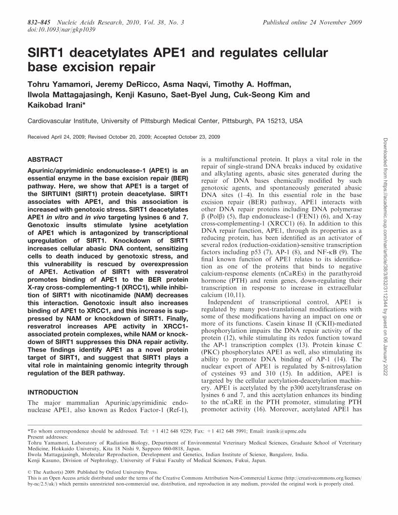
with SIRT1 in HEK293 but not with a catalyticallyinactive, dominant negative, SIRT1(H363Y) mutant(Figure 1A and B). Endogenous SIRT1 alsoco-precipitated with endogenous APE1 (Figure 1C).Moreover, APE1 and SIRT1 co-localized to the nucleiof HEK 293 cells (Figure 1D). Postulating that this asso-ciation may have functional relevance, we examined theeffect of genotoxic stress on APE1-SIRT1 binding.Because APE1 and SIRT1 protect cells against hydrogenperoxide (H2O2) (30–35), an oxidative stress that leads toDNA damage resulting in abasic DNA sites (36), we askedwhether H2O2 affects the association between the twoproteins. In HEK 293 cells H2O2 promoted the bindingof APE1 to SIRT1 (Figure 1D), suggesting that this asso-ciation may have importance in the context of genotoxicstress. Thus, APE1 and SIRT1 bind to and localize witheach other and genotoxic stress, against which APE1 andSIRT1 offer protection, increases this association.
SIRT1 deacetylates APE1 in vitro and in vivo
We then asked if SIRT1 targets APE1 for deacetylation.First, we examined if APE1 is a target of SIRT1
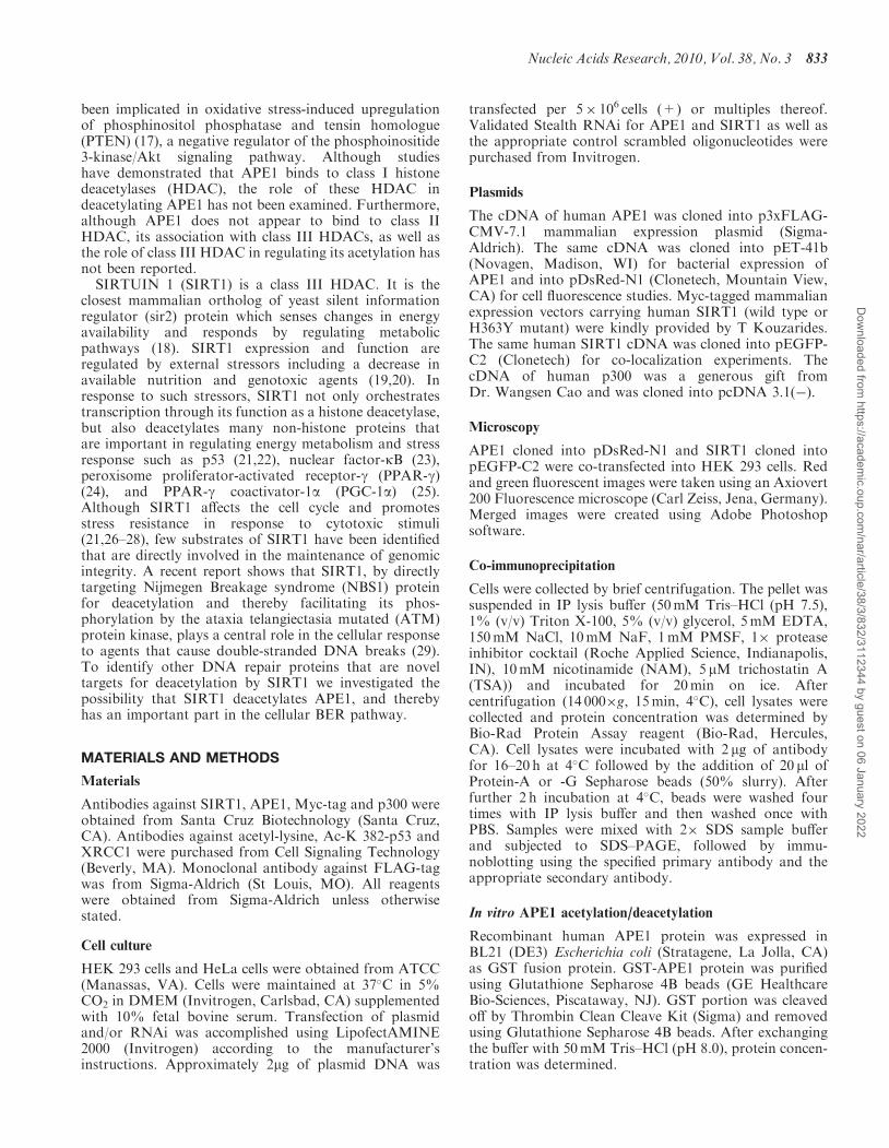
deacetylase activity in vitro. Recombinant APE1 wasenzymatically acetylated with the p300 acetyltransferasein vitro, followed by incubation with active SIRT1.SIRT1 in the presence, but not absence of its co-factornicotinamide adenine dinucleotide (NAD), deacetylatedacetylated APE1 (Figure 2A). Addition of TrichostatinA (TSA), a class I and II HDAC inhibitor, to thereaction mixture did not affect SIRT1-induceddeacetylation of APE1, whereas the SIRT1 inhibitornicotinamide (NAM) negated deacetylation of APE1,indicating that acetylated APE1 is a direct target ofSIRT1. In HEK 293 cells as well, APE1 was acetylatedby overexpression of the p300 acetyltransferase, and thisincrease in lysine acetylation was abrogated byoverexpression of SIRT1 (Figure 2B). Similarly, stimula-tion of endogenous SIRT1 with resveratrol decreasedp300-induced lysine acetylation of APE1 (Figure 2C).Notwithstanding the limitation that our data donot allow us to draw conclusions about site-specificacetylation and deacetylation, they show that APE1 isacetylated by the p300 acetyltransferase, and deacetylatedby SIRT1, in vitro and in vivo.
IP: Myc
WCLIB: FLAG
IB: Myc
IP: Myc
WCL
IB: SIRT1
A
IP: FLAGIB: Myc
IB: APE1
WCLIB: Myc
IB: FLAG
FLAG-APE1Myc-SIRT1
B
APE1-DsRed
EGFP-SIRT1
Merged
D
C
IB: SIRT1
IB: APE1
IP: N–IgG
APE1
IP: Myc
WCL
APE1
SIRT1
APE1
IB:
H2O2
FLAG-APE1 – + ++ ++Myc-SIRT1 + + + –
– – + + + + – – + –
Myc-SIRT1(H363Y) – + – – +
Myc-SIRT1 – + + +DsRed-APE1 + – + +
+ + – +
IP: N
–IgG
E
Figure 1. APE1 and SIRT1 associate with each other. (A) APE1 and SIRT1 bind to each other in HEK 293 cells. Immunoprecipitation of epitope-tagged SIRT1 co-precipitates epitope-tagged APE1 expressed in HEK 293 cells. (B) APE1 does not bind to catalytically inactive dominant negativeSIRT1. Immunoprecipitation of epitope-tagged APE1 co-precipitates epitope-tagged wild-type SIRT1 but not dominant negative SIRT1 (H363Y)expressed in HEK 293 cells. (C) Endogenous SIRT1 binds to endogenous APE1. Co-immunoprecipitation of endogenous SIRT1 and APE1 in HEK293 cells. (D) Co-localization of fluorescent epitope-tagged SIRT1 and APE1 expressed in HEK 293 cells. Co-localization of extra-nucleolar (whitearrow) APE1 but not nucleolar (blue arrow) APE1 with SIRT1 is shown. (E) Hydrogen peroxide (H2O2) promotes binding of APE1 to SIRT1. H2O2
(500 mM, 30min) increases co-precipitation of epitope-tagged SIRT1 in immunoprecipitates of epitope-tagged APE1 expressed in HEK 293 cells.WCL: whole cell lysate. N-IgG: non-immune immunoglobulin. FLAG-APE1 and Myc-SIRT1 were expressed in A and B, DsRed-APE1 and EGFP-SIRT1 in C, and DsRed-APE1 and Myc-SIRT1 in D.
Nucleic Acids Research, 2010, Vol. 38, No. 3 835
Dow
nloaded from https://academ
ic.oup.com/nar/article/38/3/832/3112344 by guest on 06 January 2022
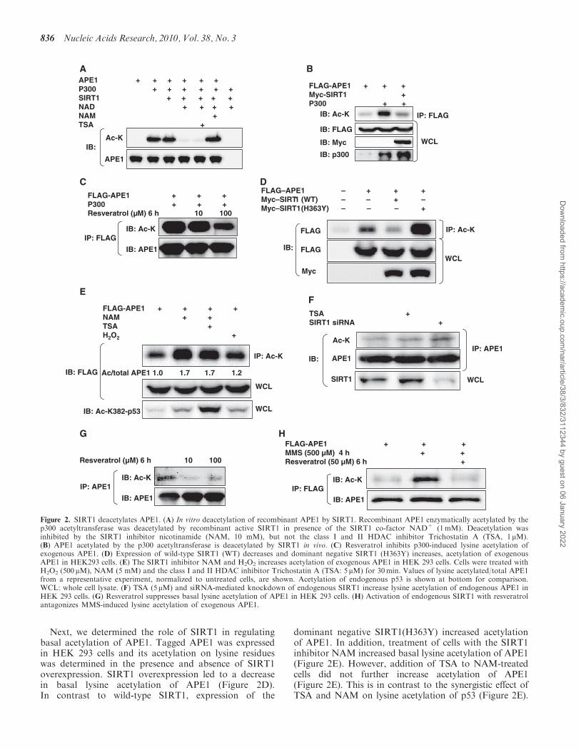
Next, we determined the role of SIRT1 in regulatingbasal acetylation of APE1. Tagged APE1 was expressedin HEK 293 cells and its acetylation on lysine residueswas determined in the presence and absence of SIRT1overexpression. SIRT1 overexpression led to a decreasein basal lysine acetylation of APE1 (Figure 2D).In contrast to wild-type SIRT1, expression of the
dominant negative SIRT1(H363Y) increased acetylationof APE1. In addition, treatment of cells with the SIRT1inhibitor NAM increased basal lysine acetylation of APE1(Figure 2E). However, addition of TSA to NAM-treatedcells did not further increase acetylation of APE1(Figure 2E). This is in contrast to the synergistic effect ofTSA and NAM on lysine acetylation of p53 (Figure 2E).
APE1 + + + + + + P300 + + + + + + SIRT1 + + + + +NAD + + + +NAM +TSA +
Ac-K
APE1
IB:
A
FLAG–APE1 – + + +Myc–SIRT1 (WT) – – + –Myc–SIRT1 (H363Y) – – – +
IP: Ac-K
WCL
FLAG
Myc
IB: FLAG
D
WCL
IP: FLAGIB: Ac-K
IB: FLAG
IB: Myc
IB: p300
FLAG-APE1 + + +Myc-SIRT1 +P300 + +
B
IB: Ac-K
IB: APE1
IP: APE1
Resveratrol (µM) 6 h 10 100
G
SIRT1
Ac-K
APE1IB:IP: APE1
WCL
TSA +SIRT1 siRNA +
F
FLAG-APE1 + + + MMS (500 µM) 4 h + +Resveratrol (50 µM) 6 h +
IB: Ac-K
IB: APE1IP: FLAG
H
FLAG-APE1 + + + +NAM + +TSA +H2O2 +
IP: Ac-K
IB: FLAG
E
WCL
WCLIB: Ac-K382-p53
Ac/total APE1 1.0 1.7 1.7 1.2
FLAG-APE1 + + +P300 + + +Resveratrol (µM) 6 h 10 100
IB: Ac-K
IB: APE1
IP: FLAG
C
Figure 2. SIRT1 deacetylates APE1. (A) In vitro deacetylation of recombinant APE1 by SIRT1. Recombinant APE1 enzymatically acetylated by thep300 acetyltransferase was deacetylated by recombinant active SIRT1 in presence of the SIRT1 co-factor NAD+ (1mM). Deacetylation wasinhibited by the SIRT1 inhibitor nicotinamide (NAM, 10 mM), but not the class I and II HDAC inhibitor Trichostatin A (TSA, 1mM).(B) APE1 acetylated by the p300 acetyltransferase is deacetylated by SIRT1 in vivo. (C) Resveratrol inhibits p300-induced lysine acetylation ofexogenous APE1. (D) Expression of wild-type SIRT1 (WT) decreases and dominant negative SIRT1 (H363Y) increases, acetylation of exogenousAPE1 in HEK293 cells. (E) The SIRT1 inhibitor NAM and H2O2 increases acetylation of exogenous APE1 in HEK 293 cells. Cells were treated withH2O2 (500 mM), NAM (5 mM) and the class I and II HDAC inhibitor Trichostatin A (TSA: 5mM) for 30min. Values of lysine acetylated/total APE1from a representative experiment, normalized to untreated cells, are shown. Acetylation of endogenous p53 is shown at bottom for comparison.WCL: whole cell lysate. (F) TSA (5mM) and siRNA-mediated knockdown of endogenous SIRT1 increase lysine acetylation of endogenous APE1 inHEK 293 cells. (G) Resveratrol suppresses basal lysine acetylation of APE1 in HEK 293 cells. (H) Activation of endogenous SIRT1 with resveratrolantagonizes MMS-induced lysine acetylation of exogenous APE1.
836 Nucleic Acids Research, 2010, Vol. 38, No. 3
Dow
nloaded from https://academ
ic.oup.com/nar/article/38/3/832/3112344 by guest on 06 January 2022
In line with the effect of SIRT1 manipulation on lysineacetylation of exogenous APE1, knockdown of endog-enous SIRT1 expression with siRNA promoted acet-ylation of endogenous APE1 (Figure 2F). In comparisonwith SIRT1 knockdown, addition of the class I and IIHDAC inhibitor TSA resulted in a smaller increase inlysine acetylation of APE1 (Figure 2F). In addition, stim-ulation of endogenous SIRT1 with resveratrol decreasedlysine acetylation of endogenous APE1 (Figure 2G). Thesefindings show that in addition to deacetylating APE1 thatis hyperacetylated by the p300 acetyltransferase, SIRT1also regulates basal acetylation of APE1. In addition,they suggest that class I and II HDAC inhibitors alsoplay a role, albeit lesser than SIRT1, in regulating basalacetylation of APE1.
APE1 acetylation by genotoxic stress isantagonized by SIRT1
Because we had noted that H2O2 increases the binding ofAPE1 to SIRT1 (Figure 1E), we also asked whether this isaccompanied by a change in lysine acetylation of APE1.
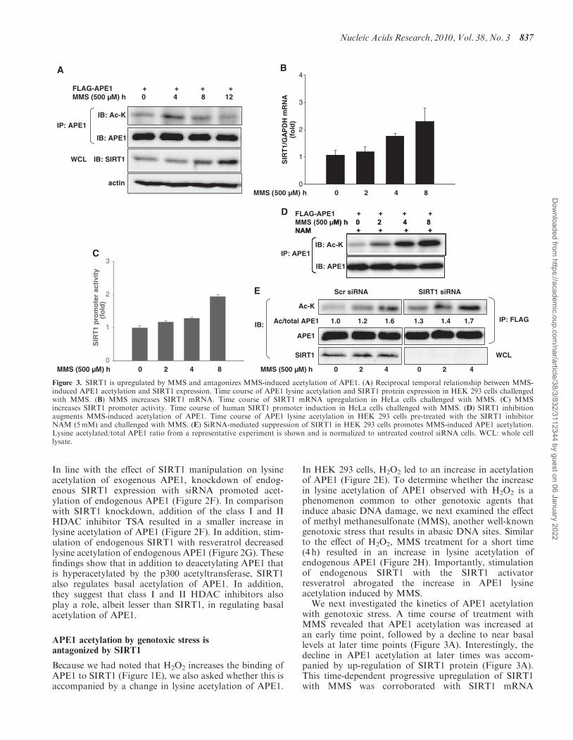
In HEK 293 cells, H2O2 led to an increase in acetylationof APE1 (Figure 2E). To determine whether the increasein lysine acetylation of APE1 observed with H2O2 is aphenomenon common to other genotoxic agents thatinduce abasic DNA damage, we next examined the effectof methyl methanesulfonate (MMS), another well-knowngenotoxic stress that results in abasic DNA sites. Similarto the effect of H2O2, MMS treatment for a short time(4 h) resulted in an increase in lysine acetylation ofendogenous APE1 (Figure 2H). Importantly, stimulationof endogenous SIRT1 with the SIRT1 activatorresveratrol abrogated the increase in APE1 lysineacetylation induced by MMS.We next investigated the kinetics of APE1 acetylation
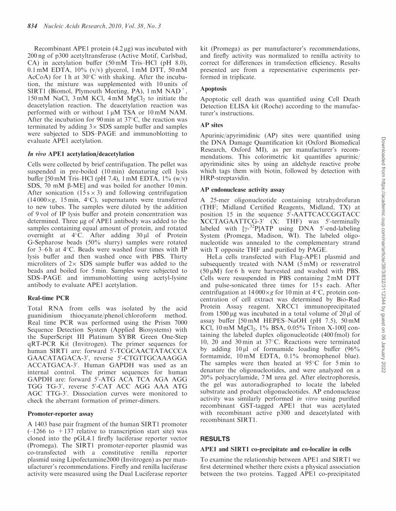
with genotoxic stress. A time course of treatment withMMS revealed that APE1 acetylation was increased atan early time point, followed by a decline to near basallevels at later time points (Figure 3A). Interestingly, thedecline in APE1 acetylation at later times was accom-panied by up-regulation of SIRT1 protein (Figure 3A).This time-dependent progressive upregulation of SIRT1with MMS was corroborated with SIRT1 mRNA
SIR
T1
pro
mo
ter
acti
vity
(fo
ld)
0
1
2
3
MMS (500 µM) h 0 2 4 8
C
MMS (500 µM) h 0 2 4 8
SIR
T1/
GA
PD
H m
RN
A(f
old
)
0
1
2
3
4B
IB: Ac-K
IB: APE1
IP: APE1
IB: SIRT1WCL
FLAG-APE1 + + + +MMS (500 µM) h 0 4 8 12
A
actin
E Scr siRNA SIRT1 siRNA
Ac-K
IB:IP: FLAG
APE1
SIRT1
MMS (500 µM) h 0 2 4 0 2 4
WCL
Ac/total APE1 1.0 1.2 1.6 1.3 1.4 1.7
-M) h 0 2 4 8
NAM + + + + MMS (500 µFLAG APE1 + + + +
M) h 0 2 4 8NAM + + + +
IP: APE1IB: Ac-K
IB: APE1
D
Figure 3. SIRT1 is upregulated by MMS and antagonizes MMS-induced acetylation of APE1. (A) Reciprocal temporal relationship between MMS-induced APE1 acetylation and SIRT1 expression. Time course of APE1 lysine acetylation and SIRT1 protein expression in HEK 293 cells challengedwith MMS. (B) MMS increases SIRT1 mRNA. Time course of SIRT1 mRNA upregulation in HeLa cells challenged with MMS. (C) MMSincreases SIRT1 promoter activity. Time course of human SIRT1 promoter induction in HeLa cells challenged with MMS. (D) SIRT1 inhibitionaugments MMS-induced acetylation of APE1. Time course of APE1 lysine acetylation in HEK 293 cells pre-treated with the SIRT1 inhibitorNAM (5mM) and challenged with MMS. (E) SiRNA-mediated suppression of SIRT1 in HEK 293 cells promotes MMS-induced APE1 acetylation.Lysine acetylated/total APE1 ratio from a representative experiment is shown and is normalized to untreated control siRNA cells. WCL: whole celllysate.
Nucleic Acids Research, 2010, Vol. 38, No. 3 837
Dow
nloaded from https://academ
ic.oup.com/nar/article/38/3/832/3112344 by guest on 06 January 2022

expression and SIRT1 promoter-reporter assays (Figure3B and C). The reciprocal relationship between APE1acetylation and SIRT1 expression suggested to us thatupregulation of endogenous SIRT1 may be a feedbackmechanism for regulation of APE1 acetylation inducedby MMS. Indeed, inhibition of endogenous SIRT1activity resulted in more robust lysine acetylation ofAPE1, even at later time points (Figure 3D). Moreover,knockdown of endogenous SIRT1 also promoted lysineacetylation of APE1 induced by MMS (Figure 3E).These findings demonstrate an important role forendogenous SIRT1 in antagonizing the lysine acetylationof APE1 induced by the genotoxic agent MMS, and impli-cate transcriptional upregulation of SIRT1 by MMS as anendogenous feedback mechanism that modulatesgenotoxic stress-induced APE1 acetylation.
Lysines 6 and 7 in APE1 are deacetylated by SIRT1
We then investigated the specific lysine residues in APE1that SIRT1 targets for deacetylation. APE1 is acetylatedon lysine residues 6 and 7 at its N-terminus in response tochanges in extracellular calcium (16). We therefore lookedat these specific residues as targets of SIRT1. Lysines6 and 7 in APE1 were mutated to non-acetylatable
arginines, individually or in concert, and these non-acetylatable forms of APE1 were expressed in HEK 293cells. Basal acetylation and change in acetylation inducedby NAM-mediated inhibition of endogenous SIRT1activity was then examined and quantified on wild-typeas well as non-acetylatable APE1 lysine mutants.Compared with wild-type protein, mutation of lysine 6did not result in a significant decrease in either basal orNAM-induced acetylation of APE1 (Figure 4A).However, APE1 that was mutated on lysine 7 did showa significant decrease in both basal and NAM-stimulatedacetylation, when compared with wild-type protein.Moreover, APE1 that was non-acetylatable at bothlysines 6 and 7 showed an even greater decrease in basaland NAM-induced acetylation. These findings show thatboth lysines 6 and 7 are targeted for deacetylation bySIRT1, and also suggest synergism between deacetylationby SIRT1 of the two lysine residues. Consistent with thisnotion, p300-stimulated lysine acetylation of APE1 wasessentially unchanged when lysine 6 was non-acetylatable,diminished when lysine 7 was non-acetylatable, andalmost completely abolished when both lysines 6 and 7were non-acetylatable (Figure 4B). However, becausereplacement of both lysines 6 and 7 with non-acetylatable
Ace
tyla
ted/
tota
l AP
E1
(fol
d)
A
0.6
0.8
1
1.2
1.4
1.6
1.8
2
APE1 WT WT K6R K6R K7R K7R K6/7R K6/7RNAM + + + +
###
****
*
IP: FLAG
WCL
Ac-K
APE1
APE1
APE1 WT K6R K7R K6/7R WT K6R K7R K6/7RP300 – – – – + + + +
Ac-K
APE1
IP: FLAG
B
IB:
IB:
#
*###
***
Figure 4. SIRT1 targets lysines 6 and 7 in APE1 for deacetylation. (A) Basal lysine acetylation, and increase in lysine acetylation by treatmentwith the SIRT1 inhibitor nicotinamide (NAM: 5mM, 16 h), of epitope-tagged wild-type and non-acetylatable mutants of APE1 expressed in HEK293 cells. Lysine acetylated/total APE1 was quantified and is expressed relative to wild-type APE1 in untreated cells. *P< 0.05 and #P> 0.05compared with untreated cells transfected with the same construct. ##P> 0.05, and **P< 0.005 compared with untreated cells transfected withWT APE1. ###P> 0.05, ***P< 0.05, and WP< 0.005 compared with NAM-treated cells transfected with WT APE1. N=4 in all conditions.(B) Targeting of the N-terminal lysines in APE1 for acetylation by the p300 acetyltransferase. Epitope-tagged wild-type (WT) APE1, or thenon-acetylatable lysine mutants of APE1, were expressed in HEK 293 cells, with and without overexpression of the p300 acetyltransferase. WCL:whole cell lysate.
838 Nucleic Acids Research, 2010, Vol. 38, No. 3
Dow
nloaded from https://academ
ic.oup.com/nar/article/38/3/832/3112344 by guest on 06 January 2022
residues did not completely abolish the increase inacetylation in response to NAM, deacetylation of lysinesother than 6 and 7 by SIRT1 remains a possibility.
APE1 mediates SIRT1-dependent protection againstgenotoxic stress
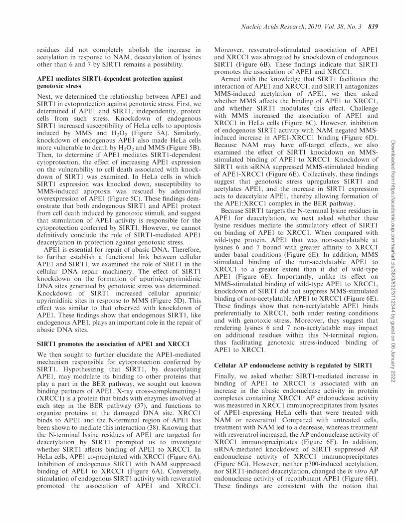
Next, we determined the relationship between APE1 andSIRT1 in cytoprotection against genotoxic stress. First, wedetermined if APE1 and SIRT1, independently, protectcells from such stress. Knockdown of endogenousSIRT1 increased susceptibility of HeLa cells to apoptosisinduced by MMS and H2O2 (Figure 5A). Similarly,knockdown of endogenous APE1 also made HeLa cellsmore vulnerable to death by H2O2 and MMS (Figure 5B).Then, to determine if APE1 mediates SIRT1-dependentcytoprotection, the effect of increasing APE1 expressionon the vulnerability to cell death associated with knock-down of SIRT1 was examined. In HeLa cells in whichSIRT1 expression was knocked down, susceptibility toMMS-induced apoptosis was rescued by adenoviraloverexpression of APE1 (Figure 5C). These findings dem-onstrate that both endogenous SIRT1 and APE1 protectfrom cell death induced by genotoxic stimuli, and suggestthat stimulation of APE1 activity is responsible for thecytoprotection conferred by SIRT1. However, we cannotdefinitively conclude the role of SIRT1-mediated APE1deacetylation in protection against genotoxic stress.
APE1 is essential for repair of abasic DNA. Therefore,to further establish a functional link between cellularAPE1 and SIRT1, we examined the role of SIRT1 in thecellular DNA repair machinery. The effect of SIRT1knockdown on the formation of apurinic/apyrimidinicDNA sites generated by genotoxic stress was determined.Knockdown of SIRT1 increased cellular apurinic/apyrimidinic sites in response to MMS (Figure 5D). Thiseffect was similar to that observed with knockdown ofAPE1. These findings show that endogenous SIRT1, likeendogenous APE1, plays an important role in the repair ofabasic DNA sites.
SIRT1 promotes the association of APE1 and XRCC1
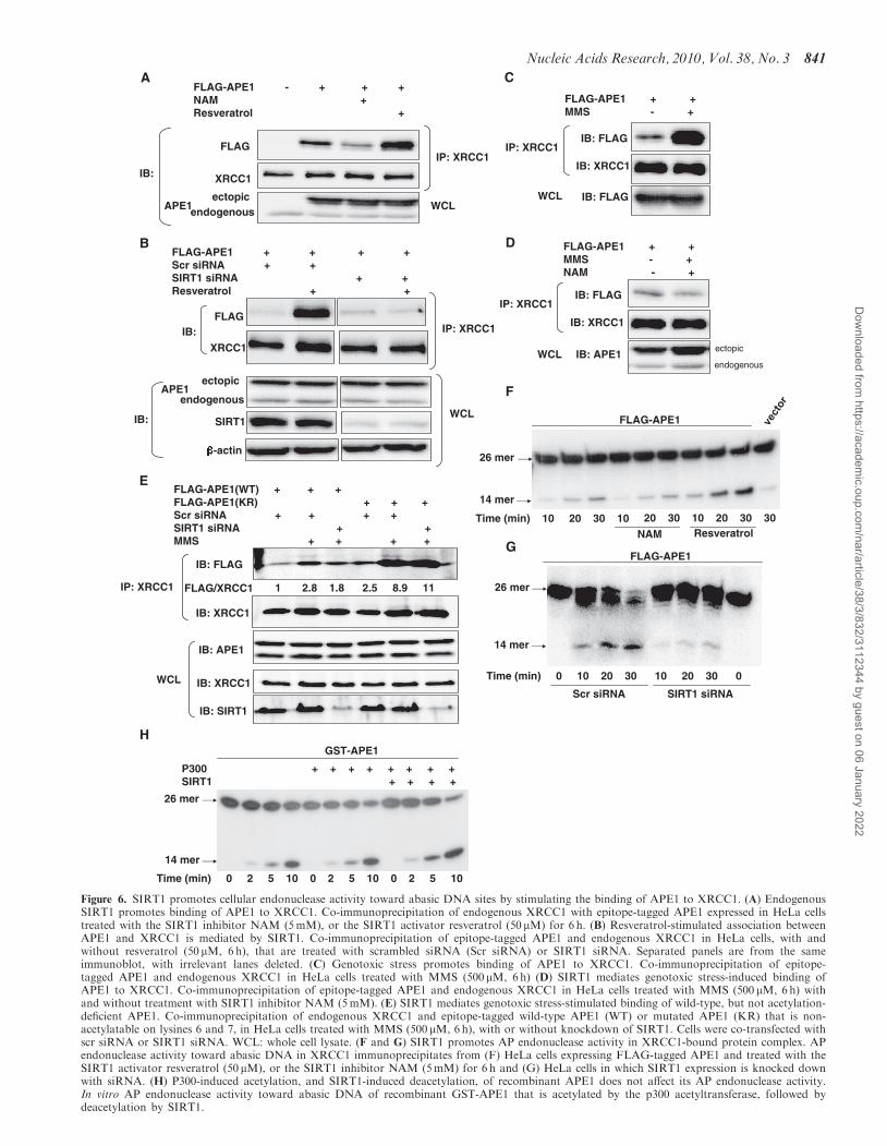
We then sought to further elucidate the APE1-mediatedmechanism responsible for cytoprotection conferred bySIRT1. Hypothesizing that SIRT1, by deacetylatingAPE1, may modulate its binding to other proteins thatplay a part in the BER pathway, we sought out knownbinding partners of APE1. X-ray cross-complementing-1(XRCC1) is a protein that binds with enzymes involved ateach step in the BER pathway (37), and functions toorganize proteins at the damaged DNA site. XRCC1binds to APE1 and the N-terminal region of APE1 hasbeen shown to mediate this interaction (38). Knowing thatthe N-terminal lysine residues of APE1 are targeted fordeacetylation by SIRT1 prompted us to investigatewhether SIRT1 affects binding of APE1 to XRCC1. InHeLa cells, APE1 co-precipitated with XRCC1 (Figure 6A).Inhibition of endogenous SIRT1 with NAM suppressedbinding of APE1 to XRCC1 (Figure 6A). Conversely,stimulation of endogenous SIRT1 activity with resveratrolpromoted the association of APE1 and XRCC1.
Moreover, resveratrol-stimulated association of APE1and XRCC1 was abrogated by knockdown of endogenousSIRT1 (Figure 6B). These findings indicate that SIRT1promotes the association of APE1 and XRCC1.Armed with the knowledge that SIRT1 facilitates the
interaction of APE1 and XRCC1, and SIRT1 antagonizesMMS-induced acetylation of APE1, we then askedwhether MMS affects the binding of APE1 to XRCC1,and whether SIRT1 modulates this effect. Challengewith MMS increased the association of APE1 andXRCC1 in HeLa cells (Figure 6C). However, inhibitionof endogenous SIRT1 activity with NAM negated MMS-induced increase in APE1-XRCC1 binding (Figure 6D).Because NAM may have off-target effects, we alsoexamined the effect of SIRT1 knockdown on MMS-stimulated binding of APE1 to XRCC1. Knockdown ofSIRT1 with siRNA suppressed MMS-stimulated bindingof APE1-XRCC1 (Figure 6E). Collectively, these findingssuggest that genotoxic stress upregulates SIRT1 andacetylates APE1, and the increase in SIRT1 expressionacts to deacetylate APE1, thereby allowing formation ofthe APE1:XRCC1 complex in the BER pathway.Because SIRT1 targets the N-terminal lysine residues in
APE1 for deacetylation, we next asked whether theselysine residues mediate the stimulatory effect of SIRT1on binding of APE1 to XRCC1. When compared withwild-type protein, APE1 that was non-acetylatable atlysines 6 and 7 bound with greater affinity to XRCC1under basal conditions (Figure 6E). In addition, MMSstimulated binding of the non-acetylatable APE1 toXRCC1 to a greater extent than it did of wild-typeAPE1 (Figure 6E). Importantly, unlike its effect onMMS-stimulated binding of wild-type APE1 to XRCC1,knockdown of SIRT1 did not suppress MMS-stimulatedbinding of non-acetylatable APE1 to XRCC1 (Figure 6E).These findings show that non-acetylatable APE1 bindspreferentially to XRCC1, both under resting conditionsand with genotoxic stress. Moreover, they suggest thatrendering lysines 6 and 7 non-acetylatable may impacton additional residues within this N-terminal region,thus facilitating genotoxic stress-induced binding ofAPE1 to XRCC1.
Cellular AP endonuclease activity is regulated by SIRT1
Finally, we asked whether SIRT1-mediated increase inbinding of APE1 to XRCC1 is associated with anincrease in the abasic endonuclease activity in proteincomplexes containing XRCC1. AP endonuclease activitywas measured in XRCC1 immunoprecipitates from lysatesof APE1-expressing HeLa cells that were treated withNAM or resveratrol. Compared with untreated cells,treatment with NAM led to a decrease, whereas treatmentwith resveratrol increased, the AP endonuclease activity ofXRCC1 immunoprecipitates (Figure 6F). In addition,siRNA-mediated knockdown of SIRT1 suppressed APendonuclease activity of XRCC1 immunoprecipitates(Figure 6G). However, neither p300-induced acetylation,nor SIRT1-induced deacetylation, changed the in vitro APendonuclease activity of recombinant APE1 (Figure 6H).These findings are consistent with the notion that
Nucleic Acids Research, 2010, Vol. 38, No. 3 839
Dow
nloaded from https://academ
ic.oup.com/nar/article/38/3/832/3112344 by guest on 06 January 2022
0
1
2
3
4
5
Ap
op
tosi
s (f
old
)
H2O2
*
*
*
*A
po
pto
sis
(fo
ld)
0
1
2
3
4
5
AdlacZ (MOI) 10AdAPE1 (MOI) 10 50
*
*APE1
SIRT1
-actin
012345678
Apo
ptos
is (f
old)
100 200H2O2 002 002001 )Mµ(
**
*
APE1
SIRT1
APE1
MMS (µM)(µM)
– 200 300 400–
AdlacZ – 10 50AdAPE1 – 10 50Scr siRNA + – – – –SIRt1 siRNA – + + + +
MMS (µM) ––
Scr siRNA + –SIRT1 siRNA – +
Scr siRNA + –APE1 siRNA – +
AB
C
Scr siRNA + +SIRT1 siRNA + +APE1 siRNA + +MMS 1 mM ( 3 h) + + +
D
AP
sit
es/1
05 b
p
0
5
10
15
20
25
**
Figure 5. SIRT1 protects from genotoxic stress-induced cell death through APE1. Apoptotic death with oxidative stress (H2O2 for 24 h) and abasicDNA damage (MMS for 24 h) in HeLa cells in which SIRT1 expression (A) or APE1 expression (B) is knocked down with SIRT1 or APE1 siRNA(black bars). Scrambled (scr) siRNA (white bars) was used as control. Apoptosis is expressed as fold change compared with untreated cells. *P< 0.05and **P< 0.01 compared with control siRNA. Knockdown of SIRT1 and APE1 is shown at bottom. (C) APE1 overexpression rescues cells withSIRT1 down-regulation from MMS-induced apoptosis. Apoptotic cell death induced by MMS (200 mM, 24 h) in HeLa cells treated with SIRT1siRNA (white bars) or scrambled (scr) siRNA (black bars) were infected with a control virus (AdLacZ) that expresses the inert E. coli LacZ gene, orand adenovirus that expresses APE1 (AdAPE1). Apoptosis is expressed as fold change compared with control siRNA. *P< 0.05 compared with cellsinfected with AdLacZ. Knockdown of SIRT1 and adenoviral overexpression of APE1 is shown at right. (D) SIRT1 plays a role in abasic DNArepair. SIRT1 or APE1 was knocked down in HeLa cells with siRNA. Apurinic/apyrimidinic DNA sites were quantified in untreated cells and cellstreated with MMS for 3 h. *P< 0.05 compared with control siRNA.
840 Nucleic Acids Research, 2010, Vol. 38, No. 3
Dow
nloaded from https://academ
ic.oup.com/nar/article/38/3/832/3112344 by guest on 06 January 2022
FLAG-APE1 - + + +NAM +Resveratrol +
IP: XRCC1
WCL
FLAG
XRCC1
APE1
IB:
A
ectopicendogenous
WCL
APE1
SIRT1
-actin
IB:
IP: XRCC1FLAG
XRCC1
IB:
FLAG-APE1 + + + +Scr siRNA + +SIRT1 siRNA + +Resveratrol + +
B
ectopic
endogenous
IB: FLAG
IB: FLAG
IB: XRCC1
IP: XRCC1
WCL
FLAG-APE1 + +MMS - +
C
IB: APE1ectopic
endogenous
FLAG-APE1 + +MMS - + NAM - +
IB: FLAG
IB: XRCC1
IP: XRCC1
WCL
D
FLAG-APE1(WT) + + +FLAG-APE1(KR) + + +Scr siRNA + + + +SIRT1 siRNA + +MMS + + + +
1 2.8 1.8 2.5 8.9 11
IB: FLAG
IB: XRCC1
IP: XRCC1
IB: APE1
IB: XRCC1
IB: SIRT1
WCL
E
FLAG/XRCC1
FLAG-APE1
Time (min) 10 20 30 10 20 30 10 20 30 30
NAM Resveratrol
F
26 mer
14 mer
G
14 mer
26 mer
Time (min) 0 10 20 30 10 20 30 0
FLAG-APE1
Scr siRNA SIRT1 siRNA
GST-APE1
P300 + + + + + + + +SIRT1 + + + +
Time (min) 0 2 5 10 0 2 5 10 0 2 5 10
H
26 mer
14 mer
Figure 6. SIRT1 promotes cellular endonuclease activity toward abasic DNA sites by stimulating the binding of APE1 to XRCC1. (A) EndogenousSIRT1 promotes binding of APE1 to XRCC1. Co-immunoprecipitation of endogenous XRCC1 with epitope-tagged APE1 expressed in HeLa cellstreated with the SIRT1 inhibitor NAM (5mM), or the SIRT1 activator resveratrol (50 mM) for 6 h. (B) Resveratrol-stimulated association betweenAPE1 and XRCC1 is mediated by SIRT1. Co-immunoprecipitation of epitope-tagged APE1 and endogenous XRCC1 in HeLa cells, with andwithout resveratrol (50 mM, 6 h), that are treated with scrambled siRNA (Scr siRNA) or SIRT1 siRNA. Separated panels are from the sameimmunoblot, with irrelevant lanes deleted. (C) Genotoxic stress promotes binding of APE1 to XRCC1. Co-immunoprecipitation of epitope-tagged APE1 and endogenous XRCC1 in HeLa cells treated with MMS (500 mM, 6h) (D) SIRT1 mediates genotoxic stress-induced binding ofAPE1 to XRCC1. Co-immunoprecipitation of epitope-tagged APE1 and endogenous XRCC1 in HeLa cells treated with MMS (500 mM, 6 h) withand without treatment with SIRT1 inhibitor NAM (5mM). (E) SIRT1 mediates genotoxic stress-stimulated binding of wild-type, but not acetylation-deficient APE1. Co-immunoprecipitation of endogenous XRCC1 and epitope-tagged wild-type APE1 (WT) or mutated APE1 (KR) that is non-acetylatable on lysines 6 and 7, in HeLa cells treated with MMS (500 mM, 6 h), with or without knockdown of SIRT1. Cells were co-transfected withscr siRNA or SIRT1 siRNA. WCL: whole cell lysate. (F and G) SIRT1 promotes AP endonuclease activity in XRCC1-bound protein complex. APendonuclease activity toward abasic DNA in XRCC1 immunoprecipitates from (F) HeLa cells expressing FLAG-tagged APE1 and treated with theSIRT1 activator resveratrol (50 mM), or the SIRT1 inhibitor NAM (5mM) for 6 h and (G) HeLa cells in which SIRT1 expression is knocked downwith siRNA. (H) P300-induced acetylation, and SIRT1-induced deacetylation, of recombinant APE1 does not affect its AP endonuclease activity.In vitro AP endonuclease activity toward abasic DNA of recombinant GST-APE1 that is acetylated by the p300 acetyltransferase, followed bydeacetylation by SIRT1.
Nucleic Acids Research, 2010, Vol. 38, No. 3 841
Dow
nloaded from https://academ
ic.oup.com/nar/article/38/3/832/3112344 by guest on 06 January 2022
SIRT1-mediated deacetylation of APE1 does not directlyaffect the AP endonuclease activity of APE1, but rather,SIRT1 stimulates cellular AP endonuclease activity bypromoting the association of APE1 to XRCC1.
DISCUSSION
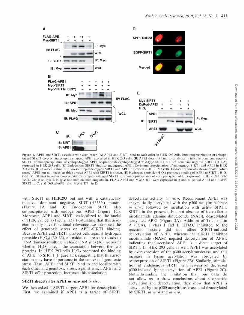
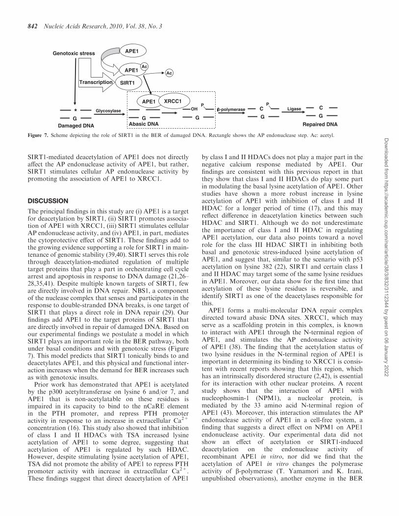
The principal findings in this study are (i) APE1 is a targetfor deacetylation by SIRT1, (ii) SIRT1 promotes associa-tion of APE1 with XRCC1, (iii) SIRT1 stimulates cellularAP endonuclease activity, and (iv) APE1, in part, mediatesthe cytoprotective effect of SIRT1. These findings add tothe growing evidence supporting a role for SIRT1 in main-tenance of genomic stability (39,40). SIRT1 serves this rolethrough deacetylation-mediated regulation of multipletarget proteins that play a part in orchestrating cell cyclearrest and apoptosis in response to DNA damage (21,26–28,35,41). Despite multiple known targets of SIRT1, feware directly involved in DNA repair. NBS1, a componentof the nuclease complex that senses and participates in theresponse to double-stranded DNA breaks, is one target ofSIRT1 that plays a direct role in DNA repair (29). Ourfindings add APE1 to the target proteins of SIRT1 thatare directly involved in repair of damaged DNA. Based onour experimental findings we postulate a model in whichSIRT1 plays an important role in the BER pathway, bothunder basal conditions and with genotoxic stress (Figure7). This model predicts that SIRT1 tonically binds to anddeacetylates APE1, and this physical and functional inter-action increases when the demand for BER increases suchas with genotoxic insults.Prior work has demonstrated that APE1 is acetylated
by the p300 acetyltransferase on lysine 6 and/or 7, andAPE1 that is non-acetylatable on these residues isimpaired in its capacity to bind to the nCaRE elementin the PTH promoter, and repress PTH promoteractivity in response to an increase in extracellular Ca2+
concentration (16). This study also showed that inhibitionof class I and II HDACs with TSA increased lysineacetylation of APE1 to some degree, suggesting thatacetylation of APE1 is regulated by such HDAC.However, despite stimulating lysine acetylation of APE1,TSA did not promote the ability of APE1 to repress PTHpromoter activity with increase in extracellular Ca2+.These findings suggest that direct deacetylation of APE1
by class I and II HDACs does not play a major part in thenegative calcium response mediated by APE1. Ourfindings are consistent with this previous report in thatthey show that class I and II HDACs do play some partin modulating the basal lysine acetylation of APE1. Otherstudies have shown a more robust increase in lysineacetylation of APE1 with inhibition of class I and IIHDAC for a longer period of time (17), and this mayreflect difference in deacetylation kinetics between suchHDAC and SIRT1. Although we do not underestimatethe importance of class I and II HDAC in regulatingAPE1 acetylation, our data also points toward a novelrole for the class III HDAC SIRT1 in inhibiting bothbasal and genotoxic stress-induced lysine acetylation ofAPE1, and suggest that, similar to the scenario with p53acetylation on lysine 382 (22), SIRT1 and certain class Iand II HDAC may target some of the same lysine residuesin APE1. Moreover, our data show for the first time thatacetylation of these lysine residues is reversible, andidentify SIRT1 as one of the deacetylases responsible forthis.
APE1 forms a multi-molecular DNA repair complexdirected toward abasic DNA sites. XRCC1, which mayserve as a scaffolding protein in this complex, is knownto interact with APE1 through the N-terminal region ofAPE1, and stimulates the AP endonuclease activityof APE1 (38). The finding that the acetylation status oftwo lysine residues in the N-terminal region of APE1 isimportant in determining its binding to XRCC1 is consis-tent with recent reports showing that this region, whichhas an intrinsically disordered structure (2,42), is essentialfor its interaction with other nuclear proteins. A recentstudy shows that the interaction of APE1 withnucleophosmin-1 (NPM1), a nucleolar protein, ismediated by the 33 amino acid N-terminal region ofAPE1 (43). Moreover, this interaction stimulates the APendonuclease activity of APE1 in a cell-free system, afinding that suggests a direct effect on NPM1 on APE1endonuclease activity. Our experimental data did notshow an effect of acetylation or SIRT1-induceddeacetylation on the endonuclease activity ofrecombinant APE1 in vitro, nor did we find that theacetylation of APE1 in vitro changes the polymeraseactivity of b-polymerase (T. Yamamori and K. Irani,unpublished observations), another enzyme in the BER
*G
Genotoxic stress
Damaged DNA
Glycosylase
GAbasic DNA
APE1
APE1Ac
SIRT1
Ac
APE1 XRCC1
G
OHP
Transcription
G
CP
-polymerase Ligase
G
C
Repaired DNA
Figure 7. Scheme depicting the role of SIRT1 in the BER of damaged DNA. Rectangle shows the AP endonuclease step. Ac: acetyl.
842 Nucleic Acids Research, 2010, Vol. 38, No. 3
Dow
nloaded from https://academ
ic.oup.com/nar/article/38/3/832/3112344 by guest on 06 January 2022
pathway that physically associated with APE1 (5). Thesefindings support our hypothesis that SIRT1 impacts oncellular AP endonuclease activity indirectly by modulatingthe interaction of APE1 with other proteins of the BERmachinery, but does not directly change the activityof APE1 or APE1-associated DNA repair enzymes.Nevertheless, based on the interesting finding that theinteraction of APE1 with NPM1 directly stimulatesits endonuclease activity, it would be worthwhile to seeif acetylation and SIRT1-induced deacetylation affectsAPE1 endonuclease activity stimulated by NPM1through modulation of APE1-NPM1 binding. Suchstudies may be especially revealing in light of the rolesof NPM1 in ribosomal biogenesis (43), and SIRT1 inribosomal RNA synthesis (44).
APE1 also interacts with the Y-box-binding protein 1(YB-1) enhancing its binding to the Y-box element, andstimulating multi-drug resistance (MDR1) gene expression(45). This interaction also occurs though the N-terminalregion of APE1, reinforcing the importance of this regionin binding of APE1 to other proteins. Notably, APE1 thatis non-acetylatable on lysines 6 and 7 displays lesserbinding to YB-1 than wild-type protein. Our findingssuggest that APE1 that is deacetylated by SIRT1preferentially binds to XRCC1. We did not examine therole of acetylation in mediating the interaction of APE1with SIRT1. However, we did observe that H2O2
increased acetylation of APE1 and its association withSIRT1, hinting at the possibility that as far as theAPE1:SIRT1 interaction is concerned, acetylation ofAPE1 may facilitate its binding to SIRT1. A similarincrease in binding of SIRT1 to FOXO transcriptionfactors, a target of SIRT1, with H2O2 has been reported(46). Increase in this APE1:SIRT1 interaction bygenotoxic stress would allow the deacetylation of APE1thus permitting APE1:XRCC1 binding. It is not clearwhat purpose acetylation of APE1 may serve in thecontext of genotoxic insults. However, one can hypothe-size that the degree of acetylation of APE1 may serve as aswitch that guides the cell down one of two paths:hyperacetylation of APE1 may induce a transcriptionalprogram of apoptosis, perhaps by activating transcriptionfactors as it does YB-1, whereas SIRT1-stimulatedhypocetylation may promote DNA repair and cellsurvival. This scenario, if true, would be reminiscent ofhow acetylation and SIRT1-mediated deacetylation ofFOXO transcription factors guides the cell toward a fateof survival or death (26).
The effect of genotoxic agents on the acetylation ofAPE1 deserves attention. Although both H2O2 andMMS increased acetylation of APE1, this effect wasmodest and became much more apparent when endo-genous SIRT1 activity was inhibited. Importantly, MMSincreased SIRT1 expression and this increase followed atime course that was reciprocal to MMS-stimulated APE1acetylation. These findings suggest that endogenousSIRT1 functions to negate the acetylation of APE1 bythese genotoxic stimuli. Although these studies establishthe role of SIRT1 in negatively regulating acetylation ofAPE1 in response to these genotoxic stimuli, we didnot explore the mechanism for acetylation of APE1 by
these stimuli. It is noteworthy however, that MMSincreases the acetylation of Werner (WRN), anotherimportant protein in base excision repair pathways, in ap300-dependent fashion (47), suggesting that H2O2 andMMS-induced acetylation of APE1 may also bemediated by p300. However, we cannot exclude the pos-sibility that other acetyltransferases may also target APE1for lysine acetylation in response to DNA damagingagents, as has been observed for p53 acetylation (48).We observed that MMS leads to transcriptional
upregulation of SIRT1 expression. SIRT1 is known tobe transcriptionally induced by the DNA damagingagent etopside (41). This transcriptional induction ismediated by the transcription factor E2F1, with aconserved E2F1-binding element present in the humanand mouse SIRT1 promoters. Therefore, it is likely thatinduction of SIRT1 by MMS is also dependent on E2F1.In support of this, reporter analysis of truncated humanSIRT1 promoters have shown that the sequence 115 bpupstream of the transcription start site, which includes theE2F1 binding site, is sufficient for induction of promoteractivity by MMS (T. Yamamori and K. Irani, unpublishedobservations).Finally, although our study focused on exploring the
consequence of the APE1:SIRT1 interaction on lysineacetylation of APE1, it is tempting to speculate that thisassociation may also facilitate a yet unknown effect ofAPE1 on SIRT1. One attractive possibility is that thisinteraction may allow the regulation of SIRT1 by APE1,similar to the role that APE1 plays in the regulation ofAP-1 transcriptional activity through its physical associa-tion with thioredoxin (49).
FUNDING
Funding for open access charge: National Institutes ofHealth (R01 HL070929, R01 HL094959, and P01HL065608) and the Japan Society for the Promotion ofScience.
Conflict of interest statement. None declared.
REFERENCES
1. Demple,B. and Harrison,L. (1994) Repair of oxidative damage toDNA: enzymology and biology. Annu. Rev. Biochem., 63, 915–948.
2. Izumi,T., Wiederhold,L.R., Roy,G., Roy,R., Jaiswal,A.,Bhakat,K.K., Mitra,S. and Hazra,T.K. (2003) Mammalian DNAbase excision repair proteins: their interactions and role in repairof oxidative DNA damage. Toxicology, 193, 43–65.
3. Mitra,S., Izumi,T., Boldogh,I., Bhakat,K.K., Hill,J.W. andHazra,T.K. (2002) Choreography of oxidative damage repair inmammalian genomes. Free Radic. Biol. Med., 33, 15–28.
4. Tell,G., Damante,G., Caldwell,D. and Kelley,M.R. (2005) Theintracellular localization of APE1/Ref-1: more than a passivephenomenon? Antioxid. Redox Signal., 7, 367–384.
5. Bennett,R.A., Wilson,D.M. 3rd, Wong,D. and Demple,B. (1997)Interaction of human apurinic endonuclease and DNA polymerasebeta in the base excision repair pathway. Proc. Natl Acad. Sci.USA, 94, 7166–7169.
6. Dianova,II, Bohr,V.A. and Dianov,G.L. (2001) Interaction ofhuman AP endonuclease 1 with flap endonuclease 1 andproliferating cell nuclear antigen involved in long-patch baseexcision repair. Biochemistry, 40, 12639–12644.
Nucleic Acids Research, 2010, Vol. 38, No. 3 843
Dow
nloaded from https://academ
ic.oup.com/nar/article/38/3/832/3112344 by guest on 06 January 2022
7. Gaiddon,C., Moorthy,N.C. and Prives,C. (1999) Ref-1 regulatesthe transactivation and pro-apoptotic functions of p53 in vivo.EMBO J., 18, 5609–5621.
8. Xanthoudakis,S. and Curran,T. (1992) Identification andcharacterization of Ref-1, a nuclear protein that facilitates AP-1DNA-binding activity. EMBO J., 11, 653–665.
9. Flaherty,D.M., Monick,M.M. and Hunninghake,G.W. (2001) APendonucleases and the many functions of Ref-1. Am. J. Respir. CellMol. Biol., 25, 664–667.
10. Fuchs,S., Philippe,J., Corvol,P. and Pinet,F. (2003) Implication ofRef-1 in the repression of renin gene transcription by intracellularcalcium. J. Hypertens., 21, 327–335.
11. Okazaki,T., Chung,U., Nishishita,T., Ebisu,S., Usuda,S.,Mishiro,S., Xanthoudakis,S., Igarashi,T. and Ogata,E. (1994)A redox factor protein, ref1, is involved in negative generegulation by extracellular calcium. J. Biol. Chem., 269,27855–27862.
12. Yacoub,A., Kelley,M.R. and Deutsch,W.A. (1997) The DNA repairactivity of human redox/repair protein APE/Ref-1 is inactivated byphosphorylation. Cancer Res., 57, 5457–5459.
13. Fritz,G. and Kaina,B. (1999) Phosphorylation of the DNA repairprotein APE/REF-1 by CKII affects redox regulation of AP-1.Oncogene, 18, 1033–1040.
14. Hsieh,M.M., Hegde,V., Kelley,M.R. and Deutsch,W.A. (2001)Activation of APE/Ref-1 redox activity is mediated by reactiveoxygen species and PKC phosphorylation. Nucleic Acids Res., 29,3116–3122.
15. Qu,J., Liu,G.H., Huang,B. and Chen,C. (2007) Nitric oxidecontrols nuclear export of APE1/Ref-1 through S-nitrosation ofcysteines 93 and 310. Nucleic Acids Res., 35, 2522–2532.
16. Bhakat,K.K., Izumi,T., Yang,S.H., Hazra,T.K. and Mitra,S. (2003)Role of acetylated human AP-endonuclease (APE1/Ref-1) inregulation of the parathyroid hormone gene. EMBO J., 22,6299–6309.
17. Fantini,D., Vascotto,C., Deganuto,M., Bivi,N., Gustincich,S.,Marcon,G., Quadrifoglio,F., Damante,G., Bhakat,K.K., Mitra,S.et al. (2008) APE1/Ref-1 regulates PTEN expression mediated byEgr-1. Free Radic. Res., 42, 20–29.
18. Michan,S. and Sinclair,D. (2007) Sirtuins in mammals: insightsinto their biological function. Biochem. J., 404, 1–13.
19. Cohen,H.Y., Miller,C., Bitterman,K.J., Wall,N.R., Hekking,B.,Kessler,B., Howitz,K.T., Gorospe,M., de Cabo,R. andSinclair,D.A. (2004) Calorie restriction promotes mammaliancell survival by inducing the SIRT1 deacetylase. Science, 305,390–392.
20. Yang,T., Fu,M., Pestell,R. and Sauve,A.A. (2006) SIRT1and endocrine signaling. Trends Endocrinol. Metab., 17,186–191.
21. Luo,J., Nikolaev,A.Y., Imai,S., Chen,D., Su,F., Shiloh,A.,Guarente,L. and Gu,W. (2001) Negative control of p53 bySir2alpha promotes cell survival under stress. Cell, 107, 137–148.
22. Vaziri,H., Dessain,S.K., Ng Eaton,E., Imai,S.I., Frye,R.A.,Pandita,T.K., Guarente,L. and Weinberg,R.A. (2001)hSIR2(SIRT1) functions as an NAD-dependent p53 deacetylase.Cell, 107, 149–159.
23. Gao,F., Cheng,J., Shi,T. and Yeh,E.T. (2006) Neddylation of abreast cancer-associated protein recruits a class III histonedeacetylase that represses NFkappaB-dependent transcription.Nat. Cell Biol., 8, 1171–1177.
24. Picard,F., Kurtev,M., Chung,N., Topark-Ngarm,A., Senawong,T.,Machado De Oliveira,R., Leid,M., McBurney,M.W. andGuarente,L. (2004) Sirt1 promotes fat mobilization in whiteadipocytes by repressing PPAR-gamma. Nature, 429, 771–776.
25. Rodgers,J.T., Lerin,C., Haas,W., Gygi,S.P., Spiegelman,B.M. andPuigserver,P. (2005) Nutrient control of glucose homeostasisthrough a complex of PGC-1alpha and SIRT1. Nature, 434,113–118.
26. Brunet,A., Sweeney,L.B., Sturgill,J.F., Chua,K.F., Greer,P.L.,Lin,Y., Tran,H., Ross,S.E., Mostoslavsky,R., Cohen,H.Y. et al.(2004) Stress-dependent regulation of FOXO transcription factorsby the SIRT1 deacetylase. Science, 303, 2011–2015.
27. Cohen,H.Y., Lavu,S., Bitterman,K.J., Hekking,B.,Imahiyerobo,T.A., Miller,C., Frye,R., Ploegh,H., Kessler,B.M. andSinclair,D.A. (2004) Acetylation of the C terminus of Ku70 by CBP
and PCAF controls Bax-mediated apoptosis. Mol. Cell, 13,627–638.
28. Motta,M.C., Divecha,N., Lemieux,M., Kamel,C., Chen,D., Gu,W.,Bultsma,Y., McBurney,M. and Guarente,L. (2004) MammalianSIRT1 represses forkhead transcription factors. Cell, 116, 551–563.
29. Yuan,Z., Zhang,X., Sengupta,N., Lane,W.S. and Seto,E. (2007)SIRT1 regulates the function of the Nijmegen breakage syndromeprotein. Mol. Cell, 27, 149–162.
30. Grosch,S., Fritz,G. and Kaina,B. (1998) Apurinic endonuclease(Ref-1) is induced in mammalian cells by oxidative stress andinvolved in clastogenic adaptation. Cancer Res., 58, 4410–4416.
31. Kume,S., Haneda,M., Kanasaki,K., Sugimoto,T., Araki,S.,Isono,M., Isshiki,K., Uzu,T., Kashiwagi,A. and Koya,D. (2006)Silent information regulator 2 (SIRT1) attenuates oxidative stress-induced mesangial cell apoptosis via p53 deacetylation. Free Radic.Biol. Med., 40, 2175–2182.
32. Pillai,J.B., Isbatan,A., Imai,S. and Gupta,M.P. (2005) Poly(ADP-ribose) polymerase-1-dependent cardiac myocyte cell death duringheart failure is mediated by NAD+ depletion and reducedSir2alpha deacetylase activity. J. Biol. Chem., 280, 43121–43130.
33. Tomicic,M., Eschbach,E. and Kaina,B. (1997) Expression ofyeast but not human apurinic/apyrimidinic endonuclease rendersChinese hamster cells more resistant to DNA damaging agents.Mutat. Res., 383, 155–165.
34. Walker,L.J., Craig,R.B., Harris,A.L. and Hickson,I.D. (1994) Arole for the human DNA repair enzyme HAP1 in cellularprotection against DNA damaging agents and hypoxic stress.Nucleic Acids Res., 22, 4884–4889.
35. Yang,Y., Fu,W., Chen,J., Olashaw,N., Zhang,X., Nicosia,S.V.,Bhalla,K. and Bai,W. (2007) SIRT1 sumoylation regulates itsdeacetylase activity and cellular response to genotoxic stress.Nat. Cell Biol., 9, 1253–1262.
36. Sung,J.S. and Demple,B. (2006) Roles of base excision repairsubpathways in correcting oxidized abasic sites in DNA. FEBS J.,273, 1620–1629.
37. Fan,J. and Wilson,D.M. 3rd (2005) Protein-protein interactionsand posttranslational modifications in mammalian base excisionrepair. Free Radic. Biol. Med., 38, 1121–1138.
38. Vidal,A.E., Boiteux,S., Hickson,I.D. and Radicella,J.P. (2001)XRCC1 coordinates the initial and late stages of DNA abasicsite repair through protein-protein interactions. EMBO J., 20,6530–6539.
39. Oberdoerffer,P., Michan,S., McVay,M., Mostoslavsky,R.,Vann,J., Park,S.K., Hartlerode,A., Stegmuller,J., Hafner,A.,Loerch,P. et al. (2008) SIRT1 redistribution on chromatinpromotes genomic stability but alters gene expression duringaging. Cell, 135, 907–918.
40. Wang,R.H., Sengupta,K., Li,C., Kim,H.S., Cao,L., Xiao,C.,Kim,S., Xu,X., Zheng,Y., Chilton,B. et al. (2008) Impaired DNAdamage response, genome instability, and tumorigenesis in SIRT1mutant mice. Cancer Cell, 14, 312–323.
41. Wang,C., Chen,L., Hou,X., Li,Z., Kabra,N., Ma,Y., Nemoto,S.,Finkel,T., Gu,W., Cress,W.D. et al. (2006) Interactions betweenE2F1 and SirT1 regulate apoptotic response to DNA damage.Nat. Cell Biol., 8, 1025–1031.
42. Mol,C.D., Izumi,T., Mitra,S. and Tainer,J.A. (2000) DNA-boundstructures and mutants reveal abasic DNA binding by APE1 andDNA repair coordination [corrected]. Nature, 403, 451–456.
43. Vascotto,C., Fantini,D., Romanello,M., Cesaratto,L.,Deganuto,M., Leonardi,A., Radicella,J.P., Kelley,M.R.,D’Ambrosio,C., Scaloni,A. et al. (2009) APE1/Ref-1 interactswith NPM1 within nucleoli and plays a role in the rRNA qualitycontrol process. Mol. Cell Biol., 29, 1834–1854.
44. Murayama,A., Ohmori,K., Fujimura,A., Minami,H.,Yasuzawa-Tanaka,K., Kuroda,T., Oie,S., Daitoku,H.,Okuwaki,M., Nagata,K. et al. (2008) Epigenetic control of rDNAloci in response to intracellular energy status. Cell, 133, 627–639.
45. Chattopadhyay,R., Das,S., Maiti,A.K., Boldogh,I., Xie,J.,Hazra,T.K., Kohno,K., Mitra,S. and Bhakat,K.K. (2008)Regulatory role of human AP-endonuclease (APE1/Ref-1) inYB-1-mediated activation of the multidrug resistance gene MDR1.Mol. Cell Biol., 28, 7066–7080.
46. van der Horst,A., Tertoolen,L.G., de Vries-Smits,L.M., Frye,R.A.,Medema,R.H. and Burgering,B.M. (2004) FOXO4 is acetylated
844 Nucleic Acids Research, 2010, Vol. 38, No. 3
Dow
nloaded from https://academ
ic.oup.com/nar/article/38/3/832/3112344 by guest on 06 January 2022

upon peroxide stress and deacetylated by the longevity proteinhSir2(SIRT1). J. Biol. Chem., 279, 28873–28879.
47. Muftuoglu,M., Kusumoto,R., Speina,E., Beck,G., Cheng,W.H. andBohr,V.A. (2008) Acetylation regulates WRN catalytic activitiesand affects base excision DNA repair. PLoS ONE, 3, e1918.
48. Liu,L., Scolnick,D.M., Trievel,R.C., Zhang,H.B., Marmorstein,R.,Halazonetis,T.D. and Berger,S.L. (1999) p53 sites acetylated in
vitro by PCAF and p300 are acetylated in vivo in response toDNA damage. Mol. Cell Biol., 19, 1202–1209.
49. Hirota,K., Matsui,M., Iwata,S., Nishiyama,A., Mori,K. andYodoi,J. (1997) AP-1 transcriptional activity is regulated by adirect association between thioredoxin and Ref-1. Proc. Natl Acad.Sci. USA, 94, 3633–3638.
Nucleic Acids Research, 2010, Vol. 38, No. 3 845
Dow
nloaded from https://academ
ic.oup.com/nar/article/38/3/832/3112344 by guest on 06 January 2022