scl érodermie syst émique: hypertension art érielle ... · scl érodermie syst émique:...
TRANSCRIPT
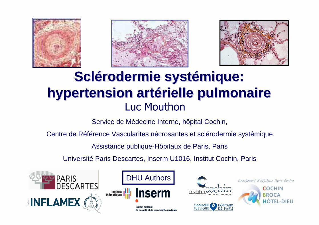
SclScl éérodermierodermie systsyst éémiquemique : : hypertension hypertension artart éériellerielle pulmonairepulmonaire
Service de Médecine Interne, hôpital Cochin,
Centre de Référence Vascularites nécrosantes et sclérodermie systémique
Assistance publique-Hôpitaux de Paris, Paris
Université Paris Descartes, Inserm U1016, Institut Cochin, Paris
Luc Mouthon
DHU Authors
Introduction
�Connective tissue disorders contribute for more than 10% of severe PAH�Systemic sclerosis (SSc): at risk to developPAH: 5-35%�Today, pulmonary fibrosis and PAH representthe first two causes of mortality in SSc patients �ESC-ERS do not recommend to performannual echocardiographic evaluation in SScpatients
Introduction
• Pulmonary arterial hypertension (PAH) is a devastating vascular complication of a number of connective tissue diseases including scleroderma (SSc).
• PAH related to SSc (SSc-PAH) has a dramatic impact on the clinical course and overall survival and is the single most common cause of death in patients afflicted with this syndrome.
• Despite the development of disease-targeted therapies for the idiopathic form of this condition (iPAH), the response to therapy is suboptimal in SSc-PAH and survival remains very poor.
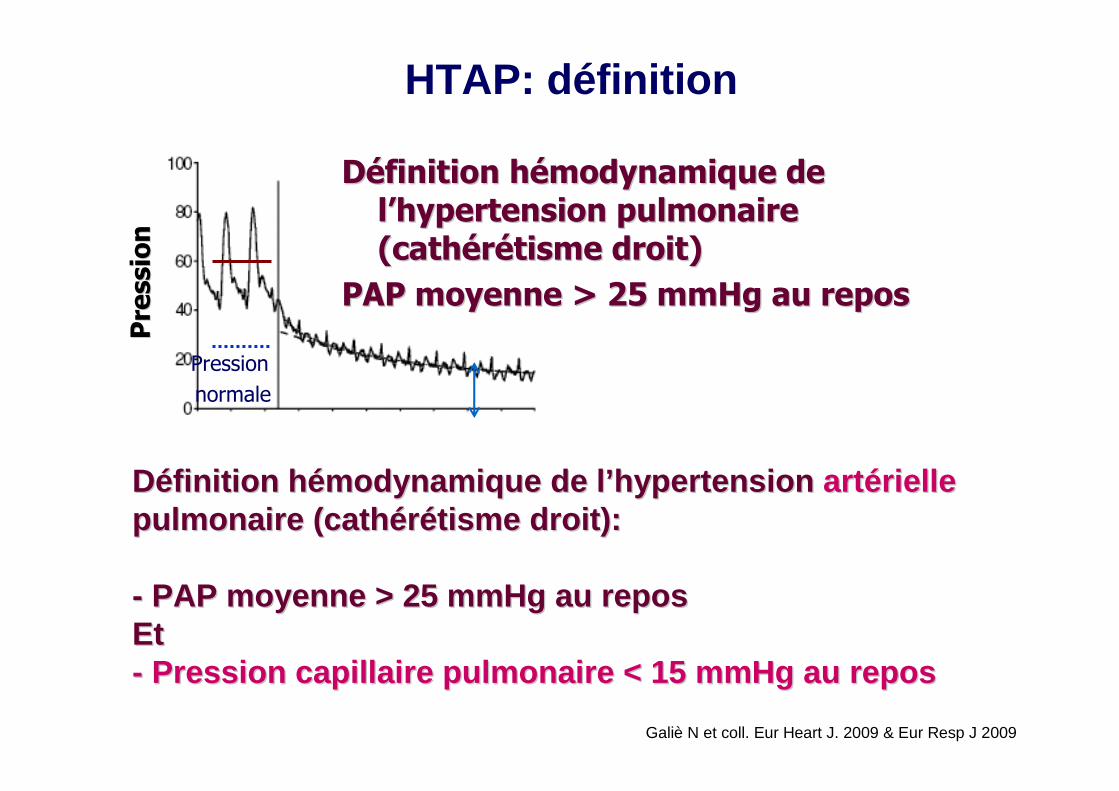
HTAP: définition
Galiè N et coll. Eur Heart J. 2009 & Eur Resp J 2009
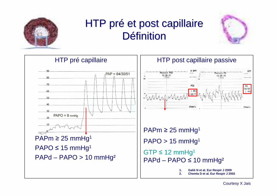
DDééfinition hfinition héémodynamique de modynamique de ll’’hypertension pulmonaire hypertension pulmonaire ((cathcathéérréétismetisme droit)droit)
PAP moyenne > 25 PAP moyenne > 25 mmHgmmHg au reposau repos
Pre
ssion
Pre
ssion
Pression
normale
DDééfinition hfinition h éémodynamique de lmodynamique de l ’’hypertension hypertension artart éérielleriellepulmonaire (pulmonaire ( cathcath éérréétismetisme droit):droit):
-- PAP moyenne > 25 PAP moyenne > 25 mmHgmmHg au reposau reposEtEt-- Pression capillaire pulmonaire < 15 Pression capillaire pulmonaire < 15 mmHgmmHg au reposau repos
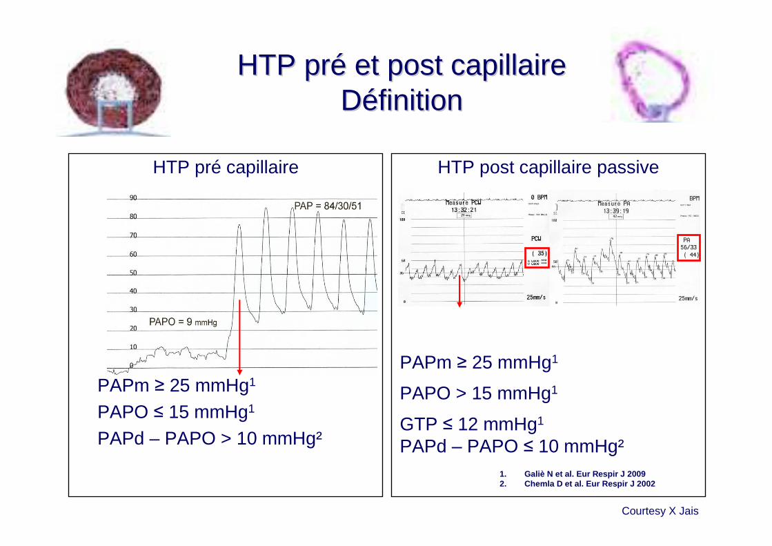
HTP pré capillaire
PAPm ≥ 25 mmHg1
PAPO ≤ 15 mmHg1
PAPd – PAPO > 10 mmHg²
HTP post capillaire passive
PAPm ≥ 25 mmHg1
PAPO > 15 mmHg1
GTP ≤ 12 mmHg1
PAPd – PAPO ≤ 10 mmHg²
HTP prHTP préé et post capillaireet post capillaireDDééfinitionfinition
1. Galiè N et al. Eur Respir J 20092. Chemla D et al. Eur Respir J 2002
Courtesy X Jais
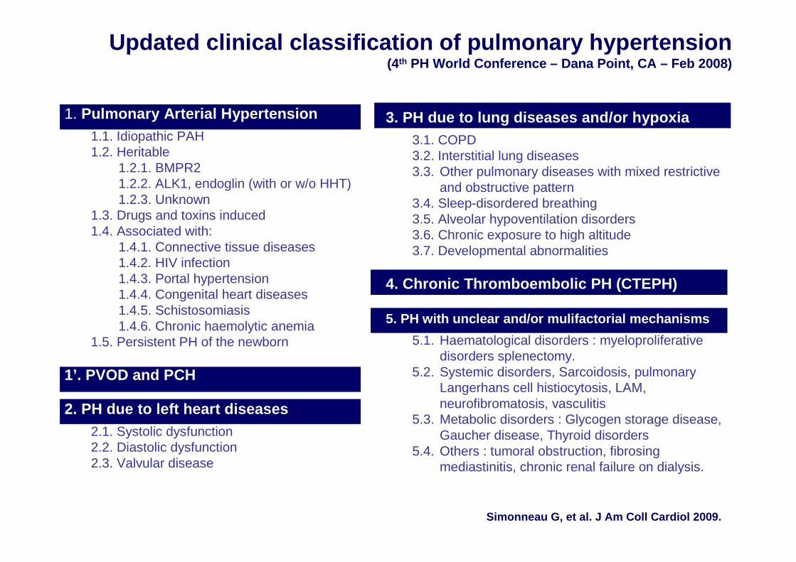
Updated clinical classification of pulmonary hypert ension(4th PH World Conference – Dana Point, CA – Feb 2008)
Simonneau G, et al. J Am Coll Cardiol 2009.
3. PH due to lung diseases and/or hypoxia3.1. COPD3.2. Interstitial lung diseases3.3. Other pulmonary diseases with mixed restrictive
and obstructive pattern3.4. Sleep-disordered breathing3.5. Alveolar hypoventilation disorders3.6. Chronic exposure to high altitude3.7. Developmental abnormalities
4. Chronic Thromboembolic PH (CTEPH)
5. PH with unclear and/or mulifactorial mechanisms
5.1. Haematological disorders : myeloproliferativedisorders splenectomy.
5.2. Systemic disorders, Sarcoidosis, pulmonary Langerhans cell histiocytosis, LAM, neurofibromatosis, vasculitis
5.3. Metabolic disorders : Glycogen storage disease, Gaucher disease, Thyroid disorders
5.4. Others : tumoral obstruction, fibrosingmediastinitis, chronic renal failure on dialysis.
1. Pulmonary Arterial Hypertension1.1. Idiopathic PAH1.2. Heritable
1.2.1. BMPR21.2.2. ALK1, endoglin (with or w/o HHT)1.2.3. Unknown
1.3. Drugs and toxins induced1.4. Associated with:
1.4.1. Connective tissue diseases1.4.2. HIV infection1.4.3. Portal hypertension1.4.4. Congenital heart diseases1.4.5. Schistosomiasis1.4.6. Chronic haemolytic anemia
1.5. Persistent PH of the newborn
1’. PVOD and PCH
2. PH due to left heart diseases2.1. Systolic dysfunction2.2. Diastolic dysfunction2.3. Valvular disease
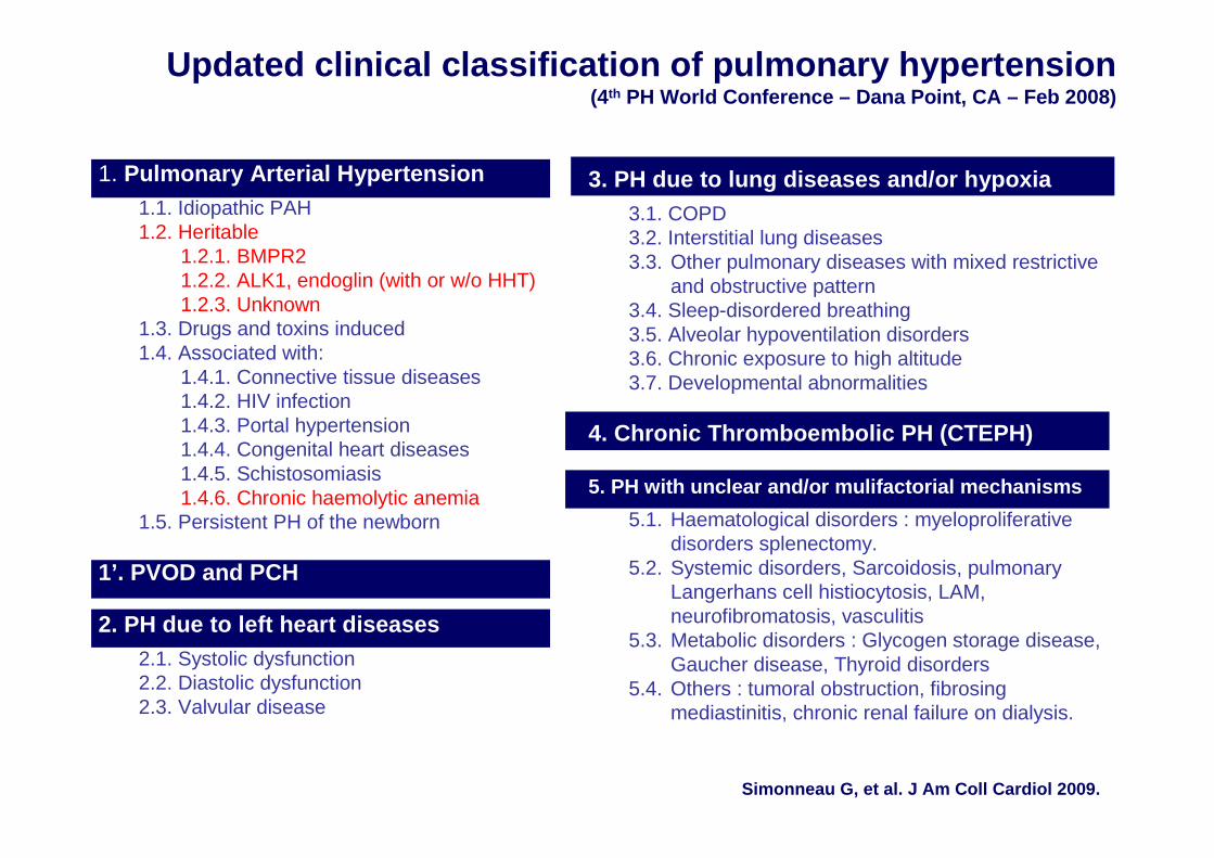
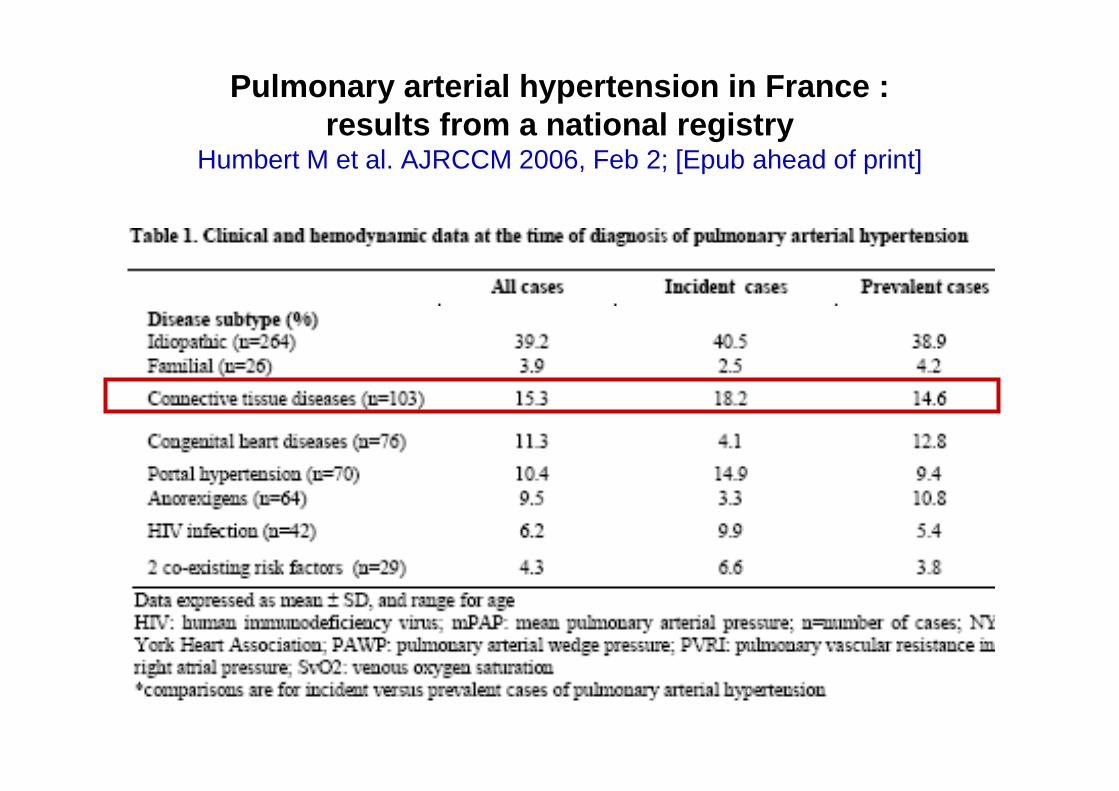
Pulmonary arterial hypertension in France : results from a national registry
Humbert M et al. AJRCCM 2006, Feb 2; [Epub ahead of print]
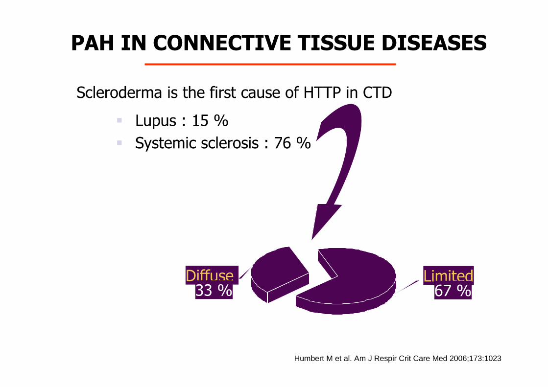
Scleroderma is the first cause of HTTP in CTD
� Lupus : 15 %
� Systemic sclerosis : 76 %
Limited67 %
Diffuse 33 %
Humbert M et al. Am J Respir Crit Care Med 2006;173:1023
PAH IN CONNECTIVE TISSUE DISEASES
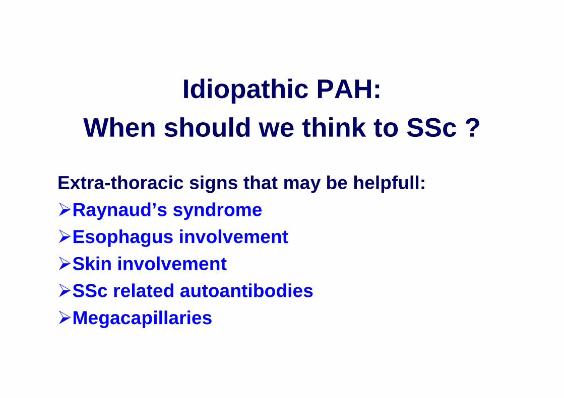
Idiopathic PAH: When should we think to SSc ?
Extra-thoracic signs that may be helpfull:�Raynaud ’s syndrome�Esophagus involvement�Skin involvement�SSc related autoantibodies�Megacapillaries
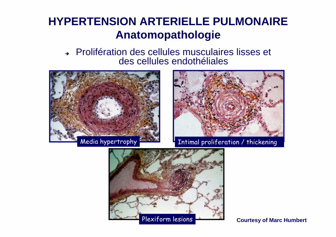
� Prolifération des cellules musculaires lisses et des cellules endothéliales
HYPERTENSION ARTERIELLE PULMONAIREAnatomopathologie
Media hypertrophy Intimal proliferation / thickening
Plexiform lesions Courtesy of Marc Humbert
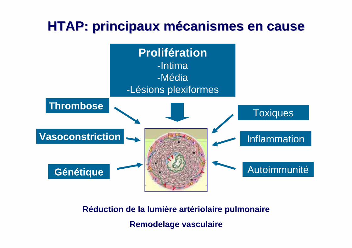
HTAP: principaux mHTAP: principaux m éécanismes en causecanismes en cause
Réduction de la lumière artériolaire pulmonaire
Remodelage vasculaire
Vasoconstriction
Prolifération-Intima-Média
-Lésions plexiformes
Thrombose
Inflammation
Génétique
Toxiques
Autoimmunité
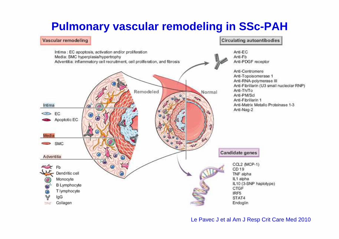
Pulmonary vascular remodeling in SSc-PAH
Le Pavec J et al Am J Resp Crit Care Med 2010
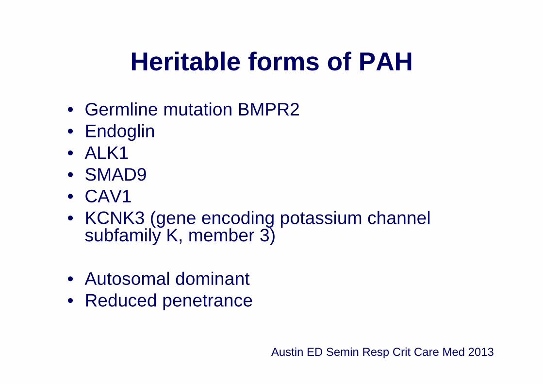
Heritable forms of PAH
• Germline mutation BMPR2• Endoglin• ALK1• SMAD9• CAV1• KCNK3 (gene encoding potassium channel
subfamily K, member 3)
• Autosomal dominant • Reduced penetrance
Austin ED Semin Resp Crit Care Med 2013
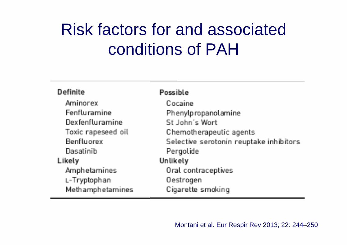
Risk factors for and associatedconditions of PAH
Montani et al. Eur Respir Rev 2013; 22: 244–250
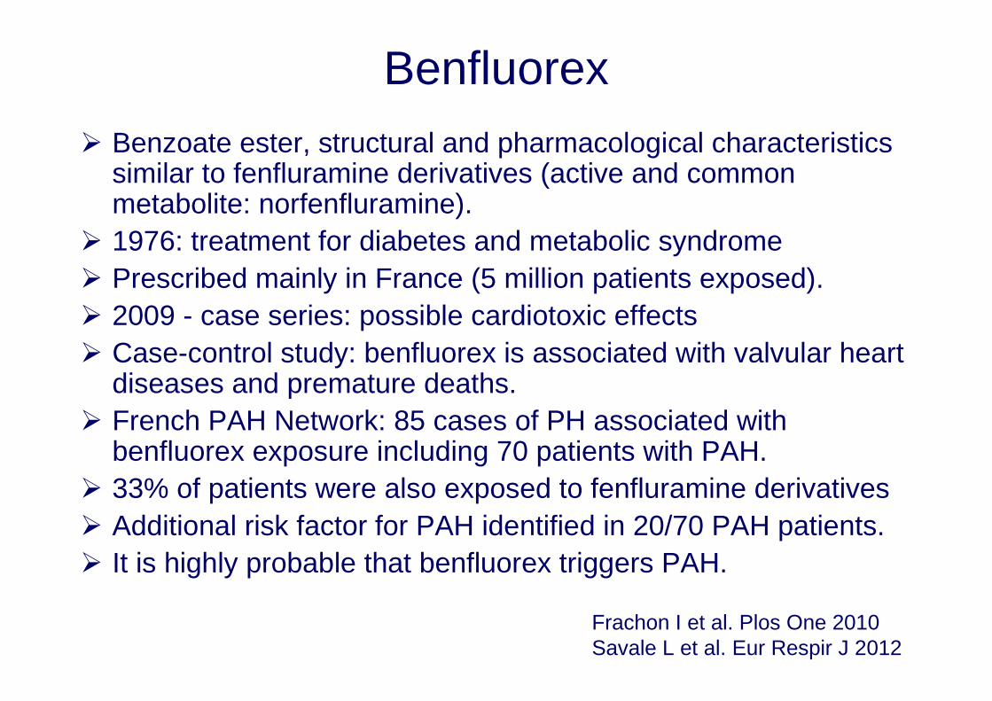
Benfluorex
� Benzoate ester, structural and pharmacological characteristicssimilar to fenfluramine derivatives (active and commonmetabolite: norfenfluramine).
� 1976: treatment for diabetes and metabolic syndrome� Prescribed mainly in France (5 million patients exposed). � 2009 - case series: possible cardiotoxic effects� Case-control study: benfluorex is associated with valvular heart
diseases and premature deaths. � French PAH Network: 85 cases of PH associated with
benfluorex exposure including 70 patients with PAH. � 33% of patients were also exposed to fenfluramine derivatives� Additional risk factor for PAH identified in 20/70 PAH patients. � It is highly probable that benfluorex triggers PAH.
Frachon I et al. Plos One 2010Savale L et al. Eur Respir J 2012
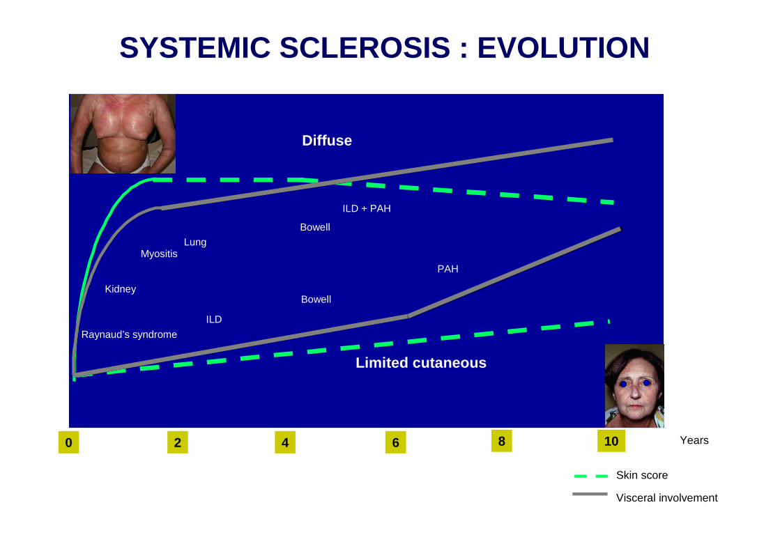
Skin score
Visceral involvement
SYSTEMIC SCLEROSIS : EVOLUTION
Diffuse
Limited cutaneous
0 102 4 6 8
Raynaud’s syndrome
Kidney
ILD + PAH
Myositis
Bowell
ILD
PAH
Bowell
0 102 4 6 8 Years
Lung
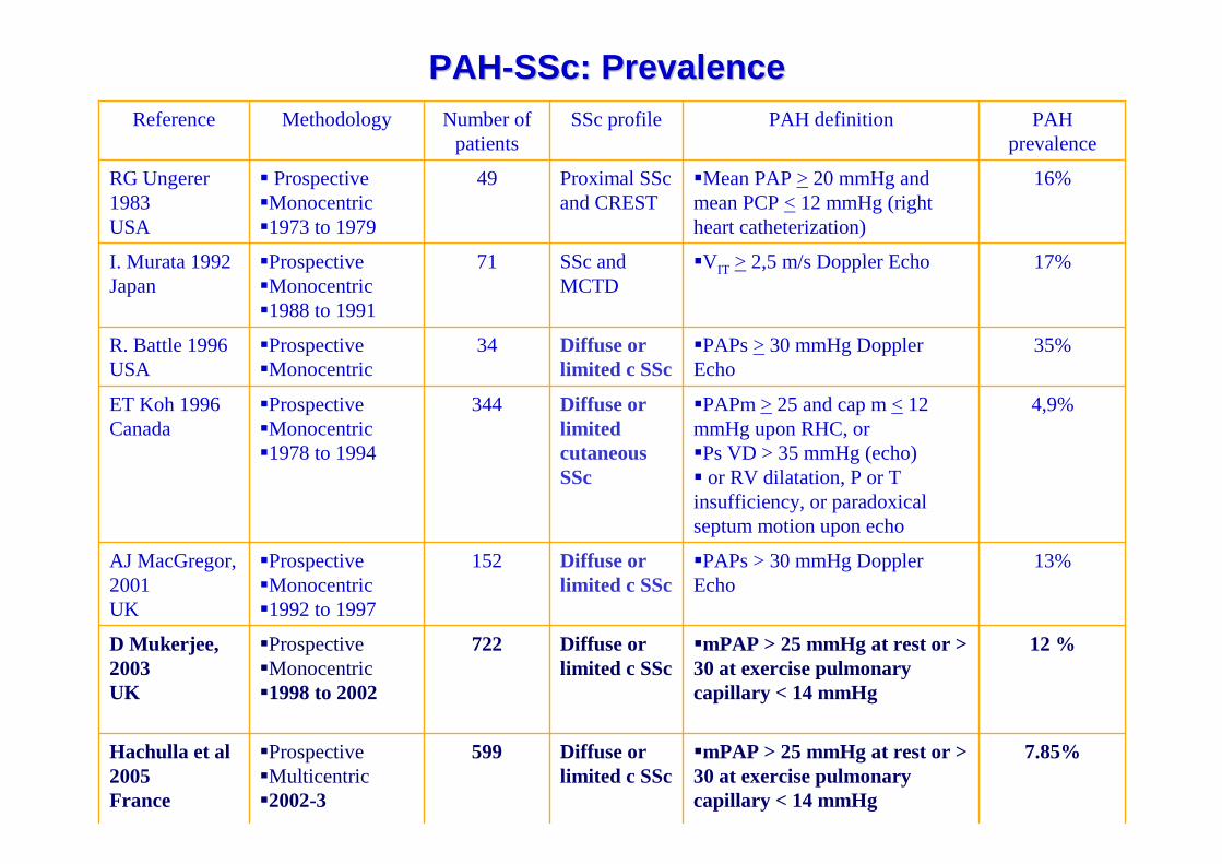
PAHPAH--SScSSc: : PrevalencePrevalence
7.85%�mPAP > 25 mmHg at rest or > 30 at exercise pulmonarycapillary < 14 mmHg
Diffuse or limited c SSc
599�Prospective�Multicentric�2002-3
Hachulla et al2005France
12 %�mPAP > 25 mmHg at rest or > 30 at exercise pulmonarycapillary < 14 mmHg
Diffuse or limited c SSc
722�Prospective�Monocentric�1998 to 2002
D Mukerjee, 2003UK
13%�PAPs > 30 mmHg Doppler Echo
Diffuse or limited c SSc
152�Prospective�Monocentric�1992 to 1997
AJ MacGregor, 2001UK
4,9%�PAPm >25 and cap m <12 mmHg upon RHC, or�Ps VD > 35 mmHg (echo)� or RV dilatation, P or T insufficiency, or paradoxicalseptum motion upon echo
Diffuse or limitedcutaneousSSc
344�Prospective�Monocentric�1978 to 1994
ET Koh 1996Canada
35%�PAPs >30 mmHg Doppler Echo
Diffuse or limited c SSc
34�Prospective�Monocentric
R. Battle 1996USA
17%�VIT > 2,5 m/s Doppler EchoSSc and MCTD
71�Prospective�Monocentric�1988 to 1991
I. Murata 1992Japan
16%�Mean PAP >20 mmHg and mean PCP <12 mmHg (right heart catheterization)
Proximal SScand CREST
49� Prospective�Monocentric�1973 to 1979
RG Ungerer 1983USA
PAH prevalence
PAH definitionSSc profileNumber of patients
MethodologyReference
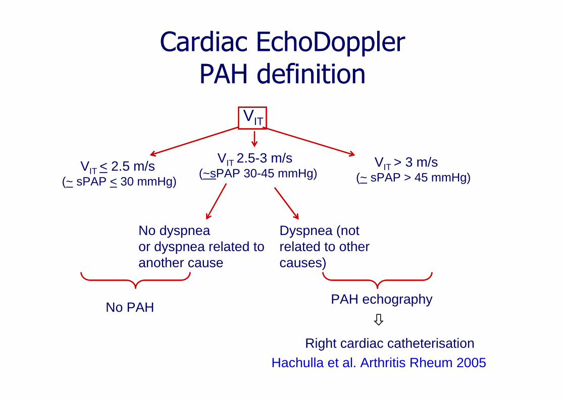
VIT > 3 m/s(~ sPAP > 45 mmHg)
VIT
VIT < 2.5 m/s(~ sPAP < 30 mmHg)
VIT 2.5-3 m/s(~sPAP 30-45 mmHg)
No dyspneaor dyspnea related toanother cause
Dyspnea (notrelated to other causes)
No PAHPAH echography
�
Right cardiac catheterisation
Cardiac EchoDopplerPAH definition
Hachulla et al. Arthritis Rheum 2005
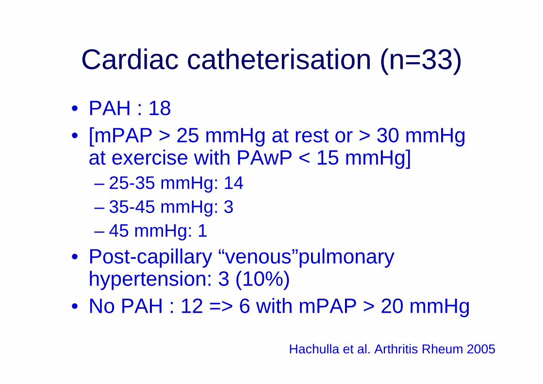
Cardiac catheterisation (n=33)
• PAH : 18 • [mPAP > 25 mmHg at rest or > 30 mmHg
at exercise with PAwP < 15 mmHg]– 25-35 mmHg: 14– 35-45 mmHg: 3– 45 mmHg: 1
• Post-capillary “venous”pulmonary hypertension: 3 (10%)
• No PAH : 12 => 6 with mPAP > 20 mmHg
Hachulla et al. Arthritis Rheum 2005
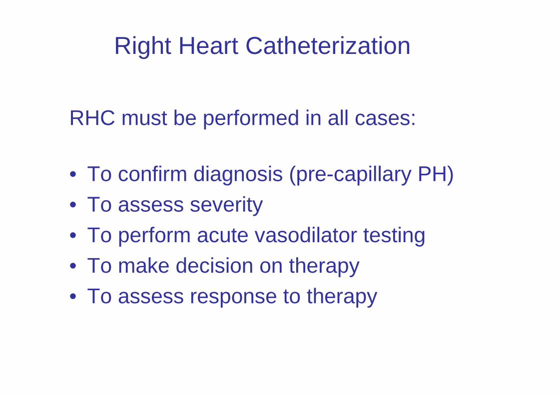
Right Heart Catheterization
RHC must be performed in all cases:
• To confirm diagnosis (pre-capillary PH)• To assess severity• To perform acute vasodilator testing• To make decision on therapy• To assess response to therapy
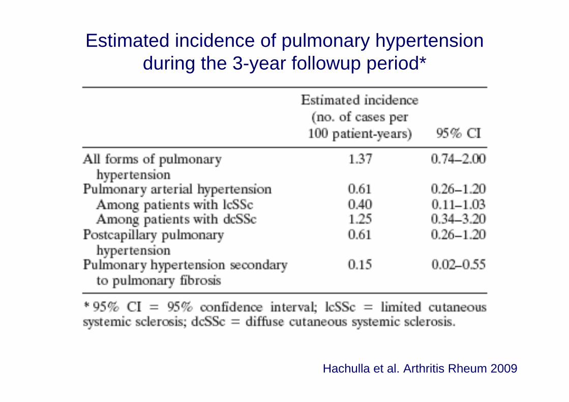
Estimated incidence of pulmonary hypertension during the 3-year followup period*
Hachulla et al. Arthritis Rheum 2009
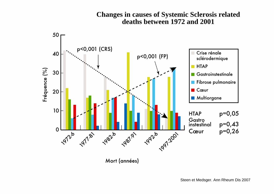
Changes in causes of Systemic Sclerosis relateddeaths between 1972 and 2001
Steen et Medsger. Ann Rheum Dis 2007
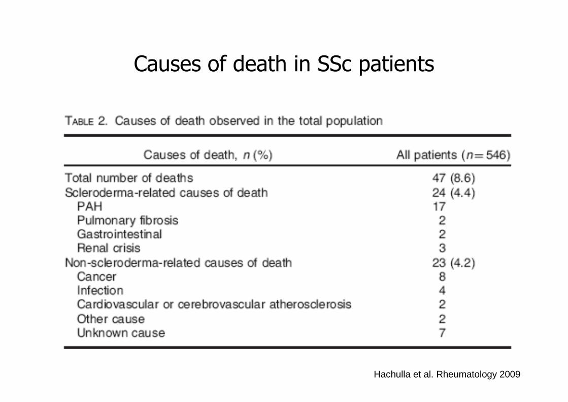
Causes of death in SSc patients
Hachulla et al. Rheumatology 2009
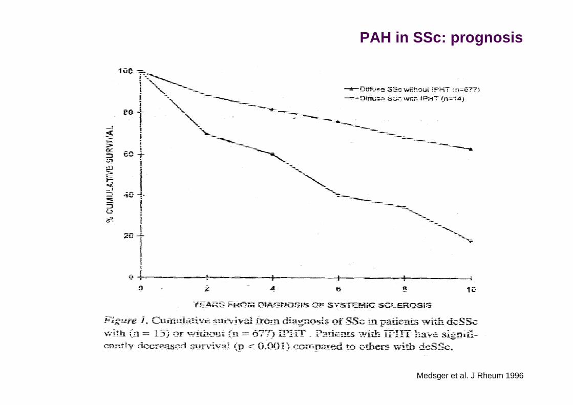
Medsger et al. J Rheum 1996
PAH in SSc: prognosis
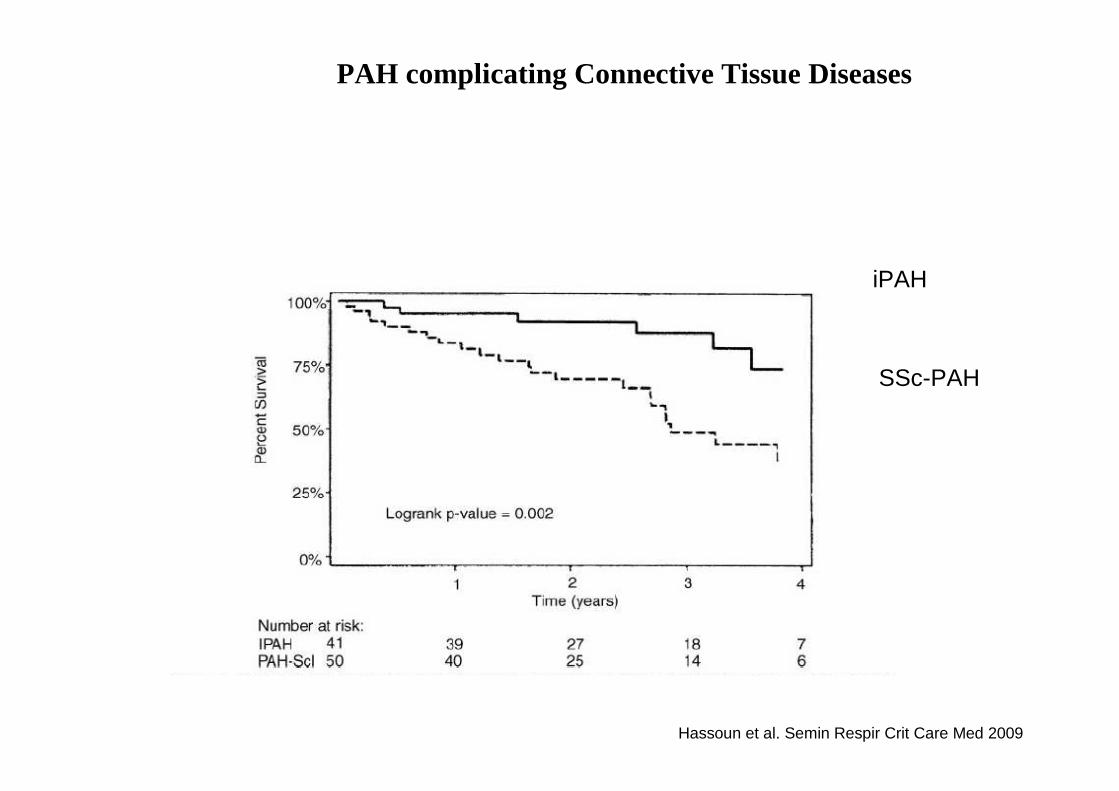
PAH complicating Connective Tissue Diseases
SSc-PAH
iPAH
Hassoun et al. Semin Respir Crit Care Med 2009
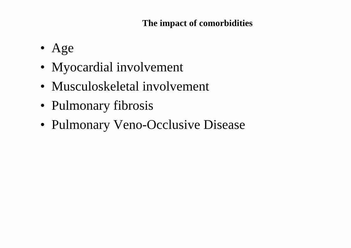
The impact of comorbidities
• Age
• Myocardial involvement
• Musculoskeletal involvement
• Pulmonary fibrosis
• Pulmonary Veno-Occlusive Disease
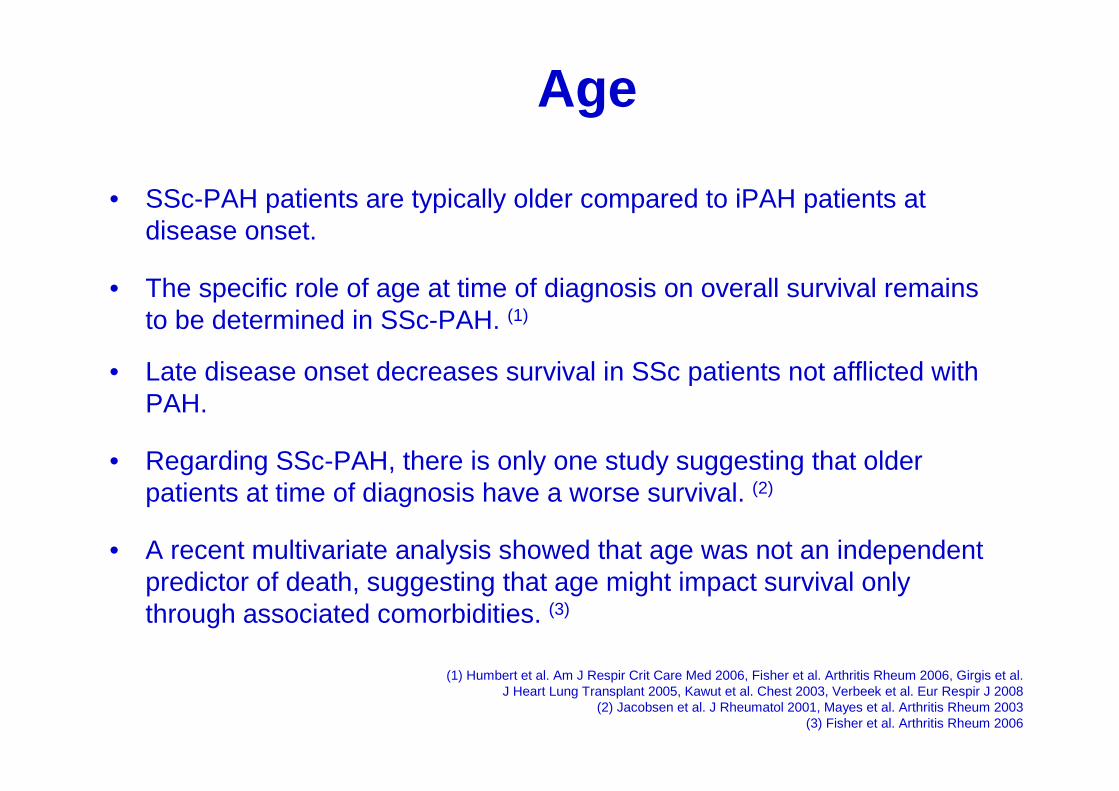
Age
• SSc-PAH patients are typically older compared to iPAH patients at disease onset.
• The specific role of age at time of diagnosis on overall survival remains to be determined in SSc-PAH. (1)
• Late disease onset decreases survival in SSc patients not afflicted with PAH.
• Regarding SSc-PAH, there is only one study suggesting that older patients at time of diagnosis have a worse survival. (2)
• A recent multivariate analysis showed that age was not an independent predictor of death, suggesting that age might impact survival only through associated comorbidities. (3)
(1) Humbert et al. Am J Respir Crit Care Med 2006, Fisher et al. Arthritis Rheum 2006, Girgis et al.J Heart Lung Transplant 2005, Kawut et al. Chest 2003, Verbeek et al. Eur Respir J 2008
(2) Jacobsen et al. J Rheumatol 2001, Mayes et al. Arthritis Rheum 2003(3) Fisher et al. Arthritis Rheum 2006
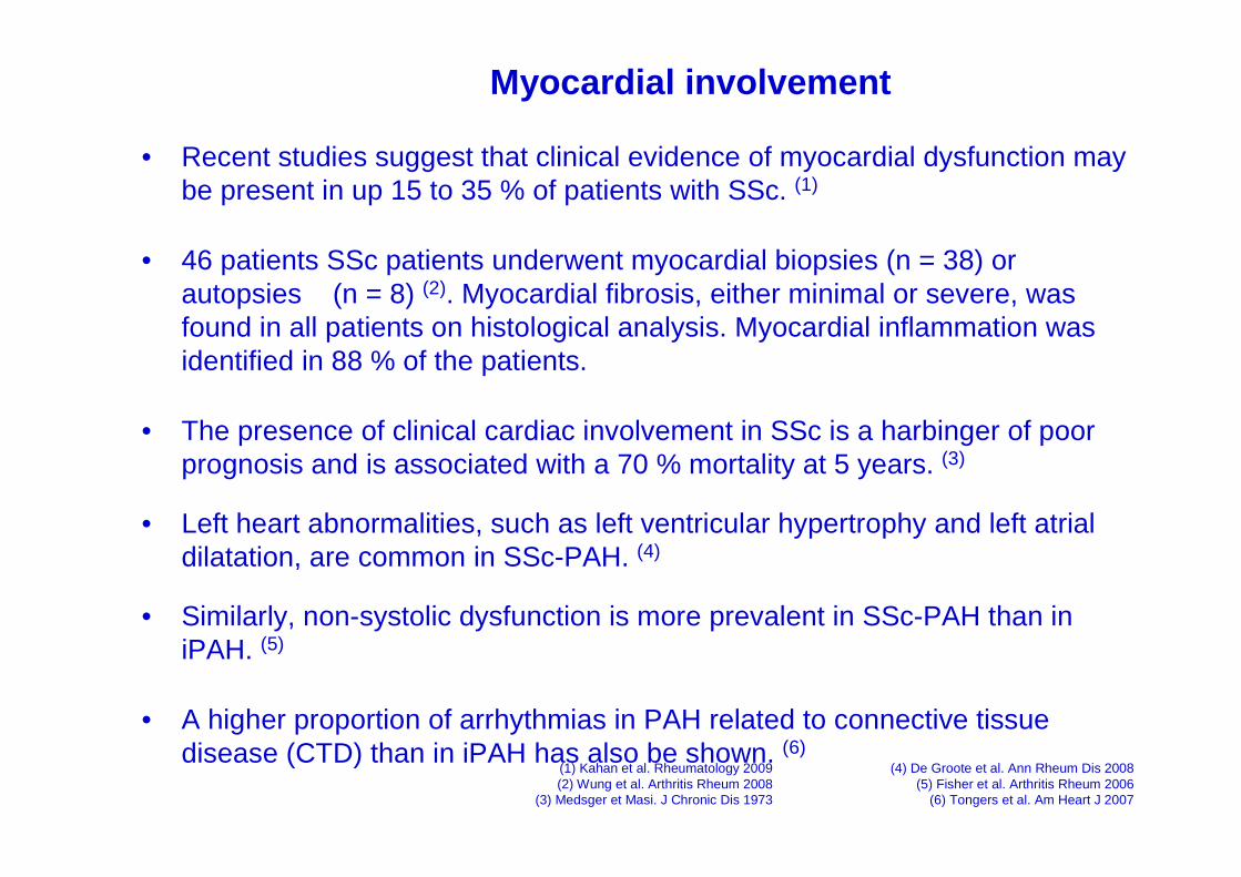
Myocardial involvement
• Recent studies suggest that clinical evidence of myocardial dysfunction may be present in up 15 to 35 % of patients with SSc. (1)
• 46 patients SSc patients underwent myocardial biopsies (n = 38) or autopsies (n = 8) (2). Myocardial fibrosis, either minimal or severe, was found in all patients on histological analysis. Myocardial inflammation was identified in 88 % of the patients.
• The presence of clinical cardiac involvement in SSc is a harbinger of poor prognosis and is associated with a 70 % mortality at 5 years. (3)
• Left heart abnormalities, such as left ventricular hypertrophy and left atrialdilatation, are common in SSc-PAH. (4)
• Similarly, non-systolic dysfunction is more prevalent in SSc-PAH than in iPAH. (5)
• A higher proportion of arrhythmias in PAH related to connective tissue disease (CTD) than in iPAH has also be shown. (6)
(1) Kahan et al. Rheumatology 2009(2) Wung et al. Arthritis Rheum 2008
(3) Medsger et Masi. J Chronic Dis 1973
(4) De Groote et al. Ann Rheum Dis 2008(5) Fisher et al. Arthritis Rheum 2006
(6) Tongers et al. Am Heart J 2007
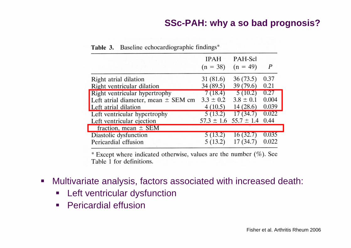
SSc-PAH: why a so bad prognosis?
� Multivariate analysis, factors associated with increased death:� Left ventricular dysfunction� Pericardial effusion
Fisher et al. Arthritis Rheum 2006
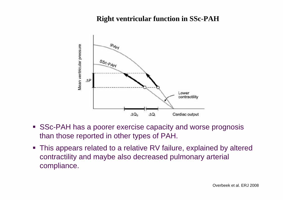
Right ventricular function in SSc-PAH
� SSc-PAH has a poorer exercise capacity and worse prognosisthan those reported in other types of PAH.
� This appears related to a relative RV failure, explained by alteredcontractility and maybe also decreased pulmonary arterialcompliance.
Overbeek et al. ERJ 2008
Musculoskeletal involvement
• Musculoskeletal involvement is frequent in SSc patients.
• Muscle involvement occurs in up to 96 % of cases and correlates strongly with myocardial disease in SSc. (1)
• In an analysis of over 300 SSc patients with skeletal myopathy, diffuse SSc, the presence of pulmonary fibrosis, and male gender tend to be risk factors for development of myopathy. (2)
• While the role of musculoskeletal involvement in overall prognosis is unclear, it does impact function, such as the 6 minute walk distance (6MWD) as recently suggested (3), raising very strong doubt about the validity of this functional end-point in SSc-PAH. (1) Randone et al. Best Pract Res Clin Rheumatol 2008
(2) Mimura et al. Clin Rheumatol 2005(3) Garin et al. J Rheumatol 2009
Pulmonary fibrosis
• Patients with limited SSc disease will typically develop isolated PAH 10-15 years after the onset of their disease. (1)
• In contrast, patients with diffuse SSc are at greater risk for ILD, usually within the first 5 years after diagnosis (2), but may develop PH at any stage in the course of their disease. (3)
• However, while SSc patients with ILD alone have a median survival of 5-8 years (4), development of PH (PH-ILD) will shorten survival.
• In a study by Mathai et al, PH-ILD was associated with a 5-fold increased risk of death compared to SSc-PAH. (5)
• Similarly, in a recent large study by Condliffe et al, the 3-year survival was significantly worse (28 %) in the group of patients with ILD compared to patients with isolated SSc-PAH (47 %). (6)
(1) Steen et Medsger. Arthritis Rheum 2003(2) Steen. Ann Rheum Dis 2003
(3) Chang et al. J Rheumatol 2003
(4) Altman et al. Arthritis Rheum 1991(5) Mathai et al. Arthritis Rheum 2009
(6) Condliffe et al. Am J Respir Crit Care Med 2009
Pulmonary Veno-Occlusive Disease
• PVOD is characterized by intimal proliferation and fibrosis of the intrapulmonary veins and venules, occasionally extending to the arteriolar bed. (1)
• PVOD is an underrecognized cause of PAH in SScpatients. (2)
• A recent histological study suggested that SSc-PAH may be characterized by a more frequent involvement of pulmonary veins than previously recognized perhaps explaining in part why these patients are less responsive to specific PAH treatment as compared with iPAH patients. (1)
(1) Dorfmüller et al. Hum Pathol 2007(2) Montani et al. Eur Respir J 2009
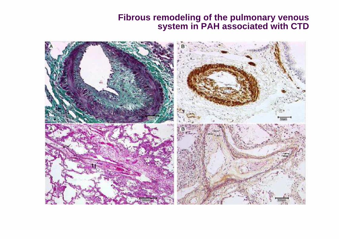
Fibrous remodeling of the pulmonary venoussystem in PAH associated with CTD
Conclusions
�8-12% of SSc patients develop PAH
�Detection: echodardiography
�Comfirmation: Right heart catheterization
(treshold….)
�Prognosis: reserved
�Impact of comorbidities
�Lower efficacy than observed in idiopathic
PAH