scientific document - cmap
TRANSCRIPT
PROGRAMME COSINUS
EDITION 2010
Projet XXX
DOCUMENT SCIENTIFIQUE
1/28
Acronyme SIMUDMRI
Titre du projet en
français
Simulation du signal d’IRM diffusion dans tissu
biologique
Titre du projet en anglais
Simulation of diffusion MRI signals in biological tissue
Axe(s) thématique(s)
x 1 2 3
Type de recherche Recherche Fondamentale Recherche Industrielle
Développement Expérimental
Types de projets
spécifiques
Plate-forme (cocher si ce projet est une mise en
place/construction de plate-forme au sens de l’appel à projets)
Aide totale demandée
400038 € Durée du projet 36 mois
1. RÉSUMÉ ....................................................................................... 2
2. CONTEXTE ET POSITIONNEMENT DU PROJET .............................................. 3 2.1. Contexte et enjeux économiques et sociétaux .............................................. 4 2.2. Positionnement du projet .......................................................................... 4
3. DESCRIPTION SCIENTIFIQUE ET TECHNIQUE .............................................. 6 3.1. État de l'art ............................................................................................. 6 3.2. Objectifs et caractère ambitieux/novateur du projet ..................................... 7
4. PROGRAMME SCIENTIFIQUE ET TECHNIQUE, ORGANISATION DU PROJET ............... 9 4.1. Programme scientifique et structuration du projet ........................................ 9 4.2. Management du projet .............................................................................11 4.3. Description des travaux par tâche .............................................................12
4.3.1 Tâche 1 12 4.3.2 Tâche 2 14 4.3.3 Tâche 3 15 4.3.4 Tâche 4 17 4.3.5 Tâche 5 18 4.3.6 Tâche 6 18
4.4. Calendrier des tâches, livrables et jalons ....................................................20
5. STRATEGIE DE VALORISATION DES RESULTATS ET MODE DE PROTECTION ET
D’EXPLOITATION DES RESULTATS ........................................................ 21
6. ORGANISATION DU PARTENARIAT ........................................................ 22 6.1. Description, adéquation et complémentarité des partenaires.........................22 6.2. Qualification du coordinateur du projet ......................................................23
7. JUSTIFICATION SCIENTIFIQUE DES MOYENS DEMANDES ............................... 24 7.1. Partenaire 1 : INRIA ................................................................................24 7.2. Partenaire 2 : CEA I²BM NeuroSpin ...........................................................25
PROGRAMME COSINUS
EDITION 2010
Projet XXX
DOCUMENT SCIENTIFIQUE
2/28
8. ANNEXES .................................................................................... 26 8.1. Références bibliographiques .....................................................................26
1. RÉSUMÉ
Water diffusion magnetic resonance imaging (DMRI) is a method which uses a combination
of applied magnetic fields to measure, statistically, the diffusion of water molecules due to
Brownian motion. Its spatial resolution is on the order of millimeters. An apparent diffusion
coefficient (ADC) or diffusion tensor (DT) is computed for each voxel based on a model
relating these quantities to signal attenuation. In the past three decades, DMRI has been
successfully used to track brain white matter fibers and to detect acute brain ischemia.
Diffusion functional MRI has also begun to gain momentum as an active area of research.
The goal of this project is to go beyond the ADC or the DT to get more detailed information
on tissue properties from the DMRI signal.
The cellular structure inside the human brain varies on the scale of micrometers, which is
much smaller than the size of a voxel. There may be thousands of irregularly-shaped cells
within a voxel, and they all contribute to the environment seen by water molecules whose
displacement is measured by the MRI scanner. In the typical DMRI experiment, the time
interval over which water diffusion is measured is in the range of 50-100 milliseconds. Using
the diffusion coefficient of 'free' water at 37 degrees Celsius, D = 3e-9 m2/s, we get an
estimated diffusion distance of 15-25 micrometers. Clearly, in a DMRI experiment, water
molecules encounter numerous times inhomogeneities in the cellular environment, such as
cell membranes, fibers, and macromolecules.
We simulate the DMRI signal at the scale of a single voxel, while taking into account realistic
cellular structure and the true shape and duration of the diffusion gradients. We will obtain
the geometrical description of several samples of brain tissue from 3D electron microscopy.
The membranes of the various cells making up each sample will then be extracted using
dedicated segmentation tools. The finite duration and ramp time of the gradient coils will be
accurately reflected in the simulation.
The numerical simulation will approached in two directions. One is based on the numerical
solution of the Bloch-Torrey partial differential equation using Green's functions. The other
is Monte Carlo simulation. The end result is a hybrid code incorporating both methods: the
Green's function method will be used to speed up the Monte Carlo simulation in regimes
where the results of the simulation satisfies the Bloch-Torrey equation and where the
geometry can be modelled by simple objects such as smooth surfaces. The two methods will
be coupled in a unified numerical code where the coupling will occur across spatial regions
and also in time.
The end goal of this project is the simulation of DMRI signal taking into account realistic 3D
cellular structure, gradient sequence, while producing results which are reliably accurate,
scalable, all in reasonable simulation time. This simulation tool will be valuable in
PROGRAMME COSINUS
EDITION 2010
Projet XXX
DOCUMENT SCIENTIFIQUE
3/28
understanding the tissue microstructure that gives rise to the DMRI signals and also aid in
the design of new diffusion imaging protocols.
2. CONTEXTE ET POSITIONNEMENT DU PROJET
This project concerns fundamental research into the understanding of the origins of the
signals of DMRI. In the past few decades, DMRI has become a fundamental research tool in
mapping brain architecture and its many potential uses in clinical diagnosis are being
actively pursued by many teams across the world. DMRI is a non-invasive method of
imaging which measures the effect of the natural random movements of water molecules in
tissue by way of applied of magnetic fields. In acute brain ischemia, it was demonstrated
both in animal models and human patients with stroke that the measured water diffusion
coefficient decreased significantly within several minutes [12, 13]. By contrast, T2-weighted
images remained normal for several hours. More recently, in functional imaging, it has been
shown that the diffusion of water slightly slows down in the activated brain cortical areas,
several seconds in advance of hemodynamic response detected by BOLD fMRI [6].
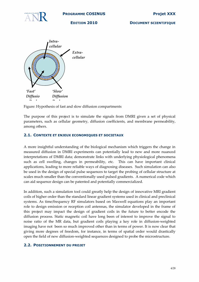
The basic mechanism underlying the decrease in measured diffusion is still unknown. Since
free diffusion of water in a homogeneous medium would give rise to a single decaying
exponential and the signal from DMRI experiments in biological tissue is more accurately fit
by the sum of two decaying exponentials, a frequent conjecture is the existence of (at least)
two distinct diffusion compartments. Often one considers that biological tissue consists of
intracellular space and extracellular space in which water molecules diffuse 'freely' except
where they encounter the cell membrane (or possibly macro-molecules). It is assumed that
the intracellular and extracellular compartments each possess a diffusion coefficient and the
exchange of water between the compartments occurs through the cell membranes.
Different hypotheses to explain the decrease in measured diffusion include a decrease in
either or both of the diffusion coefficients in the intra-cellular or the extra-cellular
compartments, a shift of water from the fast diffusion compartment (supposed to be the
extra-cellular) to the slow diffusion compartment (intra-cellular), a decreased permeability of
the cell membranes, increased 'tortuosity' in the extracellular space due to cell swelling. In
[6] the proposed mechanism is the increased volume of a slowly diffusing compartment near
the cell membranes.
PROGRAMME COSINUS
EDITION 2010
Projet XXX
DOCUMENT SCIENTIFIQUE
4/28
Figure: Hypothesis of fast and slow diffusion compartments
The purpose of this project is to simulate the signals from DMRI given a set of physical
parameters, such as cellular geometry, diffusion coefficients, and membrane permeability,
among others.
2.1. CONTEXTE ET ENJEUX ECONOMIQUES ET SOCIETAUX
A more insightful understanding of the biological mechanism which triggers the change in
measured diffusion in DMRI experiments can potentially lead to new and more nuanced
interpretations of DMRI data; demonstrate links with underlying physiological phenomena
such as cell swelling, changes in permeability, etc. This can have important clinical
applications, leading to more reliable ways of diagnosing diseases. Such simulation can also
be used in the design of special pulse sequences to target the probing of cellular structure at
scales much smaller than the conventionally used pulsed gradients. A numerical code which
can aid sequence design can be patented and potentially commercialized.
In addition, such a simulation tool could greatly help the design of innovative MRI gradient
coils of higher order than the standard linear gradient systems used in clinical and preclinical
systems. As time/frequency RF simulators based on Maxwell equations play an important
role to design emission or reception coil antennas, the simulator developed in the frame of
this project may impact the design of gradient coils in the future to better encode the
diffusion process. Static magnetic coil have long been of interest to improve the signal to
noise ratio of the MR data, but gradient coils playing a key role in diffusion-weighted
imaging have not been so much improved other than in terms of power. It is now clear that
giving more degrees of freedom, for instance, in terms of spatial order would drastically
open the field of new diffusion-weighted sequences designed to probe the microstructure.
2.2. POSITIONNEMENT DU PROJET
‘Fast’
Diffusio
n Pool
‘Slow’
Diffusion
Pool
Extra-
cellular
compartme
nt
Intra-
cellular
compartme
nt
PROGRAMME COSINUS
EDITION 2010
Projet XXX
DOCUMENT SCIENTIFIQUE
5/28
Many simulations of the diffusion process have been attempted in the past, but none of them
allow large scale simulations in a large field of view or use realistic shapes corresponding to
what can be found in the brain parenchyma. They are all focused either on simplistic
geometries (spheres, planes, cylinders) lacking the plethora of tortuous and anisotropic
shapes of the brain microstructure or make too many assumptions on the diffusion-weighted
pulse sequence leading to practically unfeasible experiments on real imaging systems.
Early in 2008, we (Denis LeBihan, Cyril Poupon, Chun-Hung Yeh) started to develop a
Monte-Carlo simulator in the frame of an ANR Programme Blanc France-Taiwan focused on
functional diffusion imaging (project acronym=”DFMRI”) aimed at mimicking the reality of
diffusion experiments in terms of brain structure and function as well as of real imaging
system possibilities, in order to better understand the structural modifications occurring
during brain activity, and to better correlate this information with the signal acquired on
clinical or preclinical systems. We have now reached a point where, to go to the next step, we
need to improve the scale of our code using a large computing facility in order to perform
some large scale realistic simulations using real geometries and large sample sizes. We have
increased the manpower working on the simulation with the addition of Jing-Rebecca Li and
Antoine Lejay and Marina Spivak who will work on speeding up the Monte Carlo simulation
by a Green’s function method.
This project is unique in that it combines the expertise of MRI physicists who have
developed innovative imaging modalities in both anatomical and functional MRI, with the
expertise of biologists who can give some advices about the cellular environment in order to
establish an accurate model of the microstructure and of the biological parameters
underlying the various functions of the brain that may modify the diffusion process inside
tissues, with computer scientists who have experience with large scale code development,
on parallel and cluster environments along with applied mathematicians experienced in the
numerical simulation of diffusion phenomena in complex media.
The availability of advanced imaging facilities at Neurospin, a world class high field NMR
facility, located on the site of the CEA, means that MRI experiments can be conducted to
complement the effort at simulation. The ready access to high power computing at the
CCRT will make the parallelization of the simulation code in the latter stage of the project
convenient. The numerical algorithms experts of INRIA have experience formulating
accurate and reliable algorithms in both stochastic and analytical modelling.
This project corresponds to 'Axe thématique 1: simulation et calcul intensif' of the call. Both
the Green's function method code and the Monte-Carlo code are large scale simulations.
The typical time step used in Monte Carlo is on the order of a microsecond; the time interval
to be simulated is around 100 milliseconds, hence 100,000 time steps are to be used. At each
time step, Monte-Carlo may involve moving 100,000 to 1 million particles. The Green's
function simulation uses larger time steps and may require 50,000 to 200,000 degrees of
freedom depending on the desired accuracy. Both approaches are highly parallelizable. The
spatial scale of the problem is also challenging, the typical voxel is on the order millimeters
PROGRAMME COSINUS
EDITION 2010
Projet XXX
DOCUMENT SCIENTIFIQUE
6/28
and the dimension of the cells can be on the order of micrometers. Hence in three
dimensions, spatial domain to be simulated can be 1 million times the spatial resolution
required. The dimension of the cell membranes can be on the order of nanometers; hence, if
cell membranes are to be treated as a separate diffusion compartment the degrees of
freedoms in space would be even vastly larger. In this case, it is essential to couple a code
that computes the diffusion near the membrane with a much more coarsely discretized
solver away from the membrane. An efficient and accurate coupling between the different
scales is needed. Parallel implementation is essential.
This is also an element of visualization of the results, to be able to visualize the diffusion of
water molecules in the complex cellular environment over a large interval of time.
3. DESCRIPTION SCIENTIFIQUE ET TECHNIQUE
3.1. ÉTAT DE L'ART
Bloch-Torrey equation and Karger’s model:
In a MR experiment, a uniform magnetic field is applied in the z direction. After a 90 degree
pulse the spins lie along the x − y plane and a linear gradient field g(t) = gf(t)u, in the
direction u with time profile gf(t), is applied. The Bloch-Torrey equation describes the
evolution of the complex transverse magnetization m(x,t,g,u) while taking into account
diffusion:
λ= 42.58 × 2π MHz is the gyromagnetic ratio and D is the diffusion coefficient. The diffusion
MRI signal is the measured (normalized) signal attenuation as a function of g and u over the
volume of a voxel. There are a few groups [14, 4] working on finite difference or finite
elements methods in the simulation by solving the Bloch-Torrey equation. There are also
several researchers who worked on expressing the solution of the Bloch-Torrey equation
under simplified conditions in analytical terms (see [2] for a review). An alternate approach
to Bloch-Torrey is the Karger’s model, where is a model based on physical considerations by
using quantities called residence times in the different diffusion compartments [5]. The
residence times derived analytically, but under simplified conditions.
We solve the Bloch-Torrey equation with a Green’s function method that is more efficient
and accurate than finite different or finite elements methods. This method has been
explained for the diffusion equation [8, 9]. Its use in the Bloch-Torrey equation appears
preliminarily in [10]. We use more reasonable and realistic boundary conditions between the
diffusing compartments than either the groups working on analytical expressions or the
Karger model.
PROGRAMME COSINUS
EDITION 2010
Projet XXX
DOCUMENT SCIENTIFIQUE
7/28
Monte Carlo Brownian dynamics simulator:
In a parallel effort, we design and code a Monte Carlo Brownian dynamics simulator capable
of simulating diffusion of spins in arbitrarily complex geometries with a DW signal
integrator emulating various MR pulse sequences. The flexibility and power of Monte-Carlo
modeling enable us to investigate detail dynamics and mechanisms of molecular diffusion in
complex systems which cannot be handled through analytical models [11, 3]. Hence, the
CEA NeuroSpin partner has developed software to reproduce various tissue configurations
using dynamic meshes. Complicated geometries mimicking neural tissue components, such
as neurons, astrocytes, axons, etc. can be emulated, as well as tissue features (e.g. cell size,
density, membrane permeability) and basic diffusion mechanisms in different compartments
(presence of attractors, local viscosity, membrane interactions, etc.). This framework allows
to bridge the gap between elementary processes occurring at a micrometer scale and the
resulting DW signal measured at millimeter scale, providing a better understanding of the
features observed in DW MRI (variation of apparent diffusion coefficient (ADC) with cell
size, diffusion anisotropy) and to optimize acquisition schemes for different applications (e.g.
fiber-tracking algorithms).
The Monte-Carlo simulation software described in [15] and is general and flexible to mimic
diffusion in biological tissues and to generate realistic synthetic data for diffusion MRI. Since
the properties of the diffusing spins and tissue structure are all manageable, this software
enables investigations on the underpinning mechanisms affecting the DW signal and the
consequent diffusion measured parameters (e.g. ADC and fractional anisotropy (FA) [1].
Specifically, it can be utilized to investigate mechanistic hypotheses for various scenarios
(acute ischemia or neuronal activation and cell swelling, cancer and cell proliferation, axonal
fiber anisotropy in complex bundles or cortex, etc.).
One main objective of the project will be to merge both Green’s function and Monte-Carlo
approaches in an hybrid simulator developed by the two partners in order to go beyond the
actual limitations of the multiple compartment model stemming from Karger’s equations.
This project is developed in the next session.
3.2. OBJECTIFS ET CARACTERE AMBITIEUX/NOVATEUR DU PROJET
The Green's function method which we propose has the advantage over competing methods
in that it is reliably accurate. There is a reliable way of estimating the degrees of freedom
needed to achieve a certain level of accuracy. The aim of this project is to provide a code that
produces results within 1 percent relative accuracy in fairly complex geometry.
The Green’s method is a competing method to finite difference or finite elements methods
and offers the advantage of high accuracy and is not subject to stability restrictions on the
time step of simulation, hence larger time steps can be taken in the simulation compared to
finite difference/elements methods. This method has been implemented for the short pulse
PROGRAMME COSINUS
EDITION 2010
Projet XXX
DOCUMENT SCIENTIFIQUE
8/28
approximation of pulsed field gradient. The extension for generally shaped time-varying
gradients is underway. Only the Green's function changes from the short pulse
approximation.
The Monte-Carlo part of the project will be highly optimized and the geometrical description
very efficient and flexible. The large scale CCRT facility of the CEA proposes an adequate
grid configuration of 1024 processors. The Monte-Carlo code is highly scalable as all the
simulated water molecules move independently following a random walk. Only the
geometrical scene needs to be shared among nodes of the computer grid, but with read-only
access. Therefore, the parallelization of the code will be straightforward, and it is likely that
the computational efficiency of the code will be almost linear with the number of processors,
as no communication between processors will occur during the simulation.
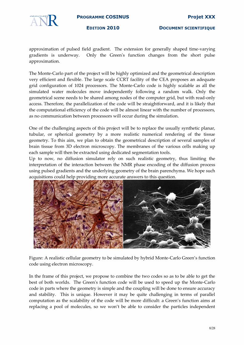
One of the challenging aspects of this project will be to replace the usually synthetic planar,
tubular, or spherical geometry by a more realistic numerical rendering of the tissue
geometry. To this aim, we plan to obtain the geometrical description of several samples of
brain tissue from 3D electron microscopy. The membranes of the various cells making up
each sample will then be extracted using dedicated segmentation tools.
Up to now, no diffusion simulator rely on such realistic geometry, thus limiting the
interpretation of the interaction between the NMR phase encoding of the diffusion process
using pulsed gradients and the underlying geometry of the brain parenchyma. We hope such
acquisitions could help providing more accurate answers to this question.
Figure: A realistic cellular geometry to be simulated by hybrid Monte-Carlo Green’s function
code using electron microscopy.
In the frame of this project, we propose to combine the two codes so as to be able to get the
best of both worlds. The Green's function code will be used to speed up the Monte-Carlo
code in parts where the geometry is simple and the coupling will be done to ensure accuracy
and stability. This is unique. However it may be quite challenging in terms of parallel
computation as the scalability of the code will be more difficult: a Green’s function aims at
replacing a pool of molecules, so we won’t be able to consider the particles independent
PROGRAMME COSINUS
EDITION 2010
Projet XXX
DOCUMENT SCIENTIFIQUE
9/28
anymore, but we’ll have to determine the good code granularity that yield the optimum
efficiency of the computer grid.
Last, it is obvious that NeuroSpin is the most adequate place for such a project, as its
platform already makes available a range of MRI system covering all our needs (3T and 7T
human clinical systems, 7T and 17T preclinical systems that will be used to perform imaging
of the previous samples of brain parenchyma). MR physicists will also participate to the
tuning of the high resolution imaging protocols that will be used to validate the simulator
using either synthetic phantoms or post-mortem brain tissue samples. The NeuroSpin centre
has also got a strong expertise in computational modelling and image processing through its
LNAO laboratory (Laboratoire de Neuroimagerie Assistée par Ordinateur, headed by Dr
Jean-François Mangin) and its INRIA Saclay and Rocquencourt onsite teams (Parietal team,
headed by Dr Bertrand Thirion; Dr Jing Rebecca Li).
The final results will be a high optimized parallel code for the simulation in three dimensions
of the DMRI signal for complicated cellular geometries obtained from electron micrographs
and allow arbitrary diffusion gradients.
4. PROGRAMME SCIENTIFIQUE ET TECHNIQUE, ORGANISATION DU
PROJET
4.1. PROGRAMME SCIENTIFIQUE ET STRUCTURATION DU PROJET
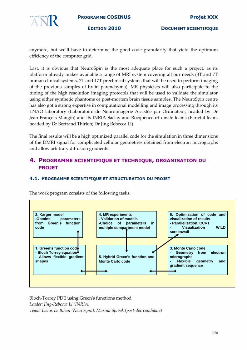
The work program consists of the following tasks.
Bloch-Torrey PDE using Green's functions method
Leader: Jing-Rebecca Li (INRIA)
Team: Denis Le Bihan (Neurospin), Marina Spivak (post-doc candidate)
1. Green’s function code - Bloch Torrey equation - Allows flexible gradient shapes
3. Monte Carlo code - Geometry from electron micrographs - Flexible geometry and gradient sequence
2. Karger model -Obtains parameters from Green’s function code
4. MR experiments - Validation of models -Choice of parameters in
multiple compartment model
5. Hybrid Green’s function and
Monte Carlo code
6. Optimization of code and visualization of results - Parallelization, CCRT - Visualization WILD screenwall
PROGRAMME COSINUS
EDITION 2010
Projet XXX
DOCUMENT SCIENTIFIQUE
10/28
The numerical solution of the Bloch-Torrey partial differential equation using a Green's
functions method. This task will be conducted by Jing-Rebecca Li (INRIA) and a post-
doctoral researcher to be funded by the project. A candidate for this post-doctorial position
has been found. She is Marina Spivak, who expects to obtain a Ph.D. in Computer Science
from New York University in June 2010. Denis Le Bihan will work on the input of
biologically reasonable parameters into the simulation. The code for simulation in 3
dimensions will be written in C++. We will pay particular attention to ensure its
compatibility with existing simulation code at Neurospin.
Solution from the Green's functions method provides parameters for the Karger model
Leader: Jing-Rebecca Li (INRIA)
Team: Denis Le Bihan (Neurospin) and Irina Kezele (CNRS)
The Green's functions method will provide an accurate solution to multiple compartment
diffusion. In particular, it will provide computed average residence times for the different
compartments, given cellular geometry and membrane permeability. The compartment
residence times are the input parameters to a popular 'approximate analytical model' called
the Karger model for diffusion in composite systems. The Karger model is used by MRI
scientists because it captures certain important properties of diffusion MRI signals and the
model is simple to use in that it is 'a mixture of Gaussians'. By comparing the Karger model
using these computed parameters with the simulation from the Green's function method, we
will know for which regimes the Karger model is or is not valid.
Monte Carlo simulation in realistic cellular environment
Leader: Cyril Poupon (Neurospin)
Team: Chun-Hung Yeh (Neurospin), PhD student to be hired (Neurospin)
The Monte Carlo simulation of the water diffusion process in a heterogeneous cellular
environment will ensure speed, flexibility, portability, and scalability. We combine a Monte
Carlo Brownian dynamics simulator capable of simulating diffusion of spins in arbitrarily
complex geometries with a DW signal integrator emulating various MR pulse sequences.
The code for simulation in 3 dimensions will be written in C++. We will pay particular
attention to ensure its compatibility with existing simulation code at NeuroSpin and to be
fully compliant with the requirements of the large scale computation facility that will be
used to accelerate the simulation.
It also includes the automatic segmentation of cellular geometry from electron micrographs.
Validation of model by MR experiments
Leader: Denis Le Bihan (Neurospin)
Team: Jing-Rebecca Li (INRIA)
Denis Le Bihan is in charge of the modeling of diffusion at the cellular level, including the
validation of model parameters, such as intra-cellular, extra-cellular, and membrane
diffusion coefficients, membrane permeability, cell swelling. He will be assisted by Jing-
Rebecca Li. This task includes conducting DMRI experiments on constructed phantoms on
the 7 Tesla small animal available system and the future 17T small animal system at
PROGRAMME COSINUS
EDITION 2010
Projet XXX
DOCUMENT SCIENTIFIQUE
11/28
NeuroSpin. The results of the simulation for both the Green's function code and the Monte-
Carlo code will be checked against the results of the DMRI experiments.
Hybrid Green's function and Monte Carlo simulation code
Leader: Cyril Poupon (Neurospin)
Team: Jing-Rebecca Li (INRIA), Chun-Hung Yeh (Neurospin), Antoine Lejay (INRIA), and the
future PhD student to be hired (Neurospin).
We formulate a hybrid method which switches between the Green's function code and the
Monte Carlo code while ensuring accuracy and stability of the overall method. In smooth
regions where the magnetization of the water molecules satisfies the Bloch-Torrey equation,
the Green's function code will be used; the results are then exported to as inputs to the
Monte-Carlo simulator to treat the regime with high heterogeneity where the Green's
function method would be too slow.
Code optimization and parallelization and visualization of simulation
Leader: Cyril Poupon (Neurospin)
Team: Chun-Hung Yeh (Neurospin), and the future PhD student to be hired (Neurospin).
This task include the implementation and fine tuning of the hybrid code to run on the
parallel and distributed architecture of the CCRT (Centre de Calcul Recherche et
Technologie, CEA) and the visualization of the results of the large scale simulation: to be able
to see the evolving diffusion of spins on water molecules in the cellular environment on a
voxel scale.
4.2. MANAGEMENT DU PROJET
There are two partners in this project, one is INRIA and the other is Neurospin, CEA.
Participants from INRIA consist of Jing-Rebecca Li (INRIA-Rocquencourt, chercheur
permanent) and Antoine Lejay (INRIA-Nancy, chercheur permanent) and a post-doctorial
candidate Marina Spivak, who will arrive in France in September 2010. Jing-Rebecca Li and
Marina Spivak will have an office at Neurospin and will be present at least 3-4 days per week
at Neurospin.
The participants from Neurospin consist of Cyril Poupon (chercheur permanent), Denis
LeBihan (permanent, directeur de Neurospin) and Chun-Hung Yeh (PhD student).
Other collaborators of this project include Irina Kezele (post-doc CNRS, based at Neurospin)
and the IN-SITU team, Laboratoire de Recherche en Informatique (LRI), Université Paris Sud
XI, co-headed by Dr Michel Beaudouin-Lafon.
All participants except Antoine Lejay (INRIA-Nancy) are in the Paris region. And all except
Antoine Lejay and Michel Beaudouin-Lafon will have a regular presence at Neurospin.
Jing-Rebecca Li is the coordinator of the project. She is also charge of the Green's function
code. She and Marina Spivak, the post-doctoral candidate, will complete and optimize this
PROGRAMME COSINUS
EDITION 2010
Projet XXX
DOCUMENT SCIENTIFIQUE
12/28
code. Denis LeBihan and Jing-Rebecca Li will validate model parameters, such as intra-
cellular, extra-cellular, and membrane diffusion coefficients, membrane permeability, cell
swelling by conducting DMRI experiments on the 17 Tesla MRI scanner available at
Neurospin. Jing-Rebecca Li and Irina Kezele will use this code to obtain residence times to
enter as parameters in the Karger model. A weekly meeting will occur between Jing-Rebecca
Li, Marina Spivak, Denis LeBihan (and during the Karger model phase Irina Kezele).
Cyril Poupon is the coordinator of the Monte-Carlo code. He and Chun-Hung Yeh will
complete, optimize, and parallelize it for running on the CCRT. They will also be in charge
of the visualization of the simulation results. A weekly meeting will occur between Cyril
Poupon and Chun-Hung Yeh with the presence of Jing-Rebecca Li after the Green’s function
code has been completed.
After the completion of the two codes, Jing-Rebecca Li, Cyril Poupon and Antoine Lejay will
implement a hybrid method combining the two codes. This will involve monthly meetings
between the three people either at Neurospin or in Nancy. When we enter the visualization
phase of the project, a meeting will be held with Michel Beaudouin-Lafon.
4.3. DESCRIPTION DES TRAVAUX PAR TACHE
4.3.1 TÂCHE 1
Bloch-Torrey PDE using Green's functions method
Leader: Jing-Rebecca Li (INRIA)
Team: Denis Le Bihan (Neurospin), Marina Spivak (post-doc candidate)
This task consists of producing a code that outputs the solution of the Bloch-Torrey partial
differential equation using a Green's functions method. This task will be conducted by Jing-
Rebecca Li (INRIA) and a post-doctoral researcher to be funded by the project. A candidate
for this post-doctorial position has been found. She is Marina Spivak, who expects to obtain
a Ph.D. in Computer Science from New York University in June 2010. Denis LeBihan will
work on the input of biologically reasonable parameters into the simulation. The code for
simulation in 3 dimensions will be written in C++. We will pay particular attention to ensure
its compatibility with existing simulation code at Neurospin.
We solve the Bloch-Torrey equation with a Green’s function method that is more efficient
and accurate than finite different or finite elements methods. This method has been
explained for the diffusion equation [8, 9, 7]. Its use in the Bloch-Torrey equation appears
preliminarily in [10]. We use more reasonable and realistic boundary conditions between the
diffusing compartments than either the groups working on analytical expressions or the
Karger model.
PROGRAMME COSINUS
EDITION 2010
Projet XXX
DOCUMENT SCIENTIFIQUE
13/28
Within the voxel, the diffusion environment is in fact heterogeneous. In a simple model, we
will suppose that the diffusion coefficient is constant within the intra-cellular, the extra-
cellular, and the membrane compartments. The volume V is the union of a very large
number of these three kinds of compartments. The ensemble of all intra-cellular regions will
be denoted by Vin, all the extra-cellular regions will be denoted by Vex, and the membranes
by Vmem. The volume will be the union of these three regions, with diffusion coefficients Din ,
Dex, and Dmem , respectively. In each Vj, the following equation is satisfied,
,
and it is complemented by the following boundaries conditions, on Bjk, the boundary
separating Vj and Vk:
This is solved using a Green’s functions method with the Green’s function being
The solution mj(x,t,g,u) will be written as a sum of an initial potential, a single layer
potential, and a double layer potential. The layer potentials are convolutions of the above
Green’s function with unknown densities on the interfaces of the cells. The unknown
densities are solved by matching the continuity boundary conditions above.
The advantages of this approach are that large time steps can be taken, up to 10 to 100 times
larger than in Monte-Carlo simulation, and the high accuracy of the results. The
implementation we use takes advantage of the Fourier transform of the Green’s function,
given above, and avoids the algorithmic complexity of the typical Green’s function method.
Instead of O(M2) work, where M is the number of time steps taken, we can return an accurate
answer in O(M) work. More details of the method can be found in [9].
The challenges of our approach are that integrals with singular integrands must be
computed (numerically) accurately and the geometry must be simple enough to allow
efficient and accurate computation of these singular or nearly singular integrals.
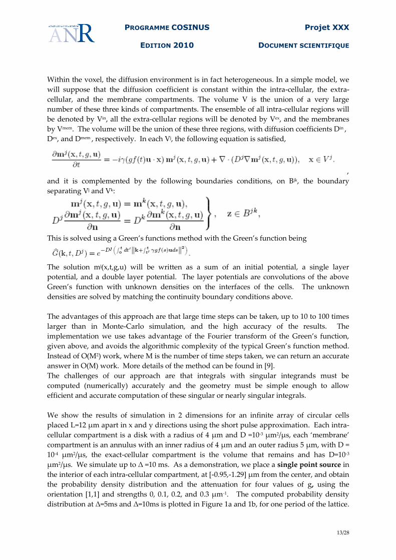
We show the results of simulation in 2 dimensions for an infinite array of circular cells
placed L=12 μm apart in x and y directions using the short pulse approximation. Each intra-
cellular compartment is a disk with a radius of 4 μm and D =10-3 μm2/μs, each ‘membrane’
compartment is an annulus with an inner radius of 4 μm and an outer radius 5 μm, with D =
10-4 μm2/μs, the exact-cellular compartment is the volume that remains and has D=10-3
μm2/μs. We simulate up to Δ =10 ms. As a demonstration, we place a single point source in
the interior of each intra-cellular compartment, at [-0.95,-1.29+ μm from the center, and obtain
the probability density distribution and the attenuation for four values of g, using the
orientation *1,1+ and strengths 0, 0.1, 0.2, and 0.3 μm-1. The computed probability density
distribution at Δ=5ms and Δ=10ms is plotted in Figure 1a and 1b, for one period of the lattice.
PROGRAMME COSINUS
EDITION 2010
Projet XXX
DOCUMENT SCIENTIFIQUE
14/28
We see that there is high probability of finding the particle in the interior compartment and
the membrane compartment by the end of simulation time, but very little probability that it
has entered the exterior compartment by that time. The attenuation is plotted in Figure 1c
and 1d at Δ= 0.5ms, and10ms. The bottom line is a plot of e-ig*gDΔ, D =10-3 μm2/μs, the free
diffusion attenuation with the larger D, the top line the free diffusion attenuation due to D =
10-4 μm2/μs. The black line (diamond) is the volume weighted sum of the two free diffusion
exponentials (no exchange case). The black line (circle) is free diffusion with a single
weighted-averaged D = 8.23x10⁻⁴ μm2/μs. The red line (x) is the computed attenuation,
exhibiting a behaviour that changes from fast free diffusion at the short times (the particle
not having seen the membrane) and exhibits slow diffusion for long times as the particle
seems trapped by the slow diffusion compartment near the membrane. The time step we
used in the present simulation was 100 μs, hence only 100 time steps were needed to
compute the results discussed here.
Figure 1a. t=5ms Figure 1b. t=10ms Figure 1c. t=0.5ms Figure 1d. t=10ms
4.3.2 TÂCHE 2
Solution from the Green's functions method provides parameters for the Karger model
Leader: Jing-Rebecca Li (INRIA)
Team: Denis Le Bihan (Neurospin) and Irina Kezele (CNRS)
The Green's functions method will provide an accurate solution to multiple compartment
diffusion. In particular, it will provide computed average residence times for the different
compartments, given cellular geometry and membrane permeability. The compartment
residence times are the input parameters to a popular 'approximate analytical model' called
the Karger model for diffusion in composite systems. The Karger model is used by MRI
scientists because it captures certain important properties of diffusion MRI signals and the
model is simple to use in that it is 'a mixture of Gaussians'. By comparing the Karger model
using these computed parameters with the simulation from the Green's function method, we
will know for which regimes the Karger model is or is not valid.
This task simply involves running the Green’s function code for diffusion (Bloch Torrey is
not needed, the simple diffusion equation can be used) and compute the average residence
times in each compartment and put these times into the Karger model. By comparing the
sum of mixed Gaussians given by the Karger model thus obtained with the actual simulation
results of the Bloch Torrey equation (which we trust is accurate using our code), we can
definitely judge the validity of the Karger model in different regimes (how long or short
diffusion times must be compared to average compartment sizes, among other possibilities).
PROGRAMME COSINUS
EDITION 2010
Projet XXX
DOCUMENT SCIENTIFIQUE
15/28
The end results may be some rules of thumb on when the Karger model should be used
when modeling multiple compartment diffusion.
4.3.3 TÂCHE 3
Monte Carlo simulation in realistic cellular environment
Leader: Cyril Poupon (Neurospin)
Team: Chun-Hung Yeh (Neurospin), PhD student to be hired (Neurospin)
Monte Carlo (MC) simulations have been shown to be a powerful and flexible tool to explore
a wider class of systems and address several questions in DW MRI (Balls 2009, Farrell 2009,
Fieremans 2009a, Fieremans 2009b, Fonteijn 2009, Ford 1997, Grinberg 2009, Hall 2009,
Harkins 2009, Landman 2009, Liu 2004, Nilsson 2009). Particularly, MC framework allows
tracking the dynamic events over space and time. Therefore, diffusion MR synthetic data
based on MC approach provides the ability to examine the influence of specific biological
properties on the diffusion process. Since the shape and the organization of biological
structures are extremely complicated at the cellular level (e.g. the gray matter (GM)), the
issue turns into how to represent the geometry of neural tissue in the MC simulation
framework. Several studies have been presented to simulate water diffusion in various
geometric models. Hall and Alexander optimize the parameters for the diffusion MC
simulations in packed and swelling cylindrical tissue model (Hall 2009, Cook 2006).
Meanwhile, Landman et al. and Farrell et al. simulate diffusion in mesh-based geometries to
study the effect of axonal injury and beading (Landman 2009, Farrell 2009). The MC
simulations described above mainly focused on the geometry of neuronal fibre bundles (i.e.
WM), while it is interesting to investigate the behavior of water diffusion in cerebral cortex
and deep nuclei areas (i.e. GM) as well. Lipinski carried out the first MC simulation in a
simplified 2D environment with tissue geometries created using digital images captured
from the histological preparations of GM (Lipinski 1990). Extension from a 2D MC
simulation to a 3D space is closer to the realistic situation, whereas it is also a great
computational challenge. Balls developed the famous MCell physiological simulation
framework and mainly focused on large-scale MC simulations of high angular resolution
DW imaging (HARDI) experiments (Balls 2004, Balls 2009). The large-scale MC method
allows simulations to efficiently process a large amount of random walkers at high temporal
resolution, and hence has great potential in mimicking water diffusion in a complicated
neural medium.
In this task we present a novel large-scale MC simulation tool dedicated to DW MR
experiments. We combine a Monte Carlo Brownian dynamics simulator capable of
simulating diffusing spins in arbitrarily complex geometries with a DW signal integrator
feasible to simulate various MR pulse sequences. Furthermore, we develop methods to form
various tissue configurations using dynamic meshes in our software, which is applicable to
create complicated geometries analogous to neural structures, such as neurons, axons,
myelin, astrocytes, oligodendrocytes, macromolecules, etc.., with different tissue features
PROGRAMME COSINUS
EDITION 2010
Projet XXX
DOCUMENT SCIENTIFIQUE
16/28
(e.g. cell size, density, membrane permeability). In addition, we have developed different
types of spin-barrier interaction models (presence of attractors, local viscosity, membrane
interactions, etc).
This software allows investigating the mechanism driving the DW signal and the features
observed in DW MRI. The simulation code is fully 3D and is implemented in C++. Main
features are described as following: 'Cell membranes' are generated using meshes to model
different cell types with heterogeneous shapes and sizes. This module allows users to specify
cell properties including the thickness, permeability, and diffusion coefficient (D). In
addition, we incorporates the mesh with dynamic morphological evolution, thus it can be
utilized to simulate sequential changes of tissue shapes including expansion, shrinkage, and
deformation. 'Diffusing particle' ('spin') is modeled as a random walker and the average free
displacement for an elementary time step of length is scaled to its associated compartmental
D. For each simulation step, the spatial positions of particles are updated subject to a series of
potential interactions: (i) In accordance with the permeability, the particle may penetrate
through the related cell membrane or be elastically reflected. (ii) The diffusivity of the
particle may be modified into that of the interacting membrane layers. 'The MR pulse
sequence' module is flexible to model a variety of pulse sequences with different
combinations of radio frequency and gradient pulses. 'DW signal' of each synthetic MR voxel
is computed numerically by performing integration on the accumulated phase of each
particle belonging to the voxel. Users can add imaging (Rician) noise to the synthetic MR
signal.
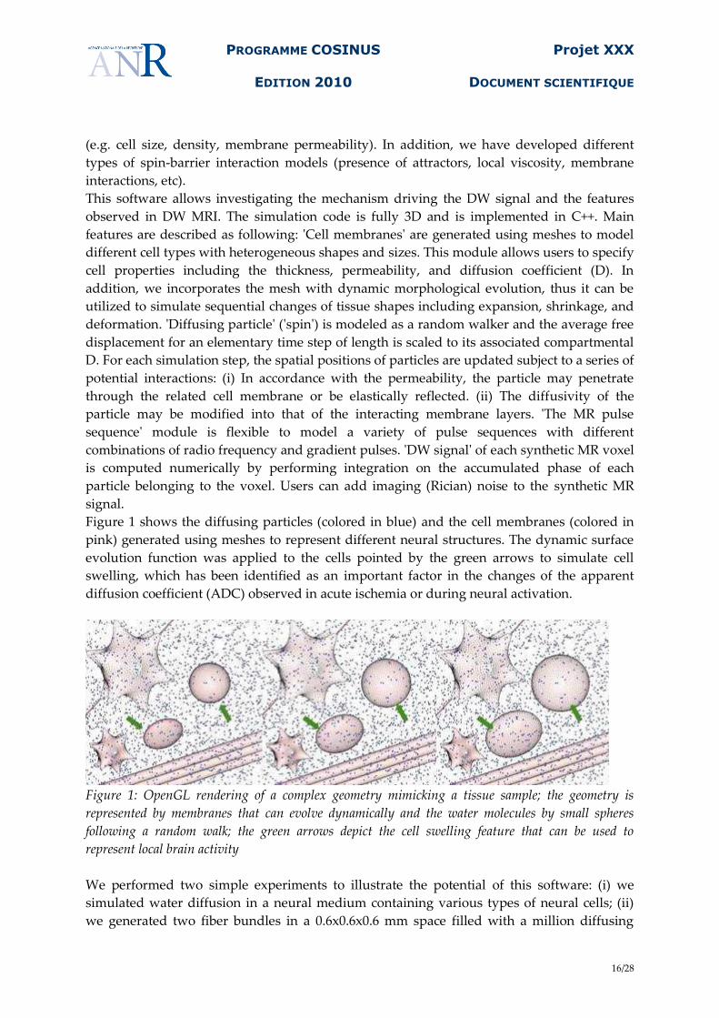
Figure 1 shows the diffusing particles (colored in blue) and the cell membranes (colored in
pink) generated using meshes to represent different neural structures. The dynamic surface
evolution function was applied to the cells pointed by the green arrows to simulate cell
swelling, which has been identified as an important factor in the changes of the apparent
diffusion coefficient (ADC) observed in acute ischemia or during neural activation.
Figure 1: OpenGL rendering of a complex geometry mimicking a tissue sample; the geometry is
represented by membranes that can evolve dynamically and the water molecules by small spheres
following a random walk; the green arrows depict the cell swelling feature that can be used to
represent local brain activity
We performed two simple experiments to illustrate the potential of this software: (i) we
simulated water diffusion in a neural medium containing various types of neural cells; (ii)
we generated two fiber bundles in a 0.6x0.6x0.6 mm space filled with a million diffusing
PROGRAMME COSINUS
EDITION 2010
Projet XXX
DOCUMENT SCIENTIFIQUE
17/28
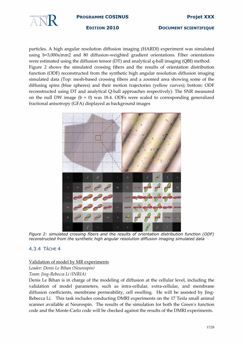
particles. A high angular resolution diffusion imaging (HARDI) experiment was simulated
using b=3,000s/mm2 and 80 diffusion-weighted gradient orientations. Fiber orientations
were estimated using the diffusion tensor (DT) and analytical q-ball imaging (QBI) method.
Figure 2 shows the simulated crossing fibers and the results of orientation distribution
function (ODF) reconstructed from the synthetic high angular resolution diffusion imaging
simulated data (Top: mesh-based crossing fibers and a zoomed area showing some of the
diffusing spins (blue spheres) and their motion trajectories (yellow curves); bottom: ODF
reconstructed using DT and analytical Q-ball approaches respectively). The SNR measured
on the null DW image (b = 0) was 18.4. ODFs were scaled to corresponding generalized
fractional anisotropy (GFA) displayed as background images
Figure 2: simulated crossing fibers and the results of orientation distribution function (ODF)
reconstructed from the synthetic high angular resolution diffusion imaging simulated data
4.3.4 TÂCHE 4
Validation of model by MR experiments
Leader: Denis Le Bihan (Neurospin)
Team: Jing-Rebecca Li (INRIA)
Denis Le Bihan is in charge of the modeling of diffusion at the cellular level, including the
validation of model parameters, such as intra-cellular, extra-cellular, and membrane
diffusion coefficients, membrane permeability, cell swelling. He will be assisted by Jing-
Rebecca Li. This task includes conducting DMRI experiments on the 17 Tesla small animal
scanner available at Neurospin. The results of the simulation for both the Green's function
code and the Monte-Carlo code will be checked against the results of the DMRI experiments.
PROGRAMME COSINUS
EDITION 2010
Projet XXX
DOCUMENT SCIENTIFIQUE
18/28
4.3.5 TÂCHE 5
Hybrid Green's function and Monte Carlo simulation code
Leader: Cyril Poupon (Neurospin)
Team: Jing-Rebecca Li (INRIA), Chun-Hung Yeh (Neurospin), Antoine Lejay (INRIA), and the
future PhD student to be hired (Neurospin).
We formulate a hybrid method which switches between the Green's function code and the
Monte Carlo code while ensuring accuracy and stability of the overall method. In smooth
regions where the magnetization of the water molecules satisfies the Bloch-Torrey equation,
the Green's function code will be used; the results are then exported to as inputs to the
Monte-Carlo simulator to treat the regime with high heterogeneity where the Green's
function method would be too slow.
In terms of software development, the hybrid method will rely on the design and
architecture of the existing Monte-Carlo code and will be modified accordingly. A specific
API will be developed in order to manage a pool of molecules. This API will allow dealing
with this pool according to the Green’s function code or according to the Monte Carlo code.
It will facilitate the distribution and parallelism of the code that will occur during the next
optimization task.
4.3.6 TÂCHE 6
Code optimization and parallelization and visualization of simulation
Leader: Cyril Poupon (Neurospin)
Team: Chun-Hung Yeh (Neurospin), and the future PhD student to be hired (Neurospin).
This task include the implementation and fine tuning of the hybrid code to run on parallel
and distributed architecture of the CCRT (Centre de Calcul Recherche et Technologie, CEA)
and the visualization of the results of the large scale simulation: to be able to see the evolving
diffusion of spins on water molecules in the cellular environment on a voxel scale.
Three actions have to be conducted during this task:
- optimization of the code on a single processor basis
- multiple threading of the code
- distribution of the code on a grid of processors of the CCRT.
This work must be done for the two simulators, the Monte Carlo simulator and the Hybrid
simulator. A preliminary work was performed on the existing Monte Carlo prototype. The
code was designed in such a way that it is already optimum: quad-trees have been
implement to accelerate the localization of the closest membrane for a given particle, access
to the triangles interacting with a specific particle was also accelerated using look-up tables,
and profiling of the code was performed in order to systematically optimize each C++
function starting with the most time consuming methods to the less time consuming
methods. For a simple scene containing a set of 1000000 particles interacting with a single
PROGRAMME COSINUS
EDITION 2010
Projet XXX
DOCUMENT SCIENTIFIQUE
19/28
sphere located at the centre of the field of view, the code was speeded-up by a factor of
around 100.
Two levels of distribution have to be considered:
- Inside a single node of the grid, POSIX based multiple threading was used to split the
simulation over the 16 processors, and another gain of 15 was observed, as almost no
communication occurs between moving particles during the full simulation. This gain
was reached because the distribution of the Monte Carlo simulation is
straightforward, which might not be necessarily the case when the Hybrid simulator
will be implemented. However, we believe that the concept of pool of particles may
efficiently drive the implementation of the Hybrid simulator to a strongly scalable
code, thus taking full advantage of the use of the CCRT facility. The choice of POSIX
threading may be modified in the future to possibly use the MPI library if the
resulting implementation results in a faster code.
- The distribution of the simulation will be performed using the MPI library. This
choice was guided by the recommendations of the CCRT facility that mainly offers
the MPI infrastructure to users. The MPI API is adequate to create a server that will
distribute the motion of the particles in the case of pure Monte Carlo simulation or
the motion of the pools of particles in the case of Hybrid simulation on clients, each
client corresponding to a single thread on a single processor.
It is likely that the Hybrid code will speed-up the simulation since each pool represent
putatively a large set of particles, but the design will be challenging because the
continuity between pools may be preserved using boundary conditions.
In order to prepare a first prototype of this MPI implementation of both the Monte Carlo and
Hybrid simulators, we will use a small cluster of 3 high performance Dell R900 nodes. Each
node is equipped with 16 processors and 64 GB of memory, and the 3 nodes will be
connected through a 1Gbit network. This small architecture is mandatory, as making test
directly at the CCRT facility won’t be possible without a specific booking procedure. The
CEA CCRT facility generally ask for developing the code on a third-part cluster before
allocating large bay time on its computer grids or clusters.
Last, large scale simulations using the complex geometry stemming from electron
microscopy will be launched at the CCRT facility. We will use the new Titane architecture.
The Titane supercomputer is a hybrid cluster. 1068 processing nodes are provided, each
equipped with 2 Intel Nehalem quad-core processors at 2.93GHz and 24GB of memory. 24
further nodes are dedicated to administration and I/O tasks. This cluster also provides 48
graphical NVidia Tesla S1070 servers, each equiped with 4 processors and 4 GB of memory.
Each server is attached to two processing nodes through a PCI-express connection.
Processing nodes are connected with a Voltaire network based n the InfiniBand technology.
The storage infrastructure offers a 500TB disk space. The operating system is based on Linux
(NovaScale Master) and on the Bull HPC software platform, providing a C++ compiler, and
the OpenMPI library.
PROGRAMME COSINUS
EDITION 2010
Projet XXX
DOCUMENT SCIENTIFIQUE
20/28
The visualization of the huge amount of particles will be addressed using the WILD
screenwall (IN-SITU team, Laboratoire de Recherche en Informatique (LRI), Université Paris
Sud XI, co-headed by Dr Michel Beaudouin-Lafon). This task will be conducted within a
scientific collaboration already started between the Laboratory of Computer Assisted
Neuroimaging of NeuroSpin and the LRI, under the joint management of Dr Jean-François
Mangin and Dr Michel Beaudouin-Lafon. The WILD project proposes a unique wall made up
of 32 screens equipped with a specific OpenGL software platform and with specific tools to
interactively manipulate, visualize and analyze the dynamic geometrical scene.
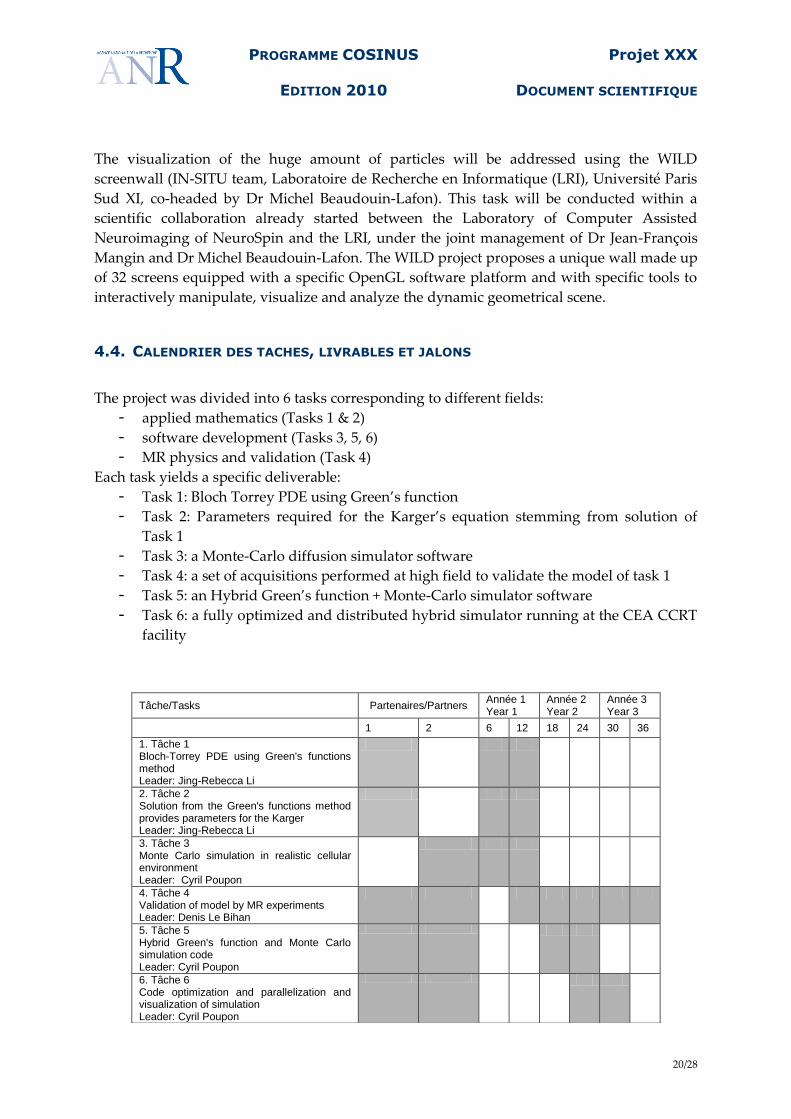
4.4. CALENDRIER DES TACHES, LIVRABLES ET JALONS
The project was divided into 6 tasks corresponding to different fields:
- applied mathematics (Tasks 1 & 2)
- software development (Tasks 3, 5, 6)
- MR physics and validation (Task 4)
Each task yields a specific deliverable:
- Task 1: Bloch Torrey PDE using Green’s function
- Task 2: Parameters required for the Karger’s equation stemming from solution of
Task 1
- Task 3: a Monte-Carlo diffusion simulator software
- Task 4: a set of acquisitions performed at high field to validate the model of task 1
- Task 5: an Hybrid Green’s function + Monte-Carlo simulator software
- Task 6: a fully optimized and distributed hybrid simulator running at the CEA CCRT
facility
Tâche/Tasks Partenaires/Partners Année 1 Year 1
Année 2 Year 2
Année 3 Year 3
1 2 6 12 18 24 30 36
1. Tâche 1 Bloch-Torrey PDE using Green's functions method Leader: Jing-Rebecca Li
2. Tâche 2 Solution from the Green's functions method provides parameters for the Karger Leader: Jing-Rebecca Li
3. Tâche 3 Monte Carlo simulation in realistic cellular environment Leader: Cyril Poupon
4. Tâche 4 Validation of model by MR experiments Leader: Denis Le Bihan
5. Tâche 5 Hybrid Green's function and Monte Carlo simulation code Leader: Cyril Poupon
6. Tâche 6 Code optimization and parallelization and visualization of simulation Leader: Cyril Poupon
PROGRAMME COSINUS
EDITION 2010
Projet XXX
DOCUMENT SCIENTIFIQUE
21/28
5. STRATEGIE DE VALORISATION DES RESULTATS ET MODE DE
PROTECTION ET D’EXPLOITATION DES RESULTATS
The results will be published in scientific journals (NeuroImage, Medical Image Analysis,
Magnetic Resonance in Medicine, Journal of Magnetic Resonance Imaging) and conferences
(ISMRM, MICCAI).
A more insightful understanding of the biological mechanism which triggers the change in
measured diffusion in DMRI experiments can potentially lead to a new and more nuanced
interpretation of DMRI data, demonstrating links with underlying physiological phenomena
as cell swelling, changes in permeability, etc. This can have important clinical applications,
leading to more reliable ways of diagnosing diseases. Such simulation can also be used in
the design of special pulse sequences to target the probing of cellular structure at scales
much smaller than the conventionally used pulsed gradients. A numerical code which can
aid sequence design can be patented and potentially commercialized.
In French:
"Les conditions de dévolution des droits de la propriété intellectuelle et d'exploitation des
résultats seront indiquées dans l'accord de consortium qui sera établi après le démarrage du
projet, sur la base des principes suivants :
1.1.1 Propriété intellectuelle et exploitation des résultats
Propriété intellectuelle antérieure
Chaque partenaire conserve la propriété (information, données techniques brevetées ou
non...) de la connaissance produite avant le projet ou indépendamment des travaux du projet
(savoir-faire ou connaissance pré existant) et des améliorations de ces connaissances. Le
savoir-faire ou les connaissances antérieures utiles pour le projet seront listées dans l'annexe
de l'accord de consortium.
Propriété des résultats ex-nihilo
Les résultats produits et réalisés par un seul partenaire dans le cadre du projet seront la
propriété de ce partenaire (résultats propres).
Les résultats produits et réalisés conjointement par plusieurs partenaires dans le cadre du
projet (résultats communs) seront la copropriété desdits partenaires. Les parts de copropriété
(ou parts indivises) seront déterminées au prorata des apports intellectuels des inventeurs
(ou auteurs) des partenaires. En revanche, en cas de valorisation commercial d'un actif, les
partenaires ayant fait un apport financier ou matériel substantiel pour la création de l'actif
auront droit à un juste retour.
Gestion et exploitation
Rapports d’avancement semestriel Progress report/expenses
Accord de consortium / rapport final Consortium agreement/final report
PROGRAMME COSINUS
EDITION 2010
Projet XXX
DOCUMENT SCIENTIFIQUE
22/28
Les résultats communs feront l'objet d'un accord indiquant le mécanisme de distribution de
la propriété et les droits d'exploitation de ces résultats communs, toute exploitation par un
des copropriétaires devant donner lieu à la rémunération des autres copropriétaires.
Un mandataire unique pour la valorisation d'un actif ou d'un groupe d'actifs pourra être
désigné.
1.1.2 Confidentialité
Des engagements de confidentialité seront également écrits dans l'accord de consortium. Dès
que cet accord sera signé, chaque partenaire ayant accès à des informations, données ou
plans confidentiels, en particulier les modèles scientifiques de n'importe quelle nature,
appartenant aux autres partenaires desquels il pourrait avoir été informé au moment du
montage ou de l'exécution du projet, ne pourra pas les divulguer.
1.1.3 Publications
Tout résultat du projet soumis à publication dans une conférence, un journal ou une revue,
devra comporter la mention du financement de l'ANR.
Les partenaires seront libres de publier sur leurs résultats propres mais devront obtenir
l'accord préalable des partenaires intéressés en cas de résultats communs.
"
6. ORGANISATION DU PARTENARIAT
6.1. DESCRIPTION, ADEQUATION ET COMPLEMENTARITE DES PARTENAIRES
This project will be driven by the two scientific partners:
INRIA (Partner 1), National institute of computer science and applied mathematics with
multiple sites in France.
NeuroSpin (Partner 2), a ultra high-field MRI facility dedicated to neurosciences within the
Institute of BioMedical Imaging (I2BM) of the Life Science Division (DSV) of the
Commissariat à l’Energie Atomique (CEA), Saclay, France
The two partners have long been collaborating mainly about image processing and
neuroimaging for approximately two decades. They share the same scientific projects, i.e.
delivering an anatomo-functional mapping of the human brain. The strong relationship
between the two institutions was recognized by the creation of a first INRIA Saclay team,
Parietal, directly located inside the NeuroSpin building, and in the future of a second INRIA
team more focused on the topic of this project, ie at the interface between applied
mathematics and physics, under the direction of Dr Jing-Rebecca Li.
The two partners started to collaborate on the simulation of the diffusion process of water
molecules in the brain parenchyma early in 2009. In the past year, there were two common
publications involving the members of the Neurospin team and the INRIA team [15, 10].
The two teams have complementary skills: applied mathematics and computational biology
for the INRIA team, MR physics, neurobiology, image processing using large scale
computing facilities for the NeuroSpin team. The two teams have developed substantial
PROGRAMME COSINUS
EDITION 2010
Projet XXX
DOCUMENT SCIENTIFIQUE
23/28
skills that have been regularly acknowledged by publications in the major journals
(NeuroImage, IEEE Transaction in Medical Imaging, Magnetic Resonance in Medicine, …) of
the field and by communications in the recognized conferences of the domain (ISMRM, ISBI,
IPMI, MICCAI).
INRIA team:
INRIA has several sites across France grouping computer scientists and applied
mathematicians. The researchers work in research teams (equipe-projet) around a unifying
topic. The two participating teams of INRIA are
Action exploratoire DMRI: Jing-Rebecca Li, who is currently a member of the team
POEMS (modelization of waves), is starting an entity called an ‘action exploratoire’
at the Saclay site of INRIA. This is a step before the creation of a new research team
on this topic. She is a specialist in the simulation of diffusion phenomena in physical
applications.
Equipe-project TOSCA: Antoine Lejay. The research theme of TOSCA is the
simulation and calibration of stochastic models. Antoine Lejay is a specialist in the
simulation of stochastic models and the analysis of Monte Carlo methods in
heterogeneous media.
NeuroSpin team:
NeuroSpin is a ultra-high field MRI facility which opened in 2007. Its is dedicated to
molecular imaging and translational research in neurosciences to man with a strong
emphasis on clinical and cognitive neurosciences. The aim of NeuroSpin is to push as far as
possible the current limits of Magnetic Resonance Imaging to study the central nervous
system, from mouse to man. NeuroSpin is equipped with 2 human MRI systems operating at
3T and 7T, a small animal horizontal MRI system operating at 17.7T (first in the world) and
will house the first whole-body 11.7T MRI system in 2013. NeuroSpin staff includes basic
scientists in many disciplines from physics to cognitive neurobiololgy and clinicians. It is
organized into 5 laboratories:
a Nuclear Magnetic Resonance lab (LRMN)
a Computer Assisted Neuroimaging lab (LNAO)
a Cognitive Neurosciences lab (LCOGN)
a Clinical (Human) Investigation Center (LBIOM)
a Preclinical (Animals) Integrated Biology lab (LBIOP)
The members of the NeuroSpin centre (partner 2) involved in this project are:
Dr. Cyril Poupon, permanent researcher
Dr. Denis Le Bihan, director of Neurospin
Chun-Hung Yeh, PhD student
6.2. QUALIFICATION DU COORDINATEUR DU PROJET
Jing-Rebecca Li obtained her PhD in applied mathematics from MIT in the United States in
2000, was a post-doc at the Courant Institute, NYU from 2000-2003 and was hired at INRIA
as a permanent researcher in 2003. She is preparing her 'habilitation a diriger des
recherches' to be defended in 2010.
PROGRAMME COSINUS
EDITION 2010
Projet XXX
DOCUMENT SCIENTIFIQUE
24/28
Jing-Rebecca Li holds a permanent research position at INRIA (chargee de recherche (CR1))
and is in the process of transfering to INRIA-Saclay and will start a new research axis (action
exploratoire) on DMRI simulation. She has directed internships (stage de DEA) while at
INRIA and was on the organizing committee of the conference WAVES 2007. She is an
associate editor of SIAM Journal on Scientific Computing and was an editor of a special issue
of Journal of Computational and Applied Mathematics for Waves 2007.
7. JUSTIFICATION SCIENTIFIQUE DES MOYENS DEMANDES
7.1. PARTENAIRE 1 : INRIA
• Équipement
n/a
• Personnel
We request the funding of 24 months of a post-doc who will participate in the completion
and optimization of the Green’s function code and the hybrid Green’s function Monte Carlo
code. This position requires a Ph.D. in computer science or applied mathematics or related
fields, experience working in the formulation and implementation of numerical algorithms
for PDEs and knowledge of basic biology. We have a candidate who is interesting in
obtaining this position and a meeting has taken place between her and the main participants
of this project. The candidate is Marina Spivak, who expects to obtain a Ph.D. in Computer
Science from New York University in June 2010.
• Prestation de service externe
We request 2 individual Matlab licenses and 2 Mathematica licences, a high performance
desk top computer for the post-doc and a high performance laptop for Jing-Rebecca Li.
Total: 10 000 euros.
We request 150 hours of scanner time on the 7 Tesla small animal system and the future 17T
small animal system at NeuroSpin. Total: 80 000 euros.
• Missions
We request funding for the travel of Antoine Lejay from Nancy to the Paris regions and the
travel of Jing-Rebecca Li from Paris to Nancy. There will be 3 trips for each person. We also
request travel and conference fees for the post-doc candidate and Irena Kezele to MICCAI
(Medical Image Computing and Computer Assisted Intervention) and ISMRM (conference of
the International Society for Magnetic Resonance in Medicine) and Jing-Rebecca Li to
ISMRM. Total: 10000 euros.
PROGRAMME COSINUS
EDITION 2010
Projet XXX
DOCUMENT SCIENTIFIQUE
25/28
• Dépenses justifiées sur une procédure de facturation interne
n/a
• Autres dépenses de fonctionnement
n/a
7.2. PARTENAIRE 2 : CEA I²BM NEUROSPIN
• Équipement
The following equipment is required to achieve this project:
2 Server Dell R900 (16 processors, 64GB of memory) = 13.2k€
2 NetApp controlers to manage large disk storage space V3140A-IB-BASE-R5 + software +
maintenance : 34000€
The two servers will be associated to an already existing server in order to create a small
parallel architecture of 48 processors that will be used to study different strategies of
parallelization and distribution of the simulation code. This preliminary step will help
defining the optimum strategy of distribution on the target CCRT facility and corresponds to
the preparation of the code that will then be launched on the Titanium supercomputer of the
CEA CCRT facility.
The network active cards will be used to extend the NeuroSpin network file system, as it is
actually too small to manage the really huge amount of data that will be collected in the
frame of this project.
• Personnel
A PhD student (Chun-Hung Yeh) is already participating to the development of the
prototype of the diffusion Monte-Carlo simulator, and has published the first results using
this prototype at ISMRM conference. Chun-Hung Yeh will defend his PhD thesis in spring
2011, and the project needs to maintain his task-force to perform the developments required
to achieve the goals of this project.
We propose to hire a second PhD student funded by the ANR that will work at the
beginning in close collaboration with Chun-Hung Yeh and will then replace him after he
leaves the NeuroSpin centre.
This new PhD student must have a Master 2 and/or engineering diploma in image
processing and computer science. Knowledge in MR physics or medical imaging is also
recommended, even if it is less mandatory.
He will be involved in the software development of the hybrid simulation code as well as its
distribution and optimization on the cluster provided by the CEA CCRT facility. He will also
take in charge the development of realistic geometries from electron micrographs of tissue
samples, and launch large scale simulations based on them. Last, he will investigate the role
of the geometry of the membranes (permeability, tortuosity, cell swelling, viscosity) on the
diffusion-weighted signal stemming from MRI experiments.
PROGRAMME COSINUS
EDITION 2010
Projet XXX
DOCUMENT SCIENTIFIQUE
26/28
• Prestation de service externe
The NeuroSpin centre will pay a electron microscopy facility to get 3D high resolution
images of several post-mortem brain samples. Many such facilities belong to academic
laboratories and provides this external service (Laboratoire de Physique des Solides,
Université Paris Sud XI; Institut Curie, Paris; Laboratoire de Microscopie Electronique
Analytique INSERM ERM 0203. Université de Reims Champagne Ardenne). The cost is
around 500€ per acquisition and sample.
We planned to acquire 20 different samples covering various typical regions of the brain,
yielding a cost equal to 20x500=10000€
• Missions
A participation to the international conference of the Society for Magnetic Resonance in
Medicine will be paid per year, yielding a travel and accomodation budget of 3x2000€=6000€.
• Dépenses justifiées sur une procédure de facturation interne
N/A
• Autres dépenses de fonctionnement
A 10k€ budget is asked to cover the purchase of Linux workstations, the manufacturing of
synthetic harware phantoms used to validate the simulations , software licenses and lab
materials used during the MRI experiments.
8. ANNEXES
8.1. RÉFÉRENCES BIBLIOGRAPHIQUES
Common publications of the project participants:
- Jing-Rebecca Li, Cyril Poupon, and Denis LeBihan. A theoretical framework to model
diffusion mri signals taking into account cell membranes. In ISMRM, 2010.
- Chun-Hung Yeh, Denis Le Bihan, Jing-Rebecca Li, Jean-François Mangin, Ching-Po Lin,
and Cyril Poupon. Monte-carlo simulation software dedicated to diffusion weighted mr
experiments in neural media. In ISMRM, 2010.
Selected other related publications by project participants:
- Le Bihan D. The Wet Mind: Water and Functional Neuroimaging. Phys. Med. Biol. 2007 R2 :
R57-R90.
- Le Bihan D, Poupon C, Amadon A, Lethimonnier F. Artifacts and pitfalls in diffusion MRI. J
Magn Reson Imaging. 2006 Sep;24(3):478-88.
- Le Bihan D, Urayama S, Aso T, Hanakawa T, Fukuyama H. Direct and fast detection of
neuronal activation in the human brain with diffusion MRI. Proc Natl Acad Sci U S A. 2006
May 23;103(21):8263-8.
PROGRAMME COSINUS
EDITION 2010
Projet XXX
DOCUMENT SCIENTIFIQUE
27/28
- Perrin M, Poupon C, Rieul B, Leroux P, Constantinesco A, Mangin JF, Le Bihan D.
Validation of q-ball imaging with a diffusion fibre-crossing phantom on a clinical scanner.
Philos Trans R Soc Lond B Biol Sci. 2005
- Le Bihan D. Looking into the functional architecture of the brain with diffusion MRI. Nat
Rev Neurosci. 2003 Jun;4(6):469-80.
- Poupon C., Mangin J.-F., Frouin V., Régis J., LeBihan D., Bloch I., 2000. Toward inference of
human brain connectivity. NeuroImage. 12, 184-195
- Darquié A., Poline J.-B., Poupon C., LeBihan D., 2001. Transient decrease in water diffusion
observed in human occipital cortex during visual stimulation. Proc. Natl. Acad. Sci. ,USA.
98(16), 9391-9395
- Perrin M., Poupon C., Rieul B., Leroux P., Constantinesco A., Mangin J.-F., LeBihan D.,
2005. Validation of q-ball imaging with a diffusion fibre-crossing phantom on a clinical
scanner. Phil. Trans. Royal Soc. B. 360, 881-891
- LeBihan D., Poupon C., Amadon A., Lethimonnier F., 2006. Artifacts and pitfalls in
diffusion MRI. J. Magn. Reson. Imaging. 24(3) , 478-488
- Poupon C., Poupon F., Roche A., Cointepas Y., Dubois J., Mangin J.-F, 2007. Real-time MR
diffusion tensor and Q-ball imaging using Kalman filtering. MICCAI 2007. 10: 27-35
- Jing-Rebecca Li and Leslie Greengard. On the numerical solution of the heat equation i: Fast
solvers in free space. Journal of Computational Physics, 226(2):1891–1901, October 2007.
- Jing-Rebecca Li and Leslie Greengard. High order accurate methods for the evaluation of
layer heat potentials. SIAM J. Sci. Comput., 31(5):3847–3860, 2009.
- Jing-Rebecca Li, Donna Calhoun, and Lucien Brush. Efficient thermal field computation in
phase-field models. Journal of Computational Physics, 228(24):8945–8957, December 2009.
- Yeh CH, Cho KH, Lin HC, Wang JJ, Lin CP (2008) Assessment of Reduced Diffusion
Spectrum Imaging Implemented with Bi-Gaussian Model Using Phantoms and
Manganese-Enhanced Optic Tracts. IEEE Transactions on Medical Imaging. (In press)
- Chao YP, Chen JH, Cho KH, Yeh CH, Chou KH, Lin CP (2008) A Multiple Streamline
Approach to High Angular Resolution Diffusion Tractography. Medical Engineering &
Physics. (In press)
- S. Zein, A. Lejay et M. Deaconu, An Efficient Algorithm to Simulate a Brownian Motion
Over Irregular Domains, Comm. Comput. Phys. (2010), HAL inria-00444056.
- M. Deaconu et A. Lejay, Simulation of diffusions by means of importance sampling
paradigm, Ann. Appl. Probab. (2009), HAL inria-00126339.
- A. Lejay et M. Martinez, A scheme for simulating one-dimensional diffusion processes with
discontinuous coefficients, Ann. Appl. Probab. 16:1 (2006), 107–139, DOI
10.1214/105051605000000656, arXiv math.PR/0603214, HAL inria-00000410.
Articles referenced in the text of the project
[1] P.J. Basser, J. Mattiello, and D. LeBihan. Mr diffusion tensor spectroscopy and
imaging. Biophysical Journal, 66(1):259–267, January 1994.
[2] Denis Grebenkov. Nmr survey of reflected brownian motion. Reviews of Modern
Physics, 79(3):1077–1137, 2007.
PROGRAMME COSINUS
EDITION 2010
Projet XXX
DOCUMENT SCIENTIFIQUE
28/28
[3] Alexander D.C. Hall, M.G. Convergence and parameter choice for monte-carlo
simulations of diffusion mri. Medical Imaging, IEEE Transactions on, 28(9):1354
–1364, Sept. 2009.
[4] T Imae, H Shinohara, M Sekino, S Ueno, H Ohsaki, K Mima, and K Ootomo.
Estimation of cell membrane permeability of the rat brain using diffusion magnetic
resonance imaging. In Proceedings of the 52nd Annual Conference on Magnetism
and Magnetic Materials, volume 103, pages –, Tampa, Florida (USA), 2008. AIP.
[5] J. Karger, H. Pfeifer, and W. Heinik. Principles and application of self-diffusion
measurements by nuclear magnetic resonance. Advances in magnetic resonance,
12:1–89, 1988.
[6] Denis Le Bihan, Shin-ichi Urayama, Toshihiko Aso, Takashi Hanakawa, and
Hidenao Fukuyama. Direct and fast detection of neuronal activation in the human
brain with diffusion mri. 103(21):8263–8268, 2006.
[7] Jing-Rebecca Li, Donna Calhoun, and Lucien Brush. Efficient thermal field
computation in phase-field models. Journal of Computational Physics,
228(24):8945–8957, December 2009.
[8] Jing-Rebecca Li and Leslie Greengard. On the numerical solution of the heat
equation i: Fast solvers in free space. Journal of Computational Physics,
226(2):1891–1901, October 2007.
[9] Jing-Rebecca Li and Leslie Greengard. High order accurate methods for the
evaluation of layer heat potentials. SIAM J. Sci. Comput., 31(5):3847–3860, 2009.
[10] Jing-Rebecca Li, Cyril Poupon, and Denis LeBihan. A theoretical framework to
model diffusion mri signals taking into account cell membranes. In ISMRM -
ESMRMB Joint Annual Meeting, 2010.
[11] Hans-G Lipinski. Monte carlo simulation of extracellular diffusion in brain tissues.
Physics in Medicine and Biology, (3):441, 1990.
[12] ME Moseley, J Kucharczyk, J Mintorovitch, Y Cohen, J Kurhanewicz, N Derugin,
H Asgari, and D Norman. Diffusion-weighted mr imaging of acute stroke:
correlation with t2- weighted and magnetic susceptibility-enhanced mr imaging in
cats. AJNR Am J Neuroradiol, 11(3):423–429, 1990.
[13] S. Warach, D. Chien, W. Li, M. Ronthal, and R. R. Edelman. Fast magnetic
resonance diffusion-weighted imaging of acute human stroke. Neurology,
42(9):1717–, 1992.
[14] J Xu, MD Does, and JC Gore. Numerical study of water diffusion in biological
tissues using an improved finite difference method. Physics in medicine and
biology, 52(7):–, April 2007.
[15] Chun-Hung Yeh, Denis Le Bihan, Jing-Rebecca Li, Jean-François Mangin, Ching-
Po Lin, and Cyril Poupon. Monte-carlo simulation software dedicated to diffusion
weighted mr experiments in neural media. In ISMRM, 2010.