protein quantitation techniques
TRANSCRIPT

Proteomics
Presented By: Girish Kumar K III MSc Biomedical Science
Protein Quantitation
• Bradford method• Bicinchonic Acid• Nano orange quantitation

Protein quantitation• Accurate determination of protein concentration
is fundamental for all quantitative measurements of biochemical interactions.
• It is critical step in the purification protocol that should not be delayed.
• The protein quantitation assay should be rapid, reliable and resistance to potentially interfering substances.
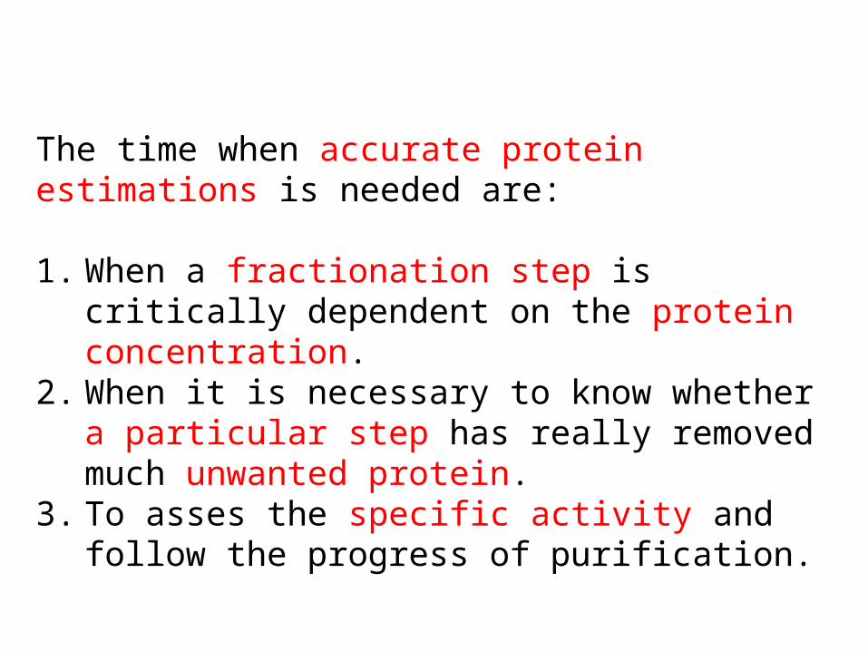
The time when accurate protein estimations is needed are:
1. When a fractionation step is critically dependent on the protein concentration.
2. When it is necessary to know whether a particular step has really removed much unwanted protein.
3. To asses the specific activity and follow the progress of purification.

• To the importance of protein concentration during analysis and purification several methods are presented.
• While choosing a method potential interfering substances, availability of instrumentation and desired sensitivity are considered.
• The methods include:
1. Bradford method 2. Bicinchoninic Acid (BCA) 3. Nano orange quantitation.
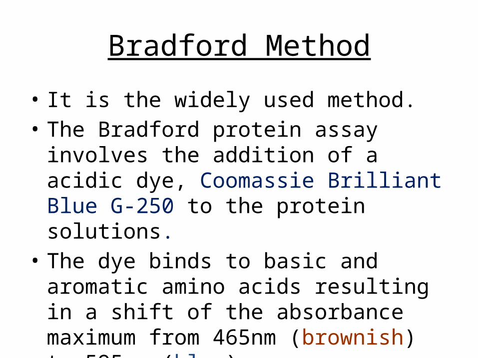
Bradford Method• It is the widely used method.• The Bradford protein assay involves the
addition of a acidic dye, Coomassie Brilliant Blue G-250 to the protein solutions.
• The dye binds to basic and aromatic amino acids resulting in a shift of the absorbance maximum from 465nm (brownish) to 595nm (blue).

Bradford method is based on the production of standard curve, OD(595nm) generates fractions fitted to standard curves and the protein concentration is determined.

Bradford Standard Assay• The Bradford standard assay detects proteins
with molecular weight greater than 3-5kDa.• The amount of protein present in a sample is
determined by performing a simple colorimetric reaction and comparing the results with those obtained from standard amount of protein.

This particular protocol is recommended for 20-140mg protein. (200-1400 mg/ml)MaterialsPlastic disposable cuvettesBradford dye reagent concentrateDiluted dye reagent 1 part Dye reagent concentrate + 4 parts distilled water. Filter through Whatman #1 or equivalent filter. The diluted dye reagent may be used for approx. 2 weeks when kept at room temperature.Test tubes: 13*100mm SpectrophotometerProtein standard: 1mg/ml BSA or IgG in water.

Procedure 1. Construct a standard curve by preparing four dilutions from
a protein stock solution of BSA or IgG. This is accomplished by adding 0 (the blank), 10, 20, 40 and 80ml of protein stock solution to individual tubes and adjusting the volume to 100ml with water. The tubes will contain 0-80mg of total protein in 100ml. It is recommended to prepare a standard curve each time that the assay is performed.
2. Pipet 0.1ml of the experimental samples into clean, dry test tubes. Also prepare two blanks.
3. Add 5ml of diluted dye reagent to all the test tubes.4. Mix the tubes by gentle vortexing. Avoid foaming.5. Let the test tube stand at the room temperature for 5
minutes.

6. Auto zero the spectrophotometer by reading the reagent blanks against each other at 595nm in the reference and sample cells of your double beam spectrophotometer.
7. With a blank in the reference of the spectrometer, measure the OD(595nm). Alternatively, all the tubes can be read versus water and the readings corrected for blank. Preferably, use plastic disposable cuvettes
8. Construct the standard curve. From the graph, determine the concentration of unknown protein by reading the values from standard curve.Calculate the protein concentration from the experimental sample.
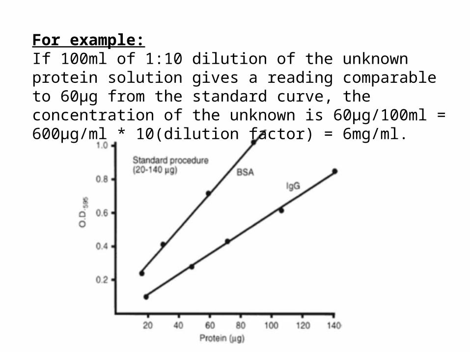
For example:If 100ml of 1:10 dilution of the unknown protein solution gives a reading comparable to 60µg from the standard curve, the concentration of the unknown is 60µg/100ml = 600µg/ml * 10(dilution factor) = 6mg/ml.

Bicinchonic acid (BCA)• When the protein is placed in an alkaline system containing
Cu⁺², a colored complex forms between the protein and copper atoms.
• BCA is a compound that forms a complex with cuprous ion (Cu+1) in an alkaline environment resulting in a stable, highly coloured chromophore with an absorbance maximum at 562nm.
• This reaction forms the basis of BCA protein quantitation system.
• The BCA protein assay is favored for determining protein concentrations in the presence of detergents.

Materials Reagent A: 1% BCA-Na₂, 2% Na₂CO₃.H₂O, 0.16% Na₂ tartrate,
0.4% NaOH and 0.95% NaHCO₃. If needed make the appropriate addition of NaOH(50%) or solid NaHCO₃ to adjust pH to 11.25.
Reagent B: 4% CuSO₄ 5H₂O Reagents A and B are stable indefinitely at room temperature. Standard working reagent solution: Mix 100vol of reagent A
with 2vol of reagent B. The mixture must be green. Spectrophotometer Cuvettes: 1ml plastic disposable Protein standard: BSA 1mg/ml in H₂O. Store aliquotted at -20.

Procedure
• Follow the instructions in the step 1 of Bradford assay.• Add 2ml of S-WR to all test tubes.• Incubate at 37˚C for 30mins. Color development begins
immediately. Cool the samples to room temperature and the measure the absorbance at 562nm versus the reaction blank.
• Construct a standard curve by plotting the absorbance at 562nm of the standard versus µg protein. Determine the amount of protein in the sample from standard curve. Knowing the volume of sample used and dilution used calculate protein concentration.

NanoOrange Protein Quantitation
• Fluorescence based techniques offer higher sensitivity, lower background signals and a wider dynamic range than absorbance based techniques.
• There are two method one that is independent of dyes and another that depends on dyes.
• NanoOrange protein quantitation depends on the dye.

• NanoOrange is non fluorescent aqueous solution but becomes instantly fluorescent upon binding to detergent coated proteins or hydrophobic regions of proteins.
• As little as 100ng/ml protein could be detected in 200µl volume, yielding a sensitivity of 20ng protein per sample using a fluorescent micro plate reader.

MaterialsNanoOrange protein quantitation reagentAssay diluent: 10mM Tris-HCl, pH 7.5, with a
proprietary mixture of anionic detergents BSA standard: 2mg/ml in waterGlass test tubes 13 * 100 disposable or
microfuge tubes Acrylic cuvettes or acrylic microplatesFlurometer

Procedure 1. Prepare the working NanoOrange reagent solution by
diluting the dye 500-fold into assay diluent as described in the manufacturer protocol.
2. Dilute BSA standards into the NanoOrange reagent in assay diluent using 2.5ml for 13mm disposable glass test tubes with no protein as background fluorescence control.
3. Protect all tubes from lighting throughout the procedure to reduce photobleaching.
4. Heat the sample for 10mins at 96 and cool for 20mins at room temperature, protected from light.
5. Briefly mix the samples and transfer them to disposable acrylic cuvettes or acrylic microplate wells.
6. Read fluorescence intensities at 485nm excitation and 590nm emission setting or suitable filters.7. Determine sample protein concentrations from standard curve.

Thank you