polysaccharides-rich extract of ganoderma lucidum (m.a
TRANSCRIPT
Hindawi Publishing CorporationEvidence-Based Complementary and Alternative MedicineVolume 2013, Article ID 671252, 9 pageshttp://dx.doi.org/10.1155/2013/671252
Research ArticlePolysaccharides-Rich Extract of Ganoderma lucidum(M.A. Curtis:Fr.) P. Karst Accelerates WoundHealing in Streptozotocin-Induced Diabetic Rats
Poh-Guat Cheng,1,2 Chia-Wei Phan,1,2 Vikineswary Sabaratnam,1,2 Noorlidah Abdullah,1,2
Mahmood Ameen Abdulla,1,3 and Umah Rani Kuppusamy1,3
1 Mushroom Research Centre, University of Malaya, 50603 Kuala Lumpur, Malaysia2 Institute of Biological Sciences, Faculty of Science, University of Malaya, 50603 Kuala Lumpur, Malaysia3 Department of Biomedical Science, Faculty of Medicine, University of Malaya, 50603 Kuala Lumpur, Malaysia
Correspondence should be addressed to Vikineswary Sabaratnam; [email protected]
Received 2 June 2013; Revised 8 October 2013; Accepted 8 October 2013
Academic Editor: Mohamed Eddouks
Copyright © 2013 Poh-Guat Cheng et al. This is an open access article distributed under the Creative Commons AttributionLicense, which permits unrestricted use, distribution, and reproduction in any medium, provided the original work is properlycited.
Ganoderma lucidum (M.A. Curtis:Fr.) P. Karst is a popular medicinal mushroom. Scientific reports had shown that the woundhealing effects ofG. lucidumwere partly attributed to its rich polysaccharides. However, little attention has been paid to its potentialeffects onwounds associated with diabetesmellitus. In this study, we evaluated the wound healing activity of the hot aqueous extractofG. lucidum in streptozotocin-induced diabetic rats.The extract ofG. lucidumwas standardised based on chemical contents (w/w)of total polysaccharides (25.1%), ganoderic acid A (0.45%), and adenosine (0.069%). Six groups of six rats were experimentallywounded in the posterior neck region. Intrasite gel was used as a positive control and aqueous cream as the placebo. Topicalapplication with 10% (w/w) of mushroom extract-incorporated aqueous cream was more effective than that with Intrasite gel interms of wound closure.The antioxidant activity in serumof rats treatedwith aqueous extract ofG. lucidumwas significantly higher;whereas the oxidative protein products and lipid damagewere lowerwhen compared to those of the controls.These findings stronglysupport the beneficial effects of standardised aqueous extract ofG. lucidum in acceleratingwoundhealing in streptozotocin-induceddiabetic rats.
1. Introduction
Diabetes mellitus is a metabolic disorder characterised byhyperglycemia with impaired carbohydrate, fat, and proteinmetabolism. The diabetic condition can be due to defects ininsulin secretion, action, or both [1]. It affects more than 180million individuals worldwide and by 2030 these numbers areprojected to double [2]. Diabetes mellitus has led to debili-tating consequences such as vasculopathy, retinopathy, andneuropathy [1]. More than 80% of diabetes mellitus is Type 2diabetes (noninsulin dependent) characterised by peripheralresistance to the action of insulin and decreased peripheralglucose uptake or increased hepatic glucose output [1].
According to the statistics provided by the NationalDiabetes Information Clearinghouse, 15% of diabetic indi-viduals suffered from diabetic foot ulcers that caused lower
limbs to be amputated [3].Thehyperglycemic state in diabeticpatients, especially those with peripheral vasculopathy in-terrupts proper wound healing. Wound healing occursas a cellular response to injury and involves activation ofkeratinocytes, fibroblasts, endothelial cells, macrophages,and platelets. Many growth factors and cytokines releasedby these cell types are needed to coordinate and maintainhealing [1, 4]. Furthermore, poor blood circulation and theoxygen supply in the affected area could lead to infectionand gangrene formation [5]. This often leads to increasedmorbidity andmortality. Despite the existence of protocols tostandardise wound care, the physiological impairments thatcan result in a diabetic foot ulcer (DFU) further complicatethe healing process [6]. Currently, the wounds in diabeticpatients are managed with antiseptic or antibiotic creams or
2 Evidence-Based Complementary and Alternative Medicine
gels such as Intrasite gel for wound dressing. Wound healingtakes prolonged periods and the healed wounds leave scars.In the search and discovery of safe and effective woundhealing agents, natural products from plant and mushroomsare currently being explored.
Mushrooms have enormous potential as a source of bothdietary protein and health-enhancing dietary supplements[7]. Among the medicinal mushrooms, Ganoderma speciesare much sought after for their wide array of medicinalproperties. Ganoderma lucidum (M.A. Curtis:Fr.) P. Karstwhich belongs to the Polyporaceae family has long beenknown and used extensively in traditional Chinese medicine.In Malaysia, due to the high humidity and temperaturethroughout the year this mushroom is widely cultivated.Chemical analysis of the fruiting bodies of G. lucidumshowed the presence of polysaccharides and triterpenoids[8–10] that may have therapeutic values in the treatment orprevention of peripheral or central inflammatory diseases.Ganoderma spp. are a natural source of potent bioactiveantioxidant metabolites [11]. Total phenols were the majornaturally occurring antioxidant components found in bothG.lucidum and G. tsugae [12]. It has been reported that someplants, for example, Annona squamosa L. (Annonaceae),commonly known as the custard apple [13], as well as Catha-ranthus roseus L (Apocynaceae) flower [14], promotedwoundhealing in diabetic rats via free radical scavenging activityof flavonoids. To date, reports of the applications of themedicinal properties of mushrooms in the healing of woundsin diabetic rats are rather rare. Kwon et al. [15] reportedthat the cauliflower mushroom, Sparassis crispa Wulf.:Fr.(Aphyllophoromycetideae), improved the healing of diabeticwounds. The high 𝛽-glucan (more than 40%) accounted forthe increase in the migration of macrophages and fibroblastsas well as elevated collagen synthesis. Sacchachitin and chitinmembrane prepared from the aqueous extracts of G. tsugaewere also found to have wound healing properties [16, 17].
In our preliminary study using normal rats withoutdiabetes, the period of reepithelialisation and wound closureshowed no significant difference (𝑃 > 0.05) between theIntrasite-gel- and G. lucidum-treated groups (unpublisheddata). The effect of wound healing by the hot aqueous extractof G. lucidumwas comparable to that by Intrasite gel and thismight be due to its high content of polysaccharides (25.1%)and the synergistic reactions combining all the medicinalproperties as a whole. As wound healing in hyperglycemicstate is difficult and challenging, the aim of this study wasto further investigate the effect of the hot aqueous extract ofG. lucidum on wound healing and the oxidative damage instreptozotocin-induced diabetic rats.
2. Materials and Methods
2.1. Preparation and Standardisation of Hot Aqueous Extractof G. lucidum. The fresh fruiting bodies of G. lucidum wereobtained from Ganofarm Sendirian Berhad, a mushroomfarm in Tanjung Sepat, Selangor, Malaysia. The productionof G. lucidum was reported previously [18, 19]. The pow-dered fruiting bodies were subjected to hot water extraction
(5 : 200, w/v) at 100∘C for 8 hours. The resulting aqueousextractwas freeze-dried and kept at−20∘Cprior to use.Heavymetal composition and microbial load of selected pathogenswere analyzed by Nova Laboratory, Sepang, Malaysia, usingproprietary methodology. A voucher specimen ofG. lucidum(KLU-M 1233) was deposited in the herbarium ofMushroomResearch Centre, University of Malaya.
2.2. Determination of Total Polysaccharides Content in HotAqueous Extracts of G. lucidum. The total polysaccharidecontent of the hot aqueous extract was determined usingthe phenol-sulphuric acid method with d-glucose as in [20].Briefly, one mL of 5% (w/v) phenol was added to one mL ofsample solution, followed by five mL of concentrated H
2SO4.
The absorbance was measured using a spectrophotometer(Shimadzu series 1601UV/Vis) after 10 minutes at 483 nm.
2.3. Quantification of Ganoderic Acid and Adenosine UsingHPLC. For determination of ganoderic acid A, Perkin ElmerSeries 200 liquid chromatography equipped with a PerkinElmer Series 200 UV detector was used. The detector signalwas recorded by the Turbochrom workstation software. Thecolumn was Hypersil BDS C18 (4.6 × 250mm) with Alltechrefillable C18 Guard column (10 × 4.6mm) (Alltech, USA).The mobile phase consisted of 5% (v/v) acetic acid inmethanol and the flow rate was 1.0mL/min. The calibrationcurve was prepared by injecting a series of ganoderic acidA (Sigma) reference standard dilutions. Quantification andvalidation of adenosine were also performed in Perkin ElmerSeries 200 liquid chromatography as mentioned. The mobilephasewasmethanol: 10mMmonobasic potassiumphosphate(15 : 85), pH 5.0, and the flow rate was 1.5mL/min. Bothganoderic acid A and adenosine were quantified by meansof calibration curves obtained from commercial standards ofthese compounds (Sigma).
2.4. Determination of Cupric Reducing Antioxidant Capacity(CUPRAC) of Hot Aqueous Extract of G. lucidum. The cupricreducing antioxidant capacity (CUPRAC) of the hot aqueousextract of G. lucidum was determined according to themethod of Apak et al. [21]. Briefly, to a mixture of onemL of copper(II) (10−2M), neocuproine (7.5 × 10⁻³M), andammonium acetate buffer solution (1M), freshly-preparedmushroom extracts of varying concentrations were added tomake up a final volume of four mL. After incubation at roomtemperature (25 ± 2∘C) for 30 minutes, the absorbance at450 nm was recorded against a reagent blank. The results ofantioxidant activity were expressed in absorbance at 450 nmand compared with ascorbic acid as a positive control.
2.5. Preparation ofMushroomExtract-Incorporated TreatmentCream. The hot aqueous extract at concentrations of 5%,10%, 15%, and 20% (w/w) was mixed with aqueous creamhomogeneously. Aqueous cream was obtained from theDepartment of Pharmacy, Faculty of Medicine, Universityof Malaya (a product of Sunward Pharmaceutical SendirianBerhad, MAL 19920890 X).
2.6. Experimental Animals. Healthy adult male SpragueDawley rats were obtained from the animal house, Faculty
Evidence-Based Complementary and Alternative Medicine 3
of Medicine, University of Malaya. The rats were dividedrandomly into six groups of six rats each. Each rat weighedbetween 180 to 250 g and was housed separately (one ratper cage). The animals were maintained on a standard pelletdiet and tap water. The study conformed to the Principlesof Laboratory Animal Care and was approved by the EthicsCommittee of University of Malaya with the Ethic numberISB/14/10/2009/CPG (R). All animals received care accordingto the criteria outlined in the “Guide for the Care and Use ofLaboratory Animals” prepared by the National Academy ofSciences and published by the National Institutes of Health.
2.7. Diabetes Induction. Streptozotocin (STZ) was purchasedfromSigma-Aldrich (St. Louis,MO,USA). After an overnightfast, diabetes mellitus was induced in six groups of ratsby a single intraperitoneal injection of streptozotocin (STZ;45mg/kg) dissolved in citrate buffer (0.1M, pH 4.0). Bloodwas drawn from the tail vein on the 7th day after theSTZ injection, and the fasting blood glucose levels wereestimated using a glucometer (Ames, Bayer Diagnostic). Ratswith fasting blood glucose levels higher than 10mmol/L (or200mg/dL) were considered diabetic and were included forthe experiment [22].
2.8. ExcisionWound Creation. Excision wounds were createdon the 7th day after the induction of diabetes in all rats. Theanimals were anaesthetisedwith 2mLof diethyl ether (Sigma,98% purity). The skin was shaved using an electric clipper,disinfected with 70% alcohol, and 0.5mL of lignocaine HCl(2%, 20mg/mL) was injected as a local anaesthetic agent.The area of wound was outlined with methylene blue usinga circular stencil. A full thickness of the excision wound of2.0 cm in length and 0.2 cm depth was created as described byNayak andPinto Pereira [14]. Carewas taken to avoid injuringthe muscle layer, and the tension of skin was kept constantduring the procedure.The wound areas were measured usinga graph paper.
2.9. Topical Application of Treatment Creams. Thewounds ofGroup 1 rats were dressed with a thin layer of aqueous creamas a blank placebo twice daily. The wounds of Group 2 ratswere dressed topically twice daily with 0.2mL of Intrasite gelas the positive control. Intrasite gel, which is a trademark ofSmith and Nephew Ltd., was purchased from the UniversityMalaya Medical Centre Pharmacy. Rats in Group 3, 4, 5,and 6 rats were treated with a thin layer of aqueous creamcontaining 5%, 10%, 15%, and 20% (w/w) aqueous extractsof G. lucidum, respectively. All applications of cream wereperformed with appropriate care twice a day.
2.10. Determination of the Period of Reepithelialisation. Thewounds of all animals under the different treatments wereobserved daily. The period of reepithelisation was assessedby counting the number of days required for the completehealing including eschar falloff without any residual rawwound [13].
2.11. Determination of the Wound Closure. The wound area(mm2) was measured at 0, 1, 4, 8, 12, and 16 days after wound-ing using transparency paper and a permanent marker. The
rate of wound closure which is expressed as the percentageof wound reduction from the original wound was calculatedusing the following formula [14]:
Percentage of wound closure (%)
= [(wound area on day 0 − wound area on
postoperation day)
×(wound area on day 0)−1] × 100%.
(1)
2.12. Histological Evaluation of Healed Wounds. The speci-mens of skin from healed wounds and surrounding tissueswere excised and stained for histological studies. Threesections (5 𝜇m thickness) from each rat were prepared forhematoxylin and eosin (H&E) and Masson Trichrome Stain-ing, and stained sections of each wound were examined bylight microscopy. Scar width (mm) which is the junction gapbetween the normal dermis and dermis in the wound tissueswas measured [23]. The morphological changes (fibroblast,inflammatory cell, neovascularisation, and collagen) wererecorded.
2.13. Determination of In Vivo Antioxidant Activity of Exper-imental Rats. Blood sample was collected from the exper-imental rats on postoperation days 7 and 16. Antioxidantactivity in the blood serum was determined by the CUPRACmethod [21], which utilises copper(II)-neocuproine reagentas the chromogenic oxidising agent. Briefly, the mixture of1mL of CuCl
2(10−2M), neocuproine (7.5 × 10⁻³M), and
ammonium acetate buffer solution (1M) were added into acuvette. Then, 1090𝜇L of distilled water with 10 𝜇L of bloodserum was added into the reagent mixture and incubated atroom temperature (25± 2∘C) for 30 minutes.The absorbanceat 450 nm was recorded against a reagent blank. The result ofantioxidant activity was expressed as absorbance at 450 nmagainst blank. Ascorbic acid was used as positive control.Each sample of serumwas triplicated for absorbance reading,𝑛 = 6 for each group of experimental animals.
2.14. Assessment of Oxidative Damage
2.14.1. Advanced Oxidation Protein Product (AOPP) Assay.Advanced oxidation protein product (AOPP) was deter-mined by the method of Witko-Sarsat et al. [24]. Briefly,AOPP was determined spectrophotometrically using amicroplate reader and was calibrated with chloramine-Tsolutions. Reagent mixture was prepared by adding 81mLof PBS solution, 15mL of acetic acid (50%), and 4mL ofpotassium iodide. Then, 18 𝜇L of plasma sample was addedto 200𝜇L of reagent mixture in a 96-well microplate reader.The absorbance at 340 nm was recorded against a reagentblank. AOPP assay for each plasma sample was carried outin triplicates, 𝑛 = 6 for each group of experimental animals.AOPPs were expressed as 𝜇mol/L chloramine-T equivalents.
2.14.2. Lipid Hydroperoxide (LPO) Assay. Lipid hydroper-oxide (LPO) was determined according to the method ofEsterbauer and Cheeseman [25]. Malondialdehyde (MDA)
4 Evidence-Based Complementary and Alternative Medicine
was assayed as a marker of lipid peroxidation using colori-metric reaction, which uses 1-methyl-2-phenylindole (MPI)as chromogen. Condensation of one molecule of MDAwith two molecules of MPI under acidic condition resultsin the formation of a chromophore which has maximumabsorbance at 586 nm. A total of 150 𝜇L of serum sample wasadded to 375 𝜇L of MPI (10.3mM) in acetonitrile and 225 𝜇LHCl (5M). The mixture was incubated in a water bath at45∘C for 40 minutes. Tetraethoxypropane (TEP) was usedas standard solution. After centrifugation at 10,000×g for 5minutes, 200𝜇L of the reaction mixture was pipetted into a96-well plate and read at 586 nm in an ELISA reader (UV1601 spectrophotometer, Shimadzu, Japan). Concentration ofMDA was expressed as nM of TEP equivalents. Each of theserum samples was assayed in triplicates, 𝑛 = 6 for each groupof experimental animals.
2.15. Statistical Analysis. All values are reported as mean ±standard errormean (S.E.M). Statistical evaluation of the datawas done by one way analysis of variance (ANOVA) followedby Bonferroni’s multiple comparison tests. Differences wereconsidered statistically significant when 𝑃 < 0.05.
3. Results
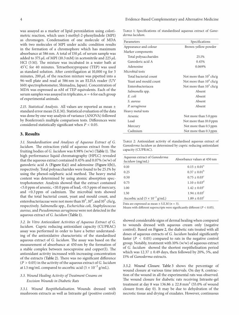
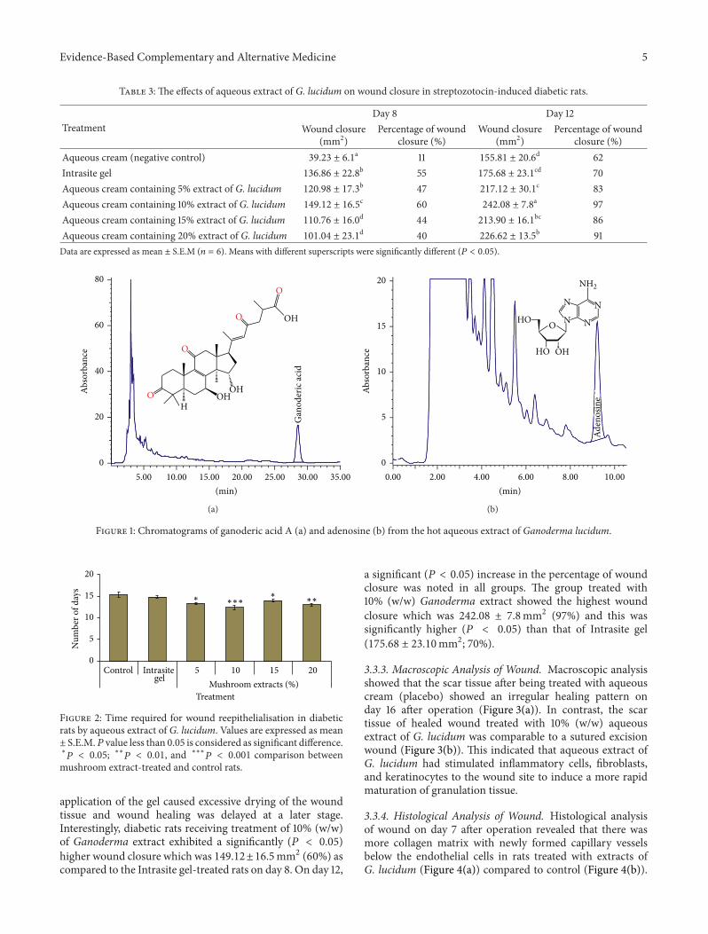
3.1. Standardisation and Analyses of Aqueous Extract of G.lucidum. The extraction yield of aqueous extract from thefruiting bodies of G. lucidum was 8.98% (w/w) (Table 1). Thehigh performance liquid chromatography (HPLC) revealedthat the aqueous extract contained 0.45% and 0.07% (w/w) ofganoderic acid A (Figure 1(a)) and adenosine (Figure 1(b)),respectively. Total polysaccharides were found to be 25.1% byusing the phenol-sulphuric acid method. The heavy metalcontent was determined by using atomic absorption spec-trophotometer. Analysis showed that the extract contained<5.0 ppm of arsenic, <10.0 ppm of lead, <0.5 ppm of mercury,and <0.3 ppm of cadmium. The microbial tests showedthat the total bacterial count, yeast and mould count, andenterobacteriaceae were notmore than 105, 104, and 105 cfu/g,respectively. Salmonella spp., Escherichia coli, Staphylococcusaureus, and Pseudomonas aeruginosawere not detected in theaqueous extract of G. lucidum (Table 1).
3.2. In Vitro Antioxidant Activities of Aqueous Extract of G.lucidum. Cupric reducing antioxidant capacity (CUPRAC)assay was performed in order to have a better understand-ing of the antioxidative characteristic of the standardisedaqueous extract of G. lucidum. The assay was based on themeasurement of absorbance at 450 nm by the formation ofa stable complex between neocuproine and copper(I). Theantioxidant activity increased with increasing concentrationof the extracts (Table 2). There was no significant difference(𝑃 > 0.05) in the activity of the aqueous extract ofG. lucidumat 1.5mg/mL compared to ascorbic acid (5 × 10⁻³ g/mL).
3.3. Wound Healing Activity of Treatment Creams onExcision Wounds in Diabetic Rats
3.3.1. Wound Reepithelialisation. Wounds dressed withmushroom extracts as well as Intrasite gel (positive control)
Table 1: Specifications of standardised aqueous extract of Gano-derma lucidum.
Parameters SpecificationsAppearance and colour Brown-yellow powderMarker components
Total polysaccharides 25.1%Ganoderic acid A 0.45%Adenosine 0.069%
Microbial testsTotal bacterial count Not more than 105 cfu/gYeast and mould count Not more than 104 cfu/gEnterobacteriacea Not more than 103 cfu/gSalmonella spp. AbsentE. coli AbsentS. aureus AbsentP. aeruginosa Absent
Heavy metal testsArsenic Not more than 5.0 ppmLead Not more than 10.0 ppmMercury Not more than 0.5 ppmCadmium Not more than 0.3 ppm
Table 2: Antioxidant activity of standardised aqueous extract ofGanoderma lucidum as determined by cupric reducing antioxidantcapacity (CUPRAC).
Aqueous extract of Ganodermalucidum (mg/mL) Absorbance values at 450 nm
0.10 0.15 ± 0.01a
0.25 0.37 ± 0.01b
0.50 0.75 ± 0.03c
0.75 1.10 ± 0.03d
1.00 1.42 ± 0.03e
1.50 1.94 ± 0.03f
Ascorbic acid (5 × 10⁻³ g/mL) 1.89 ± 0.03f
Data are expressed as mean ± S.E.M (𝑛 = 3).Means with different superscripts were significantly different (𝑃 < 0.05).
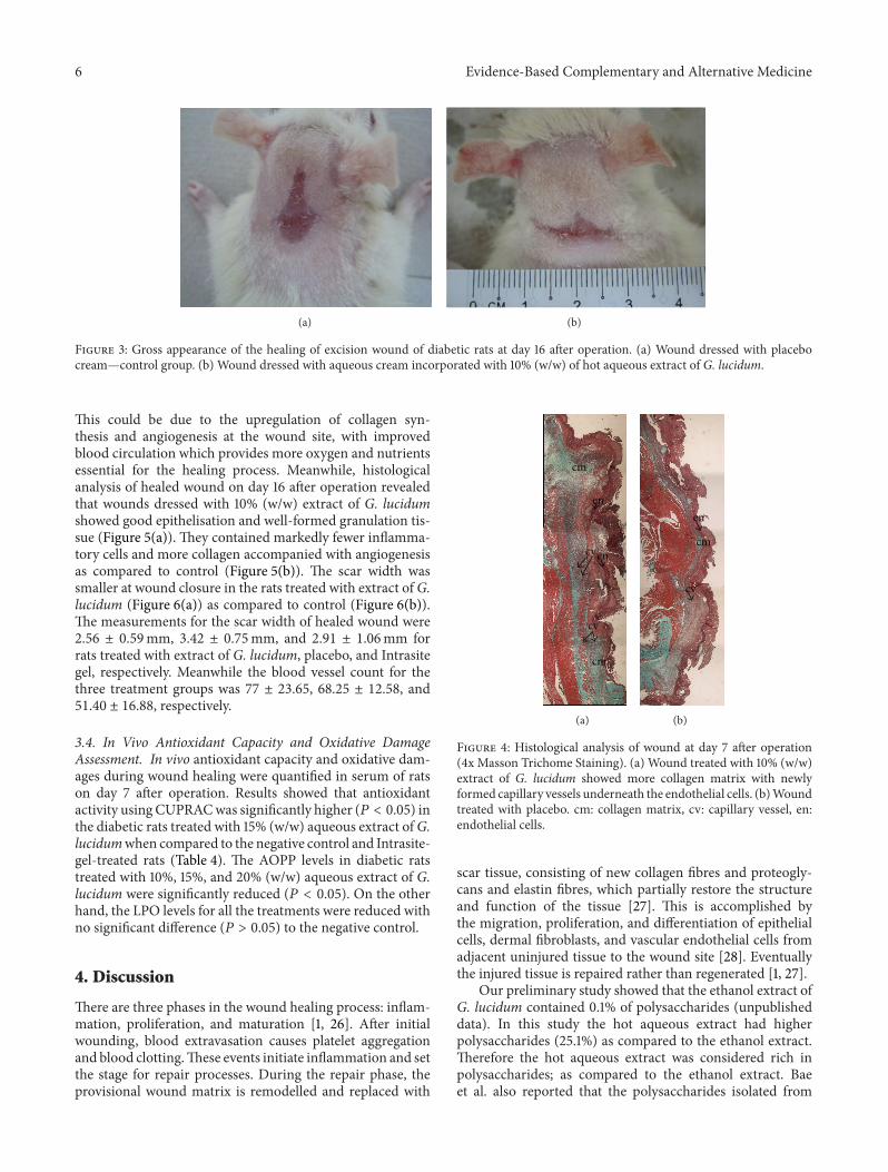
showed considerable signs of dermal healing when comparedto wounds dressed with aqueous cream only (negativecontrol). Based on Figure 2, the diabetic rats treated with alldoses of aqueous extracts of G. lucidum healed significantlyfaster (𝑃 < 0.05) compared to rats in the negative controlgroup. Notably, treatment with 10% (w/w) of aqueous extractof G. lucidum showed the shortest reepithelisation periodwhich was 12.37 ± 0.49 days, then followed by 20%, 5%, and15% of Ganoderma extracts.
3.3.2. Wound Closure. Table 3 shows the percentage ofwound closure at various time intervals. On day 8, contrac-tion of the wound in all the experimental rats was observed.The wound closure for diabetic rats receiving Intrasite-geltreatment at day 8 was 136.86 ± 22.8mm2 (55.0% of woundclosure from day 0). It may be due to dehydration of thenecrotic tissue and drying of exudates. However, continuous
Evidence-Based Complementary and Alternative Medicine 5
Table 3: The effects of aqueous extract of G. lucidum on wound closure in streptozotocin-induced diabetic rats.
TreatmentDay 8 Day 12
Wound closure(mm2)
Percentage of woundclosure (%)
Wound closure(mm2)
Percentage of woundclosure (%)
Aqueous cream (negative control) 39.23 ± 6.1a 11 155.81 ± 20.6d 62Intrasite gel 136.86 ± 22.8b 55 175.68 ± 23.1cd 70Aqueous cream containing 5% extract of G. lucidum 120.98 ± 17.3b 47 217.12 ± 30.1c 83Aqueous cream containing 10% extract of G. lucidum 149.12 ± 16.5c 60 242.08 ± 7.8a 97Aqueous cream containing 15% extract of G. lucidum 110.76 ± 16.0d 44 213.90 ± 16.1bc 86Aqueous cream containing 20% extract of G. lucidum 101.04 ± 23.1d 40 226.62 ± 13.5b 91Data are expressed as mean ± S.E.M (𝑛 = 6). Means with different superscripts were significantly different (𝑃 < 0.05).
80
60
40
20
05.00 10.00 15.00 20.00 25.00 30.00 35.00
Abso
rban
ce
(min)
Gan
oder
ic ac
idO
O
O
O OHOH
OH
H
(a)
20
15
10
5
02.000.00 4.00 6.00 8.00 10.00
(min)
Abso
rban
ce HO
HO
OH
O
NN
N
N
NH2
Aden
osin
e
(b)
Figure 1: Chromatograms of ganoderic acid A (a) and adenosine (b) from the hot aqueous extract of Ganoderma lucidum.
0
5
10
15
20
Control Intrasite 5 10 15 20Mushroom extracts (%)
Num
ber o
f day
s
Treatment
∗ ∗∗∗∗
∗∗
gel
Figure 2: Time required for wound reepithelialisation in diabeticrats by aqueous extract of G. lucidum. Values are expressed as mean± S.E.M.𝑃 value less than 0.05 is considered as significant difference.∗
𝑃 < 0.05; ∗∗𝑃 < 0.01, and ∗∗∗𝑃 < 0.001 comparison betweenmushroom extract-treated and control rats.
application of the gel caused excessive drying of the woundtissue and wound healing was delayed at a later stage.Interestingly, diabetic rats receiving treatment of 10% (w/w)of Ganoderma extract exhibited a significantly (𝑃 < 0.05)higher wound closure which was 149.12±16.5mm2 (60%) ascompared to the Intrasite gel-treated rats on day 8. On day 12,
a significant (𝑃 < 0.05) increase in the percentage of woundclosure was noted in all groups. The group treated with10% (w/w) Ganoderma extract showed the highest woundclosure which was 242.08 ± 7.8mm2 (97%) and this wassignificantly higher (𝑃 < 0.05) than that of Intrasite gel(175.68 ± 23.10mm2; 70%).
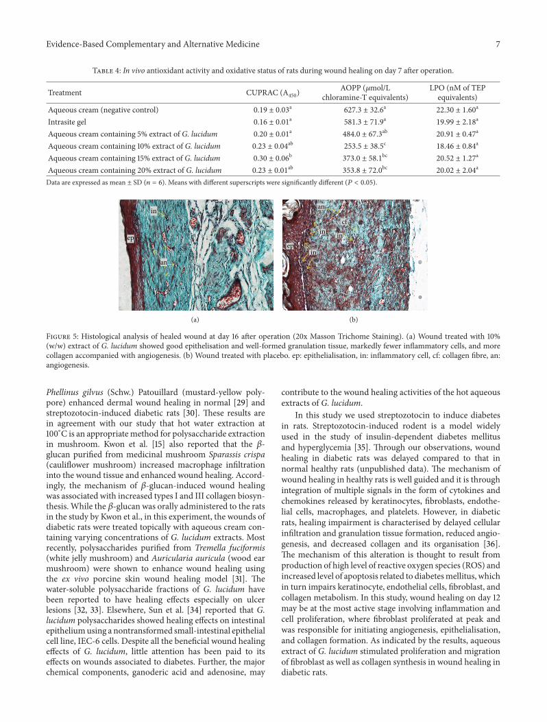
3.3.3. Macroscopic Analysis of Wound. Macroscopic analysisshowed that the scar tissue after being treated with aqueouscream (placebo) showed an irregular healing pattern onday 16 after operation (Figure 3(a)). In contrast, the scartissue of healed wound treated with 10% (w/w) aqueousextract of G. lucidum was comparable to a sutured excisionwound (Figure 3(b)). This indicated that aqueous extract ofG. lucidum had stimulated inflammatory cells, fibroblasts,and keratinocytes to the wound site to induce a more rapidmaturation of granulation tissue.
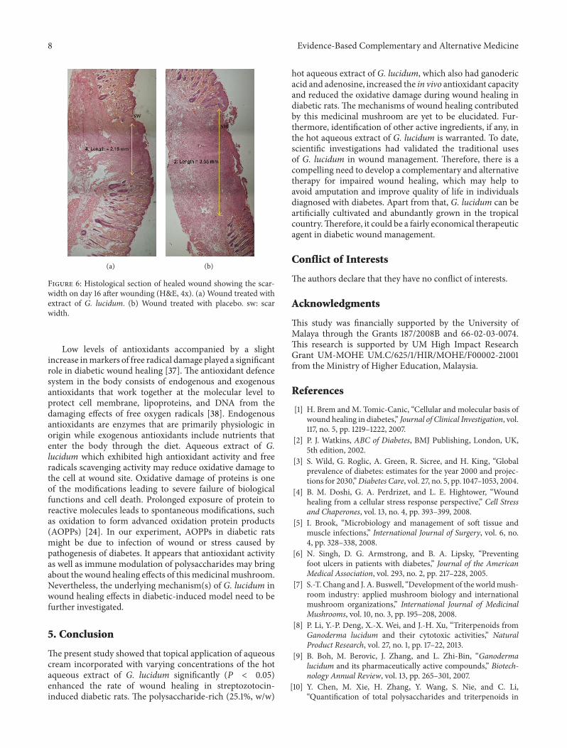
3.3.4. Histological Analysis of Wound. Histological analysisof wound on day 7 after operation revealed that there wasmore collagen matrix with newly formed capillary vesselsbelow the endothelial cells in rats treated with extracts ofG. lucidum (Figure 4(a)) compared to control (Figure 4(b)).
6 Evidence-Based Complementary and Alternative Medicine
(a) (b)
Figure 3: Gross appearance of the healing of excision wound of diabetic rats at day 16 after operation. (a) Wound dressed with placebocream—control group. (b) Wound dressed with aqueous cream incorporated with 10% (w/w) of hot aqueous extract of G. lucidum.
This could be due to the upregulation of collagen syn-thesis and angiogenesis at the wound site, with improvedblood circulation which provides more oxygen and nutrientsessential for the healing process. Meanwhile, histologicalanalysis of healed wound on day 16 after operation revealedthat wounds dressed with 10% (w/w) extract of G. lucidumshowed good epithelisation and well-formed granulation tis-sue (Figure 5(a)). They contained markedly fewer inflamma-tory cells and more collagen accompanied with angiogenesisas compared to control (Figure 5(b)). The scar width wassmaller at wound closure in the rats treated with extract ofG.lucidum (Figure 6(a)) as compared to control (Figure 6(b)).The measurements for the scar width of healed wound were2.56 ± 0.59mm, 3.42 ± 0.75mm, and 2.91 ± 1.06mm forrats treated with extract of G. lucidum, placebo, and Intrasitegel, respectively. Meanwhile the blood vessel count for thethree treatment groups was 77 ± 23.65, 68.25 ± 12.58, and51.40 ± 16.88, respectively.
3.4. In Vivo Antioxidant Capacity and Oxidative DamageAssessment. In vivo antioxidant capacity and oxidative dam-ages during wound healing were quantified in serum of ratson day 7 after operation. Results showed that antioxidantactivity using CUPRACwas significantly higher (𝑃 < 0.05) inthe diabetic rats treated with 15% (w/w) aqueous extract ofG.lucidumwhen compared to the negative control and Intrasite-gel-treated rats (Table 4). The AOPP levels in diabetic ratstreated with 10%, 15%, and 20% (w/w) aqueous extract of G.lucidum were significantly reduced (𝑃 < 0.05). On the otherhand, the LPO levels for all the treatments were reduced withno significant difference (𝑃 > 0.05) to the negative control.
4. Discussion
There are three phases in the wound healing process: inflam-mation, proliferation, and maturation [1, 26]. After initialwounding, blood extravasation causes platelet aggregationand blood clotting.These events initiate inflammation and setthe stage for repair processes. During the repair phase, theprovisional wound matrix is remodelled and replaced with
cm
cm
en
encv
cv
(a)
en
cv
cm
(b)
Figure 4: Histological analysis of wound at day 7 after operation(4x Masson Trichome Staining). (a) Wound treated with 10% (w/w)extract of G. lucidum showed more collagen matrix with newlyformed capillary vessels underneath the endothelial cells. (b)Woundtreated with placebo. cm: collagen matrix, cv: capillary vessel, en:endothelial cells.
scar tissue, consisting of new collagen fibres and proteogly-cans and elastin fibres, which partially restore the structureand function of the tissue [27]. This is accomplished bythe migration, proliferation, and differentiation of epithelialcells, dermal fibroblasts, and vascular endothelial cells fromadjacent uninjured tissue to the wound site [28]. Eventuallythe injured tissue is repaired rather than regenerated [1, 27].
Our preliminary study showed that the ethanol extract ofG. lucidum contained 0.1% of polysaccharides (unpublisheddata). In this study the hot aqueous extract had higherpolysaccharides (25.1%) as compared to the ethanol extract.Therefore the hot aqueous extract was considered rich inpolysaccharides; as compared to the ethanol extract. Baeet al. also reported that the polysaccharides isolated from
Evidence-Based Complementary and Alternative Medicine 7
Table 4: In vivo antioxidant activity and oxidative status of rats during wound healing on day 7 after operation.
Treatment CUPRAC (A450)AOPP (𝜇mol/L
chloramine-T equivalents)LPO (nM of TEPequivalents)
Aqueous cream (negative control) 0.19 ± 0.03a 627.3 ± 32.6a 22.30 ± 1.60a
Intrasite gel 0.16 ± 0.01a 581.3 ± 71.9a 19.99 ± 2.18a
Aqueous cream containing 5% extract of G. lucidum 0.20 ± 0.01a 484.0 ± 67.3ab 20.91 ± 0.47a
Aqueous cream containing 10% extract of G. lucidum 0.23 ± 0.04ab 253.5 ± 38.5c 18.46 ± 0.84a
Aqueous cream containing 15% extract of G. lucidum 0.30 ± 0.06b 373.0 ± 58.1bc 20.52 ± 1.27a
Aqueous cream containing 20% extract of G. lucidum 0.23 ± 0.01ab 353.8 ± 72.0bc 20.02 ± 2.04a
Data are expressed as mean ± SD (𝑛 = 6). Means with different superscripts were significantly different (𝑃 < 0.05).
ep
in
an
(a)
inin
ep
an
in
cf
(b)
Figure 5: Histological analysis of healed wound at day 16 after operation (20x Masson Trichome Staining). (a) Wound treated with 10%(w/w) extract of G. lucidum showed good epithelisation and well-formed granulation tissue, markedly fewer inflammatory cells, and morecollagen accompanied with angiogenesis. (b) Wound treated with placebo. ep: epithelialisation, in: inflammatory cell, cf: collagen fibre, an:angiogenesis.
Phellinus gilvus (Schw.) Patouillard (mustard-yellow poly-pore) enhanced dermal wound healing in normal [29] andstreptozotocin-induced diabetic rats [30]. These results arein agreement with our study that hot water extraction at100∘C is an appropriatemethod for polysaccharide extractionin mushroom. Kwon et al. [15] also reported that the 𝛽-glucan purified from medicinal mushroom Sparassis crispa(cauliflower mushroom) increased macrophage infiltrationinto the wound tissue and enhanced wound healing. Accord-ingly, the mechanism of 𝛽-glucan-induced wound healingwas associated with increased types I and III collagen biosyn-thesis. While the 𝛽-glucan was orally administered to the ratsin the study by Kwon et al., in this experiment, the wounds ofdiabetic rats were treated topically with aqueous cream con-taining varying concentrations of G. lucidum extracts. Mostrecently, polysaccharides purified from Tremella fuciformis(white jelly mushroom) and Auricularia auricula (wood earmushroom) were shown to enhance wound healing usingthe ex vivo porcine skin wound healing model [31]. Thewater-soluble polysaccharide fractions of G. lucidum havebeen reported to have healing effects especially on ulcerlesions [32, 33]. Elsewhere, Sun et al. [34] reported that G.lucidum polysaccharides showed healing effects on intestinalepitheliumusing a nontransformed small-intestinal epithelialcell line, IEC-6 cells. Despite all the beneficial wound healingeffects of G. lucidum, little attention has been paid to itseffects on wounds associated to diabetes. Further, the majorchemical components, ganoderic acid and adenosine, may
contribute to the wound healing activities of the hot aqueousextracts of G. lucidum.
In this study we used streptozotocin to induce diabetesin rats. Streptozotocin-induced rodent is a model widelyused in the study of insulin-dependent diabetes mellitusand hyperglycemia [35]. Through our observations, woundhealing in diabetic rats was delayed compared to that innormal healthy rats (unpublished data). The mechanism ofwound healing in healthy rats is well guided and it is throughintegration of multiple signals in the form of cytokines andchemokines released by keratinocytes, fibroblasts, endothe-lial cells, macrophages, and platelets. However, in diabeticrats, healing impairment is characterised by delayed cellularinfiltration and granulation tissue formation, reduced angio-genesis, and decreased collagen and its organisation [36].The mechanism of this alteration is thought to result fromproduction of high level of reactive oxygen species (ROS) andincreased level of apoptosis related to diabetesmellitus, whichin turn impairs keratinocyte, endothelial cells, fibroblast, andcollagen metabolism. In this study, wound healing on day 12may be at the most active stage involving inflammation andcell proliferation, where fibroblast proliferated at peak andwas responsible for initiating angiogenesis, epithelialisation,and collagen formation. As indicated by the results, aqueousextract of G. lucidum stimulated proliferation and migrationof fibroblast as well as collagen synthesis in wound healing indiabetic rats.
8 Evidence-Based Complementary and Alternative Medicine
sw
(a)
sw
(b)
Figure 6: Histological section of healed wound showing the scar-width on day 16 after wounding (H&E, 4x). (a) Wound treated withextract of G. lucidum. (b) Wound treated with placebo. sw: scarwidth.
Low levels of antioxidants accompanied by a slightincrease inmarkers of free radical damage played a significantrole in diabetic wound healing [37]. The antioxidant defencesystem in the body consists of endogenous and exogenousantioxidants that work together at the molecular level toprotect cell membrane, lipoproteins, and DNA from thedamaging effects of free oxygen radicals [38]. Endogenousantioxidants are enzymes that are primarily physiologic inorigin while exogenous antioxidants include nutrients thatenter the body through the diet. Aqueous extract of G.lucidum which exhibited high antioxidant activity and freeradicals scavenging activity may reduce oxidative damage tothe cell at wound site. Oxidative damage of proteins is oneof the modifications leading to severe failure of biologicalfunctions and cell death. Prolonged exposure of protein toreactive molecules leads to spontaneous modifications, suchas oxidation to form advanced oxidation protein products(AOPPs) [24]. In our experiment, AOPPs in diabetic ratsmight be due to infection of wound or stress caused bypathogenesis of diabetes. It appears that antioxidant activityas well as immune modulation of polysaccharides may bringabout the wound healing effects of this medicinal mushroom.Nevertheless, the underlying mechanism(s) of G. lucidum inwound healing effects in diabetic-induced model need to befurther investigated.
5. Conclusion
The present study showed that topical application of aqueouscream incorporated with varying concentrations of the hotaqueous extract of G. lucidum significantly (𝑃 < 0.05)enhanced the rate of wound healing in streptozotocin-induced diabetic rats. The polysaccharide-rich (25.1%, w/w)
hot aqueous extract of G. lucidum, which also had ganodericacid and adenosine, increased the in vivo antioxidant capacityand reduced the oxidative damage during wound healing indiabetic rats. The mechanisms of wound healing contributedby this medicinal mushroom are yet to be elucidated. Fur-thermore, identification of other active ingredients, if any, inthe hot aqueous extract of G. lucidum is warranted. To date,scientific investigations had validated the traditional usesof G. lucidum in wound management. Therefore, there is acompelling need to develop a complementary and alternativetherapy for impaired wound healing, which may help toavoid amputation and improve quality of life in individualsdiagnosed with diabetes. Apart from that, G. lucidum can beartificially cultivated and abundantly grown in the tropicalcountry.Therefore, it could be a fairly economical therapeuticagent in diabetic wound management.
Conflict of Interests
The authors declare that they have no conflict of interests.
Acknowledgments
This study was financially supported by the University ofMalaya through the Grants 187/2008B and 66-02-03-0074.This research is supported by UM High Impact ResearchGrant UM-MOHE UM.C/625/1/HIR/MOHE/F00002-21001from the Ministry of Higher Education, Malaysia.
References
[1] H. Brem and M. Tomic-Canic, “Cellular and molecular basis ofwound healing in diabetes,” Journal of Clinical Investigation, vol.117, no. 5, pp. 1219–1222, 2007.
[2] P. J. Watkins, ABC of Diabetes, BMJ Publishing, London, UK,5th edition, 2002.
[3] S. Wild, G. Roglic, A. Green, R. Sicree, and H. King, “Globalprevalence of diabetes: estimates for the year 2000 and projec-tions for 2030,”Diabetes Care, vol. 27, no. 5, pp. 1047–1053, 2004.
[4] B. M. Doshi, G. A. Perdrizet, and L. E. Hightower, “Woundhealing from a cellular stress response perspective,” Cell Stressand Chaperones, vol. 13, no. 4, pp. 393–399, 2008.
[5] I. Brook, “Microbiology and management of soft tissue andmuscle infections,” International Journal of Surgery, vol. 6, no.4, pp. 328–338, 2008.
[6] N. Singh, D. G. Armstrong, and B. A. Lipsky, “Preventingfoot ulcers in patients with diabetes,” Journal of the AmericanMedical Association, vol. 293, no. 2, pp. 217–228, 2005.
[7] S.-T. Chang and J. A. Buswell, “Development of theworldmush-room industry: applied mushroom biology and internationalmushroom organizations,” International Journal of MedicinalMushrooms, vol. 10, no. 3, pp. 195–208, 2008.
[8] P. Li, Y.-P. Deng, X.-X. Wei, and J.-H. Xu, “Triterpenoids fromGanoderma lucidum and their cytotoxic activities,” NaturalProduct Research, vol. 27, no. 1, pp. 17–22, 2013.
[9] B. Boh, M. Berovic, J. Zhang, and L. Zhi-Bin, “Ganodermalucidum and its pharmaceutically active compounds,” Biotech-nology Annual Review, vol. 13, pp. 265–301, 2007.
[10] Y. Chen, M. Xie, H. Zhang, Y. Wang, S. Nie, and C. Li,“Quantification of total polysaccharides and triterpenoids in
Evidence-Based Complementary and Alternative Medicine 9
Ganoderma lucidum and Ganoderma atrum by near infraredspectroscopy and chemometrics,” Food Chemistry, vol. 135, pp.268–275, 2012.
[11] J.-L.Mau,H.-C. Lin, andC.-C. Chen, “Antioxidant properties ofseveralmedicinalmushrooms,” Journal of Agricultural and FoodChemistry, vol. 50, no. 21, pp. 6072–6077, 2002.
[12] J.-H. Yang, H.-C. Lin, and J.-L. Mau, “Antioxidant properties ofseveral commercial mushrooms,” Food Chemistry, vol. 77, no. 2,pp. 229–235, 2002.
[13] T. Ponrasu and L. Suguna, “Efficacy of Annona squamosaon wound healing in streptozotocin-induced diabetic rats,”International Wound Journal, vol. 9, pp. 613–623, 2012.
[14] B. S. Nayak and L.M. Pinto Pereira, “Catharanthus roseus flowerextract has wound-healing activity in Sprague Dawley rats,”BMC Complementary and Alternative Medicine, vol. 6, article41, 2006.
[15] A.-H. Kwon, Z. Qiu, M. Hashimoto, K. Yamamoto, and T.Kimura, “Effects of medicinal mushroom (Sparassis crispa)on wound healing in streptozotocin-induced diabetic rats,”American Journal of Surgery, vol. 197, no. 4, pp. 503–509, 2009.
[16] C.-H. Su, C.-S. Sun, S.-W. Juan, C.-H. Hu, W.-T. Ke, and M.-T.Sheu, “Fungal mycelia as the source of chitin and polysaccha-rides and their applications as skin substitutes,” Biomaterials,vol. 18, no. 17, pp. 1169–1174, 1997.
[17] C.-H. Su, C.-S. Sun, S.-W. Juan, H.-O. Ho, C.-H. Hu, and M.-T.Sheu, “Development of fungalmycelia as skin substitutes: effectsonwound healing and fibroblast,”Biomaterials, vol. 20, no. 1, pp.61–68, 1999.
[18] S. K. Chin, C. L. Law, C. V. Supramaniam, P. G. Cheng, andA.Mujumdar, “Convective drying ofGanoderma tsugaeMurrilland effect of temperature on basidiospores,”Drying Technology,vol. 26, no. 12, pp. 1524–1533, 2008.
[19] S. K. Chin, C. L. Law, C. V. Supramaniam, and P. G. Cheng,“Thin-Layer drying characteristics and quality evaluation of air-driedGanoderma tsugaeMurrill,”DryingTechnology, vol. 27, no.9, pp. 975–984, 2009.
[20] M. Dubois, K. A. Gilles, J. K. Hamilton, P. A. Rebers, and F.Smith, “Colorimetric method for determination of sugars andrelated substances,”Analytical Chemistry, vol. 28, no. 3, pp. 350–356, 1956.
[21] R. Apak, K. Guclu, M. Ozyurek, and S. E. Karademir, “Noveltotal antioxidant capacity index for dietary polyphenols andvitamins C and E, using their cupric ion reducing capabilityin the presence of neocuproine: CUPRAC method,” Journal ofAgricultural and Food Chemistry, vol. 52, no. 26, pp. 7970–7981,2004.
[22] J. J. Morton and M. H. Malone, “Evaluation of vulneray activityby an open wound procedure in rats,” Archives Internationalesde Pharmacodynamie et de Therapie, vol. 196, no. 1, pp. 117–126,1972.
[23] F. Al-Bayaty and M. A. Abdulla, “A comparison of woundhealing rate following treatment with aftamed and chlorinedioxide gels in streptozotocin-induced diabetic rats,” Evidence-Based Complementary and Alternative Medicine, vol. 2012,Article ID 468764, 6 pages, 2012.
[24] V. Witko-Sarsat, M. Friedlander, C. Capeillere-Blandin et al.,“Advanced oxidation protein products as a novel marker ofoxidative stress in uremia,” Kidney International, vol. 49, no. 5,pp. 1304–1313, 1996.
[25] H. Esterbauer and K. H. Cheeseman, “Determination ofaldehydic lipid peroxidation products: malonaldehyde and 4-hydroxynonenal,”Methods in Enzymology, vol. 186, pp. 407–421,1990.
[26] M. A. Abdulla, K. A.-A. Ahmed, F. M. Abu-Luhoom, and M.Muhanid, “Role of Ficus deltoidea extract in the enhancement ofwound healing in experimental rats,” Biomedical Research, vol.21, no. 3, pp. 241–245, 2010.
[27] P. Martin, “Wound healing—aiming for perfect skin regenera-tion,” Science, vol. 276, no. 5309, pp. 75–81, 1997.
[28] R. A. F. Clark, “Regulation of fibroplasia in cutaneous woundrepair,” American Journal of the Medical Sciences, vol. 306, no. 1,pp. 42–48, 1993.
[29] J.-S. Bae, K.-H. Jang, S.-C. Park, and H. K. Jin, “Promotionof dermal wound healing by polysaccharides isolated fromPhellinus gilvus in rats,” Journal of Veterinary Medical Science,vol. 67, no. 1, pp. 111–114, 2005.
[30] J.-S. Bae, K.-H. Jang, and H.-K. Jin, “Polysaccharides iso-lated from Phellinus gilvus enhances dermal wound healingin streptozotocin-induced diabetic rats,” Journal of VeterinaryScience, vol. 6, no. 2, pp. 161–164, 2005.
[31] R. Khamlue, N. Naksupan, A. Ounaroon, and N. Saelim, “Skinwound healing promoting effect of polysaccharides extractsfrom Tremella fuciformis and Auricularia auricula on the ex-vivo porcine skin wound healing odel,” in Proceedings ofthe 4th International Conference on Chemical, Biological andEnvironmental Engineering, pp. 93–98, 2012.
[32] Y. Gao, W. Tang, H. Gao, E. Chan, J. Lan, and S. Zhou, “Gan-oderma lucidum polysaccharide fractions accelerate healing ofacetic acid-induced ulcers in rats,” Journal of Medicinal Food,vol. 7, no. 4, pp. 417–421, 2004.
[33] Y. Gao, S. Zhou, J. Wen, M. Huang, and A. Xu, “Mechanism ofthe antiulcerogenic effect of Ganoderma lucidum polysaccha-rides on indomethacin-induced lesions in the rat,” Life Sciences,vol. 72, no. 6, pp. 731–745, 2002.
[34] L.-X. Sun, L.-H. Chen, Z.-B. Lin et al., “Effects of Ganodermalucidum polysaccharides on IEC-6 cell proliferation, migrationand morphology of differentiation benefiting intestinal epithe-lium healing in vitro,” Journal of Pharmacy and Pharmacology,vol. 63, no. 12, pp. 1595–1603, 2011.
[35] D. A. Rees and J. C. Alcolado, “Animal models of diabetesmellitus,” Diabetic Medicine, vol. 22, no. 4, pp. 359–370, 2005.
[36] D. G. Greenhalgh, “Wound healing and diabetes mellitus,”Clinics in Plastic Surgery, vol. 30, no. 1, pp. 37–45, 2003.
[37] A. M. Rasik and A. Shukla, “Antioxidant status in delayedhealing type of wounds,” International Journal of ExperimentalPathology, vol. 81, no. 4, pp. 257–263, 2000.
[38] J. M. C. Gutteridge and B. Halliwell, “Free radicals and antiox-idants in the year 2000. A historical look to the future,” Annalsof the New York Academy of Sciences, vol. 899, pp. 136–147, 2000.
Submit your manuscripts athttp://www.hindawi.com
Hindawi Publishing Corporationhttp://www.hindawi.com Volume 2013
Oxidative Medicine and Cellular Longevity
Hindawi Publishing Corporation http://www.hindawi.com Volume 2013Hindawi Publishing Corporation http://www.hindawi.com Volume 2013
The Scientific World Journal
International Journal of
EndocrinologyHindawi Publishing Corporationhttp://www.hindawi.com
Volume 2013
ISRN Anesthesiology
Hindawi Publishing Corporationhttp://www.hindawi.com Volume 2013
OncologyJournal of
Hindawi Publishing Corporationhttp://www.hindawi.com Volume 2013
PPARRe sea rch
Hindawi Publishing Corporationhttp://www.hindawi.com Volume 2013
OphthalmologyJournal of
Hindawi Publishing Corporationhttp://www.hindawi.com Volume 2013
ISRN Allergy
Hindawi Publishing Corporationhttp://www.hindawi.com Volume 2013
BioMed Research International
Hindawi Publishing Corporationhttp://www.hindawi.com Volume 2013
ObesityJournal of
Hindawi Publishing Corporationhttp://www.hindawi.com Volume 2013
ISRN Addiction
Hindawi Publishing Corporationhttp://www.hindawi.com Volume 2013
Hindawi Publishing Corporationhttp://www.hindawi.com Volume 2013
Computational and Mathematical Methods in Medicine
ISRN AIDS
Hindawi Publishing Corporationhttp://www.hindawi.com Volume 2013
Clinical &DevelopmentalImmunology
Hindawi Publishing Corporationhttp://www.hindawi.com
Volume 2013
Diabetes ResearchJournal of
Hindawi Publishing Corporationhttp://www.hindawi.com Volume 2013
Evidence-Based Complementary and Alternative Medicine
Volume 2013Hindawi Publishing Corporationhttp://www.hindawi.com
Hindawi Publishing Corporationhttp://www.hindawi.com Volume 2013
Gastroenterology Research and Practice
Hindawi Publishing Corporationhttp://www.hindawi.com Volume 2013
ISRN Biomarkers
Hindawi Publishing Corporationhttp://www.hindawi.com Volume 2013
MEDIATORSINFLAMMATION
of