molecular analysis of translocations associated with ewing
TRANSCRIPT
218
CaSE rEPort
Molecular analysis of translocations associated with Ewing sarcoma: case report
Análise molecular de translocações associadas ao sarcoma de Ewing: relato de caso
Leonardo R. Soares; Hugo A. Bayeh; Izabela Cristina Albuquerque; Marise A. R. Moreira; Miguel A. C. Coutinho; Debora F. Rodrigues; Paula O. C. Queiroz; Ruffo Freitas-Junior
Universidade Federal de Goiás (UFG), Goiânia, Goiás, Brazil.
First submission on 09/15/18; last submission on 09/15/18; accepted for publication on 12/05/18; published on 04/20/19
aBStraCt
We report the case of a 25-year-old female patient having a large lesion in the right breast. The core biopsy revealed a high-grade fusocellular neoplasm. Immunostains show no evidence of a specific line of differentiation. The patient underwent radical surgery and adjuvant treatment. Due to the immunohistochemical profile and the rosette arrangement in fusocellular injury, a molecular evaluation of translocations associated to the Ewing sarcoma (ES) was made. However, translocations commonly associated with this disease [t(11;22)(q24;q12)] were not observed. The present report can contribute to the diagnostic investigation of similar cases and to the pre-test orientation of the molecular evaluation.
Key words: breast neoplasms; pathology; Ewing sarcoma.
J Bras Patol Med Lab. 2019; 55(2): 218-229.
rESuMo
Relatamos o caso de uma paciente, 25 anos de idade, portadora de lesão volumosa em mama direita. A biópsia da lesão evidenciou neoplasia fusocelular de alto grau; a imuno-histoquímica não revelou a histogênese da lesão. A paciente foi submetida à cirurgia radical e ao tratamento adjuvante. Diante do perfil imuno-histoquímico e do arranjo em roseta em lesão fusocelular, foi realizada avaliação molecular de translocações associadas ao sarcoma de Ewing (SE); entretanto não foram observadas translocações comumente associadas a essa patologia [t(11;22)(q24;q12)]. O presente relato pode contribuir para a investigação diagnóstica de casos semelhantes, bem como para a orientação pré-teste da avaliação molecular.
Unitermos: neoplasias da mama; patologia; sarcoma de Ewing.
rESuMEn
Presentamos el caso de una paciente de 25 años con lesión voluminosa en mama derecha. La biopsia de la lesión mostró neoplasia fusocelular de alto grado; la inmunohistoquímica no ha determinado la histogénesis de la lesión. La paciente fue sometida a la cirugía radical y al tratamiento adyuvante. Ante el perfil inmunohistoquímico y la formación en roseta de la lesión fusocelular, se ha realizado una evaluación molecular de translocaciones asociadas al sarcoma de Ewing (SE); sin embargo no han sido observadas translocaciones comúnmente asociadas a esa patología [t(11;22)(q24;q12)]. El presente reporte puede ayudar en la investigación diagnóstica en casos similares así como orientar la evaluación molecular.
Palabras clave: neoplasias de la mama; patología; sarcoma de Ewing.
10.5
935/
1676
-244
4.20
1900
20
219
introDuCtion
The undifferentiated and unclassified sarcomas constitute a heterogeneous group of sarcomas, which include the Ewing sarcoma family of tumors (ESFT) and some lineages of tumors in which all recognizable lines of differentiation have been excluded(1). Although they may present an overlap of morphological or immunohistochemical characteristics, they own some chromosome specific translocations observed through molecular analysis(2-5). These morphological and genetic characteristics probably represent distinct clinical entities, but the exact characterization of each entity remains uncertain(1).
The ESFT encompasses aggressive sarcomas in bones and soft parts, which usually present intense immunostaining of CD99 and translocations involving the EWSR1 and FLI-1 genes(5). In the breast, the occurrence of sarcomas is unusual, representing less than 1% of the malignant breast neoplasms and less than 5% of all sarcomas(6). Nevertheless, it is a heterogeneous group of tumors, with different behavioral, evolution and therapeutic response patterns(5-7). In this context, the advances in the molecular and immunohistochemical evaluation might help in the differentiation of the Ewing-like tumors from other undifferentiated and unclassified sarcomas. However, some tumors with Ewing sarcoma (ES) morphology remain with undifferentiated histogenesis due to negative or non-informative molecular results, which may generate differences in the treatment and the clinical follow-up(5, 6, 8).
The objective of the present study is to report a case of undifferentiated and unclassified sarcoma with ES morphology, in the breast and chest wall topography, whose molecular evaluation was shown negative for the translocation t(11;22)(q24;q12).
CaSE rEPort
Diagnosis
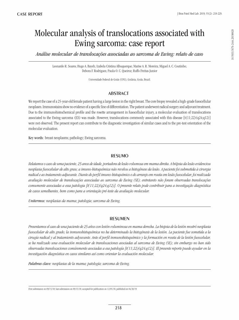
A 25-year-old woman went to the service in December 2016 due to a voluminous nodule in the right breast, which had been presenting progressive growth for six months. The patient said to have had menarche at age 14 and a previous cesarean delivery, and denied other previous surgeries, the presence of clinical comorbidities and family history of breast neoplasm. The physical examination indicated the presence of a voluminous lesion occupying practically all of the right breast parenchyma, mobile, of hardened appearance and regular surface, measuring 18 cm in its largest diameter. The skin in the right breast was tense and had diffuse hyperemia, but without aspect of neoplastic infiltration. In
the axillary palpation, bilateral, mobile and soft, non-coalescent, painless lymph nodes were identified, measuring approximately 0.5 cm in their largest diameters (Figure 1).
FigurE 1 − Physical examination, inspection: A) frontal photograph of the patient, with a mass occupying almost the whole right breast parenchyma, associated with cutaneous hyperemia and visualization of the superficial vascular network; B) photograph in profile, with observation of bulging in the upper right breast pole; C) frontal photograph, on the second postoperative day
A B C
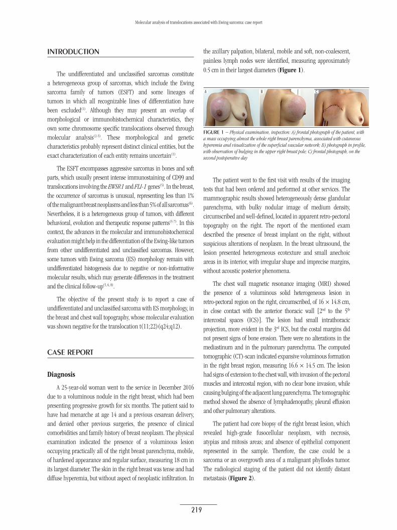
The patient went to the first visit with results of the imaging tests that had been ordered and performed at other services. The mammographic results showed heterogeneously dense glandular parenchyma, with bulky nodular image of medium density, circumscribed and well-defined, located in apparent retro-pectoral topography on the right. The report of the mentioned exam described the presence of breast implant on the right, without suspicious alterations of neoplasm. In the breast ultrasound, the lesion presented heterogeneous ecotexture and small anechoic areas in its interior, with irregular shape and imprecise margins, without acoustic posterior phenomena.
The chest wall magnetic resonance imaging (MRI) showed the presence of a voluminous solid heterogeneous lesion in retro-pectoral region on the right, circumscribed, of 16 × 14.8 cm, in close contact with the anterior thoracic wall [2nd to the 5th intercostal spaces (ICS)]. The lesion had small intrathoracic projection, more evident in the 3rd ICS, but the costal margins did not present signs of bone erosion. There were no alterations in the mediastinum and in the pulmonary parenchyma. The computed tomographic (CT)-scan indicated expansive voluminous formation in the right breast region, measuring 16.6 × 14.5 cm. The lesion had signs of extension to the chest wall, with invasion of the pectoral muscles and intercostal region, with no clear bone invasion, while causing bulging of the adjacent lung parenchyma. The tomographic method showed the absence of lymphadenopathy, pleural effusion and other pulmonary alterations.
The patient had core biopsy of the right breast lesion, which revealed high-grade fusocellular neoplasm, with necrosis, atypias and mitosis areas; and absence of epithelial component represented in the sample. Therefore, the case could be a sarcoma or an overgrowth area of a malignant phyllodes tumor. The radiological staging of the patient did not identify distant metastasis (Figure 2).
Molecular analysis of translocations associated with Ewing sarcoma: case report
220
Surgical treatment
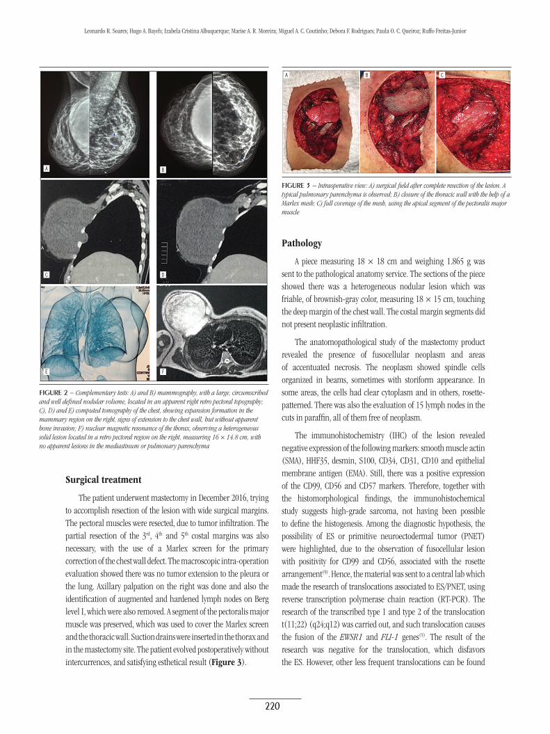
The patient underwent mastectomy in December 2016, trying to accomplish resection of the lesion with wide surgical margins. The pectoral muscles were resected, due to tumor infiltration. The partial resection of the 3rd, 4th and 5th costal margins was also necessary, with the use of a Marlex screen for the primary correction of the chest wall defect. The macroscopic intra-operation evaluation showed there was no tumor extension to the pleura or the lung. Axillary palpation on the right was done and also the identification of augmented and hardened lymph nodes on Berg level I, which were also removed. A segment of the pectoralis major muscle was preserved, which was used to cover the Marlex screen and the thoracic wall. Suction drains were inserted in the thorax and in the mastectomy site. The patient evolved postoperatively without intercurrences, and satisfying esthetical result (Figure 3).
Pathology
A piece measuring 18 × 18 cm and weighing 1.865 g was sent to the pathological anatomy service. The sections of the piece showed there was a heterogeneous nodular lesion which was friable, of brownish-gray color, measuring 18 × 15 cm, touching the deep margin of the chest wall. The costal margin segments did not present neoplastic infiltration.
The anatomopathological study of the mastectomy product revealed the presence of fusocellular neoplasm and areas of accentuated necrosis. The neoplasm showed spindle cells organized in beams, sometimes with storiform appearance. In some areas, the cells had clear cytoplasm and in others, rosette-patterned. There was also the evaluation of 15 lymph nodes in the cuts in paraffin, all of them free of neoplasm.
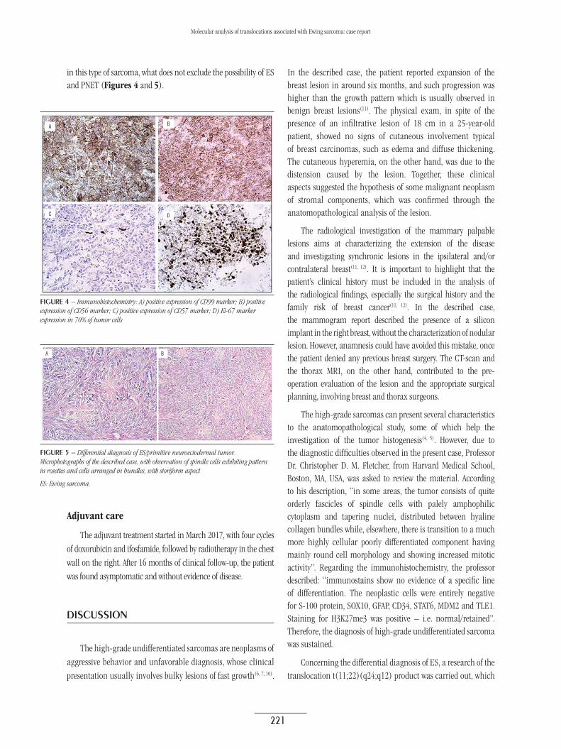
The immunohistochemistry (IHC) of the lesion revealed negative expression of the following markers: smooth muscle actin (SMA), HHF35, desmin, S100, CD34, CD31, CD10 and epithelial membrane antigen (EMA). Still, there was a positive expression of the CD99, CD56 and CD57 markers. Therefore, together with the histomorphological findings, the immunohistochemical study suggests high-grade sarcoma, not having been possible to define the histogenesis. Among the diagnostic hypothesis, the possibility of ES or primitive neuroectodermal tumor (PNET) were highlighted, due to the observation of fusocellular lesion with positivity for CD99 and CD56, associated with the rosette arrangement(9). Hence, the material was sent to a central lab which made the research of translocations associated to ES/PNET, using reverse transcription polymerase chain reaction (RT-PCR). The research of the transcribed type 1 and type 2 of the translocation t(11;22) (q24;q12) was carried out, and such translocation causes the fusion of the EWSR1 and FLI-1 genes(5). The result of the research was negative for the translocation, which disfavors the ES. However, other less frequent translocations can be found
FigurE 2 − Complementary tests: A) and B) mammography, with a large, circumscribed and well defined nodular volume, located in an apparent right retro pectoral topography; C), D) and E) computed tomography of the chest, showing expansion formation in the mammary region on the right, signs of extension to the chest wall, but without apparent bone invasion; F) nuclear magnetic resonance of the thorax, observing a heterogeneous solid lesion located in a retro pectoral region on the right, measuring 16 × 14.8 cm, with no apparent lesions in the mediastinum or pulmonary parenchyma
A
C
E
B
D
F
FigurE 3 − Intraoperative view: A) surgical field after complete resection of the lesion. A typical pulmonary parenchyma is observed; B) closure of the thoracic wall with the help of a Marlex mesh; C) full coverage of the mesh, using the apical segment of the pectoralis major muscle
A B C
Leonardo R. Soares; Hugo A. Bayeh; Izabela Cristina Albuquerque; Marise A. R. Moreira; Miguel A. C. Coutinho; Debora F. Rodrigues; Paula O. C. Queiroz; Ruffo Freitas-Junior
221
Molecular analysis of translocations associated with Ewing sarcoma: case report
in this type of sarcoma, what does not exclude the possibility of ES and PNET (Figures 4 and 5).
In the described case, the patient reported expansion of the breast lesion in around six months, and such progression was higher than the growth pattern which is usually observed in benign breast lesions(11). The physical exam, in spite of the presence of an infiltrative lesion of 18 cm in a 25-year-old patient, showed no signs of cutaneous involvement typical of breast carcinomas, such as edema and diffuse thickening. The cutaneous hyperemia, on the other hand, was due to the distension caused by the lesion. Together, these clinical aspects suggested the hypothesis of some malignant neoplasm of stromal components, which was confirmed through the anatomopathological analysis of the lesion.
The radiological investigation of the mammary palpable lesions aims at characterizing the extension of the disease and investigating synchronic lesions in the ipsilateral and/or contralateral breast(11, 12). It is important to highlight that the patient’s clinical history must be included in the analysis of the radiological findings, especially the surgical history and the family risk of breast cancer(11, 12). In the described case, the mammogram report described the presence of a silicon implant in the right breast, without the characterization of nodular lesion. However, anamnesis could have avoided this mistake, once the patient denied any previous breast surgery. The CT-scan and the thorax MRI, on the other hand, contributed to the pre-operation evaluation of the lesion and the appropriate surgical planning, involving breast and thorax surgeons.
The high-grade sarcomas can present several characteristics to the anatomopathological study, some of which help the investigation of the tumor histogenesis(4, 5). However, due to the diagnostic difficulties observed in the present case, Professor Dr. Christopher D. M. Fletcher, from Harvard Medical School, Boston, MA, USA, was asked to review the material. According to his description, “in some areas, the tumor consists of quite orderly fascicles of spindle cells with palely amphophilic cytoplasm and tapering nuclei, distributed between hyaline collagen bundles while, elsewhere, there is transition to a much more highly cellular poorly differentiated component having mainly round cell morphology and showing increased mitotic activity”. Regarding the immunohistochemistry, the professor described: “immunostains show no evidence of a specific line of differentiation. The neoplastic cells were entirely negative for S-100 protein, SOX10, GFAP, CD34, STAT6, MDM2 and TLE1. Staining for H3K27me3 was positive – i.e. normal/retained”. Therefore, the diagnosis of high-grade undifferentiated sarcoma was sustained.
Concerning the differential diagnosis of ES, a research of the translocation t(11;22)(q24;q12) product was carried out, which
FigurE 5 − Differential diagnosis of ES/primitive neuroectodermal tumor. Microphotographs of the described case, with observation of spindle cells exhibiting pattern in rosettes and cells arranged in bundles, with storiform aspect
ES: Ewing sarcoma.
A B
FigurE 4 − Immunohistochemistry: A) positive expression of CD99 marker; B) positive expression of CD56 marker; C) positive expression of CD57 marker; D) Ki-67 marker expression in 70% of tumor cells
A
C
B
D
Adjuvant care
The adjuvant treatment started in March 2017, with four cycles of doxorubicin and ifosfamide, followed by radiotherapy in the chest wall on the right. After 16 months of clinical follow-up, the patient was found asymptomatic and without evidence of disease.
DiSCuSSion
The high-grade undifferentiated sarcomas are neoplasms of aggressive behavior and unfavorable diagnosis, whose clinical presentation usually involves bulky lesions of fast growth(6, 7, 10).
222
is observed in the majority of the cases of the disease(2, 13). This translocation causes the fusion of the EWSR1 and FLI-1 genes, mapped in 22q12 and 11q24, respectively. Nevertheless, depending on the breakpoints in these genes, 18 different in-frame chimeric transcripts can be generated. Of these, transcripts type 1 and type 2 must be highlighted, originated from the fusion of exon 7 of the EWSR1 gene with the exon 6 or the exon 5 of the FLI-1, respectively(2, 13). These transcripts are present in approximately 85% of the ES/PNET cases with t(11;22)(q24;q12) and in 60% of the total cases of the disease(2, 5, 13). However, other less frequent translocations can be found in this kind of sarcoma, which does not exclude the possibility of ES and PNET(2-5).
Considering the surgical treatment, the patient of the case described underwent mastectomy due to the tumor extension in the physical examinations and the imaging tests. Despite the description of deep involved margins, the surgery was considered adequate because of the fact that this margin coincided with the extension of the chest wall, with its enlargement not being possible. Similarly to the carcinomas, higher rates of local recurrence in the presence of surgical involved margins were observed in the approach of the malignant neoplasms of mesenchymal origin(10, 14). Nonetheless, in the occurrence of free margins, there were no differences in the global survival and free survival of disease in comparison with conservative surgeries and mastectomies(10, 14).
Regarding the axillary involvement, the lymphatic dissemination for the regional chains was observed to be rare, given the prevalence of hematogenous dissemination(15-17). Therefore, the investigation of the axillary status is not commonly indicated in the surgical approach to breast sarcomas(16). However, in the cases of clinically affected axillary lymph nodes, in patients with high-grade sarcomas and with locally advanced disease, the biopsy of sentinel lymph node or the axillary lymphadenectomy can be performed(17, 18). In the case in question, despite the identification of suspicious lymph nodes during the intraoperative evaluation, only the microscopy reaction aspect was observed.
In the case described, the indication for chemotherapy and supporting radiotherapy was individualized and justified due to the tumor aggression and the disease extension(10). However, considering the clinical heterogeneity of the undifferentiated sarcomas and the absence of the randomized clinical trials with this population, the controversies regarding the commonly used chemotherapy protocols remain. In the case of confirmed ES, on the other hand, the vincristine-doxorubicin-cyclophosphamide
(VDC) protocol altered with the ifosfamide-etoposide was shown to be superior to the VDC isolated scheme, with global survival of five years of 72% and 61%, respectively(19). However, this is a high-toxicity protocol, with approximately 12 months duration, performing local therapy (surgery, radiotherapy or both) after 12 weeks of treatment(19, 20). Thus, before the final diagnosis of undifferentiated and unclassified sarcoma, chemotherapy with standard scheme for soft tissue sarcomas was chosen, with doxorubicin and ifosfamide(21). In the case of the radiotherapy, in spite of the controversy in some cases of sarcomas, it reduces the local recurrences in patients that carry locally advanced diseases, with high-grade tumors or whose surgery revealed affected margins(10).
ConCLuSion
We described a case of undifferentiated and unclassified sarcoma, whose histology and immunohistochemistry showed to be compatible with the differential diagnosis of ES. However, there were no translocations commonly observed with this pathology through molecular evaluation. The present report can contribute to the diagnostic investigation of similar cases and the pre-test orientation of the molecular evaluation.
aCknowLEDgMEntS
We thank all the multiprofessional healthcare team involved in the clinical follow-up of the patient; and Prof. Dr. Christopher D. M. Fletcher, for assistance in the histological review of the case described.
DECLaration oF ConFLiCting intErEStS
The authors declared no potential conflicts of interest with respect to the research, authorship, and/or publication of this article.
FunDing
The authors received no financial support for the research, authorship, and/or publication of this article.
Leonardo R. Soares; Hugo A. Bayeh; Izabela Cristina Albuquerque; Marise A. R. Moreira; Miguel A. C. Coutinho; Debora F. Rodrigues; Paula O. C. Queiroz; Ruffo Freitas-Junior
223
rEFErEnCES
1. Jo VW, Fletcher CDM. WHO classification of soft tissue tumours: an update based on the 2013. 4th edition. Pathology. 2014; 46: 95-104.
2. Sorensen PH, Lessnick SL, Lopez-Terrada D, Liu XF, Triche TJ, Denny CT. A second Ewing’s sarcoma translocation, t(21;22), fuses the EWS gene to another ETS-family transcription factor, ERG. Nat Genet. 1994; 6: 146-51.
3. Argani P, Perez-Ordoñez B, Xiao H, Caruana SM, Huvos AG, Ladanyi M. Olfactory neuroblastoma is not related to the Ewing family of tumors: absence of EWS/FLI1 gene fusion and MIC2 expression. Am J Surg Pathol. 1998; 22: 391-8.
4. Evola FR, Costarella L, Pavone V, et al. Biomarkers of osteosarcoma, chondrosarcoma, and Ewing sarcoma. Front Pharmacol. 2017; 8: 150.
5. Machado I, Navarro L, Pellin A, et al. Defining Ewing and Ewing-like small round cell tumors (SRCT): the need for molecular techniques in their categorization and differential diagnosis. A study of 200 cases. Ann Diagn Pathol. 2016; 22: 25-32.
6. Lim SZ, Ong KW, Tan BK, Selvarajan S, Tan PH. Sarcoma of the breast: an update on a rare entity. J Clin Pathol. 2016; 69: 373-81.
7. Bousquet G, Confavreux C, Magné N, et al. Outcome and prognostic factors in breast sarcoma: a multicenter study from the rare cancer network. Radiother Oncol. 2007; 85: 355-61.
8. Llombart-Bosch A, Machado I, Navarro S, et al. Histological heterogeneity of Ewing’s sarcoma/PNET: an immunohistochemical analysis of 415 genetically confirmed cases with clinical support. Virchows Arch. 2009; 455: 397-411.
9. Weiss SW, Goldblum JR. Ewing’s sarcoma/PNET tumor family and related lesions. In: Weiss SW, Goldblum JR, editors. Enzinger and Weiss’s soft tissue tumors. 5th ed. Philadelphia: Mosby; 2008. pp. 945-87.
10. Hu QC, Mei X, Feng Y, et al. Early local recurrence presents adverse effect on outcomes of primary breast sarcoma: a Retrospective Study From Single Institute in China. Medicine (Baltimore). 2016; 95: e2422.
11. Gao Y, Saksena MA, Brachtel EF, terMeulen DC, Rafferty EA. How to approach breast lesions in children and adolescents. Eur J Radiol. 2015; 84: 1350-64.
12. Pruthi S. Detection and evaluation of a palpable breast mass. Mayo Clin Proc. 2001; 76: 641-7.
13. de Alava E, Kawai A, Healey JH, et al. EWS-FLI1 fusion transcript structure is an independent determinant of prognosis in Ewing’s sarcoma. J Clin Oncol. 1998; 16: 1248-55. Erratum in: J Clin Oncol. 1998 Aug; 16: 2895.
14. McGowan TS, Cummings BJ, O’Sullivan B, Catton CN, Miller N, Panzarella T. An analysis of 78 breast sarcoma patients without distant metastases at presentation. Int J Radiat Oncol Biol Phys. 2000; 46: 383-90.
15. Weigrand DN, Rosenberg AS. Early lymphatic spread of osteogenic and soft tissue sarcomas. Surgery. 1978; 84: 231-40.
16. Li N, Cusidó MT, Navarro B, et al. Breast sarcoma. A case report and review of literature. Int J Surg Case Rep. 2016; 24: 203-5.
17. Diaz Brito JA, Gatti G, Vento AR, et al. Report on a case of breast sarcoma metastatic to the axillary lymph nodes. Tumori. 2006; 92: 188-90.
18. Blazer DG 3rd, Sabel MS, Sondak VK. Is there a role for sentinel lymph node biopsy in the management of sarcoma? Surg Oncol. 2003; 12: 201-6.
19. Grier HE, Krailo MD, Tarbell NJ, et al. Addition of ifosfamide and etoposide to standard chemotherapy for Ewing’s sarcoma and primitive neuroectodermal tumor of bone. N Engl J Med. 2003; 348: 694-701.
20. Womer RB, West DC, Krailo MD, et al. Randomized controlled trial of interval-compressed chemotherapy for the treatment of localized Ewing sarcoma: a report from the Children’s Oncology Group. J Clin Oncol. 2012; 30: 4148-54.
21. Colia V, Fiore M, Provenzano S, et al. Activity of anthracycline- and ifosfamide-based chemotherapy in a series of patients affected by advanced myxofibrosarcoma. Clin Sarcoma Res. 2017; 7: 16.
Molecular analysis of translocations associated with Ewing sarcoma: case report
CorrESPonDing author
Leonardo Ribeiro Soares 0000-0002-9448-6114e-mail: [email protected].
This is an open-access article distributed under the terms of the Creative Commons Attribution License.