modeling the impact on virus transmission of wolbachia ... · dengue modeling the impact on virus...
TRANSCRIPT
R E S EARCH ART I C L E
DENGUE
Modeling the impact on virus transmission ofWolbachia-mediated blocking of dengue virusinfection of Aedes aegyptiNeil M. Ferguson,1* Duong Thi Hue Kien,2 Hannah Clapham,1 Ricardo Aguas,1 Vu Tuan Trung,2
Tran Nguyen Bich Chau,2 Jean Popovici,3 Peter A. Ryan,3 Scott L. O’Neill,3
Elizabeth A. McGraw,3 Vo Thi Long,2 Le Thi Dui,2 Hoa L. Nguyen,2 Nguyen Van Vinh Chau,4
Bridget Wills,2,5 Cameron P. Simmons2,5,6
http://stm.science
Dow
nloaded from
Dengue is the most common arboviral infection of humans and is a public health burden in more than 100 countries.Aedes aegyptimosquitoes stably infected with strains of the intracellular bacteriumWolbachia are resistant to denguevirus (DENV) infection and are being tested in field trials. To mimic field conditions, we experimentally assessed thevector competence of A. aegypti carrying theWolbachia strains wMel and wMelPop after challenge with viremic bloodfrom dengue patients. We found that wMelPop conferred strong resistance to DENV infection of mosquito abdomentissue and largely prevented disseminated infection. wMel conferred less resistance to infection of mosquito abdomentissue, but it did reduce the prevalence of mosquitoes with infectious saliva. A mathematical model of DENV trans-mission incorporating the dynamics of viral infection in humans and mosquitoes was fitted to the data collected.Model predictions suggested that wMel would reduce the basic reproduction number, R0, of DENV transmission by66 to 75%. Our results suggest that establishment of wMelPop-infected A. aegypti at a high frequency in a dengue-endemic setting would result in the complete abatement of DENV transmission. Establishment of wMel-infected A.aegypti is also predicted to have a substantial effect on transmission that would be sufficient to eliminate dengue inlow or moderate transmission settings but may be insufficient to achieve complete control in settings where R0 ishigh. These findings develop a framework for selecting Wolbachia strains for field releases and for calculating theirlikely impact.
mag
by guest on March 30, 2020
.org/
INTRODUCTIONDengue is an acute systemic viral infection (1). In 2010, there were anestimated 100 million apparent infections globally (2). The etiologicalagents of dengue are four dengue viruses (DENV1 to DENV4), withtransmission from human to human primarily byAedes aegyptimosqui-toes. Existing disease prevention strategies are based on reducing themosquito vector population, yet this has been largely unsuccessful inhalting dengue transmission in endemic countries.
A new entomologically based control method uses the phenotypeofA. aegypti experimentally infected with strains (wMel andwMelPop)of the bacterial symbiontWolbachia (3, 4). The heritable wMelPop in-fection of A. aegypti is characterized by widely disseminated and denseinfection of mosquito tissues (3). wMelPop infection confers nu-merous phenotypic traits on A. aegypti, including refractoriness toDENV infection (5), reduced life span (3), reduced viability ofdesiccated eggs (6), and reduced blood feeding success (7). The her-itable wMel infection of A. aegypti is associated with a relatively lowerintensity of tissue infection and can also confer complete resistance todisseminated DENV infection after laboratory challenge (4). The
1Medical Research Council Centre for Outbreak Analysis andModelling, School of PublicHealth, Imperial College London, Norfolk Place, London W2 1PG, UK. 2Oxford UniversityClinical Research Unit, Hospital for Tropical Diseases, 764 Võ Vǎn Kiêt, District 5, Ho ChiMinh City 748010, Vietnam. 3School of Biological Sciences, Monash University, Clayton,Victoria 3800, Australia. 4Hospital for Tropical Diseases, 190 Ben Hám Tú, District 5, HoChi Minh City 748010, Vietnam. 5Centre for Tropical Medicine and Global Health,Nuffield Department of Medicine, University of Oxford, Oxford OX1 7FZ, UK.6Department of Microbiology and Immunology and Nossal Institute of Global Health,University of Melbourne, Carlton, Victoria 3010, Australia.*Corresponding author. E-mail: [email protected]
www.Scien
mechanism of virus interference is unknown, but could potentiallybe mediated by Wolbachia-triggered changes in immunoregulatorymicroRNAexpression, elevation of reactive oxygen species, or compe-tition between DENV andWolbachia for critical metabolic resources(8–10). Successful field releases of wMel-A. aegypti have occurred inthe northern Australian city of Cairns (11), providing proof of conceptthat stable, long-term establishment ofWolbachia in mosquito popu-lations can be achieved.
The cost of developing a new operationalized vector control mea-sure and testing its effectiveness in the field makes it a priority to tryto predict the likely impact of the introduction of Wolbachia intoA. aegypti populations on dengue transmission. However, previousvector competence studies ofWolbachia-infected A. aegypti had sig-nificant limitations in that they used a single serotype of laboratory-passaged DENV that was spiked into animal or human blood tocreate infectious blood meals (4, 5). This model system probably doesnot accurately mimic a human DENV infection in that DENVs haveevolved to be efficiently transmitted tomosquitoes through fresh bloodmeals from infected human hosts. We describe here vector com-petence studies that use viremic blood from dengue patients toblood-feed field-derivedWolbachia-infected A. aegypti and thus pro-vide “real-world”measures of vector competence.
More generally, translating laboratory studies of vector compe-tence into an assessment of the potential effectiveness ofWolbachiain reducing dengue transmission to human populations requires anunderstanding of multiple interacting aspects of mosquito ecologyand the biology of DENV infection. In addition to characterizingthe invasion dynamics of Wolbachia into A. aegypti populations
ceTranslationalMedicine.org 18 March 2015 Vol 7 Issue 279 279ra37 1
R E S EARCH ART I C L E
(the goal of field trials currently under way), we require better un-derstanding of (i) the development of DENV infection inmosquitoes(and how this is modified by Wolbachia), (ii) the within-host dy-namics of DENV infection in humans, and (iii) DENV transmissionfrom mosquitoes to humans and from humans to mosquitoes (andhow this is modified byWolbachia). Here, we begin to address thesedata needs by combining experimental characterization of the impactof Wolbachia infection on vector competence with mathematicalmodeling of the natural history of DENV infection in humans andvectors. By using more biologically realistic experimental and mathe-matical models than hitherto possible, we have generated estimates ofthe impact of Wolbachia strains on dengue transmission that can beused with greater confidence to inform future field trials in dengue-endemic areas and to guide the development of additionalWolbachiastrains in A. aegypti.
by guest on March 30, 2020
http://stm.sciencem
ag.org/D
ownloaded from
RESULTS
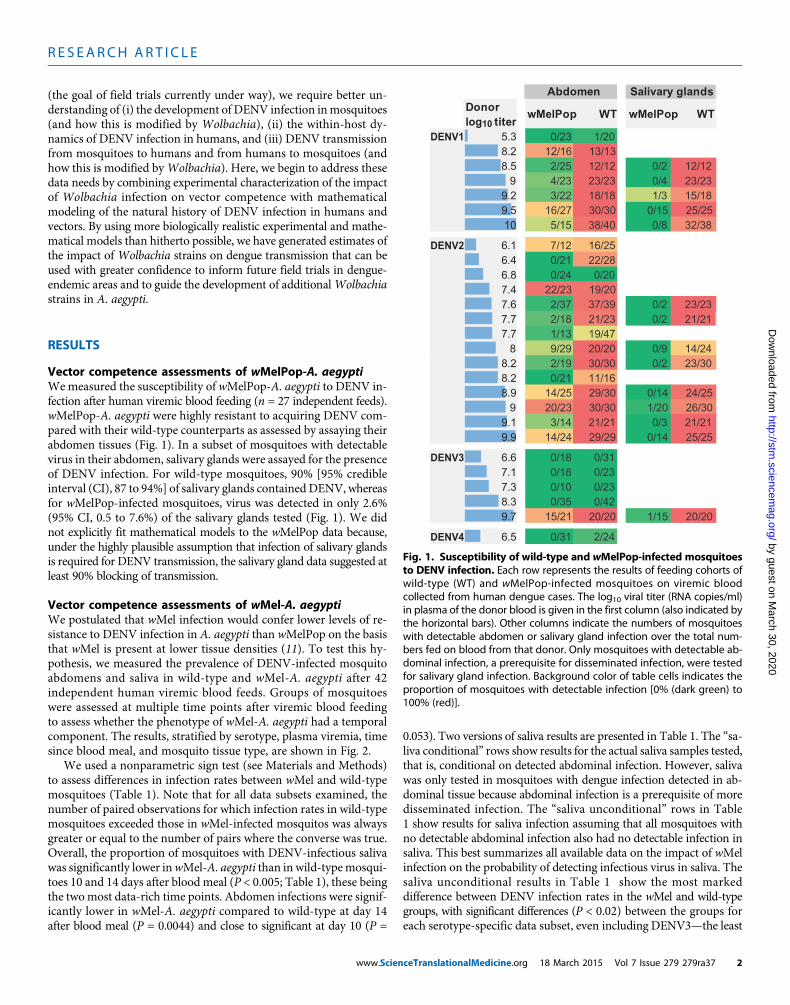
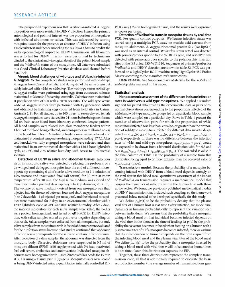
Vector competence assessments of wMelPop-A. aegyptiWemeasured the susceptibility of wMelPop-A. aegypti to DENV in-fection after human viremic blood feeding (n = 27 independent feeds).wMelPop-A. aegypti were highly resistant to acquiring DENV com-pared with their wild-type counterparts as assessed by assaying theirabdomen tissues (Fig. 1). In a subset of mosquitoes with detectablevirus in their abdomen, salivary glands were assayed for the presenceof DENV infection. For wild-type mosquitoes, 90% [95% credibleinterval (CI), 87 to 94%] of salivary glands contained DENV, whereasfor wMelPop-infected mosquitoes, virus was detected in only 2.6%(95% CI, 0.5 to 7.6%) of the salivary glands tested (Fig. 1). We didnot explicitly fit mathematical models to the wMelPop data because,under the highly plausible assumption that infection of salivary glandsis required for DENV transmission, the salivary gland data suggested atleast 90% blocking of transmission.
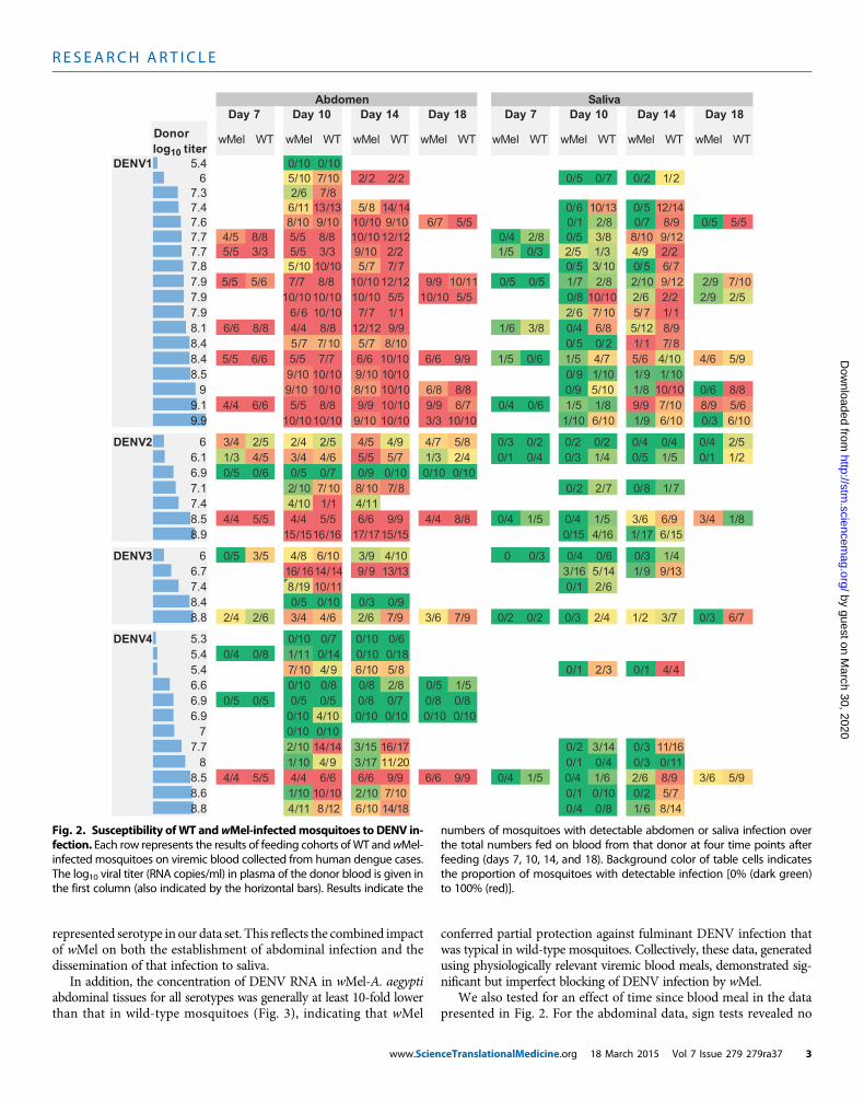
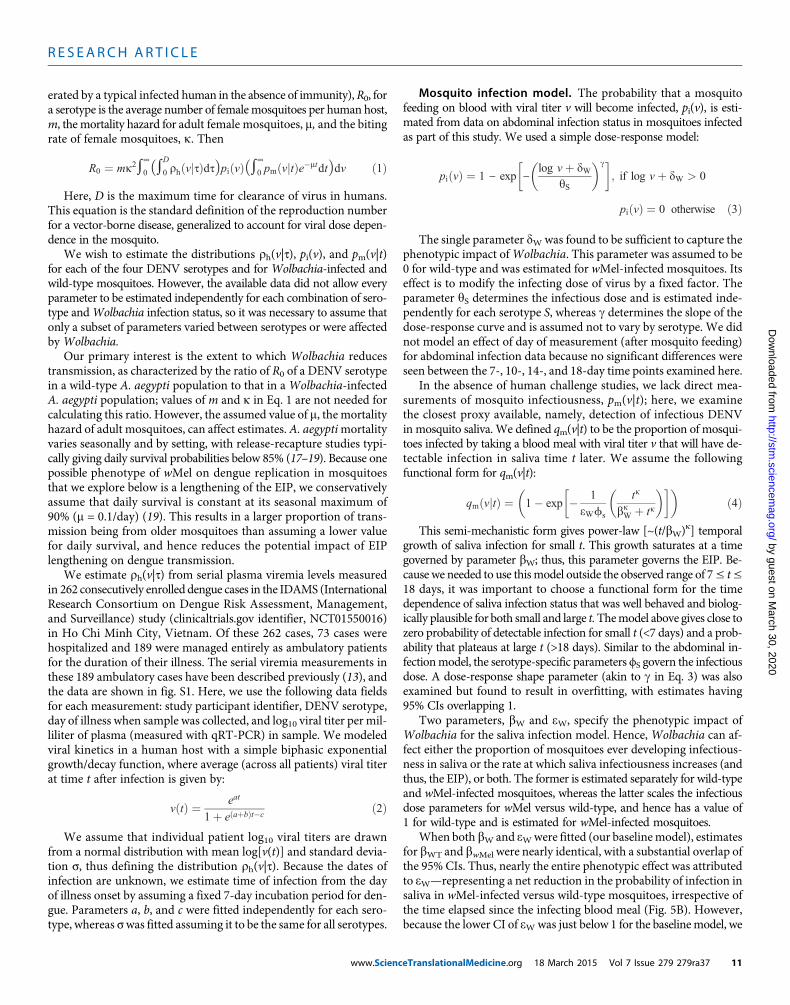
Vector competence assessments of wMel-A. aegyptiWe postulated that wMel infection would confer lower levels of re-sistance to DENV infection in A. aegypti than wMelPop on the basisthat wMel is present at lower tissue densities (11). To test this hy-pothesis, we measured the prevalence of DENV-infected mosquitoabdomens and saliva in wild-type and wMel-A. aegypti after 42independent human viremic blood feeds. Groups of mosquitoeswere assessed at multiple time points after viremic blood feedingto assess whether the phenotype of wMel-A. aegypti had a temporalcomponent. The results, stratified by serotype, plasma viremia, timesince blood meal, and mosquito tissue type, are shown in Fig. 2.
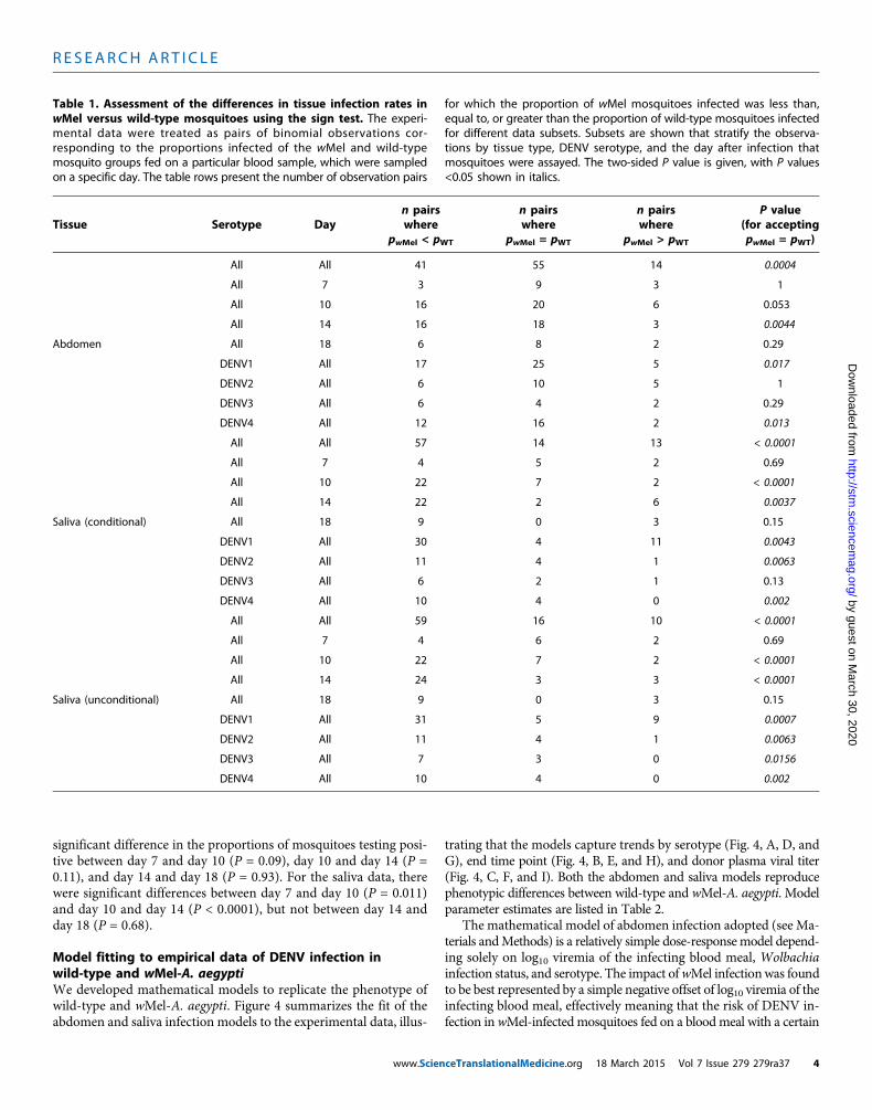
We used a nonparametric sign test (see Materials and Methods)to assess differences in infection rates between wMel and wild-typemosquitoes (Table 1). Note that for all data subsets examined, thenumber of paired observations for which infection rates in wild-typemosquitoes exceeded those in wMel-infected mosquitos was alwaysgreater or equal to the number of pairs where the converse was true.Overall, the proportion of mosquitoes with DENV-infectious salivawas significantly lower inwMel-A. aegypti than in wild-typemosqui-toes 10 and 14 days after blood meal (P < 0.005; Table 1), these beingthe twomost data-rich time points. Abdomen infections were signif-icantly lower in wMel-A. aegypti compared to wild-type at day 14after blood meal (P = 0.0044) and close to significant at day 10 (P =
www.Scien
0.053). Two versions of saliva results are presented in Table 1. The “sa-liva conditional” rows show results for the actual saliva samples tested,that is, conditional on detected abdominal infection. However, salivawas only tested in mosquitoes with dengue infection detected in ab-dominal tissue because abdominal infection is a prerequisite of moredisseminated infection. The “saliva unconditional” rows in Table1 show results for saliva infection assuming that all mosquitoes withno detectable abdominal infection also had no detectable infection insaliva. This best summarizes all available data on the impact of wMelinfection on the probability of detecting infectious virus in saliva. Thesaliva unconditional results in Table 1 show the most markeddifference between DENV infection rates in the wMel and wild-typegroups, with significant differences (P < 0.02) between the groups foreach serotype-specific data subset, even including DENV3—the least
Donor log10 titer
wMelPop WT wMelPop WT
DENV1 5.3 0/23 1/208.2 12/16 13/138.5 2/25 12/12 0/2 12/12
9 4/23 23/23 0/4 23/239.2 3/22 18/18 1/3 15/189.5 16/27 30/30 0/15 25/2510 5/15 38/40 0/8 32/38
DENV2 6.1 7/12 16/256.4 0/21 22/286.8 0/24 0/207.4 22/23 19/207.6 2/37 37/39 0/2 23/237.7 2/18 21/23 0/2 21/217.7 1/13 19/47
8 9/29 20/20 0/9 14/248.2 2/19 30/30 0/2 23/308.2 0/21 11/168.9 14/25 29/30 0/14 24/25
9 20/23 30/30 1/20 26/309.1 3/14 21/21 0/3 21/219.9 14/24 29/29 0/14 25/25
DENV3 6.6 0/18 0/317.1 0/18 0/237.3 0/10 0/238.3 0/35 0/429.7 15/21 20/20 1/15 20/20
DENV4 6.5 0/31 2/24
Abdomen Salivary glands
Fig. 1. Susceptibility of wild-type and wMelPop-infected mosquitoesto DENV infection. Each row represents the results of feeding cohorts of
wild-type (WT) and wMelPop-infected mosquitoes on viremic bloodcollected from human dengue cases. The log10 viral titer (RNA copies/ml)in plasma of the donor blood is given in the first column (also indicated bythe horizontal bars). Other columns indicate the numbers of mosquitoeswith detectable abdomen or salivary gland infection over the total num-bers fed on blood from that donor. Only mosquitoes with detectable ab-dominal infection, a prerequisite for disseminated infection, were testedfor salivary gland infection. Background color of table cells indicates theproportion of mosquitoes with detectable infection [0% (dark green) to100% (red)].ceTranslationalMedicine.org 18 March 2015 Vol 7 Issue 279 279ra37 2
R E S EARCH ART I C L E
by guest on March 30, 2020
http://stm.sciencem
ag.org/D
ownloaded from
represented serotype in our data set. This reflects the combined impactof wMel on both the establishment of abdominal infection and thedissemination of that infection to saliva.
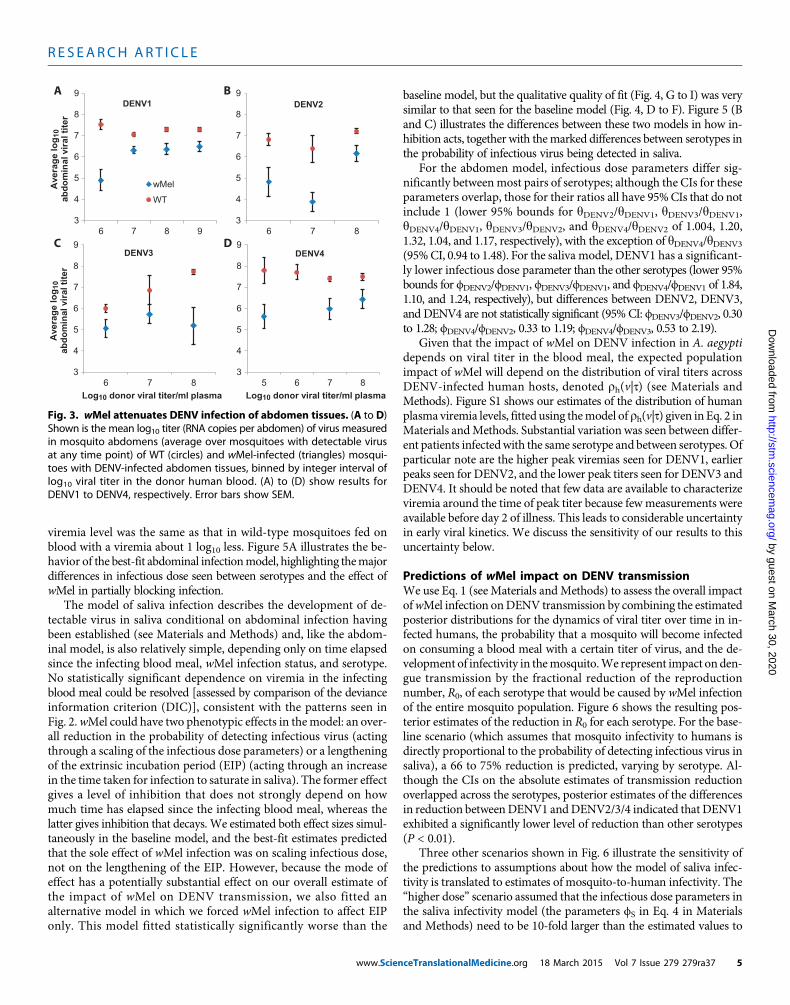
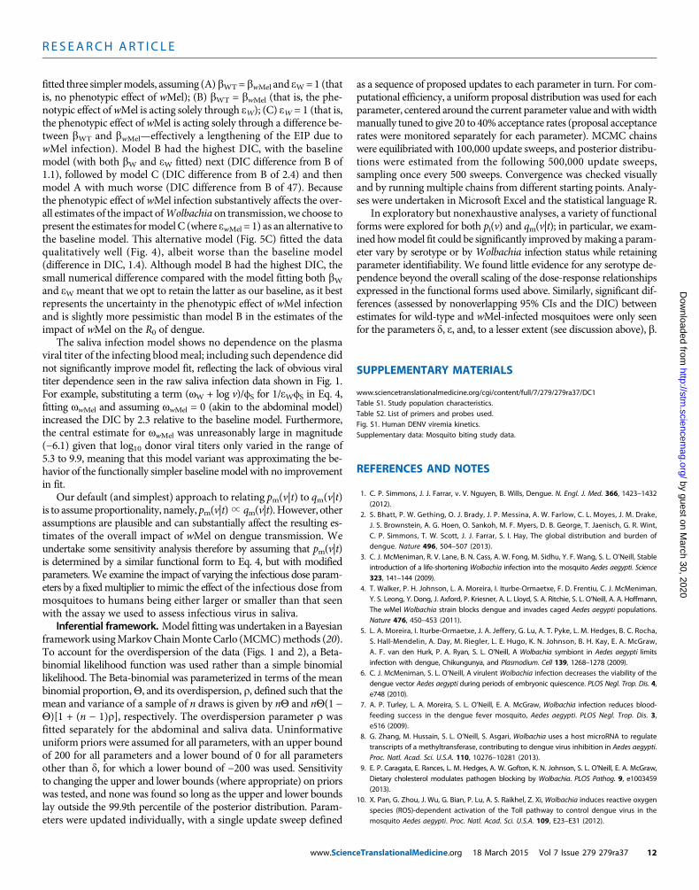
In addition, the concentration of DENV RNA in wMel-A. aegyptiabdominal tissues for all serotypes was generally at least 10-fold lowerthan that in wild-type mosquitoes (Fig. 3), indicating that wMel
www.Scien
conferred partial protection against fulminant DENV infection thatwas typical in wild-type mosquitoes. Collectively, these data, generatedusing physiologically relevant viremic blood meals, demonstrated sig-nificant but imperfect blocking of DENV infection by wMel.
We also tested for an effect of time since blood meal in the datapresented in Fig. 2. For the abdominal data, sign tests revealed no
Day 7 Day 10 Day 14 Day 18 Day 7 Day 10 Day 14 Day 18
Donor log10 titer
wMel WT wMel WT wMel WT wMel WT wMel WT wMel WT wMel WT wMel WT
DENV1 5.4 0/10 0/106 2/12/07/05/02/22/201/701/5
7.3 2/6 7/87.4 41/215/031/016/041/418/531/3111/67.6 8/10 9/10 10/10 9/10 6/7 5/5 0/1 2/8 0/7 8/9 0/5 5/57.7 4/5 8/8 5/5 8/8 10/1012/12 0/4 2/8 0/5 3/8 8/10 9/127.7 5/5 3/3 5/5 3/3 9/10 2/2 1/5 0/3 2/5 1/3 4/9 2/27.8 7/65/001/35/07/77/501/0101/57.9 5/5 5/6 7/7 8/8 10/1012/12 9/9 10/11 0/5 0/5 1/7 2/8 2/10 9/12 2/9 7/107.9 10/1010/10 10/10 5/5 10/10 5/5 0/8 10/10 2/6 2/2 2/9 2/57.9 1/17/501/76/21/17/701/016/68.1 6/6 8/8 4/4 8/8 12/12 9/9 1/6 3/8 0/4 6/8 5/12 8/98.4 8/71/12/05/001/87/501/77/58.4 5/5 6/6 5/5 7/7 6/6 10/10 6/6 9/9 1/5 0/6 1/5 4/7 5/6 4/10 4/6 5/98.5 01/19/101/19/001/0101/910/1001/9
9 9/10 10/10 8/10 10/10 6/8 8/8 0/9 5/10 1/8 10/10 0/6 8/89.1 4/4 6/6 5/5 8/8 9/9 10/10 9/9 6/7 0/4 0/6 1/5 1/8 9/9 7/10 8/9 5/69.9 10/1010/10 9/10 10/10 3/3 10/10 1/10 6/10 1/9 6/10 0/3 6/10
DENV2 6 3/4 2/5 2/4 2/5 4/5 4/9 4/7 5/8 0/3 0/2 0/2 0/2 0/4 0/4 0/4 2/56.1 1/3 4/5 3/4 4/6 5/5 5/7 1/3 2/4 0/1 0/4 0/3 1/4 0/5 1/5 0/1 1/26.9 0/5 0/6 0/5 0/7 0/9 0/10 0/10 0/107.1 7/18/07/22/08/701/801/701/27.4 4/10 1/1 4/118.5 4/4 5/5 4/4 5/5 6/6 9/9 4/4 8/8 0/4 1/5 0/4 1/5 3/6 6/9 3/4 1/88.9 51/671/161/451/051/5171/7161/6151/51
DENV3 6 0/5 3/5 4/8 6/10 3/9 4/10 0 0/3 0/4 0/6 0/3 1/46.7 31/99/141/561/331/319/941/4161/617.4 6/21/011/0191/88.4 0/5 0/10 0/3 0/98.8 2/4 2/6 3/4 4/6 2/6 7/9 3/6 7/9 0/2 0/2 0/3 2/4 1/2 3/7 0/3 6/7
DENV4 5.3 0/10 0/7 0/10 0/65.4 0/4 0/8 1/11 0/14 0/10 0/185.4 4/41/03/21/08/501/69/401/76.6 0/10 0/8 0/8 2/8 0/5 1/56.9 0/5 0/5 0/5 0/5 0/8 0/7 0/8 0/86.9 0/10 4/10 0/10 0/10 0/10 0/10
7 0/10 0/107.7 61/113/041/32/071/6151/341/4101/2
8 11/03/04/01/002/1171/39/401/18.5 4/4 5/5 4/4 6/6 6/6 9/9 6/6 9/9 0/4 1/5 0/4 1/6 2/6 8/9 3/6 5/98.6 7/52/001/01/001/701/201/0101/18.8 41/86/18/04/081/4101/621/811/4
avilaSAbdomen
Fig. 2. Susceptibility ofWT andwMel-infectedmosquitoes to DENV in-fection. Each row represents the results of feeding cohorts of WT andwMel-
numbers of mosquitoes with detectable abdomen or saliva infection overthe total numbers fed on blood from that donor at four time points after
infected mosquitoes on viremic blood collected from human dengue cases.The log10 viral titer (RNA copies/ml) in plasma of the donor blood is given inthe first column (also indicated by the horizontal bars). Results indicate the
feeding (days 7, 10, 14, and 18). Background color of table cells indicatesthe proportion of mosquitoes with detectable infection [0% (dark green)to 100% (red)].
ceTranslationalMedicine.org 18 March 2015 Vol 7 Issue 279 279ra37 3
R E S EARCH ART I C L E
by guest on March 30, 2020
http://stm.sciencem
ag.org/D
ownloaded from
significant difference in the proportions of mosquitoes testing posi-tive between day 7 and day 10 (P = 0.09), day 10 and day 14 (P =0.11), and day 14 and day 18 (P = 0.93). For the saliva data, therewere significant differences between day 7 and day 10 (P = 0.011)and day 10 and day 14 (P < 0.0001), but not between day 14 andday 18 (P = 0.68).
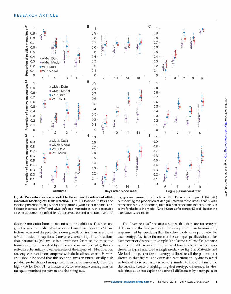
Model fitting to empirical data of DENV infection inwild-type and wMel-A. aegyptiWe developed mathematical models to replicate the phenotype ofwild-type and wMel-A. aegypti. Figure 4 summarizes the fit of theabdomen and saliva infection models to the experimental data, illus-
www.Scien
trating that the models capture trends by serotype (Fig. 4, A, D, andG), end time point (Fig. 4, B, E, and H), and donor plasma viral titer(Fig. 4, C, F, and I). Both the abdomen and saliva models reproducephenotypic differences between wild-type and wMel-A. aegypti. Modelparameter estimates are listed in Table 2.
The mathematical model of abdomen infection adopted (see Ma-terials andMethods) is a relatively simple dose-responsemodel depend-ing solely on log10 viremia of the infecting blood meal, Wolbachiainfection status, and serotype. The impact ofwMel infectionwas foundto be best represented by a simple negative offset of log10 viremia of theinfecting blood meal, effectively meaning that the risk of DENV in-fection inwMel-infected mosquitoes fed on a bloodmeal with a certain
Table 1. Assessment of the differences in tissue infection rates inwMel versus wild-type mosquitoes using the sign test. The experi-mental data were treated as pairs of binomial observations cor-responding to the proportions infected of the wMel and wild-typemosquito groups fed on a particular blood sample, which were sampledon a specific day. The table rows present the number of observation pairs
for which the proportion of wMel mosquitoes infected was less than,equal to, or greater than the proportion of wild-type mosquitoes infectedfor different data subsets. Subsets are shown that stratify the observa-tions by tissue type, DENV serotype, and the day after infection thatmosquitoes were assayed. The two-sided P value is given, with P values<0.05 shown in italics.
Tissue
Serotype Day n pairswherepwMel < pWT
ceTranslatio
n pairswhere
pwMel = pWT
nalMedicine.org 18 Ma
n pairswhere
pwMel > pWT
rch 2015 Vol 7 Issue 2
P value(for acceptingpwMel = pWT)
All
All 41 55 14 0.0004All
7 3 9 3 1All
10 16 20 6 0.053All
14 16 18 3 0.0044Abdomen
All 18 6 8 2 0.29DENV1
All 17 25 5 0.017DENV2
All 6 10 5 1DENV3
All 6 4 2 0.29DENV4
All 12 16 2 0.013All
All 57 14 13 < 0.0001All
7 4 5 2 0.69All
10 22 7 2 < 0.0001All
14 22 2 6 0.0037Saliva (conditional)
All 18 9 0 3 0.15DENV1
All 30 4 11 0.0043DENV2
All 11 4 1 0.0063DENV3
All 6 2 1 0.13DENV4
All 10 4 0 0.002All
All 59 16 10 < 0.0001All
7 4 6 2 0.69All
10 22 7 2 < 0.0001All
14 24 3 3 < 0.0001Saliva (unconditional)
All 18 9 0 3 0.15DENV1
All 31 5 9 0.0007DENV2
All 11 4 1 0.0063DENV3
All 7 3 0 0.0156DENV4
All 10 4 0 0.00279 279ra37 4
R E S EARCH ART I C L E
by guest on March 30, 2020
http://stm.sciencem
ag.org/D
ownloaded from
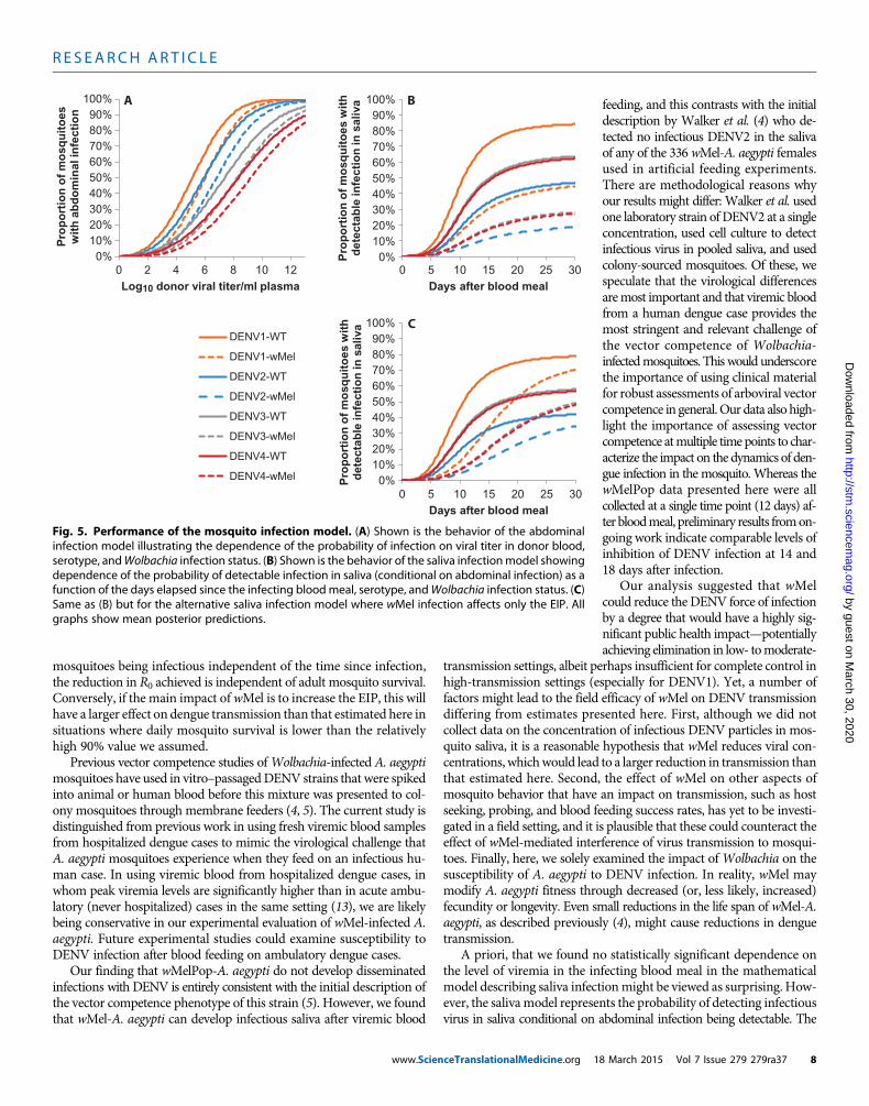
viremia level was the same as that in wild-type mosquitoes fed onblood with a viremia about 1 log10 less. Figure 5A illustrates the be-havior of the best-fit abdominal infectionmodel, highlighting themajordifferences in infectious dose seen between serotypes and the effect ofwMel in partially blocking infection.
The model of saliva infection describes the development of de-tectable virus in saliva conditional on abdominal infection havingbeen established (see Materials and Methods) and, like the abdom-inal model, is also relatively simple, depending only on time elapsedsince the infecting blood meal, wMel infection status, and serotype.No statistically significant dependence on viremia in the infectingblood meal could be resolved [assessed by comparison of the devianceinformation criterion (DIC)], consistent with the patterns seen inFig. 2.wMel could have two phenotypic effects in themodel: an over-all reduction in the probability of detecting infectious virus (actingthrough a scaling of the infectious dose parameters) or a lengtheningof the extrinsic incubation period (EIP) (acting through an increasein the time taken for infection to saturate in saliva). The former effectgives a level of inhibition that does not strongly depend on howmuch time has elapsed since the infecting blood meal, whereas thelatter gives inhibition that decays. We estimated both effect sizes simul-taneously in the baseline model, and the best-fit estimates predictedthat the sole effect of wMel infection was on scaling infectious dose,not on the lengthening of the EIP. However, because the mode ofeffect has a potentially substantial effect on our overall estimate ofthe impact of wMel on DENV transmission, we also fitted analternative model in which we forced wMel infection to affect EIPonly. This model fitted statistically significantly worse than the
www.Scien
baseline model, but the qualitative quality of fit (Fig. 4, G to I) was verysimilar to that seen for the baseline model (Fig. 4, D to F). Figure 5 (Band C) illustrates the differences between these two models in how in-hibition acts, together with themarked differences between serotypes inthe probability of infectious virus being detected in saliva.
For the abdomen model, infectious dose parameters differ sig-nificantly betweenmost pairs of serotypes; although the CIs for theseparameters overlap, those for their ratios all have 95%CIs that do notinclude 1 (lower 95% bounds for yDENV2/yDENV1, yDENV3/yDENV1,yDENV4/yDENV1, yDENV3/yDENV2, and yDENV4/yDENV2 of 1.004, 1.20,1.32, 1.04, and 1.17, respectively), with the exception of yDENV4/yDENV3(95%CI, 0.94 to 1.48). For the saliva model, DENV1 has a significant-ly lower infectious dose parameter than the other serotypes (lower 95%bounds for ϕDENV2/ϕDENV1, ϕDENV3/ϕDENV1, and ϕDENV4/ϕDENV1 of 1.84,1.10, and 1.24, respectively), but differences between DENV2, DENV3,and DENV4 are not statistically significant (95% CI: ϕDENV3/ϕDENV2, 0.30to 1.28; ϕDENV4/ϕDENV2, 0.33 to 1.19; ϕDENV4/ϕDENV3, 0.53 to 2.19).
Given that the impact of wMel on DENV infection in A. aegyptidepends on viral titer in the blood meal, the expected populationimpact of wMel will depend on the distribution of viral titers acrossDENV-infected human hosts, denoted rh(v|t) (see Materials andMethods). Figure S1 shows our estimates of the distribution of humanplasma viremia levels, fitted using themodel ofrh(v|t) given in Eq. 2 inMaterials andMethods. Substantial variation was seen between differ-ent patients infectedwith the same serotype and between serotypes. Ofparticular note are the higher peak viremias seen for DENV1, earlierpeaks seen for DENV2, and the lower peak titers seen for DENV3 andDENV4. It should be noted that few data are available to characterizeviremia around the time of peak titer because fewmeasurements wereavailable before day 2 of illness. This leads to considerable uncertaintyin early viral kinetics. We discuss the sensitivity of our results to thisuncertainty below.
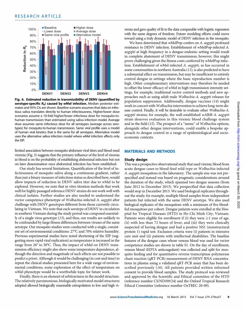
Predictions of wMel impact on DENV transmissionWe use Eq. 1 (seeMaterials andMethods) to assess the overall impactofwMel infection onDENV transmission by combining the estimatedposterior distributions for the dynamics of viral titer over time in in-fected humans, the probability that a mosquito will become infectedon consuming a blood meal with a certain titer of virus, and the de-velopment of infectivity in themosquito.We represent impact on den-gue transmission by the fractional reduction of the reproductionnumber, R0, of each serotype that would be caused by wMel infectionof the entire mosquito population. Figure 6 shows the resulting pos-terior estimates of the reduction in R0 for each serotype. For the base-line scenario (which assumes that mosquito infectivity to humans isdirectly proportional to the probability of detecting infectious virus insaliva), a 66 to 75% reduction is predicted, varying by serotype. Al-though the CIs on the absolute estimates of transmission reductionoverlapped across the serotypes, posterior estimates of the differencesin reduction betweenDENV1 andDENV2/3/4 indicated that DENV1exhibited a significantly lower level of reduction than other serotypes(P < 0.01).
Three other scenarios shown in Fig. 6 illustrate the sensitivity ofthe predictions to assumptions about how the model of saliva infec-tivity is translated to estimates of mosquito-to-human infectivity. The“higher dose” scenario assumed that the infectious dose parameters inthe saliva infectivity model (the parameters ϕS in Eq. 4 in Materialsand Methods) need to be 10-fold larger than the estimated values to
3
4
5
6
7
8
9
6 7 8 9
Ave
rag
e lo
g10
abd
om
inal
vir
al t
iter
wMel
WT
DENV1
3
4
5
6
7
8
9
6 7 8
DENV2
3
4
5
6
7
8
9
6 7 8
Ave
rag
e lo
g10
abd
om
inal
vir
al t
iter
Log10 donor viral titer/ml plasma
DENV3
3
4
5
6
7
8
9
5 6 7 8Log10 donor viral titer/ml plasma
DENV4
A B
C D
Fig. 3. wMel attenuates DENV infection of abdomen tissues. (A to D)Shown is the mean log titer (RNA copies per abdomen) of virus measured
10in mosquito abdomens (average over mosquitoes with detectable virusat any time point) of WT (circles) and wMel-infected (triangles) mosqui-toes with DENV-infected abdomen tissues, binned by integer interval oflog10 viral titer in the donor human blood. (A) to (D) show results forDENV1 to DENV4, respectively. Error bars show SEM.
ceTranslationalMedicine.org 18 March 2015 Vol 7 Issue 279 279ra37 5
R E S EARCH ART I C L E
by guest on March 30, 2020
http://stm.sciencem
ag.org/D
ownloaded from
describe mosquito-human transmission probabilities. This scenariogave the greatest predicted reduction in transmission due to wMel in-fection because of the predicted slower growth of viral titers in saliva ofwMel-infected mosquitoes. Conversely, assuming those infectiousdose parameters (ϕS) are 10-fold lower than for mosquito-mosquitotransmission (as quantified by our assay of saliva infectivity), this re-sulted in substantially lower estimates of the impact ofwMel infectionon dengue transmission compared with the baseline scenario. Howev-er, it should be noted that this scenario gives an unrealistically highper-bite probabilities of mosquito-human transmission and, thus, veryhigh (>10 for DENV1) estimates of R0 for reasonable assumptions onmosquito numbers per person and the biting rate.
www.Scien
The “average dose” scenario assumed that there are no serotypedifferences in the dose parameter for mosquito-human transmission,implemented by specifying that the saliva model dose parameter foreach serotype (ϕS) takes themean of the serotype-specific estimates foreach posterior distribution sample. The “same viral profile” scenarioignored the differences in human viral kinetics between serotypesshown in fig. S1 and used a single model (see Eq. 2 in Materials andMethods) of rh(v|t) for all serotypes fitted to all the patient datashown in that figure. The estimated reductions in R0 due to wMelin both of these scenarios were very similar to those obtained forthe baseline scenario, highlighting that serotype differences in vire-mia kinetics do not explain the overall differences by serotype seen
00.10.20.30.40.50.60.70.80.9
1
wMel: DatawMel: ModelWT: DataWT: Model
1 2 3 4
A
00.10.20.30.40.50.60.70.80.9
1
7 10 14 18
B
00.10.20.30.40.50.60.70.80.9
1
5 6 7 8 9
C
00.10.20.30.40.50.60.70.80.9
1wMel: DatawMel: ModelWT: DataWT: Model
1 2 3 4
D
0
0.1
0.2
0.3
0.4
0.5
0.6
0.7
0.8
0.9
7 10 14 18
E
00.10.20.30.40.50.60.70.80.9
1
6 7 8 9
F
00.10.20.30.40.50.60.70.80.9
1
Pro
port
ion
of p
ositi
ve m
osqu
itoes
Pro
port
ion
of p
ositi
ve m
osqu
itoes
Pro
port
ion
of p
ositi
ve m
osqu
itoes
Serotype
wMel: DatawMel: ModelWT: DataWT: Model
1 2 3 4
G
0
0.1
0.2
0.3
0.4
0.5
0.6
0.7
0.8
0.9
Days after blood meal7 10 14 18
H I
00.10.20.30.40.50.60.70.80.9
1
Log10 plasma viral titer6 7 8 9
Fig. 4. Mosquito infectionmodel fit to the empirical evidence ofwMel-mediated blocking of DENV infection. (A to C) Observed (“Data”) and
log10 donor plasma virus titer band. (D to F) Same as for panels (A) to (C)but showing the proportion of dengue-infected mosquitoes (that is, with
median posterior fitted (“Model”) proportions (with exact binomial con-fidence intervals) of WT and wMel-infected mosquitoes with detectablevirus in abdomen, stratified by (A) serotype, (B) end time point, and (C)
detectable virus in abdomen) that also had detectable infectious virus insaliva for the baselinemodel. (G to I) Same as for panels (D) to (F) but for thealternative saliva model.
ceTranslationalMedicine.org 18 March 2015 Vol 7 Issue 279 279ra37 6
R E S EARCH ART I C L E
by guest on March 30, 2020
http://stm.sciencem
ag.org/D
ownloaded from
in Fig. 6. Rather, the lower impact of wMel in DENV1 is largely causedby the differences in infectious dose parameters for saliva and abdom-inal infection between serotypes (Fig. 1).
The last alternative model scenario of Fig. 6 shows the resultswhen the alternative saliva infection model is used, solely representingthe impact ofwMel as a lengthening of the EIP (Table 1 and Fig. 5C). Inthis model, the predicted impact of wMel on transmission was about10% lower, that is, a 57 to 66% reduction depending on serotype.
DISCUSSION
We have experimentally characterized the phenotype ofWolbachia-infected A. aegypti mosquitoes challenged with viremic blood fromsymptomatic dengue patients. wMelPop conferred very strong resist-ance to DENV infection of the mosquito body and, more importantly,the salivary glands. wMel conferred an intermediate phenotype inwhich abdomen tissues were susceptible to DENV infection, but dis-semination was diminished as evidenced by a lower prevalence ofmosquitoes with infectious saliva.
The profound level of virus blocking conferred by wMelPop in-fection is predicted to cause dramatic reductions inDENV transmissionin settings where wMelPop is successfully and stably introduced. Theimpact of wMel on DENV transmission is more nuanced and serotype-
www.Scien
dependent; DENV1 transmission is the least affected, with a predicted66% reduction in R0 for the baseline scenario. For the other serotypes,higher estimated infectious dose parameters (compared with DENV1)for both the abdominal and saliva infectionmodels lead to larger predictedreductions in transmission of about 75%. To put these reductions in con-text, estimates of the basic reproduction number (R0) for dengue lie in therange of 1.3 to 6.3 (12), with 2 to 5 being typical of endemic settings. Areduction of 66% is sufficient to eliminate dengue in a settingwhereR0 =3, whereas a 75% reduction will achieve elimination for R0 = 4.
Our study highlights three effects of wMel infection on DENVinfection in A. aegypti mosquitoes: an increase (compared with wild-type) in blood meal viremia required to achieve a certain probabilityof abdominal infection, a substantial reduction in the probability ofdetecting infectious virus in saliva, and a lengthening of the EIP. Inour best-fit models, only the first two of these effects were found tobe significant. However, an alternative saliva model that solely repre-sented the impact of wMel in terms of an increased EIP gave anadequate (though statistically poorer) fit to the data and predicted lowerreductions inR0 than the baseline model. Additional data, particularlyif it included time points beyond 18 days, might more conclusively re-solve the extent to which the impact ofwMel is to reduce or just delaythe onset of infectiousness in saliva. This issue is important for under-standing the extent to which the estimated impact of wMel can begeneralized to different settings: if wMel reduces the probability of
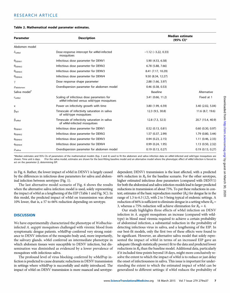
Table 2. Mathematical model parameter estimates.
Parameter
DescriptionceTranslationalMedi
Median estimate(95% CI)*
Abdomen model
dwMel
Dose-response intercept for wMel-infectedmosquitoes−1.12 (−3.22, 0.33)
yDENV1
Infectious dose parameter for DENV1 5.90 (4.53, 6.58)yDENV2
Infectious dose parameter for DENV2 6.78 (5.88, 7.66)yDENV3
Infectious dose parameter for DENV3 8.41 (7.17, 10.29)yDENV4
Infectious dose parameter for DENV4 9.50 (8.34, 12.27)g
Dose response shape parameter 2.88 (1.66, 3.97)rabdomen
Overdispersion parameter for abdomen model 0.46 (0.38, 0.53)Saliva model†
Baselinecine.org 18 March 2015 Vol 7 Issue 2
Alternative
ewMel
Scaling of infectious dose parameters forwMel-infected versus wild-type mosquitoes3.41 (0.66, 11.2)
Fixed at 1k
Power on infectivity growth with time 3.80 (1.99, 6.59) 3.40 (2.02, 5.04)bWT
Timescale of infectivity saturation in salivaof wild-type mosquitoes12.3 (9.5, 30.8)
11.6 (8.7, 19.6)bwMel
Timescale of infectivity saturation in salivaof wMel-infected mosquitoes12.8 (7.3, 32.5)
20.7 (15.4, 40.9)fDENV1
Infectious dose parameter for DENV1 0.52 (0.13, 0.81) 0.60 (0.30, 0.97)fDENV2
Infectious dose parameter for DENV2 1.57 (0.37, 2.99) 1.79 (0.80, 3.44)fDENV3
Infectious dose parameter for DENV3 0.94 (0.23, 2.15) 1.11 (0.46, 2.33)fDENV4
Infectious dose parameter for DENV4 0.99 (0.24, 1.95) 1.13 (0.50, 2.32)rsaliva
Overdispersion parameter for abdomen model 0.19 (0.13, 0.27) 0.19 (0.13, 0.27)*Median estimates and 95% CIs of parameters of the mathematical models (Eqs. 3 and 4) used to fit the abdomen and saliva infection data on wMel-infected and wild-type mosquitoes areshown. Time unit is days. †For the saliva model, estimates are shown for the best-fitting baseline model and an alternative model where the phenotypic effect of wMel infection is forced toact on the parameter b, determining EIP.
79 279ra37 7
R E S EARCH ART I C L E
by guest on March 30, 2020
http://stm.sciencem
ag.org/D
ownloaded from
mosquitoes being infectious independent of the time since infection,the reduction in R0 achieved is independent of adult mosquito survival.Conversely, if the main impact ofwMel is to increase the EIP, this willhave a larger effect on dengue transmission than that estimated here insituations where daily mosquito survival is lower than the relativelyhigh 90% value we assumed.
Previous vector competence studies ofWolbachia-infected A. aegyptimosquitoes have used in vitro–passagedDENV strains that were spikedinto animal or human blood before this mixture was presented to col-ony mosquitoes through membrane feeders (4, 5). The current study isdistinguished from previous work in using fresh viremic blood samplesfrom hospitalized dengue cases to mimic the virological challenge thatA. aegypti mosquitoes experience when they feed on an infectious hu-man case. In using viremic blood from hospitalized dengue cases, inwhom peak viremia levels are significantly higher than in acute ambu-latory (never hospitalized) cases in the same setting (13), we are likelybeing conservative in our experimental evaluation of wMel-infected A.aegypti. Future experimental studies could examine susceptibility toDENV infection after blood feeding on ambulatory dengue cases.
Our finding that wMelPop-A. aegypti do not develop disseminatedinfections with DENV is entirely consistent with the initial description ofthe vector competence phenotype of this strain (5). However, we foundthat wMel-A. aegypti can develop infectious saliva after viremic blood
www.ScienceTranslationalMedicine.org 1
feeding, and this contrasts with the initialdescription by Walker et al. (4) who de-tected no infectious DENV2 in the salivaof any of the 336wMel-A. aegypti femalesused in artificial feeding experiments.There are methodological reasons whyour results might differ: Walker et al. usedone laboratory strain of DENV2 at a singleconcentration, used cell culture to detectinfectious virus in pooled saliva, and usedcolony-sourced mosquitoes. Of these, wespeculate that the virological differencesaremost important and that viremic bloodfrom a human dengue case provides themost stringent and relevant challenge ofthe vector competence of Wolbachia-infectedmosquitoes. This would underscorethe importance of using clinical materialfor robust assessments of arboviral vectorcompetence in general.Our data also high-light the importance of assessing vectorcompetence atmultiple time points to char-acterize the impact on the dynamics of den-gue infection in the mosquito. Whereas thewMelPop data presented here were allcollected at a single time point (12 days) af-ter bloodmeal, preliminary results fromon-going work indicate comparable levels ofinhibition of DENV infection at 14 and18 days after infection.
Our analysis suggested that wMelcould reduce the DENV force of infectionby a degree that would have a highly sig-nificant public health impact—potentiallyachieving elimination in low- tomoderate-
transmission settings, albeit perhaps insufficient for complete control inhigh-transmission settings (especially for DENV1). Yet, a number offactors might lead to the field efficacy of wMel on DENV transmissiondiffering from estimates presented here. First, although we did notcollect data on the concentration of infectious DENV particles in mos-quito saliva, it is a reasonable hypothesis that wMel reduces viral con-centrations, whichwould lead to a larger reduction in transmission thanthat estimated here. Second, the effect of wMel on other aspects ofmosquito behavior that have an impact on transmission, such as hostseeking, probing, and blood feeding success rates, has yet to be investi-gated in a field setting, and it is plausible that these could counteract theeffect of wMel-mediated interference of virus transmission to mosqui-toes. Finally, here, we solely examined the impact ofWolbachia on thesusceptibility of A. aegypti to DENV infection. In reality, wMel maymodify A. aegypti fitness through decreased (or, less likely, increased)fecundity or longevity. Even small reductions in the life span of wMel-A.aegypti, as described previously (4), might cause reductions in denguetransmission.
A priori, that we found no statistically significant dependence onthe level of viremia in the infecting blood meal in the mathematicalmodel describing saliva infectionmight be viewed as surprising. How-ever, the salivamodel represents the probability of detecting infectiousvirus in saliva conditional on abdominal infection being detectable. The
0%10%20%30%40%50%60%70%80%90%
100%
0 2 4 6 8 10 12
seot i
uq s
om f
o n
o itro
po r
P wit
h a
bd
om
inal
infe
ctio
n
Log10 donor viral titer/ml plasma
0%10%20%30%40%50%60%70%80%90%
100%
0 5 10 15 20 25 30
Pro
po
rtio
n o
f m
osq
uit
oes
wit
hd
etec
tab
le in
fect
ion
in s
aliv
a
Days after blood meal
0%10%20%30%40%50%60%70%80%90%
100%
0 5 10 15 20 25 30
Pro
po
rtio
n o
f m
osq
uit
oes
wit
hd
etec
tab
le in
fect
ion
in s
aliv
a
Days after blood meal
DENV1-WT
DENV1-wMel
DENV2-WT
DENV2-wMel
DENV3-WT
DENV3-wMel
DENV4-WT
DENV4-wMel
A B
C
Fig. 5. Performance of the mosquito infection model. (A) Shown is the behavior of the abdominalinfection model illustrating the dependence of the probability of infection on viral titer in donor blood,
serotype, andWolbachia infection status. (B) Shown is the behavior of the saliva infectionmodel showingdependence of the probability of detectable infection in saliva (conditional on abdominal infection) as afunction of the days elapsed since the infecting bloodmeal, serotype, andWolbachia infection status. (C)Same as (B) but for the alternative saliva infection model where wMel infection affects only the EIP. Allgraphs show mean posterior predictions.8 March 2015 Vol 7 Issue 279 279ra37 8
R E S EARCH ART I C L E
by guest on March 30, 2020
http://stm.sciencem
ag.org/D
ownloaded from
limited association betweenmosquito abdomen viral titers and bloodmealviremia (Fig. 3) suggests that the primary influence of the level of viremiain blood is on the probability of establishing abdominal infection but noton later dissemination once abdominal infection has been established.
Our study has several limitations. Quantification of the level of in-fectiousness of mosquito saliva along a continuous gradient, ratherthan just a binarymeasure of infectious status as described here, wouldallow impacts of reduction in DENV saliva titer due to wMel to beexplored. However, we note that in vitro titration methods that workwell for highly passaged referenceDENV strains do not workwell withclinical isolates. Further studies are also needed to understand thevector competence phenotype of Wolbachia-infected A. aegypti afterchallenge with DENV genotypes different from those currently circu-lating in Vietnam.We note that each serotype of DENV in circulationin southernVietnam during the study period was composed essential-ly of a single virus genotype (13), and thus, our results are unlikely tobe confounded by large fitness differences between viruses of the sameserotype. Our mosquito studies were conducted with a single, consist-ent set of environmental conditions: 27°C and 70% relative humidity.Previous experimental studies have noted shortening of the EIP (sug-gestingmore rapid viral replication) as temperature is increased in therange from 26° to 30°C. Thus, the impact of wMel on DENV trans-mission efficiencymight also show some temperature dependence, al-though the direction and magnitude of such effects are not possible topredict a priori. Although it would be challenging (in cost and time) torepeat the clinical studies presented here for a wide range of environ-mental conditions, some exploration of the effect of temperature onwMel phenotype would be a worthwhile topic for future work.
Finally, there is an element of arbitrariness in themodel structure.The relatively parsimonious, biologicallymotivatedmodel structuresadopted allowed biologically reasonable extrapolation to low and high vi-
www.Scien
remia and gave quality of fit to the data comparable with logistic regressionwith the same degrees of freedom. Future modeling efforts could movetoward using a truly dynamic model of DENV infection in the mosquito.
We have determined that wMelPop confers on A. aegypti profoundresistance to DENV infection. Establishment of wMelPop-infected A.aegypti at high frequency in a dengue-endemic setting would resultin complete abatement of DENV transmission; however, this mightprove challenging given the fitness costs conferred bywMelPop infec-tion. Establishment of wMel-infected A. aegypti, as has occurred insome communities in northernAustralia (11), is also predicted to havea substantial effect on transmission, but may be insufficient to entirelycontrol dengue in settings where the basic reproduction number ishigh. Other complementary interventions may therefore be neededto offset the lower efficacy of wMel in high transmission intensity set-tings, for example, traditional vector control methods and new ap-proaches such as using adult male Wolbachia-A. aegypti releases forpopulation suppression. Additionally, dengue vaccines (14) mightwork in concert withWolbachia intervention to achieve long-termdis-ease control. Finally, it will be desirable to evaluate other Wolbachia-A.aegypti strains; for example, the well-established wAlbB-A. aegyptistrain deserves evaluation in this viremic blood challenge systemand in the field (15). The prospect of a “menu” ofWolbachia options,alongside other dengue interventions, could enable a bespoke ap-proach to dengue control in a range of epidemiological and socio-economic contexts.
MATERIALS AND METHODS
Study designThiswas a prospective observational study that used viremic blood fromacute dengue cases to blood-feed wild-type or Wolbachia-infectedA. aegyptimosquitoes in the laboratory. The sample size was not pre-specified and instead was based on pragmatic considerations aroundthe duration of the study, which spanned two dengue seasons (fromJune 2012 to December 2013). We prespecified that data collectionwould stop in December 2013. We used biological replicates through-out the study; for example, multiple blood samples from independentpatients but infected with the same DENV serotype. We also usedbiological replicates of the mosquitoes with a minimum of five blood-fed mosquitoes per cohort. Dengue patients were enrolled at the Hos-pital for Tropical Diseases (HTD) in Ho Chi Minh City, Vietnam.Patients were eligible for enrollment if (i) they were ≥1 year of age,(ii) with less than 72 hours of fever, and (iii) they were clinicallysuspected of having dengue and had a positive NS1 (nonstructuralprotein 1) rapid test. Exclusion criteria were (i) patients in intensivecare unit and (ii) patients with intellectual disabilities. The baselinefeatures of the dengue cases whose venous blood was used for vectorcompetence studies are shown in table S1. On the day of enrollment,venous blood (EDTA anticoagulant) was collected and split for mos-quito feeding and for quantitative reverse transcriptase polymerasechain reaction (qRT-PCR) measurement of DENV RNA concentra-tions in plasma using a validated qRT-PCR assay that has been de-scribed previously (16). All patients provided written informedconsent to provide blood samples. The study protocol was reviewedand approved by the Scientific and Ethical committee of the HTD(reference number CS/ND/09/24) and the Oxford Tropical ResearchEthical Committee (reference number OxTREC 20-09).
0%
10%
20%
30%
40%
50%
60%
70%
80%
90%
100%
DENV1 DENV2 DENV3 DENV4
Red
uct
ion
in R
0Baseline Higher doseLower dose Average doseSame viral profile Alternative model
Fig. 6. Estimated reduction in transmissibility of DENV (quantified byserotype-specific R0) caused by wMel infection. Median posterior esti-
mates and 95%CIs are shown. Baseline scenario assumes that data on infec-tious saliva translates directly to human infectiousness. Higher/lower dosescenarios assume a 10-fold higher/lower infectious dose for mosquito-to-human transmission than estimated using saliva infection model. Averagedose assumes same infectious dose for all serotypes (average across sero-types) for mosquito-to-human transmission. Same viral profile uses a modelof human viral kinetics that is the same for all serotypes. Alternative modeluses the alternative saliva infection model where wMel infection affects onlythe EIP.ceTranslationalMedicine.org 18 March 2015 Vol 7 Issue 279 279ra37 9
R E S EARCH ART I C L E
by guest on March 30, 2020
http://stm.sciencem
ag.org/D
ownloaded from
The prespecified hypothesis was thatWolbachia-infected A. aegyptimosquitoes weremore resistant toDENV infection.Hence, the primaryentomological end point of interest was the proportion of mosquitoeswith infected abdomens or saliva. This was addressed by scoringmosquito tissues for the presence or absence of DENV infection usingamolecular test and thencemodeling the results as a basis to predict thewider epidemiological impact on DENV transmission. All laboratoryassays to test for DENV infection were performed by techniciansblinded to the clinical and virological details of the patient blood sampleand theWolbachia status of the mosquitoes. All data were submittedto a Good Clinical Laboratory Practice database and cleaned beforedata lock.
Viremic blood challenges of wild-type and Wolbachia-infectedA. aegypti. Vector competence studies were performed with wild-typeA. aegypti from Cairns, Australia, and A. aegypti of the same origin butstably infected with wMel or wMelPop. The wild-type versus wMelPop-A. aegypti studies were performed using eggs from outcrossed coloniesmaintained atMonashUniversity, Australia. Colonies weremaintainedat population sizes of 400 with a 50:50 sex ratio. The wild-type versuswMel-A. aegypti studies were performed with F2 generation adultsand obtained by hatching eggs collected from field sites in Cairns,Australia (11). For all studies, up to 100 three- to seven-day-old femaleA. aegyptimosquitoeswere starved for 24hours before beingmembranefed on fresh acute blood from laboratory-confirmed dengue patients.All blood samples were placed into glass membrane feeders within1 hour of the blood being collected, andmosquitoes were allowed accessto the blood for 1 hour. Membrane feeders were water-jacketed andmaintained at constant temperatureduringmosquito feeding (37°C).Aftercold knockdown, fully engorged mosquitoes were selected and thenmaintained in an environmental chamber with a 12:12-hour light/darkcycle, at 27°C and 70% relative humidity, with access to 10% sucrosesolution.
Detection of DENV in saliva and abdomen tissues. Infectiousvirus in mosquito saliva was detected by placing the proboscis of ade-winged and de-legged mosquito into the end of a filtered micro-pipette tip containing 6 ml of sterile saliva medium (a 1:1 solution of15% sucrose and inactivated fetal calf serum) for 30 min at roomtemperature. After 30 min, the 6-ml saliva medium was ejected andthen drawn into a pointed glass capillary tube (tip diameter, <0.3 mm).The volume of saliva medium derived from one mosquito was theninjected into the thorax of between four and sixA. aegyptimosquitoes(4 to 7 days old; ~1 ml injected permosquito), and the injectedmosqui-toes were maintained for 7 days in an environmental chamber with a12:12 light/dark cycle, at 28°C and 80% relative humidity. After 7 days,the injected mosquitoes for each saliva sample were killed; the bodieswere pooled, homogenized, and tested by qRT-PCR for DENV infec-tion, with saliva samples scored as positive or negative depending onthis result. Saliva samples were collected from all mosquitoes, but onlysaliva samples frommosquitoes with infected abdomenswere evaluatedfor their infection status because pilot studies confirmed that abdomeninfection was a prerequisite for the saliva to contain infectious virus.After collection of saliva samples, the abdomen was dissected from themosquito body. Dissected abdomens were suspended in 0.5 ml ofmosquito diluent (RPMI 1640 supplemented with 2% heat-inactivatedfetal calf serum, antibiotics, and antimycotics). Individual mosquito ab-domenswere homogenizedwith 1-mmZirconia/Silica beads for 15minat 30 Hz using a TissueLyser II (Qiagen). Mosquito tissues were scoredas being DENV-infected using a quantitative, internally controlled RT-
www.Scienc
PCR assay (16) on homogenized tissue, and the results were expressedas copies per tissue.
Detection of Wolbachia status in mosquito tissues by real-timePCR. For quality control purposes, Wolbachia infection status wasscored using a multiplex PCR assay on nucleic acid extracts frommosquito abdomens. A. aegypti ribosomal protein S17 (Ae-RpS17)was used as an internal control. Wolbachia strain wMel was detectedwith primers/probes specific to the WD0513 gene, and wMelPop wasdetected with primers/probes specific to the polymorphic insertionsites of the IS5 at loci IS5-WD1310. Sequences of primers/probes forWolbachia and DENV detection are shown in table S2. PCR was per-formed on a LightCycler 480 II machine using LightCycler 480 ProbesMaster according to the manufacturer’s instructions.
Data release. See Supplementary Materials for the wMel andwMelPop data analyzed in this paper.
Statistical analysisNonparametric assessment of thedifferences in tissue infection
rates in wMel versus wild-type mosquitoes. We applied a standardsign test for paired data, treating the experimental data as pairs of bi-nomial observations corresponding to the proportions infected of thewMel andwild-typemosquito groups fed on a particular blood sample,which were sampled on a particular day. Rows in Table 1 present thenumber of observation pairs for which the proportion of wMelmosquitoes infectedwas less than, equal to, or greater than the propor-tion of wild-type mosquitoes infected for different data subsets, desig-nated as npairs(pwMel < pWT), npairs(pwMel = pWT), and npairs(pwMel >pWT), respectively. If there was no difference between the infectionrates of wMel and wild-type mosquitoes, npairs(pwMel < pWT) wouldbe expected to be drawn from a binomial distribution with P = 0.5 andN = npairs(pwMel < pWT) + npairs(pwMel > pWT). The two-sided P value inthe final column of Table 1 is the probability of a sample from thatdistribution being equal to or more extreme than the observed value ofnpairs(pwMel < pWT).
Transmission model. Because the probability of a mosquito be-coming infected with DENV from a blood meal depends strongly onthe viral titer in that blood meal, quantitative assessment of the impactof Wolbachia on transmission requires a mathematical model thatcouples the dynamics of infection within the human host with thosein the vector. We found no previously published mathematical modelsof DENV transmission that included such coupling, so the frameworkpresented below needed to be developed specifically for this study.
We define rh(v|t) to be the probability density that the plasmaviral titer of a human host is v at time t after infection; we model viraldynamics in humans probabilistically to represent the variation seenbetween individuals. We assume that the probability that a mosquitotaking a blood meal on that individual becomes infected depends onthe viral titer in the blood at the time of feeding: let pi(v) be the prob-ability that a vector becomes infected when feeding on a humanwith aplasma viral titer of v. If a mosquito becomes infected, thenwe assumethat its infectiousness to humans depends on the time elapsed fromthe infecting blood meal and the plasma viral titer of the blood meal.We define pm(v|t) to be the probability that a mosquito infected bytaking a blood meal with viral titer v will infect another human hostit bites time t later; this distribution captures the EIP.
Together, these three distributions represent the complete trans-mission cycle; all that is additionally required to calculate the basicreproduction number (the average number of human infections gen-
eTranslationalMedicine.org 18 March 2015 Vol 7 Issue 279 279ra37 10
R E S EARCH ART I C L E
by guest on March 30, 2020
http://stm.sciencem
ag.org/D
ownloaded from
erated by a typical infected human in the absence of immunity),R0, fora serotype is the average number of femalemosquitoes per human host,m, the mortality hazard for adult female mosquitoes, m, and the bitingrate of female mosquitoes, k. Then
R0 ¼ mk2∫∞
0 ð∫D0 rhðvjtÞdtÞpiðvÞð∫∞0 pmðvjtÞe�mtdtÞdv ð1Þ
Here, D is the maximum time for clearance of virus in humans.This equation is the standard definition of the reproduction numberfor a vector-borne disease, generalized to account for viral dose depen-dence in the mosquito.
We wish to estimate the distributions rh(v|t), pi(v), and pm(v|t)for each of the four DENV serotypes and for Wolbachia-infected andwild-type mosquitoes. However, the available data did not allow everyparameter to be estimated independently for each combination of sero-type andWolbachia infection status, so it was necessary to assume thatonly a subset of parameters varied between serotypes or were affectedbyWolbachia.
Our primary interest is the extent to which Wolbachia reducestransmission, as characterized by the ratio of R0 of a DENV serotypein a wild-type A. aegypti population to that in aWolbachia-infectedA. aegypti population; values of m and k in Eq. 1 are not needed forcalculating this ratio. However, the assumed value of m, the mortalityhazard of adult mosquitoes, can affect estimates.A. aegyptimortalityvaries seasonally and by setting, with release-recapture studies typi-cally giving daily survival probabilities below 85% (17–19). Because onepossible phenotype of wMel on dengue replication in mosquitoesthat we explore below is a lengthening of the EIP, we conservativelyassume that daily survival is constant at its seasonal maximum of90% (m = 0.1/day) (19). This results in a larger proportion of trans-mission being from older mosquitoes than assuming a lower valuefor daily survival, and hence reduces the potential impact of EIPlengthening on dengue transmission.
We estimate rh(v|t) from serial plasma viremia levels measuredin 262 consecutively enrolled dengue cases in the IDAMS (InternationalResearch Consortium on Dengue Risk Assessment, Management,and Surveillance) study (clinicaltrials.gov identifier, NCT01550016)in Ho Chi Minh City, Vietnam. Of these 262 cases, 73 cases werehospitalized and 189 were managed entirely as ambulatory patientsfor the duration of their illness. The serial viremia measurements inthese 189 ambulatory cases have been described previously (13), andthe data are shown in fig. S1. Here, we use the following data fieldsfor each measurement: study participant identifier, DENV serotype,day of illness when sample was collected, and log10 viral titer per mil-liliter of plasma (measured with qRT-PCR) in sample. We modeledviral kinetics in a human host with a simple biphasic exponentialgrowth/decay function, where average (across all patients) viral titerat time t after infection is given by:
v tð Þ ¼ eat
1þ eðaþbÞt�cð2Þ
We assume that individual patient log10 viral titers are drawnfrom a normal distribution with mean log[v(t)] and standard devia-tion s, thus defining the distribution rh(v|t). Because the dates ofinfection are unknown, we estimate time of infection from the dayof illness onset by assuming a fixed 7-day incubation period for den-gue. Parameters a, b, and c were fitted independently for each sero-type, whereas swas fitted assuming it to be the same for all serotypes.
www.Scienc
Mosquito infection model. The probability that a mosquitofeeding on blood with viral titer v will become infected, pi(v), is esti-mated from data on abdominal infection status in mosquitoes infectedas part of this study. We used a simple dose-response model:
pi vð Þ ¼ 1 − exp −log vþ dW
yS
� �g� �; if log v þ dW > 0
piðvÞ ¼ 0 otherwise ð3Þ
The single parameter dWwas found to be sufficient to capture thephenotypic impact ofWolbachia. This parameter was assumed to be0 for wild-type and was estimated for wMel-infected mosquitoes. Itseffect is to modify the infecting dose of virus by a fixed factor. Theparameter yS determines the infectious dose and is estimated inde-pendently for each serotype S, whereas g determines the slope of thedose-response curve and is assumed not to vary by serotype. We didnot model an effect of day of measurement (after mosquito feeding)for abdominal infection data because no significant differences wereseen between the 7-, 10-, 14-, and 18-day time points examined here.
In the absence of human challenge studies, we lack direct mea-surements of mosquito infectiousness, pm(v|t); here, we examinethe closest proxy available, namely, detection of infectious DENVin mosquito saliva. We defined qm(v|t) to be the proportion of mosqui-toes infected by taking a blood meal with viral titer v that will have de-tectable infection in saliva time t later. We assume the followingfunctional form for qm(v|t):
qm vjtð Þ ¼ 1� exp � 1
eWϕs
tk
bkW þ tk
� �� �� �ð4Þ
This semi-mechanistic form gives power-law [~(t/bW)k] temporal
growth of saliva infection for small t. This growth saturates at a timegoverned by parameter bW; thus, this parameter governs the EIP. Be-cause we needed to use thismodel outside the observed range of 7≤ t≤18 days, it was important to choose a functional form for the timedependence of saliva infection status that was well behaved and biolog-ically plausible for both small and large t.Themodel above gives close tozero probability of detectable infection for small t (<7 days) and a prob-ability that plateaus at large t (>18 days). Similar to the abdominal in-fectionmodel, the serotype-specific parameters ϕS govern the infectiousdose. A dose-response shape parameter (akin to g in Eq. 3) was alsoexamined but found to result in overfitting, with estimates having95% CIs overlapping 1.
Two parameters, bW and eW, specify the phenotypic impact ofWolbachia for the saliva infection model. Hence, Wolbachia can af-fect either the proportion of mosquitoes ever developing infectious-ness in saliva or the rate at which saliva infectiousness increases (andthus, the EIP), or both. The former is estimated separately for wild-typeand wMel-infected mosquitoes, whereas the latter scales the infectiousdose parameters for wMel versus wild-type, and hence has a value of1 for wild-type and is estimated for wMel-infected mosquitoes.
When both bW and eWwere fitted (our baselinemodel), estimatesfor bWT and bwMel were nearly identical, with a substantial overlap ofthe 95% CIs. Thus, nearly the entire phenotypic effect was attributedto eW—representing a net reduction in the probability of infection insaliva in wMel-infected versus wild-type mosquitoes, irrespective ofthe time elapsed since the infecting blood meal (Fig. 5B). However,because the lower CI of eWwas just below 1 for the baselinemodel, we
eTranslationalMedicine.org 18 March 2015 Vol 7 Issue 279 279ra37 11
R E S EARCH ART I C L E
by guest on March 30, 2020
http://stm.sciencem
ag.org/D
ownloaded from
fitted three simplermodels, assuming (A)bWT= bwMel and eW=1 (thatis, no phenotypic effect of wMel); (B) bWT = bwMel (that is, the phe-notypic effect ofwMel is acting solely through eW); (C) eW = 1 (that is,the phenotypic effect of wMel is acting solely through a difference be-tween bWT and bwMel—effectively a lengthening of the EIP due towMel infection). Model B had the highest DIC, with the baselinemodel (with both bW and eW fitted) next (DIC difference from B of1.1), followed by model C (DIC difference from B of 2.4) and thenmodel A with much worse (DIC difference from B of 47). Becausethe phenotypic effect of wMel infection substantively affects the over-all estimates of the impact ofWolbachia on transmission, we choose topresent the estimates formodelC (where ewMel = 1) as an alternative tothe baseline model. This alternative model (Fig. 5C) fitted the dataqualitatively well (Fig. 4), albeit worse than the baseline model(difference in DIC, 1.4). Although model B had the highest DIC, thesmall numerical difference compared with the model fitting both bWand eWmeant that we opt to retain the latter as our baseline, as it bestrepresents the uncertainty in the phenotypic effect of wMel infectionand is slightly more pessimistic than model B in the estimates of theimpact of wMel on the R0 of dengue.
The saliva infection model shows no dependence on the plasmaviral titer of the infecting bloodmeal; including such dependence didnot significantly improve model fit, reflecting the lack of obvious viraltiter dependence seen in the raw saliva infection data shown in Fig. 1.For example, substituting a term (wW + log v)/ϕS for 1/eWϕS in Eq. 4,fitting wwMel and assuming wwMel = 0 (akin to the abdominal model)increased the DIC by 2.3 relative to the baseline model. Furthermore,the central estimate for wwMel was unreasonably large in magnitude(−6.1) given that log10 donor viral titers only varied in the range of5.3 to 9.9, meaning that this model variant was approximating the be-havior of the functionally simpler baselinemodel with no improvementin fit.
Our default (and simplest) approach to relating pm(v|t) to qm(v|t)is to assume proportionality, namely, pm(v|t)º qm(v|t). However, otherassumptions are plausible and can substantially affect the resulting es-timates of the overall impact of wMel on dengue transmission. Weundertake some sensitivity analysis therefore by assuming that pm(v|t)is determined by a similar functional form to Eq. 4, but with modifiedparameters.We examine the impact of varying the infectious dose param-eters by a fixedmultiplier tomimic the effect of the infectious dose frommosquitoes to humans being either larger or smaller than that seenwith the assay we used to assess infectious virus in saliva.
Inferential framework. Model fittingwas undertaken in a Bayesianframework usingMarkovChainMonte Carlo (MCMC)methods (20).To account for the overdispersion of the data (Figs. 1 and 2), a Beta-binomial likelihood function was used rather than a simple binomiallikelihood. The Beta-binomial was parameterized in terms of the meanbinomial proportion,Q, and its overdispersion, r, defined such that themean and variance of a sample of n draws is given by nQ and nQ(1 −Q)[1 + (n − 1)r], respectively. The overdispersion parameter r wasfitted separately for the abdominal and saliva data. Uninformativeuniform priors were assumed for all parameters, with an upper boundof 200 for all parameters and a lower bound of 0 for all parametersother than d, for which a lower bound of −200 was used. Sensitivityto changing the upper and lower bounds (where appropriate) on priorswas tested, and none was found so long as the upper and lower boundslay outside the 99.9th percentile of the posterior distribution. Param-eters were updated individually, with a single update sweep defined
www.Scienc
as a sequence of proposed updates to each parameter in turn. For com-putational efficiency, a uniform proposal distribution was used for eachparameter, centered around the current parameter value andwithwidthmanually tuned to give 20 to 40% acceptance rates (proposal acceptancerates were monitored separately for each parameter). MCMC chainswere equilibriated with 100,000 update sweeps, and posterior distribu-tions were estimated from the following 500,000 update sweeps,sampling once every 500 sweeps. Convergence was checked visuallyand by running multiple chains from different starting points. Analy-ses were undertaken in Microsoft Excel and the statistical language R.
In exploratory but nonexhaustive analyses, a variety of functionalforms were explored for both pi(v) and qm(v|t); in particular, we exam-ined howmodel fit could be significantly improved bymaking a param-eter vary by serotype or by Wolbachia infection status while retainingparameter identifiability. We found little evidence for any serotype de-pendence beyond the overall scaling of the dose-response relationshipsexpressed in the functional forms used above. Similarly, significant dif-ferences (assessed by nonoverlapping 95% CIs and the DIC) betweenestimates for wild-type and wMel-infected mosquitoes were only seenfor the parameters d, e, and, to a lesser extent (see discussion above), b.
SUPPLEMENTARY MATERIALS
www.sciencetranslationalmedicine.org/cgi/content/full/7/279/279ra37/DC1Table S1. Study population characteristics.Table S2. List of primers and probes used.Fig. S1. Human DENV viremia kinetics.Supplementary data: Mosquito biting study data.
REFERENCES AND NOTES
1. C. P. Simmons, J. J. Farrar, v. V. Nguyen, B. Wills, Dengue. N. Engl. J. Med. 366, 1423–1432(2012).
2. S. Bhatt, P. W. Gething, O. J. Brady, J. P. Messina, A. W. Farlow, C. L. Moyes, J. M. Drake,J. S. Brownstein, A. G. Hoen, O. Sankoh, M. F. Myers, D. B. George, T. Jaenisch, G. R. Wint,C. P. Simmons, T. W. Scott, J. J. Farrar, S. I. Hay, The global distribution and burden ofdengue. Nature 496, 504–507 (2013).
3. C. J. McMeniman, R. V. Lane, B. N. Cass, A. W. Fong, M. Sidhu, Y. F. Wang, S. L. O’Neill, Stableintroduction of a life-shortening Wolbachia infection into the mosquito Aedes aegypti. Science323, 141–144 (2009).
4. T. Walker, P. H. Johnson, L. A. Moreira, I. Iturbe-Ormaetxe, F. D. Frentiu, C. J. McMeniman,Y. S. Leong, Y. Dong, J. Axford, P. Kriesner, A. L. Lloyd, S. A. Ritchie, S. L. O’Neill, A. A. Hoffmann,The wMel Wolbachia strain blocks dengue and invades caged Aedes aegypti populations.Nature 476, 450–453 (2011).
5. L. A. Moreira, I. Iturbe-Ormaetxe, J. A. Jeffery, G. Lu, A. T. Pyke, L. M. Hedges, B. C. Rocha,S. Hall-Mendelin, A. Day, M. Riegler, L. E. Hugo, K. N. Johnson, B. H. Kay, E. A. McGraw,A. F. van den Hurk, P. A. Ryan, S. L. O’Neill, A Wolbachia symbiont in Aedes aegypti limitsinfection with dengue, Chikungunya, and Plasmodium. Cell 139, 1268–1278 (2009).
6. C. J. McMeniman, S. L. O’Neill, A virulent Wolbachia infection decreases the viability of thedengue vector Aedes aegypti during periods of embryonic quiescence. PLOS Negl. Trop. Dis. 4,e748 (2010).
7. A. P. Turley, L. A. Moreira, S. L. O’Neill, E. A. McGraw, Wolbachia infection reduces blood-feeding success in the dengue fever mosquito, Aedes aegypti. PLOS Negl. Trop. Dis. 3,e516 (2009).
8. G. Zhang, M. Hussain, S. L. O’Neill, S. Asgari, Wolbachia uses a host microRNA to regulatetranscripts of a methyltransferase, contributing to dengue virus inhibition in Aedes aegypti.Proc. Natl. Acad. Sci. U.S.A. 110, 10276–10281 (2013).
9. E. P. Caragata, E. Rances, L. M. Hedges, A. W. Gofton, K. N. Johnson, S. L. O’Neill, E. A. McGraw,Dietary cholesterol modulates pathogen blocking by Wolbachia. PLOS Pathog. 9, e1003459(2013).
10. X. Pan, G. Zhou, J. Wu, G. Bian, P. Lu, A. S. Raikhel, Z. Xi, Wolbachia induces reactive oxygenspecies (ROS)-dependent activation of the Toll pathway to control dengue virus in themosquito Aedes aegypti. Proc. Natl. Acad. Sci. U.S.A. 109, E23–E31 (2012).
eTranslationalMedicine.org 18 March 2015 Vol 7 Issue 279 279ra37 12
R E S EARCH ART I C L E
http://stm.
Dow
nloaded from
11. A. A. Hoffmann, B. L. Montgomery, J. Popovici, I. Iturbe-Ormaetxe, P. H. Johnson, F. Muzzi,M. Greenfield, M. Durkan, Y. S. Leong, Y. Dong, H. Cook, J. Axford, A. G. Callahan, N. Kenny,C. Omodei, E. A. McGraw, P. A. Ryan, S. A. Ritchie, M. Turelli, S. L. O’Neill, Successfulestablishment of Wolbachia in Aedes populations to suppress dengue transmission. Nature476, 454–457 (2011).
12. M. A. Johansson, J. Hombach, D. A. Cummings, Models of the impact of dengue vaccines:A review of current research and potential approaches. Vaccine 29, 5860–5868 (2011).
13. M. N. Nguyet, T. H. Duong, V. T. Trung, T. H. Nguyen, C. N. Tran, V. T. Long, T. Dui le, H. L. Nguyen,J. J. Farrar, E. C. Holmes, M. A. Rabaa, J. E. Bryant, T. T. Nguyen, H. T. Nguyen, L. T. Nguyen,M. P. Pham, H. T. Nguyen, T. T. Luong, B. Wills, C. V. Nguyen, M. Wolbers, C. P. Simmons,Host and viral features of human dengue cases shape the population of infected andinfectious Aedes aegypti mosquitoes. Proc. Natl. Acad. Sci. U.S.A. 110, 9072–9077 (2013).
14. A. Sabchareon, D. Wallace, C. Sirivichayakul, K. Limkittikul, P. Chanthavanich, S. Suvannadabba,V. Jiwariyavej, W. Dulyachai, K. Pengsaa, T. A. Wartel, A. Moureau, M. Saville, A. Bouckenooghe,S. Viviani, N. G. Tornieporth, J. Lang, Protective efficacy of the recombinant, live-attenuated,CYD tetravalent dengue vaccine in Thai schoolchildren: A randomised, controlled phase 2btrial. Lancet 380, 1559–1567 (2012).
15. Z. Xi, C. C. Khoo, S. L. Dobson, Wolbachia establishment and invasion in an Aedes aegyptilaboratory population. Science 310, 326–328 (2005).
16. K. D. Hue, T. V. Tuan, H. T. Thi, C. T. Bich, H. H. Anh, B. A. Wills, C. P. Simmons, Validation ofan internally controlled one-step real-time multiplex RT-PCR assay for the detection andquantitation of dengue virus RNA in plasma. J. Virol. Methods 177, 168–173 (2011).
17. L. C. Harrington, J. P. Buonaccorsi, J. D. Edman, A. Costero, P. Kittayapong, G. G. Clark,T. W. Scott, Analysis of survival of young and old Aedes aegypti (Diptera: Culicidae)from Puerto Rico and Thailand. J. Med. Entomol. 38, 537–547 (2001).
18. R. Maciel-de-Freitas, C. T. Codeço, R. Lourenço-de-Oliveira, Daily survival rates and disper-sal of Aedes aegypti females in Rio de Janeiro, Brazil. Am. J. Trop. Med. Hyg. 76, 659–665(2007).
19. P. M. Sheppard, W. W. Macdonald, R. J. Tonn, B. Grab, The dynamics of an adult populationof Aedes aegypti in relation to dengue haemorrhagic fever in Bangkok. J. Anim. Ecol. 38,661–702 (1969).
www.Scienc
20. W. R. Gilks, S. Richardson, D. Spiegelhalter, Markov Chain Monte Carlo in Practice (Chap-man & Hall, London, 1996).
Funding: Supported by the Wellcome Trust; the Bill and Melinda Gates Foundation (BMGF);the Foundation for the NIH, as part of the Grand Challenges in Global Health Initiative ofBMGF; the National Health and Medical Research Council, Australia; the U.K. Medical ResearchCouncil; the National Institute of General Medical Sciences Models of Infectious Disease AgentStudy initiative; and the European Union Seventh Framework Programme European Manage-ment Platform for Emerging and Re-emerging Infectious Disease Entities consortium. Authorcontributions: C.P.S., N.M.F., and S.L.O. designed the study; B.W., D.T.H.K., V.T.T., T.N.B.C.,V.T.L., L.T.D., H.L.N., and N.V.V.C. performed the mosquito biting experiments; J.P., P.A.R.,S.L.O., and E.A.M. developed theWolbachia-infected A. aegypti; N.M.F., H.C., and R.A. performed theanalysis; and N.M.F. and C.P.S. drafted the manuscript. Competing interests: N.F. is an informaland unpaid advisor on dengue control measures (including Wolbachia and vaccines) and denguemodeling for BMGF and Sanofi Pasteur Inc. C.S. has a paid consulting position with Sanofi PasteurInc., which has a business interest in developing dengue vaccines. S.L.O., P.A.R., and E.A.M. arenamed as coinventors on a patent for Wolbachia mosquito strains: Modified Arthropod andMethod of Use; Filing # 14/304,919; Filing date: 06/14/2014. The other authors declare that theyhave no competing interests. Data andmaterials availability: The data collected in this study areprovided in Supplementary Materials.
Submitted 15 August 2014Accepted 26 February 2015Published 18 March 201510.1126/scitranslmed.3010370
Citation: N. M. Ferguson, D. T. H. Kien, H. Clapham, R. Aguas, V. T. Trung, T. N. B. Chau,J. Popovici, P. A. Ryan, S. L. O’Neill, E. A. McGraw, V. T. Long, L. T. Dui, H. L. Nguyen,N. V. V. Chau, B. Wills, C. P. Simmons, Modeling the impact on virus transmission ofWolbachia-mediated blocking of dengue virus infection of Aedes aegypti. Sci. Transl. Med. 7,279ra37 (2015).
sc
eTranslationalMedicine.org 18 March 2015 Vol 7 Issue 279 279ra37 13
by guest on March 30, 2020
iencemag.org/
Aedes aegyptivirus infection of -mediated blocking of dengueWolbachiaModeling the impact on virus transmission of
Van Vinh Chau, Bridget Wills and Cameron P. SimmonsJean Popovici, Peter A. Ryan, Scott L. O'Neill, Elizabeth A. McGraw, Vo Thi Long, Le Thi Dui, Hoa L. Nguyen, Nguyen Neil M. Ferguson, Duong Thi Hue Kien, Hannah Clapham, Ricardo Aguas, Vu Tuan Trung, Tran Nguyen Bich Chau,
DOI: 10.1126/scitranslmed.3010370, 279ra37279ra37.7Sci Transl Med
moderate transmission settings.Mel could reduce the transmissibility of dengue by 66 to 75%, enough to eliminate dengue in low orwthat
Mel strain suggestedwpartially blocked dengue infection. A mathematical model fitted to the data collected on the Mel)wMelPop) almost completely prevented dengue infection. A second strain (w strain (Wolbachiapatients. One
those mosquitoes from being infected with dengue virus after they were fed with blood collected from dengue was able to preventWolbachiahave assessed the extent to which infecting mosquitoes with a bacterium called
et al.Dengue is the most common mosquito-borne viral infection in humans. In this new work, Ferguson Use a bug to fight a bug
ARTICLE TOOLS http://stm.sciencemag.org/content/7/279/279ra37
MATERIALSSUPPLEMENTARY http://stm.sciencemag.org/content/suppl/2015/03/16/7.279.279ra37.DC1
CONTENTRELATED
http://science.sciencemag.org/content/sci/366/6469/1056.fullhttp://stm.sciencemag.org/content/scitransmed/11/491/eaav3523.fullhttp://stm.sciencemag.org/content/scitransmed/11/483/eaau2086.fullhttp://science.sciencemag.org/content/sci/355/6331/1302.fullhttp://science.sciencemag.org/content/sci/353/6303/1033.fullhttp://science.sciencemag.org/content/sci/356/6334/175.fullhttp://science.sciencemag.org/content/sci/356/6333/92.fullhttp://science.sciencemag.org/content/sci/355/6323/395.fullhttp://science.sciencemag.org/content/sci/350/6261/626.fullhttp://science.sciencemag.org/content/sci/352/6290/1152.fullhttp://science.sciencemag.org/content/sci/352/6285/526.2.fullhttp://science.sciencemag.org/content/sci/352/6285/526.1.fullhttp://stm.sciencemag.org/content/scitransmed/7/318/318lr4.fullhttp://stm.sciencemag.org/content/scitransmed/7/318/318le4.fullhttp://science.sciencemag.org/content/sci/350/6257/217.fullhttp://science.sciencemag.org/content/sci/349/6254/1338.fullhttp://stm.sciencemag.org/content/scitransmed/7/304/304ra141.fullhttp://stm.sciencemag.org/content/scitransmed/7/304/304fs37.fullhttp://science.sciencemag.org/content/sci/349/6243/88.full
REFERENCES
http://stm.sciencemag.org/content/7/279/279ra37#BIBLThis article cites 19 articles, 6 of which you can access for free
Terms of ServiceUse of this article is subject to the
registered trademark of AAAS. is aScience Translational MedicineScience, 1200 New York Avenue NW, Washington, DC 20005. The title
(ISSN 1946-6242) is published by the American Association for the Advancement ofScience Translational Medicine
Copyright © 2015, American Association for the Advancement of Science
by guest on March 30, 2020
http://stm.sciencem
ag.org/D
ownloaded from
PERMISSIONS http://www.sciencemag.org/help/reprints-and-permissions
Terms of ServiceUse of this article is subject to the
registered trademark of AAAS. is aScience Translational MedicineScience, 1200 New York Avenue NW, Washington, DC 20005. The title
(ISSN 1946-6242) is published by the American Association for the Advancement ofScience Translational Medicine
Copyright © 2015, American Association for the Advancement of Science
by guest on March 30, 2020
http://stm.sciencem
ag.org/D
ownloaded from