microbiology and treatment of acute apical …and proposes future directions in research, diagnosis,...
TRANSCRIPT

Microbiology and Treatment of Acute Apical Abscesses
José F. Siqueira, Jr., Isabela N. Rôças
Department of Endodontics and Molecular Microbiology Laboratory, Estácio de Sá University, Rio de Janeiro, Brazil
SUMMARY . . . . . . . . . . . . . . . . . . . . . . . . . . . . . . . . . . . . . . . . . . . . . . . . . . . . . . . . . . . . . . . . . . . . . . . . . . . . . . . . . . . . . . . . . . . . . . . . . . . . . . . . . . . . . . . . . . . . . . . . . . . . . . . . . . . . . . . . . . . . . . . . . .255INTRODUCTION . . . . . . . . . . . . . . . . . . . . . . . . . . . . . . . . . . . . . . . . . . . . . . . . . . . . . . . . . . . . . . . . . . . . . . . . . . . . . . . . . . . . . . . . . . . . . . . . . . . . . . . . . . . . . . . . . . . . . . . . . . . . . . . . . . . . . . . . . . . .255THE DISEASE PROCESS . . . . . . . . . . . . . . . . . . . . . . . . . . . . . . . . . . . . . . . . . . . . . . . . . . . . . . . . . . . . . . . . . . . . . . . . . . . . . . . . . . . . . . . . . . . . . . . . . . . . . . . . . . . . . . . . . . . . . . . . . . . . . . . . . . . . .256
Complications Stemming from Acute Apical Abscesses . . . . . . . . . . . . . . . . . . . . . . . . . . . . . . . . . . . . . . . . . . . . . . . . . . . . . . . . . . . . . . . . . . . . . . . . . . . . . . . . . . . . . . . . . . . . . . . . .256MICROBIOLOGY OF ACUTE APICAL ABSCESSES . . . . . . . . . . . . . . . . . . . . . . . . . . . . . . . . . . . . . . . . . . . . . . . . . . . . . . . . . . . . . . . . . . . . . . . . . . . . . . . . . . . . . . . . . . . . . . . . . . . . . . . . . .257
Microbiology Diagnostic Methods . . . . . . . . . . . . . . . . . . . . . . . . . . . . . . . . . . . . . . . . . . . . . . . . . . . . . . . . . . . . . . . . . . . . . . . . . . . . . . . . . . . . . . . . . . . . . . . . . . . . . . . . . . . . . . . . . . . . . . .257Culture . . . . . . . . . . . . . . . . . . . . . . . . . . . . . . . . . . . . . . . . . . . . . . . . . . . . . . . . . . . . . . . . . . . . . . . . . . . . . . . . . . . . . . . . . . . . . . . . . . . . . . . . . . . . . . . . . . . . . . . . . . . . . . . . . . . . . . . . . . . . . . . . .257Molecular methods . . . . . . . . . . . . . . . . . . . . . . . . . . . . . . . . . . . . . . . . . . . . . . . . . . . . . . . . . . . . . . . . . . . . . . . . . . . . . . . . . . . . . . . . . . . . . . . . . . . . . . . . . . . . . . . . . . . . . . . . . . . . . . . . . . . .257
Microbial Diversity in Acute Apical Abscesses . . . . . . . . . . . . . . . . . . . . . . . . . . . . . . . . . . . . . . . . . . . . . . . . . . . . . . . . . . . . . . . . . . . . . . . . . . . . . . . . . . . . . . . . . . . . . . . . . . . . . . . . . . . .258Gram-negative bacteria. . . . . . . . . . . . . . . . . . . . . . . . . . . . . . . . . . . . . . . . . . . . . . . . . . . . . . . . . . . . . . . . . . . . . . . . . . . . . . . . . . . . . . . . . . . . . . . . . . . . . . . . . . . . . . . . . . . . . . . . . . . . . . . .258Gram-positive bacteria. . . . . . . . . . . . . . . . . . . . . . . . . . . . . . . . . . . . . . . . . . . . . . . . . . . . . . . . . . . . . . . . . . . . . . . . . . . . . . . . . . . . . . . . . . . . . . . . . . . . . . . . . . . . . . . . . . . . . . . . . . . . . . . . .259As-yet-uncultivated phylotypes . . . . . . . . . . . . . . . . . . . . . . . . . . . . . . . . . . . . . . . . . . . . . . . . . . . . . . . . . . . . . . . . . . . . . . . . . . . . . . . . . . . . . . . . . . . . . . . . . . . . . . . . . . . . . . . . . . . . . . .261Pyrosequencing analysis of abscess samples . . . . . . . . . . . . . . . . . . . . . . . . . . . . . . . . . . . . . . . . . . . . . . . . . . . . . . . . . . . . . . . . . . . . . . . . . . . . . . . . . . . . . . . . . . . . . . . . . . . . . . . . . .262
Bacterial Species and Acute Infections: Is There a Single Culprit? . . . . . . . . . . . . . . . . . . . . . . . . . . . . . . . . . . . . . . . . . . . . . . . . . . . . . . . . . . . . . . . . . . . . . . . . . . . . . . . . . . . . . . . .262The Community-as-Pathogen Concept . . . . . . . . . . . . . . . . . . . . . . . . . . . . . . . . . . . . . . . . . . . . . . . . . . . . . . . . . . . . . . . . . . . . . . . . . . . . . . . . . . . . . . . . . . . . . . . . . . . . . . . . . . . . . . . . . .263
Bacterial community patterns related to acute infections . . . . . . . . . . . . . . . . . . . . . . . . . . . . . . . . . . . . . . . . . . . . . . . . . . . . . . . . . . . . . . . . . . . . . . . . . . . . . . . . . . . . . . . . . . . . .263Geographic Differences in the Abscess Microbiota . . . . . . . . . . . . . . . . . . . . . . . . . . . . . . . . . . . . . . . . . . . . . . . . . . . . . . . . . . . . . . . . . . . . . . . . . . . . . . . . . . . . . . . . . . . . . . . . . . . . . .264
THE HOST SIDE OF THE STORY . . . . . . . . . . . . . . . . . . . . . . . . . . . . . . . . . . . . . . . . . . . . . . . . . . . . . . . . . . . . . . . . . . . . . . . . . . . . . . . . . . . . . . . . . . . . . . . . . . . . . . . . . . . . . . . . . . . . . . . . . . . . .264FACTORS INFLUENCING THE DEVELOPMENT OF ACUTE INFECTION . . . . . . . . . . . . . . . . . . . . . . . . . . . . . . . . . . . . . . . . . . . . . . . . . . . . . . . . . . . . . . . . . . . . . . . . . . . . . . . . . . . . .265
Difference in Virulence among Clonal Types of the Same Species . . . . . . . . . . . . . . . . . . . . . . . . . . . . . . . . . . . . . . . . . . . . . . . . . . . . . . . . . . . . . . . . . . . . . . . . . . . . . . . . . . . . . . .265Bacterial Interactions Resulting in Collective Pathogenicity . . . . . . . . . . . . . . . . . . . . . . . . . . . . . . . . . . . . . . . . . . . . . . . . . . . . . . . . . . . . . . . . . . . . . . . . . . . . . . . . . . . . . . . . . . . . . .265Bacterial Load . . . . . . . . . . . . . . . . . . . . . . . . . . . . . . . . . . . . . . . . . . . . . . . . . . . . . . . . . . . . . . . . . . . . . . . . . . . . . . . . . . . . . . . . . . . . . . . . . . . . . . . . . . . . . . . . . . . . . . . . . . . . . . . . . . . . . . . . . . . .265Environment-Regulated Expression of Virulence Factors. . . . . . . . . . . . . . . . . . . . . . . . . . . . . . . . . . . . . . . . . . . . . . . . . . . . . . . . . . . . . . . . . . . . . . . . . . . . . . . . . . . . . . . . . . . . . . . . .265Host Resistance and Disease Modifiers . . . . . . . . . . . . . . . . . . . . . . . . . . . . . . . . . . . . . . . . . . . . . . . . . . . . . . . . . . . . . . . . . . . . . . . . . . . . . . . . . . . . . . . . . . . . . . . . . . . . . . . . . . . . . . . . . . .266
TREATMENT OF THE ACUTE APICAL ABSCESS. . . . . . . . . . . . . . . . . . . . . . . . . . . . . . . . . . . . . . . . . . . . . . . . . . . . . . . . . . . . . . . . . . . . . . . . . . . . . . . . . . . . . . . . . . . . . . . . . . . . . . . . . . . . .266FUTURE DIRECTIONS . . . . . . . . . . . . . . . . . . . . . . . . . . . . . . . . . . . . . . . . . . . . . . . . . . . . . . . . . . . . . . . . . . . . . . . . . . . . . . . . . . . . . . . . . . . . . . . . . . . . . . . . . . . . . . . . . . . . . . . . . . . . . . . . . . . . . . .267REFERENCES . . . . . . . . . . . . . . . . . . . . . . . . . . . . . . . . . . . . . . . . . . . . . . . . . . . . . . . . . . . . . . . . . . . . . . . . . . . . . . . . . . . . . . . . . . . . . . . . . . . . . . . . . . . . . . . . . . . . . . . . . . . . . . . . . . . . . . . . . . . . . . . .268AUTHOR BIOS . . . . . . . . . . . . . . . . . . . . . . . . . . . . . . . . . . . . . . . . . . . . . . . . . . . . . . . . . . . . . . . . . . . . . . . . . . . . . . . . . . . . . . . . . . . . . . . . . . . . . . . . . . . . . . . . . . . . . . . . . . . . . . . . . . . . . . . . . . . . . .273
SUMMARY
Acute apical abscess is the most common form of dental abscessand is caused by infection of the root canal of the tooth. It isusually localized intraorally, but in some cases the apical abscessmay spread and result in severe complications or even mortality.The reasons why dental root canal infections can becomesymptomatic and evolve to severe spreading and sometimeslife-threatening abscesses remain elusive. Studies using cultureand advanced molecular microbiology methods for microbialidentification in apical abscesses have demonstrated a multispe-cies community conspicuously dominated by anaerobic bacteria.Species/phylotypes commonly found in these infections belong tothe genera Fusobacterium, Parvimonas, Prevotella, Porphyromo-nas, Dialister, Streptococcus, and Treponema. Advances in DNAsequencing technologies and computational biology have sub-stantially enhanced the knowledge of the microbiota associatedwith acute apical abscesses and shed some light on the etiopathog-eny of this disease. Species richness and abundance and the result-ing network of interactions among community members mayaffect the collective pathogenicity and contribute to the develop-ment of acute infections. Disease modifiers, including transient orpermanent host-related factors, may also influence the develop-ment and severity of acute abscesses. This review focuses on thecurrent evidence about the etiology and treatment of acute apicalabscesses and how the process is influenced by host-related factors
and proposes future directions in research, diagnosis, and thera-peutic approaches to deal with this disease.
INTRODUCTION
Apical periodontitis is an inflammatory disease affecting thetissues surrounding the root end of a tooth and is caused by
root canal (endodontic) infection. The disease can manifest itselfin different clinical ways, including the development of an acuteabscess (1). The factors influencing the development of the acuteform of the disease have been the subject of continuous interest(2–4). A recurrent theme in this regard is the association of certainbacterial species with clinical signs and symptoms. However, thesearch for a single or even a small group of species to be consideredthe major pathogen involved with acute endodontic infections hasproven fruitless. Recent studies in the fields of molecular and cel-lular microbiology and immunology have provided informationto implicate a multitude of factors in the pathogenesis of symp-tomatic apical periodontitis, including its most severe form, theacute apical abscess. Understanding the factors that make a
Address correspondence to José F. Siqueira, Jr., [email protected].
Copyright © 2013, American Society for Microbiology. All Rights Reserved.
doi:10.1128/CMR.00082-12
April 2013 Volume 26 Number 2 Clinical Microbiology Reviews p. 255–273 cmr.asm.org 255
on March 21, 2020 by guest
http://cmr.asm
.org/D
ownloaded from
chronic asymptomatic endodontic infection evolve to an acuteabscess, sometimes with severe complications, may help establishbetter strategies to prevent and deal with these conditions. Thisreview focuses on the microbiology and treatment of acute apicalabscesses and how the disease development is influenced by host-related factors. Future directions in research and therapeutic ap-proaches to deal with this disease are also discussed.
THE DISEASE PROCESS
An abscess consists of a collection of pus into a cavity formed bytissue liquefaction. The terms dental abscess, dentoalveolar ab-scess, and odontogenic abscess are often used synonymously todescribe abscesses formed in the tissues around the tooth. Thecause may be an endodontic infection (acute apical abscess) or aperiodontal infection (periodontal abscess and pericoronitis).The acute apical abscess is the most common form of dental ab-scesses and is the subject of this review.
Endodontic infection develops only in root canals of teeth de-void of a vital pulp. This may be due to necrosis of the dental pulpas a consequence of caries or trauma to the tooth or to removal ofthe pulp tissue for previous root canal treatment. Once the infec-tion is established in the root canal, bacteria may contact the peri-radicular tissues via apical and lateral foramina or root perfora-tions and induce a chronic or acute inflammatory response (5).The chronic response is usually asymptomatic and almost invari-ably leads to bone resorption around the root apex, which is thetypical radiographic feature of apical periodontitis. Acute perira-dicular inflammation in turn usually gives rise to signs and/orsymptoms, including pain and swelling. The acute (symptomatic)process may develop without previous chronic inflammation ormay be the result of exacerbation of a previously chronic asymp-tomatic lesion. It has been estimated that the incidence of exacer-bations of apical periodontitis (i.e., asymptomatic lesions becom-ing symptomatic) is about 5% per year (6).
The acute abscess can be regarded as an advanced stage of thesymptomatic form of apical periodontitis. In acute endodonticinfections, not only are the involved bacteria located in the rootcanal, but they invade the periradicular tissues and have the po-tential to spread to other anatomical spaces of head and neck toform a cellulitis or phlegmon, which is a disseminating diffuseinflammatory process with pus formation (7).
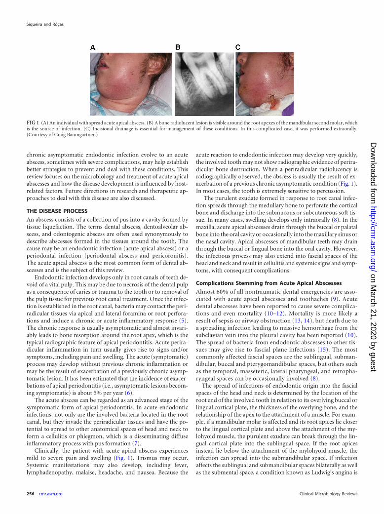
Clinically, the patient with acute apical abscess experiencesmild to severe pain and swelling (Fig. 1). Trismus may occur.Systemic manifestations may also develop, including fever,lymphadenopathy, malaise, headache, and nausea. Because the
acute reaction to endodontic infection may develop very quickly,the involved tooth may not show radiographic evidence of perira-dicular bone destruction. When a periradicular radiolucency isradiographically observed, the abscess is usually the result of ex-acerbation of a previous chronic asymptomatic condition (Fig. 1).In most cases, the tooth is extremely sensitive to percussion.
The purulent exudate formed in response to root canal infec-tion spreads through the medullary bone to perforate the corticalbone and discharge into the submucous or subcutaneous soft tis-sue. In many cases, swelling develops only intraorally (8). In themaxilla, acute apical abscesses drain through the buccal or palatalbone into the oral cavity or occasionally into the maxillary sinus orthe nasal cavity. Apical abscesses of mandibular teeth may drainthrough the buccal or lingual bone into the oral cavity. However,the infectious process may also extend into fascial spaces of thehead and neck and result in cellulitis and systemic signs and symp-toms, with consequent complications.
Complications Stemming from Acute Apical Abscesses
Almost 60% of all nontraumatic dental emergencies are asso-ciated with acute apical abscesses and toothaches (9). Acutedental abscesses have been reported to cause severe complica-tions and even mortality (10–12). Mortality is more likely aresult of sepsis or airway obstruction (13, 14), but death due toa spreading infection leading to massive hemorrhage from thesubclavian vein into the pleural cavity has been reported (10).The spread of bacteria from endodontic abscesses to other tis-sues may give rise to fascial plane infections (15). The mostcommonly affected fascial spaces are the sublingual, subman-dibular, buccal and pterygomandibular spaces, but others suchas the temporal, masseteric, lateral pharyngeal, and retropha-ryngeal spaces can be occasionally involved (8).
The spread of infections of endodontic origin into the fascialspaces of the head and neck is determined by the location of theroot end of the involved tooth in relation to its overlying buccal orlingual cortical plate, the thickness of the overlying bone, and therelationship of the apex to the attachment of a muscle. For exam-ple, if a mandibular molar is affected and its root apices lie closerto the lingual cortical plate and above the attachment of the my-lohyoid muscle, the purulent exudate can break through the lin-gual cortical plate into the sublingual space. If the root apicesinstead lie below the attachment of the mylohyoid muscle, theinfection can spread into the submandibular space. If infectionaffects the sublingual and submandibular spaces bilaterally as wellas the submental space, a condition known as Ludwig’s angina is
FIG 1 (A) An individual with spread acute apical abscess. (B) A bone radiolucent lesion is visible around the root apexes of the mandibular second molar, whichis the source of infection. (C) Incisional drainage is essential for management of these conditions. In this complicated case, it was performed extraorally.(Courtesy of Craig Baumgartner.)
Siqueira and Rôças
256 cmr.asm.org Clinical Microbiology Reviews
on March 21, 2020 by guest
http://cmr.asm
.org/D
ownloaded from
diagnosed. Swelling from Ludwig’s angina can give rise to diffi-culty breathing and potentially lethal airway obstruction (16–18).
Another example of abscess complications involves infectionsof the midface, which can be very dangerous and result in cavern-ous sinus thrombosis. This is also a life-threatening infection, inwhich a thrombus formed in the cavernous sinus breaks free andleads to spread of the infection. Under normal conditions, theangular and ophthalmic veins and the pterygoid plexus of veinsflow into the facial and external jugular veins. If an infection hasspread into the midfacial area, however, edema and the resultantincreased pressure cause the blood to back up into the cavernoussinus. Once in the sinus, the blood can stagnate and clot. Theresultant infected thrombi remain in the cavernous sinus or es-cape into the circulation (19, 20).
Other reported complications of disseminating dental infec-tions include brain abscess (21–24), septicemia in a patient withmultiple myeloma (25), deep neck infection (26, 27), mediastinitis(14, 28–30), necrotizing fasciitis (31–33), orbital abscess (34–36),and cervical spondylodiscitis with spinal epidural abscess (37). Ithas been suggested that some host-related factors may contributetoward increased morbidity and mortality associated with acutedental abscesses, including diabetes, chronic alcohol and tobaccoconsumption, malnourishment, and the use of illicit substances(26, 27, 33).
Complications of dental abscesses can be severe enough to re-quire hospitalization. A large number of hospitalizations due tooral abscesses/cellulitis with a resulting substantial economic bur-den have been recorded (38). For instance, in 2007, 7,886 hospi-talizations were attributed primarily to abscesses of endodonticorigin, with total hospital charges on the order of $100 million(39).
MICROBIOLOGY OF ACUTE APICAL ABSCESSES
Microbiology Diagnostic Methods
Culture. Culture methods have been traditionally used to inves-tigate the microbiota of acute apical abscesses and have provided asubstantial body of information about the bacterial etiology andthe species involved. However, some important limitations of cul-ture make it difficult to achieve a comprehensive analysis of theapical abscess microbiota. Because anaerobic bacteria are domi-nant in apical abscesses (40–49), samples for research or clinicaldiagnosis using culture should be collected and transported to thelaboratory under conditions that favor the survival of these bac-teria. The laboratory that will analyze the samples has to be prop-erly prepared and equipped to isolate, cultivate, and identify an-aerobes. The procedures for isolation and identification can belaborious and time-consuming, and many anaerobic species mayrequire multiple phenotype-based tests for reliable identification(50).
More important, the difficulties in culturing a large number oforal bacteria or even in identifying many species are of specialconcern (51). As early estimated by correlative microscopic andculturing analyses (52) and further elegantly demonstrated andspecified by molecular biology techniques (53–58), 40% to 70% ofthe oral bacterial species remain to be cultivated and phenotypi-cally characterized. The most possible reasons for the fact that alarge number of oral bacteria still have to be cultivated include (i)lack of essential nutrients or growth factors in the culture me-dium, (ii) overfeeding conditions during cultivation, (iii) toxicity
of the culture medium itself, (iv) inhibition by other species pres-ent in the sample, (v) metabolic dependence on other species, (vi)disruption of natural bacterial quorum-sensing systems, and (vii)cells in a “viable-but-noncultivable” state (51, 59, 60). Efforts havebeen expended toward the development of approaches that allowcultivation of as-yet-uncultivated bacteria (61–64). This is of greatimportance for description of novel species and study of theirecological and pathogenic potential, as well as the patterns of sus-ceptibility to antimicrobial drugs.
Successful cultivation does not ensure successful identifica-tion. Culture-dependent identification is based on phenotypictraits reported for reference strains, with predictable biochemicaland physical properties under optimal growth conditions. How-ever, there are several phenotype-related factors that can lead todifficulties in identification and even to misidentification. Theyinclude (i) strains of the same species showing a divergent pheno-typic behavior (65, 66), (ii) strains of different species showing aconvergent phenotypic behavior (51, 65), (iii) an altered pheno-type in response to conditions such as stress (67, 68), (iv) strainsshowing different results after repeated tests (69), (v) incompletedatabases not including newly named species and as-yet-unchar-acterized species, (vi) the fact that even small alterations in theassay may lead to false results (70), and (vii) the fact that testresults rely on individual interpretation and expertise (70).
Molecular methods. Tools and procedures based on molecularbiology have become available to sidestep the limitations of cul-ture and have been substantially improved to achieve a more re-alistic description of the microbial communities of different envi-ronments without the need for cultivation. One gene that has beenwidely used for rapid identification of known and unknown bac-terial species is the one encoding the 16S rRNA (71). There are amyriad of molecular methods for the study of bacteria in abscesssamples, and the choice of a particular approach depends on thequestions to be answered. Molecular methods for diagnostic mi-crobiology can be used for specific detection of target species (spe-cies-specific or closed-ended analysis), identification of all or themost dominant species in a sample (broad-range or open-endedanalysis), or profiling of the microbial community structure(community analysis) (72). Broad-range PCR followed by cloningand sequencing and more recently the massive parallel 454 pyro-sequencing approach can be used to unravel the breadth of bacte-rial diversity in a site. DNA hybridization arrays (e.g., checker-board arrays and microarrays), species-specific PCR, nested PCR,multiplex PCR, and quantitative real-time PCR can be used tosurvey large numbers of samples for the presence of target species.Bacterial community structures can be analyzed via the pyrose-quencing technology and by fingerprinting techniques, such as thedenaturing gradient gel electrophoresis (DGGE) and terminal re-striction fragment length polymorphism (T-RFLP) assays. As withany other technology, molecular methods have their own limita-tions. However, variations in virtually every technique haveemerged to circumvent or minimize limitations, or sometimesmore than one approach is required for reliable information to beobtained.
The chronology of the microbiological study of acute apicalabscesses can be didactically divided into five generations of stud-ies based on different strategic diagnostic approaches (73). Thefirst generation involves studies of the abscess microbiota con-ducted using open-ended (or broad-range) culture methods,which disclosed many cultivable species in association with the
Microbiology of Dental Abscesses
April 2013 Volume 26 Number 2 cmr.asm.org 257
on March 21, 2020 by guest
http://cmr.asm
.org/D
ownloaded from
disease (41–49). The second generation consisted of studies em-ploying closed-ended molecular detection methods, such as spe-cies-specific PCR and its derivatives as well as the original check-erboard hybridization assay, to target cultivable bacteria (74–82).These methods allowed the inclusion of some difficult-to-culturespecies in the set of candidate abscess pathogens. Next, a thirdgeneration of studies adopted open-ended molecular methods,such as broad-range PCR followed by cloning and sequencing orT-RFLP, which allowed an even more comprehensive investiga-tion of the bacterial diversity in abscesses (83–87). By these ap-proaches, not only cultivable species but also as-yet-uncultivatedand uncharacterized bacteria have been identified. Technical hur-dles make it difficult to analyze a large number of samples bycloning and sequencing, but cataloguing bacterial species in theoral cavity by this approach provided 16S rRNA gene sequencedata that could be used to design primers or oligonucleotideprobes to target both cultivable and as-yet-uncultivated bacteria.The fourth generation of studies involved closed-ended molecularanalyses with PCR and DNA hybridization assays (e.g., reverse-capture checkerboard) in large-scale clinical studies to investigatethe prevalence and association of cultivable and as-yet-unculti-vated bacteria with abscesses (88–90). A fifth generation has beenpossibly heralded by the use of pyrosequencing technology for adeep-coverage open-ended analysis of abscess samples (91, 92)(see below).
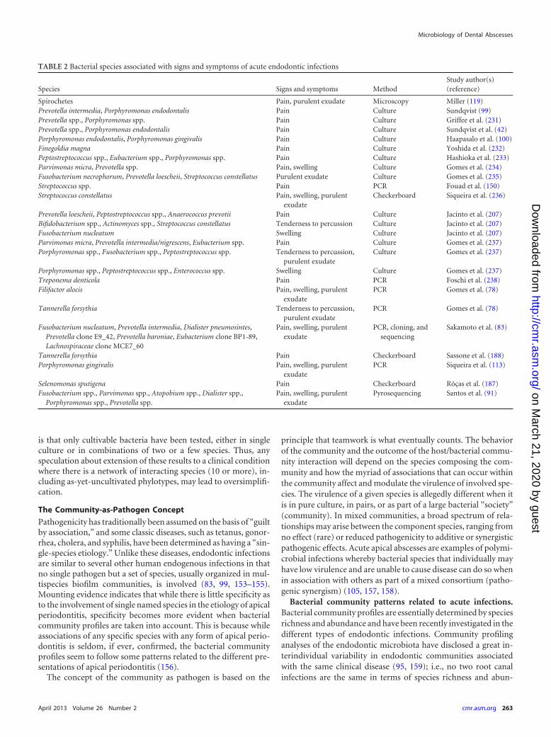
In general, culture analyses of abscesses resulted in the estab-lishment of a set of species thought to play an important role in thepathogenesis of the disease. Not only have molecular methodsconfirmed and even strengthened the association of many culti-vable bacterial species with abscesses, but they also revealed newsuspected pathogens (93). The list of candidate pathogens hasexpanded to include difficult-to-culture species or even as-yet-uncultivated bacteria that had never been previously found in ab-scesses by culturing approaches (Fig. 2). Consequently, the micro-biota of apical abscesses has been refined and redefined bymolecular methods (93). Although molecular methods have beenwidely used for research purposes, they still have yet to be imple-mented for clinical diagnosis. There is a great potential for thesemethods to be used for rapid identification of potential pathogensconcomitantly with specific antibiotic susceptibility, which wouldallow immediate and more appropriate patient care with possiblyreduced morbidity or mortality (87, 94). This application will befurther discussed in the section Future Directions.
Microbial Diversity in Acute Apical Abscesses
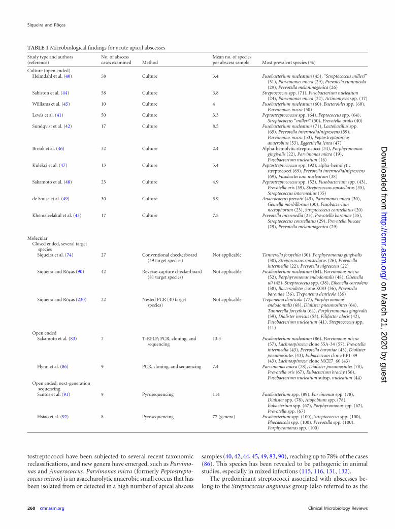
Samples for microbiological analyses of abscesses can be takeneither from the root canals of affected teeth or by aspiration of thepurulent exudate from the swollen mucosa/skin. Culture and mo-lecular microbiology studies have clearly demonstrated that theapical abscess microbiota is mixed and conspicuously dominatedby anaerobic bacteria (42, 43, 49, 83, 87, 95–96). Table 1 providesa compilation of the main microbiological findings from most ofthese studies. It is noteworthy that while some bacterial species orgroups are reported in many studies, the most prevalent speciesvary from study to study.
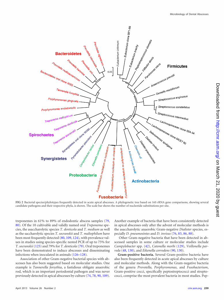
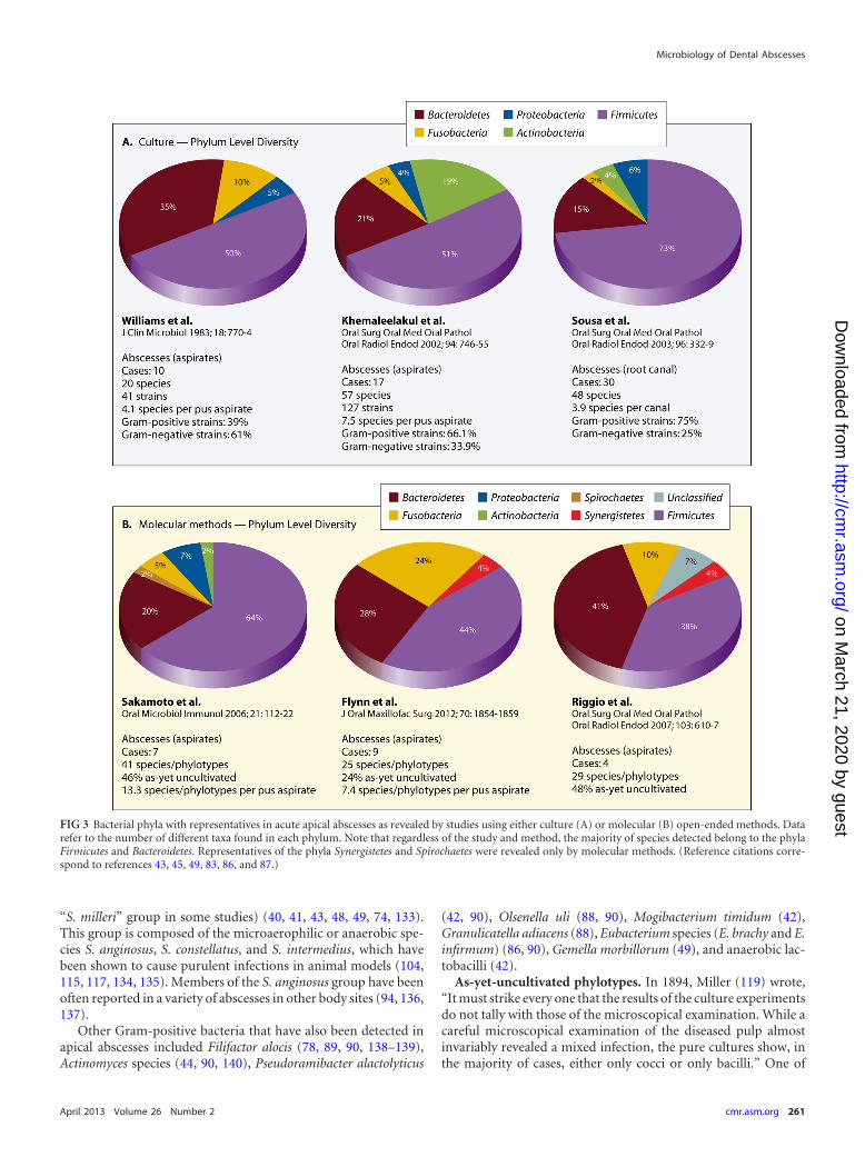
At a broader taxonomic level, the large majority of the fre-quently detected bacterial species belong to seven different bacte-rial phyla, namely, the Firmicutes (e.g., genera Streptococcus, Di-alister, Filifactor, and Pseudoramibacter), Bacteroidetes (e.g.,genera Porphyromonas, Prevotella, and Tannerella), Fusobacteria
(e.g., genera Fusobacterium and Leptotrichia), Actinobacteria (e.g.,genera Actinomyces and Propionibacterium), Spirochaetes (e.g., ge-nus Treponema), Synergistetes (e.g., genus Pyramidobacter andsome as-yet-uncultivated phylotypes), and Proteobacteria (e.g.,genera Campylobacter and Eikenella) (Fig. 2). Regardless of thestudy and method of identification, the phyla Firmicutes and Bac-teroidetes together contribute to more than 70% of the speciesfound in abscesses (Fig. 3). Representatives of Spirochaetes andSynergistetes have been revealed only by culture-independent mo-lecular methods (Fig. 3). Diverse groups of Gram-negative andGram-positive bacteria have been identified, and the most fre-quent genera and species identified in abscesses and regarded asputative pathogens are described next. The fact that many generahave undergone reclassifications over the years makes interpreta-tion of “old” studies difficult, especially when identifications wererestricted to the genus level.
Gram-negative bacteria. Dark-pigmented anaerobic bacteriahave been closely associated with acute symptoms of endodonticinfections, including abscesses (41, 47, 96–100). Two culture stud-ies (42, 101) found that virtually all abscesses of endodontic originharbored one or more species of this group. Dark-pigmented an-aerobic bacteria are usually found in mixed infection, which maybe required for their optimal growth and contributes to a signifi-cant increase in their pathogenicity (102–106). This bacterialgroup comprises two genera: Prevotella (containing saccharolyticspecies) and Porphyromonas (containing asaccharolytic species).The genus Prevotella also includes some nonpigmented species.The Prevotella species frequently found in apical abscess samplesinclude P. intermedia, P. nigrescens, P. baroniae, and P. oris, and inmost studies they are among the most prevalent and/or dominantspecies (40, 42, 43, 75, 83, 86, 87, 101, 107). Of the Porphyromonasspecies, P. endodontalis was first isolated from endodontic infec-tions (108) and has been consistently encountered in abscess sam-ples, with increased prevalence in molecular studies (42, 77, 90,101, 109, 110). Porphyromonas gingivalis is one of the most impor-tant periodontal pathogens (111, 112) and has also been detectedin association with endodontic abscesses (74, 77, 101, 113). P.gingivalis fimA genotype variants II, III, and IV and type I havebeen reported in abscess aspirates (114).
Fusobacterium nucleatum is an anaerobic spindle-shaped rodthat is one of the most commonly detected Gram-negative speciesin the large majority of culture and molecular studies of acuteapical abscesses (40, 45, 85, 90), reaching prevalence values as highas approximately 70% (42, 75) or even 86% (83) of the samples.This species induces severe abscess lesions in animals in both pureand mixed cultures (115–118). Four subspecies (F. nucleatumsubsp. nucleatum, F. nucleatum subsp. polymorphum, F. nuclea-tum subsp. vincentii, and F. nucleatum subsp. animalis) have beenidentified in apical abscesses (74, 86), but the frequency of eachone has yet to be accurately determined. The species Fusobacte-rium periodonticum has also been detected in abscess aspirates by astudy using the checkerboard hybridization assay (74).
In a seminal study of endodontic infections published in 1894(119), Willoughby Dayton Miller suggested that spirochetes couldplay a role in the etiology of abscesses. Nevertheless, it was notuntil the introduction of molecular methods in endodontic mi-crobiology research that the potential involvement of spirocheteswith this disease was confirmed. Oral spirochetes fall within thegenus Treponema and have been linked to several oral diseases(120–123). Molecular methods have revealed the occurrence of
Siqueira and Rôças
258 cmr.asm.org Clinical Microbiology Reviews
on March 21, 2020 by guest
http://cmr.asm
.org/D
ownloaded from
treponemes in 61% to 89% of endodontic abscess samples (79,80). Of the 10 cultivable and validly named oral Treponema spe-cies, the asaccharolytic species T. denticola and T. medium as wellas the saccharolytic species T. socranskii and T. maltophilum havebeen most frequently detected (80, 109, 124), with prevalence val-ues in studies using species-specific nested PCR of up to 75% forT. socranskii (125) and 79% for T. denticola (79). Oral treponemeshave been demonstrated to induce abscesses and disseminatinginfections when inoculated in animals (126–128).
Association of other Gram-negative bacterial species with ab-scesses has also been suggested based on molecular studies. Oneexample is Tannerella forsythia, a fastidious obligate anaerobicrod, which is an important periodontal pathogen and was neverpreviously detected in apical abscesses by culture (74, 78, 90, 109).
Another example of bacteria that have been consistently detectedin apical abscesses only after the advent of molecular methods isthe asaccharolytic anaerobic Gram-negative Dialister species, es-pecially D. pneumosintes and D. invisus (76, 83, 86, 88).
Other Gram-negative bacteria that have been detected in ab-scessed samples in some culture or molecular studies includeCampylobacter spp. (42), Catonella morbi (129), Veillonella par-vula (48, 130), and Eikenella corrodens (90, 130).
Gram-positive bacteria. Several Gram-positive bacteria havealso been frequently detected in acute apical abscesses by cultureand molecular methods. Along with the Gram-negative bacteriaof the genera Prevotella, Porphyromonas, and Fusobacterium,Gram-positive cocci, specifically peptostreptococci and strepto-cocci, comprise the most prevalent bacteria in most studies. Pep-
FIG 2 Bacterial species/phylotypes frequently detected in acute apical abscesses. A phylogenetic tree based on 16S rRNA gene comparisons, showing severalcandidate pathogens and their respective phyla, is shown. The scale bar shows the number of nucleotide substitutions per site.
Microbiology of Dental Abscesses
April 2013 Volume 26 Number 2 cmr.asm.org 259
on March 21, 2020 by guest
http://cmr.asm
.org/D
ownloaded from
tostreptococci have been subjected to several recent taxonomicreclassifications, and new genera have emerged, such as Parvimo-nas and Anaerococcus. Parvimonas micra (formerly Peptostrepto-coccus micros) is an asaccharolytic anaerobic small coccus that hasbeen isolated from or detected in a high number of apical abscess
samples (40, 42, 44, 45, 49, 83, 90), reaching up to 78% of the cases(86). This species has been revealed to be pathogenic in animalstudies, especially in mixed infections (115, 116, 131, 132).
The predominant streptococci associated with abscesses be-long to the Streptococcus anginosus group (also referred to as the
TABLE 1 Microbiological findings for acute apical abscesses
Study type and authors(reference)
No. of abscesscases examined Method
Mean no. of speciesper abscess sample Most prevalent species (%)
Culture (open ended)Heimdahl et al. (40) 58 Culture 3.4 Fusobacterium nucleatum (45), “Streptococcus milleri”
(31), Parvimonas micra (29), Prevotella ruminicola(29), Prevotella melaninogenica (26)
Sabiston et al. (44) 58 Culture 3.8 Streptococcus spp. (71), Fusobacterium nucleatum(24), Parvimonas micra (22), Actinomyces spp. (17)
Williams et al. (45) 10 Culture 4 Fusobacterium nucleatum (60), Bacteroides spp. (60),Parvimonas micra (50)
Lewis et al. (41) 50 Culture 3.3 Peptostreptococcus spp. (64), Peptococcus spp. (64),Streptococcus “milleri” (50), Prevotella oralis (40)
Sundqvist et al. (42) 17 Culture 8.5 Fusobacterium nucleatum (71), Lactobacillus spp.(65), Prevotella intermedia/nigrescens (59),Parvimonas micra (53), Peptostreptococcusanaerobius (53), Eggerthella lenta (47)
Brook et al. (46) 32 Culture 2.4 Alpha-hemolytic streptococci (34), Porphyromonasgingivalis (22), Parvimonas micra (19),Fusobacterium nucleatum (16)
Kulekçi et al. (47) 13 Culture 5.4 Peptostreptococcus spp. (92), alpha-hemolyticstreptococci (69), Prevotella intermedia/nigrescens(69), Fusobacterium nucleatum (38)
Sakamoto et al. (48) 23 Culture 4.9 Peptostreptococcus spp. (52), Fusobacterium spp. (43),Prevotella oris (39), Streptococcus constellatus (35),Streptococcus intermedius (35)
de Sousa et al. (49) 30 Culture 3.9 Anaerococcus prevotii (43), Parvimonas micra (30),Gemella morbillorum (30), Fusobacteriumnecrophorum (23), Streptococcus constellatus (20)
Khemaleelakul et al. (43) 17 Culture 7.5 Prevotella intermedia (35), Prevotella baroniae (35),Streptococcus constellatus (29), Prevotella buccae(29), Prevotella melaninogenica (29)
MolecularClosed ended, several target
speciesSiqueira et al. (74) 27 Conventional checkerboard
(49 target species)Not applicable Tannerella forsythia (30), Porphyromonas gingivalis
(30), Streptococcus constellatus (26), Prevotellaintermedia (22), Prevotella nigrescens (22)
Siqueira and Rôças (90) 42 Reverse-capture checkerboard(81 target species)
Not applicable Fusobacterium nucleatum (64), Parvimonas micra(52), Porphyromonas endodontalis (48), Olsenellauli (45), Streptococcus spp. (38), Eikenella corrodens(38), Bacteroidetes clone X083 (36), Prevotellabaroniae (36), Treponema denticola (36)
Siqueira and Rôças (230) 22 Nested PCR (40 targetspecies)
Not applicable Treponema denticola (77), Porphyromonasendodontalis (68), Dialister pneumosintes (64),Tannerella forsythia (64), Porphyromonas gingivalis(59), Dialister invisus (53), Filifactor alocis (42),Fusobacterium nucleatum (41), Streptococcus spp.(41)
Open endedSakamoto et al. (83) 7 T-RFLP; PCR, cloning, and
sequencing13.3 Fusobacterium nucleatum (86), Parvimonas micra
(57), Lachnospiracea clone 55A-34 (57), Prevotellaintermedia (43), Prevotella baroniae (43), Dialisterpneumosintes (43), Eubacterium clone BP1-89(43), Lachnospiracea clone MCE7_60 (43)
Flynn et al. (86) 9 PCR, cloning, and sequencing 7.4 Parvimonas micra (78), Dialister pneumosintes (78),Prevotella oris (67), Eubacterium brachy (56),Fusobacterium nucleatum subsp. nucleatum (44)
Open ended, next-generationsequencingSantos et al. (91) 9 Pyrosequencing 114 Fusobacterium spp. (89), Parvimonas spp. (78),
Dialister spp. (78), Atopobium spp. (78),Eubacterium spp. (67), Porphyromonas spp. (67),Prevotella spp. (67)
Hsiao et al. (92) 8 Pyrosequencing 77 (genera) Fusobacterium spp. (100), Streptococcus spp. (100),Phocaeicola spp. (100), Prevotella spp. (100),Porphyromonas spp. (100)
Siqueira and Rôças
260 cmr.asm.org Clinical Microbiology Reviews
on March 21, 2020 by guest
http://cmr.asm
.org/D
ownloaded from
“S. milleri” group in some studies) (40, 41, 43, 48, 49, 74, 133).This group is composed of the microaerophilic or anaerobic spe-cies S. anginosus, S. constellatus, and S. intermedius, which havebeen shown to cause purulent infections in animal models (104,115, 117, 134, 135). Members of the S. anginosus group have beenoften reported in a variety of abscesses in other body sites (94, 136,137).
Other Gram-positive bacteria that have also been detected inapical abscesses included Filifactor alocis (78, 89, 90, 138–139),Actinomyces species (44, 90, 140), Pseudoramibacter alactolyticus
(42, 90), Olsenella uli (88, 90), Mogibacterium timidum (42),Granulicatella adiacens (88), Eubacterium species (E. brachy and E.infirmum) (86, 90), Gemella morbillorum (49), and anaerobic lac-tobacilli (42).
As-yet-uncultivated phylotypes. In 1894, Miller (119) wrote,“It must strike every one that the results of the culture experimentsdo not tally with those of the microscopical examination. While acareful microscopical examination of the diseased pulp almostinvariably revealed a mixed infection, the pure cultures show, inthe majority of cases, either only cocci or only bacilli.” One of
FIG 3 Bacterial phyla with representatives in acute apical abscesses as revealed by studies using either culture (A) or molecular (B) open-ended methods. Datarefer to the number of different taxa found in each phylum. Note that regardless of the study and method, the majority of species detected belong to the phylaFirmicutes and Bacteroidetes. Representatives of the phyla Synergistetes and Spirochaetes were revealed only by molecular methods. (Reference citations corre-spond to references 43, 45, 49, 83, 86, and 87.)
Microbiology of Dental Abscesses
April 2013 Volume 26 Number 2 cmr.asm.org 261
on March 21, 2020 by guest
http://cmr.asm
.org/D
ownloaded from
Miller’s possible explanations for this finding was that “many spe-cies of bacteria occurring in the diseased pulp, vibriones, spiro-chaetes, the stiff pointed bacilli and threads, have not been foundcultivable on artificial media anyway; and possibly there are stillother uncultivable pulp-bacteria.” The development of methodsfor anaerobic cultivation showed that the bacterial diversity inendodontic infections was underestimated by previous culturestudies by excluding a high number of species that compose thelargest proportion of the bacterial community in these infections.Further breakthroughs in microbial identification represented bymolecular technologies revealed that even advances in anaerobicculturing left a large proportion of the microbiota undisclosed.
Indeed, molecular investigations of the bacteria involved inabscesses unveiled a far more complex picture than anticipated byculture studies. Noteworthy is the common occurrence of as-yet-uncultivated bacterial phylotypes, which can be regarded as spe-cies-level bacteria known only by a 16S rRNA gene sequence.Open-ended molecular analysis of acute apical abscesses revealedthat, in terms of richness, as-yet-uncultivated phylotypes encom-pass approximately 24% to 46% of the taxa found (83, 86), whilein terms of abundance, they collectively represent from 6% tomore than 30% of the clones sequenced (83, 87).
Several of the as-yet-uncultivated phylotypes are suspectedpathogens based on association data. For instance, a phylotype ofthe Bacteroidetes phylum known as oral clone X083 has beenfound in 14% to 36% of apical abscess aspirates (90, 107). OralSynergistetes phylotypes, which had been originally assigned to theFlexistipes or Deferribacteres groups, are another example of as-yet-uncultivated bacteria that have been frequently encounteredin abscesses (88–90). The great majority of Synergistetes bacteriaremain uncultivated (141), and this can be the primary reason forthe fact that their presence in abscesses has been overlooked byculture studies. Some Synergistetes phylotypes have been culti-vated and given a species name; one of them, Pyramidobacter pis-colens (previously oral clone BA121), is probably the most preva-lent representative of the Synergistetes phylum in abscess cases(88–90). Phylotypes from the family Lachnospiraceae or the generaEubacterium, Megasphaera, Leptotrichia, Oribacterium, Pepto-streptococcus, Prevotella, Selenomonas, and Solobacterium havebeen disclosed in pus samples from apical abscesses (83, 86, 87).There is no reason to believe that these previously unrecognizedand overlooked bacteria do not play a role in the pathogenesis ofthe disease.
Pyrosequencing analysis of abscess samples. Advances inDNA sequencing technologies and computational biology havesubstantially enhanced molecular phylogenetic surveys of hu-man-associated microbial communities in health and disease, of-fering a great depth of coverage at a very high analytical speed(142, 143). The 454 pyrosequencing method is one of these ad-vanced DNA sequencing techniques that has been widely used inmedical microbiology. This technology is a sequencing-by-syn-thesis method that involves a combination of emulsion PCR andpyrosequencing. Pyrosequencing relies on the detection of pyro-phosphate release and consequent light generation as nucleotidesare incorporated in a growing chain of DNA (144, 145). One of thegreatest advantages of the pyrosequencing approach over the con-ventional Sanger sequencing method is that hundreds of thou-sands of sequence reads can be obtained in a single run, generatingsequence information data that are orders of magnitude larger(146).
A recent study used 454 pyrosequencing to compare the mi-crobiota of endodontic infections associated with acute abscessesand asymptomatic chronic apical periodontitis, and it found op-erational taxonomic units (at 3% divergence) belonging to 13phyla (91). The most abundant phyla in acute infections wereFirmicutes (52%), Fusobacteria (17%), and Bacteroidetes (13%),while the dominant phyla in asymptomatic infections were Firmi-cutes (59%), Bacteroidetes (14%), and Actinobacteria (10%).Members of Fusobacteria were much more prevalent in acute(89%) than in chronic (50%) infections. Of the 49 genera detectedin acute cases, the most abundant were Fusobacterium, Parvimo-nas, and Peptostreptococcus. Fusobacterium was also the most prev-alent, followed by Parvimonas, Dialister, and Atopobium. The bac-terial communities in abscesses were significantly more diversethan those in chronic infections, and a pattern related to the pres-ence of symptoms was apparently evident through communityanalysis. The overall diversity of abscesses as revealed by pyrose-quencing was much higher than previously reported. Most ofthese findings were confirmed by another study using pyrose-quencing, which revealed representatives of 11 phyla and Fusobac-terium as the most abundant genus (92).
Bacterial Species and Acute Infections: Is There a SingleCulprit?
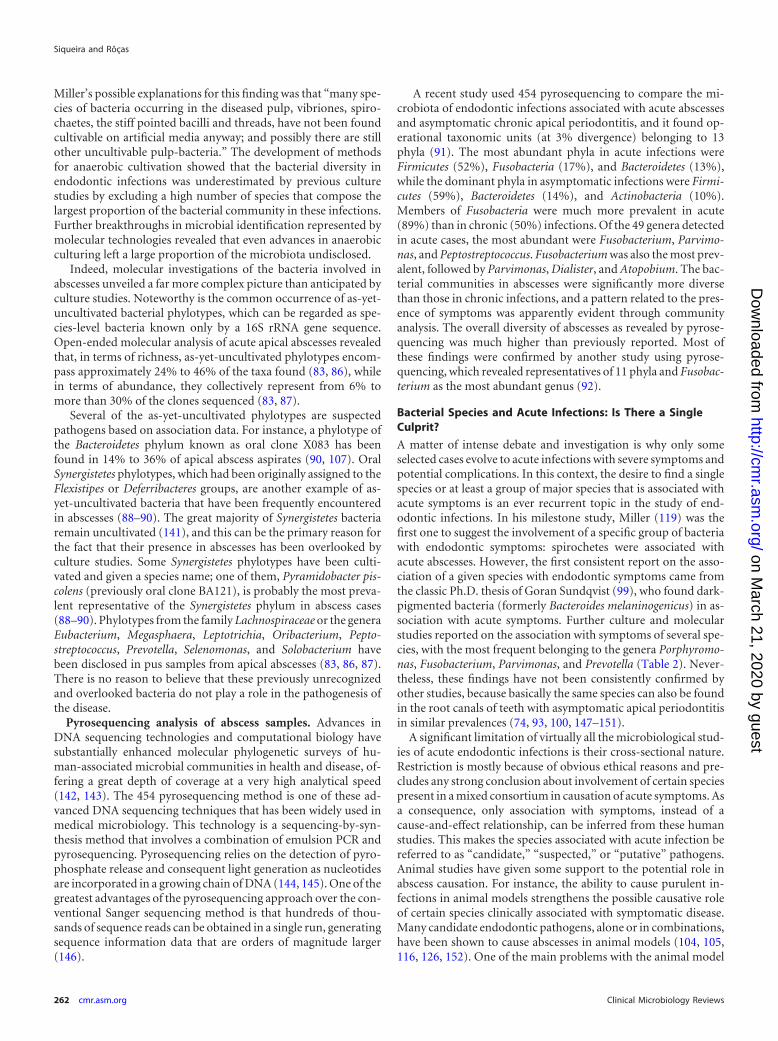
A matter of intense debate and investigation is why only someselected cases evolve to acute infections with severe symptoms andpotential complications. In this context, the desire to find a singlespecies or at least a group of major species that is associated withacute symptoms is an ever recurrent topic in the study of end-odontic infections. In his milestone study, Miller (119) was thefirst one to suggest the involvement of a specific group of bacteriawith endodontic symptoms: spirochetes were associated withacute abscesses. However, the first consistent report on the asso-ciation of a given species with endodontic symptoms came fromthe classic Ph.D. thesis of Goran Sundqvist (99), who found dark-pigmented bacteria (formerly Bacteroides melaninogenicus) in as-sociation with acute symptoms. Further culture and molecularstudies reported on the association with symptoms of several spe-cies, with the most frequent belonging to the genera Porphyromo-nas, Fusobacterium, Parvimonas, and Prevotella (Table 2). Never-theless, these findings have not been consistently confirmed byother studies, because basically the same species can also be foundin the root canals of teeth with asymptomatic apical periodontitisin similar prevalences (74, 93, 100, 147–151).
A significant limitation of virtually all the microbiological stud-ies of acute endodontic infections is their cross-sectional nature.Restriction is mostly because of obvious ethical reasons and pre-cludes any strong conclusion about involvement of certain speciespresent in a mixed consortium in causation of acute symptoms. Asa consequence, only association with symptoms, instead of acause-and-effect relationship, can be inferred from these humanstudies. This makes the species associated with acute infection bereferred to as “candidate,” “suspected,” or “putative” pathogens.Animal studies have given some support to the potential role inabscess causation. For instance, the ability to cause purulent in-fections in animal models strengthens the possible causative roleof certain species clinically associated with symptomatic disease.Many candidate endodontic pathogens, alone or in combinations,have been shown to cause abscesses in animal models (104, 105,116, 126, 152). One of the main problems with the animal model
Siqueira and Rôças
262 cmr.asm.org Clinical Microbiology Reviews
on March 21, 2020 by guest
http://cmr.asm
.org/D
ownloaded from
is that only cultivable bacteria have been tested, either in singleculture or in combinations of two or a few species. Thus, anyspeculation about extension of these results to a clinical conditionwhere there is a network of interacting species (10 or more), in-cluding as-yet-uncultivated phylotypes, may lead to oversimplifi-cation.
The Community-as-Pathogen Concept
Pathogenicity has traditionally been assumed on the basis of “guiltby association,” and some classic diseases, such as tetanus, gonor-rhea, cholera, and syphilis, have been determined as having a “sin-gle-species etiology.” Unlike these diseases, endodontic infectionsare similar to several other human endogenous infections in thatno single pathogen but a set of species, usually organized in mul-tispecies biofilm communities, is involved (83, 99, 153–155).Mounting evidence indicates that while there is little specificity asto the involvement of single named species in the etiology of apicalperiodontitis, specificity becomes more evident when bacterialcommunity profiles are taken into account. This is because whileassociations of any specific species with any form of apical perio-dontitis is seldom, if ever, confirmed, the bacterial communityprofiles seem to follow some patterns related to the different pre-sentations of apical periodontitis (156).
The concept of the community as pathogen is based on the
principle that teamwork is what eventually counts. The behaviorof the community and the outcome of the host/bacterial commu-nity interaction will depend on the species composing the com-munity and how the myriad of associations that can occur withinthe community affect and modulate the virulence of involved spe-cies. The virulence of a given species is allegedly different when itis in pure culture, in pairs, or as part of a large bacterial “society”(community). In mixed communities, a broad spectrum of rela-tionships may arise between the component species, ranging fromno effect (rare) or reduced pathogenicity to additive or synergisticpathogenic effects. Acute apical abscesses are examples of polymi-crobial infections whereby bacterial species that individually mayhave low virulence and are unable to cause disease can do so whenin association with others as part of a mixed consortium (patho-genic synergism) (105, 157, 158).
Bacterial community patterns related to acute infections.Bacterial community profiles are essentially determined by speciesrichness and abundance and have been recently investigated in thedifferent types of endodontic infections. Community profilinganalyses of the endodontic microbiota have disclosed a great in-terindividual variability in endodontic communities associatedwith the same clinical disease (95, 159); i.e., no two root canalinfections are the same in terms of species richness and abun-
TABLE 2 Bacterial species associated with signs and symptoms of acute endodontic infections
Species Signs and symptoms MethodStudy author(s)(reference)
Spirochetes Pain, purulent exudate Microscopy Miller (119)Prevotella intermedia, Porphyromonas endodontalis Pain Culture Sundqvist (99)Prevotella spp., Porphyromonas spp. Pain Culture Griffee et al. (231)Prevotella spp., Porphyromonas endodontalis Pain Culture Sundqvist et al. (42)Porphyromonas endodontalis, Porphyromonas gingivalis Pain Culture Haapasalo et al. (100)Finegoldia magna Pain Culture Yoshida et al. (232)Peptostreptococcus spp., Eubacterium spp., Porphyromonas spp. Pain Culture Hashioka et al. (233)Parvimonas micra, Prevotella spp. Pain, swelling Culture Gomes et al. (234)Fusobacterium necrophorum, Prevotella loescheii, Streptococcus constellatus Purulent exudate Culture Gomes et al. (235)Streptococcus spp. Pain PCR Fouad et al. (150)Streptococcus constellatus Pain, swelling, purulent
exudateCheckerboard Siqueira et al. (236)
Prevotella loescheii, Peptostreptococcus spp., Anaerococcus prevotii Pain Culture Jacinto et al. (207)Bifidobacterium spp., Actinomyces spp., Streptococcus constellatus Tenderness to percussion Culture Jacinto et al. (207)Fusobacterium nucleatum Swelling Culture Jacinto et al. (207)Parvimonas micra, Prevotella intermedia/nigrescens, Eubacterium spp. Pain Culture Gomes et al. (237)Porphyromonas spp., Fusobacterium spp., Peptostreptococcus spp. Tenderness to percussion,
purulent exudateCulture Gomes et al. (237)
Porphyromonas spp., Peptostreptococcus spp., Enterococcus spp. Swelling Culture Gomes et al. (237)Treponema denticola Pain PCR Foschi et al. (238)Filifactor alocis Pain, swelling, purulent
exudatePCR Gomes et al. (78)
Tannerella forsythia Tenderness to percussion,purulent exudate
PCR Gomes et al. (78)
Fusobacterium nucleatum, Prevotella intermedia, Dialister pneumosintes,Prevotella clone E9_42, Prevotella baroniae, Eubacterium clone BP1-89,Lachnospiraceae clone MCE7_60
Pain, swelling, purulentexudate
PCR, cloning, andsequencing
Sakamoto et al. (83)
Tannerella forsythia Pain Checkerboard Sassone et al. (188)Porphyromonas gingivalis Pain, swelling, purulent
exudatePCR Siqueira et al. (113)
Selenomonas sputigena Pain Checkerboard Rôças et al. (187)Fusobacterium spp., Parvimonas spp., Atopobium spp., Dialister spp.,
Porphyromonas spp., Prevotella spp.Pain, swelling, purulent
exudatePyrosequencing Santos et al. (91)
Microbiology of Dental Abscesses
April 2013 Volume 26 Number 2 cmr.asm.org 263
on March 21, 2020 by guest
http://cmr.asm
.org/D
ownloaded from
dance. This indicates that the etiology of apical periodontitis, in-cluding the acute forms, is heterogeneous (95, 160).
Of great interest is that bacterial communities seem to follow aspecific pattern according to the clinical condition (e.g., asymp-tomatic versus symptomatic disease) (95). Therefore, the severityof apical periodontitis may be related to the overall bacterial com-munity composition. In other words, from the perspective of thesingle-pathogen concept, apical periodontitis can be consideredto be of no specific microbial etiology. However, based on thecommunity-as-pathogen concept, it is possible to infer that somecommunities are more related to certain forms of the disease, suchas abscesses (83, 95). A challenge that arises from this notion is theneed to unravel the specific genotypic and phenotypic character-istics of these abscess-related bacterial communities.
Acute apical abscesses are characterized by a concomitant in-fection of the root canal and the periradicular tissues, as the latteris an extension of the former. Even so, studies have reported somediscrepancies between bacterial community profiles for matchedsamples taken from the root canal and abscess aspirates (92, 161).The differences observed suggest that some selection of speciesoccurs as the infection advances from the canal to the periradicu-lar tissues. This “filtering” process is highly likely to be a result ofthe different environments faced by the infecting bacteria: from asite relatively protected from host defenses (necrotic root canal) toa site with an exuberant acute inflammatory response (highly vas-cularized periradicular tissues). Moreover, differences in the tis-sue invasion ability of the involved species may contribute to theselective process of species present in pus aspirates.
Geographic Differences in the Abscess Microbiota
A curious observation when analyzing separate studies performedin different countries is the different prevalences of species in-volved in abscesses. Variations in microbiological diagnosticmethodologies may account for most of these differences, but ageographical variation in bacterial composition cannot be disre-garded, as it also occurs for other human body sites (162–164).Molecular studies have been conducted to directly compare thebacterial community structures and prevalences of some targetspecies in abscesses from patients residing in different geographiclocations. In two studies comparing samples from Portland, OR,and Rio de Janeiro, Brazil (75, 89), species-specific PCR analysesrevealed that there was a significant difference in prevalence be-tween the two geographical locations for P. intermedia, P. nigre-scens, Prevotella tannerae, F. nucleatum, and P. gingivalis, all ofwhich more frequent in Portland samples, while T. denticola andT. forsythia were more prevalent in Rio de Janeiro samples. Open-ended DGGE analysis of the bacterial community profiles in acuteapical abscesses from the same two locations also disclosed a ge-ography-related pattern (160). Several species were exclusive foreach location, and others shared by the two locations showed greatdifferences in prevalence. The occurrence of geographical varia-tions was confirmed when comparing abscess samples from Riode Janeiro and Seoul, South Korea (110). These differences havethe potential to translate into relevant therapeutic implications,specifically in cases requiring systemic antibiotic therapy.
THE HOST SIDE OF THE STORY
Given the microbial etiology of apical periodontitis, developmentof acute symptoms has been traditionally searched for candidatemicrobial risk factors. Although there is a clear implication of
microbiological factors, the possibility certainly exists that host-related factors (disease modifiers) can influence the severity ofapical periodontitis lesions. Examples of disease modifiers includesystemic conditions (e.g., diabetes, herpesvirus infection, stress,autoimmune diseases, and diseases that weaken the immune re-sponse) and the genetic background (e.g., gene polymorphism).
Diabetic individuals have been shown to develop complica-tions from abscesses more frequently and to have a longer dura-tion of hospital stay than nondiabetics (27, 33). Similarly, diabeticpatients can exhibit about twice the rate of interappointment ex-acerbations (flare-ups) following endodontic intervention (165,166). Furthermore, diabetic animals develop apical periodontitislesions that are larger and more severe than those in nondiabeticcontrols (167). Type 2 diabetes mellitus has been found to besignificantly associated with increased prevalence of apical perio-dontitis (168, 169). Although diabetic patients are apparentlymore prone to develop severe forms of apical periodontitis, nostudy so far has reported on the prevalence of endodontic ab-scesses in diabetic individuals.
Potentially, genetic polymorphism is another factor that canmake individuals more susceptible to develop acute infections. DeSá et al. (170) investigated the association of acute apical abscesseswith polymorphisms in the genes for interleukin-1� (IL-1�), IL-6,IL-10, tumor necrosis factor alpha (TNF-�), and CD14. Individ-uals with asymptomatic apical periodontitis, without previous ex-acerbation, were included as controls. A significant associationwas observed between the occurrence of the GG genotype or the Gallele expression of the IL-6 gene and acute abscesses in womenand in individuals younger than 35 years. The G allele is associatedwith high levels of IL-6 production compared with the C allele(171). Intermediate- and high-producer IL1B genotypes and low-producer TNFA genotype also showed some association with ab-scesses, although not as strong as observed for IL-6. Other studiesare needed to confirm and expand these findings to analyze otherpotential inflammation-related genes.
It has been hypothesized that herpesviruses, especially humancytomegalovirus (HCMV) and Epstein-Barr virus (EBV), may beimplicated in the pathogenesis of apical periodontitis as a directresult of virus infection and replication or as a result of virallyinduced impairment of local host defenses, which might give riseto overgrowth of pathogenic bacteria in the very apical part of theroot canal system (172). Infection by HCMV and EBV has beenmore frequently observed in symptomatic lesions (173, 174), andan association has been suggested. However, data related to theoccurrence of herpesviruses in acute apical abscesses are ratherinconclusive. Chen et al. (175) found herpesviruses in low preva-lences and low copy numbers in abscess samples and concludedthat herpesviruses may be present but are not required for thedevelopment of abscesses and cellulitis of endodontic origin. Fer-reira et al. (176) evaluated the presence of human herpesviruses 1to 8 and human papillomavirus in acute apical abscesses and re-ported that about 60% of the samples were positive for at least onetarget virus. Human herpesvirus 8 (HHV-8) occurred at a highprevalence (48%), followed by human papillomavirus (13%) andvaricella-zoster virus and HHV-6 (9%). Viral coinfection oc-curred in some cases. In another study, the same group (109)found positive (but weak) associations between candidate end-odontic bacterial pathogens and human viruses in samples fromacute apical abscesses. Although these findings may suggest a rolefor viruses in the etiology of endodontic abscesses, the possibility
Siqueira and Rôças
264 cmr.asm.org Clinical Microbiology Reviews
on March 21, 2020 by guest
http://cmr.asm
.org/D
ownloaded from
also exists that the presence of viruses in the purulent exudate ismerely a consequence of the bacterially induced inflammatorydisease process.
There are many other host-related factors that have the poten-tial to influence the resistance to infection, including age, stress,drug abuse, malnutrition, and other systemic disorders. Futureresearch should focus on these potential candidate disease modi-fiers and their influence on the development of acute endodonticsymptoms.
FACTORS INFLUENCING THE DEVELOPMENT OF ACUTEINFECTION
It has been suggested that although the microbial etiology of ab-scesses is characterized by low specificity, certain species have beenmore frequently detected than others and then might be consid-ered more decisive in the outcome of the infectious process (158).However, based on the discussion above, in addition to the pres-ence of some potentially pathogenic species, a multitude of otherfactors can be regarded as influential to the development of acuteendodontic infections. These factors can be summarized as fol-lows: (i) difference in virulence among clonal types of the samespecies, (ii) bacterial interactions resulting in collective pathoge-nicity, (iii) bacterial load, (iv) environment-regulated expressionof virulence factors, and (v) host resistance and disease modifiers.
Difference in Virulence among Clonal Types of the SameSpecies
Clonal types of a given pathogenic bacterial species can signifi-cantly diverge in their virulence (177–180). A disease attributed toa given species is in fact caused by specific virulent clonal types ofthat species (181). Therefore, the possibility exists that the pres-ence of virulent clonal types of candidate endodontic pathogens inthe root canal may be a predisposing factor for abscess formation.This would help explain why the same species can be found inboth symptomatic and asymptomatic infections. So far, there is nocomparative study typing bacterial strains isolated from patientswith symptomatic and asymptomatic infections.
Bacterial Interactions Resulting in Collective Pathogenicity
Bacterial combinations contribute to the development of morevirulent communities due to synergism (104, 105, 116, 118, 126,152). Abscesses are polymicrobial infections for which culturestudies have reported a mean number of species ranging from 2 to8.5 per pus specimen (41–43, 45, 46, 48, 49, 96, 97, 182), whilemolecular studies have revealed a mean of 12 to 18 species/phylo-types per case (83, 95). Findings from pyrosequencing studies sug-gest that these numbers can be much higher (91, 92). What hasbecome evident from most studies comparing acute apical ab-scesses and asymptomatic endodontic infections is that there aredifferent dominant species in the communities, and the formerare usually characterized by a significantly higher species richnessthan that of the latter (83, 91, 95, 159). Increased diversity may bean important aspect of acute infections and collective pathogenic-ity, which is expected to be a result of the incalculable synergisticinteractions among the community members and summation ofvirulence factors produced. It has been shown that every compo-nent of a polymicrobial infection, even species regarded as aviru-lent and/or in low numbers in the consortium, may somewhataffect the virulence of other members of the community (183–186). Therefore, communication between bacterial community
members can alter the production of virulence factors by certainpathogenic species and affect the collective pathogenicity of theconsortium (185).
Indeed, it has been shown that different species combinationsmay result in different outcomes because of the network of inter-actions. Rôças et al. (187) compared the prevalences of 50 bacterialspecies or phylotypes in samples from symptomatic and asymp-tomatic endodontic infections and found that none of the mostprevalent taxa were significantly associated with symptoms; i.e.,most species were as prevalent in symptomatic cases as they werein asymptomatic cases. However, cluster analyses revealed that thehighly prevalent species formed different partnerships and associ-ations according to the presence of symptoms. For example, D.invisus and P. endodontalis were found in both symptomatic andasymptomatic cases, but their common partners in the formerwere different from those in the latter. Therefore, the possibilityexists that bacterial interactions result in communities that aremore or less aggressive and consequently can cause host responsesof corresponding intensity.
Bacterial Load
The bacterial load can be a decisive factor in acute disease causa-tion. Here two aspects need to be considered: the total bacterialload (nonspecific load) and the levels of certain species (specificload). Total bacterial loads per abscess case have been reported torange from 104 to 109 cells (41, 43, 45). The overall number ofbacterial cells in the community, regardless of the species, may beimportant because it may result in a heavy bioburden to the host,given the concentration of virulence factors released from largemasses of bacteria colonizing the canal and invading the perira-dicular tissues. Clinical approaches such as chemomechanical de-bridement of the infected dental root canal or tooth extractionand incision of the mucosa/skin for drainage of pus act primarilyand in a nonspecific way on the total bacterial load, reducing theinfectious bioburden.
As for the second aspect, i.e., the specific load, the possibilityexists that the number of cells of a given species found in bothsymptomatic and asymptomatic infections is larger in the former.For instance, T. forsythia has been detected at significantly higherlevels in symptomatic than in asymptomatic endodontic infec-tions (188). The presence of a potentially virulent pathogen inhigh counts may increase the virulence of the whole communityand lead to symptomatic infection. Although logical, this assump-tion still has to be confirmed by more studies comparing the totaland specific bacterial counts in symptomatic and asymptomaticinfections.
Environment-Regulated Expression of Virulence Factors
A virulent clonal type of a given pathogenic species does not al-ways express its virulence factors throughout its lifetime. The en-vironment exerts an important role in inducing the turning on orthe turning off of virulence genes (189–192). This is a crucial partof the process of bacterial adaptation to the environment andallows for establishment, optimal growth, and survival (193).Throughout the different stages of the infectious process, differentsets of virulence genes are expressed in response to different envi-ronmental cues (193). Studies have demonstrated that environ-mental factors can influence gene/protein expression and conse-quently the behavior of some oral species commonly associatedwith abscesses, including P. gingivalis, F. nucleatum, P. intermedia,
Microbiology of Dental Abscesses
April 2013 Volume 26 Number 2 cmr.asm.org 265
on March 21, 2020 by guest
http://cmr.asm
.org/D
ownloaded from
and treponemes (194–198). Changes in the environment, inducedby the physiology of the microbial community itself (interactionsand arrival of latecomers), by treatment procedures (imbalance),or by the host (hormonal changes, disease modifiers, etc.), maycreate conditions conducive to the turning on of virulence genes,enhancing the collective pathogenicity and leading to emergenceof symptoms.
Host Resistance and Disease Modifiers
As aforementioned, although not being the cause of acute inflam-mation, disease modifiers, such as diabetes, genetic polymor-phisms, and herpesvirus infection, may influence the host re-sponse to infection and predispose to more severe forms of apicalperiodontitis. Whether the overlap of any of these disease modi-fiers with the microbiological factors listed above is required forthe development of acute forms of the disease remains to be de-termined.
TREATMENT OF THE ACUTE APICAL ABSCESS
Treatment of acute apical abscesses involves incision for drainageand root canal treatment or extraction of the involved tooth toremove the source of infection (199, 200). In some cases, drainagecan be obtained through the root canal, but when swelling is pres-ent, incision for drainage should also be performed whenever pos-sible, since this approach has been shown to produce a quickerimprovement than drainage only by opening of the root canal(200). Adjunctive systemic antibiotics are not necessary in mostcases of localized and uncomplicated apical abscesses (201–205).Analgesics may be prescribed for pain control.
The selective occasions when antibiotics are indicated in casesof acute apical abscesses include the following: abscesses associ-ated with systemic involvement, including fever, malaise, andlymphadenopathy; disseminating infections resulting in cellulitis,progressive diffuse swelling, and/or trismus; and abscesses inmedically compromised patients who are at increased risk of asecondary (focal) infection following bacteremia.
Therefore, in complicated cases, in addition to prompt andaggressive surgical drainage for treatment (8, 26), initiation ofempirical therapy with antibiotics is highly recommended. If nec-essary, it can be adjusted according to the results of antibioticsensitivity tests. The combination of early diagnosis, initiation ofempirical antibiotic therapy, and timely surgical intervention canbe regarded as the decisive triad for the successful management ofcomplications of acute dental abscesses (26).
The selection of antibiotics in clinical practice is either empir-ical or based on the results of microbial susceptibility testing. Fordiseases with known microbial causes for which the probable mi-crobiota has been established in the literature, empirical therapymay be used. This is especially applicable to acute dental abscesses,because culture-dependent antimicrobial tests of anaerobic bac-teria can take too long to provide results about antibiotic suscep-tibility (around 7 to 14 days). Therefore, it is preferable to opt foran antimicrobial agent whose spectrum of action includes themost commonly detected bacteria.
Most of the bacterial species involved with endodontic infec-tions, including abscesses, are susceptible to penicillins (43, 206–208). This makes these drugs the first choice for treatment of end-odontic infections when allergy of the patient to penicillin hasbeen ruled out. Penicillin V or amoxicillin has been commonlyprescribed. Since the use of antibiotics is restricted to severe and
complicated abscess infections, it seems prudent to use amoxicil-lin, a semisynthetic penicillin with a broader spectrum of antimi-crobial activity than penicillin V. In addition, amoxicillin mayprovide more rapid improvement in pain or swelling, and patientcompliance with the prescribed regimen may be better because ofthe longer dosage interval of amoxicillin (209). In even more se-rious cases, including life-threatening conditions, association ofamoxicillin with either clavulanic acid or metronidazole may berequired to achieve optimum antimicrobial effects as a result ofthe spectrum of action being extended to include penicillin-resis-tant strains (208, 210). A randomized clinical trial (199) comparedthe efficacy of adjunct therapy with amoxicillin-clavulanic acid tothat with penicillin V in the treatment of acute apical abscess fol-lowing surgical drainage and either tooth extraction or root canalintervention and reported that, even though symptoms improvedin all patients, those receiving amoxicillin-clavulanic acid re-corded a significantly greater decrease in symptoms during thesecond and third days. Another study (211) observed less swellingin patients in the amoxicillin group than in patients taking peni-cillin.
Clindamycin has strong antimicrobial activity against oral an-aerobes (96, 208, 212) and has been shown to produce good clin-ical results similar to those with penicillin for treatment of acutedental abscesses (213, 214). However, a higher rate of adversegastrointestinal effects and diarrhea has been reported in associa-tion with clindamycin treatment (214), leaving this drug as aneffective alternative in patients allergic to penicillin or when treat-ment with amoxicillin resulted in failure.
Moxifloxacin is a fluoroquinolone that has emerged as a po-tential drug for the treatment of abscesses, given its good antibac-terial activity against Gram-positive and Gram-negative aerobicand anaerobic bacteria isolated from odontogenic infections(215). A clinical trial showed that moxifloxacin resulted in signif-icantly better pain reduction and overall clinical response thanclindamycin for patients with dental abscesses (216).
Emerging resistance to commonly used antibiotics has beenreported for bacteria found in dental abscesses. A systematic re-view revealed that the overall results of laboratory studies indicatethat no single antibiotic is effective in vitro against all speciesfound in dental abscesses (209). Prevotella species have been re-garded as prominent sources of resistance to beta-lactam agents inthe oral cavity due to production of beta-lactamase (98, 217–220).Kuriyama et al. (221) revealed that beta-lactamase was detected in36% of the dark-pigmented Prevotella and 32% of the nonpig-mented Prevotella species isolated from pus samples of oral ab-scesses. Other enzyme-producing oral anaerobic species includestrains of the Gram-negative bacteria F. nucleatum, E. corrodens,and T. forsythia and the Gram-positive bacteria P. acnes, Actino-myces species, and Peptostreptococcus species (98, 217–219, 221–223). Bacteria that produce beta-lactamases not only may protectthemselves from penicillins but also can protect other penicillin-susceptible bacteria present in a mixed community by releasingfree beta-lactamase into the environment (224).
Susceptibility of Prevotella strains to several cephalosporins,erythromycin, and azithromycin has been found to correlate withamoxicillin susceptibility; i.e., amoxicillin-resistant strains can besimilarly resistant to these other antibiotics (208). Moreover,macrolides (erythromycin and azithromycin) have presented de-creased activity against Fusobacterium and nonpigmented Pre-votella species (40, 208, 221). Therefore, it seems to be of little
Siqueira and Rôças
266 cmr.asm.org Clinical Microbiology Reviews
on March 21, 2020 by guest
http://cmr.asm
.org/D
ownloaded from
value to use oral cephalosporins and macrolides for managementof dental abscesses as an alternative to amoxicillin.
FUTURE DIRECTIONS
There are many microbial and host-related aspects that need to beelucidated to refine and expand the knowledge of the etiology ofacute apical abscesses and therefore contribute to enhancing pre-ventive and therapeutic measures for this morbid and sometimeslife-threatening condition. This article places endodontic ab-scesses into a context of complex etiology, with many aspects stillremaining to be unearthed.
Evidence is growing that, like in other polymicrobial infec-tions, the community is the unit of pathogenicity in apical perio-dontitis, including its most severe forms. Endodontic microbialcommunities are composed of several different species, which in-teract one with the other to give rise to disease. The fact that thereis no single pathogen to blame for acute abscesses does not pre-clude the possibility of some species playing an important role ininfluencing the development of symptoms. Like a team, the bac-terial community may have specific players that can be decisive inmaking it more aggressive (virulent). If this is proven true, it re-mains to be clarified which are the key species in making a com-munity be more virulent. It is probable that there are not only onebut several species in each community that contribute to increas-ing the community aggressiveness. What key species do and pro-duce to cause acute infections should be elucidated. Techniqueswith potential to shed light on this issue include proteomic andtranscriptomic analyses of samples from abscesses and asymp-tomatic infections as well as experimental infection models usingmixed consortia of known species.
It would be interesting to evaluate whether the clonal types ofthe same species found in both acute abscesses and asymptomaticdental root canal infections are really different and whether thisdifference translates into increased virulence in the ones found inabscesses. Also, it is important to use quantitative diagnostic tech-niques to compare the total bacterial loads in symptomatic andasymptomatic infections. Comparative quantitative analysis ofspecies suspected to participate in acute infections, such as F. nu-cleatum, P. micra, Porphyromonas species, Prevotella species, andstreptococci of the S. anginosus group, is also important. There isalso a need to look for some potential pathogens in the as-yet-uncultivated portion of the abscess microbiota.
How the bacterial species interact in the multispecies commu-nity is another focus of future research. Different partnerships andassociations between community members may influence the de-velopment of symptoms. Some bacterial associations can result ina more virulent multispecies community, therefore giving rise toacute periradicular inflammation. Knowledge of the species in-volved, as well as the nature and outcome of their associations,needs to be expanded. How the environment changed by diseasecontributes to such an increased community virulence and howthe clinician may interfere with the whole process are other areasthat need further consideration.
Whether more-virulent communities develop right fromthe beginning of the infection process or are a result of a shift inthe community composition due to environmental changes re-mains to be determined. It is difficult or even impossible tolook for this answer in longitudinal studies in humans, becauseof obvious ethical reasons, but animal studies using combined
histopathological and microbiological approaches might helpclarify this issue.
Virtually all community profiling studies demonstrate a highinterindividual variation in the bacterial community composi-tion. This implies that treatment either should target the broadspectrum of species involved (nonspecific) or should be personal-ized for the individual (specific). Personalized treatment is a ten-dency in medicine and exploits information on each person’sunique clinical, genetic, and environmental aspects, not only topredict disease risk but also to devise customized interventionsand improve treatment outcome (225, 226). In the future, person-alized management of acute abscesses may be related to identify-ing bacterial or host-related biomarkers of disease progressionand using this knowledge to establish tailored strategies to prop-erly prevent or mitigate complicated infections. Personalizedtreatment in the case of acute apical abscesses might also be relatedto adjunct antimicrobial therapy dictated by microbiological find-ings. Acute abscesses may progress to life-threatening complica-tions, and, as a consequence, rapid microbiological identificationresults should be valuable for proper management. Antibiotics areusually prescribed based on the empirical knowledge of endodon-tic infections. However, patients with abscesses rapidly dissemi-nating to facial and/or neck anatomic spaces may benefit fromrapid molecular diagnosis of the bacteria involved in the abscess/cellulitis as well as of antibiotic resistance genes, allowing clini-cians to manage this infectious condition proactively. It has beenenvisioned that in the near future the pyrosequencing technologymay be routinely used for effective and rapid microbiological di-agnosis of polymicrobial infections and the prediction of antibi-otic susceptibility (94, 227).
Fast, accurate, and highly sensitive molecular assays can pro-vide microbial diagnostic results in a matter of minutes to a fewhours. Using these methods to rapidly detect the presence of an-tibiotic resistance genes in a sample seems to be an attractive wayof guiding antibiotic treatment. Although the presence of a resis-tance gene in a sample does not necessarily imply phenotypic re-sistance, its absence does imply a lack of resistance through thatparticular genetic mechanism (228). The potential exists for mo-lecular methods, such as a multiplex PCR approach or DNA mi-croarray technology, to simultaneously identify the presence ofmultiple resistance genes in just a few hours (93). Alternatively tothe detection of resistance genes, a universal 16S rRNA gene- andrpoB-based real-time quantitative PCR assay for susceptibilitytesting of bacteria has been devised to decrease the time needed forantimicrobial testing results (229).
Given the network of interactions in polymicrobial infections,it is highly likely that not all species in the consortium need to betargeted with antibiotic therapy. However, it remains to be deter-mined which species should be targeted; this might include themost pathogenic species, the most dominant species or even themost important keystone species for the community ecology, con-sidering that an imbalance might lead to catastrophic effects forcommunity survival. These aspects should be the focus of futureresearch, as this piece of information will favor proper analysis ofdata from microbial diagnostic techniques and permit rapid, pro-active, and effective therapeutic intervention.
Interindividual differences in the abscess microbiota are evenmore pronounced when individuals from different countries areevaluated (160). It remains to be elucidated whether the observeddifferences in bacterial community profiles influence the outcome
Microbiology of Dental Abscesses
April 2013 Volume 26 Number 2 cmr.asm.org 267
on March 21, 2020 by guest
http://cmr.asm
.org/D
ownloaded from
of standard local and systemic therapeutic procedures. For in-stance, a drug shown to be effective against pathogens identified ina given location will not necessarily be effective to treat the sameinfection in another location, given the differences in the compo-sition of the polymicrobial community. Adding complexity to thisissue, geographical differences in the antibiotic susceptibility pro-files of oral bacterial isolates have been reported (223).
The host side of the history has not been subject of great scru-tiny. It remains to be determined whether transient (e.g., stress,active herpesvirus infection, malnutrition, or use of corticoste-roids) or permanent (e.g., genetic polymorphism, diabetes, orHIV infection) systemic conditions may predispose to exacerba-tions and abscess complications. If this is proven so, the next focusof future research is to evaluate how the clinician can target spe-cific host processes to effectively treat patients and prevent or min-imize potential sequelae of acute apical abscesses.
REFERENCES1. Torabinejad M, Shabahang S. 2009. Pulp and periapical pathosis, p
49 – 67. In Torabinejad M, Walton RE (ed), Endodontics. Principles andpractice, 4th ed. Saunders/Elsevier, St. Louis, MO.
2. Seltzer S, Farber PA. 1994. Microbiologic factors in endodontology.Oral Surg. Oral Med. Oral Pathol. 78:634 – 645.
3. Baumgartner JC. 1991. Microbiologic and pathologic aspects of end-odontics. Curr. Opin. Dent. 1:737–743.
4. Siqueira JF, Jr. 2002. Endodontic infections: concepts, paradigms, andperspectives. Oral Surg. Oral Med. Oral Pathol. Oral Radiol. Endod.94:281–293.
5. Sasaki H, Stashenko P. 2012. Interrelationship of the pulp and apicalperiodontitis, p 277–299. In Hargreaves KM, Goodis HE, Tay FR (ed),Seltzer and Bender’s dental pulp, 2nd ed. Quintessence Publishing, Chi-cago, IL.
6. Eriksen HM. 2008. Epidemiology of apical periodontitis, p 262–274. InOrstavik D, Pitt Ford T (ed), Essential endodontology, 2nd ed. BlackwellScience Ltd, Oxford, United Kingdom.
7. Baumgartner JC, Siqueira JF, Jr, Sedgley CM, Kishen A. 2008.Microbiology of endodontic disease, p 221–308. In Ingle JI, BaklandLK, Baumgartner JC (ed), Ingle’s endodontics, 6th ed. BC Decker,Hamilton, Canada.
8. Gill Y, Scully C. 1990. Orofacial odontogenic infections: review of mi-crobiology and current treatment. Oral Surg. Oral Med. Oral Pathol.70:155–158.
9. Quinonez C, Gibson D, Jokovic A, Locker D. 2009. Emergency depart-ment visits for dental care of nontraumatic origin. Community Dent.Oral Epidemiol. 37:366 –371.
10. Green AW, Flower EA, New NE. 2001. Mortality associated with odon-togenic infection! Br. Dent. J. 190:529 –530.
11. Robertson D, Smith AJ. 2009. The microbiology of the acute dentalabscess. J. Med. Microbiol. 58:155–162.
12. Flynn TR. 2000. The swollen face. Severe odontogenic infections. Emerg.Med. Clin. North Am. 18:481–519.
13. Wong TY. 1999. A nationwide survey of deaths from oral and maxillo-facial infections: the Taiwanese experience. J. Oral Maxillofac. Surg. 57:1297–1299.
14. Baqain ZH, Newman L, Hyde N. 2004. How serious are oral infections?J. Laryngol Otol. 118:561–565.
15. Gendron R, Grenier D, Maheu-Robert L. 2000. The oral cavity as areservoir of bacterial pathogens for focal infections. Microbes Infect.2:897–906.
16. Marcus BJ, Kaplan J, Collins KA. 2008. A case of Ludwig angina: a casereport and review of the literature. Am. J. Forensic Med. Pathol. 29:255–259.
17. Boscolo-Rizzo P, Da Mosto MC. 2009. Submandibular space infection:a potentially lethal infection. Int. J. Infect. Dis. 13:327–333.
18. Larawin V, Naipao J, Dubey SP. 2006. Head and neck space infections.Otolaryngol. Head Neck Surg. 135:889 – 893.
19. Yun MW, Hwang CF, Lui CC. 1991. Cavernous sinus thrombosisfollowing odontogenic and cervicofacial infection. Eur. Arch. Otorhino-laryngol. 248:422– 424.
20. Ogundiya DA, Keith DA, Mirowski J. 1989. Cavernous sinus throm-bosis and blindness as complications of an odontogenic infection: reportof a case and review of literature. J. Oral Maxillofac. Surg. 47:1317–1321.
21. Corson MA, Postlethwaite KP, Seymour RA. 2001. Are dental infec-tions a cause of brain abscess? Case report and review of the literature.Oral Dis. 7:61– 65.
22. Ewald C, Kuhn S, Kalff R. 2006. Pyogenic infections of the centralnervous system secondary to dental affections—a report of six cases.Neurosurg. Rev. 29:163–166.
23. Al Masalma M, Lonjon M, Richet H, Dufour H, Roche PH, DrancourtM, Raoult D, Fournier PE. 2012. Metagenomic analysis of brain ab-scesses identifies specific bacterial associations. Clin. Infect. Dis. 54:202–210.
24. Marques DA, Silva R, Caugant DA, Josefsen R, Tronstad L, Olsen I.2004. Characterization of Streptococcus constellatus strains recoveredfrom a brain abscess and periodontal pockets in an immunocompro-mised patient. J. Periodontol. 75:1720 –1723.
25. Detry G, Pierard D, Vandoorslaer K, Wauters G, Avesani V, Glupc-zynski Y. 2006. Septicemia due to Solobacterium moorei in a patient withmultiple myeloma. Anaerobe 12:160 –162.
26. Daramola OO, Flanagan CE, Maisel RH, Odland RM. 2009. Diagnosisand treatment of deep neck space abscesses. Otolaryngol. Head NeckSurg. 141:123–130.
27. Huang TT, Tseng FY, Liu TC, Hsu CJ, Chen YS. 2005. Deep neckinfection in diabetic patients: comparison of clinical picture and out-comes with nondiabetic patients. Otolaryngol. Head Neck Surg. 132:943–947.
28. Bonapart IE, Stevens HP, Kerver AJ, Rietveld AP. 1995. Rare compli-cations of an odontogenic abscess: mediastinitis, thoracic empyema andcardiac tamponade. J. Oral Maxillofac. Surg. 53:610 – 613.
29. Pappa H, Jones DC. 2005. Mediastinitis from odontogenic infection. Acase report. Br. Dent. J. 198:547–548.
30. Garatea-Crelgo J, Gay-Escoda C. 1991. Mediastinitis from odontogenicinfection. Report of three cases and review of the literature. Int. J. OralMaxillofac. Surg. 20:65– 68.
31. Umeda M, Minamikawa T, Komatsubara H, Shibuya Y, Yokoo S,Komori T. 2003. Necrotizing fasciitis caused by dental infection: a ret-rospective analysis of 9 cases and a review of the literature. Oral Surg.Oral Med. Oral Pathol. Oral Radiol. Endod. 95:283–290.
32. Stoykewych AA, Beecroft WA, Cogan AG. 1992. Fatal necrotizingfasciitis of dental origin. J. Can. Dent. Assoc. 58:59 – 62.
33. Tung-Yiu W, Jehn-Shyun H, Ching-Hung C, Hung-An C. 2000. Cer-vical necrotizing fasciitis of odontogenic origin: a report of 11 cases. J.Oral Maxillofac. Surg. 58:1347–1352.
34. Udaondo P, Garcia-Delpech S, Diaz-Llopis M, Salom D, Garcia-PousM, Strottmann JM. 2008. Bilateral intraorbital abscesses and cavernoussinus thromboses secondary to Streptococcus milleri with a favorable out-come. Ophthal. Plast. Reconstr. Surg. 24:408 – 410.
35. Kim IK, Kim JR, Jang KS, Moon YS, Park SW. 2007. Orbital abscessfrom an odontogenic infection. Oral Surg. Oral Med. Oral Pathol. OralRadiol. Endod. 103:e1– e6.
36. Koch F, Breil P, Marroquin BB, Gawehn J, Kunkel M. 2006. Abscess ofthe orbit arising 48 h after root canal treatment of a maxillary first molar.Int. Endod. J. 39:657– 664.
37. Pasqualini L, Mencacci A, Scarponi AM, Leli C, Fabbriciani G, Cal-larelli L, Schillaci G, Bistoni F, Mannarino E. 2008. Cervical spondy-lodiscitis with spinal epidural abscess caused by Aggregatibacter aphro-philus. J. Med. Microbiol. 57:652– 655.
38. Kim MK, Nalliah RP, Lee MK, Allareddy V. 2012. Factors associatedwith length of stay and hospital charges for patients hospitalized withmouth cellulitis. Oral Surg. Oral Med. Oral Pathol. Oral Radiol. 113:21–28.
39. Allareddy V, Lin CY, Shah A, Lee MK, Nalliah R, Elangovan S,Karimbux NY. 2010. Outcomes in patients hospitalized for periapicalabscess in the United States: an analysis involving the use of a nationwideinpatient sample. J. Am. Dent. Assoc. 141:1107–1116.
40. Heimdahl A, von Konow L, Satoh T, Nord CE. 1985. Clinical appear-ance of orofacial infections of odontogenic origin in relation to microbi-ological findings. J. Clin. Microbiol. 22:299 –302.
41. Lewis MA, MacFarlane TW, McGowan DA. 1986. Quantitative bacte-riology of acute dento-alveolar abscesses. J. Med. Microbiol. 21:101–104.
42. Sundqvist G, Johansson E, Sjogren U. 1989. Prevalence of black-
Siqueira and Rôças
268 cmr.asm.org Clinical Microbiology Reviews
on March 21, 2020 by guest
http://cmr.asm
.org/D
ownloaded from
pigmented bacteroides species in root canal infections. J. Endod. 15:13–19.
43. Khemaleelakul S, Baumgartner JC, Pruksakorn S. 2002. Identificationof bacteria in acute endodontic infections and their antimicrobial sus-ceptibility. Oral Surg. Oral Med. Oral Pathol. Oral Radiol. Endod. 94:746 –755.
44. Sabiston CB, Jr, Grigsby WR, Segerstrom N. 1976. Bacterial study ofpyogenic infections of dental origin. Oral Surg. Oral Med. Oral Pathol.41:430 – 435.
45. Williams BL, McCann GF, Schoenknecht FD. 1983. Bacteriology ofdental abscesses of endodontic origin. J. Clin. Microbiol. 18:770 –774.
46. Brook I, Frazier EH, Gher ME. 1991. Aerobic and anaerobic microbi-ology of periapical abscess. Oral Microbiol. Immunol. 6:123–125.
47. Kulekci G, Inanc D, Kocak H, Kasapoglu C, Gumru OZ. 1996. Bacte-riology of dentoalveolar abscesses in patients who have received empiri-cal antibiotic therapy. Clin. Infect. Dis. 23(Suppl. 1):S51–S53.
48. Sakamoto H, Kato H, Sato T, Sasaki J. 1998. Semiquantitative bacte-riology of closed odontogenic abscesses. Bull. Tokyo Dent. Coll. 39:103–107.
49. de Sousa EL, Ferraz CC, Gomes BP, Pinheiro ET, Teixeira FB, deSouza-Filho FJ. 2003. Bacteriological study of root canals associatedwith periapical abscesses. Oral Surg. Oral Med. Oral Pathol. Oral Radiol.Endod. 96:332–339.
50. Engelkirk PG, Duben-Engelkirk J, Dowell VR, Jr. 1992. Principles andpractice of clinical anaerobic bacteriology. Star Publishing Company,Belmont, CA.
51. Siqueira JF, Jr, Rôças IN. 2005. Exploiting molecular methods to ex-plore endodontic infections. 1. Current molecular technologies for mi-crobiological diagnosis. J. Endod. 31:411– 423.
52. Socransky SS, Gibbons RJ, Dale AC, Bortnick L, Rosenthal E, Mac-Donald JB. 1963. The microbiota of the gingival crevice in man. 1. Totalmicroscopic and viable counts and counts of specific organisms. Arch.Oral Biol. 8:275–280.
53. Dewhirst FE, Chen T, Izard J, Paster BJ, Tanner AC, Yu WH, Laksh-manan A, Wade WG. 2010. The human oral microbiome. J. Bacteriol.192:5002–5017.
54. Aas JA, Paster BJ, Stokes LN, Olsen I, Dewhirst FE. 2005. Defining thenormal bacterial flora of the oral cavity. J. Clin. Microbiol. 43:5721–5732.
55. Paster BJ, Boches SK, Galvin JL, Ericson RE, Lau CN, Levanos VA,Sahasrabudhe A, Dewhirst FE. 2001. Bacterial diversity in human sub-gingival plaque. J. Bacteriol. 183:3770 –3783.
56. Kroes I, Lepp PW, Relman DA. 1999. Bacterial diversity within thehuman subgingival crevice. Proc. Natl. Acad. Sci. U. S. A. 96:14547–14552.
57. Kumar PS, Griffen AL, Moeschberger ML, Leys EJ. 2005. Identificationof candidate periodontal pathogens and beneficial species by quantitative16S clonal analysis. J. Clin. Microbiol. 43:3944 –3955.
58. de Lillo A, Ashley FP, Palmer RM, Munson MA, Kyriacou L, Weight-man AJ, Wade WG. 2006. Novel subgingival bacterial phylotypes de-tected using multiple universal polymerase chain reaction primer sets.Oral Microbiol. Immunol. 21:61– 68.
59. Wade W. 2002. Unculturable bacteria—the uncharacterized organismsthat cause oral infections. J. R. Soc. Med. 95:81– 83.
60. Kell DB, Young M. 2000. Bacterial dormancy and culturability: the roleof autocrine growth factors. Curr. Opin. Microbiol. 3:238 –243.
61. Breznak JA. 2002. A need to retrieve the not-yet-cultured majority.Environ. Microbiol. 4:4 –5.
62. Stevenson BS, Eichorst SA, Wertz JT, Schmidt TM, Breznak JA. 2004.New strategies for cultivation and detection of previously unculturedmicrobes. Appl. Environ. Microbiol. 70:4748 – 4755.
63. Renesto P, Crapoulet N, Ogata H, La Scola B, Vestris G, Claverie JM,Raoult D. 2003. Genome-based design of a cell-free culture medium forTropheryma whipplei. Lancet 362:447– 449.
64. Vartoukian SR, Palmer RM, Wade WG. 2010. Strategies for culture of‘unculturable’ bacteria. FEMS Microbiol. Lett. 309:1–7.
65. Tanner A, Lai Maiden C-HM. 1992. Characteristics of oral gram-negative species, p 299 –341. In Slots J, Taubman MA (ed), Contempo-rary oral microbiology and immunology. Mosby, St Louis, MO.
66. Beighton D, Hardie JM, Whiley RA. 1991. A scheme for the identifica-tion of viridans streptococci. J. Med. Microbiol. 35:367–372.
67. Petti CA, Polage CR, Schreckenberger P. 2005. The role of 16S rRNAgene sequencing in identification of microorganisms misidentified byconventional methods. J. Clin. Microbiol. 43:6123– 6125.
68. Ochman H, Lerat E, Daubin V. 2005. Examining bacterial species underthe specter of gene transfer and exchange. Proc. Natl. Acad. Sci. U. S. A.102:6595– 6599.
69. Tardif G, Sulavik MC, Jones GW, Clewell DB. 1989. Spontaneousswitching of the sucrose-promoted colony phenotype in Streptococcussanguis. Infect. Immun. 57:3945–3948.
70. Bosshard PP, Abels S, Altwegg M, Bottger EC, Zbinden R. 2004.Comparison of conventional and molecular methods for identificationof aerobic catalase-negative gram-positive cocci in the clinical labora-tory. J. Clin. Microbiol. 42:2065–2073.
71. Clarridge JE, III. 2004. Impact of 16S rRNA gene sequence analysis foridentification of bacteria on clinical microbiology and infectious dis-eases. Clin. Microbiol. Rev. 17:840 – 862.
72. Sibley CD, Peirano G, Church DL. 2012. Molecular methods for patho-gen and microbial community detection and characterization: currentand potential application in diagnostic microbiology. Infect. Genet. Evol.12:505–521.
73. Siqueira JF, Jr, Rôças IN. 2009. Diversity of endodontic microbiotarevisited. J. Dent. Res. 88:969 –981.
74. Siqueira JF, Jr, Rôças IN, Souto R, Uzeda M, Colombo AP. 2001.Microbiological evaluation of acute periradicular abscesses by DNA-DNA hybridization. Oral Surg. Oral Med. Oral Pathol. Oral Radiol.Endod. 92:451– 457.
75. Baumgartner JC, Siqueira JF, Jr, Xia T, Rôças IN. 2004. Geographicaldifferences in bacteria detected in endodontic infections using polymer-ase chain reaction. J. Endod. 30:141–144.
76. Rôças IN, Siqueira JF, Jr. 2002. Identification of Dialister pneumosintesin acute periradicular abscesses of humans by nested PCR. Anaerobe8:75–78.
77. Siqueira JF, Jr, Rôças IN, Oliveira JC, Santos KR. 2001. Moleculardetection of black-pigmented bacteria in infections of endodontic origin.J. Endod. 27:563–566.
78. Gomes BP, Jacinto RC, Pinheiro ET, Sousa EL, Zaia AA, Ferraz CC,Souza-Filho FJ. 2006. Molecular analysis of Filifactor alocis, Tannerellaforsythia, and Treponema denticola associated with primary endodonticinfections and failed endodontic treatment. J. Endod. 32:937–940.
79. Siqueira JF, Jr, Rôças IN. 2004. Treponema species associated withabscesses of endodontic origin. Oral Microbiol. Immunol. 19:336 –339.
80. Baumgartner JC, Khemaleelakul SU, Xia T. 2003. Identification ofspirochetes (treponemes) in endodontic infections. J. Endod. 29:794 –797.
81. Riggio MP, Lennon A, Smith A. 2001. Detection of Peptostreptococcusmicros DNA in clinical samples by PCR. J. Med. Microbiol. 50:249 –254.
82. Riggio MP, Lennon A. 2007. Development of a novel PCR assay fordetection of Prevotella oris in clinical specimens. FEMS Microbiol. Lett.276:123–128.
83. Sakamoto M, Rôças IN, Siqueira JF, Jr, Benno Y. 2006. Molecularanalysis of bacteria in asymptomatic and symptomatic endodontic infec-tions. Oral Microbiol. Immunol. 21:112–122.
84. Wade WG, Spratt DA, Dymock D, Weightman AJ. 1997. Moleculardetection of novel anaerobic species in dentoalveolar abscesses. Clin.Infect. Dis. 25(Suppl. 2):S235–S236.
85. Dymock D, Weightman AJ, Scully C, Wade WG. 1996. Molecularanalysis of microflora associated with dentoalveolar abscesses. J. Clin.Microbiol. 34:537–542.
86. Flynn TR, Paster BJ, Stokes LN, Susarla SM, Shanti RM. 2012. Mo-lecular methods for diagnosis of odontogenic infections. J. Oral Maxil-lofac. Surg. 70:1854 –1859.
87. Riggio MP, Aga H, Murray CA, Jackson MS, Lennon A, HammersleyN, Bagg J. 2007. Identification of bacteria associated with spreadingodontogenic infections by 16S rRNA gene sequencing. Oral Surg. OralMed. Oral Pathol. Oral Radiol. Endod. 103:610 – 617.
88. Rôças IN, Siqueira JF, Jr. 2005. Detection of novel oral species andphylotypes in symptomatic endodontic infections including abscesses.FEMS Microbiol. Lett. 250:279 –285.
89. Rôças IN, Baumgartner JC, Xia T, Siqueira JF, Jr. 2006. Prevalence ofselected bacterial named species and uncultivated phylotypes in end-odontic abscesses from two geographic locations. J. Endod. 32:1135–1138.
90. Siqueira JF, Jr, Rôças IN. 2009. The microbiota of acute apical abscesses.J. Dent. Res. 88:61– 65.
91. Santos AL, Siqueira JF, Jr, Rôças IN, Jesus EC, Rosado AS, TiedjeJM. 2011. Comparing the bacterial diversity of acute and chronic
Microbiology of Dental Abscesses
April 2013 Volume 26 Number 2 cmr.asm.org 269
on March 21, 2020 by guest
http://cmr.asm
.org/D
ownloaded from
dental root canal infections. PLoS One 6:e28088. doi:10.1371/journal.pone.0028088.
92. Hsiao WW, Li KL, Liu Z, Jones C, Fraser-Liggett CM, Fouad AF. 2012.Microbial transformation from normal oral microbiota to acute end-odontic infections. BMC Genomics 13:345.
93. Siqueira JF, Jr, Rôças IN. 2005. Exploiting molecular methods to ex-plore endodontic infections. 2. Redefining the endodontic microbiota. J.Endod. 31:488 – 498.
94. Sibley CD, Church DL, Surette MG, Dowd SE, Parkins MD. 2012.Pyrosequencing reveals the complex polymicrobial nature of invasivepyogenic infections: microbial constituents of empyema, liver abscess,and intracerebral abscess. Eur. J. Clin. Microbiol. Infect. Dis. 31:2679 –2691.
95. Siqueira JF, Jr, Rôças IN, Rosado AS. 2004. Investigation of bacterialcommunities associated with asymptomatic and symptomatic endodon-tic infections by denaturing gradient gel electrophoresis fingerprintingapproach. Oral Microbiol. Immunol. 19:363–370.
96. Kuriyama T, Karasawa T, Nakagawa K, Saiki Y, Yamamoto E, Naka-mura S. 2000. Bacteriologic features and antimicrobial susceptibility inisolates from orofacial odontogenic infections. Oral Surg. Oral Med. OralPathol. Oral Radiol. Endod. 90:600 – 608.
97. Wasfy MO, McMahon KT, Minah GE, Falkler WA, Jr. 1992. Micro-biological evaluation of periapical infections in Egypt. Oral Microbiol.Immunol. 7:100 –105.
98. Brook I, Frazier EH, Gher ME, Jr. 1996. Microbiology of periapicalabscesses and associated maxillary sinusitis. J. Periodontol. 67:608 – 610.
99. Sundqvist G. 1976. Bacteriological studies of necrotic dental pulps. Od-ontological dissertation no. 7. University of Umea, Umea, Sweden.
100. Haapasalo M, Ranta H, Ranta K, Shah H. 1986. Black-pigmentedBacteroides spp. in human apical periodontitis. Infect. Immun. 53:149 –153.
101. van Winkelhoff AJ, Carlee AW, de Graaff J. 1985. Bacteroides endodon-talis and others black-pigmented Bacteroides species in odontogenic ab-scesses. Infect. Immun. 49:494 – 498.
102. Socransky SS, Gibbons RJ. 1965. Required role of Bacteroides melani-nogenicus in mixed anaerobic infections. J. Infect. Dis. 115:247–253.
103. MacDonald JB, Socransky SS, Gibbons RJ. 1963. Aspects of the patho-genesis of mixed anaerobic infection of mucous membranes. J. Dent. Res.42:529 –544.
104. Siqueira JF, Jr, Magalhaes FA, Lima KC, de Uzeda M. 1998. Pathoge-nicity of facultative and obligate anaerobic bacteria in monoculture andcombined with either Prevotella intermedia or Prevotella nigrescens. OralMicrobiol. Immunol. 13:368 –372.
105. Sundqvist GK, Eckerbom MI, Larsson AP, Sjogren UT. 1979. Capacityof anaerobic bacteria from necrotic dental pulps to induce purulent in-fections. Infect. Immun. 25:685– 693.
106. Hafstrom C, Dahlen G. 1997. Pathogenicity of Prevotella intermediaand Prevotella nigrescens isolates in a wound chamber model in rabbits.Oral Microbiol. Immunol. 12:148 –154.
107. Rôças IN, Siqueira JF, Jr. 2009. Prevalence of new candidate pathogensPrevotella baroniae, Prevotella multisaccharivorax and as-yet-uncultivated Bacteroidetes clone X083 in primary endodontic infections.J. Endod. 35:1359 –1362.
108. van Steenbergen TJM, van Winkelhoff AJ, Mayrand D, Grenier D, deGraaff J. 1984. Bacteroides endodontalis sp. nov., an asaccharolytic black-pigmented Bacteroides species from infected dental root canals. Int. J.Syst. Bacteriol. 34:118 –120.
109. Ferreira DC, Rôças IN, Paiva SS, Carmo FL, Cavalcante FS, RosadoAS, Santos KR, Siqueira JF, Jr. 2011. Viral-bacterial associations inacute apical abscesses. Oral Surg. Oral Med. Oral Pathol. Oral Radiol.Endod. 112:264 –271.
110. Siqueira JF, Jr, Jung IY, Rôças IN, Lee CY. 2005. Differences in prev-alence of selected bacterial species in primary endodontic infections fromtwo distinct geographic locations. Oral Surg. Oral Med. Oral Pathol. OralRadiol. Endod. 99:641– 647.
111. Bostanci N, Belibasakis GN. 2012. Porphyromonas gingivalis: an invasiveand evasive opportunistic oral pathogen. FEMS Microbiol. Lett. 333:1–9.
112. Lamont RJ, Jenkinson HF. 1998. Life below the gum line: pathogenicmechanisms of Porphyromonas gingivalis. Microbiol. Mol. Biol. Rev. 62:1244 –1263.
113. Siqueira JF, Jr, Rôças IN, Silva MG. 2008. Prevalence and clonal analysisof Porphyromonas gingivalis in primary endodontic infections. J. Endod.34:1332–1336.
114. Rôças IN, Siqueira JF, Jr. 2010. Distribution of Porphyromonas gingiva-lis fimA genotypes in primary endodontic infections. Oral Surg. OralMed. Oral Pathol. Oral Radiol. Endod. 109:474 – 478.
115. Kuriyama T, Karasawa T, Nakagawa K, Kawashiri S, Nakanishi I,Nakamura S, Yamamoto E. 2000. Characterization of bacterial orofacialinfections using a new murine model. Microb. Pathog. 29:115–120.
116. Baumgartner JC, Falkler WA, Jr, Beckerman T. 1992. Experimentallyinduced infection by oral anaerobic microorganisms in a mouse model.Oral Microbiol. Immunol. 7:253–256.
117. Lewis MA, MacFarlane TW, McGowan DA, MacDonald DG. 1988.Assessment of the pathogenicity of bacterial species isolated from acutedentoalveolar abscesses. J. Med. Microbiol. 27:109 –116.
118. Feuille F, Ebersole JL, Kesavalu L, Steffen MJ, Holt SC. 1996. Mixedinfection with Porphyromonas gingivalis and Fusobacterium nucleatum ina murine lesion model: potential synergistic effects on virulence. Infect.Immun. 64:2095–2100.
119. Miller WD. 1894. An introduction to the study of the bacterio-pathologyof the dental pulp. Dent. Cosmos 36:505–528.
120. Dewhirst FE, Tamer MA, Ericson RE, Lau CN, Levanos VA, BochesSK, Galvin JL, Paster BJ. 2000. The diversity of periodontal spirochetesby 16S rRNA analysis. Oral Microbiol. Immunol. 15:196 –202.
121. Dahle UR, Titterud Sunde P, Tronstad L. 2003. Treponemes andendodontic infections. Endod. Topics 6:160 –170.
122. Edwards AM, Dymock D, Jenkinson HF. 2003. From tooth to hoof:treponemes in tissue-destructive diseases. J. Appl. Microbiol. 94:767–780.
123. Ellen RP, Galimanas VB. 2005. Spirochetes at the forefront of periodon-tal infections. Periodontol. 2000 38:13–32.
124. Sakamoto M, Siqueira JF, Jr, Rôças IN, Benno Y. 2009. Diversity ofspirochetes in endodontic infections. J. Clin. Microbiol. 47:1352–1357.
125. Montagner F, Jacinto RC, Signoretti FG, Gomes BP. 2010. Treponemaspecies detected in infected root canals and acute apical abscess exudates.J. Endod. 36:1796 –1799.
126. Kesavalu L, Holt SC, Ebersole JL. 1998. Virulence of a polymicrobiccomplex, Treponema denticola and Porphyromonas gingivalis, in a murinemodel. Oral Microbiol. Immunol. 13:373–377.
127. Foschi F, Izard J, Sasaki H, Sambri V, Prati C, Müller R, Stashenko P.2006. Treponema denticola in disseminating endodontic infections. J.Dent. Res. 85:761–765.
128. Kesavalu L, Walker SG, Holt SC, Crawley RR, Ebersole JL. 1997.Virulence characteristics of oral treponemes in a murine model. Infect.Immun. 65:5096 –5102.
129. Siqueira JF, Jr, Rôças IN. 2006. Catonella morbi and Granulicatellaadiacens: new species in endodontic infections. Oral Surg. Oral Med.Oral Pathol. Oral Radiol. Endod. 102:259 –264.
130. Rôças IN, Siqueira JF, Jr. 2006. Culture-independent detection ofEikenella corrodens and Veillonella parvula in primary endodontic infec-tions. J. Endod. 32:509 –512.
131. Araki H, Kuriyama T, Nakagawa K, Karasawa T. 2004. The microbialsynergy of Peptostreptococcus micros and Prevotella intermedia in a mu-rine abscess model. Oral Microbiol. Immunol. 19:177–181.
132. van Dalen PJ, van Deutekom-Mulder EC, de Graaff J, van SteenbergenTJ. 1998. Pathogenicity of Peptostreptococcus micros morphotypes andPrevotella species in pure and mixed culture. J. Med. Microbiol. 47:135–140.
133. Fisher LE, Russell RR. 1993. The isolation and characterization of mil-leri group streptococci from dental periapical abscesses. J. Dent. Res.72:1191–1193.
134. Kuriyama T, Nakagawa K, Kawashiri S, Yamamoto E, Nakamura S,Karasawa T. 2000. The virulence of mixed infection with Streptococcusconstellatus and Fusobacterium nucleatum in a murine orofacial infectionmodel. Microbes Infect. 2:1425–1430.
135. Takahashi Y, Yoshida A, Nagata E, Hoshino T, Oho T, Awano S,Takehara T, Ansai T. 2011. Streptococcus anginosus L-cysteine desulfhy-drase gene expression is associated with abscess formation in BALB/cmice. Mol. Oral Microbiol. 26:221–227.
136. Han JK, Kerschner JE. 2001. Streptococcus milleri: an organism for headand neck infections and abscess. Arch. Otolaryngol. Head Neck Surg.127:650 – 654.
137. Corredoira J, Casariego E, Moreno C, Villanueva L, Lopez Varela J,Rodriguez A, Alonso P, Coira A. 1998. Prospective study of Streptococ-cus milleri hepatic abscess. Eur. J. Clin. Microbiol. Infect. Dis. 17:556 –560.
Siqueira and Rôças
270 cmr.asm.org Clinical Microbiology Reviews
on March 21, 2020 by guest
http://cmr.asm
.org/D
ownloaded from
138. Montagner F, Jacinto RC, Signoretti FG, Sanches PF, Gomes BP. 2012.Clustering behavior in microbial communities from acute endodonticinfections. J. Endod. 38:158 –162.
139. Siqueira JF, Jr, Rôças IN. 2003. Detection of Filifactor alocis in endodon-tic infections associated with different forms of periradicular diseases.Oral Microbiol. Immunol. 18:263–265.
140. Xia T, Baumgartner JC. 2003. Occurrence of Actinomyces in infectionsof endodontic origin. J. Endod. 29:549 –552.
141. Vartoukian SR, Palmer RM, Wade WG. 2007. The division “Syner-gistes”. Anaerobe 13:99 –106.
142. Siqueira JF, Jr, Fouad AF, Rôças IN. 2012. Pyrosequencing as a tool forbetter understanding of human microbiomes. J. Oral Microbiol. doi:10.3402/jom.v3404i3400.10743.
143. Relman DA. 2012. The human microbiome: ecosystem resilience andhealth. Nutr. Rev. 70(Suppl. 1):S2–S9.
144. Varshney RK, Nayak SN, May GD, Jackson SA. 2009. Next-generationsequencing technologies and their implications for crop genetics andbreeding. Trends Biotechnol. 27:522–530.
145. Shendure J, Ji H. 2008. Next-generation DNA sequencing. Nat. Biotech-nol. 26:1135–1145.
146. Engelbrektson A, Kunin V, Wrighton KC, Zvenigorodsky N, Chen F,Ochman H, Hugenholtz P. 2010. Experimental factors affecting PCR-based estimates of microbial species richness and evenness. ISME J.4:642– 647.
147. Baumgartner JC, Watkins BJ, Bae KS, Xia T. 1999. Association ofblack-pigmented bacteria with endodontic infections. J. Endod. 25:413–415.
148. Jung IY, Choi BK, Kum KY, Roh BD, Lee SJ, Lee CY, Park DS. 2000.Molecular epidemiology and association of putative pathogens in rootcanal infection. J. Endod. 26:599 – 604.
149. Siqueira JF, Jr, Rôças IN, Souto R, de Uzeda M, Colombo AP. 2000.Checkerboard DNA-DNA hybridization analysis of endodontic infec-tions. Oral Surg. Oral Med. Oral Pathol. Oral Radiol. Endod. 89:744 –748.
150. Fouad AF, Barry J, Caimano M, Clawson M, Zhu Q, Carver R, HazlettK, Radolf JD. 2002. PCR-based identification of bacteria associated withendodontic infections. J. Clin. Microbiol. 40:3223–3231.
151. Rôças IN, Siqueira JF, Jr. 2008. Root canal microbiota of teeth withchronic apical periodontitis. J. Clin. Microbiol. 46:3599 –3606.
152. Yoneda M, Hirofuji T, Anan H, Matsumoto A, Hamachi T, NakayamaK, Maeda K. 2001. Mixed infection of Porphyromonas gingivalis andBacteroides forsythus in a murine abscess model: involvement of gin-gipains in a synergistic effect. J. Periodontal Res. 36:237–243.
153. Sundqvist G. 1992. Associations between microbial species in dentalroot canal infections. Oral Microbiol. Immunol. 7:257–262.
154. Munson MA, Pitt-Ford T, Chong B, Weightman A, Wade WG. 2002.Molecular and cultural analysis of the microflora associated with end-odontic infections. J. Dent. Res. 81:761–766.
155. Baumgartner JC, Falkler WA, Jr. 1991. Bacteria in the apical 5 mm ofinfected root canals. J. Endod. 17:380 –383.
156. Siqueira JF, Jr, Rôças IN. 2009. Community as the unit of pathogenicity:an emerging concept as to the microbial pathogenesis of apical periodon-titis. Oral Surg. Oral Med. Oral Pathol. Oral Radiol. Endod. 107:870 –878.
157. Brook I. 1986. Encapsulated anaerobic bacteria in synergistic infections.Microbiol. Rev. 50:452– 457.
158. Dahlen G. 2002. Microbiology and treatment of dental abscesses andperiodontal-endodontic lesions. Periodontol. 2000 28:206 –239.
159. Saito D, Marsh TL, de Souza Cannavan F, Hofling JF, Goncalves RB.2009. Assessment of intraradicular bacterial composition by terminalrestriction fragment length polymorphism analysis. Oral Microbiol. Im-munol. 24:369 –376.
160. Machado de Oliveira JC, Siqueira JF, Jr, Rôças IN, Baumgartner JC,Xia T, Peixoto RS, Rosado AS. 2007. Bacterial community profiles ofendodontic abscesses from Brazilian and USA subjects as compared bydenaturing gradient gel electrophoresis analysis. Oral Microbiol. Immu-nol. 22:14 –18.
161. Montagner F, Gomes BP, Kumar PS. 2010. Molecular fingerprintingreveals the presence of unique communities associated with paired sam-ples of root canals and acute apical abscesses. J. Endod. 36:1475–1479.
162. Herrera D, Contreras A, Gamonal J, Oteo A, Jaramillo A, Silva N, SanzM, Botero JE, Leon R. 2008. Subgingival microbial profiles in chronic
periodontitis patients from Chile, Colombia and Spain. J. Clin. Peri-odontol. 35:106 –113.
163. Haffajee AD, Bogren A, Hasturk H, Feres M, Lopez NJ, Socransky SS.2004. Subgingival microbiota of chronic periodontitis subjects from dif-ferent geographic locations. J. Clin. Periodontol. 31:996 –1002.
164. De Filippo C, Cavalieri D, Di Paola M, Ramazzotti M, Poullet JB,Massart S, Collini S, Pieraccini G, Lionetti P. 2010. Impact of diet inshaping gut microbiota revealed by a comparative study in childrenfrom Europe and rural Africa. Proc. Natl. Acad. Sci. U. S. A. 107:14691–14696.
165. Fouad AF. 2003. Diabetes mellitus as a modulating factor of endodonticinfections. J. Dent. Educ. 67:459 – 467.
166. Fouad AF, Burleson J. 2003. The effect of diabetes mellitus on endodon-tic treatment outcome: data from an electronic patient record. J. Am.Dent. Assoc. 134:43–51.
167. Armada-Dias L, Breda J, Provenzano JC, Breitenbach M, Rôças IN,Gahyva SM, Siqueira JF, Jr. 2006. Development of periradicular lesionsin normal and diabetic rats. J. Appl. Oral Sci. 14:371–375.
168. Segura-Egea JJ, Jimenez-Pinzon A, Rios-Santos JV, Velasco-Ortega E,Cisneros-Cabello R, Poyato-Ferrera M. 2005. High prevalence of apicalperiodontitis amongst type 2 diabetic patients. Int. Endod. J. 38:564 –569.
169. Marotta PS, Fontes TV, Armada L, Lima KC, Rôças IN, Siqueira JF, Jr.2012. Type 2 diabetes mellitus and the prevalence of apical periodontitisand endodontic treatment in an adult Brazilian population. J. Endod.38:297–300.
170. de Sá AR, Moreira PR, Xavier GM, Sampaio I, Kalapothakis E, DutraWO, Gomez RS. 2007. Association of CD14, IL1B, IL6, IL10 and TNFAfunctional gene polymorphisms with symptomatic dental abscesses. Int.Endod. J. 40:563–572.
171. Fishman D, Faulds G, Jeffery R, Mohamed-Ali V, Yudkin JS,Humphries S, Woo P. 1998. The effect of novel polymorphisms in theinterleukin-6 (IL-6) gene on IL-6 transcription and plasma IL-6 levels,and an association with systemic-onset juvenile chronic arthritis. J. Clin.Invest. 102:1369 –1376.
172. Slots J, Sabeti M, Simon JH. 2003. Herpesviruses in periapical pathosis:an etiopathogenic relationship? Oral Surg. Oral Med. Oral Pathol. OralRadiol. Endod. 96:327–331.
173. Sabeti M, Simon JH, Slots J. 2003. Cytomegalovirus and Epstein-Barrvirus are associated with symptomatic periapical pathosis. Oral Micro-biol. Immunol. 18:327–328.
174. Sabeti M, Valles Y, Nowzari H, Simon JH, Kermani-Arab V, Slots J.2003. Cytomegalovirus and Epstein-Barr virus DNA transcription inendodontic symptomatic lesions. Oral Microbiol. Immunol. 18:104 –108.
175. Chen V, Chen Y, Li H, Kent K, Baumgartner JC, Machida CA. 2009.Herpesviruses in abscesses and cellulitis of endodontic origin. J. Endod.35:182–188.
176. Ferreira DC, Paiva SS, Carmo FL, Rôças IN, Rosado AS, Santos KR,Siqueira JF, Jr. 2011. Identification of herpesviruses types 1 to 8 andhuman papillomavirus in acute apical abscesses. J. Endod. 37:10 –16.
177. Finlay BB, Falkow S. 1989. Common themes in microbial pathogenicity.Microbiol. Rev. 53:210 –230.
178. Griffen AL, Lyons SR, Becker MR, Moeschberger ML, Leys EJ. 1999.Porphyromonas gingivalis strain variability and periodontitis. J. Clin. Mi-crobiol. 37:4028 – 4033.
179. Baker PJ, Dixon M, Evans RT, Roopenian DC. 2000. Heterogeneity ofPorphyromonas gingivalis strains in the induction of alveolar bone loss inmice. Oral Microbiol. Immunol. 15:27–32.
180. Ozmeriç N, Preus HR, Olsen I. 2000. Genetic diversity of Porphyromo-nas gingivalis and its possible importance to pathogenicity. Acta Odontol.Scand. 58:183–187.
181. Musser JM. 1996. Molecular population genetic analysis of emergedbacterial pathogens: selected insights. Emerg. Infect. Dis. 2:1–17.
182. Sklavounos A, Legakis NJ, Ioannidou H, Patrikiou A. 1986. Anaerobicbacteria in dentoalveolar abscesses. Int. J. Oral Maxillofac. Surg. 15:288 –291.
183. Sibley CD, Duan K, Fischer C, Parkins MD, Storey DG, Rabin HR,Surette MG. 2008. Discerning the complexity of community interactionsusing a Drosophila model of polymicrobial infections. PLoS Pathog.4:e1000184. doi:10.1371/journal.ppat.1000184.
184. Duan K, Sibley CD, Davidson CJ, Surette MG. 2009. Chemical inter-
Microbiology of Dental Abscesses
April 2013 Volume 26 Number 2 cmr.asm.org 271
on March 21, 2020 by guest
http://cmr.asm
.org/D
ownloaded from
actions between organisms in microbial communities. Contrib. Micro-biol. 16:1–17.
185. Duan K, Dammel C, Stein J, Rabin H, Surette MG. 2003. Modulationof Pseudomonas aeruginosa gene expression by host microflora throughinterspecies communication. Mol. Microbiol. 50:1477–1491.
186. Peters BM, Jabra-Rizk MA, O’May GA, Costerton JW, Shirtliff ME.2012. Polymicrobial interactions: impact on pathogenesis and humandisease. Clin. Microbiol. Rev. 25:193–213.
187. Rôças IN, Siqueira JF, Jr, Debelian GJ. 2011. Analysis of symptomaticand asymptomatic primary root canal infections in adult Norwegian pa-tients. J. Endod. 37:1206 –1212.
188. Sassone LM, Fidel RA, Faveri M, Guerra R, Figueiredo L, Fidel SR,Feres M. 2008. A microbiological profile of symptomatic teeth withprimary endodontic infections. J. Endod. 34:541–545.
189. Finlay BB, Falkow S. 1997. Common themes in microbial pathogenicityrevisited. Microbiol. Mol. Biol. Rev. 61:136 –169.
190. Bassler BL. 1999. How bacteria talk to each other: regulation of geneexpression by quorum sensing. Curr. Opin. Microbiol. 2:582–587.
191. Withers H, Swift S, Williams P. 2001. Quorum sensing as an integralcomponent of gene regulatory networks in Gram-negative bacteria.Curr. Opin. Microbiol. 4:186 –193.
192. de Kievit TR, Iglewski BH. 2000. Bacterial quorum sensing in patho-genic relationships. Infect. Immun. 68:4839 – 4849.
193. Forng R-Y, Champagne C, Simpson W, Genco CA. 2000. Environmen-tal cues and gene expression in Porphyromonas gingivalis and Actinoba-cillus actinomycetemcomitans. Oral Dis. 6:351–365.
194. Kesavalu L, Holt SC, Ebersole JL. 2003. In vitro environmental regu-lation of Porphyromonas gingivalis growth and virulence. Oral Microbiol.Immunol. 18:226 –233.
195. Kesavalu L, Holt SC, Ebersole JL. 1999. Environmental modulation oforal treponeme virulence in a murine model. Infect. Immun. 67:2783–2789.
196. Frias J, Olle E, Alsina M. 2001. Periodontal pathogens produce quorumsensing signal molecules. Infect. Immun. 69:3431–3434.
197. Zhang Y, Wang T, Chen W, Yilmaz O, Park Y, Jung IY, Hackett M,Lamont RJ. 2005. Differential protein expression by Porphyromonas gin-givalis in response to secreted epithelial cell components. Proteomics5:198 –211.
198. Yuan L, Hillman JD, Progulske-Fox A. 2005. Microarray analysis ofquorum-sensing-regulated genes in Porphyromonas gingivalis. Infect.Immun. 73:4146 – 4154.
199. Lewis MA, Carmichael F, MacFarlane TW, Milligan SG. 1993. Arandomised trial of co-amoxiclav (Augmentin) versus penicillin V in thetreatment of acute dentoalveolar abscess. Br. Dent. J. 175:169 –174.
200. Kuriyama T, Absi EG, Williams DW, Lewis MA. 2005. An outcomeaudit of the treatment of acute dentoalveolar infection: impact of peni-cillin resistance. Br. Dent. J. 25:759 –763.
201. Henry M, Reader A, Beck M. 2001. Effect of penicillin on postoperativeendodontic pain and swelling in symptomatic necrotic teeth. J. Endod.27:117–123.
202. Nagle D, Reader A, Beck M, Weaver J. 2000. Effect of systemic peni-cillin on pain in untreated irreversible pulpitis. Oral Surg. Oral Med. OralPathol. Oral Radiol. Endod. 90:636 – 640.
203. Pickenpaugh L, Reader A, Beck M, Meyers WJ, Peterson LJ. 2001.Effect of prophylactic amoxicillin on endodontic flare-up in asymptom-atic, necrotic teeth. J. Endod. 27:53–56.
204. Walton RE, Chiappinelli J. 1993. Prophylactic penicillin: effect on post-treatment symptoms following root canal treatment of asymptomaticperiapical pathosis. J. Endod. 19:466 – 470.
205. Fouad AF, Rivera EM, Walton RE. 1996. Penicillin as a supplement inresolving the localized acute apical abscess. Oral Surg. Oral Med. OralPathol. Oral Radiol. Endod. 81:590 –595.
206. Baumgartner JC, Xia T. 2003. Antibiotic susceptibility of bacteria asso-ciated with endodontic abscesses. J. Endod. 29:44 – 47.
207. Jacinto RC, Gomes BP, Ferraz CC, Zaia AA, Filho FJ. 2003. Microbi-ological analysis of infected root canals from symptomatic and asymp-tomatic teeth with periapical periodontitis and the antimicrobial suscep-tibility of some isolated anaerobic bacteria. Oral Microbiol. Immunol.18:285–292.
208. Kuriyama T, Williams DW, Yanagisawa M, Iwahara K, Shimizu C,Nakagawa K, Yamamoto E, Karasawa T. 2007. Antimicrobial suscep-tibility of 800 anaerobic isolates from patients with dentoalveolar infec-tion to 13 oral antibiotics. Oral Microbiol. Immunol. 22:285–288.
209. Flynn TR. 2011. What are the antibiotics of choice for odontogenicinfections, and how long should the treatment course last? Oral Maxil-lofac. Surg. Clin. North Am. 23:519 –536, v-vi.
210. Blandino G, Milazzo I, Fazio D, Puglisi S, Pisano M, Speciale A,Pappalardo S. 2007. Antimicrobial susceptibility and beta-lactamaseproduction of anaerobic and aerobic bacteria isolated from pus speci-mens from orofacial infections. J. Chemother. 19:495– 499.
211. Paterson SA, Curzon ME. 1993. The effect of amoxycillin versus peni-cillin V in the treatment of acutely abscessed primary teeth. Br. Dent. J.174:443– 449.
212. Lakhssassi N, Elhajoui N, Lodter JP, Pineill JL, Sixou M. 2005. Anti-microbial susceptibility variation of 50 anaerobic periopathogens in ag-gressive periodontitis: an interindividual variability study. Oral Micro-biol. Immunol. 20:244 –252.
213. Gilmore WC, Jacobus NV, Gorbach SL, Doku HC, Tally FP. 1988. Aprospective double-blind evaluation of penicillin versus clindamycin inthe treatment of odontogenic infections. J. Oral Maxillofac. Surg. 46:1065–1070.
214. von Konow L, Kondell PA, Nord CE, Heimdahl A. 1992. Clindamycinversus phenoxymethylpenicillin in the treatment of acute orofacial infec-tions. Eur. J. Clin. Microbiol. Infect. Dis. 11:1129 –1135.
215. Sobottka I, Cachovan G, Sturenburg E, Ahlers MO, Laufs R, Platzer U,Mack D. 2002. In vitro activity of moxifloxacin against bacteria isolatedfrom odontogenic abscesses. Antimicrob. Agents Chemother. 46:4019 –4021.
216. Cachovan G, Boger RH, Giersdorf I, Hallier O, Streichert T, Had-dad M, Platzer U, Schon G, Wegscheider K, Sobottka I. 2011.Comparative efficacy and safety of moxifloxacin and clindamycin inthe treatment of odontogenic abscesses and inflammatory infiltrates:a phase II, double-blind, randomized trial. Antimicrob. Agents Che-mother. 55:1142–1147.
217. Handal T, Olsen I, Walker CB, Caugant DA. 2004. Beta-lactamaseproduction and antimicrobial susceptibility of subgingival bacteria fromrefractory periodontitis. Oral Microbiol. Immunol. 19:303–308.
218. Fosse T, Madinier I, Hitzig C, Charbit Y. 1999. Prevalence of beta-lactamase-producing strains among 149 anaerobic gram-negative rodsisolated from periodontal pockets. Oral Microbiol. Immunol. 14:352–357.
219. van Winkelhoff AJ, Winkel EG, Barendregt D, Dellemijn-Kippuw N,Stijne A, van der Velden U. 1997. Beta-lactamase producing bacteria inadult periodontitis. J. Clin. Periodontol. 24:538 –543.
220. Bernal LA, Guillot E, Paquet C, Mouton C. 1998. Beta-lactamase-producing strains in the species Prevotella intermedia and Prevotella ni-grescens. Oral Microbiol. Immunol. 13:36 – 40.
221. Kuriyama T, Karasawa T, Nakagawa K, Yamamoto E, Nakamura S.2001. Incidence of beta-lactamase production and antimicrobial sus-ceptibility of anaerobic gram-negative rods isolated from pus speci-mens of orofacial odontogenic infections. Oral Microbiol. Immunol.16:10 –15.
222. Herrera D, van Winkelhoff AJ, Dellemijn-Kippuw N, Winkel EG, SanzM. 2000. Beta-lactamase producing bacteria in the subgingival micro-flora of adult patients with periodontitis. A comparison between Spainand The Netherlands. J. Clin. Periodontol. 27:520 –525.
223. Veloo AC, Seme K, Raangs E, Rurenga P, Singadji Z, Wekema-MulderG, van Winkelhoff AJ. 2012. Antibiotic susceptibility profiles of oralpathogens. Int. J. Antimicrob. Agents 40:450 – 454.
224. Brook I. 2004. Beta-lactamase-producing bacteria in mixed infections.Clin. Microbiol. Infect. 10:777–784.
225. Kornman, KS, Duff, G. W. 2012. Personalized medicine: will dentistryride the wave or watch from the beach? J. Dent. Res. 91:8S–11S.
226. Chan IS, Ginsburg GS. 2011. Personalized medicine: progress andpromise. Annu. Rev. Genomics Hum. Genet. 12:217–244.
227. Dunne WM, Jr, Westblade LF, Ford B. 2012. Next-generation andwhole-genome sequencing in the diagnostic clinical microbiology labo-ratory. Eur. J. Clin. Microbiol. Infect. Dis. 31:1719 –1726.
228. Rôças IN, Siqueira JF, Jr. 2012. Antibiotic resistance genes in anaerobicbacteria isolated from primary dental root canal infections. Anaerobe18:576 –580.
229. Rolain JM, Mallet MN, Fournier PE, Raoult D. 2004. Real-time PCRfor universal antibiotic susceptibility testing. J. Antimicrob. Chemother.54:538 –541.
Siqueira and Rôças
272 cmr.asm.org Clinical Microbiology Reviews
on March 21, 2020 by guest
http://cmr.asm
.org/D
ownloaded from

230. Siqueira JF, Jr, Rôças IN. 2010. Microbiology and treatment of end-odontic infections, p 559 – 600. InHargreaves KM, Cohen S (ed), Cohen’spathways of the pulp, 10th ed. Mosby/Elsevier, St. Louis, MO.
231. Griffee MB, Patterson SS, Miller CH, Kafrawy AH, Newton CW. 1980.The relationship of Bacteroides melaninogenicus to symptoms associatedwith pulpal necrosis. Oral Surg. Oral Med. Oral Pathol. 50:457– 461.
232. Yoshida M, Fukushima H, Yamamoto K, Ogawa K, Toda T, SagawaH. 1987. Correlation between clinical symptoms and microorganismsisolated from root canals of teeth with periapical pathosis. J. Endod.13:24 –28.
233. Hashioka K, Yamasaki M, Nakane A, Horiba N, Nakamura H. 1992.The relationship between clinical symptoms and anaerobic bacteria frominfected root canals. J. Endod. 18:558 –561.
234. Gomes BP, Drucker DB, Lilley JD. 1994. Associations of specific bac-
teria with some endodontic signs and symptoms. Int. Endod. J. 27:291–298.
235. Gomes BP, Lilley JD, Drucker DB. 1996. Clinical significance of dentalroot canal microflora. J. Dent. 24:47–55.
236. Siqueira JF, Jr, Rôças IN, Souto R, de Uzeda M, Colombo AP. 2002.Actinomyces species, streptococci, and Enterococcus faecalis in primaryroot canal infections. J. Endod. 28:168 –172.
237. Gomes BP, Pinheiro ET, Gade-Neto CR, Sousa EL, Ferraz CC, ZaiaAA, Teixeira FB, Souza-Filho FJ. 2004. Microbiological examination ofinfected dental root canals. Oral Microbiol. Immunol. 19:71–76.
238. Foschi F, Cavrini F, Montebugnoli L, Stashenko P, Sambri V, Prati C.2005. Detection of bacteria in endodontic samples by polymerase chainreaction assays and association with defined clinical signs in Italian pa-tients. Oral Microbiol. Immunol. 20:289 –295.
José F. Siqueira, Jr., Ph.D., is the Chairman ofEndodontics and Director of the PostgraduateProgram in Endodontics at Estácio de Sá Uni-versity in Rio de Janeiro, Brazil. He received hisD.D.S. from Gama Filho University, Rio de Ja-neiro, in 1989, a master’s degree in microbiol-ogy and immunology from Federal Universityof Rio de Janeiro in 1996, and a Ph.D. in micro-biology and immunology from the same uni-versity in 1998. Dr. Siqueira has authored 6 text-books and several book chapters for leadinginternational endodontic textbooks as well as more than 200 peer-reviewedscientific papers in the areas of endodontics, microbiology, and infectioncontrol. Ongoing research includes studies on the composition of the end-odontic microbiota in different forms of apical periodontitis, developmentof clinical strategies to deal with endodontic infections, and the influence ofdisease modifiers on the patient response to treatment. He is actively lectur-ing worldwide.
Isabela N. Rôças, Ph.D., is Professor of End-odontics and head of the molecular microbiol-ogy laboratory at Estácio de Sá University in Riode Janeiro, Brazil. She received her D.D.S. fromState University of Rio de Janeiro in 1992 andher endodontic training at Gama Filho Univer-sity in 1996. In 2002, she earned a master’s de-gree in clinical bacteriology from State Univer-sity of Rio de Janeiro. She concluded her Ph.D.in microbiology and immunology at FederalUniversity of Rio de Janeiro in 2004. Dr. Rôçashas authored and coauthored several chapters for leading international end-odontic textbooks and more than 150 peer-reviewed scientific articles. Herprimary research interests are in the areas of endodontic microbiology andinfection control. Ongoing research from her lab includes studies on diver-sity of the endodontic microbiota using molecular techniques, control ofbiofilm infection, and disease modifiers of apical periodontitis.
Microbiology of Dental Abscesses
April 2013 Volume 26 Number 2 cmr.asm.org 273
on March 21, 2020 by guest
http://cmr.asm
.org/D
ownloaded from