methodology open access pat-chip coupled with laser
TRANSCRIPT
Amatori et al. Epigenetics & Chromatin 2014, 7:18http://www.epigeneticsandchromatin.com/content/7/1/18
METHODOLOGY Open Access
PAT-ChIP coupled with laser microdissectionallows the study of chromatin in selected cellpopulations from paraffin-embedded patientsamplesStefano Amatori1,2, Marco Ballarini2, Alice Faversani3, Elena Belloni2, Fulvia Fusar2, Silvano Bosari3,4,Pier Giuseppe Pelicci2, Saverio Minucci2,5* and Mirco Fanelli1*
Abstract
Background: The recent introduction of pathology tissue-chromatin immunoprecipitation (PAT-ChIP), a techniqueallowing chromatin immunoprecipitation from formalin-fixed and paraffin-embedded (FFPE) tissues, has expandedthe application potential of epigenetic studies in tissue samples. However, FFPE tissue section analysis is stronglylimited by tissue heterogeneity, which hinders linking the observed epigenetic events to the corresponding cellularpopulation. Thus, ideally, to take full advantage of PAT-ChIP approaches, procedures able to increase the purity andhomogeneity of cell populations from FFPE tissues are required.
Results: In this study, we tested the use of both core needle biopsies (CNBs) and laser microdissection (LMD),evaluating the compatibility of these methods with the PAT-ChIP procedure. Modifications of the original protocolswere introduced in order to increase reproducibility and reduce experimental time. We first demonstrated thatchromatin can be prepared and effectively immunoprecipitated starting from 0.6-mm-diameter CNBs. Subsequently,in order to assess the applicability of PAT-ChIP to LMD samples, we tested the effects of hematoxylin or eosinstaining on chromatin extraction and immunoprecipitation, as well as the reproducibility of our technique whenusing particularly low quantities of starting material. Finally, we carried out the PAT-ChIP using chromatin extractedfrom either normal tissue or neoplastic lesions, the latter obtained by LMD from FFPE lung sections derived frommutant K-rasv12 transgenic mice or from human adeno- or squamous lung carcinoma samples. Well characterizedhistone post-translational modifications (HPTMs), such as H3K4me3, H3K27me3, H3K27Ac, and H3K9me3, werespecifically immunoselected, as well as the CTCF transcription factor and RNA polymerase II (Pol II).
Conclusions: Epigenetic profiling can be performed on enriched cell populations obtained from FFPE tissuesections. The improved PAT-ChIP protocol will be used for the discovery and/or validation of novel epigeneticbiomarkers in FFPE human samples.
Keywords: Chromatin immunoprecipitation, PAT-ChIP, Laser microdissection, Pathology samples, FFPEsamples
* Correspondence: [email protected]; [email protected] of Experimental Oncology, European Institute of Oncology, ViaAdamello, 16, Milan 20139, Italy1Department of Biomolecular Sciences, University of Urbino ‘Carlo Bo’,Molecular Pathology Lab. ‘PaoLa’, Via Arco d'Augusto, 2, Fano 61032, ItalyFull list of author information is available at the end of the article
© 2014 Amatori et al.; licensee BioMed Central Ltd. This is an Open Access article distributed under the terms of the CreativeCommons Attribution License (http://creativecommons.org/licenses/by/4.0), which permits unrestricted use, distribution, andreproduction in any medium, provided the original work is properly credited. The Creative Commons Public DomainDedication waiver (http://creativecommons.org/publicdomain/zero/1.0/) applies to the data made available in this article,unless otherwise stated.
Table 1 Fluorimetric quantification of chromatin isolatedfrom CNBs and control FFPE sections
Sample Chromatin (μg)
FFPE sections (n.4) 4.82
CNBs (n.4) 3.50
CNBs (n.1) 1.97
Amatori et al. Epigenetics & Chromatin 2014, 7:18 Page 2 of 13http://www.epigeneticsandchromatin.com/content/7/1/18
BackgroundThe importance of epigenetic alterations in cancer, aswell as in many other diseases, has been strongly estab-lished over the last decade. However, the epigenome andits regulation, and the mechanisms responsible for theiralteration in cancer cells remain largely unknown [1-3].To date, the majority of studies have been conducted oncultured cells; however, this approach suffers from sev-eral limitations, the most important being the appear-ance of molecular alterations due to the cells' adaptationto culture conditions [4,5].In the last 20 years, chromatin immunoprecipitation
(ChIP) has become a powerful experimental strategy tostudy the epigenome [6-10]. Total DNA obtained byChIP is mainly analyzed at the level of single sequencesby quantitative PCR (qPCR; locus-specific studies), or‘genome-wide’ by ChIP-Seq, in order to investigate thedistribution of the protein of interest over the entiregenome [11,12].These studies are producing an enormous amount of
complex information that is strongly contributing to theelucidation of the epigenetic alterations involved in tumordevelopment. Indeed, many authors believe that the timewhen epigenetic biomarkers (prognostic or even predict-ive) and/or specific epigenetic targets will start to be usedin clinical practice is not far away [13,14].Formalin-fixed paraffin-embedded (FFPE) samples are
routinely used for processing and storage of pathologyspecimens. We have recently described the methodology,and the first application, of a new experimental proced-ure named pathology tissue-chromatin immunoprecipi-tation (PAT-ChIP), which shows that a ChIP assay canbe carried out using chromatin obtained from FFPEsamples [15,16]. However, a limitation of this applica-tion, common to all applications in which FFPE slidesare used as starting material, is the heterogeneity of thetissue of interest. For example, tumor samples are com-monly characterized by the presence of a variable amountof normal cells; this can prevent the correct identificationof the cellular population contributing to a specificphenomenon. Different approaches can be employedto solve this problem. For example, tissue core needlebiopsies (CNBs) directly obtained from paraffin blocksrepresent a good technical option to increase the pur-ity of a cellular population. This technique has beenwidely exploited in the last years for applications liketissue microarrays [17,18]. After determining the regionof interest, by histological staining or immunostaining of afirst tissue slide, CNBs of variable diameters can bepunched and recovered. The main limitation of this tech-nique consists in the impossibility to know the precisecomposition of an entire CNB, as this can vary when mov-ing from the first slide towards the inside of the paraffinblock. A good alternative to this approach is represented
by laser microdissection (LMD). This method exploits dir-ect microscopic visualization to select, by laser cut, ahighly enriched cellular subpopulation from the FFPEslides. Prior to excision, the slides are usually stained inorder to localize the region and cellular components ofinterest. In addition, immunohistochemistry can be per-formed in those cases where antibody usage does notinterfere with downstream applications [19,20]. In thisstudy, we evaluated the possibility to apply PAT-ChIP toCNB and LMD samples.
ResultsApplication of PAT-ChIP to the study of core needlebiopsiesWe first evaluated the application of PAT-ChIP to CNBsobtained from FFPE samples (spleens) derived from amurine model of acute promyelocytic leukemia (APL).The spleen CNBs (0.6 mm in diameter) were fragmentedby sonication (15 pulses of 15 s at 85% of amplitude):probably due to a lower specific surface than a FFPEsection of equal tissue volume, a CNB needs to be son-icated by applying a higher total energy (i.e., longertotal time and higher amplitude) than that normallyused for a 10-μm-thick FFPE tissue section (three pulsesof 30 s at 40% of amplitude). Using these conditions, wewere able to isolate from CNBs an amount of chromatincomparable with that usually obtained from FFPE sections(Table 1).The mean fragment size obtained using CNBs was ap-
proximately 300–400 bp, whereas using FFPE sections,we obtained an average of about 500 bp: both fragmentssizes are considered acceptable for what is normally re-quired for ChIP assays (Figure 1A). Chromatin obtainedfrom the CNBs was assayed by ChIP, taking advantage ofa widely studied anti-histone H3K4me3 antibody that wehad already tested for PAT-ChIP on the same FFPEspleen sections [15]. The total amount of DNA was mea-sured, and the target DNA was expressed as the ratio ofimmunoprecipitated DNA relative to the input DNA,obtaining similar values for CNBs or FFPE sections(average values ranged between 1.2% and 1.4%); in con-trast, no DNA was detected after ChIP assays in the ab-sence of the antibody (mock) (Figure 1B).The specificity of the immunoprecipitation assays was
further investigated by qPCR amplifying the promoter re-gion of four genes already used and validated in previous
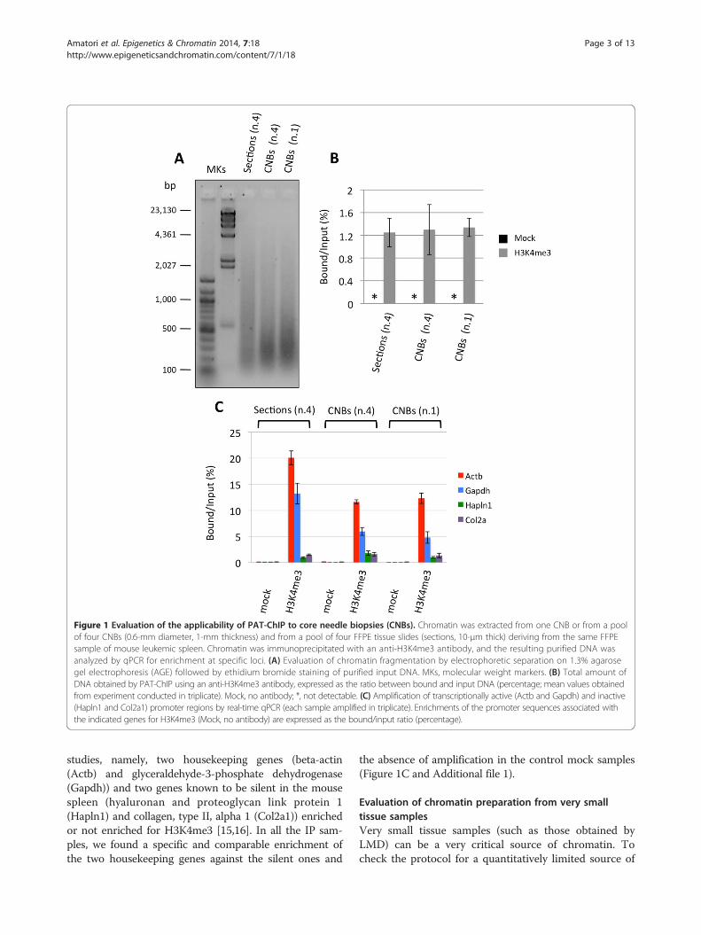
Figure 1 Evaluation of the applicability of PAT-ChIP to core needle biopsies (CNBs). Chromatin was extracted from one CNB or from a poolof four CNBs (0.6-mm diameter, 1-mm thickness) and from a pool of four FFPE tissue slides (sections, 10-μm thick) deriving from the same FFPEsample of mouse leukemic spleen. Chromatin was immunoprecipitated with an anti-H3K4me3 antibody, and the resulting purified DNA wasanalyzed by qPCR for enrichment at specific loci. (A) Evaluation of chromatin fragmentation by electrophoretic separation on 1.3% agarosegel electrophoresis (AGE) followed by ethidium bromide staining of purified input DNA. MKs, molecular weight markers. (B) Total amount ofDNA obtained by PAT-ChIP using an anti-H3K4me3 antibody, expressed as the ratio between bound and input DNA (percentage; mean values obtainedfrom experiment conducted in triplicate). Mock, no antibody; *, not detectable. (C) Amplification of transcriptionally active (Actb and Gapdh) and inactive(Hapln1 and Col2a1) promoter regions by real-time qPCR (each sample amplified in triplicate). Enrichments of the promoter sequences associated withthe indicated genes for H3K4me3 (Mock, no antibody) are expressed as the bound/input ratio (percentage).
Amatori et al. Epigenetics & Chromatin 2014, 7:18 Page 3 of 13http://www.epigeneticsandchromatin.com/content/7/1/18
studies, namely, two housekeeping genes (beta-actin(Actb) and glyceraldehyde-3-phosphate dehydrogenase(Gapdh)) and two genes known to be silent in the mousespleen (hyaluronan and proteoglycan link protein 1(Hapln1) and collagen, type II, alpha 1 (Col2a1)) enrichedor not enriched for H3K4me3 [15,16]. In all the IP sam-ples, we found a specific and comparable enrichment ofthe two housekeeping genes against the silent ones and
the absence of amplification in the control mock samples(Figure 1C and Additional file 1).
Evaluation of chromatin preparation from very smalltissue samplesVery small tissue samples (such as those obtained byLMD) can be a very critical source of chromatin. Tocheck the protocol for a quantitatively limited source of
Amatori et al. Epigenetics & Chromatin 2014, 7:18 Page 4 of 13http://www.epigeneticsandchromatin.com/content/7/1/18
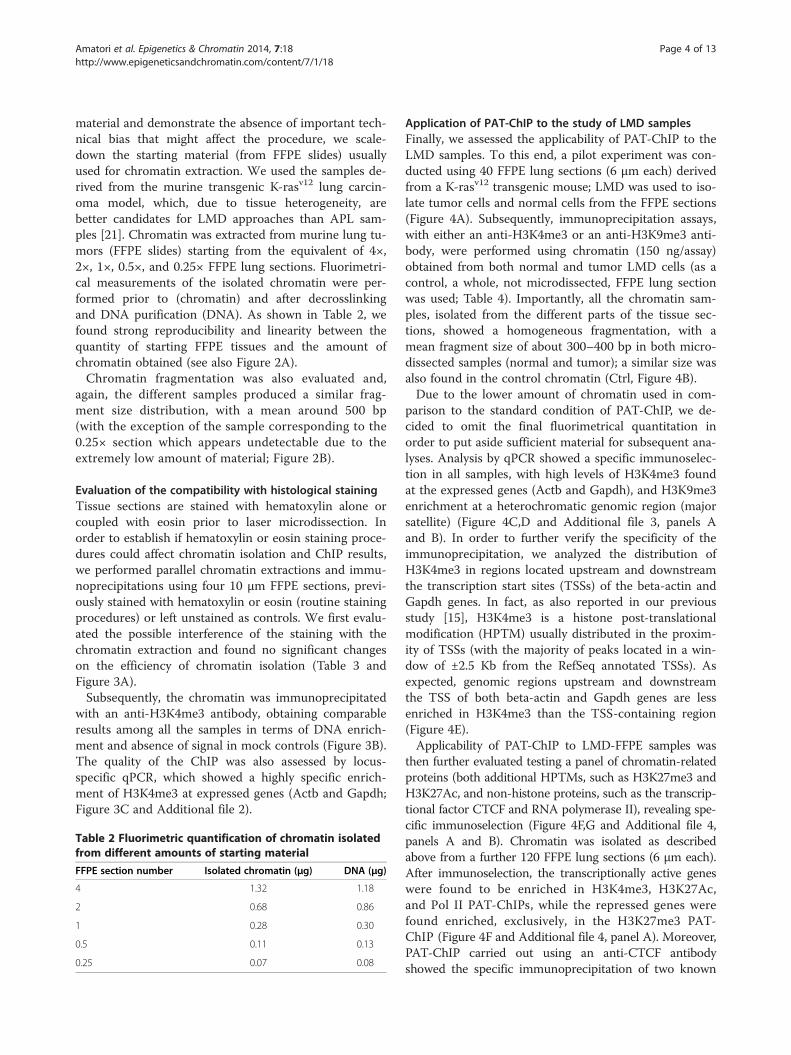
material and demonstrate the absence of important tech-nical bias that might affect the procedure, we scale-down the starting material (from FFPE slides) usuallyused for chromatin extraction. We used the samples de-rived from the murine transgenic K-rasv12 lung carcin-oma model, which, due to tissue heterogeneity, arebetter candidates for LMD approaches than APL sam-ples [21]. Chromatin was extracted from murine lung tu-mors (FFPE slides) starting from the equivalent of 4×,2×, 1×, 0.5×, and 0.25× FFPE lung sections. Fluorimetri-cal measurements of the isolated chromatin were per-formed prior to (chromatin) and after decrosslinkingand DNA purification (DNA). As shown in Table 2, wefound strong reproducibility and linearity between thequantity of starting FFPE tissues and the amount ofchromatin obtained (see also Figure 2A).Chromatin fragmentation was also evaluated and,
again, the different samples produced a similar frag-ment size distribution, with a mean around 500 bp(with the exception of the sample corresponding to the0.25× section which appears undetectable due to theextremely low amount of material; Figure 2B).
Evaluation of the compatibility with histological stainingTissue sections are stained with hematoxylin alone orcoupled with eosin prior to laser microdissection. Inorder to establish if hematoxylin or eosin staining proce-dures could affect chromatin isolation and ChIP results,we performed parallel chromatin extractions and immu-noprecipitations using four 10 μm FFPE sections, previ-ously stained with hematoxylin or eosin (routine stainingprocedures) or left unstained as controls. We first evalu-ated the possible interference of the staining with thechromatin extraction and found no significant changeson the efficiency of chromatin isolation (Table 3 andFigure 3A).Subsequently, the chromatin was immunoprecipitated
with an anti-H3K4me3 antibody, obtaining comparableresults among all the samples in terms of DNA enrich-ment and absence of signal in mock controls (Figure 3B).The quality of the ChIP was also assessed by locus-specific qPCR, which showed a highly specific enrich-ment of H3K4me3 at expressed genes (Actb and Gapdh;Figure 3C and Additional file 2).
Table 2 Fluorimetric quantification of chromatin isolatedfrom different amounts of starting material
FFPE section number Isolated chromatin (μg) DNA (μg)
4 1.32 1.18
2 0.68 0.86
1 0.28 0.30
0.5 0.11 0.13
0.25 0.07 0.08
Application of PAT-ChIP to the study of LMD samplesFinally, we assessed the applicability of PAT-ChIP to theLMD samples. To this end, a pilot experiment was con-ducted using 40 FFPE lung sections (6 μm each) derivedfrom a K-rasv12 transgenic mouse; LMD was used to iso-late tumor cells and normal cells from the FFPE sections(Figure 4A). Subsequently, immunoprecipitation assays,with either an anti-H3K4me3 or an anti-H3K9me3 anti-body, were performed using chromatin (150 ng/assay)obtained from both normal and tumor LMD cells (as acontrol, a whole, not microdissected, FFPE lung sectionwas used; Table 4). Importantly, all the chromatin sam-ples, isolated from the different parts of the tissue sec-tions, showed a homogeneous fragmentation, with amean fragment size of about 300–400 bp in both micro-dissected samples (normal and tumor); a similar size wasalso found in the control chromatin (Ctrl, Figure 4B).Due to the lower amount of chromatin used in com-
parison to the standard condition of PAT-ChIP, we de-cided to omit the final fluorimetrical quantitation inorder to put aside sufficient material for subsequent ana-lyses. Analysis by qPCR showed a specific immunoselec-tion in all samples, with high levels of H3K4me3 foundat the expressed genes (Actb and Gapdh), and H3K9me3enrichment at a heterochromatic genomic region (majorsatellite) (Figure 4C,D and Additional file 3, panels Aand B). In order to further verify the specificity of theimmunoprecipitation, we analyzed the distribution ofH3K4me3 in regions located upstream and downstreamthe transcription start sites (TSSs) of the beta-actin andGapdh genes. In fact, as also reported in our previousstudy [15], H3K4me3 is a histone post-translationalmodification (HPTM) usually distributed in the proxim-ity of TSSs (with the majority of peaks located in a win-dow of ±2.5 Kb from the RefSeq annotated TSSs). Asexpected, genomic regions upstream and downstreamthe TSS of both beta-actin and Gapdh genes are lessenriched in H3K4me3 than the TSS-containing region(Figure 4E).Applicability of PAT-ChIP to LMD-FFPE samples was
then further evaluated testing a panel of chromatin-relatedproteins (both additional HPTMs, such as H3K27me3 andH3K27Ac, and non-histone proteins, such as the transcrip-tional factor CTCF and RNA polymerase II), revealing spe-cific immunoselection (Figure 4F,G and Additional file 4,panels A and B). Chromatin was isolated as describedabove from a further 120 FFPE lung sections (6 μm each).After immunoselection, the transcriptionally active geneswere found to be enriched in H3K4me3, H3K27Ac,and Pol II PAT-ChIPs, while the repressed genes werefound enriched, exclusively, in the H3K27me3 PAT-ChIP (Figure 4F and Additional file 4, panel A). Moreover,PAT-ChIP carried out using an anti-CTCF antibodyshowed the specific immunoprecipitation of two known
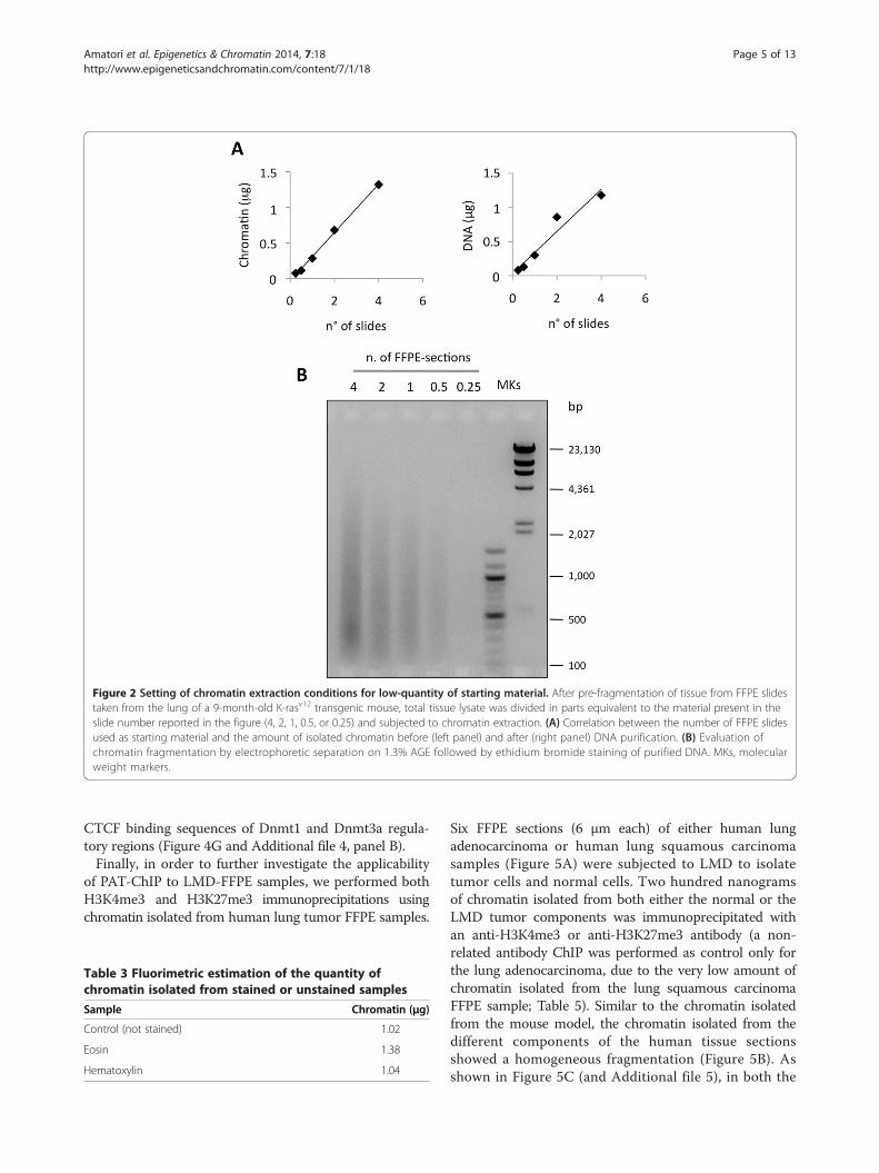
Figure 2 Setting of chromatin extraction conditions for low-quantity of starting material. After pre-fragmentation of tissue from FFPE slidestaken from the lung of a 9-month-old K-rasv12 transgenic mouse, total tissue lysate was divided in parts equivalent to the material present in theslide number reported in the figure (4, 2, 1, 0.5, or 0.25) and subjected to chromatin extraction. (A) Correlation between the number of FFPE slidesused as starting material and the amount of isolated chromatin before (left panel) and after (right panel) DNA purification. (B) Evaluation ofchromatin fragmentation by electrophoretic separation on 1.3% AGE followed by ethidium bromide staining of purified DNA. MKs, molecularweight markers.
Amatori et al. Epigenetics & Chromatin 2014, 7:18 Page 5 of 13http://www.epigeneticsandchromatin.com/content/7/1/18
CTCF binding sequences of Dnmt1 and Dnmt3a regula-tory regions (Figure 4G and Additional file 4, panel B).Finally, in order to further investigate the applicability
of PAT-ChIP to LMD-FFPE samples, we performed bothH3K4me3 and H3K27me3 immunoprecipitations usingchromatin isolated from human lung tumor FFPE samples.
Table 3 Fluorimetric estimation of the quantity ofchromatin isolated from stained or unstained samples
Sample Chromatin (μg)
Control (not stained) 1.02
Eosin 1.38
Hematoxylin 1.04
Six FFPE sections (6 μm each) of either human lungadenocarcinoma or human lung squamous carcinomasamples (Figure 5A) were subjected to LMD to isolatetumor cells and normal cells. Two hundred nanogramsof chromatin isolated from both either the normal or theLMD tumor components was immunoprecipitated withan anti-H3K4me3 or anti-H3K27me3 antibody (a non-related antibody ChIP was performed as control only forthe lung adenocarcinoma, due to the very low amount ofchromatin isolated from the lung squamous carcinomaFFPE sample; Table 5). Similar to the chromatin isolatedfrom the mouse model, the chromatin isolated from thedifferent components of the human tissue sectionsshowed a homogeneous fragmentation (Figure 5B). Asshown in Figure 5C (and Additional file 5), in both the
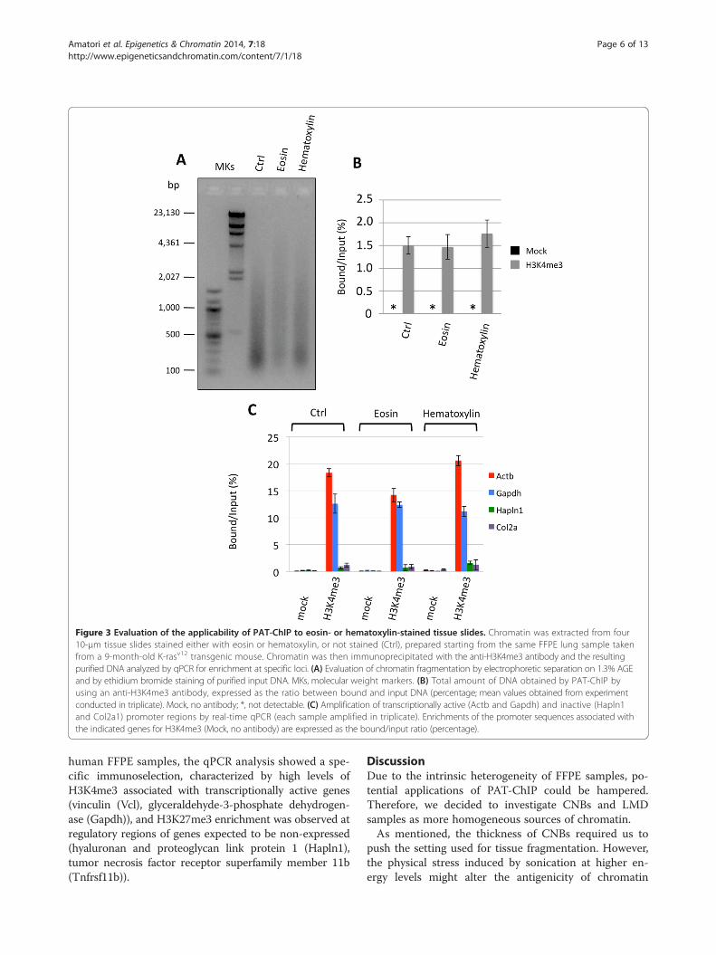
Figure 3 Evaluation of the applicability of PAT-ChIP to eosin- or hematoxylin-stained tissue slides. Chromatin was extracted from four10-μm tissue slides stained either with eosin or hematoxylin, or not stained (Ctrl), prepared starting from the same FFPE lung sample takenfrom a 9-month-old K-rasv12 transgenic mouse. Chromatin was then immunoprecipitated with the anti-H3K4me3 antibody and the resultingpurified DNA analyzed by qPCR for enrichment at specific loci. (A) Evaluation of chromatin fragmentation by electrophoretic separation on 1.3% AGEand by ethidium bromide staining of purified input DNA. MKs, molecular weight markers. (B) Total amount of DNA obtained by PAT-ChIP byusing an anti-H3K4me3 antibody, expressed as the ratio between bound and input DNA (percentage; mean values obtained from experimentconducted in triplicate). Mock, no antibody; *, not detectable. (C) Amplification of transcriptionally active (Actb and Gapdh) and inactive (Hapln1and Col2a1) promoter regions by real-time qPCR (each sample amplified in triplicate). Enrichments of the promoter sequences associated withthe indicated genes for H3K4me3 (Mock, no antibody) are expressed as the bound/input ratio (percentage).
Amatori et al. Epigenetics & Chromatin 2014, 7:18 Page 6 of 13http://www.epigeneticsandchromatin.com/content/7/1/18
human FFPE samples, the qPCR analysis showed a spe-cific immunoselection, characterized by high levels ofH3K4me3 associated with transcriptionally active genes(vinculin (Vcl), glyceraldehyde-3-phosphate dehydrogen-ase (Gapdh)), and H3K27me3 enrichment was observed atregulatory regions of genes expected to be non-expressed(hyaluronan and proteoglycan link protein 1 (Hapln1),tumor necrosis factor receptor superfamily member 11b(Tnfrsf11b)).
DiscussionDue to the intrinsic heterogeneity of FFPE samples, po-tential applications of PAT-ChIP could be hampered.Therefore, we decided to investigate CNBs and LMDsamples as more homogeneous sources of chromatin.As mentioned, the thickness of CNBs required us to
push the setting used for tissue fragmentation. However,the physical stress induced by sonication at higher en-ergy levels might alter the antigenicity of chromatin
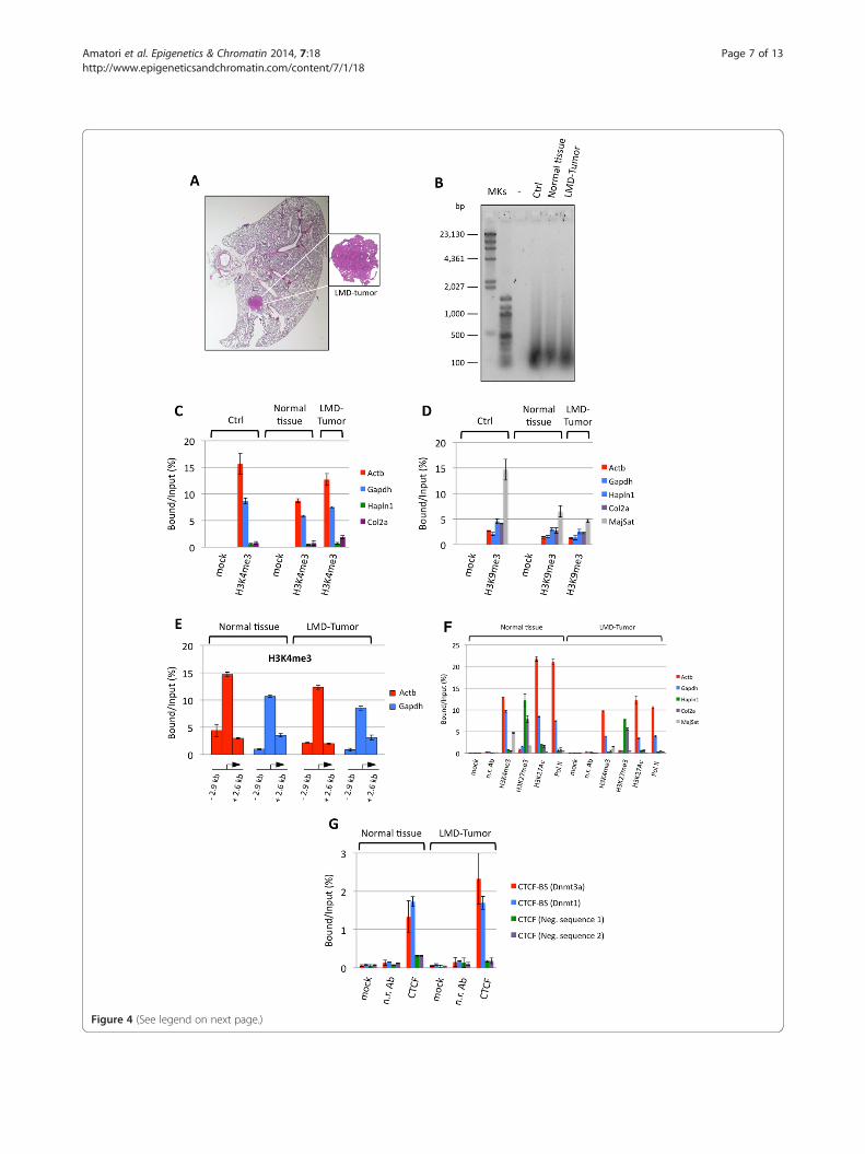
Figure 4 (See legend on next page.)
Amatori et al. Epigenetics & Chromatin 2014, 7:18 Page 7 of 13http://www.epigeneticsandchromatin.com/content/7/1/18
(See figure on previous page.)Figure 4 Application of PAT-ChIP to mouse LMD samples. Forty tissue slides were prepared from the lung of K-rasv12 transgenic mouse andsubjected to LMD to isolate both normal and tumor cells. Chromatin was immunoprecipitated with the reported antibodies and the DNA analyzedby real-time qPCR for enrichment at specific loci (each sample amplified in triplicate). Enrichments of the amplified sequences are expressed asthe ratio between bound and input (percentage). (A) Representative hematoxylin and eosin staining of the lung of one mice in which theexpression of the oncogene was induced with 4-hydroxytamoxifen (4-OHT, right panel). (B) Evaluation of chromatin fragmentation by 1.3%AGE and SYBR® Gold staining of DNA purified from unbound fractions after ChIP with the H3K4me3 antibody. (C) Amplification of transcriptionallyactive (Actb and Gapdh) and inactive (Hapln1 and Col2a1) promoter regions after H3K4me3 immunselection. Mock, no antibody. (D) Amplification oftranscriptionally active (Actb and Gapdh) and inactive (Hapln1 and Col2a1) promoter regions, and major satellite, after H3K9me3 immunoselection Mock,no antibody. (E) Amplification of regions located upstream and downstream from the transcription start site (TSS; at the indicated distance from TSS, seealso Table 6) of the beta-actin and Gapdh genes, after H3K4me3 immunoselection. (F) Amplification of transcriptionally active (Actb and Gapdh)and inactive (Hapln1 and Col2a1) gene promoter regions, and major satellite sequence, after H3K4me3, H3K27me3, H3K27Ac, and Pol IIimmunoselections. Mock, no antibody; n.r. Ab, non-related antibody. (G) Amplification of two CTCF binding sites (CTCF-BS of DNA-methyltransferase3a (Dnmt3a) and DNA-methyltransferase 1 (Dnmt1) genes) and two CTCF unrelated genomic regions as controls (CTCF neg. sequences 1 and 2), afterCTCF immunoselection. Mock, no antibody; n.r. Ab, non-related antibody).
Amatori et al. Epigenetics & Chromatin 2014, 7:18 Page 8 of 13http://www.epigeneticsandchromatin.com/content/7/1/18
proteins, causing excessive chromatin fragmentationand/or loss of the epitopes recognized by the antibody.A sign of the increased level of physical stress to whichthe chromatin was subjected in our experiments wasthe higher level of fragmentation reached with respectto control slides. Notably, the efficiency of chromatinisolation seems to increase with smaller amounts ofCNB-derived samples, as demonstrated by comparingthe total amount of chromatin obtained from one andfour CNBs (Table 1).Interestingly, although both samples originated from
the same paraffin block, the expressed genes showed anapparent lower level of H3K4me3 enrichment when theChIP was performed using CNBs instead of tissue sec-tions (Figure 1C). This lower enrichment could be theresult of structural changes in the chromatin—due tothe higher energy used for sonication (e.g., affecting theintegrity of the epitopes recognized by the antibody)—which, however, do not prevent a clear discriminationbetween expressed and silent genes.The use of CNBs is not devoid of limitations. The
main limit consists in the impossibility of knowing, pre-cisely, the cellular composition of the underlying tissue.Thus, we also considered the use of LMD: unlike
CNBs, LMD allows the direct separation and collectionof different cell populations from the same tissue sec-tion, thus reaching higher levels of purity. LMD is now awell-established technique, and it is used in conjunctionwith many different downstream applications (e.g., DNAgenotyping and loss of heterozygosity (LOH) analysis,DNA methylation analysis, RNA transcript profiling,
Table 4 Fluorimetric quantification of chromatin isolatedfrom normal and LMD tumor tissue fractions
Sample Chromatin (μg)
Normal tissue 3.56
LMD tumor 0.44
cDNA library generation, proteomics discovery, andsignal-pathway profiling [19]).As a preliminary step, we verified whether the proced-
ure could maintain its linearity in terms of total amountof extracted chromatin, and, importantly, if it produces acomparable chromatin fragmentation in function of theprogressive reduction of the tissue dimensions. The in-vestigation of this aspect was, in our opinion, of funda-mental importance, since chromatin isolation could havebeen strongly affected by sonication performance andmicrococcal nuclease digestion efficiency (used at a fixedenzyme concentration). Interestingly, we found that, atleast within the range of amounts of material tested,chromatin extraction maintains an almost perfect linear-ity in terms of quantities of isolated chromatin. Similarly,chromatin fragmentation does not seem to be affected,indicating that the same concentration of micrococcalnuclease can be used irrespective of variations in thequantities of starting material. This last observation is veryuseful, since it will allow a better standardization of theentire chromatin extraction procedure, especially import-ant when the amount of starting tissue is not quantifiable.We thus applied the PAT-ChIP procedure to LMD
lung tumor samples originating from the K-rasv12 trans-genic mouse model using six different antibodies; not-ably, the LMD procedure did not impair the analysis byChIP of the extracted chromatin, even when studyingvery small amounts and after histological staining. Wealso found that in addition to HPTMs, non-histone pro-teins such as Pol II and the transcription factor CTCFcan be investigated in LMD samples. Most importantly,we demonstrated that the procedure can be used to in-vestigate HPTMs in human archival samples.Currently, a limitation of our approach to study LMD
FFPE samples is that is limited to specific loci. In fact,probably due to the more extensive crosslinking procedureroutinely applied to human FFPE tissues (normally fixed byusing 4% of formaldehyde for a variable incubation timeranging between 16 and 48 hours), the isolation of
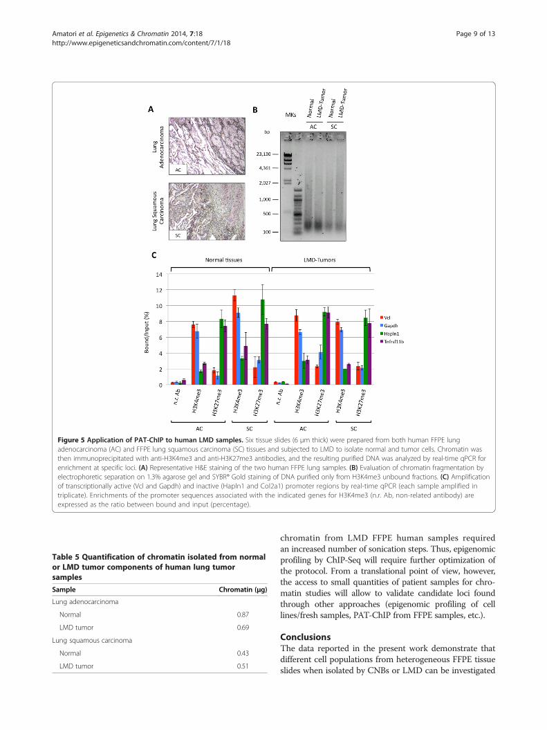
Figure 5 Application of PAT-ChIP to human LMD samples. Six tissue slides (6 μm thick) were prepared from both human FFPE lungadenocarcinoma (AC) and FFPE lung squamous carcinoma (SC) tissues and subjected to LMD to isolate normal and tumor cells. Chromatin wasthen immunoprecipitated with anti-H3K4me3 and anti-H3K27me3 antibodies, and the resulting purified DNA was analyzed by real-time qPCR forenrichment at specific loci. (A) Representative H&E staining of the two human FFPE lung samples. (B) Evaluation of chromatin fragmentation byelectrophoretic separation on 1.3% agarose gel and SYBR® Gold staining of DNA purified only from H3K4me3 unbound fractions. (C) Amplificationof transcriptionally active (Vcl and Gapdh) and inactive (Hapln1 and Col2a1) promoter regions by real-time qPCR (each sample amplified intriplicate). Enrichments of the promoter sequences associated with the indicated genes for H3K4me3 (n.r. Ab, non-related antibody) areexpressed as the ratio between bound and input (percentage).
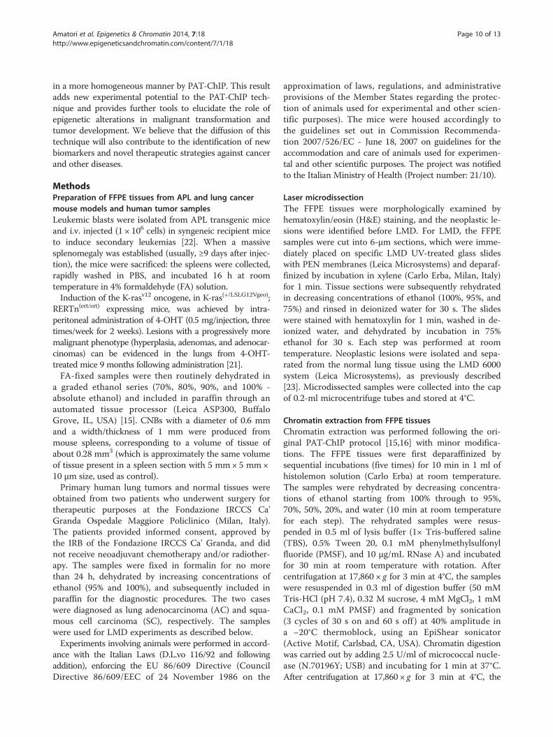
Table 5 Quantification of chromatin isolated from normalor LMD tumor components of human lung tumorsamples
Sample Chromatin (μg)
Lung adenocarcinoma
Normal 0.87
LMD tumor 0.69
Lung squamous carcinoma
Normal 0.43
LMD tumor 0.51
Amatori et al. Epigenetics & Chromatin 2014, 7:18 Page 9 of 13http://www.epigeneticsandchromatin.com/content/7/1/18
chromatin from LMD FFPE human samples requiredan increased number of sonication steps. Thus, epigenomicprofiling by ChIP-Seq will require further optimization ofthe protocol. From a translational point of view, however,the access to small quantities of patient samples for chro-matin studies will allow to validate candidate loci foundthrough other approaches (epigenomic profiling of celllines/fresh samples, PAT-ChIP from FFPE samples, etc.).
ConclusionsThe data reported in the present work demonstrate thatdifferent cell populations from heterogeneous FFPE tissueslides when isolated by CNBs or LMD can be investigated
Amatori et al. Epigenetics & Chromatin 2014, 7:18 Page 10 of 13http://www.epigeneticsandchromatin.com/content/7/1/18
in a more homogeneous manner by PAT-ChIP. This resultadds new experimental potential to the PAT-ChIP tech-nique and provides further tools to elucidate the role ofepigenetic alterations in malignant transformation andtumor development. We believe that the diffusion of thistechnique will also contribute to the identification of newbiomarkers and novel therapeutic strategies against cancerand other diseases.
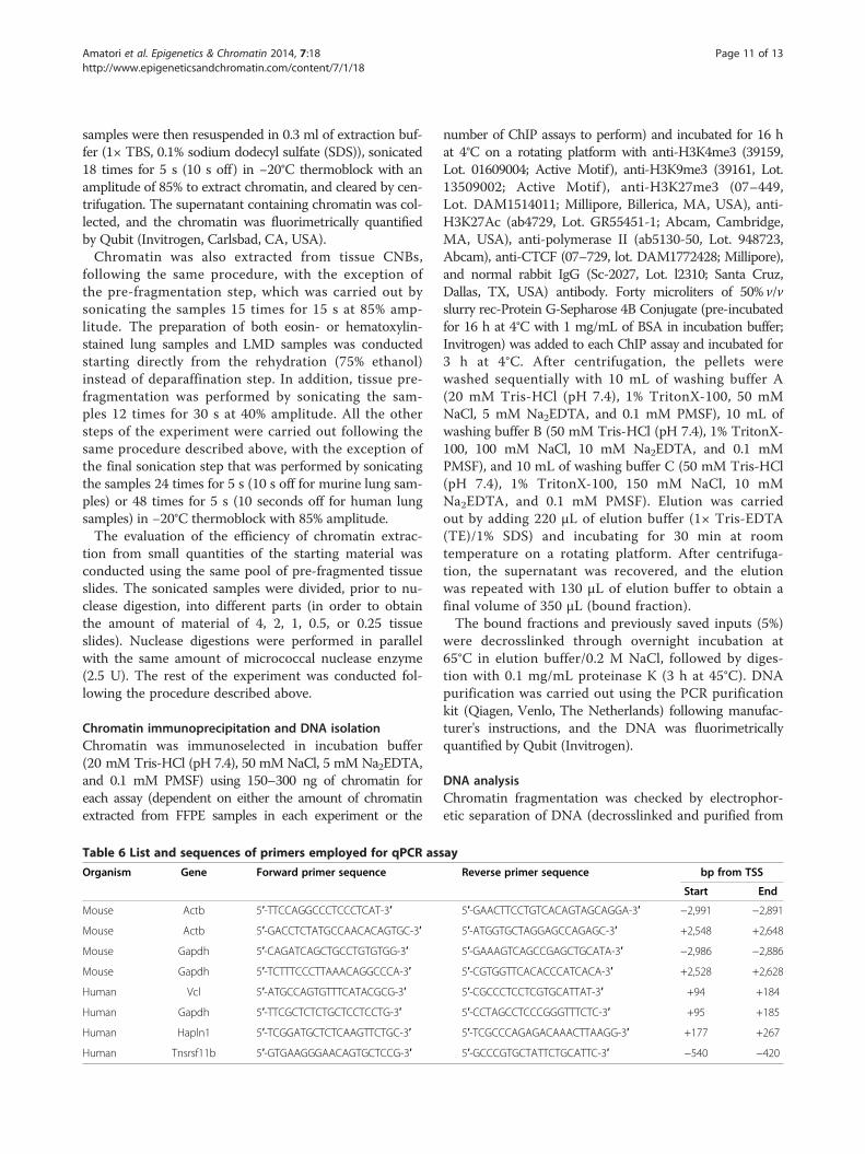
MethodsPreparation of FFPE tissues from APL and lung cancermouse models and human tumor samplesLeukemic blasts were isolated from APL transgenic miceand i.v. injected (1 × 10⁶ cells) in syngeneic recipient miceto induce secondary leukemias [22]. When a massivesplenomegaly was established (usually, ≥9 days after injec-tion), the mice were sacrificed: the spleens were collected,rapidly washed in PBS, and incubated 16 h at roomtemperature in 4% formaldehyde (FA) solution.Induction of the K-rasv12 oncogene, in K-ras(+/LSLG12Vgeo);
RERTn(ert/ert) expressing mice, was achieved by intra-peritoneal administration of 4-OHT (0.5 mg/injection, threetimes/week for 2 weeks). Lesions with a progressively moremalignant phenotype (hyperplasia, adenomas, and adenocar-cinomas) can be evidenced in the lungs from 4-OHT-treated mice 9 months following administration [21].FA-fixed samples were then routinely dehydrated in
a graded ethanol series (70%, 80%, 90%, and 100% -absolute ethanol) and included in paraffin through anautomated tissue processor (Leica ASP300, BuffaloGrove, IL, USA) [15]. CNBs with a diameter of 0.6 mmand a width/thickness of 1 mm were produced frommouse spleens, corresponding to a volume of tissue ofabout 0.28 mm3 (which is approximately the same volumeof tissue present in a spleen section with 5 mm× 5 mm×10 μm size, used as control).Primary human lung tumors and normal tissues were
obtained from two patients who underwent surgery fortherapeutic purposes at the Fondazione IRCCS Ca'Granda Ospedale Maggiore Policlinico (Milan, Italy).The patients provided informed consent, approved bythe IRB of the Fondazione IRCCS Ca' Granda, and didnot receive neoadjuvant chemotherapy and/or radiother-apy. The samples were fixed in formalin for no morethan 24 h, dehydrated by increasing concentrations ofethanol (95% and 100%), and subsequently included inparaffin for the diagnostic procedures. The two caseswere diagnosed as lung adenocarcinoma (AC) and squa-mous cell carcinoma (SC), respectively. The sampleswere used for LMD experiments as described below.Experiments involving animals were performed in accord-
ance with the Italian Laws (D.L.vo 116/92 and followingaddition), enforcing the EU 86/609 Directive (CouncilDirective 86/609/EEC of 24 November 1986 on the
approximation of laws, regulations, and administrativeprovisions of the Member States regarding the protec-tion of animals used for experimental and other scien-tific purposes). The mice were housed accordingly tothe guidelines set out in Commission Recommenda-tion 2007/526/EC - June 18, 2007 on guidelines for theaccommodation and care of animals used for experimen-tal and other scientific purposes. The project was notifiedto the Italian Ministry of Health (Project number: 21/10).
Laser microdissectionThe FFPE tissues were morphologically examined byhematoxylin/eosin (H&E) staining, and the neoplastic le-sions were identified before LMD. For LMD, the FFPEsamples were cut into 6-μm sections, which were imme-diately placed on specific LMD UV-treated glass slideswith PEN membranes (Leica Microsystems) and deparaf-finized by incubation in xylene (Carlo Erba, Milan, Italy)for 1 min. Tissue sections were subsequently rehydratedin decreasing concentrations of ethanol (100%, 95%, and75%) and rinsed in deionized water for 30 s. The slideswere stained with hematoxylin for 1 min, washed in de-ionized water, and dehydrated by incubation in 75%ethanol for 30 s. Each step was performed at roomtemperature. Neoplastic lesions were isolated and sepa-rated from the normal lung tissue using the LMD 6000system (Leica Microsystems), as previously described[23]. Microdissected samples were collected into the capof 0.2-ml microcentrifuge tubes and stored at 4°C.
Chromatin extraction from FFPE tissuesChromatin extraction was performed following the ori-ginal PAT-ChIP protocol [15,16] with minor modifica-tions. The FFPE tissues were first deparaffinized bysequential incubations (five times) for 10 min in 1 ml ofhistolemon solution (Carlo Erba) at room temperature.The samples were rehydrated by decreasing concentra-tions of ethanol starting from 100% through to 95%,70%, 50%, 20%, and water (10 min at room temperaturefor each step). The rehydrated samples were resus-pended in 0.5 ml of lysis buffer (1× Tris-buffered saline(TBS), 0.5% Tween 20, 0.1 mM phenylmethylsulfonylfluoride (PMSF), and 10 μg/mL RNase A) and incubatedfor 30 min at room temperature with rotation. Aftercentrifugation at 17,860 × g for 3 min at 4°C, the sampleswere resuspended in 0.3 ml of digestion buffer (50 mMTris-HCl (pH 7.4), 0.32 M sucrose, 4 mM MgCl2, 1 mMCaCl2, 0.1 mM PMSF) and fragmented by sonication(3 cycles of 30 s on and 60 s off ) at 40% amplitude ina −20°C thermoblock, using an EpiShear sonicator(Active Motif, Carlsbad, CA, USA). Chromatin digestionwas carried out by adding 2.5 U/ml of micrococcal nucle-ase (N.70196Y; USB) and incubating for 1 min at 37°C.After centrifugation at 17,860 × g for 3 min at 4°C, the
Amatori et al. Epigenetics & Chromatin 2014, 7:18 Page 11 of 13http://www.epigeneticsandchromatin.com/content/7/1/18
samples were then resuspended in 0.3 ml of extraction buf-fer (1× TBS, 0.1% sodium dodecyl sulfate (SDS)), sonicated18 times for 5 s (10 s off) in −20°C thermoblock with anamplitude of 85% to extract chromatin, and cleared by cen-trifugation. The supernatant containing chromatin was col-lected, and the chromatin was fluorimetrically quantifiedby Qubit (Invitrogen, Carlsbad, CA, USA).Chromatin was also extracted from tissue CNBs,
following the same procedure, with the exception ofthe pre-fragmentation step, which was carried out bysonicating the samples 15 times for 15 s at 85% amp-litude. The preparation of both eosin- or hematoxylin-stained lung samples and LMD samples was conductedstarting directly from the rehydration (75% ethanol)instead of deparaffination step. In addition, tissue pre-fragmentation was performed by sonicating the sam-ples 12 times for 30 s at 40% amplitude. All the othersteps of the experiment were carried out following thesame procedure described above, with the exception ofthe final sonication step that was performed by sonicatingthe samples 24 times for 5 s (10 s off for murine lung sam-ples) or 48 times for 5 s (10 seconds off for human lungsamples) in −20°C thermoblock with 85% amplitude.The evaluation of the efficiency of chromatin extrac-
tion from small quantities of the starting material wasconducted using the same pool of pre-fragmented tissueslides. The sonicated samples were divided, prior to nu-clease digestion, into different parts (in order to obtainthe amount of material of 4, 2, 1, 0.5, or 0.25 tissueslides). Nuclease digestions were performed in parallelwith the same amount of micrococcal nuclease enzyme(2.5 U). The rest of the experiment was conducted fol-lowing the procedure described above.
Chromatin immunoprecipitation and DNA isolationChromatin was immunoselected in incubation buffer(20 mM Tris-HCl (pH 7.4), 50 mM NaCl, 5 mM Na2EDTA,and 0.1 mM PMSF) using 150–300 ng of chromatin foreach assay (dependent on either the amount of chromatinextracted from FFPE samples in each experiment or the
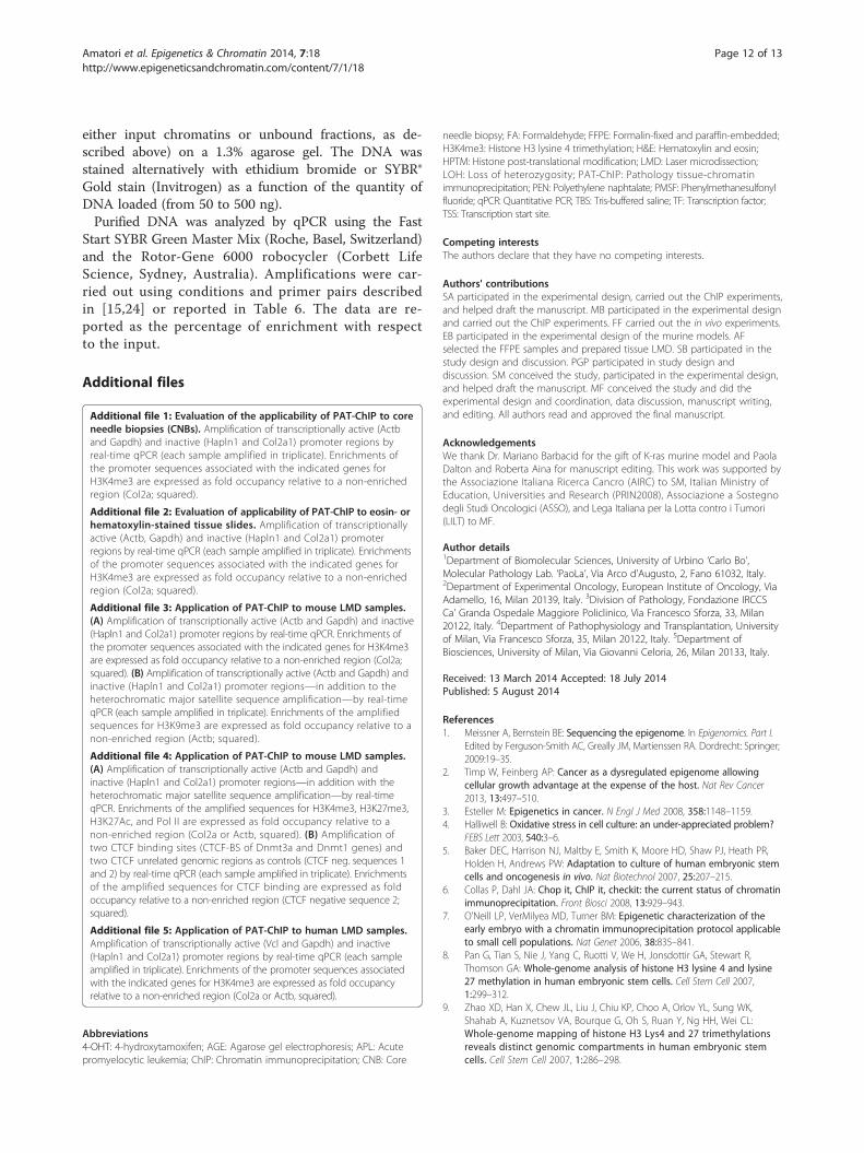
Table 6 List and sequences of primers employed for qPCR ass
Organism Gene Forward primer sequence
Mouse Actb 5′-TTCCAGGCCCTCCCTCAT-3′
Mouse Actb 5′-GACCTCTATGCCAACACAGTGC-3′
Mouse Gapdh 5′-CAGATCAGCTGCCTGTGTGG-3′
Mouse Gapdh 5′-TCTTTCCCTTAAACAGGCCCA-3′
Human Vcl 5′-ATGCCAGTGTTTCATACGCG-3′
Human Gapdh 5′-TTCGCTCTCTGCTCCTCCTG-3′
Human Hapln1 5′-TCGGATGCTCTCAAGTTCTGC-3′
Human Tnsrsf11b 5′-GTGAAGGGAACAGTGCTCCG-3′
number of ChIP assays to perform) and incubated for 16 hat 4°C on a rotating platform with anti-H3K4me3 (39159,Lot. 01609004; Active Motif), anti-H3K9me3 (39161, Lot.13509002; Active Motif ), anti-H3K27me3 (07–449,Lot. DAM1514011; Millipore, Billerica, MA, USA), anti-H3K27Ac (ab4729, Lot. GR55451-1; Abcam, Cambridge,MA, USA), anti-polymerase II (ab5130-50, Lot. 948723,Abcam), anti-CTCF (07–729, lot. DAM1772428; Millipore),and normal rabbit IgG (Sc-2027, Lot. l2310; Santa Cruz,Dallas, TX, USA) antibody. Forty microliters of 50% v/vslurry rec-Protein G-Sepharose 4B Conjugate (pre-incubatedfor 16 h at 4°C with 1 mg/mL of BSA in incubation buffer;Invitrogen) was added to each ChIP assay and incubated for3 h at 4°C. After centrifugation, the pellets werewashed sequentially with 10 mL of washing buffer A(20 mM Tris-HCl (pH 7.4), 1% TritonX-100, 50 mMNaCl, 5 mM Na2EDTA, and 0.1 mM PMSF), 10 mL ofwashing buffer B (50 mM Tris-HCl (pH 7.4), 1% TritonX-100, 100 mM NaCl, 10 mM Na2EDTA, and 0.1 mMPMSF), and 10 mL of washing buffer C (50 mM Tris-HCl(pH 7.4), 1% TritonX-100, 150 mM NaCl, 10 mMNa2EDTA, and 0.1 mM PMSF). Elution was carriedout by adding 220 μL of elution buffer (1× Tris-EDTA(TE)/1% SDS) and incubating for 30 min at roomtemperature on a rotating platform. After centrifuga-tion, the supernatant was recovered, and the elutionwas repeated with 130 μL of elution buffer to obtain afinal volume of 350 μL (bound fraction).The bound fractions and previously saved inputs (5%)
were decrosslinked through overnight incubation at65°C in elution buffer/0.2 M NaCl, followed by diges-tion with 0.1 mg/mL proteinase K (3 h at 45°C). DNApurification was carried out using the PCR purificationkit (Qiagen, Venlo, The Netherlands) following manufac-turer's instructions, and the DNA was fluorimetricallyquantified by Qubit (Invitrogen).
DNA analysisChromatin fragmentation was checked by electrophor-etic separation of DNA (decrosslinked and purified from
ay
Reverse primer sequence bp from TSS
Start End
5′-GAACTTCCTGTCACAGTAGCAGGA-3′ −2,991 −2,891
5′-ATGGTGCTAGGAGCCAGAGC-3′ +2,548 +2,648
5′-GAAAGTCAGCCGAGCTGCATA-3′ −2,986 −2,886
5′-CGTGGTTCACACCCATCACA-3′ +2,528 +2,628
5′-CGCCCTCCTCGTGCATTAT-3′ +94 +184
5′-CCTAGCCTCCCGGGTTTCTC-3′ +95 +185
5′-TCGCCCAGAGACAAACTTAAGG-3′ +177 +267
5′-GCCCGTGCTATTCTGCATTC-3′ −540 −420
Amatori et al. Epigenetics & Chromatin 2014, 7:18 Page 12 of 13http://www.epigeneticsandchromatin.com/content/7/1/18
either input chromatins or unbound fractions, as de-scribed above) on a 1.3% agarose gel. The DNA wasstained alternatively with ethidium bromide or SYBR®Gold stain (Invitrogen) as a function of the quantity ofDNA loaded (from 50 to 500 ng).Purified DNA was analyzed by qPCR using the Fast
Start SYBR Green Master Mix (Roche, Basel, Switzerland)and the Rotor-Gene 6000 robocycler (Corbett LifeScience, Sydney, Australia). Amplifications were car-ried out using conditions and primer pairs describedin [15,24] or reported in Table 6. The data are re-ported as the percentage of enrichment with respectto the input.
Additional files
Additional file 1: Evaluation of the applicability of PAT-ChIP to coreneedle biopsies (CNBs). Amplification of transcriptionally active (Actband Gapdh) and inactive (Hapln1 and Col2a1) promoter regions byreal-time qPCR (each sample amplified in triplicate). Enrichments ofthe promoter sequences associated with the indicated genes forH3K4me3 are expressed as fold occupancy relative to a non-enrichedregion (Col2a; squared).
Additional file 2: Evaluation of applicability of PAT-ChIP to eosin- orhematoxylin-stained tissue slides. Amplification of transcriptionallyactive (Actb, Gapdh) and inactive (Hapln1 and Col2a1) promoterregions by real-time qPCR (each sample amplified in triplicate). Enrichmentsof the promoter sequences associated with the indicated genes forH3K4me3 are expressed as fold occupancy relative to a non-enrichedregion (Col2a; squared).
Additional file 3: Application of PAT-ChIP to mouse LMD samples.(A) Amplification of transcriptionally active (Actb and Gapdh) and inactive(Hapln1 and Col2a1) promoter regions by real-time qPCR. Enrichments ofthe promoter sequences associated with the indicated genes for H3K4me3are expressed as fold occupancy relative to a non-enriched region (Col2a;squared). (B) Amplification of transcriptionally active (Actb and Gapdh) andinactive (Hapln1 and Col2a1) promoter regions—in addition to theheterochromatic major satellite sequence amplification—by real-timeqPCR (each sample amplified in triplicate). Enrichments of the amplifiedsequences for H3K9me3 are expressed as fold occupancy relative to anon-enriched region (Actb; squared).
Additional file 4: Application of PAT-ChIP to mouse LMD samples.(A) Amplification of transcriptionally active (Actb and Gapdh) andinactive (Hapln1 and Col2a1) promoter regions—in addition with theheterochromatic major satellite sequence amplification—by real-timeqPCR. Enrichments of the amplified sequences for H3K4me3, H3K27me3,H3K27Ac, and Pol II are expressed as fold occupancy relative to anon-enriched region (Col2a or Actb, squared). (B) Amplification oftwo CTCF binding sites (CTCF-BS of Dnmt3a and Dnmt1 genes) andtwo CTCF unrelated genomic regions as controls (CTCF neg. sequences 1and 2) by real-time qPCR (each sample amplified in triplicate). Enrichmentsof the amplified sequences for CTCF binding are expressed as foldoccupancy relative to a non-enriched region (CTCF negative sequence 2;squared).
Additional file 5: Application of PAT-ChIP to human LMD samples.Amplification of transcriptionally active (Vcl and Gapdh) and inactive(Hapln1 and Col2a1) promoter regions by real-time qPCR (each sampleamplified in triplicate). Enrichments of the promoter sequences associatedwith the indicated genes for H3K4me3 are expressed as fold occupancyrelative to a non-enriched region (Col2a or Actb, squared).
Abbreviations4-OHT: 4-hydroxytamoxifen; AGE: Agarose gel electrophoresis; APL: Acutepromyelocytic leukemia; ChIP: Chromatin immunoprecipitation; CNB: Core
needle biopsy; FA: Formaldehyde; FFPE: Formalin-fixed and paraffin-embedded;H3K4me3: Histone H3 lysine 4 trimethylation; H&E: Hematoxylin and eosin;HPTM: Histone post-translational modification; LMD: Laser microdissection;LOH: Loss of heterozygosity; PAT-ChIP: Pathology tissue-chromatinimmunoprecipitation; PEN: Polyethylene naphtalate; PMSF: Phenylmethanesulfonylfluoride; qPCR: Quantitative PCR; TBS: Tris-buffered saline; TF: Transcription factor;TSS: Transcription start site.
Competing interestsThe authors declare that they have no competing interests.
Authors' contributionsSA participated in the experimental design, carried out the ChIP experiments,and helped draft the manuscript. MB participated in the experimental designand carried out the ChIP experiments. FF carried out the in vivo experiments.EB participated in the experimental design of the murine models. AFselected the FFPE samples and prepared tissue LMD. SB participated in thestudy design and discussion. PGP participated in study design anddiscussion. SM conceived the study, participated in the experimental design,and helped draft the manuscript. MF conceived the study and did theexperimental design and coordination, data discussion, manuscript writing,and editing. All authors read and approved the final manuscript.
AcknowledgementsWe thank Dr. Mariano Barbacid for the gift of K-ras murine model and PaolaDalton and Roberta Aina for manuscript editing. This work was supported bythe Associazione Italiana Ricerca Cancro (AIRC) to SM, Italian Ministry ofEducation, Universities and Research (PRIN2008), Associazione a Sostegnodegli Studi Oncologici (ASSO), and Lega Italiana per la Lotta contro i Tumori(LILT) to MF.
Author details1Department of Biomolecular Sciences, University of Urbino ‘Carlo Bo’,Molecular Pathology Lab. ‘PaoLa’, Via Arco d'Augusto, 2, Fano 61032, Italy.2Department of Experimental Oncology, European Institute of Oncology, ViaAdamello, 16, Milan 20139, Italy. 3Division of Pathology, Fondazione IRCCSCa' Granda Ospedale Maggiore Policlinico, Via Francesco Sforza, 33, Milan20122, Italy. 4Department of Pathophysiology and Transplantation, Universityof Milan, Via Francesco Sforza, 35, Milan 20122, Italy. 5Department ofBiosciences, University of Milan, Via Giovanni Celoria, 26, Milan 20133, Italy.
Received: 13 March 2014 Accepted: 18 July 2014Published: 5 August 2014
References1. Meissner A, Bernstein BE: Sequencing the epigenome. In Epigenomics. Part I.
Edited by Ferguson-Smith AC, Greally JM, Martienssen RA. Dordrecht: Springer;2009:19–35.
2. Timp W, Feinberg AP: Cancer as a dysregulated epigenome allowingcellular growth advantage at the expense of the host. Nat Rev Cancer2013, 13:497–510.
3. Esteller M: Epigenetics in cancer. N Engl J Med 2008, 358:1148–1159.4. Halliwell B: Oxidative stress in cell culture: an under-appreciated problem?
FEBS Lett 2003, 540:3–6.5. Baker DEC, Harrison NJ, Maltby E, Smith K, Moore HD, Shaw PJ, Heath PR,
Holden H, Andrews PW: Adaptation to culture of human embryonic stemcells and oncogenesis in vivo. Nat Biotechnol 2007, 25:207–215.
6. Collas P, Dahl JA: Chop it, ChIP it, checkit: the current status of chromatinimmunoprecipitation. Front Biosci 2008, 13:929–943.
7. O'Neill LP, VerMilyea MD, Turner BM: Epigenetic characterization of theearly embryo with a chromatin immunoprecipitation protocol applicableto small cell populations. Nat Genet 2006, 38:835–841.
8. Pan G, Tian S, Nie J, Yang C, Ruotti V, We H, Jonsdottir GA, Stewart R,Thomson GA: Whole-genome analysis of histone H3 lysine 4 and lysine27 methylation in human embryonic stem cells. Cell Stem Cell 2007,1:299–312.
9. Zhao XD, Han X, Chew JL, Liu J, Chiu KP, Choo A, Orlov YL, Sung WK,Shahab A, Kuznetsov VA, Bourque G, Oh S, Ruan Y, Ng HH, Wei CL:Whole-genome mapping of histone H3 Lys4 and 27 trimethylationsreveals distinct genomic compartments in human embryonic stemcells. Cell Stem Cell 2007, 1:286–298.
Amatori et al. Epigenetics & Chromatin 2014, 7:18 Page 13 of 13http://www.epigeneticsandchromatin.com/content/7/1/18
10. O'Neill LP, Turner BM: Histone H4 acetylation distinguishes codingregions of the human genome from heterochromatin in adifferentiation-dependent but transcription-independent manner.EMBO J 1995, 14:3946–3957.
11. Collas P: The current state of chromatin immunoprecipitation.Mol Biotechnol 2010, 45:87–100.
12. Park PJ: ChIP–seq: advantages and challenges of a maturing technology.Nat Rev Genet 2009, 10:669–680.
13. Einav Nili GY, Saito Y, Egger G, Jones PA: Cancer epigenetics:modifications, screening, and therapy. Annu Rev Med 2008, 59:267–280.
14. Laird PW: The power and the promise of DNA methylation markers.Nat Rev Cancer 2003, 3:253–266.
15. Fanelli M, Amatori S, Barozzi I, Soncini M, Dal Zuffo R, Bucci G, Capra M,Quarto M, Dellino GI, Mercurio C, Alcalay M, Viale G, Pelicci PG, Minucci S:Pathology tissue-chromatin immunoprecipitation, coupled withhigh-throughput sequencing, allows the epigenetic profiling of patientsamples. Proc Natl Acad Sci USA 2010, 107:21535–21540.
16. Fanelli M, Amatori S, Barozzi I, Minucci S: Chromatin immunoprecipitationand high-throughput sequencing from paraffin-embedded pathologytissue. Nat Protoc 2011, 6:1905–1919.
17. Bubendorf L, Nocito A, Moch H, Sauter G: Tissue microarray (TMA)technology: miniaturized pathology archives for high-throughput in situstudies. J Pathol 2001, 195:72–79.
18. Simon R, Mirlacher M, Sauter G: Tissue microarrays. In Molecular Diagnosisof Cancer. 2nd edition. Edited by Roulston JE, Barlett JMS. New York:Humana Press; 2004:377–389.
19. Espina V, Wulfkuhle JD, Calvert VS, Van Meter A, Zhou W, Coukos G, GehoDH III, Petricoin EF, Liotta LA: Laser-capture microdissection. Nat Protoc2006, 1:586–603.
20. Bonner RF, Emmert-Buck M, Cole K, Pohida T, Chuaqui R, Goldstein S,Liotta LA: Laser capture microdissection: molecular analysis of tissue.Science 1997, 278:1481–1483.
21. Guerra C, Mijimolle N, Dhawahir A, Dubus P, Barradas M, Serrano M,Campuzano V, Barbacid M: Tumor induction by an endogenous K-rasoncogene is highly dependent on cellular context. Cancer Cell 2003,4:111–120.
22. Minucci S, Monestiroli S, Giavara S, Ronzoni S, Marchesi F, Insinga A, Diverio D,Gasparini P, Capillo M, Colombo E, Matteucci C, Contegno F, Lo-Coco F,Scanzini E, Gobbi A, Pelicci PG: PML-RAR induces promyelocytic leukemiaswith high efficiency following retroviral gene transfer into purifiedmurine hematopoietic progenitors. Blood 2002, 100:2989–2995.
23. Vaira V, Faversani A, Dohi T, Montorsi M, Augello C, Gatti S, Coggi G, Altieri DC,Bosari S: miR-296 regulation of a cell polarity-cell plasticity module controlstumor progression. Oncogene 2012, 31(1):27–38.
24. Amatori S, Bagaloni I, Macedi E, Formica M, Giorgi L, Fusi V, Fanelli M:Malten, a new synthetic molecule showing in vitro antiproliferativeactivity against tumor cells and induction of complex DNA structuralalterations. Br J Cancer 2010, 2:239–248.
doi:10.1186/1756-8935-7-18Cite this article as: Amatori et al.: PAT-ChIP coupled with lasermicrodissection allows the study of chromatin in selected cellpopulations from paraffin-embedded patient samples. Epigenetics &Chromatin 2014 7:18.
Submit your next manuscript to BioMed Centraland take full advantage of:
• Convenient online submission
• Thorough peer review
• No space constraints or color figure charges
• Immediate publication on acceptance
• Inclusion in PubMed, CAS, Scopus and Google Scholar
• Research which is freely available for redistribution
Submit your manuscript at www.biomedcentral.com/submit