interlaboratory ring test - umr pvbmt
TRANSCRIPT
MINISTERE DE L’AGRICULTURE ET DE LA PÊCHE
1/47
Ministère de l’agriculture et de la pêche
Direction générale de l’alimentation, sous direction de la qualité et de la protection des végétaux (France)
Laboratoire National de la Protection des Végétaux (France, Réunion)
Centre de coopération Internationale en Recherche Agronomique pour le
Développement (France, Réunion)
2009, July
IInntteerrllaabboorraattoorryy RRiinngg tteesstt::
XXaanntthhoommoonnaass aaxxoonnooppooddiiss ppvv.. ddiieeffffeennbbaacchhiiaaee iinn AAnntthhuurriiuumm
MINISTERE DE L’AGRICULTURE ET DE LA PÊCHE
2/47
Contents
1. Ring-test Organisation...................................................................................................................... 3 2. Goal of the Test ................................................................................................................................. 3 3. Methods Characteristics................................................................................................................... 4 4. Host/pathogen Combination ............................................................................................................ 4 5. Equipment and consumables .......................................................................................................... 5 6. Estimation of Working Time ............................................................................................................. 5 7. Provisional time schedule ................................................................................................................ 5 8. Instructions ........................................................................................................................................ 6 9. Constitution of Samples ................................................................................................................... 6 10. Data Analyses .................................................................................................................................. 8 11. Laboratory Duties............................................................................................................................ 9 12. References ..................................................................................................................................... 11 APPENDIX 1 Recommended chronology for analyses ................................................................... 13 APPENDIX 2 Method for reviving freeze-dried bacteria (control P1) ............................................. 14 APPENDIX 3 The serial dilution method of counting bacteria (control T6)................................... 15 APPENDIX 4 Preparation of the subsamples ................................................................................... 16 APPENDIX 5 Protocol for the detection and identification of Xad by isolation and I-ELISA....... 17 APPENDIX 6 Protocol for the detection and identification of Xad by nested-PCR ...................... 21 APPENDIX 7 Protocol for the detection and identification of Xad by Immunofluorescence ...... 23 APPENDIX 8 Protocol for the detection of Xad by DAS-ELISA (PRI) ............................................. 26 APPENDIX 9 Consent to participate to the ring-test and confidentiality undertaking ................. 29 APPENDIX 10 Acknowledgement for the receipt of the critical consumables ............................. 30 APPENDIX 11 Acknowledgement for the receipt of the samples and comments ........................ 31 APPENDIX 12 Comments on the interlaboratory ring test.............................................................. 32 APPENDIX 13 Results form ................................................................................................................ 33
Contents
MINISTERE DE L’AGRICULTURE ET DE LA PÊCHE
3/47
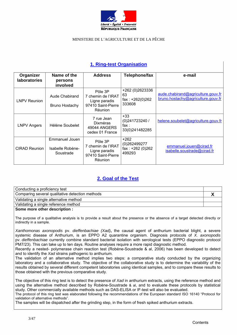
2. Goal of the Test
Conducting a proficiency test Comparing several qualitative detection methods X Validating a single alternative method Validating a single reference method Some more other description : The purpose of a qualitative analysis is to provide a result about the presence or the absence of a target detected directly or indirectly in a sample. Xanthomonas axonopodis pv. dieffenbachiae (Xad), the causal agent of anthurium bacterial blight, a severe systemic disease of Anthurium, is an EPPO A2 quarantine organism. Diagnosis protocols of X. axonopodis pv. dieffenbachiae currently combine standard bacterial isolation with serological tests (EPPO diagnostic protocol PM7/23). This can take up to ten days. Routine analyses require a more rapid diagnostic method. Recently a nested- polymerase chain reaction test (Robène-Soustrade & al, 2006) has been developed to detect and to identify the Xad strains pathogenic to anthurium. The validation of an alternative method implies two steps: a comparative study conducted by the organizing laboratory and a collaborative study. The objective of the collaborative study is to determine the variability of the results obtained by several different competent laboratories using identical samples, and to compare these results to those obtained with the previous comparative study. The objective of this ring test is to detect the presence of Xad in anthurium extracts, using the reference method and using the alternative method described by Robène-Soustrade & al, and to evaluate these protocols by statistical study. Other commercially available methods such as DAS-ELISA or IF-test will also be evaluated. The protocol of this ring test was elaborated following the recommendations of the European standard ISO 16140 “Protocol for validation of alternative methods”. The samples will be dispatched after the grinding step, in the form of fresh spiked anthurium extracts.
1. Ring-test Organisation
Organizer laboratories
Name of the persons involved
Address Telephone/fax e-mail
LNPV Reunion Aude Chabirand
Bruno Hostachy
Pôle 3P 7 chemin de l’IRAT
Ligne paradis 97410 Saint-Pierre
Réunion
+262 (0)2623336 63 fax : +262(0)262 333608
[email protected]@agriculture.gouv.fr
LNPV Angers Hélène Soubelet
7 rue Jean Dixméras
49044 ANGERS cedex 01 France
+33 (0)241723240 / fax : 33(0)241482285
CIRAD Reunion
Emmanuel Jouen
Isabelle Robène-Soustrade
Pôle 3P 7 chemin de l’IRAT
Ligne paradis 97410 Saint-Pierre
Réunion
+262 (0)262499277 fax : +262 (0)262 499293
[email protected] [email protected]
Contents
MINISTERE DE L’AGRICULTURE ET DE LA PÊCHE
4/47
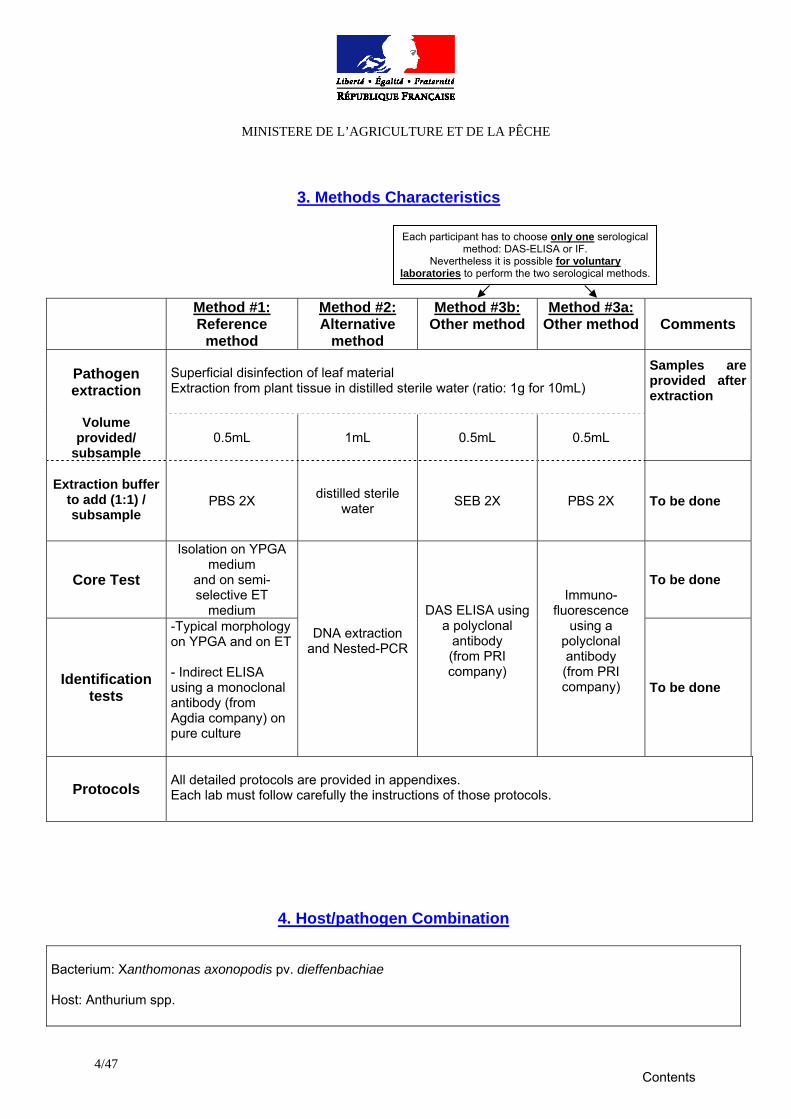
3. Methods Characteristics
Method #1:
Reference method
Method #2: Alternative
method
Method #3b: Other method
Method #3a: Other method Comments
Pathogen extraction
Superficial disinfection of leaf material Extraction from plant tissue in distilled sterile water (ratio: 1g for 10mL)
Volume provided/
subsample 0.5mL 1mL 0.5mL 0.5mL
Samples are provided after extraction
Extraction buffer
to add (1:1) / subsample
PBS 2X distilled sterile water SEB 2X PBS 2X To be done
Core Test
Isolation on YPGA medium
and on semi-selective ET
medium
To be done
Identification tests
-Typical morphology on YPGA and on ET - Indirect ELISA using a monoclonal antibody (from Agdia company) on pure culture
DNA extraction and Nested-PCR
DAS ELISA using a polyclonal
antibody (from PRI company)
Immuno-fluorescence
using a polyclonal antibody (from PRI company)
To be done
Protocols All detailed protocols are provided in appendixes. Each lab must follow carefully the instructions of those protocols.
4. Host/pathogen Combination Bacterium: Xanthomonas axonopodis pv. dieffenbachiae Host: Anthurium spp.
Each participant has to choose only one serological method: DAS-ELISA or IF.
Nevertheless it is possible for voluntary laboratories to perform the two serological methods.
Contents
MINISTERE DE L’AGRICULTURE ET DE LA PÊCHE
5/47
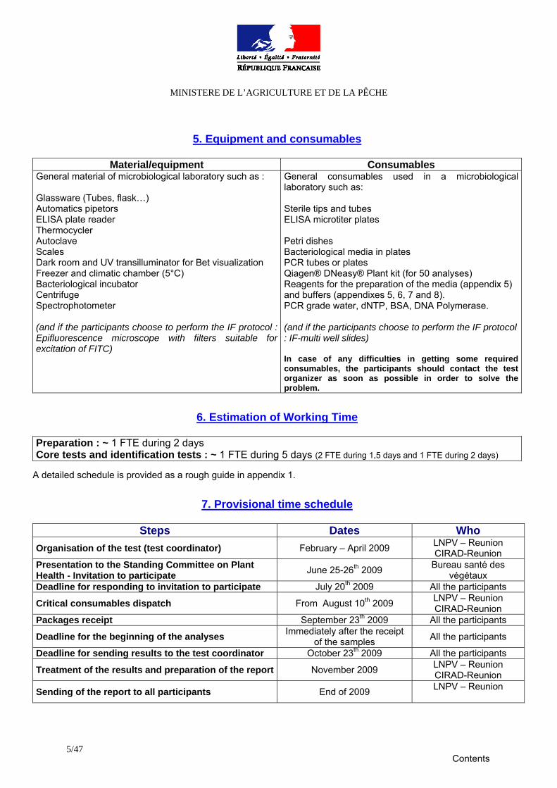
5. Equipment and consumables
Material/equipment Consumables
General material of microbiological laboratory such as : Glassware (Tubes, flask…) Automatics pipetors ELISA plate reader Thermocycler Autoclave Scales Dark room and UV transilluminator for Bet visualization Freezer and climatic chamber (5°C) Bacteriological incubator Centrifuge Spectrophotometer (and if the participants choose to perform the IF protocol : Epifluorescence microscope with filters suitable for excitation of FITC)
General consumables used in a microbiological laboratory such as: Sterile tips and tubes ELISA microtiter plates Petri dishes Bacteriological media in plates PCR tubes or plates Qiagen® DNeasy® Plant kit (for 50 analyses) Reagents for the preparation of the media (appendix 5) and buffers (appendixes 5, 6, 7 and 8). PCR grade water, dNTP, BSA, DNA Polymerase. (and if the participants choose to perform the IF protocol : IF-multi well slides) In case of any difficulties in getting some required consumables, the participants should contact the test organizer as soon as possible in order to solve the problem.
6. Estimation of Working Time
Preparation : ~ 1 FTE during 2 days Core tests and identification tests : ~ 1 FTE during 5 days (2 FTE during 1,5 days and 1 FTE during 2 days)
A detailed schedule is provided as a rough guide in appendix 1.
7. Provisional time schedule
Steps Dates Who Organisation of the test (test coordinator) February – April 2009 LNPV – Reunion
CIRAD-Reunion Presentation to the Standing Committee on Plant Health - Invitation to participate June 25-26th 2009 Bureau santé des
végétaux Deadline for responding to invitation to participate July 20th 2009 All the participants
Critical consumables dispatch From August 10th 2009 LNPV – Reunion CIRAD-Reunion
Packages receipt September 23th 2009 All the participants
Deadline for the beginning of the analyses Immediately after the receipt of the samples All the participants
Deadline for sending results to the test coordinator October 23th 2009 All the participants
Treatment of the results and preparation of the report November 2009 LNPV – Reunion CIRAD-Reunion
Sending of the report to all participants End of 2009 LNPV – Reunion
Contents
MINISTERE DE L’AGRICULTURE ET DE LA PÊCHE
6/47
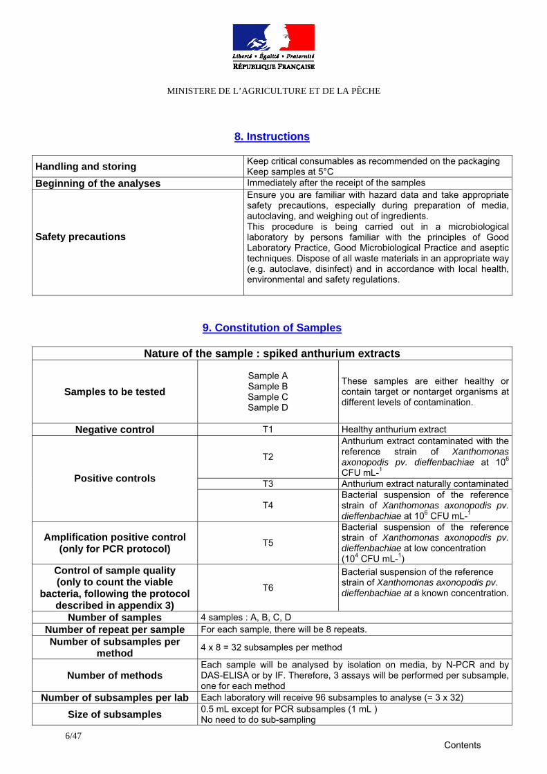
8. Instructions
Handling and storing Keep critical consumables as recommended on the packaging
Keep samples at 5°C Beginning of the analyses Immediately after the receipt of the samples
Safety precautions
Ensure you are familiar with hazard data and take appropriate safety precautions, especially during preparation of media, autoclaving, and weighing out of ingredients. This procedure is being carried out in a microbiological laboratory by persons familiar with the principles of Good Laboratory Practice, Good Microbiological Practice and aseptic techniques. Dispose of all waste materials in an appropriate way (e.g. autoclave, disinfect) and in accordance with local health, environmental and safety regulations.
9. Constitution of Samples
Nature of the sample : spiked anthurium extracts
Samples to be tested Sample A Sample B Sample C Sample D
These samples are either healthy or contain target or nontarget organisms at different levels of contamination.
Negative control T1 Healthy anthurium extract
T2
Anthurium extract contaminated with the reference strain of Xanthomonas axonopodis pv. dieffenbachiae at 106 CFU mL-1
T3 Anthurium extract naturally contaminatedPositive controls
T4 Bacterial suspension of the reference strain of Xanthomonas axonopodis pv. dieffenbachiae at 106 CFU mL-1
Amplification positive control (only for PCR protocol) T5
Bacterial suspension of the reference strain of Xanthomonas axonopodis pv. dieffenbachiae at low concentration (104 CFU mL-1)
Control of sample quality (only to count the viable
bacteria, following the protocol described in appendix 3)
T6
Bacterial suspension of the reference strain of Xanthomonas axonopodis pv. dieffenbachiae at a known concentration.
Number of samples 4 samples : A, B, C, D Number of repeat per sample For each sample, there will be 8 repeats. Number of subsamples per
method 4 x 8 = 32 subsamples per method
Number of methods Each sample will be analysed by isolation on media, by N-PCR and by DAS-ELISA or by IF. Therefore, 3 assays will be performed per subsample, one for each method
Number of subsamples per lab Each laboratory will receive 96 subsamples to analyse (= 3 x 32)
Size of subsamples 0.5 mL except for PCR subsamples (1 mL ) No need to do sub-sampling
Contents
MINISTERE DE L’AGRICULTURE ET DE LA PÊCHE
7/47
Codification of subsamples
Each subsample will receive a coded number and the protocol to be used. Each set of 96 subsamples will be the same for all participants, but the individual coding of the samples will be randomized and different for each participant e.g. : L01-025-F L01 = subsample analysed in Laboratory 1 025 = randomized number of the subsample F = subsample to be analysed by Immunofluorescence (“I“ for isolation, “F” for immunofluorescence, “P” for PCR and “E” for DAS-ELISA)
Total number of results 96 X number of laboratories
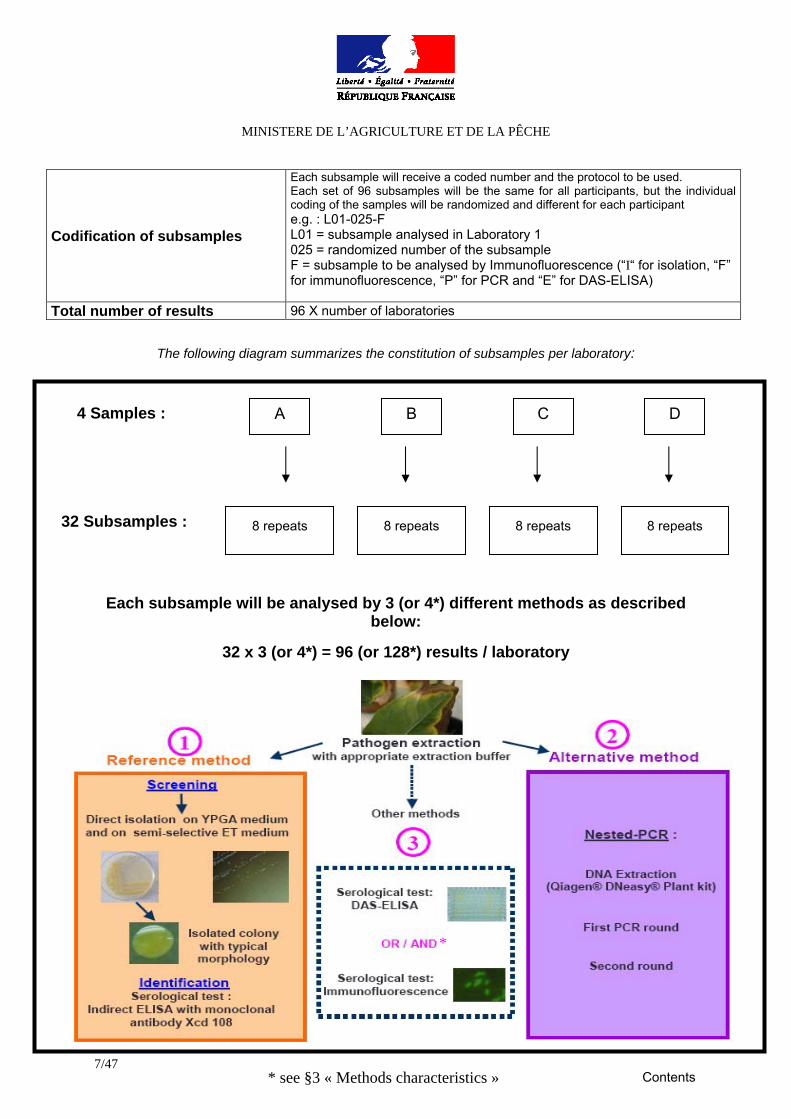
The following diagram summarizes the constitution of subsamples per laboratory:
Each subsample will be analysed by 3 (or 4*) different methods as described below:
32 x 3 (or 4*) = 96 (or 128*) results / laboratory
A B C D
8 repeats
8 repeats
8 repeats
8 repeats
4 Samples :
32 Subsamples :
* see §3 « Methods characteristics »
*
Contents
MINISTERE DE L’AGRICULTURE ET DE LA PÊCHE
8/47
10. Data Analyses
The results will be analysed according to the following criteria (reference: EN ISO 16140:2003 – Protocol for the validation of alternative methods).
1. Descriptive statistics of the qualitative results The following parameters will concern only the results on the samples A, B, C, D. For each sample analysed, each participant will have to answer preferably by “0” or “1”. In case of real doubt, that is to say if the result is twice ambiguous, the data “2” should be used but this result will not be included in the statistical analysis. For each method, the following values will be calculated : Positive accord (PA) between the expected results and the results obtained with the evaluated method Negative accord (NA) between the expected results and the results obtained with the evaluated method Positive deviation (PD) between the expected results and the results obtained with the evaluated method Negative deviation (ND) between the expected results and the results obtained with the evaluated method Four parameters will be computed, based on these results pairs:
• Relative accuracy of the methods (AC) • Relative specificity of the methods (SP) • Relative sensitivity of the methods (SE) • Estimation of confidence intervals (CI)
2. Variability of the methods The collaborative study will enable to characterize the dispersion of qualitative results when repeating the analysis on the same material both within a laboratory (qualitative repeatability or accordance) and between laboratories (qualitative reproducibility or concordance). Concordance odd ratio will also be calculated. Details on the computing will be provided enclosed with the collaborative study report.
Contents
MINISTERE DE L’AGRICULTURE ET DE LA PÊCHE
9/47
11. Laboratory Duties 1. Answer following questions: Did you detect Xad in the subsamples with the prescribed methods? 2. Information supplied to the participants
• Ring test organisation (this document) • Recommended chronology for analyses (Appendix 1) • Method for reviving freeze-dried bacteria (Appendix 2) • The serial dilution method of counting bacteria (Appendix 3) • Preparation of subsamples (Appendix 4) • Isolation protocol (Appendix 5) • N-PCR protocol (Appendix 6) • Immunofluorescence protocol (Appendix 7) • DAS ELISA protocol (Appendix 8) • Consent to participate to the collaborative study and confidentiality undertaking (Appendix 9) • Acknowledgement for the receipt of the critical consumables (Appendix 10) • Acknowledgement for the receipt of the samples (Appendix 11) • Comments on the interlaboratory ring-test (Appendix 12) • Results form (Appendix 13)
3. Packages receipt
o Verify the content of the packages and fill appendix 10 and appendix 11. o Store consumables and samples in conformity with instructions (table 8).
In case of difficulties or questions, contact the test organizer. 4. Test realisation Follow strictly the protocol provided in the appendixes. Register strictly any deviation and comments in the table (appendix 12). This information will be very important for the data analysis. Respect conscientiously the schedule (table 7). If strains of Xad are isolated, please keep them for further tests.
Contents
MINISTERE DE L’AGRICULTURE ET DE LA PÊCHE
10/47
5. Record of the results Laboratories must send the qualitative results of each analysis to the organizing laboratory as indicated in the results form (appendix 13). Therefore, the laboratories must keep the rough data for the organizer in case of need. The different methods are qualitative: data are to be written according to this rule:
0 if negative result 1 if positive result 2 if ambiguous result (only if the response is twice ambiguous: an ambiguous result will not be included in the statistical analysis): to be described
6. Confidentiality The participants will have to sign and return appendix 9 , 10, 11 and 12 dealing with confidentiality, use of the results obtained by the collaborative study, acknowledgments and comments about the study.
7. Sending results The results form (appendix 13) must be completed and sent to LNPV: [email protected]
Contents
MINISTERE DE L’AGRICULTURE ET DE LA PÊCHE
11/47
12. References
1. Technical references 1-Alvarez, A.; Lipp, R.; Norman, D. (1988). Detection and serological studies. In: Proceedings of the 1st Anthurium blight Conference, pp. 11-15. Hilo, USA. 2-European and Mediterranean Plant Protection Organization Standards (2004) Diagnostic protocols for regulated pests Xanthomonas axonopodis pv. dieffenbachiae . PM 7/23. Bulletin OEPP/EPPO °34: p. 155 –157 3-Kelman A. (1954). The relationship of pathogenicity in Pseudomonas solanacearum to colony appearance on a tetrazolium medium. Studies on Pseudomonas solanacearum. 693-695 4-Jouen, E., et al. (2006). First report in new Caledonia of Bacterial Blight of Anthurium caused by Xanthomonas axonopodis pv. dieffenbachiae , Plant Dis.,. 91 Vol n°4: p. 462. 5-Lipp, R.L.; Alvarez, A.M.; Benedict, A.A.; Berestecky, J. (1992) Use of monoclonal antibodies and pathogenicity tests to characterize strains of Xanthomonas campestris pv. dieffenbachiae. Phytopathology 82, 677-682. 6-Norman, D.J.; Alvarez, A.M. (1989) A rapid method for presumptive identification of Xanthomonas campestris pv. dieffenbachiae and other xanthomonads. Plant Disease 73, 654-658. 7-Norman, D.J.; Alvarez, A.M. (1994a) Rapid detection of Xanthomonas campestris pv. dieffenbachiae in Anthurium plants with a miniplate enrichment/ELISA system. Plant Disease 78, p. 954-958. 8-Norman, D.J.; Alvarez, A.M. (1994b) Latent infections of in vitro Anthurium caused by Xanthomonas campestris pv. dieffenbachiae. Plant Cell, Tissue and Organ Culture 39, p.55-61. 9-Puławska, J.; Kordyla-Bronka, M.; Jouen, E.; Robene-Soustrade, I.; Gagnevin, L.; Pruvost, O.; Sobiczewski, P.; Orlikowski, L. First report of bacterial blight of Anthurium andreanum in Poland. Plant Pathology 57, 4, 775. 10-Pruvost O. (2003) Analyse du Risque Phytosanitaire Filière de production Aracées, organisme nuisible Xanthomonas axonopodis pv. dieffenbachiae .ARA-b1. 11-Robène-Soustrade I., Laurent P., Gagnevin L. Jouen E. and Pruvost O. (2006) Specific detection of Xanthomonas axonopodis pv. dieffenbachiae in Anthurium tissues by Nested PCR. Applied and Environmental Microbiology. Feb. 2006. Vol. 72, No. 2: p. 1072–1078. 12-Soustrade, I., et al. (2000), First report of Anthurium Blight Caused by Xanthomonas axonopodis pv. dieffenbachiae in Reunion Island. Plant Dis.,. 84: p. 1343. 13-Vauterin, L.; Hoste, B.; Kersters, K.; Swings, J. (1995) Reclassification of Xanthomonas. International Journal of Systematic Bacteriology 45, 472-489.
Contents
MINISTERE DE L’AGRICULTURE ET DE LA PÊCHE
12/47
2. Normative references 1-AFNOR (2008) XP V 03-043: Exigences générales pour la réalisation d'analyses utilisant la biologie moléculaire pour la détection et l'identification d'organismes pathogènes, d'altération et ravageurs des végétaux et produits dérivés. 2-AFNOR (2003) NF EN ISO 16140: Microbiology of foods and animal feeding stuffs – Protocol for the validation of alternative methods. 3-AFNOR (1995) XP V03-111: Analyse des produits agricoles et alimentaires : Protocoles d’évaluation intra-laboratoire d’une méthode alternative d’analyse qualitative par rapport à une méthode de référence. 4-Ministère de l’Agriculture et de la Pêche, Direction Générale de l’Alimentation, Sous Direction de la Qualité et de la Protection des Végétaux, Laboratoire National de la Protection des Végétaux (2005) Directive Générale Techniques qualitatives immuno-enzymatiques de type ELISA (Enzyme Linked Immuno Sorbent Assay) : DAS (Double Antibody Sandwich) et dérivés. DG1/98/ version b. 5-Ministère de l’Agriculture et de la Pêche, Direction Générale de l’Alimentation, Sous Direction de la Qualité et de la Protection des Végétaux, Laboratoire National de la Protection des Végétaux (2008) Guide méthodologique Evaluation intra-laboratoire d’une méthode d’analyse qualitative par rapport à une méthode de référence. LNPV/ Guide01 (en cours de validation). 6-Schweizerische Akkreditierungsstelle (2006) Guide pour la validation de méthodes d’essais microbiologiques et l’évaluation de leur incertitude de mesure dans les domaines de la microbiologie alimentaire et de l’environnement Document N° 328.fw. Edition février 2006, rév. 01
Contents
MINISTERE DE L’AGRICULTURE ET DE LA PÊCHE
13/47
APPENDIX 1
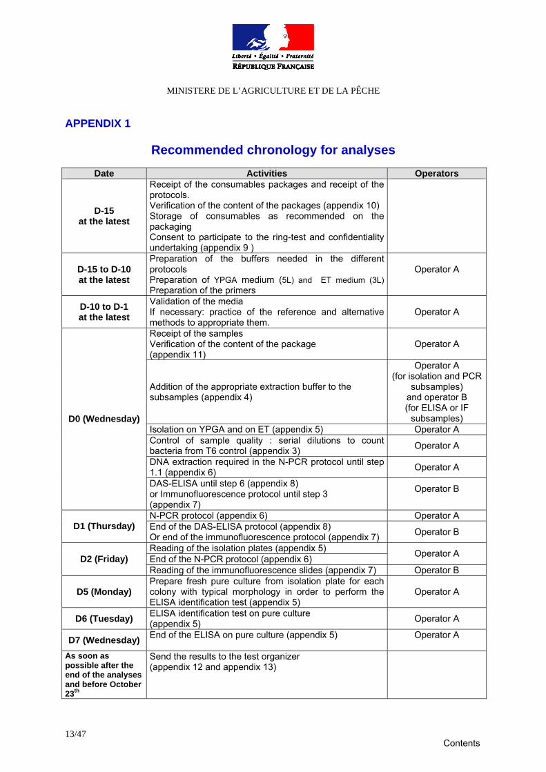
Recommended chronology for analyses
Date Activities Operators
D-15 at the latest
Receipt of the consumables packages and receipt of the protocols. Verification of the content of the packages (appendix 10) Storage of consumables as recommended on the packaging Consent to participate to the ring-test and confidentiality undertaking (appendix 9 )
D-15 to D-10 at the latest
Preparation of the buffers needed in the different protocols Preparation of YPGA medium (5L) and ET medium (3L) Preparation of the primers
Operator A
D-10 to D-1 at the latest
Validation of the media If necessary: practice of the reference and alternative methods to appropriate them.
Operator A
Receipt of the samples Verification of the content of the package (appendix 11)
Operator A
Addition of the appropriate extraction buffer to the subsamples (appendix 4)
Operator A (for isolation and PCR
subsamples) and operator B (for ELISA or IF
subsamples) Isolation on YPGA and on ET (appendix 5) Operator A Control of sample quality : serial dilutions to count bacteria from T6 control (appendix 3) Operator A
DNA extraction required in the N-PCR protocol until step 1.1 (appendix 6) Operator A
D0 (Wednesday)
DAS-ELISA until step 6 (appendix 8) or Immunofluorescence protocol until step 3 (appendix 7)
Operator B
N-PCR protocol (appendix 6) Operator A D1 (Thursday) End of the DAS-ELISA protocol (appendix 8)
Or end of the immunofluorescence protocol (appendix 7) Operator B
Reading of the isolation plates (appendix 5) End of the N-PCR protocol (appendix 6) Operator A D2 (Friday) Reading of the immunofluorescence slides (appendix 7) Operator B
D5 (Monday) Prepare fresh pure culture from isolation plate for each colony with typical morphology in order to perform the ELISA identification test (appendix 5)
Operator A
D6 (Tuesday) ELISA identification test on pure culture (appendix 5) Operator A
D7 (Wednesday) End of the ELISA on pure culture (appendix 5) Operator A
As soon as possible after the end of the analyses and before October 23th
Send the results to the test organizer (appendix 12 and appendix 13)
Contents
MINISTERE DE L’AGRICULTURE ET DE LA PÊCHE
14/47
APPENDIX 2
Method for reviving freeze-dried bacteria (control P1) 1-Media -Nutrient broth (e.g.: peptone nitrated water, pH 7.2: biogelytone 10g ; NaCl 5g ; distilled water 1000mL) - YPGA plates (for the recipe, see appendix 5 § 1.1) 2-Reviving bacteria 1-Make stripes all around the narrow part of the vial with diamond tool or a file. Sterilize the narrow part of the vial. Break the vial in aseptic conditions. 2-Resuspend the lyophilisate with 300µl of the nutrient broth by multiple pipeting. Ensure that the lyophilisate is well resuspended. 3-One drop is laid down on the YPGA plate and streaked in quadrants in order to obtain isolated colonies after incubation; the residual bacterial suspension is added to the broth in order to preserve a concentrated inoculum. 4-Agar plates and tubes are incubated at 28°C for 48h. If no growth occurs on the plates after recommended incubation time, it is possible to spread the nutrient broth on a new YPGA plate. .
Contents
MINISTERE DE L’AGRICULTURE ET DE LA PÊCHE
15/47
APPENDIX 3
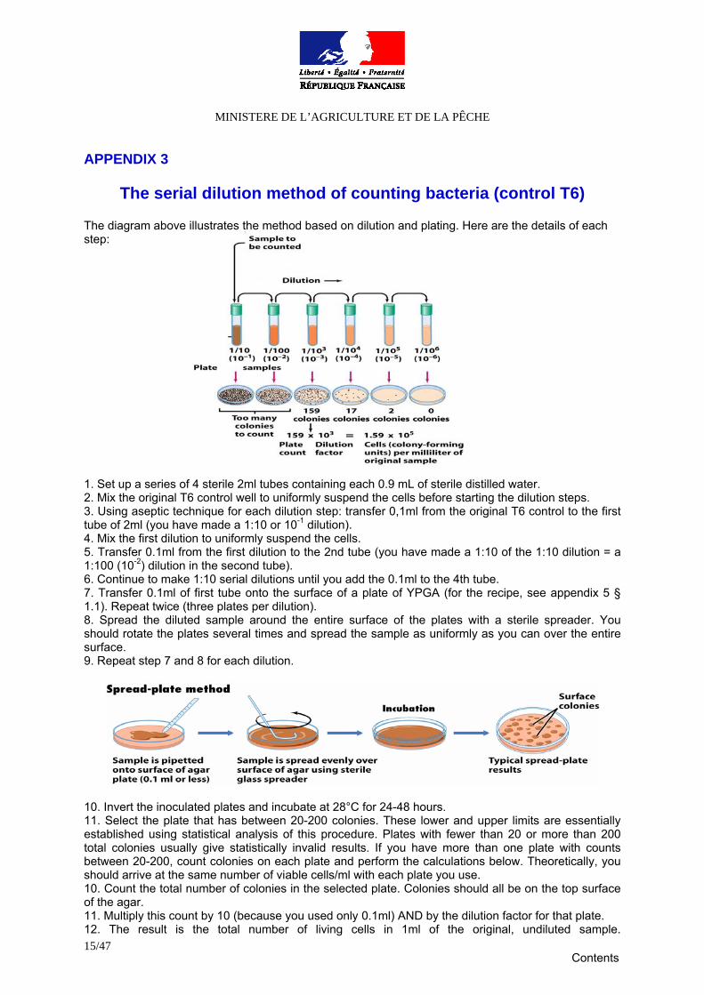
The serial dilution method of counting bacteria (control T6)
The diagram above illustrates the method based on dilution and plating. Here are the details of each step: 1. Set up a series of 4 sterile 2ml tubes containing each 0.9 mL of sterile distilled water. 2. Mix the original T6 control well to uniformly suspend the cells before starting the dilution steps. 3. Using aseptic technique for each dilution step: transfer 0,1ml from the original T6 control to the first tube of 2ml (you have made a 1:10 or 10-1 dilution). 4. Mix the first dilution to uniformly suspend the cells. 5. Transfer 0.1ml from the first dilution to the 2nd tube (you have made a 1:10 of the 1:10 dilution = a 1:100 (10-2) dilution in the second tube). 6. Continue to make 1:10 serial dilutions until you add the 0.1ml to the 4th tube. 7. Transfer 0.1ml of first tube onto the surface of a plate of YPGA (for the recipe, see appendix 5 § 1.1). Repeat twice (three plates per dilution). 8. Spread the diluted sample around the entire surface of the plates with a sterile spreader. You should rotate the plates several times and spread the sample as uniformly as you can over the entire surface. 9. Repeat step 7 and 8 for each dilution.
10. Invert the inoculated plates and incubate at 28°C for 24-48 hours. 11. Select the plate that has between 20-200 colonies. These lower and upper limits are essentially established using statistical analysis of this procedure. Plates with fewer than 20 or more than 200 total colonies usually give statistically invalid results. If you have more than one plate with counts between 20-200, count colonies on each plate and perform the calculations below. Theoretically, you should arrive at the same number of viable cells/ml with each plate you use. 10. Count the total number of colonies in the selected plate. Colonies should all be on the top surface of the agar. 11. Multiply this count by 10 (because you used only 0.1ml) AND by the dilution factor for that plate. 12. The result is the total number of living cells in 1ml of the original, undiluted sample.
Contents

MINISTERE DE L’AGRICULTURE ET DE LA PÊCHE
16/47
APPENDIX 4
Preparation of the subsamples
Already done:
Infected leaves or stems have been surface sterilized using 70° GL ethanol. Pieces of leaves have been removed with a disinfected scalpel blade and transferred to extraction bags. Sterile distilled water has been added (ratio: 1/10). Grinding has been performed using a homogenizer grinder (e.g. Homex grinder from Bioreba). After spiking with appropriate bacterial suspension, aliquots of extracts have been done.
To be done: In each aliquot (subsample and control) it is necessary to add appropriate concentrated buffers (to a 1x final concentration in plant sample) according to the test to be performed. Subsamples Quantities provided Appropriate extraction
buffer Quantities of extraction buffer
to add to each subsample PCR (P)
1 mL sterile distilled water 1mL
Isolation (I)
0.5mL
PBS 0,01M 2X 0,5mL
ELISA (E)
0.5mL SEB 2X 0,5mL
IF (F)
0.5mL
PBS 0,01M 2X 0,5mL
Contents
MINISTERE DE L’AGRICULTURE ET DE LA PÊCHE
17/47
APPENDIX 5
Protocol for the detection and identification of Xanthomonas axonopodis pv. dieffenbachiae by isolation
I- Isolation
1-Media for isolation
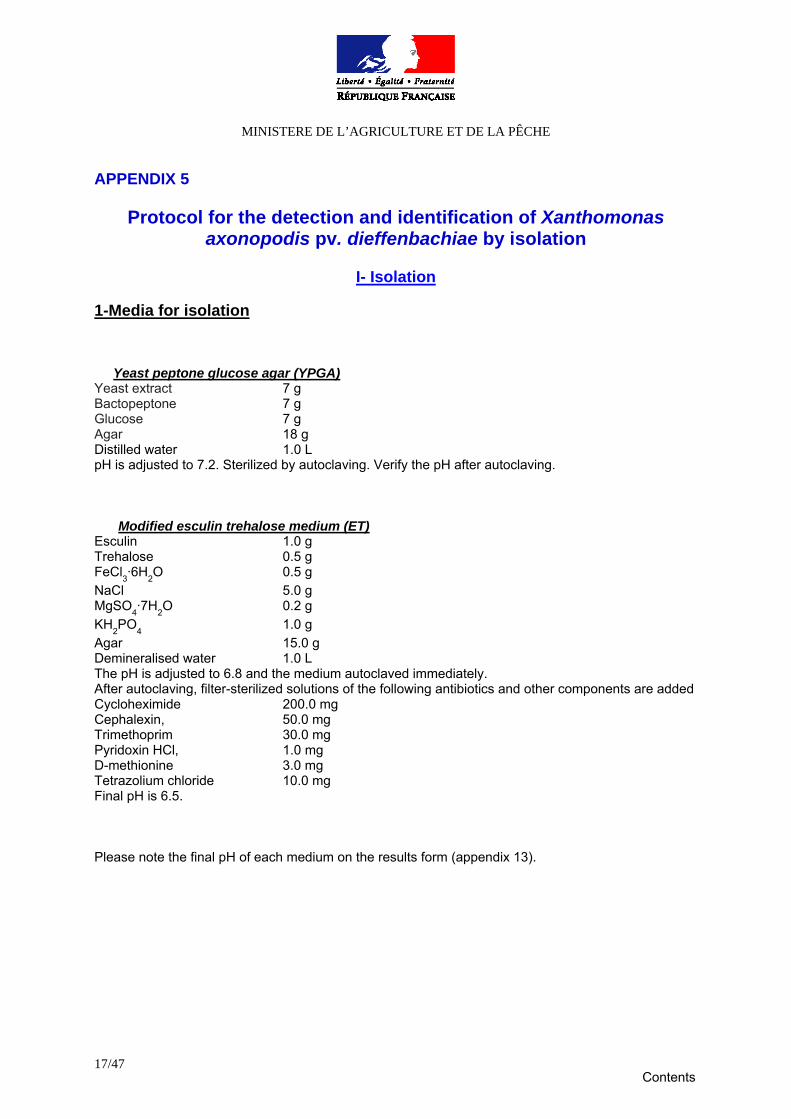
Yeast peptone glucose agar (YPGA) Yeast extract 7 g Bactopeptone 7 g Glucose 7 g Agar 18 g Distilled water 1.0 L pH is adjusted to 7.2. Sterilized by autoclaving. Verify the pH after autoclaving.
Modified esculin trehalose medium (ET) Esculin 1.0 g Trehalose 0.5 g FeCl3·6H2O 0.5 g NaCl 5.0 g MgSO4·7H2O 0.2 g KH2PO4 1.0 g Agar 15.0 g Demineralised water 1.0 L The pH is adjusted to 6.8 and the medium autoclaved immediately. After autoclaving, filter-sterilized solutions of the following antibiotics and other components are added Cycloheximide 200.0 mg Cephalexin, 50.0 mg Trimethoprim 30.0 mg Pyridoxin HCl, 1.0 mg D-methionine 3.0 mg Tetrazolium chloride 10.0 mg Final pH is 6.5. Please note the final pH of each medium on the results form (appendix 13).
Contents
MINISTERE DE L’AGRICULTURE ET DE LA PÊCHE
18/47
2. Plating 50 µl of each subsample and each control are streaked in quadrants on YPGA medium and on ET medium. Plates are incubated 48h at 28°C. 3. Examination of the plates X. axonopodis pv. dieffenbachiae colonies are defined as follows: round, convex, yellow, mucoid on YPGA medium; round, convex, mucoid with a light brown to dark brown halo of esculin hydrolysis surrounding the colonies for ET medium. First check whether reference isolates (control T4) did grow well and compare the suspicious bacteria isolates with the reference isolates. In case of negative result, re-examinate the plates after an additional incubation of 48h at 28°C. 4. Interpretation of the test If no suspicious colony is isolated on the media the subsample must be considered as negative. Please fill the results form (appendix 13). If there is at least one suspicious colony isolated on the media, the subsample must be considered as suspicious. In such case, the bacteria are identified with the ELISA identification test as described below. For the identification test it is recommended to use fresh pure culture. So suspicious colonies must be picked out and inoculated on a new plate 24h before performing the ELISA test.
II- Identification by Indirect ELISA (AGDIA) performed on fresh pure culture
1-Buffers for Indirect-ELISA (AGDIA)
Carbonate Coating buffer (1X) pH=9.6 Na2CO3 1.59 g NaHCO3 2.93 g Distilled water 1.0 L pH is adjusted to 9.6 with HCl. Sterilized by autoclaving.
PBS buffer (1X) NaCl 8.0 g Na2HPO4 1.15 g KH2PO4 0.2 g KCl 0.2 g Distilled water 1.0 L pH is adjusted to 7.4 if necessary. Sterilized by autoclaving.
Contents
MINISTERE DE L’AGRICULTURE ET DE LA PÊCHE
19/47
PBS-L buffer (1X) Non fat dry milk 50.0 g PBS 1X 1.0 L
PBS-TL buffer (1X) Non fat dry milk 25.0 g Tween 20 0.5 mL PBS 1X 1.0 L
Substrate buffer (for alkaline phosphatase) Diethanolamine 97.0 mL Distilled water 1.0 L pH is adjusted to 9.8 with concentrate HCl solution ( HCl solution volume is deduced from distilled water volume). Sterilized by autoclaving.
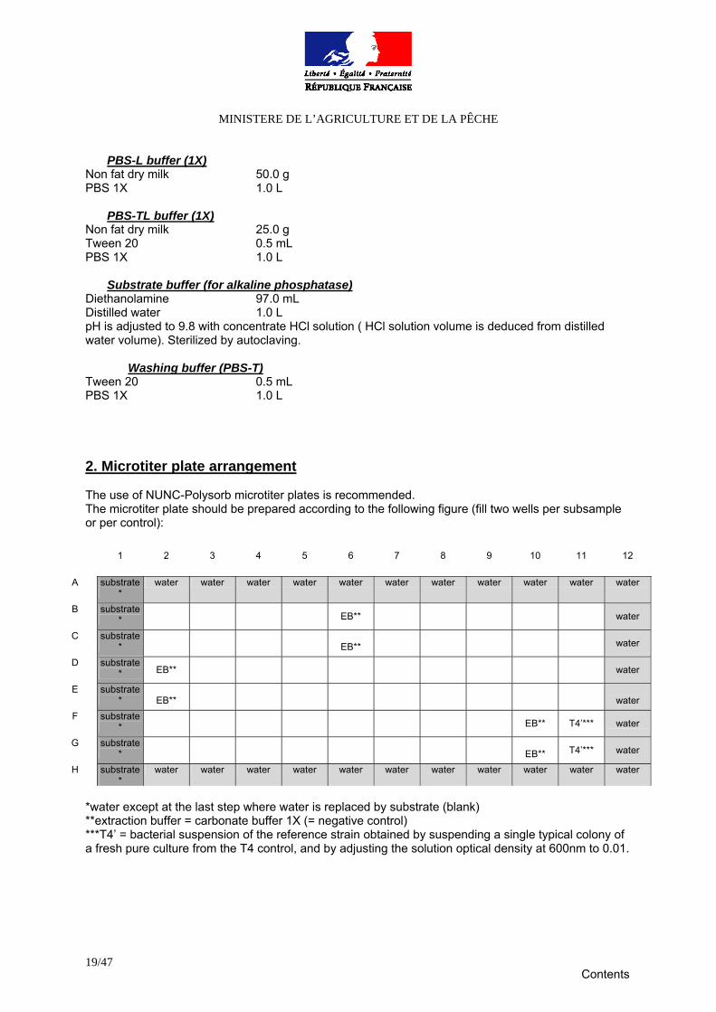
Washing buffer (PBS-T) Tween 20 0.5 mL PBS 1X 1.0 L 2. Microtiter plate arrangement The use of NUNC-Polysorb microtiter plates is recommended. The microtiter plate should be prepared according to the following figure (fill two wells per subsample or per control):
*water except at the last step where water is replaced by substrate (blank) **extraction buffer = carbonate buffer 1X (= negative control) ***T4’ = bacterial suspension of the reference strain obtained by suspending a single typical colony of a fresh pure culture from the T4 control, and by adjusting the solution optical density at 600nm to 0.01.
1 2 3 4 5 6 7 8 9 10 11 12
A substrate*
water water water water water water water water water water water
B substrate* EB** water
C substrate*
EB** water
D substrate* EB** water
E substrate*
EB**
water
F substrate* EB** T4’*** water
G substrate*
EB** T4’*** water
H substrate*
water water water water water water water water water water water
Contents

MINISTERE DE L’AGRICULTURE ET DE LA PÊCHE
20/47
3- Indirect-ELISA test
1. Prepare the samples: suspend a single colony of a fresh pure culture in carbonate buffer. A too concentrated solution could lead to false positive results. So it is recommended to adjust the solutions optical density at 600nm to 0.01, which is equivalent to about 107 cfu/mL (this equivalence should be verified beforehand). 2. Dispense 100µL of prepared sample into the wells of the ELISA plate (two wells per sample). Don’t forget the positive and negative controls. 3. Place the plate in an oven at 37°C overnight to dry. Be sure wells are completely dry before continuing. 4. Add 200µL of blocking solution PBS-L to each well, and incubate the plate in a humid box for 30 min at room temperature. 5. Wash the plate three times 5 min with the washing buffer PBS-T. 6. Dilute the coating antibody 200 times in PBS-TL . 7. Dispense 100 μL of prepared detection antibody in each well. 6. Set the plate inside the humid box and incubate for 1 hour at room temperature. 7. Wash the plate eight times with PBS-T. 8. Dilute the alkaline phosphatase conjugate 200 times in PBS-TL and dispense 100 μl of the prepared enzyme conjugate per well. 9. Set the plate inside the humid box and incubate for 1 hour at room temperature. 10. Wash the plate eight times with PBS-T. 11. Prepare a 1 mg/mL alkaline phosphatase substrate (para nitrophenyl-phosphate) in substrate buffer. Add 100 μL to each well, incubate at room temperature in a dark place and read at 405 nm four times: after 30 min, 1 hour, 2 hours and 3 hours of incubation. Blank with substrate. Results will be interpreted after 2 hours but it is important to conserve the results of the three other readings which may be used in case of ambiguous results. Samples are positive when their O.D. are at least twice the one obtained with the negative control. Please fill the results form (appendix 13).
Contents
MINISTERE DE L’AGRICULTURE ET DE LA PÊCHE
21/47
APPENDIX 6
Protocol for the detection and identification of Xanthomonas axonopodis pv. dieffenbachiae by nested-PCR
I- DNA Extraction with the Qiagen® DNeasy® Plant kit (Qiagen®, Courtaboeuf, France)
1. Centrifuge each PCR subsample and control (2 mL) for 10 min at 20000 x g and discard supernatant. The obtained homogenate can be stored at -20°C. 2. Add 400 µL of supplied buffer AP1 and 4µL of RNAase A 100mg/mL. To lyse the cells incubate for 10 min at 65°C, mixing by inverting tubes 2 or 3 times during incubation. 3. Add 130 µL of supplied buffer AP2 to the lysate, mix and incubate for 5 min on ice. 4. Centrifuge the lysate for 5 min at 20000 x g. Pipet the lysate into the supplied spin column placed in a 2 mL collection tube. 5. Centrifuge for 2 min at 20000 x g. Transfer the flow-through fraction into a new tube and add 1.5 volumes of supplied buffer AP3/E. Mix by pipetting. Pipet 650µL of the mixture into a supplied DNeasy spin column placed in a 2mL collection tube. 6. Centrifuge at 6000 x g for 1 min and discard flow-through. Repeat with remaining sample, discard flow-through and collection tube. Place the DNeasy spin column into a new 2mL collection tube and add 500µL of supplied buffer AW. 7. Centrifuge for 1 min at 6000 x g, discard flow-through and re-use the collection tube in the following step. 8. Add 500 µL buffer AW to the DNeasy spin column and centrifuge for 2 min at 20000 x g to dry the membrane. 9. Transfer the DNeasy spin column to a 2 mL microcentrifuge tube and pipet 50 µL buffer AE directly onto the membrane. Incubate for 5 min at room temperature, centrifuge for 1 min at 6000 x g to elute and repeat the step once. DNA thus extracted can be stored at -20°C.
II-Amplification For the detection of Xad from plant material, the nested PCR protocol is required because the second PCR round greatly increases sensitivity. The first PCR round produces an amplicon of 1570 bp, using primers PXadU (5’-AGGGCTCCCCATGCCGGAAT) and PXadL (5’-ACGCAATGCGCAGGGGAAAT-3’).The primers used in the second round are NXadU (5’-AGCGCGGTACATTGTTGTTCGT-3’) and NXadL (5’-GCGGATCCTGACTGAGCAAAG-3’), producing an amplicon of 785 bp. The nested-PCR is followed by a digestion step with restriction enzyme HincII. This RFLP step allows to distinguish Xad strains pathogenic to anthurium from closely related strains belonging to Xanthomonas campestris pv. syngonii and not pathogenic to anthurium. 1- First PCR round Prepare the following master mix for each sample (final volume 25 µL): 16.95 µL PCR grade water, 2.5 µL PCR buffer 10X, 1.1 mM MgCl2, 100 µM of each dNTP, 0.2 µM of each primer (PXadU and PXadL) and 1U Taq polymerase. Add 2µL of plant extracts. Use following PCR cycling parameters : 94°C for 3 min (initial denaturation), 35 cycles of 94°C for 30s, 70°C for 30s, 72°C for 2 min and a final extension step of 72°C for 10 min. PCR amplification products can be used immediately or stored at
Contents
MINISTERE DE L’AGRICULTURE ET DE LA PÊCHE
22/47
5°C for further use. For medium and long term storage (more than 24 hours), they should be kept at a temperature below -18°C. 2- Nested PCR For the second round of PCR prepare the same master mix than described above except primers which are NXadU and NXadL for this round. PCR tubes containing the first reaction amplicons must be opened with extreme care to avoid creation of aerosols which would cause contamination by amplification products. Pipet 1µL from the first reaction mixture into 24µL of this master mix. Use the following PCR cycling parameters: 94°C for 3 min (initial denaturation), 20 cycles of 94°C for 30s, 70°C for 30s and 72°C for 30s and a final extension step of 72°C for 5 min. PCR amplification products are detected by electrophoresis on 1% agarose gel and are stained with ethidium bromide. The nested PCR products can be stored at a temperature below -18°C. 3- Interpretation of results The PCR is negative if the expected amplicon is not observed or if the observed amplicons are not of the expected size, and if the amplicon is detected for positive control samples. Please fill the results form (appendix 13). The PCR detection test is positive if an amplicon of the expected size is observed (785 bp) and if there is no amplification for the negative control sample.
Contents
MINISTERE DE L’AGRICULTURE ET DE LA PÊCHE
23/47
APPENDIX 7
Protocol for the detection and identification of Xanthomonas axonopodis pv. dieffenbachiae by Immunofluorescence (PRI)
1. Buffers for Immunofluorescence
Evans blue Evans blue 0.1 g PBS (0.01 M) 10.0 mL The buffer will be not autoclaved. Stored at 5°C and kept away from light
Phosphate-buffered saline (0.01M) (PBS 0.01M) NaCl 8.0 g Na2HPO4, 12 H2O 2.7 g KH2PO4 1.0 g Distilled water 1.0 L pH is adjusted to 7.4. Sterilized by autoclaving.
Phosphate buffered glycerine (0.1 M) Na2HPO4, 12 H2O 3.2 g NaH2PO4 0.15 g Glycerol 50.0 mL Distilled water 100.0 mL pH is adjusted to pH 7.6 if necessary. Sterilized by autoclaving.
Washing buffer: PBS 0.001M PBS 0.01M 0.1 L Distilled water 0.9 L Sterilized by autoclaving.
Contents
MINISTERE DE L’AGRICULTURE ET DE LA PÊCHE
24/47
2. Deposit of samples:
1. Clean the slides extensively with alcohol. 2. Pipet a standard volume of the plant extract or bacterial suspension and of decimal dilutions
(1/10 and 1/100) on each window of a multiwell microscope slides (preferably with 6 or 8 windows of at least 8 mm diameter). 40 µL are appropriate for 8 mm window diameter (scale up or down for larger or smaller window diameters). Do not forget negative (T1) and positive (T2 to T4) controls.
To avoid any contaminations, it is recommended to deposit only one subsample (and its decimal dilutions) per slide. The IF-slides should be prepared according to the following figure:
1/1 (IgG : 1/400) (FITC : 1/400)
Dilution 1/10 (IgG: 1/400)
(FITC : 1/400)
Dilution 1/100 (IgG: 1/400)
(FITC : 1/400)
One subsample / slide 1/1
(IgG:1/10000) (FITC : 1/400)
Dilution 1/10 (IgG:1/10000) (FITC : 1/400)
Dilution 1/100 (IgG:1/10000) (FITC : 1/400)
3- Fixation of extracts and Immunofluorescence test
3. Dry the slides on the heating plate (at a maximum temperature of 55°C). Fix the samples by adding a droplet of ethanol (titre > 95° GL) on each window, and dry the slides on the heating plate at a maximum temperature of 55°C. It is preferable to process slides further as soon as possible. Nevertheless, if necessary, fixed slides may then be stored at 5°C for less than 48 hours.
4. Cover each window with the appropriate antiserum IgG at the appropriate dilution (two
dilutions are evaluated: 1/400 and 1/10 000) in PBS 0.01M.
5. Incubate the slides 30 min in a moist, dark place at 27°C (e.g. horizontal on wet filter paper in a closed box in a stove).
6. Shake the droplets off each slide and rinse for approximately five min twice with washing
buffer PBS 0.001 M. Wash the slides one minute with demineralised water and dry them on the heating plate at a maximum temperature of 55°C.
7. Cover the window with appropriate FITC conjugate at appropriate dilution (1/400) and Evans
blue at the dilution 1/300 in PBS 0.01M. Evans blue is added as counter staining due to high self fluorescence of anthurium compounds.
8. Incubate the slides 30 min in a moist, dark place at 27°C (eg. horizontal on wet filter paper in a
closed box in a stove).
9. Shake the droplets off each slide and rinse for approximately five min (+/- 1 min) twice with washing buffer PBS 0.001 M. Wash the slides one minute with demineralised water and dry them on the heating plate at a maximum temperature of 55°C.
Contents
MINISTERE DE L’AGRICULTURE ET DE LA PÊCHE
25/47
10. Apply appropriate mounting buffer (phosphate buffered glycerine 0.1 M). Cover with a cover glass and avoid exposure of the slides to excess of light.
11. Examine immediately each well under an epifluorescence microscope with filters suitable for
excitation of FITC at 600-1000 magnification (use oil immersion). Scan windows across one diameter. If no colonies are observed, extend the search to one supplementary diameter at right angle and to a quarter of perimeter. It is important to examine the positive control slides first. Cells must be bright fluorescent and completely stained at the antibody titre.
4- Interpretation of results Reading: validate the analysis by the observation of the positive and negative controls. A positive presumptive diagnostic test is achieved if typical well-stained bacterial cells with the appropriate size (2-3µm) and form (rod) are seen as in the positive control. Please fill the results form (appendix 13).
Contents
MINISTERE DE L’AGRICULTURE ET DE LA PÊCHE
26/47
APPENDIX 8
Protocol for the detection of Xanthomonas axonopodis pv. dieffenbachiae by DAS ELISA (PRI)
1-Buffers for DAS-ELISA
Carbonate buffer 1X pH=9.6 Na2CO3 1.59 g NaHCO3 2.93 g Distilled water 1.0 L pH is adjusted to 9.6 with HCl (HCl solution volume is deduced from distilled water volume). Sterilized by autoclaving.
Extraction buffer (SEB 1X) Tween 20 1.0 mL PVP-25 20.0 g (ovalbumine, grade IV 2.0 g) PBS (0.01M) 1.0 L
Phosphate-buffered saline (0.01M) (PBS 0.01M) NaCl 8.0 g KH2PO4 1.0 g Na2HPO4, 12 H2O 14.5 g NaH2PO4, 2 H2O 0.4 g Distilled water 1.0 L pH is adjusted to 7.4 if necessary. Sterilized by autoclaving.
Substrate buffer (for alkaline phosphatase)
Diethanolamine 97.0 mL Distilled water 1.0 L pH is adjusted to 9.8 with concentrate HCl solution ( HCl solution volume is deduced from distilled water volume). Sterilized by autoclaving.
Washing buffer (PBS-T) Tween 20 1.0 mL PBS (0.01M) 1.0 L
Contents
MINISTERE DE L’AGRICULTURE ET DE LA PÊCHE
27/47
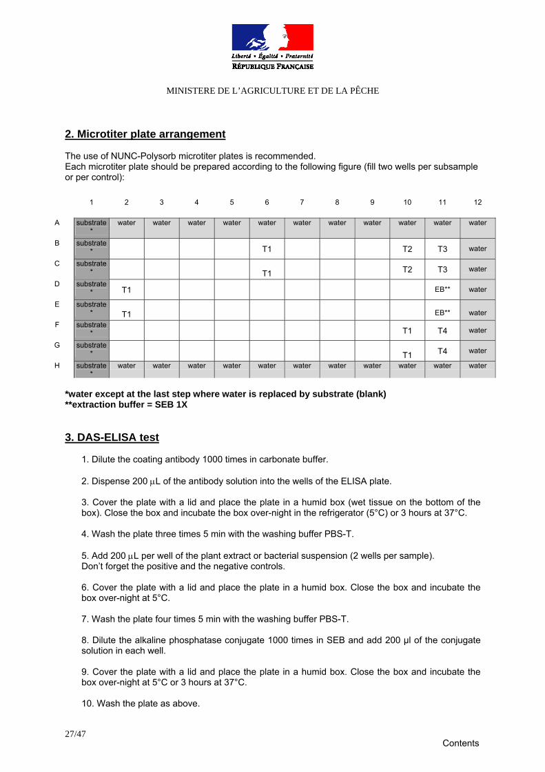
2. Microtiter plate arrangement The use of NUNC-Polysorb microtiter plates is recommended. Each microtiter plate should be prepared according to the following figure (fill two wells per subsample or per control):
*water except at the last step where water is replaced by substrate (blank) **extraction buffer = SEB 1X 3. DAS-ELISA test
1. Dilute the coating antibody 1000 times in carbonate buffer.
2. Dispense 200 μL of the antibody solution into the wells of the ELISA plate. 3. Cover the plate with a lid and place the plate in a humid box (wet tissue on the bottom of the box). Close the box and incubate the box over-night in the refrigerator (5°C) or 3 hours at 37°C. 4. Wash the plate three times 5 min with the washing buffer PBS-T. 5. Add 200 μL per well of the plant extract or bacterial suspension (2 wells per sample). Don’t forget the positive and the negative controls. 6. Cover the plate with a lid and place the plate in a humid box. Close the box and incubate the box over-night at 5°C. 7. Wash the plate four times 5 min with the washing buffer PBS-T. 8. Dilute the alkaline phosphatase conjugate 1000 times in SEB and add 200 μl of the conjugate solution in each well. 9. Cover the plate with a lid and place the plate in a humid box. Close the box and incubate the box over-night at 5°C or 3 hours at 37°C. 10. Wash the plate as above.
1 2 3 4 5 6 7 8 9 10 11 12
A substrate*
water water water water water water water water water water water
B substrate* T1 T2 T3 water
C substrate*
T1 T2 T3 water
D substrate* T1 EB** water
E substrate*
T1
EB**
water
F substrate* T1 T4 water
G substrate*
T1 T4 water
H substrate*
water water water water water water water water water water water
Contents

MINISTERE DE L’AGRICULTURE ET DE LA PÊCHE
28/47
11. Prepare a 1 mg/mL alkaline phosphatase substrate (para nitrophenyl-phosphate) in substrate buffer. Add 200 μL to each well, incubate at room temperature in a dark place and read at 405 nm four times: after 30 min, 1 hour, 2 hours and 3 hours of incubation. Blank with substrate. Results will be interpreted after 2 hours but it is important to keep the results of the three other readings which may be used in case of ambiguous results. Samples are positive when their O.D. are at least twice the one obtained with the negative control plant sample (T1). Please fill the results form (appendix 13).
Contents
MINISTERE DE L’AGRICULTURE ET DE LA PÊCHE
29/47
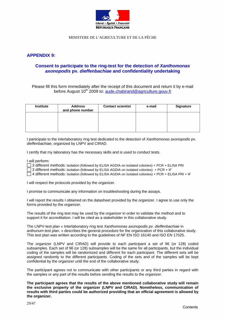
APPENDIX 9:
Consent to participate to the ring-test for the detection of Xanthomonas axonopodis pv. dieffenbachiae and confidentiality undertaking
Please fill this form immediately after the receipt of this document and return it by e-mail
before August 10th 2009 to: [email protected]
Institute Address and phone number
Contact scientist e-mail Signature
I participate to the interlaboratory ring test dedicated to the detection of Xanthomonas axonopodis pv. dieffenbachiae, organized by LNPV and CIRAD. I certify that my laboratory has the necessary skills and is used to conduct tests. I will perform:
3 different methods: Isolation (followed by ELISA AGDIA on isolated colonies) + PCR + ELISA PRI 3 different methods: Isolation (followed by ELISA AGDIA on isolated colonies) + PCR + IF 4 different methods: Isolation (followed by ELISA AGDIA on isolated colonies) + PCR + ELISA PRI + IF
I will respect the protocols provided by the organizer. I promise to communicate any information on troubleshooting during the assays. I will report the results I obtained on the datasheet provided by the organizer. I agree to use only the forms provided by the organizer. The results of the ring test may be used by the organizer in order to validate the method and to support it for accreditation. I will be cited as a stakeholder in this collaborative study.
The LNPV-test plan « Interlaboratory ring test Xanthomonas axonopodis pv. dieffenbachiae in anthurium test plan. » describes the general procedure for the organization of this collaborative study. This test plan was written according to the guidelines of NF EN ISO 16140 and ISO EN 17025. The organizer (LNPV and CIRAD) will provide to each participant a set of 96 (or 128) coded subsamples. Each set of 96 (or 128) subsamples will be the same for all participants, but the individual coding of the samples will be randomized and different for each participant. The different sets will be assigned randomly to the different participants. Coding of the sets and of the samples will be kept confidential by the organizer until the end of the collaborative study. The participant agrees not to communicate with other participants or any third parties in regard with the samples or any part of the results before sending the results to the organizer. The participant agrees that the results of the above mentioned collaborative study will remain the exclusive property of the organizer (LNPV and CIRAD). Nonetheless, communication of results with third parties could be authorized providing that an official agreement is allowed by the organizer.
Contents
MINISTERE DE L’AGRICULTURE ET DE LA PÊCHE
30/47
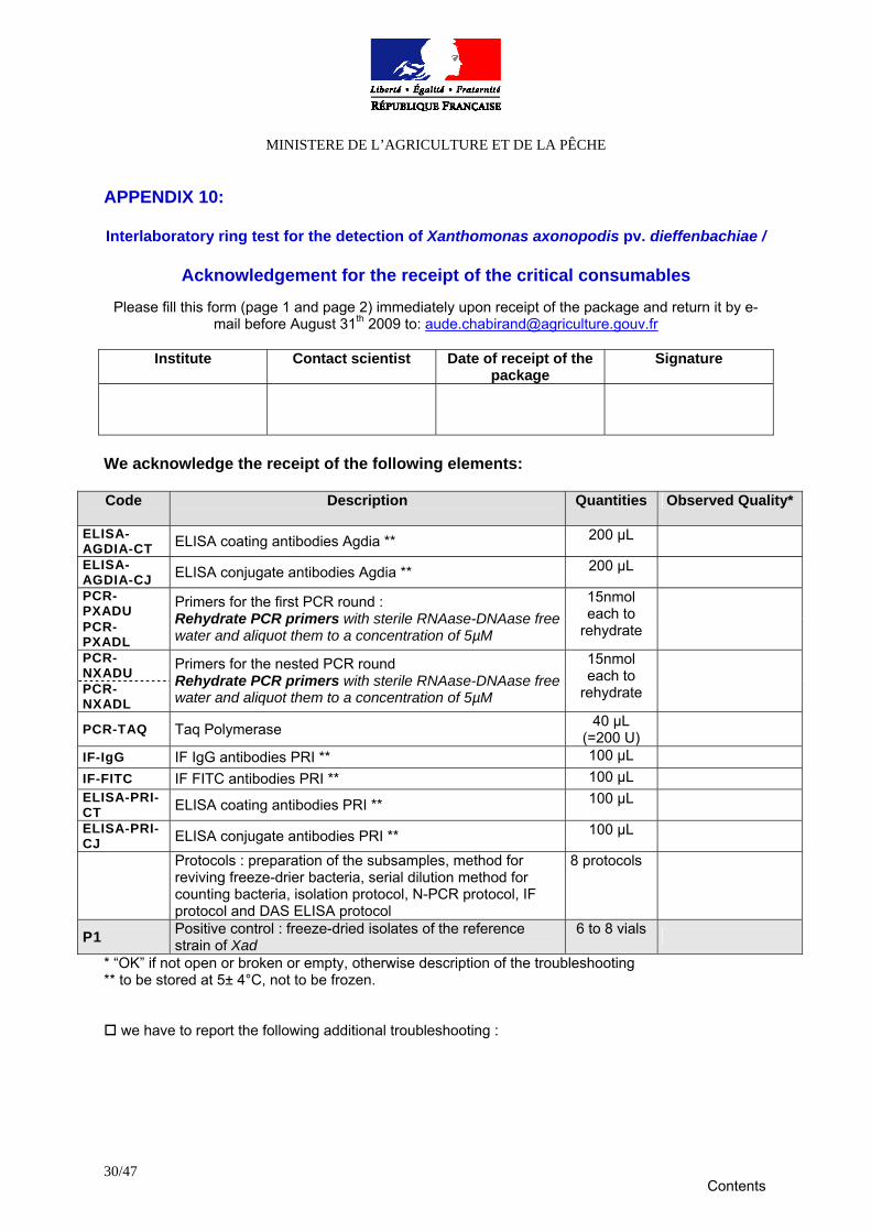
APPENDIX 10: Interlaboratory ring test for the detection of Xanthomonas axonopodis pv. dieffenbachiae /
Acknowledgement for the receipt of the critical consumables
Please fill this form (page 1 and page 2) immediately upon receipt of the package and return it by e-
mail before August 31th 2009 to: [email protected]
Institute Contact scientist Date of receipt of the package
Signature
We acknowledge the receipt of the following elements: Code
Description Quantities
Observed Quality*
ELISA-AGDIA-CT ELISA coating antibodies Agdia ** 200 µL ELISA-AGDIA-CJ ELISA conjugate antibodies Agdia ** 200 µL PCR-PXADU PCR-PXADL
Primers for the first PCR round : Rehydrate PCR primers with sterile RNAase-DNAase free water and aliquot them to a concentration of 5µM
15nmol each to
rehydrate
PCR-NXADU PCR-NXADL
Primers for the nested PCR round Rehydrate PCR primers with sterile RNAase-DNAase free water and aliquot them to a concentration of 5µM
15nmol each to
rehydrate
PCR-TAQ Taq Polymerase 40 µL (=200 U)
IF-IgG IF IgG antibodies PRI ** 100 µL IF-FITC IF FITC antibodies PRI ** 100 µL ELISA-PRI-CT ELISA coating antibodies PRI ** 100 µL ELISA-PRI-CJ ELISA conjugate antibodies PRI ** 100 µL
Protocols : preparation of the subsamples, method for reviving freeze-drier bacteria, serial dilution method for counting bacteria, isolation protocol, N-PCR protocol, IF protocol and DAS ELISA protocol
8 protocols
P1 Positive control : freeze-dried isolates of the reference strain of Xad
6 to 8 vials
* “OK” if not open or broken or empty, otherwise description of the troubleshooting ** to be stored at 5± 4°C, not to be frozen.
we have to report the following additional troubleshooting :
Contents
MINISTERE DE L’AGRICULTURE ET DE LA PÊCHE
31/47
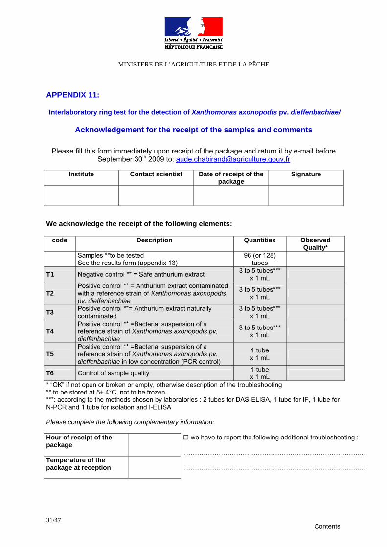
APPENDIX 11: Interlaboratory ring test for the detection of Xanthomonas axonopodis pv. dieffenbachiae/
Acknowledgement for the receipt of the samples and comments
Please fill this form immediately upon receipt of the package and return it by e-mail before September 30th 2009 to: [email protected]
Institute Contact scientist Date of receipt of the
package Signature
We acknowledge the receipt of the following elements:
code Description Quantities Observed Quality*
Samples **to be tested See the results form (appendix 13)
96 (or 128) tubes
T1 Negative control ** = Safe anthurium extract 3 to 5 tubes*** x 1 mL
T2 Positive control ** = Anthurium extract contaminated with a reference strain of Xanthomonas axonopodis pv. dieffenbachiae
3 to 5 tubes*** x 1 mL
T3 Positive control **= Anthurium extract naturally contaminated
3 to 5 tubes*** x 1 mL
T4 Positive control ** =Bacterial suspension of a reference strain of Xanthomonas axonopodis pv. dieffenbachiae
3 to 5 tubes*** x 1 mL
T5 Positive control ** =Bacterial suspension of a reference strain of Xanthomonas axonopodis pv. dieffenbachiae in low concentration (PCR control)
1 tube x 1 mL
T6 Control of sample quality 1 tube x 1 mL
* “OK” if not open or broken or empty, otherwise description of the troubleshooting ** to be stored at 5± 4°C, not to be frozen. ***: according to the methods chosen by laboratories : 2 tubes for DAS-ELISA, 1 tube for IF, 1 tube for N-PCR and 1 tube for isolation and I-ELISA Please complete the following complementary information: Hour of receipt of the package
Temperature of the package at reception
we have to report the following additional troubleshooting :
………………………………………………………………………...
………………………………………………………………………...
Contents
MINISTERE DE L’AGRICULTURE ET DE LA PÊCHE
32/47
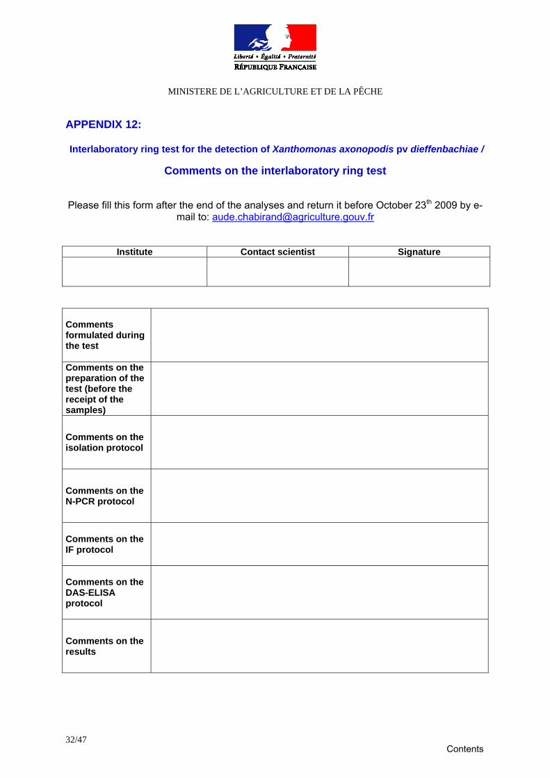
APPENDIX 12: Interlaboratory ring test for the detection of Xanthomonas axonopodis pv dieffenbachiae /
Comments on the interlaboratory ring test
Please fill this form after the end of the analyses and return it before October 23th 2009 by e-
mail to: [email protected]
Institute Contact scientist Signature
Comments formulated during the test
Comments on the preparation of the test (before the receipt of the samples)
Comments on the isolation protocol
Comments on the N-PCR protocol
Comments on the IF protocol
Comments on the DAS-ELISA protocol
Comments on the results
Contents
MINISTERE DE L’AGRICULTURE ET DE LA PÊCHE
33/47
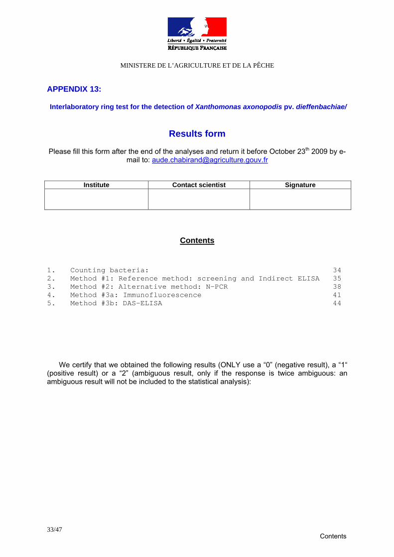
APPENDIX 13: Interlaboratory ring test for the detection of Xanthomonas axonopodis pv. dieffenbachiae/
Results form
Please fill this form after the end of the analyses and return it before October 23th 2009 by e-
mail to: [email protected]
Institute Contact scientist Signature
Contents
1. Counting bacteria: 34 2. Method #1: Reference method: screening and Indirect ELISA 35 3. Method #2: Alternative method: N-PCR 38 4. Method #3a: Immunofluorescence 41 5. Method #3b: DAS-ELISA 44
We certify that we obtained the following results (ONLY use a “0” (negative result), a “1“ (positive result) or a “2” (ambiguous result, only if the response is twice ambiguous: an ambiguous result will not be included to the statistical analysis):
Contents
MINISTERE DE L’AGRICULTURE ET DE LA PÊCHE
34/47
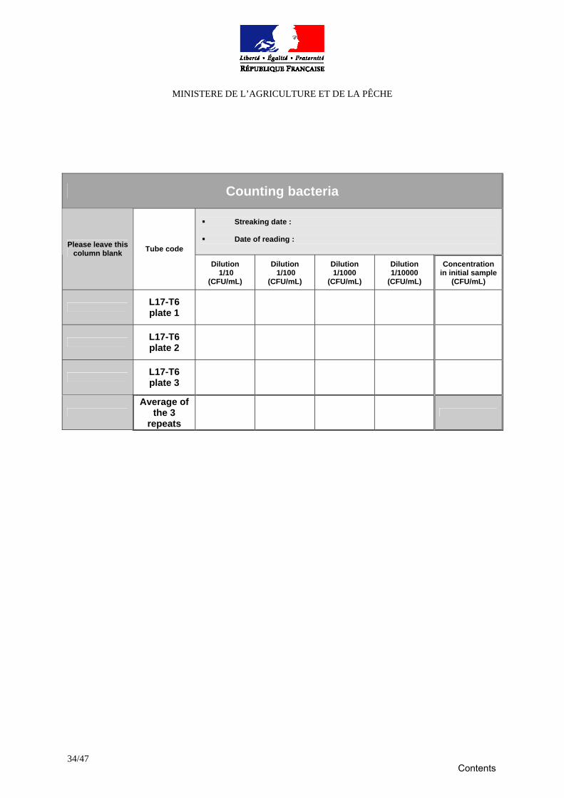
Counting bacteria
Streaking date :
Date of reading :
Please leave this column blank Tube code
Dilution 1/10
(CFU/mL)
Dilution 1/100
(CFU/mL)
Dilution 1/1000
(CFU/mL)
Dilution 1/10000
(CFU/mL)
Concentration in initial sample
(CFU/mL)
L17-T6 plate 1
L17-T6 plate 2
L17-T6 plate 3
Average of
the 3 repeats
Contents
MINISTERE DE L’AGRICULTURE ET DE LA PÊCHE
35/47
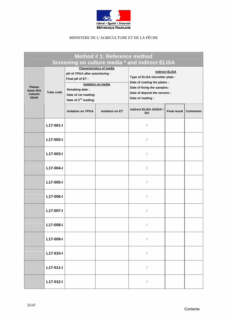
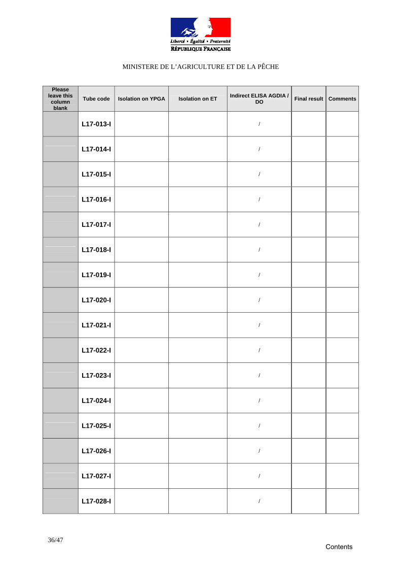
Method # 1: Reference method Screening on culture media * and indirect ELISA
Characteristics of media pH of YPGA after autoclaving : Final pH of ET :
Isolation on media Streaking date : Date of 1st reading: Date of 2nd reading:
Indirect ELISA Type of ELISA microtiter plate : Date of coating the plates : Date of fixing the samples : Date of deposit the serums :
Date of reading :
Please leave this column blank
Tube code
Isolation on YPGA Isolation on ET Indirect ELISA AGDIA / DO Final result Comments
L17-001-I /
L17-002-I /
L17-003-I /
L17-004-I /
L17-005-I /
L17-006-I /
L17-007-I /
L17-008-I /
L17-009-I /
L17-010-I /
L17-011-I /
L17-012-I /
Contents
MINISTERE DE L’AGRICULTURE ET DE LA PÊCHE
36/47
Please leave this column blank
Tube code Isolation on YPGA Isolation on ET Indirect ELISA AGDIA / DO Final result Comments
L17-013-I /
L17-014-I /
L17-015-I /
L17-016-I /
L17-017-I /
L17-018-I /
L17-019-I /
L17-020-I /
L17-021-I /
L17-022-I /
L17-023-I /
L17-024-I /
L17-025-I /
L17-026-I /
L17-027-I /
L17-028-I /
Contents
MINISTERE DE L’AGRICULTURE ET DE LA PÊCHE
37/47
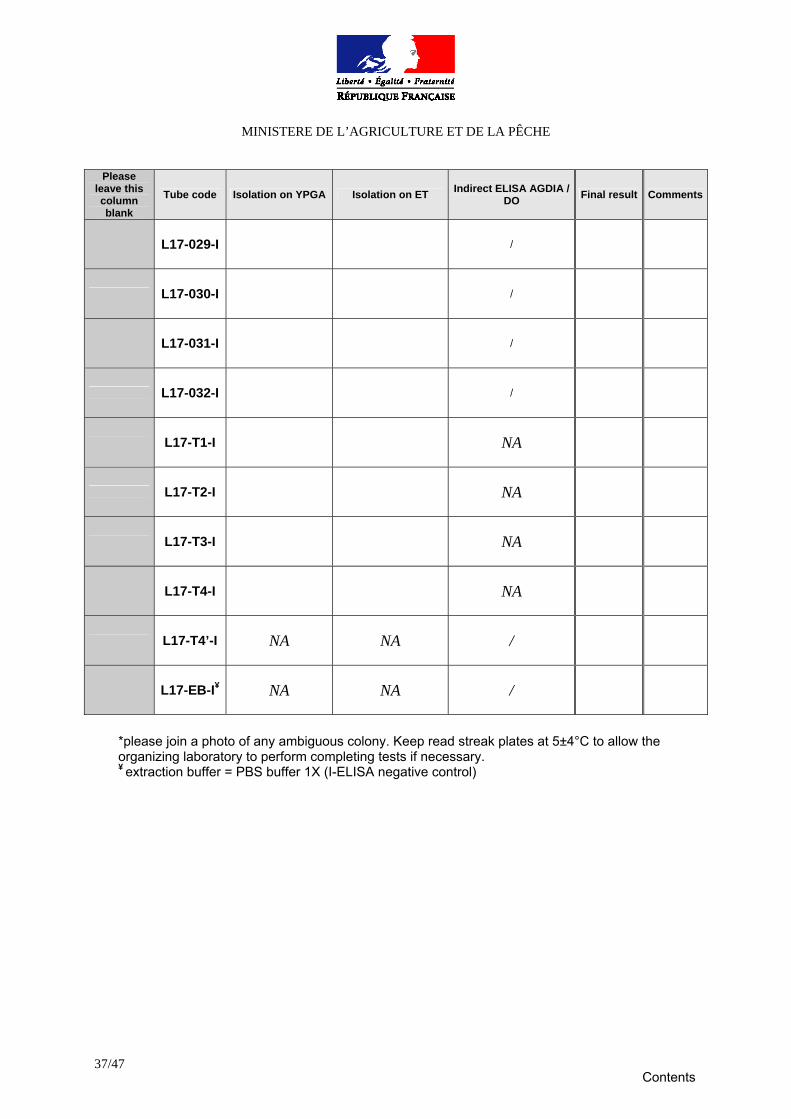
Please leave this column blank
Tube code Isolation on YPGA Isolation on ET Indirect ELISA AGDIA / DO Final result Comments
L17-029-I /
L17-030-I /
L17-031-I /
L17-032-I /
L17-T1-I NA
L17-T2-I NA
L17-T3-I NA
L17-T4-I NA
L17-T4’-I NA NA /
L17-EB-I¥ NA NA /
*please join a photo of any ambiguous colony. Keep read streak plates at 5±4°C to allow the organizing laboratory to perform completing tests if necessary. ¥ extraction buffer = PBS buffer 1X (I-ELISA negative control)
Contents
MINISTERE DE L’AGRICULTURE ET DE LA PÊCHE
38/47
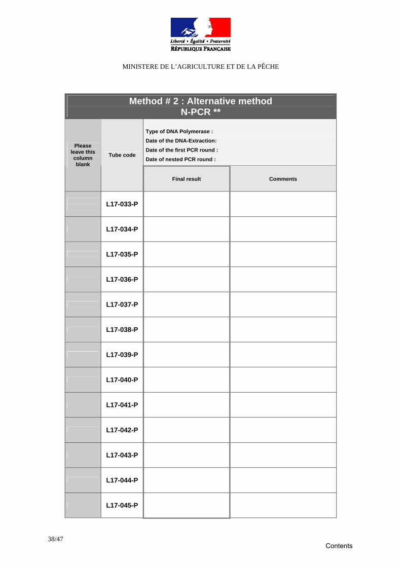
Method # 2 : Alternative method N-PCR **
Type of DNA Polymerase : Date of the DNA-Extraction: Date of the first PCR round : Date of nested PCR round :
Please leave this column blank
Tube code
Final result Comments
L17-033-P
L17-034-P
L17-035-P
L17-036-P
L17-037-P
L17-038-P
L17-039-P
L17-040-P
L17-041-P
L17-042-P
L17-043-P
L17-044-P
L17-045-P
Contents
MINISTERE DE L’AGRICULTURE ET DE LA PÊCHE
39/47
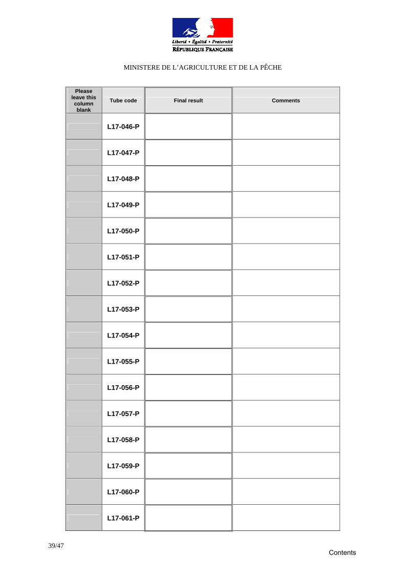
Please leave this column blank
Tube code Final result Comments
L17-046-P
L17-047-P
L17-048-P
L17-049-P
L17-050-P
L17-051-P
L17-052-P
L17-053-P
L17-054-P
L17-055-P
L17-056-P
L17-057-P
L17-058-P
L17-059-P
L17-060-P
L17-061-P
Contents
MINISTERE DE L’AGRICULTURE ET DE LA PÊCHE
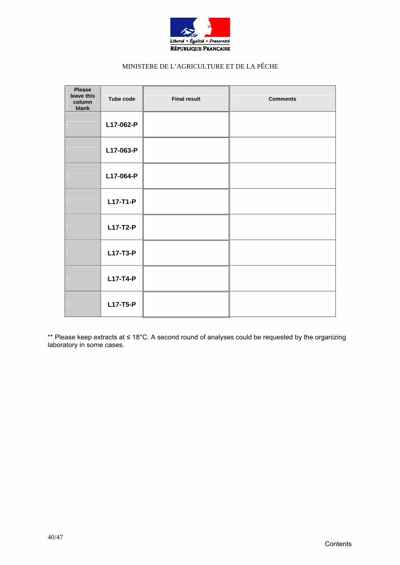
40/47
Please leave this column blank
Tube code Final result Comments
L17-062-P
L17-063-P
L17-064-P
L17-T1-P
L17-T2-P
L17-T3-P
L17-T4-P
L17-T5-P
** Please keep extracts at ≤ 18°C. A second round of analyses could be requested by the organizing laboratory in some cases.
Contents
MINISTERE DE L’AGRICULTURE ET DE LA PÊCHE
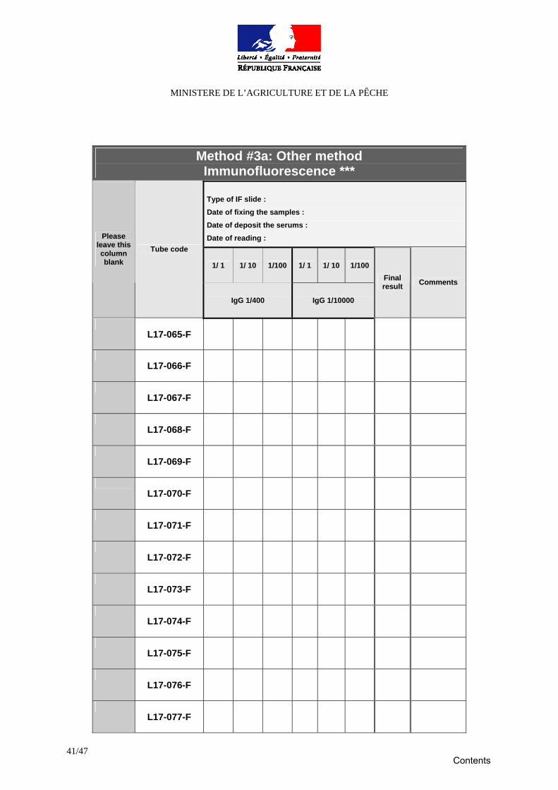
41/47
Method #3a: Other method Immunofluorescence *** Type of IF slide : Date of fixing the samples : Date of deposit the serums : Date of reading :
1/ 1 1/ 10 1/100 1/ 1 1/ 10 1/100
Please leave this column blank
Tube code
IgG 1/400 IgG 1/10000
Final result Comments
L17-065-F
L17-066-F
L17-067-F
L17-068-F
L17-069-F
L17-070-F
L17-071-F
L17-072-F
L17-073-F
L17-074-F
L17-075-F
L17-076-F
L17-077-F
Contents
MINISTERE DE L’AGRICULTURE ET DE LA PÊCHE
42/47
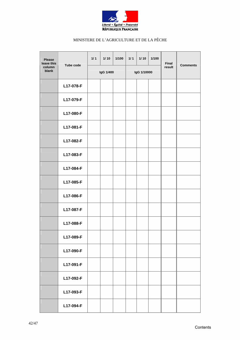
1/ 1 1/ 10 1/100 1/ 1 1/ 10 1/100 Please leave this column blank
Tube code
IgG 1/400 IgG 1/10000
Final result Comments
L17-078-F
L17-079-F
L17-080-F
L17-081-F
L17-082-F
L17-083-F
L17-084-F
L17-085-F
L17-086-F
L17-087-F
L17-088-F
L17-089-F
L17-090-F
L17-091-F
L17-092-F
L17-093-F
L17-094-F
Contents
MINISTERE DE L’AGRICULTURE ET DE LA PÊCHE
43/47
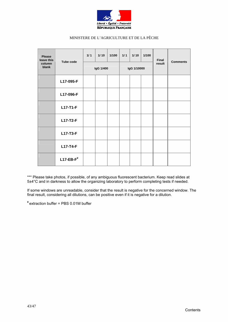
1/ 1 1/ 10 1/100 1/ 1 1/ 10 1/100 Please leave this column blank
Tube code
IgG 1/400 IgG 1/10000
Final result Comments
L17-095-F
L17-096-F
L17-T1-F
L17-T2-F
L17-T3-F
L17-T4-F
L17-EB-F¥
*** Please take photos, if possible, of any ambiguous fluorescent bacterium. Keep read slides at 5±4°C and in darkness to allow the organizing laboratory to perform completing tests if needed. If some windows are unreadable, consider that the result is negative for the concerned window. The final result, considering all dilutions, can be positive even if it is negative for a dilution. ¥ extraction buffer = PBS 0.01M buffer
Contents
MINISTERE DE L’AGRICULTURE ET DE LA PÊCHE
44/47
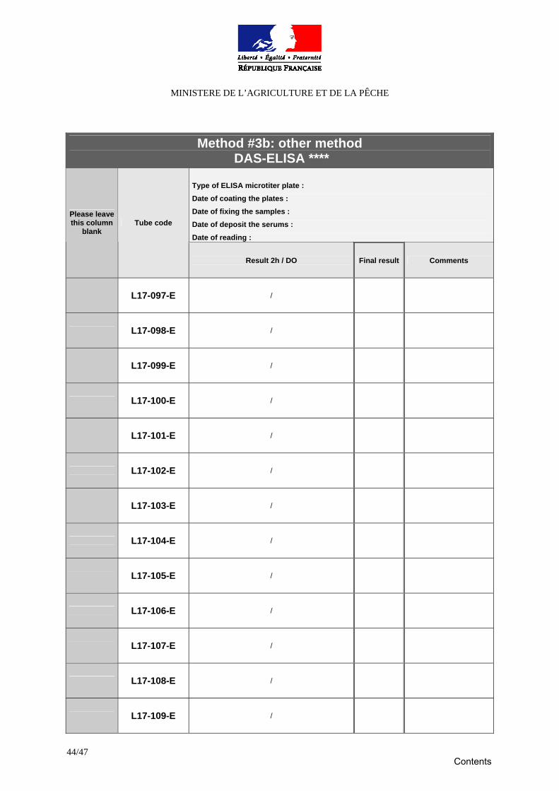
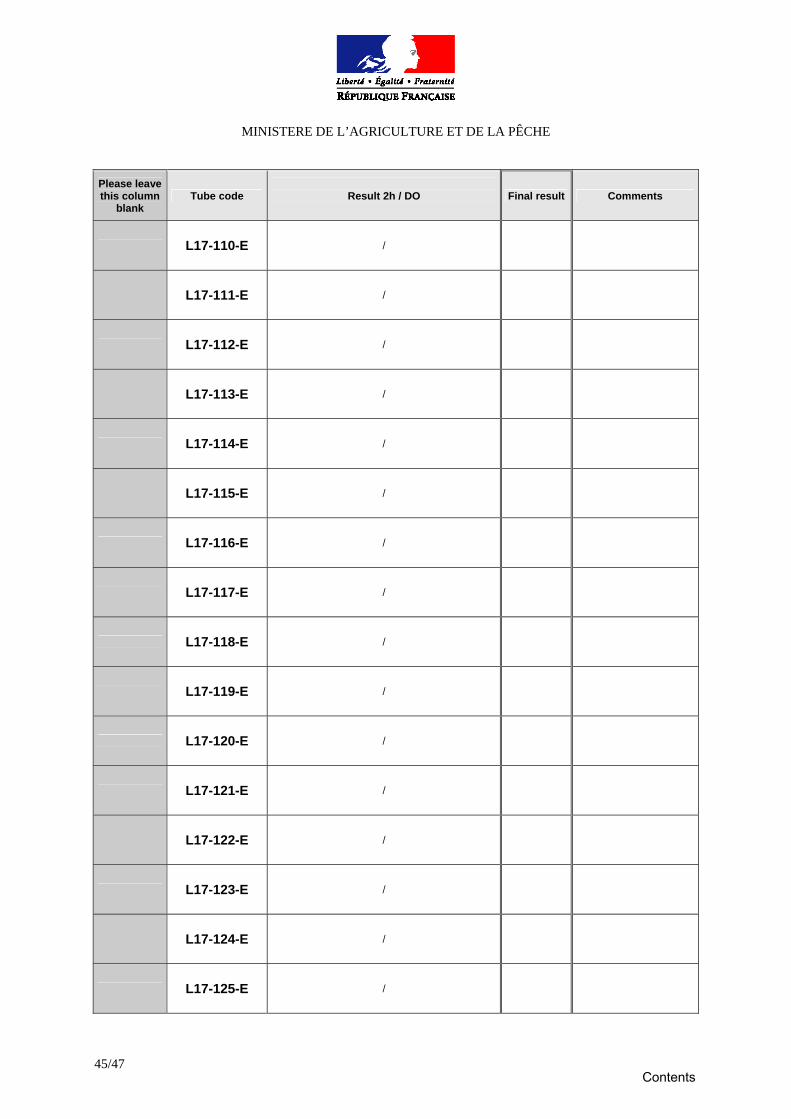
Method #3b: other method
DAS-ELISA **** Type of ELISA microtiter plate : Date of coating the plates : Date of fixing the samples : Date of deposit the serums :
Date of reading :
Please leave this column
blank Tube code
Result 2h / DO
Final result Comments
L17-097-E /
L17-098-E /
L17-099-E /
L17-100-E /
L17-101-E /
L17-102-E /
L17-103-E /
L17-104-E /
L17-105-E /
L17-106-E /
L17-107-E /
L17-108-E /
L17-109-E /
Contents
MINISTERE DE L’AGRICULTURE ET DE LA PÊCHE
45/47
Please leave this column
blank Tube code
Result 2h / DO
Final result Comments
L17-110-E /
L17-111-E /
L17-112-E /
L17-113-E /
L17-114-E /
L17-115-E /
L17-116-E /
L17-117-E /
L17-118-E /
L17-119-E /
L17-120-E /
L17-121-E /
L17-122-E /
L17-123-E /
L17-124-E /
L17-125-E /
Contents
MINISTERE DE L’AGRICULTURE ET DE LA PÊCHE
46/47
Please leave this column
blank Tube code
Result 2h / DO
Final result Comments
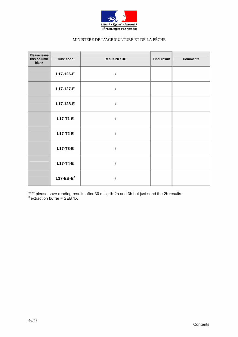
L17-126-E /
L17-127-E /
L17-128-E /
L17-T1-E /
L17-T2-E /
L17-T3-E /
L17-T4-E /
L17-EB-E¥ /
**** please save reading results after 30 min, 1h 2h and 3h but just send the 2h results. ¥ extraction buffer = SEB 1X
Contents

MINISTERE DE L’AGRICULTURE ET DE LA PÊCHE
47/47
we have to report the following additional troubleshooting :
Contents