interaction between bacteria, nannobacteria, and mineral ... · nannobacteria, and mineral...
TRANSCRIPT
Érudit est un consortium interuniversitaire sans but lucratif composé de l'Université de Montréal, l'Université Laval et l'Université du Québec à
Montréal. Il a pour mission la promotion et la valorisation de la recherche. Érudit offre des services d'édition numérique de documents
scientifiques depuis 1998.
Pour communiquer avec les responsables d'Érudit : [email protected]
Article
"Interaction Between Bacteria, Nannobacteria, and Mineral Precipitation in Hot Springs ofCentral Italy"
Robert L. FolkGéographie physique et Quaternaire, vol. 48, n° 3, 1994, p. 233-246.
Pour citer cet article, utiliser l'information suivante :
URI: http://id.erudit.org/iderudit/033005ar
DOI: 10.7202/033005ar
Note : les règles d'écriture des références bibliographiques peuvent varier selon les différents domaines du savoir.
Ce document est protégé par la loi sur le droit d'auteur. L'utilisation des services d'Érudit (y compris la reproduction) est assujettie à sa politique
d'utilisation que vous pouvez consulter à l'URI https://apropos.erudit.org/fr/usagers/politique-dutilisation/
Document téléchargé le 12 février 2017 06:54
Géographie physique et Quaternaire, 1994, vol. 48, n" 3, p. 233-246, 24 fig.
INTERACTION BETWEEN BACTERIA, NANNOBACTERIA, AND MINERAL PRECIPITATION IN HOT SPRINGS OF CENTRAL ITALY Robert L. FOLK, Department of Geological Sciences and Bureau of Economic Geology, University of Texas at Austin, Austin,
Texas 78712-1101, U.S.A.
ABSTRACT A complex of inorganic and organic factors controls precipitation of carbonates in hot springs of Lazio, central Italy. A plot of data from this area shows that the main /norganic controls are temperature and Mg/Ca ratio of the spring waters. Virtually all springs with waters hotter than 4O0C precipitate aragonite, and cooler ones form calcite. Furthermore, even cold-water springs precipitate aragonite if the Mg/Ca ratio exceeds 1:1, except in two cases. To what extent is the precipitation of travertine inorganic vs. biochemical? Surely, conditions in diverse localities can vary between both end-points, but Le Zitelle springs, at the north flank of the caldera of Viterbo, provide a biochemical extreme. Waters are hot (600C), with Mg/Ca of .2, and are highly sulfurous. Carbonate precipitation rates can exceed 2 mm/day. /Vonetched samples of carbonate crusts, only minutes to a few hours old, exhibit aragonite, calcite, and 1- to 5- (im euhedral rhombs of probable dolomite. Aragonite forms spherical "pincushions" of radial needles, each needle tipped with a nannobacte-rial body of the same diameter as the needles, 0.1 to 0.4 jim. Each nannobacterium precipitated its own needle, and was propelled outward by needle growth. As little or no later "fattening" of the needle occurred, inorganic precipitation must have been insignificant here. fVonetched calcite crystals are composed of 0.05 (xm nannobacterial spheres that were incorporated into each layer of the crystal as it grew. No evidence of bacteria was found on the ?dolomite rhomb surfaces. Ironically, aragonite, calcite, and euhedral ?dolomite rhombs all grew within minutes to an hour of each other in the same solution under the same conditions, savaging all the rules exposed at the beginning they remain a baffling problem unresolved by chemistry, physics, or microbiology.
RÉSUMÉ L'interaction entre les bactéries, les nannobactéries et la précipitation des minéraux dans les sources chaudes du centre de l'Italie. Un ensemble de facteurs inorganiques et organiques sont à l'origine de la précipitation de carbonates dans les sources chaudes de Lazio. L'étude des données montre que les facteurs inorganiques principaux sont la température et le rapport Mg/Ca dans les eaux de source. Presque toutes les sources chaudes à plus de 400C font précipiter l'aragonite et les eaux plus froides, la calcite. De plus, même les sources froides font précipiter l'aragonite si le rapport Mg/Ca dépasse 1:1, sauf exceptions. Dans quelle mesure la précipitation du travertin est-elle inorganique plutôt que biochimique? Certes, les conditions varient d'un milieu à l'autre entre les deux extrémités, mais la source Le Zitelle constitue un extrême biochimique. Les eaux sont chaudes (60°C), avec un rapport de Mg/Ca de 0,2, et très sulfureuses. Les taux de précipitation de carbonates peuvent dépasser 2 mm/jour. Les échantillons de croûtes de carbonate non corrodées (par l'acide hydrochlorique), formées dans les minutes ou les heures précédentes, présentent de l'aragonite, de la calcite et des rhomboèdres automorphes de 1 à 5 fim probablement de dolomite. L'aragonite forme des « pelotes >• sphériques, d'aiguilles radiales, dont chacun des bouts se termine par un corps nannobactériel de même diamètre, soit 0,1 à 0,4 p.m, Des cristaux non corrodés de calcite se composent de sphères de nannobactérie de 0,05 y.m incorporées dans chacune des couches de cristal au fur et à mesure du développement. Aucun signe de bactérie n'a été trouvé sur les surfaces de rhomboèdres de dolomite (?). Ironiquement, l'aragonite, la calcite et les rhomboèdres de dolomite (?) ont tous cru à peu près en même temps, dans la même solution soumise aux mêmes conditions, anéantissant ainsi toutes les règles déjà émises. Le problème persiste donc et n'a été résolu ni par la chimie, la physique ou la microbiologie.
ZUSAMMENFASSUNG Wechselbeziehung zwischen Bakterien, Nannobakterien und mineralischer Ausscheidung in heiBen Quellen von Zentralitalien. Ein vielschichti-ges Zusammenspiel anorganischer und organischer Faktoren regelt die Ausscheidung von Karbonaten in den heiBen Quellen von Lazio, Zentralitalien. Daten von diesem Gebiet zeigen, daB die Temperatur und das Verhàltnis Mg/Ca in den Quell-wassern die hauptsâchlichen anorganischen Faktoren sind. Praktisch scheiden aile Quellen mit Wasser, das heiBer als 4O0C ist, Aragonit aus, wenn das Verhàltnis Mg/Ca 1:1 ùberschreitet, auBer in zwei Fallen. In welchem MaBe ist die Ausscheidung von Kalktuff eher anorganisch als biochemisch? Sicher kônnen die Bedingungen an verschie-denen Plâtzen zwischen den zwei End-punkten variieren, aber die Quellen Le Zitelle an der Nordflanke der caldera von Viterbo stellen ein biochemisches Extrem dar. Die Wasser sind heiB (6O0C) mit einem Mg/Ca Verhàltnis von 2 und sie sind sehr schwefel-haltig. Die Karbonat-Ausscheidungsrate kann 2 mm/Tag ùberschreiten. Proben nich-tausgewaschener Karbonatkrusten, die nur Minuten bis zu wenige Stunden ait sind, zeigen Aragonit, Kalkspat und automorphe Rhomben von 1 bis 5 ^m, wahrscheinlich von Dolomit. Der Aragonit bildet kugelfôr-mige "Nadelkissen" mit strahlenfôrmigen Nadeln, die in einem nannobakteriellen Kôrper vom selben Durchmesser wie die Nadel enden, 0.1 bis 0.4 ^m. Die nichausge-waschenen Kalkspatkristalle bestehen aus kugelfôrmigen Nannobakterien von 0.05jj.m, welche in jede Schicht des Kristalls wàhrend seines Wuchses aufgenommen wurden. Auf den Dolomit(?)-Rhombenoberflàchen wurde kein Hinweis auf Bakterien gefunden. SeIt-samerweise wuchsen Aragonit, Kalkspat und Dolomit(?)-Rhomben aile in einem Abstand von Minuten bis zu einer Stunde voneinander in derselben Losung unter den-selben Bedingungen und haben so sâmtli-che Regeln des ersten Absatzes umges-toBen; sie bleiben ein durch Chemie, Physik oder Mikrobiologie ungelôstes Problem.
Manuscrit reçu le 25 mai 1993; manuscrit révisé accepté le 7 janvier 1994
234 R.L FOLK
INTRODUCTION
Volumetrically, travertines deposited by hot springs form an extremely minuscule fraction of the earth's crust, previously of interest only to a few hydrologists and geochemists who liked "way-out" environments. Pursell (1985) made the first pétrographie examination of hot-water aragonitic travertines when she investigated Mammoth Hot Springs terraces at Yellowstone, Wyoming. During literature research, she came across a reference by Malesani and Vannucci (1975) listing geochemistry of various springs in central Italy; in their table they mentioned the hot springs at Viterbo, and presented X-ray evidence of aragonite (no petrography was done). In order to investigate another aragonite-precipitating spring, in 1988 work was begun at Viterbo; the research has become a triumph in inadvertency because these springs had excellently preserved bacteria and introduced me to the minute but very important world of the dwarfed forms, "ultrami-crobacteria" or nannobacteria, averaging around 0.1 |im (Folk, 1990,1992,1993). These are about 1/10 the diameter of ordinary bacteria and have not been previously recognized in sediments or minerals, usually not seen at all (because they require superb resolution at 35,000X) or passed over as "artifacts" or "contamination."
GEOLOGICAL SETTING OF THE VITERBO HOT SPRINGS
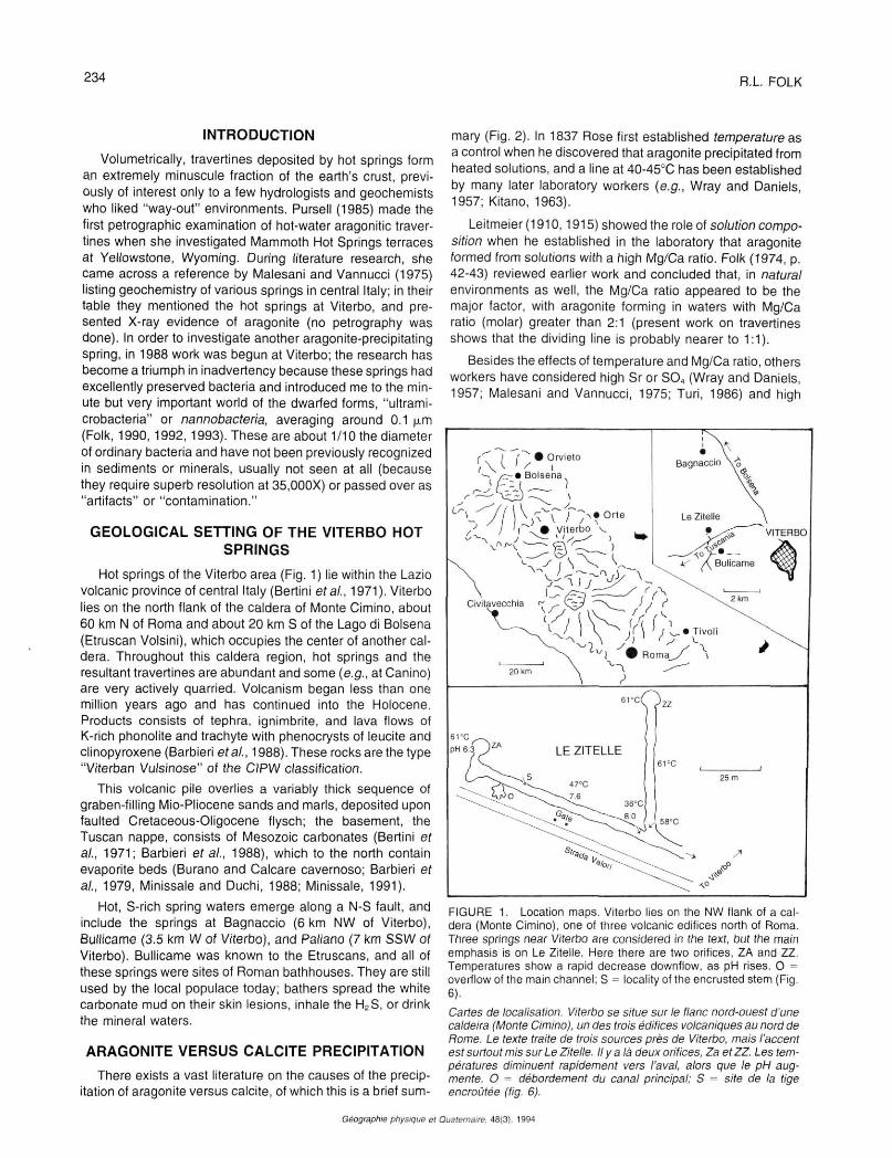
Hot springs of the Viterbo area (Fig. 1) lie within the Lazio volcanic province of central Italy (Bertini et al., 1971). Viterbo lies on the north flank of the caldera of Monte Cimino, about 60 km N of Roma and about 20 km S of the Lago di Bolsena (Etruscan Volsini), which occupies the center of another caldera. Throughout this caldera region, hot springs and the resultant travertines are abundant and some {e.g., at Canino) are very actively quarried. Volcanism began less than one million years ago and has continued into the Holocene. Products consists of tephra, ignimbrite, and lava flows of K-rich phonolite and trachyte with phenocrysts of leucite and clinopyroxene (Barbieri era/., 1988). These rocks are the type "Viterban Vulsinose" of the CIPW classification.
This volcanic pile overlies a variably thick sequence of graben-filling Mio-Pliocene sands and marls, deposited upon faulted Cretaceous-Oligocene flysch; the basement, the Tuscan nappe, consists of Mesozoic carbonates (Bertini et al., 1971; Barbieri et al., 1988), which to the north contain evaporite beds (Burano and Calcare cavemoso; Barbieri et al., 1979, Minissale and Duchi, 1988; Minissale, 1991).
Hot, S-rich spring waters emerge along a N-S fault, and include the springs at Bagnaccio (6 km NW of Viterbo), Bullicame (3.5 km W of Viterbo), and Paliano (7 km SSW of Viterbo). Bullicame was known to the Etruscans, and all of these springs were sites of Roman bathhouses. They are still used by the local populace today; bathers spread the white carbonate mud on their skin lesions, inhale the H2S, or drink the mineral waters.
ARAGONITE VERSUS CALCITE PRECIPITATION
There exists a vast literature on the causes of the precipitation of aragonite versus calcite, of which this is a brief sum
mary (Fig. 2). In 1837 Rose first established temperature as a control when he discovered that aragonite precipitated from heated solutions, and a line at 40-45'C has been established by many later laboratory workers (e.g., Wray and Daniels, 1957; Kitano, 1963).
Leitmeier (1910,1915) showed the role of solution composition when he established in the laboratory that aragonite formed from solutions with a high Mg/Ca ratio. Folk (1974, p. 42-43) reviewed earlier work and concluded that, in natural environments as well, the Mg/Ca ratio appeared to be the major factor, with aragonite forming in waters with Mg/Ca ratio (molar) greater than 2:1 (present work on travertines shows that the dividing line is probably nearer to 1:1).
Besides the effects of temperature and Mg/Ca ratio, others workers have considered high Sr or SO4 (Wray and Daniels, 1957; Malesani and Vannucci, 1975; Turi, 1986) and high
FIGURE 1. Location maps. Viterbo lies on the NW flank of a caldera (Monte Cimino), one of three volcanic edifices north of Roma. Three springs near Viterbo are considered in the text, but the main emphasis is on Le Zitelle. Here there are two orifices, ZA and ZZ. Temperatures show a rapid decrease downflow. as pH rises. O = overflow of the main channel; S = locality of the encrusted stem (Fig. 6).
Cartes de localisation. Viterbo se situe sur le flanc nord-ouest d'une caldeira (Monte Cimino), un des trois édifices volcaniques au nord de Rome. Le texte traite de trois sources près de Viterbo, mais l'accent est surtout mis sur Le Zitelle. Il y a là deux orifices, Za et ZZ. Les températures diminuent rapidement vers l'aval, alors que le pH augmente. O = débordement du canal principal; S = site de la tige encroûtée (fig. 6).
Géographie physique et Quaternaire. 48(3), 1994
INTERACTION BETWEEN BACTERIA, NANNOBACTERIA, AND MINERAL PRECIPITATION 235
pC02 or high precipitation rate (Duchi, Giordano and Martini, 1978; Given and Wilkinson, 1985). Organics In solution have also been considered (Kitano and Hood, 1965).
I now restrict the discussion to carbonates deposited rather rapidly (say, 1 mm to 1 m per year) under surface conditions — this includes spring travertines, cave deposits, stream and lake tufas, etc. These deposits seem mainly to follow the same temperature and Mg/Ca rules first established by Rose and Leitmeier. Only under extraordinarily rapid precipitation such as inner sides of bubble walls (Chafetz, Rush and Utech, 1991), crystal rafts at the water/air interface or in areas of high local turbulence (Kitano, 1963) does aragonite form in cool, Ca-rich waters. And travertines of Tivoli, Lazio, Italy, with growth rates ranging from a few millimeters to a meter per year, are calcite despite a very rapid precipitation rate (Chafetz and Folk, 1984; Folk, Chafetz and Tiezzi, 1985). From this discussion, I exclude subsurface cements, which are calcite instead of aragonite because of the slow deposition rate and low Mg/Ca ratio, and in spite of the high subsurface temperatures.
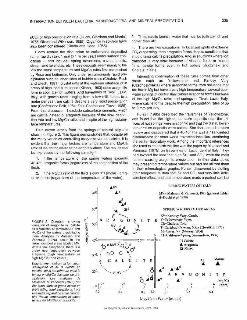
Data drawn largely from the springs of central Italy are shown in Figure 2. This figure demonstrates that, despite all the many variables controlling aragonite versus calcite, it is evident that the major factors are temperature and Mg/Ca ratio of the spring water at the earth's surface. The results can be expressed by the following paradigm:
1. If the temperature of the spring waters exceeds 40-45°, aragonite forms (regardless of the composition of the fluid).
2. If the Mg/Ca ratio of the fluid is over 1:1 (molar), aragonite forms (regardless of the temperature of the water).
3. Thus, calcite forms in water that must be both Ca-rich and cooler than 40°.
4. There are two exceptions. In localized spots of extreme CO2 outgassing, then aragonite forms despite conditions that should favor calcite precipitation. And, in situations where ion transport is very slow because of viscous fluids or mucus films, calcite forms even in hot waters (Buczynski and Chafetz, 1991).
Interesting confirmation of these rules comes from other areas such as Yellowstone and Karlovy Vary (Czechoslovakia) where aragonite forms from solutions that are low in Mg but have a very high temperature; several cool-water springs of central Italy, where aragonite forms because of the high Mg/Ca ratio; and springs of Tivoli, Lazio, Italy, where calcite forms despite the high precipitation rates of up to 3 mm per day.
Pursell (1985) described the travertines of Yellowstone, and found that the high-temperature deposits near the orifices of hot springs were aragonitic and that the distal, lower-temperature deposits were calcite. She then did a literature review and discovered that a 40-45° line was a near-perfect discriminator for other world travertine localities, confirming the earlier laboratory work. Among the important references she used to establish this line was the paper by Malesani and Vannucci (1975) on travertines of Lazio, central Italy. They had favored the idea that high Sr^ and SO/ were the main factors causing aragonite precipitation; in their data tables they presented temperature values but had not utilized them in their mineralogical graphs. Pursell discovered by plotting their temperature data that Sr and SO4 had very little independent effect, and that temperature made a perfect split but
FIGURE 2. Diagram showing formation of aragonite vs. calcite as a function of temperature and Mg/Ca of the waters precipitating them. Analyses by Malesani and Vannucci (1975) occur in the larger rounded areas labeled MV. With a few exceptions, there is a pretty neat separation between aragonite (high temperature or high Mg/Ca) and calcite.
Diagramme montrant la formation d'aragonite et de la calcite en fonction de la température et de la teneur en Mg/Ca des eaux de précipitation. Les analyses de Malesant et Vannucci (1975) ont été faites dans le grand cercle en tireté (MV). Sauf exceptions, il y a une nette séparation entre !'aragonite (haute température et haute teneur en Mg/Ca) et la calcite.
70°
30^
20^-
^ K V
AY
/ ChO O \
) C A L C I T E
-otJW-
ioc .125 l
.25 ô
M O
d ,4
.5
SPRING WATERS OF ITALY
MV= Malesani & Vannucci, 1975 (general fields) d=Duchi et al. 1978)
SPRING WATERS, OTHER AREAS
KV=Karlovy Vary, Czech. Y=Yellowstone, Wyo. Ch=Chafetz, Colo. T=Carlsbad Caverns, NMx (Thrailkill, 1971) M=Caves, Va. (Murray, 1954) Cf=CaIifornia Spring (Amundson, 1987)
O Calcite A Aragonite Qj Mixed
Cf •
Marine
A A" AdAd
A R A G O N I T E
1 Mg/Ca (ppm)
0.2 0.4 0.8 1.0 1.6 3.2
Mg/Ca in Water (molar)
6.4
Géographie physique el Quaternaire. 48(3). 1994
236 R.L FOLK
for two exceptions (which we can now in 1993 explain). For example, aragonite forms at Yellowstone because of the high temperature, despite very low Sr" and S04
=,.
Other central Italian travertines were studied by Duchi, Giordano and Martini (1978); they contradicted the Sr and SO4 idea of Malesani and Vannucci, and favored the idea of high temperature and pC02. However, I replotted their data in a graph showing temperature versus Mg/Ca ratio, and found the same clear distinction seen before between calcite and aragonite fields. If pC02 has any effect, it is not evident from their data.
At Le Zitelle, however, there are significant and still puzzling exceptions to these general rules, which will be discussed in the following sections.
VITERBO: LE ZITELLE
This paper will discuss the carbonates at two springs in a locality known as "Le Zitelle" (the Spinsters), about 3 km W of Viterbo (Fig. 1). They lie across the paved highway (Strada Provinciale Tuscanese) from the more famous Bullicame springs, where there is a monument to Dante1. A dirt road called "Strada Valori" trends N70W for a few hundred meters to a stone gatepost marked "Le Zitelle" near a large ruined stone house called "Casale Le Zitelle". I visited there first in May 1992 with Allan Pentecost, Henry S. Chafetz, and Rachel Eustice. In June 1992, I made a second visit with Sydney and Gary Hemming. In June 1993, Emma T. Rasbury and I planted an experimental pipe there to evaluate photo-synthetic vs. inorganic precipitation rates (see addendum).
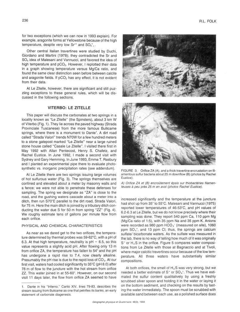
At Le Zitelle there are two springs issuing large volumes of hot sulfurous water (Fig. 3). The springs themselves are confined and elevated about a meter by masonry walls and a fence; we were not able to penetrate these defenses for sampling. The spring we designate as "ZA" is close to the road, and the gushing waters cascade about a meter into a ditch, then run S70°E parallel to the dirt road, Strada Valori, for 75 m. Here the main ditch is joined by a tributary ditch conducting the water due S for 50 m from spring "ZZ" (Fig. 4). We roughly estimate tens of gallons per minute flow from each orifice.
PHYSICAL AND CHEMICAL CHARACTERISTICS
As near as we dared get to the two orifices, the temperature determined by thermal probes was 59-62°C, with a pH of 6.3. At that high temperature, neutrality is pH = 6.5, so this value represents a slightly acid pH. After flowing only 13 m from orifice ZA, the temperature has fallen to 54° and the pH has undergone a rapid rise to 7.4, now clearly alkaline. Presumably the pH rise is due to the rapid loss of CO2. At our first visit, waters had cooled significantly to 350C (pH 8.0) after 78 m of flow to the juncture with the hot stream from orifice ZZ. This water joined in at 55-60°. However, on our second visit 11 days later, the flow from orifice ZA seemed to have
1. Dante in his "Inferno," Canto XIV, lines 79-83, describes the stream issuing from Bulicame as one that petrifies its banks, an early statement of carbonate diagenesis.
FIGURE 3. Orifice ZA (A), and a thick travertine encrustation on filamentous sulfur bacteria about 25 m downflow (B) (photos by Rachel Eustice).
A) Orifice ZA et (B) encroûtement épais sur thiobactéries filamenteuses à peu près 25 m en aval (photos Rachel Eustice).
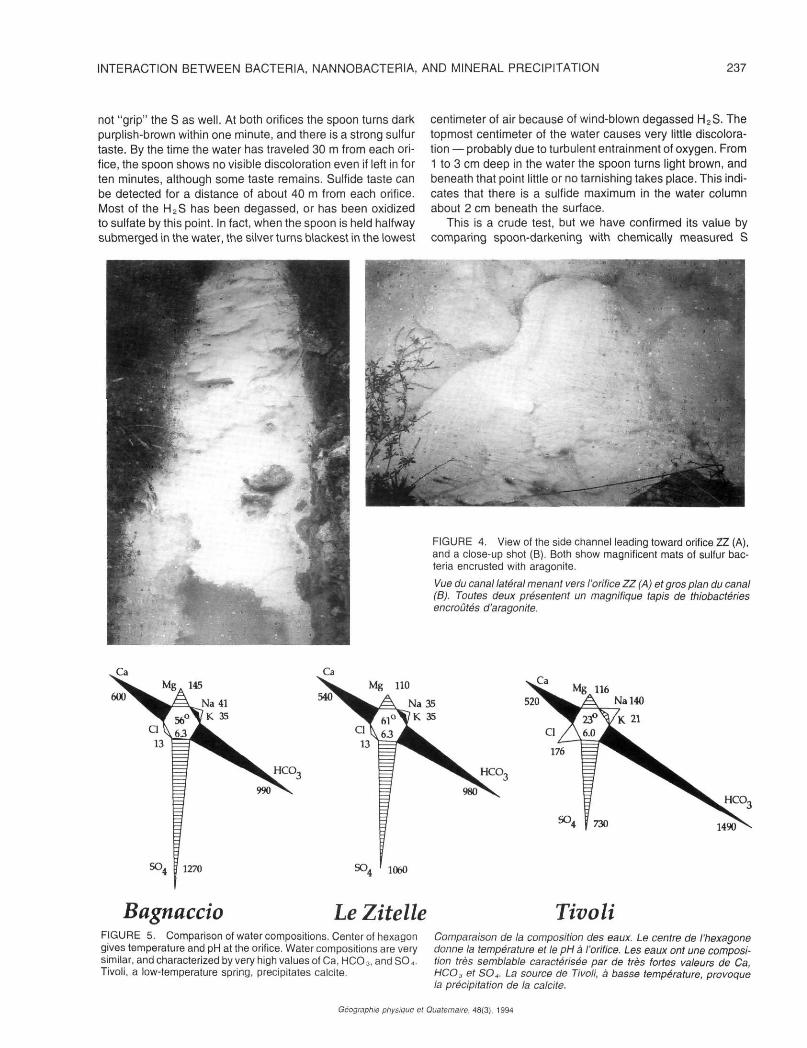
increased significantly and the temperature at the juncture had shot up from 35° to 55°C. Malesani and Vannucci (1975) reported lower temperatures of 46-530C, and pH values of 6.2-6.3 at Le Zitelle, but we do not know precisely where their sampling was done. They report 540 ppm Ca, 110 ppm Mg (Mg/Ca ratio of 1:5), with 35 ppm Na and 35 ppm K. Anions were recorded as 980 ppm HCO3" (measured on site), 1060 ppm S0 4
= , and 13 ppm Cl; thus, the springs are calcium sulfate/ bicarbonate waters. As the sulfate was measured in the lab, there is no way of telling how much of it was originally S= or H2S in the orifice. Figure 5 compares water compositions from Le Zitelle with those at Bagnaccio and at Tivoli, where major calcitic travertines occur because of the low temperature. All three waters have substantially similar compositions.
At both orifices, the smell of H2S was very strong, but we needed a better estimate of S= or S04
=. Thus we have estimated the sulfur content qualitatively by using a freshly scrubbed silver spoon and holding it in the water or laying it on the bottom sediment, and checking on the results by tasting the water immediately. The spoon must be scrubbed with available sand between each use, as a polished surface does
Géographie physique et Quaternaire. 48(3). 1994
INTERACTION BETWEEN BACTERIA, NANNOBACTERIA, AND MINERAL PRECIPITATION 237
not "grip" the S as well. At both orifices the spoon turns dark purplish-brown within one minute, and there is a strong sulfur taste. By the time the water has traveled 30 m from each orifice, the spoon shows no visible discoloration even if left in for ten minutes, although some taste remains. Sulfide taste can be detected for a distance of about 40 m from each orifice. Most of the H2S has been degassed, or has been oxidized to sulfate by this point. In fact, when the spoon is held halfway submerged in the water, the silver turns blackest in the lowest
centimeter of air because of wind-blown degassed H2S. The topmost centimeter of the water causes very little discoloration — probably due to turbulent entrainment of oxygen. From 1 to 3 cm deep in the water the spoon turns light brown, and beneath that point little or no tarnishing takes place. This indicates that there is a sulfide maximum in the water column about 2 cm beneath the surface.
This is a crude test, but we have confirmed its value by comparing spoon-darkening with chemically measured S
FIGURE 4. View of the side channel leading toward orifice ZZ (A), and a close-up shot (B). Both show magnificent mats of sulfur bacteria encrusted with aragonite.
Vue du canal latéral menant vers l'orifice ZZ (A) et gros plan du canal (B). Toutes deux présentent un magnifique tapis de thiobactéries encroûtés d'aragonite.
HCO-,
Bagnaccio Le Zitelle FIGURE 5. Comparison of water compositions. Center of hexagon gives temperature and pH at the orifice. Water compositions are very similar, and characterized by very high values of Ca, HCO3, and SO.,. Tivoli, a low-temperature spring, precipitates calcite.
Tivoli Comparaison de la composition des eaux. Le centre de l'hexagone donne la température et le pH à l'orifice. Les eaux ont une composition très semblable caractérisée par de très fortes valeurs de Ca, HCO3 et SO+ La source de Tivoli, à basse température, provoque la précipitation de la calcite.
Géographie physique et Quaternaire. 48(3). 1994
238 R.L. FOLK
tests done on site by A. Pentecost at Bagnaccio Springs (Viterbo). The spoon rapidly shows eloquent details of sulfur distribution; for example, it can be laid across two currents of water to see if one is more sulfurous than the other, or it can be stuck into the sediment to see S variation with depth.
As one might expect, a sequence of biota and minerals can be followed downstream as the water cools, becomes more alkaline, and loses sulfide. At the two Le Zitelle orifices, the water gushes forth and there is not much carbonate precipitated. Nor are visible algal or bacterial structures evident. The water may be too hot, or too sulfurous, or too acid for bacteria to thrive or for carbonate to be precipitated. One copper plate placed in the water near the orifice turned black and slimy in 24 hours, presumably changed to some copper sulfide, but grew no carbonate crust.
In the channel within a meter or so from the actual orifice, there begins a very heavy growth of carbonate-encrusted filamentous bacterial mat, the ?main component identified as Chloroflexus by A. Pentecost in the field. The mat in the side channel (Fig. 4) is pale salmon-colored, but that in the main channel is snow-white; H2S content is high, and carbonate precipitation, mainly as aragonite, is very rapid. Farther down-channel, the mat disappears and the material is mostly loose carbonate sediment, small shrubs, encrusted bubbles, and rafts of carbonate floating on the surface. There are some visible filamentous bacteria, but not dense enough to make mats. Areas of various shades of green occur, but these are probably micron-sized coccoid algae or bacteria. In this stretch of the channel, there is very little sulfide evident. Thus the filamentous mats must thrive best at some intermediate level of ratio between sulfur and oxygen. Still farther downstream, carbonate crystals encrust vegetation fallen into the stream, but there is little megascopic organic growth in the water itself. No animal life is visible at a hand-lens scale, except for bees taking on water and the occasional cooked and carbonate-coated snake fallen into the ditch.
At one point in the main channel (Fig. 1), the water spilled over the side of the channel and spread out to form an avulsion splay of water only a few millimeters deep, and relatively cool — 34° when we visited. Here there were abundant deposits of "ice-floe" carbonate rafts coating the water surface. This material is a mixture of aragonite and calcite crystals.
MINERAL PRECIPITATION
At Le Zitelle, carbonate precipitation is very rapid, up to 3 mm or more per day, quickly encrusting vegetation. It is so copious that the meter-deep ditch alongside the road fills up and the carbonate has to be bulldozed out at frequent intervals, so that the hot waters won't spill across the road and lith-ify it. Because of the high temperature, most of the precipitate takes the form of aragonite radial-needle spherulites, but there is also considerable calcite — which remains a puzzle — and occasional rhombohedra of probable dolomite. Chafetz and Lawrence (1994) has collected geochemical data on the waters and precipitation. Hordes of nannobacte-ria are entombed within the aragonite and calcite as has been found at the other Viterbo springs (Folk, 1990, 1992, 1993),
and evidence will be presented that these bacteria are the active precipitating agents, not simply incorporated passively as crystals grow.
RATE OF PRECIPITATION
To get a quantitative fix on precipitation rates, we placed three types of objects at the bottom of the ditch where the temperature was 51°. Chafetz put in a small copper plate, under the idea that this should poison growth of most of the bacteria and, at least at first, all the precipitation should be inorganic (see Chafetz et al., 1991). I placed a broken piece of ceramic tile in the same stream at the same time, a few centimeters away, to provide a neutral, non-poisoning surface. I also put the stem of a field weed in the same place at the same time, to provide a more organic substrate. We let these objects stay in overnight and collected them the next day after 22 hours had elapsed. The resulting encrustation was quite irregular, resembling a field of cabbage heads, but the copper plate accumulated about 0.2-0.3 mm, the ceramic tile 1 mm, and the plant stem 2 mm. Thus our prediction was confirmed; where organic material is the substrate, providing food for bacteria, the encrustation rate was about 5-10 times that of the copper plate where precipitation was largely inorganic. From this crude experiment, one could argue that most — perhaps over three-fourths — of the precipitation of CaCO3 at Le Zitelle is due to the metabolic activities of organisms, in this case bacteria. In all examples, there was a thicker encrustation on the upstream sides or at corners and edges, presumably because of more turbulence.
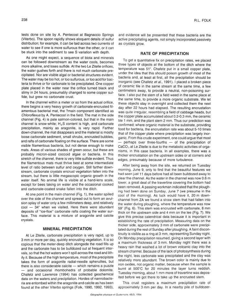
After being away from the site, we returned on Tuesday morning, June 9, only to find that the beautiful features we had seen just 11 days before had all been bulldozed away to clear the channel. As the water in the channel was now 0.6 m lower, a great deal of the travertine covering the bottom had been removed. A passing workman indicated that the ploughing had been done on Sunday, June 7 (we presume in the cool of the morning). As luck would have it, 20 m down-channel from ZA we found a straw stem that had fallen into the water during ploughing, where the temperature was now 59° (Fig. 6). This stem was encrusted with carbonate, 8 mm thick on the upstream side and 4 mm on the lee (Fig. 7). We give this precise calendrical data because it is important in establishing the rate of precipitation. Measuring data on the thicker side, approximately 3 mm of carbonate were precipitated during the rest of Sunday after ploughing. A faint discontinuity is visible as a ring at 3 mm, representing Sunday night. On Monday precipitation resumed, giving a second layer with a maximum thickness of 3 mm. Monday night there was a heavy rain that washed a lot of brown volcanic clay into the stream channel. Because of the lack of photosynthesis during the night, less carbonate was precipitated and the clay was relatively more abundant. The brown color is mainly due to iron oxides, not organic matter, because when the sample is burnt at 5000C for 20 minutes the layer turns reddish. Tuesday morning, about 1 mm more of travertine was deposited before we got there to take up the encrusted stem.
This crust registers a maximum precipitation rate of approximately 3 mm per day. In a nearby pile of bulldozer-
Géographie physique et Quaternaire. 48(3). 1994
INTERACTION BETWEEN BACTERIA, NANNOBACTERIA, AND MINERAL PRECIPITATION 239
FIGURE 6. Encrusted straw stem that fell into the water 2 1/2 days previously.
Tige de paille encroûtée immergée deux jours et demi auparavant.
dumped travertine, we found another encrusted stem registering 8 nightly bands in a 20 mm thickness, for an average rate of 2.5 mm/day. Such rates are not uncommon in travertines of other areas such as Tivoli (Italy), Yellowstone (Wyoming), and Japan (see references in Folk era/., 1985).
This lucky stem gave further insight into the process of carbonate precipitation. The changing thickness of the crust down-stem registers the changing rate of carbonate precipitation as a function of water depth (Fig. 6). At the water surface, the thickness was zero. At 1 cm depth, the crust diameter was 7 mm, and by 3 cm depth it had reached nearly its full diameter of 13 mm. At the maximum depth of 10.5 cm, the thickness was 15 mm. What does this mean? Had the precipitation been largely /norganic, one would have expected greatest precipitation rate at the water/air interface, where degassing of CO2 and turbulence would have been greatest. However, a better answer is provided by the silver spoon, which shows that sulfide content is low in the near-surface layers of the water, where admixing with oxygen occurs, and S is greater at a depth of several centimeters. We would propose that S-oxidizing bacteria thrive where the available S content is high; thus, maximum carbonate precipitation rate also occurs at that deeper level on the water column. Many other stems fallen in the water showed this same thickness profile. Again, this is a strong point in favor of bio-induced precipitation of carbonate at Le Zitelle.
FIGURE 7. Close-up of the encrusted stem. In the photocopied cross-section, the two dark night bands are indicated by arrows.
Gros plan de la tige encroûtée. Dans la photocopie de la coupe, les deux anneaux de formation nocturne sont identifiés par les flèches.
COMPOSITION OF THE PRECIPITATE
ARAGONITE
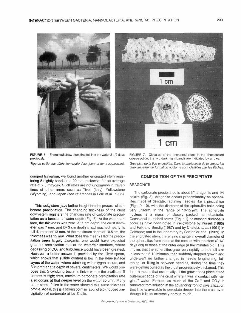
The carbonate precipitated is about 3/4 aragonite and 1/4 calcite (Fig. 8). Aragonite occurs predominantly as spheru-lites made of delicate, radiating needles like a pincushion (Figs. 9, 10), with the diameter of the spherulite balls being very uniform, in the range of 10-15 u.m. The spherulite nucleus is a mass of closely packed nannobacteria. Occasional dumbbell forms (Fig. 11) or crossed dumbbells occur as have been noted in Yellowstone by Pursell (1985) and Folk and Bendig (1987) and by Chafetz, et al. (1991) in Colorado; and in the laboratory by Castanier etal. (1989). In the encrusted stem, there is no change in overall diameter of the spherulites from those at the contact with the stem (2 1/2 days old) to those at the outer edge (a few minutes old). This implies that the spherulites grew very rapidly to their full size in less than 5-10 minutes, then suddenly stopped growth and underwent no further changes in needle lengthening, fattening, or filling-in between needles, during the time they were getting buried as the crust progressively thickened. This in turn means that essentially all the growth took place at the outermost edge of the crust where it was in contact with "virginal" water. Perhaps so much of the Ca++ and C03
= is removed from solution at the advancing front of crystallization that little is available to percolate deeper into the crust even though it is an extremely porous mush.
Géographie physique et Quaternaire. 48(3). 1994
240 R.L. FOLK
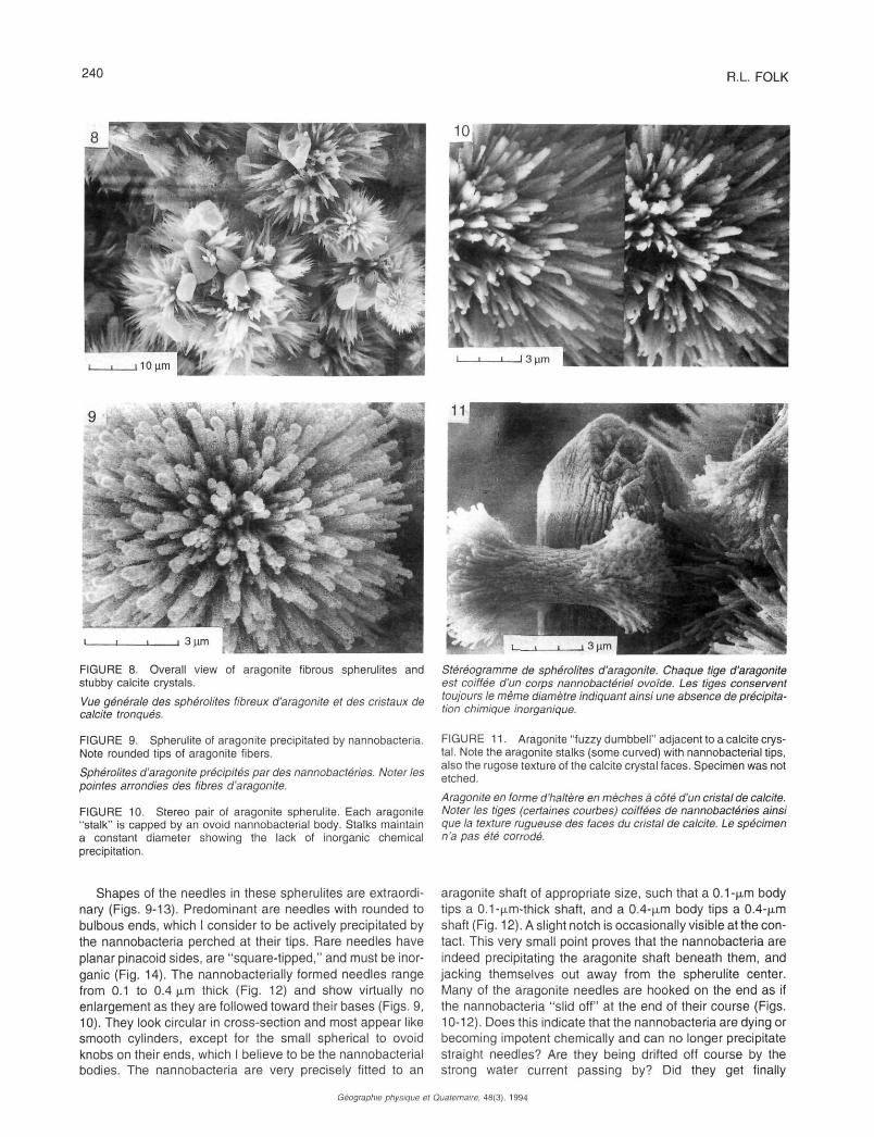
FIGURE 8. Overall view of aragonite fibrous spherulites and stubby calcite crystals.
Vue générale des sphérolites fibreux d'aragonite et des cristaux de calcite tronqués.
FIGURE 9. Spherulite of aragonite precipitated by nannobacteria. Note rounded tips of aragonite fibers.
Sphérolites d'aragonite précipités par des nannobactéries. Noter les pointes arrondies des fibres d'aragonite.
FIGURE 10. Stereo pair of aragonite spherulite. Each aragonite "stalk" is capped by an ovoid nannobacterial body. Stalks maintain a constant diameter showing the lack of inorganic chemical precipitation.
Shapes of the needles in these spherulites are extraordinary (Figs. 9-13). Predominant are needles with rounded to bulbous ends, which I consider to be actively precipitated by the nannobacteria perched at their tips. Rare needles have planar pinacoid sides, are "square-tipped," and must be inorganic (Fig. 14). The nannobacterially formed needles range from 0.1 to 0.4 u.m thick (Fig. 12) and show virtually no enlargement as they are followed toward their bases (Figs. 9, 10). They look circular in cross-section and most appear like smooth cylinders, except for the small spherical to ovoid knobs on their ends, which I believe to be the nannobacterial bodies. The nannobacteria are very precisely fitted to an
Stereogramme de sphérolites d'aragonite. Chaque tige d'aragonite est coiffée d'un corps nannobactériel ovoïde. Les tiges conservent toujours le même diamètre indiquant ainsi une absence de précipitation chimique inorganique.
FIGURE 11. Aragonite "fuzzy dumbbell" adjacent to a calcite crystal. Note the aragonite stalks (some curved) with nannobacterial tips, also the rugose texture of the calcite crystal faces. Specimen was not etched.
Aragonite en forme d'haltère en mèches à côté d'un cristal de calcite. Noter les tiges (certaines courbes) coiffées de nannobactéries ainsi que la texture rugueuse des faces du cristal de calcite. Le spécimen n'a pas été corrodé.
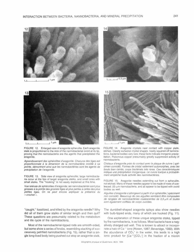
aragonite shaft of appropriate size, such that a 0.1-u,m body tips a 0.1-u.m-thick shaft, and a 0.4-u.m body tips a 0.4-u.m shaft (Fig. 12). A slight notch is occasionally visible at the contact. This very small point proves that the nannobacteria are indeed precipitating the aragonite shaft beneath them, and jacking themselves out away from the spherulite center. Many of the aragonite needles are hooked on the end as if the nannobacteria "slid off" at the end of their course (Figs. 10-12). Does this indicate that the nannobacteria are dying or becoming impotent chemically and can no longer precipitate straight needles? Are they being drifted off course by the strong water current passing by? Did they get finally
Géographie physique el Quaternaire. 48(3), 1994
INTERACTION BETWEEN BACTERIA, NANNOBACTERIA, AND MINERAL PRECIPITATION 241
FIGURE 12. Enlarged view of aragonite spherulite. Each aragonite stalk is proportioned to the size of the nannobacterial ovoid at its tip, proving that the nannobacteria are the agents that precipitated the aragonite.
Agrandissement des sphérolites d'aragonite. Chacune des tiges est proportionnnée à la dimension de la nannobactérie ovoïde à sa pointe, démontrant ainsi que les nannobactéhes sont les agents de précipitation de !'aragonite.
FIGURE 13. Side view of aragonite spherulite; large nannobacteria occur at the tips of larger aragonite stalks, and small ones with small stalks. The "hooking" is not easily explained at this time.
Vue latérale de sphérolites d'aragonite; les nannobactéhes sont plus grosses à la pointe des grosses tiges et plus petites à celles des plus petites tiges. On ne peut encore expliquer la présence du « crochet ».
"caught," fossilized, and killed by the aragonite needle? Why did all of them grow stalks of similar length and then quit? These questions are presumably related to the metabolism and life cycle of the nannobacteria.
Most of the nannobacterial-tipped rods are smooth-sided, but some show a series of knobs, resembling stacking of successively petrified nannobacteria (Fig. 15), rather than a single long-lived body being pushed out atop an aragonite stalk.
FIGURE 14. Aragonite crystals near contact with copper plate, etched. Clearly euhedral crystal shapes, neatly squared-off terminations, bacterial bodies very rare; these facts indicate inorganic precipitation. Poisonous copper presumably greatly suppressed activity of nannobacteria.
Cristaux d'aragonite près du contact avec Ia plaque de cuivre ( spécimen corrodé). Formes de cristal nettement automorphes, avec les bouts bien carrés, corps bactériels très rares. Ces caractéristiques indique une précipitation inorganique. Le cuivre toxique a probablement empêché toute activité des nannobactéhes.
FIGURE 15. Aragonite needles extending out from a spherulite, not etched. Many of these needles appear to be made of rows of coalesced .05'fim nannobacteria, and all appear to be tipped with ovoid bodies as well.
Aiguilles d'aragonite s'allongeant à partir d'un sphérolite ( spéciment non corrodé). Beaucoup de ces aiguilles semblent être composées de rangées de nannobactéhes coalescentes de 0,5\x.m et toutes sont également coiffées de corps ovoïdes.
The dumbbell-shaped aragonite splays also show needles with bulb-tipped ends, many of which are hooked (Fig. 11).
One explanation of these unique aragonite stalks, tipped by the nannobacteria, is as follows. Most bacteria have a negatively charged cell wall. This is known to attract or incorporate a halo of Ca " ions (Rosen, 1987; Beveridge, 1989). With the abundance of CO3" in the water, this leads to a high ionic product for [Ca **] [CO 3
=] in the fraction of a micron
Géographie physique et Quaternaire. 48(3), 1994
242 R.L FOLK
surrounding the body, and the supersaturated mineral will be forced to precipitate. Where will it most likely do this? On the aragonite stalk immediately below, which acts as a convenient seed crystal (Fig. 16). Thus, a thin layer of CaCO3 is added to the crystallographic lattice between the nannobac-terial body and the growing aragonite crystal. Now, the newly denuded portion of negative bacterial cell wall attracts more Ca*+ ions from the supersaturated fluid, and the process is repeated. The nannobacterium is acting like an assembly-line worker putting an automobile together; the Ca++ does not pass into or through the nannobacterium, but it acts only as a catalyst in forming aragonite. Thus, it is a Talmudic question whether the aragonite is an "organic" or "inorganic" construction, but I would contend that the automobile is just the same; it may be made of inorganic materials, but organisms put it together and, had the organisms not been there, the steel, copper, plastic, etc., would have gone elsewhere into something else; and had the nannobacterium not been there, the Ca++ and CO3= would have gone elsewhere and not into an aragonite spherulite.
If, indeed, the nannobacteria are only acting like assembly-line workers and not passing the Ca, C, and O through their bodies, there may well be no isotopic fractionation of the resulting precipitate. And because they must be capable of obtaining all the C, H, and O that they need for construction of body tissues out of the very oversaturated water, even if it were possible to analyze their "flesh" one probably would not get the low '3C values expected in more normal bacteria that feed on organic matter.
Analyzing the deposit for total organic biomass and from that calculating the relative importance of "organic" vs. "inorganic" precipitation would be fruitless here. It would be like making an overall chemical analysis of the total annual output of an automobile factory; the total "biomass" of the workers would be infinitesimal compared to the mass of cars they have produced.
What I consider to be the inorganic aragonite needles are relatively rare. I have found them near the base of the incrustation on Chafetz' copper plate (one guesses that the copper at first prevented organic activity). These needles are larger, longer, and wider, show good crystallographic terminations (a "squared off" end representing a pinacoid face), and side faces also are smooth and planar (Fig. 14). There is no evidence of the rounded or bulbous tips or hooking, nor are nannobacteria found on etching. Also, these needles widen as they near the center of the spherulite, so, here, sideways growth did continue but at a slower rate than the needle lengthened.
CALCITE
Calcite crystals form about 1/4 of the precipitate (Fig. 8). They are also quite uniform in size and shape, varying mainly from 7 to 11 n.m long by 3 to 7 (j.m wide, with a usual UW ratio of around 2. They are terminated by three unequally developed rhomb faces, which may be the unit rhomb (10T1), or more commonly the intermediate or acute rhombs 2021, or 4041 ). The body shape is almost circular or somewhat barrel-shaped, though some hint of hexagonal cross-section is sometimes visible.
• • + + O O
:&fifô
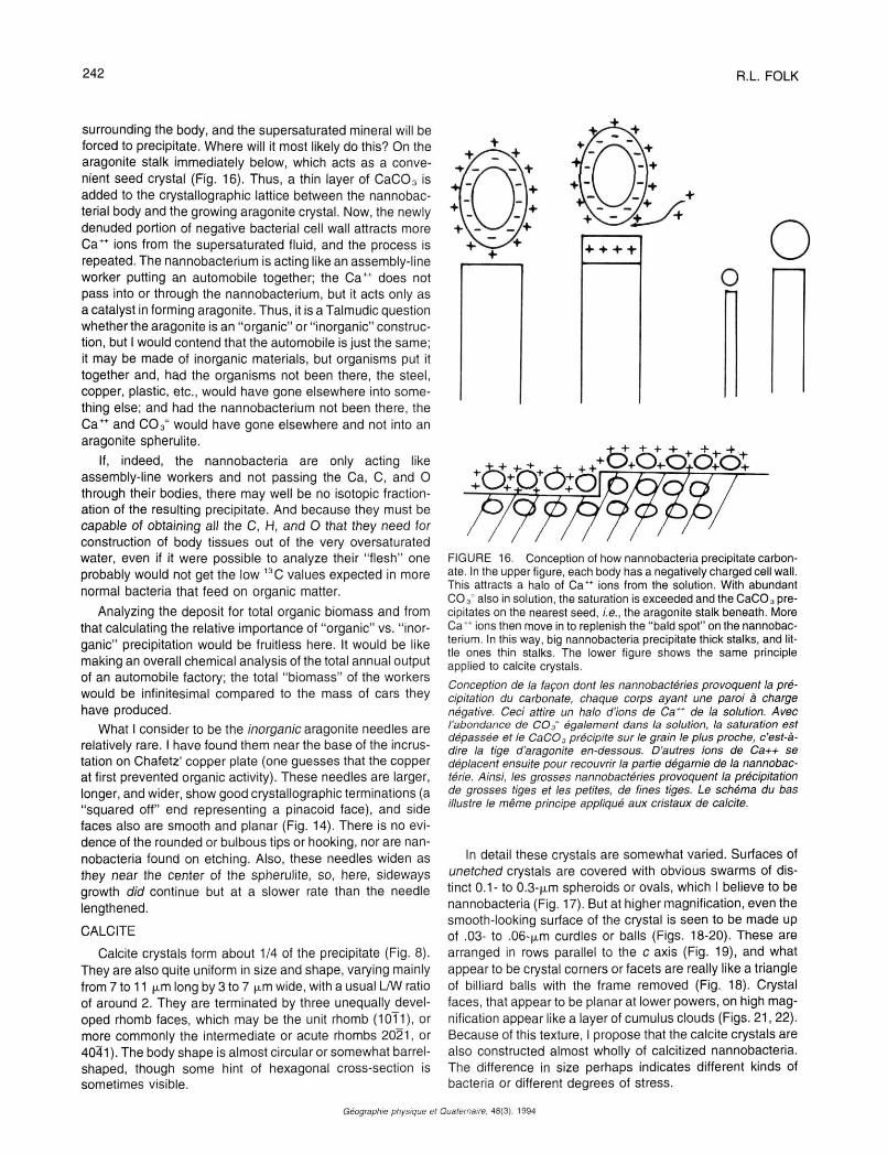
FIGURE 16. Conception of how nannobacteria precipitate carbonate. In the upper figure, each body has a negatively charged cell wall. This attracts a halo of C a " ions from the solution. With abundant CO3 ' also in solution, the saturation is exceeded and the CaCO3 precipitates on the nearest seed, i.e., the aragonite stalk beneath. More C a " ions then move in to replenish the "bald spot" on the nannobacterium. In this way, big nannobacteria precipitate thick stalks, and little ones thin stalks. The lower figure shows the same principle applied to calcite crystals.
Conception de la façon dont les nannobactéries provoquent la précipitation du carbonate, chaque corps ayant une paroi à charge négative. Ceci attire un halo d'ions de Ca" de la solution. Avec l'abondance de CO3' également dans la solution, la saturation est dépassée et le CaCO3 précipite sur le grain le plus proche, c'est-à-dire la tige d'aragonite en-dessous. D'autres ions de Ca++ se déplacent ensuite pour recouvrir la partie dégarnie de la nannobac-térie. Ainsi, les grosses nannobactéries provoquent la précipitation de grosses tiges et les petites, de fines tiges. Le schéma du bas illustre le même principe appliqué aux cristaux de calcite.
In detail these crystals are somewhat varied. Surfaces of unetched crystals are covered with obvious swarms of distinct 0.1- to 0.3-u.m spheroids or ovals, which I believe to be nannobacteria (Fig. 17). But at higher magnification, even the smooth-looking surface of the crystal is seen to be made up of .03- to .06-u.m curdles or balls (Figs. 18-20). These are arranged in rows parallel to the c axis (Fig. 19), and what appear to be crystal corners or facets are really like a triangle of billiard balls with the frame removed (Fig. 18). Crystal faces, that appear to be planar at lower powers, on high magnification appear like a layer of cumulus clouds (Figs. 21, 22). Because of this texture, I propose that the calcite crystals are also constructed almost wholly of calcitized nannobacteria. The difference in size perhaps indicates different kinds of bacteria or different degrees of stress.
Géographie physique et Quaternaire. 48(3). 1994
INTERACTION BETWEEN BACTERIA, NANNOBACTERIA, AND MINERAL PRECIPITATION 243
FIGURE 17. Encrusted stem. This calcite "crystal" appears to be made of solid masses of .05 ^m nannobacteria. Not etched. This crystal is at most a few hours old, as it occurs near the outside of the encrusted stem.
Tige encroûtée. Ce « cristal » de calcite semble être composé de masses compactes de nannobactéries de 0,5 \im (non corrodé). Ce cristal date d'à peine quelques heures, puisqu'il se trouve près de l'extérieur de la tige encroûtée.
FIGURE 18. Enlarged view at the termination of a calcite crystal, unetched.
Agrandissement de l'extrémité d'un cristal de calcite (non corrodé).
These calcite crystals show no sign of having enlarged after their original precipitation, which required only a few minutes. Those at the stem contact — 2 1/2 days old — look the same as those at the outer edge of the crust, a few minutes old.
Though essentially contemporaneous within a few minutes within any one layer, the calcite formed shortly after the aragonite spherulites as the calcite engulfs the radial needles, and I have not yet seen aragonite needles nucleating on the surfaces of calcite crystals. Of course, as the crust accretes outward, the youngest aragonite spherulites can fall down and come to lie atop calcite crystals formed a few minutes before on the underlying, slightly older layer of crust. But
FIGURE 19. View of the prism face of an unetched calcite crystal. Nannobacteria form rows parallel to the c axis.
Vue de Ia face d'un prisme d'un cristal de calcite non corrodé. Les nannobactéries forment des rangées parallèles à l'axe c.
FIGURE 20. Looking down upon the rhomb face of an unetched calcite crystal. This is an end-on view of the nannobacterial rows seen in Figure 19. Light streaks are due to charging.
Vue descendante sur la face rhomboèdre d'un cristal de calcite non corrodé. Il s'agit d'une vue des fins de rangées de la figure 19.
the calcite crystals do not form a nucleating substrate for aragonite needles.
EUHEDRAL RHOMBIC MINERAL — 7DOLOMITE
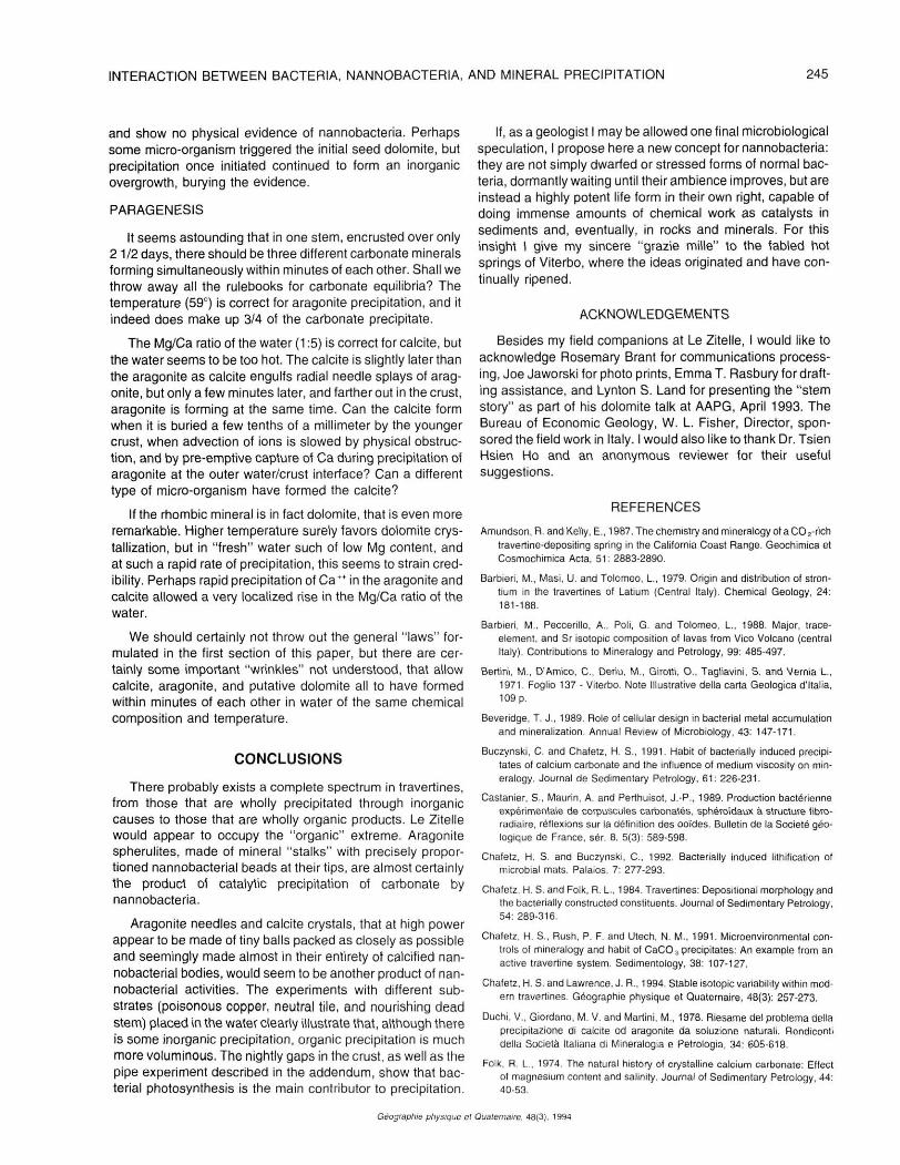
About two-thirds of the way out from stem/crust contact to the brownish Monday night layer, there occurs a zone where occasional, neatly rhombic euhedral crystals 3 to 6 u.m in diameter are found. I have not yet been able to establish their Mg content by EDAX, but believe them to be dolomite for the following reasons: (1) they have the simple elemental rhomb ( 10T1 ) crystal form typical of dolomite but uncommon in Viterbo calcite (Fig. 23); (2) all the known calcite in the Viterbo region hot springs — Bullicame, Bagnaccio, and Le Zitelle —
Géographie physique el Quaternaire. 48(3), 1994
244 R.L. FOLK
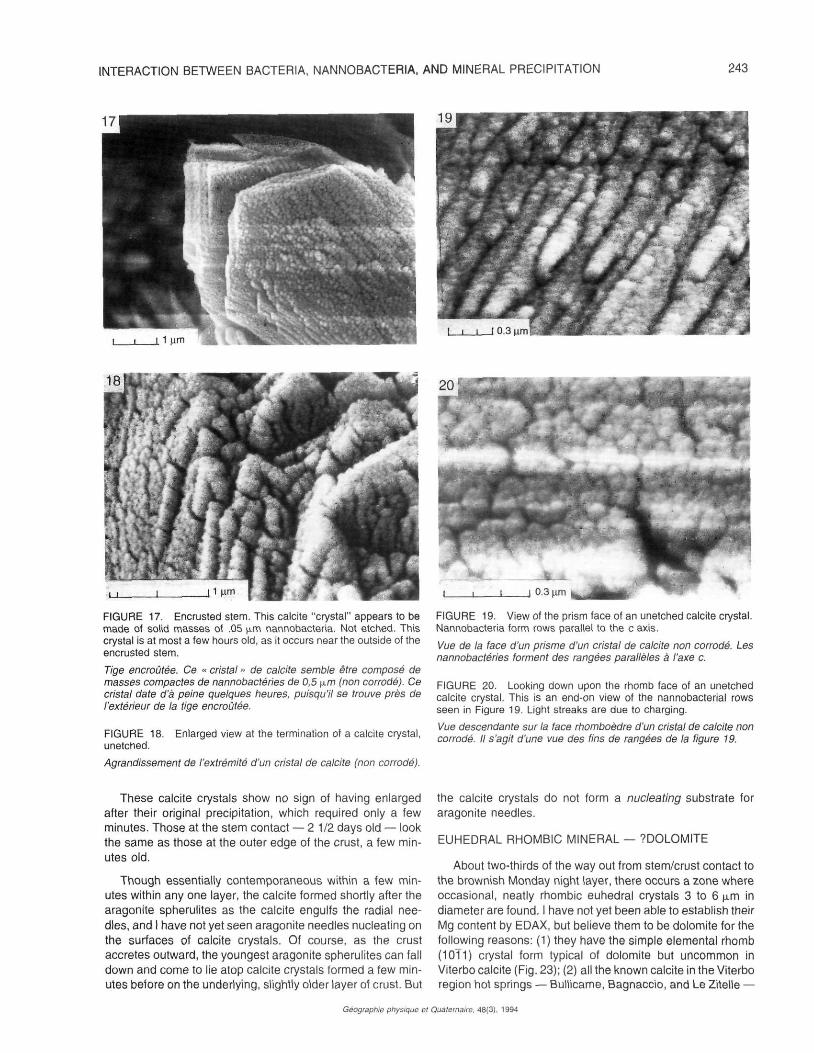
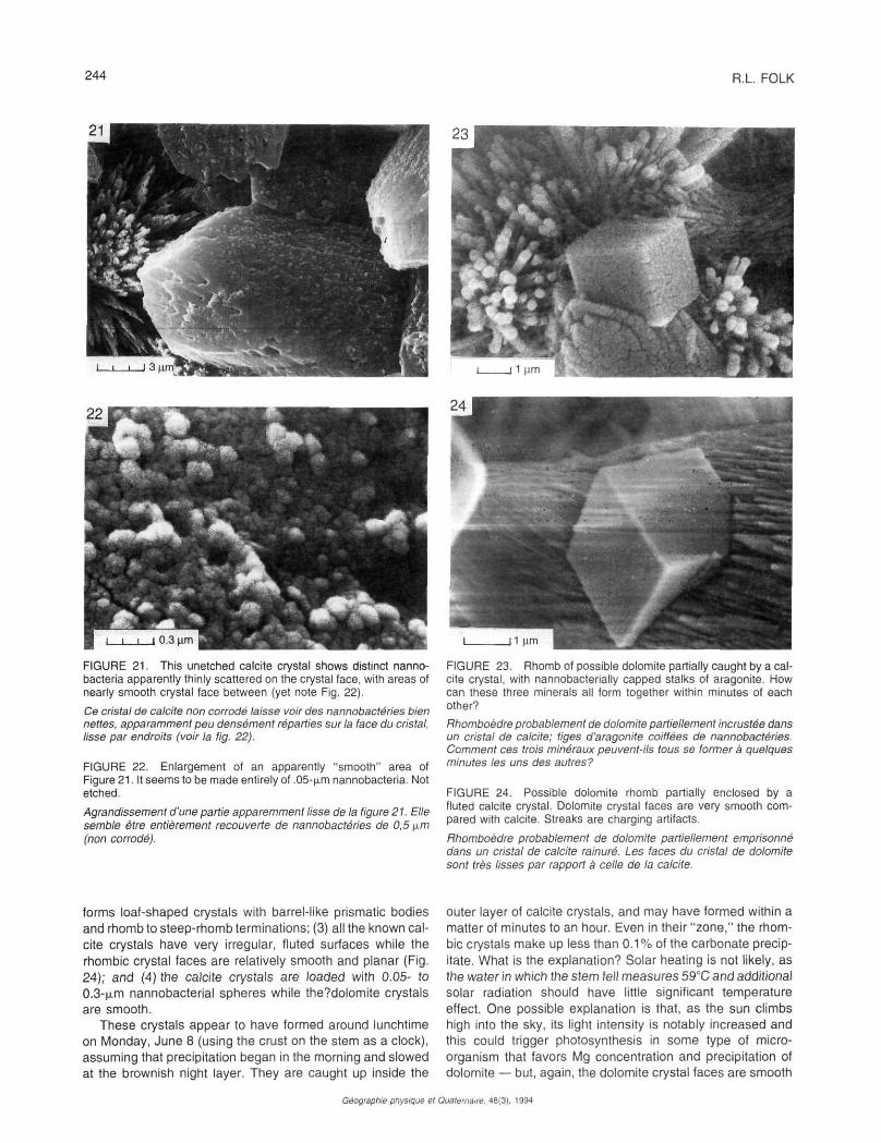
FIGURE 21. This unetched calcite crystal shows distinct nanno-bacteria apparently thinly scattered on the crystal face, with areas of nearly smooth crystal face between (yet note Fig. 22).
Ce cristal de calcite non corrodé laisse voir des nannobactéries bien nettes, apparamment peu densément réparties sur la face du cristal, lisse par endroits (voir la fig. 22).
FIGURE 22. Enlargement of an apparently "smooth" area of Figure 21. It seems to be made entirely of .05-^m nannobacteria. Not etched.
Agrandissement d'une partie apparemment lisse de la figure 21. Elle semble être entièrement recouverte de nannobactéries de 0,5 (xm (non corrodé).
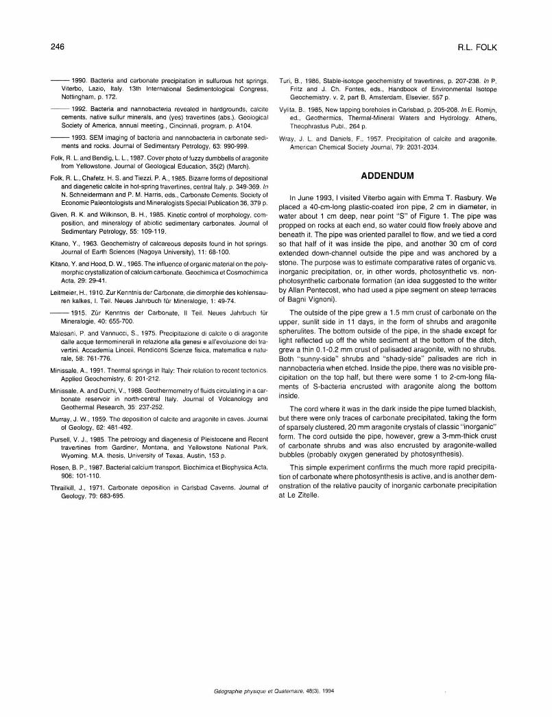
forms loaf-shaped crystals with barrel-like prismatic bodies and rhomb to steep-rhomb terminations; (3) all the known calcite crystals have very irregular, fluted surfaces while the rhombic crystal faces are relatively smooth and planar (Fig. 24); and (4) the calcite crystals are loaded with 0.05- to 0.3-u.m nannobacterial spheres while the?dolomite crystals are smooth.
These crystals appear to have formed around lunchtime on Monday, June 8 (using the crust on the stem as a clock), assuming that precipitation began in the morning and slowed at the brownish night layer. They are caught up inside the
FIGURE 23. Rhomb of possible dolomite partially caught by a calcite crystal, with nannobacterially capped stalks of aragonite. How can these three minerals all form together within minutes of each other?
Rhomboèdre probablement de dolomite partiellement incrustée dans un cristal de calcite; tiges d'aragonite coiffées de nannobactéries. Comment ces trois minéraux peuvent-ils tous se former à quelques minutes les uns des autres?
FIGURE 24. Possible dolomite rhomb partially enclosed by a fluted calcite crystal. Dolomite crystal faces are very smooth compared with calcite. Streaks are charging artifacts.
Rhomboèdre probablement de dolomite partiellement emprisonné dans un cristal de calcite rainure. Les faces du cristal de dolomite sont très lisses par rapport à celle de la calcite.
outer layer of calcite crystals, and may have formed within a matter of minutes to an hour. Even in their "zone," the rhombic crystals make up less than 0.1% of the carbonate precipitate. What is the explanation? Solar heating is not likely, as the water in which the stem fell measures 59°C and additional solar radiation should have little significant temperature effect. One possible explanation is that, as the sun climbs high into the sky, its light intensity is notably increased and this could trigger photosynthesis in some type of microorganism that favors Mg concentration and precipitation of dolomite — but, again, the dolomite crystal faces are smooth
Géographie physique el Quaternaire, 48(3). 1994
INTERACTION BETWEEN BACTERIA, NANNOBACTERIA, AND MINERAL PRECIPITATION 245
and show no physical evidence of nannobacteria. Perhaps some micro-organism triggered the initial seed dolomite, but precipitation once initiated continued to form an inorganic overgrowth, burying the evidence.
PARAGENESIS
It seems astounding that in one stem, encrusted over only 2 1/2 days, there should be three different carbonate minerals forming simultaneously within minutes of each other. Shall we throw away all the rulebooks for carbonate equilibria? The temperature (59°) is correct for aragonite precipitation, and it indeed does make up 3/4 of the carbonate precipitate.
The Mg/Ca ratio of the water (1:5) is correct for calcite, but the water seems to be too hot. The calcite is slightly later than the aragonite as calcite engulfs radial needle splays of aragonite, but only a few minutes later, and farther out in the crust, aragonite is forming at the same time. Can the calcite form when it is buried a few tenths of a millimeter by the younger crust, when advection of ions is slowed by physical obstruction, and by pre-emptive capture of Ca during precipitation of aragonite at the outer water/crust interface? Can a different type of micro-organism have formed the calcite?
If the rhombic mineral is in fact dolomite, that is even more remarkable. Higher temperature surely favors dolomite crystallization, but in "fresh" water such of low Mg content, and at such a rapid rate of precipitation, this seems to strain credibility. Perhaps rapid precipitation of Ca+* in the aragonite and calcite allowed a very localized rise in the Mg/Ca ratio of the water.
We should certainly not throw out the general "laws" formulated in the first section of this paper, but there are certainly some important "wrinkles" not understood, that allow calcite, aragonite, and putative dolomite all to have formed within minutes of each other in water of the same chemical composition and temperature.
CONCLUSIONS
There probably exists a complete spectrum in travertines, from those that are wholly precipitated through inorganic causes to those that are wholly organic products. Le Zitelle would appear to occupy the "organic" extreme. Aragonite spherulites, made of mineral "stalks" with precisely proportioned nannobacterial beads at their tips, are almost certainly the product of catalytic precipitation of carbonate by nannobacteria.
Aragonite needles and calcite crystals, that at high power appear to be made of tiny balls packed as closely as possible and seemingly made almost in their entirety of calcified nannobacterial bodies, would seem to be another product of nannobacterial activities. The experiments with different substrates (poisonous copper, neutral tile, and nourishing dead stem) placed in the water clearly illustrate that, although there is some inorganic precipitation, organic precipitation is much more voluminous. The nightly gaps in the crust, as well as the pipe experiment described in the addendum, show that bacterial photosynthesis is the main contributor to precipitation.
If, as a geologist I may be allowed one final microbiological speculation, I propose here a new concept for nannobacteria: they are not simply dwarfed or stressed forms of normal bacteria, dormantly waiting until their ambience improves, but are instead a highly potent life form in their own right, capable of doing immense amounts of chemical work as catalysts in sediments and, eventually, in rocks and minerals. For this insight I give my sincere "grazie mille" to the fabled hot springs of Viterbo, where the ideas originated and have continually ripened.
ACKNOWLEDGEMENTS
Besides my field companions at Le Zitelle, I would like to acknowledge Rosemary Brant for communications processing, Joe Jaworski for photo prints, Emma T. Rasbury for drafting assistance, and Lynton S. Land for presenting the "stem story" as part of his dolomite talk at AAPG, April 1993. The Bureau of Economic Geology, W. L. Fisher, Director, sponsored the field work in Italy. I would also like to thank Dr. Tsien Hsien Ho and an anonymous reviewer for their useful suggestions.
REFERENCES
Amundson, R. and Kelly, E., 1987. The chemistry and mineralogy of a CO 2-rich travertine-depositing spring in the California Coast Range. Geochimica et Cosmochimica Acta, 51: 2883-2890.
Barbieri, M., Masi, U. and Tolomeo, L., 1979. Origin and distribution of strontium in the travertines of Latium (Central Italy). Chemical Geology, 24: 181-188.
Barbieri, M.. Peccerillo, A., PoIi, G. and Tolomeo, L., 1988. Major, trace-element, and Sr isotopic composition of lavas from Vico Volcano (central Italy). Contributions to Mineralogy and Petrology, 99: 485-497.
Bertini, M., D'Amico, C , Deriu, M., Girotti, 0., Tagliavini, S. and Vernia L., 1971. Foglio 137 - Viterbo. Note Illustrative della carta Geologica d'ltalia, 109 p.
Beveridge, T. J., 1989. Role of cellular design in bacterial metal accumulation and mineralization. Annual Review of Microbiology, 43: 147-171.
Buczynski, C. and Chafetz, H. S., 1991. Habit of bacterially induced precipitates of calcium carbonate and the influence of medium viscosity on mineralogy. Journal de Sedimentary Petrology, 61: 226-231.
Castanier, S., Maurin, A. and Perthuisot, J.-P., 1989. Production bactérienne expérimentale de corpuscules carbonates, sphéroïdaux à structure fibro-radiaire, réflexions sur la définition des ooïdes. Bulletin de la Société géologique de France, sér. 8, 5(3): 589-598.
Chafetz, H. S. and Buczynski, C, 1992. Bacterially induced lithification of microbial mats. Palaios, 7: 277-293.
Chafetz, H. S. and Folk, R. L., 1984. Travertines: Depositional morphology and the bacterially constructed constituents. Journal of Sedimentary Petrology, 54: 289-316.
Chafetz, H. S., Rush, P. F. and Utech, N. M., 1991. Microenvironmental controls of mineralogy and habit of CaCO3 precipitates: An example from an active travertine system. Sedimentology, 38: 107-127.
Chafetz, H. S. and Lawrence, J. R., 1994. Stable isotopic variability within modern travertines. Géographie physique et Quaternaire, 48(3): 257-273.
Duchi, V,, Giordano, M. V. and Martini, M., 1978. Riesame del problema della precipitazione di calcite od aragonite da soluzione naturali. Rendiconti della Società llaliana di Mineralogia e Petrologia, 34: 605-618.
Folk. R. L, 1974. The natural history of crystalline calcium carbonate: Effect of magnesium content and salinity. Journal of Sedimentary Petrology, 44: 40-53.
Géographie physique el Quaternaire, 48(3). 1994
246 R.L FOLK
1990. Bacteria and carbonate precipitation in sulfurous hot springs, Viterbo, Lazio, Italy. 13th International Sedimentological Congress, Nottingham, p. 172.
1992. Bacteria and nannobacteria revealed in hardgrounds, calcite cements, native sulfur minerals, and (yes) travertines (abs.). Geological Society of America, annual meeting., Cincinnati, program, p. A104.
1993. SEM imaging of bacteria and nannobacteria in carbonate sediments and rocks. Journal of Sedimentary Petrology, 63: 990-999.
Folk, R. L. and Bendig, L. L., 1987. Cover photo of fuzzy dumbbells of aragonite from Yellowstone. Journal of Geological Education, 35(2) (March).
Folk, R. L.Chafetz, H. S. andTiezzi, PA. , 1985. Bizarre forms of depositional and diagenetic calcite in hot-spring travertines, central Italy, p. 349-369. In N. Schneidermann and P. M. Harris, eds., Carbonate Cements. Society of Economic Paleontologists and Mineralogists Special Publication 36,379 p.
Given, R. K. and Wilkinson, B. H., 1985, Kinetic control of morphology, composition, and mineralogy of abiotic sedimentary carbonates. Journal of Sedimentary Petrology, 55: 109-119.
Kitano, Y., 1963. Geochemistry of calcareous deposits found in hot springs. Journal of Earth Sciences (Nagoya University), 11: 68-100.
Kitano, Y. and Hood, D. W., 1965. The influence of organic material on the polymorphic crystallization of calcium carbonate. Geochimica et Cosmochimica Acta, 29: 29-41.
Leitmeier, H., 1910. Zur Kenntnis der Carbonate, die dimorphie des kohlensau-ren kalkes, I. Teil. Neues Jahrbuch fur Minéralogie, 1: 49-74.
1915. Zur Kenntnis der Carbonate, Il Teil. Neues Jahrbuch fur Minéralogie, 40: 655-700.
Malesani, P. and Vannucci, S., 1975. Precipitazione di calcite o di aragonite dalle acque termominerali in relazione alia genesi e all'evoluzione dei tra-vertini. Accademia Linceii, Rendiconti Scienze fisica, matematica e natu-rale, 58: 761-776.
Minissale, A.. 1991. Thermal springs in Italy: Their relation to recent tectonics Applied Geochemistry, 6: 201-212.
Minissale, A. and Duchi, V., 1988. Geothermometry of fluids circulating in a carbonate reservoir in north-central Italy. Journal of Volcanology and Geothermal Research, 35: 237-252.
Murray, J. W., 1959. The deposition of calcite and aragonite in caves. Journal of Geology, 62: 481-492.
Pursell, V. J., 1985. The petrology and diagenesis of Pleistocene and Recent travertines from Gardiner, Montana, and Yellowstone National Park. Wyoming. M.A. thesis, University of Texas, Austin, 153 p.
Rosen, B. P., 1987. Bacterial calcium transport. BiochimicaetBiophysicaActa, 906: 101-110.
Thrailkill, J., 1971. Carbonate deposition in Carlsbad Caverns. Journal of Geology, 79: 683-695.
Turi, B., 1986, Stable-isotope geochemistry of travertines, p. 207-238. In P. Fritz and J. Ch. Fontes, eds., Handbook of Environmental Isotope Geochemistry, v. 2, part B, Amsterdam, Elsevier, 557 p.
Vylita. B., 1985, New tapping boreholes in Carlsbad, p. 205-208. In E. Romijn, éd., Geothermics, Thermal-Mineral Waters and Hydrology. Athens, Theophrastus Publ., 264 p.
Wray, J. L. and Daniels, F.. 1957. Precipitation of calcite and aragonite. American Chemical Society Journal, 79: 2031-2034.
ADDENDUM
In June 1993, I visited Viterbo again with Emma T. Rasbury. We placed a 40-cm-long plastic-coated iron pipe, 2 cm in diameter, in water about 1 cm deep, near point " S " of Figure 1. The pipe was propped on rocks at each end, so water could flow freely above and beneath it. The pipe was oriented parallel to flow, and we tied a cord so that half of it was inside the pipe, and another 30 cm of cord extended down-channel outside the pipe and was anchored by a stone. The purpose was to estimate comparative rates of organic vs. inorganic precipitation, or, in other words, photosynthetic vs. non-photosynthetic carbonate formation (an idea suggested to the writer by Allan Pentecost, who had used a pipe segment on steep terraces of Bagni Vignoni).
The outside of the pipe grew a 1.5 mm crust of carbonate on the upper, sunlit side in 11 days, in the form of shrubs and aragonite spherulites. The bottom outside of the pipe, in the shade except for light reflected up off the white sediment at the bottom of the ditch, grew a thin 0.1-0.2 mm crust of palisaded aragonite, with no shrubs. Both "sunny-side" shrubs and "shady-side" palisades are rich in nannobacteria when etched. Inside the pipe, there was no visible precipitation on the top half, but there were some 1 to 2-cm-long filaments of S-bacteria encrusted with aragonite along the bottom inside.
The cord where it was in the dark inside the pipe turned blackish, but there were only traces of carbonate precipitated, taking the form of sparsely clustered, 20 mm aragonite crystals of classic "inorganic" form. The cord outside the pipe, however, grew a 3-mm-thick crust of carbonate shrubs and was also encrusted by aragonite-walled bubbles (probably oxygen generated by photosynthesis).
This simple experiment confirms the much more rapid precipitation of carbonate where photosynthesis is active, and is another demonstration of the relative paucity of inorganic carbonate precipitation at Le Zitelle.
Géographie physique et Quaternaire. 48(3). 1994