human milk oligosaccharides: chemical structure, …
TRANSCRIPT
J. Sci. & Devel., Vol. 10, No. 5: 693-706
Tạp chí Khoa học và Phát triển 2012 Tập 10, số 5: 693-706 www.hua.edu.vn
693
HUMAN MILK OLIGOSACCHARIDES: CHEMICAL STRUCTURE, FUNCTIONS AND ENZYMATIC SYNTHESIS
Hoang Anh Nguyen1, Thu-Ha Nguyen2, Dietmar Haltrich2 1Department of Biochemistry and Food Biotechnology, Faculty of food science and Technology, Hanoi
University of Agriculture, Hanoi, Vietnam; 2Food Biotechnology Laboratory, Department of Food Sciences and Technology, University of Natural Resources and Life Sciences Vienna, Austria
Email: [email protected]
Received date: 25.05.2012 Accepted date: 21.08.2012
ABSTRACT
Human milk is considered as the best form of nutrition for the first few months of human life. The part that contributes to the important function of human milk contains oligosaccharides which are not found in infant formulas. Human milk oligosaccharides (HMOs) are the third most abundant molecular species in human milk after lactose and fat and its amount approximates 15 g/L. To date, about 200 HMOs have been purified and their structures have been determined. Basic core structure of HMOs is lactose at the reducing end elongated by fucose, N-acetylglucosamine and sialic acid. HMOs are considered to be one of the most important growth factors for intestinal bifidobacteria, beneficial bacteria dominated in gastrointestinal tract of breast-fed infants, and potential inhibitors of adhesion of pathogenic bacteria to epithelial surfaces. For this reason, there is a continuous interest in finding structures as well as synthesis of HMOs by enzymatic method that can be applied for infant foods and drugs. This review focuses on structure and functions of HMOs, and enzymatic synthesis of some well known HMOs.
Keywords: Human milk oligosaccharides (HMOs), lactose, fucose, N-acetylglucosamine, probiotic
Các Oligosaccharide từ Sữa Người: Cấu trúc Hóa học, Vai Trò và Sinh Tổng hợp Chúng Bằng Enzyme
TÓM TẮT
Sữa người được coi là nguồn dinh dưỡng tốt nhất cho con người ở giai đoạn mấy tháng đầu đời. Thành phần quyết định đến vai trò quan trọng này của sữa người mà không có ở sữa sản xuất nhân tạo là các oligosaccharide (HMOs). Hàm lượng HMOs chiếm thứ ba trong sữa người chỉ đứng sau lactose và chất béo, trung bình khoảng 15g/lít sữa. Đến nay, khoảng 200 HMOs đã được tinh sạch và xác định cấu trúc. Cấu trúc cơ bản của HMOs bao gồm lõi lactose ở đầu khử và được kéo dài bởi fucose, N-acetylglucosamine và axit sialic. HMOs được coi là nhân tố quan trọng nhất cho sự phát triển của vi khuẩn đường ruột có lợi, có rất nhiều trong hệ thống tiêu hóa dạ dày ruột của trẻ sơ sinh được nuôi bằng sữa mẹ, và HMOs là chất ức chế sự bám dính của các vi khuẩn độc lên bề mặt của tế bào biểu mô. Với vai trò quan trọng này của HMOs, việc tìm ra cấu trúc cũng như sinh tổng hợp HMOs bằng phương pháp enzyme để ứng dụng trong việc sản xuất thực phẩm cho trẻ sơ sinh và thuốc đang rất được quan tâm. Bài viết này sẽ tập trung tóm lược về cấu trúc và vai trò của HMOs, và quá trình tổng hợp một số HMOs phổ biến trong sữa người bằng phương pháp enzyme.
Từ khóa: Các oligosaccharide trong sữa người (HMOs), lactose; fucose, N-acetylglucosamine, probiotic
1. INTRODUCTION
Human gastrointestinal tract (GIT) comprises a healthy microbiota dominated by
bifidobacteria (intestinal probiotic bacteria) that beneficially affect intestinal microbial balance through a variety of mechanisms (2005). Many attempts have been made to maintain adequate
Human milk oligosaccharides: chemical structure, functions and enzymatic synthesis
694
amounts of probiotic bacteria in colon, and they must be taken in sufficient quantities (>1 x 10¹⁰/day) (Duggan et al., 2002). Basically, there are two major strategies for stimulation of the growth and/or activity of the healthy promoting bacteria. One approach is supplement of living bacteria (probiotics) mostly of human origin (Bifidobacterium and Lactobacillus) to foods, which must survive the gastrointestinal tract and beneficially affect the host by improving its intestinal microbial balance. The second approach is supplement of non-digestible oligosaccharides (prebiotics) to foods which stimulate the growth and /or activity of one or number of heath promoting colon bacteria and thus improve host health (Gibson and Roberfroid, 1995). Probiotics, however, can not be used in a wide range of food products as they can not have long life in their active form. Currently, they are predominantly used in fermented dairy products that are required refrigeration to maintain the shelf life (Sangwan et al., 2011). Prebiotics can be applied in wide range of foodstuffs because of their known advantages: (i) They may be manufactured by extraction from plan sources, enzymatic synthesis and enzymatic hydrolysis of polysaccharides; (ii) Prebiotics are usually stable in the presence of oxygen, over a wide range of pH, temperature, and time, which is not the case for probiotics (Figueroa-Gonzalez et al., 2011)
In particular, many oligosaccharides have been commercially produced for functional foods (fermented milks and yogurts, baby foods, sugar free confectionary and chewing gum) such as inulin, fructo-oligosaccharides, galacto-oligosaccharides, xylo-oligosaccharides, isomalto- oligosaccharides, etc. (Figueroa-Gonzalez et al., 2011). However, there are still many remaining questions regarding the relation between the structures of non-milk-derived oligosaccharides and their biological functions. Whereas, HMOs have been wildly proved to putatively modulate the intestinal microbiota of breast-fed infants by acting as decoy binding sites for pathogens and as
prebiotics for enrichment of beneficial bacteria (Marcobal et al., 2010). This work aims to review current knowledge about structures and functions of HMOs in the GIT of infants whose immune system is not perfectly developed, and continuous interest in finding enzymes that can be applied for HMOs production, especially in large-scale.
2. STRUCTRURES, BIOSYNTHESIS AND FUNCTIONS OF HMOs
2.1. Infant microflora Immediately after a human being is born,
the breast-fed infant gastrointestinal tract is rapidly colonized by a microbial system often dominated by bifidobacteria. This microbial ecosystem consisting a wide range of bacteria commensally and pathogenically resides is called infant microflora (German et al., 2008). To prevent toxicity from pathogenic bacteria, the constant interaction between the host and beneficial bacteria in GIT is required. Beneficial strains may protect host from pathological bacteria through competition for binding sites or nutrients, production of inhibitory substances such as bacteriocin and organic acids (Claud and Walker 2001)
Bacterial diversity and density in the gut lumen increase from the upper (esophagus, stomach and duodenum) to the lower (small intestine, large intestine and anus) GIT, from an almost sterile content in the stomach to colon and faecal sample (Kelly et al., 2005). Once established, the adult human GIT remains stable and comprises more than 1000 billion bacteria with over 1000 different species (Dethlefsen et al., 2006). The number of microbial cells in gut lumen is about 10 times higher than the number of eukaryote cells in human body (Guarner and Malagelada, 2003). In contrast, the infant GIT is more variable in its composition and less stable over time. The foetal GIT is sterile and bathed in swallowed amniotic fluid and rapidly colonized few days after birth. Bacterial diversity and density are influenced by factors such as mode of delivery,
Hoang Anh Nguyen, Thu Ha Nguyen, Dietmar Haltrich
695
the maternal microbiota, gestational age, the surrounding environment and antibiotic treatment, and especially infant’s diet (breast versus formula feeding). This change continues up to two years of age when microbiota stabilizes and resembles that of adult (Fanaro et al., 2003). The bacterial flora is usually heterogeneous during the first few days of life, independently of feeding habits, in the subsequent few days, the composition of the enteric microbiota of infant is strongly influenced by diet. Many studies have reported that bifidobacteria and lactobacilli are dominant in breast-fed infants, while formula feeding generally results in a more diverse microbial population such as E. coli, Clostridia and Staphylococci… (Martin et al., 2003; Sinkiewicz and Nordstrom, 2005). A diet of breast milk creates an environment favoring bifidobacteria in breast- fed neonates. By the end of first week, bifidobacteria represent 95% of total bacteria population in the faeces of exclusively breast-fed infants, whereas in formula-fed infants they form less than 70%, and by day 6 bifidobacteria in the GIT of breast-fed infants already exceeded enterobacteria by a ratio of 1000/1 (Yoshioka et al., 1983). Human breast milk is a significant source of commensal bacteria for infants’ GIT, contains up to 109 microbes/L in a healthy mother (Moughan et al., 1992). The predominance of beneficial bacteria in the intestinal microbiota of breast-fed infants, can infer important health benefits to infants as well as health status in later life (Palmer et al., 2007).
2.2. Structures and biosynthesis of HMOs HMOs are the third most abundant
molecular species in human milk after lactose and fat and amount approximately 15g/L (Coppa et al., 1993). They are quantitatively higher than that of the most relevant domestic mammals’ milks by a factor of 10 to 100 (Boehm and Stahl, 2007). Currently, about 200 HMOs have been purified and determined. However, detailed structural identification of the HMOs is still lacking because of the complexity and the
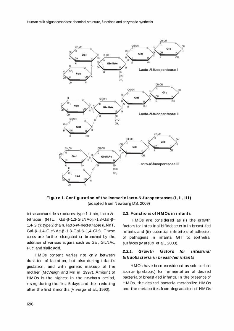
diversity of the structures (Rockova et al., 2011). Basically, most HMOs contain a lactose at the reducing end as the core structure, elongated by N-acetylglucosamine (GlcNAc), galactose (Gal), sialic acid (also known as N-acetylneuraminic acid; NeuAc), and fucose (Fuc) at non-reducing end with many and varied linkages between them. They range from three to ten monosaccharides in length (McVeagh and Miller. 1997). As an example, figure 1 indicates the structures of lacto-N-fucopentaose I, lacto-N-fucopentaose II and lacto-N-fucopentaose III.
Few unusual oligosaccharides found in human milk which do not contain the core structure, even without lactose at reducing end. The mechanism to produce these unusual oligosaccharides is yet unknown. They might be the products of unknown degradation from larger HMOs (Kobata, 2010). Due to structural complexity and variety, HMOs are resistant to enzymatic hydrolysis in upper gastrointestinal tract of host. This has been proved by Engfer and Gnoth with in vitro digestion studies in which they used human pancreatic juice and brush border membranes prepared from human or porcine intestinal tissue samples as enzyme sources (Engfer et al., 2000; Gnoth et al., 2000).
HMOs are produced with large amount in milk secreted at early stages of lactation in Golgi apparatus of cells lining the alveoli and smaller ductules. Alpha-lactabumin firstly regulates enzyme galactosyltransferase to produce lactose in a reaction between UDP-galactose and glucose. The biosynthetic steps leading from lactose to HMOs are currently not clear (Bode, 2009). However, well known structures of HMOs (galactosyl, N- acetylglucosaminyl, fucosyl and sialyl) are supposed to form by concerted action of glycosyltransferases (Kobata, 2010). The elongation of lactose may start by the action of β-3-N-acetyl-glucosaminyltransferase with an enzymatic transfer of N-acetyl glucosamine (GlcNAc) residue through β-1,3-linkage to the galactose (Gal) residue of lactose, followed by further addition of Gal through either β-1,3- or β-1,4 linkage to GlcNAc to create two major core
Human milk oligosaccharides: chemical structure, functions and enzymatic synthesis
696
Figure 1. Configuration of the isomeric lacto-N-fucopentaoses (I, II, III) (adapted from Newburg DS, 2009)
tetrasaccharride structures: type 1 chain, lacto-N-tetraose (NTL, Gal--1,3-GlcNAc--1,3-Gal--1,4-Glc); type 2 chain, lacto-N-neotetraose (LNnT, Gal--1,4-GlcNAc--1,3-Gal--1,4-Glc). These cores are further elongated or branched by the addition of various sugars such as Gal, GlcNAc, Fuc, and sialic acid.
HMOs content varies not only between duration of lactation, but also during infant’s gestation, and with genetic makeup of the mother (McVeagh and Miller, 1997). Amount of HMOs is the highest in the newborn period, rising during the first 5 days and then reducing after the first 3 months (Viverge et al., 1990).
2.3. Functions of HMOs in infants HMOs are considered as (i) the growth
factors for intestinal bifidobacteria in breast-fed infants and (ii) potential inhibitors of adhesion of pathogens in infants’ GIT to epithelial surfaces (Matsuo et al., 2003).
2.3.1. Growth factors for intestinal bifidobacteria in breast-fed infants
HMOs have been considered as sole carbon source (prebiotic) for fermentation of desired bacteria of breast-fed infants. In the presence of HMOs, the desired bacteria metabolize HMOs and the metabolites from degradation of HMOs
Hoang Anh Nguyen, Thu Ha Nguyen, Dietmar Haltrich
697
serve not only as beneficial components such as short chain fatty acids for the growth of desired bacteria but also as growth inhibitors to undesired bacteria (Bode, 2009).
Many HMO molecules have been purified from human milk and used in vivo as sole carbon source for fermentation of bifidobacteria and lactobacilli. These analyses have shown that several bifidobacterial species can grow well on HMOs (Kiyohara et al., 2009; Marcobal et al., 2010; Rockova et al., 2011). In addition, amount of intact HMOs were found very low in the feces of term and preterm breast-fed infants (Sabharwal et al., 1988; Sabharwal et al., 1988). This postulates that a majority of HMOs reaches the large intestine, where they are preferably used as substrates for bifidobacteria. The function of HMOs for the enrichment of bifidobacteria has also been known when a study indicated the acidity level (metabolites from the fermentation of bifidobacteria) in feces of breast-fed babies is higher than that in feces of formula-nourished babies (Kobata, 2003). Moreover, a cluster of genes encoding for glycosidases (sialidase, fucosidase, N-acetyl-β-hexosaminidase, β-galactosidase), that cleave HMOs into its constituent monosaccharides, and HMO transporters have been found recently in the genome of Bifidobacterium longum subsp. infantis ATCC1569. They are likely linked to genomic mechanisms of milk utilization for infants’ bifidobacteria (Sela et al., 2008).
Even though HMOs have been considered as a sole carbon source for beneficial bacteria in GIT of infants, direct fermentation of HMOs by bifidobacteria as well as intestinal bacteria has been poorly investigated. Rockova and coworkers (2011) (Rockova et al., 2011) found a great variability of bifidobateria in the ability to grow on HMOs. Bifidobacteria of human origin (Bifidobacterium bifidum, Bifidobacterium longum) have a better growth on human milk compared to those of animal origin (B.animalis). Ward and co-workers (2006) (Ward et al., 2006) pointed out that Bifidobacterium infantis fermented purified HMOs as a sole carbon source, while Lactobacillus gasseri, another gut commensal did not ferment HMO.
These results support the hypothesis that HMOs selectively affect the commensal bacteria in the intestinal tract.
2.3.2. Potential inhibitors of pathogen adhesion
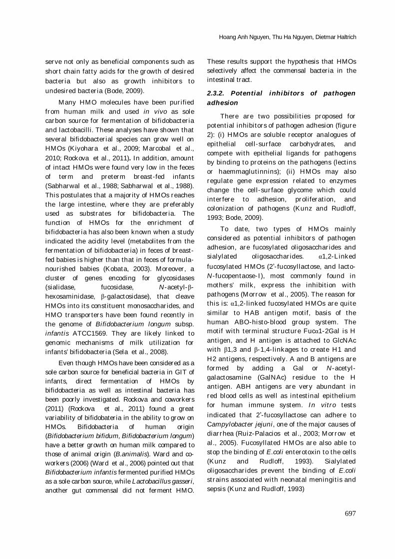
There are two possibilities proposed for potential inhibitors of pathogen adhesion (figure 2): (i) HMOs are soluble receptor analogues of epithelial cell-surface carbohydrates, and compete with epithelial ligands for pathogens by binding to proteins on the pathogens (lectins or haemmaglutinnins); (ii) HMOs may also regulate gene expression related to enzymes change the cell-surface glycome which could interfere to adhesion, proliferation, and colonization of pathogens (Kunz and Rudloff, 1993; Bode, 2009).
To date, two types of HMOs mainly considered as potential inhibitors of pathogen adhesion, are fucosylated oligosaccharides and sialylated oligosaccharides. α1,2-Linked fucosylated HMOs (2’-fucosyllactose, and lacto-N-fucopentaose-I), most commonly found in mothers’ milk, express the inhibition with pathogens (Morrow et al., 2005). The reason for this is: α1,2-linked fucosylated HMOs are quite similar to HAB antigen motif, basis of the human ABO-histo-blood group system. The motif with terminal structure Fucα1-2Gal is H antigen, and H antigen is attached to GlcNAc with β1,3 and β-1,4-linkages to create H1 and H2 antigens, respectively. A and B antigens are formed by adding a Gal or N-acetyl-galactosamine (GalNAc) residue to the H antigen. ABH antigens are very abundant in red blood cells as well as intestinal epithelium for human immune system. In vitro tests indicated that 2’-fucosyllactose can adhere to Campylobacter jejuni, one of the major causes of diarrhea (Ruiz-Palacios et al., 2003; Morrow et al., 2005). Fucosyllated HMOs are also able to stop the binding of E.coli enterotoxin to the cells (Kunz and Rudloff, 1993). Sialylated oligosaccharides prevent the binding of E.coli strains associated with neonatal meningitis and sepsis (Kunz and Rudloff, 1993)
Human milk oligosaccharides: chemical structure, functions and enzymatic synthesis
698
Figure 2. Anti-adhesive and glycome-modifying effects of HMOs (adapted from Bode L, 2009) “Most bacteria (commensals and pathogens) express glycan-binding proteins (lectins), that bind to glycans on the host’s epithelial cell surface (A), which is essential for bacteria to attach (a), and to
proliferate and colonize the intestine (b). Some pathogens need to attach to the intestinal epithelial cell surface prior to invading the host (c). HMOs are structurally similar to the intestinal epithelial
cell surface glycans. They can serve as bacterial lectin ligand analogs and block bacterial attachment (B). HMOs may also alter the intestinal epithelial glycosylation machinery and modify the
cell-surface glycome (“glycocalyx”), which could impact bacterial attachment, proliferation, colonization (C)” (Bode 2009)
3. ENZYMATIC SYNTHESIS OF HMOs FOR APPLICATIONS IN FOODS AND DRUGS
Due to important biological functions, HMOs have attracted considerable interest. Many methods have been developed for the synthesis of HMOs that can be applied as ingredients in infant foods as well as drug development. In principle, HMOs can be synthesized by application of enzymes or by chemical approaches. However, great effort nowadays has been put into enzymatic methods, because chemical methods still require multiple steps to get rid of side products, and this complexity does not render chemical syntheses realistic for industrial applications (Scigelova et al., 1998). Enzymes used for synthesis of oligosaccharides can be either glycosyltransferases or glycosidases. However, currently enzymatic methods using glycosyltransferases are mostly used because of highly stereoselective and regioselective bond formation and no side products formed (Endo and Koizumi, 2000).
Despite their recognized importance for infant health, synthesis of HMOs have been hindered by the fact that it is still very difficult to obtain large quantity of them by enzymatic synthesis (Chen et al., 2000). Wild type enzymes originated from plants and animals are difficult to obtain in large amount. Moreover, genes encoding for mammalian glycosyltransferases are difficult to be functionally expressed in E.coli. Thus, production of recombinant eukaryotic glycosyltransferases generally requires eukaryotic expression systems which often render the production tedious and expensive. These points limit the use of enzymatic methods for industrial production of oligosaccharides (Matsuo et al., 2003). By contrast, cloning and expression of bacterial glycosyltransferase genes in E.coli is much more convenient and efficient. Recently, the use of metabolically engineered bacteria to over-express heterologous glycosyltransferase and glycosidase genes is a powerful new technique
Hoang Anh Nguyen, Thu Ha Nguyen, Dietmar Haltrich
699
Table 1. HMOs mentioned in this review
Names Abbreviation Structures*
N-acetyl oligosaccharides
Lacto-N-biose I LNB Gal-β-1,3-GlcNAc
GlcNAc-β-1,3-Gal
N-acetyllactosamine LacNAc Gal-β-1,4-GlcNAc
Allo-LacNAc Gal-β-1,6-GlcNAc
Lacto-N-triose LNT-2 GlcNAc-β-1,3-Gal-β-1,4Glc
Lacto-N-neotetraose LNnT Gal-β-1,4-GlcNAc-β-1,3-Gal-β-1,4-Glc
Gal-β-1,4-Gal-β-1,4-GlcNAc
Gal-β-1,4-Gal-β-1,4-Gal-β-1,4-GlcNAc
Sialylated oligosaccharides
α 2,3-sialyllactose 3’-SL Neu5Ac-α-2,3-Gal-β-1,4-Glc
α 2,6-sialyllactose 6’-SL Neu5Ac-α-2,6-Gal-β-1,4Glc
Fucosyloligosaccharides
2’-fucosyllactose 2’FL Fuc-α-1,2-Gal-β-1,4-Glc
Lacto-N-neo-fucopentaose-1 LNF-1 Fuc-α-1,2-Gal-β-1,4-GlcNAc-β-1,3-Gal-β-1,4-Glc
Lacto-N-neo-fucopentaose LNnFP Gal-β-1,4-GlcNAc-β-1,3-Gal-β-1,4-(Fuc-α-1,3)-Glc
Lacto-N-neodifucohexaose LnNDFH Gal-β-1,4-(Fuc-α-1,3)-GlcNAc-β-1,3-Gal-β1,4-(Fuc-α-1,3)-Glc
Lacto-N-neodifucooctaose Gal-β-1,4-GlcNAc-β-1,3-Gal-β-1,4-(Fuc-α-1,3)-GlcNAc-β-1,3-Gal-β-1,4-(Fuc-α-1,3)-Glc
* GlcNAc; N-acetylglucosamine, Gal; galactose, Glc; glucose, Neu5Ac; N-acetylneuraminic acid, Fuc; fucose
that makes the production of HMOs in large amount with lower cost feasible. Using of whole cells or glycosyltransferases isolated from engineered microorganisms as the enzyme sources may open the way to produce HMOs at commercial scale that had been not yet successful. (Endo and Koizumi, 2000; Schwab and Gaenzle, 2011).
Generally, the steps for enzymatic synthesis of HMOs using metabolically engineered E.coli are the following: (1) designate a β-galactosidase-negative (lacZ-) E.coli strain in which a lacY gene encoding for β-galactoside permease still remains; (2) transform the genes encoding for glycosyltransferases that use lactose as acceptor to the above strain; (3) cultivate this strain at high cell density on alternative carbon source, such as glycerol, under the conditions that allow both glycosyltransferase and β-galactoside permease genes express; (4) feed the culture with lactose that should be actively internalized
by the permease and glycosylated by the transferase; (5) purify and structurally characterizatize HMOs by chromatography and NMR (Priem, et al., 2002). This section focuses on the syntheses, which could be promising for the applications in large scale production, of some well known HMOs (Table 1).
3.1. N-acetyloligosaccharides HMOs containing GlcNAc (the bifidus
factor) are necessary for the growth of bifidobacteria. These oligosaccharides form precursors in the biosynthesis of muramic acid, a component of the bacterial cell wall (McVeagh and Miller, 1997). Reports, to date, have indicated that N-acetyloligosaccharides of HMOs can be produced by N-acetylglucosaminyltransferases, β-N-acetylhexosaminidases (β-N-acetylglucosaminidases/ β-N acetylgalactosaminidases) or β-galactosidases.
Blixt and coworkers (1999) have over-expressed the Neisseria meninggitidis lgtA gene encoding for β-1,3-N-acetylglucosaminyltransferase (β-1,3-
Human milk oligosaccharides: chemical structure, functions and enzymatic synthesis
700
GlcNAcT) in E. coli. Characterization of the recombinant enzyme indicated that this enzyme is capable to catalyze the transfer of GlcNAc from UDP-GlcNAc in a β-1,3 linkage to acceptor (Gal residues) to create oligosaccharides with GlcNAc-β-1,3-Gal linkage (Blixt et al., 1999). Johnson and coworkers (1999) have developed enzyme-based technologies to successively synthesize several relevant HMOs using cloned bacterial glycosyltransferases (β-1,3-GlcNAcT; β1,4-galactotransferase (β-1,4-GalT); and α2-trans-sialidase). In the first step, they successfully scaled up and produced 250 grams of LNT-2 (GlcNAc-β-1,3-Gal-β-1,4Glc) from 100 L reactor containing lactose, UDP-GlcNAc and β-1,3-GlcNAcT, and then in the second step more than 300 grams of lacto-N-neotetraose (LNnT; Gal-β-1,4-GlcNAc-β-1,3Gal-β1,4-Glc) were formed from 100 L reactor containing LNT-2, UDP-Gal and β-1,4-GalT (Johnson, 1999).
However, these methods still require nucleotide-substrates. Liu and coworkers (2003) have co-expressed 4 enzymes (sucrose synthase (SusA); UDP-Glc-4-epimerase (GalE); β-1,4-GalT; α-1,4-galactotransferase (α-1,4GalT) in a single genetically engineered E.coli strain with high level of UTP production. SusA catalyzes the cleavage of sucrose to UDP-glucose and fructose. UDP-glucose is converted into UDP-galactose by GalE, and then β-1,4-GalT transfers galactose from UDP-galactose to acceptor (GlcNAc) to form N-acetyllactosamine (LacNAc, Gal-β-1,4-GlcNAc). LacNAc is then combined with an additional galactosyl by α-1,4GalIT, resulting in the synthesis of 5.4 g of Gal-α-1,4-Gal-β-1,4-GlcNAc in 200ml reaction volume with 67% yield based on the consumption of GlcNAc (Liu et al., 2003).
A new fermentation process allowing large-scale production of HMOs by metabolically engineered bacteria has been reported by Priem and coworkers (2002). A β-galactosidase - negative (LacZ-) E.coli strain carrying lgtA gene from Neisseria meningitides was cultivated at high density with glycerol as the sole carbon source using classical fed-batch strategy. This
fermentation resulted in over-expession of lgtA and the synthesis of 6 g.L-1 of expected extracellular trisaccharide LNT-2 by β-1,3-GlcNAcT transfers GlcNAc to lactose. When lgtB gene encoding for the β-1,4-GalT from Neisseria meningitides was co-expressed with lgtA, LNT-2 was further converted to lacto-N-neotetraose (Gal-β-1,4-GlcNAc-β-1,3-Gal-β-1,4-Glc). However, for this co-expression, glucose instead of glycerol has to be used as sole carbon source for cultivation, and the product mainly remained intracellular (Priem et al., 2002).
β-N-acetylhexosaminidases (EC 3.2.1.52) are glycoside hydrolases, like typical exo-enzymes. Some of them (mostly from fungi) not only can cleave the terminal β-D-GlcNAc and β-D-GalNAc residues in N-acetyl-β-D-hexosaminides, but also can then transfer β-D-GlcNAc and β-D-GalNAc residues to broad variety of glycosidic and non-glycosidic acceptors (Slamova et al., 2010). N-acetyl-β-D-hexosaminides are easily obtained from hydrolysis of chitin, a second most abundant polysaccharide in nature after cellulose, using chitinases (Lee et al., 2007). Thus, the promising strategy is finding suitable β-N-acetylhexosaminidases that can be applied to produce HMOs using N-acetyl-chiooligosaccharides, products of chitin degradation, as donors. This would enable the use of low cost and easily available starting materials for the large-scale synthesis of novel oligosaccharides. To date, this strategy has been successfully used for activated substrates (derivatives of GlcNAc or N-acetyl-chitooligosaccharides), however it is not yet applied for food applications and large scale production because of toxicity and high cost (Singh, et al., 1997; Kurakake et al., 2003; Weignerova et al., 2003).
Matsuo and coworkers (2003) have used recombinant β-N-acetylglucosaminidases from Aspergillus ozyrae to produce HMOs by reverse hydrolysis reaction, but the yield was very low with only 0.21 % of LNT-2 and 0.15% of its isomer (GlcNAc-β-1,6-Gal-β-1,4-Glc) (Matsuo et al., 2003).
Hoang Anh Nguyen, Thu Ha Nguyen, Dietmar Haltrich
701
Recently, enzyme β-galactosidase from Bacillus circulans was found that they can hydrolyze lactose (donor) and then transfer galactosyl products to receptors (GlcNAc or GalNAc) (Sakai et al., 1992; Usui et al., 1996; Hernaiz and Crout 2000; Li et al., 2010). Some N-acetyloligosaccharides have been produced, such as Gal-β-1,4-GlcNAc with yield of 23.2% (Sakai et al., 1992), a mixture of LacNAc, allo-LacNAc (Gal-β-1,6-GlcNAc), Gal-β-1,4-Gal-β-1,4-GlcNAc, and Gal-β-1,4-Gal-β-1,4-Gal-β-1,4-GlcNAc with ratio of 28.75 %, 2.29%, 9.47%, 5.67%, respectively (Li et al., 2010)
3.2. Sialylated oligosaccharides Human milk, containing more than three
times of sialylated oligosaccharides compared to cow’ milk, is an important source of sialic acids for breast-fed infants. Sialylated oligosaccharides are used for biosynthesis of mucins, glycoproteins and gangliosides which are concentrated in plasma membranes of nerve cells (McVeagh and Miller, 1997; Wang et al., 2001). Sialylated oligosaccharides are also known to have both anti-infective and immunostimulating properties (Boehm and Stahl 2007). Sialylated HMOs, believed to protect breast-fed infants from infection, consist of N-acetylneuraminic acid (NeuAc) attached to Gal through α-(2,3) or α-(2,6) linkage.
From general principle of syalyllactose biosynthesis (figure 3), Gilbert and co-workers (1997) have characterized the gene encoding for α-2,3-sialyltransferase from Neisseria meningitides (Gilbert et al., 1997), then fused it with gene encoding for CMP-Neu5Ac synthetase and expressed in E.coli. The fusion protein was used to produce α-2,3-sialyllactose at the 100 g scale using a sugar nucleotide cycle reaction, starting from lactose, sialic acid, phosphoenolpyruvate and catalytic amounts of ATP and CMP. However, this method requires expensive substrates, thus it is not applicable for large scale. To solve this drawback, permeabilized and alive whole E.coli cells have been used.
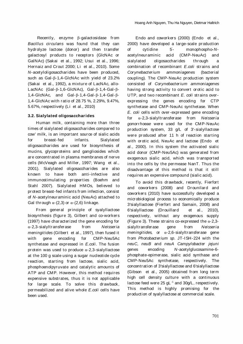
Endo and coworkers (2000) (Endo et al., 2000) have developed a large-scale production of cytidine 5’- monophospho-N-acetylneuraminic acid (CMP-NeuAc) and sialylated oligosaccharides through a combination of recombinant E.coli strains and Corynebacterium ammoniagenes (bacterial coupling). The CMP-NeuAc production system consisted of Corynebacterium ammoniagenes having strong activity to convert orotic acid to UTP, and two recombinant E. coli strains over-expressing the genes encoding for CTP synthetase and CMP-NeuAc synthetase. When E. coli cells with over-expressed gene encoding for α-2,3-sialyltransferase from Neisseria gonorrhoeae were used for the CMP-NeuAc production system, 33 g/L of 3′-sialyllactose were produced after 11 h of reaction starting with orotic acid, NeuAc and lactose (Endo et al., 2000). In this system the activated sialic acid donor (CMP-Neu5Ac) was generated from exogenous sialic acid, which was transported into the cells by the permease NanT. Thus the disadvantage of this method is that it still requires an expensive compound (sialic acid).
To avoid this drawback, recently, Fierfort and coworkers (2008) and Drounilard and coworkers (2010) have successfully developed a microbiological process to economically produce 3′sialyllactose (Fierfort and Samain, 2008) and 6′sialyllactose (Drouillard et al., 2010), respectively, without any exogenous supply (Figure 3). These strains co-expressed the α-2,3-sialyltransferase gene from Neisseria meningitides, or α-2,6-sialyltransferase gene from Photobacterium sp. JT-ISH-224 with the neuC, neuB and neuA Campylobacter jejuni genes encoding N-acetylglucosamine-6-phosphate-epimerase, sialic acid synthase and CMP-Neu5Ac synthetase, respectively. The concentration of 3′sialyllactose and 6′sialyllactose (Gibson et al., 2005) obtained from long term high cell density culture with a continuous lactose feed were 25 gL-1 and 30g/L, respectively. This method is highly promising for the production of syalyllactose at commercial scale.
Human milk oligosaccharides: chemical structure, functions and enzymatic synthesis
702
Figure 3. Engineered metabolic pathway for the production of 6’-sialyllactose (Adapted from Drounilard, 2010) "Over-expressed heterologous genes are in bold. Discontinued arrows
represent the enzymatic activities that have been eliminated. Lactose is internalized by lactose permease and sialylated by recombinant α-2,6-sialyltransferase using CMP-Neu5Ac produced from
UDP-GlcNAc by the successive action of the N-acetylglucosamine-6-phosphate-epimerase NeuC, the sialic acid synthase NeuB, and the CMP-Neu5Ac synthetase NeuA. The β-galactosidase gene lacZ was knocked out to prevent lactose hydrolysis and the nanA and nanK genes were knocked out to
prevent the formation of futile cycles in the CMPNeu5Ac biosynthesis pathway"
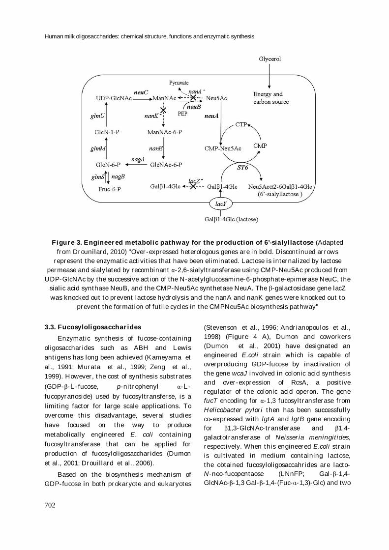
3.3. Fucosyloligosaccharides Enzymatic synthesis of fucose-containing
oligosaccharides such as ABH and Lewis antigens has long been achieved (Kameyama et al., 1991; Murata et al., 1999; Zeng et al., 1999). However, the cost of synthesis substrates (GDP-β-L-fucose, p-nitrophenyl α-L-fucopyranoside) used by fucosyltransferse, is a limiting factor for large scale applications. To overcome this disadvantage, several studies have focused on the way to produce metabolically engineered E. coli containing fucosyltransferase that can be applied for production of fucosyloligosaccharides (Dumon et al., 2001; Drouillard et al., 2006).
Based on the biosynthesis mechanism of GDP-fucose in both prokaryote and eukaryotes
(Stevenson et al., 1996; Andrianopoulos et al., 1998) (Figure 4 A), Dumon and coworkers (Dumon et al., 2001) have designated an engineered E.coli strain which is capable of overproducing GDP-fucose by inactivation of the gene wcaJ involved in colonic acid synthesis and over-expression of RcsA, a positive regulator of the colonic acid operon. The gene fucT encoding for α-1,3 fucosyltransferase from Helicobacter pylori then has been successfully co-expressed with lgtA and lgtB gene encoding for β1,3-GlcNAc-transferase and β1,4-galactotransferase of Neisseria meningitides, respectively. When this engineered E.coli strain is cultivated in medium containing lactose, the obtained fucosyloligosaccahrides are lacto-N-neo-fucopentaose (LNnFP; Gal-β-1,4- GlcNAc-β-1,3 Gal-β-1,4-(Fuc-α-1,3)-Glc) and two
Hoang Anh Nguyen, Thu Ha Nguyen, Dietmar Haltrich
703
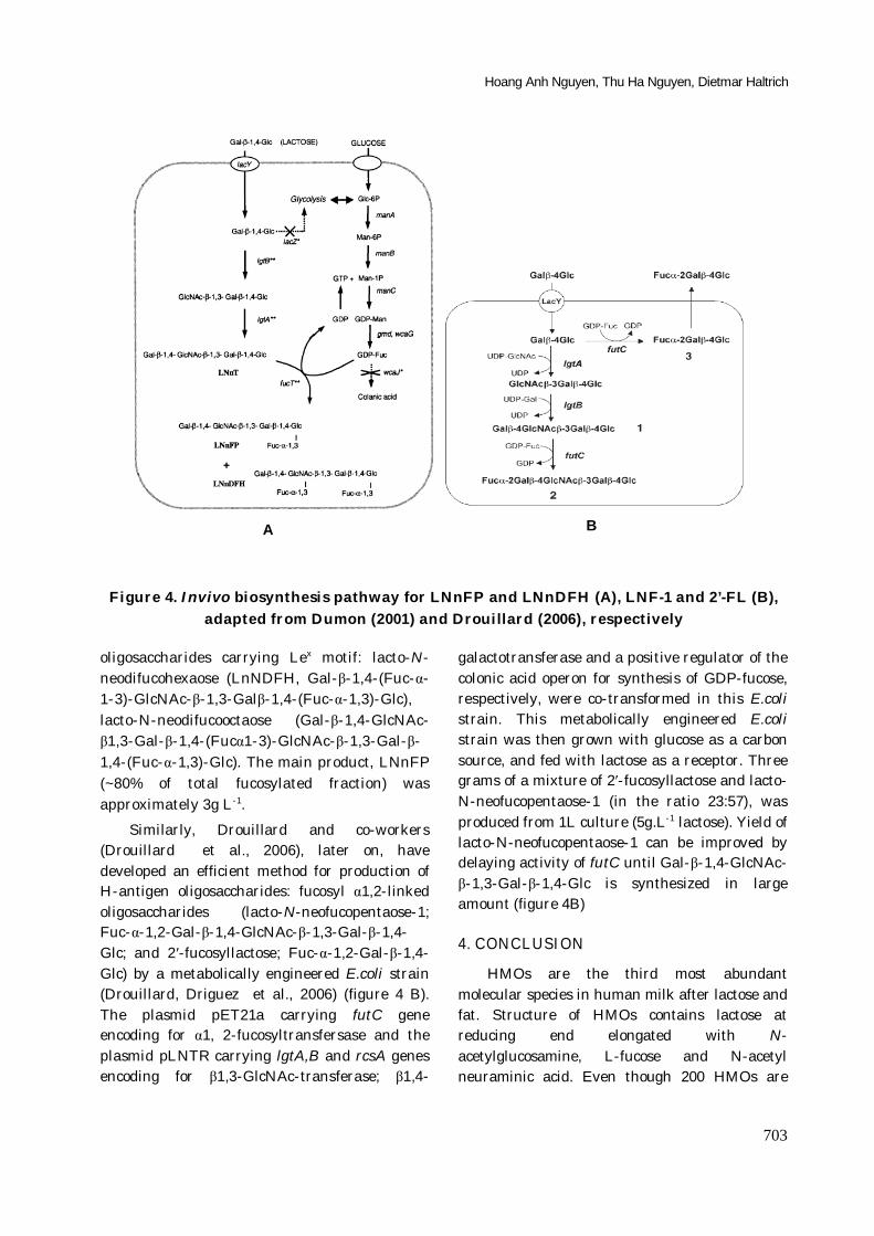
Figure 4. Invivo biosynthesis pathway for LNnFP and LNnDFH (A), LNF-1 and 2’-FL (B), adapted from Dumon (2001) and Drouillard (2006), respectively
oligosaccharides carrying Lex motif: lacto-N-neodifucohexaose (LnNDFH, Gal-β-1,4-(Fuc-α-1-3)-GlcNAc-β-1,3-Galβ-1,4-(Fuc-α-1,3)-Glc), lacto-N-neodifucooctaose (Gal-β-1,4-GlcNAc-β1,3-Gal-β-1,4-(Fucα1-3)-GlcNAc-β-1,3-Gal-β-1,4-(Fuc-α-1,3)-Glc). The main product, LNnFP (~80% of total fucosylated fraction) was approximately 3g L-1.
Similarly, Drouillard and co-workers (Drouillard et al., 2006), later on, have developed an efficient method for production of H-antigen oligosaccharides: fucosyl α1,2-linked oligosaccharides (lacto-N-neofucopentaose-1; Fuc-α-1,2-Gal-β-1,4-GlcNAc-β-1,3-Gal-β-1,4-Glc; and 2′-fucosyllactose; Fuc-α-1,2-Gal-β-1,4-Glc) by a metabolically engineered E.coli strain (Drouillard, Driguez et al., 2006) (figure 4 B). The plasmid pET21a carrying futC gene encoding for α1, 2-fucosyltransfersase and the plasmid pLNTR carrying lgtA,B and rcsA genes encoding for β1,3-GlcNAc-transferase; β1,4-
galactotransferase and a positive regulator of the colonic acid operon for synthesis of GDP-fucose, respectively, were co-transformed in this E.coli strain. This metabolically engineered E.coli strain was then grown with glucose as a carbon source, and fed with lactose as a receptor. Three grams of a mixture of 2′-fucosyllactose and lacto-N-neofucopentaose-1 (in the ratio 23:57), was produced from 1L culture (5g.L-1 lactose). Yield of lacto-N-neofucopentaose-1 can be improved by delaying activity of futC until Gal-β-1,4-GlcNAc-β-1,3-Gal-β-1,4-Glc is synthesized in large amount (figure 4B)
4. CONCLUSION
HMOs are the third most abundant molecular species in human milk after lactose and fat. Structure of HMOs contains lactose at reducing end elongated with N-acetylglucosamine, L-fucose and N-acetyl neuraminic acid. Even though 200 HMOs are
A B
Human milk oligosaccharides: chemical structure, functions and enzymatic synthesis
704
currently determined, detail structural identification of the HMOs is still lacking because of the complexity and diversity of the structures. Two potential properties of HMOs, which are as the “growth factors for intestinal bifidobacteria in breast-fed infants” and the “inhibitors of adhesion of pathogens” have been well documented. Therefore, many HMOs are of interest for applications in infant foods as well as drug development. These HMOs have been produced by enzymatic method mostly using glycosyltransferases, and especially the approaches using metabolically engineered bacteria allow the production of HMOs in large scale. However, a suitable approach for a commercial scale production of HMOs, that has not yet been successful, is of a continuous interest.
REFERENCES Andrianopoulos K., L. Wang, P. R. Reeves (1998).
"Identification of the fucose synthetase gene in the colanic acid gene cluster of Escherichia coli K-12". Journal of Bacteriology 180(4): 998-1001.
Blixt, O., I. van Die., T.Norberg., D.H. Van Den Eijnden (1999). "High-level expression of the Neisseria meningitidis lgtA gene in Escherichia coli and characterization of the encoded N-acetylglucosaminyltransferase as a useful catalyst in the synthesis of GlcNAc beta 1 -> 3Gal and GalNAc beta 1-3Gal linkages". Glycobiology 9(10): 1061-1071.
Bode, L. (2009). "Human milk oligosaccharides: prebiotics and beyond". Nutrition Reviews 67(11): S183-S191.
Boehm, G. and B. Stahl (2007). "Oligosaccharides from milk". Journal of Nutrition 137(3): 847S-849S.
Chen, X., P. Kowal., P. G. Wang (2000). "Large-scale enzymatic synthesis of oligosaccharides". Current opinion in drug discovery & development 3(6): 756-63.
Claud, E. C. and W. A. Walker (2001). "Hypothesis: inappropriate colonization of the premature intestine can cause neonatal necrotizing enterocolitis". Faseb Journal 15(8): 1398-1403.
Coppa, G. V., O. Gabrielli., P. Pierani., C.Catassi., A.Carlucci., P.L.Giorgi (1993). "Changes in Carbohydrate-Composition in Human-Milk over 4 Months of Lactation". Pediatrics 91(3): 637-641.
Dethlefsen, L., P. B. Eckburg., E. M. Bik., D. A. Relman,(2006). "Assembly of the human intestinal microbiota". Trends in Ecology & Evolution 21(9): 517-523.
Drouillard, S., H. Driguez., E Samain (2006). "Large-scale synthesis of H-antigen oligosaccharides by expressing Helicobacter pylori alpha 1,2-fucosyltransferase in metabolically engineered Escherichia coli cells". Angewandte Chemie-International Edition 45(11): 1778-1780.
Drouillard, S., T. Mine., H.Kajiwara., T.Yamamoto., E.Samain (2010). "Efficient synthesis of 6 '-sialyllactose, 6,6 '-disialyllactose, and 6 '-KDO-lactose by metabolically engineered E. coli expressing a multifunctional sialyltransferase from the Photobacterium sp JT-ISH-224". Carbohydrate Research 345(10): 1394-1399.
Duggan, C., J. Gannon., W.A.Walker (2002). "Protective nutrients and functional foods for the gastrointestinal tract". American Journal of Clinical Nutrition 75(5): 789-808.
Dumon, C., B. Priem., S.L.Martin., A.Heyraud., C.Bosso., E.Samain (2001). "In vivo fucosylation of lacto-N-neotetraose and lacto-N-neohexaose by heterologous expression of Helicobacter pylori alpha-1,3 fucosyltransferase in engineered Escherichia coli". Glycoconjugate Journal 18(6): 465-474.
Endo, T. and S. Koizumi (2000). "Large-scale production of oligosaccharides using engineered bacteria". Current Opinion in Structural Biology 10(5): 536-541.
Endo, T., S. Koizumi., K.Tabata., A.Ozaki (2000). "Large-scale production of CMP-NeuAc and sialylated oligosaccharides through bacterial coupling". Applied Microbiology and Biotechnology 53(3): 257-261.
Engfer, M. B., B. Stahl., B.Finke., G.Sawatzki., H.Daniel (2000). "Human milk oligosaccharides are resistant to enzymatic hydrolysis in the upper gastrointestinal tract". American Journal of Clinical Nutrition 71(6): 1589-1596.
Fanaro, S., R. Chierici., P.Guerrini., V.Vigi (2003). "Intestinal microflora in early infancy: composition and development". Acta Paediatrica 92: 48-55.
Fierfort, N. and E. Samain (2008). "Genetic engineering of Escherichia coli for the economical production of sialylated oligosaccharides". Journal of Biotechnology 134(3-4): 261-265.
Figueroa-Gonzalez, I., G. Quijano., G.Ramirez., A.Cruz-Guerrero (2011). "Probiotics and prebiotics - perspectives and challenges". Journal of the Science of Food and Agriculture 91(8): 1341-1348.
German, J. B., S. L. Freeman., C.B.Lebrilla., D.A.Mills (2008). "Human milk oligosaccharides: evolution, structures and bioselectivity as substrates for intestinal bacteria". Nestle Nutrition workshop series. Paediatric programme 62: 205.
Gibson, G. R., A. L. McCartney., R.A.Rastall (2005). "Prebiotics and resistance to gastrointestinal infections". British Journal of Nutrition 93: S31-S34.
Hoang Anh Nguyen, Thu Ha Nguyen, Dietmar Haltrich
705
Gibson, G. R. and M. B. Roberfroid (1995). "Dietary Modulation of the Human Colonic Microbiota - Introducing the Concept of Prebiotics". Journal of Nutrition 125(6): 1401-1412.
Gilbert, M., A. M. Cunningham., D.C.Watson., A. Martin., J.C.Richards., W.W. Wakarchuk (1997). "Characterization of a recombinant Neisseria meningitidis alpha-2,3-sialyltransferase and its acceptor specificity". European Journal of Biochemistry 249(1): 187-194.
Gnoth, M. J., C. Kunz., E. Kinne-Saffran., S. Rudloff (2000). "Human milk oligosaccharides are minimally digested in vitro". Journal of Nutrition 130(12): 3014-3020.
Guarner, F. and J. R. Malagelada (2003). "Gut flora in health and disease". Lancet 361(9356): 512-519.
Hernaiz, M. J. and D. H. G. Crout (2000). "A highly selective synthesis of N-acetyllactosamine catalyzed by immobilised beta-galactosidase from Bacillus circulans". Journal of Molecular Catalysis B-Enzymatic 10(4): 403-408.
Johnson, K. F. (1999). "Synthesis of oligosaccharides by bacterial enzymes". Glycoconjugate Journal 16(2): 141-146.
Kameyama, A., H. Ishida., M.Kiso., A.Hasegawa (1991). "Synthetic Studies on Sialoglycoconjugates. 21. Total Synthesis of Sialyl Lewis-X". Carbohydrate Research 209: C1-C4.
Kelly, D., S. Conway., R.Aminov (2005). "Commensal gut bacteria: mechanisms of immune modulation". Trends in Immunology 26(6): 326-333.
Kiyohara, M., A. Tachizawa., M.Nishimoto., M.Kitaoka., H.Ashida., K.Yamamoto (2009). "Prebiotic Effect of Lacto-N-biose I on Bifidobacterial Growth". Bioscience Biotechnology and Biochemistry 73(5): 1175-1179.
Kobata, A. (2003). "Possible application of milk oligosaccharides for drug development. "Chang Gung medical journal 26(9): 621-36.
Kobata, A. (2010). "Structures and application of oligosaccharides in human milk". Proceedings of the Japan Academy Series B-Physical and Biological Sciences 86(7): 731-747.
Kunz, C. and S. Rudloff (1993). "Biological Functions of Oligosaccharides in Human-Milk". Acta Paediatrica 82(11): 903-912.
Kurakake, M., T. Goto., K.Ashiki., Y Suenaga., T. Komaki. (2003). "Synthesis of new glycosides by transglycosylation of N-acetylhexosaminidase from Serratia marcescens YS-1". Journal of Agricultural and Food Chemistry 51(6): 1701-1705.
Lee, Y. S., I. H. Park., J.S.Yoo; S.Y.Chung., Y.C.Lee., Y.S. Cho., C.M. Kim., Y.L.Choi (2007). "Cloning, purification, and characterization of chitinase from Bacillus sp. DAU101". Bioresour Technol 98(14): 2734-41.
Li, W., Y. Sun., H.Ye., X. Zeng (2010). "Synthesis of oligosaccharides with lactose and N-acetylglucosamine as substrates by using beta-d-galactosidase from Bacillus circulans". European Food Research and Technology 231(1): 55-63.
Liu, Z. Y., Y. Q. Lu., J.B. Zhang., L.Pardee., P.G.Wang (2003). "P1 trisaccharide (Gal alpha 1,4Gal beta 1,4GlcNAc) synthesis by enzyme glycosylation reactions using recombinant Escherichia coli". Applied and Environmental Microbiology 69(4): 2110-2115.
Marcobal, A., M. Barboza., J.W.Froehlich., D.E. Block., J.B.German., C.B.Lebrilla., D.A. Mills (2010). "Consumption of Human Milk Oligosaccharides by Gut-Related Microbes". Journal of Agricultural and Food Chemistry 58(9): 5334-5340.
Martin, R., S. Langa., C. Reviriego., E.Jimenez., M.L. Marin., J.Xaus., L. Fernandez., J.M.
Rodriguez (2003). "Human milk is a source of lactic acid bacteria for the infant gut". Journal of Pediatrics 143(6): 754-758.
Matsuo, I., S. Kim., Y.Yamamoto., K.Ajisaka., J. Maruyama., H Nakajima., K.Kitamoto (2003). "Cloning and overexpression of beta-N-acetylglucosaminidase encoding gene nagA from Aspergillus oryzae and enzyme-catalyzed synthesis of human milk oligosaccharide". Bioscience Biotechnology and Biochemistry 67(3): 646-650.
McVeagh, P. and J. B. Miller (1997). "Human milk oligosaccharides: Only the breast". Journal of Paediatrics and Child Health 33(4): 281-286.
Morrow, A. L., G. M. Ruiz-Palacios., X.Jiang., D.S. Newburg (2005). "Human-milk glycans that inhibit pathogen binding protect breast-feeding infants against infectious diarrhea". Journal of Nutrition 135(5): 1304-1307.
Moughan, P. J., M. J. Birtles., P.D. Cranwell., W.C.Smith., M.Pedraza (1992). "The piglet as a model animal for studying aspects of digestion and absorption in milk-fed human infants". World review of nutrition and dietetics 67: 40-113.
Murata, T., S. Morimoto., X.X.Zeng., S.Watanabe., T.Usui (1999). "Enzymatic synthesis of alpha-L-fucosyl-N-acetyllactosamines and 3 '-O-alpha-L-fucosyllactose utilizing alpha-L-fucosidases". Carbohydrate Research 320(3-4): 192-199.
Newburg, D. S. (2009). "Neonatal protection by an innate immune system of human milk consisting of oligosaccharides and glycans". Journal of animal science 87(13): 26-34.
Palmer, C., E. M. Bik., D.B.DiGiulio., D.A.Relman., P.O. Brown(2007). "Development of the human infant intestinal microbiota". Plos Biology 5(7): 1556-1573.
Priem, B., M. Gilbert., W.W. Wakarchuk., A Heyraud., E. Samain (2002). "A new fermentation process
Human milk oligosaccharides: chemical structure, functions and enzymatic synthesis
706
allows large-scale production of human milk oligosaccharides by metabolically engineered bacteria". Glycobiology 12(4): 235-240.
Rockova, S., J. Nevoral., V. Rada., P.Marsik., J.Sklenar., A.Hinkova., E.Vlkova., M. Marounek (2011). "Factors affecting the growth of bifidobacteria in human milk". International Dairy Journal 21(7): 504-508.
Ruiz-Palacios, G. M., L. E. Cervantes., P. Ramos., B. Chavez-Munguia., D.S. Newburg (2003). "Campylobacter jejuni binds intestinal H(O) antigen (Fuc alpha 1, 2Gal beta 1, 4GlcNAc), and fucosyloligosaccharides of human milk inhibit its binding and infection". Journal of Biological Chemistry 278(16): 14112-14120.
Sabharwal, H., B. Nilsson., M.A. Chester., F. Lindh., G.Gronberg., S. Sjoblad., A. Lundblad (1988). "Oligosaccharides from Feces of a Blood-Group-B, Breast-Fed Infant". Carbohydrate Research 178: 145-154.
Sabharwal, H., B. Nilsson., G. Gronberg., M.A.Chester., J. Dakour., S. Sjoblad., A.Lundblad (1988). "Oligosaccharides from Feces of Preterm Infants Fed on Breast-Milk". Archives of Biochemistry and Biophysics 265(2): 390-406.
Sakai, K., R. Katsumi., H.Ohi., T.Usui., Y.Ishido (1992). "Enzymatic Syntheses of N-Acetyllactosamine and N-Acetylallolactosamine by the Use of Beta-D-Galactosidases". Journal of Carbohydrate Chemistry 11(5): 553-565.
Sangwan, V., S. K. Tomar., R.R.B. Singh., A.K.Singh., B. Ali (2011)".Galactooligosaccharides: Novel Components of Designer Foods". Journal of Food Science 76(4): R103-R111.
Schwab, C. and M. Gaenzle (2011). "Lactic acid bacteria fermentation of human milk oligosaccharide components, human milk oligosaccharides and galactooligosaccharides". Fems Microbiology Letters 315(2): 141-148.
Scigelova, M., P. Sedmera., V.Havlicek., V. Prikrylova., V.Kren (1998). "Glycosidase-catalysed synthesis of ergot alkaloid alpha-glycosides". Journal of Carbohydrate Chemistry 17(6): 981-986.
Sela, D. A., J. Chapman., A.Adeuya., J.H. Kim., F.Chen., T.R. Whitehead., A. Lapidus., D.S. Rokhsar., C.B.Lebrilla., J.B. German., N.P.Price., P.M.Richardson., D.A: Mills (2008). "The genome sequence of Bifidobacterium longum subsp infantis reveals adaptations for milk utilization within the infant microbiome". Proceedings of the National Academy of Sciences of the United States of America 105(48): 18964-18969.
Singh, S., M. Scigelova., P.Critchley., D.H.G.Crout (1997). "Trisaccharide synthesis by glycosyl transfer from p-nitrophenyl beta-D-N-
acetylgalactosaminide on to disaccharide acceptors catalysed by the beta-N-acetylhexosaminidase from Aspergillus oryzae". Carbohydrate Research 305(3-4): 363-370.
Sinkiewicz, G. and E. A. Nordstrom (2005). "Occurrence of Lactobacillus reuteri, Lactobacilli and Bifidobacteria in human breast milk". Pediatric Research 58(2): 415-415.
Slamova, K., P. Bojarova., L. Petraskova., V. Kren (2010). "beta-N-Acetylhexosaminidase: What's in a name... ?" Biotechnology Advances 28(6): 682-693.
Stevenson, G., K. Andrianopoulos., M.Hobbs., P.R.Reeves (1996). "Organization of the Escherichia coli K-12 gene cluster responsible for production of the extracellular polysaccharide colanic acid". Journal of Bacteriology 178(16): 4885-4893.
Usui, T., S. Morimoto., Y.Hayakawa., M.Kawaguchi., T.Murata., Y. Matahira., Y. Nishida(1996). "Regioselectivity of beta-D-galactosyl-disaccharide formation using the beta-D-galactosidase from Bacillus circulans". Carbohydrate Research 285: 29-39.
Viverge, D., L. Grimmonprez., G.Cassanas., L Bardet., M.Solere (1990). "Variations in Oligosaccharides and Lactose in Human-Milk During the 1st Week of Lactation". Journal of Pediatric Gastroenterology and Nutrition 11(3): 361-364.
Wang, B., J. B. Miller., Y.Sun., Z.Abmad., P.McVeagh., P. Petocz (2001). "A longitudinal study of salivary sialic acid in preterm infants: Comparison of human milk-fed versus formula-fed infants". Journal of Pediatrics 138(6): 914-916.
Ward, R. E., M. Ninonuevo., D.A. Mills., C.B. Lebrilla., J.B. German (2006). "In vitro fermentation of breast milk oligosaccharides by Bifidobacterium infantis and Lactobacillus gasseri". Applied and Environmental Microbiology 72(6): 4497-4499.
Weignerova, L., P. Vavruskova., A. Pisvejcova., J.Thiem., V.Kren (2003). "Fungal beta-N-acetylhexosaminidases with high beta-N-acetylgalactosaminidase activity and their use for synthesis of beta-GalNAc-containing oligosaccharides". Carbohydrate Research 338(9): 1003-1008.
Yoshioka, H., K. Iseki., K. Fujita (1983). "Development and Differences of Intestinal Flora in the Neonatal-Period in Breast-Fed and Bottle-Fed Infants". Pediatrics 72(3): 317-321.
Zeng, S., R. Gutierrez Gallego., A, Dinter., M. Malissard., J.P. Kamerling., J.F.G. Vliegenthart., E.G.Berger (1999). "Complete enzymic synthesis of the mucin-type sialyl Lewis x epitope, involved in the interaction between PSGL-1 and P-selectin". Glycoconjugate Journal 16(9): 487-497.