http:// mations.html mations.html
TRANSCRIPT

http://www.kett6.net/adulteducation/heartanimations.html
http://www.youtube.com/watch?v=84PrHxJri9Q
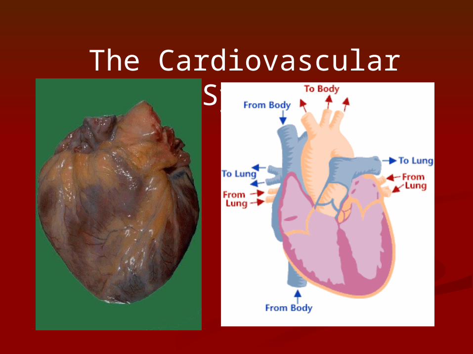
The Cardiovascular System
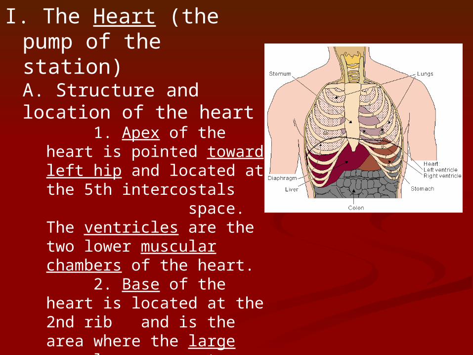
I. The Heart (the pump of the station)A. Structure and location of the heart
1. Apex of the heart is pointed toward left hip and located at the 5th intercostals
space. The ventricles are the two lower muscular chambers of the heart.
2. Base of the heart is located at the 2nd rib and is the area where the large vessels enter and leave the heart and the location of the two sac like atria chambers.
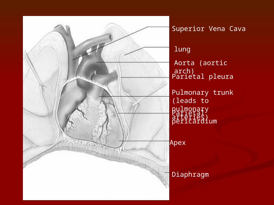
Superior Vena Cava
lung
Aorta (aortic arch)
Parietal pleura
Pulmonary trunk (leads to pulmonary arteries)Parietal pericardium
Apex
Diaphragm
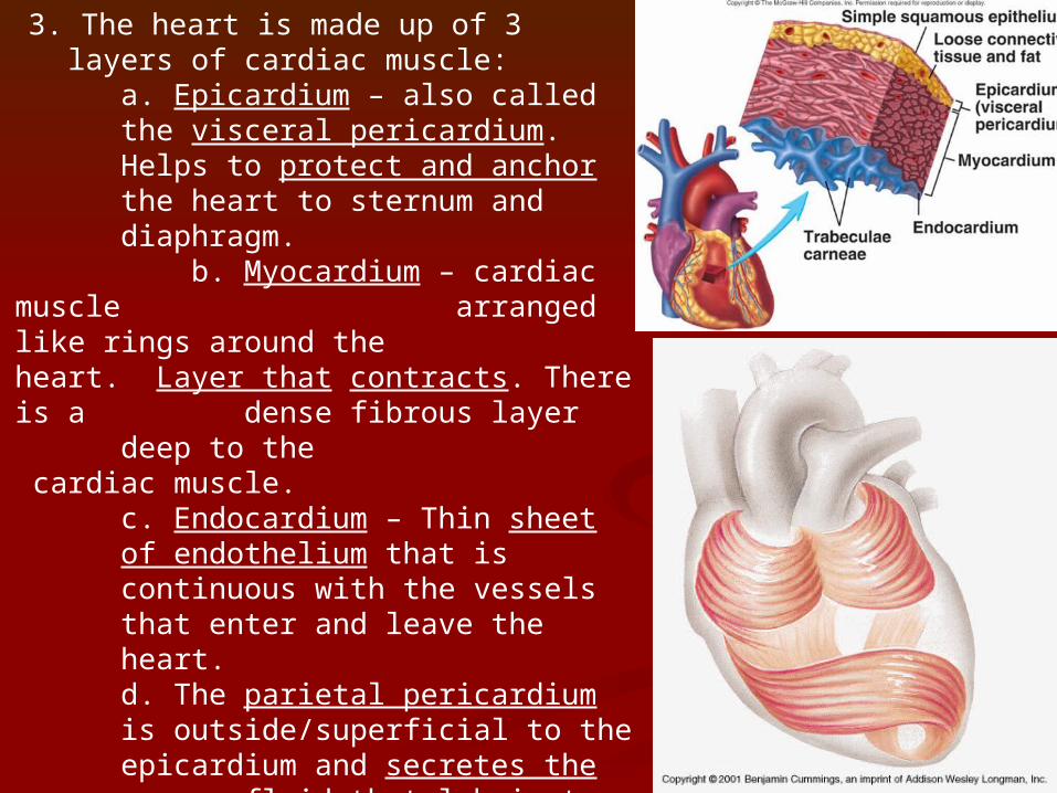
3. The heart is made up of 3 layers of cardiac muscle:
a. Epicardium – also called the visceral pericardium. Helps to protect and anchor the heart to sternum and diaphragm. b. Myocardium – cardiac muscle
arranged like rings around the heart. Layer that contracts. There is a dense fibrous layer deep to the cardiac muscle.
c. Endocardium – Thin sheet of endothelium that is continuous with the vessels that enter and leave the heart. d. The parietal pericardium is outside/superficial to the epicardium and secretes the serous fluid that lubricates the cardiac sac so the heart can beat in an almost frictionless environment.
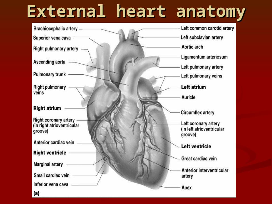
External heart anatomyExternal heart anatomy
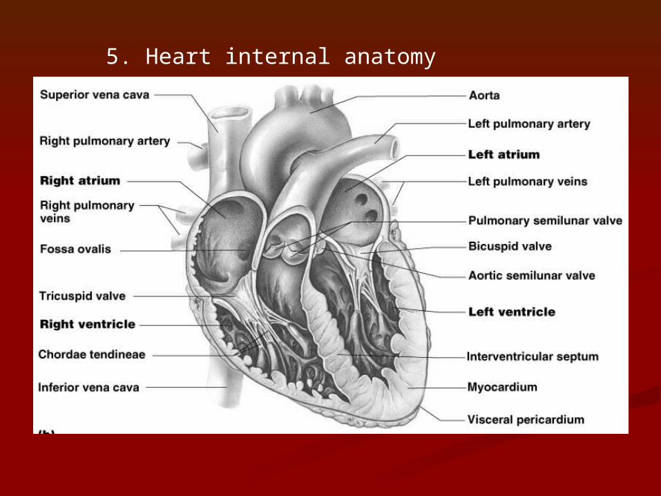
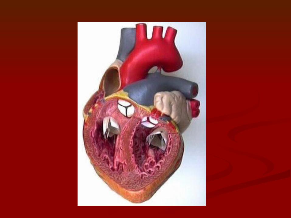
5. Heart internal anatomy
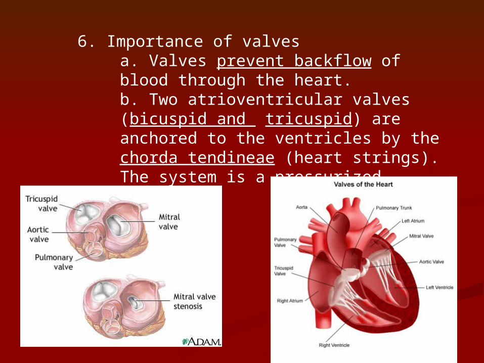
6. Importance of valvesa. Valves prevent backflow of blood through the heart. b. Two atrioventricular valves (bicuspid and tricuspid) are anchored to the ventricles by the chorda tendineae (heart strings). The system is a pressurized system.

B. 1. Blood passively fills the chamber, flaps hang down into the ventricles2. When the ventricles begin to contract, the pressure increases which forces the flaps of the valves closed. The chordae tenineae prevent the flaps from opening back up into the atria.
C. The two semilunar valves (pulmonary and aortic) have 3 cusps that fit tightly together. When the ventricles contract the force of the contractions open the valves into the pulmonary vein or aorta as the blood rushes into the vessel. As the ventricle relaxes the cusps come back together.
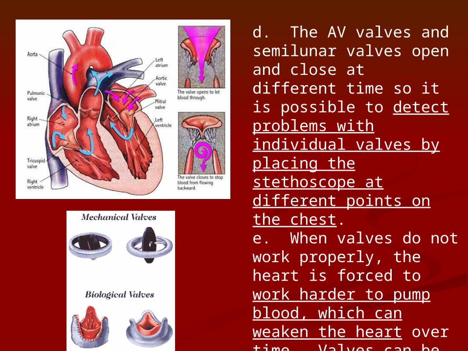
d. The AV valves and semilunar valves open and close at different time so it is possible to detect problems with individual valves by placing the stethoscope at different points on the chest. e. When valves do not work properly, the heart is forced to work harder to pump blood, which can weaken the heart over time. Valves can be replaced by pig or synthetic valves.
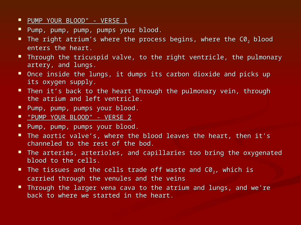
PUMP YOUR BLOOD" - VERSE 1PUMP YOUR BLOOD" - VERSE 1 Pump, pump, pump, pumps your blood. Pump, pump, pump, pumps your blood. The right atrium’s where the process begins, where the C0The right atrium’s where the process begins, where the C022 blood enters blood enters
the heart. the heart. Through the tricuspid valve, to the right ventricle, the pulmonary artery, Through the tricuspid valve, to the right ventricle, the pulmonary artery,
and lungs. and lungs. Once inside the lungs, it dumps its carbon dioxide and picks up its oxygen Once inside the lungs, it dumps its carbon dioxide and picks up its oxygen
supply. supply. Then it’s back to the heart through the pulmonary vein, through the atrium Then it’s back to the heart through the pulmonary vein, through the atrium
and left ventricle. and left ventricle. Pump, pump, pumps your blood. Pump, pump, pumps your blood. "PUMP YOUR BLOOD" - VERSE 2"PUMP YOUR BLOOD" - VERSE 2 Pump, pump, pumps your blood. Pump, pump, pumps your blood. The aortic valve’s, where the blood leaves the heart, then it's channeled to The aortic valve’s, where the blood leaves the heart, then it's channeled to
the rest of the bod. the rest of the bod. The arteries, arterioles, and capillaries too bring the oxygenated blood to The arteries, arterioles, and capillaries too bring the oxygenated blood to
the cells. the cells. The tissues and the cells trade off waste and C0The tissues and the cells trade off waste and C022, which is carried through , which is carried through
the venules and the veins the venules and the veins Through the larger vena cava to the atrium and lungs, and we're back to Through the larger vena cava to the atrium and lungs, and we're back to
where we started in the heart. where we started in the heart.
Heart soundsHeart sounds
http://depts.washington.edu/physdx/heart/demo.html
http://www.youtube.com/watch?v=q0s-1MC1hcE&feature=related Circulatory songCirculatory song
http://www.heartsite.com/html/http://www.heartsite.com/html/the_heart_3.htmlthe_heart_3.html
http://www.nlm.nih.gov/http://www.nlm.nih.gov/medlineplus/ency/anatomyvideos/medlineplus/ency/anatomyvideos/000021.htm000021.htm
Electrical conduction system Electrical conduction system animationanimation
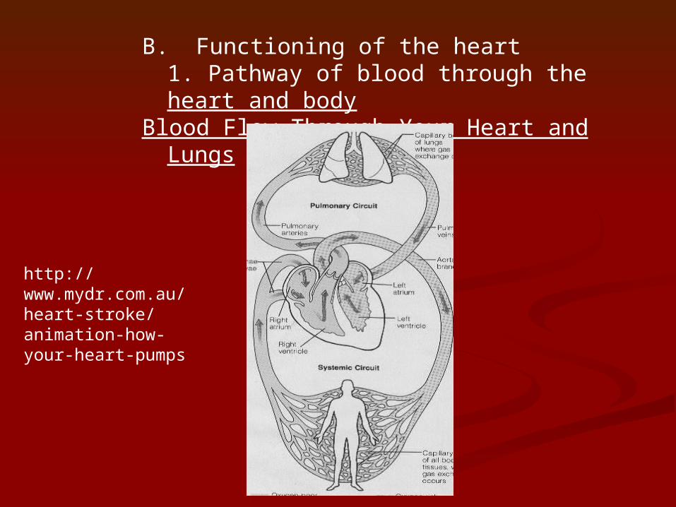
B. Functioning of the heart1. Pathway of blood through the heart and body
Blood Flow Through Your Heart and Lungs
http://www.mydr.com.au/heart-stroke/animation-how-your-heart-pumps
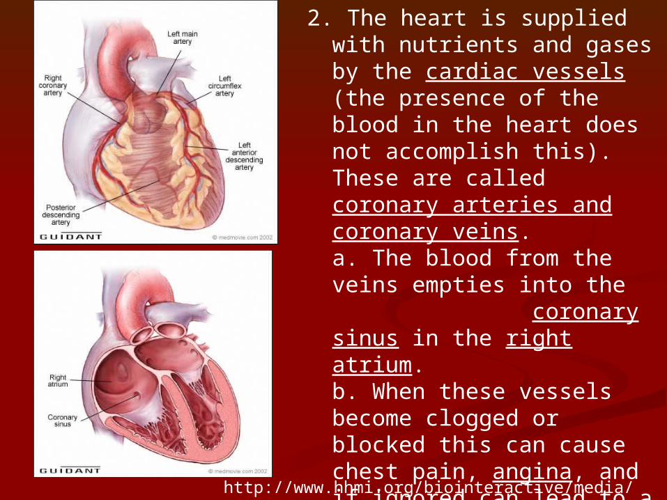
2. The heart is supplied with nutrients and gases by the cardiac vessels (the presence of the blood in the heart does not accomplish this). These are called coronary arteries and coronary veins. a. The blood from the veins empties into the coronary sinus in the right atrium. b. When these vessels become clogged or blocked this can cause chest pain, angina, and if ignored can lead to a cardiac infarction (tissue death from heart attack) when the cells are deprived of oxygen and nutrients
http://www.hhmi.org/biointeractive/media/heart_attack-lg.mov
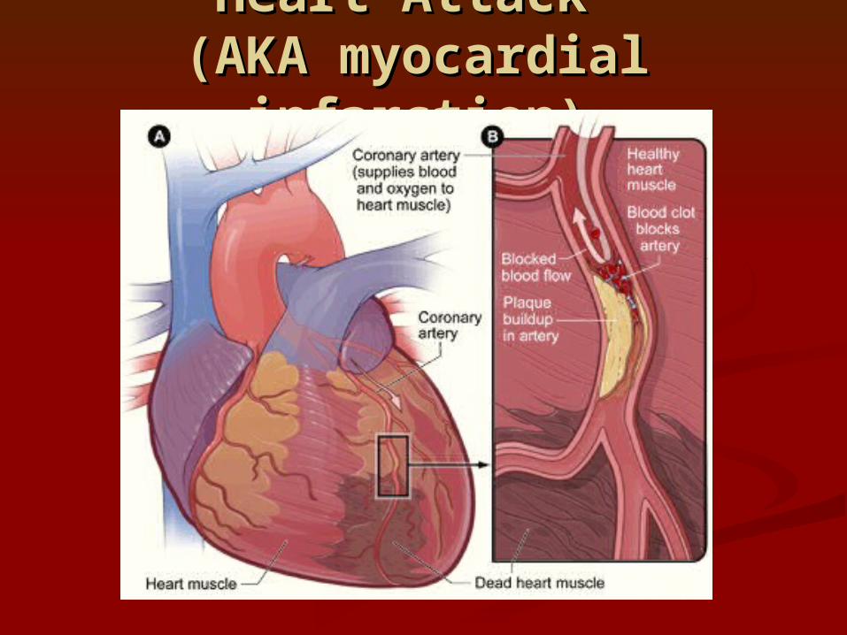
Heart Attack Heart Attack (AKA myocardial (AKA myocardial
infarction)infarction)
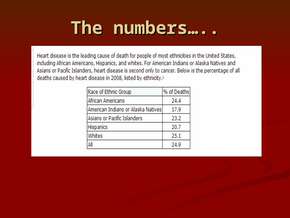
The numbers…..The numbers…..
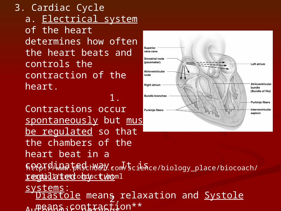
3. Cardiac Cyclea. Electrical system of the heart determines how often the heart beats and controls the contraction of the heart.
1. Contractions occur spontaneously but must be regulated so that the chambers of the heart beat in a coordinated way. It is regulated by two systems:
2. Autonomic nervous system controls the increase or decrease of heart rate.
Diastole means relaxation and Systole means contraction**
http://www.phschool.com/science/biology_place/biocoach/cardio1/intconduct.html
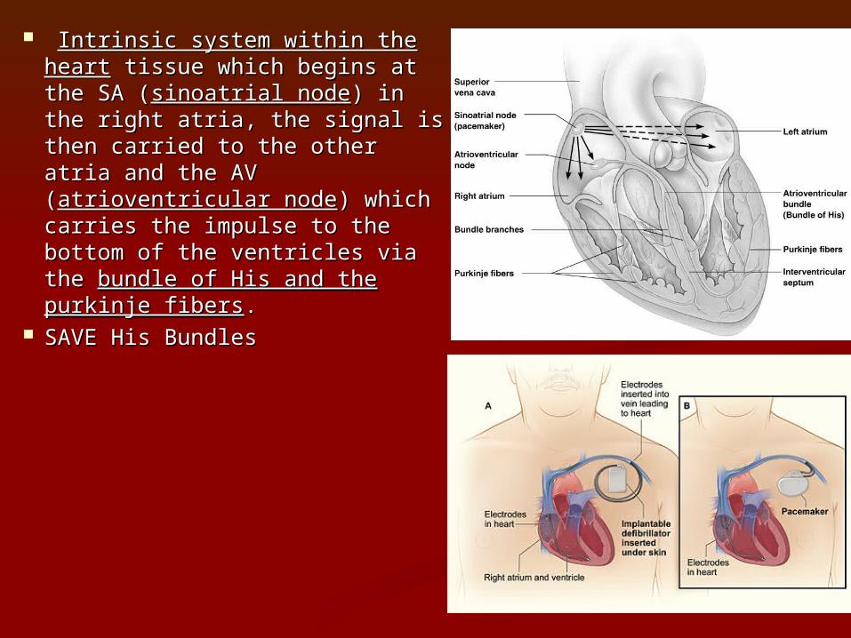
Intrinsic system within the Intrinsic system within the heartheart tissue which begins at tissue which begins at the SA (the SA (sinoatrial nodesinoatrial node) in the ) in the right atria, the signal is then right atria, the signal is then carried to the other atria and carried to the other atria and the AV (the AV (atrioventricular nodeatrioventricular node) ) which carries the impulse to which carries the impulse to the bottom of the ventricles via the bottom of the ventricles via the the bundle of His and the bundle of His and the purkinje fiberspurkinje fibers..
SAVE His BundlesSAVE His Bundles
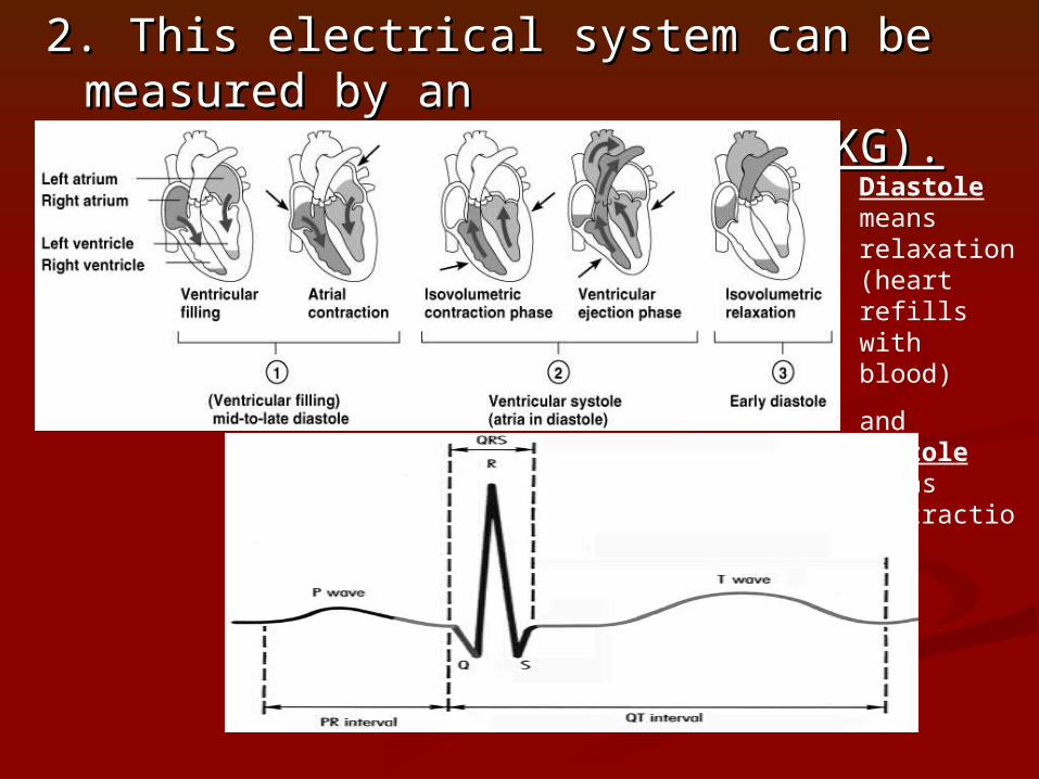
2. This electrical system can be 2. This electrical system can be measured by an measured by an electrocardiogram electrocardiogram (ECG or EKG).(ECG or EKG).
Diastole means relaxation (heart refills with blood)
and Systole means contraction**
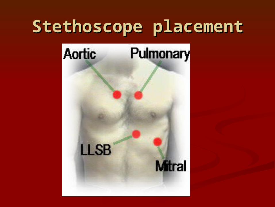
Stethoscope placementStethoscope placement
RECOVERY RATERECOVERY RATE
POOR less than 12 Beats Per Minute (BPM) recovery FAIR 12-20 Good 20-30 Excellent 30-40 Over 40 is outstanding.
C. Cardiac Output and the health of the heart1. Cardiac Output (CO) is a function of stroke
volume (volume of blood pumped out with each contraction and heart rate (number of beats per minute) CO = SV · HR , This stroke volume (SV), times the number of beats per minute (heart rate, HR), equals the cardiac output (CO). (avg. for male 5.L/min, female 4.5 L/min)
2. Factors affecting cardiac output and effects:Venous return – amount of
blood returning to the heart and enlarging the ventricles. The larger the venous return, the more stretched
the cells the stronger the contraction. Exercise can increase venous return with the enhanced squeezing action of the skeletal
muscles returning blood to the heart. Rapid heart rate or severe blood loss will decrease venous return.
b. Heart rate changes – HR can increase with the loss of blood or can be altered by other factors such as nervous stimulation (sympathetic nerves to speed up), hormones (epinephrine and thyroxine can increase), and changes in ions (lower potassium causes irregular heartbeat).
c. Age, gender, body fitness and body temperature. 1. Gender – females (72-80 avg. HR), males (64-72 avg. HR)
2. As you age your heart rate decreases, it is the fastest when you are a fetus (140 to 160)3. The colder you are the slower your heart rate.
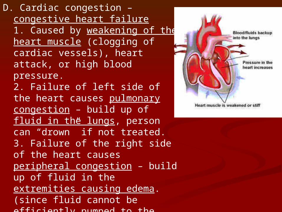
D. Cardiac congestion – congestive heart failure1. Caused by weakening of the heart muscle (clogging of cardiac vessels), heart attack, or high blood pressure.2. Failure of left side of the heart causes pulmonary congestion – build up of fluid in the lungs, person can “drown” if not treated. 3. Failure of the right side of the heart causes peripheral congestion – build up of fluid in the extremities causing edema. (since fluid cannot be efficiently pumped to the lungs)
http://www.muschealth.com/video/Default.aspx?videoId=10008&cId=7&type=rel
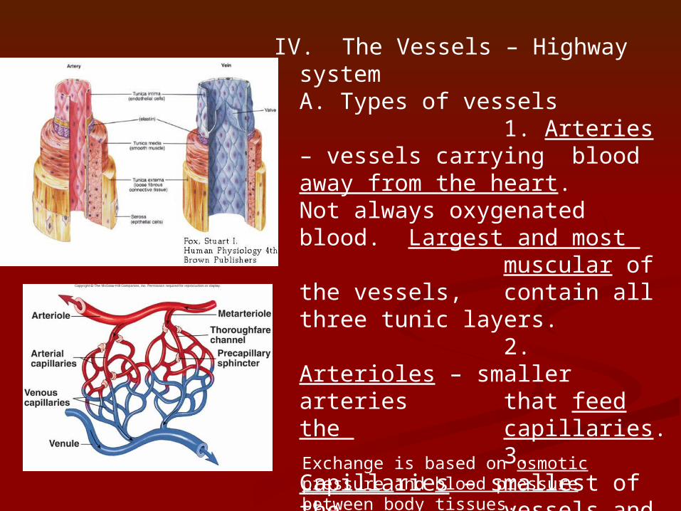
IV. The Vessels – Highway system A. Types of vessels
1. Arteries – vessels carrying blood away from the heart. Not always oxygenated blood. Largest and most muscular of the vessels, contain all three tunic layers.
2. Arterioles – smaller arteries that feed the
capillaries.3. Capillaries –
smallest of the vessels and the site of
exchange between blood vessels and body tissues. Are
only one cell layer thick.
Exchange is based on osmotic pressure and blood pressure between body tissues.
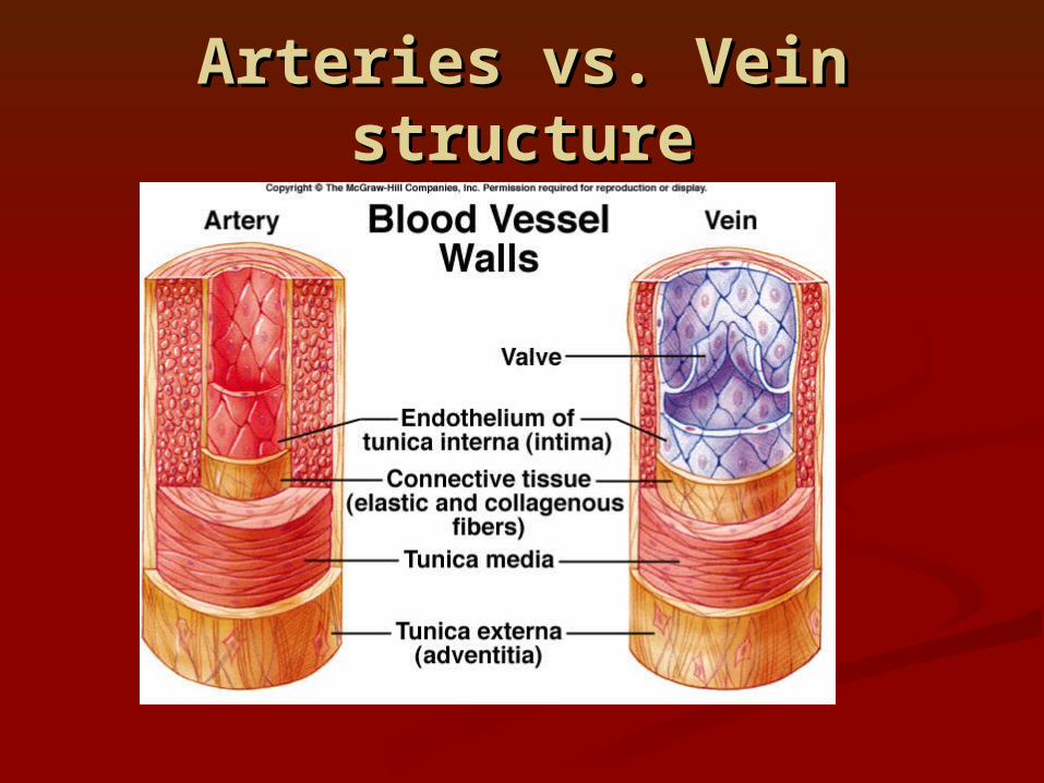
Arteries vs. Vein Arteries vs. Vein structurestructure
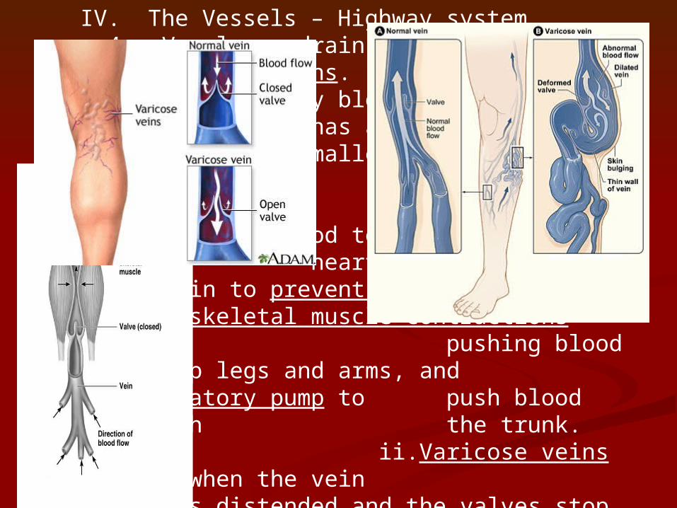
IV. The Vessels – Highway system 4. Venules – drain the capillary bed and lead to veins. 5. Veins – Carry blood back toward the heart. Vessel has a reduced smooth muscle layer (smaller and less muscular).
i.Adaptations to help return blood to the heart are valves within the vein to prevent back flow, skeletal muscle contractions
pushing blood back up legs and arms, and respiratory pump to
push blood through the trunk. ii.Varicose veins occur
when the vein becomes distended and the valves stop
functioning. Blood can pool and clot in these areas, which if the clot moves can be very dangerous.
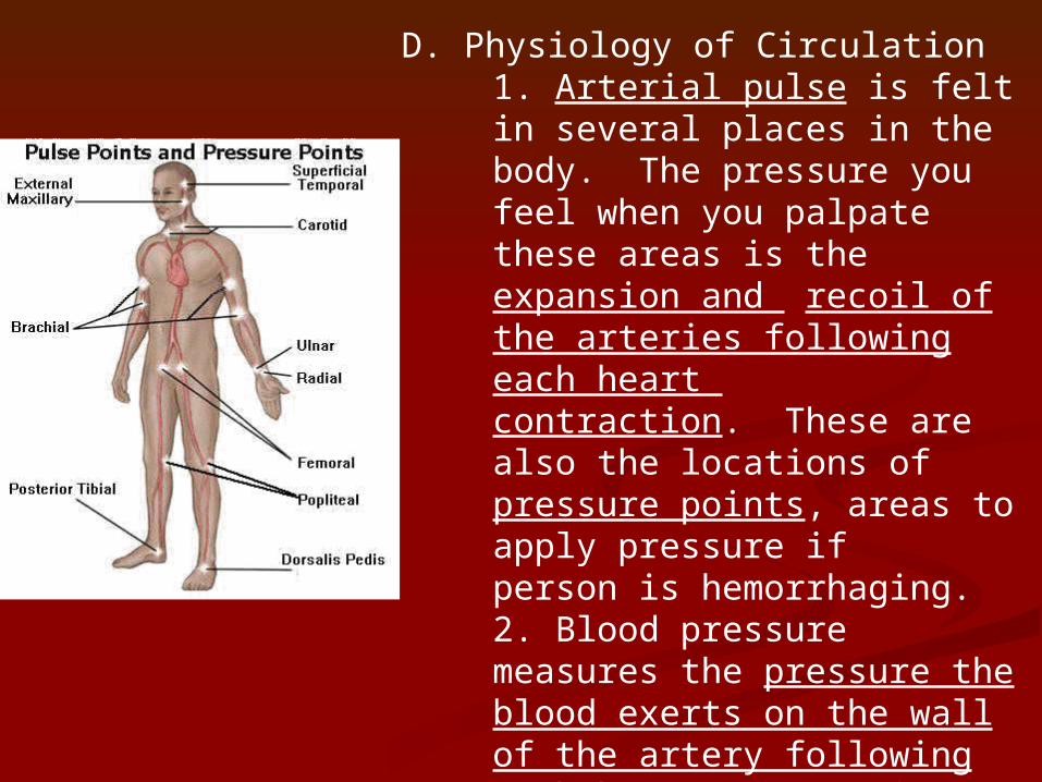
D. Physiology of Circulation1. Arterial pulse is felt in several places in the body. The pressure you feel when you palpate these areas is the expansion and recoil of the arteries following each heart contraction. These are also the locations of pressure points, areas to apply pressure if person is hemorrhaging. 2. Blood pressure measures the pressure the blood exerts on the wall of the artery following each heart contraction. The closer you are to the heart, the higher the pressure will be.
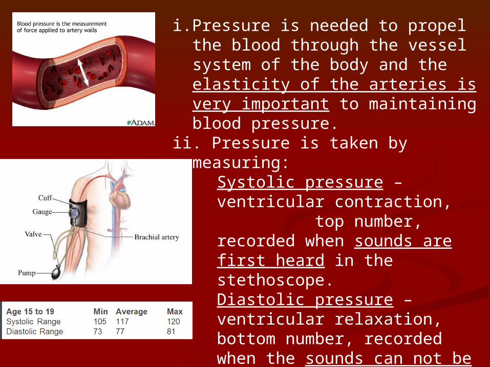
i.Pressure is needed to propel the blood through the vessel system of the body and the elasticity of the arteries is very important to maintaining blood pressure.
ii. Pressure is taken by measuring:Systolic pressure – ventricular contraction, top number, recorded when sounds are first heard in the stethoscope. Diastolic pressure – ventricular relaxation, bottom number, recorded when the sounds can not be heard in the stethoscope.
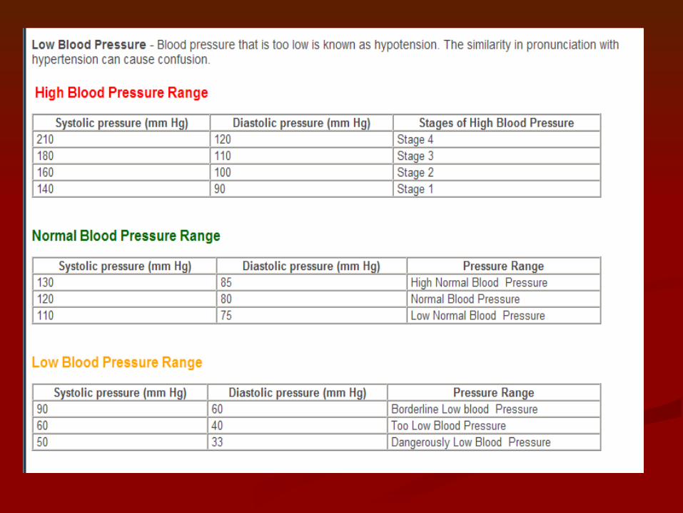
Normal blood pressure is 120/80Hypertension- high blood pressureHypotension – low blood pressure
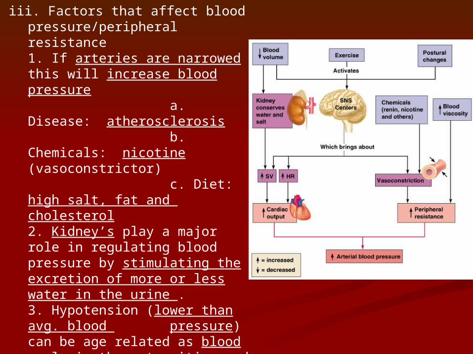
iii. Factors that affect blood pressure/peripheral resistance 1. If arteries are narrowed this will increase blood pressure
a. Disease: atherosclerosis
b. Chemicals: nicotine (vasoconstrictor)
c. Diet: high salt, fat and cholesterol2. Kidney’s play a major role in regulating blood pressure by stimulating the excretion of more or less water in the urine . 3. Hypotension (lower than avg. blood pressure) can be age related as blood pools in the extremities and when going from sitting to standing the BP drops suddenly. Can also be caused by major blood loss.
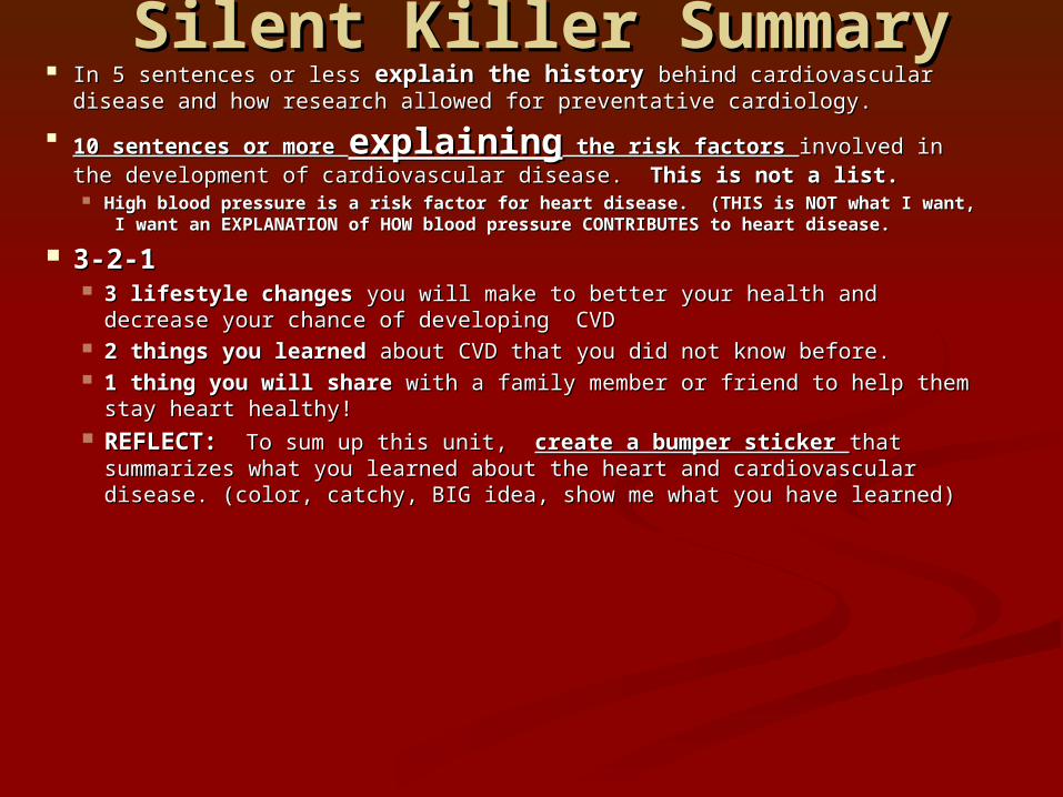
Silent Killer SummarySilent Killer Summary In 5 sentences or less In 5 sentences or less explain the historyexplain the history behind cardiovascular behind cardiovascular
disease and how research allowed for preventative cardiology.disease and how research allowed for preventative cardiology.
10 sentences or more 10 sentences or more explainingexplaining the risk factors the risk factors involved in involved in the development of cardiovascular disease. the development of cardiovascular disease. This is not a list. This is not a list. High blood pressure is a risk factor for heart disease. (THIS is NOT what I High blood pressure is a risk factor for heart disease. (THIS is NOT what I
want, I want an EXPLANATION of HOW blood pressure CONTRIBUTES to want, I want an EXPLANATION of HOW blood pressure CONTRIBUTES to heart disease. heart disease.
3-2-13-2-1 3 lifestyle changes 3 lifestyle changes you will make to better your health and decrease you will make to better your health and decrease
your chance of developing CVDyour chance of developing CVD 2 things you learned 2 things you learned about CVD that you did not know before. about CVD that you did not know before. 1 thing you will share1 thing you will share with a family member or friend to help them with a family member or friend to help them
stay heart healthy!stay heart healthy! REFLECT: REFLECT: To sum up this unit, To sum up this unit, create a bumper sticker create a bumper sticker that that
summarizes what you learned about the heart and cardiovascular summarizes what you learned about the heart and cardiovascular disease. (color, catchy, BIG idea, show me what you have learned)disease. (color, catchy, BIG idea, show me what you have learned)
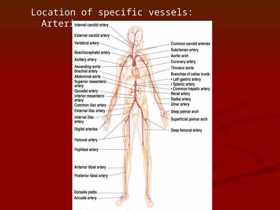
Location of specific vessels: Arteries
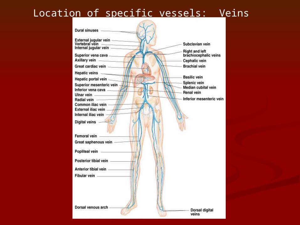
Location of specific vessels: Veins

http://www.nku.edu/~dempseyd/http://www.nku.edu/~dempseyd/heart%203%20labeled_files/heart%203%20labeled_files/frame.htmframe.htm Heart dissection labeled picturesHeart dissection labeled pictures
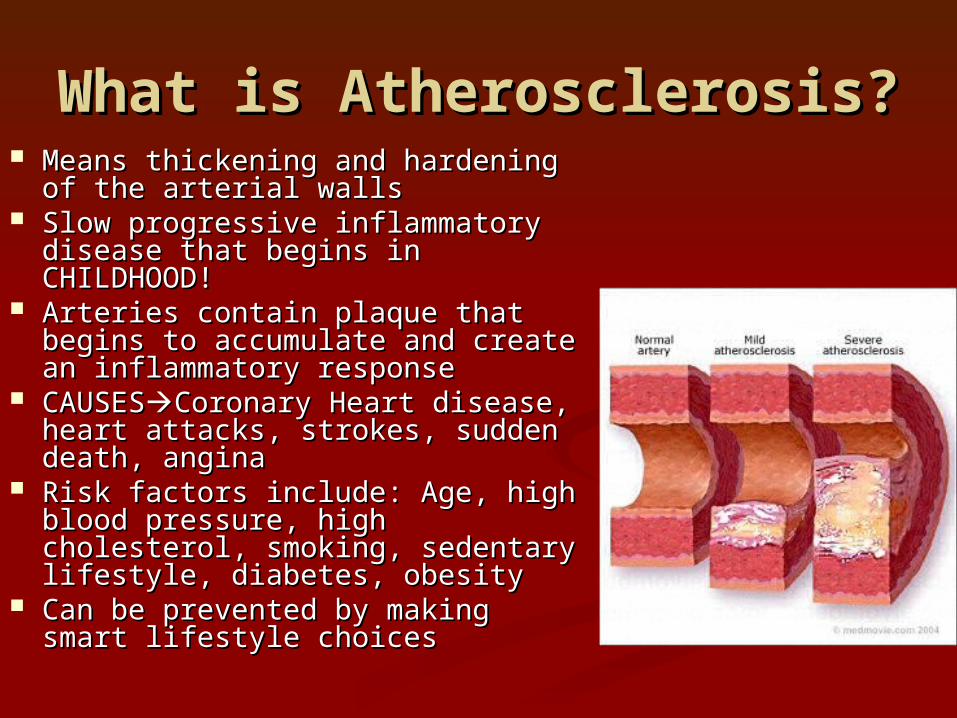
What is Atherosclerosis?What is Atherosclerosis? Means thickening and hardening Means thickening and hardening
of the arterial wallsof the arterial walls Slow progressive inflammatory Slow progressive inflammatory
disease that begins in disease that begins in CHILDHOOD!CHILDHOOD!
Arteries contain plaque that Arteries contain plaque that begins to accumulate and create begins to accumulate and create an inflammatory responsean inflammatory response
CAUSESCAUSESCoronary Heart Coronary Heart disease, heart attacks, strokes, disease, heart attacks, strokes, sudden death, anginasudden death, angina
Risk factors include: Age, high Risk factors include: Age, high blood pressure, high cholesterol, blood pressure, high cholesterol, smoking, sedentary lifestyle, smoking, sedentary lifestyle, diabetes, obesitydiabetes, obesity
Can be prevented by making Can be prevented by making smart lifestyle choicessmart lifestyle choices