glycogen, hyaluronate, and some other polysaccharides greatly
TRANSCRIPT
JOURNAL OF BACTERIOLOGY, June 1979, p. 663-670 Vol. 138, No. 30021-9193/79/06-0663/08$02.00/0
Glycogen, Hyaluronate, and Some Other PolysaccharidesGreatly Enhance the Formation of Exolipase by Serratia
marcescenstULRICH K. WINKLER* AND MARTINA STUCKMANN
Lehrstuhl fir Biologie der Mikroorganismen, Ruhr- Universitat, D-4630 Bochum, Federal Republic ofGermany
Received for publication 4 December 1978
Among 21 different polysaccharides tested, 5 greatly enhanced the spontaneousand cyclic AMP-induced formation of exolipase: glycogen, hyaluronate, laminarin,pectin B, and gum arabic. These polysaccharides have in common the tendencyto form highly ordered networks because of the branching or helical arrangement,or both, of their molecules. None of the polysaccharides could be utilized by thecells as the sole carbon source. Strong lipid extraction of four different polysac-charides did not reduce their exolipase-enhancing efficacy. At a constant celldensity the stimulation of exolipase formation by various concentrations ofglycogen followed saturation kinetics, suggesting a limited number of "sites" forthe glycogen to act. The active principle present in a solution of pectin wasdestroyed by degradation (,8-elimination) of the polymer. Hyaluronate lost itsexolipase-enhancing activity by exhaustive hydrolysis with hyaluronidase but wasresistant to proteinase K. Exopolysaccharide, isolated from growth medium ofSerratia marcescens SM-6, enhanced the exolipase formation as efficiently ashyaluronate. The results of this work are discussed mainly in terms of the"detachment hypothesis."
It was reported recently that the formation ofextracellular lipase ("exolipase") by Serratiamarcescens SM-6 was stimulated up to 100-foldwhen the standard growth medium was supple-mented with glycogen or pectin B (26, 27). Thepresent paper provides an initial experimentalanalysis and discussion of this new bacteriologi-cal phenomenon. Twenty-one different polysac-charides were screened for exolipase-enhancingactivity to determine chemical or structural re-quirements for this effect. Various wild-typestrains of S. marcescens were studied to seewhether the polysaccharidic stimulation of exo-lipase formation is restricted to strain SM-6.Kinetic measurements of the effect of glycogenon the yield ofexolipase activity were performed.Most of the experiments described in this
paper were perforned with mutant strainW1270, a derivative of S. marcescens SM-6 (28).This mutant has the advantage that exolipaseenhancement by polysaccharides can be studiedwithin 30 min.
MATERIALS AND METHODSBacteria. The strains used mainly were S. marces-
cens SM-6 F'lac+ (wild type) and double mutant
t Dedicated to Max Delbriick with gratitude.
W1270 (Cpd Cya), which lacks cyclic AMP (cAMP)-phosphodiesterase activity and requires exogenouscAMP for various cell functions, including the forna-tion of exolipase (28). In addition, S. marcescensstrains 862-57, 868-57, 4534-60, 6320-58, and 3607-60,kindly provided by B. Davis, Atlanta, Ga., were stud-ied; these strains all differ with respect to their 0- andH-antigens (Table 2). Enterobacter liquefaciensW1079 (syn. B1622) was obtained from E. T. Reese,Natick, Mass.
Media. Minimal medium M9 (M9 glc) contained 7g of Na2HPO4-2H20, 3 g of KH2PO4, 1 g of NH4Cl, 0.5g of NaCl, 4 g of glucose, 0.25 g of MgSO4 7H20, and0.02 g of CaCl2*2H20 per liter of demineralized water.Supplemented minimal medium (M9 glc-s) also con-tained 0.25% Casamino Acids (Difco). In some exper-iments glucose was replaced by glycerol.
Biochemicals. The polysaccharides used were ofthe highest grade of purity commercially available;they were purchased from Merck (Darmstadt, Ger-many), Serva (Heidelberg, Germany), Sigma (St.Louis, Mo.), and Pharmacia (Uppsala, Sweden). Pul-lulan was a gift from K. Wallenfels (Freiburg, Ger-many).
Induction of exolipase formation by cAMP.Minimal medium M9 glc-s was inoculated with mutantW1270 at an initial optical density at 580 nm (OD1o)of 0.5. After incubation on a gyratory shaker for 7 h at30°C, the OD.%o was usually 3.4. From this culture, 1part was mixed with 1 part of prewarmed glucose-freeM9 solution (diluted 1:5 in distilled water) and 1/25
663
Dow
nloa
ded
from
http
s://j
ourn
als.
asm
.org
/jour
nal/j
b on
30
Dec
embe
r 20
21 b
y 46
.32.
27.2
5.
664 WINKLER AND STUCKMANN
volume of a 0.1 M cAMP solution. After various timesof shaking at 300C, samples were quickly cooled in anice bath and centrifuged (4 min at 8,000 x g). The cell-free supernatant fluids obtained were immediatelyassayed for exolipase activity.
Testing of the exolipase-enhancing ability ofpolysaccharides. Two different tests were per-formed. (i) The polysaccharides were added to thegrowth medium (10 ml of M9 glc) of S. marcescensSM-6 at a final concentration of 0.4%. After 22 to 24 hof incubation in a rolling apparatus at 300C, the celldensity (OD,ie) was determined, the cells were re-
moved by two successive centrifugations, and the su-
pernatant fluids were assayed for exolipase, exopro-tease, and exonuclease activity. One control tube con-
tained plain M9 glc medium, and another one con-
tained M9 medium in which glucose was replaced by0.4% of the respective polysaccharide. All extracellularenzyme activities were normalized by dividing by theinitial cell density (OD5wa) of the original culture. (ii)Mutant W1270 was induced with 2 mM cAMP as
described above, except that the 1:5 diluted glucose-free M9 solution contained the polysaccharide to bestudied. All exolipase activities measured were cor-
rected for background, i.e., the enzyme activity foundin the absence ofcAMP and of the particular polysac-charide. Boiling of the polysaccharide solutions beforeuse (5 to 10 min) did not significantly alter theirexolipase-stimulating effect except in the case ofpectinB.
Isolation and chemical analysis of bacterialexopolysaccharide. S. marcescens SM-6 was grownin M9 sucrose (2%) medium on a gyratory shaker for48 h at 300C. After removal of the cells by centrifu-gation, crude exopolysaccharide was obtained by di-alysis against tap water and freeze drying. The mate-rial was then incubated for 90 min with trypsin, pan-creatic RNase, and DNase (10 Lg of each mli'). Afterheat inactivation of the enzymes, dialysis, and freeze-drying, the product obtained was used as partiallypurified exopolysaccharide. A qualitative analysis ofthe polysaccharide was performed with a sample hy-drolyzed with 1 N H2S04 at 1000C for 24 h. Thesolution was neutralized with Amberlite IRA 410.Thin-layer chromatographic analysis was performedon cellulose plates (Merck 5716) with a mixture (4:6:3)of pyridine, n-butanol, and distilled water (11). Fordetection of hexoses, pentoses, hexosamines, anduronic acids the thin-layer chromatographic plateswere developed with the appropriate reagent as de-scribed by Stahl (23).
Determination of protein and polysaccharide.Protein concentrations were estimated according toLowry et al. (17), using bovine serum albumin as astandard. Polysaccharide concentrations were deter-mined by weighing or by applying the phenol-sulfuricacid method (7), using dextran (molecular weight,50,000) as a standard.Assays of extraceilular enzymes. Exolipase was
assayed using p-nitrophenylpalrnitate (Serva) as thesubstrate (M. Thoner, Diplom-Thesis, University ofBochum, Bochum, Germany, 1973). Ten milliliters ofisopropanol containing 30 mg of p-nitrophenylpalmi-tate was mixed with 90 ml of 0.05 M Sorensen phos-phate buffer, pH 8.0, containing 207 mg of sodium
deoxycholate and 100 mg of gum arabic. A 2.4-mlamount of this freshly prepared substrate solution wasprewarmed at 370C and then mixed with 0.1 ml of cell-free supernatant fluid. After 15 min of incubation at370C, the OD41o was measured against an enzyme-freecontrol. One enzyme unit is defined as 1 nmol of p-nitrophenol enzymatically released from the substrateml-' min-'. Under the conditions described the extinc-tion coefficient of p-nitrophenol is e4io = 15,000 cm2mg'. The exoprotease was assayed according to Char-ney and Tomarelli (5) with the following modifica-tions. The enzyme substrate (azocasein, Serva) wasdissolved in 0.1; M phosphate buffer (pH 7.6) at aconcentration of 12.5 mg ml-'. Two milliliters of thissubstrate solution was mixed with 1 ml of cell-freesupernatant fluid and incubated at 300C for 30 min.One enzyme unit is defined as AOD40- 1.0 ml-' min-'.The exonuclease was assayed with RNA as a substrate(8).Enzymatic hydrolysis of hyaluronate. Potas-
sium hyaluronate was dissolved in 0.3 M phosphatebuffer, pH 5.3, and bovine testicular hyaluronidase(EC 3.2.1.35; Sigma) was added. After various times ofincubation at 300C, samples were taken and boiled for3 min each, and the amount of undegraded hyaluro-nate was estimated by applying the procedure ofTolksdorf et al. (24). Details of the method are givenin reference 2.Heat-induced -eliminon of pectin (1). Pec-
tin B was dissolved in 0.1 M sodium phosphate buffer,pH 6.8, at a concentration of 10 mg ml-' and heatedfor various times at 950C. Aliquots were taken tomeasure the decrease in viscosity (OSTWALD instru-ment, 0.6-mm capillary diameter, 22°C), the increasein absorbance at 235 nm, and the decay of the exoli-pase-enhancing capability.Enzymatic de-esterification of pectin. Pectin B
was dissolved in 0.05 N NaCl solution (5 mg mI').After neutralization pectin methylesterase (EC3.1.1.11, Sigma; 160 U/mg of protein) was added, andthe solution (300C) was kept at pH 7 by adding micro-liter amounts of0.5 N NaOH until the de-esterificationwas completed.
RESULTSScreening for exolipase-enhancing poly-
saccharides. Glycogen and pectin B are theonly polysaccharides presently reported to en-hance the formation of exolipase by S. marces-cens SM-6 (26, 27). Here 19 additional polysac-charides and 2 polyvinyls were tested for thesame effects. Three new exolipase-enhancingpolysaccharides were found (Table 1). The othersubstances tested, a list of which follows, wereinactive: arabic acid, sodium salt; amylopectin(?); amylose; carboxymethylcellulose; chondro-itin sulfate, sodium salt; colominic acid, sodiumsalt; ,f-cyclodextrine; dextran (molecular weight,60,000 to 90,000); dextran sulfate 500, sodiumsalt; Ficoll; inulin; mannan; polyethyleneglycol6000 (?), polygalacturonic acid, sodium salt; pol-yvinylalcohol; polyvinylpyrrolidone; pullulan;starch. (The polymers were tested under the
J. BACTERIOL.
Dow
nloa
ded
from
http
s://j
ourn
als.
asm
.org
/jour
nal/j
b on
30
Dec
embe
r 20
21 b
y 46
.32.
27.2
5.
ENHANCEMENT OF EXOLIPASE BY POLYSACCHARIDES 665
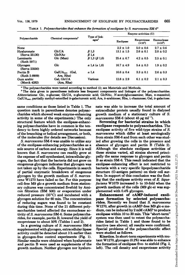
TABLE 1. Polysaccharides that enhance the formation of exolipase by S. marcescens SM-6aEnzyme activities (U)
Chal b ~~~~Tpeof ln-Polysaccharide Chemical components' ages Exoprotease ExonucleaseExolipase (x 103) (xoice)None 2.2 ± 1.0 5.0 ± 0.6 2.7 ± 0.6Hyaluronate GlcUA 6 1,3 13.1 ± 1.5 2.6 + 0.1 2.0 ± 0.2
(Serva 25130) GlcNAc ,B 1,4Laniinarin Glc (Man) ,6 1,3 (,B 1,6) 25.4 + 6.7 4.2 ± 0.5 2.5 ± 0.1
(Roth 5971)Glycogen Glc a 1,4 (a 1,6) 16.7 + 4.9 5.4 + 0.3 1.9 ± 0.2
(Serva 23550)Pectin B GalUA,t (Gal, a 1,4 16.6 ± 0.4 3.3 + 0.1 2.6 ± 0.3
(Roth 2-8909) Ara, Rha)Gum arabic Gal, GlcUA Various 12.8 ± 2.9 6.1 + 0.2 2.1 + 0.3
(Merck 4282) (Ara, Rha)
a The polysaccharides were tested according to method (i); see Materials and Methods.b The data given in parentheses indicate less frequent components and linkages of the polysaccharides.
Abbreviations: Glc, D-glucose; GlcUA, D-glucuronic acid; GlcNAc, N-acetylglucosamine; Man, D-mannitol;GalUA,n,t, partially methyl-esterified D-galacturonic acid; Ara, D-arabinose; Rha, L-rhamnose; Gal, D-galactose.
same conditions as those listed in Table 1. Thequestion mark in parentheses denotes polysac-charides which showed weak enzyme-enhancingactivity in some of the experiments.) The onlystructural feature which the exolipase-enhanc-ing polysaccharides have in common is the ten-dency to form highly ordered networks becauseof the branching or helical arrangement, or both,of the molecules (for details see Discussion).
S. marcescens SM-6 was unable to utilize anyof the exolipase-enhancing polysaccharides as asole source of carbon and energy. Since it is wellknown that S. marcescens can readily grow atthe expense of self-synthesized, intracellular gly-cogen, the fact that the bacteria did not grow onexogenous glycogen indicates that glycogen wasnot taken up by the cells. Experiments in searchof partial enzymatic breakdown of exogenousglycogen by the growth medium of S. marces-cens W1270 have failed so far. For this purposecell-free M9 glc-s growth medium from station-ary cultures was concentrated fivefold by Ami-con filtration (BM 500) or evaporation underreduced pressure (35°C) and incubated with aglycogen solution for 60 min. The concentrationof reducing sugars was found to be constantduring this time. None of the 21 different poly-saccharides tested affected the exonuclease ac-tivity of S. marcescens SM-6. Some polysaccha-rides, for example, pectin B, lowered the yield ofexoprotease to about 50% (Tables 1 and 2).
In a growing culture of S. marcescens SM-6supplemented with glycogen, extracellular lipaseactivity could be detected about 3 h earlier thanin glycogen-free control experiments (Fig. 1).Similar results were obtained when hyaluronateand pectin B were used as supplements of thegrowth medium. Neither glycogen nor hyaluro-
nate was able to increase the total amount ofextracellular protein usually found in dialyzedgrowth medium of a stationary culture of S.marcescens SM-6 (about 45 ,ug mnl-).Screening for bacterial strains in which
exolipase responds to polysaccharides. Theexolipase activity of five wild-type strains of S.marcescens which differ at least serologicallyfrom strain SM-6 and from each other was stud-ied after growing the cells in the presence andabsence of glycogen and pectin B (Table 2).Although the absolute exolipase activities ofthese strains varied, all strains showed princi-pally the same response to glycogen and pectinB as strain SM-6. This result indicated that theexolipase-enhancing effect is not restricted tobacteria with a very specific lipopolysaccharidestructure (0-antigen pattern) on their cell sur-face. In support of this conclusion was the find-ing that the exolipase activity even of E. lique-faciens W1079 increased 3- to 10-fold when thegrowth medium of the cells (M9 glc-s) was sup-plemented with 0.4% glycogen.Enhancement of cAMP-induced exoli-
pase formation by selected polysaccha-rides. Recently we found that S. marcescensW1270, after growth in cAMP-free M9 glc-s me-dium, can be induced by 2mM cAMP to produceexolipase within 10 to 30 min. This "short-term"system was then used to retest the polysaccha-rides listed in Table 1 and those found to beinactive (see above); all results were confirmed.Special problems of the polysaccharidic effectwere studied as follows.
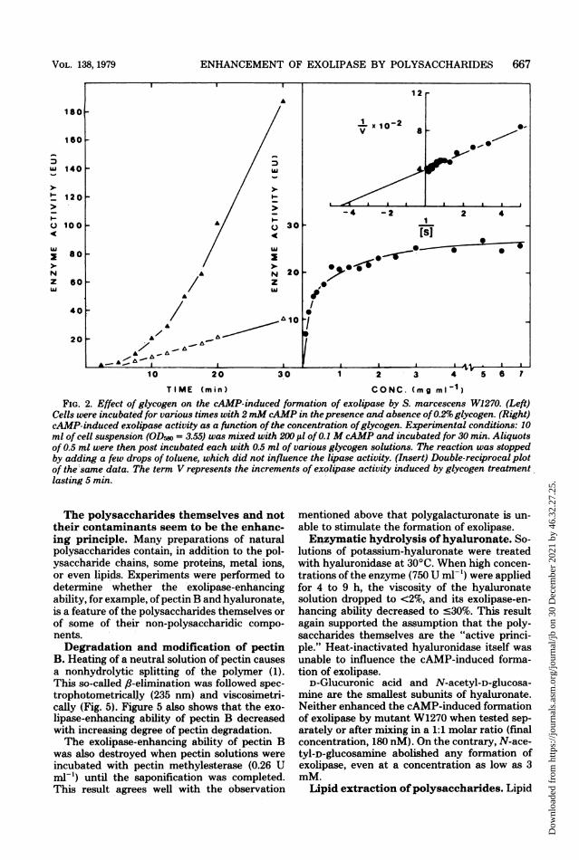
Kinetics. In short-term experiments with mu-tant W1270, glycogen (0.2%) was able to enhancethe formation of exolipase five- to sixfold (Fig. 2,left). The cell density and exoprotease activity
VOL. 138, 1979
Dow
nloa
ded
from
http
s://j
ourn
als.
asm
.org
/jour
nal/j
b on
30
Dec
embe
r 20
21 b
y 46
.32.
27.2
5.
666 WINKLER AND STUCKMANN
10n
0
a
z
la
Q
4 a 12 26
TIME( X,, )
FIG. 1. Effect ofglycogen (0.6%) on cellgrowth andexolipase activity of S. marcescens SM-6 grown in
M9glucose medium for various times. The generationtune of the cells in nonsupplemented medium was
approximately 50 min. An exolipase activity of 100%corresponds to 63 nmfol ofp-nitrophenol ml' min-'.Symbols: cell densities (A, A); enzyme activities (0,0); filled symbols indicate medium with glycogen andopen symbols indicate medium without glycogen.
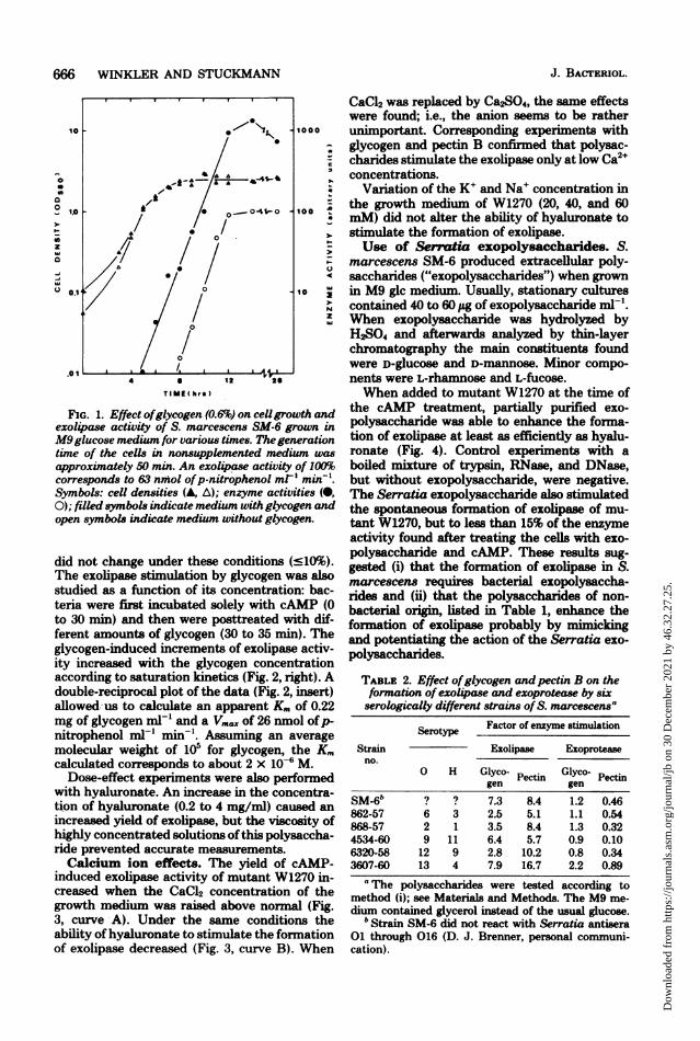
did not change under these conditions (:l10%).The exolipase stimulation by glycogen was alsostudied as a function of its concentration: bac-teria were first incubated solely with cAMP (0to 30 min) and then were posttreated with dif-ferent amounts of glycogen (30 to 35 min). Theglycogen-induced increments of exolipase activ-ity increased with the glycogen concentrationaccording to saturation kinetics (Fig. 2, right). Adouble-reciprocal plot of the data (Fig. 2, insert)allowed-us to calculate an apparent Km of 0.22mg of glycogen ml-' and a Vm.x of 26 nmol ofp-nitrophenol ml-' min-'. Assuming an averagemolecular weight of 105 for glycogen, the K.calculated corresponds to about 2 x 10⁻⁶ M.
Dose-effect experiments were also performedwith hyaluronate. An increase in the concentra-tion of hyaluronate (0.2 to 4 mg/ml) caused an
increased yield of exolipase, but the viscosity ofhighly concentrated solutions of this polysaccha-ride prevented accurate measurements.Calcium ion effects. The yield of cAMP-
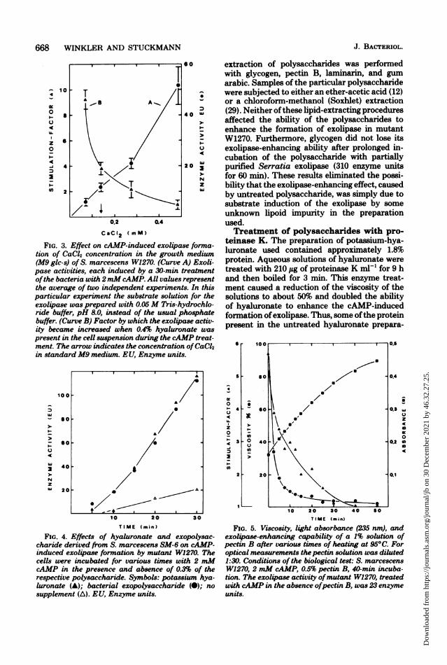
induced exolipase activity of mutant W1270 in-creased when the CaCl2 concentration of thegrowth medium was raised above normal (Fig.3, curve; A). Under the same conditions theability ofhyaluronate to stimulate the fornationof exolipase decreased (Fig. 3, curve B). When
CaCl2 was replaced by Ca2SO4, the same effectswere found; i.e., the anion seems to be ratherunimportant. Corresponding experiments withglycogen and pectin B confirmed that polysac-charides stimulate the exolipase only at low Ca2+concentrations.
Variation of the K+ and Na+ concentration inthe growth medium of W1270 (20, 40, and 60mM) did not alter the ability of hyaluronate tostimulate the formation of exolipase.Use of Sermtia exopolysaccharides. S.
marcescens SM-6 produced extracellular poly-saccharides ("exopolysaccharides") when grownin M9 glc medium. Usually, stationary culturescontained 40 to 60 Ag of exopolysaccharide mlP'.When exopolysaccharide was hydrolyzed byH2S04 and afterwards analyzed by thin-layerchromatography the main constituents foundwere D-glucose and D-mannose. Minor compo-nents were L-rhamnose and L-fucose.When added to mutant W1270 at the time of
the cAMP treatment, partially purified exo-polysaccharide was able to enhance the forma-tion of exolipase at least as efficiently as hyalu-ronate (Fig. 4). Control experiments with aboiled mixture of trypsin, RNase, and DNase,but without exopolysaccharide, were negative.The Serratia exopolysaccharide also stimulatedthe spontaneous formation of exolipase of mu-tant W1270, but to less than 15% of the enzymeactivity found after treating the cells with exo-polysaccharide and cAMP. These results sug-gested (i) that the formation of exolipase in S.marcescens requires bacterial exopolysaccha-rides and (ii) that the polysaccharides of non-bacterial origin, listed in Table 1, enhance theformation of exolipase probably by mimickingand potentiating the action of the Serratia exo-polysaccharides.
TABLE 2. Effect ofglycogen andpectin B on theformation of exol4pase and exoprotease by sixserologically different strains of S. marcescensa
Serotype Factor of enzyme stimulation
Strain Exolipase Exoproteaseno.
0 H Glyco- Pectin Glyco- Pectingen gen
b~~~~~~~~~~~~~~~~~~~~~~~~~~~~~~~~~~~~~~~~~~~~~~~~~~~~~~~SM-6" ? ? 7.3 8.4 1.2 0.46862-57 6 3 2.5 5.1 1.1 0.54868-57 2 1 3.5 8.4 1.3 0.324534-60 9 11 6.4 5.7 0.9 0.106320-58 12 9 2.8 10.2 0.8 0.343607-60 13 4 7.9 16.7 2.2 0.89aThe polysaccharides were tested according to
method (i); see Materials and Methods. The M9 me-dium contained glycerol instead of the usual glucose.
b Strain SM-6 did not react with Serratia antisera01 through 016 (D. J. Brenner, personal communi-cation).
J. BACTERIOL.
Dow
nloa
ded
from
http
s://j
ourn
als.
asm
.org
/jour
nal/j
b on
30
Dec
embe
r 20
21 b
y 46
.32.
27.2
5.
ENHANCEMENT OF EXOLIPASE BY POLYSACCHARIDES 667
I.-u
NzLa
1 2 r
IX 10-2x
8
- 4 -230-
20
A10
A
30
'B-A-,
2 4
[sI
90,-* 0 0
I.1
*0I
a AI I-
1 2 3 4 5 6 7
TIME (min) CONC. (mg mlI1)
FIG. 2. Effect of glycogen on the cAMP-induced formation of exolipase by S. marcescens W1270. (Left)Cells were incubated for various times with 2 mMcAMP in thepresence and absence of0.2% glycogen. (Right)cAMP-induced exolipase activity as a function of the concentration ofglycogen. Experimental conditions: 10ml of cell suspension (ODr, = 3.55) was mixed with 20X ,ul of 0.1 M cAMP and incubated for 30 min. Aliquotsof 0.5 ml were then post incubated each with 0.5 ml of various glycogen solutions. The reaction was stoppedby adding a few drops of toluene, which did not influence the lipase activity. (Insert) Double-reciprocal plotof the same data. The term V represents the increments of exolipase activity induced by glycogen treatment.lasting 5 min.
The polysaccharides themselves and nottheir contaminants seem to be the enhanc-ing principle. Many preparations of naturalpolysaccharides contain, in addition to the pol-ysaccharide chains, some proteins, metal ions,or even lipids. Experiments were performed todetermine whether the exolipase-enhancingability, for example, of pectin B and hyaluronate,is a feature of the polysaccharides themselves or
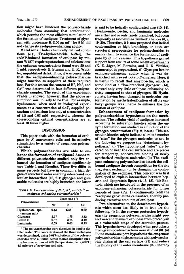
of some of their non-polysaccharidic compo-nents.Degradation and modification of pectin
B. Heating of a neutral solution of pectin causesa nonhydrolytic splitting of the polymer (1).This so-called fl-elimination was followed spec-trophotometrically (235 nm) and viscosimetri-cally (Fig. 5). Figure 5 also shows that the exo-lipase-enhancing ability of pectin B decreasedwith increasing degree of pectin degradation.The exolipase-enhancing ability of pectin B
was also destroyed when pectin solutions wereincubated with pectin methylesterase (0.26 Uml-) until the saponification was completed.This result agrees well with the observation
mentioned above that polygalacturonate is un-
able to stimulate the formation of exolipase.Enzymatic hydrolysis ofhyaluronate. So-
lutions of potassium-hyaluronate were treatedwith hyaluronidase at 30°C. When high concen-
trations of the enzyme (750 U ml-') were appliedfor 4 to 9 h, the viscosity of the hyaluronatesolution dropped to <2%, and its exolipase-en-hancing ability decreased to c30%. This resultagain supported the assumption that the poly-saccharides themselves are the "active princi-ple." Heat-inactivated hyaluronidase itself wasunable to influence the cAMP-induced forma-tion of exolipase.
D-Glucuronic acid and N-acetyl-D-glucosa-mine are the smallest subunits of hyaluronate.Neither enhanced the cAMP-induced formationof exolipase by mutant W1270 when tested sep-arately or after mixing in a 1:1 molar ratio (finalconcentration, 180 nM). On the contrary, N-ace-tyl-D-glucosamine abolished any formation ofexolipase, even at a concentration as low as 3mM.
Lipid extraction ofpolysaccharides. Lipid
A
180-
60F
FD
w 140
- 120
o 100
X 80
NZ 60w
I-I-
I-iA
40F
20 F
A
A
AI
A'.,A --
10 20
VOL. 138, 1979
Dow
nloa
ded
from
http
s://j
ourn
als.
asm
.org
/jour
nal/j
b on
30
Dec
embe
r 20
21 b
y 46
.32.
27.2
5.
668 WINKLER AND STUCKMANN
10 rf X10~~~~~~~~~~~~0
U.)
z ft.o
4 21I ~~~~~~~~N
2 uT
0.2 0.4
CSCI12 (mm)
FIG. 3. Effect on cAMP-induced exolipase forma-tion of CaC42 concentration in the growth medium(M9 glc-s) of S. marcescens W1270. (Curve A) Exoli-pase activities, each induced by a 30-min treatmentofthe bacteria with 2mM cAMP. All values representthe average of two independent experiments. In thisparticular experiment the substrate solution for theexolipase was prepared with 0.05M Tris-hydrochlo-ride buffer, pH 8.0, instead of the usual phosphatebuffer. (Curve B) Factor by which the exolipase activ-ity became increased when 0.4% hyaluronate waspresent in the cell suspension during the cAMP treat-ment. The arrow indicates the concentration ofCaC12in standard M9 medium. EU, Enzyme units.
1oo0
soj
I-
C.-
I
N
z
401
201
10 20 30
TIME (min)
FIG. 4. Effects of hyaluronate and exopolysac-charide derived from S. marcescens SM-6 on cAMP-induced exolipase formation by mutant W1270. Thecells were incubated for various times with 2 mMcAMP in the presence and absence of 0.3% of therespective polysaccharide. Symbols: potassium hya-luronate (A); bacterial exopolysaccharide (0); no
supplement (A). EU, Enzyme units.
extraction of polysaccharides was performedwith glycogen, pectin B, laminarin, and gumarabic. Samples of the particular polysaccharidewere subjected to either an ether-acetic acid (12)or a chloroform-methanol (Soxhlet) extraction(29). Neither of these lipid-extracting proceduresaffected the ability of the polysaccharides toenhance the formation of exolipase in mutantW1270. Furthermore, glycogen did not lose itsexolipase-enhancing ability after prolonged in-cubation of the polysaccharide with partiallypurified Serratia exolipase (310 enzyme unitsfor 60 min). These results eliminated the possi-bility that the exolipase-enhancing effect, causedby untreated polysaccharide, was simply due tosubstrate induction of the exolipase by someunknown lipoid impurity in the preparationused.Treatment of polysaccharides with pro-
teinase K. The preparation of potassium-hya-luronate used contained approximately 1.8%protein. Aqueous solutions of hyaluronate weretreated with 210 jig of proteinase K ml-' for 9 hand then boiled for 3 min. This enzyme treat-ment caused a reduction of the viscosity of thesolutions to about 50% and doubled the abilityof hyaluronate to enhance the cAMP-inducedformation of exolipase. Thus, some ofthe proteinpresent in the untreated hyaluronate prepara-
SIr
5
01-40C.)
z0I.- 3
-i3
2
90-
.00
co
TIME (min)
FIG. 5. Viscosity, light absorbance (235 nm), andexolipase-enhancing capability of a 1% solution ofpectin B after various times of heating at 95°C. Foroptical measurements thepectin solution was diluted1:30. Conditions of the biological test: S. marcescensW1270, 2 mM cAMP, 0.5% pectin B, 40-min incuba-tion. The exolipase activity ofmutant W1270, treatedwith cAMP in the absence ofpectin B, was 23 enzymeunits.
A
0
A
t-/;t+ I I
U
IL)z
a00)
-C04
J. BACTERIOL.
I
Dow
nloa
ded
from
http
s://j
ourn
als.
asm
.org
/jour
nal/j
b on
30
Dec
embe
r 20
21 b
y 46
.32.
27.2
5.
ENHANCEMENT OF EXOLIPASE BY POLYSACCHARIDES 669
tion might have hindered the polysaccharidemolecules from assuming that conformationwhich permits the most efficient stimulation ofthe formation of exolipase. Treatment of glyco-gen with proteinase K (150,ug ml1' for 9 h) didnot change its exolipase-enhancing ability.Metal ions. Under chemically defined condi-
tions (e.g., Tris-hydrochloride buffer), thecAMP-induced formation of exolipase by mu-tant W1270 requires potassium and calcium ions;the optimum concentrations found were 50 and1 mM, respectively (I. Stumm and U. K. Wink-ler, unpublished data). Thus, it was conceivablethat the exolipase-enhancing polysaccharidesmight function as suppliers of these requiredions. For this reason the content of K', Na+, andCa2" was determined in four different polysac-charide samples. The result of this experiment(Table 3) showed, however, that the above as-sumption was unlikely to be true. For example,hyaluronate, when used in biological experi-ments at a concentration of 0.4%, supplied po-tassium and calcium ions only at a concentrationof 4.5 and 0.03 mM, respectively, whereas thecorresponding optimal concentrations are atleast 10 times higher.
DISCUSSIONThis paper deals with the formation of exoli-
pase by S. marcescens cells and its selectivestimulation by a variety of exogenous polysac-charides.Which polysaccharides are able to en-
hance the formation of exolipase? Among 21different polysaccharides studied, only five en-hanced the formation of exolipase significantly(see Table 1 and Results). These five differ inmany respects but have in common a high de-gree of structural order enabling interstrand mo-lecular interactions (16, 21): glycogen and gumarabic molecules are highly branched; the latter
TABLE 3. Concentration ofNa', K+, and Ca2" inexolipase-enhancingpolysaccharide8a
Concn (mg g-')Polysaccharide
Na+ K+ Ca2+
Hyaluronate (po- 0.43 43.8 0.29tassium salt)
Glycogen 2.37 1.75 3.12Pectin B 0.87 3.75 3.12Laminarin 0.59 0.63 0.13
a The polysaccharides were dissolved in double-dis-tilled water. The concentration of the three metal ionswas determined, using 0.08% solutions of the polysac-charides, with a Perkin-Elmer atomic absorption spec-trophotometer, model 403 (temperature, ca. 2,400°C;4:6 mixture of acetylene and air).
is said to be helically configurated also (10, 14).Hyaluronate, pectin, and laminarin moleculesare either not or only rarely branched, but occurfrequently as (sometimes "kinked") helices (6,9,19, 20). Therefore, it is now proposed that helicalconformation or high. branching, or both, arestructural prerequisites for polysaccharides toenable them to enhance the formation of exoli-pase by S. marcescens. This hypothesis gainedsupport from results of some recent experiments(K. E. Jager, M. Portales, and U. K. Winkler,manuscript in preparation). (i) Glycogen lost itsexolipase-enhancing ability when it was de-branched with sweet potato,f-amylase. Here, itis useful to recall that amylopectin, which issome kind of a "low-branched glycogen" (14),showed only very little exolipase-enhancing ac-tivity compared to that of glycogen. (ii) Hyalu-ronate, having been changed in molecular con-formation by methylesterification of all its car-boxyl groups, was unable to enhance the for-mation of exolipase.Enhancement of exolipase formation by
polysaccharides: hypotheses on the mech-anism. The cellular yield of exolipase increasedaccording to saturation kinetics when the en-zyme formation was studied as a function of theglycogen concentration (Fig. 2, insert). This sat-uration kinetics might indicate a limited numberof "sites" for the glycogen molecules to act. Inthe following we propose the "detachment hy-pothesis." (i) The hypothetical "sites" are lo-cated on or near the cell surface and are placesfor temporary, noncovalent binding of newlysynthesized exolipase molecules. (ii) The exoli-pase-enhancing polysaccharides detach the cell-bound exolipase through competition for the site(i.e., steric exclusion) or by changing the confor-mation of the exolipase. This concept was firstdeveloped to explain interactions between hep-arin and lipoprotein lipase (4, 15, 18). (iii) Bac-teria which are incubated in the presence of anexolipase-enhancing polysaccharide for longerperiods of time (Fig. 1) continuously refill the"exolipase gaps" at the cell surface, thereby pro-ducing excessive amounts of exolipase.Two alternatives to the detachment hypoth-
esis which seem less likely to be true are thefollowing. (i) In the enzyme protection hypoth-esis the exogenous polysaccharides might pro-tect nascent chains of exolipase from proteolysisat a vulnerable stage of the secretion process.This hypothesis was developed when protoplastsfrom gram-positive bacteria were studied (3). (ii)In the membrane pore hypothesis the exogenouspolysaccharides might complex with polysaccha-ride chains at the cell surface (21) and reducethe fluidity of the outer membrane (22), thereby
VOL. 138, 1979
Dow
nloa
ded
from
http
s://j
ourn
als.
asm
.org
/jour
nal/j
b on
30
Dec
embe
r 20
21 b
y 46
.32.
27.2
5.
670 WINKLER AND STUCKMANN
helping the formation of relatively stable pore-like structures (25) for the transmembrane ex-port of exolipase molecules.The question of why the enzyme-enhancing
polysaccharides acted on exolipase but not onexoprotease and exonuclease (Table 1) remains.One answer could be that exoenzymes which areglycoproteins might preferentially bind to(polysaccharidic) cell surface sites (13) and re-spond to enzyme-enhancing polysaccharides.The exolipase produced by S. marcescens strainHY contains approximately 50% (wt/wt) firmlybound carbohydrate (J. Sossinka, personal com-munication).
ACKNOWLEDGMENTSWe are greatly indebted to G. Bergmann and H. Kaesler,
Bochum, for the quantitative detemiunation of Na+, K+, andCa2+ in some of our polysaccharide samples. We gratefullyacknowlege Betty Davis, Atlanta, Ga., for supplying us withserologically typed strains of S. marcescens and K. Wallenfels,Freiburg/Br., for his gift of pullulan. We thank Elke Schroderfor skillful assistance during the preparation and analysis ofthe exopolysaccharide of S. marcescens SM-6. Maria L. Por-tales kindly supplied us with a sample of partially purifiedSerratia exolipase.A Fulbright travel award allowed U. K. W. to visit the
University of California at San Diego where the manuscriptwas written.
LITERATURE CITED1. Albersheim, P., H. Neukom, and H. Deuel. 1960. Split-
ting of pectin chain molecules in neutral solutions. Arch.Biochem. Biophys. 90:46-51.
2. Boehringer, Mannheim. 1975. Boehringer biochemicainformation II, p. 167. Boehringer Mannheim Corp.,Mannheim, Germany.
3. Braatz, J. A., and E. C. Heath. 1974. The role ofpolysaccharide in the secretion of protein by Micrococ-cus sodonensig. J. Biol. Chem. 249:2536-2547.
4. Chajek, T., 0. Stein, and Y. Stein. 1978. Lipoproteinlipase of cultured mesenchymal rat heart cells. I. Syn-thesis, secretion, and releasability by heparin. Biochim.Biophys. Acta 528:456-465.
5. Charney, J., and R. M. Tomarelli. 1947. A colorirnetricmethod for the determination of the proteolytic activityof duodenal juice. J. Biol. Chem. 171:501-505.
6. Dea, I. C. M., R. Moorhouse, D. A. Rees, S. Arnott, J.K Guss, and E. S. Balazs. 1973. Hyaluronic acid: anovel, double helical molecule. Science 179:560-562.
7. Dubois, KL, K. A Gilles, J. KL Hamilton, P. A. Rebers,and F. Smith. 1956. Colorimetric method for determi-nation of sugars and related substances. Anal. Chem.28:350-356.
8. Eaves, G. N., and C. D. Jeffries. 1963. Isolation andproperties of an exocellular nuclease of Serratia mar-cescens. J. Bacteriol. 85:273-278.
9. Elyakowa, L A., and T. N. Zvyagintseva 1974. Astudy of the laminarins of some far-eastern, brownseaweeds. Carbohydr. Res. 34:241-248.
10. Glickanman, M., and R. E. Sand. 1973. Gum arabic, p.197-263. In R. L. Whistler and J. N. BeMiller (ed.),Industrial gums, 2nd ed. Academic Press Inc., New
York.11. Grune, A. 1957. Papierchromatographie und Papierelek-
trophorese. Chimia 11:173-203, 213-256.12. Kwapinski, J. 13. 1965. Methods of serological research.
John Wiley & Sons, New York.13. Lampen, J. 0. 1968. External enzymes of yeast: their
nature and formation. Antonie van Leeuwenhoek J.Microbiol. Serol. 34:1-18.
14. Lamer, J., B. Illingworth, G. T. Cori, and C. F. Cori.1952. Structure of glycogens and amylopectins. II. Anal-ysis by stepwise enzymatic degradation. J. Biol. Chem.199:641-651.
15. Laurent, T. C. 1977. Interaction between proteins andglycosaminoglycans. Fed. Proc. 36:24-27.
16. Lindahl, U., and M. Hook. 1978. Glycosaminoglycansand their binding to biological macromolecules. Annu.Rev. Biochem. 47:385-417.
17. Lowry, 0. H., N. J. Rosebrough, A. L Farr, and R. J.Randall. 1951. Protein measurement with the Folinphenol reagent. J. Biol. Chem. 193:265-275.
18. Olivecrona, T., G. Bengtsson, S.-E. Marklund, U.Lindahl, and M. Hook. 1977. Heparin-lipoprotein li-pase interactions. Fed. Proc. 36:60-65.
19. Park, J. W., and B. Chakrabarti. 1978. Conformationaltransition of hyaluronic acid. Carboxylic group partici-pation and thermal effect. Biochim. Biophys. Acta 341:263-269.
20. Rees, D. A. 1969. Structure, conformation, and mecha-nism in the formation of polysaccharide gels and net-works. Adv. Carbohydr. Chem. Biochem. 24:267-332.
21. Rees, D. A. 1972. Shapely polysaccharides. The EighthColworth Medal Lecture. Biochemn. J. 126:257-273.
22. Rottem, S., and L. Leive. 1977. Effect of variations inlipopolysaccharide on the fluidity of the outer mem-brane of Escherichia coli. J. Biol. Chem. 252:2077-2081.
23. Stahl, E. (ed.). 1967. Dunnschicht-Chromatographie. EinLaboratoriumshandbuch, 2nd ed. Springer-Verlag, Ber-lin.
24. Tolksdorf, S., M. H. McCready, D. R. McCullagh, andE. Schwenk. 1949. The turbidometric assay of hyalu-ronidase. J. Lab. Clin. Med. 34:74-89.
25. van Alphen, L., A. Verkleij, J. Leunissen-BiUvelt, andB. Lugtenberg. 1978. Architecture of the outer mem-brane of Escherichia coli. III. Protein-lipopolysaccha-ride complexes in intramembraneous particles. J. Bac-teriol. 134:1089-1098.
26. Wilnkler, U., and B. Folie. 1976. Mutative and environ-mental effects on the production of extracellular en-zymes by Serratia marcescen8, p. 157. In H. Dellweg(ed.), Fifth International Fermentation Symposium.Verlag Versuchs- und Lehranstalt fur Spiritusfabrika-tion und Fermentationstechnologie, Berlin.
27. Winkler, U., K. B. Heller, and B. Folle. 1978. Pleio-tropic consequences of mutations towards antibiotic-hypersensitivity in Serratia marcescens. Arch. Micro-biol. 116:259-268.
28. Winkler, U., H. Scholle, and L Bohne. 1975. Mutantsof Serratia marcescens lacking cyclic nucleotide phos-phodiesterase activity and requiring cyclic 3',5'-AMPfor the utilization of various carbohydrates. Arch. Mi-crobiol. 104:189-196.
29. Wober, W., and P. Alaupovic. 1971. Studies on theprotein moiety of endotoxin from gram-negative bacte-ria. Characterization of the protein moiety isolated byphenol treatment of endotoxin from Serratia marces-cens 08 and Escherichia coli 0 141:K85 (B). Eur. J.Biochem. 19:340-356.
J. BACTERIOL.
Dow
nloa
ded
from
http
s://j
ourn
als.
asm
.org
/jour
nal/j
b on
30
Dec
embe
r 20
21 b
y 46
.32.
27.2
5.