effet de la combinaison d'atrazine et uv-b sur le phytoplancton · two intensity of uv -b...
TRANSCRIPT
UNIVERSITÉ DU QUÉBEC À MONTRÉAL
EFFET DE LA COMBINAISON D' ATRAZINE ET UV-B SUR LE
PHYTOPLANCTON
MÉMOIRE
PRÉSENTÉ
COMME EXIGENCE PARTIELLE
DE LA MAÎTRISE EN BIOLOGIE
PAR
ANDRÉANNE GIRARD KEMP
AVRIL 2016

UNIVERSITÉ DU QUÉBEC À MONTRÉAL Service des bibliothèques
Avertissement
La diffusion de ce mémoire se fait dans le respect des droits de son auteur, qui a signé le formulaire Autorisation de reproduire et de diffuser un travail de recherche de cycles supérieurs (SDU-522- Rév.0?-2011 ). Cette autorisation stipule que «conformément à l'article 11 du Règlement no 8 des études de cycles supérieurs, [l 'auteur] concède à l'Université du Québec à Montréal une licence non exclusive d'utilisation et de publication de la totalité ou d'une partie importante de [son] travail de recherche pour des fins pédagogiques et non commerciales. Plus précisément, [l 'auteur] autorise l'Université du Québec à Montréal à reproduire, diffuser, prêter, distribuer ou vendre des copies de [son] travail de recherche à des fins non commerciales sur quelque support que ce soit, y compris l'Internet. Cette licence et cette autorisation n'entraînent pas une renonciation de [la] part [de l'auteur] à [ses] droits moraux ni à [ses] droits de propriété intellectuelle. Sauf entente contraire, [l 'auteur] conserve la liberté de diffuser et de commercialiser ou non ce travail dont [il] possède un exemplaire. »
L'expérience , ce n'est pas ce qui arrive à quelqu'un, c'est ce que
quelqu'un fait avec ce qui lui arrive.
A. Huxley , Le meilleur des mondes.

REMERCIEMENTS
Cette incroya ble expérience qu 'est de réaliser une maîtrise en biologie n'aurait
jamais été la même sans tous les gens formidables qui m'entoure. Je tiens donc à les
remercier du fond de mon cœur.
J'offre mes plus sincères remerciements à mon directeur de recherche,
Philippe Juneau , qui m 'a accueilli dans son laboratoire et qui m 'a p'ermis de mettre à
terme ce projet. Je lui suis également tellement reconnaissante pour ses conseils, ses
idées et son soutien. Je remercie aussi tous mes collègues de laboratoire : Francis
Racine pour ses explications de concept théorique; Kui Xu pour son aide avec les
appareils et les techniques de laboratoire; Marcelo Gomes pour son aide avec des
techniques de laboratoire ainsi que sa bonne humeur contagieuse et se~ très plaisantes
séances de chants au laboratoire . Je remercie également ma grande amie Sarah
Gingras Le Manac ' h pour son aide lors de manipulations, son soutien et les nombreux
fous rires qui ont transformé certaines journées de travail en moment inoubliable.
Finalement, je remercie Thibault Chesney pour ses conseils et son humour qui ont
aussi coopérés à rendre l'ambiance du laboratoire vraiment agréable.
Je remercie le professeur David Dewez pour l'accès à son laboratoire et
1'-utilisation de son U-HPLC. Je remercie aussi la technicienne du département de
chimie, Sylvie Lemieux qui , avec son expertise, m 'a permis de bien comprendre et
d ' utiliser rapidement cet appareil complexe.
Je remercie aussi Alice Doucet , pour l 'assistance qu 'elle m 'a offerte lors de
manipulation et ses petits rires qui ont contribué à rendre les journées de sondes bien
plus agréables.
Finalement, Je voudrais auss1 remerc1er Laurent Fraser. Merci pour les
merveilleux moments passer en ta compagnie, pour ton support et pour ta passion
contagieuse pour la science. Je te remercie aussi pour ta motivation débordante que tu
m'as transmise, pour ta confiance qui me fait me dépasser de jour en jour. Je te
remerci e pour tes idées géniales et, surtout , pour ton intérêt porté à mon projet qui me
redonne espoir en la science dans les moments les plus difficiles. Enfin, je n'oublie
pas de te remercier pour toute 1 'aide manuel que tu m'as offert, je te remercie donc
pour ta force, tes bras et tes outils .

TABLE DES MATIÈRES
Liste des tableaux ...... ..... .... ...... ............ .. ...... ... ... ......... .......... ... .... ........ ... ... ....... ........... vi
Li ste des figures ... ..... .................... .... ........ ...... ... .. ....... ............................. .. ........ .. ....... vii
Liste des abréviations .. ...... .. ......................... ....... ... ....... .... ... .... ... ... .............. ................ ix
RÉSUMÉ ........ .......... ... .............. ...... ............ .... .. .. : ... .. ..... ......... .......... .. ..... .. .. .. .......... .. xiii
INTRODUCTION GÉNÉRALE ...... ... ...... ....... ....... .... ... .. ...... .. .... ........ ......... .. ......... .... !
CHAPITRE I STRESS ENVIRONNEMENTAUX .... .. ...... ... ......... ......... ......... .. ... ...... .. .. ...... ...... .. ..... 6
1.1 Phytoplancton .. ..... .... ..... ... .... .. ..................................... .......... .. ... ...... ... ........ .... 6
1.2 Atrazine ...... .- ...... ............. .. ..... ......... ....... ..... ... .. ....... .. ..... .......... .... .. ... ... .... .. ...... . 6
1.3 Radiation ultraviolette ...... ........... .. ..... ... ........................................ .. ............ ..... 9
1.4 Combinaison de 1 ' atrazine et des UV -8 .... ..... ......... ....... .. ...... .. ..... .. ......... ..... 10
1 .5 Interaction de stress ....... ... .. .. .. ... .... .... ........ ...... ........ .... ... .... ....... ... .................. l 1
1 .6 FI uorescence ................. ... ........ .... ..... ...... ..... ...... ............... ..... .... .. ... ..... .. ... ..... 1 2
1.7 Protection du phytoplancton contre les stress ...... ........... .. ............. ...... .... ...... 15
1.8 Sensibilité du phytoplancton .. ... ..... ............ ... ..... .. ...... ... .. .. .... ... .... ...... ..... ....... 18
CHAPITRE II COM8INED EFFECT OF ATRAZINE AND UV-8 ON PHYTOPLANKTON ... ... 20
2.1 Abstract ... ... ................... ........... ............ .......... ........... ..... .. ........................ ... ... 21
2.2 Résumé ..... .. .. .. .... .. ... ... .... .. ....... ...... .. ..... ... ... ........... ... ............. .. ........ ............... 22
2.3 Introduction ........ ........... ............ ... ..... .. ... .................................... .... ... ............. 23
2.4 Mate rials and Methods .. ..... ... ............ ............. .. ..... ... ... ... ... .. ...... .. ...... ........ ..... 25
v
2 .4 .1 Cul tures .......... ... ........ ... ... .... .. .... ... .... .. .. .. ............ ... .. .. ... ... ...... .. .. .... ......... .... . 25
2 .4.2 Treatments ......... .... .. ... ... .. .. ..... ... ... ..... .... .......... .... ... .... .. ......... ......... ... ........ .. 25
2.4.3 Photosynthetic activity measurements ............ ...... ............... .... .... ..... .......... 27
2.4.4 Pigment analysis ..... .. ..... ........ .... ... ... ..... ........ ... ... ...... ... .. ... ..... ... ... .... .... ... .... 28
2 .4.5 Li pid Peroxydation .... .. ... .. .. ..... .. ....... ... .. .... ... .. .... .... .. .. ...... .. .... ... .. ........ ...... .. 29
2.4.6 Statistical analysi s ..... .... ...... .. .... .... ..... ....... .... ....... ...... ......... .... .. .. ... .. ...... ... .. 29
2.5 Results .... .... .. .... .... ... ....... .... ..... .. .. .. ........... ....... ..... .. ... ... ..... .... ..... ........ ....... ... ..... 3 1
2 .6 Di scussion ... .... ..... .. ...... .... ...... .... .. .. ... .. .... ... ... ........ .......... ... .. ..... .. ...... .... ... .... ... .. 37
2.6.1 Atrazine effect ....... ... ......... ..... .... ..... ... .... .... .. ... ..... ... .... ... .. ... ...... ..... .. ......... ... .. 37
2.6.2 UV-B effect ..... .... .... .. ... ...... ........ ..... ... ... ..... .... .. .... ...... ... ... ... ..... .... .......... .... . 39
2 .6.3 UV-B and atrazine combined effect .. .... .. ........ ... ... .. ... ..... ...... .. .. ..... ..... ...... .41
2 .7 Conclusion ..... ..... .. ... ........... .... .... .. ....... .... ..... .... .. ... ...... .... .. ...... .. .. .. .... ..... .. .. .. . 44
2.8 Acknowledgements ....... .... ..... ........... .... .... ... ..... .. .. ... .... .... .... .. ..... ... ... ... .. .... ... . 44
2 .9 Supplementary material .... ... .. ..... ....... .......... : ......... ...... ...... ... .... ... ... ... ... ..... ... .45
2.1 0 References ... ... ... .. ........ ...... .......... ..... ...... ....... ....... .. ......... ... ... ............... .. ... ... 46
CONCLUSION GENERALE ...... .. ......... ..... ... ... .... ... .... .... ..... .. .... ..... .... .. ... .... .. .. ........ . 54
LISTE DES RÉFÉRENCES .. ...... ..... .. ....... .. ...... .......... ........ .. ....... .... .... ... .. ...... ... .. ... ... 57
1
LISTE DES TABLEAUX
TABLEAUX PAGES
Table 2.1 Experimental treatments with the-control (PAR light only) and the
respective UV -B (Low UV-Bor high UV -B intensity ; wm-2) and atrazine (Atz;
!J.M) doses . ..... ...................... .................. .. ..... .. .. .. ........ ....... .. .............. .. ....... ......... ........ . 27
Table 2.2 Photosynthetic parameters of M. aeruginosa CPCC632, CPCC299 and S.
obliquus CPCC5 exposed to PAR light (188 ~J.mol photons m·2 s-1), two
intensities of UV -B light (0.1 wm-2and 0.2 wm-2) and atrazine (O.l!J.M) at
2:30PM .... .. .............. .. .. ... ... ... ......... .. ... ... .... .. ...... ................ .. ... ............ .......................... 31
Table 2.3 Pigments concentration (pg/cell of M. aeruginosa CPCC632, CPCC299
and S. obliquus CPCC5 exposed to PAR light (188 ~J.mol photons m·2 s·1), two
intensity of UV -B light (0.1 wm·2 and 0.2 wm-2) and atrazine (0.1 !-LM) at
2:30PM ................................................... ... ... ... .... ..... ... .. .. .... .. .. ...... .. ..... ............... .... ..... 35
Table lS . Pourcentages of photosynthetic parameters of M. aeruginosa CPCC632,
CPCC299 and S. obliquus CPCC5 exposed to PAR light (188 ~J.mol photons m·2
s-1), two intensities of UV -B light (0.1 Wm·2and 0.2 wm-2) and atrazine (O.l!J.M)
at 2:30PM . ... ....... .... ..... ............. .... ... ....................... ...... .. ..... ......... ......... ......... ...... .. ... ... 45

LISTE DES FIGURES
FIGURE PAGES
Figure 1.1 Schéma de l 'action de l'atrazine sur la photosynthèse . La chaine de
transport des électrons est illustrée avec tous ces composants. Le mécanisme
d 'action de 1 'atrazine est indiqué par des fl èches au niveau du PSII. Les Hèches
rouges représentent le sens du transport des électrons au sein du PSII depuis la
scission de l 'eau. La double barre oblique rouge représente l 'arrêt du transport
des électrons. La flèche en noire représente la liaison de l 'atrazine et les flèches
en pointillées noires , le retour d 'énergie créer par l'accumulation des électrons.
Dans la figure, Phe représente la phéophytine , (Mn)4 est le manganèse, PQ
représente la plastoquinone , Yz illustre la Tyrozine z, Atz est l'atrazine.
(Modifié de David Joly , UQTR) ........................................... .......... .. .. ........... ........ ... ..... 8
Figure 1.2 Voies de dissipation d ' énergie. La chlorophylle (Chi) absorbe la lumière ,
elle passe donc de l' état fondamental à l 'état excité ('Chi*). Pour revenir à l 'état
fondamental, 1 'énergie peut être réémise sous forme de ft uorescence ( 1) , elle
peut être utilisée pour la voie de la photochimie (2), être dissipée sous forme de
chaleur (3) ou être absorbée par les molécules d 'oxygène libres (4). En effet, la
chlorophylle excitée ('Chi *) peut produire un triplet de chlorophylle echl *) qui
peut à son tour produire des ROS ('02*; tirée de Müller et al., 2001) .... ... .......... ........ 17
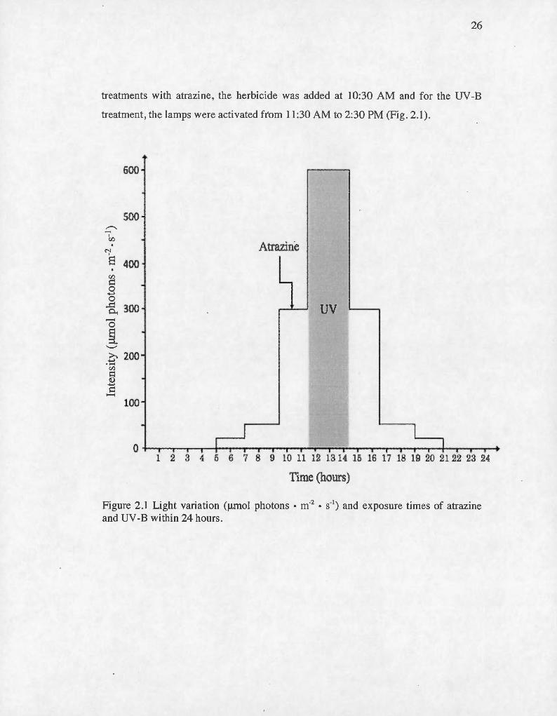
Figure 2.1 Light variation (J..Lmol photons • m·2 • s·') and exposure times of atrazine
and UV -B within 24 hours ..... ..... .............. .......... ..... .... .. .... .... .... .. ...... ...... .. ..... .... ..... ... . 26
Figure 2.2 Photosynthesis parameters of M. aeruginosa CPCC299 (a), CPCC632 (b)
and S. obliquus CPCC5 (c) exposed to PAR light (188 J..Lmol photons m·2 s· '),
two intensity of UV -B light (0.1 wm·2 and 0.2 wm-2) and atrazine (O.lf.LM) at
2 :30PM. The symbol * demonstrates a significant difference from the control
(p<0.05). The symbol ** shows a significant difference from the control (p<
0 .001) and the symbol # represent a significant difference from the respective
UV -B treatment (L-UV or H-UV) and from atrazine treatment. Analysis and
treatment means are from ANOVA (n=3) ............ ................................ ........ ... ........ .... 33
Figure 2.3 Cellular MDA content (x so-6 J..Lm/cell) of M. aerugino~a CPCC632,
CPCC299 and S. obliquus CPCC5 exposed to PAR light (188 J..Lmol photons m·2
s·'), two intensity of UV -B Iight (0.1 wm·2 and 0.2 wm-2 ) and atrazine (0.1 J..LM)

VIII
at 2:30PM. The symbol * represent a significant different difference from the
control (p<0.05). Analysis and treatment means are from ANOV A (n=3) ......... .... .... 36
'Chi, 3Chl
ABS
ADN
ANOVA
APC
APX
ATP
Atz
BBM
CAT
CFC
Chi
CPCC
Dl
D2
DCMU
LISTE DES ABRÉVIATIONS
Chlorophylle excitée sous forme de singulet et triplet
Dioxygène excité sous forme de singulet
Absorbance à 615nm
Absorbance à 650nm
Absorbance
Acide désoxyribonucléique
Analyse de variance
Allophycocyani ne
Ascorbate peroxydase
Adénosine triphosphate
Atrazine
« Bold basal medium »
Catalase
Chi orofl uorocarbone
Chlorophylle
Centre de culture phycologique du
Canada « Canadian Phycological culture center »
Protéine Dl
Protéine D2
3-(3 ,4-dichlorophenyl)-1 ,1-dimethylurea
(Diuron)
f'M l'état
F' 0
Fofcell
GR
HUFA
H-UY
LHC
L-UY
« Effective dissipation »
Probabilité de transport d'électron
Niveau de fluorescence maximale, à adapté à la lumière (centres réactionnels fermés)
x
Niveau de fluorescence minimale, à l'état adapté à la Lumière
FI uorescence à 50 flS
FI uorescence à 300 flS
FI uorescence à 2 ms
Rendement de fluorescence maximale lorsque les centres réactionnels des PSII sont fermés (quinones réduites)
Rendement de fluorescence minimal lorsque les centres réactionnels des PSII sont ouverts (quinones oxydées)
Fluorescence minimale par cellule
Fluorescence à une intensité lumineuse définie
Glutathion réductase
Peroxyde d ' hydrogène
Eau
« Highly unsaturated fatty ac id »
Forte dose d'ultraviolet « High UV »
Complexe collecteur de lumière du PSII « light harvesting complex »
Faible dose d ' ultraviolet « Low UV »
Ratio de photosystème Il fermé

Mn
MCPA
MDA
NPQ
PAM
PAR
PC
PEA
PQ
PSI
PSII
qP
RC
ROS
RuBisCo
SOD
A tome de manganèse
2-methyl-4chlorophenoxyacetic acid
Malondialdéhyde
«Non-photochemical quenching»
Radicaux superoxyde ·
Radicaux hydroxyde
«Pulse-Amplitude-Modulated fluorescence »
« Photosynthetic active radiation »
Phycocyanine
«Plant Efficiency Analyzer»
Plastoquinone oxydée
Photosystème 1
Photosystème II
Xl
Quinone A, accepteur primaire d 'électron du PSII
Quinone B, accepteur secondaire d'électron du PSII
Quenching photochimique
Centre réactionnel
Espèce d 'oxygène réactive (Reactive oxygen species)
Ri bul ose-1 ,5-bi sphosphate carboxylase/oxygénase
Superoxyde dismutase
« Maximal trapping rate »

uv
UV-A
UV-B
UV-C
Xli
Ultra violet
Ultra violet A
Ultra violet B
Ultra violet C
Taux de centre réactionnel fermé
Watt
Tyrosine z
Efficacité photochimique opérationnelle du PSU , à 1 'état stationnaire de transport des électrons.
Efficacité photochimique maximale du PSII , à 1 'état adapté à la noirceur
RÉSUMÉ
Le phytoplancton forme la base de la chaine alimentaire de beaucoup d'écosystèmes et fournit un énorme apport d'oxygène à notre planète. Cependant, l'épandage d ' herbicides dans les champs et les radiations UV-B sont des · stress environnementaux qui peuvent grandement affecter ces organismes. L'atrazine, l' herbicide Je plus retrouvé dans les eaux de surfaces et sous-terraines, nuit à la photosynthèse en provoquant une accumulation d 'électrons au niveau de la protéine Dl. Depuis la réduction de la eouche d'ozone, l 'intensité des rayons ultraviolets atteignant la surface de la Terre a été augmentée. En plus de produire des espèces d'oxygènes réactives (ROS) comme J'atrazine , ces rayons détruisent directement la protéine Dl et les pigments protecteurs de caroténoi·des. En observant comment ces deux stress interagissent au niveau de la photosynthèse, il a été possible d'évaluer J'effet de la combinaison des UV-B et de J'atrazine sur ces organismes. De plus, en mesurant cet effet sur plusieurs types de phytoplancton , il a été possible de comparer leur résistance. En chambre de croissance contrôlée, une algue verte, Scenedesmus obliquus (CPCC5), et de deux souches d ' une cyanobactérie, Microcystis aeruginosa (CPCC632 et CPCC299), ont été expose à 0,1 !-LM d'atrazine ainsi qu'à 3h d'UV -B (0,1 wm·2 ou 0,2Wm-2). Des mesures de photosynthèses ont été faites à l'aide de la fluorométrie (PAM et PEA) puis un dosage de la peroxydation des lipides et des pigments photosynthétiques a été effectué. Premièrement, cette combinaison de stress a démontré un effet synergique de la combinaison UV -B et atrazine au niveau de la photosynthèse chez S. obliquus . Cet effet serait dû à la dégradation des caroténoi.des servant à la protection contre les stress oxydatifs et à la dissipation de 1 'excès d'énergie lumineuse. Étonnamment, une différence de sensibilité a aussi été remarquée entre les deux souches de M. aeruginosa suggérant une différence mécanistique au sein de la même espèce. Au final, ces résultats démontrent que les études futures de protection de la vie aquatique devraient tenir compte de 1 'effet synergique des UV -B sur les différents pesticides retrouvés.
Mots clés : Algues, cyanobactéries, photosynthèse, herbicides, atrazine , ultraviolet, combinaison de stress.
INTRODUCTION GÉNÉRALE
Pour beaucoup d'écosystèmes , le phytoplancton constitue le premier maillon
de la chaîne alimentaire . Les algues et les cyanobactéries sont indispensables au bon
maintien de ces chaînes trophiques . En plus de leur importance alimentaire,
l'ensemble des organismes constituants le phytoplancton fournit environ 70% de
J'apport en oxygène à la planète ce qui en fait d'importants producteurs (Fenical,
1983). La plupart des organismes de la terre dépendent donc du bon maintien des
populations de phytoplancton pour survivre. La préservation de ces populations ne
doit absolument pas être négligée. Malheureusement, une grande partie de ces
organismes est sensible aux différents stress environnementaux (Neale et al .,
1998; Cloern , 1999; Rost et Wolf-Giadrow, 2008). Ainsi , lorsque ces organismes
subissent des stress, cela peut alors se répercuter sur tous les autres se trouvant dans
la chaîne. L'étude de l'impact des stress environnementaux sur ces organismes
devient alors très pertinente.
Depuis le début de l' agriculture, l' humain a essayé diverses stratégies afin de
minimiser les pertes agroalimentaires. L'utilisation d'herbicides est devenue le
moyen le plus populaire pour augmenter le rendement des récoltes. En plus, au
Canada, les herbicides sont plus utilisés que les insecticides (statistique Canada,
2008) en agriculture. En éliminant les plantes compétitrices à l'aide de ces produits,
la croissance et le développement des cultures sont maximisés. Malgré que ces
produits semblent être avantageux pour les récoltes, ils présentent énormément de
points négatifs . Ils se retrouvent dans les cours d'eau avoisinant les terrains agricoles
par ruissellement des eaux de pluie et par l'irrigation des sols (Warren et al., 2003).
Ainsi, les eaux de surfaces et souterraines se retrouvent contaminées et des
organismes non ciblés, tel que le phytoplancton, risquent d'être affectés
négativement (Lambert, 1984; Chen et al .-' 2007; Cain et al ., 2008). L'atrazine est
l' herbicide mis à l'étude au cours de cette recherche puisque son utilisation engendre

2
d'importants impacts sur l 'environnement et sur les organismes s'y retrouvant
(Graymore et al., 2001). Au niveau du phytoplancton, cet herbicide aura comme effet
d'inhiber leur photosynthèse et ainsi de réduire de beaucoup leur abondance dans les
cours d 'eau. En plus de l'abondance de phytoplanctons qui peut varier dû aux stress
environnementaux, la qualité nutritive du phytoplancton qui se retrouve dans les
cours peut aussi être bouleversé . En effet, certaines espèces plus tolérantes et de
moins bonne qualité nutritive peuvent devenir dominantes et ainsi avoir des
répercussions sur les organismes qui s'en ·nourri ssent. Heureusement, cet herbicide
ainsi que tous ceux provenant de la même famille sont désormais interdits par
l'Union européenne (Chèvre et Erkman, 2011). Par contre, l'atrazine reste l'un des
herbicides les plus utilisés dans le monde (Hayes et al, 2010). On le retrouve, entre
autres, aux États-Unis (Hayes et al, 2010) et au Canada (santé Canada , 2013) à des
concentrations allant jusqu'à 100 !J.g/L dans les cours d'eau (DeLorenzo, 2001).
L'activité humaine des dernières décennies n'a pas seu lement eu des
répercussions au ni veau terrestre. Pendant plusieurs années, des substances nocives
affectant la couche d'ozone ont été rejetées en grande quantité dans environnement.
Les hydrocarbures halogénés (halon), utilisés comme retardateur de flamme
(McCulloch, 1999), les chlorofluorocarbures (CFC) utilisés comme substance
réfrigérante (Hodnebrog et al., 2013) et les tétrachlorures de carbone, retrouvés par
exemple dans les produits · nettoyants domestiques (santé Canada, 2010) sont des
exemples de ces gaz pouvant détruire la couche d'ozone. Cette couche est
extrêmement importante puisqu'elle sert à la protection contre les rayons ultraviolets
(UV) provenant du soleil. Depuis une vingtaine d'années maintenant, une
augmentation de ces rayons pouvant avoir divers effets sur les organismes
photosynthétiques a été observée (Organisation météorologique mondiale, 2011). La
production d'oxygène réactif et la destruction des pigments de chlorophylle sont des
exemples d'effets qui résultent, tout comme pour l'atrazine, en une diminution de la
3
photosynthèse (Xiong, 2001; He et Hader, 2002 et Chen et al., 2012 ). Des lois ont
alors été mises en vigueur afin de réduire au maximum la destruction de cette couche
(Secrétariat de l 'ozone , 2006). Par contre, malgré l'interdiction de l'émission de la
majorité des gaz détruisant l'ozone , il s'avère que ceux-ci ont une durée de vie très
longue dans la stratosphère (Madronich et al., 1998). Donc , les effets nocifs que ces
gaz provoquent sur 1 'environnement se feront ressentir pour plusieurs années encore
(McKenzie et al., 2003).
En général, les rayons ultraviolets ont des effets négatifs au mveau de la
photosynthèse et de la croissance d'organismes photosynthétique (Holzinger and
Lütz, 2006; Fernanada Pessoa, 2012; Jansen et al., 2012; Zlatev et al., 2012). Par
contre, les radiations ultraviolettes se divisent en trois groupes correspondant à des
gammes de longueurs d'onde précises: les UV-A, les UV-B et les UV-C, et chacun de
ces groupes affectent différemment les organismes vivants (Fernanda Pessoa, 2012).
Ainsi, les UV-A , qui correspondent aux longueurs d'onde entre 320 et 390 nm, sont
les plus présents à la surface de notre planète, mais sont aussi les moins nocifs pour
les organismes. Les UV -B qui correspondent aux longueurs d 'onde entre 280 et
320nm sont beaucoup plus dangereux et sont habituellement absorbés en grande
quantité par la couche d'ozone (Lucas et al., 2006) . Par contre, depuis la détérioration
de celle-ci, ce type de radiation augmente considérablement à la surface terrestre
(Frederick, 1993). C'est pourquoi il s'agit de la classe d'UV dont l'amplification est
la plus redoutée. Finalement, les UV -C , qui se situe entre les longueurs d 'onde de 150
à 280 nm sur le spectre de la lumière sont extrêmement dangereux (Lucas et al.,
2006). Par contre, vu leur très basse longueur d'onde, ils sont très facilement absorbés
par la couche d'ozone (Holzinger et Lütz, 2006) et même une très mince couche
d 'ozone les empêche de pénétrer la stratosphère. Pour cette raison, ils ne sont
pratiquement pas présents à la surface terrestre et présentent ainsi un danger moindre
pour les organismes. Les UV-B sont alors les types de radiation les plus pertinentes à
4
étudier putsque ce sont ces rayons dont 1 ' intensité augmente à cause de la
détérioration de la couche d'ozone .
Les herbicides et les radiations ultraviolettes possèdent des effets puissants sur
les végétaux. Les algues et les cyanobactéries dulcicoles sont affectées par les
herbicides épandus dans les champs agricoles (Lambert, 1984; Cain et al. , 2008) et
par les rayons UV (Holzinger and Lütz , 2006; Fernanda Pessoa, 2012) . Beaucoup
d'études se sont penchées d ' une part sur les effets des radiations ultraviolettes (Vass et
al., 2005; Holzinger et Lütz, 2006; Xu et Gao , 2010; Fernanda Pessoa , 2012; Jansen
et al., 2012) et d'autre part sur les effets de l 'atrazine sur le phytoplancton (Fairchild et
al, 1998; De Lorenzo, 2001 ; Graymore et al., 2001; Fedtke et Duke,
2005; Ramakri shnan et al., 2010; Chalifour et Juneau , 2011). Par contre, aucune étude
n'a été faite sur leurs effets combinés. Quelques études existent sur la combinaison
d 'UV et d 'herbicides comme le paraquat , le glyphosate, le DCMU , le MCPA et le
simetryn (Kasai et Arts, 1997 ; Wang et al, 2007; Chen et al, 2012). En revanche ,
aucun de ces chercheurs ne se sont penché sur la combinaison atrazine et UV-B.
Malgré que 1 'effet du DCMU et du simetryn soit similaire à celui de 1 'atrazine , ces
herbicides ne sont pas couramment utilisés au Canada. Ils ne se retrouvent donc pas ou
très peu dans nos cours d'eau. L 'étude de ces herbicides se trouve, par ce fait, moins
pertinente que celle de l'atrazine .
Le but premier de ce projet de maîtrise est d 'étudier l 'effet de la combinaison
de l'atrazine et des rayons UV-B sur des organismes phytoplanctoniques. Afin d'y
parvenir, il faudra atteindre deux objectifs distincts : 1) découvrir comment ces deux
stress interagiront entre eux au niveau de la photosynthèse: 2) observer si une espèce
ou une souche de phytoplancton est plus résistante qu ' une autre à cette combinaison
de stress. Afin d 'élaborer sur le sujet, il faudra tout d 'abord définir et expliquer
quelques concepts de base. Premièrement , le phytoplancton, le rayonnement
ultraviolet et l'atrazine seront présentés . Ensuite, les effets physiologiques de ces
5
deux stress environnementaux sur les organi smes phytoplanctoniques seront
développés. Les effets physiologiques connus et attendus de leurs combinaisons
seront aussi élaborés suivis d ' une revue sur la protecti on du phytoplancton contre les
stress. Puis, notre vision de la synergie ainsi que les détail s sur les paramètres
photosynthétiques étudiés seront expliqués. Finalement, les diverses ex périences
effectuées seront présentées sous fo rme d ' un article scientif ique .
CHAPITRE!
STRESS ENVIRONNEMENTAUX
1.1 Phytoplancton
Le phytoplancton constitue les micro-organismes photosynthétiques vivant
dans les cours d'eau tels que les algues vertes et les cyanobactéries . Ils constituent le
premier maillon de la chaîne alimentaire pour beaucoup de réseaux· trophiques d'où
leur énorme importance. Les cyanobactéries sont des organismes procaryotes , qui
comme les végétaux font de la photosynthèse (Raven et al. 2003) . Par contre,
lorsqu 'elles sont présentes en trop grande quantité, elles peuvent devenir
extrêmement nuisibles pour les organismes partageant leurs habitats. En effet, ces
bactéries ont la possibilité de produire des toxines qui peuvent être neurotoxiques,
comme . les anatoxines, ou hépatotoxiques , comme les microcystines et les
nodularines (Codd et al., 1989). Ces toxines se retrouvent dans les cours d'eau et
peuvent devenir problématiques pour les organismes qui y vivent ou qui s'y
abreuvent (Wei er, 1982). Or, toutes les cyanobactéries ne sont pas toxiques. En effet,
il existe dans la même espèce, des souches pouvant produire ces toxines et d ' autres ne
pouvant pas (Ferrao-Filho et al. , 2000). Toutefois, les raisons pour lesquelles des
souches toxiques deviennent dominantes dans certains milieux ne sont pas encore
totalement comprises. Cela serait dû à des facteurs environnementaux par exemple, la
chaleur ou la lumière, ou à des facteurs chimiques comme la présence de fer ou de
zinc dans l'eau (Oberholster et al. , 2004).
1.2 Atrazine
Comme mentionné plus haut, l'atrazine est l'un des herbicides les plus utilisés
et il est retrouvé dans les eaux de surface et sous-terraines à travers le monde.
L'atrazine (C8H 14ClN5) ou 2-chloro-4-(éthylamine)-6-(isopropylamine)-s-triazine fait
partie de la famille chimique des triazines et est le composé actif de nombreuses
formulations commerciales d'herbicides tels que 1 'Aatrex, Atranex, Atrataf, Atratol
7
et plusieurs autres (Fiches Internationales de Sécurité Chimique, 1999). Toutes ces
substances phytosanitaires sont maintenant interdites par 1 'Union européenne
(Chèvre et Erkman, 2011). Cependant, elles sont couramment utilisées aux États
Unis et au Canada et on les retrouve dans les eaux de surfaces à travers le monde
(Hayes et al., 2010). En dépit de son utilisation intensive , l 'atrazine est généralement
considérée comme étant sans danger en raison de sa demi -vie plutôt faible ainsi que
par sa bioaccumulation et sa bioamplification négligeable (Hayes et al., 2010).
Par contre, ce1tains effets nocifs chez différents organismes vivants lui ont été
associés, notamment celui de perturbateur endocrinien chez les amphibiens (Hayes et
al., 2002). Au niveau végétal, l'atrazine nuit à la photosynthèse en faisant
compétition avec Q8 , une molécule ach.eminant les électrons de la protéine Dl aux
plastoquinones. Pour ce faire, cet herbicide se lie au site de liaison Q8 sur la protéine
Dl du photosystème II (PSII; Jansen et al., 1993). En bloquant ainsi le site de
liaison, l'atrazine provoque une accumulation d'électrons au niveau de la protéine Dl
qui transfère leur énergie à des molécules d 'oxygène dissoutes. Cet oxygène excité se
transforme alors en singulet d'oxygène: une des formes extrêmement réactive de
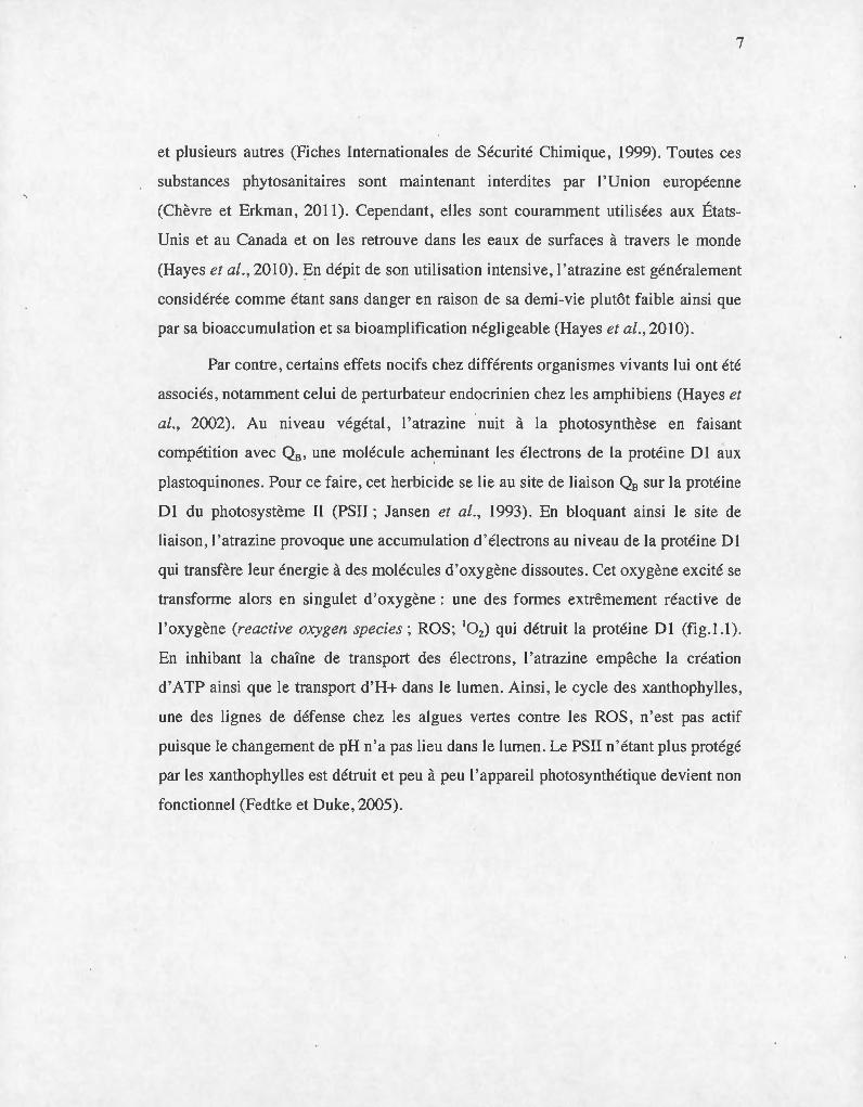
l'oxygène (reactive oxygen species; ROS; 10 2 ) qui détruit la protéine Dl (fig.l.l).
En inhibant la chaîne de transport des électrons, l'atrazine empêche la création
d'ATP ainsi que le transport d'H+ dans le lumen. Ainsi , le cycle des xanthophylles,
une des lignes de défense chez les algues vertes contre les ROS, n'est pas actif
puisque le changement de pH n'a pas lieu dans le lumen. Le PSII n'étant plus protégé
par les xanthophylles est détruit et peu à peu l'appareil photosynthétique devient non
fonctionnel (Fedtke et Duke, 2005).
8
Complexe.B6F PSI
Figure 1.1 Schéma de l'action de l'atrazine sur la photosynthèse. La chaine de transport des électrons est illustrée avec tous ces composants. Le mécanisme d'action de l'atrazine est indiqué par des flèches au niveau du PSU. Les flèches rouges représentent le sens du transport des électrons au sein du PSII depuis la scission de l'eau. La double barre oblique rouge représente l'arrêt du transport des électrons. La flèche en noire représente la liaison de l'atrazine et les flèches en pointillées noires, le retour d'énergie créer par l'accumulation des électrons. Dans la figure, Phe représente la phéophytine, (Mn)4 est le manganèse, PQ représente la plastoquinone, Yz illustre la Tyrozine z, Atz est l'atrazine. (Modifié de David Joly, UQTR)
Tous ces événements engendrerànt une diminution du bon fonctionnement des
cellules végétales pouvant mener à la mort de l'organisme photosynthétique.
L'atrazine se trouve donc à avoir énormément d'effets négatifs sur la physiologie du
phytoplancton dans les cours d'eau contaminés (Denoyelles et al, 1982). En
conséquence, la croissance et la reproduction de ces organismes se trouveront à être
extrêmement réduites, rendant leur accessibilité plus difficile pour tous les animaux
qui s'en nourrissent.
9
1.3 Radiation ultraviolette
Avec la dégradation de la couche d'ozone, l'intensité des UV-Ba augmenté à
la smface de la Terre. Cette augmentation est inquiétante puisque les UV-B ne
présenteraient que des effets négatifs sur les organismes photosynthétiques. De plus,
il est connu que ces rayons ont d'autant plus d'effets lorsqu'ils agissent sur des
organismes déjà faibles ou stressés (Jansen et al, 2012). Une énorme augmentation
d'apoptose et la formation de ROS (0.2 , o·, OR et H20 2) ont été observées en
conséquence à ce type d'exposition chez les plantes supérieures et chez les
cyanobactéries. Ces dommages peuvent par la suite mener à des problèmes au niveau
fonctionnel et structurel des protéines, des lipides et de nombreux autres composés
cellulaires (Panagopoulos et al., 1990; Foyer et al., 1994; Smirnoff, 1998;
Mahdavian, 2008 et He et Hader, 2002). En plus des dommages qu'ils peuvent causer
à l'ADN (Vincent et Roy, 1993; Karentz et al., 1991a et He et Hader, 2002), ces
rayons ont pour effet de réduire la quantité de chlorophylle chez les cyanobactéries et
les plantes (Suresh et al., 1998 et Juneau et al., 1997) et de caroténoïdes
(xanthophylles et lutéine) chez les algues (Bishof et al., 2002). En raison de cette
diminution de caroténoïdes, les organismes montrent une diminution de protection
contre une forte lumière PAR (Demmig-Adams, 1990). Les UV-B ont aussi comme
conséquence d'inhiber la transcription de gènes photosynthétiques et de diminuer
l'intégrité générale des chloroplastes résultants en l'inhibition de la photosynthèse
(Friso et al., 1994; Vass et al., 1999). Ensuite, les UV-B détruisent les acides gras
insaturés en présence d'oxygène, ils diminuent le contenu en plastoquinone des
thylakoides exposés et ils inactivent les ATP synthase (Vass et al, 2005).
De plus, il est connu que les rayons UV-B dégradent ies protéines Dl et D2
servant à la photoprotection (Xiong et al., 1997 ; Bouchard et al., 2006), ce qui a
comme conséquence une diminution du dégagement d'oxygène et du taux de
10
transport des électrons. Au final, le rendement de l'activité du PSII se trouve à en être
affecté (Bischof et al ., 1998b; Dring et al. 1996; Hanelt et al.1997; Bischof et al.
1998a) .
Finalement , les UV en général peuvent détruire les chaînes linéaires de
tétrapyrrole qui sont contenues dans les phycobiliprotéines , des pigments
photosynthétiques qui sont propres aux cyanobactéries (Vass et al, 2005).
1.4 Combinaison de l' atrazine et des UV-B
Dans la nature , les organismes subissent des stress de toutes sortes et ces
perturbations ne surviennent que très rarement , voire jamais , seules. C'est là que
l'étude de la combinaison de stress devient intéressante. En sachant que la radiation
UV-B ainsi que l'atrazine affectent différemment la photosynthèse et la croissance des
végétaux , il est possible de se questionner sur l'effet de leur combinaison sur des
organismes . Également, connaissant que les rayons ultraviolets peuvent agir à
différents endroits sur un organisme photosynthétique et que 1 'atrazine inhibe la
chaîne de transport d 'électrons aussi à un de ces endroits (protéine Dl) , il devient
pertinent de se demander si 1 ' effet de la combinaison de ces deux facteurs est
synergique, additif ou même antagoniste . Beaucoup d'études se sont penchées sur les
effets in di vi duels des radiations UV -B ainsi que sur ceux de 1 'atrazine . Par contre,
aucune étude n'a été faite sur leurs effets combinés. Des rares études faites sur la
combinaison d'UV-B et herbicides (Kasaï et Arts, 1997; He et Hader, 2002; Wang
et al, 2007 ; Chen et al, 2012), seulement deux ont étudié les effets au niveau de la
photosynthèse (Wang et al, 2007; Chen et al , 2012) et aucune n'a utilisé l'atrazine
comme herbicide agricole. Finalement, Chen et al. (2012) ont utilisé des intensités
lumineuses qui n'étaient pas représentatives de ce que l'on peut retrouver dans
l'environnement lorsqu ' il y a présence d 'UV (40 !J.Em-2s-1) . Normalement , l ' intensité
de lumière PAR retrouvée en plein jour sous une journée ensoleillée se situe autour de
1500 ~J.Em-2s· 1 (De Lange et Lürling, 2003). Chez les plantes supérieures, l'utilisation

--------
Il
d'une luminosité plus faible peut avoir comme conséquence d'augmenter leur
sensibilité face aux UV (Poulson et al, 2006; Majer and Hideg, 2012).
1.5 Interaction de stress
La combinaison de stress environnementaux suscite beaucoup de questions
puisque la réponse qu'elle engendre peut être extrêmement variable (Thompson,
1996). Lorsque plusieurs perturbations surviennent en même temps, la réponse
physiologique des organismes face à ces évènements peut varier en fonction de
différents facteurs. Par exemple, elle variera en fonction des stress qui sont imposés à
1 'organisme, en fonction des conditions environnementales dans lequel les stress sont
appliqués et en fonction des organismes qui les subissent (Thompson, 1996; Fischer
et al, 2010) . Afin caractériser cette réponse , on la différencie selon trois types :
additivité, synergisme ou antagonisme (Tamme~, 1964; Thompson, 1996 et Wang et
al ., 2007). Par ailleurs, si l 'effet engendré par une combinaison de stress est
équivalent à l'addition des effets causés par chacun des stress isolés , on dit de cette
réponse qu'elle est additive. Une réponse comme celle-ci implique que les deux
facteurs de stress ont des mécani smes d'action indépendants 1 ' un de 1 'autre. C'est-à
dire que 1 'action d'une des perturbations n'affectera pas celle de 1 'autre et les effets
de chacune seront tout simplement additionnés (Newman et Unger, 2003). Par contre,
si 1 'effet toxique produit par une combinaison est supérieur à 1 'addition des effets
provoqués par chacun des stress seuls, on dit de cet effet qu'il est synergique.
Contrairement à un effet additi_f, ce type de réponse implique que les mécanismes
d'actions des stresseurs sont dépendants l ' un de l'autre de sorte que l'action d ' un des
stress aura un rôle à jouer sur le mécanisme d'action de 1 'autre stress et aura ainsi
comme effet d'augmenter sont effet (Newman et Unger, 2003). Toutefois , si après
une exposition à une combinaison de stress, l 'effet engendré est inférieur à l'addition
des effets des deux stress seuls, on dit qu'il s'agit d ' une réponse antagoniste. Encore
une fois, ici, les mécanismes d'action sont dépendants 1 'un de 1 'autre. En effet, cette
~~~~----
12
réponse implique que le mécanisme d'action d ' un des stress interagis avec le
mécanisme d'action de l 'autre de sorte à inhiber son effet (Newman et Unger, 2003) .
1.6 Fluorescence
Dépendamment d'où ils se trouvent et du moment de la journée , les organismes
photosynthétiques sont exposés à des intensités lumineuses qui peuvent varier
considérablement. Les stress environnementaux comme les radiations ultraviolettes
provenant du soleil et 1 'exposition aux herbicides peuvent affecter les réactions de
transformation d 'énergie lumineuse en énergie chimique (He et Hader, 2002; Fedtke
et Duke , 2005 et Jansen et al., 2012). La lumière, lorsqu 'elle se trouve en trop grande
quantité ou lorsqu 'elle est exposée sur des organismes déjà stressés, peut devenir
néfaste pour ces organismes (Müller et al., 2001). Afin de résoudre ce problème, les
organismes photosynthétiques utilisent différentes façons de dissiper le surplus
d'énergie. Un phénomène extrêmement important découlant d'une surcharge
d'énergie est la fluorescence. Il s'agit tout simplement d'une émission lumineuse
provoquée lors de l'absorption d 'l;ln photon (Krause et Weis, 1991) La fluorescence
émise par un organisme photosynthétique varie d 'après le fonctionnement de son
appareil photosynthétique (Krause et Weis, 1991; Krause et Jahns , 2003; Ralph et
al., 2007). En effet, si un blocage de la chaine de transport des électrons a lieu la
proportion d 'énergie absorbée par cette voie photochimique sera diminuée et celle
réémise sous forme de fluorescence et de chaleur sera augmentée. Ainsi, la
fluorescence devient une façon efficace de mesurer la photosynthèse chez les
organismes photosynthétiques (Schreiber et al., 1994; Krause et Weis, 1991;
Marwood et al., 2000; Schreiber et al., 2002; Juneau et al., 2007). À l'aide d'un
fluorimètre, en mesurant le maximum d'énergie pouvant être transmis au PSII lors
d'un fort flash de lumière, il est possible de déterminer la fluorescence maximale
émise par le PSII et, par la suite, la diminution de fluorescence lors d'une exposition à
une lumière normale (Kautsky et Hirsh , 1931 ; Schreiber et al., 1986; Genty et al.,
1989). Cette diminution de fluorescence est appelée quenching photosynthétique. Ce

13
quenching s'explique par deux phénomènes. Premièrement, par le transport
d 'électrons dans la chaîne (Quenching photochimique: qP). Deuxièmement, par tous
les mécanismes de protection mis en branle pour éviter 1 'accumulation des électrons
au niveau du PSII et qui ne sont pas directement liés au transp01t photosynthétique
d'électrons (Quenching non-photochimique; NPQ; Schreiber et al., 1986). Le qP, le
NPQ ainsi que plusieurs autres paramètres peuvent être mesurés à l 'aide de
fi uori mètres.
À 1 'aide de la cinétique de fluorescence rapide (PEA : Plant efficiency
analyser; Hansatech , Instruments Ltd , UK), les paramètres suivants peuvent être
mesurés:
La taille des antennes collectrices par centre réactionnel actif est calculée avec
l'absorption d 'énergie lumineuse par les antennes collectrices de lumière (ABS)
distribuée par centre réactionnel du PSII actif (RC), ABS/RC (Force et al., 2003):
ABS/RC = (M0 / VJ) 1 (1- Cfo/ FM))
où:
M 0 = Vitesse initiale de l'induction de la fluorescence variable.
(4* (F300ps - Fsol" ) / (FM- Fso/ls)) .
VJ = taux de réduction de QA.
(F2rns - Fsops /FM - Fso1,s)
FM= Fluorescence maximale; fluorescence après un flash de lumière saturante
(3000 f.Lmol photons. m·2 • s·').
F0 =Fluorescence minimale ; fluorescence au noir.
Le taux de dissipation d 'énergie sous forme de chaleur est calculé grâce à la
proportion d'énergie lumineuse absorbée par centre réactionnel du PSII actif n 'étant

14
pas utilisé dans la chaine de transport des électrons, mais dissipée par la chaleur
(DI0 ), Dio /RC (Strasser et al., 2000):
Le taux de transport d 'électrons est obtenu à l' aide du taux de transport
d' électrons (ET0) par centre réactionnel actif (Force et al., 2003):
Grâce à la fluorescence chlorophyllienne modulée par Water-PAM (Water
Pulse-Amplitude-Modulated; Heinz Walz GmbH, Effeltrich, Allemagne) PAM), les
paramètres suivants peuvent être calculés:
Les dommages persistants au PSII sont mesurés grâce à l'efficacité
photochimique maximale du PSU (Kitajima et Butler, 1975) :
<PM = (FM - F0) / FM
où:
FM= Fluorescence maximale; fluorescence d'un échantillon illuminé après un
flash de lumière saturante (3000 f.Lmol photons. m·2 • s·1).
F0 =Fluorescence minimale; fluorescence au noir.
La photoinhibition et les dommages additionnels au PSII sont évalués grâce à
l'efficacité photochimique opérationnelle du PSII (Genty etal. , 1989):
<P ' M = (F' M - FI)/ F' M
où:

15
F1 =Fluorescence à un temps défini.
F' M = Fluorescence maximale d ' un échantillon illuminé après un flash de lumière
saturante.
Les mécanismes utilisés pour contrer une accumulation d 'énergie au ntveau
du PSII (NPQ; quenching non-photochimique) autre que la photochimie sont mesurés
(Bilger and Bjorkman, 1990):
1. 7 Protection du phytoplancton contre les stress
Les organismes qui dépendent de l 'énergie du soleil pour survivre ont
développé certains mécanismes afin de se protéger contre un excès d'énergie causé
soit par un stress lumineux (UV -B ou forte intensité de PAR) ou soit par tout autre
stress environnementaux comme une exposition aux herbicides . Pour échapper à un
surplus d'énergie , le phytoplancton possède plusieurs moyens , soit la fuite, les
systèmes de protection et les mécanismes de réparation . La fuite peut être effectuée
par des algues possédant des flagelles, par exemple, ou par les cyanobactéries
possédant des vacuoles faisant en sorte qu 'elles peuvent se déplacer dans les colonnes
d'eau et ainsi éviter les radiations ultraviolettes (Kruschel et Castenholz, 1998) .
Ensuite, les systèmes de protection peuvent être représentés sous forme de barrière
physique comme certains dinoflagellés , par exemple , qui développe une paroi
cellulaire à multiples épaisseurs les protégeant contre les UV (Banaszak and Trench,
2001). Certains organismes formeront des composés les protégeant contre les
radiations tels que des composés d'acides aminés , des enzymes antioxydantes , des
caroténoïdes et plusieurs autres (Karentz et al., 1991b; Salguero et al., 2003 ; Sinha
et Hader, 2007 ; Rijstenbil, 2003). Les enzymes antioxydantes vont protéger
l'appareil photosynthétique contre les molécules réactives d 'oxygène . Par exemple: la
su peroxyde dismutase (SOD) permet de réduire 1' ion su peroxyde en peroxyde
16
d'hydrogène (Wolfe-Simon et al., 2005), la catalase (CAT) réduit le peroxyde
d ' hydrogène en eau et en 0 2 (Pinto et al. , 2003) , l ' ascorbate peroxydase (APX)
permet de réduire le H20 2 en H20 (Pinto et al. , 2003) et la glutathion réductase (GR)
permet de transformer le glutathion disulfide en glutathion sulfhydryl (Rao et al.,
1996). Les caroténoi·des, quant à eux, peuvent absorber non seulement les radiations
. UV , mais aussi 1 'énergie des oxygènes réactifs et la réé mettre sous forme de chaleur
(Vincent et Roy , 1993; Stahl et Sies , 2002).
D'autres mécanismes de protection des appareils photosynthétiques existent.
A va nt de les expliquer, il faut se rappeler que la photosynthèse consiste en un
transport d'électrons entre le photosystème II et le photosystème 1. Chaque
photosystème est formé de plusieurs molécules pigmentaires constituant le complexe
antennaire et d'un centre réactionnel. Ces pigments sont en majorité chlorophylliens
et sont excités par l'absorption de photons de lumière. La chlorophylle comme tous
les autres types de pigments, absorbe les photons provenant d'une certaine longueur
d'onde. L'énergie des photons est transférée aux électrons que contiennent les ,
pigments (Raven et al. 2002; Taiz et Zeiger, 2010). Ce transfert d ' énergie fait passer
le pigment de l'état fondamental à l'état d'excitation et créé une énergie de
résonnance qui peut être dissipée de différentes façons. Elle peut être dissipée par la
voie de la photochimie , c'est à dire envoyée dans la chaîne de transport des électrons.
Elle peut être dissipée sous forme de chaleur ou absorbée par les molécules
d'oxygènes (formation de ROS) ou alors , elle peut être dissipée sous forme de
fluorescence (Müller et al. , 2001; figure 1.2).

17
4
/ 1Chl*
3Chl* fluorescence
photochemistry ( qP)
heat (NPQ) Chi
/ light
Figure 1.2 Voies de dissipation d' énergie. La chlorophylle (Chi) absorbe la lumière, elle passe donc de l ' état fondamental à l'état excité ( 1Chl*). Pour revenir à l' état fondamental, l'énergie peut être réémise sous forme de fluorescence (1), elle peut être utilisée pour la voie de la photochimie (2), être dissipée sous forme de chaleur (3) ou être absorbée par les molécules d' oxygène libres (4). En effet, la chlorophylle excitée ( 1Chl*) peut froduire un triplet de chlorophylle echl*) qui peut à son tour produire des ROS ( 0 2*; tirée de Müller et al. , 2001).
Les mécanismes de protection constituant le NPQ sont aussi différents moyens
que la cellule utilise afin de dissiper un surplus d'énergie. Le NPQ varie entre les
différents types de phytoplanctons. Chez les algues vertes, le NPQ comprend le cycle
des xanthophylles, l'état de transition et la photoinhibition ((Müller et al. , 2001 and
Schreiber et al. , 1986)). Le cycle des xanthophylles consiste en un changement de
conformation de pigments caroténoïdes. Lorsqu'il y a augmentation d'électrons dans
la chaîne de transport des électrons , de plus en plus de protons sont transférés vers le
lumen changeant ainsi son pH. Le changement de pH entraîne la transformation de la
violaxantine en anthéraxantine et éventuellement en zéaxanthine . Ce changement de
18
conformation empêche le transfert d 'énergie à la protéine Dl ralentissant ainsi le
transport des électrons (Farber et al. , 1997). Le second mécanisme de protection ,
1 'état de transition, consiste au déplacement des antennes collectrices de lumière
(LHC). Les LHC sont des pigments pouvant voyager d ' un photosystème à l 'autre .
Ainsi, lorsqu'une accumulation d'énergie survient au niveau du PSII , des antennes
peuvent transférer une quantité de cette énergie au PSI afin de la répartir plus
également entre les deux photosystèmes (Allen, 1992; Gal et al ., 1997; Wollman ,
2001). Le mécanisme de protection qu 'est la photoinhibition correspond à
l'absorption d'énergie par la protéine Dl (Dl protein turnover; Xiong et al,. 1997).
Cela prévient alors que d'autres molécules soient endommagées par cette énergie et
que des molécules réactives d 'oxygène soient produites. Cette protéine , qui sera alors
~ndommagée, sera dégradée par une protéase puis remplacée par une protéine
nouvellement synthétisée (Prasil et al,. 1992; Rintamaki et al., 1994; Leitsch et al.,
1994; Schnettger et al., 1994).
Chez les cyanobactéries, les composants du NPQ sont légèrement différents.
Mis à part 1 'état de transition et la photoinhibition qui existe aussi chez ces
organismes, ceux-ci ne possèdent pas de cycles des xanthophylles (Hager et
Holocher, 1994; Hirschberg et Chamovitz, 1994). Par contre, ils possèdent tout de
même de la zéaxanthine, un type de xanthophylle avec lequel ils arrivent à dissiper
les surplus d'énergie (Hirschberg et Chamovitz , 1994). Chez les algues vertes, le
premier mécanisme du NPQ qui est activé lors d'un excès d 'énergie est le cycle des
xanthophylles (Bilger et Bjorkman , 1 990; Ni yogi et al., 1998) alors que , chez les
cyanobactéries, l 'état de transition est celui qui est sollicité le premier (Joshua et al.,
2005 ; Bailey et Grossman, 2008).
1.8 Sensibilité du phytoplancton
Toutes les espèces de phytoplancton ne sont pas également sensibles aux stress
environnementaux. En effet, certaines espèces sont extrêmement sensibles et sont

19
utilisées comme biomarqueur d ' un environnement pollué (Torres et al,. 2008).
D'autres sont au contraire beaucoup' plus résistantes et peuvent donc devenir
dominantes dans un environnement stressé . Par exemple, il a été démontré que
certaines chlorophytes et baccilariophytes sont plus résistantes à l'atrazine et aux
changements de lumière (Deblais et al., 2013). Alors que certaines cyanobactéries
peuvent être plus résistantes aux changements de température (Chalifour et Juneau ,
2011) ou encore, plus résistantes aux UV-B (Campbell et al., 1998) que d 'autres
espèces. Si des organismes comme les cyanobactéries deviennent dominantes dans un
milieu , cela peut poser problème. En effet , il est connu que certaines souches peuvent
être toxiques et ainsi avoir des répercussions sur les autres organismes vivants dans
ces mêmes milieux (Demott et al, 1991 ; Christoffersen , 1996; Ferrao-Filho et al.,
2000; Anderson et al., 2002). De plus, les cyanobactéries, qu'elles soient toxiques ou
non, constituent une nourriture de moins bonne qualité pour les animaux se
nourrissant de phytoplanctons . En effet, leur constitution en acide gras hautement
saturé (HUFA: highly unsaturated fatty acid) est moins importante que pour les
algues vertes par exemple (Demott et Müller-Navarra, 1997). Étant moins nutritive ,
elles peuvent nuire à la santé des animaux qui s'en nourri ssent , pouvant aussi
engendrer un débalancement des populations de ces écosystèmes.
CHAPITRE II
COMBINED EFFECT OF ATRAZINE AND UV-BON PHYTOPLANKTON.
ANDRÉANNE GIRARD KEMP, MARCELO PEDROSA GOMES AND PHILIPPE JUNEAU*.
Department of Biological Sciences, TOXEN, Ecotoxicology of Aquatic
Microorganisms Laboratory, Université du Québec à Montréal, Suce. Centre-Ville,
Montréal, Québec, Canada
* Author for correspondence
To be submitted to Aquatic Toxicology.
Contributions: Pour ce chapitre, Andréanne Girard Kemp a élaboré le plan
d' expérience avec l'aide de Philippe Juneau. Elle a réalisé les bioessais en laboratoire
et l' analyses des résultats. Marcelo Pedrosa Gomes a participé à certains bioessais. La
rédaction a été effectuée par Andréanne Girard Kemp et Philippe Juneau.
21
2.1 Abstract
The use of pesticides such as atrazine and the increase of ultraviolet radiation (UV -B) due to the depletion of the ozone layer are environ mental stresses that can affect organisms such as phytoplankton. Since environmental stresses never act atone, it is important to study the effects engendered by their combination. To better understand how the combination of UV -B and atrazine affects the health of primary producers , photosynthesis · of green algae and two strains of cyanobacteria were measured. In growth cham ber, Scenedesmus obliquus (CPCC5) and Microcystis aeruginosa (CPCC632 and CPCC299) were submitted to 0.1!-LM of atrazine and to 3 hours of UV-B (O.Iwm-2 or 0 .2wm-2) exposure. Photosynthetic parameters were studied to assess the combined effects of the stress factors in the phytoplankton health. For M. aeruginosa CPCC299, the decrease in photosynthesis caused by atrazine (33 %) was amplified by 30% by a UV-B exposure and for S. obliquus, it was amplified by 67%. The synergistic effect observed in S. obliquus is caused by the increase of reactive . oxygen species (ROS) and the degradation of carotenoids pigments. In addition, the different effects found between species can be explained by the differences in their photoprotective pigments (carotenoids) content. Our results reinforce the importance of considering environmental factors such as UV -B in toxicological studies involving pesticides.
Key words: algae, cyanobacteria, UV-B, herbicide, photosynthesis, photoinhibition, non-photochemical quenching, PSII quantum yield, heat-dissipation.
22
2.2 Résumé
L'épandage de pesticides dans les champs et 1 'augmentation de rayon ultraviolet à la surface de la Terre dû à la détérioration de la couche d 'ozone sont des stress envÏ!"onnementaux qui peuvent affecter certains organismes comme le phytoplancton. Puisque les stress environnementaux ne survie1ment jamais un à un, il est très important d' étudier les impacts qu ' engendre leur combinaison. Afin de mieux comprendre comment cette combinaison affecte la santé de ces producteurs primaires, la photosynthèse d'une algue verte et de deux souches de cyanobactéries a été mesurée. En chambre de croissance contrôlée, Scenedesmus obliquus et Microcystis aeruginosa (CPCC632 et CPCC299) ont été exposé à û,l!lM d' atrazine ainsi qu'à 3h/jour d'UVB (0,15 Wm-2 ou 0,4Wm-2
) . Plusieurs paramètres ont été étudiés pour évaluer l' effet combiné de ces stress environnementaux sur la santé de ces organismes. Pour M. aeruginosa CPCC299, la diminution de la photosynthèse cause par l'atrazine (33 %) a été amplifiée de 30% par une exposition aux UV-B alors que pour S. obliquus, elle a été amplifiée de 67%. Cet effet synergique observe chez S . obliquus est causé par l'augmentation d'espèces réactives d'oxygène (ROS) et la dégradation de pigments de caroténo1"des . De plus, les différents effets remarqués entre les espèces peuvent être expliqués par leur différence de contenu de pigments photo protecteur ( caroténo1"des). Nos résultats démontrent 1' importance de prendre en considération les facteurs environnementaux tels que les UV-B lors d'étude toxicologique impliquant des pesticides.
Mots clés: algues , cyanobactéries, UV-B, herbicide, photosynthèse , photoinhibition, quenching non photochimique , rendement photosynthétique du PSII , dissipation de chaleur.
23
2.3 Introduction
Phytoplankton is very important for aquatic ecosystems since it provides food
for many organisms and is responsible for about 70% of the global oxygen
production (Fenical , 1983). However, it is also very sensitive to anthropogenic and
environmental stressors, such as herbicides and light (Neale et al., 1998; Cloern, 1999
and Rostand Wolf-Giadrow, 2008). Among the herbicides, atrazine is widely used in
agriculture around the world (Graymore et al., 2001), is the most frequently detected
pesticides in aquatic ecosystems (Sullivan et al., 2009; Giroux, 2010). This herbicide
blacks the electron transport chain at the Q8 binding site on the Dl protein of
photosystem Il (Fedtke and Duke, 2005), causing an accumulation of energy at the
PSU reaction center. This ex cess of energy is th en dissipated by fi uorescence, heat
emission (Müller et al., 2001 and Schreiber et al., 1986). Excess energy can be
transferred to oxygen molecules resulting in the generation of reactive oxygen species
(ROS). ROS can have sorne deleterious effect on the cell, such as, pigment
degradation, lipid peroxidation and inhibition of the photodamage's repair processes
(Nishiyama et al., 2001; Murata et al., 2007 and Krieger-Liszkay et al., 2008). For
green algae, those deleterious effects can decrease the photosynthetic electron
transport and, consequently , the pH gradient formation across the thylakoid
membranes. This disturbance on the pH leads to the non-induction of the xanthophyll
cycle, which is a protective mechanism against light stress (Demmig-Adams, 1990;
Müller et al ., 2001).
Anthropogenic activities have been shawn to cause the depletion of the
atmospheric ozone layer, resulting in a significant increase in the UV-B radiation
reaching Earth's surface (Frederick, 1993). Like atrazine, UV-B radiation also
induces the formation of ROS in phytoplankton (Panagopoulos et al ., 1990; Foyer et
al., 1994; Smirnoff, 1998; He and Hader, 2002, and Mahdavian, 2008), which can
provoke lipid peroxidation and structural and func_tional damage to severa! proteins
24
and other cell components (Panagopoulos et al., 1990; Foyer et al., 1994; Smirnoff,
1998; Mahdavian , 2008 and He and Hader , 2002). In green algae and cyanobacteria,
UV -B also directly degrades chlorophyll and carotenoids (Suresh et al. 1998 and
Bischof et al. , 2002). Furthermore , UV -B radiation affects the Dl and D2 proteins of
the PSII leading to a decrease in oxygen evolution and electron transport (Xiong et
al., 1997 and Bouchard et al., 2006).
Si nee both atrazine and UV -B radiation are environmental stresses that can
affect phytoplankton photosynthesis through their inhibitory effects on Dl protein,
one can advance that a combination of these stressors would have an additive effect.
However, UV -B also interacts with photoprotective pigments and enzymes. Most of
the ti me UV -B are known to degrade carotenoids (Bishof et al., 2002), which could
lead to an amplification of atrazine toxicity and then to a synergistic taxie effect. On
the other hand , it · was also demonstrated that UV radiation could enhance the
production of photosynthetic pigments such as chlorophyll a and carotenoid
(Yakovleva and Titlyanov , 2001) , which could lead to the diminution of the atrazine
t~::>Xicity. Only a few studies have investigated the combined effects of herbicide and
UV -B on photosynthesis (Wang et al, 2007 and Chen et al , 2012) and none of them
used atrazine, although its frequent utilization . Understanding the combined mode of
action of these two stressors on phytoplankton may help to better understand the
species dynamic in natural aquatic environments of agricultural areas.
The main goal of this study is to determine how atrazine and UV -B affect the
photosynthesis of a taxie and a non-taxie strain of cyanobacteria and green algae.
This study also aims to evaluate the phytoplankton sensitivity variation by evaluating
photosynthetic performances.
25
2.4 Materials and Methods
2.4.1 Cultures
The green alga Scenedesmus obliquus (CPCC5), the toxic and the non-toxic
strains of cyanobacteria Microcystis aeruginosa (CPCC299 and CPCC632) were
grown at 24°C in Erlenmeyer flasks containing fresh Bold basal medium (BBM) . The
cultures were adapted for at !east 10 generations to a pattern of severa( light
intensities changing over the day (Fig. 2.1). The light was provided by a combination
of fluorescent tubes (Philips F72T8/TL841/HO , USA) and incandescent bulbs
(DuraMax, Philips Electronics LDT , Canada). Light intensity and quality was
measured by the HR4000 High-Resolution Spectrometer (Ocean optics Inc., USA)
and analyzed with Spectra Suite software (Ocean optics Inc ., USA).
2.4.2 Treatments
For each UV -B or atrazine treatments , when adapted cultures reached 5 x 10⁵
cell /ml , they were divided into nine beakers that were placed on a turning plate in a
growth chamber (MTR30, Conviron , Manitoba , Canada) (under the same light
conditions than described previously) for further treatments . Cellular concentrations
were obtained using a Multisizer 3 Coulter Counter particle analyzer (Beckman
Coulter lnc. , USA).The plate rotates at one turn per hour so every replicate were
exposed to the exact! y same light intensity/quality.
Cultures were exposed to six different treatments (Table 2.1). Atrazine
concentration and UV -B doses were chosen based on what can be found in natural
environment (Salomon et al. , 1996; Giroux, 2010, West et al ., 2003 and Arts and Rai,
1997). The atrazine solution was obtained from the dilution of the commercial Aatrex
480 Iiquid herbicide (Syngenta , Plattsville , Canada) in distilled water. The UV -B
radiation was provided by UV -B Broadband TL tubes (Philips TL20W/12, USA) and
Mylar polyester film (DuPont Teijin Films™) used to remove UV -C radiations . For
26
treatments with atrazine, the herbicide was added at 10:30 AM and for the UV -B
treatrnent, the lamps were activated from l l :30 AM to 2:30PM (Fig. 2.1).
600 .
.
500 ,..---. --;'
crJ . . ("oo s 400
crJ 0 0 ...... 0
..J:::l 300 o.. 0 §_ .
"--" >.. 200 ....... . _, Cl}
0 <!) .
...... q
......... 100 .
. 0
Atrazine
1•
uv
1 1 1 l . ' ' ' • • ,,
'· .• 1 2 2 ·1 o e 7 s 9 10 11 12 1314 15 16 11 18 19 2o 21 22 za Z4
Time (hours)
Figure 2.1 Light variation (l..tmol photons • m·2 • s·1
) and exposure times of atrazine and UV-B within 24 hours.
27
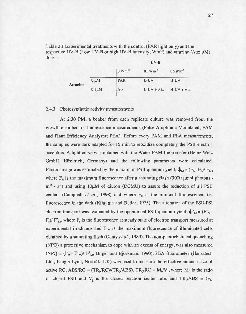
Table 2.1 Experimental treatments with the control (PAR light only) and the respective UV-B (Low UV -Bor high UV-B intensity; wm-2 ) and atrazine (Atz; f..IM) doses.
UV-B
owm-~ O.lwm-~ 0.2wm-~
ÜJ.LM PAR L-UV H-UY Atrazine
O.IJ.LM Atz L-UV + Atz H-UV + Atz
2.4.3 Photosynthetic activity measurements
At 2:30 PM, a beaker from each replicate culture was removed from the
growth chamber for fluorescence measurements (Pulse Amplitude Modulated; PAM
and Plant Efficiency Analyzer; PEA). Before every PAM and PEA measurements,
the samples were dark adapted for 15 min to reoxidize completely the PSII electron
acceptors . A light curve was obtained with the Water-PAM fluorometer (Heinz Walz
GmbH, Effeltrich , Germany) and the following parameters were calculated.
Photodamage was estimated by the maximum PSII quantum yield, <PM= (FM-F0)/ FM,
where FM is the maximum fluorescence after a saturating flash (3000 f.Lmol photons·
m·2 • s·1
) and using lOf.LM of diuron (DCMU) to assure the reduction of ail PSII
centers (Campbell et al., 1998) and where F0 is the minimal fluorescence, i.e.
fluorescence in the dark (Kitajima and Butler, 1975). The alteration of the PSII-PSI
electron transport was evaluated by the operational PSII quantum yield , <P' M = (F' M
F5)/ F' M' where Fs is the fluorescence at steady state of electron transport measured at
experimental irradiance and F' M is the maximum fluorescence of illuminated cells
obtained by a saturating flash (Genty et al., 1989). The non-photochemical quenching
(NPQ) a protective mechanism to cope with an excess of energy, was also measured
(NPQ =(FM- F'M)/ F'M; Bilger and Bjërkmari , 1990). PEA fluorometer (Hansatech
Ltd. , King's Lynn, Norfolk, UK) was used to measure the effective antenna size of
active RC, ABS/RC = (TRofRC)/(TRo/ABS), TRJRC = MJ Vj, where M0 is the ratio
of closed PSII and Vj is the closed reaction center rate, and TR0/ABS = (FM
28
-F501JS)IFM, where F501J.S is the fluorescence at 501J.S. PEA was also used to evaluate
the electron transport in active RC, ETJ RC = (MofVj)•(l -Vj), and the effective
dissipation in active RC, DlofRC=((ABS/RC)-(TRo/RC); Forces et al .. 2003).
2.4.4 Pigment analysis
For pigment determination, cultures were filtered on membrane filters (0.8f.Lm,
(Xingya Purifying Materials Factory, Shanghai, China) and stored at -80°C until
analysis. Chlorophyll a and b, ,8-carotene and zeaxantin were extracted from frozen
filters by adding 1ml of acetone and sonicated (Fisher Scientific Sonic Dismembrator,
leve! 4) six times for 30 seconds in ice cold water. The extracts were tentrifugated at
15000g for 20 minutes. The supernatant was collect for U-HPLC analysis. U-HPLC
analysis were done using an Agitent 1290 infinity LC with a C-18 reverse phase
column (2.1 X 100mm, l.8Jtm) with the EZchrom software according to Garcla
Plazaola and Becerril (1999) with the following adjustment for the use of an U
HPLC. The mobile phase consisted of two" components: solvent A,
acetonitrile:methanol:water (84:9:7); and solvent B, methanol:ethyl acetate (68:32).
The pigments were eluted using a linear gradient from 100% A to 100% Bof 6 min,
followed by an isocratic elution with 100% B of 4 min. This was followed by a 0.33
min linear gradient from 100% B to 100% A and an isocratic elution with 100% A for
a further 7 min to allow the column to re-equilibrate with solvent A prior to the next
injection. The solvent flow rate was 0.5 mL/min, the pressure was around 150 bars
and the injection volume was 5 f.LL. For overnight storage, the column was rinsed
with 100% acetonitrile. Finally, the peaks were detected and integrated at 445 nm.
Pigments concentrations were calculated from peak areas of authentic externat
standards (Sigma-Aldrich, St Louis, MO, USA) and spike recovery tests were
conducted to assure the efficiency of the extraction method. More specifically, tissue
spikes of the Chlorophyll a, /3-carotene and zeaxanthin standards were rec9vered at
98%, 95% , and 90 % , respectively . Pigment concentrations were normalized to the
number of cells.
29
For the two strains of cyanobacteria , the concentration of phycocyanin (PC)
and allophycocyanin (APC) were also measured. Filters were homogenized in O.lM
phosphate buffer pH 6 .8 and frozen and tawed four times for extraction . The extract
was then centrifuged at 6000g for 10 minutes and the absorbance of the supernatant
was measured at 565 nm , 615 nm and 650 nm. The PC and APC concentration was
calculated using the following equations: PC (~-.Lglml) = 163.2 A6 15 - 117.1 A650 , and
APC (1-.Lg/ml) = 165.6 A650 - 16.4 A615 (Lüder et al, 2001). Pigment concentrations
were normalized to the number of cells.
2.4 .5 Lipid Peroxydation
To assess the extent of oxidative stress , lipid peroxydation was measured after
harvesting phytoplankton on filters as described for the pigment determination.
Malondialdehyde (MDA), the product of lipid peroxydation, was determined and
adapted from a colorimetrie method (Heath and Packer, 1968). Briefly, frozen filters
were homogenized in 1 ml of phosphate buffer (50mM) containing 0.67%
trichloroacetic acid . The extraction was performed by a 3 minutes (3 x 1 minute)
sonication followed by an incubation at 95°C for 30 min. The reaction was stopped
by putting the samples in a bucket of ice cold water. The samples were centrifuged at
15000 g for 20 min and the supernatant was read at 532nm with a correction at 600nm
for nonspecific absorption . Finally , the MDA concentrations were normalized by the
cells number .
2.4.6 Statistical analysis
JMP 5.1 statistical software (SAS institute , USA) was the program used to
perform statistical tests. One-way analysis of variance (ANOV A) and Student t-test
were used to compare treatments. Significant differences were accepted when p<0.05.
A synergistic effect was considered when the combined effect was significantly
higher than the addition of the two stres'ses effect atone. One-way analysis of variance

30
(ANOV A) was also performed to compare the effects with significant differences
accepted when p<O.OS.
Species
31
2.5 Results
The atrazine treatment had no significant effect on Fofcell for the two strains
of cyanobacteria. On the other hand , it induced an increase of 76% for S. obliquus.
Atrazine caused a decrease of 10% and 16% in the maximal PSII quantum yield C<PM)
for M. aeruginosa CPCC299 and M. aeruginosa CPCC632 respectively. lt decreased
also the operational PSII quantum yield ( <P ' M) of the three species (Table 2.2). The
NPQ was dimini shed by 34% and 35% for M. aeruginosa CPCC299 and S. obliquus
respectively (Table 1S).
Table 2.2 Photosynthetic parameters of M. aeruginosa CPCC632, CPCC299 and S. obliquus CPCC5 exposed to PAR light (188 j.Lmol photons m-2 s-'), two intensities of UV -B light (0.1 wm-2and 0.2 wm-2) and atrazine (O.lj.LM) at 2:30PM.
Parameters PAR Atz L- UV L-UV + Atz H-UV H-UY+ Atz
M. aeruginosa F0 x 1 o•/cell 3.65 (0 .18) 4 .08 (0 .30) 4 .50 (0 .26)* 4.87(0 .3 1)* 4 .72 (0.42)* 5.17 (0.30)*
CPCC299 <I>M 0.546 (0 .0 19) 0.489 (0 .002)* 0.458 (0 .042)* 0.414 (0 .010)* 0.336 (0 .026)* 0.354 (0.029)*
<j>' M 0.312 (0 .34) 0.209 (0.006)* 0.252 (0.038)* 0 .1 60 (0.0 10)* 0.194 (0.029)* 0.121 (0.023)**
NPQ 0.246 (0.033) 0.163 (0 .0 10)* 0.209 (0 .023)* 0. 127 (0.0 14)** 0.15 1 (0 .0 13)* 0.146 (0 .018)*
M. aeruginosa F0 x 1 o•/cell 3.58 (0.41) 4.42 (0. 14) 8.05 (0,69)* 6.30 (0.43)** 5. 19 (0 .19)* 5.93 (0.77)**
CPCC632 cpM 0.609 (0.008) 0.547 (0 .026)* 0.458 (0 .0 12)* 0.398 (0 .007)** 0.268 (0 .008)* 0.352 (0.022)**
<I>' M 0.39 1 (0 .006) 0 .289 (0.007)* 0.267 (0.026)* 0.142 (0 .026)** 0.056 (0.008)* 0.044 (0.038)*
NPQ 0.260 (0 .037) 0.207 (0 .042) 0 .202 (0.074) 0.134 (0.032)* 0. 122 (0.0 18)* 0.168 (0 .0 12)*
S. obliquus F0 x 10•/cell 3.17 (0 .19) 5 .58 (0.78)* 6 .80 (0.65)* 6.98 (0.44)* 4 .37 (0 .34)* 7.49 (0.33)** CPCC5
cpM 0.7 17 (0 .006) 0.702 (0.005) 0.472 (0 .0 13)* 0.397 (0.035)** 0.53 1 (0.053)* 0.260 (0 .0 15)**
<j>' M 0.481 (0.0 13) 0.409 (0 .0 10)* 0.29 1 (0 .008)* 0.20 1 (0.028)** 0.307 (0 .057)* 0.089 (0.012)**
NPQ 0.772 (0 .0 14) 0.502 (0 .034)* 0.5 19 (0 .1 5 1)* 0.237 (0 .084)** 0.490 (0.095)* 0.11 7 (0.059)**
The symbol * represents significant difference (p< 0.05) from the control (PAR), the symbol ** represents significant difference from the control (PAR), the atrazine treatment (Atz) and the respective UV -B treatment (L-UV or H-UV). Analysis and treatment means are from ANOVA (n=3).

32
The L-UV treatment caused an increase of the FJcell for the three studied
species. We also noticed an incr~ase of the FJcell of ali species for the H-UV
treatment (Table 2.2). A decrease in the <PM and the cp' M was detected for both high
and low UV -B intensities for the three species (Table 2.2). The L-UV treatment
caused a decrease of the NPQ for M . aeruginosa CPCC299 (15%) and S. obliquus
(32%), while the H-UV treatment caused a decrease of the NPQ for the three species
(Table lS).
The addition of a low dose of UV -B to the atrazine treatment caused an
increase in the FJ cell compared to the control for ali species (Table 2.2). This
treatment induced a decrease of the photosynthetic activity and the NPQ compared to
the control for ali species (Table 2.2) Compared to the effect induced by the UV -B
treatment al one, the combination of UV -B and atrazine induced a more important
decrease only for the NPQ of M. aeruginosa CPCC299 (NPQ-L-UV + Atz: 48%;
NPQ-L-UV: 15%), for the photosynthetic activity of M. aeruginosa CPCC632 (Table
lS) and for the NPQ and the photosynthetic activity of S. obliquus (Table lS).
The addition of a high dose of UV -B to the atrazine treatment caused an
increase of the FJcell compared to the control for ali species and this increase was
al ways significantly stronger than the one caused by UV -B al one (CPCC299: 41 %;
CPCC632: 66% and CPCC5: 136%; Table lS). Moreover, this treatment caused a
decrease in the <PM, cp ' M and NPQ compared to the control for the three studied
phytoplankton (Table 2.2). Finally, the decrease is greater than the one generated by
UV -B al one only for the operational PSU quantum yield of M. aeruginosa CPCC299
parameters (cp'M -H-UV + Atz: 61 % and <P'M -H-UV: 38%) and for the three studied
parameters of S. obliquus (Table lS).
15 #
* *
10
# # 5 ** **
El/ RC A BS/RC
a
c
0 PAR D Atz fimlrUV D lrUV + Atz ~H-UY • H-UV+Atz
33
Figure 2 .2 Photosynthesis parameters of M. aeruginosa CPCC299 (a) , CPCC632 (b) and S. obliquus CPCC5 (c) exposed to PAR light (188 j.Lmol photons m·2 s·1
), two intensity of UV -B light (0.1 wm·2 and 0.2 wm-2) and atrazine (O.lj.LM) at 2:30PM . The symbol * demonstrates a significant difference from the control (p<0.05). The symbol * * shows a significant difference from the control (p< 0.001) and the symbol # representa significant difference from the respective UV -B treatment (L-UV or HUY) and from atrazine treatment . Analysis and treatment means are from ANOVA (n=3).
34
The atrazine treatment induced a significant decrease of the EtJRC for the
toxic strain of M. aeruginosa (61 %) and for S. obliquus (27%) , but did not affect the
EtofRC for the non-toxic strain of M. aeruginosa. The atrazine treatment also
increased significantly thé DI0/RC (17%) for the green alga.
The L-UV treatment caused no significant effect for M. aeruginosa
CPCC299, but it induced an increase of the ABS/RC (104%) and the DiofRC (840%)
for M. aeruginosa CPCC632. For S. obliquus, a low dose of UV-B decreased the
ETofRC (32%) and increased the ABS/RC (170%), the TRJ RC (14%) and the
DIJ RC (81 %). On the other hand, the H-UY treatment increased the ABS/RC
(CPCC299: 110%; CPCC632: 181 % and CPCC5: 57%) and the DI0/RC (CPCC299:
473 %; CPCC632: 1418% and CPCC5: 33%) for the three studied phytoplankton .
The combination of a low UV -B dose to an atrazine treatment decreased
the ET JRC (48%) and increased the ABS/RC (138%) and the DIJRC (663 %) for M .
aeruginosa CPCC299 , but for M. aeruginosa CPCC632, it only increased the
ABS/RC (135%) and the 010/RC (1124%). The green alga showed a decrease in the
ETJ RC (57%) and an increase in the ABS/RC (350%), the TRJ RC (19%) and the
DIJRC (33 %). Moreover, the effect caused by the combination of the two stresses
was more important than the one caused by an exposure of L-UY alone. The addition
of a high dose of UV -B to an atrazine treatment caused a decrease in the ET ofRC
(52%) and an increase in the ABS/RC (128%) and the DI0/RC (619%) for the toxic
strain of cyanobacteria . On the other hand , this treatment caused an increase in the
ET JRC (86%) and the DlofRC (426%) for the non-toxic strain of cyanobacteria .
Fin ally , S. obliquus showed a decrease in the ET J RC (54%) and an increase in the
ABS/RC (357%), the TRJ RC (15 %) and the DlofRC (199%). Also, the effect caused
by the two stresses together was more important than the one caused by the UV -B
treatment alone for M . aeruginosa CPCC632 and for S. obliquus.
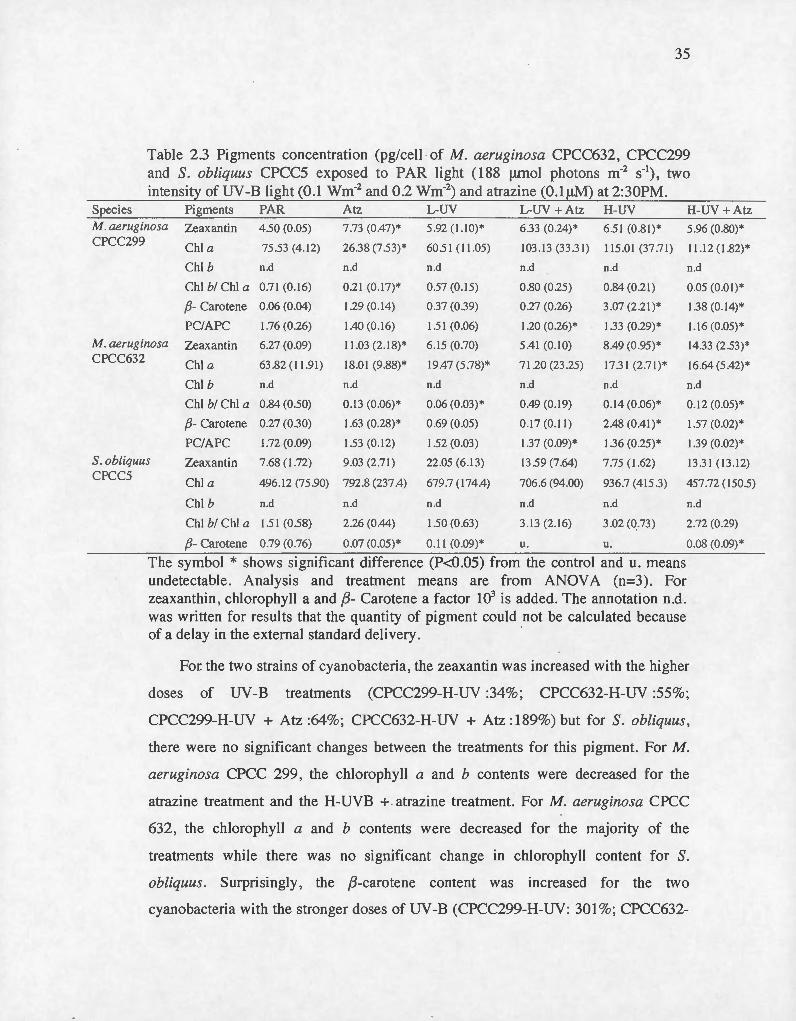
Species
35
Table 2.3 Pigments concentration (pg/cell · of M. aeruginosa CPCC632, CPCC299 and S. obliquus CPCC5 exposed to PAR li ght (188 f..Lmol photons m-2 s-'), two intensity of UV -B light (0 .1 wm-2 and 0.2 wm-2) and atrazine (O.lf..LM) at 2:30PM.
Pi gments PAR Atz L-UV L -UV + Atz H-UY H-UY+ Atz
M. aeruginosa Zeaxantin 4.50 (0 .05) 7 .73 (0 .47)* 5 .92 ( l.IO)* 6.33 (0.24)* 6.5 1 (0.81) * 5.96 (0.80)*
CPCC299 Chi a 75.53 (4. 12) 26.38 (7.53)* 60.5 1 ( 11.05) 103 .13 (33.3 1) 11 5.0 1 (37.7 1) 11.1 2 ( 1.82)*
M . aeruginosa CPCC632
S. obliquus PC 5
Chi b n.d
Chi bi Chi a 0.7 1 (0. 16)
{3- Carotene 0.06 (0.04)
PCIAPC 1.76 (0 .26)
Zeaxantin 6.27 (0 .09)
Chi a 63.82 ( 11.91)
Chi b n.d
Chi bi Chi a 0.84 (0.50)
{3- Carotene 0.27 (0.30)
PCIAPC 1.72 (0 .09)
Zeaxantin 7.68 ( 1.72)
n.d
0.2 1 (0. 17)*
1.29 (0. 14)
1.40 (0. 16)
11 .03 (2. 18)*
18.0 1 (9.88)*
n.d
0.13 (0.06)*
1.63 (0 .28)*
1.53 (0 .1 2)
9.03 (2.7 1)
Chi a 496. 12 (75 .90) 792.8 (237 .4)
Chi b n.d n.d
Chi bi Chi a 1.5 1 (0.58) 2.26 (0.44)
n.d
0.57 (0. 15)
0.37 (0.39)
1.51 (0.06)
6.15 (0.70)
19.47 (5 .78)*
n.d
0.06 (0.03)*
0.69 (0.05)
1.52 (0.03)
22 .05 (6. 13)
679.7 ( 174.4)
n.d
1.50 (0.63)
n.d
0 .80 (0.25)
0.27 (0.26)
1 .20 (0 .26)*
5.4 1 (0. 10)
71 .20 (23 .25)
n.d
0.49 (0. 19)
0 .17 (0 .11 )
1 .37 (0.09)*
13.59 (7.64)
706.6 (94.00)
n.d
3. 13 (2. 16)
n.d
0.84 (0.21)
3.07 (2.2 1)*
1.33 (0 .29)*
8.49 (0.95)*
17.3 1 (2.7 1)*
n.d
0.14 (0.06)*
2.48 (0.41)*
1.36 (0.25)*
7.75 (1.62)
936.7 (4 15.3)
n.d
3.02 (0.73)
{3- Carotene 0.79 (0.76) 0.07 (0.05)* 0 .11 (0.09)* u. u.
The symbol * shows significant difference (P<0 .05) from the control and u. means undetectable . Analysis and treatment means are from ANOV A (n=3) . For zeaxanthin, chlorophyll a and {3- Carotene a factor 103 is added. The annotation n.d. was written for results that the quantity of pigment could not be calculated because of a delay in the external standard delivery. ·
n.d
0.05 (0.01 )*
1.38 (0. 14)*
1.1 6 (0.05)*
14.33 (2.53)*
16.64 (5.42)*
n.d
0.12 (0.05)*
1 .57 (0 .02)*
1 .39 (0.02)*
13.3 1 (13.12)
457.72 (150.5)
n.d
2.72 (0.29)
0.08 (0.09)*
For. the two strains of cyanobacteria, the zeaxantin was increased with the higher
doses of UV-B treatments (CPCC299-H-UV :34%; CPCC632-H-UV :55%;
CPCC299-H-UV + Atz :64%; CPCC632-H-UV + Atz :1 89%) but for S. obliquus,
there were no significant changes between the treatments for thi s pigment. For M.
aeruginosa CPCC 299 , the chlorophyll a and b contents were decreased for the
atrazine treatment and the H-UVB + . atrazine treatment. For M. aeruginosa CPCC
632, the chlorophyll a and b contents were decreased for the majority of the
treatments while there was no significant change in chlorophyll content for S.
obliquus . Surprisingly , the {3-carotene content was increased for the two
cyanobacteria with the stronger doses of UV -B (CPCC299-H-UV: 301 %; CPCC632-
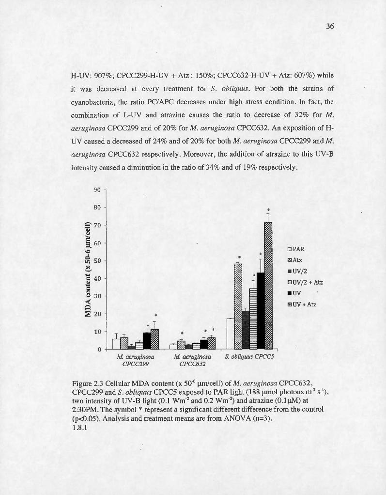
36
H-UV: 907%; CPCC299-H-UV + Atz: 150%; CPCC632-H-UV + Atz: 607%) while
it was decreased at every treatment for S. obliquus. For both the strains of
cyanobacteria, the ratio PC/APC decreases under high stress condition. In fact, the
combination of L-UV and atrazine causes the ratio to decrease of 32% for M.
aeruginosa CPCC299 and of 20% for M. aeruginosa CPCC632. An exposition of H
UY caused a decreased of 24% and of 20% for both M. aeruginosa CPCC299 and M .
aeruginosa CPCC632 respectively . Moreover, the addition of atrazine to this UV-B
intensity caused a diminution in the ratio of 34% and of 19% respective! y.
90
80
r::::- 70 Q:j ~
§ 60 -\0 1
~50 ><: "-' ..... c 40 ~ c 8 30 < Q
;:; 20
10
M aeruginosa CPCC299
M aeruginosa CPCC632
*
S obliquus CPCC5
DPAR
~Atz
• UV/2
l::::lUV/2 + Atz
•uv E2UV + Atz
Figure 2 .3 Cellular MDA content (x 50'6 !-Lm/cell) of M. aeruginosa CPCC632 , CPCC299 and S. obliquus CPCC5 exposed to PAR light (188 !-LffiOI photons m·2 s·1
) ,
two intensi ty of uv=B light (0.1 wm·2 and 0.2 Wm'2) and atrazine (0.1 !-LM) at 2:30PM. The symbol * representa significant different difference from the control (p<0.05). Analysis and treatment means are from ANOVA (n=3). 1.8.1

37
For M. aeruginosa, the MDA content was significantly increased for the H
UY treatment and the combination H-UV with atrazine (Figure 2.3). The non-toxic
strain of M. aeruginosa seems also to increase its MDA content in presence of
atrazine alone. For the green alga, the MDA content was increased for ali treatments
except the low dose of UV-B.
2.6 Discussion
2.6.1 Atrazine effect
Despite the maximum concentration of 1.8 f.,Lg/L (0.0084f..LM) established by
Canada for the protection of aquatic life (CCME, 1999), atrazine is often present in
rivers at concentrations higher than 20 f..Lg/L (0.092 f..LM; Solomon et al., 1996;
Giroux, 2010) and can reach 100f..Lg/L (0.46f..LM; DeLorenzo, 200l).lt was previously
shown that a concentration as low as 0.1 f..LM can have a deleterious effect on
photosynthesis of phytoplankton (Chalifour and Juneau, 2011; Ralph 2000).
Our photosynthetic results further support these observations. In accordance
with previous studies (Chalifour and Juneau, 2011; Deblois et al., 2013), we noticed
a decreased in the maximal PSII quantum yield of the two M. aeruginosa strains
(although not significantly for 632). The electron transport rate in active reaction
centers (EtJRC) was also affected by the presence of atrazine. This effect on the PSII
electron transport rate may be explained by the mode of action of atrazine, which
interacts with the Q8 binding site on the Dl protein (Fedtke and Duke, 2005). A
blockage of the electron transport leads to an accumulation of energy at the PSII
leve) that needs to be dissipated (Müller et al., 2001 and Schreiber et al., 1986). One
of the efficient energy dissipation pathways is beat dissipation and as we found in
our experiment, DI0 /RC increased for ali strains in presence of atrazine. lt seems
however that beat dissipation was not sufficient to dissipate excess energy and sorne
production of Reactive Oxygen Species occurred (as seen by the lipid peroxidation
damages observed; Table 4). The observed effects on the PSII photochemical activity
38
may therefore be explained by the electron transport blockage and the increased
presence of ROS. Higher ROS production was previously demonstrated when
Chlorella vulgaris was exposed to atrazine (Qian et al., 2008) , and was shawn to
increase MDA leve! and alter gene transcription, subcellular structure, and
physiological state. This ROS production may lead to the destruction of various
proteins such as Dl and therefore induces photoinhibition (Vass et al. , 1992; Xiong
et al. , 1997; Allakhverdiev and Murata, 2004; Nishiyama et al. , 2004 and Murata et
al., 2007)
The effects on the PSII photochemical activity are also reflected in a drop in
the operational quantum yield (cp' M; Table 2.2). For S. obliquus this decrease causes
the reduction of pH gradient across the thylakoid membrane and therefore a decrease
in the xanthophyll cycle activity , which is the main component of NPQ. Eventually,
the reduction of NPQ leads to a decreased on the PSII photochemical acti vity. On the
other hand , cyanobacteria do not have xanthophyll cycle then the main component of
NPQ become state transition (Bailey and Grossman, 2008). State transition is the
movement of light harvesting complexes from photosystem II to photosystem 1 to
equilibrate energy (Bonaventura and Myers, 1969 and Wollman, 2001) between
them in arder to prevent photoinhibition (Keren and Ohad, 1998) and it is regulated
by the quinone reduction (Mullineaux and Allen, 1992). Here, the electron transport
chain was blocked by atrazine leading to the quinones oxidation, the inhibition of the
state transition and the reduction of the NPQ. A decrease in NPQ after an atrazine
exposure was expected . Indeed , similar results were obtained in previous studies
(Deblais et al., 2013; Chalifour and Juneau, 2011).
Finally, it was showed by our results that atrazine increased the Fofcell of S.
obliquus while the chlorophyll content decreased. Since the antenna size of the
reaction centers (ABS/RC) was not significantly affected, we may hypothesize that
this treatment has also an effect on the number of reaction centers, due to the
induction of oxidative stress , showed by the MDA results .

39
2.6.2 UV -B effect
As a consequence of the ozone layer depletion , UV -B radiation was shown to
increase at the earth surface and doses as high as 1.62 wm-2 was observed in
America (Gleason and Wellington, 1995) but it can reach 2.1 wm-2 in other countries
(O·uce et al. 2012) . These doses of UV-B can cause severa! damages to
phytoplankton (Holzinger and Lütz , 2006 and Fernanda Pessoa, 2012). One of the
main observed damage could be the direct photodegradation of the PSII Dl protein
(Xiong et al ., 1997 and Bouchard et al., 2006) . lt was previously shown that UV
radiation induced the direct degradation of the Dl protein in higher plants
(Panagopoulos et al., 1990; Foyer et al., 1994; Smirnoff, 1998; Mahdavian, 2008) as
weil as in cyanobacteria (He et Hader, 2002). This phenomenon is known to induce a
decrease of the PSll activity (Walter and Horton, 1993), as demonstrated by our
results where we observed a decrease of the maximal PSII quantum yield C<!>M). This
decrease in PSII activity leads to an accumulation of energy at this leve!. This energy
will be caught by soluble oxygen molecules and will generate ROS. Here, the
presence of ROS was , in fact , detected by the increased of MDA (Fig 2.3) .
Our results showed also that , at both doses, UV -B treatments increased the
Fofcell of ali species and the chlorophyll content was either decreased for M.
aeruginosa CPCC632 or not significantly changing. Indeed, one of the consequences
of an UV -B exposure is the chlorophyll degradation (Xue et al. , 2005). However, as
it was showed by previous studies (Sfichi et al. , 2004; Albert et al., 2008), the
antenna size of the reaction centers (ABS/RC) was increased by the two UV -B
treatments , definitely explaining the rise in fluorescence emission (Fofcell).
Since beat dissipation helps the cell to cope with an accumulation of energy at
the PSI! (Schreiber et al ., 1986; Schreiber et al. , 1994; Krause et Weis, 1991; Juneau
et al ., 2007; Marwood et al., 2000 and Schreiber et al. , 2002), it would explain the
rise of this parameter (DI0/RC) for M. Aeruginosa CPCC 632 at both doses.
Moreover, sorne pigments found in the light harvesting complexes, or

40
phycobilisomes, also participate in the excess energy dissipation. Indeed, pigments
like carotenoids protect the photosynthetic apparatus by absorbing the excess of light
energy or by acting like antioxidant molecules (Hirschberg and Chamovitz , 1994 and
Salguero et al., 2003) . The effect of UV -B on carotenoids pigments can be very
variable . Most of the ti me , carotenoids are degraded by UV -B radiation (Bishof et al .,
2002) but it was also showed that for sorne species, UV radiation could increase
production of photosynthetic pigments (Yakovleva and Titlyanov, 200 l). Here ,
zeaxanthin and {3- carotene contents were increased for M. aeruginosa while they
were degraded for S. obliquus. Based on the photosynthetic activity results, although
the carotenoid content was increased for M . aeruginosa , it was not enough to protect
the photoinhibition caused by the two UV-B treatments (0.1Wm·2 and 0.2wm-2). In
cyanobacteria, other pigments, like allophycocyanin (APC) and phycocyanin (PC)
and orange carotenoid protein (OCP), exist to protect the photosynthetic apparatus
against a UV -B stress . Allophycocyanin and phycocyanin transfer the excitation
energy to the chlorophylls antenna of PSII and PSI (Kirilovsky, 2007). Here, a
decrease of the PC/A PC ratio was observed for the higher doses of UV -B explaining
the increase of photoinhibition despite the increase of carotenoid content. MoreC?ver,
according to Kirilovsky (2007) , OCP may acts as the thermal dissipator of the excess
absorbed energy in cyanobacteria. During this experiment, OCP were not dosed but
we may hypothesize that by degrading those pigments, UV -B decreases protection
against light stress. However, it would be interesting to measure OCP content after a
UV -B exposure to confirm our hypothesis.
ASB/RC can be influenced by the pigment content and the number of PSII
reaction centers (Force et al., 2003). We demonstrated here that the presence of UV
B radiation increases the antenna size (ABS/RC) at low and high doses. The
unchanged chlorophyll content between treatments combined to the increase in
ABS/RC suggests a decrease in the numbers of reaction centers, which is also
suggested by the decrease in <PM- Furthermore, it is known that the ROS production
41
and the damage to the Dl protein may lead to the reduction of the operational
photosynthetic rate (cp 'M) of phytoplankton (Vass et al., 1992; Xiong et al., 1997;
Allakhverdiev and Murata, 2004; Nishiyama et al., 2004 and Murata et al., 2007).
Since UV-B directly degrades Dl protein , it was expected to see, as for the atrazine, a
decrease in electron transport rate showed by the decrease of <P ' M in ali studied
species. Consequent) y, this decrease causes the reduction of NPQ due to non
activation of the xanthophyll cycle for green algae and the state transition for
cyanobacteria.
2.6.3 UV -B and atrazine combined effect
According to our study, a three-hour UV-B treatment (O.lwm-2 and 0.2wm-2)
m combination to a O.lf.JM atrazine treatment has a drastic effect on the
photosynthetic activity and energy dissipation processes of the studied species.
Indeed, EtJRC was highly inhibited while the ABS/RC and the DIJRC were highly
stimulated due to the electron transport chain blockage by atrazine and Dl protein
degradation by UV-B radiation . However , for M. aeruginosa CPCC632 exposed to
the highest dose of UV-B radiation , we observed unexpected results. The electron
transport rate (EtJRC) was increased whi le the rise of the effective antenna size
(ABS/RC) and of the effective dissipation (DI0/RC) was not significant . These results
suggest that the differences in the toxicity of the cyanobacteria may influence the
effect of environmental factors as it was previously demonstrated for light intensity
(Deblais et al., 2013), but further research is needed to elucidate the mechanisms
involved.
In presence of atrazine and for both doses of UV -B radiation the minimal
fluorescence (FJcell) was increased whereas the <PM and the <P' Mwere reduced for the
studied species. As explain previously a decrease in the <PM and the <P ' M was also
expected because of the degradation of Dl protei.ns and pigments by UV -B and the
blockage of the electron transport chain by atrazine. For the same reasons as
42
explained earlier, a decrease in NPQ was also expected after an exposure of both UV
B and atrazine. However, the combination of these factors on Scenedesmus obliquus
had a more important effect than their individual effect. Indeed , the atrazine causes a
blockage of the electron transpo1t chain, which leads to an energy accumulation at the
PSII. In addition, UV -B radiations are known to degrade DI protein and also lead to
an energy accumulation at the PSII (Xiong et al., 1997 ; Bouchard et al. , 2006).
Moreover ,. UV -B radiations have severa! other effects on the photosynthetic
apparatus. As shown in our study , UV -B can degrade chlorophyll, which should
decrease the fluorescence per cell and ·the maximal PSII quantum yield . Surprisingly ,
the minimal fluorescence per cell is always higher for the treated samples compared
to the control. As explain earlier, if the size of antenna (ABS/RC) is increasing it will
affect the fluorescence emi ssion explaining the increase of the Fofcell. lt is also
known that UV -B can also degrade other pigments like carotenoids (Bischof et al.,
2002), which are protective pigments against photodamages and ROS. He and Hader
(2002) showed that UV -B could generate ROS direct! y , which is in accordance with
our MDA results (Fig 2.3). Thus, the synergistic effect that was found here on
Scenedesmus obliquus can easily be explained by an enhancement of the atrazine
effect caused by the degradation of protective pigments (table 2 .3) against oxidative
stress by UV-B radiation.
Cyanobacterial allophycocyanin and phycocyanin transfer the excitation energy
to the chlorophylls antenna of PSII and PSI (Kirilovsky, 2007). Moreover, it was
found that phycocyanin tends to bleach more rapidly than allophycocyanin (Sinha et
al. 1995) and that UV -B also decrease more rapidly the content of phycocyanin than
other biliproteins like allophycocyanin (Rinalducci et al. 2006) . Recently, it was
suggested that NPQ in cyanobacteria may be mediated by phycobilisomes
(Kirilovsky, 2007). Th us, the decrease in the ratio PC/APC that was found in our
study in presence of atrazine and UV -B can explain the decrease of NPQ found for ·
the two strains of cyanobacteria.
43
For Microcystis aeruginosa CPCC299, we found that the operational PSII
quantum yield (<f>' M) was more affected by the combination of stress than the
maximal PSU quantum yield (cpM). Indeed , when exposed to UV -B and atrazine, the
ROS production would be higher under light because of the accumulatio!) of energy
that the stresses generate. This is why the PSII photosynthetic activity would be more
affected at light than in the dark. It is also known that UV -B radiation can reduce
RuBisCo activity (Strid et al. 1994) , and, therefore, induce a decrease of carbon
fixation. Consequently, when exposed to light, a blockage of the electron transpo1t
chain will occur (decrease of cp' M), but in the dark this effect will not be shown (as
seen by the unchanged <PM).
Different effects were expected between species (Microcystis aeruginosa vs
Scenedesmus obliquus) given that one is a cyanobacteria white the other is a green
alga. However, it is interesting to see important differences of effect between the
toxic and non-toxic strain of cyanobacteria. According to our photosynthetic results
(<f>M and <P'M) , the toxic strain (Microcystis aeruginosa CPCC299) was less affected
by ali treatments by UV -B whereas it is the opposite for the atrazine treatment. Those
results may suggest the involvement of microcystins in the protection against
environmental factors, but further studies need to be done.
44
2. 7 Conclusion
Although environmental stresses occur simultaneously In natural
environment , very few studies were done in the past to study their interactions. Mode
of action of atrazine and UV -B radiations al one are weil known (Jansen et al., 2012;
Delorenzo et al., 2001) , but to our knowledge the interaction of these two stresses has
never been investigated on different phytoplankton species. We demonstrated that the
combination of environ mental doses of UV -B and atrazine had a greater effect on
sorne species of phytoplankton than the exposure of these stresses al one. As the toxic
strain of cyanobacteria proved to be Jess affected , these results are somewhat
alarming, given the enormous environmental impact they can generate. We also
showed that toxic strain of cyanobacteria had a different response to environmental
stresses than the non-toxic ones suggesting a mechanistic difference between the two
strains of the same species. We strongly recommended that future studies for aquatic
li fe protection consider the effect that UV -B can have on many pesticides fou nd in
watercourses.
2.8 Acknowledgements
The authors would like to thank David Dewez to allow them to use his U
HPLC. This research would not have been possible without the financial support
provided by Natural science engineering research council of Canada.
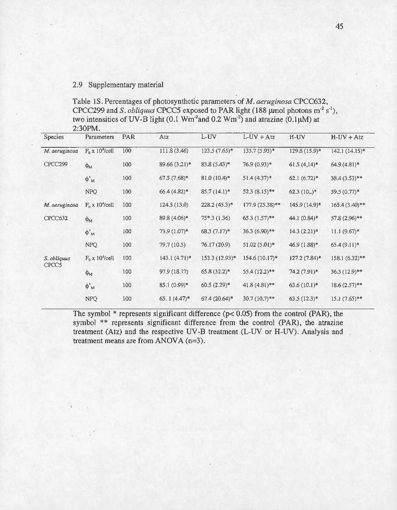
45
2.9 Supplementary material
Table lS. Percentages of photosynthetic parameters of M. aeruginosa CPCC632, CPCC299 and S. obliquus CPCC5 exposed to PAR Iight (188 f.Lmol photons m·2 s·'), two intensities of UV-8 light (0.1 wm-2and 0 .2 wm-2) and atrazine (O.lf.LM) at 2:30PM.
S pec ies Parameters PAR Atz L-U Y L-UY + Atz H-UY H-UY+ Atz
M. aeruginosa F0 x 1 0"/cell 100 1 1 1 .8 (3 .46) 123.5 (7 .65)* 133.7 (5 .93)* 129.8 (15.9)* 142.1 (14. 15)*
CPCC299 cpM 100 89 .66 (3.2 1)* 83.8 (5.43)* 76.9 (0 .93)* 6 1.5 (4, 14)* 64.9 (4.8 1)*
cp' M 100 67 .5 (7 .68) * 8 1.0( 10.4)* 51.4 (4.37)* 62 .1 (6.72)* 38.4 (3.53)**
NPQ 100 66.4 (4.82)* 85 .7 (14 .1)* 52.3 (8.15) ** 62 .3 ( 10 ,.)* 59 .5 (0 .77)*
M. aeruginosa F0 x 104/cell 100 124.5 ( 13.0) 228.2 (45 .3)* 177.9 (25.38)** 145 .9 ( 14 .9)* 165.4 (3 .40)**
CPCC632
S . obliquus CPCC5
cpM 100 89 .8 (4.06)* 75 *.3 (1.36) 65.3 ( 1.57) ** 44.1 (0 .84)* 57 .8 (2.96)**
cp 'M 100 73 .9 ( 1.07) * 68.3 (7 .17)* 36.3 (6.90) ** 14 .3 (2.21 )* Il .1 (9.67)*
NPQ 100 79.7 ( 10 .5) 76 .17 (20.9) 51 .02 ( 5 .0 1 ) * 46 .9 ( 1.88)* 65.4 (9 .11 )*
F0 x 1 0"/cell 100 143 .1 (4 .7 1)* 153 .3 ( 12.93)* 154.6 ( 10.17)* 127.2 (7 .84)* 158 .1 (6.32)**
cpM 100 97.9 ( 18 .17) 65 .8 (32 .2)* 55.4 ( 12 .2)** 74.2 (7 .9 1)* 36.3 ( 12 .9)**
cp 'M 100 85. 1 (0 .99)* 60.5 (2.29)* 4 1.8 (4.8 1 )** 63 .6 (10 .1)* 18 .6 (2 .57)**
NPQ 100 65 . 1 (4.47)* 67.4 (20.64)* 30.7 ( 10.7)** 63 .5 ( 12.3)* 15.1 (7 .65)**
The symbol * represents significant difference (p< 0.05) from the control (PAR), the symbol ** represents significant difference from the control (PAR) , the atrazine treatment (Atz) and the respective UV-8 treatment (L-UV or H-UV). Analysis and treatment means are from ANOVA (n=3).
46
2.10 References
Arts, M., & Rai, H. (1997). Effects of enhanced ultraviolet-B radiation on the production of lipid , polysaccharide and protein in three freshwater algal species. Freshwater Biology , 38(3), 597- 610.
Albert, K. R., Mikkelsen, T. N., & Ro-Poulseri , H. (2008). Ambient UV-B radiation decreases photosynthesis in high arctic Vaccinium uliginosum. Physiologia Plantarum , 133(2), 199-210.
Allakhverdiev, S. 1., & Murata , N. (2004). Environmental stress inhibits the synthesis de novo of proteins involved in the photodamage-repair cycle of Photosystem II in Synechocystis sp. PCC 6803. Biochimica et Biophysica Acta- Bioenergetics, 1657, 23-32.
Allen JF (1992) Protein phosphorylation in regulation of photosynthesis. Biochimica et Biophysica Acta, 1098(3), 275-335.
Bailey , S., & Grossman, A. (2008). Photoprotection in cyanobacteria: Regulation of light harvesting. Photochemistry and Photobiology , 84(6), 1410-1420.
Bilger, W., Bjorkman, 0., (1990). Role of the xanthophyll cycle in photoprotection elucidated by measurements of light-induced absorbance changes, fluores-cence and photosynthesis in leaves of Hedera canariensis . Photosynthesys Research 25, 173-185.
Bischof, K. , Krabs, G., Wiencke, C., et Hanelt, D. (2002). Solar ultraviolet radiation affects the activity of ribulose-1 ,5-bisphosphate carboxylase-oxygenase and the composition of photosynthetic and xanthophyll cycle pigments in the intertidal green alga Ulva lactucaL. Planta 215(3), 502- 509.
Bonaventura C, Myers J (1969) Fluorescence and oxygen evolution from Chlorella pyrenoidosa. Biochimica et BiophysicaActa (BEA)- Bioenergetics, 189(3),366-383.
Bouchard, J. N., Roy, S., et Campbell, D. A. (2006). Symposiurn-in-Print: UV Effects on Aquatic and Coastal Ecosystems UVB Effects on the Photosystem IlDl Protein ofPhytoplankton and Natural Phytoplankton Communities .
. Photochemistry and photobiology 82(2), 936-951.
Campbell, D. , Hurry, V., Clarke, a K., Gustafsson, P., & Oquist, G. (1998). Chlorophyll fluorescence analysis of cyanobacterial photosynthesis and
acclimation. Microbiology and Molecular Biology Reviews: MMBR, 62(3), 667--683.
47
CCME. (1999) . Canadian water quality guidelines for the protection of aquatic !ife: atrazine. In: Canadian Environmental Quality Guidel ines . Canadian Council of Ministers of the Environment, Winnipeg, Canada, 4 pp.
Chalifour, A. et Juneau, P. (2011). Temperature-dependent sensitivity of growth and photosynthesis of Scenedesmus obliquus, Navicula pelliculosa and two strains of Microcystis aeruginosa to the herbicide atrazine. Aquatic Toxicology, 1 03(1 ), 9-17.
Chen, Z., Juneau, P. et Qiu, B. (2007). Effects ofthree pesticides on the growth, photosynthesis and photoinhibition of the edible cyanobacterium Ge-Xian-Mi (Nostoc). Aquatic Toxicology, 81(3), 256-265.
Chen, L. , Xie, M., Bi, Y. , Wang, G. , Deng, S. et Liu, Y. (2012). The combined effects ofUV-B radiation and herbicides on photosynthesis, antioxidant enzymes and DNA damage in two .bloom-fom1ing cyanobacteria. Ecotoxicology and environmental safety, 80, 224-30.
Cloern, J. E. (1999) . The relative importance of light and nutrient limitation of phytoplankton growth: a simple index of coastal ecosystem sensitivity to nutrient enrichment. Aquatic ecology, 33(1), 3-15.
Cruces, E., Huovinen, P., & G6mez, 1. (2012).lnteractive effects of UV radiation and enhanced temperature on photosynthesis, phlorotannin induction and antioxidant activities of two sub-Antarctic brown algae. Marine Biology, 160(1) , 1-13 .
Deblois, C. P ., Dufresne, K ., & Juneau , P. (2013). Response to variable light intensity in photoacclimated algae and cyanobacteria exposed to atrazine. Aquatic Toxicology (Amsterdam, Netherlands), 126,77-84.
Delorenzo , M. E., Scott, G. 1., & Ross , P. E. (2001). Toxicity of pesticides to aquatic microorganisms : A review. Environmental Toxicology and Chemistry, 20( 1) , 84--98.
Demmig-Adams , B. (1990). Carotenoids and photoprotection in plants: A role for the xanthophyll zeaxanthin. Biochimica et Biophysica Acta- Bioenergetics, 1020(1), 1-24.

Fedtke, C. et Duke, S. O. (2005). Herbicides. Dans Hock, B. et Elstner, E . F. (Ed.) Plant toxicology (4e édition) (p.247-330) . New York, NY : Marcel Dekker.
48
Fenical, W. (1983) . Marine plants: A unique and unexplored resource. Dans Banta, H. D. (Ass. Dir.) Plants: The Potentialsfor Extracting Protein, Medicines, and Other Useful Chemicals (p.147-153) . Washington, D.C.: U.S. Congress, Office ofTechnology Assessment.
Fernanda Pessoa, M. (2012) . Harmful effects of UV radiation in Algae and aquatic macrophytes-A review. Emirates Journal of Food and Agriculture , 24(6).
Force, L. , Critchley , C. , & Rensen , 1. 1. S. Van. (2003). New fluorescence parameters for monitoring photosynthesis in plants. Photosynthesis Research, 78, 17-33.
Foyer, C. H., M. Lelandais et K. 1. Kunert. (1994). Photooxidative stress in plants. Physiologia plantarum 92(4), 696- 717.Gal A. , Zer H . et Ohad, I. (1997) . Redoxcontrolled thylakoid protein phosphorylaticin. News and views. Physiologia Plantarum, 100(4), 869-885.
Frederick, 1. E. (1993) . Ultraviolet sunlight reaching the Earth ' s surface: A review of recent research . Photochemistry and Photobiology , 5 7(1) , 175-178 .
Garcfa-Piazaola , 1. I. , & Becerril , 1. M . (1999). A rapid high-performance liquid chromatography method to measure lipophilic antioxidants in stressed plants: Simultaneous determination of carotenoids and tocopherols. Phytochemical Analysis, 10(6), 307-313.
Genty, B., Briantais,1 . M. et Baker, N. R. (1989). The relationship between the
quantum yield of photosynthetie electron transport and quenching of chlorophyll
fluorescence. Biochimica et Biophysica Acta (BBA)-General Subjects, 990 (1),
87-92.
Giroux , 1. , (2010). Présence de pesticides dans l ' eau au Québec- Bilan dans quatre cours d'eau de zones en culture de maïs et de soya en 2005,2006 et 2007 et dans des réseaux de distribution d'eau potable. MinistèreduDéveloppement durable, de l 'Environnement et des Parcs, Direction du suivi de l'état de l'environnement, 78 p.
Gleason, D . F. , & Wellington , G. M. (1995). Variation in UVB sensitivity of planula larvae of the coral Agaricia agaricites along a depth gradient. Marine Biology, 123(4) , 693-703 .
Graymore, M. , Stagnitti, F. et Allinson, G. (2001). Impacts of atrazine in aquatic ecosystems. Environment international, 26(7), 483-495.
49
Hayes, T. B., Khoury, V., Narayan, A., Nazir, M. , Park, A., Brown, T. , Adame, L., Chan, E., Buchholz, D, Stueve, T. et Gallipeau, S. (2010). Atrazine induces complete feminization and chemical castration in male African clawed frogs (Xenopus laevis). Proceedings of the National Academy ofSciences of the USA. 107(10), 4612-4617.
He, Y. Y. et Hader, D. P. (2002). Involvement of reactive oxygén species in the UVB damage to the cyanobacterium Anabaena sp. Journal of photochemistry and photobiology, B : Biology, 66(1), 73-80.
Heath. R.L, Parker. (1968). Photoperoxidation in isolated chloroplasts. 1. Kinetics and stoichiometry of fatty acid peroxidation. Arch. Biochem. Biophys. 125: 189- 198 .
Hirschberg, J. , & Chamovitz, D. (1994). Carotenoids in cyanobacteria. The Molecular Biology of Cyanobacteria, 559-5579.
Holzinger, A. et Lütz, C. (2006). Algae and UV irradiation: effects on ultrastructure and related metabolic functions. Micron, 37(3), 190-207.
Jansen , A. K., M., Hideg, É. , & J. C. Lidon, F. (2012). UV -B radiation: "When does the stressor cause stress?" Emirates Journal of Food and Agriculture, 24(6).
Juneau P. , Qiu B. et Deblois C.P. (2007) . Use of chlorophyll fluorescence as a tool for determination of herbicide toxic effect: Review . Toxicological and Environ Chemistry 89 (4):609-625
Keren, N., & Ohad, l. (1998). State Transition and Photoinhibition . In Trends in biochemical sciences (pp. 569-590).
Kitajima, M., Butler , W.L., (1975). Quenching of chlorophyll fluorescence and primary photochemistry in chloroplasts by dibromothymoquinone .. Biochimica et Biophysica Acta, 376, 105-115 .
Kirilovsky, D. (2007) . Photoprotection in cyanobacteria: the orange carotenoid protein (OCP)-related non-photochemical-quenching mechanism. Photosynthesis Research, 93(1-3), 7-16.
Krause, G. et Weis, E. (1991). Chlorophyll fluorescence and photosynthesis: the basics. Annual Review of Plant Biology, 42(1), 313-349.

50
Krieger-Liszkay, A., Fufezan, C., & Trebst, A. (2008). Singlet oxygen production in photosystem Il and related protection mechanism. Photosynthesis Research, 98, 551-564 .
. Lüder , U . H ., Knoetzel, J. , & Wiencke , C. (2001). Acclimation of photosynthesis and · pigments to seasonally changing light conditions in the endemie antarctic red macroalga Pal maria decipiens. Polar Biology, 24(8), 598-603.
Mahdavian, K., K. M. Kalantari, M. Ghorbanli et M. Torkzade. (2008). The effects of salicylic acid on pigment contents in ultraviolet radiation stressed pepper plants. Biologia plantarum 52(1), 170-172.
Marwood, C.A., Smith, R. E., Furgal, J. A., Charlton, M. N. , Salomon, K. R. et Green berg, B. M. (2000). Photo inhibition of natural phytoplankton assemblages in Lake Erie exposed to solar ultraviolet radiation. Canadian Journal of Fisheries and Aquatic Sciences, 5 7(2), 371-379.
Müller, P. , Li, X. P. et Niyogi, K. K. (2001). Non-photochernical quenching. A
response to excess light energy. Plant Physiology, 125(4), 1558-1566.
Mullineaux, C. W. , & Allen, J .F. (1990). State 1-State 2 transitions in the cyanobacterium Synechococcus 6301 are controlled by the redox state of electron carriers between Photosystem~ I and Il . Photosynthesis Research, 23(3), 297-311.
Murata, N., Takahashi , S ., Nishiyama, Y., & Allakhverdiev, S . l. (2007). Photoinhibition of photosystem II under environmental stress. Biochimica et Biophysica ACta- Bioenergetics, 1767, 414-421.
Neale, P. 1. , Cullen, J. J. , et Davis, R.F. (1998). Inhibition of marine photosynthesis by ultraviolet radiation: Variable sensitivity ofphytoplankton in the WeddellScotia Confluence during the austral spring. Limnology and Oceanography, 43(3), 433-448 .
Nishiyama, Y., Allakhverdiev, S.l. , Yamamoto , H., Hayashi , H. , & Murata, N. (2004) . Singlet oxygen inhibits the repair of photosystem II by suppressingthe translation elongation of the Dl protein in Synechocystis sp . PCC 6803. Biochemistry, 43, 11321- 11330.
Nishiyama, Y., Yamamoto, H ., Allakhverdiev , S. 1., Inaba, M ., Yokota, A., & Murata, N. (2001). Oxidative stress inhibits the repair of photodamage to the photosynthetic machinery. EMBO Journal, 20(20), 5587-5594.

Panagopoulos, L., J.F. Bornman et L. O. Bjorn. (1990). Effects ofultraviolet radiation and visible light on growth, fluorescence ind\lction, ultra weak luminescence and peroxidase activity in sugar beet plants. Journal of Photochemistry and Photobiology. 8(2),73-87.
Qian , H ., Sheng , G. D. , Liu , W. , Yingcong , L. , Liu , Z. , & Zhengwei , F. (2008). Inhibitory effects of atrazine on Chlorella vulgarisas assessed by real-time polymerase.chain reaction. Environmental Toxicology and Chemistry, 27(1), 182-187 .
51
Ralph, P.J . (2000). Herbicide toxicity of Halophila ovalis assessed by chlorophyll a fluorescence. Aquatic Botany, 66(2), 141-152.
Rinalducci, S., Hideg, É., Vass, 1., & Zolla, L. (2006). Effect of moderate UV -B irradiation on Synechocystis PCC 6803 biliproteins. Biochemical and Biophysical Research Communications, 341(4), 1105_:1112.
Rost, B. , Zondervan, I. et Wolf-Gladrow, D. (2008). Sensitivity ofphytoplankton to future changes in ocean carbonate chemistry: current knowledge, contradictions and research directions. Marine ecology progress series, 373, 227-237.
Salguero, A., de la Morena, B., Vigara, J., Vega, J. M., Vilchez, C., & Leon, R. (2003). Carotenoids as protective response against oxidative damage in Dunaliella bardawil. Biomolecular Engineering, 20(4-6), 249-253 .
Schreiber, U., Bilger, W. et Neubauer, C. (1994). Chlorophyll fluorescence as a nonintrusive indicator for rapid assessment of in vivo photosynthesis. Dans Schulze (E.d.), Ecophysiology ofphotosynthesis (p.49- 70). Berlin: Springer.
Scbreiber, U., Müllerm J.F. , Haugg, A. et Gademann, R. (2002) . New type of dualchannel P AM chlorophyll fluorometer for highly sensitive water toxicity biotests. Photosynthesis Research, 74(3) , 317-330.
Schreiber, U., Schliwa, U . et Bilger, W. (1986). Continuons recording of photochemical and non-photochemical chlorophyll fluorescence quenching with a new type of modulation fluorometer. Photosynthesis Research, 10(1), 51-62.
Sfichi , L. , Ioannidis, N., & Kotzabasis, K. (2004). Thylakoid-associated polyamines ad just the UV -B sensitivity of the photosynthetic apparatus by means of Iightharvesting complex II changes. Photochemistry and Photobiology, 80(3), 499-506.

52
Sinha, R. P., Kumar, H. D., Kumar, A., & Hader, D.-P. (1995). Effects of UV-B irradiation on growth , survival, pigmentation and nitrogen metabolism enzymes in cyanobacteria . Acta Protozoologica , 34, 187-192.
Smirnoff, N. (1998). Plant resistance to environmental stress. Current Opinion in Biotechnology 9(2), 214-219.Stah1, W. et Sies, H. (2002). Carotenoids and protection against solar UV radiation. Skin Pharmacology and Physiology, 15(5), 291 -296. .
Salomon, K. R ., Baker, D. B., Richards , R. P., Dixon, K. R., Klai- ne, S. J., La Point, T. W., Kendall, R. J ., Weiskopf, C. P., Giddings, J. M., Giesy , J. P., Hall, L. W. & Marty Williams, W. (1996). Ecological risk assessment of atrazine in North American surface waters. Environ. Toxicol. Chem. 15(1):31- 76
Strid, A., W. S. Chow and J. M. Anderson. (1994). UV-B damage and protection at the molecular leve) in plants . Photosynth. Res. 39:475-489.
Sullivan, D.J. , Vecchia, A.V. , Lorenz, D.L. , Gilliom, R.J., Martin, J.D., (2009). Trends in pes-ticide concentrations in corn-belt streams, 1996- 2006: U.S. Geological Survey Scientific Investigations Report 2009-51 32. 75 p.
Suresh Babu, G., Joshi, P.C., et Viswanathan, P.N. (1998). UVB-Induced reduction in biomass and overall productivity of cyanobacteria. Biochemical and Biophysical Research Communications 244( 1), 138- 142.
Vass, 1., Styring, S., Hundal, T., Koivuniemj, a, Aro, E., & Andersson, B. (1992). Reversible and irreversible intermediates during photoinhibition of photosystem II: stable reduced QA species promote chlorophyll triplet formation. Proceedings of the National Academy of Sciences of the United States of America, 89(February), 1408-1412.
Walters, R. G., & Horton , P. (1993). Theoretical assessment of alternative mechanisms for non-photochemical quenching of PS II fluorescence in barley leaves. Photosynthesis Research, 36, 119-139 .
Wang, S., Duan, L., Li, J. , Tian, X. et q , Z. (2007). UV -B radiation increases paraquat tolerance oftwo broad-leaved and two grass weeds in relation to changes in herbicide absorption and photosynthesis. Weed Research, 47(2), 122-128.
West, L. J. a, Li, K., Greenberg, B . M., Mierle, G., & Smith, R. E. H. (2003). Combined effects of copper and ultraviolet radiation on a microscopie green al ga

53
in natural soft lake waters of varying dissolved organic carbon content. Aquatic Toxicology Amsterdam Netherlands, 64(1) , 39-52.
Wollman FA. (2001). State transitions reveal the dynamics and flexibility of the photosynthetic apparatus . The EMBO journa/20 (14):3623-3630.
Xiong, F. , Komenda, J., Kopecicy, J. et Nedbal, L. (1997). Strategies ofultraviolet-B
protection in microscopie algae. Physiologia Plantarum, 100(2), 378-388.
Xue, L., Zhang , Y. , Zhang, T., An, L. , & Wang , X. (2005). Effects of enhanced ultraviolet-B radiation on algae and cyanobacteria . Critical Reviews in Microbiology , 31(2), 79-89.
Yakovleva , 1. M. andE. A. Titlyanov. (2001) . Effect of high visible and UV irradiance on subtidal Chondrus crispus: stress, photoinhibition and protective mechanisms. Aquatic Botany . 71:47-61.
CONCLUSION GENERALE
L ' industrialisation de notre société a engendré d'énormes changements au niveau
de l'environnement. L'utilisation d ' insecticides et d'herbicides ainsi que la
destruction de la couche d'ozone en sont deux exemples. Plus notre société grandira,
plus ces changements seront importants et nombreux. Le nombre important de stress
environnementaux auxquelles sont exposés les écosystèmes implique nécessairement
qu'ils peuvent interagir entre eux. L'étude de la combinaison de stress
environnementaux sur les orgamsmes devient donc primordiale afin d 'avoir un
pouvoir de prédiction et ainsi , de nous aider à mieux gérer ces nombreux
changements à venir.
L 'objectif de cette étude était d'évaluer l'effet de la combinaison de l'atrazine,
un herbicide utilisé de façon importante dans le monde, et de la radiation UV -B qui a
été augmentée à la surface de la terre ces dernières années, sur les organismes
phytoplanctoniques. En évaluant comment ces stress agissent sur la photosynthèse de
ces organismes et en comparant la résistance de certaines espèces il a été possible de
déterminer comment ces stress environnementaux pouvait débalancer les écosystèmes
dans lesquels ils vivent.
Tout d'abord , les résultats obtenus lors de cette étude ont permis de voir que trois
heures de radiation UV-B combiné à 0,1!-LM d'atrazine engendre un important effet
négatif au niveau de la photosynthèse et ce, chez les trois phytoplanctons étudiés.
Puisque la photosynthèse constitue la base de leur physiologie, l'inhibition de celle-ci
chez le phytoplancton peut mener non seulement à la détérioration de leur
communauté, mais aussi à la réduction de leur qualité nutritive pour tous les autres
organismes qui s'en nourrissent . De plus , la comparaison des résultats des différents
paramètres photosynthétiques a démontré une énorme différence physiologique entre
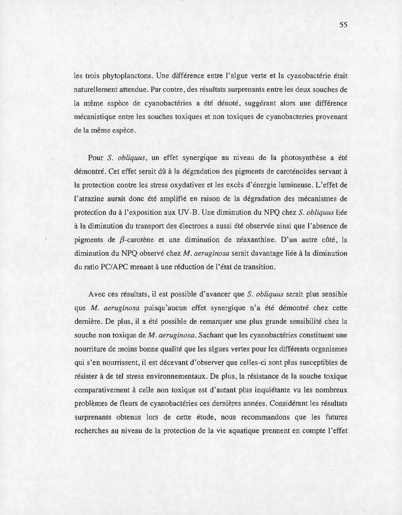
55
les trois phytoplanctons . Une différence entre l'algue verte et la cyanobactérie était
naturellement attendue. Par contre, des résultats surprenants entre les deux souches de
la même espèce de cyanobactéries a été dénoté, suggérant alors une différence
mécanistique entre les souches toxiques et non toxiques de cyanobacteries provenant
de la même espèce.
Pour S. obliquus, un effet synergique au mveau de la photosynthèse a été
démontré. Cet effet serait dû à la dégradation des pigments de caroténoïdes servant à
la protection contre les stress oxydatives et les excès d 'énergie lumineuse. L'effet de
1 'atrazine aurait donc été amplifié en raison de la dégradation des mécanismes de
protection du à l'exposition aux UV-B . Une diminution du NPQ chez S. obliquus liée
à la diminution du transport des électrons a aussi été observée ainsi que 1 'absence de
pigments de ,8-carotène et une diminution de zéaxanthine. D ' un autre côté, la
diminution du NPQ observé chez M. aeruginosa serait davantage liée à la diminution
du ratio PÇ/APC menant à une réduction de l'état de transition.
Avec ces résultats , il est possible d ' avancer que S. obliquus serait plus sensible
que M. aeruginosa puisqu'aucun effet synergique n'a été démontré chez cette
dernière. De plus, il a été possible de remarquer une plus grande sensibilité chez la
souche non toxique de M. aeruginosa. Sachant que les cyanobactéries constituent une
nourriture de moins bonne qualité que les algues vertes pour les différents organismes
qui s'en nourrissent , il est décevant d'observer que celles-ci sont plus susceptibles de
résister à de tel stress environnementaux. De plus, la résistance de la souche toxique
comparativement à celle non toxique est d 'autant plus inquiétante vu les nombreux
problèmes de fleurs de cyanobactéries ces dernières années . Considérant les résultats
surprenants obtenus lors de cette étude, nous recommandons que les futures
recherches au niveau de la protection de la vie aquatique prennent en compte l'effet

56
que peut avoir les radi ations UV -B sur les diffé rents pesticides retrouvées dans les
cours d 'eau.
LISTE DES RÉFÉRENCES
Allen, J. F. (1992). Protein phosphorylation in regulation of photosynthesis. Biochimica et Biophysica Acta, 1098(3), 275-335.
Anderson, D. M. , Glibert, P. M. , et Burkholder, J. M. (2002). Harrnful algal blooms and eutrophication: nutrient sources, composition, and consequences. Estuaries 25(4) , 704.:726.
Bailey, S., & Grossman, A. (2008). Photoprotection in cyanobacteria: Regulation of light harvesting. Photochemistry and Photobiology, 84(6), 1410- 1420.
Banaszak, A.T. et Trench, R. K. (2001). Ultraviolet sunscreens in dinoflagellates. Protist, 15 2(2), 93-1 01.
Bilger, W ., & Bjorkman, O. (1990). Role of the xanthophyll cycle in photoprotection elucidated by measurements of light-induced absorbance changes, fluorescence and photosynthesis in leaves of Hedera canariensis. Photosynthesis Research, 25(3), 173-185.
Bischof, K., Hanelt, D. , Tug, H. , Karsten, U. , Brower, P.E.M., et Wiencke, C. (1998a). Acclimation of brown al gal photosynthesis to ultraviolet radiation in Arctic coastal waters. (Spitzbergen, Norway). Polar Biology 20 (6), 388- 395.
Bischof, K. , Hanelt, D. , et Wiencke, C. (1998b). UV-radiation can affect depthzonation of Antarctic macroalgae. Marine Biology 131(4), 597- 605.
Bischof, K. , Krabs, G. , Wiencke, C. , et Hanelt, D. (2002). Solar ultraviolet radiation affects the activity of ribulose-1 ,5-bisphosphate carboxylase-oxygenase and the composition of photosynthetic and xanthophyll cycle pigments in the intertidal green alga Ulva lactucaL. Planta 215(3), 502- 509.
Bouchard, J. N. , Roy, S., et Campbell, D. A. (2006). Symposium-in-Print: UV Effects on Aquatic and Coastal Ecosystems UVB Effects on the Photosystem IlD 1 Protein of Phytoplankton and Natural Phytoplankton Communities. Photochemistry and photobiology 82(2), 936- 951 .
Cain, M. L., Bowman, W. D. et Hacker, S. D. (2008). Ecology. Sunderland: Sinauer associates. 621 p.

58
Campbell, D. , Eriksson, M. J., Oquist, G., Gustafsson, P. et Clarke, A. K. (1998). The cyanobacterium Synechococcus resists UV-B by exchanging photosystem II reaction-center Dl proteins. Proceedings of the National Academy ofSciences of the USA., 95(1), 364-369.
Chalifour, A. et Juneau, P. (2011). Temperature-dependent sensitivity of growth and photosynthesis of Scenedesmus obliquus, Navicula pelliculosa and two strains of Microcystis aeruginosa to the herbicide atrazine. A qua tic Toxicology, 1 03( 1 ), 9-17.
Chen, Z., Juneau, P. et Qiu, B. (2007). Effects ofthree pesticides on the growth, photosynthesis and photoinhibition of the edible cyanobacterium Ge-Xian-Mi (Nostoc) . Aquatic Toxicology, 81(3), 256-265.
Chen, L., Xie, M. , Bi, Y. , Wang, G. , Deng, S. et Liu, Y. (2012). The combined effects ofUV-B radiation and herbicides on photosynthesis, antioxidant enzymes and DNA damage in two bloom-forming cyanobacteria. Ecotoxicology and environmental safety, 80, 224- 30.
Chèvre, N. et Erkman, S. (2011). Alerte aux micropolluants: pesticides, biocides, détergents, médicaments et autres substances chimiques dans l'environnement. Lausanne: Presses polytechniques et universitaires romandes. 84 p.
Cloem, J. E. (1999). The relative importap.ce of light and nutrient limitation of phytoplankton growth: a simple index of coastal ecosystem sensitivity to nutrient enrichment. Aquatic ecology, 33(1), 3-15.
Codd GA, Bell SG et Brooks, WP. (1989). Cyanobacterial toxins in water. Water Science & Technology, 21(3), 1-13.
Deblais, C. P. , Dufresne, K. et Juneau, P. (2013). Response to variable light intensity in photoacclimated algae and cyanobacteria exposed to atrazine. Aquatic toxicology, 126, 77- 84.
DeLange, H. J. et Lürling, M. (2003). Effects ofUV-B irradiated algae on zooplankton grazing. Hydrobiologia, 491(1 -3), 133-144.
Delorenzo, M. E., Scott, G.I. et Ross, P. E. (2001). Toxicity of Pesticides to Aquatic Microorganisms: A Review. Environmental Toxicology and Chemistry, 20(1 ), 84-98.

59
Denoyelles , F. , Kettle , W . D. , et Sinn , D. E. (1982). The responses of plankton communities in experimental ponds to atrazine, the most heavily used pesticide in the United States. Ecology, 63(5), 1285- 1293.
Demmig-Adams , B . (1990). Carotenoids and photoprotection in plants: A role for the xanthophyll zeaxanthin. Biochimica et Biophysica Acta- Bioenergetics, 1020(1) , 1-24.
Demott, W. et Müller-Navarra, D. (1997). The importance ofhighly unsaturated fatty acids in zooplankton nutrition: evidence from experiments with Daphnia, a cyanobacterium and lipid emulsions. Freshwater Biology, 38(3), 649-664.
Demott, W. R. , Zhang, Q.-X. , et Carmichael, W. W. (1991). Effects oftoxic cyanobacteria and purified toxins on the survival and feeding of a copepod and three species ofDaphnia. Limnology and Oceanography, 36(7), 1346--1357.
Dring, J.M., Wagner, A., Boeskov, J. , et Luning, K. (1996). Sensitivity ofintertidal and subtidal red algae toUVA and UVB radiation, as monitored by chlorophyll. fluorescence measurements: Influence of collection depth and season, and length of irradiation. European Journal Phycology 31(4), 293- 302.
Fairchild, J.F. , Ruessler, D. S. et Carlson, A. R. (1998) . Comparative Sensitivity of five Species of Macrophytes and six Species of Algae to Atrazine, Metribuzin, Alachlor and Metolachlor. Environmental Toxicology and Chemistry, 17(9) , 1830- 1834.
Farber, a., Young , a. J. , Ruban , a. V., Horton , P ., & Jahns, P. (1997) . Dynamics of Xanthophyii -Cycle Activity in Different Antenna Subcomplexes in the Photosynthetic Membranes of Higher Plants (The Relationship between Zeaxanthin Conversion and Nonphotochemical Fluorescence Quenching) . Plant Physiology, 115(4) , 1609- 1618.
Fedtke, C. et Duke, S. O. (2005) . Herbicides . Dans Hock, B . et Elstner, E. F. (Ed.) Plant toxicology (4e édition) (p.247-330). New York, NY: Marcel Dekker.
Fenical, W. (1983). Marine plants: A unique and unexplored resource. Dans Banta, H. D. (Ass. Dir.) Plants: The Potentialsfor Extracting Protein, Medicines, and Other Useful Chemicals (p.147-153) . Washington, D.C.: U.S. Congress, Office ofTechnology Assessment.
Femanda Pessoa, M. (2012). Harmful effects of UV radiation in Algae and aquatic macrophytes-A review. Emirates Journal of Food and Agriculture, 24(6) .

Ferrao-Filho A. S., Azevedo, S. M. F. O. et Demott W. R. (2000). Effects oftoxic and non-toxic cyanobacteria on the !ife history of tropical and temperate cladocerans. Freshwater Biol ogy, 45, 1- 19.
60
Fiche de sécurité du Programme Intematio~al sur la Sécurité des Substances
Chimiques (1999). A TRAZINE [archive], consultée le 15 septembre 2013.
Fischer, B. B. , Rüfenacht, K. , Dannenhauer, K ., Wiesendanger , M. , & Eggen, R. 1. L. (2010). Multiple stressor effects of high light irradiance and photosynthetic herbicides on growth and survival of the green alga Chlamydomonas reinhardtii. Environmental Toxicology and Chemistry, 29 (10) , 2211-2219.
Force L. , Critchley C ., van Rensen J. (2003) New fluorescence parameters for monitoring photosynthesis in plants . Photosynthesis Research 78 (1) ,17-33.
Foyer, C. H. , M. Lelandais et K. J. Kunert. (1994). Photooxidative stress in plants. Physiologiaplantarum 92(4) , 696- 717.Gal A., ZerH. et Ohad, 1. (1997). Redoxcontrolled thylakoid protein phosphorylation. News and views. Physiologia Plantarum, 100(4), 869-885.
Frederick, J. E . (1993) . Yearly review ultraviolet sunlight reaching the Earth's surface :A Review of recent research. Photochemistry and Photobiology, 5 7(1), 175-178.
Friso, G. , R. Barbato, G. M . Giacometti et J. Barber. (1994). Degradation ofthe D2 protein due to UV-B irradiation ofthe reaction center ofphotosystem II. FEES Letter 339(3), 217- 221.
Gal , A., Zer H., et Ohad 1. (1997) . Redox-controlled thylakoid protein
phosphorylation. News and views. Physiologia Plantarum 100(40, 869-885.
Genty, B., Briantais, J. M. et Baker , N. R. (1989). The relationship between the
quantum yield of photosynthetie electron transport and quenching of chlorophyll
fluorescence. Biochimica et Biophysica Acta (BBA)-General Subjects, 990 (1),
87-92.
Graymore, M., Stagnitti, F. et Allinson, G . (200 1 ). Impacts of atrazine in aquatic ecosystems. Environment international, 26(7), 483-495.
Hager, A., et Holocher, K. (1994). Localization of the xanthophyll-cycle enzyme violaxanthin de-epoxidase within the thylakoid lumen and abolition of its mobility by a (light-dependent) pH decrease. Planta, 192(4), 581-589.
61
Hanelt, D., Wiencke, C., et Nultsch, W. (1997). Influence of UV radiation on the photosynthesis of Arctic macroalgae in the field. Journal of Photochemistry and Photobiology B : Biology 38 (1), 40-47.
Hayes, T. B. , Collins, A. , Lee, M. , Mendoza, M. , Noriega, N. , Stuart, A. et V onk, A. (2002). Hermaphroditic, demasculinized frogs after exposure to the herbicide atrazine at low ecologically relevant doses. Proceedings of the National Ac ade my ofSciences o.fthe USA. 99(8) , 5476- 5480.
Hayes, T. B. , Khoury, V., Narayan, A. , Nazir, M., Park, A. , Brown, T. , Adame, L. , Chan, E., Buchholz, D, Stueve, T. et Gallipeau, S. (201 0). Atrazine induces complete feminization and chemical castration in male African clawed frogs (Xenopus laevis). Proceedings of the National Academy o.fSciences of the USA. 107(10) , 4612-4617.
He, Y. Y. et Hader, D. P. (2002). Involvement of reactive oxygen species in the UVB damage to the cyanobacterium Anabaena sp. Journal ofphotochemistry and photobiology, B : Biology, 66(1), 73- 80.
Hirschberg, J ., et Chamovitz , D. (1994). Carotenoids in cyanobacteria. The Molecular Bio/ogy ofCyanobacteria, 559-5579.
Hodnebrog, 0. , Etminan, M. , Fuglestvedt, J. S. , Marston, G., Myhre, G. , Nielsen, C. J., Shine, K. P. et Wallington, t . J. (2013). Global Warming Potentials and Radiative Efficiencies of Halocarbons and Related Compounds: A Comprehensive Review. Reviews ofGeophysics, 51(2), 300-378.
Holzinger, A. et Lütz, C. (2006) . Algae and UV irradiation: effects on ultrastructure and related metabolic functions. Micron , 37(3), 190- 207.
Jansen, M. A., Depka, B., Trebst, A., et Edelman, M. (1993). Engagement of specifie sites in the plastoquinone niche regulates degradation of the Dl protein in photosystem II . Journal of Biological Chemistry , 268(28), 21246-21252 .
Jansen, M. A. K., Hideg, É., et Lidon, F. J. C. (2012). UV-B radiation: "When does the stressor cause stress?" Emirates Journal of Food and Agriculture, 24(6).
Joshua , S. , Bailey, S ., Mann, N. H. , & Mullineaux, C. W. (2005) . lnvolvement of phycobilisome diffusion in energy quenching in cyanobacteria . Plant Physiology, 138(3), 1577-1585.
Juneau P. , Qiu B. et Deblois C.P. (2007). Use of chlorophyll fluorescence as a tool
for determination of herbicide toxic effect: Review . Tox icological and Environ
Chemistry 89 (4):609-625
Karentz, D., Cleaver, J. E. , et Mitchell, D. L. (1991a). Cel! survival characteristics and molecular responses of antartic phytoplankton to ultraviolet-B radiation. Journal of Phycology, 27(3), 326-341.
62
Karentz, D. , McEuen, F. S., Land, M. C. , et Dunlap, W. C. (199i b). Survey of mycosporine-like amino acid compounds in Antarctic marine organisms: potential protection from ultraviolet exposure. Marine Biology, 108(1), 157-166.
Kasaï, F. et Arts M. T. (1997). The interactive effects of UV -B radiation and a herbicide on uptake and allocation of carbon in two strains of the green al ga Scenedesmus. Aquatic Ecology, 31(3) , 261- 272.
Kautsky , H. et Hirsch, A. ( 1931 ). Neue versuche zur kohlensaureassimilation. Naturwissenschaften, 19( 48), 964-964.
Kitajima M, Butler W (1975) Fluorescence quenching in photosystem II of chloroplasts. Biochimica et Biophysica Acta ( BBA)-Bioenergetics 376 (1): 116-125
Krause, G. et Weis, E. (1991). Chlorophyll fluorescence and photosynthesis: the basics. Annual Review of Plant Biology, 42(1) , 313-349.
Kruschel, C. , et Castenholz, R. W. (1998). The effect of solar UV and visible irradiance on the vertical movements of cyanobacteria in microbial mats of hypersaline waters, FEMS microbiology ecology 27(1), 53- 72.
Lambert, M. R. K. (1984). Amphibians and Reptiles . Dans Cloudsley-Thompson J. L. (Ed.), Sahara Desert Key Environments (p.205-22 7). Londre: Pergamon Press.
Leitsch, J. , Schnettger, B. , Critchley, C. , et Krause, G. H. (1994). Two mechanisms of recovery from photoinhibition in vivo: reactivation of photosystem II related and unrelated to Dl-protein turnover. Planta, 194(1), 15-21.

Lucas, R. , McMichael, T. , Smith, W. et Am1strong, B. (2006). Solar ultraviolet radiation. Global burden of disease from solar ultraviolet radiation. Environmental Burden ofDisease Series No 13. Genève. 17 p.
63
Madronich, S., McKenzie, R. L. , Bjom, L. O. et Caldwel, M. M. (1998). Changes in biologically active ultraviolet radiation reaching the Earth's surface. Journal of Photochemistry and Photobiology B: Biology, 46(1-3) , 5-19
Mahdavian, K. , K. M. Kalantari , M. Ghorbanli et M. Torkzade. (2008). The effects of salicylic acid on pigment contents in ultraviolet radiation stressed pepper plants. Biologia plant arum 52(1 ), 170-172.
Majer, P. et Hideg, É. (20 12). Existing antioxidant levels are more important in acclimation to supplemental UV-B irradiation than inducible ones: Studies with high light pretreated tobacco leaves. Emirate journal of Food and Agriculture, 24(6) , 598-606.
Marwood, C. A., Smith, R. E., Furgal, J. A. , Charlton, M. N. , Solomon, K. R. et Greenberg, B. M. (2000). Photoinhibition of natural phytoplankton assemblages in Lake Erie exposed to solar ultraviolet radiation. Canadian Journal of Fisheries and Aquatic Sciences, 57(2), 371-379.
McCulloch , A . (1999). CFC and Halon replacements in the environment. Journal of Fluorine Chemistry, 100(1-2), 163-173 . ·
McKenzie, R. L., Bjorn, L. 0., Bais, A. , et Ilyasd, M. (2003). Changes in biologically active ultraviolet radiation reaching the Earth' s surface. Photochemical & Photobiological Sciences, 2(1), 5-15.
Müller, P. , Li, X. P. et Niyogi, K. K. (2001) . Non-photochemical quenching. A response to excess light energy. Plant Physiology, 125(4) , 1558-1566.
Neale, P. J., Cullen, J. J. , et Davis, R. F. (1998). Inhibition of marine photosynthesis by ultraviolet radiation: Variable sensitivity ofphytoplankton in the WeddellScotia Confluence during the austral spring. Limnology and Oceanography, 43(3) , 433-448.
Newman , M.C., Unger , M. A. (2003) . Fundamentals of ecotoxicology. 2sd Edition . Lewis Publishers: New-York.
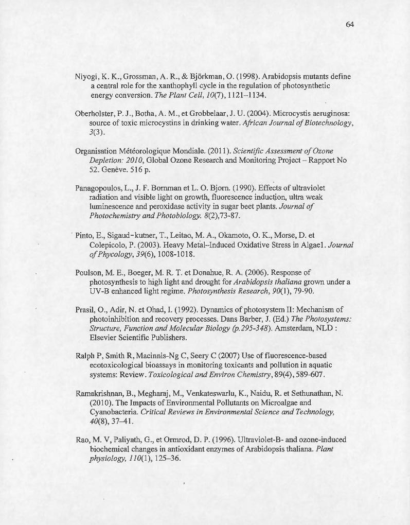
64
Niyogi, K. K., Grossman , A. R ., & Bjorkman, O. (1998) . Arabidopsis mutants define a central role for the xanthophyll cycle in the regulation of photosynthetic energy conversion. The Plant Cell, 10(7), 1121-1134.
Oberholster , P.J. , Botha , A . M ., et Grobbelaar , J. U. (2004). Microcystis aeruginosa: source of toxic microcystins in drin king water. African Journal of Biotechnology, 3(3) .
Organisation Météorologique Mondiale. (20 11). Scientific Assessment of Ozone Depletion: 2010, Global Ozone Research and Monitoring Project - Rapport No 52. Genève. 516 p.
Panagopoulos, L. , J.F. Bornman et L. O. Bjorn. (1990). Effects of ultraviolet radiation and visible light on growth, fluorescence induction, ultra weak luminescence and peroxidase activity in sugar beet plants. Journal of Photochemistry and Photobiology. 8(2),73-87.
· Pinto, E., Sigaud-kutner, T. , Leitao, M. A. , Okamoto, O. K. , Morse, D. et Colepicolo, P. (2003). Heavy Metal- Induced Oxidative Stress in Algae1. Journal ofPhycology, 39(6), 1008-1018.
Poulson, M. E. , Boeger, M. R. T. et Donahue, R. A. (2006). Response of photosynthesis to high light and drought for Arabidopsis thaliana grown under a UV-B enhanced light regime. P hotosynthesis Research, 90( 1 ), 79-90.
Prasil, 0 ., Adir, N . et Ohad, I. (1992). Dynamics ofphotosystem II: Mechanism of photoinhibition and recovery processes. Dans Barber, J. (Ed.) The Photosystems: Structure, Function and Molecular Biology (p.295-348) . Amsterdam, NLD : Elsevier Scientific Publishers.
Ralph P, Smith R, Macinnis-Ng C , Seery C (2007) Use offluorescence-based ecotoxicological bioassays in monitoring toxicants and pollution in aquatic systems: Review. Toxicological and Environ Chemistry , 89(4), 589-607.
Ramakrishnan, B., Megharaj , M. , Venkateswarlu, K. , Naidu, R. et Sethunathan, N. (20 1 0). The Impacts of Environmental Pollutants on Microalgae and Cyanobacteria. Critical Reviews in Environmental Science and Technology, 40(8), 37-41.
Rao, M. V, Paliyath, G., et Ormrod, D. P. (1996). Ultraviolet-B-and ozone-induced biochemical changes in antioxidant enzymes of Arabidopsis thaliana. Plant physiology, 110(1), 125-36.
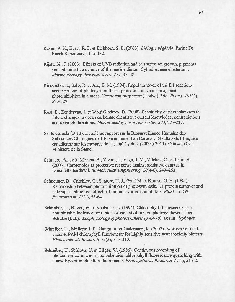
Raven, P. H. , Evert, R.F. et Eichhom, S.E. (2003). Biologie végétale. Paris : De Boeck Supérieur. p.115-130.
Rijstenbil, J. (2003). Effects ofUVB radiation and salt stress on growth, pigments and antioxidative defence of the marine diatom Cylindrotheca closterium. Marine Ecology Progress Series 254, 37-48.
Rintamaki, E., Salo, R. et Aro, E. M. (1994). Rapid turnover of the D1 .reactioncenter protein of photosystem II as a protection mechanism against photoinhibition in a moss, Ceratodon purpureus (Hedw.) Brid. Planta, 193( 4), 520-529.
65
Rost, B., Zondervan, I. et Wolf-Gladrow, D. (2008). Sensitivity of phytoplankton to future changes in ocean carbonate chemistry: current knowledge, contradictions and research directions. Marine ecology progress series, 373, 227-237.
Santé Canada (2013). Deuxième rapport sur la Biosurveillance Humaine des Substances Chimiques de l'Environnement au Canada: Résultats de l'Enquête canadienne sur les mesures de la santé Cycle 2 (2009 à 2011). Ottawa, ON: Ministère de la Santé.
Salguero, A., de la Morena, B., Vigara, J. , Vega, J. M. , Vilchez, C., et Le6n, R. (2003). Carotenoids as protective response against oxidative damage in Dunaliella bardawil. Biomolecular Engineering, 20( 4-6), 249-253.
Schnettger, B., Critchley, C., Santore, U. J. , Graf, M. et Krause, G. H. (1994). Relationship between photoinhibition of photosynthesis, D 1 prote in turnover and chloroplast structure: effects of protein synthesis inhibitors. Plant, Cel! & Environment, 17(1 ), 55-64.
Schreiber, U., Bilger, W. et Neubauer, C. (1994). Chlorophyll fluorescence as a nonintrusive indicator for rapid assessment of in vivo photosynthesis. Dans Schulze (E.d.), Ecophysiology ofphotosynthesis (p.49-70) . Berlin: Springer.
Schreiber, U., Müllerm J.F., Haugg, A. et Gademann, R. (2002). New type of dualchannel P AM chlorophyll fluorometer for highly sensitive water toxicity bio tests. Photosynthesis Research, 74(3) , 317:..330.
Schreiber, U., Schliwa, U. et Bilger, W. (1986) . Continuous recording of photochernical and non-photochemical chlorophyll fluorescence quenching with a new type of modulation fluorometer. Photosynthesis Research, 1 0(1 ), 51-62.

66
Secrétariat de 1 'ozone. 2006. Manuel du protocole de Montréal relatif à des substances qui appauvrissent la couche d'ozone . Programme des Nations Unies pour l'environnement (PNUE), 7ième edition, Nairobi, Kenya
Sinha, R. P., et Hader, D.-P. (2007). UV-protectants in cyanobacteria. Plant Science, 174(3), 278- 289.
Smirnoff, N. (1998). Plant resistance to environrnental stress. Current Opinion in Biotechnology 9(2) , 214-219.Stahl, W. et Sies, H. (2002). Carotenoids and protection against solar UV radiation. Skin Pharmacology and Physiology, 15(5), 291-296.
Stahl, W., et Sies, H. (2002). Carotenoids and protection against solar UV radiation. Skin Pharmacology and Physiology, 15(5) , 291-296.
Statistique Canada (2008). L'activité humaine et l'environement: statistique annuelles. Ottawa, ON : Ministre de l'Industrie
Strasser R, Srivastava A, Tsimilli-Michael M (2000) The fluorescence transient as a tool to characterize and screen photosynthetic samples . Probing photosynthesis: mechanisms , regulation and adaptation:445-483
Suresh Babu, G., Joshi, P.C. , et Viswanathan, P.N. (1998). UVB-Induced reduction in biomass and overall productivity of cyanobacteria. Biochemical and Biophysical Research Communications 244(1), 138- 142.
Taiz, L. et Zeiger, E. (2010) . Plantphysiology, 5e édition, Sunderland: Sinauer Associates, 782p.
Tammes, P. M. L. (1964). Isoboles, a graphie representation of synergism in pesticides. Netherlands Journal of Plant Pathology, 70(3), 73-80.
Thompson, H. M. (1996). Interactions between pesticides; a review of reported effects and their implications for wildlife risk assessment. Ecotoxicology, 5(2), 59-81.
Torres, M. A. , Barres, M. P. , Campos, S. C., Pinto, E., Rajamani, S., Sayre, R. T. et Colepicolo, P. (2008). Biochemical biomarkers in algae and marine pollution: a review. Ecotoxicology and environmental safety, 71(1), 1-15.

67
Vass, I., D. Kirilovsky et A.-L. Etienne. (1999). UV -B Radiation-induced donor- and acceptor- si de modifications of photo system II in the Cyanobacterium Synechocystis sp. PCC 6803. Biochemistry 38(39) , 12786- 12794.
Vass, I., Szilard, A. et Sicora, C. (2005). Adverse Effects ofUV-B Light on the Structure and Function of the Photosynthetic Apparatus. Dans Pessarakli, M. Handbook ofPhotosynthesis (2e édition) (p.82 7-844). Boca Raton, FL: CRC Press.
Vincent, W. F., et Roy, S . (1993). Sol ar ultraviolet-B radiation and aquatic primary production: damage, protection, and recovery. Environmental Reviews, 1(1), 1-12.
Wang, S. , Duan, L., Li, J., Tian, X. et Li, Z. (2007). UV -B radiation increases paraquat tolerance oftwo broad-leaved and two grass weeds in relation to changes in herbicide absorption and photosynthesis. Weed Research, 47(2), 122-128.
Warren, N. , Allan, I. J. , Carter, J. E. , House, W. A. et Parker, A. (2003). Pesticides and Other Micro-organic Contaminants in Freshwater Sedimentary Environments - A Review. Applied Geochemistry, 18(2), 159-194.
Weier TE, Stocking RC, Barbour MG et Rost, TL. (1982). Botany.lntroduction to Plant Biology. 6th Edition. John Wiley and Sons, New York, pp . 720.
Wolfe-Simon, F., Grzebyk, D. , Schofield, O. et Falkowski, P. G. (2005). The Role and Evolution ofSuperoxide Dismutases in Algael. Journal ofphycology, 41(3), 453-465 .
Wollman, F. A. (2001). State transitions reveal the dynamics and flexibility ofthe photosynthetic apparatus. The European Molecular Biology Organization Journal, 20(14), 3623-3630.
Xiong, F., Komenda, J., Kopeccy, J. et Nedbal, L. (1997). Strategies of ultraviolet-B protection in microscopie algae. Physiologia Plantarum, 1 00(2), 378-388.
Xiong, F., 2001. Evidence that UV -B tolerance of the photosynthetic apparatus in microalgae is related to the Dl-turnover mediated repair cycle in vivo. J. Plant Physiol. 158,285-294.
Xu, J. , et Gao, K. (2010). UV-A enhanced growth and UV-B induced positive effects in the recovery of photochemical yield in Gracilaria lemaneiformis

68
(Rhodophyta). Journal ofphotochemistry andphotobiology B: Biology, 100(3), 117- 22.
Zlatev, Z. S., Lidon, F. J. C. , et Kaimakanova, M. (2012). Plant physiological responses to UV-B radiation. Emirates Journal of Food and Agriculture, 24(6), 481-501.
-------------------------------------------------------------------