description of the naupliar stages of megabalanus
TRANSCRIPT
Description of the Naupliar Stages of Megabalanus tintinnabulum (Cirripedia: Balanidae)Author(s): V. Thiyagarajan, V. P. Venugopalan, T. Subramoniam, K. V. K. NairSource: Journal of Crustacean Biology, Vol. 17, No. 2 (May, 1997), pp. 332-342Published by: The Crustacean SocietyStable URL: http://www.jstor.org/stable/1549282Accessed: 26/10/2010 05:12
Your use of the JSTOR archive indicates your acceptance of JSTOR's Terms and Conditions of Use, available athttp://www.jstor.org/page/info/about/policies/terms.jsp. JSTOR's Terms and Conditions of Use provides, in part, that unlessyou have obtained prior permission, you may not download an entire issue of a journal or multiple copies of articles, and youmay use content in the JSTOR archive only for your personal, non-commercial use.
Please contact the publisher regarding any further use of this work. Publisher contact information may be obtained athttp://www.jstor.org/action/showPublisher?publisherCode=crustsoc.
Each copy of any part of a JSTOR transmission must contain the same copyright notice that appears on the screen or printedpage of such transmission.
JSTOR is a not-for-profit service that helps scholars, researchers, and students discover, use, and build upon a wide range ofcontent in a trusted digital archive. We use information technology and tools to increase productivity and facilitate new formsof scholarship. For more information about JSTOR, please contact [email protected].
The Crustacean Society is collaborating with JSTOR to digitize, preserve and extend access to Journal ofCrustacean Biology.
http://www.jstor.org

JOURNAL OF CRUSTACEAN BIOLOGY, 17(2): 332-342, 1997
DESCRIPTION OF THE NAUPLIAR STAGES OF MEGABALANUS TINTINNABULUM (CIRRIPEDIA: BALANIDAE)
V Thiyagarajan, V P. Venugopalan, T Subramoniam, and K. V K. Nair
ABSTRACT
Larval development of Megabalanus tintinnabulum was studied in the laboratory. At 26 ? 1 ?C the development from nauplius I-VI required about 4 days, when the diatom Chaetoceros wighami was used as food. Detailed morphological drawings and setation formulae of appendages of all 6 naupliar stages are presented for the first time. Notable features of the nauplii include the presence of a spinulated lateral margin, a trilobed labrum with teeth, long dorsal and posterior shield spines, a single tooth on the inner prong of the antennal gnathobase, comb-shaped cuspidate setae on the antennae, and spines and simple denticulate setae on the mandibular endopodite in naupliar stage II. The present description of the nauplii is not in full agreement with the previous description of this species from this coast by Daniel (1958). Differences in stage sizes, naupliar morphology, and setation of appendages are used to distinguish this species from other megabalanines and the nau- plii of megabalanines from those of balanines in the Balanus amphitrite group.
Megabalanus tintinnabulum (L.) is a large acorn barnacle commonly encountered in the shallow waters of both east and west coasts of India. It has been reported as a major com- ponent of fouling communities from both coasts (Anil, 1986; Venugopalan et al., 1990; Rajagopal, 1991). Sasikumar (1991) reported various aspects of the biology of this barna- cle, especially those related to reproduction and settlement. However, studies on the lar- val stages of this important barnacle species are sparse. Daniel (1958) briefly described the larval development in this species. Never- theless, his report is incomplete with respect to detailed description of diagnostic features, such as carapace spine, labrum, abdominal process, and setation of appendages. In this paper, we give a detailed account of the lar- val development of M. tintinnabulum, em- phasizing the changes in larval size, shape, general morphology and setation of ap- pendages, along with data on duration of each stage.
MATERIALS AND METHODS
Larval Rearing.-Adult M. tintinnabulum were collected from the approach jetty piers of the Madras Atomic Power Station, Kalpakkam (12?23 N, 80?11 ?E), east coast of In- dia. Release of the first (sometimes second) stage larvae from mature broods occurred after 24 h of aerial expo- sure, followed by immersion in clean sea water (Thiya- garajan et al., 1996). Larvae were cultured following the method of Rittschof et al. (1984) with certain modi- fications, such as (1) instead of Skeletonema costatum (Grev.), another common diatom, Chaetoceros wighami
Brightwell (13-17 x 105 cells/ml), was used as food, and (2) larvae were reared at 26 ? 1?C in Millipore (0.22 gm)- filtered sea water (1 larva/ml and salinity 30 ppt) under constant illumination. Every 6 h, 3-ml aliquots were re- moved from each replicate culture (6) and the larvae were observed to determine their stage of development and per- centage of survival. Naupliar stages were determined us- ing the key provided by Karande (1974a).
Description of Larvae.-The nauplii were observed un- der a Nikon Optiphot microscope for detailed analysis of larval morphology and limb setation. Drawings were made using a camera lucida and size was determined with the help of a calibrated ocular micrometer. Total length (TL) of larvae was determined by measuring the distance between the frontal margin of the carapace and the tip of the caudal spine (CS) or the abdominal furcal tip (whichever was longer). The carapace width (CW) was measured at the widest point behind the naupliar eye. Ten larvae of each stage were examined for determining size, shape, morphology, and setal formulae. The setal types present on the limbs are expressed by alphabetical for- mulae (Newman, 1965). The terminology used to describe the naupliar morphology follows that given by Kado and Hirano (1994).
RESULTS
The intermolt period and percentage sur- vival of the naupliar stages are given in Table 1. Stage II nauplii reached stage VI within 4 days. The mean values of total length (TL) and carapace width (CW) of various larval stages are given in Table 2. Typical setal types and shape used in the present study are illustrated in Fig. 1. The outline of the larval carapace (dorsal view), abdominal processes (AP) (lateral view), and details of the labrum
332

THIYAGARAJAN ET AL.: NAUPLII OF MEGABALANUS TINTINNABULUM
Table 1. Megabalanus tintinabulum. Time taken for the appearance of naupliar stages in cultures along with per- centage survival at each stage (culture temperature 26 ? I?C).
Nauplius stage
I II III IV V VI
Survival (%) 100 100 100 100 89 82 Time of appearance* 0 0 1.6 2 3.6 4
* Mean time (days) after which the naupliar stage appeared in culture (calculated from the initial time of release).
are shown in Figs. 2-4, respectively. The se- tal formulae of the antennule, antenna, mandible, and maxilla are given in Table 3 and illustrated in Figs. 5, 6, 7.
Each naupliar stage shows characters which are helpful in larval identification. Sig- nificant features of the various larval stages are given below.
Nauplius I Figs. 2A, 3A, 4A, 5A, 6A, 7A
The body is elongated and pear-shaped with a mean total length of 264 ? 6 gm. The anterior margin of the carapace is markedly convex. Frontal horns (FH) are folded back toward the long axis of the body. The trilobed labrum is visible without teeth. Frontal fila- ments (FF) cannot be seen and the setae of the limbs are simple. The abdominal process (AP) and caudal spine (CS) are not fully dif- ferentiated.
The presence of posteriorly folded FH and simple setae on the appendages are charac- teristic of this stage. The larvae swim actively using three pairs of limbs and molt to stage II within 30 min.
Nauplius II Figs. 2B, 3B, 4B, 5B, 6B, 7B
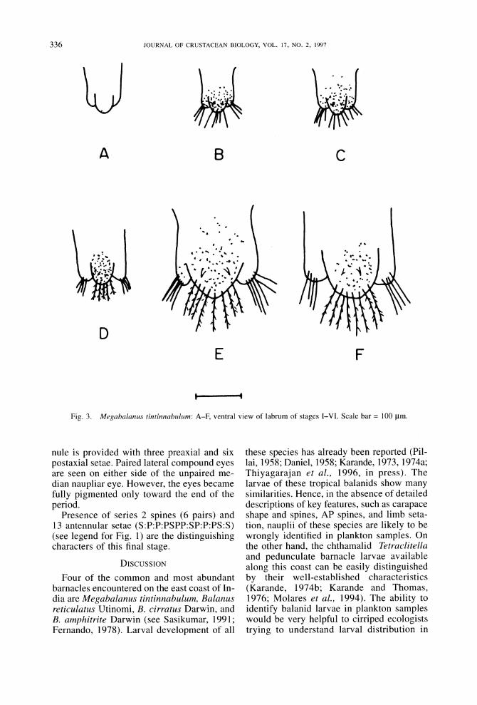
At this stage, the nauplius has a mean length of 404 ?+ 21 gm and mean width of 203 ? 4 gm. The carapace has extended in all di- rections, becoming bell-shaped with a less convex anterior margin. The FH are 93 ? 15 gim in length and are extended either anteri- orly or are perpendicular to the long axis of the body. A pair of FF 80 ? 6 gm long can now be observed. The entire lateral margin of the carapace is spinulous with numerous small spines and with a pair of prominent spines. The labrum bears many short setules on each lobe. The distal margin of the median labral lobe bears teeth from this stage onward.
Table 2. Total length (TL) and carapace width (CW) in microns of naupliar stages of Megabalanus tintinnabulum (MT)-present study; Balanus amphitrite (BA)-Egan and Anderson 1986; Megabalanus rosa (MR) and Megabalanus volcano (MV)-Kado and Hirano, 1994; and Megabalanus tintinnabulum (MT-D)-Daniel, 1958.
Stages MT BA MR MV MT-D
I TL 264 220 245 229 CW 160 140 130 126 180
II TL 404 350 434 390 CW 203 150 214 201 240
III TL 516 370 503 476 CW 280 200 258 255 300
IV TL 610 400 604 552 CW 364 200 334 312 330
V TL 784 470 780 670 CW 480 270 437 416 370
VI TL 880 540 963 941 CW 560 310 540 543 420
The AP has a pair of large serrated series 1 spines and a long distal forked portion (furca). The furcal stem has circlets of spines at the distal end. The CS is longer than the AP. The CS and furcal branches are covered with small spines from this stage onward.
The maxillae appear as long setae on the AP. At this stage, no preaxial setae are pres-
S
p
$D
D
AC
MC
AG MG
Fig. 1. Setal types used in the setation formulae of nau- pliar stages of Megabalanus tintinnabulum; S = simple, SD = simple-denticulate, P = plumose, D = plumodentic- ulate, MC = mandible-cuspidate, AC = antenna-cuspidate, AG = antennal gnathobase, MG = mandibular gnathobase setae, B, = series 1 bristle, B2 = series 2 bristle, ss = seti- form spine.
333

334 ~~~~~JOURNAL OF CRUSTACEAN BIOLOGY, VOL. 17, NO. 2, 1997
Table 3. Setal formulae (Newman, 1965) of six naupliar stages of Megabalanus tintinnabulum (MT)-present study; Megabalanus rosa (MR) and Megabalanus volcano (MV)-Kado and Hirano, 1994; Balanus amphitrite (BA)-Egan and Anderson, 1986. s = setiform spine, S = simple seta, P = plumose, C = cuspidate, G = gnathobase, SI simple den- ticulate, and D = denticulate.
Antenna Mandible Maxilta
Stages Antennule Exopodite Endopodite Exopodite Endopodite
I MT SSSS:SS:S:S 5S SSS:SS:SS:SS:G 4S SSS:SS:SS:SS:G 00000 MR SSSS:SS:S:S 5S SSS:SS:SS:SS:G 45 SSS:SS:SS:SS:G 00000 MV SSSS:SS:S:S 55 SSS:SS:SS:SS:G 4S SSS:SS:SS:SS:G 00000 BA S.SSS:SS:S:S 55 SSS:SS:SS:SS:G 45 SSS:SS:SS:SS:G 00000
II MT SSPS:SP:P:S SP:4P:S PPS:SP:PD:SPC:G 4P:S SSS:SP:PSDIC:sPPC:G 00000 MR SSPS:SP:P:S SP:4P:S PPS:SP:PD:PSC:G 4P:S SSS:SD:DSC:PC:G 00000 MV SSPS:SP:P:S SP:4P:S PPS:SP:PD:PSC:G 4P:S SSS:SD:DSC:PC:G 00000 BA SSPS:PS:P:S SPARPS PPS:SP:PD:SPC:G 4P:S SSS:SP:SPC:sPC:G 00000
III MT S:PSPP:SP:P:S 7P PPP:SP:PD:PSPC:G 4P:S SSS:SPS:PCD:sPCP:G 55000 MR S:PSPP:SP:P:S 7P PPP:SP:PD:PSSC:G 4P:S SSS:SDS:DDC:sPCP:G 50000 MV S:PSPP:SP:P:S 7P PPP:SP:PD:PSSC:G 4P:S SSS:SDS:DDC:sPCP:G 00000 BA SSPS:PS:P:S 7P PPS:SP:PD:SPC:G 4P:S SSS:SP:SPC:sPC:G 00000
IV MT SP:PSPP:SP:P:S 9P PPSPP:SPS:PD:PSPC:G 5P 4S:SPP:SPCD:sPPC:G 50000 MR SP:PSPP:SP:P:S 9P PPSPS:SPS:PD:DSPC:G 5P 4S:SDD:SDDC:sPCP:G 55000 MV SP:PSPP:SP:P:S 9P PPSPS:SPS:PD:PSPC:G 5P 4S:SPD:SDDC:sPCP:G 55000 BA S:S:PSPP:PS:P:S 3P:5P:S PPPSS:SPS:PD:SPPC:G 4PS 4S:SPP:DSPC:sDPC:G SSSSO
V MT S:P:P:PSPP:SP:S:P:S lip PPSPP:SPP:PD:PSPC:G 5PS 4S:SPD:SPCD:sPPC:G SSSSS MR S:P:P:PSPP:SP:S:P:S lip PPSPP:SPP:PD:DSDC:G 5PS 4S:SSDD:SDDC:sPDC:G SSSSS MV S:S:P:PSPP:SP:S:P:S lip PPSPP:SPP:PD:PSPC:G 5PS 4S:SSDD:SDDC:sPPC:G SSSSS BA S:S:P:PSPP:PS:S:P:S 4P:6P:S PPPPS:SPP:PD:SPPC:G 5PS 4S:SSDD:SDPC:sPDC:G SSSSS
VI MT S:P:P:PSPP:SP:P:PS:S 12P PPSPP:SPP:PD:SPCP:G 6P 4S:SSPD:SDCD:sPPC:G SSSSS MR S:P:P:PSPP:SP:P:PS:S 12P PPSPP:SPP:PD:DDDC:G 6P 4S:SSDD:SDDC:sDDC:G SSPSP MV S:P:P:PSPP:SP:P:PS:S 12P PPSPP:SPP:PD:PSPC:G 6P 4S:SSDD:SDDC:sPPC:G PPPSP BA S:S:P:PPPP:PS:P:PS:S 12P PPPPS:SPP:PD:SPPC:G 6P 4S:SSDD:SPPC:sPDC:G SSSSS
ent on the antennules (Fig. 5B). The anten- nal gnathobase is well developed with three apical spines (inner, median, and outer prongs) and series 1 bristle (Fig. 6B). Sim- ple denticulate setae (SD) on the endopodite of the mandible are present. This stage molts to stage III in 38 h.
Nauplius III Figs. 2C, 3C, 4C, SC, 6C, 7C
Stage III nauplii are 516 ? 12 gim long and 280 ? 2 gim wide. The carapace is more bul- bous than in the earlier stages. The prominent spines on the lateral margin of the carapace have disappeared, but small spines are re- tained throughout the naupliar stages. The AP bears only a pair of series 1 (distal) spines. A pair of dorsal shield spines (DSS) appears in this stage and is retained till stage VI. The maxillae appear as two stout simple setae in the thoracic region of AP. The first preaxial seta appears on the antennule. Other setation features of this stage are given in Table 3. Nauplii of this stage molt to stage IV within 10 h.
Nauplius IV Figs. 2D, 3D, 4D, SD, 6D, 7D
The larvae are 610 ?10 gim in total length and the shield is 364 ?4 gim in width. The FH and FF are 96 ? 12 jim and 100 ? 8 gim long, respectively. The median lobe of the labrum bears serrated setules, but the lateral lobes still have only simple short setules. In the posterior region of the larva, the AP has separated from the carapace, so that it now has an entire posterior margin, bearing a pair of posterior shield spines (PSS). This cara- pace form is retained until stage VI. The AP bears series 1 (distal) and series 2 (proximal) spines. The series 1 spines are larger than the series 2 spines. A median nonserrated spine appears between the pair of serrated series 2 spines. CS and AP are almost equal in length. The maxillae are seen as long simple setae. The antennule now bears two preaxial setae. From this stage onward the antennal en- dopodite bears iS setae.
The presence of an entire shield, long PSS, two preaxial setae on the antennule, and se- ries 1 and 2 spines on the AP are character-
334

THIYAGARAJAN ET AL.: NAUPLII OF MEGABALANUS TINTINNABULUM
A B
D
A I A
C
F
BCDEF C DE f
Fig. 2. Megabalanus tintinnabulum: A-F, shield outline (dorsal view) of naupliar stages I-VI. Scale bar = 100 tm.
istic features of this stage. These larvae molt to stage V after 38 h.
Nauplius V Figs. 2E, 3E, 4E, 5E, 6E, 7E
Stage V larvae are 784 ? 35 gm long and 480 ? 57 gm wide. The shield margin is bul- bous with a slightly convex anterior margin. The well-developed long DSS are now seen along with many small spines on the shield. The PSS are now 122 ? 4 ugm long. The tho- racic area is swollen and segmented. A pair of series 3 spines appears anterior to the fur- cal stem, in addition to the series 1 and 2 spines. The antennule now has three preax- ial setae and five postaxial setae.
The presence of 12 setae on the antennule and three series of spines (1, 2, and 3) on the AP (Fig. 3) are characteristic of this stage. This stage molts to stage VI within 10 h.
Nauplius VI Figs. 2F, 3F, 4F, 5F, 6F, 7F
The larva measures 880 ? 2 gm in length and 560 ? 3 tm in width. The FF and FH measure about 136 ? 4 itm and 124 ? 8 gjm, respectively. A median spine makes its ap- pearance between the pair of DSS. The PSS is 163 ugm long. Six pairs of series 2 spines (the primordial thoracopode) are present along the ventral side of the thoraco-abdom- inal process in two parallel lines. The anten-
335
JOURNAL OF CRUSTACEAN BIOLOGY, VOL. 17, NO. 2, 1997
YJ
A B
D EF
Fig. 3. Megabalanus tintinnabulum: A-F, ventral view of labrum of stages I-VI. Scale bar = 100 km.
nule is provided with three preaxial and six postaxial setae. Paired lateral compound eyes are seen on either side of the unpaired me- dian naupliar eye. However, the eyes became fully pigmented only toward the end of the period.
Presence of series 2 spines (6 pairs) and 13 antennular setae (S:P:P:PSPP:SP:P:PS:S) (see legend for Fig. 1) are the distinguishing characters of this final stage.
DISCUSSION
Four of the common and most abundant barnacles encountered on the east coast of In- dia are Megabalanus tintinnabulum, Balanus reticulatus Utinomi, B. cirratus Darwin, and B. amphitrite Darwin (see Sasikumar, 1991; Fernando, 1978). Larval development of all
these species has already been reported (Pil- lai, 1958; Daniel, 1958; Karande, 1973, 1974a; Thiyagarajan et al., 1996, in press). The larvae of these tropical balanids show many similarities. Hence, in the absence of detailed descriptions of key features, such as carapace shape and spines, AP spines, and limb seta- tion, nauplii of these species are likely to be wrongly identified in plankton samples. On the other hand, the chthamalid Tetraclitella and pedunculate barnacle larvae available along this coast can be easily distinguished by their well-established characteristics (Karande, 1974b; Karande and Thomas, 1976; Molares et al., 1994). The ability to identify balanid larvae in plankton samples would be very helpful to cirriped ecologists trying to understand larval distribution in
C
336

THIYAGARAJAN ET AL.: NAUPLII OF MEGABALANUS TINTINNABULUM
A
D
B C
E
SCOF AE
Fig. 4. Megabalanus tintinnabulum: A-F, thoraco-abdominal process with maxilla (lateral view) of naupliar stages I-VI. SI = series 1 spines, S2 = series 2 spines, S3 = series 3 spines. Scale bar = 100 gm.
coastal waters. The larval description of M. tintinnabulum by Daniel (1958) is insufficient to fully differentiate the species from other closely related balanids.
Larval Development Larval development times of all tropical
balanids studied in the laboratory are similar (Karande, 1974a). Variations in duration of larval development in the laboratory are pos- sibly due to differences in culture conditions (Kado and Kim, 1996). Daniel (1958) re- ported the appearance of cyprids of M. tintinnabulum in laboratory culture within 48
h (in the present study it took four days), but he did not describe the culture conditions used. It is possible that sea water used for Daniel's culture might have been contami- nated by larval barnacles.
Larval Morphology
Except body size, larval morphological fea- tures such as body shape, spines on the ab- domen, and setation of appendages are not likely to vary between larvae reared in the laboratory and those collected from natural plankton (Ovsyannikova and Kom, 1984; Miller and Roughgarden, 1994). Therefore,
337

JOURNAL OF CRUSTACEAN BIOLOGY, VOL. 17, NO. 2, 1997
B c
E F
Fig. 5. Antennules of naupliar stages I-VI (A-F) of Megabalanus tintinnabulum. Scale bar = 100 gm.
the size of laboratory-reared larvae is of lit- tle value in the identification of species. How- ever, other features, such as shape of the cara- pace, spines on the AP, and setation of ap- pendages are considered crucial in larval identification (Lang, 1979; Egan and Ander- son, 1986; Miller and Roughgarden, 1994).
The margin of the carapace of M. tintinnab- ulum (present study), M. volcano (Pilsbry), (see Kado and Hirano, 1994), B. reticulatus Utinomi (see Thiyagarajan et al., in press b), B. cirratus and B. amaryllus euamaryllus Broch (see Karande, 1974a, b) is spinulated, whereas in B. amphitrite (see Karande, 1973;
A
338

THIYAGARAJAN ET AL.: NAUPLII OF MEGABALANUS TINTINNABULUM
B A
D
B F BDEF
C
F E
A I AC
Fig. 6. Antennae of naupliar stages I-VI (A-F) of Megabalanus tintinnabulum. Scale bar = 100 gm.
Egan and Anderson, 1986), B. albicostatus Pilsbry (see Lee and Kim, 1991), and M. rosa (Pilsbry) (see Kado and Hirano, 1994), it is smooth. The presence of DSS in naupliar stages III-VI distinguishes larvae of M. tintinnabulum from other balanid larvae in plankton samples in Indian waters. However, it has been reported for other megabalanines, such as M. rosa and M. volcano (see Choi et al., 1992; Kado and Hirano, 1994) from Japan. The DSS, therefore, are helpful only in differentiating naupliar stages III-VI of this species from other balanid larvae avail- able locally. The carapace becomes entire and develops a pair of long PSS in stage IV as in other barnacles, except in chthamalids
(Karande and Thomas, 1976). These large spines are visible under a dissection micro- scope (104 gm in stage IV, 122 gm in stage V, and 163 gm in stage VI). No other cirriped larva described from Indian waters has a PSS longer than 110 gm (Karande, 1974b). Thus, it is also possible to separate this species from others by virtue of the presence of this long PSS. The presence of teeth on the trilobed labrum of this species is a feature similar to that of many other barnacle species. However, in most balanid species the teeth are lost af- ter stage II (Egan and Anderson, 1986), while in megabalanids they are retained to stage VI.
The relative length of CS and AP may vary with the orientation in which the larvae are
339

JOURNAL OF CRUSTACEAN BIOLOGY, VOL. 17, NO. 2, 1997
B
~~E F
AB
Fig. 7. Mandibles of naupliar stages I-VI (A-F) of Megabalanus tintinnabulum. Scale bar = 100 tm.
observed. Therefore, it is not of much value in the determination of a stage or identifica- tion of species. In many balanids, the se- quence of appearance and nature of spines on the AP are of diagnostic value. In B. cir- ratus both the proximal and distal AP spines appear in stage II (Karande, 1974a). The pres- ence of a median spine between the series 2 spines of stage IV of M. tintinnabulum is characteristic of many balanids except B. am- phitrite (see Karande, 1973).
The salient morphological features of nau- plii of M. tintinnabulum are, therefore, their large size (Table 2), spinulated lateral mar- gin, trilobed labrum with teeth, long posterior shield spines, and presence of dorsal shield spines. The present observations differ from those of Daniel (1958) with regard to larval size, shape, and morphology, such as FF in stage I, development of paired compound
eyes in stage V, appearance of six pairs of series 2 spines in stage V, and the setation of antennules and mandibles (see below).
Setation of Appendages The antennular setation of M. tintinnabu-
lum, in general, conforms to that of other cir- ripeds thus far described. That is, there are no preaxial setae in stages I and II; the setae ap- pear in the following order: one in stage III, two in stage IV, and three in stages V and VI. Stage VI larva has six setae on the postax- ial side. Thus, it is possible to stage the nau- plii by looking at antennular setation with the help of a dissection microscope. Among the three preaxial setae, which develop on the an- tennule during stages V and VI, two setae are always plumose in all megabalanids, whereas in other balanids, namely, B. reticu- latus, B. cirratus, and B. amphitrite (Table 3),
A
D
CDEF
340

THIYAGARAJAN ET AL.: NAUPLII OF MEGABALANUS TINTINNABULUM
only one seta is plumose and two are simple. In B. reticulatus, the presence of preaxial "hairs" on the antennule has been considered a distinguishing character (Thiyagarajan et al., in press b). In M. tintinnabulum such preaxial "hairs" are absent.
In naupliar stages IV-VI of M. rosa the an- tenna has three denticulate setae (Table 3) on the fifth setal quadrat (Kado and Hirano, 1994). In other balanids, including the pres- ent species, not more than one such seta is observed. The size and shape of the antennal gnathobase in successive naupliar stages of this species are similar to those of B. am- phitrite, M. rosa, and M. volcano. However, in M. rosa and M. volcano, two teeth have been reported on the inner prong, whereas in the present species only one such tooth was observed. Moreover, in M. tintinnabulum, Daniel (1958) observed only 21, 22, and 22 setae on the antenna of stages IV, V, and VI, respectively, whereas we observed 23, 25, and 26, respectively, confirming the observations of others who have worked on megabalanids (Miller and Roughgarden, 1994; Kado and Hirano, 1994).
In general, no differences are observed in the setation of mandibles among the species mentioned above, except that in M. rosa and M. volcano 2-4 denticulate setae appear dur- ing naupliar stages II-VI (Table 3). In M. tintinnabulum and B. amphitrite, not more than three setae appear during the same pe- riod. Daniel (1958) also observed fewer se- tae on the mandibles (19, instead of 20, 21, and 22 in stages IV, V, and VI, respectively).
ACKNOWLEDGEMENTS
This work was supported by a grant made to (TS) by the Board of Research in Nuclear Sciences, Department of Atomic Energy, India. Thanks are due to the Station Director of the Madras Atomic Power Station (MAPS), for facilities. We are grateful to Dr. P. N. Moorthy, Head, Applied Chemistry Division (APCD), Bhabha Atomic Re- search Centre (BARC), Mumbai, and Dr. P. K. Mathur, Head, Water and Steam Chemistry Laboratory (WSCL), Kalpakkam, for support. We are thankful to Dr. K. Nan- dakumar for his critical comments on the manuscript.
LITERATURE CITED
Anil, A. C. 1986. Studies on marine fouling in the Zuari estuary (Goa), west coast of India.-Doctoral thesis, Karnataka University, Karnataka, India. Pp. 1-175.
Choi, H. K., D. T. Anderson, and C. H. Kim. 1992. Lar-
val development of the megabalanine balanomorph Megabalanus rosa (Pilsbry) (Cirripedia, Balanidae).- Proceedings of the Linnaean Society 113: 175-184.
Daniel, A. 1958. The development and metamorphosis of three species of sessile barnacles.-Journal of Madras University (B) 28: 23-47.
Egan, E. A., and D. T. Anderson. 1986. Larval develop- ment of Balanus amphitrite Darwin and Balanus var- iegatus Darwin (Cirripedia: Balanidae) from New South Wales, Australia.-Crustaceana 51: 188-207.
Fernando, S. A. 1978. Studies on the biology of barna- cles of Porto Novo region.-Doctoral thesis, Anna- malai University, Chidambaram, India. Pp. 1-156.
Kado, R., and R. Hirano. 1994. Larval development of two Japanese megabalanine barnacles, Megabalanus volcano (Pilsbry) and Megabalanus rosa (Pilsbry) (Cir- ripedia, Balanidae), reared in the laboratory.-Journal of Experimental Marine Biology and Ecology 175: 17-41.
, and M. H. Kim. 1996. Larval development of Octomeris sulcata Nilsson-Cantell (Cirripedia: Thora- cica: Chthamalidae) from Japan and Korea.-Hydro- biologia 325: 65-76.
Karande, A. A. 1973. Larval development of Balanus am- phitrite amphitrite D. reared in the laboratory.-Pro- ceedings of the Indian Academy of Sciences (B) 77: 56-63.
. 1974a. Balanus variegatus Darwin: the labora- tory reared larvae compared with Balanus amphitrite amphitrite Darwin (Cirripedia).-Crustaceana 26: 229-235.
. 1974b. Larval development of the barnacle Tet- raclitella karandei reared in the laboratory.-Biologi- cal Bulletin 146: 249-257.
, and M. K. Thomas. 1976. The larvae of the in- tertidal barnacle Chthamalus malayensis Pilsbry.-Pro- ceedings of the Indian Academy of Sciences 83: 210-219.
Lang, W. H. 1979. Larval development of shallow water barnacles of the Carolinas (Cirripedia: Thoracica) with keys to naupliar stages.-National Oceanic and At- mospheric Administration, Technical Report, National Marine Fisheries Service, Circular 421: 1-39.
Lee, C., and C. H. Kim. 1991. Larval development of Bal- anus albicostatus Pilsbry (Cirripedia, Thoracica) reared in the laboratory.-Journal of Experimental Marine Bi- ology and Ecology 147: 231-244.
Miller, M. K., and J. Roughgarden. 1994. Descriptions of the larvae of Tetraclita rubescens and Megabalanus californicus with a comparison of the common barna- cle larvae of the central California coast.-Journal of Crustacean Biology 14: 579-600.
Molares, J., F. Tilves, and C. Pascual. 1994. Larval de- velopment of the pedunculate barnacle Pollicipes cor- nucopia (Cirripedia: Scalpellomorpha) reared in the laboratory.-Marine Biology 120: 261-264.
Newman, W. A. 1965. Prospectus on larval cirripede se- tation formulae.-Crustaceana 9: 51-56.
Ovsyannikova, I. I., and 0. M. Korn. 1984. Naupliar de- velopment of the barnacle Balanus crenatus in Peter
341

JOURNAL OF CRUSTACEAN BIOLOGY, VOL. 17, NO. 2, 1997
the Great Bay (Sea of Japan).-Biologya Morya 5: 34-40.
Pillai, K. N. 1958. Development of Balanus amphitrite with a note on the early larvae of Chelonibia testudi- naria.-Bulletin of the Central Research Institute, Ker- ala 6: 117-130
Rajagopal, S. 1991. Biofouling problems in the condenser cooling circuit of a coastal power station with special reference to green mussel, Perna viridis.-Doctoral thesis, University of Madras, Madras, India. Pp. 1-125.
Rittschof, D., E. S. Branscomb, and J. D. Costlow. 1984. Settlement and behaviour in relation to flow and sur- face in larval barnacles, Balanus amphitrite Darwin.- Journal of Experimental Marine Biology and Ecology 82: 131-146.
Sasikumar, N. 1991. Ecology and control of biofouling in a coastal power station with special reference to Megabalanus tintinnabulum.-Doctoral thesis, Uni- versity of Madras, Madras, India. Pp. 1-73.
Thiyagarajan, V., V. P. Venugopalan, T. Subramoniam and K. V. K. Nair. 1996. Laboratory rearing of barnacle lar-
vae (Balanus reticulatus) using Chaetoceros wighami as food.-Indian Joumal of Marine Sciences 25: 365-367.
Thiyagarajan, V., V. P. Venugopalan, K. V. K. Nair and T. Subramoniam. (In press.) Larval description of Balanus reticulatus Utinomi (Cirripedia, Balanidae), reared in the laboratory.-Journal of Experimental Ma- rine Biology and Ecology.
Venugopalan, V. P., S. Rajagopal, N. Sasikumar, and K. V. K. Nair. 1990. Marine biology of a seawater tun- nel.-In: B. N. Desai, ed., Oceanography of the In- dian Ocean. Pp. 253-259. Oxford & IBH Publishers Pvt. Ltd., New Delhi, India.
RECEIVED: 12 June 1996. ACCEPTED: 15 October 1996.
Addresses: (VT and TS) Department of Zoology, Uni- versity of Madras, Guindy Campus, Madras 600 025, India; (VPV and KVKN), Marine Biology Programme, Water & Steam Chemistry Laboratory, BARC, IGCAR Campus, Kalpakkam 603 102, India.
342