calcium bioavailability of opuntia ficus-indica cladodes in
TRANSCRIPT
nutrients
Article
Calcium Bioavailability of Opuntia ficus-indicaCladodes in an Ovariectomized Rat Model ofPostmenopausal Bone Loss
Michelle Quintero-García 1,2, Elsa Gutiérrez-Cortez 3, Alejandra Rojas-Molina 2 ,Monsserrat Mendoza-Ávila 4, Alicia Del Real 5 , Efraín Rubio 6, Daniel Jiménez-Mendoza 7,8
and Isela Rojas-Molina 2,*1 Programa de Maestría en Ciencias Químico Biológicas, Facultad de Química, Universidad Autónoma de
Querétaro, Cerro de las Campanas S/N, Querétaro C.P. 76010, Mexico; [email protected] Laboratorio de Química Medicinal, Facultad de Química, Universidad Autónoma de Querétaro, Cerro de las
Campanas S/N, Querétaro C.P. 76010, Mexico; [email protected] Laboratorio de procesos de transformación y tecnologías emergentes en alimentos, Facultad de Estudios
Superiores-Cuautitlán, Universidad Nacional Autónoma de México, Km 2.5 CarreteraCuautitlán–Teoloyucan, San Sebastián Xhala, Cuautitlán-Izcalli C.P. 54714, Mexico;[email protected]
4 Programa de Maestría en Ciencias de la Nutrición Humana, Facultad de Ciencias Naturales, UniversidadAutónoma de Querétaro, Av. de las Ciencias S/N, Juriquilla C.P. 76230, Querétaro, Mexico;[email protected]
5 Centro de Física Aplicada y Tecnología Avanzada, Universidad Nacional Autónoma de México,Juriquilla C.P. 7600, Querétaro, Mexico; [email protected]
6 Centro Universitario de Vinculación y Transferencia de Tecnología, Benemérita Universidad Autónoma dePuebla, Centro Universitario, Col. San Manuel S/N, Puebla C.P. 72540, Mexico; [email protected]
7 Departamento de Ingeniería Física, División de Ciencias e Ingenierías, Universidad de Guanajuato, CampusLeón, Lomas del Bosque 103, Col. Lomas del Campestre, León C.P. 37150, Guanajuato, Mexico;[email protected]
8 Departamento de Ingeniería Electromecánica, Tecnológico Nacional de México/ITS de Purísima del Rincón.Blvd. Del Valle 2301, Col. Guardarrayas, Purísima del Rincón C.P. 36413, Guanajuato, Mexico
* Correspondence: [email protected]; Tel.: +52-442-192-1200 (ext. 75030); Fax: +52-442-192-1302
Received: 10 April 2020; Accepted: 12 May 2020; Published: 15 May 2020�����������������
Abstract: Osteoporosis is a disease of the skeletal system characterized by low bone mass and boneweakening, which increase the risk of fracture. This disease is associated with menopause becausehypoestrogenism induces the maturation and activation of osteoclasts. In addition, a low dietary intakeof calcium leads to low bone mineral density and postmenopausal osteoporosis. The objectives of thiswork were to determine calcium bioavailability of Opuntia ficus-indica cladodes at a late maturity stageand to assess its contribution in improving bone health in an ovariectomized rat model. Two-month-oldWistar female rats (n = 35) were used and distributed in seven experimental groups: (i) controlgroup (Crtl), (ii) sham group (SH), (iii) ovariectomized group (OVX), (iv) ovariectomized groupsupplemented with calcium citrate (CCa), (v) ovariectomized group supplemented with O. ficus-indicapowder (NI), (vi) ovariectomized group supplemented with soluble fiber from O. ficus-indica (FS) and(vii) ovariectomized group supplemented with insoluble fiber from O. ficus-indica (FI). Our resultsshowed that calcium in the soluble fiber of O. ficus-indica is bioavailable and contributes to improve thephysical, densitometric, biomechanical and microstructural properties of bones in ovariectomized rats.These findings indicated that O. ficus-indica cladodes at a late maturity stage represent a good sourceof bioavailable calcium and consumption of these cladodes might be beneficial for the prevention ofosteoporosis and other bone diseases.
Nutrients 2020, 12, 1431; doi:10.3390/nu12051431 www.mdpi.com/journal/nutrients
Nutrients 2020, 12, 1431 2 of 20
Keywords: Opuntia ficus-indica; calcium bioavailability; bone mineral density; remodeling biomarkers;ovariectomy; osteoporosis
1. Introduction
An adequate bone mineralization is achieved through calcium and vitamin D diet ingestion.Foods that have high calcium content are dairy products; however, people with lactose intoleranceare predisposed to low calcium intake, and therefore are at higher risk of bone demineralization [1].The recommended daily intake (RDI) for calcium is 1200 mg/day in adults. The deficiency of thismineral in diet is directly related to diseases, such as osteoporosis, as well as to the propensityto suffer bone fractures [2]. In Mexico, it has been estimated that approximately 168 women and98 men per 100,000 people have a proximal femur fracture, which means that one in 12 women andone in 20 men over 50 years of age will suffer a fracture [3]. All bone cells (osteoblasts, osteoclastsand osteocytes) contain functional estrogen receptors (ERs), which play an important role in bonemetabolism and inhibition of osteoclast differentiation. Moreover, ER activation promotes osteoclastapoptosis, subsequently reducing bone resorption [4]. Currently, there are several treatments for theprevention of osteoporosis, among which, dietary supplements that contain calcium salts, such ascalcium carbonate and citrate, are widely employed. Unfortunately, these supplements cause somegastrointestinal complications [5]. Vitamin D supplementation is also used in the preventive treatmentof osteoporosis, however, its complete absorption is very limited in geriatric patients, since they barelycarry out any type of exercise, such as walking, to take advantage of UV rays to achieve vitamin Dfixation [6]. Another prevention and treatment option for osteoporosis is hormone replacement, whichinduces antiresorptive effects and has been used for decades in menopausal and postmenopausalwomen; nevertheless, it is accompanied by side effects associated with increased risk of breast cancer [7].Our research group previously reported that Opuntia ficus-indica (nopal) has high levels of calcium,approximately 164 mg/100 g dried weight [8]. We also found that calcium content in O. ficus-indicaincreases approximately by 70% in cladodes at late maturity stages compared with that of cladodes atan early stage [9]. Moreover, we demonstrated that calcium content in the soluble fiber of O. ficus-indicacladodes is significantly higher than that found in the insoluble fiber. Interestingly, most of this mineralin the soluble fiber is in the form of calcium carbonate, whereas it is in the form of calcium oxalatein the insoluble fiber [10]. To our knowledge, there are no studies regarding the effects of calciumpresent in O. ficus-indica cladodes and fibers extracted from this cactus to improve bone properties inovariectomized rats. Based on the aforementioned data, we hypothesized that calcium in O. ficus-indicacladodes is bioavailable and useful to enhance bone health. Therefore, the objectives of this work wereto determine the bioavailability of calcium in O. ficus-indica cladodes at a late maturity stage and toassess its contribution to improve physical, densitometric, biomechanical, microstructural and mineralcontent properties of bones in an ovariectomized rat model of postmenopausal bone loss.
2. Materials and Methods
2.1. Vegetal Material
Opuntia ficus-indica cladodes were harvested during the spring of 2016 in the experimental field ofAmazcala in the Engineering Department of the Autonomous University of Queretaro. The cladodeswere collected at 135 days of maturity stage from the sprout.
2.2. Preparation of O. ficus-indica Powder
The thorns of cladodes were removed, and then the cladodes were cut and placed in stainlesssteel trays, which were introduced in a forced air oven (BG Didacta, Torino, Italy) to be dehydrated at70 ◦C for 12 h [11]. Subsequently, the cladodes were ground in a hammer mill (Pulvex 200, Mexico).
Nutrients 2020, 12, 1431 3 of 20
2.3. Extraction of Soluble and Insoluble Fibers from O. ficus-indica Cladodes
Suspensions of O. ficus-indica powder were prepared for the extraction of soluble and insolublefibers from the cactus based on the methodology reported by Rojas-Molina et al. [10].
2.4. Determination of Calcium and Phosphorus Content in O. ficus-indica Cladodes
Analyses of calcium (Ca) of dehydrated O. ficus-indica cladodes were carried out in triplicate byusing a double-beam atomic absorption spectrophotometer (Analyst 300, Perkin Elmer, Boston, MA,USA). Quantifications were performed in accordance with the methods established by the Associationof Official Analytical Chemists (AOAC, 2000) [12]. Phosphorus content was analyzed according toprevious reports [9].
2.5. Experimental Design
Thirty-five adult female Wistar rats (2 months of age), which underwent ovariectomy surgery,were used as an experimental model of postmenopausal osteoporosis. The rats were randomly assignedto the experimental groups. Experimental subjects were placed in stainless steel metabolic cagesunder controlled conditions of temperature and light (light–dark cycles of 12 h:12 h). The studywas approved by the Bioethics Committee of the Natural Sciences Department of the AutonomousUniversity of Queretaro. Animals had ad libitum access to deionized water and diets. The subjectswere randomly classified into 7 experimental groups with 5 rats in each one as follows: (i) controlgroup fed with diet AIN-93M for maintenance of adult rodents (Ctrl group), (ii) sham group fed withdiet AIN-93M (SH group), (iii) group of ovariectomized rats fed with diet AIN-93M (OVX group),(iv) ovariectomized group fed with diet AIN-93M adjusted with calcium contained in the soluble fiberextracted from O. ficus-indica cladodes (FS group), (v) ovariectomized group fed with AIN-93M dietadjusted with calcium contained in the insoluble fiber extracted from O. ficus-indica cladodes (FI group),(vi) ovariectomized group fed with diet AIN-93M adjusted with calcium contained in O. ficus-indicacladodes (NI group) and (vii) ovariectomized group fed with diet AIN-93M adjusted with calciumfrom calcium citrate (Cca group). The experimental period was 9 weeks. The experimental diets wereprepared with AIN-93M modifications (Table 1) including an addition of vitamin mix (AIN-93-VX,Harlan Inc., IN, USA, TD 94047) and mineral mix without calcium (AIN-93-MX, Harlan Inc., IN, USA,TD 04374). The calcium content in all diets was 5 g/kg diet and the calcium source in the controldiet was calcium carbonate (Merck 2066, Darmstadt, Germany). Different amounts of dehydratedO. ficus-indica cladodes and insoluble and soluble dietary fiber extracted from O. ficus-indica cladodeswere added to experimental diets as the calcium source to achieve the aforementioned calcium content.Energy values were calculated with standard factors as follows: 4 kcal for available carbohydrates andproteins and 9 kcal for lipids.
Table 1. Ingredient composition of the experimental diets (g/kg).
Groups Control(Ctrl)
Calcium Citrate(CCa)
O. ficus-indicaPowder (NI)
Soluble DietaryFiber (FS)
Insoluble DietaryFiber (FI)Ingredients
Corn starch 621 621 621 621 621Sucrose 140 140 140 140 140Casein a 100 100 100 100 100Soybean oil 40 40 40 40 40Fiber b 50 50 33 43 35MixMin c 10 10 10 10 10MixVit d 35 35 35 35 35L-Cystine 1.8 1.8 1.8 1.8 1.8Choline bitartrate 2.5 2.5 2.5 2.5 2.5CaCO3
e 12.5 - - - -
Nutrients 2020, 12, 1431 4 of 20
Table 1. Cont.
Groups Control(Ctrl)
Calcium Citrate(CCa)
O. ficus-indicaPowder (NI)
Soluble DietaryFiber (FS)
Insoluble DietaryFiber (FI)Ingredients
Calcium citrate - 0.207 - - -O. ficus-indica powder - - 48 - -Soluble fiber extracted fromO. ficus-indica - - - 86 -
Insoluble fiber extractedfrom O. ficus-indica - - - - 105
a C-7078, Casein Sigma Chemical, Inc., St. Louis, MO, USA. b α-Cell Fiber Solft Zolca, MP Biomedicals, Santa Ana, CA,USA. c Mineral mix without calcium (AIN-93-MX, Harlan Inc., IN, USA, TD 04374) [13]. d Vitamin mix (AIN-93-VX,Harlan Inc., IN, USA, TD 94047) [13]. e Control diet contained CaCO3 (Merck 2066, Darmstadt, Germany) as thecalcium source. In experimental diets, O. ficus-indica powder provided 5 g/kg of calcium, as well as carbohydrates,proteins and lipids, that complemented nutritional requirements in experimental diets (AIN-93G) [13].
2.6. Chemical Composition of Experimental Diets
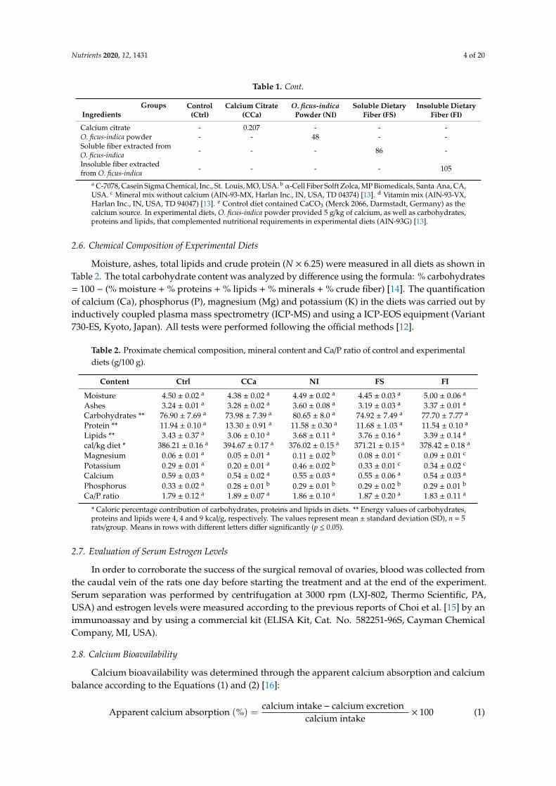
Moisture, ashes, total lipids and crude protein (N × 6.25) were measured in all diets as shown inTable 2. The total carbohydrate content was analyzed by difference using the formula: % carbohydrates= 100 − (% moisture + % proteins + % lipids + % minerals + % crude fiber) [14]. The quantificationof calcium (Ca), phosphorus (P), magnesium (Mg) and potassium (K) in the diets was carried out byinductively coupled plasma mass spectrometry (ICP-MS) and using a ICP-EOS equipment (Variant730-ES, Kyoto, Japan). All tests were performed following the official methods [12].
Table 2. Proximate chemical composition, mineral content and Ca/P ratio of control and experimentaldiets (g/100 g).
Content Ctrl CCa NI FS FI
Moisture 4.50 ± 0.02 a 4.38 ± 0.02 a 4.49 ± 0.02 a 4.45 ± 0.03 a 5.00 ± 0.06 a
Ashes 3.24 ± 0.01 a 3.28 ± 0.02 a 3.60 ± 0.08 a 3.19 ± 0.03 a 3.37 ± 0.01 a
Carbohydrates ** 76.90 ± 7.69 a 73.98 ± 7.39 a 80.65 ± 8.0 a 74.92 ± 7.49 a 77.70 ± 7.77 a
Protein ** 11.94 ± 0.10 a 13.30 ± 0.91 a 11.58 ± 0.30 a 11.68 ± 1.03 a 11.54 ± 0.10 a
Lipids ** 3.43 ± 0.37 a 3.06 ± 0.10 a 3.68 ± 0.11 a 3.76 ± 0.16 a 3.39 ± 0.14 a
cal/kg diet * 386.21 ± 0.16 a 394.67 ± 0.17 a 376.02 ± 0.15 a 371.21 ± 0.15 a 378.42 ± 0.18 a
Magnesium 0.06 ± 0.01 a 0.05 ± 0.01 a 0.11 ± 0.02 b 0.08 ± 0.01 c 0.09 ± 0.01 c
Potassium 0.29 ± 0.01 a 0.20 ± 0.01 a 0.46 ± 0.02 b 0.33 ± 0.01 c 0.34 ± 0.02 c
Calcium 0.59 ± 0.03 a 0.54 ± 0.02 a 0.55 ± 0.03 a 0.55 ± 0.06 a 0.54 ± 0.03 a
Phosphorus 0.33 ± 0.02 a 0.28 ± 0.01 b 0.29 ± 0.01 b 0.29 ± 0.02 b 0.29 ± 0.01 b
Ca/P ratio 1.79 ± 0.12 a 1.89 ± 0.07 a 1.86 ± 0.10 a 1.87 ± 0.20 a 1.83 ± 0.11 a
* Caloric percentage contribution of carbohydrates, proteins and lipids in diets. ** Energy values of carbohydrates,proteins and lipids were 4, 4 and 9 kcal/g, respectively. The values represent mean ± standard deviation (SD), n = 5rats/group. Means in rows with different letters differ significantly (p ≤ 0.05).
2.7. Evaluation of Serum Estrogen Levels
In order to corroborate the success of the surgical removal of ovaries, blood was collected fromthe caudal vein of the rats one day before starting the treatment and at the end of the experiment.Serum separation was performed by centrifugation at 3000 rpm (LXJ-802, Thermo Scientific, PA,USA) and estrogen levels were measured according to the previous reports of Choi et al. [15] by animmunoassay and by using a commercial kit (ELISA Kit, Cat. No. 582251-96S, Cayman ChemicalCompany, MI, USA).
2.8. Calcium Bioavailability
Calcium bioavailability was determined through the apparent calcium absorption and calciumbalance according to the Equations (1) and (2) [16]:
Apparent calcium absorption (%) =calcium intake− calcium excretion
calcium intake× 100 (1)
Nutrients 2020, 12, 1431 5 of 20
Calcium balance (%) = Calcium intake− (urinary calcium + calcium in feces) (2)
The amount of food consumption was measured for four continuous days before the animalswere sacrificed. Ingested calcium, fecal calcium and urine calcium were measured [17].
2.9. Assessment of Femoral Dimensions and Weight
Experimental animals were fasted for 12 h before sacrifice by decapitation. Afterward, the rightand left femurs were excised, and adherent soft tissue and bone marrow were removed. Bones werestored at 4 ◦C until further analysis. Weight gain was measured, as well as the length and diameter ofthe bones, according to a method described previously in [9]. Briefly, the fresh femurs were weighedand measured using a Vernier caliper (Absolute Digimatic, Mitutoyo, Japan). Length measurementswere made from the greater trochanter to the lateral condyle. The width and thickness were measuredat the midpoint (diaphysis) of the femurs. Subsequently, the femurs were labeled and stored at −20 ◦Cuntil analysis.
2.10. Analyses of the Biomechanical Properties of Femurs
Biomechanical properties of femoral bone were determined in the right femur through theforce required to break the bone by using a material testing machine (Mod. Z005, load cell 5000N,Zwick/Roell, Ulm, Germany), employing the TestXpert Intelligent testing version 12.0 software [9].In brief, the samples were defrosted and all the measurements were carried out at room temperature.The failure load of femurs was evaluated by three-point bending (maximum breaking force of failurewhen the load is applied in a perpendicular plane to the longitudinal axis of the femur, denoted bythe symbol Pmax) and compression tests (maximum force of failure when the force is applied in avertical plane to the longitudinal axis of the femur, denoted by the symbol Fmax). Additionally, Young’smodulus or elastic modulus (E) was calculated. All the tests were evaluated in the mid-diaphysealregion of the femur.
2.11. Determination of Calcium and Phosphorus Content in the Femurs
Left femurs were used for Ca and P analysis. Both minerals were determined by inductivelycoupled plasma mass spectrometry (ICP-MS) by using an ICP-EOS equipment (730-ES, Agilent,Santa Clara, CA, USA) [9]. First, the bones were defrosted and crushed with a laboratory mortar andthen oven-dried at 60 ◦C to a constant weight. The dry samples were weighed and a mixture of nitricacid and perchloric acid at a ratio of 3:1 was added to digest the samples (MarsXpress, CEM, Matthews,NC). Then, the samples were transferred and diluted with 10 mL of distilled water. Reagent blankswere prepared with the same digestion procedures.
2.12. Analyses of Bone Mineral Density
Rats belonging to the experimental groups were subjected to densitometry at the beginning andat the end of the experiment by using a simple X-ray equipment (X-Mind®, Satelec, Cologne, France)with a potential of 70 kV, a current of 8 mA and a wavelength of 0.177 × 10⁻¹⁰ m (DG-073B-DC X-raytube, Toshiba, Tokyo, Japan), as well as a detector (S10835 CMOS sensor, Hamamatsu, Iwata, Japan)and a computer platform, in order to determine bone mineral density (BMD) [9,18]. The neck anddiaphysis of femurs (cortical tissue) and metaphysis (cortical-trabecular tissues) were selected formeasurements. For each bone, three measurements were obtained.
2.13. Evaluation of the Microstructural Properties of Rat Femoral Bone
Microstructural parameters of the femurs, such as trabecular separation (Tb.Sp), trabecularthickness (Tb.Th) and cortical thickness (Ct.Wi), were determined in a scanning electron microscope(JSM 6060LV, Jeol, Japan) as previously described [9,19]. Prior to the analysis, the left femurs wereexposed at 130 ◦C in a Papin reactor (6 L, Cinsa, Mexico City, Mexico) for 1 h, and thereafter, the bones
Nutrients 2020, 12, 1431 6 of 20
were dried in an oven (FE-295A, Felisa, Zapopan, Jalisco, Mexico) at 60 ◦C to a constant weight.Subsequently, the femurs were each cut longitudinally from the intercondylar line to the diaphysiswith a disc diamond saw (Diaflex-Transvident 350-352, Berlin, Germany). The femur sections wereincubated with protease (P-6911, 1% w/v, Sigma Chemical Co., St. Louis, MO, USA) at 37 ◦C for 24 h.Bones were subjected to a second digestion process with aminopeptidase (P-6887, 0.4% w/v, SigmaChemical Co.) at 37 ◦C for 18 h. Then, lipids in bones were removed with ethyl ether (Cat. No. 9240-03,J.T. Baker Center Valley, PA, USA) and acetone (Cat. No. 9006-03, J.T. Baker Center Valley, PA, USA)on a shaker (AR-100, Daigger, Vernon Hills, IL, USA) for 12 h. The femurs were dried at 60 ◦C to aconstant weight. Finally, the bones were mounted on stubs and coated with gold using an ion sputterfor observation.
2.14. Measurement of Bone Remodeling Biochemical Biomarkers
At the beginning of the biological experiment, blood samples were collected from the caudalvein of each rat and serum separation was performed. Serum samples were stored at 4 ◦C for lateranalysis. At the end of the study, animals were sacrificed by decapitation, and their serum was obtained.Serum levels of three bone formation biomarkers, including osteocalcin (ELISA Kit Bioassay, Cat. No.200767-96T, USB Biologicals, MA, USA), amino-terminal procollagen type 1 (ELISA Kit Bioassay, Cat.No. 202110-96T USB Biologicals, MA, USA) and alkaline phosphatase (ELISA Kit Bioassay, Cat. No.183576-96T, USB Biologicals, MA, USA), were determined according to the methodology reported byLee [20].
2.15. Statistical Analyses
Results are expressed as mean values ± standard deviation (SD). All data were analyzed usingone-way analysis of variance (ANOVA) followed by Tukey’s test with α = 0.05 and using the GraphPadPrism 6 procedure (GraphPad Software Inc., USA). The calcium source in diets was considered as thevariation factor.
3. Results
3.1. Analyses of Ca and P Content in O. ficus-indica Cladodes, and the Soluble and Insoluble Fiber Extractedfrom O. ficus-indica
In order to formulate the experimental diets, a minerals analysis was performed. The calciumcontent of O. ficus-indica powder, soluble fiber and insoluble fiber was 10.5% ± 0.1%, 5.79% ± 0.04%and 4.73% ± 0.05%, respectively. The insoluble fiber extracted from O. ficus-indica cladodes had thelowest content of this mineral. Regarding phosphorus content, O. ficus-indica cladodes showed thehighest values (0.17% ± 0.01%) compared with soluble fiber (0.10% ± 0.01%) and insoluble fiber(0.07% ± 0.01%).
3.2. Chemical Composition and Mineral Content in Experimental Diets
To examine the chemical composition and mineral content in experimental diets, a proximatechemical analysis was realized. The results are shown in Table 2. No significant differences (p ≤ 0.05)were detected in moisture, lipid, protein, carbohydrate and ash contents between the experimentaldiets, which was reflected in a similar energy density of diets. Likewise, no significant differences incalcium content were detected (p ≤ 0.05) between diets. Regarding phosphorus content, our resultsshowed that the control diet had a significantly (p ≤ 0.05) higher level of this mineral (0.33% ± 0.16%)compared with all experimental diets. On the other hand, potassium and magnesium contents weresignificantly higher (p ≤ 0.05) in the diet prepared with O. ficus-indica powder (0.46% ± 0.02% and0.11% ± 0.05%, respectively). Finally, no statistically significant differences (p ≤ 0.05) were detected inthe Ca/P molar ratio between experimental diets, whose values ranged from 1.79 to 1.89.

Nutrients 2020, 12, 1431 7 of 20
3.3. Serum Estrogen Levels in Ovariectomized Rats
Estrogen levels in rats were analyzed to ensure surgery success. Figure 1 shows that serumestrogen levels of rats from the control (47.97 ± 0.31 pg/mL) and sham (43.31 ± 0.28 pg/mL) groupsat the beginning of the experiment were significantly higher (p ≤ 0.05) that those of the OVX group(11.91 ± 0.69 pg/mL). This same trend was observed at the end of the experiment. These results supportthe efficacy of ovariectomy for decreasing estrogen serum levels in rats.
Nutrients 2020, 12, x FOR PEER REVIEW 7 of 21
significantly higher (p ≤ 0.05) in the diet prepared with O. ficus-indica powder (0.46% ± 0.02% and
0.11% ± 0.05%, respectively). Finally, no statistically significant differences (p ≤ 0.05) were detected in
the Ca/P molar ratio between experimental diets, whose values ranged from 1.79 to 1.89.
3.3. Serum Estrogen Levels in Ovariectomized Rats
Estrogen levels in rats were analyzed to ensure surgery success. Figure 1 shows that serum
estrogen levels of rats from the control (47.97 ± 0.31 pg/mL) and sham (43.31 ± 0.28 pg/mL) groups at
the beginning of the experiment were significantly higher (p ≤ 0.05) that those of the OVX group
(11.91 ± 0.69 pg/mL). This same trend was observed at the end of the experiment. These results
support the efficacy of ovariectomy for decreasing estrogen serum levels in rats.
Figure 1. Effect of ovariectomy in serum estrogen levels at the beginning and at the end of the
treatment. The results represent the average ± SD of the experimental groups, n = 5 in control (Ctrl),
sham (SH) and ovariectomized (OVX) groups. Means in bars with different letters indicate
significant differences (p ≤ 0.05) between groups with Tukey’s test.
3.4. Calcium Bioavailability
Calcium bioavailability was studied by measuring the apparent calcium absorption and
calcium balance. The results shown in Figure 2a indicate that the apparent calcium absorption was
significantly higher (p ≤ 0.05) in the group fed with soluble fiber (93.87% ± 3.15%) compared with
that observed in the groups fed with diets supplemented with calcium citrate (83.81% ± 2.12%), O.
ficus-indica powder (78.26% ± 3.46%) and insoluble fiber (77.96% ± 5.16%) and the ovariectomized
group (OVX) fed with AIN-93M (78.13% ± 6.31%). Rats that showed lower apparent calcium
absorption belonged to the sham (67.89% ± 2.53%) and control (68.99% ± 3.35%) groups. Regarding
calcium balance assessment (Figure 2b), we found that the highest value corresponded to the group
fed with the diet supplemented with soluble fiber (0.35 ± 0.027 mg/d/Ca), which showed a
significantly higher value (p ≤ 0.05) than the groups fed with diet supplemented with insoluble fiber
(0.27 ± 0.03 mg/d/Ca), O. ficus-indica powder (0.30 ± 0.04 mg/d/Ca) and calcium citrate (0.32 ± 0.20
mg/d/Ca) and the OVX group fed with the standard diet (0.23 ± 0.03 mg/d/Ca). Finally, calcium
balance in the control and sham groups was significantly lower (0.18 ± 0.04 mg/d/Ca and 0.18 ± 0.03
mg/d/Ca, respectively) compared with that of other experimental groups.
Figure 1. Effect of ovariectomy in serum estrogen levels at the beginning and at the end of the treatment.The results represent the average ± SD of the experimental groups, n = 5 in control (Ctrl), sham (SH)and ovariectomized (OVX) groups. Means in bars with different letters indicate significant differences(p ≤ 0.05) between groups with Tukey’s test.
3.4. Calcium Bioavailability
Calcium bioavailability was studied by measuring the apparent calcium absorption and calciumbalance. The results shown in Figure 2a indicate that the apparent calcium absorption was significantlyhigher (p ≤ 0.05) in the group fed with soluble fiber (93.87% ± 3.15%) compared with that observed inthe groups fed with diets supplemented with calcium citrate (83.81% ± 2.12%), O. ficus-indica powder(78.26% ± 3.46%) and insoluble fiber (77.96% ± 5.16%) and the ovariectomized group (OVX) fed withAIN-93M (78.13% ± 6.31%). Rats that showed lower apparent calcium absorption belonged to thesham (67.89% ± 2.53%) and control (68.99% ± 3.35%) groups. Regarding calcium balance assessment(Figure 2b), we found that the highest value corresponded to the group fed with the diet supplementedwith soluble fiber (0.35 ± 0.027 mg/d/Ca), which showed a significantly higher value (p ≤ 0.05) than thegroups fed with diet supplemented with insoluble fiber (0.27 ± 0.03 mg/d/Ca), O. ficus-indica powder(0.30 ± 0.04 mg/d/Ca) and calcium citrate (0.32 ± 0.20 mg/d/Ca) and the OVX group fed with thestandard diet (0.23 ± 0.03 mg/d/Ca). Finally, calcium balance in the control and sham groups wassignificantly lower (0.18 ± 0.04 mg/d/Ca and 0.18 ± 0.03 mg/d/Ca, respectively) compared with that ofother experimental groups.
3.5. Physical and Biomechanical Properties of the Femur
To study the bone resistance to fracture, physical and biomechanical properties of femurs wereanalyzed. Femur length of rats from the OVX group (3.3 ± 0.25 cm) was significantly shorter (p ≤ 0.05)than that of the rest of the experimental groups as seen in Table 3. Furthermore, there were no statisticaldifferences (p ≤ 0.05) in the weight, width and thickness of the femur of the animals correspondingto the different experimental groups. In the mechanical compression test (Fmax), the highest valueswere observed in the Ctrl (610.3 ± 46.6 N), SH (634 ± 45.8 N) and FS (606.5 ± 15.8 N) groups withoutsignificant statistical differences (p ≤ 0.05) between them. Contrastingly, a significantly lower valuewas detected in the OVX group (496.1 ± 24.9 N) in comparison with the rest of the experimental groups

Nutrients 2020, 12, 1431 8 of 20
(p ≤ 0.05). In the three-point bending test (Pmax), no significant differences (p ≤ 0.05) were observedbetween the Ctrl (86.3 ± 5.5 N), SH (90.6 ± 1.1 N), FS (89.9 ± 4.3 N) and CCa (85.4 ± 4.4 N) groups.On the other hand, the OVX group displayed a significantly lesser Pmax value (65.7 ± 3.5 N, p ≤ 0.05)compared with all the study groups. Regarding Young’s modulus (E), femurs of the rats fed with thediet supplemented with soluble fiber had significantly higher values (596.1 ± 78.4 N/mm2, p ≤ 0.05)than those shown by the rest of the groups, while the FI and OVX groups showed the lowest values(476.2 ± 19.7 and 439.3 ± 15.4 N/mm2, respectively).Nutrients 2020, 12, x FOR PEER REVIEW 8 of 21
Figure 2. (a) Apparent calcium absorption and (b) calcium balance in experimental groups. The
results represent the average ± SD of experimental groups, n = 5 rats/group. Means in bars with
different letters indicate significant differences (p ≤ 0.05) between groups with Tukey’s test. Ctrl =
control group, SH = sham group, OVX = ovariectomized group fed with AIN-93M, FS =
ovariectomized group fed with diet supplemented with soluble fiber extracted from O. ficus-indica,
CCa = ovariectomized group fed with diet supplemented with calcium citrate, NI = ovariectomized
group fed with diet supplemented with O. ficus-indica powder, FI = ovariectomized group fed with
diet supplemented with insoluble fiber extracted from O. ficus-indica.
3.5. Physical and Biomechanical Properties of the Femur
To study the bone resistance to fracture, physical and biomechanical properties of femurs were
analyzed. Femur length of rats from the OVX group (3.3 ± 0.25 cm) was significantly shorter (p ≤
0.05) than that of the rest of the experimental groups as seen in Table 3. Furthermore, there were no
statistical differences (p ≤ 0.05) in the weight, width and thickness of the femur of the animals
corresponding to the different experimental groups. In the mechanical compression test (Fmax), the
highest values were observed in the Ctrl (610.3 ± 46.6 N), SH (634 ± 45.8 N) and FS (606.5 ± 15.8 N)
groups without significant statistical differences (p ≤ 0.05) between them. Contrastingly, a
significantly lower value was detected in the OVX group (496.1 ± 24.9 N) in comparison with the rest
of the experimental groups (p ≤ 0.05). In the three-point bending test (Pmax), no significant differences
(p ≤ 0.05) were observed between the Ctrl (86.3 ± 5.5 N), SH (90.6 ± 1.1 N), FS (89.9 ± 4.3 N) and CCa
(85.4 ± 4.4 N) groups. On the other hand, the OVX group displayed a significantly lesser Pmax value
(65.7 ± 3.5 N, p ≤ 0.05) compared with all the study groups. Regarding Young’s modulus (E), femurs
of the rats fed with the diet supplemented with soluble fiber had significantly higher values (596.1 ±
78.4 N/mm2, p ≤ 0.05) than those shown by the rest of the groups, while the FI and OVX groups
showed the lowest values (476.2 ± 19.7 and 439.3 ± 15.4 N/mm2, respectively).
Table 3. Physical and mechanical properties of femoral bone in rats fed with the experimental diets.
Parameters Ctrl SH OVX FS CCa NI FI
Length (cm) 3.6 ± 0.16 a 3.5 ± 0.15 a 3.3 ± 0.25 b 3.6 ± 0.12 a 3.6 ± 0.08 a 3.6 ±
0.13 a
3.6 ±
0.13 a
Weight (g) 0.92 ± 0.16 a 0.94 ± 0.03 a 0.97 ± 0.09 a 0.95 ± 0.12 a 0.96 ± 0.07
a
0.97 ±
0.05 a
0.94 ±
0.08 a
Width (mm) 4.0 ± 0.10 a 4.0 ± 0.09 a 4.0 ± 0.10 a 4.0 ± 0.11 a 4.0 ± 0.15 a 4.0 ±
0.10 a
4.0 ±
0.10 a
Thickness (mm) 5.1 ± 0.05 a 5.1 ± 0.03 a 5.0 ± 0.05 a 5.1 ± 0.04 a 5.2 ± 0.06 a 5.1 ±
0.05 a
5.1 ±
0.05 a
Compression test
Fmax (N) 610.3 ± 46.6 a 634 ± 45.8 a 496.1 ± 24.9 b 606.5 ± 15.8 a
555.9 ±
14.8 c
530.9 ±
37.7 c
533.4 ±
26.5 c
Three-point
bending test Pmax 86.3 ± 5.5 a 90.6 ± 1.1 a 65.7 ± 3.5 b 89.9 ± 4.3 a 85.4 ± 4.4 a
74.87 ±
3.6 c
73.0 ±
5.9 c
Figure 2. (a) Apparent calcium absorption and (b) calcium balance in experimental groups. The resultsrepresent the average ± SD of experimental groups, n = 5 rats/group. Means in bars with differentletters indicate significant differences (p ≤ 0.05) between groups with Tukey’s test. Ctrl = controlgroup, SH = sham group, OVX = ovariectomized group fed with AIN-93M, FS = ovariectomized groupfed with diet supplemented with soluble fiber extracted from O. ficus-indica, CCa = ovariectomizedgroup fed with diet supplemented with calcium citrate, NI = ovariectomized group fed with dietsupplemented with O. ficus-indica powder, FI = ovariectomized group fed with diet supplemented withinsoluble fiber extracted from O. ficus-indica.
Table 3. Physical and mechanical properties of femoral bone in rats fed with the experimental diets.
Parameters Ctrl SH OVX FS CCa NI FI
Length (cm) 3.6 ± 0.16 a 3.5 ± 0.15 a 3.3 ± 0.25 b 3.6 ± 0.12 a 3.6 ± 0.08 a 3.6 ± 0.13 a 3.6 ± 0.13 a
Weight (g) 0.92 ± 0.16 a 0.94 ± 0.03 a 0.97 ± 0.09 a 0.95 ± 0.12 a 0.96 ± 0.07 a 0.97 ± 0.05 a 0.94 ± 0.08 a
Width (mm) 4.0 ± 0.10 a 4.0 ± 0.09 a 4.0 ± 0.10 a 4.0 ± 0.11 a 4.0 ± 0.15 a 4.0 ± 0.10 a 4.0 ± 0.10 a
Thickness (mm) 5.1 ± 0.05 a 5.1 ± 0.03 a 5.0 ± 0.05 a 5.1 ± 0.04 a 5.2 ± 0.06 a 5.1 ± 0.05 a 5.1 ± 0.05 a
Compressiontest Fmax (N) 610.3 ± 46.6 a 634 ± 45.8 a 496.1 ± 24.9 b 606.5 ± 15.8 a 555.9 ± 14.8 c 530.9 ± 37.7 c 533.4 ± 26.5 c
Three-pointbending testPmax (N)
86.3 ± 5.5 a 90.6 ± 1.1 a 65.7 ± 3.5 b 89.9 ± 4.3 a 85.4 ± 4.4 a 74.87 ± 3.6 c 73.0 ± 5.9 c
E (N/mm2) 545.3 ± 82.5 a 556.1 ± 23.7 a 439.3 ± 15.4 b 596.1 ± 78.4 c 556.6 ± 19.6 a 539.3 ± 15.9 a 476.2 ± 19.7 b
The values represent mean ± SD, n = 5 rats/group. Means in rows with different letters differ significantly (p ≤ 0.05).Fmax: failure load evaluated by the compression test, Pmax: failure load evaluated by the three-point bending test.Ctrl = control group, SH = sham group, OVX = ovariectomized group fed with AIN-93M, FS = ovariectomizedgroup fed with diet supplemented with soluble fiber extracted from O. ficus-indica, CCa = ovariectomized groupfed with diet supplemented with calcium citrate, NI = ovariectomized group fed with diet supplemented withO. ficus-indica powder, FI = ovariectomized group fed with diet supplemented with insoluble fiber extracted fromO. ficus-indica.
3.6. Mineral Content in the Femur
Bone mineral absorption was determined with an analysis of mineral content in the target tissues(femurs). Table 4 shows the mineral content in the femur of experimental subjects. The highest
Nutrients 2020, 12, 1431 9 of 20
calcium content was detected in the bones of SH (23.87% ± 0.19%), Ctrl (22.64% ± 0.20%), FS (22.91%± 0.24%) and CCa (21.13% ± 0.18%) groups, while the groups that presented the lowest content ofthis mineral were the OVX (16.98% ± 0.98%), NI (20.41% ± 0.22%) and FI groups (20.11% ± 0.22%).Regarding the phosphorus content, there were no statistical differences (p ≤ 0.05) between the groupswith the exception of the OVX group, which presented the lowest level of this mineral (5.18% ± 0.05%).Similarly, the femurs of rats in the OVX group showed the lowest content (0.14% ± 0.10%) of potassiumwith significant differences (p ≤ 0.05) between groups. In addition, the magnesium content in thebones of Ctrl and SH groups (0.74% ± 0.04% and 0.73% ± 0.02%, respectively) was significantly higherin comparison with the other experimental groups. Regarding the Ca/P ratio, the Ctrl (1.28 ± 0.06),SH (1.29 ± 0.01) and FS (1.23 ± 0.06) groups did not show statistical differences between them (p ≤ 0.05).However, the CCa, NI and FI groups showed significantly lower Ca/P ratio values (p ≤ 0.05) than theCtrl, SH and SF groups. The highest value of the Ca/P ratio was detected in the OVX group (3.27 ±0.16), which presented significant differences (p ≤ 0.05) compared with that detected in the rest of theexperimental groups.
Table 4. Mineral content in femoral bone of rats fed with the experimental diets.
Group Ca (%) P (%) K (%) Mg (%) Ca/P Ratio
Ctrl 22.64 ± 0.20 a 17.66 ± 0.13 a 0.21 ± 0.04 a 0.74 ± 0.04 a 1.28 ± 0.06 a
SH 23.87 ± 0.19 a 18.46 ± 0.14 a 0.21 ± 0.06 a 0.73 ± 0.02 a 1.29 ± 0.01 a
OVX 16.98 ± 0.98 b 5.18 ± 0.05 b 0.14 ± 0.10 b 0.51 ± 0.02 b 3.27 ± 0.16 b
FS 22.91 ± 0.24 a 18.60 ± 0.12 a 0.19 ± 0.04 a 0.43 ± 0.02 b 1.23 ± 0.06 a
CCa 21.13 ± 0.18 a 17.99 ± 0.14 a 0.18 ± 0.09 a 0.44 ± 0.02 b 1.17 ± 0.05 c
NI 20.41 ± 0.22 c 18.32 ± 0.09 a 0.20 ± 0.04 a 0.47 ± 0.02 b 1.11 ± 0.08 c
FI 20.11 ± 0.22 c 18.77 ± 0.15 a 0.19 ± 0.04 a 0.45 ± 0.02 b 1.07 ± 0.05 c
The values represent mean± SD, n = 5 rats/group. Means in columns with different letters differ significantly (p ≤ 0.05).Ctrl = control group, SH = sham group, OVX = ovariectomized group fed with AIN-93M, FS = ovariectomizedgroup fed with diet supplemented with soluble fiber extracted from O. ficus-indica, CCa = ovariectomized groupfed with diet supplemented with calcium citrate, NI = ovariectomized group fed with diet supplemented withO. ficus-indica powder, FI = ovariectomized group fed with diet supplemented with insoluble fiber extracted fromO. ficus-indica.
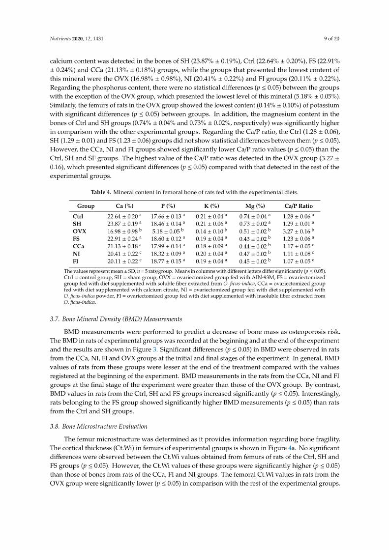
3.7. Bone Mineral Density (BMD) Measurements
BMD measurements were performed to predict a decrease of bone mass as osteoporosis risk.The BMD in rats of experimental groups was recorded at the beginning and at the end of the experimentand the results are shown in Figure 3. Significant differences (p ≤ 0.05) in BMD were observed in ratsfrom the CCa, NI, FI and OVX groups at the initial and final stages of the experiment. In general, BMDvalues of rats from these groups were lesser at the end of the treatment compared with the valuesregistered at the beginning of the experiment. BMD measurements in the rats from the CCa, NI and FIgroups at the final stage of the experiment were greater than those of the OVX group. By contrast,BMD values in rats from the Ctrl, SH and FS groups increased significantly (p ≤ 0.05). Interestingly,rats belonging to the FS group showed significantly higher BMD measurements (p ≤ 0.05) than ratsfrom the Ctrl and SH groups.
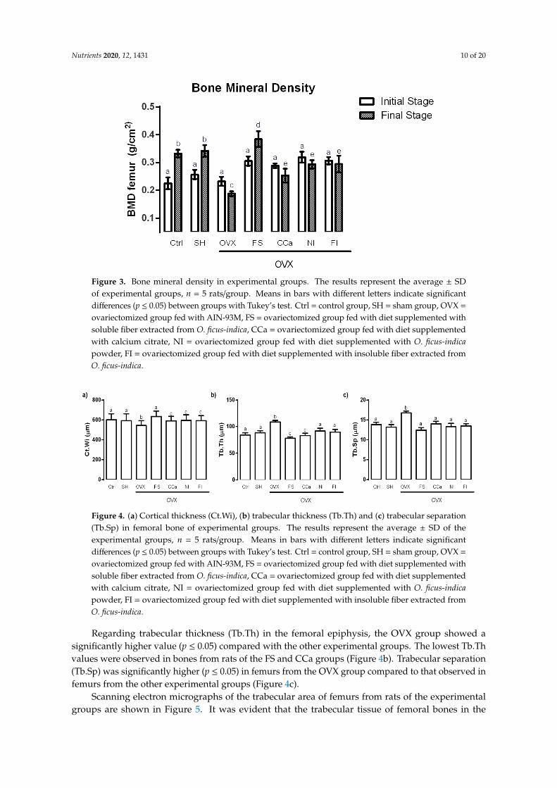
3.8. Bone Microstructure Evaluation
The femur microstructure was determined as it provides information regarding bone fragility.The cortical thickness (Ct.Wi) in femurs of experimental groups is shown in Figure 4a. No significantdifferences were observed between the Ct.Wi values obtained from femurs of rats of the Ctrl, SH andFS groups (p ≤ 0.05). However, the Ct.Wi values of these groups were significantly higher (p ≤ 0.05)than those of bones from rats of the CCa, FI and NI groups. The femoral Ct.Wi values in rats from theOVX group were significantly lower (p ≤ 0.05) in comparison with the rest of the experimental groups.
Nutrients 2020, 12, 1431 10 of 20
Nutrients 2020, 12, x FOR PEER REVIEW 10 of 21
compared with the values registered at the beginning of the experiment. BMD measurements in the
rats from the CCa, NI and FI groups at the final stage of the experiment were greater than those of
the OVX group. By contrast, BMD values in rats from the Ctrl, SH and FS groups increased
significantly (p ≤ 0.05). Interestingly, rats belonging to the FS group showed significantly higher
BMD measurements (p ≤ 0.05) than rats from the Ctrl and SH groups.
Figure 3. Bone mineral density in experimental groups. The results represent the average ± SD of
experimental groups, n = 5 rats/group. Means in bars with different letters indicate significant
differences (p ≤ 0.05) between groups with Tukey’s test. Ctrl = control group, SH = sham group, OVX
= ovariectomized group fed with AIN-93M, FS = ovariectomized group fed with diet supplemented
with soluble fiber extracted from O. ficus-indica, CCa = ovariectomized group fed with diet
supplemented with calcium citrate, NI = ovariectomized group fed with diet supplemented with O.
ficus-indica powder, FI = ovariectomized group fed with diet supplemented with insoluble fiber
extracted from O. ficus-indica.
3.8. Bone Microstructure Evaluation
The femur microstructure was determined as it provides information regarding bone fragility.
The cortical thickness (Ct.Wi) in femurs of experimental groups is shown in Figure 4a. No significant
differences were observed between the Ct.Wi values obtained from femurs of rats of the Ctrl, SH and
FS groups (p ≤ 0.05). However, the Ct.Wi values of these groups were significantly higher (p ≤ 0.05)
than those of bones from rats of the CCa, FI and NI groups. The femoral Ct.Wi values in rats from
the OVX group were significantly lower (p ≤ 0.05) in comparison with the rest of the experimental
groups.
Regarding trabecular thickness (Tb.Th) in the femoral epiphysis, the OVX group showed a
significantly higher value (p ≤ 0.05) compared with the other experimental groups. The lowest Tb.Th
values were observed in bones from rats of the FS and CCa groups (Figure 4b). Trabecular
separation (Tb.Sp) was significantly higher (p ≤ 0.05) in femurs from the OVX group compared to
that observed in femurs from the other experimental groups (Figure 4c).
Figure 3. Bone mineral density in experimental groups. The results represent the average ± SDof experimental groups, n = 5 rats/group. Means in bars with different letters indicate significantdifferences (p ≤ 0.05) between groups with Tukey’s test. Ctrl = control group, SH = sham group, OVX =
ovariectomized group fed with AIN-93M, FS = ovariectomized group fed with diet supplemented withsoluble fiber extracted from O. ficus-indica, CCa = ovariectomized group fed with diet supplementedwith calcium citrate, NI = ovariectomized group fed with diet supplemented with O. ficus-indicapowder, FI = ovariectomized group fed with diet supplemented with insoluble fiber extracted fromO. ficus-indica.
Nutrients 2020, 12, x FOR PEER REVIEW 11 of 21
Figure 4. (a) Cortical thickness (Ct.Wi), (b) trabecular thickness (Tb.Th) and (c) trabecular separation
(Tb.Sp) in femoral bone of experimental groups. The results represent the average ± SD of the
experimental groups, n = 5 rats/group. Means in bars with different letters indicate significant
differences (p ≤ 0.05) between groups with Tukey’s test. Ctrl = control group, SH = sham group, OVX
= ovariectomized group fed with AIN-93M, FS = ovariectomized group fed with diet supplemented
with soluble fiber extracted from O. ficus-indica, CCa = ovariectomized group fed with diet
supplemented with calcium citrate, NI = ovariectomized group fed with diet supplemented with O.
ficus-indica powder, FI = ovariectomized group fed with diet supplemented with insoluble fiber
extracted from O. ficus-indica.
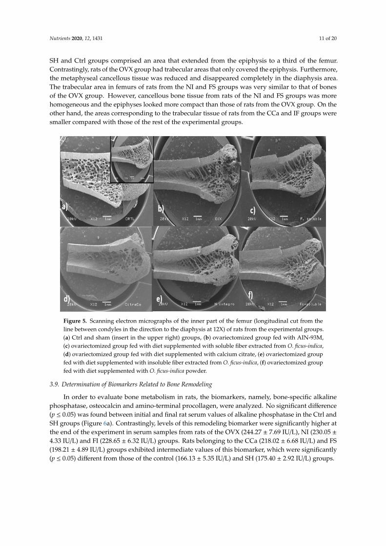
Scanning electron micrographs of the trabecular area of femurs from rats of the experimental
groups are shown in Figure 5. It was evident that the trabecular tissue of femoral bones in the SH
and Ctrl groups comprised an area that extended from the epiphysis to a third of the femur.
Contrastingly, rats of the OVX group had trabecular areas that only covered the epiphysis.
Furthermore, the metaphyseal cancellous tissue was reduced and disappeared completely in the
diaphysis area. The trabecular area in femurs of rats from the NI and FS groups was very similar to
that of bones of the OVX group. However, cancellous bone tissue from rats of the NI and FS groups
was more homogeneous and the epiphyses looked more compact than those of rats from the OVX
group. On the other hand, the areas corresponding to the trabecular tissue of rats from the CCa and
IF groups were smaller compared with those of the rest of the experimental groups.
Figure 4. (a) Cortical thickness (Ct.Wi), (b) trabecular thickness (Tb.Th) and (c) trabecular separation(Tb.Sp) in femoral bone of experimental groups. The results represent the average ± SD of theexperimental groups, n = 5 rats/group. Means in bars with different letters indicate significantdifferences (p ≤ 0.05) between groups with Tukey’s test. Ctrl = control group, SH = sham group, OVX =
ovariectomized group fed with AIN-93M, FS = ovariectomized group fed with diet supplemented withsoluble fiber extracted from O. ficus-indica, CCa = ovariectomized group fed with diet supplementedwith calcium citrate, NI = ovariectomized group fed with diet supplemented with O. ficus-indicapowder, FI = ovariectomized group fed with diet supplemented with insoluble fiber extracted fromO. ficus-indica.
Regarding trabecular thickness (Tb.Th) in the femoral epiphysis, the OVX group showed asignificantly higher value (p ≤ 0.05) compared with the other experimental groups. The lowest Tb.Thvalues were observed in bones from rats of the FS and CCa groups (Figure 4b). Trabecular separation(Tb.Sp) was significantly higher (p ≤ 0.05) in femurs from the OVX group compared to that observed infemurs from the other experimental groups (Figure 4c).
Scanning electron micrographs of the trabecular area of femurs from rats of the experimentalgroups are shown in Figure 5. It was evident that the trabecular tissue of femoral bones in the
Nutrients 2020, 12, 1431 11 of 20
SH and Ctrl groups comprised an area that extended from the epiphysis to a third of the femur.Contrastingly, rats of the OVX group had trabecular areas that only covered the epiphysis. Furthermore,the metaphyseal cancellous tissue was reduced and disappeared completely in the diaphysis area.The trabecular area in femurs of rats from the NI and FS groups was very similar to that of bonesof the OVX group. However, cancellous bone tissue from rats of the NI and FS groups was morehomogeneous and the epiphyses looked more compact than those of rats from the OVX group. On theother hand, the areas corresponding to the trabecular tissue of rats from the CCa and IF groups weresmaller compared with those of the rest of the experimental groups.
Nutrients 2020, 12, x FOR PEER REVIEW 11 of 21
Figure 4. (a) Cortical thickness (Ct.Wi), (b) trabecular thickness (Tb.Th) and (c) trabecular separation
(Tb.Sp) in femoral bone of experimental groups. The results represent the average ± SD of the
experimental groups, n = 5 rats/group. Means in bars with different letters indicate significant
differences (p ≤ 0.05) between groups with Tukey’s test. Ctrl = control group, SH = sham group, OVX
= ovariectomized group fed with AIN-93M, FS = ovariectomized group fed with diet supplemented
with soluble fiber extracted from O. ficus-indica, CCa = ovariectomized group fed with diet
supplemented with calcium citrate, NI = ovariectomized group fed with diet supplemented with O.
ficus-indica powder, FI = ovariectomized group fed with diet supplemented with insoluble fiber
extracted from O. ficus-indica.
Scanning electron micrographs of the trabecular area of femurs from rats of the experimental
groups are shown in Figure 5. It was evident that the trabecular tissue of femoral bones in the SH
and Ctrl groups comprised an area that extended from the epiphysis to a third of the femur.
Contrastingly, rats of the OVX group had trabecular areas that only covered the epiphysis.
Furthermore, the metaphyseal cancellous tissue was reduced and disappeared completely in the
diaphysis area. The trabecular area in femurs of rats from the NI and FS groups was very similar to
that of bones of the OVX group. However, cancellous bone tissue from rats of the NI and FS groups
was more homogeneous and the epiphyses looked more compact than those of rats from the OVX
group. On the other hand, the areas corresponding to the trabecular tissue of rats from the CCa and
IF groups were smaller compared with those of the rest of the experimental groups.
Figure 5. Scanning electron micrographs of the inner part of the femur (longitudinal cut from theline between condyles in the direction to the diaphysis at 12X) of rats from the experimental groups.(a) Ctrl and sham (insert in the upper right) groups, (b) ovariectomized group fed with AIN-93M,(c) ovariectomized group fed with diet supplemented with soluble fiber extracted from O. ficus-indica,(d) ovariectomized group fed with diet supplemented with calcium citrate, (e) ovariectomized groupfed with diet supplemented with insoluble fiber extracted from O. ficus-indica, (f) ovariectomized groupfed with diet supplemented with O. ficus-indica powder.
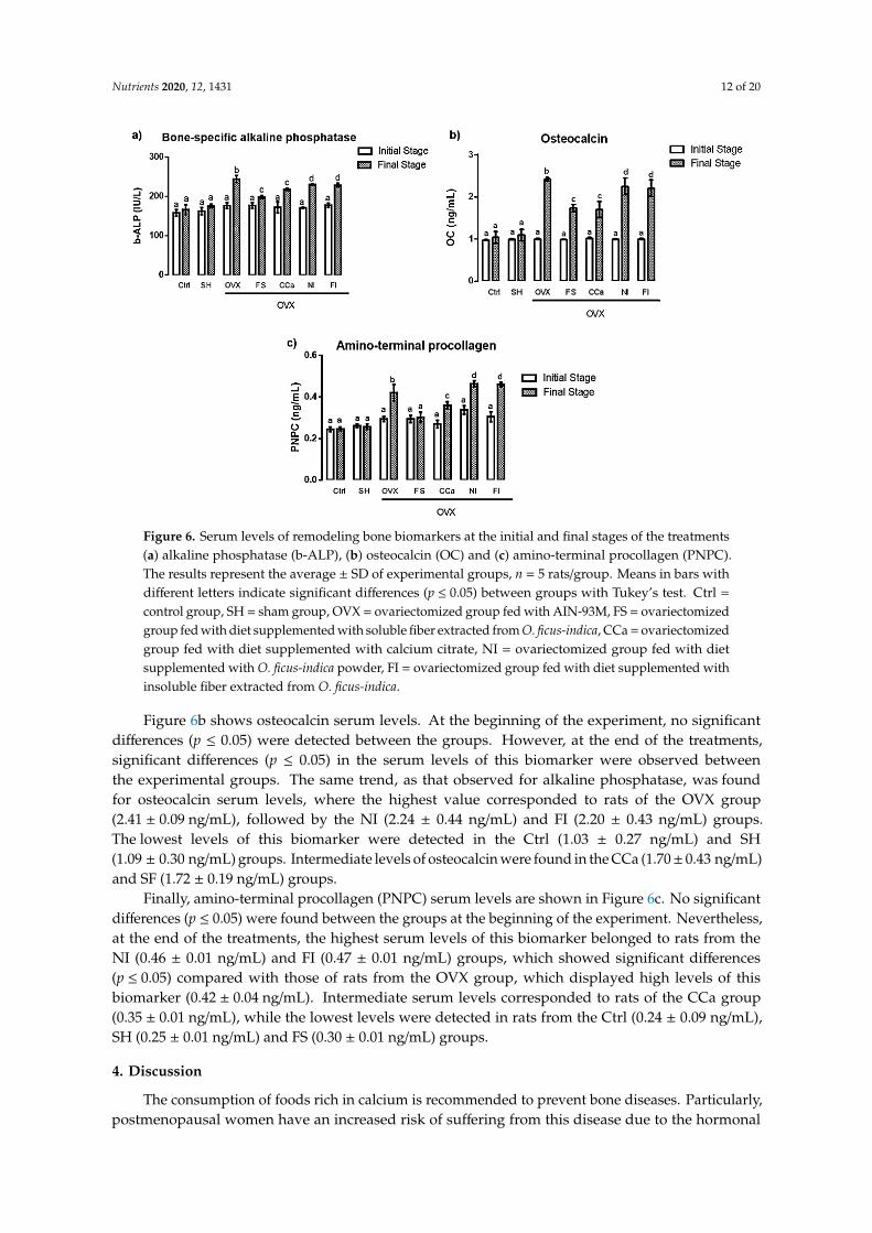
3.9. Determination of Biomarkers Related to Bone Remodeling
In order to evaluate bone metabolism in rats, the biomarkers, namely, bone-specific alkalinephosphatase, osteocalcin and amino-terminal procollagen, were analyzed. No significant difference(p ≤ 0.05) was found between initial and final rat serum values of alkaline phosphatase in the Ctrl andSH groups (Figure 6a). Contrastingly, levels of this remodeling biomarker were significantly higher atthe end of the experiment in serum samples from rats of the OVX (244.27 ± 7.69 IU/L), NI (230.05 ±4.33 IU/L) and FI (228.65 ± 6.32 IU/L) groups. Rats belonging to the CCa (218.02 ± 6.68 IU/L) and FS(198.21 ± 4.89 IU/L) groups exhibited intermediate values of this biomarker, which were significantly(p ≤ 0.05) different from those of the control (166.13 ± 5.35 IU/L) and SH (175.40 ± 2.92 IU/L) groups.
Nutrients 2020, 12, 1431 12 of 20Nutrients 2020, 12, x FOR PEER REVIEW 13 of 21
Figure 6. Serum levels of remodeling bone biomarkers at the initial and final stages of the treatments
(a) alkaline phosphatase (b-ALP), (b) osteocalcin (OC) and (c) amino-terminal procollagen (PNPC).
The results represent the average ± SD of experimental groups, n = 5 rats/group. Means in bars with
different letters indicate significant differences (p ≤ 0.05) between groups with Tukey’s test. Ctrl =
control group, SH = sham group, OVX = ovariectomized group fed with AIN-93M, FS =
ovariectomized group fed with diet supplemented with soluble fiber extracted from O. ficus-indica,
CCa = ovariectomized group fed with diet supplemented with calcium citrate, NI = ovariectomized
group fed with diet supplemented with O. ficus-indica powder, FI = ovariectomized group fed with
diet supplemented with insoluble fiber extracted from O. ficus-indica.
4. Discussion
The consumption of foods rich in calcium is recommended to prevent bone diseases.
Particularly, postmenopausal women have an increased risk of suffering from this disease due to the
hormonal decrease of estrogens [21]. One of the most commonly used experimental models to study
this condition is the ovariectomized rat model of postmenopausal bone loss [22], which was
employed in the present study. As expected, the experimental group that underwent ovariectomy
showed significantly lesser serum estrogen levels compared with the simulated surgery group (SH)
and the control group (Ctrl). Reported values of circulating estrogen in healthy 3-month-old female
Wistar rats range from 40 to 55 pg/mL, while ovariectomized rats of the same age show estrogen
levels ranging from 5 to 12 pg/mL [23–25]. In this study, estrogen levels of rats from the Ctrl (47.9 ±
0.3 pg/mL) and SH (43.3 ± 2.5 pg/mL) groups were within the range previously reported in healthy
rats. Estradiol serum levels of ovariectomized rats (11.2 ± 0.6 pg/mL) were also similar to those
reported in previous studies [25].
The dietary fiber improves mineral absorption and therefore, favors mineral bioavailability
[26]. Soluble fiber contains inulin, which is not digested during its passage through the intestine and
therefore, remains intact until it reaches the colon, where it is fermented by the microbiota, which
stimulate the growth of Bifidobacterium strains, subsequently modifying intestinal pH [27]. Soluble
fiber fermentation produces short-chain fatty acids, such as butyrate, propionate and acetate, among
other organic acids [28]. These fatty acids diminish the luminal pH in the colon and, as a
consequence, Ca2+ absorption is augmented by a mechanism that comprises the exchange of cellular
H+ for luminal Ca2+. In addition, absorption of this mineral by passive diffusion is also increased [29].
Figure 6. Serum levels of remodeling bone biomarkers at the initial and final stages of the treatments(a) alkaline phosphatase (b-ALP), (b) osteocalcin (OC) and (c) amino-terminal procollagen (PNPC).The results represent the average ± SD of experimental groups, n = 5 rats/group. Means in bars withdifferent letters indicate significant differences (p ≤ 0.05) between groups with Tukey’s test. Ctrl =
control group, SH = sham group, OVX = ovariectomized group fed with AIN-93M, FS = ovariectomizedgroup fed with diet supplemented with soluble fiber extracted from O. ficus-indica, CCa = ovariectomizedgroup fed with diet supplemented with calcium citrate, NI = ovariectomized group fed with dietsupplemented with O. ficus-indica powder, FI = ovariectomized group fed with diet supplemented withinsoluble fiber extracted from O. ficus-indica.
Figure 6b shows osteocalcin serum levels. At the beginning of the experiment, no significantdifferences (p ≤ 0.05) were detected between the groups. However, at the end of the treatments,significant differences (p ≤ 0.05) in the serum levels of this biomarker were observed betweenthe experimental groups. The same trend, as that observed for alkaline phosphatase, was foundfor osteocalcin serum levels, where the highest value corresponded to rats of the OVX group(2.41 ± 0.09 ng/mL), followed by the NI (2.24 ± 0.44 ng/mL) and FI (2.20 ± 0.43 ng/mL) groups.The lowest levels of this biomarker were detected in the Ctrl (1.03 ± 0.27 ng/mL) and SH(1.09 ± 0.30 ng/mL) groups. Intermediate levels of osteocalcin were found in the CCa (1.70± 0.43 ng/mL)and SF (1.72 ± 0.19 ng/mL) groups.
Finally, amino-terminal procollagen (PNPC) serum levels are shown in Figure 6c. No significantdifferences (p ≤ 0.05) were found between the groups at the beginning of the experiment. Nevertheless,at the end of the treatments, the highest serum levels of this biomarker belonged to rats from theNI (0.46 ± 0.01 ng/mL) and FI (0.47 ± 0.01 ng/mL) groups, which showed significant differences(p ≤ 0.05) compared with those of rats from the OVX group, which displayed high levels of thisbiomarker (0.42 ± 0.04 ng/mL). Intermediate serum levels corresponded to rats of the CCa group(0.35 ± 0.01 ng/mL), while the lowest levels were detected in rats from the Ctrl (0.24 ± 0.09 ng/mL),SH (0.25 ± 0.01 ng/mL) and FS (0.30 ± 0.01 ng/mL) groups.
4. Discussion
The consumption of foods rich in calcium is recommended to prevent bone diseases. Particularly,postmenopausal women have an increased risk of suffering from this disease due to the hormonal
Nutrients 2020, 12, 1431 13 of 20
decrease of estrogens [21]. One of the most commonly used experimental models to study thiscondition is the ovariectomized rat model of postmenopausal bone loss [22], which was employedin the present study. As expected, the experimental group that underwent ovariectomy showedsignificantly lesser serum estrogen levels compared with the simulated surgery group (SH) and thecontrol group (Ctrl). Reported values of circulating estrogen in healthy 3-month-old female Wistar ratsrange from 40 to 55 pg/mL, while ovariectomized rats of the same age show estrogen levels rangingfrom 5 to 12 pg/mL [23–25]. In this study, estrogen levels of rats from the Ctrl (47.9 ± 0.3 pg/mL) andSH (43.3 ± 2.5 pg/mL) groups were within the range previously reported in healthy rats. Estradiolserum levels of ovariectomized rats (11.2 ± 0.6 pg/mL) were also similar to those reported in previousstudies [25].
The dietary fiber improves mineral absorption and therefore, favors mineral bioavailability [26].Soluble fiber contains inulin, which is not digested during its passage through the intestine and therefore,remains intact until it reaches the colon, where it is fermented by the microbiota, which stimulate thegrowth of Bifidobacterium strains, subsequently modifying intestinal pH [27]. Soluble fiber fermentationproduces short-chain fatty acids, such as butyrate, propionate and acetate, among other organicacids [28]. These fatty acids diminish the luminal pH in the colon and, as a consequence, Ca2+
absorption is augmented by a mechanism that comprises the exchange of cellular H+ for luminal Ca2+.In addition, absorption of this mineral by passive diffusion is also increased [29].
On the other hand, CO2 (another product of fiber fermentation), in the presence of bicarbonate,which is secreted by the pancreas in order to neutralize the acidity achieved by fatty acids [30], interactswith calcium carbonate, which is found in soluble fiber [10]. This reaction releases ionic calcium andcontributes to maintain an optimal pressure of intestinal CO2, which in turn improves the absorption ofthis mineral [31]. In vitro studies have demonstrated that CO2 production helps to solubilize calcium,when it is in the form of carbonate [32]. In fact, the administration of calcium carbonate (1000 mg/day)to premenopausal women increased serum calcium levels and reduced circulating levels of parathyroidhormone more efficiently than the administration of calcium citrate (1000 mg/day) [33].
The results obtained from the determination of the apparent absorption of calcium correlatewith the reabsorption of calcium in the renal tubules. Approximately, 20% of filtered calcium isreabsorbed into the loop of Henle by means of the Na+/K+ pump and Ca2+/Na+ exchanger. In the distalconvoluted tubule, approximately 8% of the filtered calcium is actively reabsorbed; therefore, this isthe segment where the highest regulation of calcium excretion occurs [34]. Rats from the FS group hadthe highest value of calcium balance (0.35 mg/d/Ca), which indicated a better efficiency of calciumreabsorption compared with that of rats from the CCa, NI and IF groups. It is worth mentioning thatdiets administered to rats of the FI and NI groups contained higher levels of insoluble fiber, whichis the least fermentable fiber in the colon [28]. Insoluble fiber is rich in cellulose, which limits theaccessibility of fermentation enzymes in the colon, producing only acetic acid [35]. On the contrary,final products of soluble fiber fermentation (short-chain fatty acids, lactic acid, etc.) are more numerousand varied, which allow better calcium reabsorption [30].
Ovariectomized rats (OVX group) showed lower calcium balance values than rats from the FS,FI, CCa and NI groups. This can be attributed to the lack of estrogens, which provoke an increasedcalcium loss through bone resorption and hypercalciuria [36]. The main regulator of renal calciumexcretion is parathyroid hormone (PTH) and its secretion can be indirectly activated by growth factor23 (FGF23), which is synthesized in the osteoblasts and interacts with FGF23 receptors that require aKloto co-receptor (whose gene is expressed predominantly in kidney) [37]. It has been demonstratedthat estrogens increase FGF23 mRNA expression, so that in the absence of estradiol, this expressiondiminishes, causing a rise in glomerular filtration rate and a decrease in tubular resorption, whichlead to calciuria [38]. In the case of rats from the Ctrl and SH groups, a net calcium absorption andan adequate calcium balance were observed. Contrastingly, ovariectomized rats, whose calciumabsorption was higher than that of rats from the Ctrl and SH groups, experienced a higher calciumloss, as evidenced by their calcium balance values.
Nutrients 2020, 12, 1431 14 of 20
Regarding the analysis of the physical and biomechanical properties of the rat femoral bones,it was evident that rats from the Ctrl, SH and FS groups showed the highest values in the compressionand three-point bending tests, which indicated that greater force was required to achieve bone breakage.These results are according to Hernández-Becerra et al. [9], who reported that bones of rats in growingstage, fed with O. ficus-indica cladodes at a late maturity stage (100 and 135 days) as the only source ofcalcium, required greater strength for bone fracture than the bones of rats fed with cladodes at an earlymaturity stage (25 and 60 days).
Probably, a lower mechanical resistance in the bones of rats from the NI and FI groups was dueto the presence of calcium oxalate and phytates in O. ficus-indica cladodes at a late maturity stageand their insoluble fiber. These components act as chelating agents of various minerals, such ascalcium, magnesium, copper and zinc, resulting in a lower absorption of these minerals, as reported inprevious studies regarding the presence of mineral-sequestering agents in O. ficus-indica species andtheir influence on calcium bioavailability [39].
Femurs from rats fed with diets supplemented with calcium carbonate (CCa group) showed lessmechanical resistance and apparent calcium absorption than bones of rats fed with diets supplementedwith O. ficus-indica soluble fiber. The solubility product constant (Ksp) value of calcium carbonateis lower (3.36 × 10⁻⁹) than that of calcium citrate (9.6 × 10⁻²). However, it is important to take intoaccount that these values are modified depending on the pH. In the case of the gastrointestinal tract,pH values greatly vary from highly acid in the stomach to a pH value of 6 (approximately) in theduodenum; therefore, the solubility of the compounds that transit in the intestine changes. That is thecase for calcium salts, whose absorption and bioavailability differs [33], as has been demonstrated in aclinical study carried out in adult women (>45 years old), who ingested calcium carbonate, calciumlactate, calcium glutamate and calcium citrate as sources of calcium. The investigation indicated thatthe absorption and bioavailability of calcium lactate was significantly higher than that of the otherthree calcium salts. The absorption and bioavailability of calcium carbonate was better than that ofcalcium citrate, whereas calcium glutamate had the lowest values [40].
Regarding the elasticity bone module test (Young’s module test), the highest values correspondedto femurs of rats of the FS group. This means that these bones will undergo small deformations withgreat efforts [41].
On the other hand, calcium content of femoral rat bones from the Ctrl, SH, FS and CCa groupsdid not show statistical differences (p ≤ 0.05), while bones of rats from the OVX group presented lowercalcium content. Hydroxyapatite (Ca5(PO4)3(OH)) is the main bone component, which confers rigidityand resistance to bones [42]. Therefore, calcium and phosphorus are essential for bone formation(mineralization), although Mg, K and Zn (trace elements) are also bone components [43].
The Ca/P ratio in bone has been used as an indicator for osteoporosis. Previous studies haveindicated that estrogen deficiency induced by ovariectomy in female rats leads to an increase in Ca/Pratios in both tibial and femoral bones. The Ca/P ratio observed in ovariectomized rats is >2.0, whereashealthy female rats show values <2.0 [44–46]. Ca/P ratio increment in rats that underwent ovariectomysuggests a negative effect on the balance between bone resorption and bone formation activity [43].In this study, the Ca/P ratio was <2.0 in rats of the control and sham groups and in the ovariectomizedrats which were fed with diets containing calcium citrate, O. ficus-indica powder and soluble andinsoluble fiber from O. ficus-indica. However, the Ca/P ratio was lower in bones from rats of the CaC,NI and FI groups, which indicated inadequate calcium absorption reflected in a decrease in bonemineralization [47].
A satisfactory calcium intake accompanied by adequate bioavailability increases BMD [33].BMD values diminish when estrogen production is reduced due to the constant loss of calcium,provoked by an increment in bone resorption [48]. Wistar rats with ovariectomy show femoral meanBMD values of 0.208 g/cm2 [49], In this investigation, rats that showed the highest BMD measurementswere those fed with a diet containing the O. ficus-indica soluble fiber. It has been previously reportedthat mucilage from O. ficus-indica is constituted of soluble dietary fiber (ranging from 51.70% to
Nutrients 2020, 12, 1431 15 of 20
67.51%) [50]. This polysaccharide consists of alternating rhamnose and galacturonic acid residues,which are attached to the side chains composed of three galactose residues. In addition, arabinose andxylose sugars are branched on the galactose side chains [51]. These structural characteristics of solublefiber from O. ficus-indica positions it as a potential prebiotic, that can be hydrolyzed and fermented bythe colonic microbiota in the large intestine [26].
Prebiotic fibers have been associated with increases in bifidobacteria in doses up to 20 g/day,administered for 7 to 64 days in human adults [52]. It has been observed that the type of prebioticfiber favors the production of bifidobacteria and contributes to a better mineral absorption, whichis reflected in a higher BMD [53]. Numerous studies have repeatedly shown that prebiotics, suchas oligofructose, inulin and galacto-oligosaccharides, effectively stimulate calcium absorption inrats [29,54–58]. This fact was previously observed, where loss of minerals in bones of menopausalwomen and ovariectomized rats was prevented with a combination of prebiotics (oligofructose plusAcacia gum) added to experimental diets [59,60].
To complement bone quality characterization, the microstructure of the rat femoral bones wasanalyzed. The trabecular and compact bone act together in order to meet the physiological needsof the organism [42]. The proportion of trabecular and cortical bone can affect fracture resistance,while the negative imbalance of remodeling and increased bone resorption cause greater porosity inboth trabecular and cortical areas, which are factors to increase the risk of fracture [61]. In this study,femurs of rats from the OVX group had a lower cortical thickness, which consequently had a negativeimpact on the mechanical tests, since femoral bones of rats belonging to this group displayed a lowerresistance to fracture. By contrast, femurs of rats from the Ctrl, SH and FS groups showed a greaterresistance to bone breakage. This was in accordance with their greater cortical thickness (Ct.Wi) andless trabecular space (Tb.Sp).
Interestingly, Tb.Th values observed in femoral bones of rats from the FS group were similarto those of rats from the CCa group, indicating that the effect of calcium present in the soluble fiberextracted from O. ficus inica might be comparable to that of calcium citrate, a common supplementrecommended in patients with osteoporosis [62]. Micrographs of femoral bones of rats from the FSgroup showed that the trabecular zone covered part of the diaphysis, unlike what happened in bonesof rats from the CCa group, in which the trabecular zone was only observed in the metaphysis. In thecase of femoral bones of rats from the Ctrl and SH groups, the trabecular zone completely covered thediaphysis, confirming that these experimental subjects did not experience calcium loss due to estrogenabsence. Regarding rats fed with diets containing O. ficus-indica powder and its insoluble fiber, it wasevident that they had a significant bone resorption, since the trabecular area was restricted to a part ofthe femoral epiphysis.
Bones of ovariectomized rats had the greatest values of trabecular thickness. Moreover, somefissures were observed in their bones, indicating a destruction of bone tissue, which evidently wasrelated to low calcium content. Our results are in accordance with previous studies, which demonstratedthat ovariectomized rats had low BMD values and their bones had large trabecular thickness and lowerresistance to fracture, which was attributed to the presence of fissures and a greater porosity of thebones [47,60]. Bones of rats of the other experimental groups did not show significant differences interms of their trabecular separation.
Rats from the CCa, IF and NI groups had a higher percentage of calcium and phosphorus than ratsof the OVX group, as well as greater absorption and calcium balance. These findings were indicativeof osteoblastic activation to recover bone loss provoked by osteoclastic action. However, recovery ofbones of these rats was not completely achieved (Figure 5). On the other hand, greater trabecularareas were observed in femoral bones of rats fed with diets containing soluble fiber extracted fromO. ficus-indica, indicating that this component acts a protector for osteoblastic action.
Bone-specific alkaline phosphatase, amino-terminal procollagen and osteocalcin are themost commonly biomarkers used to study bone remodeling. Mineralization is the formationof hydroxyapatite crystals in the superficial membrane of the osteoblasts, followed by the
Nutrients 2020, 12, 1431 16 of 20
propagation of hydroxyapatite in the extracellular matrix and its deposition between the collagenfibrils [63]. This occurs through extracellular inorganic pyrophosphate that is provided by nucleotidepyrophosphatase phosphodiesterase 1, so the role of the alkaline phosphatase is to hydrolyzepyrophosphate and provide inorganic phosphate to promote bone mineralization [64]. High levels ofalkaline phosphatase have been observed in both postmenopausal women and ovariectomized rats,due to the increase in bone remodeling [43]. Reported serum level of bone-specific alkaline phosphatasein healthy adult Wistar rats is <200 IU/L, while in ovariectomized rats the level is >200 IU/L [65].In this study, rats that underwent ovariectomy showed high values of this marker (>200 IU/L) atthe end of the experiment, indicating an increase in bone remodeling due to the negative balance inbone resorption. In contrast, rats from the FS group showed values <200 IU/L, which might reflect aprotective mechanism against osteoclastic resorption due to the effectiveness of calcium absorptionpromoted by the soluble fiber of Opuntia ficus-indica in diet. Osteocalcin is a peptide synthesized byosteoblasts in the last stages of bone formation in conjunction with vitamin D [66]. The levels of thisbiochemical marker of bone remodeling, both in postmenopausal women and in adult ovariectomizedWistar rats, are high. Reference values in healthy adult rats are <1.0 ng/mL and in ovariectomized ratsare >1.0 ng/mL [67,68]. In the case of rats from the OVX, NI and FI groups, osteocalcin serum valueswere above 2.0 ng/mL. These results further confirmed that rats had experienced bone resorption,and therefore, it was necessary to activate osteoblasts to initiate bone remodeling in such a way thatbone mineralization is “increased”, in order to recover bone lost by the action of osteoclasts. On thecontrary, Ctrl, SH, FS and CCa groups showed values below 2.0 ng/mL, indicating that a markedosteoblastic activation was not necessary as in the case of rats from the OVX, NI and FI groups.
Collagen is a heterodimer that contributes to the integrity and strength of bone matrix. Serumvalues of this marker increase during growth and in situations of augmented bone formation [69].Reported collagen levels in healthy Wistar rats are <0.20 mg/L, while those of ovariectomized Wistarrats are >0.20 mg/L [48,70]. In the present study, serum levels of this biomarker displayed the sametrend as that of alkaline phosphatase and osteocalcin; rats from the OVX, NI and FI groups showedvalues >0.40 mg/L, while rats from the FS group showed values <0.40 mg/L.
In summary, serum levels of biomarkers related to bone remodeling in rats fed with dietscontaining O. ficus-indica powder, insoluble fiber and calcium citrate were similar to those of rats withestrogen deficiency and weakened bones. However, these biomarker values in rats fed with the solublefiber of O. ficus-indica were consistent with improved calcium absorption, which avoids the need forincreased remodeling activity.
The present study demonstrated the beneficial effects of the consumption of soluble fiber fromO. ficus-indica on the physicochemical and structural properties of bones. Nevertheless, the short- andlong-term effects of low, medium and high doses of the soluble fiber were not assessed in order todetermine the minimum effective dose required to improve bone structure and strength. Evidently,it is also necessary to establish if soluble fiber extracted from O. ficus-indica is able to favor calciumabsorption and induce changes in gut microbial bacteria, so it could be considered as a prebiotic. It isalso important to further assess the influence of this soluble fiber on osteoblastic/osteoclastic activitybalance and to analyze the percentage of calcium from O. ficus-indica’s soluble fiber that is fixed directlyto the bone. These analyses are worthy of further study and will allow a broader understanding of thespecific mechanisms by which the soluble fiber of O. ficus-indica contributes to preserve bone health.
5. Conclusions
The highest values of apparent calcium bioavailability, calcium absorption, fracture resistance,calcium content and bone mineral density were obtained in rats, whose diet was supplementedwith the soluble fiber extracted from O. ficus-indica. Moreover, femurs from these rats had greatercortical thickness and trabecular separation, which evidently contributed to improve their bonemineral density and biomechanical properties, as clearly evidenced by a better resistance to fracture.Serum values of bone remodeling biomarkers were lower in rats fed with the diet containing the
Nutrients 2020, 12, 1431 17 of 20
soluble fiber of O. ficus-indica. Our results demonstrated that consumption of the soluble fiberextracted from O. ficus-indica cladodes protects bones from osteoclastic activity, therefore contributingto improve the physical, densitometric, biomechanical and microstructural properties of bones. Furtherwork is needed to identify the exact mechanisms involved by which soluble fiber extracted fromO. ficus-indica improves calcium absorption and the role of this soluble fiber as a prebiotic and toevaluate different concentrations of soluble fiber in the diet to identify the adequate dose that improvescalcium bioavailability, when the level of this mineral in the diet is low.
Author Contributions: Conceptualization, supervision, funding acquisition and writing—original draftpreparation, I.R.-M.; methodology and writing—original draft preparation, M.Q.-G.; investigation, E.G.-C.;formal analysis, M.Q.-G., M.M.-Á., A.D.R. and E.R.; software and validation D.J.-M.; writing—review and editing,A.R.-M. All authors have read and agreed to the published version of the manuscript.
Funding: This research was funded by CONACYT (Basic Science Projects, Convocation 2011-02, Project No.167769) and the Fund for Research Strengthening of the Autonomous University of Queretaro (Project FCQ-2018-08FOFI-UAQ-2018).
Acknowledgments: Michelle Quintero-García thanks CONACYT for her M.Sc. scholarship. The authors thankGerardo A. Fonseca-Hernández (Departamento de Nanotecnología, CFATA-UNAM), Mayté Juárez Meneses(Investigación Aplicada, Centro Universitario de Vinculación-BUAP), Ezequiel Hernández-Becerra (Departamentode Nanotecnología, CFATA-UNAM), Francisco Josué López Martínez and Verónica Andrade Portillo (Facultad deCiencias Naturales-UAQ) for their technical support. The authors also thank the National Laboratory for theCharacterization of Materials (CFATA-UNAM) for the facilities provided for the assessment of the densitometric,biomechanical and microstructural properties of experimental samples.
Conflicts of Interest: The authors declare no conflicts of interest.
References
1. Hodges, J.K.; Cao, S.; Cladis, D.P.; Weaver, C.M. Lactose intolerance and Bone Health: The Challenge ofEnsuring Adequate Calcium Intake. Nutrients 2019, 11, 718. [CrossRef] [PubMed]
2. Trajanoska, K.; Rivadeneira, F. The genetic architecture of osteoporosis and fracture risk. Bone 2019, 126, 2–10.[CrossRef] [PubMed]
3. Nieto, L.; Zamora, E.; Reséndiz, A.; Camacho, S.; Espinosa, J.; Torres, R.; Aguilar, G.; Loreto, U.; Roque, I.;González, R. Consideraciones epidemiológicas de las fracturas del fémur proximal. Mediagraphic 2012,8, 135–139.
4. Khosla, S.; Melton, J.L.; Riggs, L.B. The unitary model for estrogen deficiency and the pathogenesisosteoporosis: Is a revision needed? J. Bone Miner. Res. 2011, 26, 441–451. [CrossRef] [PubMed]
5. Watts, N.; Bilezikian, J.P.; Camacho, P.M.; Greenspan, S.L.; Harris, S.T.; Hodgson, S.F.; Kleerekoper, M.;Luckey, M.M.; McClung, M.R.; Pollack, R.P.; et al. American Association of Clinical Endocrinologists MedicalGuidelines for Clinical Practice for the Diagnosis and Treatment of Postmenopausal Osteoporosis. Endocr.Pract. 2010, 16 (Suppl. 3), 4–37. [CrossRef] [PubMed]
6. Setsuo Maeda, S.; Lazaretti-Castro, M. An overview on the treatment of postmenopausal osteoporosis. J. Braz.Soc. Endocr. Metabolism. 2014, 58, 162–171.
7. Drake, M.T.; Clarke, B.L.; Lewiecki, E.M. The pathophysiology and treatment of Osteoporosis. Clin. Ther.2015, 37, 1837–1850. [CrossRef]
8. Hernández-Becerra, E.; Mendoza-Ávila, M.; Jiménez-Mendoza, D.; Gutiérrez-Cortez, E.;Rodríguez-García, M.E.; Rojas-Molina, J.I. Effect of Nopal (Opuntia ficus indica) consumption atdifferent maturity stages as an only calcium source on bone mineral metabolism in growing rats. Biol. TraceElem. Res. 2019. [CrossRef]
9. Hernández-Becerra, E.; Gutiérrez-Cortez, E.; Del Real, A.; Rojas-Molina, A.; Rodríguez-García, M.; Rubio, E.;Quintero-García, M.; Rojas-Molina, I. Bone mineral density, mechanical, microstructural properties andmineral content of the femur in growing rats fed with Opuntia ficus indica as calcium source in diet. Nutrients2017, 9, 108. [CrossRef]
10. Rojas-Molina, I.; Gutiérrez-Cortez, E.; Bah, M.; Rojas-Molina, A.; Ibarra-Alvarado, C.; Rivera-Muñoz, E.; DelReal, A.; Aguilera-Barreiro, M.A. Characterization of Calcium Compounds in Opuntia ficus indica as a Sourceof Calcium for Human Diet. J. Chem. 2015. [CrossRef]
Nutrients 2020, 12, 1431 18 of 20
11. Contreras-Padilla, M.; Pérez-Torrero, E.; Hernández-Urbiola, M.I.; Hernández-Quevedo, G.; Del Real, A.;Rivera-Muñoz, E.; Rodríguez-García, M. Evaluation of oxalates and calcium in nopal pads (Opuntia ficus-indicavar. redonda) at different maturity stages. J. Food Compos. Anal. 2011, 24, 38–43. [CrossRef]
12. Official Methods of Analysis of the Association of Official Analytical Chemists, 17th ed.; AOAC International:Gaithersburg, MD, USA, 2000.
13. Reeves, P.G.; Nielsen, F.H.; Fahey, J.G.C. AIN-93 Purified diets for laboratory rodents, final report of theAmerican Institute of Nutrition ad hoc writing committee on reformulation of the AIN-76A rodent diet.J. Nutr. 1993, 123, 1939–1951. [CrossRef] [PubMed]
14. Pomeranz, Y.; Meloan, C. Carbohydrates. In Food Analysis. Theory and Practice, 3rd ed.; Chapman & Hall Inc.:New York, NY, USA, 1994; pp. 625–669.
15. Choi, K.O.; Lee, I.; Paik, S.Y.; Kim, D.E.; Lim, J.D.; Kang, W.S.; Ko, S. Ultrafine Angelica gigas powdernormalizes ovarian hormone levels and has antiosteoporosis properties in ovariectomized rats: Particle sizeeffect. J. Med. Food. 2012, 15, 863–872. [CrossRef] [PubMed]
16. Zhang, Y.; Lai, W.P.; Leung, P.C.; Wu, C.F.; Yao, X.S.; Wong, M.S. Effects of Fructus Ligustri Lucidi extract onbone turnover and calcium balance in ovariectomized rats. Biol. Pharm. Bull. 2006, 29, 291–296. [CrossRef]
17. Zafar, T.A.; Weaver, C.M.; Zhao, Y.; Martin, B.R.; Wastney, M.E. Nondigestible oligosaccharides increasecalcium absorption and supress bone resorption in ovariectomized rats. Nutr. J. 2003, 17, 339–402.
18. Jimenez-Mendoza, D.; Espinosa-Arbelaez, D.G.; Giraldo-Betancur, A.L.; Hernandez-Urbiola, M.I.;Vargas-Vazquez, D.; Rodriguez-Garcia, M.E. Single x-ray transmission system for bone mineral densitydetermination. Rev. Sci. Instrum. 2011, 82, 125105. [CrossRef]
19. García-Vieyra, M.I.; Del Real, A.; López, M.G. Agave fructans, their effect on mineral absorption and bonemineral content. J. Med. Food. 2014, 17, 1247–1255. [CrossRef]
20. Lee, Y.S.; Choi, C.W.; Kim, J.J.; Ganapathi, A.; Udayakumar, R.; Kim, S. Determination of mineral content inmethanolic safflower (Carthamus tinctorius L.) seed extract and its effect on osteoblast markers. Int. J. Mol.Sci. 2009, 10, 292–305. [CrossRef]
21. National Institutes of Health. Calcium: Dietary Supplement Fact Sheet. Office of DietarySupplements. Available online: https://ods.od.nih.gov/factsheets/Calcium-HealthProfessional/ (accessed on24 September 2018).
22. Dike, N.K.; Chung-Ching, L.; Hardin, R.R.; Bruce, W.H. The aged rat model of ovarian hormone deficiencybone loss. Endocrinology 1988, 124, 7–15.
23. Liu, B.; Wu, P.; Bringhurts, R.F.; Pein-Wen, W.; Jeng-Tzung, W. Estrogen inhibition of PTH-stimulatedosteoclast formation and attachment in vitro: Involvement of both PKA and PKC. Endocrinology 2002,2, 627–635. [CrossRef]
24. Carrillo-López, N.; Román-García, P.; Rodríguez-Rebollar, A.; Fernández-Martín, J.L.; Naves-Díaz, M.;Cannata-Andía, J.B. Indirect regulation of PTH by estrogens may require FGF23. J. Am. Soc. Nephrol. 2009,20, 2009–2017. [CrossRef] [PubMed]
25. Li, C.W.; Liang, B.; Shi, X.L.; Wang, H. Opg/Rankl mRNA Dynamic expression in the bone tissue ofovariectomized rats with osteoporosis. Genet. Mol. Res. 2015, 14, 9215–9224. [CrossRef] [PubMed]
26. Whisner, C.M.; Castillo, L.F. Prebiotics, bone and mineral metabolism. Calcif. Tissue Int. 2018, 102, 443–479.[CrossRef] [PubMed]
27. Lavanda, I.; Isay-Saad, S.M.; Colli, C. Prebiotics and their effect on calcium bioavailability. Nutr. J. 2011,24, 333–344.
28. Escudero, E.; González-Sánchez, P. La fibra dietética. Nutr. Hosp. 2006, 21, 61–72.29. Cashman, K. Prebiotics and calcium bioavailability. Curr. Issues Intest. Microbiol. 2003, 4, 21–32.30. Rowland, I.; Gibson, G.; Heinken, A.; Scott, K.; Swann, J.; Thiele, I.; Tuohy, K. Gut microbiota functions:
Metabolism of nutrients and other food components. Eur. Eur. J. Nutr. 2018, 57, 1–24. [CrossRef]31. Pacifici, R. Bone remodeling and the microbiome. Cold Spring Harb. Perspect. Med. 2018. [CrossRef]32. Goss, S.L.; Lemons, K.A.; Kerstetter, J.E.; Bogner, R.H. Determination of calcium salt solubility with changes
in pH and P(CO2) simulating varying gastrointestinal environments. J. Pharm. Pharmacol. 2007, 59, 1485–1492.[CrossRef]
33. Wang, H.; Bua, P.; Capodice, J. A comparative study of calcium absorption following a single servingadministration of calcium carbonate powder versus calcium citrate tablets in healthy premenopausal women.Food. Nutr. Res. 2014. [CrossRef]
Nutrients 2020, 12, 1431 19 of 20
34. González, M.J.; Olmos-Martínez, J.M. Fisiopatología de la osteoporosis y mecanismo de acción de la PTH.Rev. Osteoporos. Metab. Miner. 2010, 2, 85–117.
35. Gibson, G.R.; Probert, H.M.; Loo, J.V.; Rastall, R.A.; Roberfroid, M.B. Dietary modulation of the humancolonic microbiota: Updating the concept of prebiotics. Nutr. Res. Rev. 2004, 17, 259–275. [CrossRef][PubMed]
36. Piri, F.; Khosravi, A.; Moayeri, A.; Moradipour, A.; Derakhshan, S. The effects of dietary supplementsof calcium, vitamin D and estrogen hormone on serum levels of OPG and RANKL cytokines and theirrelationship with increased bone density in rats. J. Clin. Diagn. Res. 2016, 10, 101–104. [CrossRef] [PubMed]
37. Liu, S.; Quarles, L.D. How fibroblast growth factor 23 works. J. Am. Soc. Nephrol. 2007, 18, 1637–1647.[CrossRef] [PubMed]
38. Wetmore, J.B. The link between estrogen and fibroblast growth factor 23. Am. J. Kidney Dis. 2011. [CrossRef]39. McConn, M.M.; Nakata, P.A. Oxalate reduces calcium availability in the pads of the Prickly Pear cactus
through formation of calcium oxalate crystals. J. Agric. Food Chem. 2004, 52, 1371–1374. [CrossRef]40. Kressel, G.; Maike, W.; Hahn, A. Bioavailability and solubility of different calcium-salts as a basis for calcium
enrichment of beverages. J. Food Nutr. Res. 2010, 1, 53–58. [CrossRef]41. Guede, D.; González, P.; Caeiro, J.R. Biomeccánica y hueso (I): Conceptos básicos y ensayos mecánicos
clásicos. Rev. Osteoporos. Metab. Miner. 2013, 5, 43–50. [CrossRef]42. Clarke, B. Normal bone anatomy and phisiology. Clin. J. Am. Soc. Nephrol. 2008, 3, 131–139. [CrossRef]43. Martin, B.R.; Mohammad, S.; Legette, L. Diet calcium level but not calcium supplement particle size affects
bone density and mechanical properties in ovariectomized rats. Nutr. J. 2009. [CrossRef]44. Reddy, P.N.; Lakshmana, M. Prevention of bone loss in calcium deficient ovariectomized rats by OST-6, a
herbal preparation. J. Ethnopharmacol. 2003, 84, 259–264. [CrossRef]45. Azuma, Y.; Oue, Y.; Kanatani, H.; Ohta, T.; Kiyoki, M.; Kmoriya, K. Effects of continuous alendronate
treatment on bone mass and mechanical properties in ovariectomized rats: Comparison with pamidronateand etidronate in growing rats. J. Pharmacol. Exp. Ther. 1998, 286, 128–135. [PubMed]
46. Koshihara, M.; Masuyama, R.; Uehara, M.; Suzuki, K. Reduction in dietary calcium/phosphorus ratio reducesbone mass and strength in ovariectomized rats enchancing bone turnover. Biosci. Biotechnol. Biochem. 2005,69, 1970–1977. [CrossRef] [PubMed]
47. Koshihara, M.; Masuyama, R.; Uehara, M.; Suzuki, K. Effect of dietary calcium: Phosphorus ratio on bonemineralization and intestinal calcium absorption in ovariectomized rats. Biofactors 2008, 22, 39–42. [CrossRef][PubMed]
48. Huang, G.; Wu, J.; Wang, S.; Wei, Y.; Chen, F.; Chen, J.; Shi, J.; Xia, J. Pycnogenol treatment inhibits bonemineral density los and trabecular deterioration in ovariectomized rats. Int. J. Clin. Exp. Med. 2015,8, 10893–10901. [PubMed]
49. Zhu, L.; Zhao, X.; Lu, X. Ovariectomy-associated changes in bone mineral density and bone marrowhaematopoiesis in rats. Int. J. Exp. Pathol. 2009, 90, 512–519.
50. Rodríguez-González, S.; Martínez-Flores, H.E.; Chávez-Moreno, C.K.; Macías-Rodríguez, L.I.;Zavala-Mendoza, E.; Garnica-Romo, M.G.; Chacón-García, L. Extraction and characterization of mucilagefrom wild species of Opuntia. J. Food Process Eng. 2014, 37, 285–292. [CrossRef]
51. McGarvie, D.; Parolis, H. Methylation analysis of the mucilage of Opuntia ficus-indica. Carbohydr. Res. 1981,88, 305–314. [CrossRef]
52. Ariefdjohan, M.W.; Brown-Esters, O.N.; Savaiano, D.A. Intestinal microflora and diet in health. In Nutritionin the Prevention and Treatment of Disease, 3rd ed.; Coulston, A.M., Boushey, C.J., Ferruzzi, M.G., Eds.; Elsevier,Inc.: San Diego, CA, USA, 2013; pp. 719–738.
53. Weaver, C.M. Diet, gut microbiome, and bone health. Curr. Osteoporos. Rep. 2015, 2, 125–130. [CrossRef]54. Chonan, O.; Watanuki, M. The effect of 6′-galactooligosaccharides on bone mineralization of rats adapted to
different levels of dietary calcium. Int. J. Vitam. Nutr. Res. 1996, 66, 244–249.55. Rashka, L.; Daniel, H. Diet composition and age determine the effects of inulin-type fructans on intestinal
calcium absorption in rat. Eur. J. Nutr. 2005, 44, 360–364. [CrossRef] [PubMed]56. Pérez-Conesa, D.; López, G.; Abellán, P.; Ros, G. Bioavailability of calcium, magnesium and phosphorus in
rats fed probiotic, prebiotic and symbiotic powder follow-up infant formulas and their effect on physiologicaland nutritional parameters. J. Sci. Food Agric. 2006, 86, 2327–2336. [CrossRef]
Nutrients 2020, 12, 1431 20 of 20
57. Pérez-Conesa, D.; López, G.; Ros, G. Effects of probiotic, prebiotic and symbiotic follow-up infant formulason large intestine morphology and bone mineralization in rats. J. Sci. Food Agric. 2007, 87, 1059–1068.[CrossRef]
58. Weaver, C.M.; Martin, B.R.; Nakatsu, C.H.; Armstrong, A.P.; Clavijo, A.; McCabe, L.D.; McCabe, G.P.;Duignan, S.; Schoterman, M.H.; van den Heuvel, E.G. Galactooligosaccharides improve mineral absorptionand bone properties in growing rats through gut fermentation. J. Agric. Food Chem. 2011, 59, 6501–6510.[CrossRef] [PubMed]
59. Scholz-Ahrens, K.; Schaafsma, G.; van den Heuvel, E.; Schrezenmeir, J. Effects of prebiotics on mineralmetabolism. Am. J. Clin. Nutr. 2001, 73, 459–464. [CrossRef]
60. Scholz-Ahrens, K.E.; Acil, Y.; Schrezenmeir, J. Effect of oligofructose or dietary calcium on repeated calciumand phosphorus balances, bone mineralization and trabecular structure in ovariectomized rats. Br. J. Nutr.2002, 88, 365–377. [CrossRef]
61. Bala, Y.; Bui, Q.M.; Wang, X.F.; Luliano, S.; Wang, Q.; Ghasem-Zadeh, A.; Rozental, T.D.; Bouxsein, M.L.;Zebaze, R.M.; Seeman, E. Trabecular and cortical microstructure and fragility of the distal radius women.J. Bone Miner. Res. 2015, 30, 21–629. [CrossRef]
62. National Osteoporosis Foundation. Balancing the Benefits and Risks of Osteoporosis Treatment. Availableonline: https://www.nof.org/preventing-fractures/nutrition-for-bone-health/webinar-series/ (accessed on 12November 2019).
63. Beto, J.A. The role of calcium in human aging. Clin. Nutr. Res. 2015, 4, 1–8. [CrossRef]64. Orimo, H. The mechanism of mineralization and the role of alkaline phosphatase in health and disease.
J. Nippon. Med. Sch. 2010, 77, 4–12. [CrossRef]65. Grigoryan, A.V.; Dimitrova, A.A.; Kostov, K.G.; Russeva, A.L.; Atanasova, M.A.; Blagev, A.B.; Betova, T.M.;
Trifonov, R.G. Changes of serum concentrations of alkaline phosphatase and metalloproteinase-9 inovariectomized wistar rat model of osteoporosis. J. Biomed. Clin. Res. 2017, 10, 32–36. [CrossRef]
66. Wheater, G.; Elshahaly, M.; Tuck, S.P.; Datta, H.K.; Laar, J.M. The clinical utility of bone marker measurementsin osteoporosis. J. Transl. Med. 2013. [CrossRef] [PubMed]
67. Patti, A.; Gennari, L.; Merlotti, D.; Dotta, F. Endocrine actions of osteocalcin. Int. J. Endocrinol. 2013.[CrossRef] [PubMed]
68. Ce, C.; Zhou, L.; Yu, D.; Zhao, Y.; Yang, N. Serum osteocalcin levels and bone mineral density in ovariectomizedrats. Int. J. Innov. Sci. Eng. Technol. 2014, 5, 1–8.
69. Usha, K.; Nandeesh, B.N. Physiology of Bone Formation Remodeling, and Metabolism; Springer: Berlin/Heidelberg,Germany, 2012. [CrossRef]
70. Sartori, M.; Giavaresi, G.; Parrilli, A.; Ferrari, A.; Aldini, N.N.; Morra, M.; Cassinelli, C.; Bollati, D.; Fini, M.Collagen type I coating stimulates bone regeneration and osteointegration of titanium implants in theosteopenic rat. Int. Orthop. 2015, 39, 2041–2052. [CrossRef] [PubMed]
© 2020 by the authors. Licensee MDPI, Basel, Switzerland. This article is an open accessarticle distributed under the terms and conditions of the Creative Commons Attribution(CC BY) license (http://creativecommons.org/licenses/by/4.0/).