bone scan and vascular disorders scintigraphie osseuse et ... · bone scan and vascular disorders...
TRANSCRIPT
Bone scan and vascular disordersScintigraphie osseuse et désordres vasculairesBotscan en vasculaire stoornissen
Avascular necrosisFemoral headFem. head necrosis affects frequently patients who need steroid treatment, alcoholic patients and those with a subcapital neck fracture
Other sites of AVN: - second metatarsal head (Freiberg’s disease)- navicular (Kohler’s disease)- lunate (Kienbock’s disease)- scaphoid
More frequent if blood supply derived from limited vascular distribution
AVN can be a consequence of disruption of arterial flow (mechanicalinterruption, embolism): rapid death of osteocytes (cold phase)
Surrounding bone response: necrotic tissue removed by osteoclastsnew bone formation (osteoblatic response: increased activity)
Posttraumatic osteonecrosis
More frequent in displaced fractures of femoral head
Mechanism: compression/transsection/occlusion by fracture of- femoral circumflex artery (supplies lower femoral head)-lateral epiphyseal artery (supplies upper half of fem head)-retinacular arteries (peripheral vessels)
Bone scan: + within 72 hours, MRI: variation of fat cells withinmarrow after 5 days
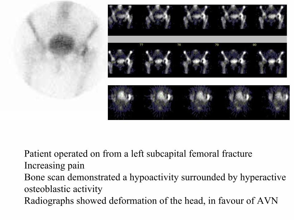
Patient operated on from a left subcapital femoral fractureIncreasing painBone scan demonstrated a hypoactivity surrounded by hyperactiveosteoblastic activityRadiographs showed deformation of the head, in favour of AVN
Secondary osteonecrosis
Induced by -high doses of corticosteroids (increase of size of marrow fat cells caused bytreatment responsible for diminution ofarterial blood flow)
-alcohol abuse (initial insult is generally a stressfracture, with induced ischaemia)
Spontaneous osteonecrosis
Joints frequently involved: femoral heads > knees> humeral heads
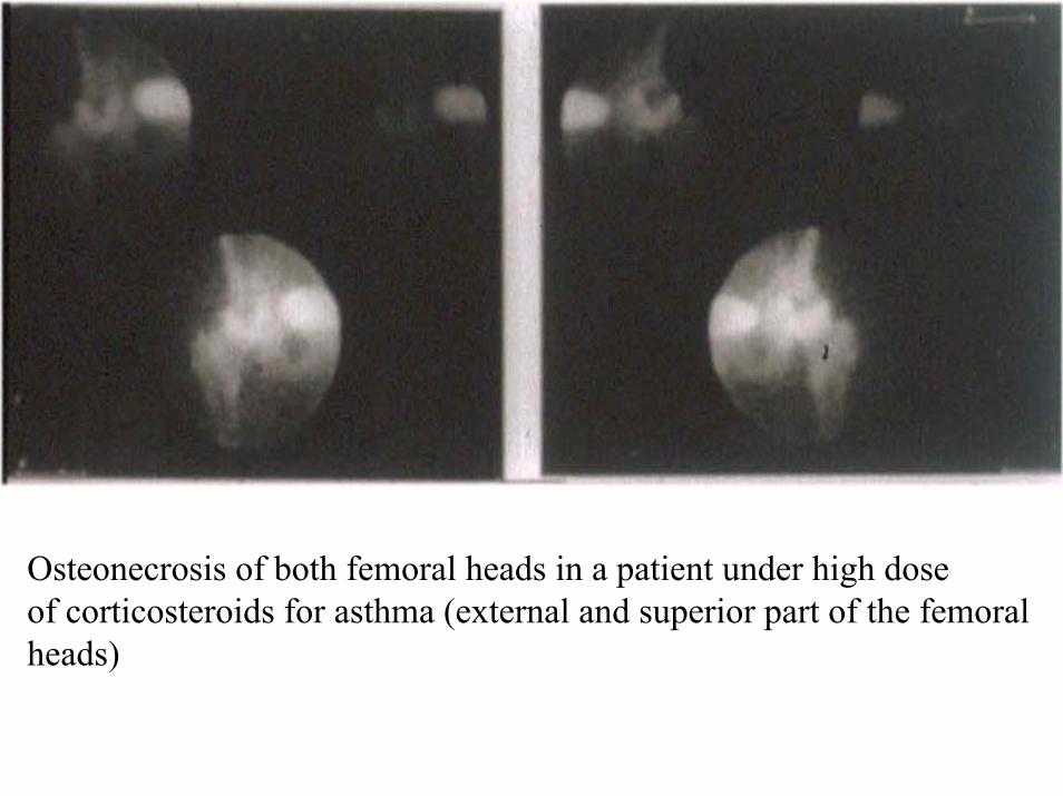
Osteonecrosis of both femoral heads in a patient under high doseof corticosteroids for asthma (external and superior part of the femoralheads)
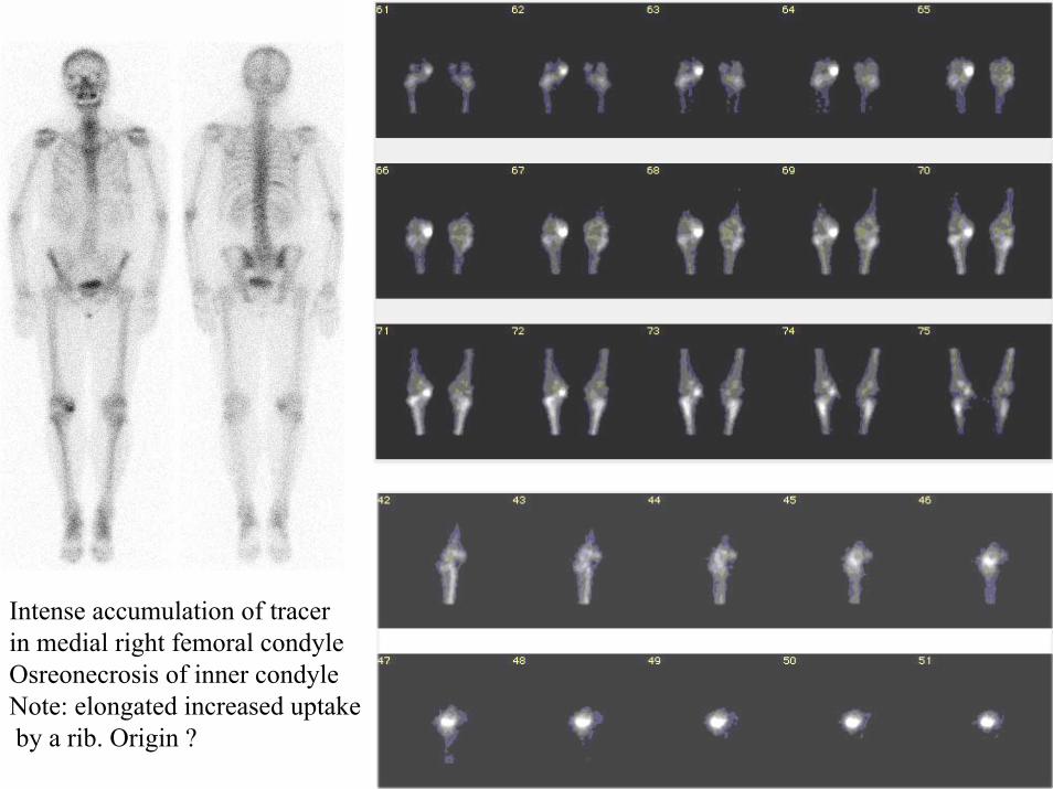
Intense accumulation of tracerin medial right femoral condyleOsreonecrosis of inner condyleNote: elongated increased uptakeby a rib. Origin ?
Sickle cell disease
Occlusion of small vessels occurs frequently causing infarction
Weight-bearing areas of long bones frequently affected
Infarction induce photopenia, followed by increased uptakeas revascularization occurs
Differential diagnosis: osteomyelitis (frequent in this disease)
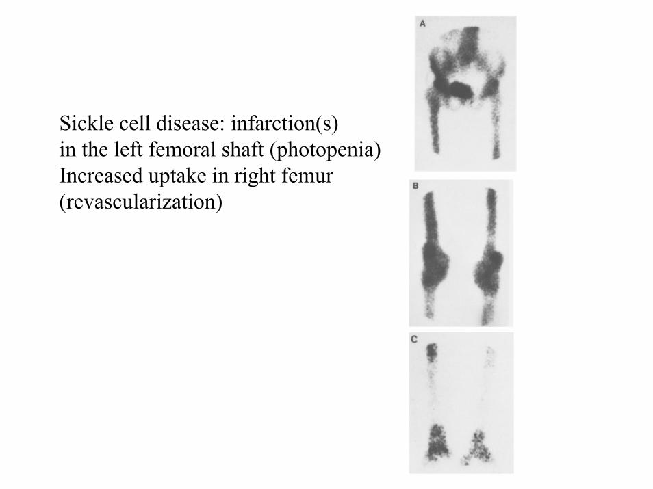
Sickle cell disease: infarction(s)in the left femoral shaft (photopenia)Increased uptake in right femur(revascularization)
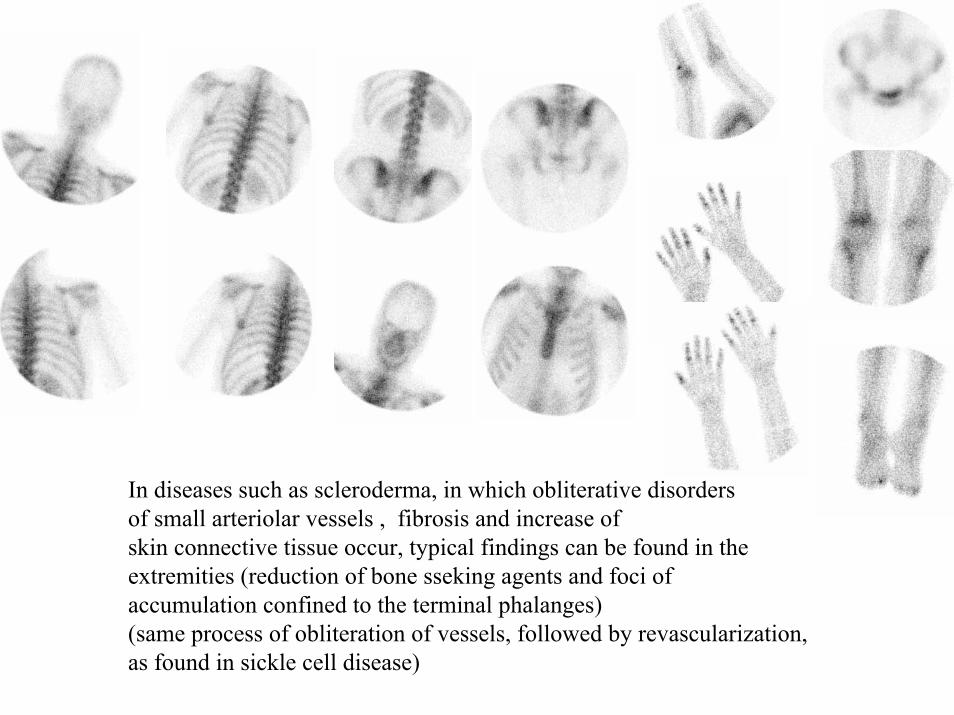
In diseases such as scleroderma, in which obliterative disordersof small arteriolar vessels , fibrosis and increase ofskin connective tissue occur, typical findings can be found in theextremities (reduction of bone sseking agents and foci of accumulation confined to the terminal phalanges)(same process of obliteration of vessels, followed by revascularization,as found in sickle cell disease)

Child presenting witha Legg Calve Perthe’sdisease of left hip, well seen in Normal position and also in frogpositionPin hole collimator
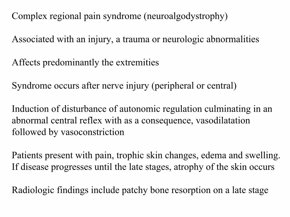
Complex regional pain syndrome (neuroalgodystrophy)
Associated with an injury, a trauma or neurologic abnormalities
Affects predominantly the extremities
Syndrome occurs after nerve injury (peripheral or central)
Induction of disturbance of autonomic regulation culminating in anabnormal central reflex with as a consequence, vasodilatationfollowed by vasoconstriction
Patients present with pain, trophic skin changes, edema and swelling.If disease progresses until the late stages, atrophy of the skin occurs
Radiologic findings include patchy bone resorption on a late stage
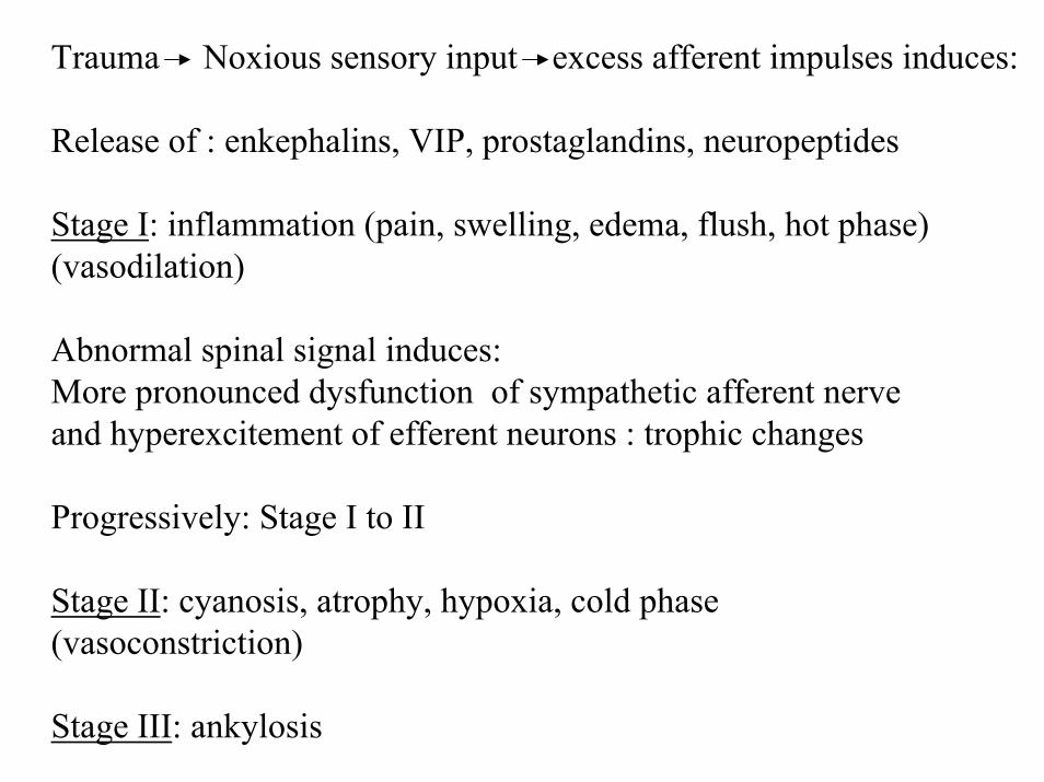
Trauma Noxious sensory input excess afferent impulses induces:
Release of : enkephalins, VIP, prostaglandins, neuropeptides
Stage I: inflammation (pain, swelling, edema, flush, hot phase)(vasodilation)
Abnormal spinal signal induces:More pronounced dysfunction of sympathetic afferent nerveand hyperexcitement of efferent neurons : trophic changes
Progressively: Stage I to II
Stage II: cyanosis, atrophy, hypoxia, cold phase(vasoconstriction)
Stage III: ankylosis
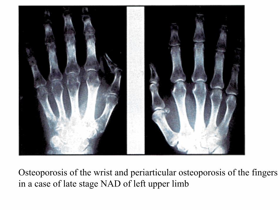
Osteoporosis of the wrist and periarticular osteoporosis of the fingersin a case of late stage NAD of left upper limb
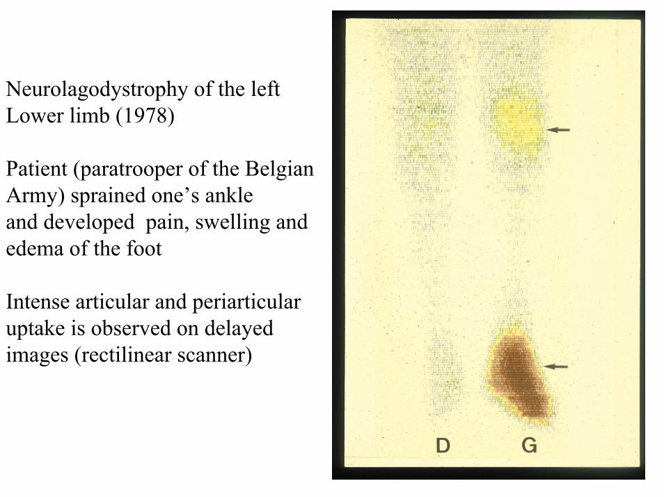
Neurolagodystrophy of the leftLower limb (1978)
Patient (paratrooper of the BelgianArmy) sprained one’s ankleand developed pain, swelling andedema of the foot
Intense articular and periarticular uptake is observed on delayed images (rectilinear scanner)
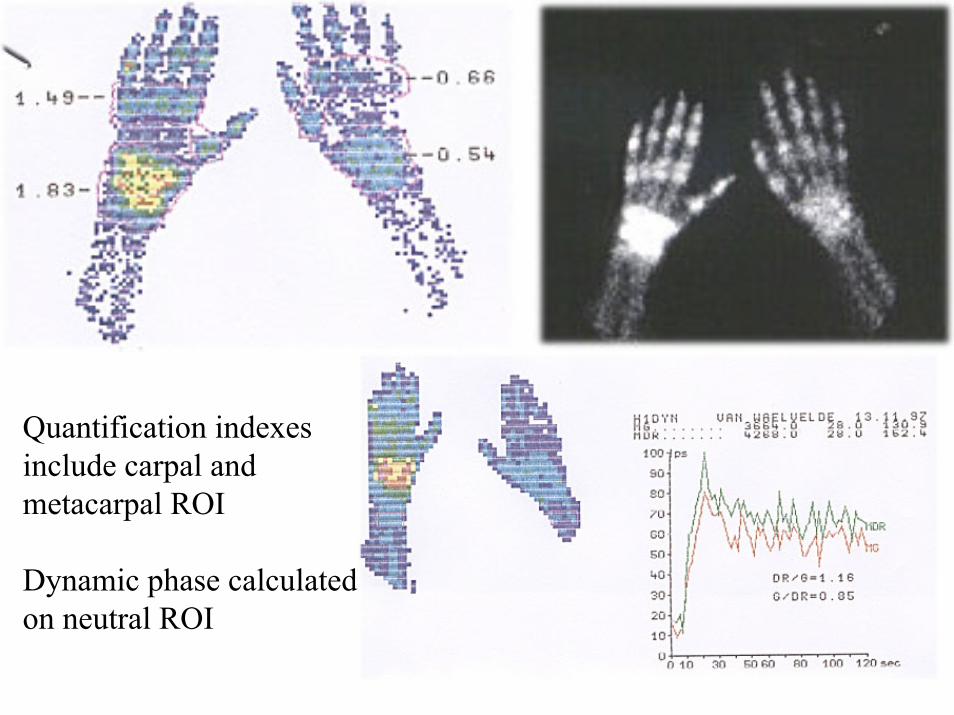
Quantification indexesinclude carpal and metacarpal ROI
Dynamic phase calculatedon neutral ROI
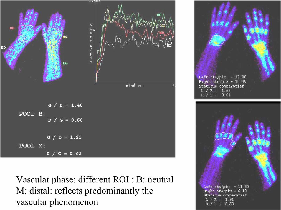
Vascular phase: different ROI : B: neutralM: distal: reflects predominantly the vascular phenomenon
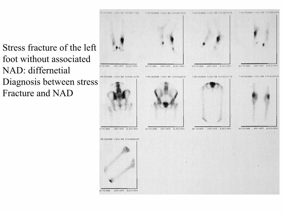
Stress fracture of the leftfoot without associatedNAD: differnetial Diagnosis between stressFracture and NAD
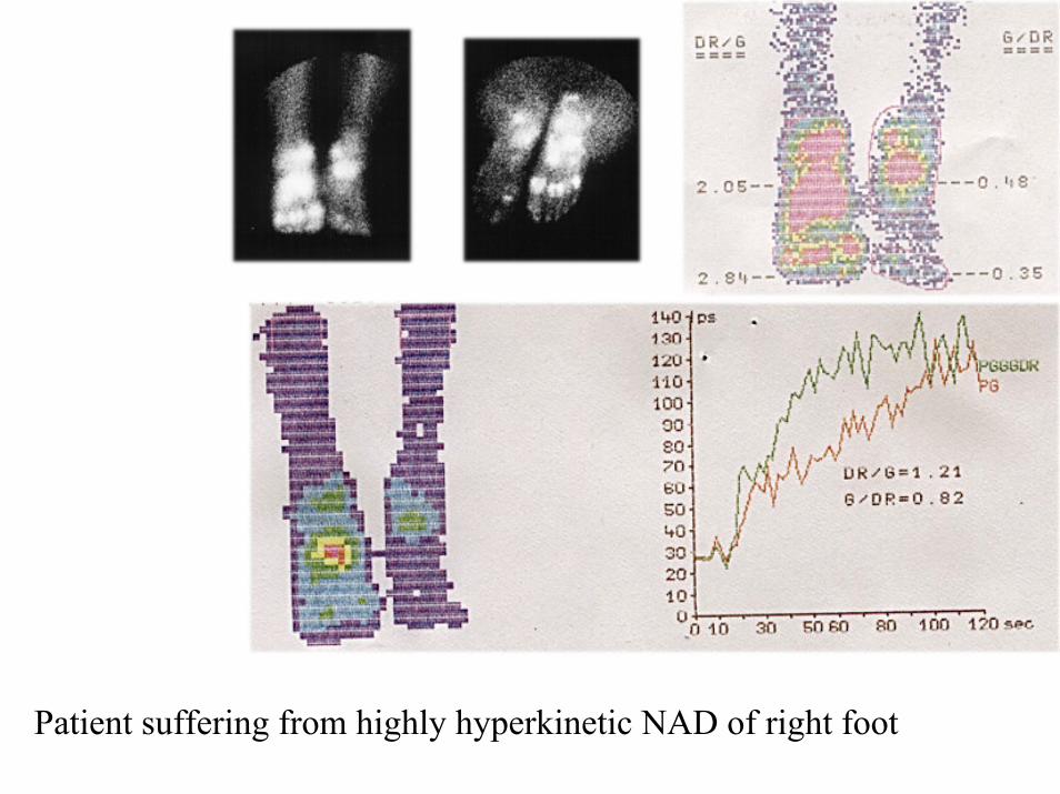
Patient suffering from highly hyperkinetic NAD of right foot
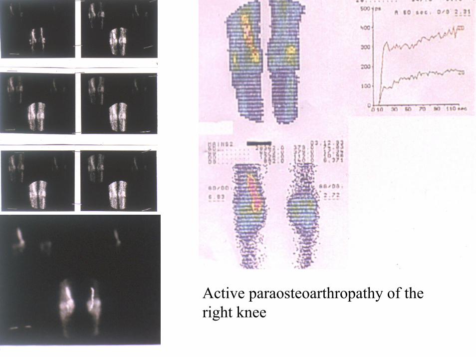
Active paraosteoarthropathy of theright knee