สััตวแพทย ์มหานครสาร · review of plant dye classification...
TRANSCRIPT
บทความวชาการ
สตวแพทยสตวแพทยมหานครสารมหานครสาร JJOOUURRNNAALL OOFF MMAAHHAANNAAKKOORRNN VVEETTEERRIINNAARRYY MMEEDDIICCIINNEE Available online: www.vet.mut.ac.th/journal_jmvm
การใชสสกดจากพชในการยอมสเนอเยอ
อารยา สบขาเพชร1,# และณฐกาญจน นายมอญ2
1ภาควชากายวภาคศาสตร คณะสตวแพทยศาสตร มหาวทยาลยเทคโนโลยมหานคร กรงเทพฯ 10530
2ภาควชาเทคนคการสตวแพทย คณะเทคนคการสตวแพทย มหาวทยาลยเกษตรศาสตร กรงเทพฯ 10900
บทคดยอ: ปจจบนการศกษาทางจลกายวภาคศาสตรไดมการใชสยอมมากมายหลายชนดเพอยอมเนอเยอตางๆ ซงขนกบวตถประสงคในการศกษา สทใชยอมเนอเยอโดยทวไปมแหลงทมาทงจากแหลงธรรมชาตและจากการสงเคราะห ดวยในปจจบนทวโลกใหความสาคญในการใชสารตางๆ ทไมมผลกระทบตอสขภาพของมนษยและเปนมตรกบสงแวดลอม การใชสธรรมชาตจากพชในการยอมเนอเยอจงไดรบความสนใจมากขน เนองจากสวนตางๆ ของพชนนมสวนประกอบของสอยมากมาย อกทงยงมราคาถกกวาและสามารถใชงานไดอยางยงยนกวาสทไดจากการสงเคราะห รายงานฉบบนไดทวนสรปโดยยอเกยวกบการแบงชนดของสธรรมชาตจากพชในแงของโครงสรางทางเคมของส วธการสกด มอรแดนท และไดยกตวอยางงานวจยทศกษาการใชสสกดจากพชในการยอมเนอเยอชนดตางๆ ซงอาจนามาใชเปนขอมลพนฐานและเปนทางเลอกในการศกษาและพฒนาการยอมสเนอเยอจากสทสกดจากพช
คาสาคญ: สยอมจากพช สยอมจากธรรมชาต การยอม เนอเยอสตว #ผรบผดชอบบทความ สตวแพทยมหานครสาร. 2557. 9(1): 63-78. E-mail address: [email protected]
Araya Suebkhampet and Nattakarn Naimon / J. Mahanakorn Vet. Med. 2014. 9(1): 63-78.
64
Using Dye Plant Extract for Histological Staining
Araya Suebkhampet1,# and Nattakarn Naimon2
1Department of Anatomy, Faculty of Veterinary Medicine, Mahanakorn University of Technology, Bangkok 10530, THAILAND
2Department of Veterinary Technology, Faculty of Veterinary Technology, Kasetsart University, Bangkok 10900, THAILAND
Abstract: Nowadays, many and various kinds of dyes are used for histological staining due to the purpose of study. They are generally from both natural and synthetic origins. With the worldwide concern over the use of human health and eco-friendly materials, the use of natural dyes from plants has gain interested since most of them contain plenty of dye from their parts. They are also cheaper and more sustainable than the synthetic dyes. A brief review of plant dye classification based on their chemical structures of major dye pigments, methods of plant dye extraction, mordants and some of its application in diverse tissues are presented in this report. These would be helpful to further study and development of alternative tissue stains from plant natural dyes for tissue staining.
Keywords: Plant dye, Natural dye, Staining, Animal histology #Corresponding author J. Mahanakorn Vet. Med. 2014. 9(1): 63-78. E-mail address: [email protected]
Introduction
Histology is the study of cells and tissues by using a light microscope. The ability to visualize or identify histological structures is frequently enhanced through the use of histological stains. They give contrast to the tissue as well as highlighting particular features of interest. There are two types of dyes that classified by their origins, synthetic and natural dyes. The natural dyes
are obtained from natural sources such as plants, insects, animals, clays and minerals (Carleton et al., 1976). Plants and insects known to be used in histological staining for animal tissues are Haematoxylon campechianum (logwood), from which haematoxylin stain is obtained and Dactylopius cacti, from which carmine stain is obtained (Egbujo et al., 2008), respectively. The majority of natural dyes
Araya Suebkhampet and Nattakarn Naimon / J. Mahanakorn Vet. Med. 2014. 9(1): 63-78.
65
are extracted from plant parts such as roots, barks, leaves, berries and seeds and wood.
The dyeing with natural dyes was one of the oldest techniques practiced by the ancient people such as wall paintings in the caves or textile dyeing. The first use of dye in histology credited to Antonie van Leeuwenhoek, the father of microbiology who worked with saffron, a natural dye extracted from saffron crocus. In the mid 1800s, amateur microscopists first used haematoxylin to stain cellular components (Titford M., 2005). Later scientists developed a wide range of haematoxylin staining techniques. The systemically studied dye for histology started in the second half of the 19th century by Weigert C, J Gerlach, P Ehrlich and H Gierke. By then coloring materials were mostly still natural origin like carmine, cochineal, haematoxylin and indigo. Gradually the use of the natural dyes has decreased due to the introduction of synthetic dyes (aniline dye from extract of coal tar) which first invention by William Henry Perkin in 1856. They widely applied in many fields as food, cosmetic and textile industries (Prabhu and Bhute, 2012). They are rapid stain with vast range of new colors, easy to use and commercially available. From then, the synthetic dyes together with those for histological staining have developed.
Many dyes, techniques and procedures are utilized to stain various types of animal tissues. At present, haematoxylin and eosin stain (H&E) is widely used as a light microscopical stain in histology and histopathology. It is combination of natural haematoxylin stain and the synthetic eosin stain. Haematoxylin stains the cell nuclei dark blue while eosin stains cell cytoplasm, most connective tissue fibers and matrices in varying shades of pink and red. Haematoxylin obtained from logwood tree that is native to the Mexico and northern Central America (Baker & Silverton, 1976), but is now mainly cultivated in the West Indies (Bancrot and Gamble, 2008). Haematoxylin can be prepared in numerous ways and applied to tissues from different sites. It has to be oxidized to haematein before using (Culling, 1974). Haematein can be produced from haematoxylin in two ways: natural oxidation and chemical oxidation. For natural oxidation, haematoxylin is exposed to light and air (Ehrlich’s and Delafield’s haematoxylin solution), and for chemical oxidation, sodium iodate (e.g. Mayer’s haematoxylin) or mercuric oxide (e.g. Harris’s haematoxylin) are used. Natural oxidation of haematoxylin seems to retain its staining ability longer than that of the chemical oxidation. However, it takes much longer time for preparation. Haematoxylin has also been
Araya Suebkhampet and Nattakarn Naimon / J. Mahanakorn Vet. Med. 2014. 9(1): 63-78.
66
prepared synthetically (Morsingh and Robinson, 1970) but the natural dye is mostly used. Haematein is anionic charge, having a poor affinity for tissues without the presence of mordant. A mordant is a substance used to set dyes on tissue sections. The common mordants for haematoxylin are aluminium, iron and tungsten. The dye-mordant complex possesses a net positive charge and enables to bind to anionic tissue regions including nuclear chromatin. Mordant is regularly included in the dyeing protocols when natural dyes are used in order to fix or intensify the stains in cell or tissue staining. It is mainly a polyvalent metal ion that forms coordination complexes with certain dyes which then attaches to the tissues (Llewellyn, 2005). Different kinds of mordants give a different hue and stability of the dye in the tissues.
The use of natural dyes which are cheaper, eco-friendly and biodegradable has become a matter of significant important due to the increased environmental awareness and in order to avoid some hazardous synthetic dyes to humans and animals (Eom, et al., 2001). Several synthetic dyes (e.g. dyes with azo bonds nitro- or amino-groups) cause allergic-like symptoms or are carcinogenic (Ratna and Padhi, 2012). Therefore, alternative natural dyes have been currently more interested for their
potential use in various purposes. It is interesting that over 2000 dyes are synthesized from various parts of plants, of which only about 150 have been commercially exploited (Gulrajani, 1992). They are used for applying to different substrates such as textiles, paper, leather, wood, hair etc. They are also widely used in cosmetic, food and pharmaceutical industries. A number of them own the pharmaceutical properties and have been used in traditional medicine. Curcumin, the bright yellow dye of turmeric has been used traditionally as a remedy for stomach and liver ailments, as well as topically to heal sores and revitalizes the skin (Chaturvedi, 2009). It has anti-oxidant, anti-inflammatory, anti-cancer and anti-septic properties (Chengaiah et al., 2010). Many other common natural dyes such as lycopene (red dye) in tomatoes or lawsone (varies in color from orange to reddish brown) in henna are reported as antimicrobial agents (Siva, 2007; Habbal et al., 2011).
In last 10 years, natural dyes have been studied for their potential use in various fields including histological staining. The present report describes the basic information about of plant dye classification based on their chemical structures, methods of the dye extraction, mordants and some of its application for tissue staining. These would be useful information to the
Araya Suebkhampet and Nattakarn Naimon / J. Mahanakorn Vet. Med. 2014. 9(1): 63-78.
67
researchers who are searching for the new natural dyes for tissue staining. Classification of natural dyes from plants
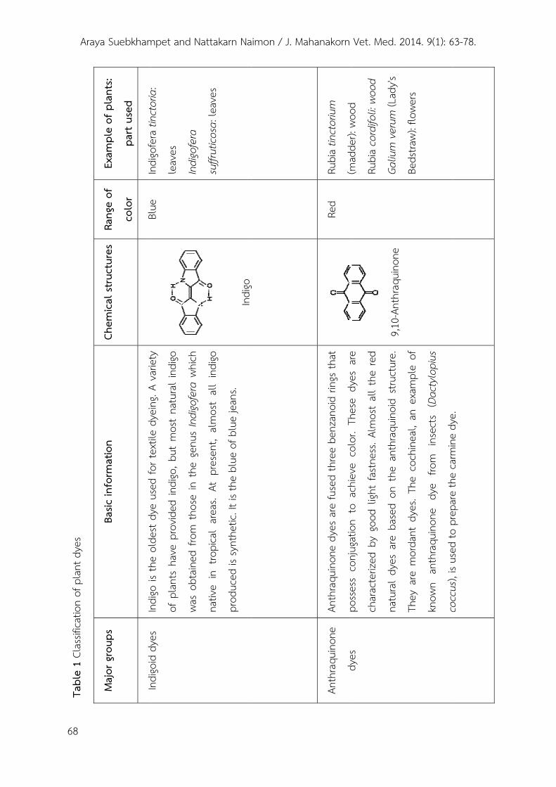
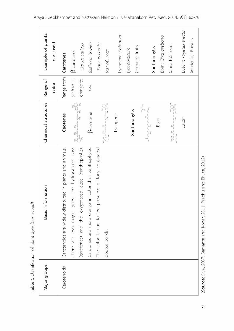
Natural dyes can be classified in different ways such as based on their chemical structure, source, hue and method of application, etc. The plant dye classification described in this review is based on their chemical structures which are divided into 7 major groups (Siva, 2007; Samanta and Konar, 2011; Prabhu and Bhute, 2012) as presented in Table1.
Methods of plant dye extraction Various parts of plants such as roots,
stems, leaves, flowers, fruits and seeds are used for dye extraction. Some plants may have more than one color depending on which part of the plant one uses. The color yield and shade of the color a plant produces vary according to time of the year the plant is picked, how it is grown, soil conditions, etc. Before extraction, the parts of plants are collected and generally shade dried in air or sun dried. Then the grinding is carried out to break down the material into very small pieces or powder using the manual or electric grinding machines. Optimum conditions of extraction are determined by varying extraction parameters such as type of solvents, time of extraction, ratio between plant material and solvent,
temperature and pH which depend on the properties of particular dye components (Prabhu and Bhute, 2012). After extraction, the extracts are generally filtered through various filters such as cheesecloth, cotton wool or paper filter. The filtrates may be freshly used (normally aqueous extract) or further evaporated of solvent, washing and drying to get purified dye. There are mainly four methods used in extraction of natural dyes (Samanta and Konar, 2011). 1) Aqueous extraction
This method is has long been used for natural dye extraction for a certain of time. The dye components in dried plant powder are extracted in water at boil or particular temperature. Then, the extract is cooled down and filtered. The dye solution is carried out under varying conditions as mentioned above in order to get the optimal extraction condition. The optimum condition is determined by studying the optical density value at definite wavelength for the extracted solution using UV-Visible absorbance spectrophotometer (Samanta and Konar, 2011). The filtrate is further applied for dyeing. Here are some studies that used the aqueous extract from plants for animal tissue staining. Natural dyes were extracted from kujarat flowers (Hibiscus sadariffa) in aqueous medium and were used to replace of eosin stain in kidney from
Araya
68
Tabl
e 1
Clas
sifica
tion
of p
lant
dye
s
a Suebkham
Exam
ple
of p
lant
s:
part
used
Indi
gofe
ratin
ctor
ia
Rang
e of
colo
r
Blue
Chem
ical s
truct
ures
Ba
sic in
form
atio
n
Indi
gois
the
olde
stdy
eus
edfo
rtex
tile
dyein
gA
varie
ty
Maj
or g
roup
s
Indi
goid
dyes
mpet and N
Indi
gofe
ra ti
ncto
ria:
leav
es
Indi
gofe
ra
Blue
In
digo
is th
e ol
dest
dye
used
for t
extil
e dy
eing.
A va
riety
of p
lant
s ha
ve p
rovid
ed in
digo
, but
mos
t na
tura
l ind
igo
was
obta
ined
from
tho
se in
the
gen
us In
digo
fera
whi
ch
Indi
goid
dye
s
attakarn Na
suffr
utico
sa: l
eave
s
In
digo
nativ
e in
tro
pica
l ar
eas.
At p
rese
nt,
alm
ost
all
indi
go
prod
uced
is sy
nthe
tic. It
is th
e bl
ue o
f blu
e jea
ns.
aimon / J. M
g
Mahanakorn
Rubi
a tin
ctor
ium
(mad
der):
wood
Red
Anth
raqu
inon
e dy
es a
re fu
sed
thre
e be
nzan
oid
rings
that
poss
ess
conj
ugat
ion
toac
hiev
eco
lor
Thes
edy
esar
e
Anth
raqu
inon
e
dyes
Vet. Med. 2
(mad
der):
woo
d
Rubi
a co
rdifo
li: wo
od
Galiu
m v
erum
(Lad
y's
9,1
0-An
thra
quin
one
poss
ess
conj
ugat
ion
to a
chiev
e co
lor.
Thes
e dy
es a
re
char
acte
rized
by
good
ligh
t fa
stnes
s. Al
mos
t al
l the
red
natu
ral
dyes
are
bas
ed o
n th
e an
thra
quin
oid
struc
ture
.
dyes
2014. 9(1): 6
Beds
traw)
: flo
wers
They
are
mor
dant
dye
s. Th
e co
chin
eal,
an e
xam
ple
of
know
n an
thra
quin
one
dye
from
in
sect
s (D
acty
lopi
us
cocc
us)
isus
edto
prep
are
the
carm
inedy
e
63-78.
cocc
us),
is us
ed to
pre
pare
the
carm
ine d
ye.
Araya
Tabl
e 1
Clas
sifica
tion
of p
lant
dye
s (Co
ntin
ued)
a Suebkham
Exam
ple
of p
lant
s:
part
used
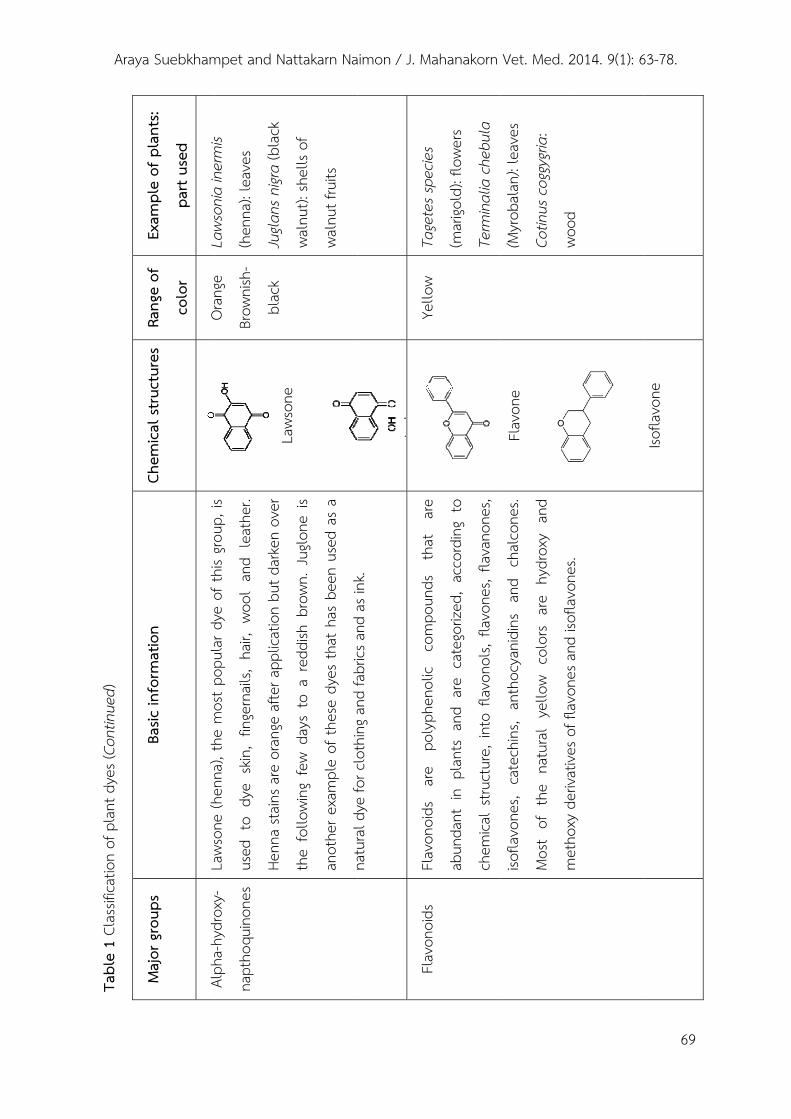
Laws
onia
iner
mis
Rang
e of
colo
r
Oran
ge
Chem
ical s
truct
ures
Ba
sic in
form
atio
n
Laws
one
(hen
na)
the
mos
tpop
ular
dye
ofth
isgro
upis
Maj
or g
roup
s
Alph
ahy
drox
y
mpet and N
Laws
onia
iner
mis
(hen
na):
leav
es
Jugla
ns n
igra
(bla
ck
Oran
ge
Brow
nish
-
blac
k
Laws
one
Laws
one
(hen
na),
the
mos
t pop
ular
dye
of t
his g
roup
, is
used
to
dye
skin
, fin
gern
ails,
hair,
wool
and
lea
ther
.
Henn
a sta
ins a
re o
rang
e af
ter a
pplic
atio
n bu
t dar
ken
over
Alph
a-hy
drox
y-
napt
hoqu
inon
es
attakarn Na
waln
ut):
shel
ls of
waln
ut fr
uits
Laws
one
the
follo
wing
few
day
s to
a r
eddi
sh b
rown
. Jug
lone
is
anot
her e
xam
ple
of th
ese
dyes
that
has
bee
n us
ed a
s a
natu
rald
yefo
rclo
thin
gand
fabr
icsan
das
ink
aimon / J. M
Tage
tess
pecie
sYe
llow
J
l
natu
ral d
ye fo
r clo
thin
g and
fabr
ics a
nd a
s ink
.
Flav
onoi
dsar
epo
lyph
enol
icco
mpo
unds
that
are
Flav
onoi
ds
Mahanakorn
Tage
tes s
pecie
s
(mar
igold
): flo
wers
Term
inal
ia c
hebu
la
Yello
w Fl
avon
oids
ar
e po
lyph
enol
ic co
mpo
unds
th
at
are
abun
dant
in
plan
ts an
d ar
e ca
tego
rized
, ac
cord
ing
to
chem
ical
struc
ture
, int
o fla
vono
ls, f
lavo
nes,
flava
none
s,
Flav
onoi
ds
Vet. Med. 2
(Myr
obal
an):
leav
es
Cotin
us c
oggy
gria:
wood
Fl
avon
e iso
flavo
nes,
cate
chin
s, an
thoc
yani
dins
and
cha
lcone
s.
Mos
t of
the
nat
ural
yel
low
colo
rs ar
e hy
drox
y an
d
met
hoxy
deriv
ative
soff
lavo
nesa
ndiso
flavo
nes
2014. 9(1): 6
wood
met
hoxy
der
ivativ
es o
f fla
vone
s and
isof
lavo
nes.
63-78.
69
Isofla
vone
Araya
70
Tabl
e 1
Clas
sifica
tion
of p
lant
dye
s (Co
ntin
ued)
a Suebkham
Exam
ple
of p
lant
s:
part
used
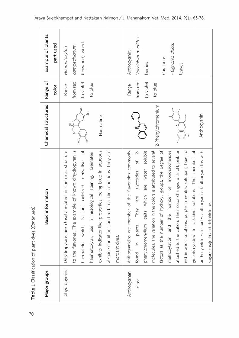
Haem
atox
ylon
Rang
e of
colo
r
Rang
e
Chem
ical s
truct
ures
Ba
sic in
form
atio
n
Dihy
drop
yran
sar
eclo
sely
rela
ted
inch
emica
lstr
uctu
re
Maj
or g
roup
s
Dihy
drop
yran
s
mpet and N
Haem
atox
ylon
cam
pech
ianu
m
(logw
ood)
: woo
d
Rang
e
from
red
to v
iole
t
Dihy
drop
yran
s ar
e clo
sely
rel
ated
in c
hem
ical
struc
ture
to t
he fl
avon
es. T
he e
xam
ple
of k
nown
dih
ydro
pyra
n is
haem
atein
wh
ich
is an
ox
idize
d de
rivat
ive
of
Dihy
drop
yran
s
attakarn Na
to b
lue
Ha
emat
ine
haem
atox
ylin,
use
in
histo
logic
al s
tain
ing.
Haem
atein
exhi
bits
indi
cato
r-like
pro
perti
es, b
eing
blue
in a
queo
us
alka
line
cond
itions
and
red
inac
idic
cond
itions
They
are
aimon / J. M
Anth
ocya
nin:
Rang
e
al
kalin
e co
nditio
ns, a
nd re
d in
acid
ic co
nditio
ns. T
hey
are
mor
dant
dye
s.
Anth
ocya
nidi
nsar
em
embe
rof
the
flavo
noid
sco
mm
only
Anth
ocya
nani
Mahanakorn
Anth
ocya
nin:
Vacc
iniu
m m
yrtil
lus:
berri
es
Rang
e
from
red
to v
iole
t
Anth
ocya
nidi
ns a
re m
embe
r of
the
fla
vono
ids
com
mon
ly
foun
d in
plan
ts.
They
ar
e gly
cosid
es
of
2-
phen
ylch
rom
enyl
ium
salts
wh
ich
are
wate
r so
lubl
e
Anth
ocya
nani
dins
:
Vet. Med. 2
Cara
jurin
:
- Bign
onia
chica
:
to b
lue
2-
Phen
ylch
rom
eniu
m
mol
ecul
es. T
he v
ariat
ion
in th
e co
lors
is at
tribu
ted
to se
vera
l
fact
ors
as t
he n
umbe
r of
hyd
roxy
l gro
ups,
the
degre
e of
met
hoxy
latio
n an
d th
e nu
mbe
r of
m
onos
acch
arid
es
2014. 9(1): 6
Bign
onia
chic
a:
leav
es
atta
ched
to th
e ca
tion.
The
ir co
lor c
hang
es w
ith p
H, p
ink
or
red
in a
cidic
solu
tions
, pur
ple
in n
eutra
l sol
utio
ns, b
lue
to
green
ish-y
ello
win
alka
line
solu
tions
The
mem
ber
of
63-78.
An
thoc
yani
n gre
enish
yello
w in
al
kalin
e so
lutio
ns.
The
mem
ber
of
anth
ocya
nidi
nes
inclu
des
anth
ocya
nins
(ant
hocy
anid
ins
with
suga
r), c
araju
rin a
nd d
elph
inid
ine.
Araya Suebkhampet and Nattakarn Naimon / J. Mahanakorn Vet. Med. 2014. 9(1): 63-78.
72
albino mice (Hashim, 2006). Suebkhumpet and Sotthibandhu (2012) used aqueous extract of butterfly pea flowers (Clitoria ternatea) to stain animal peripheral blood smears. 2) Extraction by non-aqueous and other solvent assisted system
The solvents generally used for plant dye extraction are ethanol, methanol, acetone, chloroform, ether, clove oil, etc. The dried material powder is weighed and soaked in solvent in different percentages and time durations. The crude dye extract can be used for tissue staining after extraction or can be further applied for solvent evaporation in order to concentrate the dye solution before staining. Alternatively, the dried plant powder is soaked in solvent to allow effective percolation, then the soaked powder is extracted in the solvent using Soxhlet Extractor (Steam Heated Extractor). The extract is then concentrated using rotary evaporator and may further drying in the drying oven (Okolie. 2008). Finally, the extract is obtained in powdered form which will be dissolved in the solvent or buffer at the desire concentrations before tissue staining.
Supercritical fluid extraction (SFE), an alternative to conventional solvent extraction for separation of organic
compound in many analytical processes as well as extraction of plant natural dye has gained wide acceptance in recent years since this technique is safe for health, inexpensive and harmonize with nature. SFE uses carbon dioxide as a solvent. This method is based on the enhanced solvating power of gases above their critical point (Samanta and Konar, 2011). Cardoni et al. (2000) studied the extraction of lycopene
and β-carotene from ripe tomatoes using SFE. The detail information and procedure of this method has been reported by Sapkale et al. (2010).
3) Extraction by acid or alkaline assisted system This method adjusts the pH of the extraction by adding hydrochloric acid or sodium carbonate in aqueous medium. The different pH in extraction protocols may give differently results as percent yield and hue of the extracted dye which due to the reaction of the chemical structure of the dye to the pH variation. Samanta et al, 2007 studied the extraction of color from jackfruit wood under various pH conditions (ranged between pH 4-12) and reported that the optimum condition for the extraction is at pH 11.0. 4) Extraction by other methods
4.1) Ultrasound assisted extraction
Araya Suebkhampet and Nattakarn Naimon / J. Mahanakorn Vet. Med. 2014. 9(1): 63-78.
73
This method is carried out by mixing dried and ground plant materials in solvent in a flask, which was then placed in an ultrasonic bath. The ultrasound applied for the extraction results in intermolecular tearing and surface scrubbing, causing plant tissue rupture and improving the release of intracellular substances into the solvent. The extraction is repeated two-three times before the extract is collected. Sivakumar et al. (2009) used ultrasound with 80W ultrasonic power for 3h contact time assisted enhancement in natural dye extraction from beetroot for industrial applications and natural dyeing of leather.
4.2) Enzyme assisted extraction This method uses enzymes (pectinase,
cellulose, protease, esterase etc.) for dye extraction. The enzymes are usually applied as a pre-treatment of plant materials before subjecting the plant material to solvent extraction. Various enzyme combinations are used to loosen the structural integrity of plant materials thereby enhancing the extraction of the desired color components. Conditions for optimum activity and selection of the right type of enzymes are essential to use them effectively for extraction (Sowbhagya and Chitra, 2010). Ultrasound assisted and enzyme assisted extraction can be used in combination in order to improve the extraction efficiency.
Mordants Natural dyes are mostly non-
substantive and must be applied by adding mordants which after combining with dye in the materials including the tissues, it forms an insoluble precipitate or lake and thus both the dye and mordant get fixed to become wash and light fast. Mordants are also often added to keep dyes from fading, or to brighten, deepen, or dull a color. They can be used before, during or after the dye bath due to the desired effect. There are different kinds of mordant as metallic mordants, tannin and oil mordants that have been generally used for fabric dyeing by natural dye for a long time. Similarly, they are also added in the histological staining protocols when the plant dyes are used (Al Tikritti and Walker, 1977; Avwioro et al., 2007). The metallic mordants are common used for tissue staining.
Metalic mordants are polyvalent metal ions such as metal salts of aluminium, potassium, chromium, iron, copper and tin. The two most common metals used in natural dyeing are aluminium and ferric ions that having valences of three. Two types of bonds are involved in the reaction between a mordant dye and a mordant. One is covalent bond and the other is coordinate bond. Different mordants give different hue colors with the same dye. Metalic mordants are divided into two types as brightening
Araya Suebkhampet and Nattakarn Naimon / J. Mahanakorn Vet. Med. 2014. 9(1): 63-78.
74
and dulling mordants. Alum (potassium aluminum sulfate), potassium dichromate and stannous chloride are brightening mordants while copper sulphate and ferrous sulfate are dulling mordants Prabhu and Bhute, 2012). Although metallic mordants are effective, they are relatively toxic and environmentally pollutants. Thus, the natural mordants are considered. The sample of natural mordant is tannin which found in plant parts as Tara pods (Caesalpinia spinosa) and gallnuts from Rhus semialata. Ali et al. (2010) studied the effect of tannic acid and metallic mordants on the dyeing properties of natural dye extracted from Acacia nilotica bark.
Discussion and Conclusion The natural dyes have long been used
in various purposes including tissue staining. However, their application has reduced since the synthetic dyes were developed. Haematoxylin is an example of natural dye that widely used in histology, histopathology and histochemistry. At present, many commercial synthetic and some natural dyes used for tissue staining are available in the markets. However, the hazardous effects of synthetic dyes on human and environment caused scientists to concern about using the natural dyes instead of synthetic dyes. Therefore, alternative natural dyes have been studied for their potential use in
histological staining. There are many studies that investigated plant dye staining in diverse tissues as follow. The dried skin of pomegranate (Punica Granatum) fruit extract was used for rat brain staining and the yellow staining was detected in the astrocytes, neurons, red blood cells and elastic fibers (Gharravi et al., 2006). Bassey et al. (2011) stained the sections of the testes with the ethanolic extract of Curcuma longa. They used this extract as a counterstain for haematoxylin. Their results indicated that the dye distinctly stained the seminiferous epithelium and interstitium yellow that provided a good counter stain for haematoxylin. Kola nut (cola acuminata) extract was also used as a substitute to histological tissue stain eosin in rat tissues. The dye extract was detected in the cytoplasm of various tissues with yellow-brown color (Shehu et al., 2012). Suabjakyong et al. (2011) investigated the extraction of dye from black plum fruit (Syzygium cumini) using various solvents and its staining property on the rat hepatic tissue.
Suebkhumpet and Sotthibandhu (2012) used aqueous extract of butterfly pea flowers (Clitoria ternatea) to stain blood cells in different animal peripheral blood smears. They presented that the dye extract could stain and differentiate blood cells on the blood smears. Jan et al. (2011) studied
Araya Suebkhampet and Nattakarn Naimon / J. Mahanakorn Vet. Med. 2014. 9(1): 63-78.
75
the staining effect of dry extracted from dry leaves of henna (Lawsonia intermis) on histological sections of angiospermic stem. Okolie (2008) studied the staining of ova of intestinal parasites with extracts of Hibiscus Sabdariffa and Azadirachta Indica. The plant extract produced satisfactory staining in comparison to conventional methods used in identifying intestinal parasites on a preparation of stool sample. Ihuma et al. (2012) used methanolic extracts from Hisbiscus sabdariffa as a biological staining agent for some fungal species. Their results suggested that the dye extract could be use as a mycological stain. Al-Amura et al. (2012) studied the staining technique for helminthes by using red beet (Beta vulgaris) extract. They reported that stained helminthes were acquired a good stain with distinction their internal structures. Tousson and AL-Behbehani (2011) used black mulberries as a natural dye for tissue staining.
Although using natural dyes have many advantages, there still be some limitations that should be concerned. They are difficult to standardize the dye and its application because dyes collected from similar plants or natural sources vary due to climate, soil, maturity period and cultivation methods etc. Moreover, most natural dyes require mordants for fixing them on the stained tissues. The widely used metallic
mordants that applied in the dye solutions may cause health and disposal problems. Therefore, searching for the new natural mordants is an option for safer tissue staining. Many factors should be considered, if the natural dyes are used for tissue staining. One of them is the source of plants selected, it should be available in the endemic region in order to reduce the cost and to avoid raw materials insufficiency. The known chemical structures and properties of some plant dye extracts are also important and useful for determining the optimal extraction and tissue staining procedures. More detailed studies are needed to evaluate the potential and availability of natural dyes-yielding resources in many others plants. In addition, biotechnology and various fields of studies are required to increase the quantity and improve quality of plant natural dyes. More study about staining procedures of each dye plant still be required to establish new permanent preparations in the future.
References Al-Amura, M. F.A., Hassen, Z.A. and AL-
Mhanawi, B.H. 2012. Staining technique for helminth parasites by use red beet (Beta Vulgaris L.) extract. Bas. J. Vet. Res. 11(1): 283-292.
Ali, A., Ali, S., Saleem, H. and Hussain, T. 2010. Effect of tannic and metallic
Araya Suebkhampet and Nattakarn Naimon / J. Mahanakorn Vet. Med. 2014. 9(1): 63-78.
76
mordants on the dyeing properties of natural dye extracted from Acacia nilotica bark. Asian J. Chem. 22(9): 7065-7069.
Al-Tikritti, S.A. and Walker, F. 1977. Anthocyanin BB: anuclear stain substitute for haematoxylin. J. Clin. Pathol. 31: 194-196.
Avwioro, O.G., Onwuka, S.K., Moody, J.O., Agbedahunsi, J.M., Oduola, T., Ekpo, O.E. and Oladele, A.A. 2007. Curcuma longa extract as a histological dye for collagen fibres and red blood cells. J. Anat. 210: 600–603.
Baker, F.J. and Silverton, R.E. 1976. The theory of staining: Introduction to Medical Laboratory Technology. 6th edition. Butterworths. 385-391p.
Bancrot, J.D. and Gamble, M. 2008. Theory and practice of histological techniques. 6th edition. Churchild Vivingstone Elsevier. 725p.
Bassey, R.B., Oremosu, A.A. and Osinubi, A. A.A. 2011. Curcuma Longa: Staining effect on histomorphology of the testis. Maced. J. Med. Sci. http://dx.doi.org/10.3889/MJMS.1857-5773.2011.0209.
Carleton, H.M., Drumy, R.A.B., Wallington, E.A. and Cameron R. 1976. Histological technique. 4th edition. Oxford University Press. London 108p.
Chaturvedi, T.P. 2009. Uses of turmeric in dentistry: an update. Indian J. Dent. Res. 20(1): 107–109.
Chengaiah, B., Rao, K.M., Kumar, K.M., Alagusundaram,M. and Chetty, C.M. 2010. Medicinal important of natural dyes-a review. Int. J. Pharm. Tech. Res. 2(1): 144-154.
Culling, C.F.A. 1974. Handbook of histopathological and histochemical techniques. 3rd edition. London: Butterworth. 712p.
Egbujo E. C., Adisa O. J. and Yahaya, A. B 2008. A study of the staining effect of Roselle (Hibiscus sabdariffa) on the histologic section of the testis. Int. J. Morphol. 26(4): 927-930.
Eom, S., Shin, D., and Yoon, K. 2001. Improving the dyeability of natural colourants on cotton by cationization. Ind. J. Fibre Text Res. 26:425–431.
Gharravi, A.M., Golarlipour, M.J., Ghorbani, R. and Khazaei, M. 2006. Natural dye for staining astrocytes and neurons. J. Neuro. Sci. (Turkisk) 23(3): 215-218.
Gulrajani, M.L. 1992. Dyeing properties of natural dyes extracted from eucalyptus. J. Textile I. 98: 431-437.
Habbal, O., Hasson, S.S., El-Hag, A.H., Al-Mahrooqi, Z., Al-Hashmi, N., Al-Bimani, Z., Al-Balushi, M.S. and Al-Jabri, A.A. 2011. Antibacterial activity of Lawsonia inermis Linn (Henna) against
Araya Suebkhampet and Nattakarn Naimon / J. Mahanakorn Vet. Med. 2014. 9(1): 63-78.
77
Pseudomonas aeruginosa. Asian Pac. J. Trop. Biomed. 1(3): 173–176.
Hashim, E.A. 2006. The use of watery extract of kujarat flowers Hibiscus Sabdariffa as a natural histological stain. Iraqi J. Med. Sci. 5(1): 29-33.
Ihuma, J.O., Asenge, G. H., Abioye, J.O.K and Dick, S.K. 2012. Application of methanoloc extracts from Hisbiscus Sabdariffa Linn as a biological staining agent for some fungal species. Int. J. Plant Anim. Environ. Sci. 2(2): 254-259.
Jan, H.U., Shinwari, Z.K. and Khan, A.A. 2011. Staining effect of dye ectracted from dry leaves of Lawsonia Intermis Linn (Henna) on angiospermic stem tissue. Pak. J. Bot. 43(1): 383-389.
Llewellyn, B.D. 2005 (cited 7 November 2013). Stains File: mordants. Available from: http://stainsfile.info/StainsFile/ theory/mordant.htm.
Morsingh, F. and Robinson, R. 1970. The synthesis of diazilin and haematoxylin. Tetrahedron. 26: 281-289.
Okolie, N.J.C. 2008. Staining of ova of intestinal parasites with extracts of Hibiscus Sabdariffa and Azadirachta Indica. Inter. Sci. Res. J. 1(2): 116-119.
Prabhu, K. H. and Bhute, A.S. 2012. Plant based natural dyes and mordnats: A review. J. Nat. Prod. Plant Resour. 2(6): 649-664.
Ratna and Padhi, B.S. 2012. Pollution due to synthetic dyes toxicity & carcinogenicity studies and remediation. Int. J. Environ. Sci. 3(3): 940-955.
Samanta, A. K., Agarwal, P. and Datta, S. 2007. Dyeing of jute and cotton fabrics using jackfruit wood extract: Part-I : Effects of mordanting and dyeing process variables on colour yield and colour fastness properties. Indian J Fibre & Text Res. 32(12): 466-476.
Samanta, A.K. and Konar, A. (cited 7 November 2013) 2011. Dyeing of textiles with natural dyes. Available from: http://www.intechopen.com/ books/natural-dyes
Sapkale, G. N., Patil, S.M., Surwase, U. S. and Bhatbhage, P.K. 2010. A review: supercritical fluid extraction. Int. J. Chem. Sci. 8(2): 729-743.
Shehua, S.A., Sonfada, M.L., Danmaigoro, A., Umar, A.A., Hena, S.A. and Wiam, I.M. 2012. Kola nut (Cola acuminata) extract as a substitute to histological tissue stain eosin. Sci. J. Vet. Adv. 1(2): 33-37.
Siva, R. 2007. Status of natural dyes and dye yielding plants in India. Curr. Sci. 92(7): 916-925.
Sivakumar, V., Anna, J.L., Vijayeeswarri, J. and Swaminathan, G. 2009. Ultrasound assisted enhancement in natural dye extraction from beetroot for industrial
Araya Suebkhampet and Nattakarn Naimon / J. Mahanakorn Vet. Med. 2014. 9(1): 63-78.
78
applications and natural dyeing of leather. Ultrason. Sonochem. 16(6): 782-789.
Sowbhagya, H. B. and Chitra, V. N. 2010. Enzyme-assisted extraction of flavorings and colorants from plant materials. Crit. Rev. Food Sci. Nutr. 50: 146–161.
Suabjakyong, P., Romratanapun, S. and Thitipramote, N. 2011. Extraction of natural histological dye from black plum fruit (Syzygium cumini). J. Microsc. Soc. Thailand. 4(1): 13-15.
Suebkhampet, A. and Sotthibandhu, P. 2012. Effect of using aqueous crude extract from butterfly pea flowers (Clitoria ternatea L.) as a dye on animal blood smear staining. Suranaree J. Sci. Technol. 19(1): 15-19.
Titford M. 2005. The long history of haematoxylin. Biotech Histochem. 80(2): 73-78.
Tousson, E. and Al-Behbehani, B. 2011. Black Mulberries (Morus nigra) as a natural dye for animal tissue staining. Animal Biol. 62(11): 49-56.