48 congress girso - dbc.wroc.pl · après décalcification et enrobage à la paraffine, nous avons...
TRANSCRIPT

48 Congress GIRSO22–24 April 2004
Abstracts
Wroclaw, Poland
Groupement Internationalepour la Recherche Scientifiqueen Stomatologie et Odontologie


O1La mandibule: histoire d’un os à nul autre pareil
DHEM A.
Professeur Émérite d’Anatomie Humaine; UCL, Belgique
Avant d’aborder l’analyse morphologique de la mandibule, nous nous sommes tout d’abord beaucoupintéressé à tout ce qui a trait à la croissance, au remaniement et au vieillissement des os du squelette appen-diculaire.Soucieux de recueillir des données susceptibles d’être un jour d’application en clinique, nous avons alorsdonné la préférence aux mandibules (plus d’une centaine) provenant d’embryons et de foetus humains ainsique de quelques enfants dont le plus âgé avait 9 ans. Pour le matériel expérimental, nous avons, comme pourle squelette appendiculaire, étudié le chien et le chat. Ce ne fut que pour vérifier le bien-fondé de certainesassertions rencontrées dans la littérature que nous nous sommes adressé au rat.Toutes les techniques histologiques modernes ont été utilisées. En plus des coupes classiques, obtenuesaprès décalcification et enrobage à la paraffine, nous avons eu recours à l’analyse microradiographique,qualitative et quantitative, de coupes épaisses, non décalcifiées, préparées par sciage, après enrobage auméthacrylate de méthyle, à la microscopie en lumière ultraviolette, pour la détection des marqueurs fluo-rescents de la calcification ou des réactions antigènes/anticorps et à la coloration de surface à l’aide de bleude méthylène. Des préparations en microscopie électronique à transmission ont également été étudiées.Au point de vue des résultats, notons que la mandibule en voie de développement est dépourvue de carti−lage de croissance. Son cartilage condylien est le siège d’une ossification endochondrale qui se compare àcelle rencontrée dans un noyau épiphysaire en croissance ou dans l’extrémité dite non fertile d’un petit oslong. Contrairement à ce qui a été décrit, la résection du muscle ptérygoïdien latéral ne modifie pas l’allurede cette ossification.Le corps de la mandibule se développe le long du cartilage de Meckel sans qu’il soit établi que la présencede ce dernier soit nécessaire au bon développement de l’os.La région symphysaire en croissance est d’apparence très complexe au plan morphologique, avec laprésence, notamment, des chondriola symphysea ou îlots meckéliens et des osselets du menton. Cependant,au plan physiologique, le fait que chaque hémi-mandibule soit enduite de tissu chondroïde, un tissu dontnous avons vérifié en détail la nature histologique et la signification précise, indique clairement que la régionsymphysaire en croissance est soumise à une traction exercée par le développement de la langue. La régionsymphysaire se compare donc à une suture du crâne.L’ensemble des observations que nous allons présenter ont été recueillies, à l’aide d’une collaboration par-ticulièrement efficace, de nos élèves MUNIRA AWN, MICHÈLE GORET-NICAISE, CRISTINA MANZANARES etCHARLES PIPIPILI.
O2 In Vivo Micro−Raman and Intravital Microscopy of Bone
DELFOSSE C., PENEL G., SEGUY F., LEROY G.
LBM Raman, Faculté d’Odontologie, Université de Lille 2, France
Objectives. Bone healing process and biomaterials integration are still not completely explained. Differentin vivo and in vitro techniques have been used to investigate biominerals and bone. Our model is based onintravital microscopy. It combines the advantages of in vitro and in vivo models. The volume of examina−tion is controlled, just like the in vitro model, and the chemical complexity of the in vivo model is respect−ed. Additionally, the observations are performed on the same animal (same sample) at different periods of
ORAL PRESENTATIONS

time. Without any preparation of the tissue, the Raman spectroscopy is a powerful technique for physico−chemical investigations. Morphometric examinations are also performed at different scales by the use of dig−ital pictures and numerical videos.Material and Methods. A model for in vivo micro−Raman study and imaging of bone is presented. Titaniumbone chamber equipped with a fused silica optical window has been designed (with OPA Opticad S.A.) andimplanted in New Zealand rabbit's calvaria in a transcutaneous position. The optical chamber was perfect−ly tolerated, and Raman spectrum acquisitions were performed without any additional invasive procedure.Hydroxyapatite (HA) and β−tricalciumphosphate (β−TCP) powders were also tested after different times ofimplantation. Digital pictures are obtained with a Nikon® coolpix 5700. Numerical videos are shot with theCCD camera of the spectrograph. Results. The characteristic Raman bands of PO4
3– and CO32– of bone, HA, and β−TCP are clearly identified.
The Raman signature of the organic part of bone is also clearly detected. The pictures performed at differentpost−operative periods show the dissolution of β−TCP whereas HA is still in situ present after 7 months. Thesepictures also show on the same animal, at different periods of time, modifications in blood vessels. Blood cir−culation is observed on videos. Additional studies are required to explain these phenomena.Conclusions. It is now possible to investigate simultaneously bone biominerals composition and bone heal−ing process or biomaterials integration at different periods of time on the same bone site. New and comple−mentary informations on the field of bone biomaterials and drugs evaluation could be obtained by thismodel.
O3 Nouvelles utilisations du collagène de type 1 dans la determination du potentiel métastatique des cellules tumorales: etudes morphologique et biochimique
LEONE A.1, 2, GERBINO A.1, BUSCEMI M.1, ALESSANDRINI A.2
1 Dipartimento di Medicina Sperimentale, sezione di Istologia ed Embriologia, Facoltà di Medicina, Palermo, Italia.2 MGH, Department of Medicin, Harvard Medical School, Charlestown, Massachusetts, USA
Objectif de l’étude. Nous présentons une nouvelle utilisation du collagène de type 1 à fin de montrer le car-actère invasif des cellules tumorales, le comportement et les changements morphologiques suite à des stim-ulations avec facteurs de croissances. Matériels et Méthodes. Six puits ont été remplis avec du collagène du type 1, pH 7.5, et portés à l’état degel à 37°C pendant 15 minuts. On ajouta aux puits un millilitre de suspension cellulaire pour chacune descellules tumorales en moyenne (50.000/ml) en les laissant à 37 C pendant 30 minuts. La suspension cellu-laire restante fut aspirée et on ajouta une autre couche de collagène préparé comme décrit ci-dessus. Deuxmillilitres de Dulbecco Medium furent ajoutés à chaque puits et les cellules placées dans une couveuse.Après sept jours, les cellules furent photographiées à l’aide d’un microscope à fluorescence inverti.Résultas. Les cellules tumorales qui montraient une augmentation de la prolifération et du développementdes structures à branches dans le collagène avaient plus de probabilité des former des métastases après injec-tion dans la souris. Le cellules qui restaient à structures non modifiées dans le collagène avaient moins deprobabilité de former des métastases dans les souris. De plus, nous avons montré que la transfection desgènes suppresseurs des métastases dans les cellules invasives, rèsultait diminuée dans son caractère invasifdans notre échantillon avec collagène, en bon accord avec les données in vivo. Ces ètudes immunocy-tochimique et biochimique ont été effectuées à l’aide des cellules épithéliales. Après trois jours de crois-sance dans le collagène comme décrit ci-dessus, on ajouta, 40 ng/mL de HGF jusq’à la moitié des puits.Quatre jours après on enleva une partie de collagène de chaque puit, fixé avec paraformaldehyde et immuno-cytochimie pour la Beta-caténine à été réalisée. Le collagène restant fut traité avec collagènase pendant5 minuts à 37°C, les cellules centrifugées à 1000 g et subirent une lyse et fut effectué un Western Blot pourla Beta-Caténine.Conclusions. Nous décrivons les variation morphologiques, sur un échantillon en vitro des cellules inva−sives, déterminées après traitement avec facteur de croissances. La technique nous permet d’utiliser le mêmeéchantillon pour l’études des variations morphologique (microscopie confocal) à travers l’immunocy-tochimie et investigation biochimique (Western Blotting).
48 Congress GIRSO322

O4 Application of in vitro Cultured Autogenous Epidermal and Epithelial Cells in Facial and Oral Surgery
SPORNIAK−TUTAK K.1, DEMBOWSKA E. 2, KOWALCZYK R.3, KOWALSKA E.1
1 Department of Oral Surgery, Pomeranian Medical Academy, Poland2 Department of Periodontology, Pomeranian Medical Academy, Poland3 Department of Maxillofacial Surgery, Pomeranian Medical Academy, Poland
A dynamic development of molecular biology, medical sciences and genetics contributed to the establishingof a state−of−the art approach to tissue treatment and regeneration. It also gave rise to some attempts to con−trol both phenomena. The posibility to culture autogenous cells and tissues and use them as autografts orimmediate biological wound dressings seemed to be of interest. This dissertation describes the experiencewhich the authors gained while using in vitro cultured keratinocytes and epithelial cells of oral mucosa. The autogenous epidermal cells were successfully applied in the healing of facial wounds resulting fromdermabrasive treatment of scars. Autogenous epithelial cells of oral mucosa were used as biological dress−ing after the surgical correction of the oral cavity floor as well as in the treatment of extensive leukoplakia. An attempt was also made to use autogenous cells of mucosal epithelium in a periodontal surgical proce−dure which was aimed at widening the narrow part of the attached gingiva. The results obtained seem to stimulate further studies and search for new indications for the use of in vitrocultured autogenous epidermal and mucosal cells in facial and oral surgery.
O5Periostin Involvement in Tooth Development
KRUŻYŃSKA−FREJTAG A, ROGERS R., CONWAY S. J.
Department Experiments were conducted at the Institute of Molecular Medicine and Genetics, Medical College ofGeorgia, Augusta, GA, USA
Objectives. The aim of this study was to investigate the spatio−temporal pattern of periostin expression dur−ing tooth and periodontal ligament development.Material and Methods. Mouse samples were obtained from wild−type mice and either sectioned or homog−enized for total RNA isolation. RT−PCR was carried out using Stratagene First Strand Synthesis Kit. In situhybridization was performed using PCR−amplified, radio−labeled RNA probe. Subsequently, slides wereanalyzed and photographed using Zeiss Photomicroscope. Results. Periostin is present in first branchial arch epithelium prior to the onset of the epithelial ingrowths.In later stages, the expression disappears from the epithelium and asymmetrical pattern of mRNA expres−sion was detected in condensing ectomesenchyme surrounding the tooth bud. In bell and crown stages,periostin expression is limited to preodontoblasts, dental folicule cells and ectomesenchymal cells adjacentto the proliferating cervical loop. In adults the gene is present at high levels in periodontal ligament and alsois expressed within the dental pulp in pulp chamber and root canal, by cells located closely to predentin. Conclusions. 1. This is the first study showing periostin presence in the developing dental tissues.2. Periostin is expressed at the sites of epithelial−mesenchymal interaction in teeth and periodontium duringall stages of morphogenesis. 3. The expression switch from epithelium to the ectomesenchyme correlateswith the odontogenic inductive signals shift that occurs prior to the tooth bud formation. 4. Periostin isexpressed in tissues destined to mineralize, but is absent from calcified structures. Periostin presence in adulthard−soft tissue interface may indicate its role in maintaining the periodontal ligament as a non−mineralizedtissue. 5. Patterns of periostin and tenascin expression are similar, what may suggest periostin function increating proper environment for cell migration.
Oral Presentations 323

O6 The Evidence of Oxidative DNA Damage in Gingival Blood and Peripheral Blood and Total Antioxidant Status Evaluation in Periodontitis Patients
KRÓL K.1, KONOPKA T.2, KOPEĆ W.3
1 Department of General Dentistry, Pomeranian Medical Academy, Poland2 Department of Oral Pathology, Wroclaw Medical University, Poland3 Department of Nephrology, Wroclaw Medical University, Poland
Background. Oxidative stress is characterised by the loss of the balance between production of reactive oxy−gen species (ROS) and the activity of antioxidant mechanisms. This is one of the etiopathogenetic factors innumerous diseases, e.g. arterioscleroses, thrombosis, diabetes mellitus, neoplasms. The involvment of theoxidative stress in development and progression of periodontitis was the aim of only a few studies.Objectives. The aim of the study was: to estimate the 8−hydroxy−2−deoxygunosine (8−OHdG) concentration,as a marker of oxidative DNA damage and total antioxidant status (TAS) in gingival and peripheral bloodserum of periodontitis patients. Material and Methods. 56 periodontitis patients were included in the study (20 subjects with GAP, 6 oth−ers with LAP, next 25 subjects with CP). The control group consisted of 25 volunteers, without any patho−logical changes in periodontium. 8−OHdG concentration was measured by immuno−enzymatic method. TASconcentration was estimated by calorimetric method on the ground of ABTSo+ generation reduction. Results. The most important following data of the study are: negative correlation between 8−OHdG con−centration in venous blood and total antioxidant status (TAS) in gingival blood serum in the group of allexamined subjects and control one; significantly increased level of 8−OHdG in gingival blood in a group ofpatients with each form of periodontitis compared to the control one; significantly decreased TAS level invenous blood serum in each examined group compared to control one, negative correlation between 8−OHdG concentration in gingival blood and TAS in venous blood serum.Conclusion. The oxidative burst in periodontitis leads to local DNA damage parallel to decreasing antioxi−dant activity in gingival blood. It’s probable that it is the factor of increased periodontal tissues destructionand has consequences for systemic diseases, for which periodontitis is a risk factor.
O7 Leukemias in Children – Oral Lesions, Saliva as a Diagnostic Material, Treatment Principles
KAROLEWSKA E.1, KOZŁOWSKI Z.1, KONOPKA T. 1, PUPEK M.2, TOPORSKI J.3
1 Department of Oral Pathology, Wroclaw Medical University, Poland2 Department of Chemistry and Immunochemistry, Wroclaw Medical University, Poland3 Department of Children Oncology and Haematology, Wroclaw Medical University, Poland
Background. Oral lesions in leukemic children concerned with the disease itself and the treatment wereevaluated for few years. Objectives. The aim of the study is to present the most important clinical oral manifestations, to analyse thebiochemical findings in mixed whole saliva and to propose treatment methods for these lesions. Material and Methods. 62 children (23 girls, 39 boys) with leukemia (ALL−48, AML−11, CML−2) aged3.5–17 (mean 10.39 ± 5.07) were examined. 60 mixed saliva samples were collected. 20 saliva samples fromhealthy children (12 boys, 8 girls; aged 8.35 ± 2.4) were collected as a control group. The levels of total pro−tein (bicinchonic method), lysozyme (reaction based on cetyltrimethylammonium bromide and Micrococcusluteus), peroxidase and myeloperoxidase (Mansson−Rahemtulla et al. method) and the marker evidence ofoxidative DNA damage – 8−hydroxy−2−deoxyguanosine (8−OHdG) (immunoenzymatic method) were deter−mined in salivary supernatant. The evaluated parameters were correlated with the oral lesions and bloodmorphology on the day of the examination. Results. In the course of leukemia significant decrease of lysozyme and myeloperoxidase concentrationswas observed. There was also significant correlation between the level of salivary peroxidase and myeloper−oxidase, lysozyme and total protein and also between 8−OHdG salivary concentration and platelet count.Conclusions. Methodology of prevention and treatment was proposed taking into consideration the wholespectrum of infectious, drug dependant and other lesions present in the oral cavity of leukemic children.
48 Congress GIRSO324

O8Enamel Hypoplasia in the Medieval Agricultural Population from Sypniewo
STANIOWSKI T.1, DąBROWSKI P.2, KACZMAREK U.1
1 Department of Conservative and Pediatric Dentistry, Wroclaw Medical University, Poland2 Department of Antropology, University of Wroclaw, Poland
Objectives. The aim of the study was to evaluate the frequency and the biological age of forming enamelhypoplasia in the medieval agricultural population.Material and Methods. 1112 permanent teeth coming from 135 medieval skulls discovered in Sypniewowere examined. Enamel hypoplasia was assessed according to DDE−Index criteria. Moreover, basing on themeasurements the defect location on dental crown and using the Massler’s diagram of teeth mineralizationmodified by Swärdstedt the biological age of lesion formation was established.Results. The frequency of enamel hypoplasia was assessed at 41.5%. Among the lesions hypoplasia in formof grooves was dominated (77.1%) and the rest cases single or multiple pits were found (22.9%). Defectswere localized mostly in canines (35.1%) and in decreasing order in premolars (22.9%), incisors (14.5%),first molars (11.5%), second molars (10.7%) and third molars. The intensity of casual agents was assessedbetween age 1.5 and 4 with the peek between 2.5 and 4 years of life.Conclusions. The high frequency and the individual age of development the first hypoplastic lesion point−ed out the stronger adverse environmental influence at children in the Middle Ages than nowadays.Moreover, it can also suggest the important contribution, beside the change of nutritional mode, suffering oflife in enamel defects development.
O9 Dental Materials and Forensic Odontology
MERLATI G., SAVIO C., MENGHINI P.
Dental Materials Unit – Department of Odontostomatology, University of Pavia, Italy
Objectives. Forensic odontology has gradually established as an important and often indispensable inmedicolegal cases, in particular for identification of the dead. The specialty of forensic odontology general−ly covers four basic areas, namely: 1) age estimation, 2) identifications of human remains, 3) litigation relat−ing to malpractice or damages evaluations, and 4) criminal proceedings, primarily in the areas of bite−markevaluation and child abuse. Much of its expertise is based on clinical experience, fundamental research andadvances in knowledge in relation to dentistry in general. Particularly over the past decades, there has alsobeen increasing research on dental materials. In this paper, the relationship between the study of dental mate−rials and the forensic odontology practice will be reviewed. Material and Methods. A systematic review of the identification methods used in forensic odontology andtheir application in mass disasters is edited concerning the assesment of the dental materials. A wide rangeof in vitro experiments are reported and discussed.Results and Conclusions. Dental materials science seems to be important in the aim to aid the forensicodontology practice. The characteristics of some dental prosthetics devices and restorative materials showa high resistance to fire temperatures. Moreover, when restorative materials are lost because of detachmentor change of state their ante−mortem presence can be confirmed and detected by the examination of the toothsurface. Furthermore, a reasonably reliable estimation of the temperature of exposure to fire can be editedfrom an analysis of the materials remains.
Oral Presentations 325

O10 Enquête épidémiologique après dépistage et prévention bucco-dentaire dans un service de diabétologie
KREMER B.1, CELEBI H.2, ROUSSET M. M.3, LIBERSA J. C.2, FONTAINE P.3
1 Laboratoire de Morphogenèse Céphalique et Prévention, Lille, France2 Service d’Odontologie, Centre Abel Caumartin, CHRU, Lille, France3 Service de Diabétologie, Clinique Marc LINQUETTE, Lille, France
A Lille, le Centre Abel CAUMARTIN, en partenariat avec la Clinique Marc LINQUETTE, organise unevacation hebdomadaire de dépistage et de prévention bucco−dentaire dans le service de Diabétologie duProfesseur Fontaine. Chaque nouveau patient bénéficie d’une consultation au cours de laquelle différentsindices et anomalies sont relevés (indice CAO, indice de plaque, présence de tartre, d’interférences ouencore de pathologie des muqueuses). L’analyse des premiers résultats montre que l’interrelation entre le contrôle du diabète et le bon état buccodentaire est loin d’être intégrée par les patients.Les résultats montrent que la polymédicalisation fréquente de ces patients est responsable d’un renvoi de laconsultation chez le chirurgien dentiste en bout de la chaîne des soins. L’équipe médicale soignante est pour−tant sensibilisée à la nécessité d’élimination de tout foyers infectieux récurrents ou aigus. L’absence de vis-ibilité directe de la cavité buccale ou encore de matériel approprié peuvent expliquer la non systématisationde contrôle simple de leur part.
O11 Ultrasonography in Determination of Large Salivary Glands Size and Volume Normal Ranges
RÓŻYŁO−KALINOWSKA I.1, DENKOWSKA M.2, JURKIEWICZ−MAZUREK M.3
1 2nd Department of Medical Radiology, Medical University of Lublin, Poland2 Students’ Scientific Association at 2nd Department of Medical Radiology, Medical University of Lublin, Poland3 Department of Dental and Maxillofacial Radiology, Medical University of Lublin, Poland
Background. According to literature reports on size ranges of normal large salivary glands are scarce andin everyday practice description of glandular size depends rather on symmetry of pairs of glands than onexact measurements. Although salivary glands are well accessible for ultrasound diagnostics, anatomic vari−ants of the deep lobe of the parotid gland contribute to diagnostic difficulties.Objectives. The aim of the study was to determine normal ranges of size and volume of large salivaryglands.Material and Methods. The material consisted of 100 healthy young adults of both sexes in whom med−ical history concerning salivary pathology was negative. There were measured three dimensions and volumeof submandibular and parotid glands. There were analyzed statistical relationships between the results of themeasurements and body mass, height, BMI and sex. Results. It was proved that normal ranges of size and volume were broad and variable and therefore defi−nite elaboration of normal ranges is compromised. Moreover, no statistically significant correlation has beenfound between salivary glands dimensions and patient sex or height.Conclusion. In conclusion, it proved impossible to determine normal size and volume ranges of large sali−vary glands by means of ultrasonography.
48 Congress GIRSO326

O12 Ultrasonographic Findings in Normal and Pathologically Changed Masseter Muscles
RÓŻYŁO−KALINOWSKA I.1, LENARD R.2, JURKIEWICZ−MAZUREK M.3
1 2nd Department of Medical Radiology, Medical University of Lublin, Poland2 Students’ Scientific Association at 2nd Department of Medical Radiology, Medical University of Lublin, Poland3 Department of Dental and Maxillofacial Radiology, Medical University of Lublin, Poland
Background. Masseter muscle belongs to masticatory muscles taking part in movements of the mandible.However, despite its undoubtedly considerable role, this muscle seldom becomes the subject of imagingstudies.Objectives. The purpose of the investigation was sonographic determination of dimensions ranges of nor−mal masseter muscles and possibilities of imaging of their pathology.Material and Methods. One hundred healthy young adults aged 20–26 with no signs and symptoms of mas−seter pathology were observed. By means of sonography, dimensions of normal masseter muscles weredetermined: in planes parallel to inferior border of the mandibular body and to the anterior margin ofmandibular ramus. Medical history concerning pathologic masticatory habits was registered as well as rela−tionships between dimensions of the studied muscles. Additionally imaging findings were presented in8 patients aged 16–42, who presented with uni− or bilateral enlargements of mandibular angle regions inwhom salivary pathology was suspected on the basis of clinical examination.Results. There were presented dimension ranges of normal masseter muscle in correlation with age andmasticatory habits. There were also described imaging findings of uni− or bilateral masseter hyperplasia inpatients suspected of salivary pathology. In conclusion, ultrasonography proves to be an effective method indetermination of normal dimensions and imaging of pathologic findings in masseter muscles.
O13 Spiral Computed Tomography Examination in Management of Impacted Teeth
SIEGEL R.1, STÓS W.2, DYRAS M.1, ZALEWSKA A.1, KONTY−GIBIŃSKA W.1,WOJCIECHOWSKI W.2, URBANIK A.2, SZTUK S.2
1 Department of Orthodontics, Collegium Medicum Jagiellonian University, Cracow, Poland2 Department of Radiology, Collegium Medicum Jagiellonian University, Cracow, Poland
Eruption disturbances such as retention, impaction, delayed or ectopic eruption are real challenge for ortho−dontists considering diagnostic difficulties and treatment method choice. Early and correct diagnose has fun−damental importance because of the risk of complication that impacted or displaced teeth could cause inneighbouring anatomical structures: development of dentigerous cysts, retarted eruption of adjacent teeth ortheir root resorption. Radiography is the only accurate procedure for detection and diagnosis of impactedteeth. Not only it demonstrates their presence, but also provides information regarding morphology, incli−nation and relation to adjacent structures (neighbouring teeth, maxillary sinus, mandibular canal).Conventional radiology methods like orthopantomography and intraoral or occlusal radiographs successful−ly support clinical examination. They help to localise displaced teeth, but they fail if root resorption is sus−pected, because they can only suggest it. The lack of third dimension makes the detection and assessmentof injuries impossible or at least very difficult. Relatively new imaging technique which is computed tomog−raphy (CT) localises examined teeth precisely, presents their morphology, and also permits to depict rootresorption with the measurements of its extent. The aim of the research is to compare spiral CT with con−ventional radiography in planning the orthodontic treatment of impacted teeth. The examinations were per−formed with the Helicat Flash CT scanner with special software for dental appliances. The slice thicknesswas 1 mm with 0.1 overlap. Scans were taken parallely to the hard palate. Multiplanar (MPR) and three−−dimensional (3D) reconstructions were obtained.
Oral Presentations 327

O14 Hypodontia – Multiprofesional Treatment of Lack of Lateral Incisors
ADAMCZYK E.1, GŁADKOWSKI J.1, MIERZWIŃSKA−NASTALSKA E.1, SPIECHOWICZ E.1, MATEŃKO D.2, WOJTOWICZ A.2
1 Department of Prosthetic Dentistry, Medical University of Warsaw, Poland2 Department of Oral Surgery, Medical University of Warsaw, Poland
Hypodontia is the most common congenital defect in maxillofacial area and most frequently refers to thelack of germs of premolars or lateral incisors. It is the mildest form of manifestation of ectodermal dyspla−sia. Patients lacking teeth in front areas of dental arch often require orthodontic pretreatment, to obtain ade−quate space for reconstruction of lateral incisors. At the Center of Implantology at Warsaw MedicalUniversity the interdisciplinary treatment of hypodontic patients has been performed for couple of yearswith the use of interosseus implants and fixed appliances supported on implants.The clinical and radiological examinations of those patients often reveal insufficient amount of bone tissueto perform routine implantological treatment. Frequently, the use of law−diameter implants or the techniquesof guided bone regeneration is necessary.The aim of this presentation is to shed light on stages of surgical and prosthodontic treatment of patients suf−fering from congenital lack of permanent lateral incisors. The result of clinical and radiological examina−tions, preceded by two years of orthodontic preparation, allowed for the installation of Branemark implants.Case of insufficient amount of bone tissue completed the authors to use the techniques of guided bone regen−eration with xenogenic Bio−Oss and resorbable Bio−Gide membrane. After several months of initial bone−implant integration, prosthodontics were made for the patients.
O15 Incidence and Clinical Features of Delayed Onset Infections after Extraction of Lower Third Molars
FIGUEIREDO R., VALMASEDA−CASTELLÓN E., BERINI−AYTÉS L., GAY−ESCODA C.
Master of Oral Surgery and Implantology, University of Barcelona, Spain
Objectives. To describe the incidence and clinical features of delayed onset wound infections after lowerthird molar extractions in an outpatient clinic. Material and Methods. Retrospective forward study of 958 lower third molar extractions in the OralSurgery and Implantology Department of the School of Dentistry of the University of Barcelona. Delayedonset infection of the lower third molar wound was defined as an infectious swelling with onset after patientdischarge, generally one week after extraction. Patients developing postoperative wound infection within thefirst week after surgery were excluded.Results. Fourteen delayed onset wound infections were recorded after suture removal (1.5% of the extrac−tions). These infections usually developed 3−5 weeks after extraction. Half of the cases presented a radio−transparency around the crown of the lower third molar. Ten patients showed no prior history of pain orinfection associated to the extracted lower third molar. In most cases, ostectomy (13/14) and tooth section−ing (11/14) had been made. Pharmacological treatment was effective in 10 cases and a new surgical proce−dure was conducted in 4 patients who failed to respond to antibiotic treatment.Conclusions. Delayed onset infections are a rare complication of lower third molar extractions but shouldnot be neglected. There is a higher risk of delayed onset infection in surgical extractions, with ostectomyand tooth sectioning, of asymptomatic lower third molars. In order to prevent infection, measures of hygieneshould be applied for at least one month after the surgical procedure.
48 Congress GIRSO328

O16 Incidence and Prognosis of the Lingual Nerve Injury after Lower Third Molar Extractions
QUERAL−GODOY E., VALMASEDA−CASTELLÓN E., BERINI−AYTÉS L., GAY−ESCODA C.
Master of Oral Surgery and Implantology, University of Barcelona, Spain
Objectives. To determine the incidence of the lingual nerve injury after lower third molar extractions underlocal anaesthesia in an outpatient clinic. To describe the clinical features of patients with this complication.To identify preoperative factors that allow to predict the risk of the lingual nerve injury.Material and Methods. A retrospective cohort study of 4995 lower third molar extractions. All cases ofsensitive impairment in the lingual nerve territory were registered when suture removal took place. The fol−lowing data were retrieved: sex, gender, Nolla’s Stage, operated site, position of the lower third molaraccording to the classification of Pell & Gregory and Winter, surgical technique, surgeon’s experience, heal−ing time or permanent impairment and the follow−up period. To determine the prognosis a Kaplan−Meiertechnique, actuarial and Cox regression were used.Results. Twenty−four cases of postoperative lingual nerve impairment were found, which represents an inci−dence of 0.5% with a 95% confidence interval between 0.3% and 0.7%. The median age of the patients was26 years old with an interquartilic range (IQR) of 12 years. All lesions were produced after an extractionwith ostectomy and 83% after an extraction with ostectomy and tooth sectioning. The mean difficulty, cal−culated according to the classifications of Pell & Gregory and Winter, was 6 out of 10 with an IQR of 2.Four patients were lost before full recovery. Only one case was considered to have a permanent injury. Noneof the preoperative data retrieved were considered important in establishing a prognosis regarding the riskof lingual nerve injury.Conclusions. The incidence of lingual nerve injury after lower third molar removal under local anaesthesiais between 0.3% and 0.7%. The surgical technique can reduce the incidence of such lesions. More than 90%of the injuries take less than 9 months to recover fully. The permanent injuries of the lingual nerve afterlower third molar extraction are rare.
O17The Effect of a Combination Therapy (Interferon Alpha + Ribavirin) on the Condition of Oral Mucosa in Patients Suffering from Chronic Hepatitis C
SULKA A.1, SIMON K.2, PISZKO P.2
1 Department of Oral Surgery, Wroclaw Medical University, Poland 2 Department and Clinic of Infectious Diseases, Wroclaw Medical University, Poland
Objectives. The aim of the study was to evaluate the effect of IFN−α + RBV therapy on the oral mucosa inpatients suffering from chronic hepatitis C.Material and Methods. The study groups consisted of 15 patients treated with IFN−α + RBV for chronichepatitis due to HCV infection. The control group comprised 23 untreated patients with chronic hepatitis C.The clinical evaluation of the oral cavity was carried out at 0, 3, 6 and 12 months of the follow−up. Oralmucosa pathology found on clinical examination was confirmed histopathologically.Results. In patients receiving IFN−α + RBV treatment, labial herpes was the most common lesion observedin the 3rd month of therapy (4/15), while the control subjects most commonly revealed leukoplakia through−out the whole follow−up period (6/23). The intragroup analysis of all the subjects did not show any statisti−cal difference in the number of lesions during the whole follow up period. Moreover, no statistically signif−icant changes were found in the number of oral mucosa lesions between the groups.Conclusions. No significant influence of IFN−α + RBV therapy on the incidence of oral mucosa lesions wasobserved in patients suffering from chronic hepatitis C. Only the incidence of labial herpes was found to beslightly increased in patients treated with IFN−α + RBV, however the difference was statistically insignificant.
Oral Presentations 329

O18Le long terme en parodontologie/implantologie
BENQUÉ P. E., BRUNEL G.
1bis, rue du Pont de Tounis, 31−100 Toulouse, France
Le fondement de nos traitements doit nécessairement s’appuyer sur des évaluations à long terme. La valeurde cette notion est aussi vraie pour la parodontologie que pour l’implantologie. Concernant les traitementsparodontaux, le recours à la chirurgie a souvent été opposé à des thérapeutiques moins invasives dites plusconservatrices par simple débridement/surfaçage. Mais peu nombreuses sont les études qui confrontent les2 alternatives sur le long terme. Il est proposé ici une analyse critique de trois de ces études, parmi les plusreprésentatives sur la stabilité des résultats comparés en parodontie. Elles ont paru dans les dernières décen-nies. En implantologie, notre étude multicentrique sur plus de 1000 implants a évalué dans le temps, l’inci−dence de paramètres divers liés au profil des patients, au type de restauration ou encore au « design » implan-taire. Seuls, les travaux bénéficiant d’un recul suffisant permettent aujourd’hui de faire évoluer les dogmesd’hier.
O19 Contemporary Possibilities of Therapy for the Patient with Periodontal Disease
GÓRSKA R.
Department of Oral Medicine and Periodontology, Medical University of Warsaw, Poland
This presentation will provide the periodontologist with a guide by which to do the treatment procedureswith individual patients. There are many aspects of comprehensive therapy for periodontitis (non−surgicaland resective and/or regenerative surgical therapy, genetic susceptibility testing anaerobic culturing, antibi−otics, occlusal/splint therapy, restorative dentistry and implant dentistry). The periodontologist must assessthe feasibility of long term success with any therapeutic treatment plan after: a) understanding the roleplayed by local and systemic risk factors in the pathogenesis and prognosis of patients with periodontitis,b) deciding which diagnostic and therapeutic approaches are relevant for an individual patient.
O20 Contemporary Periodontics: Immunomodulation, Steam Cells
PU−RIENE· A.
Institute of Odontology, Faculty of Medicine, Vilnius University, Lithuania
The human recombinant (EPO) has been proved as stimulator of erythropoesis and a vital factor increasingthe survival of affected cells in the treatment acute disorders of central neurosystem, chronic hepatitis, stom−ach ulcer and severe autoimmune disease such as rheumatoid arthritis. Clinical efficiency preparations ofEPO might be explained by its direct participation in regulation of inflammatory processes through regula−tion of apoptosis and nitrogen oxide (NO) production, particularly, and anti−inflammatory action via indi−rect antagonism with TNF−α, as well as regulation of autoimmune reactions. The immunopathologicalmechanism of tissue dectruction during periodontitis is as follows: longitudinal persistence of infection inthe mouth starting mechanism of main immunological disturbances provokes hyper stimulation ofmacrophages and other cells of immune system. Disbalance of cytokines correlated with destruction of theperiodontal tissues in periodontal diseases. The positive effect of EPO into cytokines production is normal−ization of pro− and anti−inflammatory cytokines balance, and it was observed after immunomodulation byerythropoietin. The concept of repairing damaged or loss tissues is a major goal of reconstructive tissue ther−apy. Recent findings in post−natal stem cell biology and their role in tissue repair significantly alter the per−spective of regenerative medicine. Stem cells are capable of forming hard tissue and they have been identi−fied not only in post−natal bone marrow stoma, but in cementum, dental pulp and in peripheral blood. Stemcells are unique in the repeated types of cell and tissues, they will form upon in vivo transplantation, also.Further characterization of post−natal stem cells will provide real opportunities to regenerate craniofacialstructures and periodontal tissue jointly, and to restore normal function of the tissue.
48 Congress GIRSO330

O21 Transforming Growth Factor Beta−1 (TGF− ββ1) in Renal Transplant Recipients with and without Gingival Overgrowth
RADWAN−OCZKO M.1, BORATYŃSKA M.2, KOPEĆ W.2, BANASIK M.2, ZIĘTEK M.1
1 Department of Periodontology, Wroclaw Medical University, Poland2 Department of Nefrology and Transplant Medicine, Wroclaw Medical University, Poland
Objectives. Transforming growth factor beta−1 (TGF−β1) is a multifunctional cytokine, an important medi−ator of wound healing and tissue regeneration, which stimulates fibroblasts to produce extracellular matrixmaterials. TGF−β1 has been involved in the pathogenesis of organ fibrosis such as pulmonary, renal andkeloid formation. Recent reports also point out its influence on hereditary gingival fibromatosis. TGF−β1
expression by fibroblasts is related to drug use and hereditary factors. Gingival overgrowth (GO) is a rela−tively common side effect of treatment with cyclosporine A and is present only in a group of patients whoseem to be prone to this disorder. CsA stimulates T lymphocytes and other cells to produce TGF−β1. CsAstimulates gingival fibroblasts proliferation through TGF−β1 mediation in an autocrine fashion. The mainobjective of this study was to establish the level of TGF−β1 in renal allograft recipients with and without GOtreated with calcineurine inhibitors and calcium−channel blocking agents. Material and Methods. Three groups, each of 30 renal allograft recipients were studied: Group I – treatedwith CsA consisted of patients exhibiting GO, Group II – treated with CsA without GO, Group III – con−sisted of patients treated with tacrolimus In the investigated groups TGF−β1 plasma level, CsA/tacrolimustrough concentration in whole blood and dose CsA/tacrolimus were measured. Moreover, age, sex, timeafter transplantation, blood protein level, creatinine concentration and sort of calcium−channel blockingagents ware analyzed. Results. Plasma TGF−β1 levels were similar in CsA treated patients; 3,21±2,21ng/ml in patients of Group Iand 3.36 ± 2.11 ng/ml in Group II. In tacrolimus treated group TGF−β1 level was lower 2.63 ± 1.87 µg/mlbut not significantly. Also total CsA dosage (298 ± 190 g Group I and 236 ± 106 g –Group II) and CsAtrough level in first year after transplantation (294 ± 35 ng/ml – Group I and 252 ± 36 ng/ml – Group II)were similar in both investigated groups with and without GO. The mean serum creatinine concentrationwas 1,4 mg/dl in both CsA treated groups and 1.6 mg/d in the tacrolimus treated group. Conclusions. Our studies found slightly higher TGF−β1 level in patients treated with CsA than in patientstreated with tacrolimus. We did not find any correlation between TGF−β1 level and gingival overgrowth orgraft function. CsA total dose and CsA trough level had no influence on TGF−β1 level. No influence can alsobe attributed to time after transplantation. A similar analysis carried out on tacrolimus treated group did notshow any correlation of TGF−β1 level with investigated parameters. In the light of the presented results, sys−temic TGF−β1 level has no significant effect on the clinical course of renal transplant patients.
Oral Presentations 331

O22 The Periodontal Status and Periodontal Treatment Needs in Examined Population of Patients Suffering from Cystic Fibrosis
OLEJNICZAK M., EMERICH−POPLATEK K., WIERCHOŁA B., ADAMOWICZ−KLEPALSKA B.
Department of Paediatric Dentistry, Medical University of Gdansk, Poland
Background. Cystic fibrosis (CF) is a genetically determined disease inherited in an autosomal recessivemanner. It affects many organs and causes chronic and severe changes of many systems, e.g.: respiratory,alimentary, skeletal, urinary and procreative systems. The disease itself, as well as its pharmacotherapy com−menced early in infant age, have a huge influence on development of patient’s body including masticatorysystem as well.Material and Methods. Dental clinical examinations were performed on a group of 69 people aged 6 to 29years suffering from CF from the territory of whole Poland and 69 healthy people in control group matched byage and sex. Both populations were divided according to physiology of body development into 3 age groupsi.e.: 6 to 12, 13 to 18 and 19 to 29 years of age. The periodontal status and periodontal treatment needs wereassessed using the CPITN test. The results of examinations of dental health status and treatment needs werefilled into patients examination charts of own design as well as into WHO Oral Health Assessment Form. Results. 75 percent of children and adolescents suffering from cystic fibrosis aged 6–18 years had healthyperiodontium (CPI = 0), 19.64 percent of them had bleeding gingivae (CPI = 1) and 5.36 percent had den−tal calculus (CPI = 2). In the group of their peers, 50 percent of children had healthy periodontium, 39.66percent had bleeding gingivae and 10.34 percent had dental calculus. In the group of young adults sufferingfrom cystic fibrosis aged 19–29 years, 45.55 percent had healthy periodontium, 54.55 percent had bleedinggingivae and none of them had dental calculus. In examined control population, there were no individualswith healthy periodontium, 18.18 percent had bleeding gingivae, 63.64 percent had dental calculus and18.18 percent had 4–5 mm gingival pockets (CPI = 3). The percentage of individuals suffering from cysticfibrosis with clinically healthy periodontium (CPI = 0) was higher than in a control group of their peers,together with lower percentage of bleeding gingivae (CPI = 1) and dental calculus (CPI = 2). Conclusions. Very intensive pharmacologic treatment applied since early years of life of patients sufferingfrom cystic fibrosis together with change of ionic composition of body fluids due to disease itself show theneed of creating a dental health promotion programme together with the prophylaxis, treatment and reha−bilitation approach.
O23 Modern Clinical Management of Dental Caries
PITTS N. B.
Centre for Clinical Innovations, University of Dundee, Scotland, UK
Backround. This presentation will cover dental caries as a disease, its aetiology, pathogenesis, prevention,clinical management, sequelae, costs and impact. The concepts of Evidence Based Dentistry (EBD) will bereviewed and explained.Objectives. The aims of this presentation are to consider 1) the evolution of clinical caries management overtime and 2) to outline a rational, contemporary and Evidence Based approach for caries management.The evolution of clinical caries management. The three “Eras” of caries management will be considered:the extractive phase, the restorative phase, the preventive phase/the preservative approach.Key stages in contemporary clinical caries management will be outlined: diagnosis (detection, assessmentand synthesis of information), risk assessment, Monitoring and Prognosis, clinical management with minimalintervention, review for individualised, lifelong, preventive caries care, what can/should we do now? Contemporary Expectations. The expectations of: patients, dentists, health agencies and insurers will beconsidered.The Future of clinical caries management? Consideration will be given to: what should we do in thefuture? what technologies hold out the most promise to help? what is the contemporary clinical researchagenda? how can we get research into clinical practice?
48 Congress GIRSO332

O24 Dental Epithelial Histomorphogenesis in vitro
LESOT H., HU B.
INSERM U595, Faculté de Médecine, Strasbourg, France
Tooth development is regulated by continuous reciprocal epithelial−mesenchymal interactions. The dentalmesenchyme from Embryonic Day 14 mouse molar controls the crown morphogenesis. Experimentalapproaches were designed1) to determine whether the mesenchyme at Day 14 can also control the epithe−lial histogenesis, 2) to test the role of an epithelial signaling center, the primary enamel knot, in specifyingthe potentialities of the dental mesenchyme, and 3) to evaluate the importance of positional information ofepithelial cells during odontogenesis. Experiments were initially performed at Day 14 using the first lowermolars from mouse embryos. At this stage, the enamel organ already comprises the inner and outer dentalepithelium, separated by the stellate reticulum. The primary enamel knot is well formed and part of its inter−nal cells are already engaged in apoptosis. All these different cell compartments have very distinct speci−ficities and fates in the tooth. After trypsin treatment, the mesenchyme was separated from the epithelium and a further dissociation of theepithelium was performed to get individual cells. The quality of the dissociation and the absence of conta−mination of one tissue by the other was verified by scanning electron microscopy. The isolated mesenchymewas recombined with dissociated epithelial cells and cultured up to 14 days. To test the role of the primaryenamel knot, similar experiments were performed with tooth germs from Day 13 (bud stage), when this sig−naling center only started to form. The results illustrate a very high plasticity of the epithelium. Despite a complete loss of positional informa−tion after cell dissociation, the epithelial cells rapidly underwent a typical dental histogenesis with the reap−pearance of the inner and outer dental epithelium with an intercalated stellate reticulum. A primary enamelknot formed temporarily, with non−dividing or slowly−dividing cells as checked after 5−bromo−2−deoxyuri−dine incorporation. Cusps developed, showing that the restored enamel knot structure was functional.Finally, gradients of odontoblast and ameloblast differentiation progressed as in physiological conditions.The dental mesenchyme from Day 14, specified by the primary enamel knot before dissociation, can thusact on epithelial cells to stimulate histogenesis and initiate the complete cascade of reciprocal epithelial−mes−enchymal interactions, which control tooth development. Similar experiments performed at Day 13 indicat−ed that the potentialities of the mesenchyme to control the epithelial histogenesis already existed when theprimary enamel knot was still being formed suggesting that all necessary signaling might preexist the enam−el knot formation. After reassociation of a tissue, the dental mesenchyme, with dissociated epithelial cells,tooth germs can thus develop in vitro using the dental mesenchyme as a scaffold.
O25 Salivary Fibronectin and its Relationship to Cariogenic Bacteria in Women of Reproductive Age
NAKONIECZNA−RUDNICKA M.1, BACHANEK T.1, JAKIMIUK A. J.2
1 Department of Conservative Dentistry, Medical University of Lublin, Poland2 II Departament of Gynecology, Medical University of Lublin, Poland
Background. Fibronectin is a multifuncional glycoprotein that plays an important role in cariogenic processin the oral cavity. It is present in soluble form in whole saliva. Objectives. The aim of this study was to assess the relationship between fibronectin concentration in sali−va and the number of Streptococcus mutans and Lactobacillus which a responsible for caries process.Material and Methods. The study was performed on 65 women aged 19–38 years (mean 26.8 ± 5.3).Fibronectin in saliva was measured by the Elisa method, Streptococcus mutans and Lactobacillus werequantified by CRT bacteria (Vivadent).Results. The results showed an inverse relation between the levels of soluble fibronectin and the number ofStreptococcus mutans colony forming units.Conslusion. Fibronectin has the ability to reduce the number of cariogenic streptococci in saliva probablyby reducing the bacterial binding to the dental surfaces.
Oral Presentations 333

O26 Investigation of Marginal Fissure of Composite Dental Fillings Using in vitro Method
NIEWCZAS A., BACHANEK T.
Department of Conservative Dentistry, Medical University of Lublin, Poland
Objectives. The aim of the work was to observe development of the marginal fissure between the filling andthe wall of the cavity. It is the direct cause of bacterial micro−leakage, and negative effects are still met inthe everyday dental practice. In the stomatology, we have to deal with new filling materials. Mainly that arelight−hardening composites. New materials, with improved mechanical and aesthetic properties have theirspecific features in the area of marginal adaptation, and should be investigated in this domain.Material and Methods. For the investigation, human premolar and molar teeth were extracted for ortho−dontical, periodonthological and surgical reasons. In each of them, a cavity on the mastication surface wasprepared. The cavity was filled with light−hardening composite material with heterogeneous micro−filling.The teeth were put under programmed cycles of mechanical and thermal loads. After the load tests, the teethwere slit along the long axis. The analysis of the marginal fissure propagation was made using optical micro−scope connected to the computer image analyzer.Results. It was stated that in the range from 0 to 60.000 thermal cycles fissure width averages about 6 µm.Above 60.000 cycles its rapid growth was observed. Similar situation was noted in the case of mechanicalloads. In the range from 0 to 60.000 cycles fissure width averaged about 4 µm. Increasing number of cyclesresulted in significant enlargement in fissure width. Microscopic analysis show high probability of existenceof marginal fissure. It contains not less than 50% of contact area of the filling with the wall of the cavity. Conclusion. Comparison of results of thermal and mechanical tests shows that influence of variable tem−perature on the fissure propagation is much stronger that the influence of mechanical shock.
O27 Critères et mise au point d’un embout personnel pour la plongée subaquatique
ROUSSET M. M., KREMER B., NOULÉ M., DEVISSE T., BIDEAUX H.
Laboratoire de Morphogenèse Céphalique et Prévention, Lille, France
La pratique de la plongée sub−aquatique a demandé à l’homme la création d’artifices lui permettant derespirer sous l’eau. Le plongeur est contraint par les facteurs physiques du milieu subaquatique et de nom-breuses études ont montré les problèmes engendrés au niveau des ATM, et de la sphère bucco-dentaire parle port de l’embout intra-buccal des appareils respiratoires. En effet la préhension de cet embout se fait auniveau des prémolaires par l’intermédiaire de tétons qui ont une épaisseur de 6 à 8 mm.Les systèmes embouts standards existants sont constitués d’une collerette qui se place entre les lèvres et lesprocès alvéolaires et assure l’étanchéité. Pour ne pas lâcher l’embout et éviter les mouvements de lacollerette, le plongeur doit serrer les mâchoires avec force. Certains critères sont mis en évidence: préserver les muqueuses en respectant un tracé anatomique, notam-ment dégagé au niveau des freins, diminuer au maximum les sensibilités dentaires et gingivales dues aufroid. Eviter l’agression du parodonte, soulager les forces occlusales au niveau dentaire, préserver l’ATM.Ces critères étant définis, un véritable embout anatomique personnel est mis au point. La technique utiliséefait qu’il est parfaitement adapté à la morphologie du plongeur. Son utilisation permet la disparition despathologies enregistrées par le port d’un embout standard.
48 Congress GIRSO334

O28Aesthetic Solution for Subgingivally Fractured Permanent Incisors. Raport of 3 Cases
SAWICKI L., BODAL. M., EMERICH−POPLATEK K., ADAMOWICZ−KLEPALSKA B.
Department of Paediatric Dentistry, Medical University of Gdansk, Poland
A simple, clinical method utilizing new biomechanical construction is proposed. The technique is addressedto subgingival fractures of anterior teeth, located in the aesthetic region. In that case, treatment options upto now, are limited to: forced eruption, surgical intraalveolar transplantation, surgical crown elongation ortooth extraction with bridge or implant placement. The vertical extrusion offers a method of conserving non−restorable tooth. It allows for the state−of−the−art preparation of the tooth, remaining gingival biologicalwidth and bone margin intact.Three cases are reported. All of them represent a challenge for a general practitioner. As far as aestheticaspect of those cases is concerned, forced eruption is the treatment of choice. This article presents an alter−native, simple, reliable technique, that could be applied in each dental office. It allows for aesthetic provi−sion during the extrusion period, predictable results and cost effective treatment.
O29Applying the Overdentures in the Geriatric Patients with Residual and Reduced Dentition
KRAWCZYKOWSKA H., PANEK H.
Department of Prosthodontics, Wroclaw Medical University, Poland
Background. In the case of patients with residual dentition, prosthetic specialists are put before a difficultdecision pertaining the choice of the most beneficial prosthetic construction. With the majority of patients,there is the chance for maintaining the remaining few teeth in the mouth. This is possible thanks to the prop−er preparation of the teeth and construction of the overdentures.Objectives was to present the results of the treatment of patients with overdentures made in residual andreduced dentition, and thus to establish the usefulness of the terminal dentition in rehabilitation of the stom−atognatic systemMaterial and Methods of the study was a group of 48 patients with residual and reduced dentition who vis−ited Department of Prosthodontics in Wrocław. Age of the patients ranged from 50 to 70 years. All thepatients underwent the stomatological examination. Majority of the patients had a very long prosthetic pastwith using the partial removable dentures. The above mentioned dentures were the reason for the patientscomplaining during the anamnesis, as well as it was the cause of painful symptoms in the prosthetic foun−dation and inflammation of the periodontium. During the preprosthetic preparation, 41 teeth were removed(35 of them due to pathological mobility and 6 other teeth due to apical root changes). 114 teeth were treat−ed endodontically and were shortened to the gingival level. Among them, 112 roots were covered by metalcopings, glasionomer or amalgam in order to use them as abutments for overdentures.Results. The patients taken into treatment were provided with 96 dentures, including 57 complete overden−tures, 26 conventional total dentures, 5 removable partial dentures (4 metal framework and 1 acrylic).Moreover, 7 metal−ceramic crowns and 1 bridge were made. Also 3 types of prosthetic attachements wereapplied to increase retention especially with the lower overdentures. Success of prosthetic treatment wasevaluated using the following criteria: 1) subjective report of the patients on usage of their prostheses, 2) theevaluation of the periodontium around and mobility of the abutment teeth, 3) the hygiene of the oral mouthand dentures. Each of the above mentioned criteria was evaluated in 3 point scale. A maximum of 9 pointswas considered as a very good result of treatment, and it was obtained by 38 patients (almost 80%).Conclusions. Basing upon the conducted clinical research, it can be stated that the overdenture with andwithout attachements, applied in the treatment of patients with residual dentition are: especially useful in theextensive loss of teeth in the mandible, comfortable because they improve chewing, speech, good appear−ance, and moreover they slow down the transferring to complete edentulous state.
Oral Presentations 335

O30 In vitro Study on the Compatibility of Selected Chemical Retraction Agents with Elastomeric Impression Materials for Fixed Prosthodontics
NOWAKOWSKA D.1, PANEK H.1, MAŁECKA K.2, SOBOLEWSKA A.1
1 Department of Dental Materials, Department of Prosthodontics, Wroclaw Medical University, Poland2 Department of Pharmaceutical Technology, Faculty of Pharmacy, Wroclaw Medical University, Poland
Background. The compatibility of materials used in fixed prosthodontics is a basic demand for their clini−cal utility. Many studies revealed that commonly used elastomeric impression materials, e.g. condensation(CS) or addition silicons (PVS) or polyether ones (PE), have revealed the adequate features (viscosity,dimension stability, surface details reproduction, hydrophilic), however the ability of the impression mate−rials to flow into gingival sulcus depends on the exposure of the sulcus, which may be obtained by variousmechanical and chemical agents, mainly the metal salt solutions in various percentages: aluminum or zin−cum chlorides, aluminum or ferrric sulfates, both in clean or mixed compositions The medicaments are usedfor saturating the retraction cotton cords of various structure and thickness. However, they should not dis−turb the polymerization process of any impression material.Objectives was to evaluate the compatibility of 3 retraction medicaments with 9 elastomeric impressionmaterials used in fixed prosthetics.Material and Methods. In the study, the following originally manufactured retraction solutions were used: 10%aluminum chloride (Gingiva Liquid), 25% aluminum chloride with an addition of 0.1% 8−hydroxyquinoline sul−fate (Racestypine solution) and 15.5% ferric sulfate (Astrigedent). 0.9% solution of sodium chloride was usedas a control. Among the elastomeric impression materials, the following were selected: 3 condensation siliconsof light body (Oranwash L, Xantopren L, KKD Kondisil V−3), 3 addition silicons of light body (3 M Expresslight body, Aquasil LV, Affinis light body), and, moreover, 3 polyether elastomers of medium body (Impregum F)and light body Permadyne Garant 2 : 1, Permadyne Penta L). The fragments of retraction braided cord of medi−um thickness and 3.5 cm long (#1 Ultrapak) were saturated with the above mentioned medicaments andimmersed in impression materials mixed according to manufacture instruction and placed in the test tabs. AfterThen, the cord fragments and the surfaces of particular impression materials were examined visually. Results. Any of 3 selected medicaments did not disturb the polymerization process of condensation andaddition silicons Polyether materials did not reveal any inhibited polymerization in contact with 10% alu−minum chloride, however this was observed in contact with 25% aluminum chloride with an addition of0.1% 8−hydroxyquinoline sulfate, and in the greater degree in the presence of 15.5% ferric sulfate. Conclusions. Retraction agents with 15.5% ferric sulfate (Ultradent) should not be used while makingimpression with the polyether materials. When the impression material is to be used in the presence of agentcontaining 25% aluminum chloride with an addition of 0.1% 8−hydroxyquinoline sulfate (Racestypine solu−tion) the gingival sulcus should be carefully rinsed before making the impression.
O31 At the Sources of the Academic Dental Circles in Wrocław – the History of a Scientific School
BRUZIEWICZ−MIKŁASZEWSKA B.
Department of Prosthodontic, Wroclaw Medical University, Poland
The author has presented broad outlines of the history of contemporary scientific school of dentistry inWroclaw on the basis of archive resources from the Department of Special Collections of University Libraryand Medical University Library as well as from the University and Medical University Archives. TheWroclaw school of dentistry is a straightforward continuation of the Lvov school of dentistry created by Prof.Antoni Cieszyński at Jan Kazimierz University and the State Institute of Dentistry in Warsaw, enriched by thetradition of the Breslau Zahnarztliches Institut headed by Prof. Carl F. M. Partsch and Prof. Julius Bruck. Thescientific school was developed creatively by the disciples of Prof. A. Cieszyński and Prof. Alfred Meissner:Prof. T. Owiński, Prof. J. Pietrzycki, Prof. H. Gorczyński and Prof. N. Wigdorowicz−Makowerowa. Theirwork is our heritage as well as a significant contribution to Polish and European dentistry.
48 Congress GIRSO336

O32 TMJ Disc Displacement and it's Relation to Centric Occlusion Disturbances
KLEINROK M.
President Polish Association of Craniomandibular Disorders, Medical University of Lublin, Poland
TMJ disc and condylar displacement in the sagittal plane is widely discussed in the literature. Less attentionis paid to these disturbances in the frontal plane. On the basis of own investigations of the TMJs carried outby using MR imagings and tomogams a classification of condylar and disc displacement in the IP witha simultaneous consideration of both TMJs and of the sagittal and frontal plane was developed. Besides, acorrelation between disc and condylar displacement in the IP was found not only in the sagittal but also inthe frontal plane. Objective mandibular movements recordings, correllated with MR and x−ray findings ofthe TMJs, allowed to define the clinical diagnosis of disc displacement in both these planes on the basis ofmandibular movements and joint sounds analysis. Besides, it was found that signs and symptoms associat−ed with TMJ disc displacement can be located not only within the face and head but also within the wholebody.It was established that condylar displacement in the fontal plane can lead to a lateral displacement of themandible in CRO. It can be objectively diagnosed by recording the mandibular functiograms and CR−linedeveloped by the author. In cases with a lateral deviation of the CR−line the CR cannot be established ina proper way even by using the gothic arc tracing.The clinical diagnosis based on the above mentioned findings and classification allows to plan the treatment,to make the prognosis and to avoid a mistake in the establishment of maxillo−mandibular relationship andthus to minimize the risk of a iatrogenic mandibular pain dysfunction syndrome after prosthetic and ortho−dontic treatment.
O33 The Human Temporomandibular Joint (TMJ) Complex
FONZI L.
Department of Farmacology University of Siena, Italy
Several specific features distinguish the temporomandibular joint (TMJ) from other synovial joints: TMJdevelops from the formation and differentiation of two blastemata widely spread in both space and time; thearticular surfaces in adult are covered by fìbrous tissue; the structures of human TMJ possess a considerableplasticity as adaptive potential of articular components.The adaptive potential of the human TMJ has important biological and clinical consequences. This adapta−tion, expressed through a great variability in the condyle shape and in the thickness of the articular soft tis−sues, is thought to involve the undifferentiated mesenchymal cells of the articular proliferation zone with anincrease in the thickness of the cartilage in response to functional loading.The human TMJ therefore, is an articulation with morphostructural characteristics, which are different fromother mobile articulations. The morphogenetic characteristics, which are unique in its general development;the organization of the articular surfaces which are different from the other joints; the true functional use ofthe various sectors and the formations, which they are made up of, make this an “atypical” joint. The surgi−cal, clinical and functional approaches are conditioned by this, as is the functional anatomical approach. Thisleads us to consider the TMJ as an assembly of small functional units, each with its own characteristics, whosefunctional relationship translates itself into a series of “biochemical restrictions” or “bonds” which, in syner−gy with the muscular apparatus, allow the realization of complex mandibular movements. The masticatory muscles also present some histochemical characteristics.The adult human temporalis and masseteres muscles contain myosin isoforms that are specific to earlystages of developments in other muscles, i.e. embryonic and foetal (neonatal) myosin heavy and light chains.At the muscle−bone junction there is a different distributions of some macromolecular components.
Oral Presentations 337

O34 MR Imaging of Temporomandibular Joints versus Arthroscopic Findings
CHMIELEWSKI W., GOŁĘBIOWSKI M., WANYURA W., KOSTRZEWA−JANICKA J.
I Department of Clinical Radiology, Medical University of Warsaw, Poland
Objectives. The goal of this paper is to present the usefulness of MR imaging of the temporomandibularjoints for the aim of arthroscopic procedures.Material and Methods. Material contains 34 patients age−between 17 and 68, among them 25 underwentarthroscopic procedure. In all cases, after preliminary diagnostic containing panoramic pictures and tomo−graphic pictures of the joints in closed and open mouth position, MR imaging of the temporomandibularjoints was performed.Results. In 19 cases anterior disc displacement with reduction was stated, in 6 cases anterior disc displace−ment without reduction was noticed, in 2 cases medial disc displacement was observed, in 7 cases increasedamount of intraarticular fluid was revealed, in 5 cases MR images disclosed degenerative changes and in5 cases MRI revealed normal images of the joints. In 25 cases arthroscopic procedure was performed and in19 cases MR images were confirmed intraoperatively.Conclusions. MR imaging of the temporomandibular joints enables exact preoperative evaluation of theintraarticular diseases and allows for precise planning of arthroscopic procedure.
O35 L’application de l’appareil intraoral dans le traitement des malades avec la forme discrete du syndrome obturatif de l’apnee du sommeil
MAŚLANKA T. 1, KALECIŃSKA E.1, KAWALA B. 2, BRZECKA A.3, PORĘBSKA I.3, KOSACKA M.3, JANKOWSKA R.3
1 L’Universite de Medicine, Faculté d’Odontologie: section Prothése á Wroclaw, Pologne2 L’Universite de Medicine, Faculté d’Odontologie: section Orthodontie á Wroclaw, Pologne3 L’Universite de Medicine, Faculté d’Medicine: section Pulmonologie á Wroclaw, Pologne
Introduction. L’application de l’appareil intraoral avancant la mandibule est une des méthodes de préven−tion aussi bien du ronflement que des apnées intrasomnolent. L’objectif de ce travail était la réalisation d’é−valuation de l’efficacité de l’appareil intraoral dans le traitement des malades avec la forme discrète du syn-drome obturatif de l’apnée du sommeil et des personnes qui ronflent.Matériel et méthodes. Le matériel de recherche constituaient 75 patients âgés de 30 à 75 ans, dont14 femmes et 61 hommes qui sont arrivés à la clinique à cause de ronflement intraitable ou bien diagnoséauparavant à la base des examens OSAS. L’indicateur de l’anpée dans le groupe examiné était inférieur de30. Nous avons fait les examens de la fonction respiratoire durant le sommeil à l’aide de méthode pulsox-imétrique (nVISION) avant le traitement et après l’application de l’appareil intraoral, mais aussi l’analysecéphalométrique. De plus, nous avons mésuré le déplacement de la mandibule à l’aide d’emploi du teste EPAeffectué par l’appareil electronique ARCUSdigma. Les résultats de l’enquête qui concernait l’évaluation sub-jective d’efficacité du traitement à l’aide de la méthode prothétique après 6 et 24 mois ont été élaborés. Résultats. Durant l’application de l’appareil intraoral avancant la mandibule nous avons obtenu la diminu−tion de l’indicateur de saturation de 15,3 + 10 jusqu’à 8,7 + 1,5 (p < 0,05), l’augmentation de saturationmoyenne du sang artériel par l’oxygène (SaO2) durant les troubles respiratoires pendant le sommeil de 89 + 2%jusqu’à 91 + 1,5% (p < 0,05) et l’augmentation SaO2 minimal durant le sommeil de 78 + 8% jusqu’à 84 + 5,3%(p < 0,05). L’analyse céphalométrique a demontré les différences essentielles de valeur 7 parmi lesmesurages céphalométriques évalués. L’évaluation du déplacement de la mandibule à l’aide du teste EPAa demontré le glissement intrapostérieur essentiellement plus grand de la mandibule en position couchéechez des patients avec l’anpée et ronflant en comparaison avec le groupe contrôle.Conclusions. Chez la majorité des malades avec la forme discrète du syndrome o.a.i. l’application de l’ap-pareil intraoral avancant la mandibule cause la diminution ou bien le regressement du ronflement et l’aug-mentation du SaO2 durant le sommeil. Environ la moitié des malades chez lesquels les résultats initiaux dutraitement sont bons après 2 ans utlise toujours l’appareil intraoral.
48 Congress GIRSO338

O36 Occlusal Disorders and Oromandibular Dysfunction in Adolescents Aged 16–19
PORDES−KOTOWSKA M., SPLIT W.
Department of Neurology and Oromandibular Dysfunction, Medical University of Lodz, Poland
Objectives. The aim of the study is to ascertain whether there is any correlation between occlusal disordersand oromandibular dysfunction. Material and Methods. The total of 137 young people between 16−19 were examined including 57 withoromandibular dysfunction and 80 without it. The diagnosis was established in accordance with theInternational Headache Society (IHS) criteria and quidelines of the American Academy of Orofacial Pain(AAOP). The occlusal conditions were evaluated with the using the computer aided system T−Scan II(Tekscan, Inc, Boston, USA). According to this method, occlusal point contacts (premature contacts) wereevaluated at the beginning of the occlusion (centric relation – CR). At this time, percentage distribution ofthe relative net force between teeth was determined. Teeth contacts with maximum relative force along thedental arch were also recorded. In this way, premature traumatic contacts were determined. There were alsoevaluated areas of support for particular teeth or groups of the maximum intercuspal position (MIP). Thenthe centre of occlusal forces (COF) in MIP was recorded. Finally the percentage distribution of the valuesof the resultant occlusal force moments acting on both sides of the dental arch were evaluated. Results. The predominance to of up 9% of occlusal force moments acting on one side of the dental arch wasaccepted as normal. The predominance in the range of 10−19% was regarded as mild, 20–29% as medium,30–39% as high and above 40% as very high degree of pathological changes. The obtained results were sub−mitted to the statistical analysis. No correlation between occlusal disorders and oromandibular dysfunctionwas observed.
O37 Prevalence of Oromandibular Dysfunction and Tension−Type Headache in Adolescents
PAWLAK Ł., DOMARECKA M., SPLIT W.
Department of Neurology and Oromandibular Dysfunction, Medical University of Lodz, Poland
Objectives. The aim of the study was to evaluate the prevalence of oromandibular dysfunction and tension−type headache (T−TH). Material and Methods. The study included 2373 adolescents (942 boys and 1431 girls) aged 13–19 years,chosen at random from secondary schools in Lodz. The methods of the study were based on personal ques−tionnaire investigation and clinical verification. T−TH was diagnosed in accordance with the criteria of theInternational Headache Society (IHS) Classification of Headaches and oromandibular dysfunction – inaccordance with both IHS and the American Academy of Orofacial Pain (AAOP). Results. The results obtained were statistically analysed using independence χ2 test and F−Fisher’s exacttest. T−TH was observed in 817 students (34.4%) and oromandibular dysfunction in 639 students (26.9%),of which all concomitance of these two disease entities was diagnosed in 320 students (13.5%). T−THoccurred in 27.3% of 13−year−olds, 28.4% of 14−year−olds, 30.2% of 15−year−olds, 33.8% of 16−year−olds,36.5% of 17−year−olds, 40.7% of 18−year−olds and in 45% of 19−year−olds. Oromandibular dysfunction wasfound in 21.2% of 13−year−olds, 21.9% of 14−year−olds, 24.4% of 15−year−olds, 25.7% of 16−year−olds,27.1% of 17−year−olds, 31.8% of 18−year−olds and in 37.1% of 19−year−olds. Correlation between both enti−ties and sex and age of the adolescents studied was determined.
Oral Presentations 339

O38 The Prevalence of Oromandibular Dysfunction among Subjects with Multiple Missing Teeth
HOMA A., SPLIT W.
Department of Neurology and Oromandibular Dysfunction, Medical University of Lodz, Poland
Objectives. The aim of the investigation was to establish the occurrence of oromandibular dysfunctionamong subjects with multiple missing teeth.Material and Methods. The methods used in the investigation were based on the clinical findings andinstrumental technique – Gerber articulator and face−bow. Subjects with diagnosed anatomical/structuralchanges in TMJ were excluded. Oromandibular dysfunction was diagnosed basing on criteria IHS andAAOP. Subjects were divided into groups: without prosthetic treatment and with prosthesis. Subjects werealso divided into edentulous patients, Kennedy Class I patients and mixed (edentulous maxilla/KennedyClass I in mandible or Kennedy Class I maxilla/edentulous mandible). 100 subjects without any treatmentand 100 with prosthetic reconstruction were examined. Results. Oromandibular dysfunction was diagnosed in 46% subjects without treatment and in 42% subjectswith reconstruction. The coincidence of gender and age of patients related to dysfunction was not found.Dysfunctional symptoms were present in close percentage in two investigated groups. 52% of evaluatedpopulation without prosthetic treatment was presented with chronic T−TH. 57% of evaluated population withprosthetic treatment was presented with episodic T−TH.Conclusions. The prevalence of oromandibular disorders in evaluated patients was in accordance with itsfrequency in Polish population. T−TH are common among subjects with multiple missing teeth and oro−mandibular dysfunction.
O39 Intaoral Registration of Centric Occlusion in Edentulous Patients with Temporomandibular Disorders
WIECZOREK A.
Department of Prosthodontic, Collegium Medicum, Jagiellonian University, Cracow, Poland
Objectives. The aim of the study is to evaluate the therapeutic efficacy of centric occlusion registrationmethod in patients with stomatognathic system disorders. Rehabilitation was carried out on 32 patientsdivided into two groups. The first group consisted of patients with temporomandibular disorders with over2 mm frontal and or lateral mandible dislocations in the centric position track. The second group consistedof patients with temporomandibular disorders with limitations and/or irregularity in the movements range infrontal and/or the lateral jaw movement. The rehabilitation was conducted using centric position registrationby intraoral registration. Conclusions.1. Intraoral registration of centric occlusion can be an effective method of treatment of functional disordersof stomatognathic system in edentulous patients. 2. Dynamics of treatment with the use of intraoral registration of centric occlusion reduce symptoms of func−tional disorders and time depends on degree of illness.3. Positive results of treatment authorize the autor to propose this treatment using intraoral registration of cen−tric occlusion as method choice in treatment the edentulous with coexisting temporomandibular disorders.
48 Congress GIRSO340

O40 Morphometric Analysis of Essential Mandible Parameters
MACKIEWICZ J.1, PROŚBA−MACKIEWICZ M.1, HRECZECHA J.2, SKWAREK M.2, GRZYBIAK M.2
1 Department of Prosthetics Dentistry, Medical University of Gdansk, Poland2 Department of Clinical Anatomy, Medical University of Gdansk, Poland
Objectives. The aim of this work was to show morphometric characteristic of clinically chosen parametersspecific for normal function and dysfunction of US.Material and Methods. For investigation, 40 human mandible bones, taken from the collection of theAnatomy Department of AMG, were used. Material has been divided into 2 specific groups based on thenumber of existing alveolar holes that appear after maceration. First group were edentulous mandibles andtheir number of teeth has been described as remnant dentition. Second group were mandibles with partialdentition. Direct and indirect morphometric methods has been used. Indirect method has been used to mea−sure length of the arm and corpus of the mandible bones. The angle values have been obtained with the useof indirect method with use of overhead projector.Results. The results has been noted in protocols and statistically calculated. In each half of the mandible11 parameters have been analyzed. Statistical analysis has been used. Correlation between values of the rightand left side and 2 groups have been defined. Results of the morphometric analysis show differentiation of theparameters between left and right side of the mandible. Positive correlation values refer to parameters that arebound with deterioration of proceesus alveolaris. They are therefore correlated to the number of existing teeth.Conlusions. Results should find their use in diagnosis and treatment of temporomandibular disorders.
O41 Evaluation of Bite Force in Patients with Occlusal Parafunctions
KOSTRZEWA−JANICKA J.1, JARZĘBSKI G.2, MIERZWIŃSKA−NASTALSKA E.1
1 Department of Prosthodontics, Medical University of Warsaw, Poland2 Metal Casting Department, Institute of Materials Processing, Warsaw University of Technology, Poland
Occlusal parafunctions are the cause of many stomatognathic system disturbances. The bite forces deliveredfrom nocturnal clenching or grinding of the teeth are much stronger than physiological masticatory forces.Exorbitant masticatory muscles work leads to their fatigue and pain, and also increases muscles mass whichcan generate bigger diurnal, conscious bite force. The aims of the present study were to measure maximumbite force in patients with and without bruxism.Twenty−one healthy patients with complete dentition participated in this study. Twelve of them had diag−nosed bruxism. Nine patients were a control group. Aluminium plates were applied for bite force effect reg−istration. Plaster models of patients’ jaws were used for clench simulation with known force in order to esti−mate the value of bite force.In this study, the maximal, conscious bite force was considerably higher in the group with bruxism, in mostsubjects, compared to the healthy patients.
Oral Presentations 341

48 Congress GIRSO342

P1Preliminary Results of the Dental Caries Distribution in a Population Sample in the Dentist’s Appointments of the Instituto Superior Ciências Saúde – Norte – Portugal
VALE T., SOUSA−SANTOS P., ROMPANTE P.
Clínica Odontopediatrica II do ISCS−Norte, University of Barcelona, Spain
The dental caries is an infectious and contagious disease that originally determines a localized dissolutionof the teeth inorganic component parts, due to the organic acids deriving from the bacterial metabolism ofthe diet fermentable carbohydrates. It affects both sexes in all races, people of all social and economic back−grounds and also all the age groups. It has a special incidence among young children and adolescents.Studies carried out in Portugal show that the dental caries is a serious problem in terms of the communityhealth. The aim of the preliminary study presented by the authors is to find out the most common localiza−tion of the dental caries lesions among the population sample in the dentist’s appointments of the InstitutoSuperior Ciências Saúde – Norte, Portugal. The evaluation of the dental caries lesions distribution was car-ried out with the following methods: clinical and radiographic detection. The analysis of the preliminaryresults indicate that the most frequent distribution of the dental caries lesions in the population sampleoccurs in the first and second quadrants and the most affected teeth are the upper central incisors and thefirst molars.
P2 Oral Health Status of 7−Year−Old Children in the Mazowieckie Region
KĘPA J., MAŁKIEWICZ K., JODKOWSKA E., RACZYŃSKA M.
Department of Conservative Dentistry, Medical University of Warsaw, Poland
Objectives. The aim of the study was to evaluate the state of dental health of 7−year−old children from dif−ferent life environments. Material and Methods. The examination was performed in a group of 174 children aged seven of bothsexes. The examined group was randomly selected from the population of schoolchildren. The clinicalexamination was performed in standard conditions according to WHO criteria (Oral Health Surveys, BasicMethods, 1986). The number of permanent teeth, caries, teeth extracted and filled was recorded. The CPIindex was determined.Results. The mean number of permanent teeth in the examined group was 8.75. The mean DMFT in themixed dentition was 6.46. The frequency of caries in mixed dentition was 100%. The CPI value of 0 wasfound in 96% of examined population.Conclusions. The results imply an unsatisfactory state of dental health in the examined group. An intensi−fication of prophylactic programs and dental care in this age group is necessary.
POSTERS

P3 Oral Health Status of 12−Year−Old Children in the Mazowieckie Region
MAŁKIEWICZ K., KĘPA J., JODKOWSKA E., RACZYŃSKA M.
Department of Conservative Dentistry, Medical University of Warsaw, Poland
Objectives. The aim of the study was to evaluate the health status of 12−year−old children from different liv−ing environments. Material and Methods. The examination was performed in a group of 200 children, chosen by randomfrom a city and rural area. The examination was carried out in accordance with WHO recommendations, instandard conditions using a dental mirror and probe. The results were recorded in an epidemiological studycard.Results. The mean number of permanent teeth in the examined group was 25.9. The mean frequency ofcaries in the permanent dentition was 95.5%. The mean DMFT in this group reached 4.8. The CPI value of0 was found in 84% of examined population. Data was analyzed statistically with no significant differencesbetween sexes and living areas. Conclusions. The results show a poor state of permanent dentition of the examined children. An intensifi−cation of dental care and caries prevention for children in this age group is necessary.
P4Oral Health Status of the 18−Year−Old Adolescents from Pomorskie District in 2003
EMERICH−POPLATEK K., ADAMOWICZ−KLEPALSKA B., SAWICKI L.
Department of Paediatric Dentistry, Medical University of Gdansk, Poland
Objectives. The dental examination carried out in Pomorskie district in 2003 allowed for real evaluation ofthe oral health status and the availibility of dental healthcare applied to adolescents concerning the economicchanges and new, existing since 1999, healthcare insurance system.Material and Methods. The study consisted of 180 people (90 boys, 90 girls) at the age of 18, split equal−ly (60 people) from city, town and village. The dental examination was carried out according to the WHOguidelines, recording the DMFT, DMFT for the first permanent molars and CPITN indexes.Results. The results showed that the DMFT index was the lowest among the population from the city(DMFT = 5.65) and was the biggest in the town population (DMFT = 7.73). Among the 18−year−old evalu−ated population the main value of DMFT index was F, despite that in the village population, decay and filledteeth values were almost at the same level (3.07 and 3.22 respectively). The mean value of the DMFT indexfor the first permanent molars was high (2.94) among the examined population. The periodontal statusassessment was as follows: 35% with healthy periodontium in the city comparing to the 27% with healthyperiodontium in the hole district. The dental calc was observed in the 38% of adolescents from thePomorskie district comparing to the 26.7% in the city and 45% in the town.Conclusion. The evaluated oral health status of the 18−year−old indicates the need to intensify the prophy−laxis and the implementation of educational programs, which should allow for decreasing of oral mastica−tion system diseases.
48 Congress GIRSO344

P5Alveolar Bone Status in 11th–18th Century Poles. A Paleopathological Study
GERRETH K., BORYSEWICZ−LEWICKA M.
Department of Paediatric Dentistry, Poznan University of Medical Sciences, Poland
Paleopathological studies on the severity of lesions of fossil osseous remains found at various historicalburying grounds provide the researcher with valuable anthropological and medical information.The paper describes the degree of the alveolar bone loss in 11th–18th century Polish population as found infossil osseous remains from medieval and modern burying grounds and cemeteries at: Kołobrzeg, Słabosze−wo, Chełmska Góra near Koszalin, Poznań, Ląd near Słupca and on the island of Wolin.836 dry skulls were assessed, including 248 female and 375 male adultus, maturus and senilis; in 213 skullsthe age and sex could not be determined.The assessement was carried out using a four−grade scale referring to the severity of the mandibular andmaxillar alveolar bone loss on incisors, canines, premolars and molars.The study of the fossil material has revealed that the highest percentage of skulls with 3rd grade alveolar boneloss, both in males and females, and irrespective of the kind of tooth, was characteristic of maturus individ−uals and of those whose age and sex could not be determined. This can be attributed to the age as well asthe duration of the disease.In conclusion, one can say that periodontopathies have been afflicting man for centuries and that the olderthe individual is, the more advanced the changes within the alveolar bone are.
P6Morphology of Permanent Teeth on the Basis of Archival and Contemporary Materials
ŁAGOWSKA K., KURLEJ W.
Department of Dental Anatomy, Wroclaw Medical University, Poland
Objectives. The object of the paper is comparison of anatomic structures of dentition in several modern pop−ulations. On the basis of different works available, it can be seen that there is a wide diversity of morphologyof anatomic dentition of present−day man. There are opinions that cross – population differences play a crucialrole here. What has the decisive impact on the diversity is the environment, namely the diet which not onlyconstantly changes in different stages of development but also varies from one population to another.Material and Methods. The research was carried on archival roentgenograms of head which come from the1950’s and are kept in the Department of Dental Anatomy Wrocław Medical University (130), as well as oncontemporary roentgenograms taken from a stomatological outpatient clinic in Wroclaw (100). The researchmaterials were supplemented with some roentgenograms from a closed population enclave in one of theregions of Austria (89). In the paper, we used a length dental index which is the quotient of the length of thecrown and the root of tooth. Another thing we studied was morphology of full dentition (all kinds of per−manent teeth of upper and lower jaws), bilateral and gender differences as well as age variability. The valueof analogical length dental indexes was totalled on the basis of works available (from different times andresearch fields).Results and Conclusions. The research results let us describe variability within morphology of dentition in”time and space”. We found interdependence between indexes from different populations and different timesof observation.
Posters 345

P7Variability of Some Selected Traits of Human Dentition
ŁAGOWSKA K., KURLEJ W.
Department of Dental Anatomy, Wroclaw Medical University, Poland
Objectives. The object of the paper is to qualify microevolutional changes which occur in human dentition.Material and Methods. In the research, the authors used their own observations, archival material and someavailable works – modern and archival.Results and Conclusions. The authors received diverse information concerning the morphology of teeth,the order of their eruption and caries changes, the time of replacement of milk dentition with permantentdentition as well as the time of the loss of teeth. Some studies inform that the human dentition has not beenchanged since at least the bronze age. Other studies present a secular direction within various dentition traitsover just several decades. What the authors also find interesting is the observation of the behaviour of per−manent dentition under constantly improving pro−health prophylaxis in stomatology on the one hand anddegradational changes resulting from present environment on the other. The latter are connected with thediet. We qualified caries changes in human dentition, changes connected with the primary and secondaryeruption as well as variability of morphology of human dentition. Various aspects of the dentition act likea very precise measurer of changes occuring in the phylogenetic and ontogenetic human development.Considering permanence of dentition these processes could be studied both on present materials and miner−alized excavated ones. The problems connected with the dentition, influence almost all other human sys−tems. Vice versa, changes in particular human systems imply changes in the dentition. Therefore, researchinto the dentition is extremely useful in monitoring broadly understood phenomenon of human ecology.
P8Evaluation of the Eruption of the Deciduous and Permanent Teeth
ŁAGOWSKA K., KURLEJ W., PUżYŃSKI M.
Department of Dental Anatomy, Wroclaw Medical University, Poland
Objectives. The object of the paper is to define precisely the time of replacement of the deciduous dentitionwith the permanent one. Evaluation of this replacement is difficult because the eruption of particular teethis unique in each individual. There are also remains of deciduous dentition in people who have full perma−nent dentition. These remaining forms might be connected with too strong dental prophylaxis. What hasa crucial influence on the process of the eruption of deciduous and permanent teeth are genetic factors aswell as broadly understood environmental factors – mainly the ones connected with the individual diet.Material and Methods. The research was carried on 60 pairs of twins (18 male, 21 female and 21 dizygoticones) aged from 4 to 23, and also into 250 children and teenagers (130 male and 120 female) aged from 9 to16 years old. The authors collected the twins’ material during the events of Twin Festival in Szczecin (in theyears 1999–2002), whereas the children’s material was gathered in a school in Wroclaw. In our study, weused an indicator of the replacement of dentition which is the quotient of the number of deciduous teeth andthe number of permanent teeth in an exact moment of the developmental (chronological) age of each indi−vidual under the research.Results and Conclusions. On stating the amount of the erupted permanent teeth, we gave up third molarsbecause earlier we had found wide variability of their eruption. Average values of the discussed indicatorwere compared in both genders. We found out that the transformations occur earlier in females in compari−son with the archival material. In case of the twins, we compared the compatibility of the indicator of thereplacement of dentition in pairs with regard to their genders. In dizygotic twins we found a confirmationof the tendency to earlier moment of replacement of deciduous dentition into permanent one. On the basisof changes of the gradient of the discussed indicator, the authors can point out the conventional and com−parable time of replacement of the deciduous dentition into permanent.
48 Congress GIRSO346

P9Diagnostic Accuracy of Chosen Methods of Early Caries Lesion on Molars Occlusal Surface
WOźNIAK J., KACZMAREK U., ŁOŚ P., PIESIAK D., CZAJCZYŃSKA A.
Department of Conservative and Pediatric Dentistry, Wroclaw Medical University, Poland
Backrogund. According to the current knowledge, early carious lesions should be treated in non−invasiveways which promote their remineralisation. There is no ideal diagnostic method, i.e. method characterizedby 100% sensitivity, 100% specificity, 100% reproducibility intra−examiner and 100% reproducibility inter−examiners up till know, so still new methods are searched and developed.Objectives. Evaluation of sensitivity and specificity of visual examination, conventional X−ray, digitalX−ray, diagnostic laser and impedance spectroscopy verified by histological examination.Material and Methods. The study was conducted on occlusal surfaces of 48 extracted for orthodontic orperiodontal reasons, not filled third molars. The teeth were examined by visual examination (without mag−nification), conventional X−ray, digital X−ray method (Digora System), diagnostic laser device (Diagnodent)and ac impedance spectroscopy in two−electrode system in the range of frequency from 100 to 400 KHzusing an Autolab 30 Analyser linked to a PC. The obtained data were analysed using the software Zplot andZview. Agreement between two independent examiners (inter−examiners) – κ value was counted for everyused method and it ranged from 0.72 to 1 depending on the applied method. After all measurements hadbeen completed, 40 µm thick tooth sections were examined using light microscope (Olimpus BX 50).Demineralisation depth in enamel and dentin was evaluated. The data were subjected to statistical analysisusing suitable tests.Results. Sensitivity (x) and specificity (y) of examined methods referring to early caries lesions were as fol−lows, for: visual examination x = 50.0%, y = 88.2%; laser device x = 87.5%, y = 93.5%; conventional X−rayx = 0%, y = 100%; digital X−ray x = 12.5%, y = 100%; and for most important parameter of impedancespectroscopy ϕ x = 100%, y = 100%.Conclusions. Basing on the obtained results, it can be concluded that parameter ϕ (phase angle) of theimpedance spectroscopy method has the best diagnostic accuracy referring to the early caries lesion onoccusal surface of molars.
P10The Influence of the Early Caries Lesion Acid – Etching on the Remineralization Process – in vitro Study
PIESIAK−PAŃCZYSZYN D., KACZMAREK U., WOźNIAK J.
Department of Conservative and Pediatric Dentistry, Wroclaw Medical University, Poland
Objectives. The evaluation of the influence of the 37% phosphoric acid – etching of the artificial earlycaries lesion on the remineralization process using toothpaste with fluoride and hydroxyapatite.Materials and Methods. The material of the study consisted of 24 samples with artificial early caries on thesmooth surfaces of human extracted teeth. The teeth were immersed in a demineralizating solution at a 4.4 pHto obtain these lesions. Early caries lesion depth reached up to 150 µm ± 30 in enamel. The material wasdivided into four groups A, B, C and D, each group contained 6 samples. Groups A and B were treated withfloride toothpaste for 3 minutes twice a day, groups C and D – with toothpaste with phosphorus and hydroxy−apatite for 3 minutes twice a day. Additionally, in groups B and D brushing was preceeded by the 30 s etch−ing with 37% phosphoric acid. Estimation of histological changes in samples was done after 4 and 10 weeksof experiment with the polarized light microscope (Olympus BX 50) connected with the computer pictureanalysis system – Multiscan program (08.98 version).Results. The following results were obtained: in group A – the change of caries depth between the fourthand tenth week of the study was around 8%, i.e. it decreased in average 0.0116 mm ± 0.002, in group B –13% (0.0166 mm ± 0.007), in group C – 13% (0.0188 mm ± 0.006) and in group D – 22% (0.0326 mm ± 0.008).In group D the depth of early caries lesion decreased nearly of ¼ in 6 weeks, which in comparison with theremaining groups is a significant result.Conclusion. The obtained data may suggest that the best therapeutic results were achieved in the group inwhich the brushing with phosphorus and hydroxyapatite toothpaste of early caries lesion was preceeded bythe etching with 37% phosphoric acid.
Posters 347

P11An Evaluation of the Air−Abrasive Method in the Treatment of Pit and Fissure Carious Lesions – 12 Months Observations
JODKOWSKA E.1, RACZYŃSKA M.1 SOBCZAK M.2*, REMISZEWSKI A.2
1 Department of Conservative Dentistry, Medical University of Warsaw, Poland2 Department Paediatric Dentistry, Medical University of Warsaw, Poland
Objectives. The aim of this study was to evaluate the efficacy of preparing superficial cavities, localized inthe pits and fissures of molars and premolars using an air−abrasive method.Materials and Methods. In a group of 50 children cavities localized on the occlusal surfaces of 120 teethwere prepared using an air−abrasive method. Teeth were then filled with the pit and fissure sealant HelosealFor a composite material Tetric Flow using acid etch technique. After 12 months 117 occlusal surfaces wereexamined. Retention of sealants and occurence of caries were recorded.Results. The sealant was retained fully in 97%, partially in 3% of cases. No surfaces with caries wereobserved.
P12Influence of Sugarfree Drops on Some Parameters of Mixed Saliva
KACZMAREK U.1, BORYSEWICZ−LEWICKA M.2, PIETKIEWICZ−RZEPA A.1, GRZESIAK I.1
1 Department of Conservative and Paediatric Dentistry, Wroclaw Medical University, Poland 2 Department of Pediatric Dentistry, Poznan University of Medical Sciences, Poland
Background. Sweets which contains sugar substitutes have been available on the market for a few years.Objectives. The aim of the study was to evaluate the influence of the sugarfree drops on the flow rate, pHand buffering capacity of mixed saliva.Material and Methods. ORBIT drops, which are avaliable in four tastes: Orange Mint, Lemon Mint,Natural Mint and Wild Mint, were assessed. They contains isomaltose, aspartam and acesulfam K as sweet−ener and lemon and apple acids as taste substances. 40 people of both sexes, between 21 and 25 years old(average age 23) were examined. The salivary flow rate (ml/min), pH and buffering capacity were assayedrepeatedly in same patient. Results. Drops sucking increased flow rate by 150% and buffering capacity by 10% in comparison withresting saliva and decreased pH by 0.27. Increase of the flow rate by 50% lasted 15 minutes after finisheddrops sucking as well as pH by 0.22 and buffering capacity by 30%. The authors observed increase of flowrate (22%), pH (0.18) and buffering capacity (17%) also 30 minutes after finished drops sucking, in com−parison with resting saliva.Conclusions. Drops could promote balance between demineralization and remineralization in oral cavityenvironment, because they contain sweetener substitutes of saccharose which are not metabolized to acidsby bacteria of dental plaque and stimuled increase of flow rate, buffering capacity and after temporarydecrease values of pH its increase over salivary resting parameters.
48 Congress GIRSO348

P13An Assessment of the Effectiveness of the Home Bleaching System
MAŁKIEWICZ K.1, JODKOWSKA E.1, WAGNER L.2, PONTO M.2
1 Department of Conservative Dentistry, Medical University of Warsaw, Poland2 Department of Dental Prophedeutic, Medical University of Warsaw, Poland
Objectives. The aim of the study was to evaluate the efficacy of the Simply White (Colgate) whitening gel.Material and Methods. A total of 15 subjects were treated for 14 days by applying the Simply White twicea day for 30 minutes. Tooth color shade was evaluated before starting bleaching, after 7 days and after 14 daysof treatment using the ShadeAnalyzer System (IvoclarVivadent). Analysis of digital images of the maxillarycentral incisors was performed to assess changes in tooth color. Whitening efficacy was determined by eval−uating color changes from baseline in tooth yellowness (delta b*) and tooth brightness (delta L*).Results. The evaluation of the results showed a reduction of yellowness after 14 days of bleaching givinga delta b* value of –0.501. Analysis of measurements revealed an increase in brightness 0.068 for subjectstreated with Simply White. Conclusions. The study showed reduction of yellowness of bleached teeth and insignificant increase of theirbrightness, but the assessment of the efficacy require further investigation.
P14An Assessment of Effectiveness of the Vivastyle Tray Bleaching System
MAŁKIEWICZ K., JODKOWSKA E.
Department of the Conservative Dentistry, Medical University of Warsaw, Poland
Objectives. The aim of the study was to determine the tooth whitening effect of the VivaStyle(IvoclarVivadent) by the use of the Shade Analyzer System. Material and Methods. A total of 20 adults was examined. They used the 10% VivaStyle gel in trays for2 hours per day over 14 days. The change of L*a*b* colour parameters was assessed by use of a digital cam−era – Shade Analyzer. A series of pictures of the central incisors was taken for each patient. The first pic−ture was taken before bleaching, the second after seven days and the third after 14 days of treatment. Pictureswere analyzed to reveal the changes of the L*a*b* values.Results. An analysis of the data revealed a significant (p < 0.005) change (delta) in the colour of bleachedteeth. The results show an improvement of 1–2 shades on the shade guide (Vita). The mean value of deltaL* (lightness) was 2.88 after 14 days of bleaching. The mean reduction of yellowness was 1.11 for subjectstreated with VivaStyle.Conclusion. The findings suggest that the VivaStyle bleaching system can be considered as an effectivemethod of teeth bleaching. The study confirms the potential usefulness of Shade Analyzer in the assessmentof tooth colour change.
Posters 349
P15SEM Observation of Human Dentine after Different Methods of Cavity Preparation
RACZYŃSKA M.1, JODKOWSKA E.1, MAŁKIEWICZ K.1, POKORA L.2, LEWANDOWSKA M.2, KURZYDŁOWSKI K.2, SOBCZAK M.3, REMISZEWSKI A.3
1 Department of Conservative Dentistry, Medical University of Warsaw, Poland2 Faculty of Material Science and Engineering, Warsaw University of Technology, Poland3 Department of Paediatric Dentistry, Medical University of Warsaw, Poland
Objectives. The aim of this study was to analyse the differences between the dentine surface after prepara−tion using five different methods: 1. slow−speed bur, 2. Carisolv gel, 3. Er:YAG laser, 4. air−abrasion, 5. sono−abrasion. Material and Methods. Five cavities were prepared for each method in extracted human molars. The den−tine surface was observed in SEM HITACHI 3500 in magnifications of 500 and 1000×.Results. Each excavation method produced a characteristic and different surface, but the smear layer wascompletely removed only after laser preparation and excavation using Carisolv gel. These two methodsleave the dentine tubules exposed after cavity preparation.
P16Phosphoric Acid Etching: anything Better?
SOUSA−SANTOS P., VALE T., ROMPANTE P.
Clínica Odontopediatrica II do ISCS−Norte, University of Barcelona, Spain
Objectives. The authors present a bibliographical revision of the alternatives to the most used dental tissuesconditioning: the total−etch. Material and Methods. Replacing the traditional total−etch, the dental tissues conditioning, can be accom−plished through three alternative means: chemical (self−etch), thermal−mechanic (laser) and mechanic(microabrasion). The most used technique for the dental tissues conditioning nowadays is the total−etch withorthophosphoric acid 35–40%. However, the use of other methods has been increasing progressively so thatsome of the inconveniences, with the conditioning with acid presents, can be avoided; namely the post−operative sensibility. Results. The analysis of the results obtained by several authors leads us to statement that the investigation−al hypothesis of obtaining similar or superior adhesive forces through the conditioning for laser Er:YAG,comparatively to the acid conditioning, is not correct. Due to the multiplicity of different laser systems, thereare conclusive works about the use of a specific laser as conditioning dental tissues. Nevertheless there aregood expectations for its use as an efficient auxiliary method in the conditioning. Conclusions. The indications that the microabrasion of air may contribute to a better adhesion with a lowerenamel loss, would turn this method an election technique in the orthodontic clinic practise. Among the pre−sented means, for the dental tissues conditioning, the are being most commonly used and with more pre−dictable results continues to be the acid conditioning with orthophosphoric acid 35–40%. At the enamellevel it presents practically standardized results and at the dentine level a fast and total removal of the denti−nal mud. Besides, it is easy to use and it presents good clinical results.
48 Congress GIRSO350

P17The Morphological Effects of Five Different Self−Etching Adhesives on Human Dentine
KAITSAS V.1, SIGNORE A.1, FRAZZINGARO G.1, BENEDICENTI S.2, TONOLI A.3
1 Department of Anatomical and Biomedical Sciences, University of Siena, Italy2 Department of Oral Pathology, University of Genoa, Italy3 Fatebenefratelli Hospital, Rome, Italy
Background. In an Affort to simplify clinical bonding procedures, self−etching adhesives were developed,eliminating separate acid−etching and primer−application steps. Dentine hybridization should be obtained inonly one step, through non−rinsing acidic polymerizable monomers (primer) which partially dissolve thesmear layer, promoting superficial dentine demineralisation.Objectives. The purpose of this study was to observe the morphological changes on dentine after five dif−ferent self−etching adhesives application evaluated by SEM. Material and Methods. 60 recently extracted, single−rooted human teeth were stored in 10% formalin solu−tion and used for this study. The cavities of each specimens were prepared using a high−speed diamond burand polished with 600−grit silicon carbide papers, then randomly distributed in six adhesive groups: TenContax (DMG), ten Xenon III (Dentsply), ten i−Bond (Kulzer); ten Adhese (Ivoclar); ten Nano−Bond(Pentron), ten Ena Bond (Micerium), a one bottle system which needs separate etching with 37% phosphoricacid, used as control. All adhesives were applied according to the manufacturer’s instructions and lightcured for 20 s. Every sample was stored in 10% cloridric acid solution for 72 h for demineralisation of enam−el and dentine, then prepared for electronic scanning microscope evaluation. The examination was carriedout in a Philips 505 SEM.Results. The SEM photomicrograph showed that the smear layer and smear plugs were not completelyremoved but modified being only partly dissolved and incorporated into the adhesive layer. Intertubular andperitubular dentine demineralisation was partly observed. Hybridization in combination with resin tags for−mation were observed. The resin monomers penetrated into the dentinal tubules to form resin tags, but longtags represent the minor fraction. Some photomicrograph shows lateral branching formation.Conclusions. Based on the results of this scanning−electron microscopic evaluation, all self−etching adhe−sives used in this investigation penetrate, partially dissolve and incorporate the smear layer with resin tagsformation into the adhesive process in a single step. More research on effectiveness, long−term durabilityand interfacial degradation in clinical situations should be performed. Supported by the Department ofAnatomical and Biomedical Science of the University of Siena−Italy, Dir. Prof. L. Fonzi.
P18Effect of the Technique of Cavity Preparation on the Shear Bond Strength of the Dentine to Glass−Ionomer Cement
RACZYŃSKA M.1, JODKOWSKA E.1, MAŁKIEWICZ K.1, KARAŚ J.1, POKORA L.1
1 Department of Conservative Dentistry, Medical University of Warsaw, Poland2 Department of Paediatric Dentistry, Medical University of Warsaw, Poland3 Institute for Glass and Ceramics, Warsaw, Poland
Objectives. The aim of this study was to compare dentine shearing strength after different methods of cav−ity preparation.Material and Methods. Extracted human teeth were prepared to obtain cylindrical specimens of dentine6 mm in height and 6 mm in diameter – 5 speciments for each method. The surface of each specimen wasprepared using one of four methods of cavity preparation: slow−speed diamond bur, YAG:Er laser, air−abra−sion, sonoabrasion. Results. The best results were observed for the laser preparation – 9.4 MPa and for slow−speed bur –8.15 MPa.For sonoabrasion the results were 5.4 MPa and for air−abrasion – 3.7 MPa.
Posters 351

P19Root Surface Caries in Hospitalized Patients with Mental Disorders
DZIEDZIC A.1, KASZUBA M.2
1 Department of Conservative Dentistry, Medical University of Silesia, Bytom, Poland2 Division of Prophedeutic Dentistry, Medical University of Silesia, Bytom, Poland
Objectives. The aim of the study was to determine the prevalence and activity of root caries and its corre−lations with some clinical variables in people with the most common mental disorders. Material and Methods. Study covered 120 hospitalized patients with selected mental disorders accordingto ICD−10 classification: mental and behavioural disorders due to alcohol use (ALH), schizophrenia andschizotypal disorders (SCH), mood, affective disorders (AF) in the respective age range of 35–44 years, aswell as a comparative group comprising 52 mentally healthy participants. The severity of caries was evalu−ated by Root Caries Index (RCI) accordings. Katz with regard to anatomical group, excluding third molarsand prosthetic crowns. Associations between quantitative parameters were assessed by use of non−paramet−ric Spearman test for significance of correlation coefficient. Results. Significantly higher prevalence of root caries ranged from 47.5% (ALH) to 68.75% (AF) and64.58% (SCH), in comparison with control (p < 0.01, χ2 test). Anterior teeth in the mandible were signifi−cantly less affected by caries. The mean RCI rates in the groups ALH, SCH, AF were 19.02 ± 0.190 SD,24.87 ± 0.206 SD, 26.71 ± 0.206 SD respectively. Results were significantly higher for all groups. (p < 0.01,Mann−Whitney test). The Kruskall−Wallis test revealed insignificant differences within examined groups.A positive correlation was found between the RCI rate and duration of illness for patients with schizophrenicdisorders (Spearman’s correlation coefficient r = 0.330, p = 0.021). The variable ‘number of hospital admis−sions’ was not correlated with the RCI in any of the groups. Conclusions. Institutionalized patients treated for mental disorders had high incidence and similar severityof root caries measured by RCI parameter. The duration of psychiatric disorder may influence increasingactivity of root surface caries. Appropriate caries prevention programs should be realized in these instances.
P20Are the Root Canal Sealers Bacteriostatic? In vitro Evaluation
BOJAR W., ZARĘBA T.
National Health Institute, Warsaw, Poland
Background. According to some researchers, antimicrobial activity is a desirable feature of an endodonticsealer. Bacteriostatic effect can be helpful in preventing a growth of any remaining after chemo−mechanicaltreatment root canal microbes. Material and Methods. Modified agar diffusion method was used to evaluate eight root canal sealers:Canason, Diaket, Endion, Endomethason N, Endorez, Proxiapex, RSA Roekoseal and gutta−percha. Testswere performed 7 days after preparation of specimens. Results. Evaluated materials showed different influence on growth of bacterial and fungal strains(Streptococcus mutans, Streptococcus sanguis, Streptococcus mitis, Enterococcus faecalis, Peptostrepto−coccus anaerobius, Staphylococcus aureus, Candida albicans). Endion, RSA and gutta−percha had noantimicrobial activity, in case of Endorez such an activity was observed just for Peptostreptococus anaero−bius. Diaket, Canason and Proxiapex possessed the highest rate of antibacterial and antifungal effect.Bacteriostatic properties were registered for: Proxiapex (5 of 8 tested microbes) Canason and Endomethason(3 microbes) and Diaket (2 microbes). Enterococcus faecalis in a practical manner was resistant to all sealers.The results confirm an insignificant part of cleaning and clearing root canal system prior to obturation.
48 Congress GIRSO352

P21Lateral Condensation and Thermoplastic «Thermafil» System – Comparison of Two Techniques of Root Canal Obturation in Children
SOBCZAK M.1, RACZYŃSKA M.2, GRZYBOWSKA A.1, REMISZEWSKI A.1, JODKOWSKA E.2
1 Department of Paediatric Dentistry Medical University of Warsaw, Poland2 Department of Conservative Dentistry, Medical University of Warsaw, Poland
Objectives. The purpose of this study was to compare two different techniques of root canal obturation: lat−eral condensation of gutta−percha, and thermoplastic «Thermafil» system in children.Material and Methods. Observed teeth were divided into two groups. In group I – 20 teeth (single root)were filled with lateral condensation of gutta−percha using AH−plus sealer. In group II – 30 teeth (singleroot) were filled with thermoplastic «Thermafil» points using AH−plus sealer. Follow−up after 12 monthswas conducted.Results. In group I – acceptable obturation was found in 13 teeth, overfilling was observed in 5 teeth, andunderfilling in 1 tooth. In group II – acceptable obturation was found in 16 teeth, overfilling was observedin 11 teeth, and underfilling in 3 teeth. After 12 month a positive result of treatment was found in 19 casesin group I and in 30 cases in group II.Conclusions. The autors obtained very good clinical result in both techniques. Using thermoplastic «Ther−mafil» points, overfilling was more often observed. «Thermafil» system is an expensive obturation method,but requires small amount of time and work in comparison with lateral condensation, which is very impor−tant factor in children.
P22Parents’ Anxiety about Dental Treatment of their up−to−Three−Year−Old Children
WILK−SIECZAK B., GMYREK−MARCINIAK A.
Department of Conservative and Pediatric Dentistry, Wroclaw Medical University, Poland
Objectives. This study was aimed at finding the relationship between the child’s behaviour during the den−tal visit and his/her closest persons’ anxieties. Material and Methods. The material consisted of the statements expressed by 70 mothers and 70 guar−dians. Before a visit, the participants were asked to fill in the questionnaire assessing the level of the dentalanxiety (DAS), and the author’s own questionnaire which comprised four questions regarding anxiety aboutthe child before, during and after the dental visit. Apart from this, behaviours of 70 children during their firstappointments in the dentist surgery were analysed. The obtained results were analysed with Spearman andχ2 tests. Results. As a rule, the level of the mothers (M) and guardians’ (C) declared anxiety about their children inthe dental setting reached an average (M – 47.1%, C – 51.4%) or low (M – 42.9%, C – 40%) value, whereasthe level of the dental anxiety, as measured with the DAS scale, was usually high (M – 47%, C – 24.3%) oraverage (M – 35.6%, C – 55.7%). Most of the children had not been prepared for their dental examination(co−operative behaviours were observed in 37.2% of them, and pre−cooperative ones in 47.1%). A statisti−cally significant relationship was found between the mothers’ behaviour of hysterical type and their anxietybefore the visit (p = 0.02), during the examination (p = 0.02) and, first of all, before their children had under−gone a dental procedure (p = 0.001).Conclusion. Parents' anxieties are inadverently passed on to their children during their dental visits andhave an effect on children's behaviour in the dental surgery.
Posters 353

P23The Influence of a Family’s Attitudes on Children’s Behaviourduring the Adaptation Visit
WILK−SIECZAK B., GMYREK−MARCINIAK A.
Department of Conservative and Pediatric Dentistry, Wroclaw Medical University, Poland
Objectives. The assessment of the influence exerted on children’s behaviour during their first dental visitby their families’ attitudes. Material and Methods. The subjects were 70 children (35 girls and 35 boys) aged from 8 to 35 months.They were seen by the same doctor using an adaptation method (the Individualised Gradual AdaptationMethod) in the surgery especially developed for this purpose. The visits were aimed at introducing the chil−dren to dental treatment. They were videoed and the child’s behaviour was evaluated according to the cri−teria worked out by the author. The children’s co−operative, pre−cooperative behaviour as well as the imped−iments to co−operation were analysed. Moreover, the level of dental anxiety of the mother, and of the per−son accompanying the child most often (guardian) was measured with the DAS scale, whereas the level ofthe child’s confidence in the dentist was determined with the DBS scale. Results. The obtained results underwent the statistical analysis with the χ2 and Spearman’s test. Most of theparticipants came from the families with the secondary school education (28.6% of the mothers, 30% of thefathers) and higher education (67.1% of the mothers, 58.6% of the fathers). The level of the mothers’ anxi−ety tended to be high (47%) and average (35.6%), and the guardians’ one, as a rule, appeared to be mostlyaverage (55.8%) and high (24.3%). The preparation for the first visit was not carried out in the case of 21.4%of the subjects. In the majority of the mothers and guardians, the confidence in the dentist was 61.4% and58.6%) respectively – the relevant means in percentage terms – 27.2% and 35.8%. Co−operative behavioursmaking it possible to perform a non−invasive procedure in the course of the first appointment were observedin 15.7% children, the pre−cooperative behaviours in 47.1% of the children, and the behaviours whichimpeded co−operation were revealed in 37.1% of them.Conclusion. The dental visits of the children resulted from their parents’ concern at the health of their chil−dren’s teeth. The majority of the mothers and guardians revealed the dental anxiety at an average and highlevel, which could have affected their children’s behaviour during their visits.
48 Congress GIRSO354

P24Measurement of Radiomorphometric on Panoramic Radiographs: Relationship Between Klemetti Indices and Risk Factors of Osteoporosis
FINESTRES F., MUÑOZ J., CHIMENOS E., SALGADO T., LÓPEZ J., SABATER M.
Department of Odontostomatology (Oral Medicine), University of Barcelona, Spain
Mandibular cortical index (Klemetti et al., 1994) is a classification of the appearance on panoramic radi−ographs of the lower border of the mandible cortex: C1: the endosteal margin of the cortex appears even andsharp; C2: the endosteal margin exhibits semilunar defects (lacunar resorption) or endosteal cortical residues(one to three layers); C3: the cortical layer has heavy endosteal cortical residues and is clearly porous. Thereis a correlation between Klemetti index and osteoporosis. Osteoporosis patients were high C3 level.Methods. The patient sample was selected from the radiology department of the Public Heath Services ofthe Community. All the radiographs were taken using a Gendex Orthopantomograph®. The number ofpatients was 20. The age was: 51.2 and the gender: 8:1 female. Twenty radiographs were examined by fiveobservers.Results.
Statistical test application: Brodley−Blackword correlation test. The average measure intraclass correlationwas 0.8310. The results are very good (0.4–0.75 acceptable; > 0.75 excellent).Conclusions. Klemetti index measurements by different observers as a good correlation. The accurance ofthe results are independent to levels of diferents observers.
Posters 355
A B C D E
1 1 1 1 2 22 2 1 2 2 23 2 1 2 2 24 2 1 2 2 25 2 1 3 2 36 3 3 3 2 37 2 2 2 2 28 3 2 2 2 29 1 1 2 2 2
10 2 1 2 2 211 2 1 2 2 212 2 1 2 2 213 3 3 3 3 314 1 1 2 2 215 2 1 2 1 216 2 1 2 1 217 2 1 2 2 218 2 1 3 2 319 1 1 2 2 220 2 2 3 3 3

P25Variability in Measurement of Radiomorphometric on Panoramic Radiographs by Different Observers
MUÑOZ J., FINESTRES F., CHIMENOS E., SALGADO T., LÓPEZ J., RODRIGUEZ DE RIVERA E.
Department of Odontostomatology (Oral Medicine), University of Barcelona, Spain
Background. Osteoporosis as generalized loss of bone mass, is a major health problem for middle−aged andelderly women. The panoramic mandibular is a tool for a prediction of osteoporosis risk. There is a lot of evi−dence of oral bone loss associated with osteoporosis, particulary and increased cortical porosity of the alve−olar bone and mandibular. The variability of radiomorphometrics between different observers is very high.Objectives. The aim of this study was to assess the variability of different observers in measurements ofradiomorphometric indices.Material and Methods. 20 dental panoramic radiographs were evaluated by five observers. Indices mea−sured: 1) cortical width perpendicular to mental foramen, 2) distance between lower border of the mandibleand superior border of mental foramen, 3) distance between lower border of the mandible and inferior bor−der of mental formen. Left and right side in every panoramic radiography were measured. The five observerswere trained by one expert.Results. Cortical width (mean):
4.675 4.9 4.9 4.075 4.525
Distance between lower border mandibular and superior border of mental foramen (mean):
16.3 16.8 16.2 16 16.3
Distance between lower border and inferior border of mental foramen (mean):
13.4 13.6 13.4 11.7 13.2
Conclusions. The variability in measuring radiomorphometric indices was high only in one parameter (dis−tance between lower border mandibular and inferior border of mental foramen). However, problems withrepeatibility and measurement precision identified in this study and others, may be a considerable obstacleto their use in general practice.
48 Congress GIRSO356
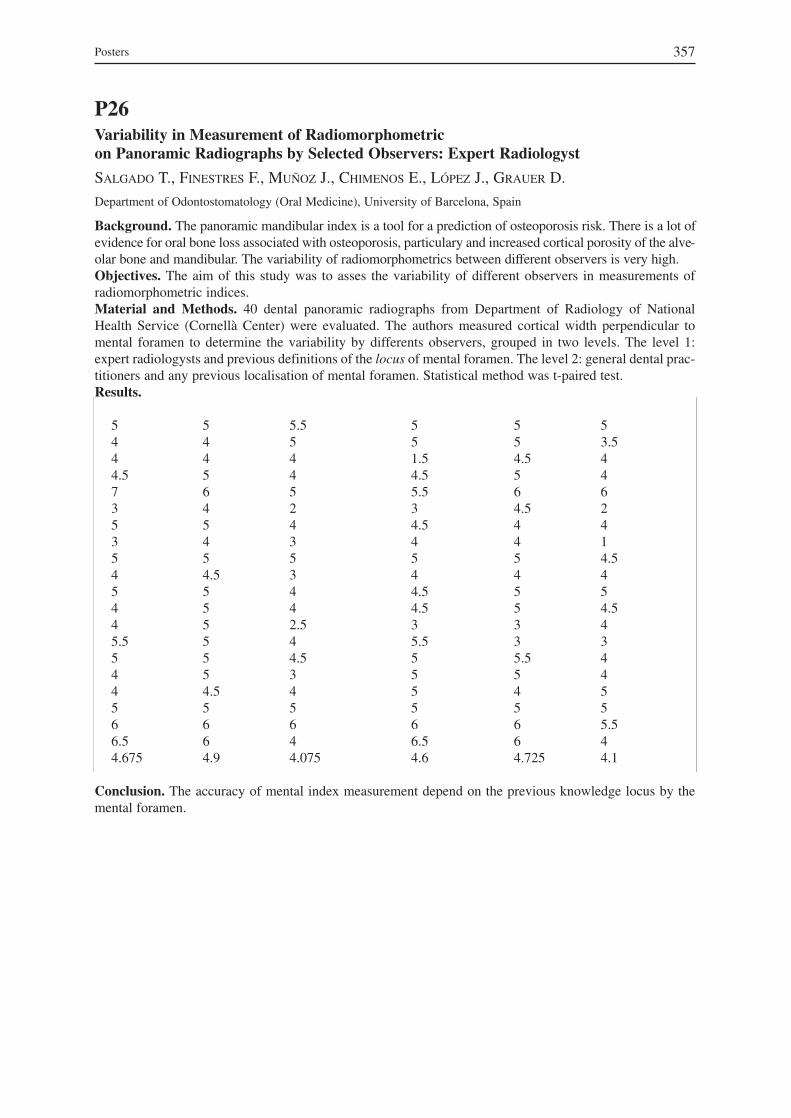
P26Variability in Measurement of Radiomorphometric on Panoramic Radiographs by Selected Observers: Expert Radiologyst
SALGADO T., FINESTRES F., MUÑOZ J., CHIMENOS E., LÓPEZ J., GRAUER D.
Department of Odontostomatology (Oral Medicine), University of Barcelona, Spain
Background. The panoramic mandibular index is a tool for a prediction of osteoporosis risk. There is a lot ofevidence for oral bone loss associated with osteoporosis, particulary and increased cortical porosity of the alve−olar bone and mandibular. The variability of radiomorphometrics between different observers is very high.Objectives. The aim of this study was to asses the variability of different observers in measurements ofradiomorphometric indices.Material and Methods. 40 dental panoramic radiographs from Department of Radiology of NationalHealth Service (Cornellà Center) were evaluated. The authors measured cortical width perpendicular tomental foramen to determine the variability by differents observers, grouped in two levels. The level 1:expert radiologysts and previous definitions of the locus of mental foramen. The level 2: general dental prac-titioners and any previous localisation of mental foramen. Statistical method was t-paired test.Results.
Conclusion. The accuracy of mental index measurement depend on the previous knowledge locus by themental foramen.
Posters 357
5 5 55 5 3.51.5 4.5 44.5 5 45.5 6 63 4.5 24.5 4 44 4 15 5 4.54 4 44.5 5 54.5 5 4.53 3 45.5 3 35 5.5 45 5 45 4 55 5 56 6 5.56.5 6 44.6 4.725 4.1
5 5 5.54 4 54 4 44.5 5 47 6 53 4 25 5 43 4 35 5 54 4.5 35 5 44 5 44 5 2.55.5 5 45 5 4.54 5 34 4.5 45 5 56 6 66.5 6 44.675 4.9 4.075

P27The Computer Program for Three−Dimensional Casts Analysis of Patients with a Complete Unilateral Cleft Lip and Palate
WOJTASZEK−SŁOMIŃSKA A., NIEKRA M., SŁOMIŃSKI P.
Medical University of Gdansk, Technical University of Gdansk, Poland
Background. It has been reported in literature that the position of the maxillary cleft segments before anoperation can determine the later craniofacial development. The conclusion has been drawn on the basis ofthe casts analysis of the examined children.Objectives. The aim of the work is to present the algorithms of a computer program used in three−dimen−sional casts analysis of patients with a complete unilateral cleft lip and palate. The program makes it possi−ble to analyse the anatomical structure of the palate and alveolar process of patients at various age.Material and Methods. The examined casts are scanned with a 3D scanner, and the acquired picture in theform of a mathematical record is then sent to a computer and digitally analysed by the Ortbaz program. Theprogram can analyse the following: sagittal arch length, anterior and posterior alveolar arch widths, and thedistance between two chosen landmarks in 2D and 3D projections. The arch shape is separately identified forboth maxillary segments. The program automatically digitises the alveolar process radius in the smaller andlarger maxillary segments. In order to establish the lip operation influence on maxillary segments positions,the inclination angle of the particular maxillary parts towards the tubercular plane is determined by the pro−gram, which computes the palatal surface, depth and width. It is possible to assess the mutual inclination ofeither the maxillary palatal processes or long teeth axes on the sections running through the landmarks cho−sen by the investigator. Fifteen children with a complete unilateral cleft lip and palate were included in thelongitudinal study lasting for three years. The casts of the children were made as follows: the first one – beforethe lip surgery at the age of 6 months; the second one – before the secondary palate surgery at the age of 12months: and the third one – during the period of complete deciduous dentition at the age of 4 years. All thecasts were then scanned with a 3D scanner and analysed by the Ortbaz program.Results. Due to treatment the cleft anterior width became smaller, however, in 5 patients there was discrep−ancy of over 4 mm in 3D measurement. Some unfavourable changes in the smaller maxillary segment shapewere also found – in 6 cases the radius grew larger to over 30 mm. The change in maxillary segments incli−nation was observed as well. Conclusion. The Ortbaz program gives one a useful tool for assessing the casts of children in both the infan−tile toothlessness and deciduous dentition periods. However, the small number of examined patients seemsinadequate to draw any conclusions referring to the maxillary structure changes, and only makes it possibleto test the program itself.
48 Congress GIRSO358

P28Advanced Possibilities of Ultrasound Imaging of Soft Tissues of Tongue and Floor of the Mouth
RÓŻYŁO−KALINOWSKA I.1, STETTNER R.2, JURKIEWICZ−MAZUREK M.3
1 2nd Department of Medical Radiology, Medical University of Lublin, Poland2 Students’ Scientific Association at 2nd Department of Medical Radiology, Medical University of Lublin, Poland3 Department of Dental and Maxillofacial Radiology, Medical University of Lublin, Poland
Background. Ultrasonographic evaluation of pathologic lesions of soft tissues of tongue and floor of themouth is difficult and rarely undertaken by radiologists as lesions of this area are relatively rare in generalpopulation and are believed to be the domain of dentists. Furthermore, specialist equipment for oral imag−ing such as intraoral probes is difficult to procure. Usually, tongue and oral floor tissues are examined fromextraoral approach.Objectives. The aim of the study was evaluation of possibilities of advanced imaging options in ultra−sonography of soft tissues of tongue and oral floor.Material and Methods. High−frequency linear transducer was used for ultrasonographic examination of15 patients with oral pathologies (neoplasms, inflammations) of comprising soft tissues of tongue and oralfloor. Furthermore, in 20 healthy young adults there were used three−dimensional imaging options of GeneralElectric Voluson 730 Pro ultrasound machine for visualization of anatomy of the tongue and oral floor.Results. It was found that three−dimensional ultrasound images may be a valuable supplement in diagnosisof oral lesions, however, conventional B−mode images remain the basic diagnostic tool.
P29Locally Applied Herbal Mixture in Treatment of Odontogenic Infections
SZYSZKOWSKA A.1, KIWORKOWA E.2, RÓŻYŁO T.2
1 Department of Oral and Maxillofacial Surgery, Medical University of Lublin, Poland2 Department of Dental and Maxillofacial Radiology, Medical University of Lublin, Poland
Background. Various bacterial pathogens present in oral environment are responsible for development ofodontogenic infections, some of which may sometimes develop after dental and periodontal surgery.Increasing bacterial resistance to antibiotics must be taken into account when planning treatment strategy. Objectives. The purpose of the study was to evaluate local application of a herbal mixture in treatment ofodontogenic inflammations.Material and Methods. The material comprised 143 patients with odontogenic inflammations, who werelocally treated with herbal infusion in the form of irrigations and herbal dressings. No antibiotics or chemio−therapeutic drugs were applied in the treatment. The herbal mixture was composed of AnthodiumChamomillae, Folium Plantaginis Lanceolatae, Herba Euphrasiae and Cortex Querqus. Microbiologicalstudies were performed in 21 patients in order to determine susceptibility of cultured pathogens to firstchoice antibiotics.Results. It was found that microorganisms coming from sites of odontogenic infections were resistant toeven several antibiotics. There were confirmed antibacterial, anti−inflammatory and healing properties of theapplied herbal mixture. Conclusion. There was confirmed that the used herbal mixture was beneficial in local treatment of oralinflammations of odontogenic origin.
Posters 359
P30Expression of Nitric Oxide Synthase in Inflammed Gingival Tissue: Immunohistochemical And Image Analysis
LEONE A., UZZO M., SPATOLA G. F., MIGLIAVACCA M., BUSCEMI M., GERBINO A.
Dipartimento di Medicina Sperimentale, Sezione di Istologia ed Embriologia, Facoltà di Medicina, Università di Palermo, Italia
Background. It is known that some bacteria induce inflammation of periodontal tissues, causing release ofnitric oxide. To clarify the relashionship between the degree of inflammation and NO production, we ana−lyzed immunohistochemically the profile of the inducible Nitric Oxide Synthase, iNOS, in several cases ofepulis exhibiting varying degree of inflammation infiltration.Materials and Methods. The authors used 12 epulis taken during surgical procedures, and also a normalgingival specimen taken during dental extraction, used as control. All the specimens were fixed in Bouin’smixture and embedded in paraffin. The authors investigated the immunohistochemical distribution of iNOSusing monoclonal antibody against iNOS. The section were processed for immunohistochemistry by thestandard streptavidin−biotin complex method. After immunostaining, the sections were covered with water−−soluble mounting medium and observed with a Leica Microscope. Subsequently, all the specimens werestudied with an image analysis Lucia M system to obtain colorimetric data.Results and Conclusion. The data show a strong positive cytosolic immunostaining of iNOS, specificallyin infiltrating cells. The strong immunostaining, which was evaluated with image analysis method, is relat−ed to theh histotype of epulis. Our investigation, along with accumulating supportive data in the literature,suggest the iNOS plays a role as an early inflammation mediator. This morphofunctional study representsthe continued advances started by some time ago in our understanding of epulis and gingival volume aug−mentation.
48 Congress GIRSO360

P31Pathologie bucco−dentaire chez les malades atteints d’anorexie restrictive
BELMONTE−CALDERON A.M.1, MARTINEZ−GONZALEZ A.M.2, GARCIA−MOLINA J.A.1, GRECO−MACHADO Y.1, CARVALHO−LOBATO P.1, SERRA−RISTOL S.1, SERRA−RENOM I.1, MANZANARES−CÉSPEDES M.C.1
1 Unité d’Anatomie Humaine, Département Anatomie et Embryologie Humaine, Université de Barcelone, Espagne2 Unité de Pshychiatrie, Département Psychiatrie et Psychobiologie, Faculté d’Odontologie, Université de Barcelone,Espagne
Les altérations de la conduite alimentaire sont devenues un sérieux problème de santé publique dans les paysindustrialisés, avec une prévalence oscillante entre un 5 à un 10% de la population. Parmi ces altérations, laplus fréquente est l’anorexie, dont le tableau clinique est caractérisé par l’alimentation restrictive et l’a-menorhée, entre d’autres signes cliniques. L’anorexie se présente en divers sous-tipes cliniques, dontl’anorexie dite « restrictive » et la boulimie, caractéerisée en plus par les vomissements compulsifs. Une descomplications les plus étudiées de l’anorexie est l’ostéoporose, associée à la sévère perte de poids desmalades, à la réduction de l’activité physique, a la durée de l’amenorhée, et au déficit oestrogénique. La péri-ode de risque pour la parution de l’ostèoporose associée à l’anorexie va des 15 aux 18 ans. Il s’agît d’unepériode de formation osseusse rapide, dans laquelle une anomalie quelconque du metabolisme minéral peutcauser une perte significative de la masse osseusse des arcades dentaires, et s’associer à un niveau inférieurd’adhésion des dents à l’alvéole dentaire, dans le cas d’une parodontite.Les vomissements de la boulimie ont eté depuis 1995 mis en rapport avec des altérations dentaires, et lasanté orale de ces patients est régulièrement revue. Par contre, même si d’accord avec les donnés bibli-ographiques, l’on pourrait supposer que les malades atteintes d’anorexie restrictive pourraient présenter desproblèmes odontologiques dûs en partie à l’ostéoporose concomitante à leur anorexie, nous n’avons trouvéque des études partielles sur la santé orale des malades atteintes d’anorexie restrictive, la mettant en rapportavec des processus inflammatoires de la cavité orale, comme la parodontite et la carie, surtout dans les pre-miers stades de la maladie.Nous avons entamé une étude pour évaluer l’impact de la santé orale sur le tableau général de l’anorexierestrictive. 90 histoires cliniques enregistrées dans le service de Psychiatrie de l’Hôpital de Bellvitge ont étérevues à la recherche de donnée sur la santé orale des malades. Seulement quatre de ces histoires avaientune section dediée à la santé orale, et les données qui y étaient recueillies faisaient étaient très peu spéci-fiques. En vue de ces données et de celles apportées par la littérature, nous proposons un questionnaire con-tenant les données les plus relevantes pour la santé orale des malades atteintes d’anorexie restrictive, en vued’améliorer leur état de santé buccale durant et surtout après le période active de leur maladie.
Posters 361
P32 Pathologie ostéo−articulaire chez le professionnel de la dentisterie
GARCIA−MOLINA J.A.1, GRECO−MACHADO Y.1, CARVALHO−LOBATO P.1, LOZANO−DE LUACES V.2, GOLANÓ ALVAREZ P.1, SERRA−RENOM I.1
1 Unité d’Anatomie Humaine, Département Anatomie et Embryologie Humaine, Faculté d’Odontologie, Universitéde Barcelone, Espagne2 Unité d’Ergonomie, Département Odontostomatologie, Faculté d’Odontologie, Université de Barcelone, Espagne
La position que le professionnel adopte durant les traitements dentaires a une influence évidente surl’éventuelle parution de lésions ou même de pathologies ostéoarticulaires, très fréquentes en cette et autresprofessions.Nous avons donc entamé une étude de l’ergonomie des positions que divers professionnels adoptent durantcertains traitements en clinique dentaire.Nous avons pris des photographies en temps réel à des praticiens de l’art dentaire, experts avec des annéesde pratique et en formation, durant des traitements faits sur des malades en deux centres, l’un public(Clinique Odontologique Universitaire de l’Université de Barcelone) et l’autre privé (InternationalOdontology Center, aussi de Barcelone). Les images des structures anatomiques concernées par les postureset les mouvements etudiés ont été obtenues sur du matériel anatomique: dissections et sections de matérielhumain non-embaumé en provenance de la Salle de Dissection du Département d’Anatomie Humaine del’Université de Barcelone.Nos études ont prouvé que le muscle le plus impliqué en les mouvements les plus fréquents et répétés parle praticien dentiste est le Trapèze, dont la position la plus correcte est de repos en un premier temps, suivid’une contraction dont le point fixe est son insertion cervicale. Ainsi, les mouvements du bras sont dusessentiellement à la rotation du caput humeral dans la cavité glenoïde, avec seulement un petit degré d’ab-duction. Quand la position n’est pas correcte, la contraction du Trapèze prend comme point fixe l’omoplate,qui commence une rotation. Ceci que porte à que les mouvements du bras aient un composant d’abductiontrès forcée, tout en causant une montée de la tension des muscles qui relient le bord vertébral de l’omoplateavec la colonne vertébrale.Ces premiers résultats de nos études ont prouvé l’utilité de la comparaison entre les images obtenues par lespraticiens dans leur entourage professionnel avec des images de ce matériel anatomique très spécial (non-embaumé), qui permet que ces pièces anatomiques puissent être placées dans la même position et pho-tographiées. Ainsi la collaboration entre les deux équipes, Anatomie et Ergonomie, nous permet d’aboutirà des résultats relevants quant à la bioméchanique des mouvements faits par les praticiens dentistes, avecpour but celui d’améliorer leur ergonomie.
P33La communication entre le practicien et le malade: un outil de traitement?
GARCIA−MOLINA J. A.1, GRECO−MACHADO Y.1, CARVALHO−LOBATO P.1, LOZANO−DE LUACES V.2, SERRA−RISTOL A.1, SERRA−RENOM I.1
1 Unité d’Anatomie Humaine, Département Anatomie et Embryologie Humaine, Faculté d’Odontologie, Universitéde Barcelone, Espagne2 Unité d’Ergonomie, Département Odontostomatologie, Faculté d’Odontologie, Université de Barcelone, Espagne
La communication entre le malade et le professionnel de la dentisterie est un composant essentiel de tout letravail fait en clinique dentaire, et, pour cette raison, il faut en faire l’objet des études detaillées. À la dif-férence de la consultation médicale, la consultation dentaire comporte presque toujours une manipulationthérapeutique. Ce traitement inclu dans la consultation, qui, forcémment est fait dans la cavité orale dumalade, requiert au préalable une claire communication avec le malade. Il s’ensuit aussi que le maladerestera assis durant une période de temps assez longue, et que durant le traitement en soi, des instrumentsseront dans sa bouche, ainsi le patient sera empêché de communiquer avec le professionnel. Il est bien connu que les patients sont souvent anxieux et peureux au sujet de leurs visites chez le dentiste.De même, ils ont une certaine expectation de sentir de la douleur durant le traitement. Les études faits ont
48 Congress GIRSO362

prouvé que une bonne communication entre le professionel et le malade a une énorme influence sur la façonde laquelle le malade réagit face au traitement.Ainsi nous avons entamé une étude sur la communication entre le professionel et le patient, moyennant desquestionnaires envoyés chez 54 professionnels de la ville de Barcelona (Espagne) en leur demandant dedécrire brèvement leur style et forme de communication avec leurs patients. Les questions étaient axéessurtout sur les explications ofertes par le professionnel au patient avant, durant et aprés le traitement. Lespremiers resultats de cette étude sont presentées.
P34Thermographic Imaging of Periodontum in Adolescents Suffering from Decompensated IDDM
BURCHARDT D.1, GÓRSKI S.2
1 Department of Paediatric Dentistry, Poznan University of Medical Sciences, Poland2 Radiophysical Laboratory, J. Struś Municipal Hospital, Poznan, Poland
Objectives. The study was undertaken to test the possibility of thermographic visualisation for assessmentof the effect of retinopathy on the periodontum status. Material and Methods. The subjects were 61 children and adolescents aged 10–19 (38 girls and 23 boys),suffering from type I diabetes for 4 to 14 years. The mean values of glycosylated haemoglobin HbA1 indi−cated metabolic decompensation. On the basis of the results of fundus of the eye examination, the subjectswere divided into 2 groups: one of 22 subjects revealing retinopathy and the other of 39 subjects do notchanges in the fundus of the eyes. The periodontal status was described by the Gingival Index according toSilness and Löe, and the Sulcus Bleeding Index according to Mühlemann and Son. The oral cavity hygienewas described by the Plaque Index according to Silness and Löe. Thermographic examination was per−formed in the vicinity of the first incisors of maxilla by a thermograph AGA Thermovision System 680 in32 randomly chosen subjects (22 with and 10 without retinopathy). Results. In the group of subjects showing retinopathy the mean values (Me) GI = 0.74 and SBI = 0.82. In thegroup of subjects showing no changes in the fundus of the eye GI = 0.50, and SBI = 0.40. The mean valuesof Pl−I (Me) in the group with retinopathy was 0.82, while in the group without retinopathy it was 0.67.Statistical analysis by the Mann−Whitney test did not show statistically significant differences between thevalues of GI and Pl−I in the two groups studied (p > 0.05). Statistically significant differences were foundbetween the mean values of SBI between these two groups (p < 0.05).Analysis of the thermographic images showed that the weighted means (Me) of the temperatures of the peri−odontum tissues before the cooling in both groups were statistically significantly different. In the group withretinopathy on the right hand side it was 30.6ºC on the left hand side 30.9ºC, in the group without retinopa-thy on the right hand side 34.1ºC and on the left hand side 34.0ºC. Conclusions. Statistically significant differences have been found between the SBI values and temperaturesof periodontum in subjects with and without retinopathy. Thermographic imaging of periodontum can be animportant supplement of clinical examination aimed at assessment of the periodontum tissue and the status ofthe blood vessels in the subjects with microangiopathy.
Posters 363

P35 Successful Treatment of Overlap Syndrome within Oral Mucosa – Case Report
HUPSCH−MARZEC H., KLIMA D., KALINA A., BARON S., KRYNICKI M.
Department of Conservative Dentistry, Medical University of Silesia, Bytom, Poland
The material of clinical observations was based on a patient, who at the moment of notification at our clinicwas loaded with 2 year history of continually progressing disease within oral mucosa. Significant, increas−ing painful complaints and the expansion of the diseases did not allow the consumption of food, use of den−ture and made speaking difficult. Up to the present, local treatment was totally inefficient. The analysis of the medical history and multistage examination allowed to establish factors which directlyor indirectly might have influenced his present condition of oral cavity and their gradual elimination led tocomplete therapeutical success. The reveal of Reiter’s syndrome at our patient confirms that because of possibility of occurance of symp−toms within oral cavity, this disease deserves the special stomatologists attention. Atypical course of the disease may be the result of superposition of others, coexisting systemic diseases,which oral mucosa is sensitive detector. In case of lesions within oral mucosa, possible influence of med−ications applied by patients must be always considered. Only holistic management of the patient gives thechance for permanent therapeutic results.
P36Syndrome de brûlure buccale. Révision du concept
RODRIGUEZ DE RIVERA E., CHIMENOS E., LÓPEZ J., SABATER M. M., FINESTRES F., MUÑOZ J., VIÑALS H.
Faculté d’Odontologie, Université de Barcelone, Espagne
Le syndrome de brûlure buccale est un entité assez fréquente encore qu’aujourd’hui nous avons de sérieuxproblèmes pour établir un diagnostic et un traitement. L’incidence est d’un 5–18% dans la pophlation adulteet certains auteurs le considèrent comme le second motif de demande dans les Centres de Médecine Orale. Il se caractérise par une sensation de brûlure, à type de picotements, dans certains points de la muqueuseorale, spécialement dans la langue, sans qu’on puisse démontrer des lésions cliniques. Il s’accompagnegénéralement d’autres manifestations comme des altérations du goût, de xérostomie et de paresthésies. Ilaffecte principalement les femmes post ménopausiques qui présentent normalement des altérations émo-tionnelles comme la dépression et l’anxiété.La symptomatologie débute normalement après une période de stress psychologique, et on a décrit aussi descas après un traitement odontologique. Son véritable étiologie résulte de connu.Dans cette communication on repasse les différents facteurs etiopatogéniques décrits et les différentes pos−sibilités térapeutiques.
48 Congress GIRSO364

P37Effect of Antiretroviral Treatment on the Oral Pathology in 90 HIV + Patients
CHIMENOS E., JANÉ E., OCAÑA I., ROSELLÓ X., LÓPEZ J., FINESTRES F., MUÑOZ J.Unité de Médicine Orale, Département d’Odontostomatologie, Faculté d’Odontologie, Université de Barcelone,Espagne
Objectives. The aim of the present study is to analize the effect of the antiretroviral treatment in HIVpatients in a population visited in the Infectious Diseases Service of the Vall d’Hebron Hospital inBarcelona. Material and Methods. 90 patients were visited, 51 men (56.7%) and 39 women (43.3%), the mean age ofthe patients was 36.2 with a deviation of ± 17.8 years and a mode of 35 years. The visits were accomplishedfrom January to December in the year 1999. These patients were receiving antiretroviral treatment of one ormore drugs and some of them (32.2%) were submitted to a high activity antiretroviral treatment (HAART);moreover, in these patients the recount of CD4 and viral load was valued in a bimodal way at the beginningof the process and at the moment of the visit.Results. The most prevalent pathology found in this study was the dry mouth sensation or xerostomia, in47.8% of the patients, followed by order of frequency, by polycaries (34.4%) and erythematous candidiasis(31.1%). Conclusion. The results of this study cannot be superposed with the results obtained by other authors, dueto the different dates and collectives of patients, before and after the utilization of the HAART: this kind oftherapy has modified the expression of oral pathology in order to suspect HIV seropositivity.
Posters 365

P38The Effect of a Combination Therapy (Interferon Alpha + Lamivudin) on the Condition of Oral Mucosa in Patients Suffering from Chronic Hepatitis B (HBV)
SULKA A.1, SIMON K.2, PISZKO P.2
1 Department of Oral Surgery, Wroclaw Medical University, Poland 2 Department and Clinic of Infectious Diseases, Wroclaw Medical University, Poland
Objectives. The aim of the study was to evaluate the condition of oral mucosa in patients suffering fromchronic hepatitis B treated with IFN−alpha (6 months) + lamivudin (12 months).Material and Methods. The study groups consisted of 18 patients treated with IFN−alpha + lamivudin forchronic hepatitis due to HBV infection. The control group comprised 23 untreated patients with chronichepatitis B. The clinical evaluation of the oral cavity was carried out at 0, 3, 6 and 12 month of the follow− up.Oral mucosa pathology found on clinical examination was confirmed histopathologically.Results. In the group of patients treated with IFN−alpha + lamivudin, the most common lesion found on ini−tial examination was leukoplakia (5/18), while on the final examination it was labial herpes (6/18). The onlyfinding in the control group was the increased incidence of labial herpes (5/20) in the 3rd month of observa−tion. The intragroup analysis of all the study groups did not show any statistically significant differences inthe number of lesions within the oral mucosa on follow−up. Comparison between groups showed a statisti−cally significant difference in the number of oral mucosa lesions which was observed in the 12th month offollow−up. The lesions were significantly more frequent in the group of patients on IFN−alpha + lamivudintherapy (p ≤ 0.05).Conclusion. IFN−alpha + lamivudin therapy seems to increase of the number of oral mucosa lesions inpatients suffering from HBV.
P39L’évaluation de la condition de dentition chez les malades atteints du syndrome de Sjögren
CACKOWSKA−LASS A., KOCHAŃSKA B., KRĘGLEWSKA B., WITEK E.
Département de Stomatologie Conservatrice de l’Académie Médicale de Gdansk, Pologne
L’évaluation de la condition de dentition chez les malades atteints du syndrome de Sjögren (sS) et chez lessujets témoins a été réalisée. Comme la méthode d’évaluation on a utilisé les critères DMF-T et DMF-S. Ona analysé la relation entre la quantité de dents conservées, l’âge des malades et la durée de maladie. Legroupe étudié a été constitué de 45 femmes avec le syndrome de Sjögren à l’âge de 23–68 ans (x– = 48.7 ans)et par 41 femmes à l’âge de 23–73 ans (x– = 49.5 ans) choisies comme les sujets normaux.Dans le groupe de malades avec sS, les chiffres DMF−T ainsi que DMF−S étaient considérablement plushauts (p < 0.001), que ceux dans un groupe témoin. La moyenne de DMF−T dans le groupe de malades étaitde 26.4 ± 6.1, tandis que, celle chez les sujets normaux était de 22.9 ± 5.7. Les moyennes de DMF−S étaientde 105.6 ± 43.4 et de 72.3 ± 24.1 respectivement.Le nombre de dents chez les malades atteints du syndrome de Sjögren, a été fortement réduit, en compara−ison au groupe témoin (p < 0.01). Les moyennes de dents conservées chez les sujets de ces deux groupesétaient de 16.3 ± 11.0 et de 24.0 ± 4.1 respectivement. On a constaté la perte totale des dents chez 20% desujets à l’âge de 36–67 ans. Dans le groupe témoin, il n’y avait pas de sujets édentés. Le nombre de dentschez les malades diminuait considérablement avec l’âge des patients (r = –0.58; p < 0.001). Chez les sujetsâgés de 60 ans et plus (10 malades), la moyenne du nombre de dents était de 4.6 ± 6.5, et chez chez les sujetsnormaux (11personnes) était de 23.4 ± 2.6. Dans un groupe témoin, le nombre de dents ne diminuait quelégèrement avec l’âge de sujets étudiés et n’était pas significatif.Chez les malades, il existait une forte corrélation négative entre la durée de maladie et le nombre de dentschez les malades avec le syndrome de Sjögren (r = –0.34; p < 0.01). Nous retiendrons de ces résultats que la condition de dentition chez les malades atteints du syndrome deSjögren était considérablement plus faible que celle de sujets normaux.
48 Congress GIRSO366

P40Les modifications de la flore buccale chez les malades atteints du syndrome de Sjögren
CACKOWSKA−LASS A.1, KOCHAŃSKA B.1, WITEK E.1, NAUMIUK Ł.2, SAMET A.2
1 Département de Stomatologie Conservatrice de l’Académie Médicale de Gdansk, Pologne2 Département de Microbiologie Clinique de l’Hôpital PSK 1 à Gdansk, Pologne
La sécrétion salivaire constante exerce un rôle principal dans la maintenance de l’écosystème de la cavitébuccale. Des altérations tant quantitatives que qualitatives de la salive peuvent provoquer les modificationsde la flore buccale. L’étude de la flore a été entreprise dans la salive stimulée chez 45 femmes atteintesdu syndrome de Sjögren (sS) à l’âge de 23–68 ans (x– = 48.7 ans) et chez 41 femmes à l’âge de 23–73 ans(x– = 49.5 ans) considérées comme les sujets normaux. Des différentes espèces bactériennes et mycoses ontété cultivées et différenciées sur des milieux suivants: Columbia Scheep Blood Agar, Columbia ScheepBlood Agar CNA avec colistine et acide nalidixique, Chocolate Agar avec colistine PoliVitex et avec baci-tracine, Candida Chromagar (ABS, Polska) et Sabouraud Agar (bioMérieux, Pologne). L’identification a étéconduite d’après la morphologie de colonies et la coloration de Gram et d’après les tests biochimiques GPI– Gram Positive Identification dans le système automatique VITEK (bioMérieux, Pologne). Cette étudemontre les modifications de proportions relatives de streptocoques dans la salive des malades ayant troublesde la sécrétion salivaire. S. salivarius a été présent chez 40% de malades, en comparaison à 60.9% de sujetsnormaux. Bien que légèrement diminué, S. sanguis a été retrouvé chez 28.9% de malades et chez 41.5% desujets du groupe témoin. On a observé une légère augmentation de S. mitis chez les patients avec sS (48.9%)en comparaison à 36.6% de sujets normaux. L’augmentation de S. intermedius chez les malades a été plusimportante (p < 0.05). S. acidominimus a été retrouvé uniquement chez les malades (13.3%). Parmi les sujetsnormaux S. aureus n’était pas présent, mais il a été identifié chez 15.5% de malades avec sS. A part cela,dans la salive de 27% de malades on a noté aussi l’apparition de C. krusei, C. tropicalis et C. glabrata, quin’étaient pas présents dans un groupe témoin. Il resort de cette étude qu’un certain nombre de modificationsse développe au niveau de la flore buccale chez les patientes atteintes du syndrome de Sjögren.
P41L’évaluation du nombre de colonies de Streptococcus mutans, Lactobacillus sp.,et Candida albicans dans la salive chez des malades avec le syndrome de Sjögren
CACKOWSKA−LASS A.1, KOCHAŃSKA B.1, WITEK E.1, NAUMIUK Ł.2, SAMET A.2
1 Département de Stomatologie Conservatrice de l’Académie Médicale de Gdansk, Pologne2 Département de Microbiologie Clinique de l’Hôpital PSK 1 à Gdansk, Pologne
L’évaluation du nombre de Streptococcus mutans, Lactobacillus sp., et Candida albicans dans la salive aété étudiée chez les malades atteints du syndrome de Sjögren (sS) et chez les sujets témoins.L’étude a été faite sur 45 femmes atteintes du syndrome de Sjögren à l’âge de 23–68 ans (x– = 48.7 ans) et sur41 femmes à l’âge de 23–73 ans (x– = 49.5 ans) considérées comme les sujets normaux. Pour évaluer le nom-bre de microbes on a utilisé des échantillons de salive stimulée mixte prélevés le matin. Streptococcus mutanset Lactobacillus sp. ont été cultivés sur des milieux sélectifs Vivacult LB i SM (Ivoclar-Vivadent, Lichtenstein).Candida albicans a été cultivé sur le milieu Sabouraud Agar (bioMérieux, Pologne) et l’identification de C.albicans de toutes les autres mycoses, a été conduite d’après un test de filaments et d’après la coloration decolonies sur le milieu Candida ID2 (bioMérieux, Pologne). Le nombre de colonies Streptococcus mutans etLactobacillus sp. ≥ 106 CFU par ml de salive et le nombre de C. albicans > 105 CFU/ml, a été vu comme élevé.Dans notre étude, le pourcentage des malades chez qui on a retrouvé le nombre élevé de Lactobacillus sp.,S. mutans et C. albicans a été plus haut que chez les sujets témoins. Le nombre de colonies de S. mutans ≥106 CFU/ml a été retrouvé chez 37.8% de malades et chez 14.6% de sujets normaux. Dans le cas de C. albi−cans, le nombre élevé de cette levure ( > 105 CFU/ml) a été détecté chez 15.5% de malades, par contre dansle groupe témoin, on n’a pas retrouvé C. albicans au nombre si élevé. L’analyse statistique a démontré quela différence entre le nombre de colonies de S. mutans et C. albicans entre deux groupes n’était pas signi−ficative. Par contre, le nombre de colonies de Lactobacillus sp. dans la salive des malades était plus élevé(p < 0.01) que celui dans le groupe témoin. Le nombre de Lactobacillus sp. ≥ 106 CFU/ml. a été observéchez 31.1% de malades, mais il n’a pas été découvert dans ce nombre chez les sujets normaux.
Posters 367

P42Le pouvoir tampon de la salive et la vitesse de la sécrétion salivaire chez les malades avec le syndrome de Sjögren
CACKOWSKA−LASS A., KOCHAŃSKA B., KRĘGLEWSKA B., WITEK E.
Département de Stomatologie Conservatrice de l’Académie Médicale de Gdansk, Pologne
Le but de cette recherche a été la détermination et la comparaison du pouvoir tampon de la salive stimuléechez les malades atteints du syndrome de Sjögren (sS) et chez les sujets témoins. L’étude a été faite sur 45femmes avec syndrome de Sjögren à l’âge de 23–68 ans (x– = 48.7 ans) et sur 41 femmes à l’âge de 23–73ans (x– = 49.5 ans) choisies comme les sujets normaux. Chez tous les sujets, on a mesuré la vitesse de lasécrétion de la salive stimulée par la mastication d’une tablette de paraffine pendant 5 min. On a déterminéle pouvoir tampon de la salive stimulée au moyen d’une bandelette de test munie d’un indicateur spécial(Vivacult Buffer Capacity Test, Ivoclar-Vivadent). Dans cette étude, on a observé l’abaissement consid−érable du pouvoir tampon de la salive chez les malades, en comparaison à celui du groupe témoin (p <0.001). Le pouvoir tampon de la salive faible (pH après 5 min de réaction ≤ 4.0), on l’a retrouvé chez 24(53.3%) malades et seulement chez 5 (12.1%) sujets normaux. Le pouvoir tampon moyen (pH après 5 minde réaction 4.5–5.5), on l’a observé chez 10 (22%) malades et chez 20 personnes (48.7%) dans un groupetémoin. Le pouvoir tampon de la salive élevé (pH après 5 min de réaction ≥ 6.0) a été présent chez 11(24.5%) sujets atteints et chez 16 (39.2%) sujets normaux. Dans un groupe de 11 malades, la sécrétion sali−vaire était fortement baissée ( < 0.2 ml/min). Chez 9 sujets de ce groupe−là on a constaté également un faiblepouvoir tampon de la salive. De plus, la sécrétion salivaire légèrement baissée (0.2 < 0.7 ml/min) on l’a trou-vée parmi 21 malades. Chez 9 personnes de ce groupe-là, on a observé le pouvoir tampon de la salive faible,chez 5 personnes – moyen et élevé chez 7 sujets atteints. Le pouvoir tampon de la salive faible a été retrou-vée chez 6 parmi 13 malades avec la sécrétion salivaire normale (≥ 7 ml/min). Chez 3 personnes de cegroupe le pouvoir tampon était moyen et chez 4 élevé. Dans un groupe témoin, il n’y avait aucune person−ne avec la sécrétion salivaire insuffisante ( < 0.2 ml/min). Par contre, il y avait 16 sujets avec la sécrétionsalivaire baissée, mais un faible pouvoir tampon de la salive on l’a trouvé seulement chez 2 personnes, lemoyen chez 10 et chez 4 – élevé. Parmi 25 témoins avec la sécrétion salivaire normale, chez 3 personnes lepouvoir tampon était faible, chez 10 – moyen et chez 12 – élevé. On peut donc conclure que chez lesmalades atteints du syndrome de Sjögren, le pouvoir tampon de la salive peut être diminué avant que lasécrétion salivaire ne soit baissée.
P43Oral Carcinoma in Atipical Patient. Clinical Case
ROSELLÓ X., LÓPEZ J., CHIMENOS E., JANÉ E., FINESTRES F., MUÑOZ J.
Faculty of Dentistry, University of Barcelona, Spain
A 34−years−old white female consulted about a red lesion on the oral mucosa. She revealed 6 months oflesion evolution. The lesion was a red ulcerated plaque affecting a zone of the right hard palate and the retro−molar attached gingiva. It was painless. This lesion was biopsied 3 months ago by a maxillofacial surgeon,and the diagnosis was displasic lesion. At the clinical examination, the patient presented many lesions thatclinically could be diagnosed as an oral lichen planus (OLP) and they were located in the tongue, cheek, andgingiva. There were not skin lesions. We performed a second biopsy of the palate lesion and from one of thelesions in the right buccal mucosa. The histologic diagnosis was OLP for the buccal mucosa lesion and oralcarcinoma on a lichen lesion in the other. Complementary exams were performed like a p53 detection andsearch for papillomavirus. The patient was referred to the Service of Dermatology of Hospital Clinico yProvincial de Barcelona for the surgical treatment.
48 Congress GIRSO368

P44Holistic Concept of the Stomatognathic System
PANEK H.
Department of Prosthodontics, Wroclaw Medical University, Poland
The earliest concepts of the stomatognathic system, derived from the studies performed by Hanau,Trappozzano and Levin, were concentrated on a relationship between occlusion and temporo−mandibularjoints. The later concepts, described by Held, Wigdorowicz−Makowerowa et al., Majewski, Fuhr andSolberg as well as by Troest, stressed the importance of additional elements, such as periodontium, neuro−muscular system and CNS. In the concept proposed by Panek an environment is included. In proposed con−cept, the stomatognathic system is presented as a pyramid with four apices imaging CNS and three “joints”(TMJ, periodontium joint, occlusal joint), and the edges imaging the neuro−muscular system. The pyramidsimply reveals that harmony in the stomatognathic system is dependent on interaction of particular elementsof the system. The pyramid is placed inside a sphere imaging a human body, that means the dependence ofstomatognathic system on general state of health. Moreover, the sphere representing the human body isplaced inside a greater sphere imaging an environment. The environment may disturb the stomatognatic sys−tem directly or indirectly or even may effect the human genom by influencing the growth process and struc−ture of stomatognathic system in a period of organopoesis. In such holistic presentation, the stomatognath−ic system may be considered as a morphological−functional unit involved in dynamic transformation con−tinued during the whole period of life. In such circumstances a “norm” of stomatognathic system in youngpersons with uncompleted growth may be different from that in older subjects, where it may be additional−ly influenced by various processes connected with ageing and “wearing” the particular elements of the sys−tem.
P45Clinico−Etiological Groups of Temporomandibular Joint Disorders. The Arthroscopy Examination
WANYURA H.1, STOPA Z.1, PRUDNICKI A.1, KOSTRZEWA−JANICKA J.2, GOŁĘBIOWSKI M.3, CHMIELEWSKI W.3, SAMOLCZYK−WANYURA D.4
1 1st Department of Maxillofacial Surgery, Medical University of Warsaw, Poland2 Department of Prosthodontic, Medical University of Warsaw, Poland3 Department of Clinical Radiology, Medical University of Warsaw, Poland4 2nd Department of Maxillofacial Surgery, Medical University of Warsaw, Poland
Based on a view of intra−articular changes registered on video tapes during the diagnostic the TMJarthroscopy, performed on patients in 1st Clinic of Maxillofacial Surgery at the Medical University ofWarsaw in years 1998–2002, the intra−articular pathology was differentiated taking into consideration theclassification of the disease of TMJ according to clinico−etiological groups. Intra−articular view of TMJ wascompared within recognized groups of patients: internal disorder (ID), myofascial pain dysfunction (MFPD)as well as degenerative disease (OA). The arthroscopy, which is supplementary to clinical and radiological exa−minations, enables evaluation of intra−articular pathology as well as settlement of final diagnosis in general.
Posters 369

P46Magnetic Resonance Imaging of the Temporomandibular Joint in Posterior Occlusion
SZTUK S.1, SIEGEL R.2, STÓS W.2, DYRAS M.2, ZALEWSKA A.2, URBANIK A.1
1 Department of Radiology, Collegium Medicum, Jagiellonian University, Cracow, Poland2 Department of Orthodontics, Collegium Medicum, Jagiellonian University, Cracow, Poland
Background. According to some authors the frequency of TMJ dysfunction in population of 15−year−oldsand older is 43% to 83%. Introduction of magnetic resonance technique allowed simultaneous visualisationof cartilage and bone structures forming TMJ as well as visualisation of these structures along differentplanes. Objectives. The objective of this study was to show the influence of continuous compression and micro−traumas of bilaminar zone caused by repeatable chewing on generating temporomandibular joint (TMJ) dis−eases in patients with posterior occlusion.Material and Methods. The examination was performed in 40 (80 TMJ’s) patients: 26 women and 14 men,(women, aged 16–32, men, 19–30) referred for further diagnosing by orthodontists before beginning of thetreatment of posterior occlusion. Each joint was tested separately both in closed and opened position usingSigna Horizon 1.5 T GEMS imaging system. The examination was performed in SET1, GET2 and PD FSEsequences. No contrast was given. Images were collected every 3 mm perpendicularly and every 2 mm par−allel to condylar process of the mandible. The location of the disc at rest and in maximal opening of the jawsin both plains, its shape in opened position and its replacement were assessed. Results. TMJ malfunction was observed in the most of the examined patients, even in young patients. Inclosed position in 16 patients the shape and position of the disc was unchanged, in 28, disc was positionedtoo low on the posterior slope of articular tuberculum. In opened position MR showed TMJ malfunction inmajority of the patients. Percent observed of different malfunction were from 21% to 67.3%.Conclusions. Continuous trauma of bilaminar zone due to repeatable microtraumas is one of the main causesof temporomandibular joint malfunction, even in young patients. The main reason for this condition is prob−ably the impairment of synovial fluid secretion but also the trophic innervation dysfunction may play veryimportant role. Posterior occlusion that is not treated early enough causes rapid progression of TMJ mal−function.
48 Congress GIRSO370
P47The Methods of Disclussion in Orthodontic Treatment – the Presentation of Representive Cases
STRZAŁKOWSKA A.1, MATTHEWS−BRZOZOWSKA T.2, 3
1 “Ortodoncja”, Wroclaw, Poland2 Department of Orthodontics, Wroclaw Medical University, Poland 3 Department of Conservative Dentistry and Periodontology, Poznan University of Medical Sciences, Poland
Background. During the orthodontic treatment of cross bites, deep bites, class II and class III malocclusionthere is the necessity of disclussion. It makes possible: tooth movement, forward displacement of mandiblein class II malocclusion, treatment of both dental arches in the same time in the case of deep bites. Objectives. The objective of the research was to present 7 methods of disclussion during the orthodontictreatment.Material and Methods. Seven different methods of disclussion, modified according to abnormality, havebeen used in orthodontically treated patients: Ercodur splint with acrylic overlays, heavy wire fixed appli−ance with occlusal rim without screw and with compact screw, heavy wire fixed appliance with acrylic over−lays and Derichsweiler’s screw [basic version and version with hooks for Face Mask (reverse pull)], heavywire fixed appliance with acrylic overlays and compact screw, heavy wire fixed appliance with thickenedsolders, Schwarz appliance, Corrector Bite Turbo, onlays made of glass ionomer cement. In the work, sevencases in which disclussion was used were presented.Results. In each of the treated malocclusion, the usage of disclussion has given the possibility of the desiredof subsequent stages of treatment plan, in some cases it allowed to reach several treatment goals comple−tion. In Class III malocclusion disclussive appliances have permitted to achieve correct overbite and over−jet. Additional use of hooks for Face Mask in children has allowed gaining orthodontic and orthopaedicresults. In the case of cross bites, the mage of proper options of disclussion also has provided the possibili−ty of free dental movement from cross bite to the desired position. In the case of deep bites, separation ofdental arches has allowed beginning and continuation of orthodontic treatment in both jaws in the same time.In class II malocclusion, disclussion has relieved the inhibitory forces such as occlusal interferences andabhorrent functional forces and therefore has allowed the manifestation of the natural growth potential.Conclusions. The proper choice of disclussive appliance in malocclusion makes the treatment time shortand effective and lets one gain other treatment aims such as expansion, compression and protraction of themaxilla and mandible.
Posters 371

P48Temporomandibular Joints Function in Clinical and Instrumental Analysesin Patients before and after Orthodontic Treatment with the Fixed Appliance
TOMASZ M., MATTHEWS−BRZOZOWSKA T.
Department of Orthodontics, Wroclaw Medical University, Poland
Background. In literature, controversial reports concerning the influence of malocclusions and orthodontictreatment on the formation of temporomandibular joint dysfunction have become more and more frequent.Objectives. The evaluation of the possibility to use the instrumental exmination in diagnosis of TMJ func−tion in patients with malocclusions and after orthodontic treatment. Material and Methods. In each of two independent groups, 33 persons were examined before the plannedorthodontic treatment (Group 1) and after the completion of treatment with fixed appliances (Group 2). Allpatients were subjected to the interview as well as to the clinical and instrumental examination of mastica−tory system function with ARCUSdigma. The data from the clinical and instrumental examination were as−sessed by statistical analysis.Results. There was joint dysfunction in the untreated group with malocclusions as well as in the group afterthe completion of orthodontic treatment, however, dysfunction occurred more often in those with untreatedmalocclusions. In the clinical examination, (Helkimo clinical dysfunction index) 76% of Group 1 and 45%of Group 2 demonstrated temporomandibular joint dysfunction; in the instrumental examination, the presen−ce of dysfunction was 91% of Group 1 and 73% of Group 2. The statistical analysis confirmed the resultsof the clinical and instrumental examination. Conclusions. The patients with malocclusions with co−existing temporomandibular joint dysfunction shouldbe treated orthodontically. The instrumental examination may be helpful in the objective evaluation of TMJfunction completing valuably the clinical evaluation at the same time
P49 Analysis of Speech Signal in Patient with Bilateral Cleft Palate
KONTY−GIBIŃSKA W., DYRAS M., SIGEL R.
Department of Orthodontics, Collegium Medicum, Jagiellonian University, Cracow, Poland
The analysis of speech signal in patients with primary and or secondary palatal schisis. The study was car−ried out in patients with primary and or secondary palatal schisis. The objective of the study was to deter−mine the impact of the kind of schisis on the speech function in patients after the operation. The phonetictranscription of the same words recorded on tape and uttered by the examined patients in echoless chamberwas assessed. The analysis of speech signals was carried out using KAY sonograph. For comparison thestudy was performed on the control group on children without palatal schisis with occlusion norme. In caseof each patient both rhinolalia and articulation fluency general assessment were marked. Moreover the diag−nostic models of patients underwent detailed maxilloorthopedic analysis. Besides standard examinationsembracement of dental arch and palatal depth were measured. Statistical analysis and graphical presentationshowed the difference in results. The biggest speech spectrum damage was observed in patients with bilat−eral lip and palatal schisis.
48 Congress GIRSO372

P50Orthodontic−Surgical Procedure in Children
KONTY−GIBIŃSKA W., SKIRPAN J.
Department of Orthodontics, Collegium Medicum, Jagiellonian University, Cracow, Poland
41 reconstruction procedures were carried out in 21 children treated for Apert Syndrome. The sampleincluded 15 boys and 6 girls. Operation treatment of cranio−facial deformation started between 6 and 8month of children’s age. In two children the primary cranio−facial reconstruction procedure was conductedafter second year of age. Due to late registration of the children at the clinic for operation. All children withApert Syndrome underwent orthodontic treatment with removable or fixed appliances due to co−disorders.Orthodontic examination proved open occlusion in 68% and anterior occlusion in 32% of the examinedpatients. 3 children were diagnosed with primary and secondary palatal schisis and 2 children were diag−nosed with choanal atresia. In the presented group od 21 children psycho−physical underdevelopment wasobserved.
P51Le bruxisme comme une pathologie du professionnel dentaire; une étude ergonomique grâce à l’anatomie sectionnelle
GARCIA−MOLINA J. A.1, GRECO−MACHADO Y.1, CARVALHO−LOBATO P.1, LOZANO−DE LUACES V.2, BELMONTE−CALDERON A. M.1, MANZANARES CÉSPEDES M. C.1,SERRA−RISTOL S.1, SERRA−RENOM I.1
1 Unité d’Anatomie Humaine, Département Anatomie et Embryologie Humaine, Faculté d’Odontologie, Universitéde Barcelone, Espagne2 Unité d’Ergonomie, Département Odontostomatologie, Faculté d’Odontologie, Université de Barcelone, Espagne
Les maladies professionnelles des praticiens de l’art dentaire sont un sujet d’interêt permanent. Notre groupea mené à terme des études dont le résultat a prouvé que le bruxisme, une altération très fréquente chez nospatients, est aussi une source de pathologies professionnelles chez les praticiens dentaires eux-mêmes. Lesmanifestations cliniques du bruxisme, comme sont les céphalées, la douleur ostéoarticulaire diffuse, etsurtout les bruits articulaires sont souvent notés par les praticiens, qui souvent les relient au stress.Pour étudier la véritable prévalence de cette pathologie chez les professionnels, tout en éclaircissant lesquestions sur cette pathologie, nous avons commencé une étude dans le but de mettre en rapport les signeset symptômes cliniques avec les structures anatomiques concernées. Cette étude, faite à l’aide de sectionsanatomiques, a été faite sur les Articulations Témporo-Mandibulaires de trois sujets, en provénance de laSalle de Dissection du Département d’Anatomie Humaine de l’Université de Barcelone.La comparaison des images obtenues des sections faites sur les plans transversal et para−sagittal montre lerapport entre les structures de l’articulation et les éléments qui les entourent: les nerfs et surtout les vais−seaux, très essartement le plexe intraparotidien et le plexus vasculosus, très dense, et qui pourrait être l’élé-ment causal des signes cliniques associés au bruxisme tels que la douleur et la céphalée. Les deux sériesd’images montrent aussi le rapport anatomique entre l’articulation et les studettes de l’ouïe externe,moyenne et interne, qui séraient en rapport avec des signes ORL du bruxisme, comme ce sont la douleur etle tinnitus. Ces images nous ont permis aussi de constater la fréquence des défauts des surfaces articulaires,aussi mandibulaires que temporales, qui sont à l’origine des bruits, le signe clinique le plus fréquent de cettepathologie.
Posters 373

P52Malocclusions in School Adolescents and Oromandibular Dysfunction
CZARNOTA M., WIERZBICKA−FERSZT A.
Department of Neurology and Oromandibular Dysfunction, Medical University of Lodz, Poland
Objectives. The aim of the study was to determine whether untreated malocclusions in school adolescentsaffected oromandibular dysfunction. Material and Methods. The total of 286 students (180 girls and 106 boys) aged 16–18 attending one ofLodz secondary schools participated in the study. The examined students were divided into 2 groups:141 subjects with malocclusions according to the Orlik−Grzybowska Classification and 145 with normalocclusions (control group). The methods of investigations were based on the clinical examination and instru−mental diagnosis (Gerber kinematic face−bow). Results. The obtained results were subjected to statistical analysis. The oromandibular dysfunction wasdetected in 61 students with malocclusions (43%) and 65 students with normal occlusions (45%) (p > 0.05).Among particular dysfunction signs, only acoustic sounds and mandibular deviation occurred more fre−quently in adolescents with malocclusions. In particular malocclusions, the most frequent occurrence of oro−mandibular dysfunction signs and symptoms, i.e. oromandibular parafunctions, masseter muscle overgrowthand mandibular movement disorders, was observed in adolescents with distoclusion. In adolescents withmesioclusion, the most often diagnosed signs and symptoms were acoustic sounds and impaired mandibu−lar movements in vertical plane (sagittal condylar path tracing). In students with crossbite, the impairmentin mandibular movements in horizontal plane was most frequently found, followed by frequent parafunc−tions, masseter muscle overgrowth and acoustic sounds while moving the mandible. In vertical malocclu−sion, the most frequently observed signs were the limitation of mandibular movements and the mandibulardeviation. Impaired mandibular movement in sagittal plane was also frequently reported. Conclusions. No effect of malocclusions on prevalence of oromandibular dysfunction was determined.Malocclusion affects more frequent occurrence of separate signs of oromandibular dysfunction (acousticsounds while moving the mandible and its deviation on lowering – i.e. mouth opening). Particular types ofmalocclusion are characterised by diverse intensity of oromandibular dysfunction sings and symptoms.
48 Congress GIRSO374

P53Analysis of Prevalence of Parafunctions in Adolescents
MANKIEWICZ M., PANEK H.
Department of Prosthodontics, Wroclaw Medical University, Poland
Background. Parafunctions may be defined as the sterotopic involuntary motor activities of the stomatog−nathic system that are not involved in normal functions of the system, e.g. mastication, swallowing orspeech. There are various classifications of the parafunctions. According to Fuchs classification they maybe divided in dependence of involved tissues as: D−D (Dens−Dens, e.g. tooth grinding and clenching – brux−ism), D−C (Dens−Corpus alienum, e.g. nail biting, pen biting, chewing the chewing gum), D−M (Dens−−Mucosa, e.g. biting the mucosa of lip or cheek), M (Mucosa, e.g. tongue pushing, jaw bracing). The mostdestructive parafunction is bruxism. Bruxism was classified as nocturnal (sleep) or diurnal one. More recent−ly, bruxism has been classified into primary (idiopathic) and secondary (iatrogenic) forms. The both formsmay occur during sleep or wakefulness, however, the secondary bruxism is associated with medical condi−tions (e.g. neurologic, psychiatric, sleep disorders, use of medication) that may exaggerate primary bruxismor even may cause bruxism. Intensity of bruxism may be measured by index proposed by Panek. The datain the literature indicated that awareness of subjects about performing some parafunctions is rather very lowas well as self−awareness of some forms of parafunctions in young healthy population.Material and Methods. Material of the study was 303 schoolboys attending the Secondary Car School inWroclaw. Mean age of subjects was 18.8 years. All schoolboys underwent a routine stomatological exami−nation and functional analyses of the stomathognatic system. Special attention was given to the signs of bit−ing lip, cheek and nails. Also signs of abrasion of anterior and posterior teeth, as well as the increased ten−sion or pain in the masticatory muscles were recorded. During history taking all subjects were asked abouttheir self−awareness of various forms of parafunctions. Results. The frequency of particular form of parafunctions according to Fuchs classification was presentedin tables. Moreover, there was established the prevalence of parafunctions in relation to data obtained fromself−report of the schoolboys.Conclusion. Parafunction D−C are very frequent in adolescents. Majority of studied subjects were not awareof presence of parafunction D−D. There is a need to introduce educational programs to schools in order toinform schoolchildren about the destructive consequencies of some parafunctions, and, moreover, to indi−cate the possibility of prophylaxis or treatment of the parafunctions.
Posters 375

P54 Evaluation of Mandibular Movements in Edentulous Patients
SPLIT M.1, ROMANOWICZ M. 2
1 Department of Prosthodontics, Medical University of Lodz, Poland2 Department of Neurology and Oromandibular Dysfunction, Medical University of Lodz, Poland
Background. Clinical observations edentulous patients reveal impaired mandibular function. Objectives. The aim of the study was to evaluate mandibular movements in horizontal and sagittal planesin edentulous patients. Material and Methods. The investigations included 10 edentulous men aged 55–75. The methods of thestudy were based on clinical and instrumental examinations using the Gerber kinematic face−bow. Patientswith oromandibular dysfunction and temporomandibular joint disorders were excluded from the study.Considering the fact that limited mandibular movements were induced by impaired proprioception, all sub−jects were instructed to perform exercises involving movements in horizontal plane before the mirror, athome, for 4 weeks. The mandibular movements were controlled after 1, 2, 3, and 4 weeks. The mandibularmovement tracings were subjected to qualitative evaluation by means of the computer programme. Results. Lack of movement in the horizontal plane was observed in 2 patients. Considerable limitation ofexamined movements was diagnosed in the remaining subjects, of which 2 had no lateral movements. Theperformed exercises improved mandibular movements in all studied patients. Movement function reap−peared in patients previously having no movement.Conclusion. Considerable limitation of mandibular movements was determined in edentulous patients.Improvement in mandibular movements following rehabilitation under the sight control indicates that theiretiology might be associated with impaired proprioception.
P55Dependence of Temporomandibular Dysfunction on Spatial Relationship of Third Molars
PANEK H., MANKIEWICZ M.
Department of Prosthodontics, Wroclaw Medical University, Poland
Background. Review of the literature has indicated that the problem of third molars was analyzed mainlyin relationship to a pathology caused by erupted or impacted third molars, especially the lower ones. Thefollowing diseases were described in the presence of third molars: pericoronitis, foliculitis, caries of third orsecond molars, anameloblastoma. Some authors advocated even the early prophylactic extraction of thirdmolars to prevent a possible prospective crowding the anterior teeth due to the molars. There were describedonly a few cases of patients with a chronic pain in the posterior region of dental arch that simulated the tem−poromandibular joint syndrom.Objectives. The aim of the present study was to establish the prevalence of temporomandibular dysfunctionin dependence on spatial relationship of erupted third molars. Material and Methods. Material of the study was a randomly selected population of 303 male adolescents– students of Secondary Car School in Wroclaw. Mean age of the subjects was 18.8 years. All subjectsunderwent the routine stomatological examination and functional clinical analysis of the stomatognathicsystem. Results. The study revealed that the examined subjects had almost complete natural dentition, however thethird molars were present in various configurations in 158 subjects (52.1%). The functional analyses of thestomathognatic system revealed that the temporomandibular dysfunction was found in 170 (56.1%) subjects.The dependence of TMJ dysfunction in relationship to various spatial positions of third molar was present−ed in tables and analyzed statistically. Conclusions. Results of the study indicated that the prevalence of TMJ dysfunction was differentiated invarious spatial relationship of erupted third molars. However, TMJ dysfunctions were more frequent in thepresence of two lower third molars than in the presence of upper ones or in the absence of the molars.
48 Congress GIRSO376

P56The Use of the Condylator Articulator in Orthodontic Practice
DUBOJSKA A. M., ŚMIECH−SŁOMKOWSKA G.
Department of Orthodontics, Medical University of Lodz, Poland
Background. Although success in orthodontic treatment rests on the appropriateness of the original diag−nosis and treatment plan, this rarely takes into account the relationship between the temporomandibularjoints (TMJ) and the occlusion of the teeth. Objectives. To assess the efficacy of an instrumental analysis of TMJ movements as an aid to treatmentplanning.Material and Methods. A registration of centric relation and mandibular movement paths was made by meansof a Gothic arch tracing together with measurement of the protrusive sagittal condylar path angles. Casts werethen mounted in a Condylator articulator using the Lauritzen split−cast technique. The positions of the condyleheads with the teeth in maximal intercuspation was compared to the position of the condyle heads in centricrelation.Conclusion. The described assessment of the relationship between the TMJs and maximal intercuspation ofthe teeth forms a reliable additional diagnostic method helpful in the planning of orthodontic treatment.
P57The Computer Analysis System of the Mechanical Axiography
KOWALSKI W., KALECIŃSKA E., MAŚLANKA T.
Department of Functional Disturbances of the Masticatory System, Wroclaw Medical University, Poland
Objectives. Elaboration of a computer programme used to evaluate the results of temporomandibular jointfunction obtained in the mechanical axiography.Material and Methods. 70 patients were examined using the Quick−Axis axiograph. The obtained resultsof patients mandible head movements were then analysed by the Axiography programme.Results and Conclusions. The programme provides a quick and easy way for analysing the results ofpatients examinations, significantly reducing the time required for the axiography analysis. It also allowsprinting of the results, which enables specialist consultation, provides an easy way for storing the data,enables to compare the results of the mandible head movements during patient’s treatment and as its statis−tical analysis using a selected patient sample.
Oral Presentations 377
P58A Clinical and Microbiological Study of Microbial Contamination Degree for Different Impression Materials and Casts in Prosthodontics
BURLIBASA M., IONESCU I., IONESCU C.
Dental Materials, Faculty of Dentistry, U.M.F Carol Davila, Bucharest, Romania
Objectives. The present study comprises two extremely important aspects: testing the antibacterial poten−tial of the impression materials mostly used in Romania and evaluating the microbial contamination degreeof these impression materials, instruments and casts.Material and Methods. The impression materials selected in both situations belong to the group of con−densation reaction silicones, polyethers, irreversible hydrocolloids and zinc oxide eugenol pastes (ZOE).The testing of the antibacterial potential of impression materials was made with the following microorgan−isms: Staphylococcus aureus, Escherichia coli, Streptococcus faecalis (2 stems), Haemophilus influenzaeand Pneumococcus. The authors used reference media of the Muller Hinton type. The evaluation of thedegree of microbial contamination from the surface of the impressions and the casts was made by gatheringsamples from a number of 74 patients. The sampling was made of sealed impression materials, from theimpression surface before being introduced in the mouth cavity, immediately after removing them from themouth cavity without being washed, after being washed and dried and after casting the gypsum. It wassupervised the presence of Gr(+) germs, Gr(–) germs, fungi, prothozors etc. Culture media were used, typ−ical for each microorganism.Results and Conclusions. Testing the antibacterial potential of impression materials was made by using pastesas reference the ZOE, due to the presence of eugenol which is a well−known antiseptic. The final results,extremely interesting, will be presented alongside the oral communication. Concerning the evaluation of thedegree of microbial contamination, the authors can say that in 65% of the cases (48 patients) the authors notedthe presence of Gr(–) bacilli (Escherichia coli, Proteus, Citrobacier, Klebsiella), in 49% (36 patients) of theGr(+) bacilli (Staphylococcus aureus, Streptococcus haemolyticus, etc.), and in 44% of the cases (33 patients)the authors marked the presence of Candida albicans. The highest microbial load was detected at the level ofirreversible hydrocolloids, followed by the polyethers, the condensation silicones and the gypsum casts.
P59The Decontamination of the Impressions and of the Casts in Prosthetic Dentistry
BURLIBASA M., IONESCU C., IONESCU I.
Dental Materials, Faculty of Dentistry, U.M.F Carol Davila, Bucharest, Romania
Objectives. The impression microbial contamination following the contact with body fluids (saliva, blood)in the oral cavity, during manufacturing fixed and removable prostheses, is a well−known phenomenon,ignored, however, by many physicians. The infectious agents, transmitted through impressions and casts,can produce professional diseases among dentists and dental technicians.Material and Methods. In this first epidemiological investigation conducted among dental practitionersfrom some of the biggest cities in Romania the authors have traced out 18 subjects suffering/carriers of HBVand 33 suffering from tuberculosis. 28 (63%) of them were dental technicians and they were contaminatedthrough impressions and casts, which has been established afterwardsResults. The first step was to demonstrate the transfer of the germs from impression materials (reversibleand irreversible hydrocolloids, condensation silicones, polyethers, polysulphides, visible light−cure impres−sion materials), casts (made from gypsum or by galvanic action) and fixed and removable prostheses. In the2nd stage, the antibacterial action of the impression materials (irreversible hydrocolloids, condensation sili−cones, polyethers, ZOE paste) and of some adhesives for prostheses was tested. The authors have deliber−ately contaminated some impression materials belonging to the main categories. For the decontaminationand the disinfection of the impressions and casts the authors have used physical (heat, radiation) and chem−ical methods (immersion, spraying or incorporation of disinfectants in impressions).Conclusion. As a result of these studies, the authors have elaborated some protocols regarding the deconta−mination of the impressions and casts, that the authors consider to be very efficient and easy to use in everydental office.
48 Congress GIRSO378

P60Esthetic Mini−Invasive Bridges Supported on Inlays
BOHATER P., PANEK H., BOGUCKI Z. A., MAKACEWICZ S.
Department of Prosthodontics, Wroclaw Medical University, Poland
Background. Clinical observation have indicated that there is often seen a loss of single molar boundedanteriorly and posteriorly by remaining teeth. In such cases, a majority of patients very seldom choose theprosthetic replacement with conventional bridges, because they are connected with a need for invasivepreparing two abutment teeth and irreversible loss of about 50–70% of hard dental tissues. Such patientsusually prefer making the conventional bridges after loosing the additional teeth neighbouring to the earliergap. If such a patient with a single tooth loss were proposed a prosthetic replacement supported on theremaining teeth by less invasive manner, then many patients would probably agree on earlier prosthetictreatment, and thus a lot of morphologic and functional disturbances of the masticatory system would beavoided due to instability of occlusion. Nowadays, there are many bridges supported non−conventionally onthe abutment teeth, e.g. Maryland, Rochette or UDA bridges. However, they have some disadvantages.Recently the composite resins with glass fiber reinforcement have been used for making bridges supportedonly on small inlays. They are thought to be more mechanically durable than the above mentioned non−con−ventional bridges.Objectives. The aim of the study was a clinical evaluation of bridges made of glass fiber reinforced with com−posite resin. Material and Methods. In a group of patients, composite bridges were made with inlays reinforced intrin−sicly with glass fibers. The composite resin Gradia (GC) and glass fibers Stick−tec were used. The compo−site bridges were replacing the lack of singular teeth in the posterior region of dental arch. For fixating thebridges a resin cement Calibra (DE Trey) was used Results. During an evaluation of the bridges performed after 1−year period of observation the followingparameters were measured: number of mechanical damages, number of resin discoloration, and changes inperiodontal status of the abutment teeth. The study revealed that after 1−year observation there was not anymechanical destruction or disconnection of the bridges from the abutment teeth. Moreover, the color ofGradia composite was stable and unchanged. The status of periodontium of the abutment teeth was almostsimilar to that before prosthetic treatment, probably due to adequate hygiene of the patients’ mouth, and,moreover, due to construction form of the bridges not disturbing the free gingival margin. Conclusion. The preliminary clinical evaluation of short span composite bridges replacing the lack of sin−gle molars revealed that such prosthetic constructions are very satisfactory and fulfill the requirements ofnowadays prosthodontics such as: an enough mechanical durability, esthetics, and minimal invasive prepa−ration of the abutment teeth. However, the further studies are needed to establish a long−lasting appropri−ateness of such constructions.
Oral Presentations 379

P61Multistage Prosthetic Rehabilitation of a Patient with Extensive Erosion of Teeth due to Environmental Microintoxication
KRAWCZYKOWSKA H., KALECIŃSKA E., PANEK H., BRUZIEWICZ−MIKŁASZEWSKA B.
Department of Prosthodontics, Wroclaw Medical University, Poland
Prosthetic rehabilitation of patients with loss of teeth hard tissue is still a difficult problem, even for a spe−cialist of prosthetic dentistry. The authors have described their own suggestions on multistage treatment fora patient with unusual tooth wear. Based upon the anamnesis, clinical examination and additional tests, anexogenes erosion of teeth conditioning by an environmental microintoxication responsible for the patient’stooth wear was diagnosed. In the paper, the documentation of the subsequent treatment stages was presented, taking into account tem−porary dentures (removable and fixed) as well as permanent ones. An analysis of occlusion and the func−tions of temporomandibular joints with the use of an axiograph Quick−Axis had been performed whichallowed for individual setting of some parameters in the articulator. The aim of the above mentioned proce−dures was to establish the correct relation of the mandible to the maxillae, restoration of the optimal staticand dynamic occlusion, as well as achievement a good a esthetic effect. The performed treatment fulfilledall the functional and esthetic criteria.
P62Subjective and Objective Prosthetic Needs in Adolescent Population
RUTAŃSKA E., PANEK H., BRUZIEWICZ−MIKŁASZEWSKA B., PANEK B., NAWROT P.
Department of Prosthodontics, Wroclaw Medical University, Poland
Background. The masticatory system of adolescent population may be involved in various pathologicalprocesses which may necessitate a demand for prosthetic treatment.Objectives. The aim of the study was to establish the kinds of needs for prosthetic treatment in adolescents.Material and Methods. Material of the study was a random selected sample of adolescents attending theSecondary College in Wroclaw. The age of the examined students ranged from 16 to 18 years. All studentsunderwent a comprehensive stomatological examination, which included also the functional analysis of par−ticular elements of the stomatognathic system. Moreover, during history taking all participants were questionedabout a self−awareness of selected local and general problems influencing the status of their mouth health. Alsothey were questioned on the subjective needs for improving the aesthetic appearance and function of the mas−ticatory system. The data were recorded in special cards and then statistically analyzed. Results. In the studied population the following pathological conditions of the masticatory system wereobserved: hereditary and inquired enamelo−dentinopathies; hypodontia of single teeth; traumatic injuries ofthe anterior teeth; caries. Also aesthetic problems due to crowding or unsatisfactory size or color of the ante−rior teeth or diastemas were reported by the students as a cause of their discomfort and need for prostheticimprovement of their appearance. Conclusion. Adolescents are especially interested in a good appearance of the anterior region of dental arch.Data from the studies indicated that in the examined population, there is a need for aesthetics veneers, sin−gle crowns, dowel and posts, and implants. However, hygiene of the mouth was estimated as adequate inabout half−population, so some educational health programs should be introduced to school schedule.
48 Congress GIRSO380
P63Analysis of Complaints Reported by Patients in their Prosthetic Past
PANEK H., DOBOSZ A., SOSNA−GRAMZA M., NAPADŁEK P.
Department of Prosthodontics, Wroclaw Medical University, Poland
Background. Successful results of prosthetic rehabilitation with using the removable dentures depends onproper executing the clinical and laboratory procedures as well as on fulfilling the patients’ needs and expec−tations. Also, data gathered during the clinical examination and history taking have a great importance forfinal success of prosthetic treatment. Particularly, information from the patients’ prosthetic past should bethoroughly analyzed to determine the nature of complaints related to previous dentures and in consequenceto eliminate the problems in the future prosthetic procedures.The aim of the study was to analyze subjective complaints and symptoms reported by patients in relation totheir previously made dentures, and, moreover, to establish a dependence of complaints on some objectivelocal and general factors.Material and Methods. Material of the study were medical report cards of 255 patients (170 female and 85male) who visited the Prosthodontic Department, Wroclaw Medical University, in order to make removabledentures during clinical trainings performed by dental students. Age of the patients ranged from 26 to 92years. The authors analyzed the particular kinds of patients’ complaints in dependence on gender, age, main−tenance of the prosthetic foundation and general status of health.Results of the study were presented in 4 tables and figures. For statistical estimation of results of the stud−ies, test χ2 was used with p−value < 0.05.Discussion and Conclusions. 1) Complaints about previously made removable dentures were reported by40% patients. 2) Among the most frequent complaints were inadequate stabilization of the dentures on pros−thetic foundation and difficulty in chewing the food. The above complaints were especially common inpatients with atrophied alveolar proccessus and in those from the oldest group of patients (> 60 years old)where various general disorders were often found. 3) Burning and pain of the mouth were the most frequentsymptoms reported by patients suffering from diabetes mellitus, while the problems with proper sound pro−nunciation in speech were mainly found in the youngest group of patients (< 45 years old). 4) Thoroughanamnesis on patients’ prosthetic past is very important, because it enables to distinguish the nature and hier−archy of reported complaints, and thus a proper decision making for future prosthetic procedures is possiblein order to eliminate the majority of the problems.
Oral Presentations 381