) cartes d’identité des tumeurs (cit) program (3) … · centre hospitalier universitaire de...
TRANSCRIPT
1
(1) Title Lung squamous cell carcinomas with basaloid histology represent a specific molecular entity (2) Authors *Christian Brambilla (1), *Julien Laffaire (2), Sylvie Lantuejoul (3), Denis Moro-Sibilot (1), Hélène Mignotte (1), François Arbib (1), Anne-Claire Toffart(1), Fabien Petel (2), Pierre Hainaut (4), Sophie Rousseaux (5), Saadi Khochbin (5), **Aurélien de Reyniès (2), **Elisabeth Brambilla (3) *: co-first authors. **: co-senior authors, jointly directed this work with their respective expertise. (3) Institutions (1) Clinique de Pneumologie - Centre Hospitalier Universitaire de Grenoble; Institut Albert Bonniot, INSERM U823 ; Université Joseph Fourrier, Grenoble, France (2) Cartes d’Identité des Tumeurs (CIT) Program - Ligue Nationale Contre le Cancer, Paris, France (3) Département d’Anatomie et Cytologie Pathologique – Centre Hospitalier Universitaire de Grenoble ; Institut Albert Bonniot, INSERM U823 ; Université Joseph Fourrier, Grenoble, France (4) International Prevention Research Institute, Lyon, France. (5) INSERM, U823; Université Joseph Fourier - Grenoble 1; Institut Albert Bonniot, Grenoble, France (4) Acknowledgements This work is part of the program Cartes d'Identité des Tumeurs (CIT, http://cit.ligue-cancer.net) funded and developed by the Ligue Nationale Contre le Cancer. This study was supported by Curelung European Project (2010 -2013), PNES cancers du poumon from Institut National Du Cancer (2010), Conseil Scientifique Agiradom, projet libre from Association pour la Recherche sur le Cancer (2010, Saadi Khochbin) and PHRC Biomarkscan (2003). (5) Corresponding authors Professor Elisabeth Brambilla Centre Hospitalier Universitaire de Grenoble Département d'Anatomie et Cytologie Pathologiques CS 10217 - 38043 Grenoble Cedex 09 - France Tel. +33 (0)4 76 76 58 75 - Fax +33 (0)4 76 76 59 29 Email : [email protected] Aurélien de Reyniès Programme Cartes d’Identité des Tumeurs Ligue Nationale Contre le Cancer 14, rue Corvisart – 75013 Paris – France Tel. +33 (0)1 53 55 25 12 – Fax +33 (0)1 53 55 25 34 Email: [email protected] (6) Running title Lung basaloid carcinoma: a histo-molecular entity (7) Public presentation of the study none
(8) Disclaimers none
Research. on April 20, 2017. © 2014 American Association for Cancerclincancerres.aacrjournals.org Downloaded from
Author manuscripts have been peer reviewed and accepted for publication but have not yet been edited. Author Manuscript Published OnlineFirst on September 4, 2014; DOI: 10.1158/1078-0432.CCR-14-0459

2
Translational Relevance
The basaloid carcinoma (pure) and the (mixed) basaloid variant of lung squamous
cell carcinoma (SCC) show a dismal prognosis compared to non-basaloid SCC. Their
molecular characteristics are largely unknown. No validated molecular marker has
yet been identified for the diagnosis of this aggressive entity. Here, we show for the
first time that pure basaloid carcinoma constitute a distinct histo-molecular entity, with
a distinct signature related to specific pathways that are highly coherent with the
poorly differentiated status and aggressiveness of these tumors. These molecular
characteristics highlight their intrinsic resistance to cytotoxic chemotherapy and could
serve as a guide for targeted therapies. Finally, we propose an
immunohistochemistry-based molecular predictor, based on two markers (SOX4,
IVL), which reliably discriminates pure and mixed basaloid tumors from non-basaloid
tumors.
Research. on April 20, 2017. © 2014 American Association for Cancerclincancerres.aacrjournals.org Downloaded from
Author manuscripts have been peer reviewed and accepted for publication but have not yet been edited. Author Manuscript Published OnlineFirst on September 4, 2014; DOI: 10.1158/1078-0432.CCR-14-0459

3
Abstract
PURPOSE
The basaloid carcinoma (pure) and the (mixed) basaloid variant of lung squamous
cell carcinoma (SCC) have a dismal prognosis but their underlying specific molecular
characteristics remain obscure and no therapy has proven to be efficient.
MATERIALS AND METHODS
In order to assess their molecular specificity among other lung squamous cell
carcinomas we analyzed DNA copy number aberrations and mRNA expression
pangenomic profiles of 93 SCC, including 42 basaloid samples (24 pure, 18 mixed).
RESULTS
Supervised analyses reveal that pure basaloid tumors display a specific mRNA
expression profile, encoding factors controlling the cell cycle, transcription, chromatin
and splicing, with prevalent expression in germline and stem cells, while genes
related to squamous differentiation are underexpressed. From this signature, we
derived a 2-genes (SOX4, IVL) immunohistochemistry-based predictor which
discriminated basaloid tumors (pure and mixed) from non-basaloid tumors with 94%
accuracy in an independent series. The pure basaloid tumors are also distinguished
through unsupervised analyses. Using a centroid-based predictor, the corresponding
molecular subtype was found in 8 independent public datasets (n=58/533), and was
shown associated to a very poor survival as compared to other SCC (adjusted HR
=2.45, p =0.000001).
CONCLUSION
This study enlightens the heterogeneity of SCC that can be subclassified in mRNA
expression subtypes. This study demonstrates, for the first time, that basaloid SCC
constitute a distinct histo-molecular entity, which justifies its recognition and
distinction from non-basaloid SCC. Additionally, their characteristic molecular profile
highlights their intrinsic resistance to cytotoxic chemotherapy and could serve as a
guide for targeted therapies.
Research. on April 20, 2017. © 2014 American Association for Cancerclincancerres.aacrjournals.org Downloaded from
Author manuscripts have been peer reviewed and accepted for publication but have not yet been edited. Author Manuscript Published OnlineFirst on September 4, 2014; DOI: 10.1158/1078-0432.CCR-14-0459
4
Introduction
Recent advances in genetic profiling revealed characteristic molecular alterations in
adenocarcinomas [1-5] and squamous cell carcinomas [6-7]. Small cell Lung
Carcinomas [8] have also been revisited at molecular levels: genomic mutations,
DNA copy number aberrations and mRNA expression, uncovering genetic alterations
with potentially driver functions.
However the genetic landscape of rare tumors entities remains largely obscure.
Basaloid carcinoma account for about 5 % of non-small cell lung carcinomas
(NSCLC) and were identified as a histopathological entity with a dismal prognosis [9-
10]. Among NSCLC and lung squamous cell carcinomas, the basaloid carcinoma
(pure type) and the squamous variant of basaloid carcinoma (mixed basaloid SCC)
(WHO 2004) show the poorest prognosis [11]. In the absence of specific molecular
characteristics, basaloid carcinomas, in their pure form, were presented as a subtype
of large cell carcinoma (WHO 2004), while they very likely present distinct biological
characteristics, whose definition would be helpful in diagnosis and in the design of
targeted treatment.
The cellular heterogeneity of mixed basaloid SCC hampers the identification of a
specific molecular signature. Here, in order to assess whether basaloid SCC
represent a distinct molecular entity as compared to non-basaloid SCC, we
performed the transcriptomic and genomic analysis of a large series of SCC including
pure and mixed basaloid tumors, as well as non-tumor tissues (lung alveoli , bronchi)
and tumor controls (non-basaloid SCC). This approach allowed for the first time to
uncover a very specific molecular profile of basaloid SCC revealing their biology and
indicating possible therapeutic approaches.
Research. on April 20, 2017. © 2014 American Association for Cancerclincancerres.aacrjournals.org Downloaded from
Author manuscripts have been peer reviewed and accepted for publication but have not yet been edited. Author Manuscript Published OnlineFirst on September 4, 2014; DOI: 10.1158/1078-0432.CCR-14-0459
5
Material and Methods
Patients and tumor samples
Ninety-three samples of squamous cell carcinoma (SCC) of the lung, referred as the
CIT (Cartes d’Identité des Tumeurs) cohort, obtained from 93 distinct patients, and
collected from 1988 to 2006, were included in the present study. All tumor specimens
were collected, stored and used with the patients’ informed consent. All samples
were independently reviewed according to the WHO criteria by two
anatomopathologists: 100% concordance was obtained for the cases classified as
basaloid carcinoma. For mixed basaloïd carcinomas (basaloid component 50%),
the two pathologists independently assessed the percentage of the basaloid
component: their evaluation was concordant for 80% of the cases. A consensus was
reached after a common review of the remaining cases. In order to confirm the
histopathological diagnosis, all basaloid carcinoma cases were immunostained with
antibodies against P40, P63, CK34betaE12 (recognizing the cytokeratins 1, 5, 10,
14), TTF-1, Chromogranin A, Synaptophysin and CD56 (antibodies are described in
Sup_Table_1A). A tumor cell rate over 70% was used as criterion to select samples.
This cohort was deliberately enriched in basaloid samples (n=42, 24 pure basaloid
and 18 mixed), the remaining samples (n=51) corresponding to non-basaloid SCC,
including 36 well differentiated SCC and 15 poorly differentiated SCC. Endoalveolar
features were observed in 28 non-basaloid SCC. Ninety-five percent of patients were
males and the median age at diagnostic was 64 years (range: 44-82). All patients but
2 were current smokers (n=84) or former smokers (n=6). Forty non-basaloid SCC
(78%) and ten basaloid SCC (24%) were classified as T1N0M0. The main clinical
and phenotypic characteristics of the patients are summarized in Sup_Table_1B.
Research. on April 20, 2017. © 2014 American Association for Cancerclincancerres.aacrjournals.org Downloaded from
Author manuscripts have been peer reviewed and accepted for publication but have not yet been edited. Author Manuscript Published OnlineFirst on September 4, 2014; DOI: 10.1158/1078-0432.CCR-14-0459
6
Microarrays and statistical analyses
All 93 tumor samples, and 16 samples collected from distant normal lung tissue (10
lung alveolar tissue, 6 bronchi), were studied on Affymetrix HG_U133_Plus_2.0 gene
expression arrays. A subset of 64 tumor samples (27 basaloid, 37 non-basaloid) was
also analyzed on Illumina HumanCNV370-Quad SNP arrays. Microarray data were
deposited to ArrayExpress under accession number E-MTAB-2435.
Public gene expression datasets
Height public expression profiling datasets of lung SCC [1,6,12-17] were collected
from GEO (http://www.ncbi.nlm.nih.gov/geo/) or supplemental data of the related
article. Affymetrix series were normalized with RMA method; for other series the
furnished normalized data were directly used. Probes (non Affymetrix chips) /probe
sets (Affymetrix chips) were then averaged per HUGO Gene Symbol.
TP53 sequencing
Mutations were analysed as previously described [18] using genomic DNA isolated
from paraffin-embedded archived tissue sections.
Immunohistochemistry-based predictor of basaloid histology
Four steps were followed to build and validate the predictor: (i) selection of
discriminative markers using genome-wide expression data in a training set S1 (CIT
series, n=93 SCC); (ii) measure by immunohistochemistry (IHC) of the selected
markers (IVL, SOX4) in a subpart S1’ of the training set S1 (n=66 SCC), including 26
basaloid SCC (pure or mixed) and 40 non-basaloid SCC (with no or less than 50%
basaloid component) and in an independent validation series S2 (n=15 basaloid SCC
+ 10 non-basaloid SCC + 10 lung adenocarcinomas); (iii) building of the predictor
Research. on April 20, 2017. © 2014 American Association for Cancerclincancerres.aacrjournals.org Downloaded from
Author manuscripts have been peer reviewed and accepted for publication but have not yet been edited. Author Manuscript Published OnlineFirst on September 4, 2014; DOI: 10.1158/1078-0432.CCR-14-0459
7
using IHC measures of the selected markers in the training set S1’; (iv) validation of
the predictor in the validation set S2. Of note, all cases included in the IHC series
(S1’+ S2) were consensus cases (2 pathologists) and no borderline case was used,
since our objective was to recognize basaloid carcinoma on the basis of the standard
of morphological classification. The tested antibodies are described in
Sup_Table_1A.
Detailed materials and methods are given in the supplemental information.
Results
Pure and mixed basaloid SCC show the same dismal prognosis
The ninety-three samples of primary squamous cell carcinoma (SCC) from the
CIT cohort were classified according to the WHO criteria into 4 histological classes,
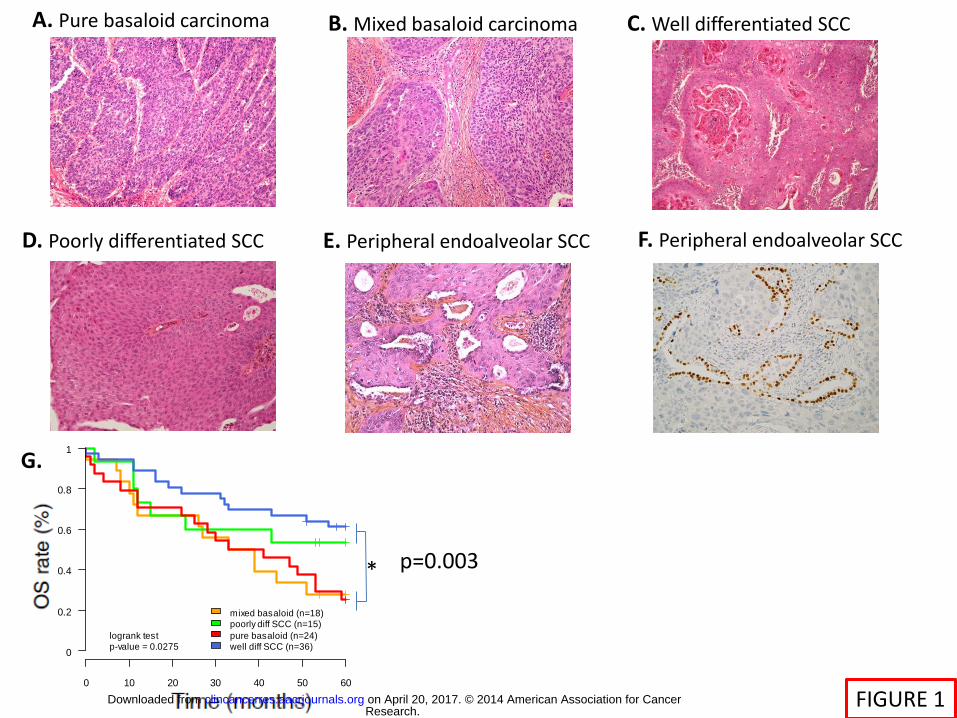
illustrated on Figure_1_A-F: pure basaloid carcinoma (BAS_p, n=24), mixed
basaloid SCC (BAS_m, n=18), non-basaloid well differentiated SCC (SCC_wd, n=36)
and non-basaloid poorly differentiated SCC (SCC_pd, n=15). All basaloid cases
expressed P40, P63 and Cytokeratins 1, 5, 10, 14 but did not express TTF-1 and
neuroendocrine markers [19].
Pure and mixed basaloid tumors showed the same poor prognosis in our
dataset (Figure_1_G), indicating that the presence of a basaloid cellular contingent is
of most interest for prognosis. Misclassified mixed basaloid samples could explain
the lack of prognostic significance of the basaloid feature in previous studies [20].
Pure basaloid SCC show a specific transcriptome profile, related to the deregulation of
specific pathways
Research. on April 20, 2017. © 2014 American Association for Cancerclincancerres.aacrjournals.org Downloaded from
Author manuscripts have been peer reviewed and accepted for publication but have not yet been edited. Author Manuscript Published OnlineFirst on September 4, 2014; DOI: 10.1158/1078-0432.CCR-14-0459

8
Expression profiles of 42 basaloid (24 BAS_p, 18 BAS_m), and 51 non-
basaloid SCC (36 SCC_wd, 15 SCC_pd) were obtained. A high proportion of genes
were found differentially expressed between BAS_p and non-basaloid SCC (H1
proportion=47 %, Sup_Table_2_A), indicating a specific mRNA profile for BAS_p.
Significantly up-regulated genes in BAS_p (limma q-value < 0.05) included genes
related to TP53 mutation signature (TMSB15A, PROM1), transcription factors (SOX4,
SOX9, SOX11, MYB, E2F3, E2F5), embryonic development (FGF3, FGF19),
methylation regulation (TET1, DNMT1, DNMT3A), cell cycle (MKI67, BUB1, DTL),
splicing (SFRS1,2,3) and survival (BCL2), while the most down-regulated genes were
related to epithelial cells and keratinocytes differentiation (KRT6, IVL, SPRR genes
and SFN). Additionally, the recently defined ectopic male germ cell /placenta specific
gene expression signature, as indicative of very aggressive lung tumours [21], also
highly correlated with pure basaloid in our cohort (chi2 p < 0.0005, Sup_Information).
An extensive pathways analysis also confirmed these findings (Sup_Table_3). The
comparison between pure basaloid and non-basaloid SCC – either well differentiated
or poorly differentiated (Figure_2), confirmed the up-regulation of pathways related to
cell cycle, transcription factors, mRNA splicing and chromatin modifications, and the
down-regulation of the squamous differentiation pathway. It also revealed the
upregulation of signatures associated to testis and embryonic stem (ES) cells and
poorly differentiated tumor markers (NANOG, OCT4, SOX2 and c-MYC targets) and
the down-regulation of signatures related to the Polycomb gene silencing system
(SUZ12, EED, H3K27ME3 and PRC2 gene sets) (Sup_Figure_1). Similarly mixed
basaloid were compared to non-basaloid SCC (H1 proportion=20 %) and as in pure
basaloid tumors, pathways related to cell cycle, spliceosome and ES cells were
upregulated (Sup_Figure_1).
Research. on April 20, 2017. © 2014 American Association for Cancerclincancerres.aacrjournals.org Downloaded from
Author manuscripts have been peer reviewed and accepted for publication but have not yet been edited. Author Manuscript Published OnlineFirst on September 4, 2014; DOI: 10.1158/1078-0432.CCR-14-0459
9
A 2-genes immunohistochemistry-based predictor discriminates pure and mixed
basaloid tumors from non-basaloid tumors
To identify molecular markers of basaloid tumors, we selected overexpressed
transcripts showing high AUC (> 75%) and specificity (> 95%), while optimizing
sensitivity (cutoff yielding a Fisher test p < 1e-6, Sup_Table_2_A). This selection
yielded 5 genes (SOX4, CBX5, PATZ1, RBMX, SFRS3), all showing a higher
sensitivity in pure basaloid than in mixed basaloid tumors. The transcription factor
SOX4 showed 100% specificity and 50% sensitivity to discriminate basaloid tumors in
our cohort; it was thus selected for further analyses.
To identify negative markers of basaloid tumors, we selected underexpressed
transcripts showing high AUC (>75%) and sensitivity (>90%), while optimizing
specificity (cutoff yielding a Fisher test p < 1e-5, Sup_Table_2_A); this selection
yielded 5 genes (IVL, KRT16, TOM1, C1ORF224, KCNK6). The squamous
differentiation marker IVL (involucrin), showing a high fold change (>2) between non-
basaloid and basaloid SCC, was selected for further analyses.
We measured by immunohistochemistry the protein expression of SOX4 and
IVL in a training set of 66 tumors common to the CIT transcriptome series (26
basaloid + 40 non-basaloid SCC). Using this training set we built a predictor based
on the quick scores (QS) related to these two genes, using the following formula: if
QS(SOX4)110 then Basaloid; if QS(SOX4) < 50 then Non-Basaloid; if QS(SOX4)
in [50;110[ and QS(SOX4) - QS(IVL) - 55 then Basaloid else Non-Basaloid (Figure
3, Sup_Table_2_B). In the training set, the predictor correctly classified all basaloid
tumors (pure and mixed) and correctly classified 90% of the SCC samples. The
predictor was then applied to an independent validation series of 35 tumors (15
Research. on April 20, 2017. © 2014 American Association for Cancerclincancerres.aacrjournals.org Downloaded from
Author manuscripts have been peer reviewed and accepted for publication but have not yet been edited. Author Manuscript Published OnlineFirst on September 4, 2014; DOI: 10.1158/1078-0432.CCR-14-0459
10
basaloid + 10 non-basaloid SCC + 10 adenocarcinomas), where it correctly classified
all samples except 2 mixed basaloid tumors (classified as non-basaloid). The
accuracy of this predictor was found to be 94% in the validation series
(sensitivity:87%, specificity:100%, positive predictive value:100%), in line with the
performances observed in the training series (accuracy:94%, sensitivity:100%,
specificity:90%, positive predictive value:87%).
SCC tumors mostly share a similar genomic aberration profile
DNA from 27 basaloid (14 Bas_p, 13 Bas_m) and 37 non-basaloid SCC (25
SCC_wd, 12 SCC_pd) was hybridized on Illumina HumanCNV370 SNP arrays. The
most frequent copy number aberrations (CNA) on the whole dataset were identified
with GISTIC2.0. This analysis identified previously reported CNA [7] such as gains
of 3q, 5p, 8q and losses of 1p, 3p, 4p, 4q, 5q, 8p, 9p. Similarly it pointed out known
target genes, including gains of SOX2, MYC, CCND1, MDM1, FGFR1 and losses of
CDKN2A, PCDH10, RB1, PTEN (Sup_Figure_2). Very few CNA were found in
significantly different proportions among histological classes (Sup_Table_4). At the
single gene level, gains of MYB, JUN, FGFR1, PIK3C3, DSC/DSG genes were found
more frequent in pure basaloid tumors. However none of these differences reached
significance after correction for multiple testing. From these data we were not able to
identify CNA being specific of basaloid SCC, either pure or mixed.
Consensus unsupervised clustering identifies a poor prognosis molecular subtype
corresponding to pure basaloid SCC
To assess whether basaloid tumors could correspond to a molecular subtype
obtained without a priori knowledge we performed unsupervised analyses of the
mRNA expression profiles. We identified consensus partitions in k= 2 to 8 clusters of
Research. on April 20, 2017. © 2014 American Association for Cancerclincancerres.aacrjournals.org Downloaded from
Author manuscripts have been peer reviewed and accepted for publication but have not yet been edited. Author Manuscript Published OnlineFirst on September 4, 2014; DOI: 10.1158/1078-0432.CCR-14-0459
11
the 93 SCC expression profiles from the CIT cohort. The consensus partition in k=4
clusters was selected (Figure_4_A) based on the gap statistic method [22]
(Sup_Figure_3_A). The underlying co-classification matrix showed a great level of
agreement between the raw partitions in k=4 clusters obtained using the different
experimental settings (Sup_Figure_3_B). Principal component analysis was
consistent with these findings (Figure_4_B). This partition was significantly
associated to histology (Fisher p= 1e-8). The 4 clusters were named Basaloid-Like
(BL) (n=21), Peripheral EndoAlveolar (PEA)(n=29), Classical_1 (n=21) and
Classical_2 (n=22).
The Basaloid-Like cluster contained almost only basaloid tumors (90%), and
mostly contained pure basaloid tumors (72%), contrary to other clusters (<15%). It
also showed enrichment in tumors with stage 2 or higher (62%) (Figure_4_A). Its
expression pattern was the most singular compared to other clusters
(Sup_Figure_3_C). Tumors from the PEA cluster mostly showed an alveolar
contingent in their microenvironment (83%), most were stage 1 (66%) without
basaloid features (79%). The PEA cluster was found enriched in non-basaloid poorly
differentiated SCC (Fisher p=3e-4), tumors with a high (>75%) diploid tumor cells rate
(Fisher p=1e-5), and TP53 wild type tumors (Fisher p=0.02). Non-basaloid well
differentiated SCC were mostly found in the Classical_1 and Classical_2 clusters
(67% and 45%) and absent in the Basaloid-Like cluster (0%). Tumors with exon 4
TP53 mutations were found enriched in the Classical_1 group (Fisher p=0.005).
Mixed basaloid tumors were not associated to any particular cluster.
To assign independent SCC datasets to these four molecular subtypes, we
built a 139-genes nearest-centroïd predictor (Sup_Table_5), and classified all SCC
samples (n=533) of 8 public expression profiling datasets (Sup_Table_6_A).
Research. on April 20, 2017. © 2014 American Association for Cancerclincancerres.aacrjournals.org Downloaded from
Author manuscripts have been peer reviewed and accepted for publication but have not yet been edited. Author Manuscript Published OnlineFirst on September 4, 2014; DOI: 10.1158/1078-0432.CCR-14-0459
12
Samples from each of the four subtypes were found in all datasets
(Sup_Table_6_A/B). Overall 58 samples (11%) were classified in the Basaloid-Like
subtype, 152 (28%) in the PEA subtype, 215 (41%) in the Classical_1 subtype and
108 (20%) in the Classical_2 subtype. A significantly poorer prognosis was observed
in the Basaloid-Like subtype compared to other molecular subtypes in the validation
datasets (Logrank p <6e-8, Figure_4_C) and in the discovery cohort (Sup_Figure_4).
Among the 3 other subtypes, Classical_2 and PEA subtypes presented similar
outcome and Classical_1 showed a significant better prognosis. These results were
conserved in stage 1 tumors (Figure_4_D).
Pathways found consistently deregulated in the Basaloid-Like cluster across
datasets (Figure_5, Sup_Table_7) were highly coherent with those identified in pure
basaloid tumors compared to non-basaloid SCC (Figure_2, Sup_Table_3). As
expected, normal bronchial basal cell signatures supposed to be the stem cell of
bronchi [23] were found highly and significantly overexpressed in the Basaloid-Like
subtype, whose expression profile was found clearly distinct from that of normal lung
(alveoli) thus confirming the proposed derivation of basaloid carcinoma from a basal
stem cell progenitor at the time of first description [9].
Finally we applied a SCC classification system, recently published by
Wilkerson et al. [6], to all SCC profiles from the discovery and validation cohorts
(n=626). Overall, the association between the CIT and Wilkerson classification
systems was found to be very high (all series, Fisher test p 0). Wilkerson subtypes
were also found very associated to histology (CIT series, Fisher test p =2e-9). To
match subtypes of both systems we then used pairwise Cohen’s Kappa statistics. A
fair and good agreement was respectively observed between Basaloid-Like and
Primitive subtypes (κ=0.56) and PEA and Secretory subtypes (κ=0.72). Other
Research. on April 20, 2017. © 2014 American Association for Cancerclincancerres.aacrjournals.org Downloaded from
Author manuscripts have been peer reviewed and accepted for publication but have not yet been edited. Author Manuscript Published OnlineFirst on September 4, 2014; DOI: 10.1158/1078-0432.CCR-14-0459

13
subtypes, related to non-basaloid well differentiated SCC, showed a poor agreement
(κ≤0.40). Despite the fair agreement between the Basaloid-Like (n=78/626, 12%) and
the Primitive (n=104/626, 17%) subtypes, the Basaloid-Like subtype was far more
associated to overall survival on the validation cohort (dataset adjusted HR=2.45,
95%CI=[1.72; 3.5], Wald p < 1e-6 ; C-index= 0.67, 95%CI = [0.52 ; 0.79]) than the
Primitive subtype (dataset adjusted HR=1.38; 95%CI=[1.00; 1.89]; Wald p =0.04; C-
index= 0.58, 95%CI = [0.45 ; 0.70]). Concerning subtypes related to non-basaloid
well differentiated SCC, contrary to that of Wilkerson, our classification reveals a
subtype (Classical_1, n=240/626 (38%)) showing a higher overall survival rate than
other subtypes in the validation cohort (dataset adjusted HR=0.61, 95%CI=[0.47;
0.79], Wald p = 2e-4; C-index=0.40, 95%CI = [0.30 ; 0.51]; Sup_Figure_5), even after
excluding the Basaloid-Like subtype (dataset adjusted HR=0.69; 95% CI=[0.53;
0.95]; Wald p = 7e-3).
In conclusion, pure basaloid tumors correspond to a specific molecular
subtype, either in our classification system or in that of Wilkerson. This finding further
demonstrates that histologically pure basaloid tumors constitute a specific molecular
entity.
Discussion
The worse prognosis of basaloid SCC as compared to non-basaloid SCC suggests
the existence of underlying biological differences. Incidentally, we show here that
Research. on April 20, 2017. © 2014 American Association for Cancerclincancerres.aacrjournals.org Downloaded from
Author manuscripts have been peer reviewed and accepted for publication but have not yet been edited. Author Manuscript Published OnlineFirst on September 4, 2014; DOI: 10.1158/1078-0432.CCR-14-0459
14
mixed basaloid SCC, representing 45% of the basaloid tumors in our series, share
the same poor prognosis as pure basaloid SCC, meaning that the presence of
basaloid contingents in SCC is informative for prognosis. To identify the molecular
characteristics of basaloid SCC we analyzed mRNA expression and DNA copy
number aberrations profiles of a large cohort of this rare entity (n=42) with tumor
controls (51 non-basaloid SCC). Mixed basaloid SCC are expected to be
heterogeneous at the molecular level, contrary to pure basaloid SCC. We thus mainly
concentrated on pure basaloid tumors to identify molecular specificities of this
histological group. Supervised analyses revealed that pure basaloid SCC display a
specific mRNA expression profile as compared to non-basaloid SCC. Enrichment in
TP53 mutation signature, upregulation of transcription, epigenetic, cell cycle, splicing
and survival factors, as well as that of male germ cells, embryonic development,
stemness/poor differentiation genes characterized pure basaloid SCC, which also
downregulated keratinocyte differentiation genes. Mixed basaloid SCC also showed
upregulation of proliferation and embryonic development related genes as compared
to non-basaloid SCC, but to a lesser extent than pure basaloid SCC, likely due to
their heterogeneous cellular content. These molecular observations are in line with
the poorly differentiated status and aggressiveness of the basaloid SCC.
DNA copy number aberrations (CNA) were found non-informative to distinguish
between SCC histological subgroups. All SCC, either basaloid or not, showed very
similar CNA profiles, pointing at regions previously reported [7], such as gain or
amplicon of the 3q region around SOX2, found in almost all SCC samples. However
CNA are potentially highly informative concerning targeted therapies [24]. In
particular, FGFR1 (8p12) and MYB (6q22-q23), found here in peak regions of gain,
are targetable: MYB could be targeted by several drugs under development [25-26],
Research. on April 20, 2017. © 2014 American Association for Cancerclincancerres.aacrjournals.org Downloaded from
Author manuscripts have been peer reviewed and accepted for publication but have not yet been edited. Author Manuscript Published OnlineFirst on September 4, 2014; DOI: 10.1158/1078-0432.CCR-14-0459
15
and FGFR1 amplified Non-Small Cell Lung Cancer have been shown to be sensitive
to FGFR1 inhibitors [27-28].
By unsupervised analyses of SCC mRNA expression profiles we identified four
molecular subtypes: Basaloid-Like, PeriEndoAlveolar (PEA), Classical_1 and
Classical_2. The Basaloid-Like cluster was shown highly associated to (pure)
basaloid histology. Similarly, among Wilkerson molecular subtypes (Primitive,
Secretory, Basal, Classical), the Primitive cluster was found associated to basaloid
histology, a key characteristic that was not reported by Wilkerson et al.. Altogether,
analysis of the molecular subtypes obtained either from our series or the literature
unambiguously shows that pure basaloid SCC do correspond to a specific molecular
subtype.
Our classification system, applied to a large validation cohort via a centroid-based
predictor, outperforms that of Wilkerson in predicting overall survival. The CIT
Basaloid-Like subtype, while agreeing with Wilkerson Primitive subtype, is far more
associated to a poor prognosis. A substantial proportion of the samples classified as
Primitive were predicted as non Basaloid-like (44%). Expression-based analyses
support that these samples could be more differentiated than Basaloid-like Primitive
samples (data not shown). Moreover, contrary to Wilkerson, among well differentiated
SCC we identify a subtype (Classical_1) showing a significantly better prognosis.
These clinical observations suggest that our subtypes are more homogeneous at the
molecular level than that of Wilkerson. Indeed, assuming that tumor molecular
characteristics may greatly determine prognosis, then defining more homogeneous
molecular subgroups may yield greater prognosis differences between subgroups. In
particular, it supports that the Basaloid-Like subtype is more specifically related to
Research. on April 20, 2017. © 2014 American Association for Cancerclincancerres.aacrjournals.org Downloaded from
Author manuscripts have been peer reviewed and accepted for publication but have not yet been edited. Author Manuscript Published OnlineFirst on September 4, 2014; DOI: 10.1158/1078-0432.CCR-14-0459
16
basaloid SCC than the Primitive subtype of Wilkerson, even if not significantly
different in the discovery cohort (NB: unavailable data in validation series).
Interestingly, most of the poorly differentiated non-basaloid SCC of our series (11/15)
were found in the PEA subtype, shown to be very similar to Wilkerson Secretory
subtype. Contrary to Wilkerson, suggesting secretory properties for this tumor
subtype, our study reveals that these tumors show a periendoalveoloar
microenvironment, which much more likely explains the “secreting” signatures of this
group. Moreover comparison of PEA profiles with that of bronchi and lung controls
unambiguously shows that the PEA subtype –contrary to all other subtypes- has
much more in common with lung profiles (which include alveoli) than with bronchi
profiles. From our data it seems excluded that PEA could be driven by non-tumor
cell related artifacts, as a tumor cells rate higher than 70% criterion was strictly
applied within our series, and given that the percentage of tumor cells was not found
different across subtypes. Interestingly this subtype was characterized by a smaller
fraction of tumors cells with genome instability and a smaller TP53 mutation rate.
Accordingly, pathway analysis revealed upregulation of TP53 targets signatures and
downregulation of cell cycle in this subtype (supplemental data). Upregulation of
immune signatures was also found very significantly associated to the PEA subtype.
Our study also completely renews the molecular subtyping of non-basaloid well
differentiated SCC. In the discovery cohort, the Classical_1 subtype is specifically
mutated in the exon 4 of TP53 (NB: unavailable information in validation series).
Pathway analysis across 5 series (supplemental data) revealed that TP53 target
genes are more expressed in Classical_1 than in Classical_2 and Basaloid-Like
subtypes, while having a similar TP53 mutation rate. These observations suggest
that mutations of the 4th exon of TP53 could be less deleterious than mutations of
Research. on April 20, 2017. © 2014 American Association for Cancerclincancerres.aacrjournals.org Downloaded from
Author manuscripts have been peer reviewed and accepted for publication but have not yet been edited. Author Manuscript Published OnlineFirst on September 4, 2014; DOI: 10.1158/1078-0432.CCR-14-0459
17
other exons observed within our series. Biological consequences of a more functional
p53 protein are also supported by pathway analysis as we found a relative higher
expression of pro-apoptotic genes and lower expression of cell cycle genes in
Classical_1 tumors as compared to Basaloid-Like and Classical_2 subgroups. Finally
a clinical support of these biological hypotheses is given by the better prognostic
observed in the Classical_1 subtype both in our cohort and in the validation series.
Mixed basaloid SCC were not found associated to any particular molecular subtype,
as expected due to their cellular heterogeneity. Accordingly, mRNA expression
profiles yielded very specific but moderately sensitive markers for the identification of
basaloid SCC as a whole, due to a lack of sensitivity concerning mixed basaloid
SCC. Using immunohistochemistry-based markers we could overcome this limitation:
we derived a 2-genes predictor based on a positive (SOX4) and a negative (IVL)
marker of the basaloid SCC, which showed a great accuracy (94%) in an
independent validation set (n=35). This simple predictor, used in addition to histologic
examination, should considerably help the identification of basaloid tumors in clinical
routine.
Of note, none of the present data on basaloid carcinoma (pure and mixed) is
applicable to large cell carcinoma (NOS) of the WHO 2004 classification nor to the
revised one (to be published in 2014), since a thorough revision of the concept of
large cell carcinoma based on immunohistochemical differentiation markers and on
the last genomic–based proposed classification of lung cancer restricted this class to
a few cases with no clear differentiation phenotype, which is not the case for basaloid
carcinoma.
In conclusion, our results establish that pure basaloid SCC correspond to a specific
molecular entity, fully justifying its histologic recognition and distinction from non-
Research. on April 20, 2017. © 2014 American Association for Cancerclincancerres.aacrjournals.org Downloaded from
Author manuscripts have been peer reviewed and accepted for publication but have not yet been edited. Author Manuscript Published OnlineFirst on September 4, 2014; DOI: 10.1158/1078-0432.CCR-14-0459
18
basaloid SCC. Related deregulated pathways enlighten its intrinsic resistance to
cytotoxic chemotherapy and should serve as a guide to targeted therapies.
References
1. Takeuchi T, Tomida S, Yatabe Y, Kosaka T, Osada H, Yanagisawa K et al. Expression profile-defined
classification of lung adenocarcinoma shows close relationship with underlying major genetic changes and
clinicopathologic behaviors. J Clin Oncol 2006; 24:1679–1688.
2. Ding L, Getz G, Wheeler DA, Mardis ER, McLellan MD, Cibulskis K et al. Somatic mutations affect key
pathways in lung adenocarcinoma. Nature 2008;455:1069–1075.
3. Shedden K, Taylor JMG, Enkemann SA, Tsao MS, Yeatman TJ, Gerald WL et al. Gene expression-based
survival prediction in lung adenocarcinoma: a multi-site, blinded validation study. Nat Med 2008;14:822–
827.
4. Weir BA, Woo MS, Getz G, Perner S, Ding L, Beroukhim R et al. Characterizing the cancer genome in lung
adenocarcinoma. Nature 2007;450:893–898.
5. Govindan R, Ding L, Griffith M, Subramanian J, Dees ND, Kanchi KL et al. Genomic landscape of non-small
cell lung cancer in smokers and never-smokers. Cell 2012;150:1121–1134.
6. Wilkerson MD, Yin X, Hoadley KA, Liu Y, Hayward MC, Cabanski CR et al. Lung squamous cell carcinoma
mRNA expression subtypes are reproducible, clinically important, and correspond to normal cell types. Clin
Cancer Res 2010;16:4864–4875.
7. Cancer Genome Atlas Research Network. Comprehensive genomic characterization of squamous cell lung
cancers. Nature 2012;489:519–525.
8. Peifer M, Fernández-Cuesta L, Sos ML, George J, Seidel D, Kasper LH et al. Integrative genome analyses
identify key somatic driver mutations of small-cell lung cancer. Nat Genet 2012;44:1104–1110.
9. Brambilla E, Moro D, Veale D, Brichon PY, Stoebner P, Paramelle B et al. Basal cell (basaloid) carcinoma of
the lung: a new morphologic and phenotypic entity with separate prognostic significance. Hum Pathol
1992;23:993–1003.
10. Moro D, Brichon PY, Brambilla E, Veale D, Labat F, Brambilla C. Basaloid bronchial carcinoma. A histologic
group with a poor prognosis. Cancer 1994;73:2734–2739.
11. Moro-Sibilot D, Lantuejoul S, Diab S, Moulai N, Aubert A, Timsit JF et al. Lung carcinomas with a basaloid
pattern: a study of 90 cases focusing on their poor prognosis. Eur Respir J 2008;31:854–859.
12. Bild AH, Yao G, Chang JT, Wang Q, Potti A, Chasse D et al. Oncogenic pathway signatures in human cancers
as a guide to targeted therapies. Nature 2006;439:353–357.
Research. on April 20, 2017. © 2014 American Association for Cancerclincancerres.aacrjournals.org Downloaded from
Author manuscripts have been peer reviewed and accepted for publication but have not yet been edited. Author Manuscript Published OnlineFirst on September 4, 2014; DOI: 10.1158/1078-0432.CCR-14-0459
19
13. Zhu CQ, Ding K, Strumpf D, Weir BA, Meyerson M, Pennell N et al. Prognostic and predictive gene signature
for adjuvant chemotherapy in resected non-small-cell lung cancer. J Clin Oncol 2010;28:4417–4424.
14. Potti A, Mukherjee S, Petersen R, Dressman HK, Bild A, Koontz J et al. A genomic strategy to refine prognosis
in early-stage non-small-cell lung cancer. N Engl J Med 2006;355:570–580.
15. Raponi M, Zhang Y, Yu J, Chen G, Lee G, Taylor JMG et al. Gene expression signatures for predicting
prognosis of squamous cell and adenocarcinomas of the lung. Cancer Res 2006;66:7466–7472.
16. Lee ES, Son DS, Kim SH, Lee J, Jo J, Han J et al. Prediction of recurrence-free survival in postoperative non-
small cell lung cancer patients by using an integrated model of clinical information and gene expression. Clin
Cancer Res 2008;14:7397–7404.
17. Roepman P, Jassem J, Smit EF, Muley T, Niklinski J, van de Velde T et al. An immune response enriched 72-
gene prognostic profile for early-stage non-small-cell lung cancer. Clin Cancer Res 2009;15:284–290.
18. Ma X, Rousseau V, Sun H, Lantuejoul S, Filipits M, Pirker R et al. Significance of TP53 mutations as predictive
markers of adjuvant cisplatin-based chemotherapy in completely resected non-small-cell lung cancer. Mol
Oncol 2014 (Epub).
19. Sturm N, Lantuéjoul S, Laverrière MH, Papotti M, Brichon PY, Brambilla C et al. Thyroid transcription factor 1
and cytokeratins 1, 5, 10, 14 (34betaE12) expression in basaloid and large-cell neuroendocrine carcinomas
of the lung. Hum Pathol 2001;32:918–925.
20. Kim DJ, Kim KD, Shin DH, Ro JY, Chung KY. Basaloid carcinoma of the lung: a really dismal histologic variant?
Ann Thorac Surg 2003;76:1833–1837.
21. Rousseaux S, Debernardi A, Jacquiau B, Vitte AL, Vesin A, Nagy-Mignotte H, et al. Ectopic activation of
germline and placental genes identifies aggressive metastasis-prone lung cancers. Sci Transl Med
2013;5:186ra66.
22. Tibshirani R, Walther G, Hastie T. Estimating the number of clusters in a data set via the gap statistic.
Journal of the Royal Statistical Society 2001;63:411–423.
23. Hackett NR, Shaykhiev R, Walters MS, Wang R, Zwick RK, Ferris B et al. The Human Airway Epithelial Basal
Cell Transcriptome. PLoS ONE 2011;6:e18378.
24. Perez-Moreno P, Brambilla E, Thomas R, Soria JC. Squamous cell carcinoma of the lung: molecular subtypes
and therapeutic opportunities. Clin Cancer Res 2012;18:2443–2451.
25. Bujnicki T, Wilczek C, Schomburg C, Feldmann F, Schlenke P, Müller-Tidow C et al. Inhibition of Myb-
dependent gene expression by the sesquiterpene lactone mexicanin-I. Leukemia 2012;26 :615–622.
26. Amaru Calzada A, Todoerti K, Donadoni L, Pellicioli A, Tuana G, Gatta R, et al. The HDAC inhibitor Givinostat
modulates the hematopoietic transcription factors NFE2 and C-MYB in JAK2(V617F) myeloproliferative
neoplasm cells. Exp Hematol 2012;40:634–645.e10.
27. Weiss J, Sos ML, Seidel D, Peifer M, Zander T, Heuckmann JM et al. Frequent and focal FGFR1 amplification
associates with therapeutically tractable FGFR1 dependency in squamous cell lung cancer. Sci Transl Med
2010;2:62ra93.
28. Dutt A, Ramos AH, Hammerman PS, Mermel C, Cho J, Sharifnia T, Chande A et al. Inhibitor-sensitive FGFR1
amplification in human non-small cell lung cancer. PLoS ONE 2011;6:e20351.
Research. on April 20, 2017. © 2014 American Association for Cancerclincancerres.aacrjournals.org Downloaded from
Author manuscripts have been peer reviewed and accepted for publication but have not yet been edited. Author Manuscript Published OnlineFirst on September 4, 2014; DOI: 10.1158/1078-0432.CCR-14-0459
20
Legends
FIGURE 1: Pathology review
(A) Basaloid carcinoma: tumor cells are small, monomorphic, cuboidal or fusiform, with a scant
cytoplasm and peripheral palisading. (B) Mixed basaloid carcinoma (SYN. Basaloid variant of
squamous cell carcinoma): Basaloid component on the right field, squamous cell component on the
left field. (C) Well differentiated Squamous cell Carcinoma: squamous differentiation and
keratinisation are frequent and obvious. (D) Poorly differentiated squamous cell carcinoma: most of
the tumor shows absence of definite differentiation with few intercellular bridges and keratinization.
(E-F) Peripheral endoalveolar squamous cell carcinoma: the SCC shows predominant intra-alveolar
topography entrapping numerous strands of Type 1 alveolar residual pneumocytes (E).
(G) Kaplan Meier curves of Overall Survival in the CIT cohort, stratified by subhistology. P-values refer
to logrank tests comparing overall survival between subhistologies.
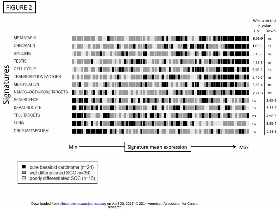
FIGURE 2: Deregulated pathways in pure basaloid tumors
Illustration of 13 signatures / pathways found among the most deregulated ones between pure
basaloid tumors (red) and non-basaloid tumors (poorly differentiated SCC (light gray); well
differentiated SCC (dark gray)) in our dataset. Samples (n=75) are independently ordered for each
signature, on the mean expression of the corresponding signature. References of the signatures and
pathways are indicated in Sup_Table_3_B. The Wilcoxon test p-values for up-regulated (left) and
down-regulated (right) mean expression in basaloid vs non-basaloid SCC, are shown for each
pathway.
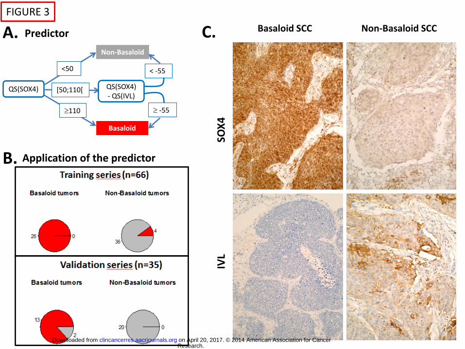
FIGURE 3: Immunohistochemistry-based predictor of the basaloid entity.
(A) To discriminate basaloid from non-basaloid tumors, the following predictor is used, based on the
quick score (QS) values of SOX4 and IVL: if QS(SOX4)110 then Basaloid; if QS(SOX4) < 50 then Non-
Basaloid; if QS(SOX4) in [50;110[ and QS(SOX4) - QS(IVL) - 55 then Basaloid else Non-Basaloid. (B)
Pie charts showing the results obtained (red: predicted basaloid; gray: predicted non-basaloid) when
applying this predictor to the tumors from the training series (upper part) and the validation series
(lower part), in basaloid (left) and non-basaloid (right) tumors. (C) Illustration of the
immunohistochemical measures of SOX4 (upper part) and IVL (lower part) in a basaloid (left part) and
a non-basaloid SCC (right part) tumor.
Research. on April 20, 2017. © 2014 American Association for Cancerclincancerres.aacrjournals.org Downloaded from
Author manuscripts have been peer reviewed and accepted for publication but have not yet been edited. Author Manuscript Published OnlineFirst on September 4, 2014; DOI: 10.1158/1078-0432.CCR-14-0459
21
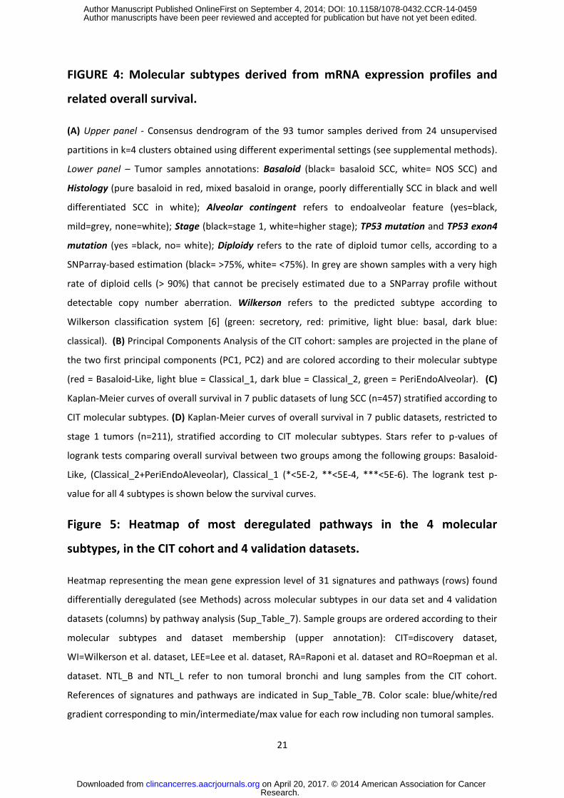
FIGURE 4: Molecular subtypes derived from mRNA expression profiles and
related overall survival.
(A) Upper panel - Consensus dendrogram of the 93 tumor samples derived from 24 unsupervised
partitions in k=4 clusters obtained using different experimental settings (see supplemental methods).
Lower panel – Tumor samples annotations: Basaloid (black= basaloid SCC, white= NOS SCC) and
Histology (pure basaloid in red, mixed basaloid in orange, poorly differentially SCC in black and well
differentiated SCC in white); Alveolar contingent refers to endoalveolar feature (yes=black,
mild=grey, none=white); Stage (black=stage 1, white=higher stage); TP53 mutation and TP53 exon4
mutation (yes =black, no= white); Diploidy refers to the rate of diploid tumor cells, according to a
SNParray-based estimation (black= >75%, white= <75%). In grey are shown samples with a very high
rate of diploid cells (> 90%) that cannot be precisely estimated due to a SNParray profile without
detectable copy number aberration. Wilkerson refers to the predicted subtype according to
Wilkerson classification system [6] (green: secretory, red: primitive, light blue: basal, dark blue:
classical). (B) Principal Components Analysis of the CIT cohort: samples are projected in the plane of
the two first principal components (PC1, PC2) and are colored according to their molecular subtype
(red = Basaloid-Like, light blue = Classical_1, dark blue = Classical_2, green = PeriEndoAlveolar). (C)
Kaplan-Meier curves of overall survival in 7 public datasets of lung SCC (n=457) stratified according to
CIT molecular subtypes. (D) Kaplan-Meier curves of overall survival in 7 public datasets, restricted to
stage 1 tumors (n=211), stratified according to CIT molecular subtypes. Stars refer to p-values of
logrank tests comparing overall survival between two groups among the following groups: Basaloid-
Like, (Classical_2+PeriEndoAleveolar), Classical_1 (*<5E-2, **<5E-4, ***<5E-6). The logrank test p-
value for all 4 subtypes is shown below the survival curves.
Figure 5: Heatmap of most deregulated pathways in the 4 molecular
subtypes, in the CIT cohort and 4 validation datasets.
Heatmap representing the mean gene expression level of 31 signatures and pathways (rows) found
differentially deregulated (see Methods) across molecular subtypes in our data set and 4 validation
datasets (columns) by pathway analysis (Sup_Table_7). Sample groups are ordered according to their
molecular subtypes and dataset membership (upper annotation): CIT=discovery dataset,
WI=Wilkerson et al. dataset, LEE=Lee et al. dataset, RA=Raponi et al. dataset and RO=Roepman et al.
dataset. NTL_B and NTL_L refer to non tumoral bronchi and lung samples from the CIT cohort.
References of signatures and pathways are indicated in Sup_Table_7B. Color scale: blue/white/red
gradient corresponding to min/intermediate/max value for each row including non tumoral samples.
Research. on April 20, 2017. © 2014 American Association for Cancerclincancerres.aacrjournals.org Downloaded from
Author manuscripts have been peer reviewed and accepted for publication but have not yet been edited. Author Manuscript Published OnlineFirst on September 4, 2014; DOI: 10.1158/1078-0432.CCR-14-0459
A. Pure basaloid carcinoma B. Mixed basaloid carcinoma C. Well differentiated SCC
D. Poorly differentiated SCC
FIGURE 1
E. Peripheral endoalveolar SCC F. Peripheral endoalveolar SCC
G.
times
fre
qu
en
cy
0 10 20 30 40 50 60
0
0.2
0.4
0.6
0.8
1
mixed basaloid (n=18)poorly diff SCC (n=15)
pure basaloid (n=24)well diff SCC (n=36)
logrank testp-value = 0.0275
* p=0.003
Research. on April 20, 2017. © 2014 American Association for Cancerclincancerres.aacrjournals.org Downloaded from
Author manuscripts have been peer reviewed and accepted for publication but have not yet been edited. Author Manuscript Published OnlineFirst on September 4, 2014; DOI: 10.1158/1078-0432.CCR-14-0459
Max Min Signature mean expression
FIGURE 2 Si
gnat
ure
s
Wilcoxon test p-value
Up Down
8.5E-9 ns
1.0E-6 ns
5.1E-6 ns
4.2E-5 ns
5.5E-5 ns
2.4E-6 ns
3.6E-4 ns
2.2E-3 ns
ns 2.6E-2
ns 3.5E-2
ns 4.9E-2
ns 3.9E-4
ns 2.3E-5
Research. on April 20, 2017. © 2014 American Association for Cancerclincancerres.aacrjournals.org Downloaded from
Author manuscripts have been peer reviewed and accepted for publication but have not yet been edited. Author Manuscript Published OnlineFirst on September 4, 2014; DOI: 10.1158/1078-0432.CCR-14-0459
Basaloid SCC Non-Basaloid SCC C.
IVL
S
OX
4
FIGURE 3
A.
QS(SOX4)
Basaloïd
Non-Basaloïd
QS(SOX4) - QS(IVL)
110
[50;110[
<50 < -55
-55
B.
Predictor
Application of the predictor
Research. on April 20, 2017. © 2014 American Association for Cancerclincancerres.aacrjournals.org Downloaded from
Author manuscripts have been peer reviewed and accepted for publication but have not yet been edited. Author Manuscript Published OnlineFirst on September 4, 2014; DOI: 10.1158/1078-0432.CCR-14-0459

FIGURE 4
B A
C D
Principal Component Analysis
Public datasets – all stages Public datasets – stage 1
-Like
Basaloid-Like (n=47) Classical_1 (n=194) Classical_2 (n=90) PeriEndoAlveo (n=126)
Basaloid-Like (n=19) Classical_1 (n=86) Classical_2 (n=41) PeriEndoAlveo (65)
Fisher test Basaloid p=5.2e-6 Histology p=1.1e-8 Alveolar contingent p=0.37 Stage p=0.18 TP53 mutation p=0.18 TP53 exon4 mutation p=0.033 Tobacco p=0.57 Diploidy p=1.9e-4 Wilkerson p=1.9e-20
PeriEndoAlveolar Basaloid-like Classical_1 Classical_2
Research. on April 20, 2017. © 2014 American Association for Cancerclincancerres.aacrjournals.org Downloaded from
Author manuscripts have been peer reviewed and accepted for publication but have not yet been edited. Author Manuscript Published OnlineFirst on September 4, 2014; DOI: 10.1158/1078-0432.CCR-14-0459
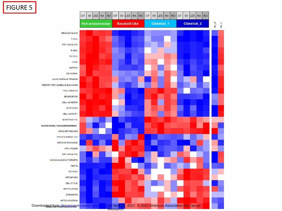
FIGURE 5
Basaloid-Like
GLYCOLYSYSIS / GLUCONEOGENESIS
OXIDATIVE PHOSPHORYLATION Research. on April 20, 2017. © 2014 American Association for Cancerclincancerres.aacrjournals.org Downloaded from
Author manuscripts have been peer reviewed and accepted for publication but have not yet been edited. Author Manuscript Published OnlineFirst on September 4, 2014; DOI: 10.1158/1078-0432.CCR-14-0459
Published OnlineFirst September 4, 2014.Clin Cancer Res Christian G Brambilla, Julien Laffaire, Sylvie Lantuejoul, et al. represent a specific molecular entityLung squamous cell carcinomas with basaloid histology
Updated version
10.1158/1078-0432.CCR-14-0459doi:
Access the most recent version of this article at:
Material
Supplementary
http://clincancerres.aacrjournals.org/content/suppl/2014/09/05/1078-0432.CCR-14-0459.DC1
Access the most recent supplemental material at:
Manuscript
Authorbeen edited. Author manuscripts have been peer reviewed and accepted for publication but have not yet
E-mail alerts related to this article or journal.Sign up to receive free email-alerts
Subscriptions
Reprints and
To order reprints of this article or to subscribe to the journal, contact the AACR Publications
Permissions
To request permission to re-use all or part of this article, contact the AACR Publications
Research. on April 20, 2017. © 2014 American Association for Cancerclincancerres.aacrjournals.org Downloaded from
Author manuscripts have been peer reviewed and accepted for publication but have not yet been edited. Author Manuscript Published OnlineFirst on September 4, 2014; DOI: 10.1158/1078-0432.CCR-14-0459